Synthesis, characterization, and electrochemical, spectroelectrochemical and electrical measurements...
11
Synthesis, characterization and electrochemical properties of functionalized graphene oxide Murugan Veerapandian a,b , Min-Ho Lee b, * , Karthikeyan Krishnamoorthy c , Kyusik Yun a, * a Department of Bionanotechnology, Gachon University, Gyeonggi-DO 461-701, Republic of Korea b Korea Electronics Technology Institute, Medical IT Technology, Gyeonggi-DO 463-816, Republic of Korea c Nanomaterials and System Lab, Department of Mechanical Engineering, Jeju National University, Jeju 690-756, Republic of Korea ARTICLE INFO Article history: Received 17 April 2012 Accepted 5 May 2012 Available online 14 May 2012 ABSTRACT Graphene oxide (GO) was functionalized by a simple reaction of its carboxylic acid groups with a silanized-metalloid polymer, which gave the resulting hybrid GO the property of efficient dispersion in a variety of solvents. Spectroscopic investigations show that the covalent attachment is effectively accomplished through an amidation process. The com- bination of a metalloid polymer and GO is unique and the composite material exhibits interesting features not seen in the individual structures. The electrochemical properties of this metalloid–polymer-GO were demonstrated by immobilizing the sample on a con- ventional gold-printed circuit board (Au-PCB) electrode. Functionalized GO showed a per- fect scaling of steady-state currents with correlation coefficients of 0.9600 (I pc ) and 0.9552 (I pa ), indicating the promise of this new GO hybrid as a transducer material for many sens- ing applications. Ó 2012 Elsevier Ltd. All rights reserved. 1. Introduction The design and fabrication of novel nanostructures with im- proved properties by using novel techniques is an alluring prospect for nanotechnology. Graphene and graphene oxide are novel nanomaterials that have recently attracted a great deal of consideration due to their wide variety of applications in nanoelectronics [1], sensors [2], nanocomposites [3], batter- ies [4], supercapacitors and energy storage [5]. In particular, the unique surface properties (oxygenated functional groups on the basal planes and edges), large surface area, layered structure, and easy exfoliation into monolayers under water mean that graphene oxide (GO) is a suitable building block for fabricating versatile functional materials via covalent or non-covalent approaches [6,7]. In recent years, there has been a rush of interest in functionalizing graphene oxide materials for medical and biomedical applications. For instance, polyethylene glycol (PEG)-functionalized GO nanosheets have been used to load anti-cancer drugs, doxorubicin hydrochlo- ride and camptothecin [8]. Studies have also revealed that PEGylated nanographene sheets (NGS) exhibit an ultrahigh in vivo uptake by tumors and a significant photothermal effect in mice [9]. In addition, researchers have also successfully functionalized a natural linear cationic polysaccharide chito- san (CS) on GO as a nanocarrier for drug and gene delivery. It has been demonstrated that due to p–p stacking and hydro- phobic interactions, GO–CS possesses a superior efficiency of binding with anti-cancer drugs [10]. Besides these studies, sev- eral biomolecules such as bovine serum albumin [11], adenine, cysteine, nicotamide and ovalbumin (OVA) have also been covalently (via diimide-activated amidation) attached to GO nanosheets for the development of several biocomposites [12]. To obtain enhanced mechanical, thermal and electro- chemical properties, organic conducting polymers and/or 0008-6223/$ - see front matter Ó 2012 Elsevier Ltd. All rights reserved. http://dx.doi.org/10.1016/j.carbon.2012.05.004 * Corresponding authors: Fax: +82 31 7508819. E-mail address: [email protected] (K. Yun). CARBON 50 (2012) 4228 – 4238 Available at www.sciencedirect.com journal homepage: www.elsevier.com/locate/carbon
-
Upload
independent -
Category
Documents
-
view
1 -
download
0
Transcript of Synthesis, characterization, and electrochemical, spectroelectrochemical and electrical measurements...
C A R B O N 5 0 ( 2 0 1 2 ) 4 2 2 8 – 4 2 3 8
.sc iencedi rect .com
Avai lab le at wwwjournal homepage: www.elsev ier .com/ locate /carbon
Synthesis, characterization and electrochemical propertiesof functionalized graphene oxide
Murugan Veerapandian a,b, Min-Ho Lee b,*, Karthikeyan Krishnamoorthy c,Kyusik Yun a,*
a Department of Bionanotechnology, Gachon University, Gyeonggi-DO 461-701, Republic of Koreab Korea Electronics Technology Institute, Medical IT Technology, Gyeonggi-DO 463-816, Republic of Koreac Nanomaterials and System Lab, Department of Mechanical Engineering, Jeju National University, Jeju 690-756, Republic of Korea
A R T I C L E I N F O A B S T R A C T
Article history:
Received 17 April 2012
Accepted 5 May 2012
Available online 14 May 2012
0008-6223/$ - see front matter � 2012 Elsevihttp://dx.doi.org/10.1016/j.carbon.2012.05.004
* Corresponding authors: Fax: +82 31 7508819E-mail address: [email protected] (K
Graphene oxide (GO) was functionalized by a simple reaction of its carboxylic acid groups
with a silanized-metalloid polymer, which gave the resulting hybrid GO the property of
efficient dispersion in a variety of solvents. Spectroscopic investigations show that the
covalent attachment is effectively accomplished through an amidation process. The com-
bination of a metalloid polymer and GO is unique and the composite material exhibits
interesting features not seen in the individual structures. The electrochemical properties
of this metalloid–polymer-GO were demonstrated by immobilizing the sample on a con-
ventional gold-printed circuit board (Au-PCB) electrode. Functionalized GO showed a per-
fect scaling of steady-state currents with correlation coefficients of 0.9600 (Ipc) and 0.9552
(Ipa), indicating the promise of this new GO hybrid as a transducer material for many sens-
ing applications.
� 2012 Elsevier Ltd. All rights reserved.
1. Introduction
The design and fabrication of novel nanostructures with im-
proved properties by using novel techniques is an alluring
prospect for nanotechnology. Graphene and graphene oxide
are novel nanomaterials that have recently attracted a great
deal of consideration due to their wide variety of applications
in nanoelectronics [1], sensors [2], nanocomposites [3], batter-
ies [4], supercapacitors and energy storage [5]. In particular,
the unique surface properties (oxygenated functional groups
on the basal planes and edges), large surface area, layered
structure, and easy exfoliation into monolayers under water
mean that graphene oxide (GO) is a suitable building block
for fabricating versatile functional materials via covalent or
non-covalent approaches [6,7]. In recent years, there has been
a rush of interest in functionalizing graphene oxide materials
for medical and biomedical applications. For instance,
er Ltd. All rights reserved
.. Yun).
polyethylene glycol (PEG)-functionalized GO nanosheets have
been used to load anti-cancer drugs, doxorubicin hydrochlo-
ride and camptothecin [8]. Studies have also revealed that
PEGylated nanographene sheets (NGS) exhibit an ultrahigh
in vivo uptake by tumors and a significant photothermal effect
in mice [9]. In addition, researchers have also successfully
functionalized a natural linear cationic polysaccharide chito-
san (CS) on GO as a nanocarrier for drug and gene delivery. It
has been demonstrated that due to p–p stacking and hydro-
phobic interactions, GO–CS possesses a superior efficiency of
binding with anti-cancer drugs [10]. Besides these studies, sev-
eral biomolecules such as bovine serum albumin [11], adenine,
cysteine, nicotamide and ovalbumin (OVA) have also been
covalently (via diimide-activated amidation) attached to GO
nanosheets for the development of several biocomposites [12].
To obtain enhanced mechanical, thermal and electro-
chemical properties, organic conducting polymers and/or
.
C A R B O N 5 0 ( 2 0 1 2 ) 4 2 2 8 – 4 2 3 8 4229
inorganic metal/semiconducting nanomaterials have been
functionalized on GO surfaces. Simple solution mixing,
ultrasonication, high-speed shearing, melt compounding,
in situ polymerization/electro polymerization and layer-
by-layer (LbL) assembly have been the commonly used
methods for the fabrication of graphene based hybrids
[13]. Such advanced functional materials based on GO are
applied in several interesting applications such as the
removal of pollutants [14], desalination [15], nanofluids
[16], and solid state chemistry [17], among which biosen-
sors whose functions are based on their electrochemical
properties are predominant. The ability to retain the native
structure of graphene oxide while enabling the bioactivity
of the functionalizing moiety through a surface-confined
process, as well as effective direct electron transfer reac-
tion properties, mean that GO is a suitable material for
the construction of electrochemical substrates. Recently,
many varieties of functional GO hybrids have been re-
ported, such as SnO2/rGO, PtRu/rGO, and ZnO–rGO sheets
[18,19] and polyaniline nanofibers-chemically converted
rGO [20] for electrochemical supercapacitors, GO-Prussian
blue hybrid film [21] and rGO/AuNPs-chitosan nanocom-
posite film [22] for glucose biosensing, and poly(vinyl
pyrrolidone)-GO for selective determination of ochratoxin
A [23]. However, studies on the functionalization of metal-
loid polymer hybrids on either graphene or graphene oxide
for biomedical or electrochemical biosensors have not been
reported until now [7,13,24].
Metalloid polymer hybrids (MPHs) are well known for
their applications as coatings, reinforcements, resistance
enhancers, and substrate additives in a variety of surface
and interface studies. Our group has constructed some heter-
ogeneous MPHs, such as PEG-POSS and PEG stabilized Ag@-
SiO2NPs blended with ABA triblock copolymer, which
exhibited potential applications in the construction of thin
films [25], solid laminates [26] and as an electrochemical sub-
strate for antigen–antibody interactions [27]. In this present
work, we have demonstrated the functionalization of Ag@-
SiO2-PEG (MPHs) on GO via silanization of the MPHs and their
subsequent covalent functionalization with GO. Three differ-
ent features such as metal silver core, non-metal silica shell
and PEG as polymer layer available in single nanoplatforms
with an average particle size distribution of 12.5 ± 2 nm have
attracted us to modify on GO. Upon modification, the charac-
teristic morphology, chemical structure elucidation and
optical properties were extensively studied to ensure
successful functionalization. Additionally, the primitive elec-
trochemical properties of the final MPHs-GO were studied to
extend its further application in electrochemical biosensors.
At different scan rates, MPHs-GO exhibits a more efficient
electrochemical response with a more significant redox sig-
nal in phosphate buffered saline than that of the commercial
Au-PCB electrode, revealing the viability of MPHs-GO playing
a role in electroanalysis and the construction of an electro-
chemical biosensor. Furthermore, the synergistic properties
of the final MPHs-GO comprised of a metalloid (Ag@SiO2NPs)
and a polymer (PEG) on novel GO nanosheets provide us with
a new class of hybrid materials for use in interdisciplinary
fields.
2. Experimental
2.1. Materials
Silver nitrate (AgNO3), tetraethoxysilane (TEOS) (Si(OC2H5)4),
sodium borohydride (NaBH4), ammonium hydroxide (NH4OH),
3-aminopropyltriethoxysilane (3-APTES), phosphate buffered
saline (PBS) and anhydrous ethanol were purchased from Sig-
ma. Poly(ethylene glycol) (Mn = 10,000 g/mol) (PEG) was ob-
tained from Aldrich. Expandable graphite powder was
purchased from Sigma–Aldrich, USA. Sulfuric acid (H2SO4),
potassium permanganate (KMNO4), hydrogen peroxide
(H2O2) and hydrochloric acid (HCl) were obtained from Dae-
jung Chemicals and Metal Ltd., South Korea. Milli-Q water
with a resistance greater than 18 MX was used in all our
experiments. All chemicals were of analytical grade and were
used as received without any further purification.
2.2. Instrumentation
Structural characterizations were carried out using a conven-
tional field-emission scanning electron microscope (FE-SEM,
JEOL JSM-7500F), a high resolution transmission electron
microscope (HR-TEM, FEI Titan 80-300), and a bioatomic force
microscope (AFM: Nanowizard II, JPK Instruments) operating
in the intermittent air mode. The samples used for the mea-
surements were prepared by casting 5–10 lL of GO (0.1 mg/
mL) or MPHs-GO (0.2 mg/mL) suspension onto the surface of
a silica substrate (1 · 1 cm2) for FE-SEM, a copper grid for
HR-TEM, or a freshly cleaved mica sheet for AFM. The solvent
was allowed to evaporate before each measurement. Ultravi-
olet–visible absorbance spectra were measured using a Varian
Cary 50 UV–vis spectrophotometer. Chemical structure and
functional group modifications were examined by using a
Fourier transform-infrared spectrophotometer (FT-IR, NICO-
LET 6700) using a KBr disk at a resolution of 4 cm�1 and a
600 MHz high resolution nuclear magnetic resonance spec-
trometer (1H-NMR, AVANCE 600, Bruker) (D2O; solvent). To
complement the FT-IR and 1H-NMR data, a Raman spectral
study (T64000, HORIABA Jobin Yvon equipped with an Argon
laser source for 514 nm excitation) was performed on neat
GO as well as the MPHs-GO. Both the GO and MPHs-GO disper-
sions were drop casted onto the cleaned silica wafer
(2 · 2 cm2) and the solvent was allowed to evaporate at ambi-
ent temperature before measurements were carried out. A
Cary Eclipse fluorescence spectrophotometer was used to
examine the photo-luminescence properties of MPHs and
MPHs-GO. The fundamental electrochemical properties of
neat GO and MPHs-GO were confirmed by cyclic voltammetry
(CV) measurements using a VersaSTAT 3 in a three-electrode
configuration containing Au-PCB as the working electrode,
and a Pt wire and an Ag/AgCl electrode as the counter and ref-
erence electrodes, respectively.
2.3. Fabrication of Ag@SiO2-PEG (MPHs)
Ag@SiO2-PEG hybrids (average size distribution of 12.5 ± 2 nm)
were prepared by a sonochemical approach, following the
procedure outlined in our previous report [26]. Briefly, an
4230 C A R B O N 5 0 ( 2 0 1 2 ) 4 2 2 8 – 4 2 3 8
aqueous solution of silver precursor (AgNO3) was first added
to a reaction vessel containing the stabilizing agent PEG (Mn
= 10,000 g/mol) and the reductant NaBH4. The reaction mix-
ture was ultrasonicated (with controlled parameters) for a
period of 15 min to form the Ag core. Then the desired
amounts of aqueous TEOS and NH4OH were simultaneously
added. The reaction vessel containing the metal, non-metal
precursor, PEG and reducing agents again underwent ultra-
sonication for a period of 30 min to ensure complete reduc-
tion and growth of the hybrid structure. The resulting
colloidal solution containing hybrid particles (Ag@SiO2-PEG:
MPHs) was separated by centrifugation. The separated parti-
cles were washed twice and then utilized for further
experimentation.
2.4. Functionalization of MPHs on GO
The brownish colloidal suspensions of graphene oxide nano-
sheets (GO) utilized in the current experiment were synthe-
sized according to the modified Hummer’s method [28,29].
As-prepared Ag@SiO2-PEG hybrids were first silanized using
3-APTES, then utilized for covalent reaction with GO. Briefly,
an accurate amount of Ag@SiO2-PEG hybrids (200 lL) and
40 lL of 3-APTES (3% in C2H5OH) were added to a vial contain-
ing 3 mL of anhydrous ethanol and kept under magnetic stir-
ring (800 rpm) at room temperature for 10 h. Later, an
aqueous solution of GO (200 lL) was added and allowed to stir
(at 800 rpm) for another 10 h to ensure the covalent reaction
between silanized MPHs and GO. After the reaction process,
the Ag@SiO2-PEG functionalized GO sheets were separated
by centrifugation, washed thrice with ethanol and utilized
for further characterization.
2.5. Fabrication of Au-PCB/MPHs-GO electrode
The gold-PCB working electrode (Au-PCB; area �1 mm in
diameter) was surface-cleaned by sequential washing with
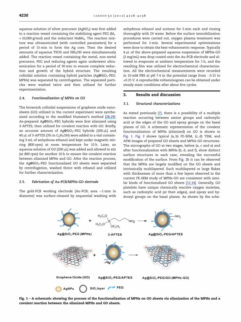
Fig. 1 – A schematic showing the process of the functionalizatio
covalent reaction between the silanized-MPHs and GO sheets.
anhydrous ethanol and acetone for 5 min each and rinsing
thoroughly with DI water. Before the surface immobilization
procedures were carried out, oxygen plasma treatment was
performed for 2 min. Several experimental optimizations
were done to obtain the best voltammetric response. Typically
4 lL of the above-prepared aqueous suspension of MPHs-GO
(2 mg/mL) was drop coated onto the Au-PCB electrode and al-
lowed to evaporate at ambient temperature for 1 h, and the
resulting film was utilized for electrochemical characteriza-
tion. All the electrochemical measurements were recorded
in 10 mM PBS at pH 7.4 in the potential range from �0.15 to
+0.25 V. A reproducible voltammogram can be obtained under
steady-state conditions after about five cycles.
3. Results and discussion
3.1. Structural characterizations
As stated previously [7], there is a possibility of a multiple
reaction occurring between amine groups and carboxylic
acid at the edges of the GO and epoxy groups on the basal
planes of GO. A schematic representation of the covalent
functionalization of MPHs (silanized) on GO is shown in
Fig. 1. Fig. 2 shows typical (a, b) FE-SEM, (c, d) TEM, and
AFM images of prepared GO sheets and MPHs-GO structures.
The micrographs of GO at two stages, before (a, c and e) and
after functionalization with MPHs (b, d, and f), show distinct
surface structures in each case, revealing the successful
modification of the surface. From Fig. 2b it can be observed
that the MPHs are largely modified on the GO sheets and
intrinsically multilayered. Such multilayered or large flakes
with thicknesses of more than a few layers observed in the
current FE-SEM study of MPHs-GO are consistent with simi-
lar kinds of functionalized GO sheets [12,14]. Generally, GO
platelets have unique chemically reactive oxygen moieties,
such as carboxylic acid (at their edges), and epoxy and hy-
droxyl groups on the basal planes. As shown by the sche-
n of MPHs on GO sheets via silanization of the MPHs and a
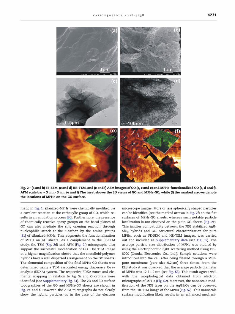
Fig. 2 – (a and b) FE-SEM, (c and d) HR-TEM, and (e and f) AFM images of GO (a, c and e) and MPHs-functionalized GO (b, d and f).
AFM scale bar = 3 lm · 3 lm. (e and f) The inset shows the 3D views of GO and MPHs-GO, while (f) the marked arrows denote
the locations of MPHs on the GO surface.
C A R B O N 5 0 ( 2 0 1 2 ) 4 2 2 8 – 4 2 3 8 4231
matic in Fig. 1, silanized-MPHs were chemically modified via
a covalent reaction at the carboxylic group of GO, which re-
sults in an amidation process [30]. Furthermore, the presence
of chemically reactive epoxy groups on the basal planes of
GO can also mediate the ring opening reaction through
nucleophilic attack at the a-carbon by the amine groups
[31] of silanized-MPHs. This augments the functionalization
of MPHs on GO sheets. As a complement to the FE-SEM
study, the TEM (Fig. 2d) and AFM (Fig. 2f) micrographs also
support the successful modification of GO. The TEM image
at a higher magnification shows that the metalloid-polymer
hybrids have a well dispersed arrangement on the GO sheets.
The elemental composition of the final MPHs-GO sheets was
determined using a TEM associated energy dispersive X-ray
analysis (EDXA) system. The respective EDXA zones and ele-
mental mapping in relation to Ag, Si and O orbitals were
identified (see Supplementary Fig. S1). The 2D and 3D surface
topographies of the GO and MPHs-GO sheets are shown in
Fig. 2e and f. However, the AFM micrographs do not clearly
show the hybrid particles as in the case of the electron
microscope images. More or less spherically shaped particles
can be identified (see the marked arrows in Fig. 2f) on the flat
surfaces of MPHs-GO sheets, whereas such notable particle
localization is not observed on the plain GO sheets (Fig. 2e).
This implies compatibility between the PEG stabilized Ag@-
SiO2 hybrids and GO. Structural characterization for pure
MPHs, such as FE-SEM and HR-TEM images, was carried
out and included as Supplementary data (see Fig. S2). The
average particle size distribution of MPHs was studied by
using the electrophoretic light scattering method using ELS-
8000 (Otsuka Electronics Co., Ltd.). Sample solutions were
introduced into the cell after being filtered through a Milli-
pore membrane (pore size 0.2 lm) three times. From the
ELS study it was observed that the average particle diameter
of MPHs was 12.5 ± 2 nm (see Fig. S3). This result agrees well
with the morphological data obtained from electron
micrographs of MPHs (Fig. S2). Moreover, the nanoscale mod-
ification of the PEG layer on the Ag@SiO2 can be observed
from the HR-TEM image of the MPHs (Fig. S2). This nanoscale
surface modification likely results in an enhanced mechani-
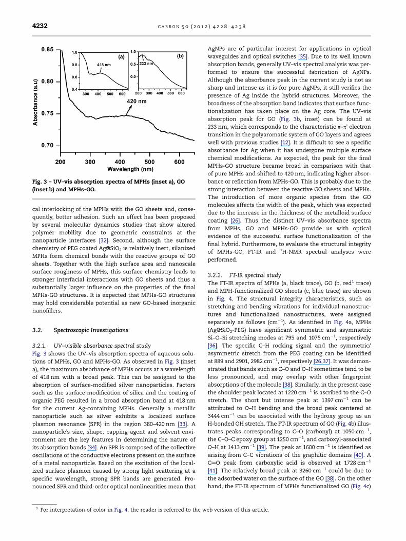
Fig. 3 – UV–vis absorption spectra of MPHs (inset a), GO
(inset b) and MPHs-GO.
4232 C A R B O N 5 0 ( 2 0 1 2 ) 4 2 2 8 – 4 2 3 8
cal interlocking of the MPHs with the GO sheets and, conse-
quently, better adhesion. Such an effect has been proposed
by several molecular dynamics studies that show altered
polymer mobility due to geometric constraints at the
nanoparticle interfaces [32]. Second, although the surface
chemistry of PEG coated Ag@SiO2 is relatively inert, silanized
MPHs form chemical bonds with the reactive groups of GO
sheets. Together with the high surface area and nanoscale
surface roughness of MPHs, this surface chemistry leads to
stronger interfacial interactions with GO sheets and thus a
substantially larger influence on the properties of the final
MPHs-GO structures. It is expected that MPHs-GO structures
may hold considerable potential as new GO-based inorganic
nanofillers.
3.2. Spectroscopic Investigations
3.2.1. UV–visible absorbance spectral studyFig. 3 shows the UV–vis absorption spectra of aqueous solu-
tions of MPHs, GO and MPHs-GO. As observed in Fig. 3 (inset
a), the maximum absorbance of MPHs occurs at a wavelength
of 418 nm with a broad peak. This can be assigned to the
absorption of surface-modified silver nanoparticles. Factors
such as the surface modification of silica and the coating of
organic PEG resulted in a broad absorption band at 418 nm
for the current Ag-containing MPHs. Generally a metallic
nanoparticle such as silver exhibits a localized surface
plasmon resonance (SPR) in the region 380–420 nm [33]. A
nanoparticle’s size, shape, capping agent and solvent envi-
ronment are the key features in determining the nature of
its absorption bands [34]. An SPR is composed of the collective
oscillations of the conductive electrons present on the surface
of a metal nanoparticle. Based on the excitation of the local-
ized surface plasmon caused by strong light scattering at a
specific wavelength, strong SPR bands are generated. Pro-
nounced SPR and third-order optical nonlinearities mean that
1 For interpretation of color in Fig. 4, the reader is referred to the w
AgNPs are of particular interest for applications in optical
waveguides and optical switches [35]. Due to its well known
absorption bands, generally UV–vis spectral analysis was per-
formed to ensure the successful fabrication of AgNPs.
Although the absorbance peak in the current study is not as
sharp and intense as it is for pure AgNPs, it still verifies the
presence of Ag inside the hybrid structures. Moreover, the
broadness of the absorption band indicates that surface func-
tionalization has taken place on the Ag core. The UV–vis
absorption peak for GO (Fig. 3b, inset) can be found at
233 nm, which corresponds to the characteristic p–p* electron
transition in the polyaromatic system of GO layers and agrees
well with previous studies [12]. It is difficult to see a specific
absorbance for Ag when it has undergone multiple surface
chemical modifications. As expected, the peak for the final
MPHs-GO structure became broad in comparison with that
of pure MPHs and shifted to 420 nm, indicating higher absor-
bance or reflection from MPHs-GO. This is probably due to the
strong interaction between the reactive GO sheets and MPHs.
The introduction of more organic species from the GO
molecules affects the width of the peak, which was expected
due to the increase in the thickness of the metalloid surface
coating [26]. Thus the distinct UV–vis absorbance spectra
from MPHs, GO and MPHs-GO provide us with optical
evidence of the successful surface functionalization of the
final hybrid. Furthermore, to evaluate the structural integrity
of MPHs-GO, FT-IR and 1H-NMR spectral analyses were
performed.
3.2.2. FT-IR spectral studyThe FT-IR spectra of MPHs (a, black trace), GO (b, red1 trace)
and MPH-functionalized GO sheets (c, blue trace) are shown
in Fig. 4. The structural integrity characteristics, such as
stretching and bending vibrations for individual nanostruc-
tures and functionalized nanostructures, were assigned
separately as follows (cm�1). As identified in Fig. 4a, MPHs
(Ag@SiO2-PEG) have significant symmetric and asymmetric
Si–O–Si stretching modes at 795 and 1075 cm�1, respectively
[36]. The specific C–H rocking signal and the symmetric/
asymmetric stretch from the PEG coating can be identified
at 889 and 2901, 2982 cm�1, respectively [26,37]. It was demon-
strated that bands such as C–O and O–H sometimes tend to be
less pronounced, and may overlap with other fingerprint
absorptions of the molecule [38]. Similarly, in the present case
the shoulder peak located at 1220 cm�1 is ascribed to the C–O
stretch. The short but intense peak at 1397 cm�1 can be
attributed to O–H bending and the broad peak centered at
3444 cm�1 can be associated with the hydroxy group as an
H-bonded OH stretch. The FT-IR spectrum of GO (Fig. 4b) illus-
trates peaks corresponding to C–O (carbonyl) at 1050 cm�1,
the C–O–C epoxy group at 1250 cm�1, and carboxyl-associated
O–H at 1413 cm�1 [39]. The peak at 1600 cm�1 is identified as
arising from C–C vibrations of the graphitic domains [40]. A
C@O peak from carboxylic acid is observed at 1728 cm�1
[41]. The relatively broad peak at 3260 cm�1 could be due to
the adsorbed water on the surface of the GO [38]. On the other
hand, the FT-IR spectrum of MPHs functionalized GO (Fig. 4c)
eb version of this article.
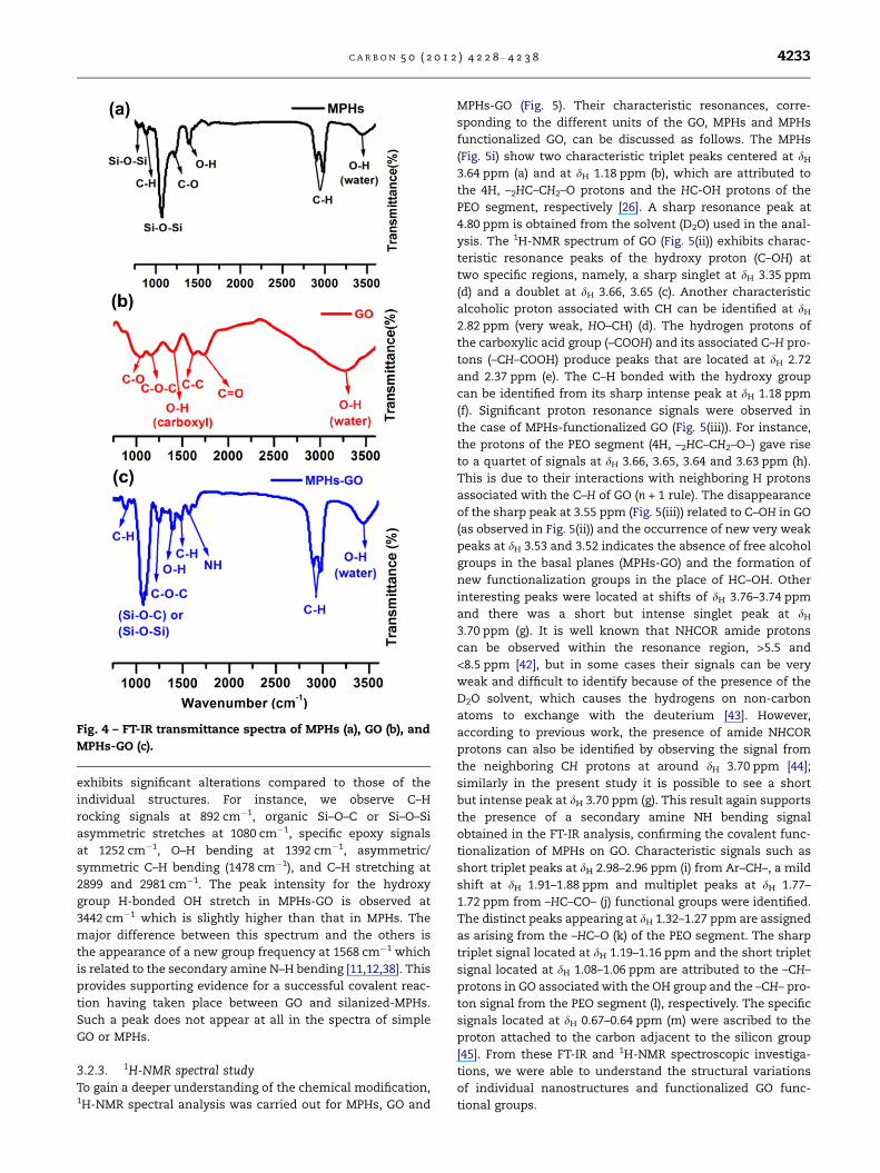
Fig. 4 – FT-IR transmittance spectra of MPHs (a), GO (b), and
MPHs-GO (c).
C A R B O N 5 0 ( 2 0 1 2 ) 4 2 2 8 – 4 2 3 8 4233
exhibits significant alterations compared to those of the
individual structures. For instance, we observe C–H
rocking signals at 892 cm�1, organic Si–O–C or Si–O–Si
asymmetric stretches at 1080 cm�1, specific epoxy signals
at 1252 cm�1, O–H bending at 1392 cm�1, asymmetric/
symmetric C–H bending (1478 cm�1), and C–H stretching at
2899 and 2981 cm�1. The peak intensity for the hydroxy
group H-bonded OH stretch in MPHs-GO is observed at
3442 cm�1 which is slightly higher than that in MPHs. The
major difference between this spectrum and the others is
the appearance of a new group frequency at 1568 cm�1 which
is related to the secondary amine N–H bending [11,12,38]. This
provides supporting evidence for a successful covalent reac-
tion having taken place between GO and silanized-MPHs.
Such a peak does not appear at all in the spectra of simple
GO or MPHs.
3.2.3. 1H-NMR spectral studyTo gain a deeper understanding of the chemical modification,1H-NMR spectral analysis was carried out for MPHs, GO and
MPHs-GO (Fig. 5). Their characteristic resonances, corre-
sponding to the different units of the GO, MPHs and MPHs
functionalized GO, can be discussed as follows. The MPHs
(Fig. 5i) show two characteristic triplet peaks centered at dH
3.64 ppm (a) and at dH 1.18 ppm (b), which are attributed to
the 4H, –2HC–CH2–O protons and the HC-OH protons of the
PEO segment, respectively [26]. A sharp resonance peak at
4.80 ppm is obtained from the solvent (D2O) used in the anal-
ysis. The 1H-NMR spectrum of GO (Fig. 5(ii)) exhibits charac-
teristic resonance peaks of the hydroxy proton (C–OH) at
two specific regions, namely, a sharp singlet at dH 3.35 ppm
(d) and a doublet at dH 3.66, 3.65 (c). Another characteristic
alcoholic proton associated with CH can be identified at dH
2.82 ppm (very weak, HO–CH) (d). The hydrogen protons of
the carboxylic acid group (–COOH) and its associated C–H pro-
tons (–CH–COOH) produce peaks that are located at dH 2.72
and 2.37 ppm (e). The C–H bonded with the hydroxy group
can be identified from its sharp intense peak at dH 1.18 ppm
(f). Significant proton resonance signals were observed in
the case of MPHs-functionalized GO (Fig. 5(iii)). For instance,
the protons of the PEO segment (4H, –2HC–CH2–O–) gave rise
to a quartet of signals at dH 3.66, 3.65, 3.64 and 3.63 ppm (h).
This is due to their interactions with neighboring H protons
associated with the C–H of GO (n + 1 rule). The disappearance
of the sharp peak at 3.55 ppm (Fig. 5(iii)) related to C–OH in GO
(as observed in Fig. 5(ii)) and the occurrence of new very weak
peaks at dH 3.53 and 3.52 indicates the absence of free alcohol
groups in the basal planes (MPHs-GO) and the formation of
new functionalization groups in the place of HC–OH. Other
interesting peaks were located at shifts of dH 3.76–3.74 ppm
and there was a short but intense singlet peak at dH
3.70 ppm (g). It is well known that NHCOR amide protons
can be observed within the resonance region, >5.5 and
<8.5 ppm [42], but in some cases their signals can be very
weak and difficult to identify because of the presence of the
D2O solvent, which causes the hydrogens on non-carbon
atoms to exchange with the deuterium [43]. However,
according to previous work, the presence of amide NHCOR
protons can also be identified by observing the signal from
the neighboring CH protons at around dH 3.70 ppm [44];
similarly in the present study it is possible to see a short
but intense peak at dH 3.70 ppm (g). This result again supports
the presence of a secondary amine NH bending signal
obtained in the FT-IR analysis, confirming the covalent func-
tionalization of MPHs on GO. Characteristic signals such as
short triplet peaks at dH 2.98–2.96 ppm (i) from Ar–CH–, a mild
shift at dH 1.91–1.88 ppm and multiplet peaks at dH 1.77–
1.72 ppm from –HC–CO– (j) functional groups were identified.
The distinct peaks appearing at dH 1.32–1.27 ppm are assigned
as arising from the –HC–O (k) of the PEO segment. The sharp
triplet signal located at dH 1.19–1.16 ppm and the short triplet
signal located at dH 1.08–1.06 ppm are attributed to the –CH–
protons in GO associated with the OH group and the –CH– pro-
ton signal from the PEO segment (l), respectively. The specific
signals located at dH 0.67–0.64 ppm (m) were ascribed to the
proton attached to the carbon adjacent to the silicon group
[45]. From these FT-IR and 1H-NMR spectroscopic investiga-
tions, we were able to understand the structural variations
of individual nanostructures and functionalized GO func-
tional groups.
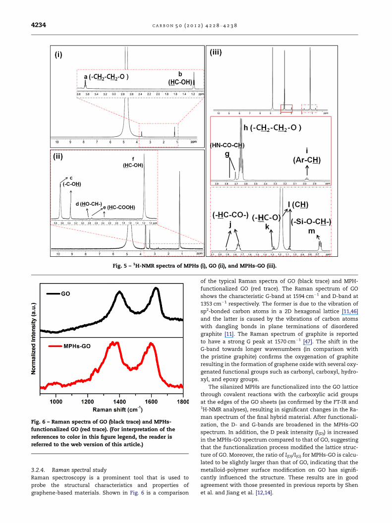
Fig. 6 – Raman spectra of GO (black trace) and MPHs-
functionalized GO (red trace). (For interpretation of the
references to color in this figure legend, the reader is
referred to the web version of this article.)
Fig. 5 – 1H-NMR spectra of MPHs (i), GO (ii), and MPHs-GO (iii).
4234 C A R B O N 5 0 ( 2 0 1 2 ) 4 2 2 8 – 4 2 3 8
3.2.4. Raman spectral studyRaman spectroscopy is a prominent tool that is used to
probe the structural characteristics and properties of
graphene-based materials. Shown in Fig. 6 is a comparison
of the typical Raman spectra of GO (black trace) and MPH-
functionalized GO (red trace). The Raman spectrum of GO
shows the characteristic G-band at 1594 cm�1 and D-band at
1353 cm�1 respectively. The former is due to the vibration of
sp2-bonded carbon atoms in a 2D hexagonal lattice [11,46]
and the latter is caused by the vibrations of carbon atoms
with dangling bonds in plane terminations of disordered
graphite [11]. The Raman spectrum of graphite is reported
to have a strong G peak at 1570 cm�1 [47]. The shift in the
G-band towards longer wavenumbers (in comparison with
the pristine graphite) confirms the oxygenation of graphite
resulting in the formation of graphene oxide with several oxy-
genated functional groups such as carbonyl, carboxyl, hydro-
xyl, and epoxy groups.
The silanized MPHs are functionalized into the GO lattice
through covalent reactions with the carboxylic acid groups
at the edges of the GO sheets (as confirmed by the FT-IR and1H-NMR analyses), resulting in significant changes in the Ra-
man spectrum of the final hybrid material. After functionali-
zation, the D- and G-bands are broadened in the MPHs-GO
spectrum. In addition, the D peak intensity (I(D)) is increased
in the MPHs-GO spectrum compared to that of GO, suggesting
that the functionalization process modified the lattice struc-
ture of GO. Moreover, the ratio of I(D)/I(G) for MPHs-GO is calcu-
lated to be slightly larger than that of GO, indicating that the
metalloid-polymer surface modification on GO has signifi-
cantly influenced the structure. These results are in good
agreement with those presented in previous reports by Shen
et al. and Jiang et al. [12,14].
C A R B O N 5 0 ( 2 0 1 2 ) 4 2 2 8 – 4 2 3 8 4235
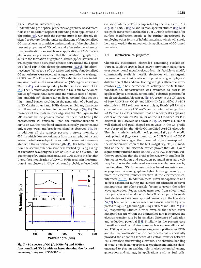
3.2.5. Photoluminescence studyUnderstanding the optical properties of graphene based mate-
rials is an important aspect of extending their applications in
photonics [48]. Although the current study is not directly de-
signed to feature the photonics applications of functionalized
GO nanosheets, a primitive understanding of the photolumi-
nescent properties of GO before and after selective chemical
functionalization can enable new applications of 2-D materi-
als. Previous reports revealed that the oxidation of graphite re-
sults in the formation of graphitic islands (sp2 clusters) in GO,
which generates a disruption of the p-network and thus opens
up a band gap in the electronic structure [49,50]. Photolumi-
nescence (PL) spectra of GO, MPHs and MPHs-functionalized
GO nanosheets were recorded using an excitation wavelength
of 325 nm. The PL spectrum of GO exhibits a characteristic
emission peak in the near ultraviolet (UV) region at around
366 nm (Fig. 7a) corresponding to the band emission of GO
[28]. The UV emission peak observed in GO is due to the amor-
phous sp3 matrix that surrounds the various sizes of crystal-
line graphitic sp2 clusters (unoxidized regions) that act as a
high tunnel barrier resulting in the generation of a band gap
in GO. On the other hand, MPHs do not exhibit any character-
istic PL emission spectrum in the near UV region (Fig. 7b). The
presence of the metallic core (Ag) and the PEG layer in the
MPHs could be the possible reason for them not having the
characteristic PL emission. Upon the functionalization of
MPHs on GO, the near band emission is nearly quenched and
only a very weak and broadened signal is observed (Fig. 7c).
In addition, all the samples possess a strong intensity at
650 nm which does not originate from the sample, but instead
arises due to the overlap of the second-order emissions associ-
ated with the excitation wavelength [46]. For better clarifica-
tion, the second-order emission was verified by using a range
of excitation wavelengths, such as 325, 400, and 500 nm. The
quenching of PL emission from MPHs-GO is due to the fact that
the surface modification of GO with MPHs results in the forma-
tion of new clusters in GO, which could probably reduce the PL
Fig. 7 – PL spectra of GO (a), MPHs (b) and MPHs-
functionalized GO (c) with an inset showing the focused
wavelength region of 350–380 nm.
emission intensity. This is supported by the results of FT-IR
(Fig. 4), 1H-NMR (Fig. 5) and Raman spectral studies (Fig. 6). It
is significant to mention that the PL of GO both before and after
surface modification needs to be further investigated by
employing other forms of hybrid materials, which will surely
help us to exploit the nanophotonic applications of GO-based
materials.
3.3. Electrochemical properties
Chemically customized electrodes containing surface-en-
trapped catalytic species have shown prominent advantages
over conventional metallic electrodes. It is feasible to modify
commercially available metallic electrodes with an organic
polymer or an inert surface to provide a good physical
distribution of the additive, leading to highly efficient electro-
catalysis [51]. The electrochemical activity of the MPH-func-
tionalized GO nanostructure was evaluated to assess its
applicability as a (transducer material) substrate platform for
an electrochemical biosensor. Fig. 8A presents the CV curves
of bare Au-PCB (a), GO (b) and MPHs-GO (c) modified Au-PCB
electrodes in PBS solution (as electrolyte, 10 mM, pH 7.4) at a
constant scan rate of 50 mV/s and a potential range from
�0.15 to +0.25 V. It is observed that no redox peaks appeared
either on the bare Au-PCB (a) or on the GO modified Au-PCB
electrode (b). However, as shown in Fig. 8A, curve c, a pair of
well defined and peak-shaped redox wave in the CV profile
was observed for the MPHs-GO modified Au-PCB electrode.
The characteristic cathodic peak potential (Epc) and anodic
peak potential (Epa) were found to be 0.198 V and �0.030 V,
respectively. We suggest that these redox peak correspond to
the oxidation–reduction of the MPHs (Ag@SiO2-PEG)-GO mod-
ified on the Au-PCB electrode, which proves that MPHs were
significantly functionalized on the GO/Au-PCB electrode. Fur-
ther we speculate that the broad redox wave with smallest dif-
ference in oxidation and reduction potential near zero volt
may be due to the enhanced electron transfer reaction by
functionalized GO. In general carbon based materials such
as graphene oxide and graphene hybrid films significantly pro-
mote the electron transfer reaction at the electrochemical
interfaces [18–21]. In addition metal silver nanoparticles and
defects associated during the surface modification of silver
nanoparticles are other possible factors to govern the redox
wave generation. Redox waves generated from silver metal
nanoparticles or silver doped amino silica nanoparticles mod-
ified electrodes have been reported previously in the literature
[52,53]. Mechanism of redox reactions associated with Ag is re-
ported to Ag! Ag2O and Ag2O!Ag at 0.37 V and �0.03 V, [54–
56] respectively. Studies further revealed that when silver
nanoparticles are within the aminosilica film it improves the
electron transfer rate by its smallest difference of oxidation
and reduction potential [53]. Similarly in the present case
the utilization of hybrid structures such as Ag core, silica shell,
and PEG layer collectively in one single nanoplatform as MPHs
and its functionalization on GO nanosheets has successfully
provided the enhanced kinetics of electron transfer between
PBS electrolyte and working electrode. The chemical bonding
of metal or oxide nanoparticles to graphene materials is dem-
onstrated to play an exciting role in electrochemical energy
generation and storage, in applications such as fuel cells,
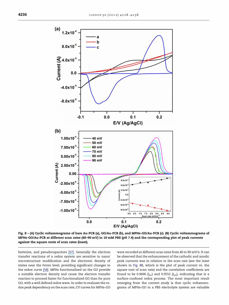
Fig. 8 – (A) Cyclic voltammograms of bare Au-PCB (a), GO/Au-PCB (b), and MPHs-GO/Au-PCB (c). (B) Cyclic voltammograms of
MPHs-GO/Au-PCB at different scan rates (40–90 mV) in 10 mM PBS (pH 7.4) and the corresponding plot of peak currents
against the square roots of scan rates (inset).
4236 C A R B O N 5 0 ( 2 0 1 2 ) 4 2 2 8 – 4 2 3 8
batteries, and pseudocapacitors [57]. Generally the electron
transfer reactions of a redox system are sensitive to nano/
microstructure modification and the electronic density of
states near the Fermi level, providing significant changes to
the redox curve [58]. MPHs functionalized on the GO provide
a suitable electron density and cause the electron transfer
reaction to proceed faster for functionalized GO than for pure
GO, with a well defined redox wave. In order to evaluate the re-
dox peak dependency on the scan rate, CV curves for MPHs-GO
were recorded at different scan rates from 40 to 90 mV/s. It can
be observed that the enhancement of the cathodic and anodic
peak currents was in relation to the scan rate (see the inset
shown in Fig. 8B, which is the plot of peak current vs. the
square root of scan rate) and the correlation coefficients are
found to be 0.9600 (Ipc) and 0.9552 (Ipa), indicating that is a
surface-confined redox process. The most important result
emerging from the current study is that cyclic voltammo-
grams of MPHs-GO in a PBS electrolyte system are valuable

C A R B O N 5 0 ( 2 0 1 2 ) 4 2 2 8 – 4 2 3 8 4237
and convenient for monitoring the surface status and barrier
of the modified electrode. Furthermore, the GO in the final hy-
brid material not only acts as the substrate for functionalizing
the MPHs but also as an active interface layer for exhibiting
the electrochemical transfer reaction between the solution
species and the electrode. The development of such a func-
tionalized GO hybrid in a controllable manner offers a new ap-
proach to construct an electrochemically active functionalized
material that can be used as a transducer for several electro-
chemical biosensor devices.
4. Conclusion
We have demonstrated the covalent functionalization of a
silanized-metalloid polymer on graphene oxide. Structural
characterization by electron and atomic force microscopy
reveals that this has been achieved. It is found that polymer
stabilized metalloid nanoparticles are uniformly distributed
on the graphene oxide sheets. Structural observations were
confirmed by UV–vis absorbance, FT-IR and 1H-NMR spectro-
scopic studies. Raman spectroscopy further shows that the
lattice structure of the graphene oxide sheets is significantly
modified after chemical bonding with the metalloid-polymer.
The quenching of near band emission from graphene oxide
hybrids (as shown by the photo-luminescent study) also sup-
ports the process of surface modification. Surface immobili-
zation of functionalized graphene oxide on the Au-PCB
electrode exhibits a characteristic voltammetric response
which is superior to that of graphene oxide and the conven-
tional Au-PCB electrode. The simple chemical functionaliza-
tion, hetero nano-environment, excellent dispersion in
various solvents, film forming ability, electrochemical proper-
ties and other synergistic effects of the obtained hybrid mate-
rial will direct us to several applications and developments in
graphene-based materials, electrocatalysis, sensor materials
and nano-electronics. Investigations into the biosensor appli-
cations of this hybrid material are currently ongoing in the
laboratory.
Acknowledgments
This research was supported by the Ministry of Knowledge
and Economy Grant No. 10039863 to K.S.Y. and the Ministry
of Knowledge and Economy Grant No. 10035501 to M.-H.L.
Appendix A. Supplementary data
Supplementary data associated with this article can be found,
in the online version, at http://dx.doi.org/10.1016/j.carbon.
2012.05.004.
R E F E R E N C E S
[1] Areshkin DA, White CT. Building blocks for integratedgraphene circuits. Nano Lett 2007;7(11):3253–9.
[2] Schedin F, Geim AK, Morozov SV, Hill EM, Blake P, KatsnelsonMI, et al. Detection of individual gas molecules adsorbed ongraphene. Nat Mater 2007;6:652–5.
[3] Stankovich S, Dikin DA, Dommett GHB, Kohlhaas KM, ZimneyEJ, Stach EA, et al. Graphene-based composite materials.Nature 2006;442:282–6.
[4] Takamura T, Endo K, Fu L, Wu YP, Lee KJ, Matsumoto T.Identification of nano-sized holes by TEM in the graphenelayer of graphite and the high rate discharge capability of Li-ion battery anodes. Eletrochim Acta 2007;53(3):1055–61.
[5] Novoselov KS, Jiang D, Schedin F, Booth TJ, Khotkevich VV,Morozov SV, et al. Two-dimensional atomic crystals. Proc NatAcad Sci USA 2005;102(30):10451–3.
[6] Ogoshi T, Ichihara Y, Yamagishi T, Nakamoto Y.Supramolecular polymer networks from hybrid betweengraphene oxide and per-6-amino-b-cyclodextrin. ChemCommun 2010;46:6087–9.
[7] Dreyer DR, Park S, Bielawski CW, Ruoff RS. The chemistry ofgraphene oxide. Chem Soc Rev 2010;39:228–40.
[8] Liu Z, Robinson JT, Sun XM, Dai HJ. PEGylated nanographeneoxide for delivery of water-insoluble cancer drugs. J AmChem Soc 2008;130(33):10876–7.
[9] Yang K, Zhang S, Zhang G, Sun X, Lee ST, Liu Z. Graphene inMICE. Ultrahigh in vivo tumor uptake and efficientphotothermal therapy. Nano Lett 2010;10(9):3318–23.
[10] Bao H, Pan Y, Ping Y, Sahoo NG, Wu T, Li L, et al. Chitosan-functionalized graphene oxide as a nanocarrier for drug andgene delivery. Small 2011;7(11):1569–78.
[11] Shen J, Shi M, Yan B, Ma H, Li N, Hu Y, et al. Covalentattaching protein to graphene oxide via diimide-activatedamidation. Colloid Surface B 2010;81(2):434–8.
[12] Shen J, Yan B, Shi M, Ma H, Li N, Ye M. Synthesis of grapheneoxide-based biocomposites through diimide-activatedamidation. J Colloid Interf Sci 2011;356(2):543–9.
[13] Huang X, Yin Z, Wu S, Qi X, He Q, Zhang Q, et al. Graphene-based materials: synthesis, characterization, properties, andapplications. Small 2011;7(14):1877–902.
[14] Jiang G, Lin Z, Chen C, Zhu L, Chang Q, Wang N, et al. TiO2
nanoparticles assembled on graphene oxide nanosheets withhigh photocatalytic activity for removal of pollutants. Carbon2011;49(8):2693–701.
[15] Mishra A, Ramaprabhu S. Functionalized graphene sheets forarsenic removal and desalination of sea water. Desalination2011;282(1):39–45.
[16] Eswaraiah V, Sankaranarayanan V, Ramaprabhu S. Graphene-based engine oil nanofluids for tribological applications. ACSAppl Mater Interfaces 2011;3(11):4221–7.
[17] Xu C, Wang X, Yang L, Wu Y. Fabrication of a graphene–cuprous oxide composite. J Solid State Chem2009;182(9):2486–90.
[18] Wang S, Jiang SP, Wang X. Microwave-assisted one-potsynthesis of metal/metal oxide nanoparticles on grapheneand their electrochemical applications. Electrochim Acta2011;56(9):3338–44.
[19] Wang J, Gao Z, Li Z, Wang B, Yan Y, Liu Q, et al. GreenSynthesis of graphene nanosheets/ZnO composites andelectrochemical properties. J Solid State Chem2011;184(6):1421–7.
[20] Wu Q, Xu Y, Yao Z, Liu A, Shi G. Supercapacitors based onflexible graphene/polyaniline nanofiber composite films. ACSNano 2010;4(4):1963–70.
[21] Zhang Y, Sun X, Zhu L, Shen H, Jia N. Electrochemical sensingbased on graphene oxide/Prussian blue hybrid film modifiedelectrode. Electrochim Acta 2011;56(3):1239–45.
[22] Shan C, Yang H, Hana D, Zhang Q, Ivaska A, Niu Li. Graphene/AuNPs/chitosan nanocomposites film for glucose biosensing.Biosens Bioelectron 2010;25(5):1070–4.
[23] Sheng L, Ren J, Miao Y, Wang J, Wang E. PVP-coated grapheneoxide for selective determination of ochratoxin A viaquenching fluorescence of free aptamer. Biosens Bioelectron2011;26(8):3494–9.
4238 C A R B O N 5 0 ( 2 0 1 2 ) 4 2 2 8 – 4 2 3 8
[24] Singh V, Joung D, Zhai L, Das S, Khondaker SI, Seal S.Graphene based materials: past, present and future. ProgMater Sci 2011;56(8):1178–271.
[25] Veerapandian M, Yun KS. Ultrasonochemical-assistedfabrication and evaporation-induced self-assembly (EISA) ofPOSS-SiO2@Ag core/ABA triblock copolymer nanocompositefilm. Polym Composite 2010;31(9):1620–7.
[26] Veerapandian M, Yun KS. Ultrasonochemically conjugatedmetalloid/triblock copolymer nanocomposite andsubsequent thin solid laminate growth for surface andinterface studies. Langmuir 2010;26(17):14216–22.
[27] Veerapandian M, Jang CH, Lim GS, Park SH, Lee MH, Yun KS.In: The fifth international conference on quantum, nano andmicro technologies (ICQNM). IARIA 2011; 63–66.
[28] Hirata M, Gotou T, Horiuchi S, Fujiwara M, Ohba M. Thin-filmparticles of graphite oxide 1: high-yield synthesis andflexibility of the particles. Carbon 2004;42(14):2929–37.
[29] Krishnamoorthy K, Mohan R, Kim S-J. Graphene oxide as aphotocatalytic material. Appl Phys Lett 2011;98:2441011-1–3.
[30] Stankovich S, Piner R, Nguyen ST, Ruoff RS. Synthesis andexfoliation of isocyanate-treated graphene oxidenanoplatelets. Carbon 2006;44(15):3342–7.
[31] Wang S, Chia PJ, Chua LL, Zhao LH, Png RQ,Sivaramakrishnan S, et al. Band-like transport in surface-functionalized highly solution-processable graphenenanosheets. Adv Mater 2008;20(18):3440–6.
[32] Ramanathan T, Abdala AA, Stankovich S, Dikin DA, Herrera-Alonso M, Piner RD, et al. Functionalized graphene sheets forpolymer nanocomposites. Nature Nanotech 2008;3:327–31.
[33] Veerapandian M, Lim SK, Nam HM, Kuppannan G, Yun KS.Glucosamine-functionalized silver glyconanoparticles:characterization and antibacterial activity. Anal BioanalChem 2010;398:867–76.
[34] Chen W, Zhang J, Shi L, Di Y, Fang Q, Cai W. Characterizationof sonochemically prepared silver–silica monolithicmesoporous nanocomposite. Compos Sci Technol2003;63:1209–12.
[35] Maruyama O, Senda Y, Omi S. Non-linear optical properties oftitanium dioxide films containing dispersed gold particles. JNon-Cryst Solids 1999;259:100–6.
[36] Wang XL, Mou YR, Chen SC, Shi J, Wang YZ. A water-solublePPDO/PEG alternating multiblock copolymer: synthesis,characterization, and its gel–sol transition behavior. EurPolym J 2009;45(4):1190–7.
[37] Remant Bahadur KC, Bhattarai SR, Aryal S, Khil MS,Dharmaraj N, Kim HY. Novel amphiphilic triblock copolymerbased on PPDO, PCL, and PEG: synthesis, characterization,and aqueous dispersion. Colloid Surface A 2007;292:69–78.
[38] Coates J. Interpretation of infrared spectra, a practicalapproach. In: Meyers RA, editor. Encyclopedia of AnalyticalChemistry. Chichester, UK: John Wiley and Sons Ltd.; 2000.p. 10815–37.
[39] Du Q, Zheng M, Zhang L, Wang Y, Chen J, Xue L, et al.Preparation of functionalized graphene sheets by a low-temperature thermal exfoliation approach and theirelectrochemical supercapacitive behaviors. Electrochim Acta2010;55(12):3897–903.
[40] Si Y, Samulski ET. Synthesis of water soluble graphene. NanoLett 2008;8(6):1679–82.
[41] Paredes JI, Rodil SV, Alonso AM, Tascon JMD. Graphene oxidedispersions in organic solvents. Langmuir2008;24(19):10560–4.
[42] Marcel SF, Jie LK, Lam WLK, Lao HB. Synthesis and propertiesof amidoalkenes involving azidoalkenoates and 1-phenylalkan-1-ols. J Chem Soc Perkin Trans 1989;1:1–11.
[43] Busenlehner LS, Armstrong RN. Insights into enzymestructure and dynamics elucidated by amide H/D exchangemass spectrometry. Arch Biochem Biophys 2005;433:34–46.
[44] Silverstein RM, Bassier GC, Morrill TC. SpectrometricIdentification of Organic Compounds. 5th ed. Wiley; 1991.
[45] Bertelsen DM, Boerio FJ. Linking mechanical properties ofsilanes to their chemical structure: an analytical study of c-GPS solutions and films. Prog Org Coat 2001;41:239–46.
[46] Krishnamoorthy K, Veerpandian M, Mohan R, Kim S-J.Investigation of Raman and photoluminescence studies ofreduced graphene oxide sheets. Appl Phys A 2011. http://dx.doi.org/10.1007/s00339-011-6720-6.
[47] Eda G, Chhowalla M. Chemically derived graphene oxide:towards large-area thin-film electronics and optoelectronics.Adv Mater 2010;22(22):2392–415.
[48] Kudin KN, Ozbas B, Schniepp HC, Prudhomme RK, Aksay IA,Car R. Raman spectra of graphite oxide and functionalizedgraphene sheets. Nano Lett 2007;8(1):36–41.
[49] Shukla S, Saxena S. Spectroscopic investigation ofconfinement effects on optical properties of graphene oxide.Appl Phys Lett 2011;98:073104-1–2.
[50] Luo Z, Vora PM, Mele EJ, Johnson ATC, Kikkawa JM.Photoluminescence and band gap modulation in grapheneoxide. Appl. Phys. Lett. 2009;94(11):111909.
[51] Veerpandian M, Subbiah R, Lim GS, Park SH, Yun KS, Lee M-H.Copper-glucosamine microcubes: synthesis,characterization, and C-reactive protein detection. Langmuir2011;27(14):8934–42.
[52] Wang GF, Wang W, Wu JF, Liu HY, Jiao SF, Fang B. Self-assembly of a silver nanoparticles modified electrode and itselectrocatalysis on neutral red. Microchim Acta2009;164:149–55.
[53] Choi YJ, Luo T-JM. Electrochemical properties of silvernanoparticle doped aminosilica nanocomposite. Int JElectrochem 2011;404937:1–6.
[54] Guo L, Nie J, Du B, Peng Z, Tesche B, Kleinermanns K.Thermoresponsive polymer-stabilized silver nanoparticles. JColloid Interf Sci 2008;319:175–81.
[55] Wang GF, Li MG, Gao YC, Fang B. Amperometric sensor usedfor determination of thiocyanate with a silver nanoparticlesmodified electrode. Sensors 2004;4:147–55.
[56] Chang G, Zhang J, Oyama M, Hirao K. Silver-nanoparticle-attached indium tin oxide surfaces fabricated by a seed-mediated growth approach. J Phys Chem B 2005;109:1204–9.
[57] Kamat PV. Graphene-based nanoassemblies for energyconversion. J Phys Chem Lett 2011;2(3):242–51.
[58] Giovannetti G, Khomyakov PA, Brocks G, Karpan VM, van denBrink J, Kelly PJ. Doping graphene with metal contacts. PhysRev Lett 2008;101(2):026803-1–4.