Synthesis, biological evaluation and molecular modeling study of 5-trifluoromethyl-Δ2-pyrazoline...
8
Original article Synthesis, biological evaluation and molecular modeling study of 5-trifluoromethyl-D 2 -pyrazoline and isomeric 5/3- trifluoromethylpyrazole derivatives as anti-inflammatory agents Ranjana Aggarwal a, * , Anshul Bansal a , Isabel Rozas b , Brendan Kelly b , Pawan Kaushik c , Dhirender Kaushik c a Department of Chemistry, Kurukshetra University, Kurukshetra, Haryana 136119, India b School of Chemistry, Trinity Biomedical Sciences Institute, Trinity College Dublin,154-160 Pearse St., Dublin 2, Ireland c Institute of Pharmaceutical Sciences, Kurukshetra University, Kurukshetra 136119, India article info Article history: Received 4 May 2013 Received in revised form 23 September 2013 Accepted 28 September 2013 Available online 7 October 2013 Keywords: 5-Trifluoromethyl-D 2 -pyrazolines 3-Trifluoromethylpyrazoles 5-Trifluoromethylpyrazoles Anti-inflammatory activity COX-2 Docking abstract Searching for new anti-inflammatory agents, we have prepared a series of potential COX-2 inhibitors, 1-(4,6- dimethylpyrimidin-2-yl)-5-hydroxy-5-trifluoromethyl-D 2 -pyrazolines (3) and 1-(4,6-dimethylpyrimidin-2- yl)-3-trifluoromethylpyrazoles (4), by refluxing 2-hydrazino-4,6-dimethylpyrimidine (1) with a number of trifluoromethyl-b-diketones (2) in ethanol. Further dehydration of compounds (3) to the corresponding 1-(4,6-dimethylpyrimidin-2-yl)-5-trifluoromethylpyrazoles (5) was also achieved. Fifteen of these com- pounds were screened for their anti-inflammatory activity using the carrageenan-induced rat paw edema assay. While all the compounds exhibited significant anti-inflammatory activity (47e76%) as compared to indomethacin (78%), 3-trifluoromethylpyrazoles (4) were found to be the most effective agents (62e76%). To rationalize this anti-inflammatory activity, docking experiments molecular dynamics simulations were per- formed to study the ability of these compounds to bind into the active site of the COX-2 enzyme. Ó 2013 Elsevier Masson SAS. All rights reserved. 1. Introduction Inflammation plays a fundamental role in the immune response of the human body to microbes, injury or trauma. Recent studies have demonstrated that inflammation is key not only in infection and cancer but also to autoimmune diseases, such as rheumatoid arthritis, multiple sclerosis, type I diabetes and Crohn’s disease, or certain neuro-degenerative conditions [1e4]. One of the most important factors in the inflammatory process is the generation of prostaglandins which are produced by the ac- tion of cyclooxygenase (COX) on arachidonic acid. For that reason, this enzymatic system became a popular target for the develop- ment of anti-inflammatory drugs such as the non-steroidal anti- inflammatory drugs or NSAIDs which exert their activity by inhibiting COX, thus preventing the synthesis of prostaglandins [5]. From the known forms of COX, it seems that COX-2 is the one responsible of prostaglandin production in inflammatory condi- tions. Thus, COX-2 inhibitors have been prepared for the treatment of chronic inflammatory diseases like rheumatoid and osteoar- thritis, and many of them, such as celecoxib or SC-588 (Fig. 1), have a similar core, the trifluoromethylpyrazole system [6]. Several pyrazole derivatives have been reported to possess pharmacological activity as anti-inflammatory agents [7e9]. Further, incorporation of the lipophilic trifluoromethyl group on pyrazole ring exerts a variety of dramatic effects on the pharma- cological properties of the molecule making its partition into cell membranes much easier and hence increasing the selectivity, effi- cacy and bioavailability [10]. Also pyrimidine derivatives, the fundamental building blocks for DNA and RNA, have attracted a great deal of attention over many years due to their broad bio- activities, including antitumor [11,12], antimicrobial [13], or anti- inflammatory [14]. In view of these observations and continuing with our work related to the synthesis and spectroscopic and biological studies of trifluoromethylpyrazoles [15e18], we have prepared and evaluated * Corresponding author. Tel.: þ91 1744238734; fax: þ91 1744238277. E-mail addresses: [email protected], [email protected] (R. Aggarwal). Contents lists available at ScienceDirect European Journal of Medicinal Chemistry journal homepage: http://www.elsevier.com/locate/ejmech 0223-5234/$ e see front matter Ó 2013 Elsevier Masson SAS. All rights reserved. http://dx.doi.org/10.1016/j.ejmech.2013.09.052 European Journal of Medicinal Chemistry 70 (2013) 350e357
-
Upload
independent -
Category
Documents
-
view
3 -
download
0
Transcript of Synthesis, biological evaluation and molecular modeling study of 5-trifluoromethyl-Δ2-pyrazoline...
lable at ScienceDirect
European Journal of Medicinal Chemistry 70 (2013) 350e357
Contents lists avai
European Journal of Medicinal Chemistry
journal homepage: http: / /www.elsevier .com/locate/ejmech
Original article
Synthesis, biological evaluation and molecular modeling studyof 5-trifluoromethyl-D2-pyrazoline and isomeric 5/3-trifluoromethylpyrazole derivatives as anti-inflammatory agents
Ranjana Aggarwal a,*, Anshul Bansal a, Isabel Rozas b, Brendan Kelly b, Pawan Kaushik c,Dhirender Kaushik c
aDepartment of Chemistry, Kurukshetra University, Kurukshetra, Haryana 136 119, Indiab School of Chemistry, Trinity Biomedical Sciences Institute, Trinity College Dublin, 154-160 Pearse St., Dublin 2, Irelandc Institute of Pharmaceutical Sciences, Kurukshetra University, Kurukshetra 136 119, India
a r t i c l e i n f o
Article history:Received 4 May 2013Received in revised form23 September 2013Accepted 28 September 2013Available online 7 October 2013
Keywords:5-Trifluoromethyl-D2-pyrazolines3-Trifluoromethylpyrazoles5-TrifluoromethylpyrazolesAnti-inflammatory activityCOX-2Docking
* Corresponding author. Tel.: þ91 1744238734; fax:E-mail addresses: [email protected], ra
(R. Aggarwal).
0223-5234/$ e see front matter � 2013 Elsevier Mashttp://dx.doi.org/10.1016/j.ejmech.2013.09.052
a b s t r a c t
Searching for new anti-inflammatory agents, we have prepared a series of potential COX-2 inhibitors,1-(4,6-dimethylpyrimidin-2-yl)-5-hydroxy-5-trifluoromethyl-D2-pyrazolines (3) and 1-(4,6-dimethylpyrimidin-2-yl)-3-trifluoromethylpyrazoles (4), by refluxing 2-hydrazino-4,6-dimethylpyrimidine (1) with a number oftrifluoromethyl-b-diketones (2) in ethanol. Further dehydration of compounds (3) to the corresponding1-(4,6-dimethylpyrimidin-2-yl)-5-trifluoromethylpyrazoles (5) was also achieved. Fifteen of these com-pounds were screened for their anti-inflammatory activity using the carrageenan-induced rat paw edemaassay. While all the compounds exhibited significant anti-inflammatory activity (47e76%) as compared toindomethacin (78%), 3-trifluoromethylpyrazoles (4) were found to be themost effective agents (62e76%). Torationalize this anti-inflammatory activity, docking experiments molecular dynamics simulations were per-formed to study the ability of these compounds to bind into the active site of the COX-2 enzyme.
� 2013 Elsevier Masson SAS. All rights reserved.
1. Introduction
Inflammation plays a fundamental role in the immune responseof the human body to microbes, injury or trauma. Recent studieshave demonstrated that inflammation is key not only in infectionand cancer but also to autoimmune diseases, such as rheumatoidarthritis, multiple sclerosis, type I diabetes and Crohn’s disease, orcertain neuro-degenerative conditions [1e4].
One of the most important factors in the inflammatory processis the generation of prostaglandins which are produced by the ac-tion of cyclooxygenase (COX) on arachidonic acid. For that reason,this enzymatic system became a popular target for the develop-ment of anti-inflammatory drugs such as the non-steroidal anti-inflammatory drugs or NSAIDs which exert their activity byinhibiting COX, thus preventing the synthesis of prostaglandins [5].
son SAS. All rights reserved.
From the known forms of COX, it seems that COX-2 is the oneresponsible of prostaglandin production in inflammatory condi-tions. Thus, COX-2 inhibitors have been prepared for the treatmentof chronic inflammatory diseases like rheumatoid and osteoar-thritis, and many of them, such as celecoxib or SC-588 (Fig. 1), havea similar core, the trifluoromethylpyrazole system [6].
Several pyrazole derivatives have been reported to possesspharmacological activity as anti-inflammatory agents [7e9].Further, incorporation of the lipophilic trifluoromethyl group onpyrazole ring exerts a variety of dramatic effects on the pharma-cological properties of the molecule making its partition into cellmembranes much easier and hence increasing the selectivity, effi-cacy and bioavailability [10]. Also pyrimidine derivatives, thefundamental building blocks for DNA and RNA, have attracted agreat deal of attention over many years due to their broad bio-activities, including antitumor [11,12], antimicrobial [13], or anti-inflammatory [14].
In view of these observations and continuing with our workrelated to the synthesis and spectroscopic and biological studies oftrifluoromethylpyrazoles [15e18], we have prepared and evaluated
N NCF3
H3C
H2NO2S
N NCF3
Br
H2NO2S
Fig. 1. Structures of celecoxib (left) and SC-588 (right).
R. Aggarwal et al. / European Journal of Medicinal Chemistry 70 (2013) 350e357 351
the anti-inflammatoryactivityof a series ofnovel compounds: 1-(4,6-dimethylpyrimidin-2-yl)-5-hydroxy-5-trifluoromethyl-D2-pyrazo-lines (3),1-(4,6-dimethylpyrimidin-2-yl)-3-trifluoromethylpyrazoles(4) and 1-(4,6-dimethylpyrimidin-2-yl)-5-trifluoromethylpyrazoles(5), with the aim of finding new and more potent anti-inflammatory agents.
Moreover, considering that computer docking techniques playan important role in drug design as well as in mechanistic studiesby placing a molecule into the binding site of the target macro-molecule in a non covalent fashion, we have docked our com-pounds into the active site of COX-2 (PDB crystal structure: 3LN1[19]). Our objective is to rationalize the potential as anti-inflammatory agents of these compounds by predicting theirbinding modes, binding affinities and optimal orientation at theactive site of the COX-2 enzyme.
2. Results and discussion
2.1. Synthesis
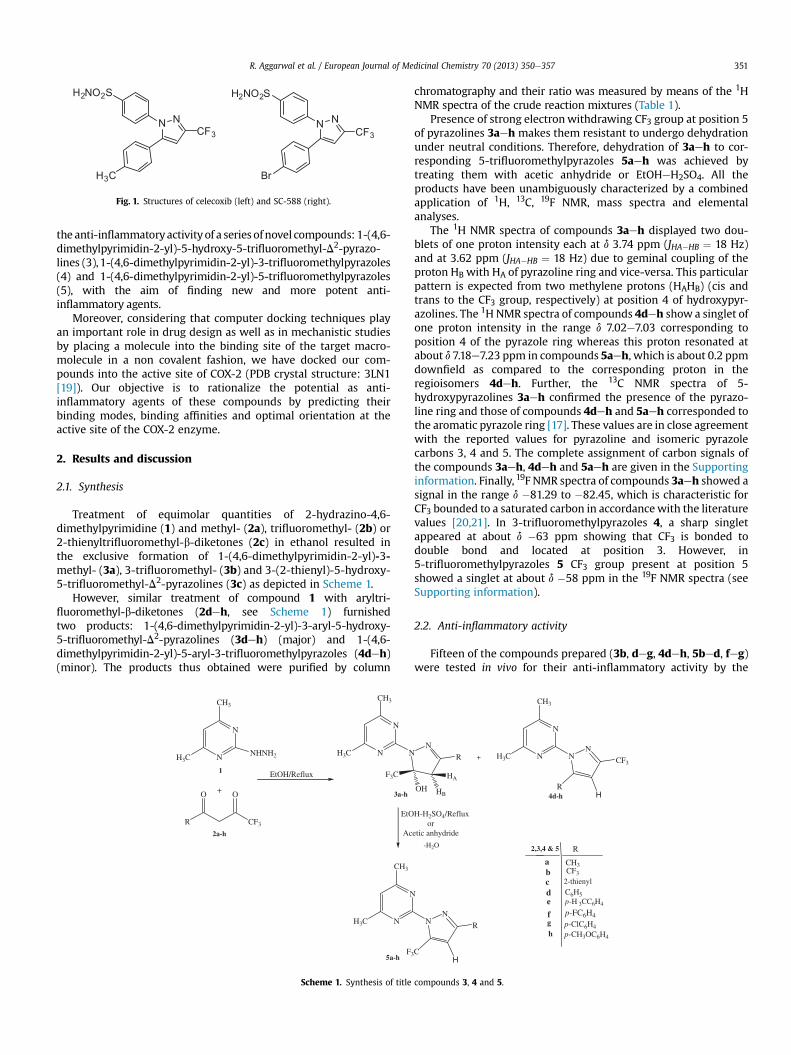
Treatment of equimolar quantities of 2-hydrazino-4,6-dimethylpyrimidine (1) and methyl- (2a), trifluoromethyl- (2b) or2-thienyltrifluoromethyl-b-diketones (2c) in ethanol resulted inthe exclusive formation of 1-(4,6-dimethylpyrimidin-2-yl)-3-methyl- (3a), 3-trifluoromethyl- (3b) and 3-(2-thienyl)-5-hydroxy-5-trifluoromethyl-D2-pyrazolines (3c) as depicted in Scheme 1.
However, similar treatment of compound 1 with aryltri-fluoromethyl-b-diketones (2deh, see Scheme 1) furnishedtwo products: 1-(4,6-dimethylpyrimidin-2-yl)-3-aryl-5-hydroxy-5-trifluoromethyl-D2-pyrazolines (3deh) (major) and 1-(4,6-dimethylpyrimidin-2-yl)-5-aryl-3-trifluoromethylpyrazoles (4deh)(minor). The products thus obtained were purified by column
N
N
CH3
H3C NHNH2
+
R CF3
O O
N
N
CH3
H3C N
EtOH/Reflux
N
N
CH3
H3C
F3
F3C
EtO Ac
1
2a-h
3a-h
5a-h
Scheme 1. Synthesis of title
chromatography and their ratio was measured by means of the 1HNMR spectra of the crude reaction mixtures (Table 1).
Presence of strong electronwithdrawing CF3 group at position 5of pyrazolines 3aeh makes them resistant to undergo dehydrationunder neutral conditions. Therefore, dehydration of 3aeh to cor-responding 5-trifluoromethylpyrazoles 5aeh was achieved bytreating them with acetic anhydride or EtOHeH2SO4. All theproducts have been unambiguously characterized by a combinedapplication of 1H, 13C, 19F NMR, mass spectra and elementalanalyses.
The 1H NMR spectra of compounds 3aeh displayed two dou-blets of one proton intensity each at d 3.74 ppm (JHA�HB ¼ 18 Hz)and at 3.62 ppm (JHA�HB ¼ 18 Hz) due to geminal coupling of theproton HB with HA of pyrazoline ring and vice-versa. This particularpattern is expected from two methylene protons (HAHB) (cis andtrans to the CF3 group, respectively) at position 4 of hydroxypyr-azolines. The 1H NMR spectra of compounds 4deh show a singlet ofone proton intensity in the range d 7.02e7.03 corresponding toposition 4 of the pyrazole ring whereas this proton resonated atabout d 7.18e7.23 ppm in compounds 5aeh, which is about 0.2 ppmdownfield as compared to the corresponding proton in theregioisomers 4deh. Further, the 13C NMR spectra of 5-hydroxypyrazolines 3aeh confirmed the presence of the pyrazo-line ring and those of compounds 4deh and 5aeh corresponded tothe aromatic pyrazole ring [17]. These values are in close agreementwith the reported values for pyrazoline and isomeric pyrazolecarbons 3, 4 and 5. The complete assignment of carbon signals ofthe compounds 3aeh, 4deh and 5aeh are given in the Supportinginformation. Finally, 19F NMR spectra of compounds 3aeh showed asignal in the range d �81.29 to �82.45, which is characteristic forCF3 bounded to a saturated carbon in accordancewith the literaturevalues [20,21]. In 3-trifluoromethylpyrazoles 4, a sharp singletappeared at about d �63 ppm showing that CF3 is bonded todouble bond and located at position 3. However, in5-trifluoromethylpyrazoles 5 CF3 group present at position 5showed a singlet at about d �58 ppm in the 19F NMR spectra (seeSupporting information).
2.2. Anti-inflammatory activity
Fifteen of the compounds prepared (3b, deg, 4deh, 5bed, feg)were tested in vivo for their anti-inflammatory activity by the
N
R N
N
CH3
H3C NN
CF3
R
NN
R
C
-H2O R
CH3CF3
C6H5
p-CH3OC6H4
p-ClC6H4
abcde
f p-FC6H4
HBOH
HA
H-H2SO4/Reflux oretic anhydride
g
p-H 3CC6H4
h
2-thienyl
4d-h
+
2,3,4 & 5
H
H
compounds 3, 4 and 5.
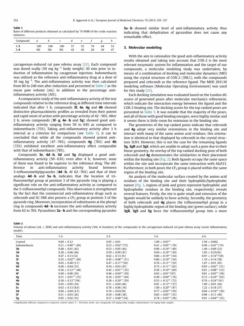
Table 1Ratio of different products obtained as calculated by 1H NMR of the crude reactionmixtures.
Compound a b c d e f g h
% 3 100 100 100 57 55 74 64 53% 4 Nil Nil Nil 43 45 26 36 47
R. Aggarwal et al. / European Journal of Medicinal Chemistry 70 (2013) 350e357352
carrageenan-induced rat paw edema assay [22]. Each compoundwas dosed orally (50 mg kg�1 body weight) 30 min prior to in-duction of inflammation by carageenan injection. Indomethacinwas utilized as the reference anti-inflammatory drug at a dose of10 mg kg�1. The anti-inflammatory activity was then calculatedfrom 60 to 240 min after induction and presented in Table 2 as themean paw volume (mL) in addition to the percentage anti-inflammatory activity (AI%).
A comparative study of the anti-inflammatory activity of the testcompounds relative to the reference drug at different time intervalsindicated that after 1 h, compounds 3f, 4e, 4g and 4h showeddistinctive pharmacokinetic profiles as revealed from their potentand rapid onset of action with percentage activity of 42e56%. After2 h, seven compounds (3feg, 4eeh and 5g) showed good anti-inflammatory activity ranging from 50 to 64% as compared toindomethacin (75%). Taking anti-inflammatory activity after 3 hinterval as a criterion for comparison (see Table 3), it can beconcluded that while all the compounds showed potent anti-inflammatory activity (47e76%), compounds 4g (76%) and 4h(72%) exhibited excellent anti-inflammatory effect comparablewith that of indomethacin (78%).
Compounds 3e, 4deh, 5d and 5g displayed a good anti-inflammatory activity (50e63%) even after 4 h; however, noneof them was found to be superior to the reference drug. The dif-ference in anti-inflammatory activity found between3-trifluoromethylpyrazoles (4deh, AI 62e76%) and that of theiranalogs 4deh and 5aeh, indicates that the location of tri-fluoromethyl group at position 3 of the pyrazole ring is playing asignificant role on the anti-inflammatory activity as compared tothe 5-trifluoromethyl compounds. This observation is strengthenedby the fact that the commercial anti-inflammatory drugs such ascelecoxib and SC-588 also possess a CF3 group at position 3 of thepyrazole ring. Moreover, incorporation of substituents at the phenylring in compounds 4deh increases the anti-inflammatory activityfrom 62 to 76%. Pyrazolines 3aeh and the corresponding pyrazoles
Table 2Volume of edema (mL � SEM) and anti-inflammatory activity (%AI, in brackets) of themodel).
Time 1 h 2 h
Control 0.69 � 0.12 0.95 � 0.01Indomethacin 0.21 � 0.06** (69) 0.23 � 0.02**3b 0.40 � 0.01 (42) 0.53 � 0.05 (43d 0.38 � 0.06 (44) 0.50 � 0.09 (43e 0.47 � 0.11(32) 0.62 � 0.11(33f 0.35 � 0.02** (49) 0.45 � 0.08**3g 0.65 � 0.40 (5.7) 0.47 � 0.11**4d 0.46 � 0.04 (33) 0.54 � 0.03 (44e 0.36 � 0.12** (48) 0.42 � 0.01**4f 0.49 � 0.06 (29) 0.46 � 0.04**4g 0.31 � 0.01** (55) 0.34 � 0.05**4h 0.30 � 0.12**(56) 0.38 � 0.20**5b 0.45 � 0.05 (34) 0.51 � 0.04 (45c 0.63 � 0.13 (8.6) 0.78 � 0.06 (15d 0.66 � 0.04 (4.3) 0.76 � 0.03(25f 0.51 � 0.03 (26) 0.59 � 0.08 (35g 0.45 � 0.02 (35) 0.57 � 0.06**
**Significantly different compared to respective control values, P < 0.01.Dose levels: test compounds (50 mg/kg b
5aeh showed similar level of anti-inflammatory activity thusindicating that dehydration of pyrazoline does not cause anyremarkable effect.
3. Molecular modeling
With the aim to rationalize the good anti-inflammatory activityresults obtained and taking into account that COX-2 is the mostrelevant enzymatic system for inflammation and the target of ourcompounds, a molecular modeling study was undertaken, bymeans of a combination of docking and molecular dynamics (MD)using the crystal structure of COX-2 (3NL1), with the compoundsprepared and celecoxib as the reference ligand. The MOE 2011.10modeling software (Molecular Operating Environment) was usedfor this study [23].
Each docking simulationwas evaluated based on the London dGscores of generated poses after molecular mechanics refinement,which indicate the interaction energy between the ligand and theCOX-2 binding site. The docking scores for the top ranked poses arepresented in Table 3. It was notable that the majority of the poses,and all of thosewith good binding energies, were highly similar andit seems there is little room for extension in the binding site.
The geometries of the top ranked poses indicate that celecoxiband 4g adopt very similar orientations in the binding site andinteract with many of the same amino acid residues; this orienta-tion is identical to that displayed by celecoxib in the crystal struc-ture 3LN1. However, this is not the case for the remaining ligands5g, 3gR and 3gS, which are unable to adopt such a pose due to theirlinear geometry. An overlay of the top ranked docking pose of bothcelecoxib and 4g demonstrates the similarities in their placementwithin the binding site (Fig. 2). Both ligands occupy the same spacewithin the site and incorporate the same interaction with Ala513.Furthermore, in both poses the CF3 group is placed within the sameregion of the binding site.
An analysis of the molecular surface created by the amino acidresidues of the binding site and their hydrophilic/hydrophobicnature (Fig. 3, regions of pink and green represent hydrophilic andhydrophobic residues in the binding site, respectively) revealsseveral features. Firstly, the site is quite small and extension of theligands would be unlikely to favor activity. Secondly, the geometryof both celecoxib and 4g places the trifluoromethyl group in ahighly hydrophobic region of the binding site (green surface), while3gR, 3gS and 5g force the trifluoromethyl group into a more
compounds in the carrageenan-induced rat paw edema assay (acute inflammatory
3 h 4 h
1.89 � 0.03** 1.94 � 0.002(75) 0.41 � 0.05** (78) 0.49 � 0.01**(74)4) 0.96 � 0.10** (49) 1.49 � 0.09 (23)7) 0.94 � 0.10** (50) 1.05 � 0.25(45)5) 0.86 � 0.19** (54) 0.97 � 0.16**(50)(51) 0.86 � 0.10** (54) 1.19 � 0.14 (38)(50) 0.76 � 0.11** (59) 1.07 � 0.01 (45)3) 0.72 � 0.11** (62) 0.95 � 0.02** (51)(55) 0.58 � 0.10** (69) 0.91 � 0.08** (53)(59) 0.62 � 0.01**(67) 0.81 � 0.02** (58)(64) 0.49 � 0.04** (76) 0.72 � 0.24** (63)(59) 0.53 � 0.12** (72) 0.76 � 0.24**(61)6) 0.81 � 0.13** (57) 1.09 � 0.01 (44)8) 1.00 � 0.20** (47) 1.22 � 0.18 (37)0) 0.77 � 0.02** (59) 0.84 � 0.10** (56)8) 0.85 � 0.05** (55) 0.98 � 0.11 (49)(54) 0.78 � 0.03** (58) 0.91 � 0.04** (53)
ody weight), Indomethacin (10 mg/kg body weight).
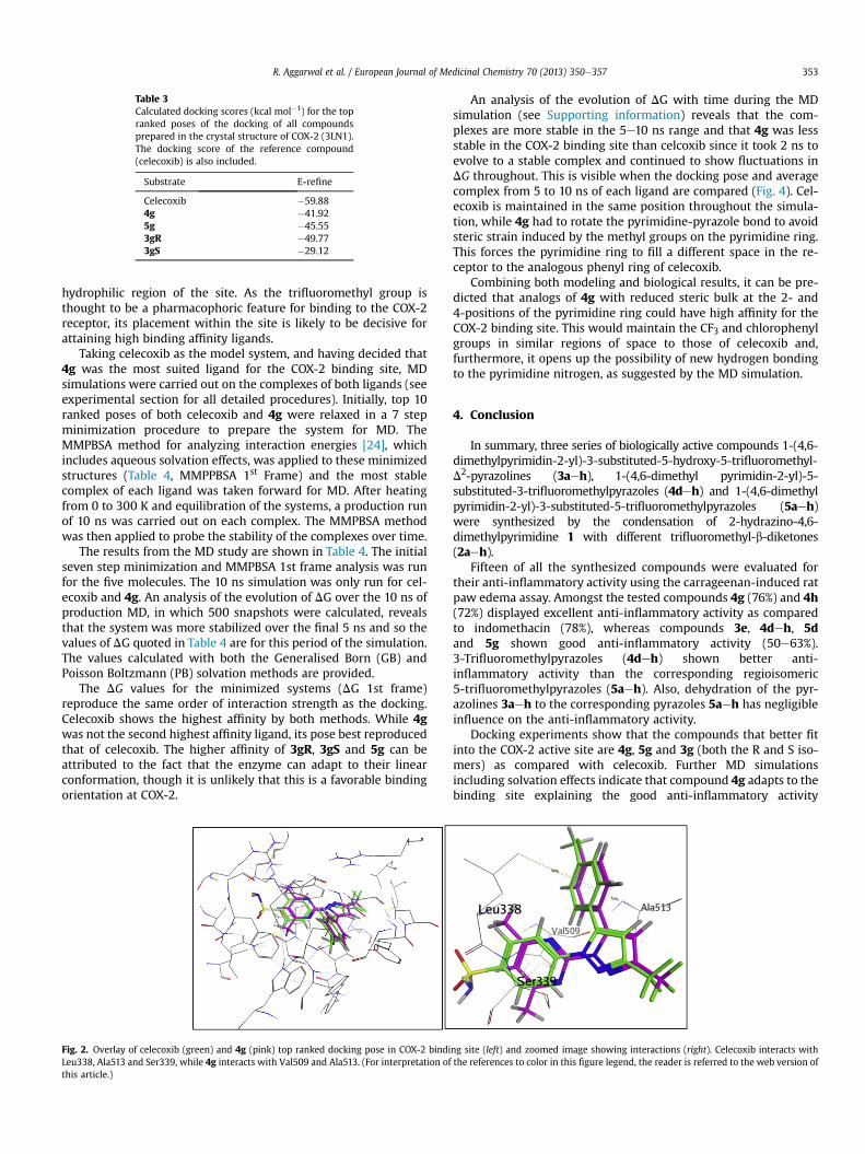
Table 3Calculated docking scores (kcal mol�1) for the topranked poses of the docking of all compoundsprepared in the crystal structure of COX-2 (3LN1).The docking score of the reference compound(celecoxib) is also included.
Substrate E-refine
Celecoxib �59.884g �41.925g �45.553gR �49.773gS �29.12
R. Aggarwal et al. / European Journal of Medicinal Chemistry 70 (2013) 350e357 353
hydrophilic region of the site. As the trifluoromethyl group isthought to be a pharmacophoric feature for binding to the COX-2receptor, its placement within the site is likely to be decisive forattaining high binding affinity ligands.
Taking celecoxib as the model system, and having decided that4g was the most suited ligand for the COX-2 binding site, MDsimulations were carried out on the complexes of both ligands (seeexperimental section for all detailed procedures). Initially, top 10ranked poses of both celecoxib and 4g were relaxed in a 7 stepminimization procedure to prepare the system for MD. TheMMPBSA method for analyzing interaction energies [24], whichincludes aqueous solvation effects, was applied to these minimizedstructures (Table 4, MMPPBSA 1st Frame) and the most stablecomplex of each ligand was taken forward for MD. After heatingfrom 0 to 300 K and equilibration of the systems, a production runof 10 ns was carried out on each complex. The MMPBSA methodwas then applied to probe the stability of the complexes over time.
The results from the MD study are shown in Table 4. The initialseven step minimization and MMPBSA 1st frame analysis was runfor the five molecules. The 10 ns simulation was only run for cel-ecoxib and 4g. An analysis of the evolution of DG over the 10 ns ofproduction MD, in which 500 snapshots were calculated, revealsthat the system was more stabilized over the final 5 ns and so thevalues of DG quoted in Table 4 are for this period of the simulation.The values calculated with both the Generalised Born (GB) andPoisson Boltzmann (PB) solvation methods are provided.
The DG values for the minimized systems (DG 1st frame)reproduce the same order of interaction strength as the docking.Celecoxib shows the highest affinity by both methods. While 4gwas not the second highest affinity ligand, its pose best reproducedthat of celecoxib. The higher affinity of 3gR, 3gS and 5g can beattributed to the fact that the enzyme can adapt to their linearconformation, though it is unlikely that this is a favorable bindingorientation at COX-2.
Fig. 2. Overlay of celecoxib (green) and 4g (pink) top ranked docking pose in COX-2 bindiLeu338, Ala513 and Ser339, while 4g interacts with Val509 and Ala513. (For interpretation ofthis article.)
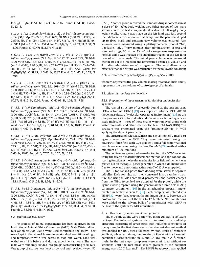
An analysis of the evolution of DG with time during the MDsimulation (see Supporting information) reveals that the com-plexes are more stable in the 5e10 ns range and that 4g was lessstable in the COX-2 binding site than celcoxib since it took 2 ns toevolve to a stable complex and continued to show fluctuations inDG throughout. This is visible when the docking pose and averagecomplex from 5 to 10 ns of each ligand are compared (Fig. 4). Cel-ecoxib is maintained in the same position throughout the simula-tion, while 4g had to rotate the pyrimidine-pyrazole bond to avoidsteric strain induced by the methyl groups on the pyrimidine ring.This forces the pyrimidine ring to fill a different space in the re-ceptor to the analogous phenyl ring of celecoxib.
Combining both modeling and biological results, it can be pre-dicted that analogs of 4g with reduced steric bulk at the 2- and4-positions of the pyrimidine ring could have high affinity for theCOX-2 binding site. This would maintain the CF3 and chlorophenylgroups in similar regions of space to those of celecoxib and,furthermore, it opens up the possibility of new hydrogen bondingto the pyrimidine nitrogen, as suggested by the MD simulation.
4. Conclusion
In summary, three series of biologically active compounds 1-(4,6-dimethylpyrimidin-2-yl)-3-substituted-5-hydroxy-5-trifluoromethyl-D2-pyrazolines (3aeh), 1-(4,6-dimethyl pyrimidin-2-yl)-5-substituted-3-trifluoromethylpyrazoles (4deh) and 1-(4,6-dimethylpyrimidin-2-yl)-3-substituted-5-trifluoromethylpyrazoles (5aeh)were synthesized by the condensation of 2-hydrazino-4,6-dimethylpyrimidine 1 with different trifluoromethyl-b-diketones(2aeh).
Fifteen of all the synthesized compounds were evaluated fortheir anti-inflammatory activity using the carrageenan-induced ratpaw edema assay. Amongst the tested compounds 4g (76%) and 4h(72%) displayed excellent anti-inflammatory activity as comparedto indomethacin (78%), whereas compounds 3e, 4deh, 5dand 5g shown good anti-inflammatory activity (50e63%).3-Trifluoromethylpyrazoles (4deh) shown better anti-inflammatory activity than the corresponding regioisomeric5-trifluoromethylpyrazoles (5aeh). Also, dehydration of the pyr-azolines 3aeh to the corresponding pyrazoles 5aeh has negligibleinfluence on the anti-inflammatory activity.
Docking experiments show that the compounds that better fitinto the COX-2 active site are 4g, 5g and 3g (both the R and S iso-mers) as compared with celecoxib. Further MD simulationsincluding solvation effects indicate that compound 4g adapts to thebinding site explaining the good anti-inflammatory activity
ng site (left) and zoomed image showing interactions (right). Celecoxib interacts withthe references to color in this figure legend, the reader is referred to the web version of
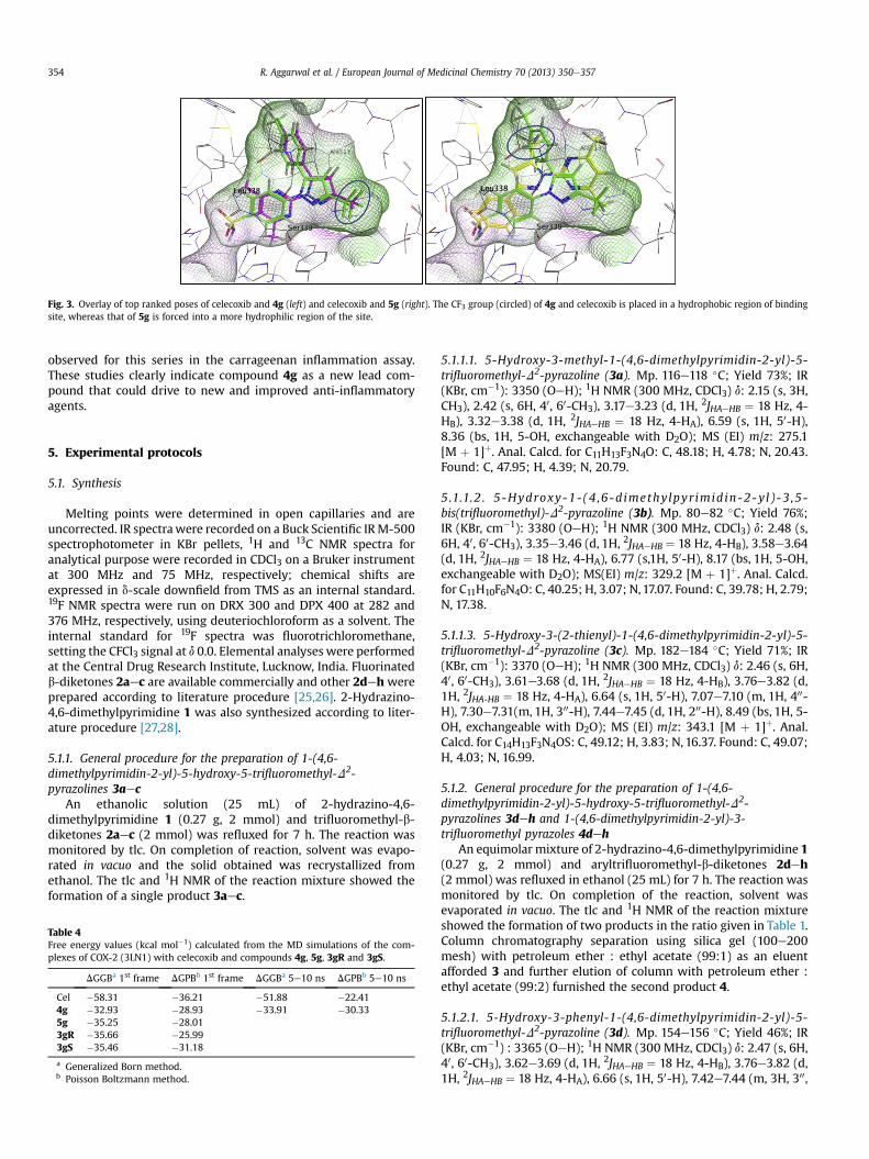
Fig. 3. Overlay of top ranked poses of celecoxib and 4g (left) and celecoxib and 5g (right). The CF3 group (circled) of 4g and celecoxib is placed in a hydrophobic region of bindingsite, whereas that of 5g is forced into a more hydrophilic region of the site.
R. Aggarwal et al. / European Journal of Medicinal Chemistry 70 (2013) 350e357354
observed for this series in the carrageenan inflammation assay.These studies clearly indicate compound 4g as a new lead com-pound that could drive to new and improved anti-inflammatoryagents.
5. Experimental protocols
5.1. Synthesis
Melting points were determined in open capillaries and areuncorrected. IR spectrawere recorded on a Buck Scientific IRM-500spectrophotometer in KBr pellets, 1H and 13C NMR spectra foranalytical purpose were recorded in CDCl3 on a Bruker instrumentat 300 MHz and 75 MHz, respectively; chemical shifts areexpressed in d-scale downfield from TMS as an internal standard.19F NMR spectra were run on DRX 300 and DPX 400 at 282 and376 MHz, respectively, using deuteriochloroform as a solvent. Theinternal standard for 19F spectra was fluorotrichloromethane,setting the CFCl3 signal at d 0.0. Elemental analyses were performedat the Central Drug Research Institute, Lucknow, India. Fluorinatedb-diketones 2aec are available commercially and other 2dehwereprepared according to literature procedure [25,26]. 2-Hydrazino-4,6-dimethylpyrimidine 1 was also synthesized according to liter-ature procedure [27,28].
5.1.1. General procedure for the preparation of 1-(4,6-dimethylpyrimidin-2-yl)-5-hydroxy-5-trifluoromethyl-D2-pyrazolines 3aec
An ethanolic solution (25 mL) of 2-hydrazino-4,6-dimethylpyrimidine 1 (0.27 g, 2 mmol) and trifluoromethyl-b-diketones 2aec (2 mmol) was refluxed for 7 h. The reaction wasmonitored by tlc. On completion of reaction, solvent was evapo-rated in vacuo and the solid obtained was recrystallized fromethanol. The tlc and 1H NMR of the reaction mixture showed theformation of a single product 3aec.
Table 4Free energy values (kcal mol�1) calculated from the MD simulations of the com-plexes of COX-2 (3LN1) with celecoxib and compounds 4g, 5g, 3gR and 3gS.
DGGBa 1st frame DGPBb 1st frame DGGBa 5e10 ns DGPBb 5e10 ns
Cel �58.31 �36.21 �51.88 �22.414g �32.93 �28.93 �33.91 �30.335g �35.25 �28.013gR �35.66 �25.993gS �35.46 �31.18
a Generalized Born method.b Poisson Boltzmann method.
5.1.1.1. 5-Hydroxy-3-methyl-1-(4,6-dimethylpyrimidin-2-yl)-5-trifluoromethyl-D2-pyrazoline (3a). Mp. 116e118 �C; Yield 73%; IR(KBr, cm�1): 3350 (OeH); 1H NMR (300 MHz, CDCl3) d: 2.15 (s, 3H,CH3), 2.42 (s, 6H, 40, 60-CH3), 3.17e3.23 (d, 1H, 2JHAeHB ¼ 18 Hz, 4-HB), 3.32e3.38 (d, 1H, 2JHAeHB ¼ 18 Hz, 4-HA), 6.59 (s, 1H, 50-H),8.36 (bs, 1H, 5-OH, exchangeable with D2O); MS (EI) m/z: 275.1[M þ 1]þ. Anal. Calcd. for C11H13F3N4O: C, 48.18; H, 4.78; N, 20.43.Found: C, 47.95; H, 4.39; N, 20.79.
5.1.1.2 . 5-Hydroxy-1-(4 ,6-d imethylpyr imidin-2-yl )-3 ,5-bis(trifluoromethyl)-D2-pyrazoline (3b). Mp. 80e82 �C; Yield 76%;IR (KBr, cm�1): 3380 (OeH); 1H NMR (300 MHz, CDCl3) d: 2.48 (s,6H, 40, 60-CH3), 3.35e3.46 (d, 1H, 2JHAeHB ¼ 18 Hz, 4-HB), 3.58e3.64(d, 1H, 2JHAeHB ¼ 18 Hz, 4-HA), 6.77 (s,1H, 50-H), 8.17 (bs, 1H, 5-OH,exchangeable with D2O); MS(EI) m/z: 329.2 [M þ 1]þ. Anal. Calcd.for C11H10F6N4O: C, 40.25; H, 3.07; N,17.07. Found: C, 39.78; H, 2.79;N, 17.38.
5.1.1.3. 5-Hydroxy-3-(2-thienyl)-1-(4,6-dimethylpyrimidin-2-yl)-5-trifluoromethyl-D2-pyrazoline (3c). Mp. 182e184 �C; Yield 71%; IR(KBr, cm�1): 3370 (OeH); 1H NMR (300 MHz, CDCl3) d: 2.46 (s, 6H,40, 60-CH3), 3.61e3.68 (d, 1H, 2JHAeHB ¼ 18 Hz, 4-HB), 3.76e3.82 (d,1H, 2JHA-HB ¼ 18 Hz, 4-HA), 6.64 (s, 1H, 50-H), 7.07e7.10 (m, 1H, 400-H), 7.30e7.31(m, 1H, 300-H), 7.44e7.45 (d, 1H, 200-H), 8.49 (bs, 1H, 5-OH, exchangeable with D2O); MS (EI) m/z: 343.1 [M þ 1]þ. Anal.Calcd. for C14H13F3N4OS: C, 49.12; H, 3.83; N, 16.37. Found: C, 49.07;H, 4.03; N, 16.99.
5.1.2. General procedure for the preparation of 1-(4,6-dimethylpyrimidin-2-yl)-5-hydroxy-5-trifluoromethyl-D2-pyrazolines 3deh and 1-(4,6-dimethylpyrimidin-2-yl)-3-trifluoromethyl pyrazoles 4deh
An equimolar mixture of 2-hydrazino-4,6-dimethylpyrimidine 1(0.27 g, 2 mmol) and aryltrifluoromethyl-b-diketones 2deh(2 mmol) was refluxed in ethanol (25 mL) for 7 h. The reaction wasmonitored by tlc. On completion of the reaction, solvent wasevaporated in vacuo. The tlc and 1H NMR of the reaction mixtureshowed the formation of two products in the ratio given in Table 1.Column chromatography separation using silica gel (100e200mesh) with petroleum ether : ethyl acetate (99:1) as an eluentafforded 3 and further elution of column with petroleum ether :ethyl acetate (99:2) furnished the second product 4.
5.1.2.1. 5-Hydroxy-3-phenyl-1-(4,6-dimethylpyrimidin-2-yl)-5-trifluoromethyl-D2-pyrazoline (3d). Mp. 154e156 �C; Yield 46%; IR(KBr, cm�1) : 3365 (OeH); 1H NMR (300 MHz, CDCl3) d: 2.47 (s, 6H,40, 60-CH3), 3.62e3.69 (d, 1H, 2JHAeHB ¼ 18 Hz, 4-HB), 3.76e3.82 (d,1H, 2JHAeHB ¼ 18 Hz, 4-HA), 6.66 (s, 1H, 50-H), 7.42e7.44 (m, 3H, 300,
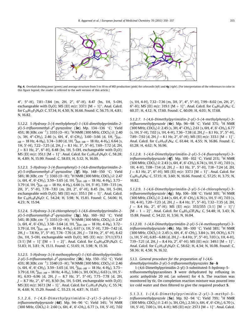
Fig. 4. Overlaid docking pose (green) and average structure from 5 to 10 ns of MD production (pink) for celecoxib (left) and 4g (right). (For interpretation of the references to color inthis figure legend, the reader is referred to the web version of this article.)
R. Aggarwal et al. / European Journal of Medicinal Chemistry 70 (2013) 350e357 355
400, 500-H), 7.81e7.84 (m, 2H, 200, 600-H), 8.47 (bs, 1H, 5-OH,exchangeable with D2O); MS (EI) m/z: 337.1 [M þ 1]þ. Anal. Calcd.for C16H15F3N4O: C, 57.14; H, 4.50; N,16.66. Found: C, 56.75; H, 4.81;N, 16.82.
5.1.2.2. 5-Hydroxy-3-(4-methylphenyl)-1-(4,6-dimethylpyrimidin-2-yl)-5-trifluoromethyl-D2-pyrazoline (3e). Mp. 134e136 �C; Yield45%; IR (KBr, cm�1): 3355 (OeH); 1H NMR (300 MHz, CDCl3) d: 2.40(s, 3H, 400-CH3), 2.46 (s, 6H, 40, 60-CH3), 3.60e3.66 (d, 1H, 2JHAeHB ¼ 18 Hz, 4-HB), 3.74e3.80 (d, 1H, 2JHAeHB ¼ 18 Hz, 4-HA), 6.64 (s,1H, 50-H), 7.22e7.25 (d, 2H, J ¼ 8.1 Hz, 300, 500-H), 7.69e7.72 (d, 2H,J ¼ 8.1 Hz, 200, 600-H), 8.48 (bs, 1H, 5-OH, exchangeable with D2O);MS (EI) m/z: 351.1 [M þ 1]þ. Anal. Calcd. for C17H17F3N4O: C, 58.28;H, 4.89; N, 15.99. Found: C, 58.03; H, 5.12; N, 16.09.
5.1.2.3. 5-Hydroxy-3-(4-fluorophenyl)-1-(4,6-dimethylpyrimidin-2-yl)-5-trifluoromethyl-D2-pyrazoline (3f). Mp. 148e150 �C; Yield58%; IR (KBr, cm�1): 3365 (OeH); 1H NMR (300 MHz, CDCl3) d: 2.47(s, 6H, 40, 60-CH3), 3.59e3.65 (d, 1H, 2JHA�HB ¼ 18 Hz, 4-HB), 3.73e3.79 (d, 1H, 2JHAeHB ¼ 18 Hz, 4-HA), 6.66 (s, 1H, 50-H), 7.09e7.15 (m,2H, 300, 500-H), 7.78e7.83 (m, 2H, 200, 600-H), 8.45 (bs, 1H, 5-OH,exchangeable with D2O); MS (EI) m/z: 355.1 [M þ 1]þ. Anal. Calcd.for C16H14F4N4O: C, 54.24; H, 3.98; N, 15.81. Found: C, 54.06; H,4.23; N, 15.94.
5.1.2.4. 5-Hydroxy-3-(4-chlorophenyl)-1-(4,6-dimethylpyrimidin-2-yl)-5-trifluoromethyl-D2-pyrazoline (3g). Mp. 160e162 �C; Yield54%; IR (KBr, cm�1): 3355 (OeH); 1H NMR (300 MHz, CDCl3) d: 2.47(s, 6H, 40, 60-CH3), 3.58e3.64 (d, 1H, 2JHAeHB ¼ 18 Hz, 4-HB), 3.72e3.79 (d, 1H, 2JHAeHB ¼ 18 Hz, 4-HA), 6.67 (s, 1H, 50-H), 7.39e7.42 (d,2H, J ¼ 7.8 Hz, 300, 500-H), 7.74e7.76 (d, 2H, J ¼ 7.8 Hz, 200, 600-H), 8.42(bs, 1H, 5-OH, exchangeable with D2O); MS (EI) m/z: 371.1/373.1(3:1) [M þ 1]þ/[M þ 1 þ 2]þ. Anal. Calcd. for C16H14ClF3N4O: C,51.83; H, 3.81; N, 15.11. Found: C, 51.65; H, 3.98; N, 15.36.
5.1.2.5. 5-Hydroxy-3-(4-methoxyphenyl)-1-(4,6-dimethylpyrimidin-2-yl)-5-trifluoromethyl-D2-pyrazoline (3h). Mp. 150e152 �C; Yield43%; IR (KBr, cm�1): 3340 (OeH); 1H NMR (300 MHz, CDCl3) d: 2.46(s, 6H, 40, 60-CH3), 3.59e3.65 (d, 1H, 2JHAeHB ¼ 18 Hz, 4-HB), 3.73e3.79 (d,1H, 2JHAeHB¼ 18 Hz, 4-HA), 3.86 (s, 3H, OCH3), 6.63 (s, 1H, 50-H), 6.93e6.96 (d, 2H, J ¼ 8.7 Hz, 300, 500-H), 7.75e7.78 (d, 2H,J ¼ 8.7 Hz, 200, 600-H), 8.50 (bs, 1H, 5-OH, exchangeable with D2O);MS (EI)m/z: 367.1 [Mþ 1]þ. Anal. Calcd. for C17H17F3N4O2: C, 55.74;H, 4.68; N, 15.29. Found: C, 55.23; H, 4.87; N, 15.67.
5 .1. 2 . 6 . 1 - ( 4 , 6 -D ime thy l py r im i d i n - 2 - y l ) - 5 - pheny l - 3 -trifluoromethylpyrazole (4d). Mp. 94e96 �C; Yield 34%; 1H NMR(300 MHz, CDCl3) d: 2.60 (s, 6H, 40, 60-CH3), 6.77 (s, 1H, 50-H), 7.02
(s, 1H, 4-H), 7.32e7.36 (m, 3H, 300, 400, 500-H), 7.99e8.02 (m, 2H, 200,600-H); MS (EI) m/z: 319.1 [M þ 1]þ. Anal. Calcd. for C16H13F3N4: C,60.37; H, 4.12; N, 17.60. Found: C, 60.09; H, 4.03; N, 17.68.
5.1.2.7. 1-(4,6-Dimethylpyrimidin-2-yl)-5-(4-methylphenyl)-3-trifluoromethylpyrazole (4e). Mp. 96e98 �C; Yield 37%; 1H NMR(300MHz, CDCl3) d: 2.45 (s, 3H, 400-CH3), 2.61 (s, 6H, 40, 60-CH3), 6.77(s, 1H, 50-H), 7.02 (s, 1H, 4-H), 7.36e7.38 (d, 2H, J ¼ 8.1 Hz, 300, 500-H),7.89e7.92 (d, 2H, J ¼ 8.1 Hz, 200, 600-H); MS (EI) m/z: 333.1 [M þ 1]þ.Anal. Calcd. for C17H15F3N4: C, 61.44; H, 4.55; N, 16.86. Found: C,61.28; H, 4.62; N, 16.96.
5.1.2.8. 1-(4,6-Dimethylpyrimidin-2-yl)-5-(4-fluorophenyl)-3-trifluoromethylpyrazole (4f). Mp. 100e102 �C; Yield 21%; 1H NMR(300MHz, CDCl3) d: 2.43 (s, 6H, 40, 60-CH3), 6.74 (s, 1H, 50-H), 7.03 (s,1H, 4-H), 7.08e7.14 (d, 2H, J ¼ 8.1 Hz, 300, 500-H), 7.18e7.24 (d, 2H,J ¼ 8.1 Hz, 200, 600-H); MS (EI) m/z: 337.1 [M þ 1]þ. Anal. Calcd. forC16H12F4N4: C, 57.15; H, 3.60; N, 16.66. Found: C, 57.25; H, 3.75; N,16.82.
5.1.2.9. 1-(4,6-Dimethylpyrimidin-2-yl)-5-(4-chlorophenyl)-3-trifluoromethylpyrazole (4g). Mp. 106e108 �C; Yield 30%; 1H NMR(300MHz, CDCl3) d: 2.44 (s, 6H, 40, 60-CH3), 6.76 (s, 1H, 50-H), 7.03 (s,1H, 4-H), 7.20e7.23 (d, 2H, J ¼ 8.4 Hz, 300, 500-H), 7.32e7.35 (d, 2H,J ¼ 8.4 Hz, 200, 600-H); MS (EI) m/z: 353/355 (3:1) [M þ 1]þ/[M þ 1 þ 2]þ. Anal. Calcd. for C16H12ClF3N4: C, 54.48; H, 3.43; N,15.88. Found: C, 54.22; H, 3.56; N, 16.04.
5.1.2.10. 1-(4,6-Dimethylpyrimidin-2-yl)-5-(4-methoxyphenyl)-3-trifluoromethylpyrazole (4h). Mp. 98e100 �C; Yield 38%; 1H NMR(300 MHz, CDCl3) d: 2.45 (s, 6H, 40, 60-CH3), 3.84 (s, 3H, OCH3), 6.71(s, 1H, 50-H), 6.85e6.88 (d, 2H, J¼ 8.4 Hz, 300, 500-H), 7.03 (s, 1H, 4-H),7.19e7.21 (d, 2H, J ¼ 8.4 Hz, 200, 600-H); MS (EI) m/z: 349.1 [M þ 1]þ.Anal. Calcd. for C17H15F3N4O: C, 58.62; H, 4.34; N, 16.08. Found: C,58.36; H, 4.58; N, 16.32.
5.1.3. General procedure for the preparation of 1-(4,6-dimethylpyrimidin-2-yl)-5-trifluoromethylpyrazoles 5aeh
1-(4,6-Dimethylpyrimidin-2-yl)-3-substituted-5-hydroxy-5-trifluoromethylpyrazolines 3 were dehydrated by refluxing inacetic anhydride 20 mL (as solvent) for 4 h. The reaction wasmonitored by tlc. On completion reaction mixture was poured intoice cold water and then filtered to give the required product 5.
5 .1. 3 .1. 1 - ( 4 , 6 -D ime thy l p y r im i d i n - 2 - y l ) - 3 -me thy l - 5 -trifluoromethylpyrazole (5a). Mp. 92e94 �C; Yield 79%; 1H NMR(300MHz, CDCl3) d: 2.41 (s, 3H, CH3), 2.56 (s, 6H, 40, 60-CH3), 6.70 (s,1H, 50-H), 7.00 (s, 1H, 4-H); MS (EI)m/z: 257.1 [Mþ 1]þ. Anal. Calcd.
R. Aggarwal et al. / European Journal of Medicinal Chemistry 70 (2013) 350e357356
for C11H11F3N4: C, 51.56; H, 4.33; N, 21.87. Found: C, 51.38; H, 4.56;N, 22.15.
5.1.3.2. 1-(4,6-Dimethylpyrimidin-2-yl)-3,5-bis(trifluoromethyl)pyr-azole (5b). Mp. 70e72 �C; Yield 69%; 1H NMR (300 MHz, CDCl3) d:2.59 (s, 6H, 40, 60-CH3), 7.09 (s, 1H, 50-H), 7.11 (s, 1H, 4-H); MS (EI)m/z: 311.1 [M þ 1]þ. Anal. Calcd. for C11H8F6N4: C, 42.59; H, 2.60; N,18.06. Found: C, 42.47; H, 2.77; N, 18.35.
5.1.3 .3 . 1-(4,6-Dimethylpyrimidin-2-yl)-3-(2-thienyl)-5-trifluoromethylpyrazole (5c). Mp. 120e122 �C; Yield 78%; 1H NMR(300 MHz, CDCl3) d: 2.53 (s, 6H, 40, 60-CH3), 6.97 (s, 1H, 50-H), 7.02(m, 1H, 400-H), 7.20 (s,1H, 4-H), 7.27e7.29 (m, 1H, 300-H), 7.42e7.44(m, 1H, 200-H); MS (EI) m/z: 325.1 [M þ 1]þ. Anal. Calcd. forC14H11F3N4S: C, 51.85; H, 3.42; N, 17.27. Found: C, 51.65; H, 3.73; N,17.98.
5 .1. 3 . 4 . 1 - ( 4 , 6 -D ime thy l py r im id i n - 2 - y l ) - 3 - pheny l - 5 -trifluoromethylpyrazole (5d). Mp. 101e103 �C; Yield 36%; 1H NMR(300MHz, CDCl3) d: 2.61 (s, 6H, 40, 60-CH3), 7.07 (s, 1H, 50-H), 7.23 (s,1H, 4-H), 7.37e7.49 (m, 3H, 300, 400, 500-H), 7.94e7.96 (m, 2H, 200, 600-H); MS (EI) m/z: 319.1 [M þ 1]þ. Anal. Calcd. for C16H13F3N4: C,60.37; H, 4.12; N, 17.60. Found: C, 60.09; H, 4.03; N, 17.68.
5.1.3.5. 1-(4,6-Dimethylpyrimidin-2-yl)-3-(4-methylphenyl)-5-trifluoromethylpyrazole (5e). Mp. 112e114 �C; Yield 28%; 1H NMR(300MHz, CDCl3) d: 2.41 (s, 3H, 400-CH3), 2.61 (s, 6H, 40, 60-CH3), 7.06(s, 1H, 50-H), 7.20 (s, 1H, 4-H), 7.25e7.28 (d, 2H, J ¼ 8.1 Hz, 300, 500-H),7.83e7.86 (d, 2H, J ¼ 8.1 Hz, 200, 600-H); MS (EI) m/z: 333.1 [M þ 1]þ.Anal. Calcd. for C17H15F3N4: C, 61.44; H, 4.55; N, 16.86. Found: C,61.28; H, 4.62; N, 16.96.
5.1.3.6. 1-(4,6-Dimethylpyrimidin-2-yl)-3-(4-fluorophenyl)-5-trifluoromethylpyrazole (5f). Mp. 114e116 �C; Yield 32%; 1H NMR(300 MHz, CDCl3) d: 2.62 (s, 6H, 40, 60-CH3), 7.08 (s, 1H, 50-H), 7.12e7.15 (m, 2H, 300, 500-H), 7.18 (s, 1H, 4-H),7.90e7.95 (m, 2H, 200, 600-H);MS (EI)m/z: 337.1 [M þ 1]þ. Anal. Calcd. for C16H12F4N4: C, 57.15; H,3.60; N, 16.66. Found: C, 57.25; H, 3.75; N, 16.82.
5.1.3.7. 1-(4,6-Dimethylpyrimidin-2-yl)-3-(4-chlorophenyl)-5-trifluoromethylpyrazole (5g). Mp. 126e128 �C; Yield 40%; 1H NMR(300MHz, CDCl3) d: 2.61 (s, 6H, 40, 60-CH3), 7.09 (s, 1H, 50-H), 7.20 (s,1H, 4-H), 7.42e7.44 (d, 2H, J ¼ 8.1 Hz, 300, 500-H), 7.88e7.90 (d, 2H,J ¼ 8.1 Hz, 200, 600-H); MS (EI) m/z: 353/355 (3:1) [M þ 1]þ/[M þ 1 þ 2]þ. Anal. Calcd. for C16H12ClF3N4: C, 54.48; H, 3.43; N,15.88. Found: C, 54.22; H, 3.56; N, 16.04.
5.1.3.8. 1-(4,6-Dimethylpyrimidin-2-yl)-3-(4-methoxyphenyl)-5-trifluoromethylpyrazole (5h). Mp. 108e110 �C; Yield 28%; 1H NMR(300 MHz, CDCl3) d: 2.63 (s, 6H, 40, 60-CH3), 3.86 (s, 3H, OCH3),6.92e6.95 (d, 2H, J ¼ 8.4 Hz, 300, 500-H), 7.03 (s, 1H, 50-H), 7.41 (s, 1H,4-H), 7.81e7.84 (d, 2H, J ¼ 8.4 Hz, 200, 600-H); MS (EI) m/z: 349.1[M þ 1]þ. Anal. Calcd. for C17H15F3N4O: C, 58.62; H, 4.34; N, 16.08.Found: C, 58.36; H, 4.58; N, 16.32.
5.2. Pharmacological assay
The protocol of animal experiments has been approved by theInstitutional Animal Ethics Committee (IAEC). Male Wister albinorats weighing 200e250 g were used throughout the study. Theywere kept in the animal house under standard conditions of lightand temperature with free access to food and water. Food waswithdrawn 12 h before and during experimental hours. The ani-mals were randomly divided into groups each consisting of six rats.One group of six rats was kept as control and received tween 80
(95:5). Another group received the standard drug indomethacin ata dose of 10 mg/kg body weight, p.o.. Other groups of rats wereadministered the test compounds at a dose of 50 mg/kg bodyweight orally. A mark was made on the left hind paw just beyondthe tidiotarsal articulation, so that every time the paw was dippedup to fixed mark and constant paw volume was ensured. Pawvolumes were measured using a plethsymometer (model 7140,UgoBasile, Italy). Thirty minutes after administration of test andstandard drugs, 0.1 mL of 1% w/v of carrageenan suspension innormal saline was injected into subplanter region of the left hindpaw of all the animals. The initial paw volume was measuredwithin 30 s of the injection and remeasured again 1 h, 2 h, 3 h and4 h after administration of carrageenan. The anti-inflammatoryeffect of ethanolic extract was calculated by the following equation:
Anti� inflammatory activityð%Þ ¼ ðVc � Vt=VcÞ � 100
where Vt represents the paw volume in drug treated animals and Vc
represents the paw volume of control group of animals.
5.3. Molecular docking methodology
5.3.1. Preparation of input structures for docking and moleculardynamics
The crystal structure of celecoxib bound at the musmusculusCOX-2 active site (3LN1) [19] was imported into the MOE 2011.10modeling software (Molecular Operating Environment) [23]. As thereceptor consists of four identical domains e each binding a cele-coxib molecule e three of these chains were removed, along withwater molecules and other small molecules (HEM, NAG, BOG). Thestructure was protonated using the Protonate 3D tool in MOEapplying the default parameters.
The structures of celecoxib, 3g (R and S enantiomers), 4g and 5gligands were built in MOE. They were minimized using theMMFF94� force field with 0.05 gradient, and a full conformationalsearch was conducted using the Low ModeMD [29] method with aminimum of 100 iterations.
The conformers generated were docked into the COX-2 receptorusing the triangle matcher placement method and the London dGscoring function. A molecular mechanics force field refinement wascarried out on the top 30 poses generated inwhich side chains werefree to move and a non-interacting cutoff of 12 �A was applied.
The 10 top ranked poses from docking were saved as separatepdb files. Each complex was then converted into an Amber struc-ture file using tLEAP. Force field parameters and partial chargesfrom the ff99sb force field were applied for the protein, while theligands were prepared using the general amber force field (GAFF)parameter assignment [30] in the antechamber program imple-mented in Amber version 11 [31]. Each complex was placed in aTIP3P [32] water box, keeping the minimum distance between theprotein and the walls of the box to 12 �A. Three Naþ counterionswere added to the solvent bulk of protein/water with tLEAP toneutralize the system for MD simulations.
5.3.2. Molecular dynamics simulation protocolThe MD simulations were performed in the AMBER 11 software
package. The solvated systems were minimized in a multistepprocedure consisting of seven steps with reducing constraints onthe system. In the first three steps, the steepest descent methodwas applied for 1000 steps, followed by 4000 steps of conjugategradient, while restraining the protein backbone with a harmonicpotential of force constant 50, 5, and 0.5 kcal mol�2 �A�2, respec-tively. In the last steps, complexes were minimized without re-strictions until the root-mean-square gradient of the potentialenergy was �0.001 kcal mol�1. The MMPBSA approach was applied
R. Aggarwal et al. / European Journal of Medicinal Chemistry 70 (2013) 350e357 357
to the conformation after the seventh minimization step for each ofthe 10 docked poses, providing generalized Born (GB) and Poisson-Boltzmann (PB) energy binding values.
The most stable complex for both Cel and 4g were then heatedin the NVT ensemble (constant volume and temperature condi-tions) from 0 to 300 K over 200 ps. The systems were equilibratedfor 50ps at a constant temperature of 300 K by coupling the systemto a thermal bath with the Berendsen [33] algorithm, a timecoupling constant of 1 ps, and a pressure of 1 atm (NPT). The SHAKE[34] algorithmwas used to constrain the bonds involving hydrogenatoms to their equilibriumvalues. A 10ns production run in the NVTensemble at a constant temperature of 300 K was then applied bycoupling the system to a thermal bath with the Berendsen algo-rithm and a time coupling constant of 2 ps. System convergencewas evaluated through evolution of DG binding energies during thelast 5 ns of simulation time.
5.3.3. MM-PB/GBSA approachBinding free energy calculations were performed using the
molecular mechanics Poisson-Boltzmann/Generalised Born surfacearea (MM-PB/GBSA) [24] implemented in Amber 11. A total of 500snapshots were extracted from the last 5 ns of the trajectory for thecalculations.
Acknowledgments
We (RA and IR) thank the Department of Science and Technol-ogy, New Delhi, India and University of Dublin, Trinity College,Dublin, Ireland for providing financial assistance under the India-Ireland Cooperative Science Program. AB is thankful to the Coun-cil of Scientific and Industrial Research, New Delhi for SeniorResearch Fellowship. BK is thankful to the HEA-PRTLI-4 program forfinancial support. Thanks are also due to the Mass SpectrometryFacility, University of California, San Francisco, USA for running themass spectra.
Appendix A. Supplementary data
Supplementary data related to this article can be found at http://dx.doi.org/10.1016/j.ejmech.2013.09.052.
References
[1] G.Y. Chen, G. Nunez, Nat. Rev. Immunol. 10 (2010) 826e837.[2] R. Medzhitov, Cell 140 (2010) 771e776.
[3] M.B. Goldring, M. Otero, Curr. Opin. Rheumatol. 23 (2011) 471e478.[4] N. Herrmann, S.A. Chau, I. Kircanski, K.L. Lanctot, Drugs 71 (2011) 2031e2065.[5] L. Minghetti, J. Neuropathol. Exp. Neurol. 63 (2004) 901e910.[6] T.D. Penning, J.J. Talley, S.R. Bertenshaw, J.S. Carter, P.W. Collins, S. Docter,
M.J. Graneto, L.F. Lee, J.W. Malecha, J.M. Miyashiro, R.S. Rogers, D.J. Rogier,S.S. Yu, G.D. Anderson, E.G. Burton, J.N. Cogburn, S.A. Gregory, C.M. Coboldt,W.E. Perkins, K. Seibert, A.W. Veenhuizen, Y.Y. Zhang, P.C. Isakson, J. Med.Chem. 40 (1997) 1347e1365.
[7] B.P. Bandgar, S.S. Gawande, R.G. Bodade, N.M. Gawande, N.C. Khobragade,Bioorg. Med. Chem. 17 (2009) 8168e8173.
[8] P. Rani, V.K. Srivastava, A. Kumar, Eur. J. Med. Chem. 39 (2004) 449e452.[9] E. Bansal, V.K. Srivastava, A. Kumar, Eur. J. Med. Chem. 36 (2001) 81e92.
[10] R. Filler, Y. Kobayashi, Biomedicinal Aspects of Fluorine Chemistry, Elsevier,New York, 1982.
[11] M. Lindvall, C. McBride, M. McKenna, T.G. Gesner, A. Yabannavar, K. Wong,S. Lin, A. Walter, C.M. Shafer, Med. Chem. Lett. 2 (2011) 720e723.
[12] T.P. Heffron, M. Berry, G. Castanedo, et al., Bioorg. Med. Chem. Lett. 20 (2010)2408e2411.
[13] N.S. Shetty, R.S. Lamani, I.M. Khazi, J. Chem. Sci. 121 (2009) 301e307.[14] A.B.A. El-gazzar, H.A.R. Hussein, H.N. Hafez, Acta Pharm. 57 (2007) 395e411.[15] V. Kumar, R. Aggarwal, S.P. Singh, Heterocycles 75 (2008) 2893e2929.[16] S.P. Singh, V. Kumar, R. Aggarwal, J. Elguero, J. Heterocycl. Chem. 43 (2006)
1003e1014.[17] R. Aggarwal, R. Kumar, S.P. Singh, J. Fluorine Chem. 130 (2009) 886e893.[18] R. Aggarwal, R. Kumar, S. Kumar, G. Garg, R. Mahajan, J. Sharma, J. Fluorine
Chem. 132 (2011) 965e972.[19] J.L. Wang, D. Limburg, M.J. Graneto, J. Springer, J.R. Hamper, S. Liao, J.L. Pawlitz,
R.G. Kurumbail, T. Maziasz, J.J. Talley, J.R. Kiefer, J. Carter, Bioorg. Med. Chem.Lett. 20 (2010) 7159e7163.
[20] V. Kumar, S.P. Singh, R. Aggarwal, J. Fluorine Chem. 127 (2006) 880e888.[21] S.P. Singh, D. Kumar, B.G. Jones, M.D. Threadgill, J. Fluorine Chem. 94 (1999)
199e203.[22] C.A. Winter, E.A. Risley, G.W. Nuss, Proc. Soc. Exp. Biol. Med. 111 (1962)
544e547.[23] MOE. 2011.10, Chemical Computing Group, Montreal, 2011. www.chemcomp.
com.[24] P.A. Kollman, I. Massova, C. Reyes, B. Kuhn, S. Huo, L. Chong, M. Lee, T. Lee,
Y. Duan, W. Wang, O. Donini, P. Cieplak, J. Srinivasan, D.A. Case, T.E. Cheatham,Acc. Chem. Res. 33 (2000) 889e897.
[25] M.J. Uddin, P.N. Rao, K. Praveen, E.E. Knaus, Bioorg. Med. Chem. 11 (2003)5273e5280.
[26] K.T. Potts, J. Bhattacharya, S.L. Smith, A.M. Ihrig, C.A. Girard, J. Org. Chem. 37(1972) 4410e4415.
[27] M.P.V. Boarland, J.F.W. McOmie, R.N. Timms, J. Chem. Soc. (1952) 4691e4694.[28] G.G. Danagulyan, N.G. Balasanyan, P.B. Terent’ev, M.G. Zalinyan, Chem. Het-
erocyclic Compounds, New York, United State. 25 (12) (1989) 1369e1373.KhimiyaGeterotsiklicheskikhSoedinenii, 12 (1989) 1644e1648.
[29] P. Labute, J. Chem. Inf. Model 50 (2010) 792e800.[30] D.A. Case, T.E. Cheatham III, T. Darden, H. Gohlke, R. Luo, K.M. Merz Jr.,
A. Onufriev, C. Simmerling, B. Wang, R.J. Woods, J. Comput. Chem. 26 (2005)1668e1688.
[31] J. Wang, R.M. Wolf, J.W. Caldwell, P.A. Kollman, D.A. Case, J. Comput. Chem. 25(2004) 1157e1174.
[32] W.L. Jorgensen, J. Chandrasekhar, J.D. Madura, R.W. Impey, M.L. Klein, J. Chem.Phys. 79 (1983) 926e935.
[33] H.J.C. Berendsen, J.P.M. Postma, W.F. Vangunsteren, A. Dinola, J.R. Haak,J. Chem. Phys. 81 (1984) 3684e3690.
[34] J.P. Ryckaert, G. Ciccotti, H.J.C. Berendsen, J. Comp. Physiol. 23 (1977) 327e341.