Synthesis and enhanced thermal stability of albumins@alumina: towards injectable sol–gel materials
7
5636 Chem. Commun., 2013, 49, 5636--5638 This journal is c The Royal Society of Chemistry 2013 Cite this: Chem. Commun., 2013, 49, 5636 Synthesis and enhanced thermal stability of albumins@alumina: towards injectable sol–gel materials† Avi Rutenberg, a Vladimir V. Vinogradov ab and David Avnir* a A major obstacle to the introduction of bioactively-doped sol–gel based materials for medical applications has been the fact that silica – the most widely studied sol–gel material – despite being a GRAS‡ material, which is widely used as an additive in foods and drug formulations, is still not approved by regulatory agencies for intramuscular injections. Here we point to a potential solution to this problem by shifting the weight to alumina, which is approved for injections as the most common immunization adjuvant. Towards the achievement of this goal we describe the develop- ment of protein entrapment methods tailored to alumina, and show high thermal stability of protein-dopants, using a newly developed DSC methodology for this purpose. The inability to use silica sol–gel materials for injections has indeed been an obstacle, because they proved to be excellent carriers of very many drugs 1–8 and of more complex bioactive molecules, such as enzymes, 9 hormones, 10 antibodies, 11 and even bacteria, 12 cells 13 and adenoviruses. 14 Enhanced stability of the dopant to heat, 15,16 to extreme pH values, 17 and to destructive chemicals 18 while maintaining the original activity, along with the versatility of sol–gel materials to be tailored to varying needs, has been the main attractive feature of this important branch of hybrid materials. As indicated in the opening paragraph, in this report we wish to suggest a route for solving the regulatory hurdle of sol–gel materials for medical applications, by shifting the focus from silica to alumina. Alumina is the only metal oxide which is approved for injection: it is the most widely used adjuvant in practically all high-volume vaccines, and has been in safe use as such for many years. 19,20 Organic and bio-organic doping of alumina sol–gels is much less developed than of silica-based chemistry, but examples for entrapment of small molecules, 21 enzymes 22–24 and E. coli 25 exist. Here we report the first steps towards this goal, focusing on (I) showing that alumina which is obtained by the sol–gel method is identical to the alumina used in vaccines; (II) developing new procedures for the entrap- ment in alumina suitable to proteins, including a new ultra- sonically driven method to prepare alumina sol–gel materials at neutral pHs; (III) showing that entrapment of proteins within alumina enhances their thermal stability significantly, a main incentive for their entrapment. Three proteins of the albumin family were selected for this study: ovalbumin (OVA), bovine serum albumin (BSA) and human serum albumin (HSA). Various entrapment procedures were used, denoted I–IV. In what follows we use the following notation: protein@alumina-method (e.g., BSA@alumina-IV), where protein is OVA, BSA or HSA, and methods are (as detailed in the Experimental details§): I – catalysis with NaF, II – peptization with acetic acid, III – peptization with nitric acid, and IV – ultrasonic preparation at neutral pH, which is the main novel procedure of this report. For the sake of brevity, typical and relevant data are presented here, and additional data are provided as ESI.† It should be noted that the use of acetic acid and particularly of ultrasonic treatment provides solution pH values of 4.8 and 7.3, respectively, which are comfortable for biomolecules and correspond to an optimum range at which the albumin molecules and the particles have opposite charges, providing the needed electrostatic interaction for entrapment. One should first be sure that the proteins were entrapped at all. The thermal behavior is a strong indicator, and we return to it below in the context of the improved thermal stability of the dopant. Yet another proof, commonly used for analyzing organic–bio-organic hybrid materials, is IR spectroscopy (FTIR), which should clearly show a superposition of the typical absorption bands of the matrix and the dopant. This was indeed confirmed, and a representative example – HSA@alumina-IV – is given in Fig. S1 (in the ESI†), where it is seen that the IR spectrum of the composite is indeed a superposition of the spectra of HSA and alumina; detailed spectral assignment of the various peaks is given in the ESI† as well. a Institute of Chemistry and the Center for Nanoscience and Nanotechnology, The Hebrew University of Jerusalem, Jerusalem 91904, Israel. E-mail: [email protected]; Fax: +972-2-6520099; Tel: +972-2-6585332 b Institute of Solution Chemistry, Russian Academy of Sciences, Akademicheskaya 1, Ivanovo 153045, Russia. E-mail: [email protected]; Fax: +7-4932-336237; Tel: +7-902-3159711 † Electronic supplementary information (ESI) available. See DOI: 10.1039/ c3cc41696h Received 6th March 2013, Accepted 23rd April 2013 DOI: 10.1039/c3cc41696h www.rsc.org/chemcomm ChemComm COMMUNICATION
-
Upload
independent -
Category
Documents
-
view
1 -
download
0
Transcript of Synthesis and enhanced thermal stability of albumins@alumina: towards injectable sol–gel materials

5636 Chem. Commun., 2013, 49, 5636--5638 This journal is c The Royal Society of Chemistry 2013
Cite this: Chem. Commun.,2013,49, 5636
Synthesis and enhanced thermal stability ofalbumins@alumina: towards injectable sol–gelmaterials†
Avi Rutenberg,a Vladimir V. Vinogradovab and David Avnir*a
A major obstacle to the introduction of bioactively-doped sol–gel
based materials for medical applications has been the fact that
silica – the most widely studied sol–gel material – despite being a
GRAS‡ material, which is widely used as an additive in foods and
drug formulations, is still not approved by regulatory agencies for
intramuscular injections. Here we point to a potential solution to
this problem by shifting the weight to alumina, which is approved
for injections as the most common immunization adjuvant.
Towards the achievement of this goal we describe the develop-
ment of protein entrapment methods tailored to alumina, and
show high thermal stability of protein-dopants, using a newly
developed DSC methodology for this purpose.
The inability to use silica sol–gel materials for injections hasindeed been an obstacle, because they proved to be excellentcarriers of very many drugs1–8 and of more complex bioactivemolecules, such as enzymes,9 hormones,10 antibodies,11 andeven bacteria,12 cells13 and adenoviruses.14 Enhanced stabilityof the dopant to heat,15,16 to extreme pH values,17 and todestructive chemicals18 while maintaining the original activity,along with the versatility of sol–gel materials to be tailored tovarying needs, has been the main attractive feature of thisimportant branch of hybrid materials.
As indicated in the opening paragraph, in this report wewish to suggest a route for solving the regulatory hurdle ofsol–gel materials for medical applications, by shifting the focusfrom silica to alumina. Alumina is the only metal oxide which isapproved for injection: it is the most widely used adjuvant inpractically all high-volume vaccines, and has been in safe use assuch for many years.19,20 Organic and bio-organic doping ofalumina sol–gels is much less developed than of silica-based
chemistry, but examples for entrapment of small molecules,21
enzymes22–24 and E. coli25 exist. Here we report the first stepstowards this goal, focusing on (I) showing that alumina whichis obtained by the sol–gel method is identical to the aluminaused in vaccines; (II) developing new procedures for the entrap-ment in alumina suitable to proteins, including a new ultra-sonically driven method to prepare alumina sol–gel materials atneutral pHs; (III) showing that entrapment of proteins withinalumina enhances their thermal stability significantly, a mainincentive for their entrapment.
Three proteins of the albumin family were selected for thisstudy: ovalbumin (OVA), bovine serum albumin (BSA) andhuman serum albumin (HSA). Various entrapment procedureswere used, denoted I–IV. In what follows we use the followingnotation: protein@alumina-method (e.g., BSA@alumina-IV),where protein is OVA, BSA or HSA, and methods are (as detailedin the Experimental details§): I – catalysis with NaF,II – peptization with acetic acid, III – peptization with nitricacid, and IV – ultrasonic preparation at neutral pH, which is themain novel procedure of this report. For the sake of brevity,typical and relevant data are presented here, and additional dataare provided as ESI.† It should be noted that the use of aceticacid and particularly of ultrasonic treatment provides solutionpH values of 4.8 and 7.3, respectively, which are comfortable forbiomolecules and correspond to an optimum range at which thealbumin molecules and the particles have opposite charges,providing the needed electrostatic interaction for entrapment.
One should first be sure that the proteins were entrapped atall. The thermal behavior is a strong indicator, and we return toit below in the context of the improved thermal stability of thedopant. Yet another proof, commonly used for analyzingorganic–bio-organic hybrid materials, is IR spectroscopy (FTIR),which should clearly show a superposition of the typicalabsorption bands of the matrix and the dopant. This was indeedconfirmed, and a representative example – HSA@alumina-IV – isgiven in Fig. S1 (in the ESI†), where it is seen that the IRspectrum of the composite is indeed a superposition of thespectra of HSA and alumina; detailed spectral assignment of thevarious peaks is given in the ESI† as well.
a Institute of Chemistry and the Center for Nanoscience and Nanotechnology,
The Hebrew University of Jerusalem, Jerusalem 91904, Israel.
E-mail: [email protected]; Fax: +972-2-6520099; Tel: +972-2-6585332b Institute of Solution Chemistry, Russian Academy of Sciences, Akademicheskaya 1,
Ivanovo 153045, Russia. E-mail: [email protected]; Fax: +7-4932-336237;
Tel: +7-902-3159711
† Electronic supplementary information (ESI) available. See DOI: 10.1039/c3cc41696h
Received 6th March 2013,Accepted 23rd April 2013
DOI: 10.1039/c3cc41696h
www.rsc.org/chemcomm
ChemComm
COMMUNICATION
This journal is c The Royal Society of Chemistry 2013 Chem. Commun., 2013, 49, 5636--5638 5637
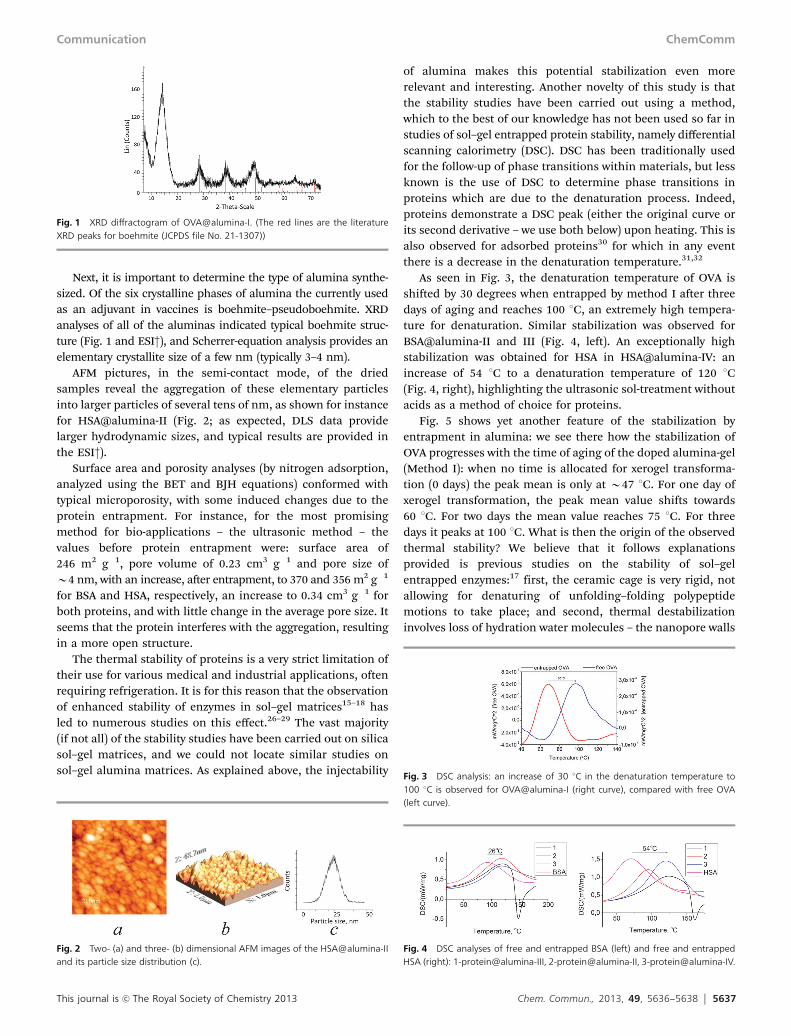
Next, it is important to determine the type of alumina synthe-sized. Of the six crystalline phases of alumina the currently usedas an adjuvant in vaccines is boehmite–pseudoboehmite. XRDanalyses of all of the aluminas indicated typical boehmite struc-ture (Fig. 1 and ESI†), and Scherrer-equation analysis provides anelementary crystallite size of a few nm (typically 3–4 nm).
AFM pictures, in the semi-contact mode, of the driedsamples reveal the aggregation of these elementary particlesinto larger particles of several tens of nm, as shown for instancefor HSA@alumina-II (Fig. 2; as expected, DLS data providelarger hydrodynamic sizes, and typical results are provided inthe ESI†).
Surface area and porosity analyses (by nitrogen adsorption,analyzed using the BET and BJH equations) conformed withtypical microporosity, with some induced changes due to theprotein entrapment. For instance, for the most promisingmethod for bio-applications – the ultrasonic method – thevalues before protein entrapment were: surface area of246 m2 g�1, pore volume of 0.23 cm3 g�1 and pore size ofB4 nm, with an increase, after entrapment, to 370 and 356 m2 g�1
for BSA and HSA, respectively, an increase to 0.34 cm3 g�1 forboth proteins, and with little change in the average pore size. Itseems that the protein interferes with the aggregation, resultingin a more open structure.
The thermal stability of proteins is a very strict limitation oftheir use for various medical and industrial applications, oftenrequiring refrigeration. It is for this reason that the observationof enhanced stability of enzymes in sol–gel matrices15–18 hasled to numerous studies on this effect.26–29 The vast majority(if not all) of the stability studies have been carried out on silicasol–gel matrices, and we could not locate similar studies onsol–gel alumina matrices. As explained above, the injectability
of alumina makes this potential stabilization even morerelevant and interesting. Another novelty of this study is thatthe stability studies have been carried out using a method,which to the best of our knowledge has not been used so far instudies of sol–gel entrapped protein stability, namely differentialscanning calorimetry (DSC). DSC has been traditionally usedfor the follow-up of phase transitions within materials, but lessknown is the use of DSC to determine phase transitions inproteins which are due to the denaturation process. Indeed,proteins demonstrate a DSC peak (either the original curve orits second derivative – we use both below) upon heating. This isalso observed for adsorbed proteins30 for which in any eventthere is a decrease in the denaturation temperature.31,32
As seen in Fig. 3, the denaturation temperature of OVA isshifted by 30 degrees when entrapped by method I after threedays of aging and reaches 100 1C, an extremely high tempera-ture for denaturation. Similar stabilization was observed forBSA@alumina-II and III (Fig. 4, left). An exceptionally highstabilization was obtained for HSA in HSA@alumina-IV: anincrease of 54 1C to a denaturation temperature of 120 1C(Fig. 4, right), highlighting the ultrasonic sol-treatment withoutacids as a method of choice for proteins.
Fig. 5 shows yet another feature of the stabilization byentrapment in alumina: we see there how the stabilization ofOVA progresses with the time of aging of the doped alumina-gel(Method I): when no time is allocated for xerogel transforma-tion (0 days) the peak mean is only at B47 1C. For one day ofxerogel transformation, the peak mean value shifts towards60 1C. For two days the mean value reaches 75 1C. For threedays it peaks at 100 1C. What is then the origin of the observedthermal stability? We believe that it follows explanationsprovided is previous studies on the stability of sol–gelentrapped enzymes:17 first, the ceramic cage is very rigid, notallowing for denaturing of unfolding–folding polypeptidemotions to take place; and second, thermal destabilizationinvolves loss of hydration water molecules – the nanopore walls
Fig. 1 XRD diffractogram of OVA@alumina-I. (The red lines are the literatureXRD peaks for boehmite (JCPDS file No. 21-1307))
Fig. 2 Two- (a) and three- (b) dimensional AFM images of the HSA@alumina-IIand its particle size distribution (c).
Fig. 3 DSC analysis: an increase of 30 1C in the denaturation temperature to100 1C is observed for OVA@alumina-I (right curve), compared with free OVA(left curve).
Fig. 4 DSC analyses of free and entrapped BSA (left) and free and entrappedHSA (right): 1-protein@alumina-III, 2-protein@alumina-II, 3-protein@alumina-IV.
Communication ChemComm

5638 Chem. Commun., 2013, 49, 5636--5638 This journal is c The Royal Society of Chemistry 2013
hold these water molecules very tightly, and thus protect theprotein from the destructive dehydration.
In conclusion, we have developed methods for the unharm-ful entrapment of proteins in alumina, showed that DSC canserve as a sensitive tool for the analysis of thermal stability ofentrapped proteins, found marked stabilization of theentrapped proteins in alumina using this method, and thusbelieve have opened a potential door for sol–gel materials to beconsidered as injectable carriers of bioactive molecules. Animalstudies on the immunogenic activity and release of OVA fromOVA@alumina (OVA is a common model for vaccinationstudies), as well as further activity and stability studies onadditional proteins will be reported in due course.
This work was supported by a Hebrew University Grant forExploratory Research, by a Golda Meir Fellowship at theHebrew University, and by a Russian President Grant for youngPhD researcher MK-2229.2012.3.
Notes and references‡ GRAS – generally regarded as safe.§ Experimental details: entrapment of the proteins in alumina: MethodI: as a first step boehmite sol was prepared as follows: 2.837 g ofAl(i-Pro)3 was hydrolyzed for 45 min in 51 ml DDW at 85 1C in an air-open flask. Then, 1.67 ml of 1 M HCl was added and the temperaturewas increased to 95 1C to catalyze the peptization. The flask was kept inopen air for additional 3 h to enable isopropanol evaporation, and thenplaced under reflux for an additional 16 hours. Then the refluxapparatus was removed and water was allowed to evaporate by air-exposure until the total sol volume was reduced to less than 25 ml,which was then re-adjusted to 25 ml with DDW (15 mg Al3+ per ml ofsol) (alumina-I). The second step – the protein entrapment – was carriedout as follows: 45.4 mg of OVA grade V was dissolved in 42 ml of 25 mML-histidine buffer at pH = 6.91. 4.0 ml of the protein-buffer solution wasadded to 1.0 ml of alumina sol, vortexed and left for 1 h under shakingin ice. Then 100 ml of a 50 mg ml�1 NaF solution was added to the5.0 ml protein-sol under vortex. The solution immediately becomesturbid and the gel forms. The tube is centrifuged for 10 min at5000 rpm and placed at 4 1C for 10 days for gel aging. The supernatantis then removed and the tube placed in a desiccator under vacuum for3–7 days for xerogel formation, until the gel shrinks to about 10% of itsinitial volume (OVA@alumina – I). The above ratios yield a 7.2 mg OVA–25 mg Al3+ composite, which is approximately 10% OVA dopant byweight. Methods II and III: the second and third methods used Yoldasprocedures by peptizing the aluminum hydroxide precipitate in acids:acetic acid (alumina-II) or nitric acid (alumina-III). In detail, 3.28 g ofAl(C3H7O)3 was added to 50 ml of deionized water and a whiteprecipitate was formed immediately. The precipitate was peptized with0.2 ml of concentrated nitric acid or with 2.0 ml of acetic acid at 90 1Cunder vigorous stirring for 2 h (also to evaporate the isopropanolformed during hydrolysis) to produce stable transparent boehmite sols(6.7 mg and 6.5 mg Al3+ per ml of sol, respectively). The pH of the final
solution was 2.8 (4.8 in acetic media). The second step – the albuminentrapment – was carried out unbuffered by dissolving 0.03 g of theprotein (BSA and HSA within alumina-II and alumina-III respectively) in20.0 ml of boehmite sol at room temperature. The suspension was agedfor 3 h and dried at 20 1C for a week. TGA analyses indicated an B8 w/w%loading in the two samples. Method IV: this method (alumina-IV)followed method II but without any acid or buffer. The white precipitatewas ultrasonically treated (37 kHz, 0.063 kW) for 4 h (final pH = 7.3). After4 h a viscous sol was formed. The second step – the albumin entrapment –was carried out at room temperature by dissolving 0.03 g of the proteinin 20.0 ml of the sol (alumina-IV): with BSA – BSA@alumina-IV, or withHSA – HSA@alumina-IV. The suspension was treated as above resultingin an B8 w/w% loading. For chemicals and characterization techni-ques, see ESI.†
1 C. Barbe, J. Bartlett, L. G. Kong, K. Finnie, H. Q. Lin, M. Larkin,S. Calleja, A. Bush and G. Calleja, Adv. Mater., 2004, 16, 1959.
2 S. Radin, P. Ducheyne, T. Kamplain and B. H. Tan, J. Biomed. Res.,2001, 57, 313.
3 M. Ahola, E. S. Sailynoja, M. H. Raitavuo, M. M. Vaahtio, J. I. Salonenand A. U. O. Yili-Urpo, Biomaterials, 2001, 22, 2163.
4 P. Kortesuo, M. Ahola, M. Kangas, T. A. Yli-Urpo, J. Kiesvaara andM. Marvola, Int. J. Pharm., 2001, 221, 107.
5 H. Bottcher, P. Slowik and W. Suss, J. Sol-Gel Sci. Technol., 1998,13, 277.
6 M. Ahola, P. Kortesuo, I. Kangasniemi, T. J. Kiesvaara and A. Yli-Urpo,Int. J. Pharm., 2000, 195, 219.
7 T. Lopez, J. Manjarrez, D. Rembao, E. Vinogradova, A. Moreno andR. D. Gonzalez, Mater. Lett., 2006, 60, 2903.
8 S. Fireman-Shoresh, N. Husing and D. Avnir, Langmuir, 2001,17, 5958.
9 S. Braun, S. Rappoport, R. Zusman, D. Avnir and M. Ottolenghi,Mater. Lett., 1990, 10, 1.
10 L. Siemenska, M. Ferguson, T. W. Zerda and E. Couch, J. Sol-Gel Sci.Technol., 1997, 8, 1105.
11 A. Turniansky, D. Avnir, A. Bronshtein, N. Aharonson andM. Altstein, J. Sol-Gel Sci. Technol., 1996, 7, 135.
12 I. Gill and A. Ballesteros, J. Am. Chem. Soc., 1998, 120, 8587.13 S. Boninsegna, P. Bosetti, G. Carturan, G. Dellagiacoma, R. Dal
Monte and M. Rossi, J. Biotechnol., 2003, 100, 277.14 G. Sailaja, H. Hogenesch, A. North, J. Hays and S. K. Mittal, Gene
Ther., 2002, 9, 1722.15 J. P. Chen, W. S. Lin and M. F. Chang, J. Am. Oil Chem. Soc., 2002,
79, 309.16 L. L. Zheng, K. Flora and J. D. Brennan, Chem. Mater., 1998, 10, 3974.17 H. Frenkel-Mullerad and D. Avnir, J. Am. Chem. Soc., 2005, 127, 8077.18 D. J. V. Unen, J. F. J. Engbersen and D. N. Reinhoudt, Biotechnol.
Bioeng., 2001, 75, 154.19 World Health Organization, Immunological Adjuvants, Technical
Report Series 595, Geneva, World Health Organization, 1976.20 R. Edelman, Rev. Infect. Dis., 1980, 2, 370.21 B. Xu, B. Xiao, Z. Yan, X. Sun, J. Sloan, S. L. Gonzalez-Cortes,
F. Alshahrani and M. L. H. Green, Microporous Mesoporous Mater.,2006, 91, 293.
22 Z. J. Liu, B. H. Liu, J. L. Kong and J. Q. Deng, Anal. Chem., 2000,72, 4707.
23 Z. Liu, B. Liu, M. Zhang, J. Kong and J. Deng, Anal. Chim. Acta, 1999,392, 135.
24 H. Yagar and A. Sagiroglu, Acta Chim. Slov., 2002, 49, 893.25 M. Amoura, N. Nassif, C. Roux, J. Livage and T. Coradin, Chem.
Commun., 2007, 4015.26 E. H. Lan, Dave, J. M. Fukuto, B. Dunn, J. I. Zink and J. S. Valentine,
J. Mater. Chem., 1999, 9, 45.27 Y. W. Cho and S. Han, Bull. Korean Chem. Soc., 1999, 20, 1363.28 K. Flora and J. D. Brennan, Anal. Chem., 1998, 70, 4505.29 E. H. Lan, B. Dunn and J. I. Zink, Chem. Mater., 2000, 12, 1874.30 E. V. Parfenyuk, G. A. Kulikova and I. V. Ryabinina, J. Therm. Anal.
Calorim., 2010, 100, 987.31 H. Larsericsdotter, S. Oscarsson and J. Buijs, J. Colloid Interface Sci.,
2001, 237, 98.32 P. Billsten, U. Caresson, B. H. Jonsson, G. Olofsson, F. Hook and
H. Elwing, Langmuir, 1999, 15, 6395.
Fig. 5 Gradual increase in stabilization with aging of OVA entrapped inalumina-gel-I, followed with the 2nd derivative of the DSC curves.
ChemComm Communication
Synthesis and enhanced thermal stability of albumins@alumina:
Towards injectable sol-gel materials
Avi Rutenberg,a Vladimir V. Vinogradov
a,b and David Avnir*
a
a Institute of Chemistry and the Center for Nanoscience and Nanotechnology, the Hebrew
University of Jerusalem, Jerusalem 91904, Israel. E-mail: [email protected] bInstitute of Solution Chemistry, Russian Academy of Sciences, Akademicheskaya 1, Ivanovo
153045, Russia. E-mail: [email protected]
Electronic Supplementary Information
1. IR interpretation of Figure 1
Note the typical finger-print bands of alumina at the 400 – 830 cm-1
region, and the
peaks at 1050 – 1070 cm-1
which are associated with the symmetric stretching vibrations of
the Al–O-H bonds. The presence of a peak at 1384 cm-1
in alumina-IV corresponds to
adsorbed CO2 [1]. The same bands can be observed in HSA@alumina-IV. The typical
amide bands I and II – the 1700–1600 cm-1
stretching vibrations of the C=O bond and the
in-plane bending vibrations of the N–H bond, and stretching vibrations of the С–N bond at
1570–1510 cm-1
[28], are clearly seen both in the IR spectrum of the HSA and in that of
the composite.
Figure 1S. FTIR spectra of the pure HSA (1), pure alumina-IV (3), and HSA@alumina-
IV(2)

2. Additional XRD observations:
Figure 2S presents the powder XRD patterns of additional alumina samples
(without albumins). All of the as-synthesized samples display diffraction lines of boehmite
(The red lines are the literature XRD peaks for Boehmite (JCPDS file No. 21-1307). These
results are in a good agreement with XRD data of commonly used alumina adjuvants [2].
Figure 2S. XRD spectra of sol-gel alumina samples
3. Additional AFM observations
AFM observations (Figure 3S) of the bioentrapped samples represent a morphology
of aggregates. The size of aggregates in the BSA@alumina-II averages 20 nm, in
BSA@alumina-III – 15 nm, in BSA@alumina-IV - 100–200 nm, and in HSA@alumina-IV
- 100 – 200nm. The absence of nanophase components of the albumin@Al2O3 composites,
confirms the homogeneous entrapment of the proteins in the alumina matrix.
BSA@alumina-II

Figure 3S. Two- and three-dimensional AFM images of albumins@aluminas
4. Dynamic light scattering results
As expected, DLS data provides larger hydrodynamic sizes, and typical results are
shown in Figure 4S for BSA@alumina and HSA@alumina prepared either by method II,
III or by method IV. It is seen that while the use of acetic acid as a catalysts keeps the
hydrodynamic radii with little change before and after entrapment of the proteins, in the
case of ultrasonic treatment, some increase in particle size is observed. It should be noted
that the use of acetic acid and ultrasonic treatment provide solution pH values of 4.8 and
7.3, respectively, that are both comfortable for biomolecules and corresponds to an
optimum range at which the albumin molecules and the particles have opposite charges,
providing the needed electrostatic interaction for entrapment.
BSA@alumina-III
BSA@alumina-IV
HSA@alumina-IV

Figure 4S. Average hydrodynamic radius of pure aluminas prepared by method III(3),
II(4), IV(5) and, of the BSA@alumina-III(6), BSA@alumina-II(7), BSA@alumina-IV(8)
and HSA@alumina-III(9), HSA@alumina-II(10), HSA@alumina-IV(11).
5. Additional Experimental Details
Chemicals: Aluminum isopropoxide (ALISP) was either from Sigma-Aldrich or from
Strem. Ovalbumin (OVA) grade V, bovine serum albumin (BSA, ≥96%) and human
serum albumin (HSA ≥97%) were purchased from the Sigma-Aldrich. All other standard
chemicals were of the highest available grades.
Characterization techniques: Specific surface area, pore volume, and pore size distribution
of the synthesized samples were determined from N2 adsorption–desorption isotherms at
77 K (Quantachrome Nova 1200). Surface area was calculated using the BET equation;
pore volume and pore size distribution were calculated by the BJH method. The samples
were evacuated at room temperature for 4 h prior to their analysis. The hydrodynamic size
distributions were measured by dynamic light scattering (DLS, Malvern, Zetasizer nano
ZS). FTIR spectroscopy was carried out with an "Avatar" spectrometer. Atomic force
microscope (AFM) studies were carried out using a SPM Solver P47H-PRO microscope.
Samples were applied on a glass plate having clean surface. XRD analyses were carried
out with a Philips automated powder diffractometer using Cu–Kα radiation (λ = 1.54 Å) at
a scanning rate of 2 deg/minute. DSC curves were obtained with either 204 F1 Phoenix
NETZSCH or with a Mettler-Toledo DSC 823e apparatus. Heating rate of 1 °C min-1
was
used from 25 °C to 170 °C under nitrogen.For the DCS time analysis, the gelation process
was stopped by lyophilization at (0, 1, 2 and 3 days).
References for ESI:
1. V.B. Kazansky, V.Yu Borovkov, A.I. Serykh, M.Bulow. Phys. Chem. Chem. Phys.
1999, 1, 3701.
2. E.B. Lindbland, Immunology and Cell Biology, 2004, 1980, 82, 497.