Acetyl analogs of combretastatin A-4: Synthesis and biological studies
Upload
khangminh22Category
view
5download
0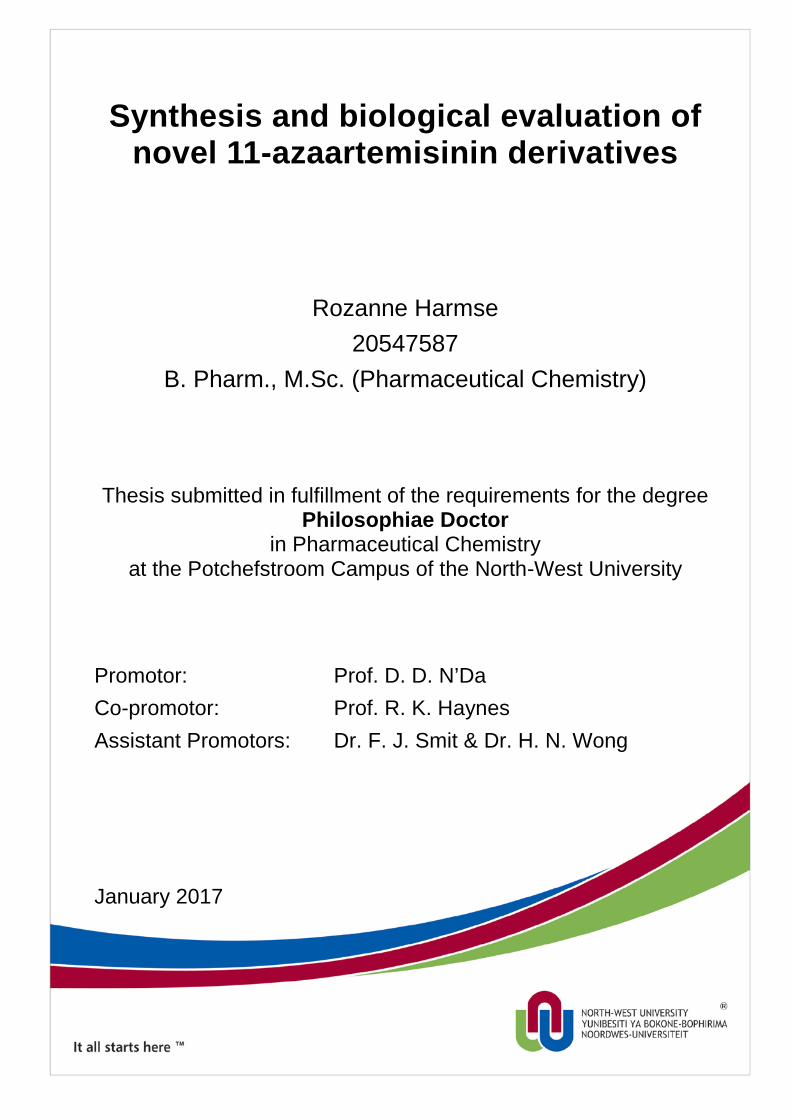
Synthesis and biological evaluation of novel 11-azaartemisinin derivatives
Rozanne Harmse 20547587
B. Pharm., M.Sc. (Pharmaceutical Chemistry)
Thesis submitted in fulfillment of the requirements for the degree Philosophiae Doctor
in Pharmaceutical Chemistry at the Potchefstroom Campus of the North-West University
Promotor: Prof. D. D. N’Da Co-promotor: Prof. R. K. Haynes Assistant Promotors: Dr. F. J. Smit & Dr. H. N. Wong
January 2017

SOLEMN DECLARATION
~ III ~
PREFACE
This thesis is submitted in an article format in accordance with the General Academic Rules
(A.13.7.3) of the North-West University. Three articles, one of which have been published, are
included in this thesis:
CHAPTER 3: Article 1 Harmse, R., Wong, H. N., Smit, F., Haynes, R. K., N’Da, D. D. 2015. The case for development
of 11-azaartemisinins for malaria. Current Medicinal Chemistry, 22:1 – 23.
CHAPTER 4: Article 2 Harmse, R., Smit, F. J., Coertzen, D., Wong, H. N., Birkholtz, L.-M., Haynes, R. K., N’Da, D. D.
2016. Antimalarial activities and cytotoxicities of N-sulfonyl-11-azaartemisinin derivatives.
European Journal of Medicinal Chemistry. To be submitted.
CHAPTER 5: Article 3 Harmse, R., Smit, F. J., Wong, H. N., Müller, J., Hemphill, A, N’Da, D. D., Haynes, R. K. 2016.
Activities of N-sulfonyl-11-azaartemisinin derivatives against the apicomplexan parasite
Neospora caninum and comparative cytotoxicities. Bioorganic & Medicinal Chemistry Letters.
To be submitted.
The respective Journals of the submitted articles grant the author the right to include these
articles in a thesis. Permission for the Journals is given within their Author's Guides:
Permission from Bentham Science: http://benthamscience.com/journals/current-medicinal-
chemistry/author-guidelines/ Permission from Elsevier: https://www.elsevier.com/journals/european-journal-of-medicinal-
chemistry/0223-5234/guide-for-authors Permission form Elsevier: https://www.elsevier.com/journals/bioorganic-and-medicinal-chemistry-
letters/0960-894X/guide-for-authors
~ IV ~
ACKNOWLEDGEMENTS
I am grateful to the individuals who gave their support, time and knowledge in order to help me
fulfil one of my biggest dreams. To each and every one who contributed in any way, I want to
express my deepest gratitude, appreciation and thanks:
My supervisor, Prof. David D. N’Da, for your patience and guidance. I have learnt so
much from you.
My co-supervisor, Prof. Richard K. Haynes, no matter how busy you were you always
made time to talk, be it for moral support, chemistry or even a good book. Thank you for
all your encouragement, guidance and the vast amount of knowledge you shared with
me.
Assistant supervisors Dr. Frans J. Smit and Dr. H. N. Wong (Coco), thank you for being
there when I needed someone to guide me in my lab work.
Prof. Jacques P. Petzer, Prof. Sandra van Dyk, Prof. Gisella Terre’blanche and Dr. Arina
Lourens for their kind words of reassurance.
Prof. Jeanetta du Plessis, for your support, advice and sympathetic ear.
Andrè Joubert and Dr. Johan Jordaan for the collection of NMR and MS data,
respectively.
The NWU and NRF for their financial support.
Lab partners Paul Joubert, Christo de Lange, Idalet Engelbrecht and Richard Beteck.
You made the lab a fun space to be in. Thanks for all the chats and the numerous
complaints we shared about how difficult (and sometimes painful) the PhD experience is.
From the bottom of my heart; Righard Lemmer and Jaco Wentzel for the every-day early
morning sessions of coffee, biscuits and the best conversations anyone could ask for. I
miss it dearly.
Special thanks to Rodè van Eeden, Dirkie Nell, Angélique Lewies and Helanie Lemmer
who contributed to the social environment of this post-graduate student.
Hannes de Wet for your love, support and understanding through all the ups and downs.
For wiping away all the tears, your continuous encouragement and general positive
outlook on life. I am glad to call you mine.
My family and friends for their love and support.
Heavenly Father, blessed is thy name.

~ V ~
ABSTRACT
Malaria is a devastating mosquito-borne disease caused by several species of Plasmodium
protozoa, of which the most important is Plasmodium falciparum (Pf). Globally, the disease
caused approximately 438 000 deaths in 2014; disease prevalence is highest in the African
region. Artemisinin and its derivatives have emerged as the drugs of choice for treatment of
malaria where they are used in artemisinin combination therapies (ACTs). However, emergence
of resistance to artemisinins poses a global threat to current treatment regimens.
Chapter 3 comprises a review article that examines a relatively new artemisinin derivative, 11-
azaartemisinin, and its scientific evolution throughout the past 20 years. Various routes to
azaartemisinin derivatives are critically discussed and the biological activities of the
azaartemisinin derivatives are examined in order to evaluate if this class of compound is
suitable for carrying forward for development into new drugs in the fight against malaria. In
general, the azaartemisinins that have been examined display promising antimalarial activities,
and would appear to have several advantages over their artemisinin predecessors in being
more stable and chemically robust. Decisively, the azaartemisinins cannot provide
dihydroartemisinin (DHA) through metabolism or via hydrolysis. As the current clinical
artemisinins, against which resistance is now emerging, characteristically provide DHA on
metabolism or hydrolysis in vivo, the newer azaartemisinins will not have this disadvantage,
especially as DHA has been fingered as the actual drug that induces resistance among the
current clinical artemisinins.
In Chapter 4 the synthesis of N-sulfonyl-11-azaartemisinin derivatives are described and the
evaluation of the antimalarial activities against intraerythrocytic stages of chloroquine (CQ)
sensitive Pf NF54 and CQ resistant Pf K1 and W2 parasites. The gametocytocidal activities
were assessed against Pf NF54 blood-stage gametocytes using the luciferase and pLDH
assays. Cytotoxicities of the compounds were also evaluated against the human fetal lung
fibroblasts WI-38 cell line (HFLF) and were shown to be relatively non-toxic. The p-
trifluoromethylbenzenesulfonyl-11-azaartemisinin derivative was the most active antimalarial
compound with IC50 values between 2 – 3 nM, whereas the 2'-thienylsulfonyl derivative
demonstrated the best late-stage (IV-V) activity against gametocytes with an IC50 value of 8.7
nM. These two compounds are thus potential candidates for further development.
In Chapter 5 the evaluation of nine of the active antimalarial N-sulfonylazaartemisinin
derivatives against the apicomplexan parasite Neospora caninum responsible for bovine
abortion in beef and dairy cattle, are described. The antitumor activities were also determined in

~ VI ~
order to assess their parasitic versus intracellular activities in general. The 2,5-
dichlorothienylsulfonyl-11-azaartemisinin was the most active against neosporosis with an IC50
value of 40 nM, whereas the hexadecanesulfonyl derivative demonstrated prominent antitumor
activity against breast cancer cells.
Overall, the current study has resulted in the identification of compounds that exhibit varying
antimalarial activities, some of which are comparable to the current clinically available
artemisinins. These compounds serve as suitable candidates for additional research in order to
evaluate their potential as future lead compounds for development into drugs against malaria.
Also, several compounds display promising activities against the causative parasite of
neosporosis, and likewise require further investigation to evaluate their potential.
Keywords: Plasmodium falciparum, malaria, azaartemisinin, N-sulfonyl-11-azaartemisinin,
gametocytocidal, neosporosis

~ VII ~
OPSOMMING
Malaria is 'n ernstige muskiet-oordraagbare siekte wat veroorsaak word deur spesies van
Plasmodium protosoë, waarvan die belangrikste Plasmodium falciparum (Pf) is. Die siekte
veroorsaak wêreldwyd ongeveer 438 000 sterftes per jaar waarvan die voorkoms van die siekte
die hoogste is in die Afrika-streek. Artemisinien en sy derivate geniet voorkeur as
geneesmiddels vir die behandeling van malaria waar dit gebruik word in kombinasie met
artemisinien terapie. Die ontwikkeling van weerstand teen artemisinien hou ongelukkig 'n
wêreldwye bedreiging in vir die huidige behandelingsvorm.
Hoofstuk 3 bevat 'n oorsig artikel wat 'n relatiewe nuwe artemisinien derivaat, 11-
azaartemisinien, en sy wetenskaplike evolusie van die afgelope 20 jaar, ondersoek. Verskeie
sintetiese metodes om derivate van azaartemisinien te bekom, word krities bespreek. Die
biologiese aktiwiteite van die azaartemisinien derivate word bestudeer om uiteindelik te bepaal
of hierdie klas van geneesmiddels geskik is vir verdere ontwikkeling as nuwe geneesmiddels
wat gebruik kan word in die stryd teen malaria. Oor die algemeen vertoon die azaartemisiniene
wat ondersoek is belowende anti-malaria aktiwiteit. Dit wil voorkom asof hierdie klas van
geneesmiddels menigte voordele inhou in vergelyking met hul artemisinien voorgangers. Die
nuwer verbindings is meer stabiel, asook chemies meer robuus. Die azaartemisiniene kan nie
dihydroartemisinien (DHA) deur middel van metabolisme of hidrolise voorsien nie. Die huidige
kliniese artemisiniene, waar weerstand nou algemeen voorkom, vorm kenmerkende DHA in vivo
wanneer dié gemetaboliseer word, of hidrolise ondergaan. Die nuwe azaartemisiniene sal dus
nie oor hierdie nadelige eienskap beskik nie, veral noudat dit bewys kan word dat DHA die
werklike oorsaak is van weerstand onder huidige kliniese artemisiniene.
Hoofstuk 4 beskryf die sintese van N-sulfoniel-11-azaartemisinien derivate asook die evaluering
van malaria-aktiwiteite teen intra-eritrositiese fases van chlorokien (CQ) sensitiewe Pf NF54 en
CQ bestande Pf K1 en W2 parasiete. Die gametosiet aktiwiteite word ook bepaal teenoor Pf
NF54 bloed-fase gametosiete met behulp van die lusiferase en pLDH toetse. Sitotoksisiteite van
die verbindings is ook geëvalueer teenoor menslike fetale long fibroblaste WI-38 sellyn (HFLF)
en het getoon dat die verbindings relatief veilig is. Die p-trifluorometielbenseensulfoniel-11-
azaartemisinien derivaat was die mees aktiewe malaria verbinding met IC50 waardes tussen 2 –
3 nM, terwyl die 2'-tiofeensulfoniel derivaat die beste laat stadium (IV-V) aktiwiteit teen
gametosiete gedemonstreer het met 'n IC50 waarde van 8,7 nM. Hierdie twee verbindings is
potensiële kandidate vir verdere ontwikkeling.
~ VIII ~
Hoofstuk 5 beskryf die evaluering van nege van die aktiewe malaria N-sulfonielazaartemisinien
derivate teenoor die apikompleksia parasiet Neospora caninum, wat verantwoordelik is vir
misgeboortes in vleis- en melkbeeste. Die antikanker aktiwiteite van die verbindings word ook
bepaal sodat hul parasitiese aktiwiteite teenoor intrasellulêre aktiwiteite beoordeel kan word.
Die 2,5-dichlorotiofeensulfoniel-11-azaartemisinien was die mees aktiewe verbinding teenoor
neosporose met 'n IC50 waarde van 40 nM, terwyl die heksadekaansulfoniel derivaat
prominente anti-kanker aktiwiteit teenoor borskankerselle gedemonstreer het.
Die huidige studie het gelei tot die identifisering van verbindings wat wissellende malaria
aktiwiteite het, waarvan sommige verbindings se aktiwiteite vergelykbaar is met die huidige
klinies beskikbare artemisiniene. Hierdie verbindings dien as geskikte kandidate vir verdere
navorsing om hul potensiaal as toekomstige leidraadverbindings vir ontwikkeling as
geneesmiddels teen malaria te evalueer. Van die verbindings toon belowende aktiwiteite
teenoor neosporose en vereis ook verdere ondersoek om hul potensiaal te evalueer.
Sleutelwoorde: Plasmodium falciparum, malaria, azaartemisinien, N-sulfoniel-11-
azaartemisinien, gametosiete, neosporose

~ IX ~
TABLE OF CONTENTS
SOLEMN DECLERATION ................................................................................................................................................................. II PREFACE ..............................................................................................................................................................................................III ACKNOWLEDGEMENTS ................................................................................................................................................................ IV ABSTRACT ............................................................................................................................................................................................ V OPSOMMING ..................................................................................................................................................................................... VII TABLE OF CONTENTS ..................................................................................................................................................................... XI LIST OF FIGURES............................................................................................................................................................................. XII LIST OF TABLES ............................................................................................................................................................................. XVI LIST OF SCHEMES ....................................................................................................................................................................... XVIII LIST OF ABBREVIATIONS .......................................................................................................................................................... XIX CHAPTER 1: INTRODUCTION AND OBJECTIVES .................................................................................................................. 1 1.1 INTRODUCTION ........................................................................................................................................................................ 1 1.2 RATIONALE ............................................................................................................................................................................... 4 1.3 AIMS AND OBJECTIVES ............................................................................................................................................................ 7
1.3.1 Aim of this study ...................................................................................................................................................... 7 1.3.2 Specific objectives of this study ........................................................................................................................ 8
REFERENCES ............................................................................................................................................................................. 9 CHAPTER 2: LITERATURE OVERVIEW .................................................................................................................................. 15 2.1 INTRODUCTION ..................................................................................................................................................................... 15 2.2 EPIDEMIOLOGY ...................................................................................................................................................................... 16 2.3 MALARIA LIFE CYCLE AND PATHOGENESIS ....................................................................................................................... 17 2.4 CLINICAL FEATURES OF MALARIA ...................................................................................................................................... 22 2.5 PATHOGENESIS ...................................................................................................................................................................... 23 2.6 SEVERE MALARIA .................................................................................................................................................................. 24 2.7 DIAGNOSIS ............................................................................................................................................................................. 25 2.8 VECTOR CONTROL AND MALARIA PREVENTION ............................................................................................................... 26 2.9 CHEMOTHERAPY ................................................................................................................................................................... 29
2.9.1 Quinoline and related antimalarials ............................................................................................................ 30 2.9.1.1 Aryl-amino alcohols .............................................................................................................................................. 30
2.9.1.2 4-Aminoquinolines ................................................................................................................................................ 33 2.9.1.3 8-Aminoquinolines ................................................................................................................................................ 35 2.9.2 Hydroxynaphthoquinones................................................................................................................................ 36 2.9.3 Antifolates ................................................................................................................................................................ 37 2.9.3.1 DHPS inhibitors ...................................................................................................................................................... 39 2.9.3.2 DHFR inhibitors ...................................................................................................................................................... 39 2.9.4 Antibiotics ................................................................................................................................................................ 41

~ X ~
2.9.5 Artemisinins ........................................................................................................................................................... 42 2.9.5.1 Introduction ............................................................................................................................................................. 42 2.9.5.2 Mechanism of action ............................................................................................................................................ 43 2.9.5.3 Artemisinin and its first generation semisynthetic peroxides ............................................................. 48 2.9.5.4 Artemisinin combination therapy (ACT) ...................................................................................................... 50 2.9.5.5 Artemisinin resistance ......................................................................................................................................... 52 2.9.5.6 Other artemisinin derivatives ........................................................................................................................... 56
REFERENCES .......................................................................................................................................................................... 59 CHAPTER 3: THE CASE FOR DEVELOPMENT OF 11-AZAARTEMISININS FOR MALARIA – ARTICLE 1 ..... 88
ABSTRACT .............................................................................................................................................................................. 90 1 INTRODUCTION ..................................................................................................................................................................... 90 2 TOTAL SYNTHESES OF N-ALKYL-11-AZA-9-DESMETHYLARTEMISININS ..................................................................... 93 3 DIRECT CONVERSION OF ARTEMISININ INTO AZAARTEMISININS .................................................................................. 94
3.1 Preperation of 11-azaartemisinin 7 from artemisinin 1 and ammonia ....................................... 94 3.2 The problem of competing formation of 11-azadeoxyartemisinin 19 ......................................... 95 3.3 Preparation of N-alkyl-11-azaartemisinins from artemisinin 1 and primary amines .......... 97 3.4 Preparation of N-funtionalized-11-azaartemisinins from artemisinin 1 and functionalized
primary amines ........................................................................................................................................................................... 98 4 DIRECT CONVERSION OF 11-AZAARTEMISININ INTO FUNCTIONALIZED N-SUBSTITUTED DERIVATIVES ............. 100
4.1 Addition of 11-azaartemisinin 7 to electron deficient alkenes ..................................................... 100 4.2 Functionalization of the acrylate adduct 54 of 11-azaartemisinin 7 by the Ugi reaction 102 4.3 Conversion of 11-azaartemisinin into N-sulfonyl- and N-carbonyl-11-azaartemisinins .. 103
CONCLUSION ....................................................................................................................................................................... 105 CONFLICT OF INTEREST .................................................................................................................................................... 106 ACKNOWLEDGEMENTS ...................................................................................................................................................... 110 REFERENCES ....................................................................................................................................................................... 110
CHAPTER 4: ANTIMALARIAL ACTIVITIES AND CYTOTOXICITIES OF N-SULFONYL-11-AZAARTEMISININ
DERIVATIVES – ARTICLE 2 ...................................................................................................................................................... 114 ABSTRACT ........................................................................................................................................................................... 116
GRAPHICAL ABSTRACT ..................................................................................................................................................... 117 INTRODUCTION .................................................................................................................................................................. 118 RESULTS AND DISCUSSION ................................................................................................................................................ 120 CONCLUSION ....................................................................................................................................................................... 124 EXPERIMENTAL SECTION .................................................................................................................................................. 124 DISCLAIMER ........................................................................................................................................................................ 135 ACKNOWLEDGEMENTS ...................................................................................................................................................... 135 REFERENCES ....................................................................................................................................................................... 136
CHAPTER 5: ACTIVITIES OF N-SULFONYL-11-AZAARTEMISININ DERIVATIVES AGAINST THE
APICOMPLEXAN PARASITE NEOSPORA CANINUM AND COMPARATIVE CYTOTOXICITIES – ARTICLE 3
(LETTER) ......................................................................................................................................................................................... 139

~ XI ~
ABSTRACT ........................................................................................................................................................................... 141 ACKNOWLEDGEMENTS ...................................................................................................................................................... 147 DISCLAIMER ........................................................................................................................................................................ 147 REFERENCES ....................................................................................................................................................................... 148
CHAPTER 6: SUMMARY AND CONCLUSION ...................................................................................................................... 152
REFERENCES ....................................................................................................................................................................... 157 ADDENDUM A: ANALYTICAL DATA FOR CHAPTER 4 .................................................................................................. 159

~ XII ~
LIST OF FIGURES
Chapter 1:
Figure 1.1: Artemisinin and derivatives dihydroartemisinin, artemether, arteether and
artesunate ...................................................................................................... 3
Figure 1.2: 11-Azaartemisinin contains the lactam moiety instead of the lactone of
artemisinin, making the compound chemically more robust ............................ 6
Chapter 2:
Figure 2.1: Countries with ongoing malaria transmission ..................................................... 16
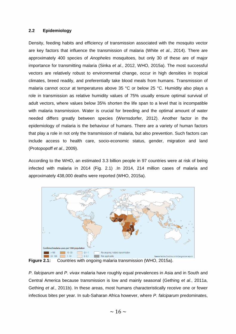
Figure 2.2: The various stages involved in the life cycle of the malaria parasite ................... 17
Figure 2.3: The merozoite .................................................................................................... 18
Figure 2.4: a. Invasion of an erythrocyte by a merozoite. b. Merozoite invading a human
red blood cell ................................................................................................ 20
Figure 2.5: The major stages throughout the erythrocytic cycle of P. falciparum .................. 21
Figure 2.6: Sexual (sporogonic) phase in mosquito midgut .................................................. 22
Figure 2.7: Types of adhesion involving erythrocytes infected with the malaria parasite ...... 23
Figure 2.8: Microscopic image of sequestration of a red blood cell ....................................... 24
Figure 2.9: Quinine 1, one of the first antimalarial drugs, and its diatereoisomer
quinidine 2 ................................................................................................... 30
Figure 2.10: Mefloquine 3, a 4-methanolquinoline .................................................................. 31
Figure 2.11: Halofantrine 4 ..................................................................................................... 32
Figure 2.12: Lumefantrine 5 ................................................................................................... 33
Figure 2.13: Chloroquine (CQ) 6 ............................................................................................ 34
~ XIII ~
Figure 2.14: Amodiaquine 7 and monodesethyl amodiaquine 8 ............................................. 34
Figure 2.15: Piperaquine 9 ..................................................................................................... 35
Figure 2.16: Primaquine 10 .................................................................................................... 36
Figure 2.17: Atovaquone 11 ................................................................................................... 37
Figure 2.18: Folate biochemical pathway in P. falciparum ...................................................... 38
Figure 2.19: Class I antifolates dapsone 12 and sulfadoxine 13 ............................................. 39
Figure 2.20: Proguanil 14 and its active metabolite cycloguanil 15 ......................................... 40
Figure 2.21: Pyrimethamine 16 .............................................................................................. 40
Figure 2.22: Clindamycin 17................................................................................................... 41
Figure 2.23: Doxycycline 18 ................................................................................................... 42
Figure 2.24: Proposed enhancement of oxidative stress facilitated by peroxidic
antimalarials ................................................................................................. 46
Figure 2.25: Proposal for cytosolic action of artemisinins ....................................................... 48
Figure 2.26: Artemisinin 19 and dihydroartemisinin (DHA) 20 ................................................ 49
Figure 2.27: Artemether 21 and arteether 22 ......................................................................... 49
Figure 2.28: Artesunate 23 ..................................................................................................... 50
Figure 2.29: Global distribution of current artemisinin-based combination therapies used
as first line treatment of uncomplicated P. falciparum malaria ...................... 52
Figure 2.30: P. falciparum resistance towards artemisinin observed in the five countries in
the Greater Mekong Subregion .................................................................... 55
Figure 2.31: Artemisone 24 .................................................................................................... 57
Figure 2.32: 11-Azaartemisinin 25.......................................................................................... 58
Chapter 3:

~ XIV ~
Figure 1: Structures of artemisinin 1 and clinically used derivatives dihydroartemisinin
(DHA) 2, artemether 3 and artesunate 5 ...................................................... 92
Figure 2: 9-Desmethylartemisinin 12 first prepared by total synthesis by Avery and co-
workers ........................................................................................................ 94
Figure 3: a. Proposal for formation of 11-aza-deoxoartemisinin 19 from ammonia and
aremisinin 1.b. Outcome of experiment involving treatment of artemisinin
1 with benzylamine and excess of N-methylmorpholine in
dichloromethane. c. Outcome of experiment involving treatment of
artemisinin with excess of benzylamine in dichloromethane ......................... 96
Figure 4: a. Products 28 and 29 obtained from artemisinin 1 and excess of
ethanolamine in methanol. b. Product 30 obtained from artemisinin 1
and excess of ethylenediamine in methanol ................................................. 98
Figure 5: Thermogravimetric analysis (TGA) of artemisinin 1 and 11-azaartemisinin 7
heated at a rate of 108 °C min-1 under N2 .................................................. 104
Figure 6: Thermogravimetric analysis (TGA) of methanesulfonylazaartemisinin 65 and
methanecarbonylazaartemisinin 72 heated at a rate of 108 °C min-1
under N2 .................................................................................................... 106
Chapter 4:
Figure 1: Structures of artemisinin 1 and its current clinical derivatives, the hemiacital
dihydroartemisinin (DHA) 2, the lactol ether artemether 3 and hemiester
artesunate 4. The latter two drugs essentially act as prodrugs for DHA
via facile metabolism or hydrolysis respectively. Azaartemisinin 5 with
the lactam replacing the lactone of artemisinin is expected to be more
stable at physiological pH, and is chemically incapable of providing DHA
by hydrolysis or metabolism ....................................................................... 118
Chapter 5:
Figure 1: Artemisinin 1 and derivatives artemether 2, artesunate 3, artemisone 4 and
11-azaartemisinin 5 .................................................................................... 143

~ XV ~
Figure 2: N-Sulfonyl-11-zaartemisinins screened against N. caninum and three cancer
cell lines TK-10 (renal), UACC-62 (melanoma) and MCF-7 (breast) ........... 144
Figure 3: In vitro activities of the sulfonyl azaartemisinins as compared with
artemisone 4; the most active compounds are depicted in red ................... 145

~ XVI ~
LIST OF TABLES
Chapter 3:
Table 1: In vitro activities expressed as relative potencies (%) of selected N-alkyl-9-
desmethyl-11-azaartemisinins against P. falciparum relative to
artemisinin 1................................................................................................. 94
Table 2: In vitro activities of selected N-alkyl-11-azaartemisinins (Scheme 3) against
P. falciparum relative to artemisinin 1 ........................................................... 97
Table 3: In vivo activities against P. berghei in mice ........................................................ 98
Table 4: Activity of compounds administered orally against P. yoelii in mice .................. 100
Table 5: In vitro antimalarial activities of selected N-substituted 11-azaartemisinins
against P. falciparum relative to artemisinin 1 ............................................ 102
Table 6: In vitro activities of polar functionalized azaartemisinins against P.
falciparum .................................................................................................. 103
Table 7: In vitro activities of selected N-sulfonyl- and N-carbonyl-11-azaartemisinins
against P. falciparum .................................................................................. 105
Table 8: Summary of antimalarial activities of selected azaartemisinins ......................... 107
Chapter 4:
Table 1: In vitro biological data for standard artemisinins, azaartemisinin 6 and N-
sulfonyl derivatives. Results are representative of three independent
biological replicates, each performed as technical triplicates. ..................... 122
Table 2: In vitro gametocytocidal data for artemisinin, azaartemisinin 6 and N-sulfonyl
derivatives on early-stage (I-III) and late-stage (IV-V) gametocytes ........... 123
Chapter 5:
Table 1: N-sulfonyl-11-azaartemisinins against N. caninum. .......................................... 145
~ XVII ~
Table 2: Antitumor activities ........................................................................................... 146

~ XVIII ~
LIST OF SCHEMES
Chapter 3:
Scheme 1: Total synthesis of 9-desmethyl-11-azaartemisinin derivatives according to
Avery and co-workers from carboxylic acid .................................................. 93
Scheme 2: Putative conversion of the six membered lactone δ-valerolactone 13 by
primary amines via δ-hydroxy amide 14 into the N-substituted δ-
valerolactam 15. ........................................................................................... 94
Scheme 3: a. Conversion of artemisinin 1 into 11-azaartemisinin according to Ziffer. b.
Modified method of conversion of artemisinin into 11-azaartemisinin 7
according to Haynes .................................................................................... 95
Scheme 4: Conversion of artemisinin into N-alkyl-11-azaartemisinins according to Ziffer .... 97
Scheme 5: Amino- and hydroxyl- functionalized azaartemisinins prepared by Singh and
co-workers ................................................................................................... 99
Scheme 6: a. Deprotonation of azaartemisinin with base to generate tha amidate anion
7- that in principal may undergo ring opening to generate ultimately the
peroxide anion 42. b. Conversion of 11-azaartemisinin 7 into the imino-
ether 43. ..................................................................................................... 101
Scheme 7: Conversion of 11-azaartemisinin 7 into N-ethyl substituted derivatives via
addition to electron-deficient alkenes. ........................................................ 101
Scheme 8: Conversion of 11-azaartemisinin into N-vinyl substituted derivatives via
addition to electron-deficient alkynes ......................................................... 102
Scheme 9: Functionalization of adduct 54 by way of the Ugi reaction ................................ 102
Scheme 10: Preparation of N-sulfonyl, N-carbonyl and acyl urea derivatives of 11-
azaartemisinin ............................................................................................ 104
Chapter 4:
Scheme 1: Preparation of N-sulfonyl-azaartemisinin derivatives ........................................ 120

~ XIX ~
LIST OF ABBREVIATIONS
ACE - Associated Chemical Enterprises
ACT - Artemisinin combination therapy
ACTs - Artemisinin combination therapies
APAD - Acetylpyridine adenine dinucleotide
ATP - Adenosine triphosphate
ATR - Attenuated total reflectance
BHT - 2,6-Di-tert-butyl-4-methylphenol
CDCl3 - Chloroform-d 13C NMR - Carbon NMR
CoMFA - Comparative molecular field analysis
CQ - Chloroquine
CYP - Cytochrome P450
DBU - 1,8-diazabicyclo(5.4.0)undec-7-ene
DDT - Dichlorodiphenyltrichloroethane
DFHS - Dihydrofolate synthase
DFO - Desferrioxamine
DHA - Dihydroartemisinin
DHFR - Dihydrofolate reductase
DHPS - Dihydropteroate synthase
DIBAL-H - Diisobutylaluminium hydride
DIPA - N,N-diisopropylamide
DMAP - 4-N,N-dimethyl-aminopyridine
DMF - Dimethyl formamide
DMP - Dimethyl phthalate
DNA - Deoxyribonucleic acid
dTMP - Deoxythymidylate
DV - Digestive vacuole
FADH2 - Flavin adenine dinucleotide
FMN - Flavin mononucleotide
FPGS - Folylpoly-gamma-glutamate synthase
Fre - Flavin oxidoreductase
G6PD - Glucose-6-phosphate dehydrogenase
GR - Glutathione reductase

~ XX ~
GSH - Glutathione
GTPCH - GTP cyclohydrolase I
GPIRM - Global plan for insecticide resistance management in malaria vectors
HFLF - Human fetal lung fibroblasts 1H NMR - Proton NMR
HPPK - Hydroxymethyl dihydropterin pyrophosphokinase,
HRMS - High resolution mass spectrometry
IC50 - 50% inhibitory concentration
ICH - International Conference on Harmonization
IR - Infrared
IRS - Indoor residual spraying
ITNs - Insecticide treated mosquito nets
LDA - Lithium diisopropylamide
LMB - Leucomethylene blue
MB - Methylene blue
MFQ - Mefloquine
mp - Melting points
MRC - Medical Research Council
NADPH - Nicotinamide adenine dinucleotide phosphate
NBT - Nitro blue tetrazoliumchloride
NMR - Nuclear magnetic resonance
NRF - National Research Foundation
PES - Phenazine ethosulphate
Pf - Plasmodium falciparum
PfCRT - P. falciparum chloroquine resistance transporter
PfHRP2 - Plasmodium falciparum histidine-rich protein 2
PfMDR1 - P. falciparum multidrug resistance transporter 1
PfMRP - P. falciparum multidrug resistance-associated protein
PfNHE - P. falciparum sodium/proton exchanger
PfPI3K - P. falciparum phosphatidylinositol-3-kinase
Pgh - P-glycoprotein homologue
ppm - Parts per million
PTPS - Pyruvyl tetrahydropterin synthase III
RDTs - Rapid detection tests
RF - Riboflavin
RFH2 - Dihydroriboflavin
RI - Resistance index

~ XXI ~
ROS - Reactive oxygen species
SERCA - Sarco-endoplasmic reticulum membrane calcium ATPase
SHMT - Serine hydroxymethyltransferase
SI - Selectivity index
SP - Sulfadoxine-pyrimethamine
SRB - Sulforhodamine B
TGA - Thermogravimetric analysis
THF - Tetrahydrofuran
TMS - Tetramethylsilane
TrxR - Thioredoxin reductase
TrxS2 - - Thioredoxin oxidized
TrxSH - Thioredoxin reduced
TS - Thymidylate synthase
ULV - Ultra-low-volume
WHO - World Health Organization
~ 1 ~
CHAPTER 1 Introduction and objectives
1.1 Introduction
Malaria is a devastating protozoan disease transmitted to humans by the female Anopheles
mosquito. Five different species of the genus Plasmodium including P. falciparum, P. vivax,
P. ovale, P. malariae and P. knowlesi cause infection in humans (White et al., 2014).
Infection by P. falciparum, if left untreated, leads to cerebral malaria; a major cause for
mortality (Opsenica and Šolaja, 2012).
Malaria is endemic in 97 countries inhabited by roughly 3.4 billion people, making it a global
health threat. In 2014, 214 million cases were reported and 438 000 people succumbed to
the disease (WHO, 2015a). Malaria is most prevalent in the sub-Saharan African region;
88% of cases and 90% of deaths occur within this region. Children under the age of 5 are
mostly affected with an infection rate of 70% (WHO, 2015a). In 2014, malaria killed an
estimated 306 000 children globally under the age of 5; this translates into a child dying
every 2 minutes of malaria (WHO, 2015a).
The World Health Organization (WHO) advocates a multi-faceted strategy to manage
malaria; this includes diagnostic testing, preventative therapies, vector control, strong
malaria surveillance and treatment with artemisinin combination therapies (ACTs) (WHO,
2015a). Vector control of malaria plays an important role with regard to the physical
eradication of the mosquito, as it can decrease the number of people being infected by the
disease. Physical eradication measures which include indoor residual spraying and
insecticide treated mosquito nets (ITNs) are effective as these methods are shown to offer
significant protection in millions of individuals in the African region (WHO, 2015a). Although
these vector eradication methods are in place to help prevent the disease, the actual
treatment of malaria is still hampered by the ability of the parasite to develop resistance
against current clinically available antimalarial drugs (Okombo et al., 2012) such as quinine,
chloroquine (CQ), mefloquine (MFQ), primaquine and other antimalarial drugs (Opsenica
and Šolaja, 2012). Parasite resistance is driven by numerous factors including lack of patient
adherence to prescribed drug regimens, inferior treatment practices, the extensive use of
monotherapy based on artemisinins and the unfortunate use of sub-standard antimalarial
drugs.

~ 2 ~
Current WHO recommended chemotherapies for treatment of uncomplicated malaria rely on
the use of combinations of drugs known as artemisinin combination therapies (ACTs) that
include an artemisinin and a longer half-life partner drug (Kantele and Jokiranta, 2011). Five
ACTs, namely dihydroartemisinin-piperaquine, artesunate-amodiaquine, artesunate-
mefloquine, artesunate-sulfadoxine-pyrimethamine (SP), and artemether-lumefantrine are
currently recommended for use by the WHO. The choice of the ACT is largely dictated by
therapeutic efficacy of the combination in the country or area of intended use. Interestingly,
the ACT partner drug only targets the asexual life cycle of the parasite which is responsible
for the manifestation of the clinical symptoms of the disease. Unfortunately, most drugs in
current use are less effective against blood-stages of the parasite that lead to transmission.
These stages are referred to as gametocytes that are sexually differentiated stages taken up
by the mosquito. Therefore, in order to prevent malaria from being transferred from host to
vector it is necessary for patients being treated for malaria to be cleared of gametocytes
(Peatey et al., 2012). This is difficult in P. falciparum infections as the gametocytes persist
much longer in the blood than the asexual stages that are most susceptible to antimalarial
drugs. This applies especially to late blood-stage gametocytes (stages IV – V) that are much
less susceptible to antimalarial drugs and metabolic inhibitors (Lang-Unnasch and Murphy,
1998). The one drug currently used that is effective against late-stage gametocytes is
primaquine (Moyo et al., 2016).
Artemisinin is the highly active antimalarial component of the ancient Chinese traditional
plant Qinghao (blue-green herb) or Artemisia annua. Also known as sweet wormwood, this
plant has been used as a remedy by Chinese herbalists for more than 2 000 years for
treatment of fevers and chills (Maude et al., 2009). Clinically used derivatives of artemisinin
(Fig 1.1) include dihydroartemisinin (DHA), artemether, arteether and artesunate.

~ 3 ~
OO
O
H
HO O
1
23
4 5
5a
67
8
910
8a
11
12
Artemisinin
OO
H
HO O
10
OH
Dihydroartemisinin
OO
H
HO O
OCH3
Artemether
OO
H
HO O
OCH2CH3
Arteether
OO
H
HO O
O
OOH
O
Artesunate
Figure 1.1: Artemisinin and derivatives dihydroartemisinin, artemether, arteether and
artesunate.
Neospora caninum, like P. falciparum, is a protozoan parasite (Dubey et al., 1988) causing
the economically important disease neosporosis that infects and induces abortion in cattle
(Goodswen et al., 2013). The parasite life cycle involves both sexual and asexual
reproduction. Sexual reproduction usually takes place in a definite host (canids such as
dogs, coyotes, grey wolves and dingoes) (McAllister et al., 1998, Gondim et al., 2004, King
et al., 2010, Dubey et al., 2011), while asexual reproduction only takes place in cattle. Unlike
P. falciparum, no vector is involved in transmission. Dogs become infected by consuming
contaminated meat containing oocysts that pass through the animal and are expelled within
the faeces. Thereafter the oocysts in the faeces can persist in the environment. Cattle
become infected by consuming pasture or water contaminated with the faeces containing the
oocysts. Once ingested, the oocysts transform into tachyzoites, that transfer from an infected
dam (mother) to foetus via the placenta (Goodswen et al., 2013). However, how dogs
become infected with N. caninum in the first place is not properly understood, in spite of
considerable research (Dubey et al., 2007). Interestingly, of the microbes that are known to
infect cattle, N. caninum is one of the most efficiently transmitted across the placenta (Dubey
et al., 2006).
The prevalence of neosporosis substantially differs between countries, regions within
countries and between beef and dairy cattle. Remarkably, the prevalence of Neospora
associated abortion appears to depend upon the particular region or country, and may
display endemic, epidemic or sporadic patterns (Goodswen et al., 2013). Sporadic abortions

~ 4 ~
within a herd rarely take place, whereas endemic abortion is characterized by chronic long-
term infection of a herd. In such a case, the parasite can be found in family lines as a
consequence of recurrent transplacental transmission (Hall et al., 2005). Primary infection of
previously uninfected dams that are exposed to a single source of infection are thought to be
the cause of epidemic abortion patterns (McAllister et al., 2000). The epidemic pattern can
result in an abortion "storm"; pregnant cows abort within a 12-week period, which can have a
devastating economic impact on the region or country where it takes place.
Although a lot of time and effort have been spent on the development of a vaccine, there has
been little success so far. Use of chemotherapeutic agents as treatment against neosporosis
has not been considered as an economical viable option until recently. This was due to the
potentially long withdrawal period during which milk and meat from drug-treated cattle
remains unacceptable (Dubey et al., 2007). Therefore, there are no safe and effective
treatment regimens currently available for neosporosis (Hemphill and Müller, 2015). Recent
studies indicated that several compounds derived from screening against Plasmodium
present potentially interesting effects (Müller and Hemphill, 2011). Their application as
agents against neosporosis would establish a good example of drug repurposing (Sateriale
et al., 2014).
1.2 Rationale
Chemoprophylaxis and chemotherapy are the primary means of combating malaria
infections in a human host as there are no vaccines available yet. Since the introduction of
synthetic and semisynthetic antimalarials, only a small number of compounds was found to
be suitable for clinical use and this limited arsenal is further compromised by the parasites’
ability to develop resistance. Although the WHO has strategies in place to suppress
development of resistance, artemisinin-resistant P. falciparum has been reported in five
countries in Southeast Asia namely Cambodia, Laos, Myanmar, Thailand and Vietnam
(O'Brien et al., 2011, Ashley et al., 2014). Although the resistance is known to be associated
with delayed clearance of the early asexual blood-stages of the parasite from the blood,
patients do respond to combination treatment so long as the partner drug retains activity
(WHO, 2015a). Therefore, use of artemisinins in monotherapy for the treatment of
uncomplicated malaria is prohibited, as poor adherence to the essential 7-day course of
treatment results only in the partial clearance of malaria parasites; this will enable resistant
parasites to survive, thus contributing to the spread of artemisinin resistance (WHO, 2015a).
Also, artemisinin monotherapy causes high rates of parasite recrudescence (Cheng et al.,
~ 5 ~
2012) and this not uncommonly occurs even when an ACT is used. Some studies (Teuscher
et al., 2010, Cheng et al., 2012) even suggest that parasite recrudescence can be linked to
artemisinin-induced dormancy where the parasite responds to stress caused by artemisinin
in such a way that parasite development at the ring-stage is temporarily halted for a period of
time. After artemisinin drug concentrations decrease, a small portion of dormant ring-stage
parasites recover and resume growth which in turn causes the recrudescence that ultimately
leads to treatment failure. It stands to reason that the efficacy of an ACT is achieved by the
companion drug having an impact on the dormant parasites and in the case of a long-acting
companion drug, by direct suppression of growth of the recovering parasites (Cheng et al.,
2012).
Resistance and parasite recrudescence are not the only problems the artemisinins face as
there are a host of other challenges that threaten their use as viable drugs against malaria.
Indeed, the majority of artemisinins that have been synthesized are mostly esters, ethers or
urethane derivatives of the hydroxyl group of DHA (Dayan et al., 1999) with the clinically
most useful artemisinins, viz. artemether and artesunate, being metabolized in vivo to DHA
(Krishna et al., 2004). DHA is an unstable drug with a very short plasma half-life and is
erratically absorbed. Of concern is the fact that DHA has proved to be neurotoxic in cell and
animal assays (Smith et al., 2001, Gordi and Lepist, 2004, Toovey, 2006, Efferth and Kaina,
2010) which raise questions about its safety in humans. The longer half-life partner drug in
ACT therapy compensates for the short half-life of the artemisinins. However, significantly
prolonged in vivo parasite clearance times have been observed in Southeast Asia for ACTs
containing artesunate and artemether (Dondorp et al., 2009, Phyo et al., 2012); in other
words, the parasite is evolving increased tolerance towards the combination partner in the
ACT.
Although ACTs are still effective for now, one cannot help but notice the barrier that keeps
us from reaching full-blown malaria drug resistance has grown remarkably thin. Considering
the above mentioned problems and challenges the artemisinins face, a new artemisinin
derivative that is thermally more stable than the current derivatives, does not provide DHA in
vivo, is non-cytotoxic, and has a longer half-life, is urgently required.
One such compound that might meet such demands is 11-azaartemisinin (Fig 1.2). This can
be easily obtained from artemisinin by a well-established method (Haynes et al., 2007a). 11-
Azaartemisinin represents a rather different structural type in that O-11 is substituted by a
nitrogen atom whereas all current clinical derivatives rely on modification and substitution at
the C-10 position on the artemisinin moiety. The substitution of O-11 by nitrogen results in a
significantly more stable compound under acidic conditions (Avery et al., 1995) that also

~ 6 ~
shows an increase in bioavailability when compared to other artemisinins. Also, the
compound and its derivatives can't undergo decomposition to DHA as the azaartemisinins
are at a higher oxidation level than DHA (Haynes et al., 1999). Whether or not
azaartemisinin itself has an increased half-life remains to be established. Azaartemisinin has
an enhanced thermal stability when compared to other artemisinins. By using
thermogravimetric analysis (TGA), Haynes and co-workers were able to compare the
thermal stabilities of current clinically used artemisinins with their novel series of N-sulfonyl-
and N-carbonyl-11-azaartemisinins (Haynes et al., 2007a). Not only did these compounds,
especially the N-sulfonyl-11-azaartemisinins, possess greatly enhanced thermal stabilities
but also some of these derivatives were potent against CQ-sensitive and -resistant strains of
P. falciparum.
NO
O
H
HO O
11
11-Azaartemisinin
H
Figure 1.2: 11-Azaartemisinin contains the lactam moiety instead of the lactone of
artemisinin, making the compound chemically more robust.
As most of the current clinically used drugs for malaria only target the asexual blood-stages
of the parasite, it is of value to investigate the gametocytocidal activity of the
azaartemisinins. Some evidence does suggest that artemisinins can reduce gametocyte
carriage (Price et al., 1996, Sutherland et al., 2005) but transmission still occurs after the use
of ACTs (Bousema et al., 2006) which may partially reflect the rapid clearance of
artemisinin-based compounds (Baker, 2010). Whereas artemisinin itself is capable of killing
the early-stage gametocytes it does not appear to affect the mature gametocytes (stage
IV/V) crucial for transmission (Kumar and Zheng, 1990, Pukrittayakamee et al., 2004,
Sutherland et al., 2005, Shekalaghe et al., 2007, Czesny et al., 2009). Currently, of the
artemisinins it is only artesunate and artemether that show moderate activity towards late-
stage gametocytes but these drugs are threatened by emerging resistance and concerns
regarding toxicity. Procuring a compound that can target late-stage gametocytes would be of
great importance and this is therefore an objective of the research.
Given that artemisinins display activity against tumour cell lines, it is of interest to establish if
11-azaartemisinins are also active. There appears to be a correlation between the
antimalarial and antitumour activities of artemisinins (Jones et al., 2009, Lombard et al.,

~ 7 ~
2012), although IC50 values against tumour cell lines are generally in the low micromolar
range, that is, they are several orders of magnitude less active against tumour cells than
against the malaria parasite. Thus, artemisinin (Lu, 2003), artemether (Singh and Panwar,
2006) and artesunate (Singh and Verma, 2002, Berger et al., 2005, Zhang et al., 2008)
display IC50 values against different tumour cell lines in the low micromolar range.
Nevertheless, the encouraging nature of the results have resulted in substantial follow up
studies involving evaluation of the effects of artemisinins against cancer tissue xenografts in
mice, and, in several cases, of the conduct of Phase II trials in humans with artesunate
coupled with other treatment modalities. Extensive mechanistic studies aimed at determining
how artemisinins exert their cytotoxicity towards cancer cells have also been carried out, and
quite a lot is understood now as to how artemisinins exert their antitumour effects. Therefore,
it is planned to evaluate the activities of the azaartemisinins against tumour cell lines as one
of the objectives of the current research.
As P. falciparum and N. caninum are both protozoan apicomplexan parasites, there is the
possibility that N. caninum can also be treated with artemisinin and its derivatives. There is
currently no vaccine available for N. caninum and the disease has a staggering worldwide
economic impact of over US$1.3 billion (Reichel et al., 2014). A couple of studies already
suggest that artemisinin is indeed effective against the disease (Kim et al., 2002, Mazuz et
al., 2012). Another study involving an examination of the use of artemisone and other
aminoartemisinin derivatives against N. caninum (Müller et al., 2015) reported good activity
and very low toxicity towards human foreskin fibroblasts infected with N. caninum. The
efficacy of 11-azaartemisinins on the other hand has not been evaluated against N.
caninum.
1.3 Aim and objectives 1.3.1 Aim of this study
In light of the above considerations, the aim of this study was to prepare new N-sulfonyl-11-
azaartemisinin derivatives, to evaluate their antimalarial activity against both CQ-sensitive
and -resistant strains of P. falciparum, to assess their gametocytocidal activity against early-
and late-stage gametocytes and to determine their toxicity against mammalian cells with the
ultimate goal to finding new artemisinin derivatives for the treatment of malaria. At the same
time, the efficacy of the N-sulfonyl-11-azaartemisinin derivatives would be evaluated against
N. caninum, with the eventual goal of evolving new treatments for neosporosis.

~ 8 ~
1.3.2 Specific objectives of this study
The initial objectives of the study are as follows:
• To synthesize new N-sulfonyl-11-azaartemisinins and to carry out their structural
characterization by means of nuclear magnetic resonance (NMR) and infrared (IR)
spectroscopy, and high resolution mass spectrometry (HRMS) (Chapter 4).
• To determine the in vitro activities of the new azaartemisinins against P. falciparum
and cytotoxicities against mammalian cells (Chapter 4).
The next objectives of the study are as follows:
• To evaluate the gametocytocidal activities of the new compounds with emphasis on
discovering compounds displaying potent activities against late-stage gametocytes
(Chapter 4).
• To determine the antitumour activities of the new derivatives and to establish if the
derivatives are selectively cytotoxic towards tumour cells but not against non-
proliferating mammalian cells (Chapter 5).
• To evaluate the potential of these compounds for use as drugs against diseases
caused by other apicomplexan parasites like Neospora caninum by determining
efficacies against this parasite (Chapter 5).

~ 9 ~
REFERENCES
Ashley, E. A., Dhorda, M., Fairhurst, R. M., Amaratunga, C., Lim, P., Suon, S., Sreng, S.,
Anderson, J. M., Mao, S. & Sam, B. (2014) Spread of artemisinin resistance in
Plasmodium falciparum malaria. New England Journal of Medicine, 371, 411-423.
Avery, M. A., Bonk, J. D., Chong, W. K. M., Mehrotra, S., Miller, R., Milhous, W., Goins, D.
K., Venkatesan, S. & Wyandt, C. (1995) Structure-activity relationships of the
antimalarial agent artemisinin. 2. Effect of heteroatom substitution at O-11: synthesis
and bioassay of N-Alkyl-11-aza-9-desmethylartemisinins. Journal of Medicinal
Chemistry, 38, 5038-5044.
Baker, D. A. (2010) Malaria gametocytogenesis. Molecular and Biochemical Parasitology,
172, 57-65.
Berger, T. G., Dieckmann, D., Efferth, T., Schultz, E. S., Funk, J.-O., Baur, A. & Schuler, G.
(2005) Artesunate in the treatment of metastatic uveal melanoma-first experiences.
Oncology Reports, 14, 1599-1603.
Bousema, J. T., Schneider, P., Gouagna, L. C., Drakeley, C. J., Tostmann, A., Houben, R.,
Githure, J. I., Ord, R., Sutherland, C. J. & Omar, S. A. (2006) Moderate effect of
artemisinin-based combination therapy on transmission of Plasmodium falciparum.
Journal of Infectious Diseases, 193, 1151-1159.
Cheng, Q., Kyle, D. E. & Gatton, M. L. (2012) Artemisinin resistance in Plasmodium
falciparum: A process linked to dormancy? International Journal for Parasitology:
Drugs and Drug Resistance, 2, 249-255.
Czesny, B., Goshu, S., Cook, J. L. & Williamson, K. C. (2009) The proteasome inhibitor
epoxomicin has potent Plasmodium falciparum gametocytocidal activity.
Antimicrobial Agents and Chemotherapy, 53, 4080-4085.
Dayan, F. E., Hernändez, A., Allen, S. N., Moraes, R. M., Vroman, J. A., Avery, M. A. &
Duke, S. O. (1999) Comparative phytotoxicity of artemisinin and several
sesquiterpene analogues. Phytochemistry, 50, 607-614.
Dondorp, A. M., Nosten, F. O., Yi, P., Das, D., Phyo, A. P., Tarning, J., Lwin, K. M., Ariey, F.,
Hanpithakpong, W. & Lee, S. J. (2009) Artemisinin resistance in Plasmodium
falciparum malaria. New England Journal of Medicine, 361, 455-467.

~ 10 ~
Dubey, J. P., Carpenter, J. L., Speer, C. A., Topper, M. J. & Uggla, A. (1988) Newly
recognized fatal protozoan disease of dogs. Journal of the American Veterinary
Medical Association, 192, 1269-1285.
Dubey, J. P., Buxton, D. & Wouda, W. (2006) Pathogenesis of bovine neosporosis. Journal
of Comparative Pathology, 134, 267-289.
Dubey, J. P., Schares, G. & Ortega-Mora, L. M. (2007) Epidemiology and control of
neosporosis and Neospora caninum. Clinical Microbiology Reviews, 20, 323-367.
Dubey, J. P., Jenkins, M. C., Rajendran, C., Miska, K., Ferreira, L. R., Martins, J., Kwok, O.
C. H. & Choudhary, S. (2011) Gray wolf (Canis lupus) is a natural definitive host for
Neospora caninum. Veterinary Parasitology, 181, 382-387.
Efferth, T. & Kaina, B. (2010) Toxicity of the antimalarial artemisinin and its derivatives.
Critical Reviews in Toxicology, 40, 405-421.
Gondim, L. F. P., McAllister, M. M., Pitt, W. C. & Zemlicka, D. E. (2004) Coyotes (Canis
latrans) are definitive hosts of Neospora caninum. International Journal for
Parasitology, 34, 159-161.
Goodswen, S. J., Kennedy, P. J. & Ellis, J. T. (2013) A review of the infection, genetics, and
evolution of Neospora caninum: from the past to the present. Infection, Genetics and
Evolution, 13, 133-150.
Gordi, T. & Lepist, E.-I. (2004) Artemisinin derivatives: toxic for laboratory animals, safe for
humans? Toxicology Letters, 147, 99-107.
Hall, C. A., Reichel, M. P. & Ellis, J. T. (2005) Neospora abortions in dairy cattle: diagnosis,
mode of transmission and control. Veterinary Parasitology, 128, 231-241.
Haynes, R. K., Pai, H. H.-O. & Voerste, A. (1999) Ring opening of artemisinin (qinghaosu)
and dihydroartemisinin and interception of the open hydroperoxides with formation of
N-oxides - a chemical model for antimalarial mode of action. Tetrahedron Letters, 40, 4715-4718.
Haynes, R. K., Wong, H. N., Lee, K. W., Lung, C. M., Shek, L. Y., Williams, I. D., Croft, S. L.,
Vivas, L., Rattray, L. & Stewart, L. (2007a) Preparation of N-Sulfonyl- and N-
Carbonyl-11-azaartemisinins with greatly enhanced thermal stabilities: in vitro
antimalarial activities. ChemMedChem, 2, 1464-1479.

~ 11 ~
Hemphill, A. & Müller, J. (2015) Vaccines and drugs against Neospora caninum, an
important apicomplexan causing abortion in cattle and other farm animals. Reports in
Parasitology, 4, 31-41.
Jones, M., Mercer, A. E., Stocks, P. A., La Pensèe, L. J. I., Cosstick, R., Park, B. K.,
Kennedy, M. E., Piantanida, I., Ward, S. A. & Davies, J. (2009) Antitumour and
antimalarial activity of artemisinin-acridine hybrids. Bioorganic & Medicinal Chemistry
Letters, 19, 2033-2037.
Kantele, A. & Jokiranta, T. S. (2011) Review of cases with the emerging fifth human malaria
parasite, Plasmodium knowlesi. Clinical Infectious Diseases, 52, 1356-1362.
Kim, J.-T., Park, J.-Y., Seo, H.-S., Oh, H.-G., Noh, J.-W., Kim, J.-H., Kim, D.-Y. & Youn, H.-
J. (2002) In vitro antiprotozoal effects of artemisinin on Neospora caninum.
Veterinary Parasitology, 103, 53-63.
King, J. S., Slapeta, J., Jenkins, D. J., Al-Qassab, S. E., Ellis, J. T. & Windsor, P. A. (2010)
Australian dingoes are definitive hosts of Neospora caninum. International Journal for
Parasitology, 40, 945-950.
Krishna, S., Uhlemann, A.-C. & Haynes, R. K. (2004) Artemisinins: mechanisms of action
and potential for resistance. Drug Resistance Updates, 7, 233-244.
Kumar, N. & Zheng, H. (1990) Stage-specific gametocytocidal effect in vitro of the
antimalaria drug qinghaosu on Plasmodium falciparum. Parasitology Research, 76, 214-218.
Lang-Unnasch, N. & Murphy, A. D. (1998) Metabolic changes of the malaria parasite during
the transition from the human to the mosquito host. Annual Reviews in Microbiology,
52, 561-590.
Lombard, M. C., N'da, D. D., Breytenbach, J. C., Kolesnikova, N. I., Van Ba, C. T., Wein, S.,
Norman, J., Denti, P., Vial, H. & Wiesner, L. (2012) Antimalarial and anticancer
activities of artemisinin-quinoline hybrid-dimers and pharmacokinetic properties in
mice. European Journal of Pharmaceutical Sciences, 47, 834-841.
Lu, L. (2003) Study on effect of Cordyceps sinensis and artemisinin in preventing recurrence
of lupus nephritis. Alternative Medicine Review, 8, 209-210.

~ 12 ~
Maude, R. J., Pontavornpinyo, W., Saralamba, S., Aguas, R., Yeung, S., Dondorp, A. M.,
Day, N. P. J., White, N. J. & White, L. J. (2009) The last man standing is the most
resistant: eliminating artemisinin-resistant malaria in Cambodia. Malaria Journal, 8, 31.
Mazuz, M. L., Haynes, R., Shkap, V., Fish, L., Wollkomirsky, R., Leibovich, B., Molad, T.,
Savitsky, I. & Golenser, J. (2012) Neospora caninum: in vivo and in vitro treatment
with artemisone. Veterinary Parasitology, 187, 99-104.
McAllister, M. M., Dubey, J. P., Lindsay, D. S., Jolley, W. R., Wills, R. A. & Mcguire, A. M.
(1998) Rapid communication: Dogs are definitive hosts of Neospora caninum.
International Journal for Parasitology, 28, 1473-1479.
McAllister, M. M., Björkman, C., Anderson-Sprecher, R. & Rogers, D. G. (2000) Evidence of
point-source exposure to Neospora caninum and protective immunity in a herd of
beef cows. Journal of the American Veterinary Medical Association, 217, 881-887.
Moyo, P., Botha, M. E., Nondaba, S., Niemand, J., Maharaj, V. J., Eloff, J. N., Louw, A. I. &
Birkholtz, L. (2016) In vitro inhibition of Plasmodium falciparum early and late-stage
gametocyte viability by extracts from eight traditionally used South African plant
species. Journal of Ethnopharmacology, 185, 235-242.
Müller, J., Balmer, V., Winzer, P., Rahman, M., Manser, V., Haynes, R. K. & Hemphill, A.
(2015) In vitro effects of new artemisinin derivatives in Neospora caninum-infected
human fibroblasts. International Journal of Antimicrobial Agents, 46, 88-93.
Müller, J. & Hemphill, A. (2011) Drug target identification in intracellular and extracellular
protozoan parasites. Current Topics in Medicinal Chemistry, 11:(16), 2029-2038.
O'Brien, C., Henrich, P. P., Passi, N. & Fidock, D. A. (2011) Recent clinical and molecular
insights into emerging artemisinin resistance in Plasmodium falciparum. Current
Opinion in Infectious Diseases, 24, 570-585.
Okombo, J., Mwai, L. & Nzila, A. (2012) Tackling the Problem of Antimalarial Resistance. IN
CHIBALE, K., DAVIES-COLEMAN, M. & MASIMIREMBWA, C. (Eds.) Drug
Discovery in Africa: Impacts of Genomics, Natural Products, Traditional Medicines,
Insights into Medicinal Chemistry, and Technology Platforms in Pursuit of New
Drugs. London, Springer. p. 301-324.
~ 13 ~
Opsenica, D. M. & Šolaja, B. A. (2012) Artemisinins and synthetic peroxides as highly
efficient antimalarials. Macedonian Journal of Chemistry and Chemical Engineering,
31, 137-182.
Peatey, C. L., Leroy, D., Gardiner, D. L. & Trenholme, K. R. (2012) Antimalarial drugs: how
effective are they against Plasmodium falciparum gametocytes? Malaria Journal, 11,
1-4.
Phyo, A. P., Nkhoma, S., Stepniewska, K., Ashley, E. A., Nair, S., McGready, R., Ler Moo,
C., Al-Saai, S., Dondorp, A. M. & Lwin, K. M. (2012) Emergence of artemisinin-
resistant malaria on the western border of Thailand: a longitudinal study. The Lancet,
379, 1960-1966.
Price, R. N., Nosten, F., Luxemburger, C., Ter Kuile, F. O., Paiphun, L.,
Chongsuphajaisiddhi, T. & White, N. J. (1996) Effects of artemisinin derivatives on
malaria transmissibility. The Lancet, 347, 1654-1658.
Pukrittayakamee, S., Chotivanich, K., Chantra, A., Clemens, R., Looareesuwan, S. & White,
N. J. (2004) Activities of artesunate and primaquine against asexual-and sexual-
stage parasites in falciparum malaria. Antimicrobial Agents and Chemotherapy, 48, 1329-1334.
Reichel, M. P., McAllister, M. M., Pomroy, W. E., Campero, C., Ortega-Mora, L. M. & Ellis, J.
T. (2014) Control options for Neospora caninum - is there anything new or are we
going backwards? Parasitology, 141, 1455-1470.
Sateriale, A., Bessoff, K., Sarkar, I. N. & Huston, C. D. (2014) Drug repurposing: mining
protozoan proteomes for targets of known bioactive compounds. Journal of the
American Medical Informatics Association, 21, 238-244.
Shekalaghe, S., Drakeley, C., Gosling, R., Ndaro, A., Van Meegeren, M., Enevold, A.,
Alifrangis, M., Mosha, F., Sauerwein, R. & Bousema, T. (2007) Primaquine clears
submicroscopic Plasmodium falciparum gametocytes that persist after treatment with
sulfadoxine-pyrimethamine and artesunate. PloS One, 2, e1023.
Singh, N. P. & Panwar, V. K. (2006) Case report of a pituitary macro-adenoma treated with
artemether. Integrative Cancer Therapies, 5, 391-394.
Singh, N. P. & Verma, K. B. (2002) Case report of a laryngeal squamous cell carcinoma
treated with artesunate. Archive of Oncology, 10, 279-280.
~ 14 ~
Smith, S. L., Sadler, C. J., Dodd, C. C., Edwards, G., Ward, S. A., Park, B. K. & Mclean, W.
G. (2001) The role of glutathione in the neurotoxicity of artemisinin derivatives in
vitro. Biochemical Pharmacology, 61, 409-416.
Sutherland, C. J., Ord, R., Dunyo, S., Jawara, M., Drakeley, C. J., Alexander, N., Coleman,
R., Pinder, M., Walraven, G. & Targett, G. A. T. (2005) Reduction of malaria
transmission to Anopheles mosquitoes with a six-dose regimen of co-artemether.
PLoS Medicine, 2, e92.
Teuscher, F., Gatton, M. L., Chen, N., Peters, J., Kyle, D. E. & Cheng, Q. (2010) Artemisinin-
induced dormancy in Plasmodium falciparum: duration, recovery rates, and
implications in treatment failure. Journal of Infectious Diseases, 202, 1362-1368.
Toovey, S. (2006) Are currently deployed artemisinins neurotoxic? Toxicology Letters, 166, 95-104.
White, N. J., Pukrittayakamee, S., Hien, T. T., Faiz, M. A., Mokuolu, O. A. & Dondorp, A. M.
(2014) Malaria. The Lancet, 383, 723-735.
Who (2015a) Global Malaria Programme: World Malaria Report. Geneva, World Health
Organization. 8-17p.
Zhang, Z. Y., Yu, S. Q., Miao, L. Y., Huang, X. Y., Zhang, X. P., Zhu, Y. P., Xia, X. H. & Li,
D. Q. (2008) Artesunate combined with vinorelbine plus cisplatin in treatment of
advanced non-small cell lung cancer: a randomized controlled trial. Zhong Xi Yi Jie
He Xue Bao = Journal of Chinese Integrative Medicine, 6, 134-138.

~ 15 ~
CHAPTER 2 Literature overview
2.1 Introduction
Malaria is an ancient disease with descriptions more or less corresponding to the disease
dating back to 2700 BC in ancient Chinese texts (Cox, 2010). For the next 2500 years it was
largely believed that the fevers and spleen enlargement associated with malaria was caused
by “bad air” arising from swamps; this lead to the use of the Italian word “mal’aria” during the
Renaissance for the disease meaning "bad air". However, in 1880 Charles Louis Alphonse
Laveran discovered that the cause of malaria was protozoan, when he observed parasites in
a blood smear of a patient that died from malaria in a military hospital in Algeria (Bruce-
Chwatt, 1981). In 1897, Ronald Ross made the discovery that mosquitoes are the vectors for
malaria (Ross, 1897). These important discoveries laid the foundation for modern malaria
research.
Thanks to the ground-breaking discoveries made by Laveran and Ross and the ensuing
research, we know now that malaria is caused by a protozoan parasite of the genus
Plasmodium transmitted by infected female Anopheles mosquitos when these take a blood
meal from a human. There are five known species of Plasmodium capable of infecting
humans namely P. falciparum, P. vivax, P. ovale, P. malariae and P. knowlesi. Most deaths
are caused by P. falciparum while the other species largely result in milder cases of malaria.
P. knowlesi, on the other hand rarely causes infection in humans. Malaria can give rise to
flu-like symptoms including headaches, fever, vomiting and fatigue with symptoms usually
beginning to show after 10 to 15 days post-infection.
In this chapter, the epidemiology of malaria, including some notable statistics from the World
Health Organization (WHO) are discussed. An overview of the malaria parasite life cycle is
examined as well as the clinical features of this deadly disease, including pathogenesis and
complications. Different methods of diagnoses of malaria will also be discussed. The effect
of malaria control and prevention and the effect it has on the burden of malaria are explored.
Finally, the different chemotherapeutic agents used to treat malaria as well as the
mechanisms used by the parasite to develop drug resistance are reviewed.
~ 16 ~
2.2 Epidemiology
Density, feeding habits and efficiency of transmission associated with the mosquito vector
are key factors that influence the transmission of malaria (White et al., 2014). There are
approximately 400 species of Anopheles mosquitoes, but only 30 of these are of major
importance for transmitting malaria (Sinka et al., 2012, WHO, 2015a). The most successful
vectors are relatively robust to environmental change, occur in high densities in tropical
climates, breed readily, and preferentially take blood meals from humans. Transmission of
malaria cannot occur at temperatures above 35 °C or below 25 °C. Humidity also plays a
role in transmission as relative humidity values of 75% usually ensure optimal survival of
adult vectors, where values below 35% shorten the life span to a level that is incompatible
with malaria transmission. Water is crucial for breeding and the optimal amount of water
needed differs greatly between species (Wernsdorfer, 2012). Another factor in the
epidemiology of malaria is the behaviour of humans. There are a variety of human factors
that play a role in not only the transmission of malaria, but also prevention. Such factors can
include access to health care, socio-economic status, gender, migration and land
(Protopopoff et al., 2009).
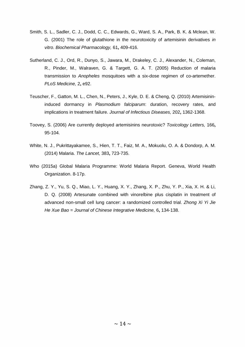
According to the WHO, an estimated 3.3 billion people in 97 countries were at risk of being
infected with malaria in 2014 (Fig. 2.1) .In 2014, 214 million cases of malaria and
approximately 438,000 deaths were reported (WHO, 2015a).
Figure 2.1: Countries with ongoing malaria transmission (WHO, 2015a).
P. falciparum and P. vivax malaria have roughly equal prevalences in Asia and in South and
Central America because transmission is low and mainly seasonal (Gething et al., 2011a,
Gething et al., 2011b). In these areas, most humans characteristically receive one or fewer
infectious bites per year. In sub-Saharan Africa however, where P. falciparum predominates,
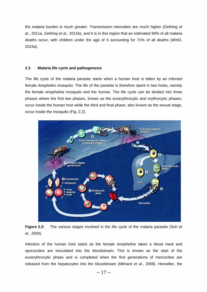
~ 17 ~
the malaria burden is much greater. Transmission intensities are much higher (Gething et
al., 2011a, Gething et al., 2011b), and it is in this region that an estimated 90% of all malaria
deaths occur, with children under the age of 5 accounting for 71% of all deaths (WHO,
2015a).
2.3 Malaria life cycle and pathogenesis
The life cycle of the malaria parasite starts when a human host is bitten by an infected
female Anopheles mosquito. The life of the parasite is therefore spent in two hosts, namely
the female Anopheline mosquito and the human. The life cycle can be divided into three
phases where the first two phases, known as the exoerythrocytic and erythrocytic phases,
occur inside the human host while the third and final phase, also known as the sexual stage,
occur inside the mosquito (Fig. 2.2).
Figure 2.2: The various stages involved in the life cycle of the malaria parasite (Suh et
al., 2004).
Infection of the human host starts as the female Anopheline takes a blood meal and
sporozoites are inoculated into the bloodstream. This is known as the start of the
exoerythrocytic phase and is completed when the first generations of merozoites are
released from the hepatocytes into the bloodstream (Ménard et al., 2008). Hereafter, the

~ 18 ~
sporozoites have up to 3 hours of motility which allow them to reach the liver (Amino et al.,
2006, Ménard et al., 2008). Some of the sporozoites are blocked by the human immune
system via antibodies and only a certain portion of the skin sporozoites invade the blood
capillaries to make their way to the target liver cells (Plebanski and Hill, 2000). After reaching
the liver and invading the hepatocytes, the sporozoites undergo mitotic replication called
schizogony, lasting between two to ten days, as they develop into liver-stage trophozoites
(Doolan et al., 2009). The nucleus of the trophozoite divides several times producing
thousands of exoerythrocytic merozoites that causes the hepatic schizont to rupture. The
release of the merozoites into the bloodstream, marks the end of the exoerythrocytic phase
(Ashley et al., 2006, White, 2008a, Doolan et al., 2009). A successful sporozoite can
produce between 10,000 to 30,000 daughter merozoites in 5 – 8 days within a hepatocyte
(White et al., 2014). Unlike the case with P. falciparum, a certain portion of the sporozoites of
both P. vivax and P. ovale form dormant hypnozoites which can remain in the liver for years
before developing into schizonts. Such development ultimately leads to relapse of malaria
caused by P. vivax and P. ovale (Fujioka and Aikawa, 2002, Dembele et al., 2011).
The erythrocytic phase starts as the exoerythrocytic merozoites enter the bloodstream, now
known as blood-stage merozoites. The merozoite with its very small size (~1 – 2 µm) (Fig.
2.3), is elegantly adapted for entering the erythrocytes (Bannister et al., 1986, Cowman et
al., 2012).
Figure 2.3: The merozoite comprises organelles incorporated within the cell exoskeleton
(Morrissette and Sibley, 2002, Cowman et al., 2012). This includes an apical complex of
secretory organelles (rhoptries, dense granules and micronemes), nucleus, mitochondrion
and apicoplast (McFadden, 1996, Roos et al., 1999, Cowman et al., 2012). Underlying the
plasma membrane is the inner membrane complex lined by two to three subpellicular
microtubules (Bannister et al., 2000, Cowman et al., 2012).

~ 19 ~
As the schizont ruptures, the mature merozoites are ejected; these then again enter the
erythrocytes (Fig. 2.4a) (Cowman et al., 2012). Initially, distortion of the erythrocyte surface
allows for an interaction between the merozoite and erythrocyte (Fig. 2.4b), where a
reorientation places the merozoite apex adjacent to the erythrocyte membrane. The parasite
enters the erythrocyte after major buckling of the erythrocyte surface (Fig. 2.4b), most likely
due to parasite-induced reorganization of the erythrocyte cytoskeleton (Zuccala and Baum,
2011, Cowman et al., 2012). After invasion of the erythrocyte is complete, the posterior is
sealed off followed by echinocytosis of the erythrocyte. The erythrocyte resumes its normal
state within 10 minutes (Gilson and Crabb, 2009, Cowman et al., 2012).

~ 20 ~
Figure 2.4: a. Schematic representation of the invasion of an erythrocyte by a merozoite
from egress to post invasion. A tight junction between the merozoite and the erythrocyte is
formed after reorientation of the merozoite (Cowman et al., 2012). Through the activity of
proteases, proteins are shed into the supernatant as the tight junction moves across the
merozoite surface. The parasite passes into the erythrocyte resulting in the ejection of the
erythrocyte contents (Cowman et al., 2012). The membrane and parasitophorous vacuole
form the space within the erythrocyte in which the parasite passes into and is fashioned out
of some leftover erythrocyte cell membrane components as well as the rhoptries from the
merozoite. As the tight junction moves to the posterior of the parasite the membranes seal
off by an unknown mechanism (Cowman et al., 2012). b. A P. falciparum merozoite in the
process of invading a human red blood cell (image S. Ralph, University of Melbourne,
Australia). Bar, 200 nm (Cowman et al., 2012).
Once inside the erythrocyte, the parasite flattens into an apparent ring form (Grüring et al.,
2011), growing and consuming the erythrocyte haemoglobin, changing the cell membrane in
order to facilitate import of nutrients as well as disposing of toxic haem waste product
through crystallization to biologically inert haemozoin (malaria pigment) (White et al., 2014).
In a susceptible individual, the parasite population will expand between six and twenty times
~ 21 ~
per cycle (Simpson et al., 2002). The majority of contents inside of the erythrocytes will be
consumed by the parasite at the end of the erythrocytic cycle.
As the parasite grows it forms trophozoites, with no fundamental structural difference except
being larger in shape and size (Fig. 2.5). These trophozoites undergo repetitive mitosis
within the erythrocyte forming an erythrocytic schizont. As the erythrocytic schizont grows in
size it will eventually rupture, releasing between 6 and 30 daughter merozoites. Each of the
daughter merozoites permeate an uninfected erythrocyte, therefore repeating the cycle
(White et al., 2014). This asexual cycle is repeated almost indefinitely and is responsible for
the clinical symptoms of the disease (Cox, 2010).
Figure 2.5: The major stages throughout the erythrocytic cycle of P. falciparum (Bozdech
et al., 2003).
As the cycle continues, some young merozoites will develop into longer-lived, non-
multiplying male and female gametocytes that flow freely in the peripheral circulation,
signalling the start of the sexual phase of the parasites’ life cycle (Kuehn et al., 2010, Cox,
2010, White et al., 2014). The catalyst for the production of macrogametocytes (female) and
microgametocytes (male) is unknown (Kamchonwongpaisan et al., 1997). Mature male and
female gametocytes are ingested by the female Anopheline mosquito when she takes a
blood meal whereupon the gametocytes are transferred into the mosquito midgut (Fig. 2.6).
The gametocytes then mature into gametes where the male gametocytes undergo rapid
nuclear division and become motile by acquiring flagella (also known as microgametocyte
exflagellation). This results in the production of haploid motile microgametes which move
rapidly in order to fertilize female macrogametes. Fusion and meiosis then takes place to
form zygotes (which takes about a day) that develop into motile ookinetes able to cross the

~ 22 ~
midgut epithelium of the mosquito. The ookinetes then travel to the extracellular space
between the midgut epithelium and overlaying basal lamina to undergo meiosis which
transforms them into oocysts. Nine to twelve days later the oocysts will rupture releasing
thousands of sporozoites that will journey through the haemolymph into the salivary glands,
where they await inoculation into the next human host, thereby restarting the life cycle
(White, 2008a, White et al., 2014).
Figure 2.6: Sexual (sporogonic) phase in mosquito midgut (Cox, 2010).
2.4 Clinical features of malaria
Clinical symptoms of malaria are associated with the rupture of schizonts and the destruction
of erythrocytes (Trampuz et al., 2003). Malaria symptoms often resemble those of common
viral infections which can lead to a delay in diagnosis (Murphy and Oldfield, 1996). When
malaria is contracted, the first symptoms are nonspecific and can include any of the
following: headache, fatigue, a vague absence of well-being, abdominal discomfort, muscle
aches and dizziness which are followed by irregular fever. Interestingly, malaria is the most
common cause of fever in endemic areas (White et al., 2014). Physical signs can include
splenomegaly, hepatomegaly, tachycardia, orthostatic hypotension, pallor and jaundice
(Trampuz et al., 2003). Nausea and vomiting also occur frequently.

~ 23 ~
Most patients with uncomplicated malaria have few abnormal physical findings apart from
fever, a palpable spleen and mild anaemia (White et al., 2014). Young children are more
susceptible to liver enlargement and in areas with persistent transmission, recurrent
infections will cause splenomegaly and chronic anaemia, while adults will suffer more from
mild jaundice.
2.5 Pathogenesis
In P. falciparum malaria, protuberances or 'knobs' form from the surface of the infected
erythrocyte some 12 – 15 h after invasion. These protuberances cause attachment of the
infected erythrocyte to other infected erythrocytes (platelet-mediated agglutination), the
vessel walls (cytoadherence) or uninfected erythrocytes (rosetting) (Doumbo et al., 2009)
(Fig. 2.7).
Figure 2.7: Types of adhesion involving erythrocytes infected with the malaria parasite
(Doumbo et al., 2009).
This attachment causes sequestration (Fig. 2.8) of erythrocytes containing mature parasites
which are carried into the vascular system of vital organs (especially the brain). The
sequestered erythrocytes interfere with microcirculatory flow and metabolism as well as the
functioning of the vascular endothelium (Pongponratn et al., 2003).

~ 24 ~
Figure 2.8: Microscopic image of sequestration of a red blood cell (Rowe, 2013).
Interestingly, different Plasmodium species can influence the shape of and have different
preference to the erythrocytes. Whilst P. vivax only invades young erythrocytes, P.
falciparum and P. knowlesi are less selective in invading erythrocytes at any stage; thus,
very high parasite densities will be attained (Buffet et al., 2011). Also in P. vivax malaria the
infected erythrocyte enlarge and deform (Suwanarusk et al., 2004), but in P. falciparum
malaria the erythrocyte becomes more spherical and rigid (White et al., 2014).
The human host responds to the infection of erythrocytes by enhancing clearance by the
spleen that accelerates removal of both infected and normal erythrocytes (Looareesuwan et
al., 1987, Buffet et al., 2011). Rupture of the schizont releases the parasites as well as the
host cellular material into the bloodstream thereby leading to activation of macrophages and
monocytes. This also induces the release of pro-inflammatory cytokines, causing fever and
other pathological effects associated with the disease (Karunaweera et al., 1992, Ayimba et
al., 2011).
2.6 Severe malaria
P. falciparum is the main cause of severe malaria involving widespread erythrocyte
sequestration and dysfunction of vital organs. The extent of microvascular obstruction
parallels clinical severity and mortality rises when the proportion of infected erythrocytes
exceeds 2% (White et al., 2014). Severe malaria is usually accompanied by major
complications which can include cerebral malaria, pulmonary oedema, severe anaemia and
acute renal failure. Acidosis and hypoglycaemia can also arise and are characteristically the
most common metabolic complications (Trampuz et al., 2003). Severe malaria together with
any of these complications can cause death within days (Trampuz et al., 2003). In many
patients, several of these complications can evolve in rapid succession within a few hours.
~ 25 ~
The manifestation of severe malaria, however, depends on age (Dondorp et al., 2008). In
children, hypoglycaemia and severe anaemia are common, whereas jaundice, acute kidney
injury and acute pulmonary oedema are more common in adults. Coma (mostly prevalent in
cerebral malaria) and acidosis occur in all age groups.
2.7 Diagnosis
As the symptomatic stage of malaria begins, parasite densities in the blood reach 50/µL
which corresponds roughly to 100 million parasites in the blood of an infected adult. When
this level is reached, the parasites become detectable by microscopy or rapid diagnostic
tests (White et al., 2014). Light microscopy of thick and thin stained blood smears seems to
be the golden standard for the diagnosis of malaria (Moody and Chiodini, 2000). Smears
permit both species identification as well as quantification of parasites, which can be
expressed as either a percentage of erythrocytes infected or as parasites per µL (Suh et al.,
2004). Thick smears are used for screening of Plasmodium parasites and are 20 – 40 times
more sensitive when compared to thin smears. Thin smears are used to identify the infecting
parasite species (including simultaneous infections from different species), to quantify
parasitaemia, and to measure the presence of malarial pigment in neutrophils and
monocytes, and to identify gametocytes and schizonts (Trampuz et al., 2003). It is important
that during diagnosis of fever that malaria should not be excluded until at least three
negative blood smears have been obtained within 48 hours (Suh et al., 2004).
There are however, certain problems which arise when the processing and interpretation of
malaria smears are carried out. Smears require the appropriate equipment as well as
substantial training, factors that can limit their use in endemic regions. Fortunately, there are
rapid malaria tests (which require minimal skill and effort) available that can overcome these
problems. The most practical and widely used are the rapid detection tests (RDTs) which
detect either P. falciparum histidine-rich protein 2 (PfHRP2), aldolase antigens, species-
specific lactate dehydrogenase, or pan-malaria in finger-prick blood samples (Moody, 2002,
WHO, 2011). Diagnosing P. falciparum malaria with PfHRP2-based RDTs are as good as
routine microscopy. Newer diagnostic methods for P. falciparum and P. vivax are based on
the detection of Plasmodium lactate dehydrogenase, given that sensitivity for P. vivax is low
at parasite densities less than 200/µL. For non-falciparum species, aldolase-based tests are
less sensitive (WHO, 2011). RDTs are valuable in epidemic investigations and surveys
because of the simplicity and speed. However, they cannot be used to quantify, that is
accurately assess the level of, parasitaemia and are quite expensive (White et al., 2014).
~ 26 ~
Measurement of PfHRP2 concentrations in plasma can be used to estimate the sequestered
parasite biomass in severe malaria (Dondorp et al., 2005).
2.8 Vector control and malaria prevention
Vector control is crucial to malaria prevention. The emergence of recent artemisinin
combination therapy (ACT) resistance, lack of vaccines and unaffordable antimalarial drugs
make vector control an important cornerstone in the control and prevention of malaria. There
are various methods that are established and well-practiced throughout endemic areas in
order to control and prevent malaria. In this section the most important methods of vector
control and the implications of employing these methods have on the overall statistics of
malaria will be discussed.
Indoor residual spraying (IRS)
In indoor residual spraying, a long-lasting insecticide is sprayed on the inside walls of
houses and other structures where people sleep, resulting in killing of the mosquitoes as
they enter houses and rest on sprayed surfaces (Karunamoorthi, 2011). Efficacy of IRS
generally depends on the flying habits of the indigenous Anopheline vector species, that is, if
the mosquitoes enter houses and rest there and the prevalence of insecticide resistance
(White et al., 2014). IRS is frequently used in epidemic-prone, malaria endemic areas as well
as areas where seasonal transmission takes place. Insecticides normally used for IRS
include pyrethroids and dichlorodiphenyltrichloroethane (DDT) (WHO, 2010a,
Karunamoorthi, 2011).
IRS as a vector control method has been widely adopted, especially in the African Region.
Even so, a decreased protection rate of the global population at risk of contracting malaria
has been reported from national control programmes, deteriorating from 5.7% in 2010 to
3.4% in 2014 (WHO, 2015a).
Insecticide treated nets (ITNs)
Insecticide treated nets (ITNs) are more effective when compared to IRS as their coverage is
easier to sustain; therefore they are the most commonly used form of vector control
(Karunamoorthi, 2011). Standard ITNs have to be retreated every year, whereas long-lasting
ITNs retain their activity during the life-span of the net. Not only does the ITN protect the
user against infectious bites, but also protects the public at large by killing Anopheline

~ 27 ~
mosquitoes (Moonen et al., 2010, Godfray, 2013). In some parts of Asia however, the chief
mosquito vectors are active either in the early evening or early mornings hence, protection
against mosquito bites are minimal (White et al., 2014).
That most malaria endemic countries have endorsed universal access to ITNs has resulted
in an increase in the portion of the population able to sleep under an ITN over the past ten
years. Most ITNs are issued to the population for free, especially in countries with on-going
malaria transmission (WHO, 2015a). This has resulted in the fall in malaria mortality and
morbidity.
Emergence of resistance to insecticides used with IRS and ITNs
As malaria mosquitoes develop resistance to insecticides used in IRS and ITNs, the
efficiency of vector control is hampered. Currently reliance is placed on a solitary class of
insecticides, namely pyrethroids. If resistance towards this class of insecticide develop it will
clearly have an effect on vector control. In identifying this threat, the WHO released the
Global plan for insecticide resistance management in malaria vectors (GPIRM) in 2012
(WHO, 2012). The GPIRM sets out five strategies: undertaking resistance monitoring,
implementing insecticide resistance management strategies, filling knowledge gaps on
mechanisms of insecticide resistance and the impact of resistance management, developing
new vector control tools, and ensuring that key enabling mechanisms are in place. Although
the GPIRM has been in place since 2012, there are several countries using insecticides for
vector control that did not monitor insecticide resistance in 2013 – 2014. Among the 96
countries that implement ITNs or IRS for vector control, 86 reported that monitoring of
insecticide resistance was undertaken, yet only 42 countries provided resistance data. Thus,
it is clear that monitoring is not conducted every year as required by the GPIRM (WHO,
2012).
Other control methods
Other methods of control include ultra-low-volume (ULV) space spraying (fogging),
repellents and larva control.
ULV space spraying – This is used globally for the control of adult mosquito populations.
Space sprays can be utilized as a fog, wherein kerosene oil is implemented as a carrier that
produces dense fog droplets for insecticides. This method of control is not very effective as
the operational costs are high and residual effects are low (Karunamoorthi, 2011).
~ 28 ~
Repellents – Chemical repellents play a vital role in protecting people from mosquitoes and
may therefore reduce the transmission of malaria. N,N-diethyl-m-toluamide, N,N-diethyl
mandelic acid amide, allethrin, dimethyl phthalate, and N,N-diethylphenylacetamide are most
commonly used in commercial repellents. However, synthetic repellents also have
drawbacks, including reduced efficacy due to sweating and allergic reactions
(Karunamoorthi, 2011).
Larva control – The emphasis is on the use of larvicides. Adult females lay between 50 and
200 eggs per oviposition. The eggs are laid singly and directly on water and are relatively
small (c. 0.5 x 0.2 mm2). Depending on the water temperature, mosquito larvae (also called
‘wigglers’) can live in water between 4 – 14 days (Enayati et al., 2009). Depending on the
accessibility and size of the breeding site and whether or not breeding sites within the
mosquito flight range can be pinpointed, larval control by either non-chemical or chemical
means becomes a practical method of vector control (Karunamoorthi, 2011).
Vaccination – Malaria vaccines are an area of intensive research as much time, money and
effort has gone into the development of a malaria vaccine. Especially with the emergence of
artemisinin and multidrug-resistant strains of P. falciparum, this is the main driving force
behind research. Various vaccines have reached clinical trials, but unfortunately so far most
have demonstrated insufficient immunogenicity. However, the RTS,S subunit vaccine which
targets the circumsporozoite protein from the pre-erythrocytic phase of P. falciparum shows
some promise (White et al., 2014). The final Phase III results for the vaccine were published
in April 2015. As per the recommendations, the vaccine will be evaluated as an addition to,
not a replacement for, existing preventive, diagnostic and treatment measures. If
RTS,S/AS01 becomes available for use artemisinin-based combination therapies as well as
long-lasting insecticidal nets, and rapid diagnostic tests will still be used continuously as the
vaccine is not sufficiently immunogenic for stand-alone use (WHO, 2015b).
Chemoprophylaxis for travellers
For travellers entering a malaria area, chemoprophylaxis has to be considered (Chen et al.,
2006). The recommendation for chemoprophylaxis usually depends on length of the planned
trip, type of travelling, the age of the traveller, host risks or pre-existing illness (del Prado et
al., 2014). The higher the traveller’s risk of succumbing to malaria and evolving
complications, the greater the demand for chemoprophylaxis. Travellers at the highest risk of
contracting malaria are babies and young children (< 5 years), the elderly, pregnant women
and immuno-compromised patients (Nujum et al., 2014). There are a variety of drugs that
can be used as chemoprophylaxis for travellers including the atovaquone-proguanil

~ 29 ~
combination, mefloquine, primaquine, doxycycline and chloroquine (CQ). Although CQ
resistance occurs in almost all regions, it may still be used in areas where the risk of P.
falciparum malaria is low. Details of further drug indications, classification, chemistry and
comments are given in section 2.9.
2.9 Chemotherapy
Effective malaria chemotherapy aims at treating the patient as well as hindering the
infectivity of the parasite to the vector via exploiting the differences in metabolism between
the malaria parasite and the host (Murambiwa et al., 2011). Repeated administration of
antimalarial drugs at sub-therapeutic levels can interrupt parasite transmission, but this
mode of treatment may induce the development of drug resistance (Watkins and Mosobo,
1993). Currently, genes that have been identified as playing a role in the development of
resistance include PfCRT (P. falciparum chloroquine resistance transporter), PfMDR1 (P.
falciparum multidrug resistance transporter 1), PfNHE (P. falciparum sodium/proton
exchanger), and PfMRP (P. falciparum multidrug resistance-associated protein).
Traditionally, the clinical action of antimalarial drugs is classified according to the life cycle
stages of the parasite, where different drugs inhibit different life stages namely blood
schizonticides, tissue schizonticides, hypnozoiticides and gametocytocides. In this review
however, we discuss the different antimalarials according to their pharmacological
classification. Currently used antimalarials can be divided into five main pharmacological
classes, namely quinoline and quinoline-related antimalarials (which can be sub-classified
further into aryl-amino alcohols, 4-aminoquinolines and 8-aminoquinolines), antifolates
(including DHPS- and DHFR-inhibitors), antibiotics, hydroxynapthoquinones and the
artemisinin class of compounds. As the development of malaria parasite resistance is
becoming more and more relevant, drug resistance genes and parasite resistance
mechanisms will also be included.
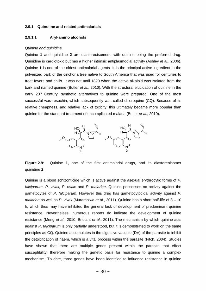
~ 30 ~
2.9.1 Quinoline and related antimalarials
2.9.1.1 Aryl-amino alcohols
Quinine and quinidine
Quinine 1 and quinidine 2 are diastereoisomers, with quinine being the preferred drug.
Quinidine is cardiotoxic but has a higher intrinsic antiplasmodial activity (Ashley et al., 2006).
Quinine 1 is one of the oldest antimalarial agents. It is the principal active ingredient in the
pulverized bark of the cinchona tree native to South America that was used for centuries to
treat fevers and chills. It was not until 1820 when the active alkaloid was isolated from the
bark and named quinine (Butler et al., 2010). With the structural elucidation of quinine in the
early 20th Century, synthetic alternatives to quinine were prepared. One of the most
successful was resochin, which subsequently was called chloroquine (CQ). Because of its
relative cheapness, and relative lack of toxicity, this ultimately became more popular than
quinine for the standard treatment of uncomplicated malaria (Butler et al., 2010).
N H
H
N
O
HOH
2
N H
H
N
O
HOH
1
Figure 2.9: Quinine 1, one of the first antimalarial drugs, and its diastereoisomer
quinidine 2.
Quinine is a blood schizonticide which is active against the asexual erythrocytic forms of P.
falciparum, P. vivax, P. ovale and P. malariae. Quinine possesses no activity against the
gametocytes of P. falciparum. However this drug has gametocytocidal activity against P.
malariae as well as P. vivax (Murambiwa et al., 2011). Quinine has a short half-life of 8 – 10
h, which thus may have inhibited the general lack of development of predominant quinine
resistance. Nevertheless, numerous reports do indicate the development of quinine
resistance (Meng et al., 2010, Briolant et al., 2011). The mechanism by which quinine acts
against P. falciparum is only partially understood, but it is demonstrated to work on the same
principles as CQ. Quinine accumulates in the digestive vacuole (DV) of the parasite to inhibit
the detoxification of haem, which is a vital process within the parasite (Fitch, 2004). Studies
have shown that there are multiple genes present within the parasite that effect
susceptibility, therefore making the genetic basis for resistance to quinine a complex
mechanism. To date, three genes have been identified to influence resistance in quinine

~ 31 ~
namely: PfCRT, PfMDR1 and PfNHE1. Interestingly, all of them are used to encode
transporter proteins (Sidhu et al., 2002, Cooper et al., 2002, Cooper et al., 2007, Nkrumah et
al., 2009). Quinine has been reserved for treatment against resistant malaria in combination
with antibiotics and also to treat severe cases of malaria (Petersen et al., 2011).
Mefloquine
Mefloquine 3 is a 4-methanolquinoline which shares structural similarities with quinine. It
inhibits the asexual stages of P. vivax as well as P. falciparum, making this drug a blood
schizonticide (Murambiwa et al., 2011). Mefloquine has a long half-life of 14 – 18 days
(Stepniewska and White, 2008).
NCF3
CF3
HOHN
3
Figure 2.10: Mefloquine 3, a 4-methanolquinoline.
Resistance of mefloquine is facilitated by the amplification of PfMDR1, which leads to the
overexpression of this gene in the DV membrane transporter (Cowman et al., 1994). The
exact mechanism of action for mefloquine is unclear, but in vitro experiments demonstrated
that mefloquine binds to haem and exerts antimalarial activity via inhibiting haem
detoxification (Fitch et al., 1979, Eastman and Fidock, 2009). A decrease of PfMDR1-
mediated influx into the DV is noted to occur with decreased parasite susceptibility to
mefloquine (Rohrbach et al., 2006, Sidhu et al., 2006). Furthermore, it has been
hypothesized that mefloquine might target the PfMDR1 transport function as mefloquine
prevents access of other solutes into the DV (Rohrbach et al., 2006). Mefloquine is used for
both treatment and as prophylaxis in Africa and is especially useful as chemoprophylaxis in
travellers from non-endemic countries (Schlagenhauf et al., 2011).
Halofantrine
Halofantrine 4 belongs to the phenanthrene class of compounds that includes lumefantrine,
is a schizonticidal drug but with no apparent action on the sporozoite, gametocyte or hepatic

~ 32 ~
stages of infection. However, it is effective against both CQ sensitive and resistant strains of
Plasmodia (Bilolikar et al., 1994).
N
OHCl
Cl
CF3
4
Figure 2.11: Halofantrine 4.
The mechanism of action appears to be due to inhibition formation of haemozoin although
the exact mode of action is unknown (Nosten et al., 2012). Recent studies have revealed
that halofantrine forms complexes with haematin (ferriprotoporphyrin IX), where inhibition of
haemozoin formation takes place at the lipid-aqueous interface, an environment that is much
more compatible with the crystal structure of halofantrine-ferriprotoporphyrin IX (De Villiers et
al., 2008). Halofantrine has poor solubility and is therefore erratically absorbed. However, fat
co-administration can enhance the absorption of this drug. When compared to mefloquine,
halofantrine is readily eliminated; it has a half-life of ~4.7 days. Few studies have been done
on the development of resistance of this drug. Altered halofantrine transport may be the
consequence of PfMDR1 mutations, which could signify a role for the efflux transport
mechanism for development of resistance to this drug (Sanchez et al., 2008). Clinical studies
in Southeast Asia have revealed cross-resistance between halofantrine and mefloquine (Ter
Kuile et al., 1993). Halofantrine has been withdrawn from use due to its cardiotoxicity
(Nosten et al., 2012).
Lumefantrine
Lumefantrine 5 is a racemic substituted fluorene derivative with a structure similar to that of
halofantrine. Lumefantrine is active against the asexual stages of P. falciparum and P. vivax.
The mechanism of action for this particular drug is not fully understood but it is believed to
inhibit polymerisation of haem resulting in parasite death (Nosten et al., 2012).

~ 33 ~
Cl
ClCl
OHN
5
Figure 2.12: Lumefantrine 5.
The terminal elimination half-life of lumefantrine is roughly 4.5 days, a much shorter half-life
than that of mefloquine or piperaquine. This drug has a low and variable bioavailability and is
highly lipophilic. However, bioavailability can be increased with the intake of fatty foods
which notably enhance drug absorption (Ashley et al., 2007). Lumefantrine resistance is
easily induced in both animal models and in vitro (Nosten et al., 2012). Single nucleotide
polymorphisms in the pfmdr1 gene have been linked with amplified IC50 values in vitro
(Anderson et al., 2005). Lumefantrine is used in combination with artemether (Coartem®) as
part of the ACT treatment regimens stipulated by the WHO.
2.9.1.2 4-Aminoquinolines
Chloroquine (CQ)
CQ 6 is a rapid-acting schizonticide against P. falciparum with activity against the asexual
erythrocytic forms of P. malariae, P. ovale and P. vivax (Murambiwa et al., 2011). CQ has
one of the longest half-life (approximately 60 days) of all antimalarials. This extreme half-life
results in exposure the parasite to the drug for a prolonged period of time after which CQ
falls below its therapeutic concentration. This also provides a chemoprophylactic effect
during the elimination phase (Stepniewska and White, 2008). CQ resistant parasites
emerged roughly around 10 years after its first official use, first along the Thai-Cambodian
border and also in Columbia in the late 1950’s (Mita et al., 2009). CQ resistant P. falciparum
is now predominant in nearly all malaria endemic regions (Petersen et al., 2011).
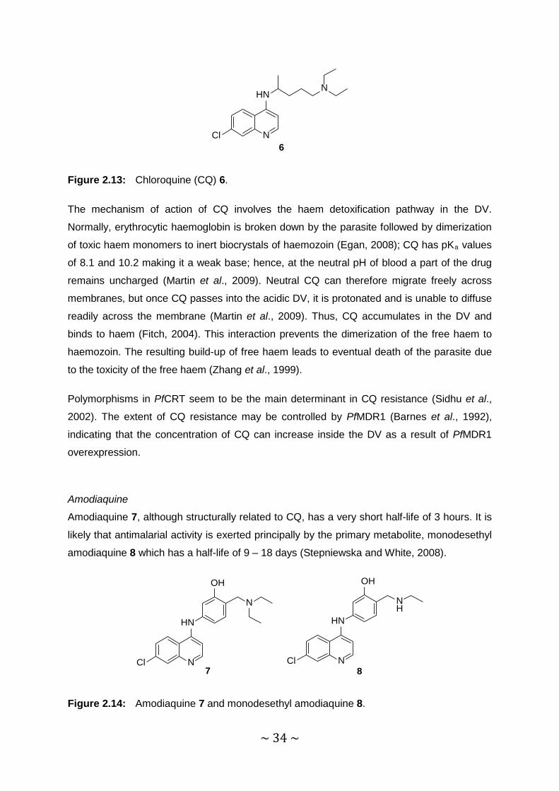
~ 34 ~
N
HNN
Cl6
Figure 2.13: Chloroquine (CQ) 6.
The mechanism of action of CQ involves the haem detoxification pathway in the DV.
Normally, erythrocytic haemoglobin is broken down by the parasite followed by dimerization
of toxic haem monomers to inert biocrystals of haemozoin (Egan, 2008); CQ has pKa values
of 8.1 and 10.2 making it a weak base; hence, at the neutral pH of blood a part of the drug
remains uncharged (Martin et al., 2009). Neutral CQ can therefore migrate freely across
membranes, but once CQ passes into the acidic DV, it is protonated and is unable to diffuse
readily across the membrane (Martin et al., 2009). Thus, CQ accumulates in the DV and
binds to haem (Fitch, 2004). This interaction prevents the dimerization of the free haem to
haemozoin. The resulting build-up of free haem leads to eventual death of the parasite due
to the toxicity of the free haem (Zhang et al., 1999).
Polymorphisms in PfCRT seem to be the main determinant in CQ resistance (Sidhu et al.,
2002). The extent of CQ resistance may be controlled by PfMDR1 (Barnes et al., 1992),
indicating that the concentration of CQ can increase inside the DV as a result of PfMDR1
overexpression.
Amodiaquine
Amodiaquine 7, although structurally related to CQ, has a very short half-life of 3 hours. It is
likely that antimalarial activity is exerted principally by the primary metabolite, monodesethyl
amodiaquine 8 which has a half-life of 9 – 18 days (Stepniewska and White, 2008).
N
HN
Cl7
OH
N
N
HN
Cl
OH
NH
8
Figure 2.14: Amodiaquine 7 and monodesethyl amodiaquine 8.

~ 35 ~
Because of the structural similarity of amodiaquine to CQ, the mechanism of action is
hypothesized to be the inhibition haem detoxification by accumulating in the DV and binding
to haem (Foley and Tilley, 1998, Hayeshi et al., 2008). Mutations in PfCRT and PfMDR1
may be the basis for cross resistance between CQ and amodiaquine (Sá et al., 2009).
Piperaquine
In order to overcome CQ resistance, two 4-aminoquinoline moieties attached via linkers of
various lengths, namely the bisquinolines, were prepared. Piperaquine 9 is one such
bisquinoline which is highly lipid soluble with a half-life of 16.5 days (Price et al., 2007).
NN N
N
N
Cl
N
Cl9
Figure 2.15: Piperaquine, 9.
Piperaquine shows remarkably good activity against CQ-resistant strains. This was held to
be due to the sterically demanding chemical structure that was presumed to prevent the drug
from fitting into the substrate binding site of PfCRT. However, piperaquine resistance duly
emerged and this, coupled with the advent of artemisinin derivatives as effective antimalarial
drugs, resulted in a marked decrease in the use of piperaquine during the 1980’s (Held et
al., 2015). However, its pharmacokinetic profile, efficacy, tolerability, and low cost make it a
favourable partner dug for use with dihydroartemisinin in artemisinin combination therapy
(O'Neill et al., 2012). This is discussed further below.
2.9.1.3 8-Aminoquinolines
Primaquine
Primaquine 10 has a half-life of 6 h (Edwards et al., 1993) and is one of the few antimalarial
drugs approved for therapy against P. vivax liver stage hypnozoites (Wells et al., 2010). This
antimalarial drug induces haemolytic anaemia in glucose-6-phosphate dehydrogenase
(G6PD) deficient patients (Burgoine et al., 2010). However, limiting the dose to one
treatment per week for 8 weeks might be safe and an effective alternative to the traditional
14-day course of the drug (Myat Phone et al., 1994). In a P. falciparum infection a single
dose of 0.75 mg/kg is well tolerated in G6PD deficient patients excluding children younger

~ 36 ~
than 4 years and pregnant women. Primaquine also eradicates the primary exoerythrocytic
stage of Plasmodium species as well as the gametocytocidal and sporontocidal phases
(Murambiwa et al., 2011).
NHN
O
H2N
10
Figure 2.16: Primaquine 10.
The mode of action for primaquine is largely unknown but it is thought that the mitochondria
of the parasite could be the biological target. Structural changes within the inner membranes
as well as swelling of the parasite mitochondria are observed upon administration of
primaquine (Beaudoin and Aikawa, 1968, Aikawa and Beaudoin, 1970, Howells et al., 1970,
Boulard et al., 1983, Lanners, 1991). These morphological changes may be associated with
destruction of mitochondrial function by the drug (Rotmann, 1975, Peters et al., 1984,
Krungkrai et al., 1999). Numerous active metabolites of primaquine appear to be responsible
for antimalarial activity (Idowu et al., 1995, Vale et al., 2009). Several other studies indicate
that primaquine can lead to the reversal of CQ resistance through binding to PfCRT; this
may lead to a synergistic action between CQ and primaquine (Sanchez et al., 2004, Bray et
al., 2005, Egan, 2008).
2.9.2 Hydroxynaphthoquinones
Atovaquone
Atovaquone 11 is used with proguanil in a combination known as Malarone® for prophylactic
medication for travellers visiting malaria endemic areas (Petersen et al., 2011). This
combination is mainly used as such because of facile resistance arising to atovaquone from
single nucleotide polymorphisms in the cytochrome β-gene (Gil et al., 2003).
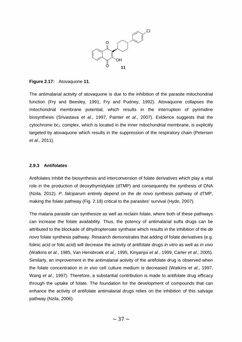
~ 37 ~
O
O
Cl
OH
11
Figure 2.17: Atovaquone 11.
The antimalarial activity of atovaquone is due to the inhibition of the parasite mitochondrial
function (Fry and Beesley, 1991, Fry and Pudney, 1992). Atovaquone collapses the
mitochondrial membrane potential, which results in the interruption of pyrimidine
biosynthesis (Srivastava et al., 1997, Painter et al., 2007). Evidence suggests that the
cytochrome bc1 complex, which is located in the inner mitochondrial membrane, is explicitly
targeted by atovaquone which results in the suppression of the respiratory chain (Petersen
et al., 2011).
2.9.3 Antifolates
Antifolates inhibit the biosynthesis and interconversion of folate derivatives which play a vital
role in the production of deoxythymidylate (dTMP) and consequently the synthesis of DNA
(Nzila, 2012). P. falciparum entirely depend on the de novo synthesis pathway of dTMP,
making the folate pathway (Fig. 2.18) critical to the parasites’ survival (Hyde, 2007).
The malaria parasite can synthesize as well as reclaim folate, where both of these pathways
can increase the folate availability. Thus, the potency of antimalarial sulfa drugs can be
attributed to the blockade of dihydropteroate synthase which results in the inhibition of the de
novo folate synthesis pathway. Research demonstrates that adding of folate derivatives (e.g.
folinic acid or folic acid) will decrease the activity of antifolate drugs in vitro as well as in vivo
(Watkins et al., 1985, Van Hensbroek et al., 1995, Kinyanjui et al., 1999, Carter et al., 2005).
Similarly, an improvement in the antimalarial activity of the antifolate drug is observed when
the folate concentration in in vivo cell culture medium is decreased (Watkins et al., 1997,
Wang et al., 1997). Therefore, a substantial contribution is made to antifolate drug efficacy
through the uptake of folate. The foundation for the development of compounds that can
enhance the activity of antifolate antimalarial drugs relies on the inhibition of this salvage
pathway (Nzila, 2006).

~ 38 ~
Figure 2.18: Folate biochemical pathway in P. falciparum. The following abbreviations are
used: GTPCH GTP cyclohydrolase I, PTPS pyruvyl tetrahydropterin synthase III, HPPK
hydroxymethyl dihydropterin pyrophosphokinase, DHPS dihydropteroate synthase, DFHS
dihydrofolate synthase, FPGS folylpoly-gamma-glutamate synthase, DHFR dihydrofolate
reductase, SHMT serine hydroxymethyltransferase, TS thymidylate synthase. PTPS has
recently been characterized in P. falciparum (Dittrich et al., 2008).
Antifolates that are in use currently target two specific enzymes namely dihydrofolate
reductase (DHFR) and dihydropteroate synthase (DHPS). Therefore, antifolates used
against malaria can be sub-divided into two classes, namely DHPS inhibitors (class I
antifolates) and DHFR inhibitors (class II antifolates) (Nzila, 2006). Combining DHFR and
DHPS inhibitors results in a synergistic effect and such combinations can be used when
treating malaria patients.
GTP
GTP-CH
Dihydroneopterin triphosphate PTPS
Hydroxymethyl-dihydropterin HPPK
Hydroxymethyl-pyrophosphate
DHPS
Dihydropteroate
DHFS
Dihydrofolate
DHFR
Tetrahydrofolate
SHMT
-CH2-THF
Monoglutamate folate
Polyglutamate
FPGS dUMP
dUMP
DNA synthesis
TS TS

~ 39 ~
2.9.3.1 DHPS inhibitors
DHPS inhibitors belong to two families: sulfone and sulfonamide with structures of
representative drugs given in Fig. 2.19.
H2N
S
NH2
O O
12 13
SNH
H2NNN
OO
O
O
Figure 2.19: Class I antifolates dapsone 12 and sulfadoxine 13.
Efforts to use drugs from this class of compound as monotherapy in the fight against malaria
were made. These efforts had to be abandoned because of these drugs elicited severe
toxicity and displayed low efficacy (Michel, 1968). However, once the discovery was made
that DHPS inhibitors work synergistically with DFHR inhibitors interest in using these
different components as combination treatment for malaria was rekindled. Dapsone 12 is the
most potent DHPS inhibitor when compared to other compounds of the same class (Nzila,
2006). The major mechanism of sulfadoxine resistance stems from point mutations in the
dhps domain of the dhps-pppk gene (Nzila, 2006).
2.9.3.2 DHFR inhibitors
Proguanil
The first reported antifolate antimalarial drug was proguanil and this is used, as mentioned
above, in combination with atovaqoune. Proguanil 14 (a prodrug) is converted in vivo into its
active metabolite cycloguanil 15, an inhibitor of DHFR, via oxidative ring closure. The
synergistic mechanism between atovaquone and cycloguanil is not fully understood.
Proguanil and dapsone can also be used as combination therapy (Lapdap®). Unfortunately,
this therapeutic combination can no longer be recommended as dapsone causes haemolysis
in G6PD deficient patients (Luzzatto, 2010).

~ 40 ~
HN
HN
HN
NH NHCl
14
N
N
N
ClNH2
H2N
15
Figure 2.20: Proguanil 14 and its active metabolite cycloguanil 15.
Pyrimethamine
Pyrimethamine 16 belongs to the 2,4-diaminopyrimidine class of compounds. Initially used
for treatment of cancer, it was only discovered later that it is effective against malaria
because of the structural resemblance to proguanil (Falco et al., 1951).
N
NCl NH2
H2N
16
Figure 2.21: Pyrimethamine 16.
Pyrimethamine is the most widely used antimalarial antifolate drug thus far, being first used
in monotherapy (Peters, 1987) and later being combined with sulfadoxine (Fansidar®). This
sulfadrug combination has a long elimination half-life of >80 h (Roper et al., 2004).
Pyrimethamine is also used in combination with dapsone (Maloprim®), where dapsone has
a short half-life of 24 h (Winstanley et al., 1997). Unfortunately, this drug combination has a
very low efficacy that can be attributed to the shorter half-life of dapsone; as soon as the
second day of treatment with this combination starts the drug efficacy decreases due to the
reduction of synergy between dapsone and pyrimethamine (Nzila, 2006).
Point mutations in the dhfr domain of the dhfr-ts gene have been linked to the development
of pyrimethamine resistance (Peterson et al., 1988, Peterson et al., 1990, Foote et al.,
1990). A lot of effort has focussed on determination of dhfr and dhps genotypes that may
predict resistance to pyrimethamine/sulfadoxine. Malaria parasites that have point mutations
at codons 51, 59 and 108 of dhfr appear to have pyrimethamine/sulfadoxine resistance.
Also, point mutations on codons 437 and/or 540 or 437 and/or 581 of the dhps gene may
enhance resistance as well (Sibley et al., 2001, Gregson and Plowe, 2005). Mutation at
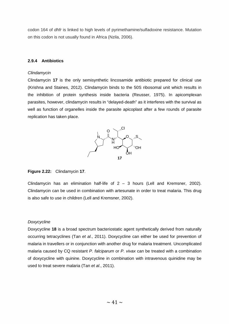
~ 41 ~
codon 164 of dhfr is linked to high levels of pyrimethamine/sulfadoxine resistance. Mutation
on this codon is not usually found in Africa (Nzila, 2006).
2.9.4 Antibiotics
Clindamycin
Clindamycin 17 is the only semisynthetic lincosamide antibiotic prepared for clinical use
(Krishna and Staines, 2012). Clindamycin binds to the 50S ribosomal unit which results in
the inhibition of protein synthesis inside bacteria (Reusser, 1975). In apicomplexan
parasites, however, clindamycin results in “delayed-death” as it interferes with the survival as
well as function of organelles inside the parasite apicoplast after a few rounds of parasite
replication has taken place.
O S
OHOH
HO
NH
ClO
N
17
Figure 2.22: Clindamycin 17.
Clindamycin has an elimination half-life of 2 – 3 hours (Lell and Kremsner, 2002).
Clindamycin can be used in combination with artesunate in order to treat malaria. This drug
is also safe to use in children (Lell and Kremsner, 2002).
Doxycycline
Doxycycline 18 is a broad spectrum bacteriostatic agent synthetically derived from naturally
occurring tetracyclines (Tan et al., 2011). Doxycycline can either be used for prevention of
malaria in travellers or in conjunction with another drug for malaria treatment. Uncomplicated
malaria caused by CQ resistant P. falciparum or P. vivax can be treated with a combination
of doxycycline with quinine. Doxycycline in combination with intravenous quinidine may be
used to treat severe malaria (Tan et al., 2011).
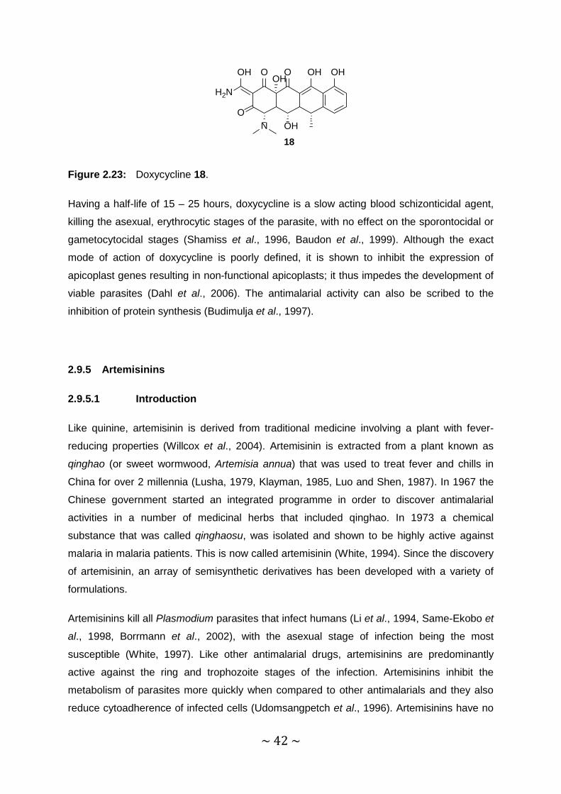
~ 42 ~
OHOHOO
O
OH
H2N
N OH
OH
18
Figure 2.23: Doxycycline 18.
Having a half-life of 15 – 25 hours, doxycycline is a slow acting blood schizonticidal agent,
killing the asexual, erythrocytic stages of the parasite, with no effect on the sporontocidal or
gametocytocidal stages (Shamiss et al., 1996, Baudon et al., 1999). Although the exact
mode of action of doxycycline is poorly defined, it is shown to inhibit the expression of
apicoplast genes resulting in non-functional apicoplasts; it thus impedes the development of
viable parasites (Dahl et al., 2006). The antimalarial activity can also be scribed to the
inhibition of protein synthesis (Budimulja et al., 1997).
2.9.5 Artemisinins
2.9.5.1 Introduction
Like quinine, artemisinin is derived from traditional medicine involving a plant with fever-
reducing properties (Willcox et al., 2004). Artemisinin is extracted from a plant known as
qinghao (or sweet wormwood, Artemisia annua) that was used to treat fever and chills in
China for over 2 millennia (Lusha, 1979, Klayman, 1985, Luo and Shen, 1987). In 1967 the
Chinese government started an integrated programme in order to discover antimalarial
activities in a number of medicinal herbs that included qinghao. In 1973 a chemical
substance that was called qinghaosu, was isolated and shown to be highly active against
malaria in malaria patients. This is now called artemisinin (White, 1994). Since the discovery
of artemisinin, an array of semisynthetic derivatives has been developed with a variety of
formulations.
Artemisinins kill all Plasmodium parasites that infect humans (Li et al., 1994, Same-Ekobo et
al., 1998, Borrmann et al., 2002), with the asexual stage of infection being the most
susceptible (White, 1997). Like other antimalarial drugs, artemisinins are predominantly
active against the ring and trophozoite stages of the infection. Artemisinins inhibit the
metabolism of parasites more quickly when compared to other antimalarials and they also
reduce cytoadherence of infected cells (Udomsangpetch et al., 1996). Artemisinins have no
~ 43 ~
prophylactic value as they have no effect on parasite development in the hepatic stages
(Woodrow and Bustamante, 2011).
2.9.5.2 Mechanism of action
A number of theories have been put forward to explain how artemisinins act as antimalarial
drugs. These theories, together with their supportive biochemistry, are outlined at length in a
number of reviews (Krishna et al., 2008, O'Neill et al., 2010, Li and Zhou, 2010, Haynes et
al., 2012). In order to understand the mechanisms of action, a few considerations must be
kept in mind. Firstly, any drug that is safe and effective should exert distinctive toxicity
towards the pathogen with respect to the host (Karunajeewa, 2012). Therefore, in the case
of the malaria parasite, drug activity may be due to the action on some process or metabolic
pathway, favoured by, or essential to the existence of, the parasite (Karunajeewa, 2012). In
the parasite DV detoxification of digested haem that rises from the breakdown of host-cell
haemoglobin takes place. Therefore, this so-called haem pathway has been investigated at
length in relation to the mode of action of artemisinin. PfATP6 plays a critical role in the
oxidative metabolism inside the parasite, whereas in mammalian cells this enzyme is absent.
Inhibition of PfATP6 has also been suggested as a mode of action and is also discussed
below. There is also interest in the relationship between artemisinins, free haem and iron
that occur in abnormal concentrations inside the parasite in an infected red blood cell. If this
is responsible for the “activation” of artemisinin, it could possibly clarify the variance in
toxicity between parasitic and host cells (Karunajeewa, 2012). Furthermore, the peroxide
moiety is required for antimalarial activity. Therefore, the direct or indirect action of the
peroxide moiety should be included in any proposed mechanism of action pathway for the
artemisinins (Karunajeewa, 2012).
For the various theories that have been put forward, it seems that some of these theories are
by no means exclusive and it is possible that the antimalarial activity of the artemisinins is a
multi-faceted process (O'Neil et al., 2007). However, aspects of the chemistry of artemisinins
as discussed in the review by Haynes (Haynes et al., 2013) would seem to negate aspects
of some of the theories.
Haem pathway hypothesis
According to this theory, the haem activates the peroxide in artemisinin to generate free
radicals that in turn alkylate the haem. The parasite evidently is unable to detoxify the

~ 44 ~
alkylated haem, for example by dimerization to form haemozoin, which is non-toxic
(Karunajeewa, 2012). Results of some studies are interpreted as supporting the idea that
parasites exposed to artemisinins and synthetic peroxides in vitro form haem-drug adducts
(Meshnick, 2003, Creek et al., 2008). However, other studies (Haynes et al., 2003, Krishna
et al., 2006) show otherwise. By using stable artemisinins that display potent antimalarial
activities under carefully controlled conditions in vitro, haem alkylation, haem dimerization
and consequently formation of haemozoin did not occur. Another study supporting these
observations with rodent malaria models demonstrate the lack of formation of haem-
artemisinin adducts, that is, alkylation does not occur as a mechanistically significant step
(Krishna et al., 2006). Artemisinins are also toxic to other parasite species (Babesia and
Toxoplasma spp.) which are either not exposed to substantial amounts of haem in vivo, or
not exposed to haem at all (Karunajeewa, 2012).
Protein alkylation hypothesis
Parasite proteins responsible for important functions within the parasite may be changed
through the formation of covalent bonds which is a result of free radical generation inside the
parasite (Meshnick, 2002). Targets are assumed to include numerous cellular enzymes,
proteases involved in haemoglobin degradation and even membrane transporters
(Karunajeewa, 2012). However, the radicals generated from artemisinins are unlikely to act
as alkylating agents, a conclusion that is supported by a wide body of evidence indicating
that such radicals would not be sufficiently reactive to act as alkylating agents (Haynes et al.,
2013).
Inhibition of PfATP6 hypothesis
The oxidative metabolism of the malaria parasite relies on the PfATP6 enzyme, a
transmembrane calcium transporter associated with the sarco-endoplasmic reticulum. It was
suggested that the inhibition of PfATP6 may be the basis for the mode of action of
artemisinins (Eckstein-Ludwig et al., 2003). The simplistic connection was made between
the structurally complex thapsigargin, a sesquiterpene lactone, and artemisinin, also a
'sesquiterpene lactone', but structurally very different. (Karunajeewa, 2012). Thapsigargin
selectively inhibits the mammalian sarco-endoplasmic reticulum membrane calcium ATPase
(SERCA), which is responsible for maintaining a low concentration of cytosolic free calcium,
a cellular signalling agent essential for the survival of cells (Karunajeewa, 2012).
Remarkably, artemisinin was reported to display similar activity on mammalian SERCA when
compared to thapsigargin (Eckstein-Ludwig et al., 2003). Thus, it was suggested that
~ 45 ~
PfATP6 could be the molecular target of artemisinin, given that PfATP6 is the only enzyme
orthologous to the mammalian SERCA. Experiments purported to demonstrate that
thapsigargin and artemisinin did inhibit PfATP6. Also, there were no effects on non-SERCA
Ca2+ ATPase or other transporters implying a rather specific mechanism of action.
Furthermore, it was reported that the effect of artemisinin was eliminated when
desferrioxamine (an iron chelator) was incubated with erythrocytes containing malaria
parasites. This observation was held up to prove that enzyme inhibition is necessary through
bioactivation of artemisinin by iron (Eckstein-Ludwig et al., 2003). However, the theory was
discredited once it was shown essentially that the work could not be repeated and that
thapsigargin does not antagonize the effect of artemisinin, that is, artemisinin cannot bind to
PfATP6 (Arnou et al., 2011, Abiodun et al., 2013). It is more likely that any effect on PfATP6
is a downstream effect involving modulation of calcium levels by elevated oxidative stress
mediated by artemisinins (Haynes et al., 2012).
Mitochondrial function hypothesis
Studies with enzymes expressed in yeast have demonstrated that artemisinin has an effect
on the membrane potential of the mitochondria through the effects on the electron transport
chain (Li et al., 2005). It was shown that artemisinin damages the malarial mitochondria
through the swift production of reactive oxygen species (ROS), the actual cytotoxic agents. It
would appear that this mechanism of toxicity is rather selective as ROS production takes
place within malaria as well as in yeast but not in mammalian cells (Wang et al., 2010).
These studies demonstrated that mitochondrial toxicity seems to be relying on the peroxide
moiety as deoxyartemisinin derivatives (with no peroxide) fail to exhibit ROS production or
toxicity within the mitochondria. The significance of this pathway has been supported by
further evidence by using desferrioxamine to disrupt the electron transport chain of the
mitochondria which result in the improvement of antimalarial activity (Hastings et al., 2002).
However, studies in transgenic parasites (Vaidya and Mather, 2009) do not agree that
parasites are killed by artemisinins through a mitochondrial-based mechanism
(Karunajeewa, 2012).
Co-factor hypothesis
It was found by Haynes and co-workers that artemisinins rapidly oxidize reduced flavin
cofactors such as flavin adenine dinucleotide (FADH2), flavin mononucleotide (FMNH2),
reduced riboflavin and model reduced flavins at physiological pH under anaerobic conditions
(Haynes et al., 2010). Intriguingly, under aerobic conditions it was found that the artemisinins

~ 46 ~
also induce rapid autoxidation of the reduced flavins. As such reduced flavin cofactors are
critical for the functioning of flavoenzyme disulfide reductases such as glutathione reductase
(GR), thioredoxin reductase (TrxR) and others that normally maintain redox homeostasis in
the malaria parasites, it is likely that artemisinins and synthetic peroxide overwhelm redox
control in the malaria parasite. Thus, rapid build-up of ROS ensues, resulting in death of the
parasite. Also, as artemisinins have the ability to induce autoxidation of the reduced
cofactors, an additional source of ROS is generated (Vennerstrom and Eaton, 1988, Hunt
and Stocker, 1989, Berman and Adams, 1997, Del Pilar Crespo et al., 2008, Kumura et al.,
2009, Haynes et al., 2010, Bousejra-El Garah et al., 2011). Therefore, the NADPH supplied
via the rate-limiting glucose-6-phosphate dehydrogenase (G6PD)-catalyzed step of the
hexose monophosphate shunt, already heightened within the stressed environment of the
cytosol (Sarma et al., 2003, Buchholz et al., 2008), is unable to meet the drug-induced
demand for additional reducing equivalents required to overcome the increased oxidative
stress (Fig. 2.24) (Becker et al., 2003, Krauth-Siegel et al., 2005, Haynes et al., 2011).
6-phosphono-gluconolactone
glucose6-phosphate
G6PD
NADP+
NADPHGRoxTrxS2
Trx(SH)2GRred
GSSG
GSH ROS
6-phosphono-gluconolactone
glucose6-phosphate
G6PD
NADP+
NADPHGRoxTrxS2
Trx(SH)2GRred
GSSG
GSH
artemisinin
deoxy-artemisinin
2FeIII
2FeII
O2
HO2-
artemisinin
TrxRox
TrxRred
TrxRox
TrxRredartemisinin
H2O
O2intracellularredox
O2
ROS
H2O
Figure 2.24: Proposed enhancement of oxidative stress facilitated by peroxidic antimalarial
drugs such as the artemisinins which leads to a loss of redox homeostasis. GR glutathione
reductase, oxidized FAD cofactor, red FADH2 cofactor; TrxSH thioredoxin reduced, TrxS2
thioredoxin oxidized; TrxR thioredoxin reductase, oxidized FAD, red FADH2. Maintenance of
GSH and reduced thioredoxin [Trx(SH)2] requires NADPH provided by the rate-limiting
hexose monophosphate shunt which is catalyzed by glucose-6-phosphate dehydrogenase
(G6PD) (Haynes et al., 2012).
This hypothesis also originates from the demonstration that artemisinins are prone to both
single-electron transfer as well as two-electron reduction processes in the presence of

~ 47 ~
methylene blue and ascorbic acid (Haynes et al., 2010). Also, this hypothesis does not
corroborate the idea that artemisinins exert their antimalarial activity by chemically reacting
in the parasite DV with FeII, either free or in the form of haem-FeII (O'Neill et al., 2010). The
study itself (Haynes et al., 2012) obviates the necessity to speculate that a chemical reaction
of the peroxide with iron takes place (Haynes et al., 2004, Coghi et al., 2009, Haynes et al.,
2011) as iron itself contributes substantially to oxidative stress on its own (Cabantchik et al.,
2002, Kakhlon and Cabantchik, 2002, Scholl et al., 2005, Mladenka et al., 2006, Kumar et
al., 2007). Notably, the reduction of haem-FeIII and non-haem-FeIII to FeII via dihydroflavins is
significantly faster than intracellular thiols (Pierre et al., 2002, Woodmansee and Imlay,
2002, Petrat et al., 2003, Kinoshita et al., 2007), which is applicable to the intraerythrocytic
malaria parasite that creates haem-iron, and thereafter free iron, by catabolism of
methaemoglobin. Haem-FeIII is produced in the DV but is still able to enter the cytosol
(Campanale et al., 2003, Fisher et al., 2007, Kumar et al., 2007) where it deteriorates to non-
haem-FeIII. Both species in the presence of redox enzymes and related cofactors undergo
redox cycling. This supplementary method of ablation of flavin co-factor by iron must work to
burden the production of NAD(P)H in the cytosol of the parasite as seen in Fig. 2.24. It is
clear that sequestration of non-haem-FeIII by iron chelators such as deferiprone or
desferrioxamine will affect the FeII-FeIII redox cycle as well as the ability of FeIII to oxidize the
reduced flavin cofactors. This is reflected in the reduction of the parasiticidal activity of
artemisinins in the presence of the iron chelators (Stocks et al., 2007, Haynes et al., 2007b,
Haynes et al., 2011).
The focal point for the mechanism of action for this hypothesis is not on the DV but rather
the parasite cytosol as artemisinins are unlikely to exert their mechanism of action in the
parasite DV (Haynes et al., 2010, Haynes et al., 2011).
According to Fig. 2.25, methylene blue and riboflavin are substrates for NADPH-glutathione
reductase and for NAD(P)H-flavin oxidoreductase that are present in the cytosol of the
parasite (Schirmer et al., 2003, Kinoshita et al., 2007, Haynes et al., 2011). The reduced
conjugates including the FADH2 cofactors of glutathione reductase, thioredoxin reductase,
and other flavoenzymes are oxidized rapidly by artemisinins. Generally, the NAD(P)H
consumption is significantly increased (as illustrated in Fig. 2.24). Therefore, treatment of
reduced glutathione reductase (FADH2 cofactor) under aerobic conditions with artemisinin
enhances NADPH consumption. The cycle involving oxidation of all reduced conjugates by
oxygen generates ROS. Pumping of haem via complexes with the quinoline and
arylmethanols into the cytosol, and generation of free FeIII leads to redox cycling of the FeII-
FeIII couple that matches the activity of the artemisinins and redox-active drugs.

~ 48 ~
Heme-FeIII
FeII
FeIII
FADRF, MB
etc.
FADH2RFH2
, LMB
etc.
GRTrxR, Fre
etc
Artemisinins
2-Deoxy-artemisinins
Membrane damage,
SERCA PfATP6
inhibition etc
GSH
GSSG
Heme-FeII
NAD(P)+
NAD(P)H
ROS:O2
-, H2O2etc.
O2
O2
Otherintracellular
redox processes
Redox cycling of
FeIII
inhibited through
sequestrationby
iron chelators e.g. DFO
Figure 2.25: Proposal for cytosolic action of artemisinins. GR: glutathione reductase, TrxR:
thioredoxin reductase, Fre: flavin oxidoreductase, RF and RFH2: riboflavin and
dihydroriboflavin, MB and LMB: methylene blue and leucomethylene blue, ROS: reactive
oxygen species, DFO: desferrioxamine (Haynes et al., 2012).
Even if thiols like GSH reduce FeIII, reduction by FADH2 and other reduced flavin cofactors
are significantly faster and involve single-electron transfer reactions (as noted in Fig. 2.24)
(Pierre et al., 2002, Woodmansee and Imlay, 2002, Petrat et al., 2003, Kinoshita et al.,
2007), such oxidation further draws on NAD(P)H demand. Also, re-oxidation of FeII by
oxygen leads to ROS production as Fenton-like chemistry involving ROS logically accounts
for membrane damage (Berman and Adams, 1997, Del Pilar Crespo et al., 2008, Kumura et
al., 2009, Bousejra-El Garah et al., 2011) and the generation of ROS must have an effect on
the P. falciparum SERCA PfATP6 (Eckstein-Ludwig et al., 2003) via modulation of calcium
levels characteristic of ROS signalling (Haynes et al., 2012).
2.9.5.3 Artemisinin and its first generation semisynthetic peroxides
Artemisinin 19 is a sesquiterpene lactone and its activity against malaria is associated with
the peroxide embedded in the 1,2,4-trioxane moiety (Haynes, 2001). It is a highly crystalline
compound that cannot dissolve in either water or oil and therefore can only be given via the
parenteral route. Artemisinin is converted into semisynthetic derivatives by modification at
the C10 position. In this way, the dihydroartemisinin (DHA), the oil-soluble artemether and
arteether, and the water soluble artesunate are prepared. However, after absorption of the
artemisinin derivatives take place they are metabolized to DHA 20 which in turn, with the

~ 49 ~
help of hepatic cytochrome P450 and other enzyme systems, is converted into inactive
metabolites (Haynes, 2001). The extent of conversion differs between derivatives (Li et al.,
1998).
O
OO
O
O
H
H10
19
O
OO
O
OH
H
H10
20
Figure 2.26: Artemisinin 19 and dihydroartemisinin (DHA) 20.
Although DHA itself is a potent antimalarial, there are problems with this compound. DHA
has an extremely short half-life of 45 minutes (Batty et al., 1998, Ilett et al., 2002) which
promotes parasite recrudescence and reduced drug efficacy which ultimately leads to the
development of parasite resistance. Additionally it has been shown that the metabolism of
artesunate and the ethers to DHA is facile (Grace et al., 1998). Secondly, according to in
vivo assays with animal models (Brewer et al., 1994, Kamchonwongpaisan et al., 1997,
Nontprasert et al., 1998) and in vitro studies with neuronal cell cultures (Wesche et al., 1994,
Fishwick et al., 1995), DHA is neurotoxic.
Artemisinin has nanomolar antimalarial activity but it suffers from low solubility in both oil and
water (Barradell and Fitton, 1995). In order to overcome the solubility problems, the first
semisynthetic analogues were prepared (China Cooperative Research Group, 1982).
Following sodium borohydride reduction of artemisinin to DHA, the lactol could be converted
to its ether (artemether 21 and arteether 22) and ester (artesunate 23) derivatives
(Ploypradith, 2004).
O
OO
O
O
H
H10
21
O
OO
O
O
H
H10
22
Figure 2.27: Artemether 21 and arteether 22.
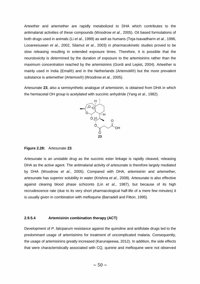
~ 50 ~
Arteether and artemether are rapidly metabolized to DHA which contributes to the
antimalarial activities of these compounds (Woodrow et al., 2005). Oil based formulations of
both drugs used in animals (Li et al., 1999) as well as humans (Teja-Isavadharm et al., 1996,
Looareesuwan et al., 2002, Silamut et al., 2003) in pharmacokinetic studies proved to be
slow releasing resulting in extended exposure times. Therefore, it is possible that the
neurotoxicity is determined by the duration of exposure to the artemisinins rather than the
maximum concentration reached by the artemisinins (Gordi and Lepist, 2004). Arteether is
mainly used in India (Emal®) and in the Netherlands (Artemotil®) but the more prevalent
substance is artemether (Artemos®) (Woodrow et al., 2005).
Artesunate 23, also a semisynthetic analogue of artemisinin, is obtained from DHA in which
the hemiacetal OH group is acetylated with succinic anhydride (Yang et al., 1982).
O
OO
O
O
H
H10
23O
OH
O
Figure 2.28: Artesunate 23.
Artesunate is an unstable drug as the succinic ester linkage is rapidly cleaved, releasing
DHA as the active agent. The antimalarial activity of artesunate is therefore largely mediated
by DHA (Woodrow et al., 2005). Compared with DHA, artemisinin and artemether,
artesunate has superior solubility in water (Krishna et al., 2008). Artesunate is also effective
against clearing blood phase schizonts (Lin et al., 1987), but because of its high
recrudescence rate (due to its very short pharmacological half-life of a mere few minutes) it
is usually given in combination with mefloquine (Barradell and Fitton, 1995).
2.9.5.4 Artemisinin combination therapy (ACT)
Development of P. falciparum resistance against the quinoline and antifolate drugs led to the
predominant usage of artemisinins for treatment of uncomplicated malaria. Consequently,
the usage of artemisinins greatly increased (Karunajeewa, 2012). In addition, the side effects
that were characteristically associated with CQ, quinine and mefloquine were not observed
~ 51 ~
when the artemisinins were used as antimalarial drugs. Also, when compared to other drugs
the artemisinins relieved symptoms of malaria more efficiently (Hinton et al., 2007).
However, it was found during the early clinical development of artemisinins that a 5-day
treatment with artemisinin monotherapy was linked to parasite recrudescence rates higher
than 25% even if the cure rates were consistently close to 100% (Hien and White, 1993,
McIntosh and Olliaro, 1999). This is very likely due to the extremely short half-lives of the
artemisinins. Normally drug concentrations must remain above the therapeutically effective
threshold for at least several complete life cycles of the malaria parasite in order to eliminate
blood-stages of the parasite. As each cycle takes 48 h to complete, in order to achieve total
elimination of the parasite burden and in doing so produce an effective cure (Karunajeewa,
2012), it was found necessary to treat at least for seven days with artemisinin monotherapy.
Normally, malaria cure using drugs with longer half-lives such as CQ and mefloquine is
possible with short-term treatment as the therapeutically effective drug concentrations
endure long enough to eliminate parasites arising in successive life cycles. That is, the
parasite recrudescence issue experienced with artemisinin use was addressed by utilizing
artemisinin treatment that lasted up to seven days (Giao et al., 2001). Before the
development of artemisinin resistance as reported from Cambodia, the 7 day treatment
courses had cure rates higher than 95% (McIntosh and Olliaro, 1999). Unfortunately, a
drawback is that the long-term treatment is likely to be undermined by poor patient
adherence after subsidence of fever, but before all parasites were eliminated (Karunajeewa,
2012).
Thus, the concept of using artemisinin combination therapies (ACTs) was introduced
wherein an artemisinin derivative was combined with a longer half-life drug such as
mefloquine, lumefantrine, piperaquine or other drug. Within an ACT, the artemisinin
component not only relieves the patient from malaria symptoms but is also responsible for
the relatively rapid decrease in the parasite burden. Total elimination of the parasites occur
several days after the last dose is taken as a result of the persistence of the partner drug
eliminating remaining parasites (Karunajeewa, 2012). A great number of clinical trials have
been carried out in order to evaluate clinical effectiveness of ACTs (Sinclair et al., 2009).
Results of some of these trials suggest that it is the efficacy of the combination that is crucial
rather than the choice of artemisinin or partner drug (Davis et al., 2005, Karunajeewa et al.,
2008). However, it is important to note that progression of resistance in a specific area can
be influenced if the artemisinin drug partner has pre-existing resistance (Karunajeewa et al.,
2008). Therefore, the main idea behind an ACT is to suppress the emergence of resistance
by using drugs with different mechanisms of action. Currently the WHO recommends one of
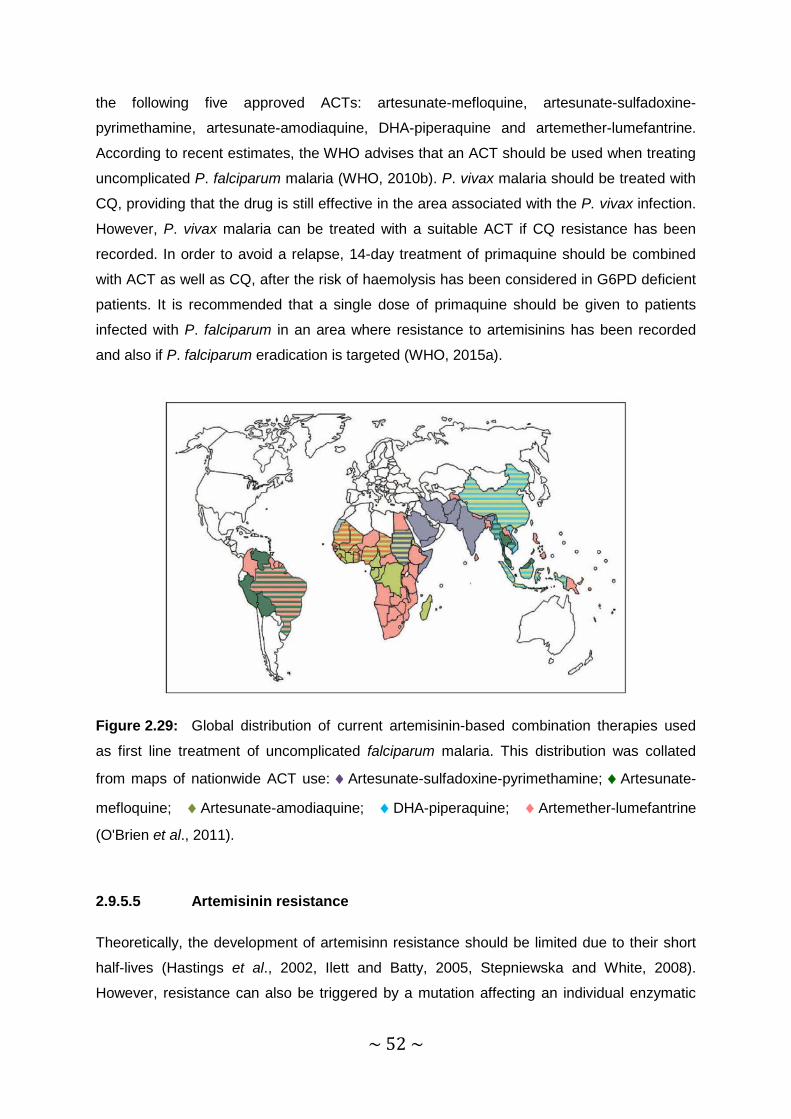
~ 52 ~
the following five approved ACTs: artesunate-mefloquine, artesunate-sulfadoxine-
pyrimethamine, artesunate-amodiaquine, DHA-piperaquine and artemether-lumefantrine.
According to recent estimates, the WHO advises that an ACT should be used when treating
uncomplicated P. falciparum malaria (WHO, 2010b). P. vivax malaria should be treated with
CQ, providing that the drug is still effective in the area associated with the P. vivax infection.
However, P. vivax malaria can be treated with a suitable ACT if CQ resistance has been
recorded. In order to avoid a relapse, 14-day treatment of primaquine should be combined
with ACT as well as CQ, after the risk of haemolysis has been considered in G6PD deficient
patients. It is recommended that a single dose of primaquine should be given to patients
infected with P. falciparum in an area where resistance to artemisinins has been recorded
and also if P. falciparum eradication is targeted (WHO, 2015a).
Figure 2.29: Global distribution of current artemisinin-based combination therapies used
as first line treatment of uncomplicated falciparum malaria. This distribution was collated
from maps of nationwide ACT use: ♦Artesunate-sulfadoxine-pyrimethamine; ♦Artesunate-
mefloquine; ♦Artesunate-amodiaquine; ♦DHA-piperaquine; ♦Artemether-lumefantrine
(O'Brien et al., 2011).
2.9.5.5 Artemisinin resistance
Theoretically, the development of artemisinn resistance should be limited due to their short
half-lives (Hastings et al., 2002, Ilett and Batty, 2005, Stepniewska and White, 2008).
However, resistance can also be triggered by a mutation affecting an individual enzymatic

~ 53 ~
pathway which is vital to the mode of action of the drug (Karunajeewa, 2012). Overall,
however, the development of resistance towards artemisinins is so important that it sparked
numerous debates as well as policy reconsiderations within the WHO (WHO, 2001, WHO,
2006, WHO, 2007). Accordingly, artemisinin was permitted to be used only in combination
therapy (White, 1999a), and monotherapy was phased out (Rehwagen, 2006).
The use of an ACT relies on the idea that parasite mutations associated with resistance
towards one drug will still allow the parasite to be vulnerable to the partner drug with its
different mechanism of action. Thus, the parasite won’t be able to survive long enough in
order to pass on their resistance features to the succeeding generation (White et al., 1999b,
White, 2004). With malaria, however, there are several limitations with regards to ACT usage
(Kremsner and Krishna, 2004). The P. falciparum parasite has a genome (2.3 x 10⁷ bp) that
is larger and more complex when compared to those of other organisms such as the virus
causing hepatitis B (3.2 x 10³ bp) or M. tuberculosis (4.4 x 10⁶ bp), there is an advantage of
having a larger number of genetic loci that could facilitate resistance development
(Karunajeewa, 2012). Thus, the development of multifaceted mechanisms of drug resistance
when compared with viruses and bacteria is not surprising. For instance, drug transport
mechanisms that are affected by resistance can result in resistance towards a variety of
drugs with different mechanisms of action involving different molecular targets. PfMDR1 is
the best example in P. falciparum as it translates the P-glycoprotein homologue (Pgh)
responsible for the efflux of drug into the parasite food vacuole (Cowman et al., 1994, Reed
et al., 2000). Single nucleotide polymorphisms or increasing the gene copy number can
facilitate resistance, where PfMDR1 has been involved in resistance towards various
antimalarial drugs such as CQ, lumefantrine, halofantrine, mefloquine, and amodiaquine
(Price et al., 2004, Price et al., 2006).
Initial warnings of treatment failure with the ACT artesunate-mefloquine were first reported
near the Thai-Cambodian border in the early 2000’s (Vijaykadga et al., 2006, Denis et al.,
2006). It was unclear at the time whether this was due to development of resistance either to
artesunate or to mefloquine (O'Brien et al., 2011). The first clinically-significant artemisinin
resistance was first described in Western Cambodia in 2008 (Noedl et al., 2008). The study
identified artesunate-resistant infections in two patients who experienced parasite
recrudescence following 7 days of artesunate monotherapy (4 mg/kg per day) and who
showed delayed parasite clearance times (133 and 95 h, as compared with a median of 52 h
for patients that were cured) in spite of maintaining acceptable drug levels. These times
represent the interval from the start of the treatment to the time the parasitaemia (i.e. the
percentage of parasitized erythrocytes) falls below the lower limit of parasite detection by

~ 54 ~
light microscopy (approximately 0.001%). Patient isolates with extended parasite clearance
times displayed DHA half maximal inhibitory concentration (IC50) values as determined by ex
vivo drug dose-response measurements four times higher than the geometric mean for
cured patients (Noedl et al., 2010).
The emergence of artemisinin resistance is supported by the results of a study published in
2009 (Dondorp et al., 2009). The study established that following artesunate-mefloquine
combination therapy or artesunate monotherapy, subjects displayed increased parasite
clearance times. The average clearance time would increase in patients treated with 2 mg/kg
of artesunate from 48 to 72 h. In patients treated with 4 mg/kg of the artesunate-mefloquine
combination, clearance times also increased from 54 to 84 h. Neither the pharmacokinetics
nor the differences in age between the two study populations could be held responsible for
these variations (O'Brien et al., 2011).
The development of artemisinin resistance in western Cambodia can be attributed to
different factors, namely the genetic background of the parasite displaying preference
towards the emergence of multidrug resistance, the sub-optimal use of drugs, and 30 years
of artemisinn monotherapy (Rathod et al., 1997, Newton et al., 2008, Dondorp et al., 2010).
The genes of the malaria parasite contribute to 56% of parasite clearance discrepancies as
determined by Pailin isolates in heritability studies, implying that determinants of resistance
could expand geographically under drug selection (Anderson et al., 2010).
The Thai-Cambodian border is a historical focal point when it comes to multidrug resistance
towards P. falciparum which adds to the concerns of weakening artemisinin efficacy
(Enserink, 2010). Resistance towards CQ, sulfadoxine-pyrimethamine and mefloquine were
the first to develop in this region; the sulfadoxine-pyrimethamine and mefloquine resistance
spread to Africa (Wellems and Plowe, 2001, Wongsrichanalai et al., 2002, Wootton et al.,
2002, Roper et al., 2004). In order to keep track of the rise and spread of artemisinin
resistance substantial efforts are being made throughout the Greater Mekong Subregion
(which is made up of Vietnam, the Yunnan Province of China, Thailand, Laos, Cambodia
and Myanmar) to carefully map out and block the spread of resistant parasites (Fig. 2.30)
(Cui et al., 2012).

~ 55 ~
Figure 2.30: P. falciparum resistance towards artemisinin observed in the five countries in
the Greater Mekong Subregion. Tier 1: areas with reliable evidence of resistance towards
artemisinin. Tier 2: areas with a substantial inflow of people from Tier 1 areas, which
includes those closely bordering Tier 1. Tier 3: areas with no evidence of artemisinin
resistance and limited contact with Tier 1 areas (WHO, 2015a).
Between 1995 and 2007, some 3,200 patients were treated with the 3-day combination
course of artesunate-mefloquine along the Thai-Myanmar border. Data collected from this
study indicated that 96% of patients were clear of parasitaemia on the third day of treatment
(Carrara et al., 2009). However, an increase in treatment failure rates (recrudescence of
parasites observed during a 42-day follow-up) were associated with a small yet significant
increase in parasite clearance times in this study. Gametocyte carriage, which increases the
transmission risk of drug-resistant parasites, was also linked to decreased parasite
clearance. Parasite clearance data was collected from 25 different countries (ranging from
low to high transmission) making up a total of 18,000 patients in a 2010 study (Stepniewska
et al., 2010), confirming the previous proposal from western Cambodia (Dondorp et al.,
2009) that delayed parasite clearance time is a clear marker for resistance against
artemisinin.
A more recent study published in 2014 identified kelch13 as the molecular marker
associated with mutations in southeast Asia (Ashley et al., 2014). The study involved 1,241
children as well as adults infected with acute, uncomplicated P. falciparum malaria in an
open-label trial at 15 sites in 10 countries (7 in Asia and 3 in Africa). Patients received
artesunate administered orally at a daily dose of either 2 mg/kg per day or 4 mg/kg, for 3
days, followed by a standard 3-day course of ACT. Results showed that the average
parasite clearance half-lives varied from 1.9 h (Democratic Republic of Congo) to 7 h (Thai-

~ 56 ~
Cambodian border). Throughout Southeast Asia, from central Myanmar to southern
Vietnam, an increase in parasite clearance times (half-life of parasite clearance > 5 h) were
observed that were associated with a single point mutation in the “propeller” region of the P.
falciparum kelch protein gene on chromosome 13 (kelch13). The occurrence of pre-
treatment and post-treatment gametocytaemia was increased among patients with sluggish
parasite clearance, signifying a superior potential for transmission. Of note, prolonged
courses of ACTs are currently efficacious in areas where standard 3-day treatments are
failing. Interestingly, emerging artemisinin resistance seems to be mainly limited to the
Greater Mekong Subregion, as nothing conclusive has been reported yet from Africa
(Kachur et al., 2010).
Another important factor that was identified for playing a role in artemisinin resistance was
recrudescence where some studies suggested that the use of artemisinin can give rise to
parasite recrudescence. In vitro studies have indicated that the P. falciparum parasite has a
unique ability to become quiescent, or dormant, when exposed to artemisinins for short
periods of time. This mechanism is believed to protect early ring-stage intraerythrocytic
parasites from the drug; quiescence implies slowed turnover of intracellular enzymes, thus
lowering susceptibility to the drug (Teuscher et al., 2010, Teuscher et al., 2011, Cheng et al.,
2012). After the concentration of artemisinin has dropped to sub-therapeutic level, a portion
of the dormant parasites continue their life cycle leading to parasite recrudescence,
resistance and treatment failure. Another pressing issue is that of neurotoxicity. As
previously mentioned, most of the artemisinins undergo facile metabolism to DHA which is
neurotoxic in cell and animal assays. It stands to reason if parasite clearance is slowed via
the ability of the parasite to become dormant in the presence of an artemisinin the length of
treatment should be increased in order to reach 100% parasite clearance. Although it
sounds like the logical step to take, it has some consequences. As the length of treatment
increase, patients are at an increased risk of being exposed to higher neurotoxicity values. If
the metabolic stability of an artemisinin derivative can be improved by, for example, not
providing DHA in vivo, it would have a major advantage over its predecessors.
2.9.5.6 Other artemisinin derivatives
Artemisone
In order to prevent the metabolic transformation of first generation artemisinins to DHA as
well as improving pharmacokinetics, several 10-(alkylamino)artemisinins were prepared.
One such is artemisone 24 that in a Phase II clinical trial was shown to be three times as

~ 57 ~
efficacious as the comparator drug artesunate, and did not give DHA on metabolism (Vivas
et al., 2007, Opsenica and Šolaja, 2012). Twelve different P. falciparum strains were used to
determine the in vitro antimalarial activities of artemisone; when these activities were
compared to other antimalarial drug classes they were independent of the drug-susceptibility
profile of the different strains (Haynes et al., 2004, Vivas et al., 2007).
O
OO
O
N
H
H10
24
SO O
Figure 2.31: Artemisone 24.
Artemisone has a minor antagonistic effect if used with CQ, pyrimethamine, atovaquone and
amodiaquine and slight synergism with mefloquine as determined by evaluation of in vitro
drug-drug interactions against multidrug-resistant strains of K1 and CQ sensitive 3D7
strains. Also, artemisone does not form DHA upon metabolism but rather forms mono-
hydroxylated metabolites with syn-hydroxyl and peroxide groups (Haynes et al., 2006). This
clearly distinguishes artemisone and analogues from the first generation artemisinins
(Opsenica and Šolaja, 2012).
Artemisone is mainly metabolized by recombinant CYP3A4 as demonstrated through the
incubation of 14 recombinant cytochrome P450 (CYP) isoforms together with microsomes
and selective inhibitors of CYP. Remarkably, artemisone is not an inducer of its metabolism;
this stands in contrast to the current clinical derivatives that induce their own metabolism
(Ashton et al., 1998) involving the principal metabolizing enzyme CYP2B6 (Svensson and
Ashton, 1999). These results indicate that although artemisinin and artemisone share
structural similarities they have notably different metabolic profiles in P. falciparum.
Compared with artesunate, artemisone demonstrated enhanced pharmacokinetics, improved
efficacy as well as the absence of neurotoxicity in vitro and in vivo (Haynes et al., 2006,
Nagelschmitz et al., 2008). The absence of neurotoxicity is noted as a decisive advantage,
and this must become important if protracted treatment regimens involving artemisone and a
combination partner against artemisinin-resistant parasites is eventually required. As noted
above, neurotoxicity is an issue in current clinical artemisinins (Brewer et al., 1994).

~ 58 ~
11-Azaartemisinin
The first generation artemisinin derivatives artemether and artesunate discussed above are
prepared from artemisinin by modification at the C10 position; all are converted into DHA in
vivo. In contrast, 11-azaartemisinin 25 is a unique derivative prepared from artemisinin by
replacement of O11 by nitrogen.
N
OO
O
H
H
25O
H 11
Figure 2.32: 11-Azaartemisinin 25.
In 1995, Avery and co-workers synthesized the first azaartemisinin derivatives namely the N-
alkyl-11-aza-9-desmethylartemisinins (Avery et al., 1995), with an elegant albeit complex
synthesis. In the same year, Ziffer and co-workers also synthesized a series of N-substituted
11-azaartemisinins with promising in vitro and in vivo results (Torok and Ziffer, 1995). It was
only relatively recently that Haynes and his co-workers were able to simplify and optimize
the synthesis of 11-azaartemisinin (Haynes et al., 2007a).
11-Azaartemisinin 25 displays a number of advantages when compared to other artemisinins
as it is easily obtained from artemisinin, contains a lactam moiety instead of the lactone of
artemisinin. This is likely to make it more stable under acidic conditions (Bach and
Dmitrenko, 2006). Therefore, it stands to reason that the azaartemisinins should have
enhanced bioavailability when compared to artemisinin. Also, this compound cannot produce
DHA in vivo as it is at a higher oxidation level than artemisinin.
An array of 11-azaartemisinin derivatives has been synthesized since 1995, many of which
appear to have potential for treatment of malaria. As 11-azaartemisinin is the parent
compound of the current study, a review discussing the synthesis, chemistry and biological
evaluations of azaartemisinin and its derivatives is presented in Chapter 3.

~ 59 ~
REFERENCES
Abiodun, O. O., Brun, R. & Wittlin, S. (2013) In vitro interaction of artemisinin derivatives or
the fully synthetic peroxidic antimalarial OZ277 with thapsigargin in Plasmodium
falciparum strains. Malaria Journal, 12, 1-4.
Abkarian, M., Massiera, G., Berry, L., Roques, M. & Braun-Breton, C. (2011) A novel
mechanism for egress of malarial parasites from red blood cells. Blood, 117, 4118-
4124.
Aikawa, M. & Beaudoin, R. L. (1970) Plasmodium fallax: high-resolution autoradiography of
exoerythrocytic stages treated with Primaquine in vitro. Experimental Parasitology,
27, 454-463.
Amino, R., Thiberge, S., Martin, B. A., Celli, S., Shorte, S., Frischknecht, F. & Ménard, R.
(2006) Quantitative imaging of Plasmodium transmission from mosquito to mammal.
Nature Medicine, 12, 220-224.
Anderson, T. J. C., Nair, S., Qin, H., Singlam, S., Brockman, A., Paiphun, L. & Nosten, F. O.
(2005) Are transporter genes other than the chloroquine resistance locus (pfcrt) and
multidrug resistance gene (pfmdr) associated with antimalarial drug resistance?
Antimicrobial Agents and Chemotherapy, 49, 2180-2188.
Anderson, T. J. C., Nair, S., Nkhoma, S., Williams, J. T., Imwong, M., Yi, P., Socheat, D.,
Das, D., Chotivanich, K. & Day, N. P. J. (2010) High heritability of malaria parasite
clearance rate indicates a genetic basis for artemisinin resistance in western
Cambodia. Journal of Infectious Diseases, 201, 1326-1330.
Arnou, B., Montigny, C. D., Morth, J. P., Nissen, P., Jaxel, C., Møller, J. V. & Le Maire, M.
(2011) The Plasmodium falciparum Ca2+-ATPase PfATP6: insensitive to artemisinin,
but a potential drug target. Biochemical Society Transactions, 39, 823-831.
Ashley, E., McGready, R., Proux, S. & Nosten, F. O. (2006) Malaria. Travel Medicine and
Infectious Disease, 4, 159-173.
Ashley, E. A., Stepniewska, K., Lindegårdh, N., Annerberg, A., Kham, A., Brockman, A.,
Singhasivanon, P., White, N. J. & Nosten, F. (2007) How much fat is necessary to
optimize lumefantrine oral bioavailability? Tropical Medicine & International Health,
12, 195-200.

~ 60 ~
Ashley, E. A., Dhorda, M., Fairhurst, R. M., Amaratunga, C., Lim, P., Suon, S., Sreng, S.,
Anderson, J. M., Mao, S. & Sam, B. (2014) Spread of artemisinin resistance in
Plasmodium falciparum malaria. New England Journal of Medicine, 371, 411-423.
Ashton, M., Hai, T. N., Sy, N. D., Huong, D. X., Van Huong, N. & Nieu, N. T. (1998)
Artemisinin pharmacokinetics is time-dependent during repeated oral administration
in healthy male adults. Drug Metabolism and Disposition, 26, 25-27.
Avery, M. A., Bonk, J. D., Chong, W. K. M., Mehrotra, S., Miller, R., Milhous, W., Goins, D.
K., Venkatesan, S. & Wyandt, C. (1995) Structure-activity relationships of the
antimalarial agent artemisinin. 2. Effect of heteroatom substitution at O-11: synthesis
and bioassay of N-Alkyl-11-aza-9-desmethylartemisinins. Journal of Medicinal
Chemistry, 38, 5038-5044.
Ayimba, E., Hegewald, J., Segbena, A. Y., Gantin, R. G., Lechner, C. J., Agosssou, A.,
Banla, M. & Soboslay, P. T. (2011) Proinflammatory and regulatory cytokines and
chemokines in infants with uncomplicated and severe Plasmodium falciparum
malaria. Clinical & Experimental Immunology, 166, 218-226.
Bach, R. D. & Dmitrenko, O. (2006) The effect of carbonyl substitution on the strain energy
of small ring compounds and their six-member ring reference compounds. Journal of
the American Chemical Society, 128, 4598-4611.
Bannister, L. H., Mitchell, G. H., Butcher, G. A., Dennis, E. D. & Cohen, S. (1986) Structure
and development of the surface coat of erythrocytic merozoites of Plasmodium
knowlesi. Cell and Tissue Research, 245, 281-290.
Bannister, L. H., Hopkins, J. M., Fowler, R. E., Krishna, S. & Mitchell, G. H. (2000) A brief
illustrated guide to the ultrastructure of Plasmodium falciparum asexual blood-stages.
Parasitology Today, 16, 427-433.
Barnes, D. A., Foote, S. J., Galatis, D., Kemp, D. J. & Cowman, A. F. (1992) Selection for
high-level chloroquine resistance results in deamplification of the pfmdr1 gene and
increased sensitivity to mefloquine in Plasmodium falciparum. The EMBO Journal,
11, 3067.
Barradell, L. B. & Fitton, A. (1995) Artesunate. Drugs, 50, 714-741.
Batty, K. T., Thu, A., Thi, L., Davis, T. M. E., Ilett, K. F., Xuan Mai, T., Canh Hung, N., Phuc
Tien, N., Powell, S. M. & Van Thien, H. (1998) A pharmacokinetic and

~ 61 ~
pharmacodynamic study of intravenous vs. oral artesunate in uncomplicated
falciparum malaria. British Journal of Clinical Pharmacology, 45, 123-129.
Baudon, D., Martet, G., Pascal, B., Bernard, J., Keundjian, A. & Laroche, R. (1999) Efficacy
of daily antimalarial chemoprophylaxis in tropical Africa using either doxycycline or
chloroquine-proguanil; a study conducted in 1996 in the French Army. Transactions
of the Royal Society of Tropical Medicine and Hygiene, 93, 302-303.
Beaudoin, R. L. & Aikawa, M. (1968) Primaquine-induced changes in morphology of
exoerythrocytic stages of malaria. Science, 160, 1233-1234.
Becker, K., Rahlfs, S., Nickel, C. & Schirmer, R. H. (2003) Glutathione - functions and
metabolism in the malarial parasite Plasmodium falciparum. Biological Chemistry,
384, 551-566.
Berman, P. A. & Adams, P. A. (1997) Artemisinin enhances heme-catalysed oxidation of
lipid membranes. Free Radical Biology and Medicine, 22, 1283-1288.
Bilolikar, H. G., Bagade, A. C., Phadke, M. A. & Gambhir, P. S. (1994) Efficacy of
halofantrine in malaria. Indian Pediatrics, 32, 77-79.
Borrmann, S., Szlezak, N., Binder, R. K., Missinou, M. A., Lell, B. & Kremsner, P. G. (2002)
Evidence for the efficacy of artesunate in asymptomatic Plasmodium malariae
infections. Journal of Antimicrobial Chemotherapy, 50, 751-754.
Boulard, Y., Landau, I., Miltgen, F., Ellis, D. S. & Peters, W. (1983) The chemotherapy of
rodent malaria, XXXIV. Causal prophylaxis Part III: Ultrastructural changes induced
in exo-erythrocytic schizonts of Plasmodium yoelii by primaquine. Annals of Tropical
Medicine and Parasitology, 77, 555-568.
Bousejra-El Garah, F., Wong, M. H.-L., Amewu, R. K., Muangnoicharoen, S., Maggs, J. L.,
Stigliani, J.-L., Park, B. K., Chadwick, J., Ward, S. A. & O'Neill, P. M. (2011)
Comparison of the reactivity of antimalarial 1, 2, 4, 5-tetraoxanes with 1, 2, 4-
trioxolanes in the presence of ferrous iron salts, heme, and ferrous iron
salts/phosphatidylcholine. Journal of Medicinal Chemistry, 54, 6443-6455.
Bozdech, Z., Llinás, M., Pulliam, B. L., Wong, E. D., Zhu, J. & Derisi, J. L. (2003) The
transcriptome of the intraerythrocytic developmental cycle of Plasmodium falciparum.
PLoS Biology, 1, e5.

~ 62 ~
Bray, P. G., Deed, S., Fox, E., Kalkanidis, M., Mungthin, M., Deady, L. W. & Tilley, L. (2005)
Primaquine synergises the activity of chloroquine against chloroquine-resistant P.
falciparum. Biochemical Pharmacology, 70, 1158-1166.
Brewer, T. G., Grate, S. J., Peggins, J. O., Weina, P. J., Petras, J. M., Levine, B. S., Heiffer,
M. H. & Schuster, B. G. (1994) Fatal neurotoxicity of arteether and artemether. The
American Journal of Tropical Medicine and Hygiene, 51, 251-259.
Briolant, S. B., Pelleau, S. P., Bogreau, H., Hovette, P., Zettor, A. S., Castello, J., Baret, E.,
Amalvict, R. M., Rogier, C. & Pradines, B. (2011) In vitro susceptibility to quinine and
microsatellite variations of the Plasmodium falciparum Na+/H+ exchanger (Pfnhe-1)
gene: the absence of association in clinical isolates from the Republic of Congo.
Malaria Journal, 10, 1.
Brown, M. & Hebert, A. A. (1997) Insect repellents: an overview. Journal of the American
Academy of Dermatology, 36, 243-249.
Bruce-Chwatt, L. J. (1981) Alphonse Laveran's discovery 100 years ago and today's global
fight against malaria. Journal of the Royal Society of Medicine, 74, 531.
Buchholz, K., Schirmer, R. H., Eubel, J. K., Akoachere, M. B., Dandekar, T., Becker, K. &
Gromer, S. (2008) Interactions of methylene blue with human disulfide reductases
and their orthologues from Plasmodium falciparum. Antimicrobial Agents and
Chemotherapy, 52, 183-191.
Budimulja, A. S., Tapchaisri, P., Wilairat, P. & Marzuki, S. (1997) The sensitivity of
Plasmodium protein synthesis to prokaryotic ribosomal inhibitors. Molecular and
Biochemical Parasitology, 84, 137-141.
Buffet, P. A., Safeukui, I., Deplaine, G., Brousse, V., Prendki, V., Thellier, M., Turner, G. D. &
Mercereau-Puijalon, O. (2011) The pathogenesis of Plasmodium falciparum malaria
in humans: insights from splenic physiology. Blood, 117, 381-392.
Burgoine, K. L., Bancone, G. & Nosten, F. O. (2010) The reality of using primaquine. Malaria
Journal, 9, 376.
Butler, A. R., Khan, S. & Ferguson, E. (2010) A brief history of malaria chemotherapy. The
Journal of the Royal College of Physicians of Edinburgh, 40, 172-177.

~ 63 ~
Cabantchik, Z. I., Kakhlon, O. R., Epsztejn, S., Zanninelli, G. & Breuer, W. (2002)
Intracellular and extracellular labile iron pools. IN HERSHKO, C. (Ed.) Iron Chelation
Therapy. 1 ed. United States, Springer. p. 55-75.
Campanale, N., Nickel, C., Daubenberger, C. A., Wehlan, D. A., Gorman, J. J., Klonis, N.,
Becker, K. & Tilley, L. (2003) Identification and characterization of heme-interacting
proteins in the malaria parasite, Plasmodium falciparum. Journal of Biological
Chemistry, 278, 27354-27361.
Carrara, V. I., Zwang, J., Ashley, E. A., Price, R. N., Stepniewska, K., Barends, M.,
Brockman, A., Anderson, T., McGready, R. & Phaiphun, L. (2009) Changes in the
treatment responses to artesunate-mefloquine on the north-western border of
Thailand during 13 years of continuous deployment. PLoS One, 4, e4551.
Carter, J. Y., Loolpapit, M. P., Lema, O. E., Tome, J. L., Nagelkerke, N. J. D. & Watkins, W.
M. (2005) Reduction of the efficacy of antifolate antimalarial therapy by folic acid
supplementation. The American Journal of Tropical Medicine and Hygiene, 73, 166-
170.
Chen, L. H., Wilson, M. E. & Schlagenhauf, P. (2006) Prevention of malaria in long-term
travellers. Jama, 296, 2234-2244.
Cheng, Q., Kyle, D. E. & Gatton, M. L. (2012) Artemisinin resistance in Plasmodium
falciparum: A process linked to dormancy? International Journal for Parasitology:
Drugs and Drug Resistance, 2, 249-255.
China Cooperative Research Group. (1982) The chemistry and synthesis of qinghaosu
derivatives. Journal of Traditional Chinese Medicine 2, 1-16.
Coghi, P., Basilico, N., Taramelli, D., Chan, W. C., Haynes, R. K. & Monti, D. (2009)
Interaction of artemisinins with oxyhemoglobin Hb-FeII, Hb-FeII, CarboxyHb-FeII,
Heme-FeII, and carboxyheme FeII: significance for mode of action and implications
for therapy of cerebral malaria. ChemMedChem, 4, 2045-2053.
Cooper, R. A., Ferdig, M. T., Su, X.-Z., Ursos, L. M. B., Mu, J., Nomura, T., Fujioka, H.,
Fidock, D. A., Roepe, P. D. & Wellems, T. E. (2002) Alternative mutations at position
76 of the vacuolar transmembrane protein PfCRT are associated with chloroquine
resistance and unique stereospecific quinine and quinidine responses in Plasmodium
falciparum. Molecular Pharmacology, 61, 35-42.
~ 64 ~
Cooper, R. A., Lane, K. D., Deng, B., Mu, J., Patel, J. J., Wellems, T. E., Su, X. & Ferdig, M.
T. (2007) Mutations in transmembrane domains 1, 4 and 9 of the Plasmodium
falciparum chloroquine resistance transporter alter susceptibility to chloroquine,
quinine and quinidine. Molecular Microbiology, 63, 270-282.
Cowman, A. F., Galatis, D. & Thompson, J. K. (1994) Selection for mefloquine resistance in
Plasmodium falciparum is linked to amplification of the pfmdr1 gene and cross-
resistance to halofantrine and quinine. Proceedings of the National Academy of
Sciences, 91, 1143-1147.
Cowman, A. F., Berry, D. & Baum, J. (2012) The cellular and molecular basis for malaria
parasite invasion of the human red blood cell. The Journal of Cell Biology, 198, 961-
971.
Cox, F. E. G. (2010) History of the discovery of the malaria parasites and their vectors.
Parasite Vectors, 3, 5.
Creek, D. J., Charman, W. N., Chiu, F. C. K., Prankerd, R. J., Dong, Y., Vennerstrom, J. L. &
Charman, S. A. (2008) Relationship between antimalarial activity and heme alkylation
for spiro-and dispiro-1, 2, 4-trioxolane antimalarials. Antimicrobial Agents and
Chemotherapy, 52, 1291-1296.
Cui, L., Yan, G., Sattabongkot, J., Cao, Y., Chen, B., Chen, X., Fan, Q., Fang, Q.,
Jongwutiwes, S. & Parker, D. (2012) Malaria in the Greater Mekong Subregion:
heterogeneity and complexity. Acta Tropica, 121, 227-239.
Dahl, E. L., Shock, J. L., Shenai, B. R., Gut, J., Derisi, J. L. & Rosenthal, P. J. (2006)
Tetracyclines specifically target the apicoplast of the malaria parasite Plasmodium
falciparum. Antimicrobial Agents and Chemotherapy, 50, 3124-3131.
Davis, T. M. E., Karunajeewa, H. A. & Ilett, K. F. (2005) Artemisinin-based combination
therapies for uncomplicated malaria. Medical Journal of Australia, 182, 181.
De Villiers, K. A., Marques, H. M. & Egan, T. J. (2008) The crystal structure of halofantrine-
ferriprotoporphyrin IX and the mechanism of action of arylmethanol antimalarials.
Journal of Inorganic Biochemistry, 102, 1660-1667.
Del Pilar Crespo, M., Avery, T. D., Hanssen, E., Fox, E., Robinson, T. V., Valente, P., Taylor,
D. K. & Tilley, L. (2008) Artemisinin and a series of novel endoperoxide antimalarials
~ 65 ~
exert early effects on digestive vacuole morphology. Antimicrobial Agents and
Chemotherapy, 52, 98-109.
Del Prado, G. R. L., García, C. H. N., Cea, L. M., Espinilla, V. F. N., Moreno, M. F. M. O.,
Marquez, A. D., Polo, M. J. P. & García, I. A. S. (2014) Malaria in developing
countries. The Journal of Infection in Developing Countries, 8, 001-004.
Dembele, L., Gego, A., Zeeman, A.-M., Franetich, J.-F. O., Silvie, O., Rametti, A., Le Grand,
R., Dereuddre-Bosquet, N., Sauerwein, R. & Van Gemert, G.-J. (2011) Towards an in
vitro model of Plasmodium hypnozoites suitable for drug discovery. PLoS One, 6, e18162.
Denis, M. B., Tsuyuoka, R., Poravuth, Y., Narann, T. S., Seila, S., Incardona, S., Lim, P.,
Sem, R., Socheat, D. & Christophel, E. M. (2006) Surveillance of the efficacy of
artesunate and mefloquine combination for the treatment of uncomplicated
falciparum malaria in Cambodia. Tropical Medicine & International Health, 11, 1360-
1366.
Dittrich, S., Mitchell, S. L., Blagborough, A. M., Wang, Q., Wang, P., Sims, P. F. G. & Hyde,
J. E. (2008) An atypical orthologue of 6-pyruvoyltetrahydropterin synthase can
provide the missing link in the folate biosynthesis pathway of malaria parasites.
Molecular Microbiology, 67, 609-618.
Dondorp, A. M., Desakorn, V., Pongtavornpinyo, W., Sahassananda, D., Silamut, K.,
Chotivanich, K., Newton, P. N., Pitisuttithum, P., Smithyman, A. M. & White, N. J.
(2005) Estimation of the total parasite biomass in acute falciparum malaria from
plasma PfHRP2. PLoS Med, 2, e204.
Dondorp, A. M., Lee, S. J., Faiz, M. A., Mishra, S., Price, R., Tjitra, E., Than, M., Htut, Y.,
Mohanty, S. & Yunus, E. B. (2008) The relationship between age and the
manifestations of and mortality associated with severe malaria. Clinical Infectious
Diseases, 47, 151-157.
Dondorp, A. M., Nosten, F. O., Yi, P., Das, D., Phyo, A. P., Tarning, J., Lwin, K. M., Ariey, F.,
Hanpithakpong, W. & Lee, S. J. (2009) Artemisinin resistance in Plasmodium
falciparum malaria. New England Journal of Medicine, 361, 455-467.
Dondorp, A. M., Yeung, S., White, L., Nguon, C., Day, N. P. J., Socheat, D. & Von Seidlein,
L. (2010) Artemisinin resistance: current status and scenarios for containment.
Nature Reviews Microbiology, 8, 272-280.

~ 66 ~
Doolan, D. L., Doban, C. & Baird, J. K. (2009) Acquired immunity to malaria. Clinical
Microbiology Reviews, 22, 13-36.
Doumbo, O. K., Thera, M. A., Kone, A. K., Raza, A., Tempest, L. J., Lyke, K. E., Plowe, C. V.
& Rowe, J. A. (2009) High levels of Plasmodium falciparum rosetting in all clinical
forms of severe malaria in African children. The American Journal of Tropical
Medicine and Hygiene, 81, 987-993.
Eastman, R. T. & Fidock, D. A. (2009) Artemisinin-based combination therapies: a vital tool
in efforts to eliminate malaria. Nature Reviews Microbiology, 7, 864-874.
Eckstein-Ludwig, U., Webb, R. J., Van Goethem, I. D. A., East, J. M., Lee, A. G., Kimura, M.,
O'Neill, P. M., Bray, P. G., Ward, S. A. & Krishna, S. (2003) Artemisinins target the
SERCA of Plasmodium falciparum. Nature, 424, 957-961.
Edwards, G., Mcgrath, C. S., Ward, S. A., Supanaranond, W., Pukrittayakamee, S., Davis, T.
M. & White, N. J. (1993) Interactions among primaquine, malaria infection and other
antimalarials in Thai subjects. British Journal of Clinical Pharmacology, 35, 193-198.
Egan, T. J. (2008) Haemozoin formation. Molecular and Biochemical Parasitology, 157, 127-
136.
Enayati, A., Lines, J., Maharaj, R. & Hemingway, J. (2009) Suppressing the vector. IN
FEACHEM, R. G. A., PHILLIPS, A. A. & TARGETT, G. A. (Eds.) Shrinking the
Malaria Map: A Prospectus on Malaria Elimination. San Francisco, The Global Health
Group. 140-154p.
Enserink, M. (2010) Malaria's drug miracle in danger. Science, 328, 844-846.
Falco, E. A., Goodwin, L. G., Hitchings, G. H., Rollo, I. M. & Russell, P. B. (1951) 2:4-
diaminopyrimidines – a new series of antimalarials. British Journal of Pharmacology
and Chemotherapy, 6, 185-200.
Fisher, N., Bray, P. G., Ward, S. A. & Biagini, G. A. (2007) The malaria parasite type II
NADH: quinone oxidoreductase: an alternative enzyme for an alternative lifestyle.
Trends in Parasitology, 23, 305-310.
Fishwick, J., Mclean, W. G., Edwards, G. & Ward, S. A. (1995) The toxicity of artemisinin
and related compounds on neuronal and glial cells in culture. Chemico-Biological
Interactions, 96, 263-271.
~ 67 ~
Fitch, C. D. (2004) Ferriprotoporphyrin IX, phospholipids, and the antimalarial actions of
quinoline drugs. Life Sciences, 74, 1957-1972.
Fitch, C. D., Chan, R. L. & Chevli, R. (1979) Chloroquine resistance in malaria: accessibility
of drug receptors to mefloquine. Antimicrobial Agents and Chemotherapy, 15, 258-
262.
Foley, M. & Tilley, L. (1998) Quinoline antimalarials: mechanisms of action and resistance
and prospects for new agents. Pharmacology & Therapeutics, 79, 55-87.
Foote, S. J., Galatis, D. & Cowman, A. F. (1990) Amino acids in the dihydrofolate reductase-
thymidylate synthase gene of Plasmodium falciparum involved in cycloguanil
resistance differ from those involved in pyrimethamine resistance. Proceedings of the
National Academy of Sciences, 87, 3014-3017.
Fry, M. & Beesley, J. E. (1991) Mitochondria of mammalian Plasmodium spp. Parasitology,
102, 17-26.
Fry, M. & Pudney, M. (1992) Site of action of the antimalarial hydroxynaphthoquinone, 2-
[trans-4-(4'-chlorophenyl) cyclohexyl]-3-hydroxy-1, 4-naphthoquinone (566C80).
Biochemical Pharmacology, 43, 1545-1553.
Fujioka, H. & Aikawa, M. (2002) Structure and life cycle. IN PERLMANN, P. & TROYE-
BLOMBERG, M. (Eds.) Malaria Immunology. Basel, Karger. 80:1-26p.
Gething, P. W., Patil, A. P., Smith, D. L., Guerra, C. A., Elyazar, I. R. F., Johnston, G. L.,
Tatem, A. J. & Hay, S. I. (2011a) A new world malaria map: Plasmodium falciparum
endemicity in 2010. Malaria Journal, 10, 1-16.
Gething, P. W., Elyazar, I. R. F., Moyes, C. L., Smith, D. L., Battle, K. E., Guerra, C. A., Patil,
A. P., Tatem, A. J., Howes, R. E. & Myers, M. F. (2011b) A long neglected world
malaria map: Plasmodium vivax endemicity in 2010. PLoS Neglected Tropical
Diseases, 6, e1814.
Giao, P. T., Binh, T. Q., Kager, P. A., Long, H. P., Van Thang, N., Van Nam, N. & De Vries,
P. J. (2001) Artemisinin for treatment of uncomplicated falciparum malaria: is there a
place for monotherapy? The American Journal of Tropical Medicine and Hygiene, 65, 690-695.

~ 68 ~
Gil, J. P., Nogueira, F., Stromberg-Norklit, J., Lindberg, J., Carrolo, M., Casimiro, C., Lopes,
D., Arez, A. P., Cravo, P. V. & Rosario, V. E. (2003) Detection of atovaquone and
Malarone - resistance conferring mutations in Plasmodium falciparum cytochrome b
gene (cytb). Molecular and Cellular Probes, 17, 85-89.
Gilson, P. R. & Crabb, B. S. (2009) Morphology and kinetics of the three distinct phases of
red blood cell invasion by Plasmodium falciparum merozoites. International Journal
for Parasitology, 39, 91-96.
Glushakova, S., Yin, D., Li, T. & Zimmerberg, J. (2005) Membrane transformation during
malaria parasite release from human red blood cells. Current Biology, 15, 1645-1650.
Godfray, H. C. J. (2013) Mosquito ecology and control of malaria. Journal of Animal Ecology,
82, 15-25.
Gordi, T. & Lepist, E.-I. (2004) Artemisinin derivatives: toxic for laboratory animals, safe for
humans? Toxicology Letters, 147, 99-107.
Grace, J. M., Aguilar, A. J., Trotman, K. M. & Brewer, T. G. (1998) Metabolism of beta-
arteether to dihydroqinghaosu by human liver microsomes and recombinant
cytochrome P450. Drug Metabolism and Disposition, 26, 313-317.
Gregson, A. & Plowe, C. V. (2005) Mechanisms of resistance of malaria parasites to
antifolates. Pharmacological Reviews, 57, 117-145.
Grüring, C., Heiber, A., Kruse, F., Ungefehr, J., Gilberger, T.-W. & Spielmann, T. (2011)
Development and host cell modifications of Plasmodium falciparum blood-stages in
four dimensions. Nature Communications, 2, 165.
Hastings, I. M., Watkins, W. M. & White, N. J. (2002) The evolution of drug-resistant malaria:
the role of drug elimination half-life. Philosophical Transactions of the Royal Society
of London B: Biological Sciences, 357, 505-519.
Hastings, I. M. (2006) Gametocytocidal activity in antimalarial drugs speeds the spread of
drug resistance. Tropical Medicine & International Health, 11, 1206-1217.
Hayeshi, R., Masimirembwa, C., Mukanganyama, S. & Ungell, A. L. B. (2008) Lysosomal
trapping of amodiaquine: impact on transport across intestinal epithelia models.
Biopharmaceutics & Drug Disposition, 29, 324-334.

~ 69 ~
Haynes, R. K. (2001) Artemisinin and derivatives: the future for malaria treatment? Current
Opinion in Infectious Diseases, 14, 719-726.
Haynes, R. K., Monti, D., Taramelli, D., Basilico, N., Parapini, S. & Olliaro, P. (2003)
Artemisinin antimalarials do not inhibit hemozoin formation. Antimicrobial Agents and
Chemotherapy, 47, 1175-1175.
Haynes, R. K., Ho, W. Y., Chan, H. W., Fugmann, B., Stetter, J. R., Croft, S. L., Vivas, L.,
Peters, W. & Robinson, B. L. (2004) Highly antimalarial-active artemisinin derivatives:
biological activity does not correlate with chemical reactivity. Angewandte Chemie
International Edition, 43, 1381-1385.
Haynes, R. K., Fugmann, B., Stetter, J. R., Rieckmann, K., Heilmann, H. D., Chan, H. W.,
Cheung, M. K., Lam, W. L., Wong, H. N. & Croft, S. L. (2006) Artemisone - a highly
active antimalarial drug of the artemisinin class. Angewandte Chemie, 118, 2136-
2142.
Haynes, R. K., Wong, H. N., Lee, K. W., Lung, C. M., Shek, L. Y., Williams, I. D., Croft, S. L.,
Vivas, L., Rattray, L. & Stewart, L. (2007a) Preparation of N-Sulfonyl- and N-
Carbonyl-11-azaartemisinins with greatly enhanced thermal stabilities: in vitro
antimalarial activities. ChemMedChem, 2, 1464-1479.
Haynes, R. K., Chan, W. C., Lung, C. M., Uhlemann, A. C., Eckstein, U., Taramelli, D.,
Parapini, S., Monti, D. & Krishna, S. (2007b) The Fe2+-mediated decomposition,
PfATP6 binding, and antimalarial activities of artemisone and other artemisinins: the
unlikelihood of C-centered radicals as bioactive intermediates. ChemMedChem, 2, 1480-1497.
Haynes, R. K., Chan, W.-C., Wong, H.-N., Li, K.-Y., Wu, W.-K., Fan, K.-M., Sung, H. H. Y.,
Williams, I. D., Prosperi, D. & Melato, S. (2010) Facile oxidation of leucomethylene
blue and dihydroflavins by artemisinins: relationship with flavoenzyme function and
antimalarial mechanism of action. ChemMedChem, 5, 1282-1299.
Haynes, R. K., Cheu, K.-W., Tang, M. M.-K., Chen, M.-J., Guo, Z.-F., Guo, Z.-H., Coghi, P. &
Monti, D. (2011) Reactions of antimalarial peroxides with each of leucomethylene
blue and dihydroflavins: flavin reductase and the cofactor model exemplified.
ChemMedChem, 6, 279-291.
Haynes, R. K., Cheu, K. W., Chan, H. W., Wong, H. N., Li, K. Y., Tang, M. M. K., Chen, M.
J., Guo, Z. F., Guo, Z. H. & Sinniah, K. (2012) Interactions between artemisinins and

~ 70 ~
other antimalarial drugs in relation to the cofactor model - a unifying proposal for drug
action. ChemMedChem, 7, 2204-2226.
Haynes, R. K., Cheu, K.-W., N'da, D., Coghi, P. & Monti, D. (2013) Considerations on the
mechanism of action of artemisinin antimalarials: Part 1-The'carbon radical' and
'heme' hypotheses. Infectious Disorders-Drug Targets (Formerly Current Drug
Targets-Infectious Disorders), 13, 217-277.
Held, J., Supan, C., Salazar, C. L., Tinto, H., Bonkian, L. N., Nahum, A., Moulero, B., Sié, A.,
Coulibaly, B., Sirima, S. B., Siribie, M., Otsyula, N., Otieno, L., Abdallah, A. M.,
Kimutai, R., Bouyou-Akotet, M., Kombila, M., Cantalloube, C., Din-Bell, C., Djeriou,
E., Waitumbi, J., Mordmuller, B., Ter-Minassian, D., Lell, B. & Kremsner, P. G. (2015)
Ferroquine and artesunate in African adults and children with Plasmodium falciparum
malaria: a phase 2, multicentre, randomised, double-blind, dose-ranging, non-
inferiority study. Lancet, 15, 1409-1419.
Hien, T. T. & White, N. J. (1993) Qinghaosu. The Lancet, 341, 603-608.
Hinton, R. L., Auwun, A., Pongua, G., Davis, T. M., Karunajeewa, H. A. & Reeder, J. C.
(2007) Caregivers’ acceptance of using artesunate suppositories for treating
childhood malaria in Papua New Guinea. The American Journal of Tropical Medicine
and Hygiene, 76, 634-640.
Howells, R. E., Peters, W. & Fullard, J. (1970) The chemotherapy of rodent malaria, XIII.
Fine structural changes observed in the erythrocytic stages of Plasmodium berghei
following exposure to primaquine and menoctone. Annals of Tropical Medicine and
Parasitology, 64, 203-7.
Hunt, N. H. & Stocker, R. (1989) Oxidative stress and the redox status of malaria-infected
erythrocytes. Blood Cells, 16, 499-526; discussion 527-30.
Hyde, J. E. (2007) Targeting purine and pyrimidine metabolism in human apicomplexan
parasites. Current Drug Targets, 8, 31.
Idowu, O. R., Peggins, J. O. & Brewer, T. G. (1995) Side-chain hydroxylation in the
metabolism of 8-aminoquinoline antiparasitic agents. Drug Metabolism and
Disposition, 23, 18-27.
~ 71 ~
Ilett, K. & Batty, K. (2005) Artemisinin and its derivatives. IN MCKINNON, P. S. &
PELOQUIN, C. A. (Eds.) Antimicrobial Therapy and Vaccines. Pitsburg, ESun
Technologies. 981-1002p.
Ilett, K. F., Batty, K. T., Powell, S. M., Binh, T. Q., Thu, L. T. A., Phuong, H. L., Hung, N. C. &
Davis, T. M. E. (2002) The pharmacokinetic properties of intramuscular artesunate
and rectal dihydroartemisinin in uncomplicated falciparum malaria. British Journal of
Clinical Pharmacology, 53, 23-30.
Kachur, S. P., Macarthur, J. R. & Slutsker, L. (2010) A call to action: addressing the
challenge of artemisinin-resistant malaria. Expert Review of Anti-Infective Therapy, 8, 365-366.
Kakhlon, O. & Cabantchik, Z. I. (2002) The labile iron pool: characterization, measurement,
and participation in cellular processes. Free Radical Biology and Medicine, 33, 1037-
1046.
Kamchonwongpaisan, S., Mckeever, P., Hossler, P., Ziffer, H. & Meshnick, S. R. (1997)
Artemisinin neurotoxicity: neuropathology in rats and mechanistic studies in vitro. The
American Journal of Tropical Medicine and Hygiene, 56, 7-12.
Karunajeewa, H. A. (2012) Artemisinins: artemisinin, dihydroartemisinin, artemether and
artesunate. IN STAINES, H. M. & KRISHNA, S. (Eds.) Treatment and Prevention of
Malaria. Basel, Springer. 157-190p.
Karunajeewa, H. A., Mueller, I., Senn, M., Lin, E., Law, I., Gomorrai, P. S., Oa, O., Griffin, S.,
Kotab, K. & Suano, P. (2008) A trial of combination antimalarial therapies in children
from Papua New Guinea. New England Journal of Medicine, 359, 2545-2557.
Karunamoorthi, K. (2011) Vector control: a cornerstone in the malaria elimination campaign.
Clinical Microbiology and Infection, 17, 1608-1616.
Karunaweera, N. D., Grau, G. E., Gamage, P., Carter, R. & Mendis, K. N. (1992) Dynamics
of fever and serum levels of tumor necrosis factor are closely associated during
clinical paroxysms in Plasmodium vivax malaria. Proceedings of the National
Academy of Sciences, 89, 3200-3203.
Kinoshita, A., Nakayama, Y., Kitayama, T. & Tomita, M. (2007) Simulation study of
methemoglobin reduction in erythrocytes. FEBS Journal, 274, 1449-1458.
~ 72 ~
Kinyanjui, S. M., Mberu, E. K., Winstanley, P. A., Jacobus, D. P. & Watkins, W. M. (1999)
The antimalarial triazine WR99210 and the prodrug PS-15: folate reversal of in vitro
activity against Plasmodium falciparum and a non-antifolate mode of action of the
prodrug. The American Journal of Tropical Medicine and Hygiene, 60, 943-947.
Klayman, D. L. (1985) Qinghaosu (artemisinin): an antimalarial drug from China. Science,
228, 1049-1055.
Krauth-Siegel, R. L., Bauer, H. & Schirmer, R. H. (2005) Dithiol proteins as guardians of the
intracellular redox milieu in parasites: old and new drug targets in Trypanosomes and
malaria-causing Plasmodia. Angewandte Chemie International Edition, 44, 690-715.
Kremsner, P. G. & Krishna, S. (2004) Antimalarial combinations. The Lancet, 364, 285-294.
Krishna, S., Bustamante, L., Haynes, R. K. & Staines, H. M. (2008) Artemisinins: their
growing importance in medicine. Trends in Pharmacological Sciences, 29, 520-527.
Krishna, S. & Staines, H. M. (2012) Non-Antifolate Antibiotics: Clindamycin, Doxycycline,
Azithromycin and Fosmidomycin. IN STAINES, H. M. & KRISHNA, S. (Eds.)
Treatment and Prevention of Malaria. Basel, Springer. 141-156p.
Krishna, S., Woodrow, C. J., Staines, H. M., Haynes, R. K. & Mercereau-Puijalon, O. (2006)
Re-evaluation of how artemisinins work in light of emerging evidence of in vitro
resistance. Trends in Molecular Medicine, 12, 200-205.
Krungkrai, J., Burat, D., Kudan, S., Krungkrai, S. & Prapunwattana, P. (1999) Mitochondrial
oxygen consumption in asexual and sexual blood-stages of the human malarial
parasite, Plasmodium falciparum. Southeast Asian Journal of Tropical Medicine and
Public Health, 30, 636-642.
Kuehn, A., Simon, N. & Pradel, G. (2010) Family members stick together: multi-protein
complexes of malaria parasites. Medical Microbiology and Immunology, 199, 209-
226.
Kumar, S., Guha, M., Choubey, V., Maity, P. & Bandyopadhyay, U. (2007) Antimalarial drugs
inhibiting hemozoin (β-hematin) formation: A mechanistic update. Life Sciences, 80, 813-828.
Kumura, N., Furukawa, H., Onyango, A. N., Izumi, M., Nakajima, S., Ito, H., Hatano, T., Kim,
H.-S., Wataya, Y. & Baba, N. (2009) Different behavior of artemisinin and tetraoxane
~ 73 ~
in the oxidative degradation of phospholipid. Chemistry and Physics of Lipids, 160, 114-120.
Lanners, H. N. (1991) Effect of the 8-aminoquinoline primaquine on culture-derived
gametocytes of the malaria parasite Plasmodium falciparum. Parasitology Research,
77, 478-481.
Lell, B. & Kremsner, P. G. (2002) Clindamycin as an antimalarial drug: review of clinical
trials. Antimicrobial Agents and Chemotherapy, 46, 2315-2320.
Li, G.-Q., Guo, X.-B., Fu, L.-C., Jian, H.-X. & Wang, X.-H. (1994) Clinical trials of artemisinin
and its derivatives in the treatment of malaria in China. Transactions of the Royal
Society of Tropical Medicine and Hygiene, 88, 5-6.
Li, Q. G., Peggins, J. O., Fleckenstein, L. L., Masonic, K., Heiffer, M. H. & Brewer, T. G.
(1998) The pharmacokinetics and bioavailability of dihydroartemisinin, arteether,
artemether, artesunic acid and artelinic acid in rats. Journal of Pharmacy and
Pharmacology, 50, 173-182.
Li, Q. G., Brueckner, R. P., Peggins, J. O., Trotman, K. M. & Brewer, T. G. (1999) Arteether
toxicokinetics and pharmacokinetics in rats after 25 mg/kg/day single and multiple
doses. European Journal of Drug Metabolism and Pharmacokinetics, 24, 213-223.
Li, J. & Zhou, B. (2010) Biological actions of artemisinin: insights from medicinal chemistry
studies. Molecules, 15, 1378-1397.
Li, W., Mo, W., Shen, D., Sun, L., Wang, J., Lu, S., Gitschier, J. M. & Zhou, B. (2005) Yeast
model uncovers dual roles of mitochondria in the action of artemisinin. PLoS
Genetics, 1, e36.
Lin, A. J., Klayman, D. L. & Milhous, W. K. (1987) Antimalarial activity of new water-soluble
dihydroartemisinin derivatives. Journal of Medicinal Chemistry, 30, 2147-2150.
Looareesuwan, S., Ho, M., Wattanagoon, Y., White, N. J., Warrell, D. A., Bunnag, D.,
Harinasuta, T. & Wyler, D. J. (1987) Dynamic alteration in splenic function during
acute falciparum malaria. New England Journal of Medicine, 317, 675-679.
Looareesuwan, S., Oosterhuis, B., Schilizzi, B. M., Sollie, F. A. E., Wilairatana, P.,
Krudsood, S., Lugt, C. B., Peeters, P. A. M. & Peggins, J. O. (2002) Dose-finding and
efficacy study for IM artemotil (beta-arteether) and comparison with IM artemether in

~ 74 ~
acute uncomplicated P. falciparum malaria. British Journal of Clinical Pharmacology,
53, 492-500.
Luo, X. D. & Shen, C. C. (1987) The chemistry, pharmacology, and clinical applications of
qinghaosu (artemisinin) and its derivatives. Medicinal Research Reviews, 7, 29-52.
Lusha, X. (1979) A new drug for malaria. China Reconstructs, 28, 48-49.
Luzzatto, L. (2010) The rise and fall of the antimalarial Lapdap: a lesson in
pharmacogenetics. The Lancet, 376, 739-741.
Martin, R. E., Marchetti, R. V., Cowan, A. I., Howitt, S. M., Bröer, S. & Kirk, K. (2009)
Chloroquine transport via the malaria parasite's chloroquine resistance transporter.
Science, 325, 1680-1682.
Mcfadden, G. I. (1996) Plastid in human parasites. Nature, 381, 482.
McIntosh, H. M. & Olliaro, P. (1999) Artemisinin derivatives for treating uncomplicated
malaria. Cochrane Database of Systematic Reviews, 2.
Ménard, R., Heussler, V., Yuda, M. & Nussenzweig, V. (2008) Plasmodium pre-erythrocytic
stages: what's new? Trends in Parasitology, 24, 564-569.
Meng, H., Zhang, R., Yang, H., Fan, Q., Su, X., Miao, J., Cui, L. & Yang, Z. (2010) In vitro
sensitivity of Plasmodium falciparum clinical isolates from the China-Myanmar border
area to quinine and association with polymorphism in the Na+/H+ exchanger.
Antimicrobial Agents and Chemotherapy, 54, 4306-4313.
Meshnick, S. R. (2002) Artemisinin: mechanisms of action, resistance and toxicity.
International Journal for Parasitology, 32, 1655-1660.
Meshnick, S. R. (2003) Artemisinin and heme. Antimicrobial Agents and Chemotherapy, 47, 2712-2713.
Michel, R. (1968) Comparative study of the association of sulfalene and pyrimethamine and
of sulfalene alone in mass chemoprophylaxis of malaria. Médecine Tropicale (Mars),
28, 488.
Mladenka, P. E., Simunek, T. Å., Hübl, M. & Hrdina, R. R. (2006) The role of reactive oxygen
and nitrogen species in cellular iron metabolism. Free Radical Research, 40, 263-
272.

~ 75 ~
Moody, A. (2002) Rapid diagnostic tests for malaria parasites. Clinical Microbiology
Reviews, 15, 66-78.
Moody, A. H. & Chiodini, P. L. (2000) Methods for the detection of blood parasites. Clinical &
Laboratory Haematology, 22, 189-201.
Moonen, B., Cohen, J. M., Snow, R. W., Slutsker, L., Drakeley, C., Smith, D. L.,
Abeyasinghe, R. R., Rodriguez, M. H., Maharaj, R. & Tanner, M. (2010) Operational
strategies to achieve and maintain malaria elimination. The Lancet, 376, 1592-1603.
Morrissette, N. S. & Sibley, L. D. (2002) Cytoskeleton of apicomplexan parasites.
Microbiology and Molecular Biology Reviews, 66, 21-38.
Murambiwa, P., Masola, B., Govender, T., Mukaratirwa, S. & Musabayane, C. T. (2011)
Antimalarial drug formulations and novel delivery systems: a review. Acta Tropica,
118, 71-79.
Murphy, G. S. & Oldfield, E. C. (1996) Falciparum malaria. Infectious Disease Clinics of
North America, 10, 747-775.
Myat Phone, K., Myint, O., Aung, N. & Aye Lwin, H. (1994) The use of primaquine in malaria
infected patients with red cell glucose-6-phosphate dehydrogenase (G6PD)
deficiency in Myanmar. Southeast Asian Journal of Tropical Medicine and Public
Health, 25, 710-713.
Nagelschmitz, J., Voith, B., Wensing, G., Roemer, A., Fugmann, B., Haynes, R. K., Kotecka,
B. M., Rieckmann, K. H. & Edstein, M. D. (2008) First assessment in humans of the
safety, tolerability, pharmacokinetics, and ex vivo pharmacodynamic antimalarial
activity of the new artemisinin derivative artemisone. Antimicrobial Agents and
Chemotherapy, 52, 3085-3091.
Newton, P. N., Fernändez, F. M., Plancon, A., Mildenhall, D. C., Green, M. D., Ziyong, L.,
Christophel, E. M., Phanouvong, S., Howells, S. & McIntosh, E. (2008) A
collaborative epidemiological investigation into the criminal fake artesunate trade in
South East Asia. PLoS Med, 5, e32.
Nkrumah, L. J., Riegelhaupt, P. M., Moura, P., Johnson, D. J., Patel, J., Hayton, K., Ferdig,
M. T., Wellems, T. E., Akabas, M. H. & Fidock, D. A. (2009) Probing the multifactorial
basis of Plasmodium falciparum quinine resistance: evidence for a strain-specific

~ 76 ~
contribution of the sodium-proton exchanger PfNHE. Molecular and Biochemical
Parasitology, 165, 122-131.
Noedl, H., Se, Y., Schaecher, K., Smith, B. L., Socheat, D. & Fukuda, M. M. (2008) Evidence
of artemisinin-resistant malaria in western Cambodia. New England Journal of
Medicine, 359, 2619-2620.
Noedl, H., Se, Y., Sriwichai, S., Schaecher, K., Teja-Isavadharm, P., Smith, B.,
Rutvisuttinunt, W., Bethell, D., Surasri, S. & Fukuda, M. M. (2010) Artemisinin
resistance in Cambodia: a clinical trial designed to address an emerging problem in
Southeast Asia. Clinical Infectious Diseases, 51, e82-e89.
Nontprasert, A., Nosten-Bertrand, M., Pukrittayakamee, S., Vanijanonta, S., Angus, B. J. &
White, N. J. (1998) Assessment of the neurotoxicity of parenteral artemisinin
derivatives in mice. The American Journal of Tropical Medicine and Hygiene, 59, 519-522.
Nosten, F., Phillips-Howard, P. A. & Ter Kuile, F. O. (2012) Other 4-methanolquinolines,
amyl alcohols and phenanthrenes: mefloquine, lumefantrine and halofantrine. IN
STAINS, H. M. & KRISHNA, S. (Eds.) Treatment and Prevention of Malaria. Basel,
Springer. 95-111p.
Nujum, Z. T., Sabarinadh, M. G., Mohan, A. & Praveen, P. S. (2014) A narrative review of
use of drugs for prevention of malaria. International Journal of Preventive and
Therapeutic Medicine, 4, 21-26.
Nzila, A. (2006) The past, present and future of antifolates in the treatment of Plasmodium
falciparum infection. Journal of Antimicrobial Chemotherapy, 57, 1043-1054.
Nzila, A. (2012) Antifolates: Pyrimethamine, Proguanil, Sulfadoxine and Dapsone. IN
STAINS, H. M. & KRISHNA, S. (Eds.) Treatment and Prevention of Malaria. Basel,
Springer. 113-125p.
Nzila, A., Ward, S. A., Marsh, K., Sims, P. F. G. & Hyde, J. E. (2005a) Comparative folate
metabolism in humans and malaria parasites (part I): pointers for malaria treatment
from cancer chemotherapy. Trends in Parasitology, 21, 292-298.
Nzila, A., Ward, S. A., Marsh, K., Sims, P. F. G. & Hyde, J. E. (2005b) Comparative folate
metabolism in humans and malaria parasites (part II): activities as yet untargeted or
specific to Plasmodium. Trends in Parasitology, 21, 334-339.

~ 77 ~
O'Brien, C., Henrich, P. P., Passi, N. & Fidock, D. A. (2011) Recent clinical and molecular
insights into emerging artemisinin resistance in Plasmodium falciparum. Current
Opinion in Infectious Diseases, 24, 570-585.
O'Neil, M. T., Korsinczky, M. L. J., Gresty, K. J., Auliff, A. & Cheng, Q. (2007) A novel
Plasmodium falciparum expression system for assessing antifolate resistance
caused by mutant P. vivax dihydrofolate reductase-thymidylate synthase. Journal of
Infectious Diseases, 196, 467-474.
O'Neill, P. M., Barton, V. E. & Ward, S. A. (2010) The molecular mechanism of action of
artemisinin - the debate continues. Molecules, 15, 1705-1721.
O'Neill, P. M., Barton, V. E., Ward, S. A. & Chadwick, J. (2012) 4-Aminoquinolines:
chloroquine, amodiaquine and next-generation analogues. IN STAINS, H. M. &
KRISHNA, S. (Eds.) Treatment and Prevention of Malaria. Basel, Springer. 19-44p.
Opsenica, D. M. & Šolaja, B. A. (2012) Artemisinins and synthetic peroxides as highly
efficient antimalarials. Macedonian Journal of Chemistry and Chemical Engineering,
31, 137-182.
Osman, M. E., Mockenhaupt, F. P., Bienzle, U., Elbashir, M. I. & Giha, H. A. (2007) Field-
based evidence for linkage of mutations associated with chloroquine (pfcrt/pfmdr1)
and sulfadoxine-pyrimethamine (pfdhfr/pfdhps) resistance and for the fitness cost of
multiple mutations in P. falciparum. Infection, Genetics and Evolution, 7, 52-59.
Painter, H. J., Morrisey, J. M., Mather, M. W. & Vaidya, A. B. (2007) Specific role of
mitochondrial electron transport in blood-stage Plasmodium falciparum. Nature, 446, 88-91.
Peters, W. (1987) Experimental resistance IV: dihydrofolate reductase inhibitors and drugs
with related activities. IN PETERS, W. (Ed.) Chemotherapy and Drug Resistance in
Malaria. London, Academic Press Ltd. 481-521p.
Peters, W., Ellis, D., Boulard, Y. & Landau, I. (1984) The chemotherapy of rodent malaria.
Part IV. The activity of a new 8-aminoquinoline, WR 225,448 against exo-erythrocytic
schizonts of Plasmodium yoelii Annals of Tropical Medicine and Parasitology, 78, 467-478.
Petersen, I., Eastman, R. & Lanzer, M. (2011) Drug-resistant malaria: molecular
mechanisms and implications for public health. FEBS Letters, 585, 1551-1562.

~ 78 ~
Peterson, D. S., Walliker, D. & Wellems, T. E. (1988) Evidence that a point mutation in
dihydrofolate reductase-thymidylate synthase confers resistance to pyrimethamine in
falciparum malaria. Proceedings of the National Academy of Sciences, 85, 9114-
9118.
Peterson, D. S., Milhous, W. K. & Wellems, T. E. (1990) Molecular basis of differential
resistance to cycloguanil and pyrimethamine in Plasmodium falciparum malaria.
Proceedings of the National Academy of Sciences, 87, 3018-3022.
Petrat, F., Paluch, S., Dogruöz, E., Dörfler, P., Kirsch, M., Korth, H.-G., Sustmann, R. & De
Groot, H. (2003) Reduction of Fe (III) ions complexed to physiological ligands by
lipoyl dehydrogenase and other flavoenzymes in vitro. Implications for an enzymatic
reduction of Fe(III) ions of the labile iron pool. Journal of Biological Chemistry, 278, 46403-46413.
Pierre, J. L., Fontecave, M. & Crichton, R. R. (2002) Chemistry for an essential biological
process: the reduction of ferric iron. Biometals, 15, 341-346.
Plebanski, M. & Hill, A. V. S. (2000) The immunology of malaria infection. Current Opinion in
Immunology, 12, 437-441.
Ploypradith, P. (2004) Development of artemisinin and its structurally simplified trioxane
derivatives as antimalarial drugs. Acta Tropica, 89, 329-342.
Pongponratn, E., Turner, G. D. H., Day, N. P. J., Phu, N. H., Simpson, J. A., Stepniewska,
K., Mai, N. T. H., Viriyavejakul, P., Looareesuwan, S. & Hien, T. T. (2003) An
ultrastructural study of the brain in fatal Plasmodium falciparum malaria. The
American Journal of Tropical Medicine and Hygiene, 69, 345-359.
Price, R. N., Uhlemann, A.-C., Brockman, A., McGready, R., Ashley, E., Phaipun, L., Patel,
R., Laing, K., Looareesuwan, S. & White, N. J. (2004) Mefloquine resistance in
Plasmodium falciparum and increased pfmdr1 gene copy number. The Lancet, 364, 438-447.
Price, R. N., Uhlemann, A.-C., Van Vugt, M., Brockman, A., Hutagalung, R., Nair, S., Nash,
D., Singhasivanon, P., Anderson, T. J. C. & Krishna, S. (2006) Molecular and
pharmacological determinants of the therapeutic response to artemether-
lumefantrine in multidrug-resistant Plasmodium falciparum malaria. Clinical Infectious
Diseases, 42, 1570-1577.
~ 79 ~
Price, R. N., Hasugian, A. R., Ratcliff, A., Siswantoro, H., Purba, H. L. E., Kenangalem, E.,
Lindegardh, N., Penttinen, P., Laihad, F. & Ebsworth, E. P. (2007) Clinical and
pharmacological determinants of the therapeutic response to dihydroartemisinin-
piperaquine for drug-resistant malaria. Antimicrobial Agents and Chemotherapy, 51, 4090-4097.
Protopopoff, N., Van Bortel, W., Speybroeck, N., Van Geertruyden, J.-P., Baza, D.,
D'alessandro, U. & Coosemans, M. (2009) Ranking malaria risk factors to guide
malaria control efforts in African highlands. PLoS One, 4, e8022.
Rathod, P. K., Mcerlean, T. & Lee, P.-C. (1997) Variations in frequencies of drug resistance
in Plasmodium falciparum. Proceedings of the National Academy of Sciences, 94, 9389-9393.
Reed, M. B., Saliba, K. J., Caruana, S. R., Kirk, K. & Cowman, A. F. (2000) Pgh1 modulates
sensitivity and resistance to multiple antimalarials in Plasmodium falciparum. Nature,
403, 906-909.
Rehwagen, C. (2006) WHO ultimatum on artemisinin monotherapy is showing results. The
British Medical Journal, 332, 1176.
Reusser, F. (1975) Effect of lincomycin and clindamycin on peptide chain initiation.
Antimicrobial Agents and Chemotherapy, 7, 32-37.
Rohrbach, P., Sanchez, C. P., Hayton, K., Friedrich, O., Patel, J., Sidhu, A. B. S., Ferdig, M.
T., Fidock, D. A. & Lanzer, M. (2006) Genetic linkage of pfmdr1 with food vacuolar
solute import in Plasmodium falciparum. The EMBO Journal, 25, 3000-3011.
Roos, D. S., Crawford, M. J., Donald, R. G. K., Kissinger, J. C., Klimczak, L. J. & Striepen, B.
(1999) Origin, targeting, and function of the apicomplexan plastid. Current Opinion in
Microbiology, 2, 426-432.
Roper, C., Pearce, R., Nair, S., Sharp, B., Nosten, F. O. & Anderson, T. (2004)
Intercontinental spread of pyrimethamine-resistant malaria. Science, 305, 1124-1124.
Ross, R. (1897) On some peculiar pigmented cells found in two mosquitos fed on malarial
blood. British Medical Journal, 2, 1786.
Rotmann, A. (1975) Genetics of a primaquine-resistant yeast. Journal of General
Microbiology, 89, 1-10.

~ 80 ~
Rowe, A. (2013) Malaria parasite adhesion. Edinburgh, The University of Edinburgh.
Sá, J. M., Twu, O., Hayton, K., Reyes, S., Fay, M. P., Ringwald, P. & Wellems, T. E. (2009)
Geographic patterns of Plasmodium falciparum drug resistance distinguished by
differential responses to amodiaquine and chloroquine. Proceedings of the National
Academy of Sciences, 106, 18883-18889.
Same-Ekobo, A., Lohoue, J., Essono, E., Ravinet, L. & Ducret, J. P. (1998) Rapid resolution
of Plasmodium ovale malarial attacks using artesunate (Arsumax). Medecine
Tropicale: Revue du Corps de Sante Colonial, 59, 43-45.
Sanchez, C. P., Mclean, J. E., Stein, W. & Lanzer, M. (2004) Evidence for a substrate
specific and inhibitable drug efflux system in chloroquine resistant Plasmodium
falciparum strains. Biochemistry, 43, 16365-16373.
Sanchez, C. P., Rotmann, A., Stein, W. D. & Lanzer, M. (2008) Polymorphisms within
PfMDR1 alter the substrate specificity for antimalarial drugs in Plasmodium
falciparum. Molecular Microbiology, 70, 786-798.
Sarma, G. N., Savvides, S. N., Becker, K., Schirmer, M., Schirmer, R. H. & Karplus, P. A.
(2003) Glutathione reductase of the malarial parasite Plasmodium falciparum: crystal
structure and inhibitor development. Journal of Molecular Biology, 328, 893-907.
Schirmer, R. H., Coulibaly, B., Stich, A., Scheiwein, M., Merkle, H., Eubel, J., Becker, K.,
Becher, H., Müller, O. & Zich, T. (2003) Methylene blue as an antimalarial agent.
Redox Report, 8, 272-275.
Schlagenhauf, P., Adamcova, M., Regep, L., Schaerer, M. T., Bansod, S. & Rhein, H.-G.
(2011) Use of mefloquine in children-a review of dosage, pharmacokinetics and
tolerability data. Malaria Journal, 10, 292.
Schlagenhauf, P., Blumentals, W. A., Suter, P., Regep, L., Vital-Durand, G., Schaerer, M. T.,
Boutros, M. S., Rhein, H.-G. & Adamcova, M. (2012) Pregnancy and fetal outcomes
after exposure to mefloquine in the pre-and periconception period and during
pregnancy. Clinical Infectious Diseases, 54, e124-e131.
Scholl, P. F., Tripathi, A. K. & Sullivan, D. J. (2005) Bioavailable iron and heme metabolism
in Plasmodium falciparum. Malaria: Drugs, Disease and Post-Genomic Biology.
Springer.

~ 81 ~
Shamiss, A., Atar, E., Zohar, L. & Cain, Y. (1996) Mefloquine versus doxycycline for malaria
prophylaxis in intermittent exposure of Israeli Air Force aircrew in Rwanda. Aviation,
Space, and Environmental Medicine, 67, 872-873.
Sibley, C. H., Hyde, J. E., Sims, P. F. G., Plowe, C. V., Kublin, J. G., Mberu, E. K., Cowman,
A. F., Winstanley, P. A., Watkins, W. M. & Nzila, A. M. (2001) Pyrimethamine-
sulfadoxine resistance in Plasmodium falciparum: what next? Trends in Parasitology,
17, 570-571.
Sidhu, A. B. S., Verdier-Pinard, D. & Fidock, D. A. (2002) Chloroquine resistance in
Plasmodium falciparum malaria parasites conferred by pfcrt mutations. Science, 298, 210-213.
Sidhu, A. B. S., Uhlemann, A.-C., Valderramos, S. G., Valderramos, J.-C., Krishna, S. &
Fidock, D. A. (2006) Decreasing pfmdr1 copy number in Plasmodium falciparum
malaria heightens susceptibility to mefloquine, lumefantrine, halofantrine, quinine,
and artemisinin. Journal of Infectious Diseases, 194, 528-535.
Silamut, K., Newton, P. N., Teja-Isavadharm, P., Suputtamongkol, Y., Siriyanonda, D.,
Rasameesoraj, M., Pukrittayakamee, S. & White, N. J. (2003) Artemether
bioavailability after oral or intramuscular administration in uncomplicated falciparum
malaria. Antimicrobial Agents and Chemotherapy, 47, 3795-3798.
Simpson, J. A., Aarons, L., Collins, W. E., Jeffery, G. M. & White, N. J. (2002) Population
dynamics of untreated Plasmodium falciparum malaria within the adult human host
during the expansion phase of the infection. Parasitology, 124, 247-263.
Sinclair, D., Zani, B., Donegan, S., Olliaro, P. & Garner, P. (2009) Artemisinin-based
combination therapy for treating uncomplicated malaria (Review). Cochrane
Database of Systematic Reviews, CD007483.
Sinka, M. E., Bangs, M. J., Manguin, S., Rubio-Palis, Y., Chareonviriyaphap, T., Coetzee,
M., Mbogo, C. M., Hemingway, J., Patil, A. P. & Temperley, W. H. (2012) A global
map of dominant malaria vectors. Parasite Vectors, 5, 69.
Srivastava, I. K., Rottenberg, H. & Vaidya, A. B. (1997) Atovaquone, a broad spectrum
antiparasitic drug, collapses mitochondrial membrane potential in a malarial parasite.
Journal of Biological Chemistry, 272, 3961-3966.

~ 82 ~
Stepniewska, K., Ashley, E., Lee, S. J., Anstey, N., Barnes, K. I., Binh, T. Q., D'alessandro,
U., Day, N. P. J., De Vries, P. J. & Dorsey, G. (2010) In vivo parasitological
measures of artemisinin susceptibility. Journal of Infectious Diseases, 201, 570-579.
Stepniewska, K. & White, N. J. (2008) Pharmacokinetic determinants of the window of
selection for antimalarial drug resistance. Antimicrobial Agents and Chemotherapy,
52, 1589-1596.
Stocks, P. A., Bray, P. G., Barton, V. E., Al-Helal, M., Jones, M., Araujo, N. C., Gibbons, P.,
Ward, S. A., Hughes, R. H. & Biagini, G. A. (2007) Evidence for a common non-heme
chelatable-iron-dependent activation mechanism for semisynthetic and synthetic
endoperoxide antimalarial drugs. Angewandte Chemie International Edition, 46, 6278-6283.
Suh, K. N., Kain, K. C., Jay, S. & Keystone, J. S. (2004) Malaria. Canadian Medical
Association Journal, 170, 1693-1702.
Suwanarusk, R., Cooke, B. M., Dondorp, A. M., Silamut, K., Sattabongkot, J., White, N. J. &
Udomsangpetch, R. (2004) The deformability of red blood cells parasitized by
Plasmodium falciparum and P. vivax. Journal of Infectious Diseases, 189, 190-194.
Svensson, U. S. H. & Ashton, M. (1999) Identification of the human cytochrome P450
enzymes involved in the in vitro metabolism of artemisinin. British Journal of Clinical
Pharmacology, 48, 528-535.
Tan, K. R., Magill, A. J., Parise, M. E. & Arguin, P. M. (2011) Doxycycline for malaria
chemoprophylaxis and treatment: report from the CDC expert meeting on malaria
chemoprophylaxis. The American Journal of Tropical Medicine and Hygiene, 84, 517-
531.
Teja-Isavadharm, P., Nosten, F., Kyle, D. E., Luxemburger, C., Kuile, F. T., Peggins, J. O.,
Brewer, T. G. & White, N. J. (1996) Comparative bioavailability of oral, rectal, and
intramuscular artemether in healthy subjects: use of simultaneous measurement by
high performance liquid chromatography and bioassay. British Journal of Clinical
Pharmacology, 42, 599-604.
Ter Kuile, F. O., Nosten, F., Chongsuphajaisiddhi, T., White, N. J., Dolan, G., Luxemburger,
C., Phaipun, L., Edstein, M. D. & Webster, H. K. (1993) Halofantrine versus
mefloquine in treatment of multidrug-resistant falciparum malaria. The Lancet, 341, 1044-1049.

~ 83 ~
Teuscher, F., Gatton, M. L., Chen, N., Peters, J., Kyle, D. E. & Cheng, Q. (2010) Artemisinin-
induced dormancy in Plasmodium falciparum: duration, recovery rates, and
implications in treatment failure. Journal of Infectious Diseases, 202, 1362-1368.
Teuscher, F., Chen, N., Kyle, D. E., Gatton, M. L. & Cheng, Q. (2011) Phenotopic changes
in artemisinn-resistant Plasmodium falciparum line in vitro: Evidence for decreased
sensitivity to dormancy and growth inhibition. Antimicrobial Agents and
Chemotherapy, 12, 428-431.
Torok, D. S. & Ziffer, H. (1995) Synthesis and reactions of 11-azaartemisinin and derivatives.
Tetrahedron Letters, 36, 829-832.
Trampuz, A., Jereb, M., Muzlovic, I. & Prabhu, R. M. (2003) Clinical review: Severe malaria.
Critical Care-London-, 7, 315-323.
Udomsangpetch, R., Pipitaporn, B., Krishna, S., Angus, B., Pukrittayakamee, S., Bates, I.,
Suputtamongkol, Y., Kyle, D. E. & White, N. J. (1996) Antimalarial drugs reduce
cytoadherence and rosetting of Plasmodium falciparum. Journal of Infectious
Diseases, 173, 691-698.
Vaidya, A. B. & Mather, M. W. (2009) Mitochondrial evolution and functions in malaria
parasites. Annual Review of Microbiology, 63, 249-267.
Vale, N., Moreira, R. & Gomes, P. (2009) Primaquine revisited six decades after its
discovery. European Journal of Medicinal Chemistry, 44, 937-953.
Van Hensbroek, M. L. B., Morris-Jones, S., Meisner, S., Jaffar, S., Bayo, L., Dackour, R.,
Phillips, C. & Greenwood, B. M. (1995) Iron, but not folic acid, combined with
effective antimalarial therapy promotes haematological recovery in African children
after acute falciparum malaria. Transactions of the Royal Society of Tropical
Medicine and Hygiene, 89, 672-676.
Vennerstrom, J. L. & Eaton, J. W. (1988) Oxidants, oxidant drugs, and malaria. Journal of
Medicinal Chemistry, 31, 1269-1277.
Vijaykadga, S., Rojanawatsirivej, C., Cholpol, S., Phoungmanee, D., Nakavej, A. &
Wongsrichanalai, C. (2006) In vivo sensitivity monitoring of mefloquine monotherapy
and artesunate-mefloquine combinations for the treatment of uncomplicated
falciparum malaria in Thailand in 2003. Tropical Medicine & International Health, 11, 211-219.

~ 84 ~
Vivas, L., Rattray, L., Stewart, L. B., Robinson, B. L., Fugmann, B., Haynes, R. K., Peters,
W. & Croft, S. L. (2007) Antimalarial efficacy and drug interactions of the novel
semisynthetic endoperoxide artemisone in vitro and in vivo. Journal of Antimicrobial
Chemotherapy, 59, 658-665.
Wang, J., Huang, L., Li, J., Fan, Q., Long, Y., Li, Y. & Zhou, B. (2010) Artemisinin directly
targets malarial mitochondria through its specific mitochondrial activation. PLoS One,
5, e:9582.
Wang, P., Sims, P. F. G. & Hyde, J. E. (1997) A modified in vitro sulfadoxine susceptibility
assay for Plasmodium falciparum suitable for investigating Fansidar resistance.
Parasitology, 115, 223-230.
Watkins, W. M., Sixsmith, D. G., Chulay, J. D. & Spencer, H. C. (1985) Antagonism of
sulfadoxine and pyrimethamine antimalarial activity in vitro by p-aminobenzoic acid,
p-aminobenzoylglutamic acid and folic acid. Molecular and Biochemical Parasitology,
14, 55-61.
Watkins, W. M. & Mosobo, M. (1993) Treatment of Plasmodium falciparum malaria with
pyrimethamine-sulfadoxine: selective pressure for resistance is a function of long
elimination half-life. Transactions of the Royal Society of Tropical Medicine and
Hygiene, 87, 75-78.
Watkins, W. M., Mberu, E. K., Winstanley, P. A. & Plowe, C. V. (1997) The efficacy of
antifolate antimalarial combinations in Africa: a predictive model based on
pharmacodynamic and pharmacokinetic analyses. Parasitology Today, 13, 459-464.
Wellems, T. E. & Plowe, C. V. (2001) Chloroquine-resistant malaria. Journal of Infectious
Diseases, 184, 770-776.
Wells, T. N. C., Burrows, J. N. & Baird, J. K. (2010) Targeting the hypnozoite reservoir of
Plasmodium vivax: the hidden obstacle to malaria elimination. Trends in
Parasitology, 26, 145-151.
Wernsdorfer, W. H. (2012) Global challenges of changing epidemiological patterns of
malaria. Acta Tropica, 121, 158-165.
Wesche, D. L., Decoster, M. A., Tortella, F. C. & Brewer, T. G. (1994) Neurotoxicity of
artemisinin analogs in vitro. Antimicrobial Agents and Chemotherapy, 38, 1813-1819.

~ 85 ~
White, N. J. (1994) Clinical pharmacokinetics and pharmacodynamics of artemisinin and
derivatives. Transactions of the Royal Society of Tropical Medicine and Hygiene, 88, 41-43.
White, N. J. (1997) Assessment of the pharmacodynamic properties of antimalarial drugs in
vivo. Antimicrobial Agents and Chemotherapy, 41, 1413.
White, N. J. (1999a) Antimalarial drug resistance and combination chemotherapy.
Philosophical Transactions of the Royal Society of London B: Biological Sciences,
354, 739-749.
White, N. J., Nosten, F. O., Looareesuwan, S., Watkins, W. M., Marsh, K., Snow, R. W.,
Kokwaro, G., Ouma, J., Hien, T. T. & Molyneux, M. E. (1999b) Averting a malaria
disaster. The Lancet, 353, 1965-1967.
White, N. J. (2004) Antimalarial drug resistance. Journal of Clinical Investigation, 113, 1084.
White, N. J. (2008) Malaria. IN MANSON, G. C. & ZUMLA, A. I. (Eds.) Manson’s Tropical
Diseases. 22 ed. Philadelphia, W.B. Saunders Company. 1201-1297p.
White, N. J., Pukrittayakamee, S., Hien, T. T., Faiz, M. A., Mokuolu, O. A. & Dondorp, A. M.
(2014) Malaria. The Lancet, 383, 723-735.
Who (2001) Roll Back Malaria: Antimalarial drug combination therapy. Report of a technical
consultation. Geneva, World Health Organization. 4-32p.
Who (2006) Global Malaria Programme: Guidelines for the treatment of malaria. Geneva,
World Health Organization. 3-33p.
Who (2007) Report of a WHO Informal Consultation: Use of rectal artemisinin based
suppositories in the management of severe malaria. Geneva, World Health
Organization. 26-27p.
Who (2010a) Roll Back Malaria. World malaria day: Africa update. Progress & Impact Series
Geneva, World Health Organization. 29-43p.
Who (2010b) Guidelines for the treatment of malaria, Second edition. Geneva, World Health
Organization. 248p.
Who (2011) Malaria rapid diagnostic test performance — results of WHO product testing of
malaria RDTs: round 3 (2010–2011). Geneva, World Health Organization. 133p.

~ 86 ~
Who (2012) Global plan for insecticide resistance management in malaria vectors. Geneva,
World Health Organization. 176p.
Who (2015a) Global Malaria Programme: World Malaria Report. Geneva, World Health
Organization. 8-17p.
Who (2015b) Malaria Vaccine Development: Summary of the October 2015 meeting of the
Strategic Advisory Group of Experts on immunization (SAGE). Geneva, Switzerland,
World Health Organization. 1-2p.
Willcox, M., Bodeker, G., Bourdy, G. V., Dhingra, V., Falquet, J., Ferreira, J. F. S., Graz, B.,
Hirt, H.-M. & Hsu, E. (2004) Artemisia annua as a traditional herbal antimalarial. IN
WILLCOX, M., BODEKER, G. & RAOANAIVO, P. (Eds.) Traditional Medicinal Plants
and Malaria. Boca Raton. 43-59p., CRC.
Winstanley, P., Watkins, W., Muhia, D., Szwandt, S., Amukoye, E. & Marsh, K. (1997)
Chlorproguanil/dapsone for uncomplicated Plasmodium falciparum malaria in young
children: pharmacokinetics and therapeutic range. Transactions of the Royal Society
of Tropical Medicine and Hygiene, 91, 322-327.
Wongsrichanalai, C., Pickard, A. L., Wernsdorfer, W. H. & Meshnick, S. R. (2002)
Epidemiology of drug-resistant malaria. The Lancet Infectious Diseases, 2, 209-218.
Woodmansee, A. N. & Imlay, J. A. (2002) Reduced flavins promote oxidative DNA damage
in non-respiring Escherichia coli by delivering electrons to intracellular free iron.
Journal of Biological Chemistry, 277, 34055-34066.
Woodrow, C. J., Haynes, R. K. & Krishna, S. (2005) Artemisinins. Postgraduate Medical
Journal, 81, 71-78.
Woodrow, C. J. & Bustamante, L. Y. (2011) Mechanisms of artemisinin action and
resistance: wider focus is needed. Proceedings of the Natural Academy of Sciences,
USA, 94, 10931-10936.
Wootton, J. C., Feng, X., Ferdig, M. T., Cooper, R. A., Mu, J., Baruch, D. I., Magill, A. J. &
Su, X.-Z. (2002) Genetic diversity and chloroquine selective sweeps in Plasmodium
falciparum. Nature, 418, 320-323.
Yang, Q. C., Shi, W. Z., Li, R. & Gan, J. (1982) The antimalarial and toxic effect of
artesunate on animal models. Journal of Traditional Chinese Medicine = Chung i Tsa

~ 87 ~
Chih Ying Wen Pan/sponsored by All-China Association of Traditional Chinese
Medicine, Academy of Traditional Chinese Medicine, 2, 99.
Zhang, J., Krugliak, M. & Ginsburg, H. (1999) The fate of ferriprotorphyrin IX in malaria
infected erythrocytes in conjunction with the mode of action of antimalarial drugs.
Molecular and Biochemical Parasitology, 99, 129-141.
Zuccala, E. S. & Baum, J. (2011) Cytoskeletal and membrane remodelling during malaria
parasite invasion of the human erythrocyte. British Journal of Haematology, 154, 680-689.

~ 88 ~
CHAPTER 3 The case for development of 11-
azaartemisinins for malaria – Article 1
Chapter 3 contains the manuscript of an article submitted to Current Medicinal Chemistry.
The article contains the background, aim, experimental details and in vitro biological results
of synthesized compounds of this study. This article is prepared according to the author’s
guidelines available in the author information pack at the journal
homepage: http://benthamscience.com/journals/current-medicinal-chemistry/author-guidelines/

~ 89 ~
The case for development of 11-azaartemisinins against malaria
Rozanne Harmsea, Ho Ning Wongb, Frans Smitb, Richard K. Haynesb, David D. N’Dab*
a Pharmaceutical Chemistry, School of Pharmacy, North-West University, Potchefstroom 2520, South
Africab Centre of Excellence for Pharmaceutical Sciences, North-West University, Potchefstroom 2520,
South Africa
*Corresponding author: email: [email protected], Tel: +2718 299 2256, Fax: +2718 299 4243
A critical overview of 11-azaartemisinin and its functionalized derivatives that has been
synthesized over the past 20 years are examined and discussed with emphasis placed on
the compounds synthetic accessibility as well as biological activities. Ultimately, to evaluate
the suitability of the compounds to serve as new derivatives for treatment of malaria.
H
HOOO
N
OH
7
H
HOOO
N
OH3C
9
H
HOOO
N
O24
IC50 nM:
Pf. W2: 6.14Pf. D6: 9.24
IC50 nM relative to artemisinin 1:
Pf. W2: 5Pf. D6: 2.13
IC50 nM relative toartemisinin 1:Pf. FCR3: 9
O
N
O O
O
H
C 57N
IC50 nM relative toartemisinin 1:Pf. W2: 1.7Pf. D6: 2.6
O
N
O O
O
H
O
N61
N
O
HIC50
nM:
Pf. FcB1: 0.3
O
N
O O
OS
H
CH3(CH2)6CH2
O O67
IC50 nM:
Pf. 3D7: 2.4Pf. K1: 4.6
O
N
O O
O
H
O74
O2N
IC50 nM:
Pf. 3D7: 1.0Pf. K1: 1.4

~ 90 ~

~ 91 ~
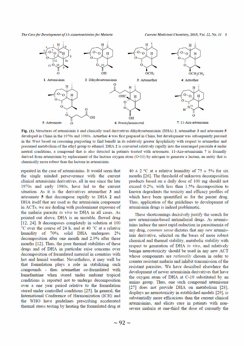
~ 92 ~
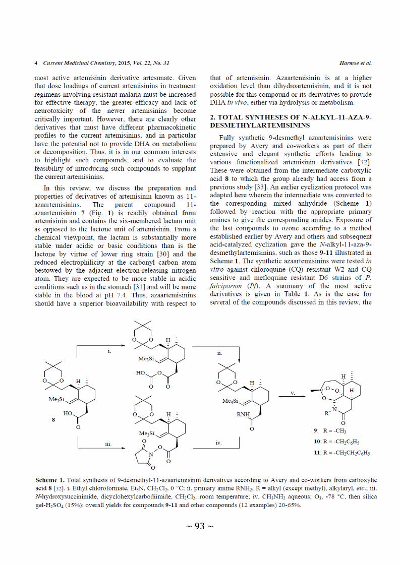
~ 93 ~

~ 94 ~
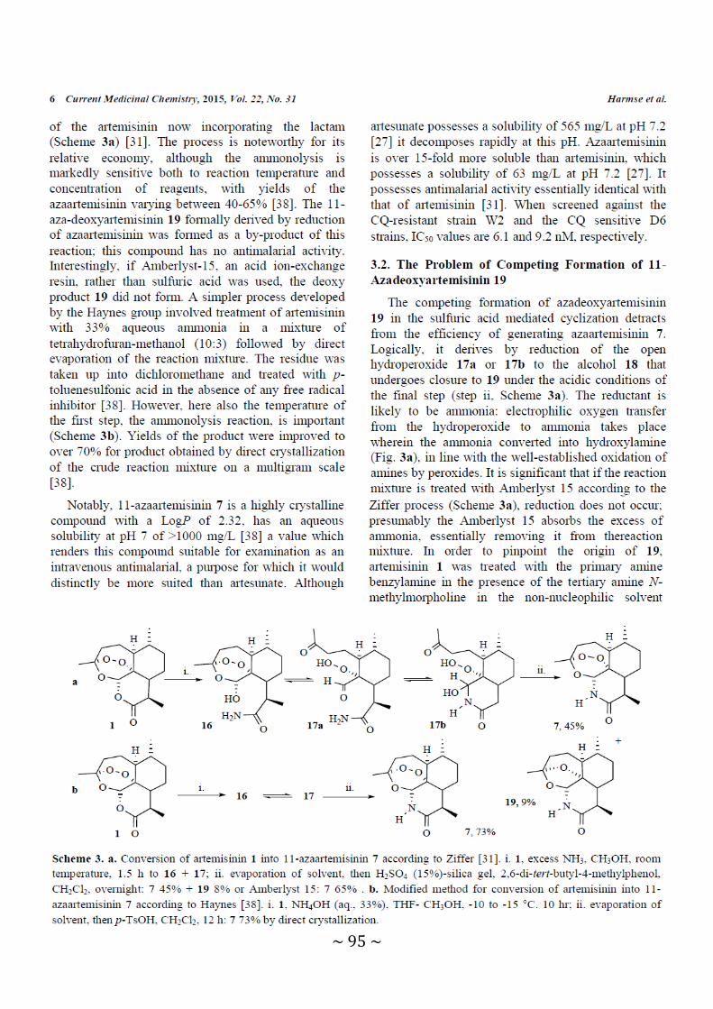
~ 95 ~

~ 96 ~

~ 97 ~
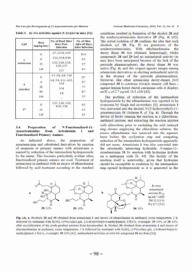
~ 98 ~

~ 99 ~

~ 100 ~

~ 101 ~

~ 102 ~
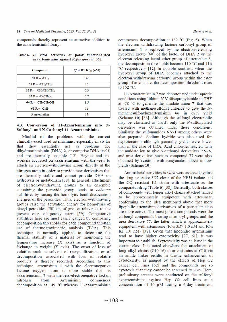
~ 103 ~
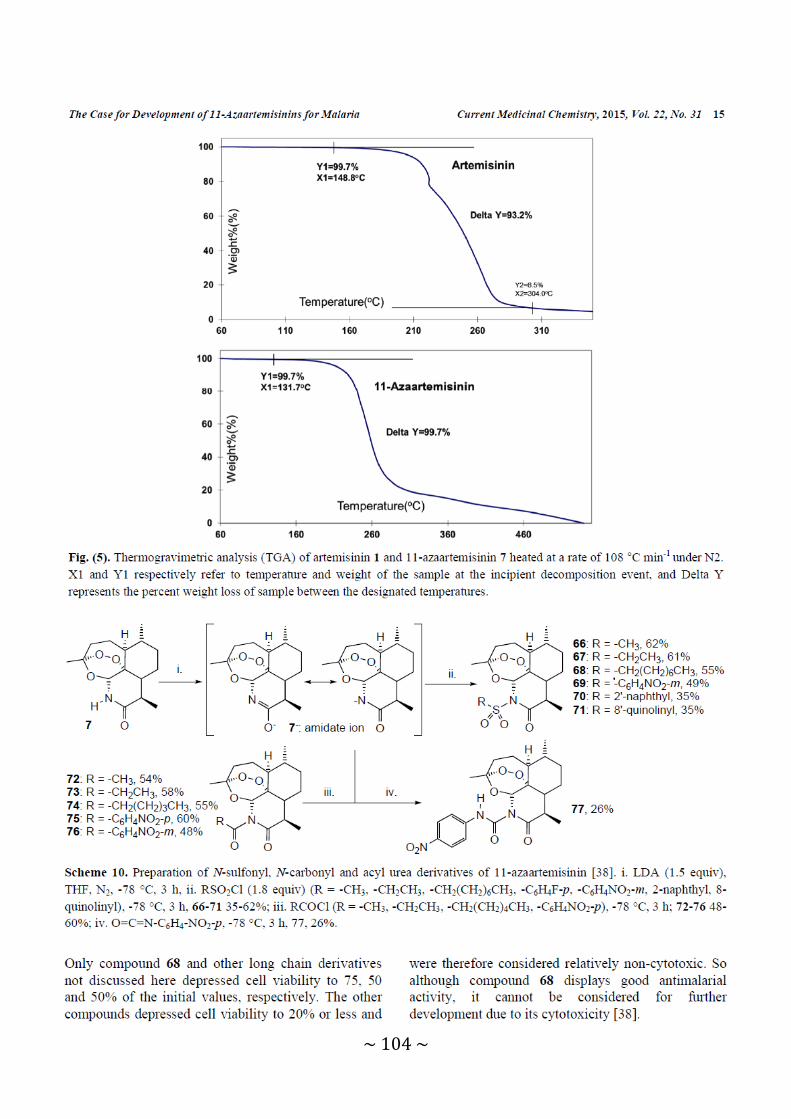
~ 104 ~

~ 105 ~
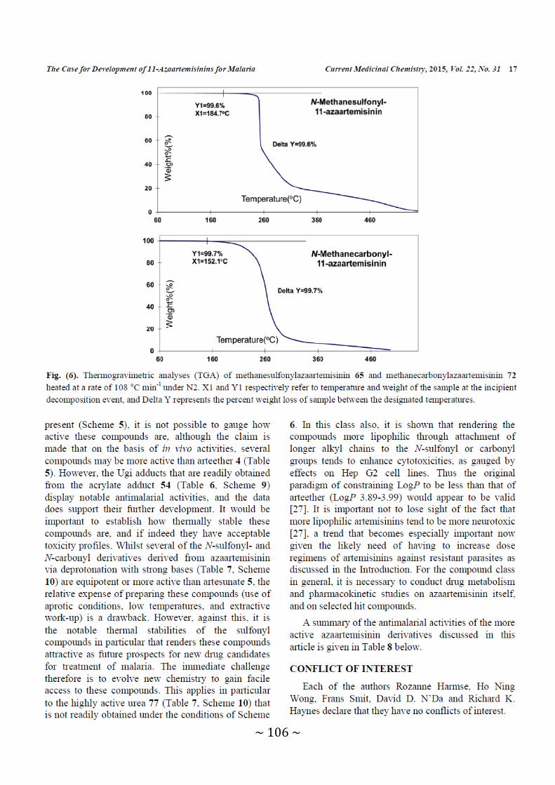
~ 106 ~

~ 107 ~

~ 108 ~

~ 109 ~
~ 110 ~

~ 111 ~
~ 112 ~
~ 113 ~

~ 114 ~
CHAPTER 4 Antimalarial activities and
cytotoxicities of N-sulfonyl-11-azaartemisinin derivatives – Article 2
This chapter contains the manuscript entitled “Antimalarial activities and cytotoxicities of N-
sulfonyl-11-azaartemisinin derivatives” that is yet to be submitted to the European Journal of
Medicinal Chemistry. This manuscript contains the aim, background, experimental details
and results of the compounds that were synthesized in this study. The manuscript was also
prepared according to the standards set out in the author information pack available in the
author’s guidelines at: https://www.elsevier.com/journals/european-journal-of-medicinal-
chemistry/0223-5234/guide-for-authors

~ 115 ~
Antimalarial Activities and Cytotoxicities of N-Sulfonyl-11-Azaartemisinin Derivatives Rozanne Harmsea, Frans J. Smitb, Dina Coertzenc, Ho Ning Wongb, Lyn-Marie Birkholtzc,
Richard K. Haynes*b, David D. N’Da*b. a Pharmaceutical Chemistry, School of Pharmacy, North-West University, Potchefstroom
2520, South Africa
b Centre of Excellence for Pharmaceutical Sciences, North-West University, Potchefstroom
2520, South Africa c Department of Biochemistry, Centre for Sustainable Malaria Control, University of Pretoria,
Pretoria 0002, South Africa
*email: [email protected], Tel: +2718 299 2256, Fax: +2718 299 4243;
[email protected], Tel: +27 18 299 4466
~ 116 ~
Abstract Given the current thesis that the antimalarial drug dihydroartemisinin (DHA) either used in its
own right, or as the active drug generated in vivo from the other current clinically-used
artemisinins artemether and artesunate, induces quiescence in ring-stage parasites of
Plasmodium falciparum (Pf) as the trigger of artemisinin resistance, we have turned our
attention to the examination of structurally disparate artemisinins that are incapable of
providing DHA on metabolism. Accordingly, N-sulfonyl-11-azaartemisinin derivatives were
prepared from 11-azaartemisinin and screened against intraerythrocytic stages of
chloroquine (CQ) sensitive Pf NF54 and CQ resistant K1 and W2 parasites. Cytotoxicities
were determined against human fetal lung fibroblasts WI-38 cell line (HFLF). Most
compounds displayed appreciable activities against all three strains with IC50 values <10
nM. The p-trifluoromethylbenzene-sulfonyl-11-azaartemisinin derivative 11 was the most
active with IC50 values between 2 – 3 nM. All compounds were relatively non-toxic to HFLF.
The compounds were also screened against Pf NF54 blood-stage gametocytes using the
luciferase and pLDH assays. The 2'-thienylsulfonyl derivative 16 was the most active against
late stage (IV-V) gametocytes with an IC50 value of 8.7 nM. Compounds 11 and 16 are
potential candidates for further development.
Keywords: Malaria, artemisinin, azaartemisinin, gametocytes

~ 117 ~
Graphical abstract
O
O
O O H
H
O
O
N
O O H
H
OS
R
O O
O
N
O O H
H
O
Azaartemisinin (5)N-sulfonyl-11-azaartemisinin
derivatives (6 - 18)
Antimalarial activity (IC50):NF54: 2 - 42 nMK1: 3 - 61 nMW2: 2 - 65 nM
H
Artemisinin (1)
Gametocytocidal activity (IC50):Early stage Luciferase: 165 - 465 nMLate stage Luciferase: 12 - 167 nMLate stage pLDH: 8 - 656 nM
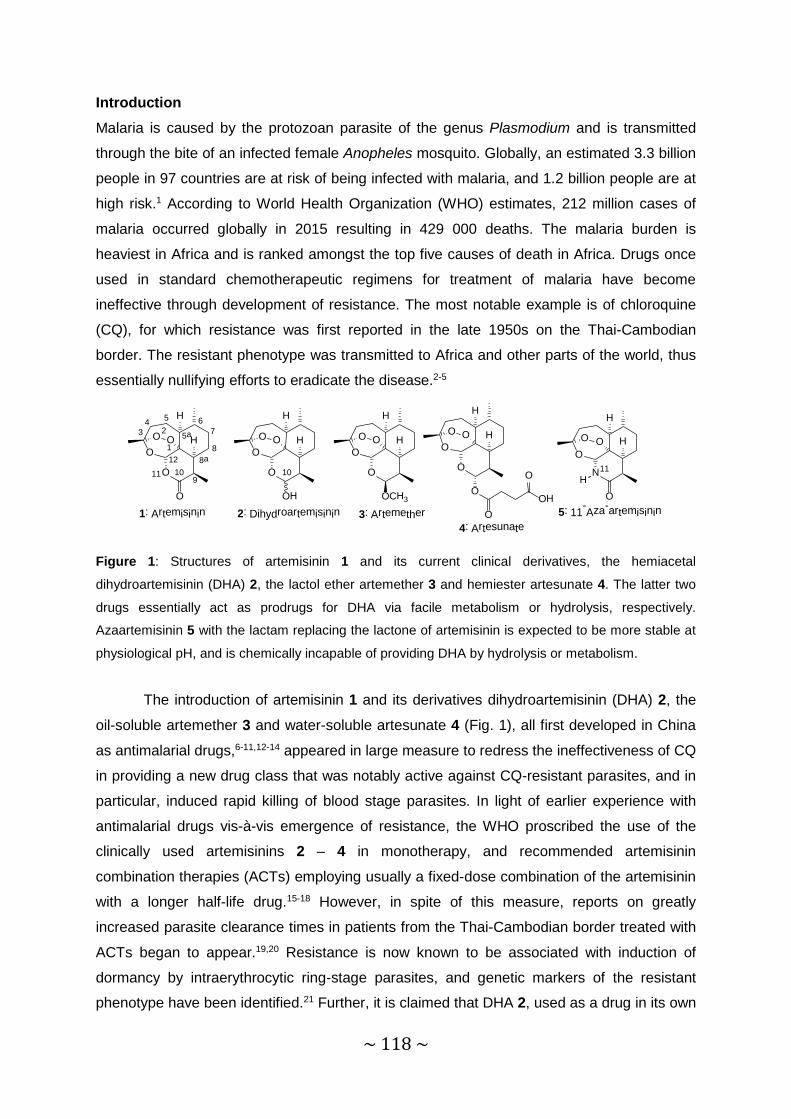
~ 118 ~
Introduction Malaria is caused by the protozoan parasite of the genus Plasmodium and is transmitted
through the bite of an infected female Anopheles mosquito. Globally, an estimated 3.3 billion
people in 97 countries are at risk of being infected with malaria, and 1.2 billion people are at
high risk.1 According to World Health Organization (WHO) estimates, 212 million cases of
malaria occurred globally in 2015 resulting in 429 000 deaths. The malaria burden is
heaviest in Africa and is ranked amongst the top five causes of death in Africa. Drugs once
used in standard chemotherapeutic regimens for treatment of malaria have become
ineffective through development of resistance. The most notable example is of chloroquine
(CQ), for which resistance was first reported in the late 1950s on the Thai-Cambodian
border. The resistant phenotype was transmitted to Africa and other parts of the world, thus
essentially nullifying efforts to eradicate the disease.2-5
1: Artemisinin 2: Dihydroartemisinin 3: Artemether4: Artesunate
O
O
O O H
H
O
O
O
O O H
H
OH
O
O
O O H
H
OCH3
O
O
O O H
H
OOH
O
O
H
HOOO
N
O5: 11-Aza-artemisinin
3
5 6
8
9
1
24
75a
8a1011
12
H1110
Figure 1: Structures of artemisinin 1 and its current clinical derivatives, the hemiacetal
dihydroartemisinin (DHA) 2, the lactol ether artemether 3 and hemiester artesunate 4. The latter two
drugs essentially act as prodrugs for DHA via facile metabolism or hydrolysis, respectively.
Azaartemisinin 5 with the lactam replacing the lactone of artemisinin is expected to be more stable at
physiological pH, and is chemically incapable of providing DHA by hydrolysis or metabolism.
The introduction of artemisinin 1 and its derivatives dihydroartemisinin (DHA) 2, the
oil-soluble artemether 3 and water-soluble artesunate 4 (Fig. 1), all first developed in China
as antimalarial drugs,6-11,12-14 appeared in large measure to redress the ineffectiveness of CQ
in providing a new drug class that was notably active against CQ-resistant parasites, and in
particular, induced rapid killing of blood stage parasites. In light of earlier experience with
antimalarial drugs vis-à-vis emergence of resistance, the WHO proscribed the use of the
clinically used artemisinins 2 – 4 in monotherapy, and recommended artemisinin
combination therapies (ACTs) employing usually a fixed-dose combination of the artemisinin
with a longer half-life drug.15-18 However, in spite of this measure, reports on greatly
increased parasite clearance times in patients from the Thai-Cambodian border treated with
ACTs began to appear.19,20 Resistance is now known to be associated with induction of
dormancy by intraerythrocytic ring-stage parasites, and genetic markers of the resistant
phenotype have been identified.21 Further, it is claimed that DHA 2, used as a drug in its own

~ 119 ~
right or that is active as the metabolite common to the most widely used clinical artemisinins
artemether 3 and artesunate 4, is key to inducing ring stage dormancy through binding to the
kinase phosphatidylinositol-3-kinase PfPI3K to prevent its ubiquitinylation.22-24 Interestingly,
in silico modelling presents an optimum pose for DHA in the kinase binding site interacting
with the key residues D1889 and Y1915. Structurally distinct artemisinins such as artelinate
or artemisone are unable to adopt the DHA pose, and do not interact with these residues. It
is notable that DHA is labile under physiological conditions, yet it is modelled intact into the
PfPKI3 target. Further, it is claimed that when the drug is 'washed out', activity of PfPKI3 is
restored.22 Yet it was not established if indeed the drug was 'washed out'; although the
normal procedure of 'washing out' was followed, no analyses for drug residues in the
washings were carried out. Given the structural diversity of artemisinins and synthetic
peroxides that elicit potent antimalarial activities, the presentation of a model invoking
classical inhibition by binding into an endogenous receptor site is not convincing. According
to a precedent for this type of target involving redox-sensitive signal transduction pathways,
the nature of the inhibition must equate with generation of reactive oxygen species (ROS) by
the artemisinin and evidently, inhibition of PI3P by ROS.25-27 Overall, irrespective of the exact
molecular mechanism that DHA does play in induction of resistance, an examination of the
efficacies of artemisinins that are unable to provide DHA on metabolism is mandated.
Further, as emphasis has to be placed on blocking transmission of resistant phenotypes,28
those stages of intraerythrocytic parasites transmitted from host to vector, namely late-stage
gametocytes, ideally should also be susceptible to the new drug. However, the current
clinical artemisinins display moderate transmission blocking activities: they are at best
moderately active against late stage mature gametocytes,29 and ACTs such as artemether-
lumefantrine and sulfadoxine-pyrimethamine-artesunate decrease gametocyte densities and
clearance times.30,31 Thus, our aim here is to develop newer artemisinins that do not provide
DHA on metabolism or hydrolysis, and are active against asexual blood-stage parasites and
late stage gametocytes.
11-Azaartemisinin 5 (Fig. 1) is readily obtained from artemisinin and incorporates a
lactam unit which typically is more stable under acidic and basic conditions than is the
lactone of artemisinin.32 It is therefore expected that azaartemisinin and its derivatives will be
more stable than artemisinin either at pH 7.4 or under the more acidic conditions of the
stomach.33 Further, azaartemisinin cannot provide DHA either by hydrolysis or metabolism
as it is at a higher oxidation level than is DHA.34 Therefore, derivatives of 11-azaartemisinin
fulfil the criterion of not providing DHA and are potentially attractive as follow up drugs to the
current clinical artemisinins. We have already demonstrated that certain azaartemisinin
derivatives possess greatly enhanced thermal stability35 and several have good antimalarial
activities.33,36

~ 120 ~
Results and Discussion Preparation of N-sulfonylalkyl and –aryl azaartemisinins
The parent compound, 11-azaartemisinin 5 was obtained in good yield (73%) from
artemisinin and aqueous ammonia in a tetrahydrofuran-methanol mixture below 0 °C
according to the literature method.35 It was deprotonated under aprotic conditions with
lithium N,N-diisopropylamide (LDA) as previously described and then treated with a sulfonyl
chloride to yield the corresponding N-sulfonyl-11-azaartemisinin derivatives 6 – 18 (Scheme
1) in low to moderate yields (1 – 38%).
O
N
O O H
H
O
H
5
i O
N
O O H
H
OS
R
O O
6: R = -CH3
, 38%7: R
= -CH2(CH2)2CH3, 34%
8: R = -CH2(CH2)14CH3
, 25%9: R
= -C6H5, 27%
10: R = -C6H4CF3
-m 1%
11: R = -C6H4CF3
-p 18%
12: R = -C6H4I-p
22%
13: R = -C6H4OCH3
-p 12%
14: R = -C6H4
-C6H4F-p 1%
15: R = -cyclopropyl
6%
O
N
O O H
H
OS
OO
S16: 10%
O
N
O O H
H
OS
OO
S
Cl
Cl
17: 15%
O
N
O O H
H
OS
O OO
18: 27%
ii
Scheme 1: Preparation of N-sulfonyl-azaartemisinin derivatives: i. LDA (1.5 equiv.), THF, N2, -78 °C,
3 h. ii. RSO2Cl (1.5 equiv.), -78 °C, 3h, silica gel chromatography, ethyl acetate-hexane (4:6), 6 – 18
(1 – 38%).
As this method follows that as previously reported by us35 and provides the products
in low yields, an examination of other potentially more economic methods was carried out in
order to improve access to the products. Previous work of Ziffer and co-workers identified
simplified conditions for converting 11-azaartemisinin into functionalized derivatives using
electron deficient alkenes.37-39 Thus, azaartemisinin in anhydrous THF containing solid
sodium hydroxide and ethyl acrylate at room temperature gave in good yield (85%) the N-
(ethoxycarbonyl)ethyl adduct.37,38 Likewise, propynoate electrophiles in acetonitrile as
solvent and with 4-N,N-dimethylaminopyridine (DMAP) as base gave adducts in acceptable
to good yields (40-56%).38 However, use of sulfonyl halides 3-(trifluoromethyl)benzene, 4-
(trifluoromethyl)benzene, 4-methoxybenzene, 4-fluorobiphenyl, cyclopropyl, thiophene and
2,5-dichlorothiophene in acetonitrile in the presence of DMAP either alone or under

~ 121 ~
microwave irradiation did give the desired products, these were not obtained in acceptable
yields, and were not easily separated from other components of the reaction mixtures.
Antimalarial activities and cytotoxicities
All compounds, with the exception of the N-alkylsulfonyl azaartemisinin 8 that was
poorly soluble in the culture medium, were screened in vitro against CQ sensitive NF54 and
CQ resistant K1 and W2 strains of intraerythrocytic P. falciparum parasites. The IC50 values
are summarized in Table 1. Overall the compounds were active against all three strains with
the most active displaying IC50 values of <10 nM. The p-trifluorobenzenesulfonyl derivative
11 was the most active against all three strains with IC50 values ranging between 2.04 – 3.05
nM. This compound displays comparable activity to artemether 3 and artesunate 4 against
all three strains. The m-triflouromethylbenzenesulfonyl derivative 10 was the least active
against all three strains with IC50 values ranging between 41.3 – 64.5 nM. Although the p-
iodobenzenesulfonyl derivative 12 also exhibited poor activities against all three strains (IC50
27.30 – 58.14 nM), it was some twofold less active against the drug resistant K1 and W2
strains, as expressed in the relatively high resistance indices (RI, Table 1).
All compounds were screened against human fetal lung fibroblasts WI-38 cell line
(HFLF) to gauge the relative cytotoxicities; emetine, known for its high toxicity,40 was used
as a standard (Table 1). Selectivity indices (SI) of the azaartemisinin derivatives indicated
several thousand-fold lower activities towards mammalian cells with respect to asexual
stages of the malaria parasite as indicative of the high selectivity indices.
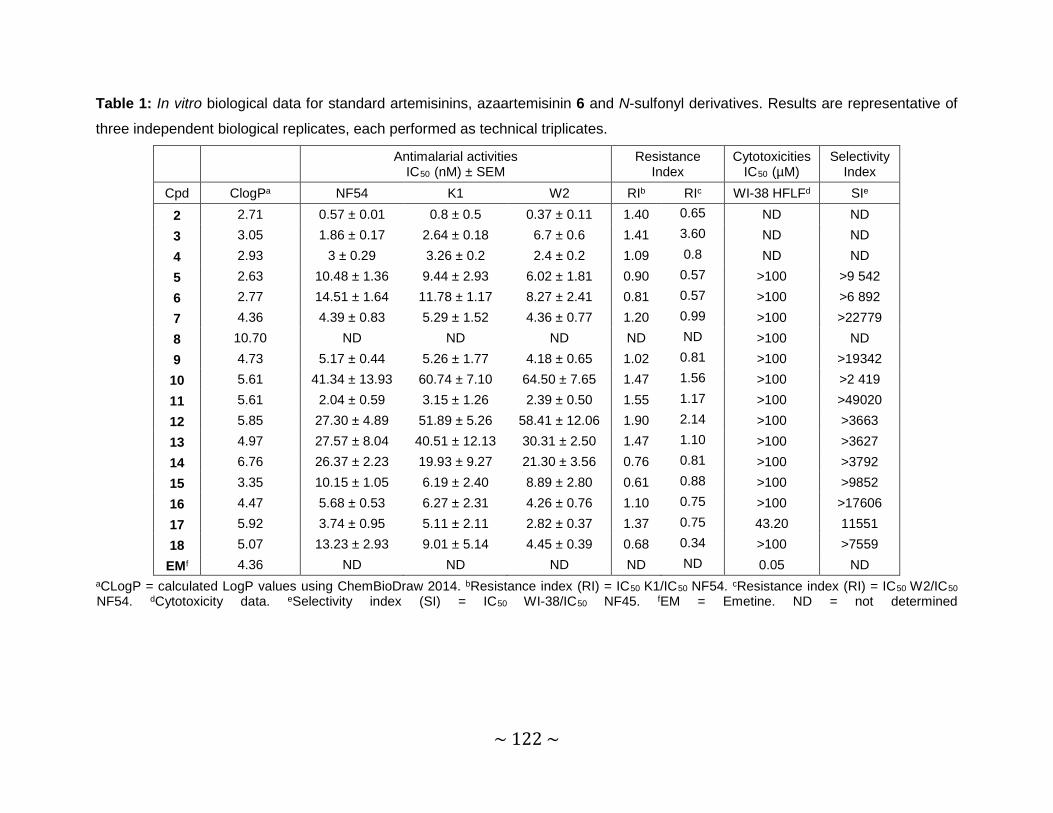
~ 122 ~
Table 1: In vitro biological data for standard artemisinins, azaartemisinin 6 and N-sulfonyl derivatives. Results are representative of
three independent biological replicates, each performed as technical triplicates.
Antimalarial activities IC50 (nM) ± SEM
Resistance Index
Cytotoxicities IC50 (µM)
Selectivity Index
Cpd ClogPa NF54 K1 W2 RIb RIc WI-38 HFLFd SIe
2 2.71 0.57 ± 0.01 0.8 ± 0.5 0.37 ± 0.11 1.40 0.65 ND ND 3 3.05 1.86 ± 0.17 2.64 ± 0.18 6.7 ± 0.6 1.41 3.60 ND ND 4 2.93 3 ± 0.29 3.26 ± 0.2 2.4 ± 0.2 1.09 0.8 ND ND 5 2.63 10.48 ± 1.36 9.44 ± 2.93 6.02 ± 1.81 0.90 0.57 >100 >9 542 6 2.77 14.51 ± 1.64 11.78 ± 1.17 8.27 ± 2.41 0.81 0.57 >100 >6 892 7 4.36 4.39 ± 0.83 5.29 ± 1.52 4.36 ± 0.77 1.20 0.99 >100 >22779 8 10.70 ND ND ND ND ND >100 ND9 4.73 5.17 ± 0.44 5.26 ± 1.77 4.18 ± 0.65 1.02 0.81 >100 >19342 10 5.61 41.34 ± 13.93 60.74 ± 7.10 64.50 ± 7.65 1.47 1.56 >100 >2 419 11 5.61 2.04 ± 0.59 3.15 ± 1.26 2.39 ± 0.50 1.55 1.17 >100 >49020 12 5.85 27.30 ± 4.89 51.89 ± 5.26 58.41 ± 12.06 1.90 2.14 >100 >3663 13 4.97 27.57 ± 8.04 40.51 ± 12.13 30.31 ± 2.50 1.47 1.10 >100 >3627 14 6.76 26.37 ± 2.23 19.93 ± 9.27 21.30 ± 3.56 0.76 0.81 >100 >3792 15 3.35 10.15 ± 1.05 6.19 ± 2.40 8.89 ± 2.80 0.61 0.88 >100 >9852 16 4.47 5.68 ± 0.53 6.27 ± 2.31 4.26 ± 0.76 1.10 0.75 >100 >17606 17 5.92 3.74 ± 0.95 5.11 ± 2.11 2.82 ± 0.37 1.37 0.75 43.20 11551 18 5.07 13.23 ± 2.93 9.01 ± 5.14 4.45 ± 0.39 0.68 0.34 >100 >7559
EMf 4.36 ND ND ND ND ND 0.05 ND aCLogP = calculated LogP values using ChemBioDraw 2014. bResistance index (RI) = IC50 K1/IC50 NF54. cResistance index (RI) = IC50 W2/IC50
NF54. dCytotoxicity data. eSelectivity index (SI) = IC50 WI-38/IC50 NF45. fEM = Emetine. ND = not determined
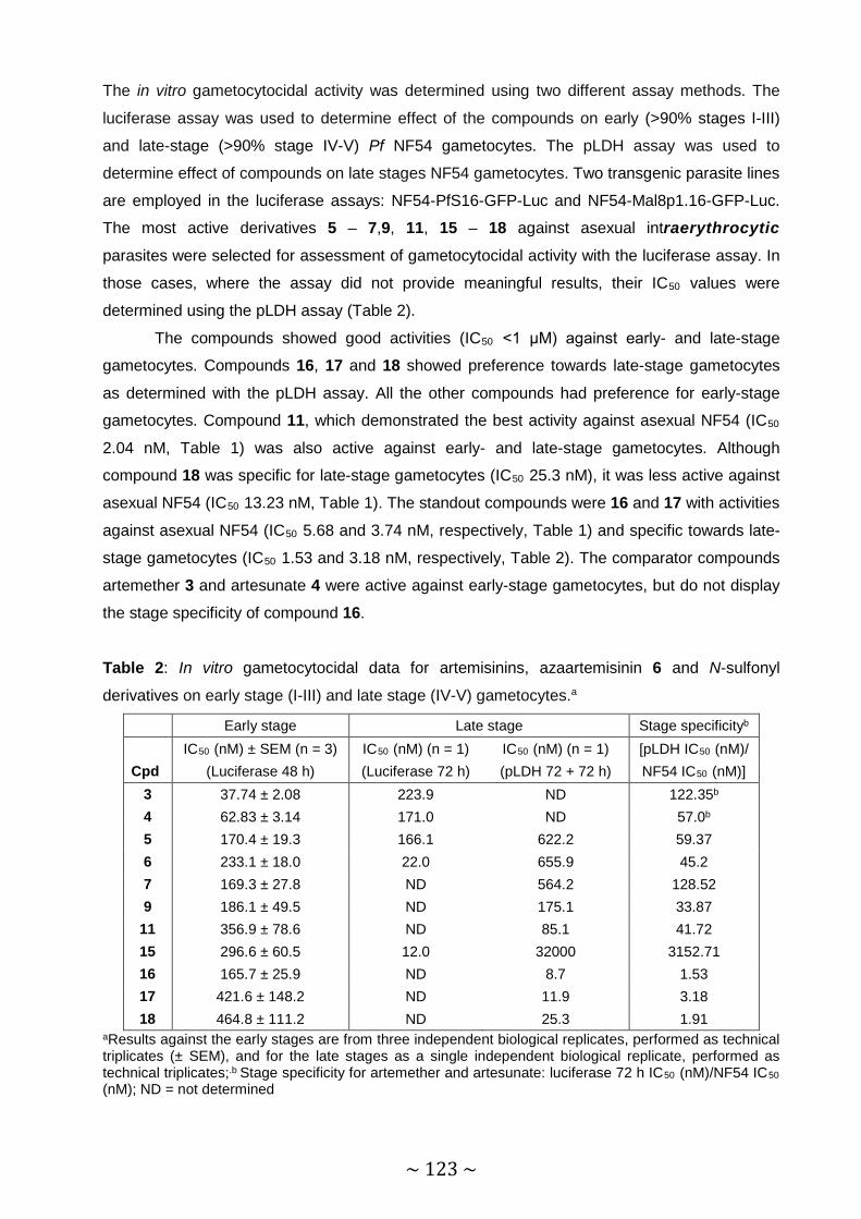
~ 123 ~
The in vitro gametocytocidal activity was determined using two different assay methods. The
luciferase assay was used to determine effect of the compounds on early (>90% stages I-III)
and late-stage (>90% stage IV-V) Pf NF54 gametocytes. The pLDH assay was used to
determine effect of compounds on late stages NF54 gametocytes. Two transgenic parasite lines
are employed in the luciferase assays: NF54-PfS16-GFP-Luc and NF54-Mal8p1.16-GFP-Luc.
The most active derivatives 5 – 7,9, 11, 15 – 18 against asexual intraerythrocytic
parasites were selected for assessment of gametocytocidal activity with the luciferase assay. In
those cases, where the assay did not provide meaningful results, their IC50 values were
determined using the pLDH assay (Table 2).
The compounds showed good activities (IC50 <1 μM) against early- and late-stage
gametocytes. Compounds 16, 17 and 18 showed preference towards late-stage gametocytes
as determined with the pLDH assay. All the other compounds had preference for early-stage
gametocytes. Compound 11, which demonstrated the best activity against asexual NF54 (IC50
2.04 nM, Table 1) was also active against early- and late-stage gametocytes. Although
compound 18 was specific for late-stage gametocytes (IC50 25.3 nM), it was less active against
asexual NF54 (IC50 13.23 nM, Table 1). The standout compounds were 16 and 17 with activities
against asexual NF54 (IC50 5.68 and 3.74 nM, respectively, Table 1) and specific towards late-
stage gametocytes (IC50 1.53 and 3.18 nM, respectively, Table 2). The comparator compounds
artemether 3 and artesunate 4 were active against early-stage gametocytes, but do not display
the stage specificity of compound 16.
Table 2: In vitro gametocytocidal data for artemisinins, azaartemisinin 6 and N-sulfonyl
derivatives on early stage (I-III) and late stage (IV-V) gametocytes.a
Early stage Late stage Stage specificityb IC50 (nM) ± SEM (n = 3) IC50 (nM) (n = 1) IC50 (nM) (n = 1) [pLDH IC50 (nM)/
Cpd (Luciferase 48 h) (Luciferase 72 h) (pLDH 72 + 72 h) NF54 IC50 (nM)] 3 37.74 ± 2.08 223.9 ND 122.35b 4 62.83 ± 3.14 171.0 ND 57.0b 5 170.4 ± 19.3 166.1 622.2 59.37 6 233.1 ± 18.0 22.0 655.9 45.2 7 169.3 ± 27.8 ND 564.2 128.52 9 186.1 ± 49.5 ND 175.1 33.87
11 356.9 ± 78.6 ND 85.1 41.72 15 296.6 ± 60.5 12.0 32000 3152.71 16 165.7 ± 25.9 ND 8.7 1.53 17 421.6 ± 148.2 ND 11.9 3.18 18 464.8 ± 111.2 ND 25.3 1.91
aResults against the early stages are from three independent biological replicates, performed as technical triplicates (± SEM), and for the late stages as a single independent biological replicate, performed as technical triplicates;.b Stage specificity for artemether and artesunate: luciferase 72 h IC50 (nM)/NF54 IC50 (nM); ND = not determined

~ 124 ~
Ideally, an antimalarial drug or drug combinations should target both the asexual blood
stages and the transmissible stages such as the gametocytes so as to hinder transmission of
resistant parasites from an infected individual. Most antimalarial drugs target the asexual blood
stage, that may cure a malaria patient but also allowing further transmission weeks after the
clearance of the asexual parasites.41 There is also evidence of enhanced rates of transmission
of drug resistant parasites from drug-treated individuals.42 Although it has been ascertained that
the current clinical artemisinins can reduce gametocyte carriage,43,44 in general these are not
regarded as transmission blocking drugs. Thus, compounds in the current series such as the p-
trifluoromethylbenzenesulfonyl derivative 11 and the 2-thienylsulfonyl derivative 16 are relatively
active against both asexual- and late stage gametocytes represents an important development
in the development of transmission blocking antimalarial drugs.
Conclusion A series of N-sulfonylazaartemisinin derivatives have been prepared, and most are shown to be
significantly active against blood stage asexual parasites, and against early and late stage
gametocytes. Overall the compounds are relatively non-toxic and displayed good selectivity
values towards the malaria parasite vis-a-vis normal mammalian cells. Compounds 11 and 16
represent hit compounds that have been taken forward for pharmacokinetic studies and further
screening studies in vivo.
Experimental Section Materials and methods
All reagents were of analytical grade, and were obtained from Sigma-Aldrich (South
Africa) and used as supplied. Solvents were purchased from Associated Chemical Enterprises
(ACE, South Africa). Tetrahydrofuran (THF) (Sigma-Aldrich) was dried by storing over sodium
and benzophenone under a nitrogen atmosphere and distilled prior to use. Lithium
diisopropylamide (LDA) was generated in situ under a nitrogen atmosphere using n-butyllithium,
N,N-diisopropylamide (DIPA) and 2,2’-bipyridine as endpoint indicator. Artemisinin was
purchased from Changzhou Kaixuan Chemical Co (Chunjiang, China). 1H and 13C NMR spectra were recorded on a Bruker AvanceTM III spectrometer as
solutions in chloroform-d (CDCl3). Chemical shifts (δ) are reported in parts per million (ppm) and
the 1H chemical shifts are reported downfield of tetramethylsilane (TMS) with internal reference
to the residual proton in CDCl3 (δ 7.25 ppm). 13C chemical shifts were internally referenced to
the CDCl3 resonances (δ 77.00 ppm). The splitting patterns are abbreviated as follows: s
(singlet), d (doublet), dd (doublet of doublets), t (triplet) and m (multiplet). Spectra were
analysed with MestReNova Software, version 5.3.2-4936. Infrared (IR) spectra were recorded
on a Bruker Alpha-P FTIR instrument in conjunction with the attenuated total reflectance (ATR)

~ 125 ~
sampling technique. High resolution mass spectrometry (HRMS) was recorded on a Bruker
MicroTOF Q II mass spectrometer, equipped with an ESI source set at 180 °C using Bruker
Compass DataAnalysis 4.0 software. A full scan from m/z 50 to 1500 was performed at a
capillary voltage of 4500 V, an end plate offset voltage of -500 V, with the nebulizer set at 0.4
Bar, r, and a collision cell RF voltage of 100 Vpp. Melting points (mp) were determined with a
Büchi melting point B-545 instrument and were uncorrected. Column chromatography was
performed using high-purity grade silica gel (pore size 60 Å, 70-230 mesh, 63-200 µm) from
Sigma Aldrich and thin layer chromatography was performed with silica gel plates (60F254) from
Merck.
Syntheses
5
O
N
O O H
H
O
3
5 6
8
9
124
75a
8a10
11
12
H13
14
15
16
11-Azaartemisinin 5
A solution of artemisinin 1 (10.0 g, 35.42 mmol) in THF (200 mL) and methanol (60 mL)
was cooled down to -15 ºC and treated with NH4OH (33% aqueous, 100 mL). The resulting
mixture was left to stir for 10 h at this temperature, during which time the color changed to a
very pale yellow. The solution was evaporated under reduced pressure, without heating, to
leave a pale yellow foam. This was dissolved in CH2Cl2 (250 mL) and treated with p-
tolenesulfonic acid monohydrate (6.8 g, 35.84 mmol) at room temperature. The resulting mixture
was left to stir for 12 h after which it was washed with 5% aqueous sodium bicarbonate (400
mL) and water (500 mL). The organic layer was separated and dried over anhydrous MgSO4.
The MgSO4 was removed via filtration and the filtrate was evaporated under reduced pressure
to leave the residue as a pale foam. The foam was crystallized using ethyl acetate: hexane (4:6)
to yield 11-azaartemisinin 5 (7.2 g, 73%) (Scheme 1) as colourless needles; yield: 7.2 g (73%);
mp: 143 – 144.5 °C; 1H NMR (600 MHz, CDCl3) δ (ppm): 6.25 (s, 1H, H-11), 5.39 (s, 1H, H-12),
3.27 – 3.19 (m, 1H, H-9), 2.44 - 2.35 (m, 1H, H-4α), 2.04 – 1.96 (m, 2H), 1.84 – 1.67 (m, 3H),
1.49 – 1.30 (m, 6H), 1.13 (d, J = 7.4 Hz, 3H, H-16), 1.04 – 0.95 (m, 5H); 13C NMR (151 MHz,
CDCl3) δ (ppm): 173.48 (C-10), 104.96 (C-3), 79.97 (C-12a), 75.70 (C-12), 51.14 (C-5a), 46.03
(C-8a), 37.75 (C-9), 36.66 (C-25), 33.89 (C-7), 32.88 (C-9), 25.71 (C-14), 25.25 (C-14), 23.16
(C-5), 19.91 (C-15), 12.25 (C-16); IR: (ATR) νmax/cm-1 3298, 2964, 2884, 1656, 1444, 1133, 830,
730, 690; HRMS (APCI) m/z [M + H]+ 282.1700 (Calcd for C15H24NO4: 282.1705).

~ 126 ~
6
O
N
O O H
H
O
3
5 6
8
9
12
475a
8a
1011
12
S
13
14
15
16O O
1'
N-Methanesulfonylazaartemisinin 6: 11-azaartemisinin 5 (1 g, 3.55 mmol) in dry THF (10 mL)
was added to a stirred solution of LDA (5.35 mmol, 1.5 equiv.) in dry THF (15 mL) at -78 ºC.
The resulting mixture was left to stir for 3 h under a nitrogen atmosphere at -78 ºC after which
methanesulfonyl chloride (0.5 mL, 6.46 mmol, 1.5 equiv.) was added to the reaction mixture.
The solution was left to stir for another 3h at -78 ºC and then for another 30 min at room
temperature. The mixture was quenched with saturated aqueous ammonium chloride (20 mL),
diluted with water (10 mL) and extracted with ethyl acetate (3 x 30 mL). The extracts were
washed with brine (30 mL), dried (MgSO4) and then filtered. The filtrate was evaporated under
reduced pressure to leave a white solid, which was submitted to flash column chromatography
with ethyl acetate-hexane (4:6). The eluent was evaporated to dryness to leave 6 as a pale
yellow powder (481 mg, 38%), mp: 245 °C; 1H NMR (600 MHz, CDCl3) δ (ppm): 6.04 (s, 1H, H-
12), 3.43 – 3.29 (m, 4H, H-1’ and H-9), 2.44 – 2.34 (m, 1H, H-4α), 2.03 – 1.95 (m, 2H), 1.84 –
1.67 (m, 3H), 1.50 - 1.30 (m, 2H), 1.43 – 1.33 (m, 4H), 1.26 – 1.20 (m, 4H), 1.06 – 0.98 (m, 4H); 13C NMR (151 MHz, CDCl3) δ (ppm): 174.09 (C-10), 105.37 (C-3), 80.37 (C-12a), 78.83 (C-12),
51.45 (C-5a), 44.43 (C-8a), 44.23 (C-1’), 37.34 (C-1’), 36.51 (C-9), 36.15 (C-4), 33.71 (CH2),
25.45 (C-14), 24.88 (CH2), 22.40 (CH2), 19.66 (C-15), 13.79 (C-16); IR: (ATR) νmax/cm-1 2978,
1709, 1446, 1349, 1165, 963, 519, 485; HRMS (APCI) m/z [M + H]+ 360.1439 (Calcd for
C16H26NO6S: 360.1480). The data is in agreement with that reported in the literature.35
7
O
N
O O H
H
O
3
5 6
8
9
1
24
75a
8a
1011
12
S
1314
15
16O O
1'
2'
3'4'
Butanesulfonyl-azaartemisinin 7: Compound 7 was obtained from 5 (1.0 g, 3.55 mmol), LDA
and 1-butanesulfonyl chloride (0.7 mL, 5.33 mmol, 1.5 equiv.), and chromatography (ethyl
acetate: hexane, 4:6) as pale yellow powder (484 mg, 34%), mp 120 °C; 1H NMR (600 MHz,
CDCl3) δ (ppm): 6.03 (s, 1H, H-12), 3.71 – 3.61 (m, 1H, H-1’α), 3.52 – 3.44 (m, 1H, H-1’β), 3.40
– 3.32 (m, 1H, H-9), 2.43 – 2.35 (m, 1H, H-4α), 2.08 – 1.99 (m, 2H), 1.96 – 1.85 (m, 2H, CH2),
1.81 – 1.66 (m, 3H), 1.55 – 1.45 (m, 4H), 1.44 - 1.30 (m, 4H, H-14 and H-5a), 1.23 – 1.17 (m,

~ 127 ~
4H), 1.06 – 0.99 (m, 4H), 0.95 (t, J = 7.4 Hz, 3H, H-4’); 13C NMR (151 MHz, CDCl3) δ (ppm):
174.19 (C-10), 105.39 (C-3), 80.38 (C-12a), 78.34 (C-12), 56.76 (C-1’), 51.54 (C-5a), 44.82 (C-
8a), 37.29 (CH), 36.59 (CH2), 35.90 (C-9), 33.66 (CH2), 25.41 (C-14), 24.85 (CH2), 24.59
(CH2), 22.33 (CH2), 21.67 (CH2), 19.65 (C-15), 13.67 (C-16), 13.52 (C-4’); IR: (ATR) νmax/cm-1
2931, 2879, 1700, 1344, 1201, 1146, 1023, 946, 648, 534, 487; HRMS (APCI) m/z [M + H]+
402.1930 (Calcd for C19H32NO6S: 402.1950).
8
O
N
O O H
H
O
3
5 6
8
9
1
24
75a
8a
1011
12
S
1314
15
16O O
1'
2'
3'
4'
5'
6'
7'
8'
9'
10'
11'
12'
13'
14'
15'16'
1-Hexadecanesulfonyl-azaartemisinin 8: Compound 8 was obtained from 5 (1.0 g, 3.55 mmol),
LDA and 1-hexadecanesulfonyl chloride (1.73 g, 5.33 mmol, 1.5 equiv.), and chromatography
(ethyl acetate:hexane, 4:6) as a bright yellow oil (498 mg, 25%); 1H NMR (600 MHz, DMSO) δ
(ppm): 5.88 (s, 1H, H-12), 3.61 (ddd, J = 14.1, 10.0, 5.6 Hz, 1H, H-1’α), 3.45 (ddd, J = 14.0,
10.1, 5.7 Hz, 1H, H-1’β), 3.30 – 3.25 (m, 1H, H-2’α), 3.18 – 3.13 (m, 1H, H-9), 2.28 – 2.20 (m,
1H, H-4α), 2.08 – 2.02 (m, 1H), 2.00 – 1.95 (m, 1H), 1.80 – 1.76 (m, 1H), 1.75 – 1.61 (m, 6H),
1.38 – 1.33 (m, 9H), 1.28 – 1.24 (m, 14H), 1.08 (d, J = 7.5 Hz, 3H, H-16), 1.01 (t, J = 8.6 Hz,
2H), 0.95 (d, J = 6.2 Hz, 3H, H-15), 0.90 – 0.81 (m, 9H); 13C NMR (151 MHz, DMSO) δ (ppm):
173.30 (C-10), 104.74 (C-3), 80.08 (C-12a), 77.72 (C-12), 55.59 (C-1’), 50.55 (C-5a), 43.30
(CH), 36.73 (CH), 35.97 (CH2), 35.43 (C-9), 32.85 (CH2), 31.31 (CH2), 29.11 – 28.21 (C-13’ –
4’), 27.28 (C-3’), 24.84 (C-14), 22.28 (CH2), 22.11 (CH2), 19.30 (C-15), 13.97 (C-16), 13.33 (C-
16’); IR: (ATR) νmax/cm-1 2921, 2852, 1707, 1571, 1377, 1366, 1306, 1040, 645, 487; HRMS
(APCI) m/z [M + H]+ 570.3819 (Calcd for C19H32NO6S: 570.3828).
9
O
N
O O H
H
O
3
5 6
8
9
1
24
75a
8a
1011
12
S
13
14
15
16OO 1'
2'
3'4'
5'
6'
Benzenesulfonyl-azaartemisinin 9: Compound 9 was obtained from 5 (1.0 g, 3.55 mmol), LDA
and benzenesulfonyl chloride (0.7 mL, 5.33 mmol, 1.5 equiv.), and chromatography (ethyl
acetate:hexane, 4:6) to give 9 as off-white crystals; yield: 400 mg (27%); mp: 147 °C; 1H NMR
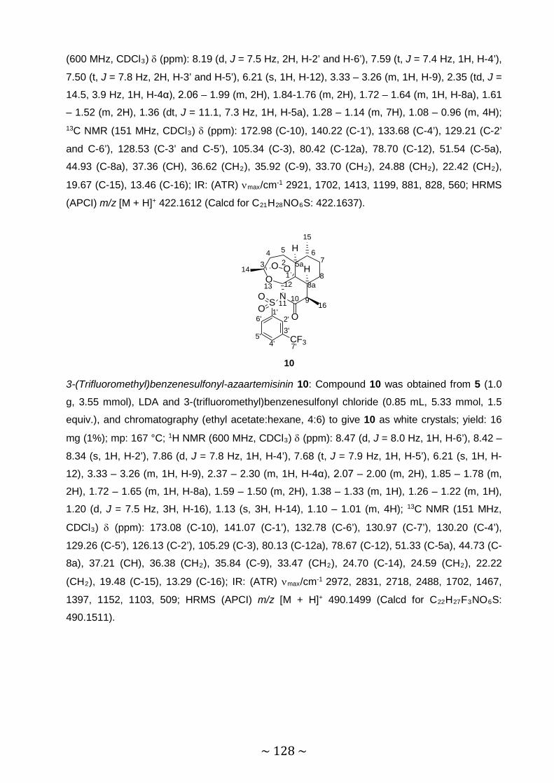
~ 128 ~
(600 MHz, CDCl3) δ (ppm): 8.19 (d, J = 7.5 Hz, 2H, H-2’ and H-6’), 7.59 (t, J = 7.4 Hz, 1H, H-4’),
7.50 (t, J = 7.8 Hz, 2H, H-3’ and H-5’), 6.21 (s, 1H, H-12), 3.33 – 3.26 (m, 1H, H-9), 2.35 (td, J =
14.5, 3.9 Hz, 1H, H-4α), 2.06 – 1.99 (m, 2H), 1.84-1.76 (m, 2H), 1.72 – 1.64 (m, 1H, H-8a), 1.61
– 1.52 (m, 2H), 1.36 (dt, J = 11.1, 7.3 Hz, 1H, H-5a), 1.28 – 1.14 (m, 7H), 1.08 – 0.96 (m, 4H);13C NMR (151 MHz, CDCl3) δ (ppm): 172.98 (C-10), 140.22 (C-1’), 133.68 (C-4’), 129.21 (C-2’
and C-6’), 128.53 (C-3’ and C-5’), 105.34 (C-3), 80.42 (C-12a), 78.70 (C-12), 51.54 (C-5a),
44.93 (C-8a), 37.36 (CH), 36.62 (CH2), 35.92 (C-9), 33.70 (CH2), 24.88 (CH2), 22.42 (CH2),
19.67 (C-15), 13.46 (C-16); IR: (ATR) νmax/cm-1 2921, 1702, 1413, 1199, 881, 828, 560; HRMS
(APCI) m/z [M + H]+ 422.1612 (Calcd for C21H28NO6S: 422.1637).
10
O
N
O O H
H
O
3
5 6
8
9
1
24
75a
8a
1011
12
S
13
14
15
16OO 1'
2'3'
4'5'
6'
CF37'
3-(Trifluoromethyl)benzenesulfonyl-azaartemisinin 10: Compound 10 was obtained from 5 (1.0
g, 3.55 mmol), LDA and 3-(trifluoromethyl)benzenesulfonyl chloride (0.85 mL, 5.33 mmol, 1.5
equiv.), and chromatography (ethyl acetate:hexane, 4:6) to give 10 as white crystals; yield: 16
mg (1%); mp: 167 °C; 1H NMR (600 MHz, CDCl3) δ (ppm): 8.47 (d, J = 8.0 Hz, 1H, H-6’), 8.42 –
8.34 (s, 1H, H-2’), 7.86 (d, J = 7.8 Hz, 1H, H-4’), 7.68 (t, J = 7.9 Hz, 1H, H-5’), 6.21 (s, 1H, H-
12), 3.33 – 3.26 (m, 1H, H-9), 2.37 – 2.30 (m, 1H, H-4α), 2.07 – 2.00 (m, 2H), 1.85 – 1.78 (m,
2H), 1.72 – 1.65 (m, 1H, H-8a), 1.59 – 1.50 (m, 2H), 1.38 – 1.33 (m, 1H), 1.26 – 1.22 (m, 1H),
1.20 (d, J = 7.5 Hz, 3H, H-16), 1.13 (s, 3H, H-14), 1.10 – 1.01 (m, 4H); 13C NMR (151 MHz,
CDCl3) δ (ppm): 173.08 (C-10), 141.07 (C-1’), 132.78 (C-6’), 130.97 (C-7’), 130.20 (C-4’),
129.26 (C-5’), 126.13 (C-2’), 105.29 (C-3), 80.13 (C-12a), 78.67 (C-12), 51.33 (C-5a), 44.73 (C-
8a), 37.21 (CH), 36.38 (CH2), 35.84 (C-9), 33.47 (CH2), 24.70 (C-14), 24.59 (CH2), 22.22
(CH2), 19.48 (C-15), 13.29 (C-16); IR: (ATR) νmax/cm-1 2972, 2831, 2718, 2488, 1702, 1467,
1397, 1152, 1103, 509; HRMS (APCI) m/z [M + H]+ 490.1499 (Calcd for C22H27F3NO6S:
490.1511).

~ 129 ~
11
O
N
O O H
H
O
3
5 6
8
9
1
24
75a
8a
1011
12
S
13
14
15
16OO 1'
2'
3'4'
5'
6'
7'CF3
4-(Trifluoromethyl)benzenesulfonyl-azaartemisinin 11: Compound 11 was obtained from 5 (1.0
g, 3.55 mmol), LDA and 4-(trifluoromethyl)benzenesulfonyl chloride (1.3 g, 5.33 mmol, 1.5
equiv.), and chromatography (ethyl acetate:hexane, 4:6) to give 11 as an off-white powder;
yield: 306 mg (18%); mp: 168.1 °C; 1H NMR (600 MHz, CDCl3) δ (ppm): 8.32 (d, J = 8.3 Hz, 2H,
H-2’ and H-6’), 7.77 (d, J = 8.4 Hz, 2H, H-3’ and H-5’), 6.21 (s, 1H, H-12), 3.34 – 3.26 (m, 1H,
H-9), 2.41 – 2.33 (m, 1H, H-4α), 2.08 – 2.00 (m, 2H), 1.86 – 1.79 (m, 2H), 1.73 – 1.67 (m, 1H,
H-8a), 1.60 – 1.51 (m, 2H), 1.40 – 1.34 (m, 1H, H-5a), 1.27 – 1.21 (m, 1H), 1.20 – 1.13 (m, 6H,
H-16 and H-14), 1.11 – 0.99 (m, 4H); 13C NMR (151 MHz, CDCl3) δ (ppm): 173.19 (C-10),
143.62 (C-1’), 135.35 (C-4’), 129.83 (C-2’), 129.79 (C-6’), 125.72 (C-3’), 125.69 (C-5’), 124.21
(C-7’), 105.44 (C-3), 80.35 (C-12a), 78.89 (C-12), 51.50 (C-5a), 44.89 (C-8a), 37.39 (CH), 36.57
(CH2), 35.99 (C-9), 33.65 (CH2), 24.88 (C-14), 24.87 (CH2), 22.43 (CH2), 19.65 (C-15), 13.44
(C-16); IR: (ATR) νmax/cm-1 2984, 2855, 1705, 1366, 1166, 711, 554, 429; HRMS (APCI) m/z [M
+ H]+ 490.1497 (Calcd for C22H27F3NO6S: 490.1511).
12
O
N
O O H
H
O
3
5 6
8
9
1
24
75a
8a
1011
12
S
13
14
15
16OO 1'
2'
3'4'
5'
6'
I 4-Iodobenzenesulfonyl-azaartemisinin 12: Compound 12 was obtained from 5 (1.0 g, 3.55
mmol), LDA and 4-iodobenzenesulfonyl chloride (1.61 g, 5.33 mmol, 1.5 equiv.), and
chromatography (ethyl acetate:hexane, 4:6) to give 12 as a pale pink powder; yield: 431 mg
(22%); mp: 182.8 °C; 1H NMR (600 MHz, CDCl3) δ (ppm): 7.90 – 7.85 (m, 4H, H-2’, H-3, H-5’
and H-6’), 6.18 (s, 1H, H-12), 3.32 – 3.27 (m, 1H, H-9), 2.36 (dt, J = 14.5, 3.8 Hz, 1H, H-4α),
2.07 – 2.00 (m, 2H), 1.84 – 1.78 (m, 2H), 1.70 – 1.66 (m, 1H, H-8a), 1.58 – 1.50 (m, 2H), 1.36
(td, J = 11.0, 6.4 Hz, 1H, H-5a), 1.25 – 1.16 (m, 7H), 1.06 – 0.96 (m, 4H); 13C NMR (151 MHz,
CDCl3) δ (ppm): 172.93 (C-10), 139.69 (C-1’), 137.66 (C-2’ and C-6’), 130.50 (C-3’ and C-5’),

~ 130 ~
105.23 (C-3), 101.49 (C-4’), 80.18 (C-12a), 78.59 (C-12), 51.36 (C-5a), 44.78 (C-8a), 37.20
(CH), 36.44 (CH2), 35.75 (C-9), 33.49 (CH2), 24.82 (C-14), 24.71 (CH2), 22.24 (CH2), 19.49 (C-
15), 13.26 (C-16); IR: (ATR) νmax/cm-1 2934, 2854, 1704, 1567, 1385, 1235, 1054, 1018, 895,
601, 561; HRMS (APCI) m/z [M + H]+ 548.0586 (Calcd for C21H27INO6S: 548.0603).
13
O
N
O O H
H
O
3
5 6
8
9
1
24
75a
8a
1011
12
S
13
14
15
16OO 1'
2'
3'4'
5'
6'
O7'8'
4-Methoxybenzenesulfonyl-azaartemisinin 13: Compound 13 was obtained from 5 (1.0 g, 3.55
mmol), LDA and 4-methoxybenzenesulfonyl chloride (1.1 g, 5.33 mmol, 1.5 equiv.), and
chromatography (ethyl acetate:hexane, 4:6) to give 13 as off-white crystals; yield: 19 mg (12%);
mp 159 °C; 1H NMR (600 MHz, CDCl3) δ (ppm): 8.11 (d, J = 9.0 Hz, 2H, H-2’ and H-6’), 6.95 (d,
J = 9.0 Hz, 2H, H-3’ and H-5’), 6.19 (s, 1H, H-12), 3.86 (s, 2H, H-8’), 3.32 – 3.25 (m, 1H, H-9),
2.38 – 2.29 (m, 1H, H-4α), 2.07 – 2.00 (m, 2H), 1.83 – 1.73 (m, 2H), 1.70 – 1.55 (m, 3H), 1.36
(dt, J = 11.0, 7.5 Hz, 1H, H-5a), 1.25 – 1.20 (m, 4H), 1.17 (d, J = 7.4 Hz, 3H, H-16), 1.05 – 0.95
(m, 4H); 13C NMR (151 MHz, CDCl3) δ (ppm): 172.79 (C-10), 163.59 (C-4’), 131.54 (C-1’),
131.45 (C-2’ and C-6’), 113.48 (C-3’ and C-5’), 105.12 (C-3), 80.23 (C-12a), 78.40 (C-12), 55.64
(C-8’), 51.43 (C-5a), 44.87 (C-8a), 37.18 (CH), 36.50 (CH2), 35.66 (C-9), 33.54 (CH2), 24.94 (C-
14), 24.73 (CH2), 22.23 (CH2), 19.51 (C-15), 13.26 (C-16); IR: (ATR) νmax/cm-1 2965, 2828,
2717, 2487, 1702, 1397, 1313, 1152, 1026, 695, 508; HRMS (APCI) m/z [M + H]+ 452.1714
(Calcd for C22H30NO7S: 452.1742).

~ 131 ~
14
O
N
O O H
H
O
3
5 6
8
9
1
24
75a
8a
1011
12
S
13
14
15
16OO 1'
2'
3'4'
5'
6'
7'8'
F
9'10'
11'
12'
4-(4-fluorophenyl)benzene-1-sulfonyl-azaartemisinin 14: Compound 14 was obtained from 5
(1.0 g, 3.55 mmol), LDA and 4-(4-fluorophenyl)benzene-1-sulfonyl chloride (1.16 g, 5.33 mmol,
1.5 equiv.), and chromatography (ethyl acetate:hexane, 4:6) to give 14 as pale yellow crystals;
yield: 21 mg (1%); mp: 142 °C; 1H NMR (600 MHz, CDCl3) δ (ppm): 8.23 (d, J = 8.5 Hz, 2H, H-
2’ and H-6’), 7.65 (d, J = 8.6 Hz, 2H, H-3’ and H-5’), 7.56 (dd, J = 8.8, 5.2 Hz, 2H, H-8’ and H-
12’), 7.18 – 7.13 (m, 2H, H-9’ and H-11’), 6.23 (s, 1H, H-12), 3.34 – 3.28 (m, 1H, H-9), 2.36 (dt,
J = 14.6, 3.8 Hz, 1H, H-4α), 2.06 – 2.02 (m, 2H), 1.84 – 1.79 (m, 2H), 1.71 – 1.67 (m, 1H, H-8a),
1.61 – 1.53 (m, 2H), 1.38 (dd, J = 11.4, 5.9 Hz, 1H, H-5a), 1.26 – 1.22 (m, 4H), 1.19 (d, J = 7.4
Hz, 3H, H-16), 1.08 – 1.02 (m, 4H); 13C NMR (151 MHz, CDCl3) δ (ppm): 172.97 (C-10), 162.30
(C-10’), 145.41 (C-1’), 138.56 (C-4’), 135.44 (C-7’), 129.71 (C-2’ and C-6’), 129.13 (C-8’ and C-
12’), 126.84 (C-3’ and C-5’), 116.06 (C-9’ and C-11’), 105.21 (C-3), 80.25 (C-12a), 78.52 (C-12),
50.86 (C-5a), 44.83 (C-8a), 37.19 (CH), 36.47 (CH2), 35.73 (C-9), 33.51 (CH2), 24.84 (C-14),
24.71 (CH2), 22.25 (CH2), 19.51 (C-15), 13.26 (C-16); IR: (ATR) νmax/cm-1 3014, 2936, 2748,
2518, 1703, 1603, 1472, 1244, 1145, 1084, 680, 550, 508; HRMS (APCI) m/z [M + H]+
516.1827 (Calcd for C27H31FNO6S: 516.1856).
15
O
N
O O H
H
O
3
5 6
8
9
1
24
75a
8a
1011
12
S
13
14
15
16O
O1'
2'
3'
Cyclopropanesulfonyl-azaartemisinin 15: Compound 15 was obtained from 5 (1.0 g, 3.55 mmol),
LDA and cyclopropanesulfonyl chloride (0.55 mL, 5.33 mmol, 1.5 equiv.), and chromatography
(ethyl acetate:hexane, 4:6) to give 15 as a white powder; yield: 76 mg (6%); mp: 133.1 °C; 1H
NMR (600 MHz, CDCl3) δ (ppm): 5.95 (s, 1H, H-12), 3.43 – 3.35 (m, 1H, H-9), 3.22 – 3.16 (m,
1H, H-1’), 2.42 – 2.30 (m, 1H, H-4α), 2.07 – 1.98 (m, 2H), 1.82 – 1.74 (m, 2H), 1.72 – 1.66 (m,

~ 132 ~
1H, H-8a), 1.58 – 1.46 (m, 3H), 1.44 – 1.35 (m, 5H), 1.25 (d, J = 7.5 Hz, 3H, H-16), 1.18 – 0.99
(m, 7H); 13C NMR (151 MHz, CDCl3) δ (ppm): 173.52 (C-10), 105.18 (C-3), 80.12 (C-12a),
77.94 (C-12), 51.38 (C-5a), 44.89 (C-8a), 37.12 (CH), 36.42 (CH2), 35.57 (C-9), 33.56 (C-1’),
33.49 (CH2), 25.41 (C-14), 24.69 (CH2), 22.17 (CH2), 19.49 (C-15), 13.27 (C-16), 6.73 (C-3’),
6.42 (C-2’); IR: (ATR) νmax/cm-1 2966, 2940, 1710, 1350, 1273, 1204, 1129, 975, 927, 707, 594,
492; HRMS (APCI) m/z [M + H]+ 386.1619 (Calcd for C18H28NO6S: 386.1637).
16
O
N
O O H
H
O
3
5 6
8
9
12
475a
8a
1011
12
S
13
14
15
16OO 1'
2'
3'4'
5'S
2'-Thiophenesulfonylazaartemisinin 16: Compound 16 was obtained from 5 (1.0 g, 3.55 mmol),
LDA and 2-thiophenesulfonyl chloride (0.97 g, 5.33 mmol, 1.5 equiv.), and chromatography
(ethyl acetate:hexane, 4:6) to give 16 as a brown powder; yield: 156 mg (10%); mp: 150.1 °C; 1H NMR (600 MHz, CDCl3) δ (ppm): 7.94 (d, J = 3.7 Hz, 1H, H-2’), 7.66 (d, J = 4.9 Hz, 1H, H-
4’), 7.09 – 7.05 (m, 1H, H-3’), 6.18 (s, 1H, H-12), 3.38 – 3.27 (m, 1H, H-9), 2.35 (td, J = 14.6,
3.9 Hz, 1H, H-4α), 2.07 – 1.96 (m, 2H), 1.83 – 1.75 (m, 2H), 1.72 – 1.65 (m, 1H, H-8a), 1.59 –
1.53 (m, 2H), 1.40 – 1.32 (m, 1H, H-5a), 1.26 – 1.19 (m, 4H), 1.15 (s, 3H, H-14), 1.07 – 0.99 (m,
4H); 13C NMR (151 MHz, CDCl3) δ (ppm): 173.08 (C-10), 140.47 (C-1’), 135.33 (C-2’), 133.68
(C-4’), 126.61 (C-3’), 105.21 (C-3), 80.29 (C-12a), 79.09 (C-12), 51.32 (C-5a), 44.53 (C-8a),
37.22 (CH), 36.41 (CH2), 35.94 (C-9), 33.57 (CH2), 24.74 (C-14), 24.71 (CH2), 22.27 (CH2),
19.51 (C-15), 13.47 (C-16); IR: (ATR) νmax/cm-1 2976, 2879, 1706, 1365, 1142, 1128, 1013, 696,
670, 596, 551, 466; HRMS (APCI) m/z [M + H]+ 428.1178 (Calcd for C19H26NO6S2: 428.1201).
17
O
N
O O H
H
O
3
5 6
8
9
1
24
75a
8a
1011
12
S
13
14
15
16OO 1'
2'
3'4'
5'S
Cl
Cl 2,5-Dichlorothiophenesulfonyl-azaartemisinin 17: Compound 17 was obtained from 5 (1.0 g,
3.55 mmol), LDA and 2,5-dichlorothiophenesulfonyl chloride (1.3 g, 5.33 mmol, 1.5 equiv.), and
chromatography (ethyl acetate:hexane, 4:6) to give 17 as a white powder; yield: 271 mg (15%);

~ 133 ~
mp: 168.3 °C; 1H NMR (600 MHz, CDCl3) δ (ppm): 7.31 (s, 1H, H-2’), 6.06 (s, 1H, H-12), 3.33 –
3.24 (m, 1H, H-9), 2.36 – 2.27 (m, 1H, H-4α), 2.00 – 1.91 (m, 2H), 1.75 – 1.66 (m, 2H), 1.65 –
1.60 (m, 1H, H-8a), 1.53 – 1.45 (m, 2H), 1.33 – 1.26 (m, 4H), 1.10 – 1.03 (m, 4H), 1.00 – 0.90
(m, 4H); 13C NMR (151 MHz, CDCl3) δ (ppm): 172.68 (C-10), 135.08 (C-1’), 132.11 (C-5’),
128.01 (C-2’), 126.68 (C-3’), 105.44 (C-3), 80.32 (C-12a), 79.26 (C-12), 51.42 (C-5a), 44.60 (C-
8a), 37.18 (CH), 36.37 (CH2), 33.55 (C-9), 24.91 (C-14), 24.66 (CH2), 22.24 (CH2), 19.51 (C-
15), 13.25 (C-16); IR: (ATR) νmax/cm-1 2976, 2879, 1642, 1515, 1425, 1212, 1155, 1090, 1065,
646, 523, 483; HRMS (APCI) m/z [M + H]+ 496.0360 (Calcd for C19H24Cl2NO6S2: 496.0422).
O
N
O O H
H
OS
O OO
18
123
4 5
5a6
7
8
91011
1213
14
15
16
8a1'2'3'4'
5'6'7'
8'9' 10'
10-Camphoryl-azaartemisinin 18: Compound 18 was obtained from 5 (1.0 g, 3.55 mmol), LDA
and (1S)-(+)-10-camphorsulfonyl chloride (1.3 g, 5.33 mmol, 1.5 equiv.), and chromatography
(ethyl acetate:hexane, 4:6) to give 18 as a white powder; yield: 484 mg (27%); mp: 186 °C; 1H
NMR (600 MHz, CDCl3) δ (ppm): 6.06 (s, 1H, H-12), 4.27 (d, J = 14.7 Hz, 1H, H-1’α), 3.40 –
3.33 (m, 2H), 2.60 – 2.53 (m, 1H, H-6’α), 2.43 – 2.36 (m, 2H, H-4α), 2.10 (t, J = 4.5 Hz, 1H, H-
7’), 2.07 – 1.98 (m, 3H), 1.94 (d, J = 18.4 Hz, 1H, H-3’β), 1.81 – 1.75 (m, 2H), 1.74 – 1.66 (m,
2H), 1.57 – 1.48 (m, 2H), 1.44 (s, 3H, H-14), 1.43 – 1.34 (m, 2H), 1.26 – 1.20 (m, 7H), 1.04 –
0.98 (m, 4H), 0.94 (s, 3H, H-9’); 13C NMR (151 MHz, CDCl3) δ (ppm): 214.15 (C-4’), 173.60 (C-
10), 105.22 (C-3), 80.31 (C-12a), 78.62 (C-12), 59.26 (C-2’), 54.27 (C-1’), 51.37 (C-5a), 47.44
(C-8’), 44.35 (C-8a), 43.24 (C-7’), 42.46 (C-3’), 37.18 (CH), 36.39 (CH2), 35.94 (C-9), 33.61
(CH2), 26.81 (C-5’), 25.86 (C-6’), 25.37 (C-14), 24.65 (CH2), 22.23 (CH2), 20.19 (C-9’), 19.76
(C-10’), 19.50 (C-15), 13.59 (C-16); IR: (ATR) νmax/cm-1 2935, 1743, 1705, 1571, 1405, 1356,
1014, 855, 645, 621; HRMS (APCI) m/z [M + H]+ 496.2383 (Calcd for C25H38NO7S: 496.2369).
Biological screening
Malaria parasite proliferation can be directly monitored in their intra-erythrocytic environment
through detecting and monitoring DNA replication (without background forthcoming from
erythrocytes, which lack DNA). SYBR Green I is a fluorescent dye that interacts with DNA,
therefore a correlation between DNA content (SYBR Green I signal) and parasitaemia can be
used to monitor decrease in parasitaemia as a measurement of inhibition of parasite
proliferation.
~ 134 ~
P. falciparum parasites were maintained at 37 °C in human erythrocytes (O+) suspended
in complete culture medium (RPMI 1640 medium supplemented with 25 mM HEPES, 20 mM D-
glucose, 200 μM hypoxanthine, 0.2% sodium bicarbonate, 24 μg/ml gentamycin and 0.5%
AlbuMAX II) in a gaseous environment of 90% N2, 5% O2, and 5% CO2 according to the
method established by Verlinden and co-workers.45 In vitro ring-stage intra-erythrocytic P.
falciparum parasite cultures (200 μL at 1% haematocrit, 1% parasitaemia) were treated with the
synthesized compounds. The controls for this assay included CQ disulphate (1 µM, as positive
control) and complete RPMI media (as negative control), grown for 96 h at 37 °C under the 90%
N2, 5% O2, and 5% CO2 gas mixture in 96-well plates. At the conclusion of the 96 h growth
period, equal volumes (100 μL each) of the P. falciparum parasite cultures were combined with
SYBR Green I lysis buffer (0.2 µL/mL 10 000 x SYBR Green I, Invitrogen; 20 mM Tris, pH 7.5; 5
mM EDTA; 0.008% (w/v) saponin; 0.08% (v/v) Triton X-100). The samples were incubated at 37
°C for 1 h after which the fluorescence was measured using a Fluoroskan Ascent FL microplate
fluorometer (Thermo Scientific, excitation at 485 nm and emission at 538 nm). The ‘background’
fluorescence (i.e. that measured in the samples derived from CQ-treated red blood cell samples
in which parasite proliferation was completely inhibited) was subtracted from the total
fluorescence measured for each sample to provide a measure of parasite proliferation.46,47 Data
obtained were analyzed in Excel 2007 and sigmoidal dose-response curves were plotted using
GraphPad 5.0. Experiments are always performed in triplicate, and repeated mainly 3 times.
The cytotoxic effects of the compounds were tested by Sulforhodamine B (SRB) assay on the
WI-38 cell line. The SRB assay was developed by Skehan and colleagues48 to measure drug-
induced cytotoxicity and cell proliferation. Its principle is based on the ability of the protein dye
sulforhodamine B (Acid Red 52) to bind electrostatically in a pH-dependent manner to protein
basic amino acid residues of trichloroacetic acid-fixed cells. Under mild acidic conditions it binds
to the fixed cellular protein, while under mild basic conditions it can be extracted from cells and
solubilized for measurement. The SRB assay is performed at the CSIR in accordance with the
protocol of the Drug Evaluation Branch, NCI, and the assay has been adopted for this screen.
The WI-38 cell line - normal Human Fetal Lung Fibroblast (HFLF) from ECACC was routinely
maintained as a monolayer cell culture at 37 ºC, 5% CO2, 95% air and 100% relative humidity in
EMEM containing 10% foetal bovine serum, 2 mM L-glutamine and 50 µg/ml gentamicin. For
the screening experiment, the cells (21 – 50 passages) were inoculated in a 96-well microtiter
plates at plating densities of 10000 cells per well and were incubated for 24 h. After 24 h the
cells were treated with the synthesized compounds which were previously dissolved in DMSO
and diluted in medium to produce 5 concentrations. Cells without any drug addition served as
control. The blank contains complete medium without cells. Emetine was used as a standard.
The plates were incubated for 48 h after addition of the synthesized compounds. Viable cells
~ 135 ~
were fixed to the bottom of each well with cold 50% trichloroacetic acid, washed, dried and dyed
by SRB. Unbound dye was removed and protein-bound dye was extracted with 10 mM Tris
base for optical density determination at the wavelength 540 nm using a multiwell
spectrophotometer. Data analysis was performed using GraphPad Prism software, where 50%
of cell growth inhibition (IC50) was determined by non-linear regression.
Gametocytes were produced as per Reader and co-workers.49 The luciferase reporter assay was established to enable accurate, reliable and quantifiable investigations of the stage-specific
action of gametocytocidal compounds for each of the early- and late-stage gametocyte marker
cell lines; NF54-PfS16-GFP-Luc and NF54-Mal8p1.16-GFP-Luc. Drug assays were set up on
day 5 and 10 (representing >90% of either early stage I/II/III or mature stage IV/V gametocytes,
respectively). In each instance, assays were set up using a 2 – 3% gametocytaemia, 1.5%
haematocrit culture and 48 h for early-stage gametocytes and 72 h for late-stage gametocytes
drug pressure in a gas chamber (90% N2, 5% O2, and 5% CO2) at 37ºC. Luciferase activity was
determined in 20 μL parasite lysates by adding 50 μL luciferin substrate (Promega Luciferase
Assay System) at room temperature and detection of resultant bioluminescence at an
integration constant of 10 s with the GloMax®-Multi+ Detection System with Instinct® Software.
The pLDH assay was set up on day 10 late-stage gametocytes (>90% mature stage IV/V
gametocytes). The assays were set up using a 1% gametocytaemia and 2% haematocrit culture
and 72 h drug pressure in a gas chamber (90% N2, 5% O2, and 5% CO2) at 37ºC, after which
the media was replaced with fresh media and the culture incubated for another 72 h.
Gametocyte viability was measured by lysing 20 μL of the parasite suspension with 100 μL of a
Malstat reagent [0.21% v/v Triton-100; 222 mM L-(+)-lactic acid; 54.5 mM Tris; 0.166 mM 3-
acetylpyridine adenine dinucleotide (APAD; Sigma-Aldrich); adjusted to pH 9 with 1 M NaOH]
followed by addition of 25 μL PES/NBT [1.96 mM nitro blue tetrazoliumchloride NBT; 0.239 mM
phenazine ethosulphate (PES)]. Absorbance was measured with a Multiskan Ascent 354
multiplate scanner (Thermo Labsystems, Finland) at 620 nm.
Disclaimer Any opinion, findings and conclusions, or recommendations expressed in this article are those
of the author(s) and therefore the NRF does not accept any liability in regard thereto.
Acknowledgements This research project is funded by the South African Medical Research Council (MRC) (Flagship
and SIR) with funds from the National Treasury under its Economic Competitiveness and
Support Package. The South African National Research Foundation is thanked for financial
support Grant No. 90682, NRF UID 87498 and 87457. The authors thank the North-West

~ 136 ~
University, Potchefstroom Campus, for the financial support, Dr. J. Jordaan for MS analysis, Mr.
A. Joubert for NMR analysis.
REFERENCES
(1) WHO; WHO: Geneva
http://www.who.int/malaria/publications/world_malaria_report_2016, 2016.
(2) Wellems, T. E.; Plowe, C. V. J. Infect. Dis. 2001, 184, 770-776.
(3) Wootton, J. C.; Feng, X.; Ferdig, M. T.; Cooper, R. A.; Mu, J.; Baruch, D. I.; Magill, A. J.;
Su, X.-z. Nature 2002, 418, 320-323.
(4) Wongsrichanalai, C.; Pickard, A. L.; Wernsdorfer, W. H.; Meshnick, S. R. Lancet Infect.
Dis. 2002, 2, 209-218.
(5) Roper, C.; Pearce, R.; Nair, S.; Sharp, B.; Nosten, F. o.; Anderson, T. Science 2004,
305, 1124-1124.
(6) Qinghaosu Antimalaria Coordinating Research, G. Chinese Medical Journal 1979, 92,
811-6.
(7) Klayman, D. L. Science 1985, 228, 1049-1055.
(8) Li, Y.; Wu, Y.-L. Curr. Med. Chem. 2003, 10, 2197-2230.
(9) Zhang, J.-F. A detailed chronological record of Project 523 and the discovery and
development of qinghaosu (artemisinin); Strategic Book Publishing: China, 2005.
(10) Woodrow, C. J.; Haynes, R. K.; Krishna, S. Postgrad. Med. J. 2005, 81, 71-78.
(11) Cui, L.; Su, X.-z. Expert Rev. Anti. Infect. Ther. 2009, 7, 999-1013.
(12) Li, Y. Reprinted from Kexue Tongbao. 1979, 24, 667-669.
(13) Li, Y.; Yu, P.-L.; Chen, Y.-X.; Li, L.-Q.; Gai, Y.-Z.; Wang, D.-S.; Zheng, Y.-P. Acta
Pharmaceutica Sinica 1981, 16, 429.
(14) China Cooperative Research, G. J. Trad. Chin. Med. 1982, 2, 1-16.
(15) White, N. J.; Olliaro, P. Med. Trop. (Mars). 1997, 58, 54-56.
(16) Nosten, F.; White, N. J. Am. J. Trop. Med. Hyg. 2007, 77, 181-192.
(17) Aweeka, F. T.; German, P. I. Clin. Pharmacokinet. 2008, 47, 91-102.
(18) Eastman, R. T.; Fidock, D. A. Nature Rev. Microbiol. 2009, 7, 864-874.
(19) Phyo, A. P.; Nkhoma, S.; Stepniewska, K.; Ashley, E. A.; Nair, S.; McGready, R.; ler
Moo, C.; Al-Saai, S.; Dondorp, A. M.; Lwin, K. M. The Lancet 2012, 379, 1960-1966.
(20) Lubell, Y.; Dondorp, A.; Guèrin, P. J.; Drake, T.; Meek, S.; Ashley, E.; Day, N. P. J.;
White, N. J.; White, L. J. Malar. J. 2014, 13, 1.
(21) Ariey, F.; Witkowski, B.; Amaratunga, C.; Beghain, J.; Langlois, A.-C.; Khim, N.; Kim, S.;
Duru, V.; Bouchier, C.; Ma, L. Nature 2014, 505, 50-55.
(22) Mbengue, A.; Bhattacharjee, S.; Pandharkar, T.; Liu, H.; Estiu, G.; Stahelin, R. V.; Rizk,
S. S.; Njimoh, D. L.; Ryan, Y.; Chotivanich, K. Nature 2015, 520, 683-687.

~ 137 ~
(23) VanHook, A. M. Sci. Signal. 2015, 8, ec118-ec118.
(24) Paloque, L.; Ramadani, A. P.; Mercereau-Puijalon, O.; Augereau, J.-M.; Benoit-Vical, F.
o. Malaria J. 2016, 15, 1.
(25) Kim, J. H.; Chu, S. C.; Gramlich, J. L.; Pride, Y. B.; Babendreier, E.; Chauhan, D.;
Salgia, R.; Podar, K.; Griffin, J. D.; Sattler, M. Blood 2005, 105, 1717-1723.
(26) Liu, J.; Zhou, J.; Xing, D. PLoS One 2012, 7, e33817.
(27) Okoh, V. O.; Felty, Q.; Parkash, J.; Poppiti, R.; Roy, D. PLoS One 2013, 8, e54206.
(28) Burrows, J. N.; van Huijsduijnen, R. H.; Möhrle, J. J.; Oeuvray, C.; Wells, T. N. C.
Malaria J. 2013, 12, 1.
(29) Chotivanich, K.; Sattabongkot, J.; Udomsangpetch, R.; Looareesuwan, S.; Day, N. P. J.;
Coleman, R. E.; White, N. J. Antimicrob. Agents Chemother. 2006, 50, 1927-1930.
(30) Bousema, J. T.; Schneider, P.; Gouagna, L. C.; Drakeley, C. J.; Tostmann, A.; Houben,
R.; Githure, J. I.; Ord, R.; Sutherland, C. J.; Omar, S. A. J. Infect. Dis. 2006, 193, 1151-1159.
(31) Bousema, T.; Okell, L.; Shekalaghe, S.; Griffin, J. T.; Omar, S.; Sawa, P.; Sutherland,
C.; Sauerwein, R.; Ghani, A. C.; Drakeley, C. Malaria J. 2010, 9, 1.
(32) Bach, R. D.; Dmitrenko, O. J. Am. Chem. Soc. 2006, 128, 4598-4611.
(33) Torok, D. S.; Ziffer, H.; Meshnick, S. R.; Pan, X.-Q.; Ager, A. J. Med. Chem. 1995, 38,
5045-5050.
(34) Haynes, R. K. Curr. Opin. Infect. Dis. 2001, 14, 719-726.
(35) Haynes, R. K.; Wong, H.-N.; Lee, K.-W.; Lung, C.-M.; Shek, L. Y.; Williams, I. D.; Croft,
S. L.; Vivas, L.; Rattray, L.; Stewart, L. ChemMedChem 2007, 2, 1464-1479.
(36) Avery, M. A.; Bonk, J. D.; Chong, W. K. M.; Mehrotra, S.; Miller, R.; Milhous, W.; Goins,
D. K.; Venkatesan, S.; Wyandt, C. J. Med. Chem. 1995, 38, 5038-5044.
(37) Mekonnen, B.; Ziffer, H. Tetrahedron Lett. 1997, 38, 731-734.
(38) Mekonnen, B.; Weiss, E.; Katz, E.; Ma, J.; Ziffer, H.; Kyle, D. E. Bioorg. Med. Chem.
2000, 8, 1111-1116.
(39) Katz, E.; Ma, J.; Kyle, D.; Ziffer, H. Bioorg. Med. Chem. Lett. 1999, 9, 2969-2972.
(40) Akinboye, E., S.; Bakare, O. J. Nat. Prod. 2011, 4.
(41) Schneider, P.; Bousema, J. T.; Gouagna, L. C.; Otieno, S.; Van de Vegte-Bolmer, M.;
Omar, S. A.; Sauerwein, R. W. Am. J. Trop. Med. Hyg. 2007, 76, 470-474.
(42) Hallett, R. L.; Sutherland, C. J.; Alexander, N.; Ord, R.; Jawara, M.; Drakeley, C. J.;
Pinder, M.; Walraven, G.; Targett, G. A. T.; Alloueche, A. Antimicrob. Agents Chemother. 2004,
48, 3940-3943.
(43) Price, R. N.; Nosten, F.; Luxemburger, C.; Ter Kuile, F. O.; Paiphun, L.;
Chongsuphajaisiddhi, T.; White, N. J. The Lancet 1996, 347, 1654-1658.
(44) Sutherland, C. J.; Ord, R.; Dunyo, S.; Jawara, M.; Drakeley, C. J.; Alexander, N.;
Coleman, R.; Pinder, M.; Walraven, G.; Targett, G. A. T. PLoS Med 2005, 2, e92.

~ 138 ~
(45) Verlinden, B. K.; Niemand, J.; Snyman, J.; Sharma, S. K.; Beattie, R. J.; Woster, P. M.;
Birkholtz, L.-M. J. Med. Chem. 2011, 54, 6624-6633.
(46) Smilkstein, M.; Sriwilaijaroen, N.; Kelly, J. X.; Wilairat, P.; Riscoe, M. Antimicrob. Agents
Chemother. 2004, 48, 1803-1806.
(47) Johnson, J. D.; Dennull, R. A.; Gerena, L.; Lopez-Sanchez, M.; Roncal, N. E.; Waters,
N. C. Antimicrob. Agents Chemother. 2007, 51, 1926-1933.
(48) Skehan, P.; Storeng, R.; Scudiero, D.; Monks, A.; McMahon, J.; Vistica, D.; Warren, J.
T.; Bokesch, H.; Kenney, S.; Boyd, M. R. J. Natl. Cancer Inst. 1990, 82, 1107-1112.
(49) Reader, J.; Botha, M. t.; Theron, A.; Lauterbach, S. B.; Rossouw, C.; Engelbrecht, D.;
Wepener, M.; Smit, A. l.; Leroy, D.; Mancama, D. Malar. J. 2015, 14, 213.

~ 139 ~
CHAPTER 5 Activities of N-sulfonyl-11-
azaartemisinin derivatives against the apicomplexan parasite Neospora
caninum and comparative cytotoxicities – Article 3
This chapter contains the manuscript entitled “Activities of N-sulfonyl-11-azaartemisinin
derivatives against the apicomplexan parasite Neospora caninum and comparative
cytotoxicities” that is yet to be submitted to Bioorganic & Medicinal Chemistry Letters. This
manuscript contains the aim, background, experimental details and results of the compounds
that were synthesized in this study. The manuscript was also prepared according to the
standards set out in the author information pack available in the author’s guidelines
at: https://www.elsevier.com/journals/bioorganic-and-medicinal-chemistry-letters/0960-894X/guide-for-
authors

~ 140 ~
Activities of N-sulfonyl-11-azaartemisinin derivatives against the apicomplexan parasite Neospora caninum and comparative cytotoxicities.
Rozanne Harmsea, Frans J. Smitb, Ho Ning Wongb, Joachim Müllerc, Andrew Hemphillc, David
D. N’Da*b, Richard K. Haynes*b
a Pharmaceutical Chemistry, School of Pharmacy, North-West University, Potchefstroom 2520,
South Africa
b Centre of Excellence for Pharmaceutical Sciences, North-West University, Potchefstroom
2520, South Africa c Institute of Parasitology, Vetsuisse Faculty, University of Berne, Länggass-Strasse 122, CH-
3012 Bern, Switzerland
* email: [email protected], Tel: +2718 299 2256, Fax: +2718 299
4243; [email protected], Tel: +27 18 299 4466
Previously synthesized N-sulfonyl-11-azaartemisinin derivatives were evaluated in vitro against
Neospora caninum. The compounds were also screened in vitro against three cancer cell lines
consisting of TK-10 (renal), UACC-62 (melanoma) and MCF-7 (breast).
N. caninum, IC50 150 nM
13: N. caninum, IC50: 40 nM5: 11-Azaartemisinin
O
N
O O H
H
OS
OO
S
Cl
Cl
O
N
O O H
H
O
H
~ 141 ~
Abstract
Activities of N-sulfonyl-11-azaartemisinins were evaluated in vitro against Neospora caninum, a
protozoan parasite afflicting dogs and cattle. As neosporosis induces abortion in dairy and beef
cattle, it is of considerable economic importance. Although drug regimens are available for
controlling or treating the disease, the number of drugs available is limited, and a major shortfall
is the persistence of drug residues in milk and meat products of treated cattle. In an effort to
enhance drug options for treatment, we have turned our attention to the naturally occurring
peroxide artemisinin and its derivatives that are active against apicomplexan parasites, in
particular Plasmodia species. 11-Azaartemisinin was converted into N-sulfonyl-11-
azaartemisinins as previously described and screened. Compound 13 presented the best IC50
value of 40 nM. The compounds were also screened in vitro against three cancer cell lines
consisting of TK-10 (renal), UACC-62 (melanoma) and MCF-7 (breast). Overall, most
compounds in the series were found to be inactive against all cancer cell lines.
Keywords: Neospora caninum, azaartemisinin, artemisinin, apicomplexan, antitumor
~ 142 ~
Neospora caninum (N. caninum) is an obligate intracellular coccidian protozoan parasite of the
phylum Apicomplexa1 causing neosporosis which induces abortion in cattle. Losses to both the
dairy and beef industries worldwide2 is estimated to be 2 – 5% annually on most farms but can
reach as high as 20%.3, 4 The global economic impact of this disease is estimated to be
approximately US$ 1.3 billion.5
The life cycle of N. caninum involves both sexual reproduction that takes place in a definitive
host such as canids and asexual reproduction that takes place in an intermediate host such as
cattle.6 Infected canids shed the unsporulated oocysts in their faeces,7 where the oocyst will
sporulate to contain two sporocysts of which each contains four sporozoites.8 These sporulated
oocysts are ingested by cattle during feeding on contaminated pasture or water and thus
become infected. Once the oocysts are ingested, sporozoites are released in the gut, invade the
gut wall and transform into tachyzoites. The tachyzoites replicate rapidly through endodyogeny
and infect different cells types in the host including fibroblast, neural, endothelial, liver,
macrophage, vascular and muscle cells.8, 9 Tachyzoites differentiate into bradyzoites which form
cysts in tissues characteristically found in neural (spinal cord and brain) or skeletal muscle
cells.10 Once a tissue cyst has formed it can persist for life without causing any significant
clinical manifestations.11 The life cycle of N. caninum is completed when contaminated meat
containing cysts are consumed by canids.12
Infection in cattle occur either by horizontal or vertical infection. Horizontal infection occurs when
the definitive host excretes the sporulated oocysts that thereafter are consumed by the
intermediate host, whereas vertical infection (congenital infection) occurs during pregnancy.13, 14
Vertical transmission from dam to foetus is highly effective where up to 95% of calves born from
infected dams are also born infected13 which as a consequence sustains the level of infection in
a herd.15 The precise mechanism of transplacental transmission and foetal loss is poorly
understood. However, acute infection in the intermediate host is caused by the tachyzoites
which are distributed through the blood stream and lymphatic system and can trigger an
immune response. Tachyzoites rupture the host cells through active replication which will lead
to cell death.16 Most Neospora-infected adult cattle do not exhibit clinical signs of the disease;
the only physical sign in serologically positive adults is weight loss.17 The majority of infected
dams have normal pregnancies, but are more likely to abort than uninfected dams, especially if
they are infected for the first time in early pregnancy. Infected dams can remain infected for their
entire lifespan and can also infect their offspring in consecutive pregnancies.18 Offspring born
with neosporosis tend to be weak, have poor balance and are unable to stand, and also may
have congenital neurological effects.6 Diagnosis of Neospora-related abortion is usually made
by pathology of aborted foetuses.19

~ 143 ~
There are a number of control options20-23 available for neosporosis infection.24 Although the
test-and-cull approach is the most popular,25 this is expensive3 especially if a large part of the
herd is infected.26 Treatment relies on a limited number of drugs. Coccidiostatic drugs such as
sulfonamides, antifolates and antibiotics are efficacious against different stages of the parasite
in vitro,27 but cost prohibits widespread use. Although ponazuril is effective against N. caninum
in mice and cattle,28, 29 it is toxic.30 Other compounds that have been evaluated against
neosporosis in animal models include sulfadiazine, amprolium,31 miltefosine32 and the
pentamidines arylimidamide and nitazoxanide, all with differing results.33, 34 A concern with the
use of these compounds is the harmful effect they may have on milk and meat products,35
especially if used in prophylaxis. The long-term treatment inherent in prophylaxis requires an
extended withdrawal period prior to slaughter.13, 23, 35 Clearly, the identification of more effective
compounds that do not experience the foregoing problems is urgently required.
Artemisinin 1 and its derivatives (Figure 1), apart from their proven efficacy as antimalarial
drugs, are active against other apicomplexan parasites including Toxoplasma and Babesia
species.5 Artemisinin is effective against Eimeria tenella, an apicomplexan parasite responsible
for haemorrhagic cecal coccidiosis in young poultry36 and Cryptosporidium parvum, also an
apicomplexan parasite causing cryptosporidiosis in mammals.37
1: Artemisinin
4: Artemisone
O
O
O O H
H
O
H
HOOO
N
O5: 11-Aza-artemisinin
3
5 6
8
9
1
24
75a
8a1011
12
H 11
O
O
O O H
H
N
10
SOO
2: Artemether
3: Artesunate
O
O
O O H
H
OCH3
O
O
O O H
H
OOH
O
O
Figure 1: Artemisinin 1 and derivatives artemether 2, artesunate 3, artemisone 4 and 11-
azaartemisinin 5.
Artemisinin is able to reduce parasite burden of N. caninum and also slows down the
intracellular multiplication of N. caninum tachyzoites.38 Clearly, examination of newer derivatives
of artemisinin already shown to be more efficacious than artemisinin against malaria is required.
Artemisone 4 (Figure 1), is a newer artemisinin derivative that is highly active against
Plasmodium falciparum39, 40 and Toxoplasma gondii.41 Artemisone exhibits inhibitory activity
against N. caninum proliferation both in vitro and in vivo.30 Totally synthetic trioxolane analogues
of artemisinin possess in vitro activity against N. caninum of <100 nM.42 These findings suggest
that artemisinin derivatives have the potential to be used as drugs for the treatment of
neosporosis. Therefore, in extending this line of research, we have screened a series of N-

~ 144 ~
sulfonyl-11-azaartemisinins (Figure 2). Although azaartemisinins have been synthesized since
the early 1990’s,43 none have been evaluated against N. caninum. The azaartemisinins are
relatively stable when compared with their first-generation artemisinin counterparts.43 As they
are unable to provide DHA as a product of metabolic breakdown characteristic of the current
artemisinins, they are unlikely to possess the neurotoxicity of the current derivatives.44-49
O
N
O O
OS
O
R O
Cpd R Cpd R
6 H3C
11 F3C
7 H3C(H2C)2H2C
12
S
8
H3C(H2C)14H2C
13
S
Cl
Cl
9 14
O
10 I
Figure 2: N-Sulfonyl-11-azaartemisinins screened against N. caninum and three cancer
cell lines TK-10 (renal), UACC-62 (melanoma) and MCF-7 (breast).
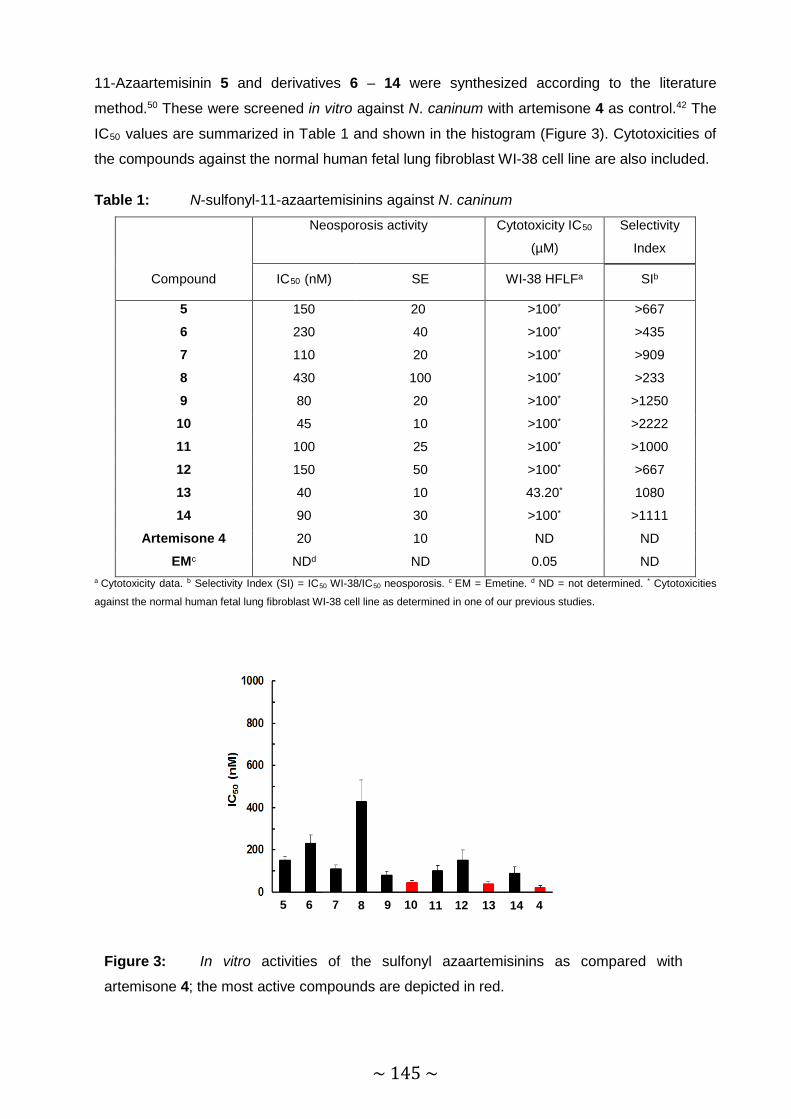
~ 145 ~
11-Azaartemisinin 5 and derivatives 6 – 14 were synthesized according to the literature
method.50 These were screened in vitro against N. caninum with artemisone 4 as control.42 The
IC50 values are summarized in Table 1 and shown in the histogram (Figure 3). Cytotoxicities of
the compounds against the normal human fetal lung fibroblast WI-38 cell line are also included.
Table 1: N-sulfonyl-11-azaartemisinins against N. caninum
Neosporosis activity Cytotoxicity IC50
(µM)
Selectivity
Index
Compound IC50 (nM) SE WI-38 HFLFa SIb
5 150 20 >100* >667
6 230 40 >100* >435
7 110 20 >100* >909
8 430 100 >100* >233
9 80 20 >100* >1250
10 45 10 >100* >2222
11 100 25 >100* >1000
12 150 50 >100* >667
13 40 10 43.20* 1080
14 90 30 >100* >1111
Artemisone 4 20 10 ND ND
EMc NDd ND 0.05 ND a Cytotoxicity data. b Selectivity Index (SI) = IC50 WI-38/IC50 neosporosis. c EM = Emetine. d ND = not determined. * Cytotoxicities
against the normal human fetal lung fibroblast WI-38 cell line as determined in one of our previous studies.
Figure 3: In vitro activities of the sulfonyl azaartemisinins as compared with
artemisone 4; the most active compounds are depicted in red.
14 5 6 7 8 9 10 11 12 13 14 14 4
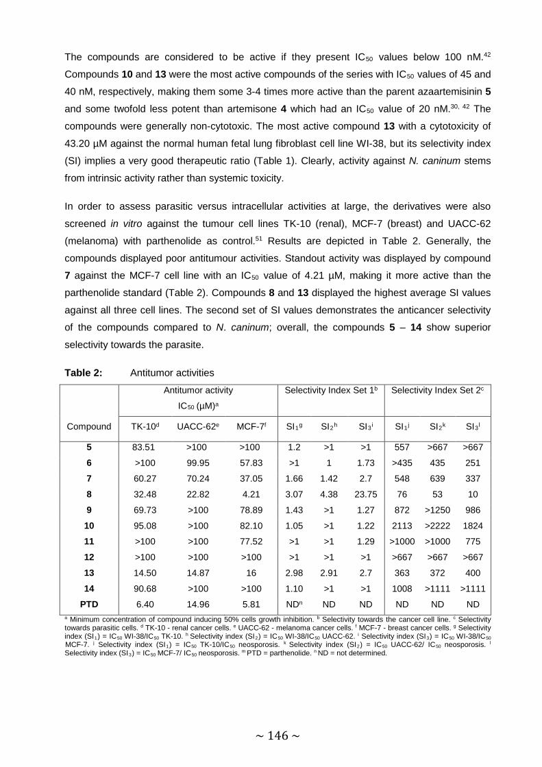
~ 146 ~
The compounds are considered to be active if they present IC50 values below 100 nM.42
Compounds 10 and 13 were the most active compounds of the series with IC50 values of 45 and
40 nM, respectively, making them some 3-4 times more active than the parent azaartemisinin 5 and some twofold less potent than artemisone 4 which had an IC50 value of 20 nM.30, 42 The
compounds were generally non-cytotoxic. The most active compound 13 with a cytotoxicity of
43.20 µM against the normal human fetal lung fibroblast cell line WI-38, but its selectivity index
(SI) implies a very good therapeutic ratio (Table 1). Clearly, activity against N. caninum stems
from intrinsic activity rather than systemic toxicity.
In order to assess parasitic versus intracellular activities at large, the derivatives were also
screened in vitro against the tumour cell lines TK-10 (renal), MCF-7 (breast) and UACC-62
(melanoma) with parthenolide as control.51 Results are depicted in Table 2. Generally, the
compounds displayed poor antitumour activities. Standout activity was displayed by compound
7 against the MCF-7 cell line with an IC50 value of 4.21 µM, making it more active than the
parthenolide standard (Table 2). Compounds 8 and 13 displayed the highest average SI values
against all three cell lines. The second set of SI values demonstrates the anticancer selectivity
of the compounds compared to N. caninum; overall, the compounds 5 – 14 show superior
selectivity towards the parasite.
Table 2: Antitumor activities
Antitumor activity
IC50 (µM)a
Selectivity Index Set 1b Selectivity Index Set 2c
Compound TK-10d UACC-62e MCF-7f SI1g SI2h SI3i SI1j SI2k SI3l
5 83.51 >100 >100 1.2 >1 >1 557 >667 >667
6 >100 99.95 57.83 >1 1 1.73 >435 435 251
7 60.27 70.24 37.05 1.66 1.42 2.7 548 639 337
8 32.48 22.82 4.21 3.07 4.38 23.75 76 53 10
9 69.73 >100 78.89 1.43 >1 1.27 872 >1250 986
10 95.08 >100 82.10 1.05 >1 1.22 2113 >2222 1824
11 >100 >100 77.52 >1 >1 1.29 >1000 >1000 775
12 >100 >100 >100 >1 >1 >1 >667 >667 >667
13 14.50 14.87 16 2.98 2.91 2.7 363 372 400
14 90.68 >100 >100 1.10 >1 >1 1008 >1111 >1111
PTD 6.40 14.96 5.81 NDn ND ND ND ND ND a Minimum concentration of compound inducing 50% cells growth inhibition. b Selectivity towards the cancer cell line. c Selectivity towards parasitic cells. d TK-10 - renal cancer cells. e UACC-62 - melanoma cancer cells. f MCF-7 - breast cancer cells. g Selectivity index (SI1) = IC50 WI-38/IC50 TK-10. h Selectivity index (SI2) = IC50 WI-38/IC50 UACC-62. i Selectivity index (SI3) = IC50 WI-38/IC50 MCF-7. j Selectivity index (SI1) = IC50 TK-10/IC50 neosporosis. k Selectivity index (SI2) = IC50 UACC-62/ IC50 neosporosis. l Selectivity index (SI3) = IC50 MCF-7/ IC50 neosporosis. m PTD = parthenolide. n ND = not determined.
~ 147 ~
In conclusion, compound 13 is the most active azaartemisinin derivative against N. caninum and
also proved to be moderately active against the three tumour cell lines. It remains to be
established if this compound can restrict the proliferation of N. caninum tachyzoites in vitro.
Acknowledgements: This research project is funded by the South African Medical Research
Council (MRC) with funds from the National Treasury under its Economic Competitiveness and
Support Package. The South African National Research Foundation is thanked for financial
support to RKH (Grant No. 90682) and to RH (NRF UID 87498). RH, FS, DDN and RKH and
also thank the North-West University for financial support. The authors express their gratitude to
Natasha Kolesnikova, Molecular and Biomedical Technologies, CSIR Biosciences, Pretoria,
South Africa, for performing the cytotoxicity screening.
Disclaimer: Any opinion, finding and conclusion or recommendation expressed in this material
is that of the author(s) and the NRF does not accept any liability in this regard.

~ 148 ~
REFERENCES
1. Dubey, J. P.; Carpenter, J. L.; Speer, C. A.; Topper, M. J.; Uggla, A. Newly recognized
fatal protozoan disease of dogs. JAVMA 1988, 192, 1269-1285.
2. Larson, R. L.; Hardin, D. K.; Pierce, V. L. Economic considerations for diagnostic and
control options for Neospora caninum-induced abortions in endemically infected herds of beef
cattle. JAVMA 2004, 224, 1597-1604.
3. Reichel, M. P.; Ellis, J. T. If control of Neospora caninum infection is technically feasible
does it make economic sense? Vet. Parasitol. 2006, 142, 23-34.
4. Reichel, M. P.; Ellis, J. T. Re-evaluating the economics of neosporosis control. Vet.
Parasitol. 2008, 156, 361-362.
5. Reichel, M. P.; McAllister, M. M.; Pomroy, W. E.; Campero, C.; Ortega-Mora, L. M.; Ellis,
J. T. Control options for Neospora caninum - is there anything new or are we going backwards?
Parasitol. 2014, 141, 1455-1470.
6. Goodswen, S. J.; Kennedy, P. J.; Ellis, J. T. A review of the infection, genetics, and
evolution of Neospora caninum: from the past to the present. Infect. Genet. Evol. 2013, 13, 133-
150.
7. Reichel, M. P.; Ellis, J. T.; Dubey, J. P. Neosporosis and hammondiosis in dogs. J. Small
Anim. Pract. 2007, 48, 308-312.
8. Dubey, J. P.; Barr, B. C.; Barta, J. R.; Bjerkås, I.; Björkman, C.; Blagburn, B. L.;
Bowman, D. D.; Buxton, D.; Ellis, J. T.; Gottstein, B. Redescription of Neospora caninum and its
differentiation from related coccidia. Int. J. Parasitol. 2002, 32, 929-946.
9. Barr, B. C.; Conrad, P. A.; Breitmeyer, R.; Sverlow, K.; Anderson, M. L.; Reynolds, J.;
Chauvet, A. E.; Dubey, J. P.; Ardans, A. A. Congenital Neospora infection in calves born from
cows that had previously aborted Neospora-infected fetuses: four cases (1990-1992). JAVMA
1993, 202, 113-117.
10. Dubey, J. P.; Koestner, A.; Piper, R. C. Repeated transplacental transmission of
Neospora caninum in dogs. JAVMA 1990, 197, 857-860.
11. Dubey, J. P.; Lindsay, D. S. A review of Neospora caninum and neosporosis. Vet.
Parasitol. 1996, 67, 1-59.
12. McAllister, M. M.; Dubey, J. P.; Lindsay, D. S.; Jolley, W. R.; Wills, R. A.; McGuire, A. M.
Rapid communication: Dogs are definitive hosts of Neospora caninum. Int. J. Parasitol. 1998,
28, 1473-1479.
13. Dubey, J. P.; Schares, G.; Ortega-Mora, L. M. Epidemiology and control of neosporosis
and Neospora caninum. Clin. Microbiol. Rev. 2007, 20, 323-367.
14. López-Pѐrez, I. C.; Collantes-Fernѐndez, E.; Rojo-Montejo, S.; Navarro-Lozano, V.;
Risco-Castillo, V.; Pѐrez-Pѐrez, V.; Pereira-Bueno, J.; Ortega-Mora, L. M. Effects of Neospora
~ 149 ~
caninum infection at mid-gestation on placenta in a pregnant mouse model. J. Parasitol. 2010,
96, 1017-1020.
15. Williams, D. J. L.; Hartley, C. S.; Björkman, C.; Trees, A. J. Endogenous and exogenous
transplacental transmission of Neospora caninum - how the route of transmission impacts on
epidemiology and control of disease. Parasitol. 2009, 136, 1895-1900.
16. Dubey, J. P.; De Lahunta, A. Neosporosis associated congenital limb deformities in a
calf. Appl. Parasitol. 1993, 34, 229-233.
17. Barling, K. S.; McNeill, J. W.; Thompson, J. A.; Paschal, J. C.; McCollum Iii, F. T.; Craig,
T. M.; Adams, L. G. Association of serologic status for Neospora caninum with post-weaning
weight gain and carcass measurements in beef calves. JAVMA 2000, 217, 1356-1360.
18. Fioretti, D. P.; Pasquali, P.; Diaferia, M.; Mangili, V.; Rosignoli, L. Neospora caninum
infection and congenital transmission: serological and parasitological study of cows up to the
fourth gestation. J. Vet. Med. B. 2003, 50, 399-404.
19. Buxton, D.; McAllister, M. M.; Dubey, J. P. The comparative pathogenesis of
neosporosis. Trends Parasitol. 2002, 18, 546-552.
20. Bartels, C. J. M.; Wouda, W.; Schukken, Y. H. Risk factors for Neospora caninum-
associated abortion storms in dairy herds in The Netherlands (1995 to 1997). Theriogenology
1999, 52, 247-257.
21. Quinn, H. E.; Windsor, P. A.; Kirkland, P. D.; Ellis, J. T. An outbreak of abortion in a dairy
herd associated with Neospora caninum and bovine pestivirus infection. Aus. Vet. J. 2004, 82,
99-101.
22. Haddad, J. P. A.; Dohoo, I. R.; Van Leewen, J. A. A review of Neospora caninum in dairy
and beef cattle - a Canadian perspective. Can. Vet. J. 2005, 46, 230-243.
23. Reichel, M. P.; Ellis, J. T. Control options for Neospora caninum infections in cattle -
current state of knowledge. N. Z. Vet. J. 2002, 50, 86-92.
24. Weston, J. F.; Heuer, C.; Williamson, N. B. Efficacy of a Neospora caninum killed
tachyzoite vaccine in preventing abortion and vertical transmission in dairy cattle. Prev. Vet.
Med. 2012, 103, 136-144.
25. Hall, C. A.; Reichel, M. P.; Ellis, J. T. Neospora abortions in dairy cattle: diagnosis, mode
of transmission and control. Vet. Parasitol. 2005, 128, 231-241.
26. Reichel, M. P.; Ellis, J. T. Neospora caninum - How close are we to development of an
efficacious vaccine that prevents abortion in cattle? Int. J. Parasitol. 2009, 39, 1173-1187.
27. Lindsay, D. S.; Rippey, N. S.; Cole, R. A.; Parsons, L. C.; Dubey, J. P.; Tidwell, R. R.;
Blagburn, B. L. Examination of the activities of 43 chemotherapeutic agents against Neospora
caninum tachyzoites in cultured cells. Am. J. Vet. Res. 1994, 55, 976-981.
~ 150 ~
28. Kritzner, S.; Sager, H.; Blum, J. R.; Krebber, R.; Greif, G.; Gottstein, B. An explorative
study to assess the efficacy of Toltrazuril-sulfone (Ponazuril) in calves experimentally infected
with Neospora caninum. Ann. Clin. Microbiol. Antimicrob. 2002, 1, 1.
29. Strohbusch, M.; Müller, N.; Hemphill, A.; Krebber, R.; Greif, G.; Gottstein, B. Toltrazuril
treatment of congenitally acquired Neospora caninum infection in newborn mice. Parasitol. Res.
2009, 104, 1335-1343.
30. Mazuz, M. L.; Haynes, R.; Shkap, V.; Fish, L.; Wollkomirsky, R.; Leibovich, B.; Molad, T.;
Savitsky, I.; Golenser, J. Neospora caninum: in vivo and in vitro treatment with artemisone. Vet.
Parasitol. 2012, 187, 99-104.
31. Lindsay, D. S.; Dubey, J. P. Effects of sulfadiazine and amprolium on Neospora caninum
(Protozoa: Apicomplexa) infections in mice. J. Parasitol. 1990, 76, 177-179.
32. Debache, K.; Hemphill, A. Effects of miltefosine treatment in fibroblast cell cultures and
in mice experimentally infected with Neospora caninum tachyzoites. Parasitol. 2012, 139, 934-
944.
33. Schorer, M.; Debache, K.; Barna, F.; Monney, T.; Müller, J.; Boykin, D. W.; Stephens, C.
E.; Hemphill, A. Di-cationic arylimidamides act against Neospora caninum tachyzoites by
interference in membrane structure and nucleolar integrity and are active against challenge
infection in mice. Int. J. Parasitol. Drugs Drug Resist. 2012, 2, 109-120.
34. Debache, K.; Guionaud, C.; Kropf, C.; Boykin, D.; Stephens, C. E.; Hemphill, A.
Experimental treatment of Neospora caninum-infected mice with the arylimidamide DB750 and
the thiazolide nitazoxanide. Exp. Parasitol. 2011, 129, 95-100.
35. Dubey, J. P.; Schares, G. Neosporosis in animals - the last five years. Vet. Parasitol.
2011, 180, 90-108.
36. Oh, H. G.; Youn, H. J.; Noh, J. W.; Jang, D. H.; Kang, Y. B. Anticoccidial effects of
artemisinin on the Eimeria tenella. Korean J. Vet. Res. 1995, 35, 123-130.
37. Noh, J. W.; Youn, H. J.; Jang, D. H. The anticryptosporidial effects of sulfadimethoxine,
toltrazuril and the extracts of herbs for Cryptosporidium parvum isolated from mouse in Korea.
Korean J. Lab. Anim. Sci. 1997.
38. Kim, J.-T.; Park, J.-Y.; Seo, H.-S.; Oh, H.-G.; Noh, J.-W.; Kim, J.-H.; Kim, D.-Y.; Youn,
H.-J. In vitro antiprotozoal effects of artemisinin on Neospora caninum. Vet. Parasitol. 2002,
103, 53-63.
39. Haynes, R. K.; Fugmann, B.; Stetter, J.; Rieckmann, K.; Heilmann, H. D.; Chan, H.-W.;
Cheung, M.-K.; Lam, W.-L.; Wong, H.-N.; Croft, S. L. Artemisone - a highly active antimalarial
drug of the artemisinin class. Angew. Chem. 2006, 118, 2136-2142.
40. Waknine-Grinberg, J. H.; Hunt, N.; Bentura-Marciano, A.; McQuillan, J. A.; Chan, H.-W.;
Chan, W.-C.; Barenholz, Y.; Haynes, R. K.; Golenser, J. Artemisone effective against murine
cerebral malaria. Malar. J. 2010, 9, 1.

~ 151 ~
41. Dunay, I. R.; Chan, W. C.; Haynes, R. K.; Sibley, L. D. Artemisone and artemiside
control acute and reactivated toxoplasmosis in a murine model. Antimicrob. Agents Chemother.
2009, 53, 4450-4456.
42. Müller, J.; Balmer, V.; Winzer, P.; Rahman, M.; Manser, V.; Haynes, R. K.; Hemphill, A.
In vitro effects of new artemisinin derivatives in Neospora caninum-infected human fibroblasts.
Int. J. Antimicrob. Agents 2015, 46, 88-93.
43. Harmse, R.; Wong, H. N.; Smit, F.; K Haynes, R.; N'Da, D. D. The case for development
of 11-aza-artemisinins for malaria. Curr. Med. Chem. 2015, 22, 3607-3630.
44. China Cooperative Research Group. The chemistry and synthesis of qinghaosu
derivatives. J. Trad. Chin. Med. 1982, 2, 1-16.
45. Brewer, T. G.; Grate, S. J.; Peggins, J. O.; Weina, P. J.; Petras, J. M.; Levine, B. S.;
Heiffer, M. H.; Schuster, B. G. Fatal neurotoxicity of arteether and artemether. Am. J. Trop.
Med. Hyg. 1994, 51, 251-259.
46. Nontprasert, A.; Nosten-Bertrand, M.; Pukrittayakamee, S.; Vanijanonta, S.; Angus, B.
J.; White, N. J. Assessment of the neurotoxicity of parenteral artemisinin derivatives in mice.
Am. J. Trop. Med. Hyg. 1998, 59, 519-522.
47. Genovese, R. F.; Newman, D. B.; Brewer, T. G. Behavioral and neural toxicity of the
artemisinin antimalarial, arteether, but not artesunate and artelinate, in rats. Pharmacol.
Biochem. Behav. 2000, 67, 37-44.
48. Schmuck, G.; Haynes, R. K. Establishment of an in vitro screening model for
neurodegeneration induced by antimalarial drugs of the artemisinin-type. Neurotox. Res. 2000,
2, 37-49.
49. Schmuck, G.; Roehrdanz, E.; Haynes, R. K.; Kahl, R. Neurotoxic mode of action of
artemisinin. Antimicrob. Agents Chemother. 2002, 46, 821-827.
50. Haynes, R. K.; Wong, H. N.; Lee, K. W.; Lung, C. M.; Shek, L. Y.; Williams, I. D.; Croft,
S. L.; Vivas, L.; Rattray, L.; Stewart, L. Preparation of N-sulfonyl- and N-carbonyl-11-
azaartemisinins with greatly enhanced thermal stabilities: in vitro antimalarial activities.
ChemMedChem 2007, 2, 1464-1479.
51. Skehan, P.; Storeng, R.; Scudiero, D.; Monks, A.; McMahon, J.; Vistica, D.; Warren, J.
T.; Bokesch, H.; Kenney, S.; Boyd, M. R. New colorimetric cytotoxicity assay for anticancer-drug
screening. JNCI 1990, 82, 1107-1112.

~ 152 ~
CHAPTER 6 Summary and conclusion
Malaria is a dreadful disease caused by the protozoan parasite of the genus Plasmodium,
affecting quite a large number of the world population. Between 2000 and 2014 the malaria
incidence rate fell by 37% globally and by 42% in the African region (WHO, 2015). During the
same time period the mortality rate of malaria fell by 60% globally and by 66% in the African
region (WHO, 2015). Although this is a quite an achievement, current statistics show that
malaria still affects 97 countries worldwide with an estimated 214 million reported cases with
438 000 deaths in 2014 (WHO, 2015). Prevalence for malaria is highest in Africa and
predominates in children under the age of 5 (WHO, 2015).
Malaria has been recorded for generations, and cases that may be attributed to malaria date as
far back as 2700 BC (Cox, 2010) to its official discovery in 1880 (Bruce-Chwatt, 1981). Although
there have been many scientific advances in treatment and prevention, the ability of the
Plasmodium parasite to develop resistance has long been a thorn in the side of scientific
advancement and often contributes to the degradation of treatment regimes. Multidrug
resistance has rendered older generations of drugs (quinolones, antifolates, etc.) useless
against the fight against malaria. In an effort to combat the spread of any further resistance, the
World Health Organization (WHO) set out standard treatment guidelines involving artemisinin
combination therapies (ACTs) comprising the short half-life artemisinin antimalarial drug
combined with a longer half-life partner drug. The rationale is that the artemisinin within its short
pharmacological half-life provides rapid reduction of the parasite burden, while the longer half-
life partner drug continues the clearance when the artemisinin concentration falls to sub-
therapeutic levels.(Wellems and Plowe, 2001) Unfortunately, current artemisinin derivatives are
also prone to developing resistance. Even with the efforts of the WHO in banning artemisinin
monotherapy and only prescribing the use of ACTs, greatly increased parasite clearance times
in patients treated with ACTs have been reported (Phyo et al., 2012, Lubell et al., 2014) and
genetic markers of the apparently resistant phenotype have been identified (Ariey et al., 2014).
The poor thermal stabilities of artemisinins also raise some concern over decomposition of
formulated material in countries with hot and humid weather (Haynes et al., 2007). Also, current
derivatives like artemether, arteether and artesunate are metabolized to dihydroartemisinin
(DHA) in vivo which gives rise to more challenges as DHA is known to display neurotoxicity in
cell and animal assays (China Cooperative Research Group, 1982, Brewer et al., 1994, Avery et
al., 1995, Nontprasert et al., 1998, Genovese et al., 2000, Schmuck and Haynes, 2000,
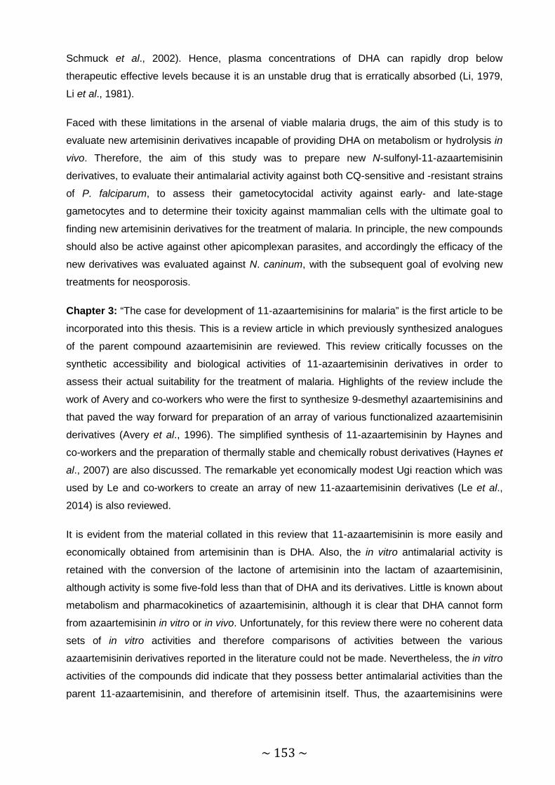
~ 153 ~
Schmuck et al., 2002). Hence, plasma concentrations of DHA can rapidly drop below
therapeutic effective levels because it is an unstable drug that is erratically absorbed (Li, 1979,
Li et al., 1981).
Faced with these limitations in the arsenal of viable malaria drugs, the aim of this study is to
evaluate new artemisinin derivatives incapable of providing DHA on metabolism or hydrolysis in
vivo. Therefore, the aim of this study was to prepare new N-sulfonyl-11-azaartemisinin
derivatives, to evaluate their antimalarial activity against both CQ-sensitive and -resistant strains
of P. falciparum, to assess their gametocytocidal activity against early- and late-stage
gametocytes and to determine their toxicity against mammalian cells with the ultimate goal to
finding new artemisinin derivatives for the treatment of malaria. In principle, the new compounds
should also be active against other apicomplexan parasites, and accordingly the efficacy of the
new derivatives was evaluated against N. caninum, with the subsequent goal of evolving new
treatments for neosporosis.
Chapter 3: “The case for development of 11-azaartemisinins for malaria” is the first article to be
incorporated into this thesis. This is a review article in which previously synthesized analogues
of the parent compound azaartemisinin are reviewed. This review critically focusses on the
synthetic accessibility and biological activities of 11-azaartemisinin derivatives in order to
assess their actual suitability for the treatment of malaria. Highlights of the review include the
work of Avery and co-workers who were the first to synthesize 9-desmethyl azaartemisinins and
that paved the way forward for preparation of an array of various functionalized azaartemisinin
derivatives (Avery et al., 1996). The simplified synthesis of 11-azaartemisinin by Haynes and
co-workers and the preparation of thermally stable and chemically robust derivatives (Haynes et
al., 2007) are also discussed. The remarkable yet economically modest Ugi reaction which was
used by Le and co-workers to create an array of new 11-azaartemisinin derivatives (Le et al.,
2014) is also reviewed.
It is evident from the material collated in this review that 11-azaartemisinin is more easily and
economically obtained from artemisinin than is DHA. Also, the in vitro antimalarial activity is
retained with the conversion of the lactone of artemisinin into the lactam of azaartemisinin,
although activity is some five-fold less than that of DHA and its derivatives. Little is known about
metabolism and pharmacokinetics of azaartemisinin, although it is clear that DHA cannot form
from azaartemisinin in vitro or in vivo. Unfortunately, for this review there were no coherent data
sets of in vitro activities and therefore comparisons of activities between the various
azaartemisinin derivatives reported in the literature could not be made. Nevertheless, the in vitro
activities of the compounds did indicate that they possess better antimalarial activities than the
parent 11-azaartemisinin, and therefore of artemisinin itself. Thus, the azaartemisinins were

~ 154 ~
selected in this PhD project for evaluation for further research and development into potential
drug candidates.
Chapter 4: “Antimalarial activities and cytotoxicities of N-sulfonyl-11-azaartemisinin derivatives”
is the second article of the thesis. In this article is described the syntheses of 13 N-sulfonyl-
azaartemisinins and their in vitro antimalarial activities against chloroquine (CQ) sensitive
(NF54) and CQ resistant (K1 and W2) strains of intraerythrocytic P. falciparum (Pf) parasites
and cytotoxicities as determined against the human fetal lung fibroblast (HFLF) WI-38 cell line.
The aim of the research was to identify possible derivatives that may be suited for further
development based on a comparison of activities with the current clinical artemisinin derivatives
DHA, artemether and artesunate. Most clinically used antimalarial drugs target the asexual life
cycle of the malaria parasite which is responsible for the clinical symptoms of the disease. The
gametocytes, which are responsible for transmission from host to vector, are overlooked.
Therefore, in this work, the gametocytocidal activities of the new compounds would also be
assessed against the Pf NF54 blood-stage gametocytes using the luciferase and pLDH assays.
This activity, namely assessment of transmission blocking potential, is now recommended by
the WHO as an essential part of development of new antimalarial drugs.
Synthesis of the target compounds firstly involved ammonolysis of artemisinin to provide the
parent compound 11-azaartemisinin which was further derivatized to yield the N-11-sulfonyl-
azaartemisinin derivatives. Structures were confirmed through use of nuclear magnetic
resonance (NMR) and infrared (IR) spectroscopic techniques, and high resolution mass
spectrometry (HRMS) techniques. Biological screening of the compounds using as comparator
drugs DHA, artemether and artesunate revealed that the N-p-trifluoromethyl-benzenesulfonyl
compound 11 had the best antiplasmodial activity (IC50 2 – 3 nM) and activity equipotent with
artemether against the Pf NF54 strain. The 3'-thienylsulfonyl derivative 17 also displayed activity
equipotent with that of artesunate against this strain. Both compounds 11 and 17 had enhanced
activity as well as lower resistance index (RI) values against the W2 cell line when compared to
artemether and artesunate. Overall the compounds displayed good selectivity values towards
the malaria parasites with respect to mammalian cells and are thus considered to be relatively
non-cytotoxic.
Gametocytocidal activity of the compounds alongside artemether and artesunate were also
determined. The N-p-trifluoromethylbenzenesulfonyl compound 11 was active against both
early- and late-stage gametocytes and was more stage specific towards later stage
gametocytes when compared with artemether and artesunate. However, the 2'-thienylsulfonyl
derivative 16 presented the better late-stage gametocytocidal activity and stage-specific value
when compared to 11. Although compound 16 (IC50 4 – 5 nM) is not as potent as 11 towards
~ 155 ~
asexual blood-stage parasites, it has better activity than artemether against Pf W2 strain, and
an improved RI value. Compound 16 also demonstrated to be non-cytotoxic.
Therefore, compounds 11 and 16 are identified as potential drug candidates for further
evaluation as potential drug development candidates. These compounds will therefore be
carried forward for pharmacokinetic studies and in vivo evaluation of efficacies.
Chapter 5: “Activities of N-sulfonyl-11-azaartemisinin derivatives against the apicomplexan
parasite Neospora caninum and comparative cytotoxicities” is the third and final article in the
thesis. It involved evaluation of the activities of nine of the N-sulfonyl-azaartemisinins against
the apicomplexan parasite Neospora caninum in vitro and comparative antitumor activities.
Neospora caninum is the causative pathogen of the disease neosporosis that has a major
economic effect on the beef- and dairy cattle industries. There is but a handful of treatment
options available for neosporosis, each with its own set of challenges. The most common
among them are the undesirable residues that are left behind in milk and meat products after
treatment. Hence, it is necessary to consider the artemisinin class of compounds; these are
notably effective in the treatment of the apicomplexan parasite that causes malaria, as
discussed above.
The new 11-azaartemisinin derivatives were screened against human foreskin fibroblasts
infected with tachyzoites of N. caninum. Artemisone was used as a standard. Evaluation of the
cytotoxicities of the compounds against the normal human fetal lung fibroblast WI-38 cell line
was also carried out. The most active compounds against neosporosis were the p-
iodobenzenesulfonyl-11-azaartemisinin derivative 10 and the 2',5'-dichloro-3'-thienylsulfonyl-11-
azaartemisinin derivative 13 with IC50 values of 45 nM and 40 nM, respectively. Compounds 10
and 13 are 3 – 4 times more active than azaartemisinin and some 2-fold less potent than
artemisone. The compounds were screened in vitro alongside parthenolide against a panel of
the TK-10 (renal), MCF-7 (breast) and UACC-62 (melanoma) cell lines. Overall, the compounds
displayed very poor activity against all three cancer cell lines except in the case of the straight
chain N-alkanesulfonylazaartemisinin 8 against the MCF-7 cell line with an IC50 value of 4.21
µM, making it more active than the parthenolide standard. Overall, the compounds proved to be
more selective towards the N. caninum tachyzoites. It is clear that we have a new set of
derivatives that should be carried forward for evaluation of their potential in leading to new
treatment options for neosporosis.
To conclude, the compounds synthesized in this study proved to be active against malaria as
well as neosporosis. Hit compounds with activities comparable to current artemisinins have

~ 156 ~
been identified. It is recommended that these compounds should be evaluated further in order
to assess their potential for development into new drugs for treatment of malaria or neosporosis.

~ 157 ~
REFERENCES
Ariey, F., Witkowski, B., Amaratunga, C., Beghain, J., Langlois, A.-C., Khim, N., Kim, S., Duru,
V., Bouchier, C. & Ma, L. (2014) A molecular marker of artemisinin-resistant Plasmodium
falciparum malaria. Nature, 505, 50-55.
Avery, M. A., Bonk, J. D., Chong, W. K. M., Mehrotra, S., Miller, R., Milhous, W., Goins, D. K.,
Venkatesan, S. & Wyandt, C. (1995) Structure-activity relationships of the antimalarial
agent artemisinin. 2. Effect of heteroatom substitution at O-11: synthesis and bioassay of
N-Alkyl-11-aza-9-desmethylartemisinins. Journal of Medicinal Chemistry, 38, 5038-5044.
Avery, M. A., Wesley, J. D. B., Chong, K. M., Mehrotra, S., Miller, R., Milhous, W., Goins, D. K.,
Venkatesan, S., Wyandt, C., Khan, I. & Avery, B. A. (1996) Structure-activity
relationships of the antimalarial agent artemisinin. 2. Effect of heteratom substitution at
O-11: Synthesis and bioassay of N-alkyl-11-aza-9-desmethylartemisinins. Journal of
Medicinal Chemistry, 38, 5038-5044.
Brewer, T. G., Grate, S. J., Peggins, J. O., Weina, P. J., Petras, J. M., Levine, B. S., Heiffer, M.
H. & Schuster, B. G. (1994) Fatal neurotoxicity of arteether and artemether. The
American Journal of Tropical Medicine and Hygiene, 51, 251-259.
Bruce-Chwatt, L. J. (1981) Alphonse Laveran's discovery 100 years ago and today's global fight
against malaria. Journal of the Royal Society of Medicine, 74, 531.
China Cooperative Research Group. (1982) The chemistry and synthesis of qinghaosu
derivatives. Journal of Traditional Chinese Medicine, 2, 1-16.
Cox, F. E. G. (2010) History of the discovery of the malaria parasites and their vectors. Parasite
Vectors, 3, 5.
Genovese, R. F., Newman, D. B. & Brewer, T. G. (2000) Behavioral and neural toxicity of the
artemisinin antimalarial, arteether, but not artesunate and artelinate, in rats.
Pharmacology Biochemistry and Behavior, 67, 37-44.
Haynes, R. K., Wong, H.-N., Lee, K.-W., Lung, C.-M., Shek, L. Y., Williams, I. D., Croft, S. L.,
Vivas, L., Rattray, L. & Stewart, L. (2007) Preparation of N-sulfonyl- and N-carbonyl-11-
azaartemisinins with greatly enhanced thermal stabilities: in vitro antimalarial activities.
ChemMedChem, 2, 1464-1479.

~ 158 ~
Le, T. N., De Borggraeve, W. M., Grellier, P., Pham, V. C. & Dehaen, W. (2014) Synthesis of
11-aza-artemisinin derivatives using the Ugi reaction and an evaluation of their
antimalarial activity. Tetrahedron Letters, 55, 4892-4894.
Li, Y. (1979) Synthesis of some derivatives of artemisinin. Reprinted from Kexue Tongbao, 24,
667-669.
Li, Y., Yu, P.-L., Chen, Y.-X., Li, L.-Q., Gai, Y.-Z., Wang, D.-S. & Zheng, Y.-P. (1981) Studies on
analogs of artemisinin. I. The synthesis of ethers, carboxylic esters and carbonates of
dihydroartemisinin. Acta Pharmaceutica Sinica, 16, 429.
Lubell, Y., Dondorp, A., Guérin, P. J., Drake, T., Meek, S., Ashley, E., Day, N. P. J., White, N. J.
& White, L. J. (2014) Artemisinin resistance - modelling the potential human and
economic costs. Malaria Journal, 13, 1.
Nontprasert, A., Nosten-Bertrand, M., Pukrittayakamee, S., Vanijanonta, S., Angus, B. J. &
White, N. J. (1998) Assessment of the neurotoxicity of parenteral artemisinin derivatives
in mice. The American Journal of Tropical Medicine and Hygiene, 59, 519-522.
Phyo, A. P., Nkhoma, S., Stepniewska, K., Ashley, E. A., Nair, S., McGready, R., Ler Moo, C.,
Al-Saai, S., Dondorp, A. M. & Lwin, K. M. (2012) Emergence of artemisinin-resistant
malaria on the western border of Thailand: a longitudinal study. The Lancet, 379, 1960-
1966.
Schmuck, G. & Haynes, R. K. (2000) Establishment of an in vitro screening model for
neurodegeneration induced by antimalarial drugs of the artemisinin-type. Neurotoxicity
Research, 2, 37-49.
Schmuck, G., Roehrdanz, E., Haynes, R. K. & Kahl, R. (2002) Neurotoxic mode of action of
artemisinin. Antimicrobial Agents and Chemotherapy, 46, 821-827.
Wellems, T. E. & Plowe, C. V. (2001) Chloroquine-resistant malaria. Journal of Infectious
Diseases, 184, 770-776.
Who (2015) Global Malaria Programme Fact Sheet: World Malaria Report 2015 Geneva, World
Health Organization, http://www.who.int/malaria/media/world-malaria-report-2015/en/.

~ 159 ~
ADDENDUM A Analytical data for Chapter 4
This addendum contains the following analytical data for compounds 5 – 18 in Chapter 4:
1H NMR, 13C NMR, DEPT 90, DEPT 135, COSY, HSQC, IR, HRMS
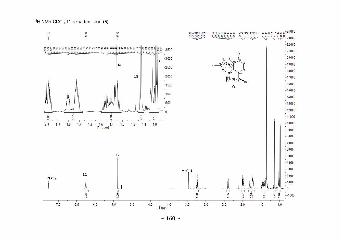
~ 160 ~
1H NMR CDCl3 11-azaartemisinin (5)
12
11 CDCl3 9
14
15
16
MeOH
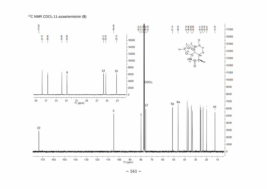
~ 161 ~
13C NMR CDCl3 11-azaartemisinin (5)
10
3 1
CDCl3
12 5a 8a
9 14 15
16

~ 162 ~
DEPT90 NMR CDCl3 11-azaartemisinin (5)

~ 163 ~
DEPT135 NMR CDCl3 11-azaartemisinin (5)
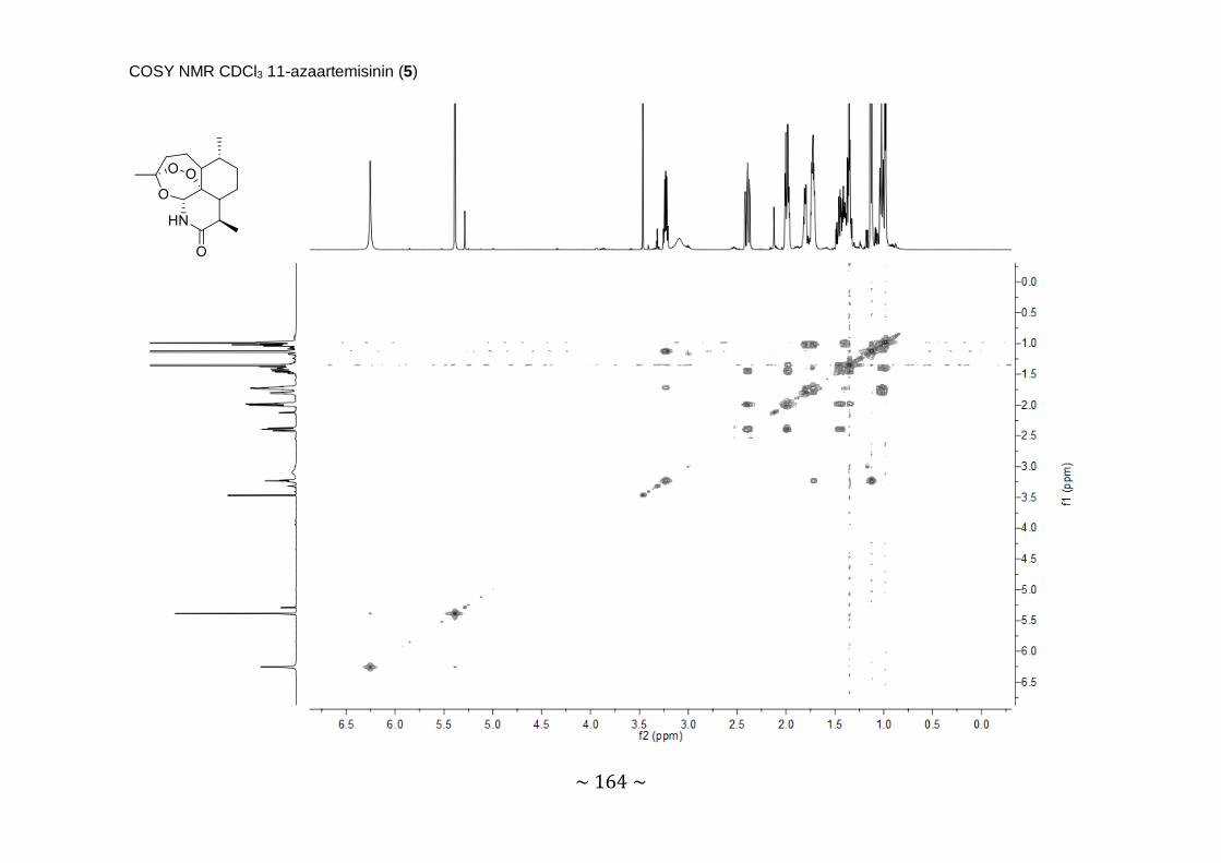
~ 164 ~
COSY NMR CDCl3 11-azaartemisinin (5)

~ 165 ~
HSQC NMR CDCl3 11-azaartemisinin (5)
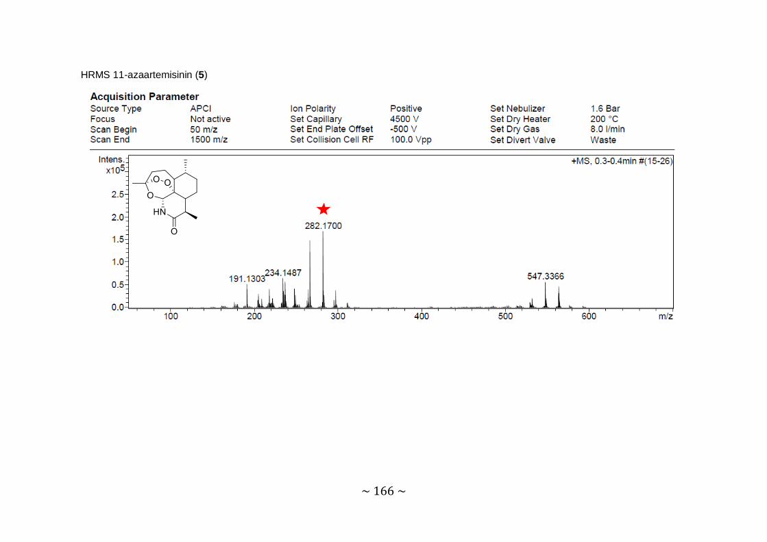
~ 166 ~
HRMS 11-azaartemisinin (5)

~ 167 ~
IR 11-azaartemisinin (5)

~ 168 ~
1H NMR CDCl3 N-Methanesulfonylazaartemisinin (6)
12 CDCl3
1’
9 4α
8a
H2O
14 5a
16 15

~ 169 ~
13C NMR CDCl3 N-Methanesulfonylazaartemisinin (6)
10
3
1
12
CDCl3
5a
8a 1’
9 14
15
16

~ 170 ~
DEPT90 CDCl3 N-Methanesulfonylazaartemisinin (6)

~ 171 ~
DEPT135 CDCl3 N-Methanesulfonylazaartemisinin (6)
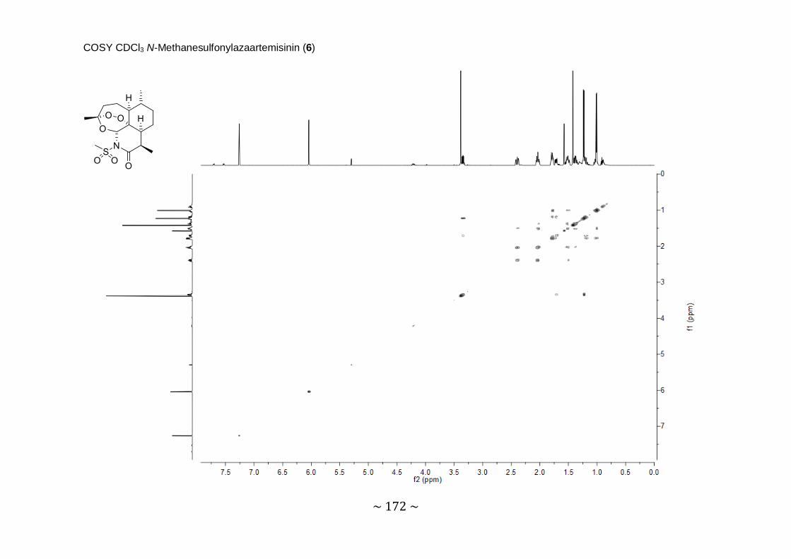
~ 172 ~
COSY CDCl3 N-Methanesulfonylazaartemisinin (6)

~ 173 ~
HSQC CDCl3 N-Methanesulfonylazaartemisinin (6)

~ 174 ~
HRMS N-Methanesulfonylazaartemisinin (6)

~ 175 ~
IR N-Methanesulfonylazaartemisinin (6)

~ 176 ~
1H NMR CDCl3 Butanesulfonyl-azaartemisinin (7)
12
CDCl3
1’α 1’β 9 4α
8a 5
14
16 4’
15

~ 177 ~
13C NMR CDCl3 Butanesulfonyl-azaartemisinin (7)
10
3 1
12
CDCl3
1’ 5a
8a
14 15 16 4

~ 178 ~
DEPT90 CDCl3 Butanesulfonyl-azaartemisinin (7)

~ 179 ~
DEPT135 CDCl3 Butanesulfonyl-azaartemisinin (7)

~ 180 ~
COSY CDCl3 Butanesulfonyl-azaartemisinin (7)

~ 181 ~
HSQC CDCl3 Butanesulfonyl-azaartemisinin (7)

~ 182 ~
HRMS Butanesulfonyl-azaartemisinin (7)

~ 183 ~
IR Butanesulfonyl-azaartemisinin (7)
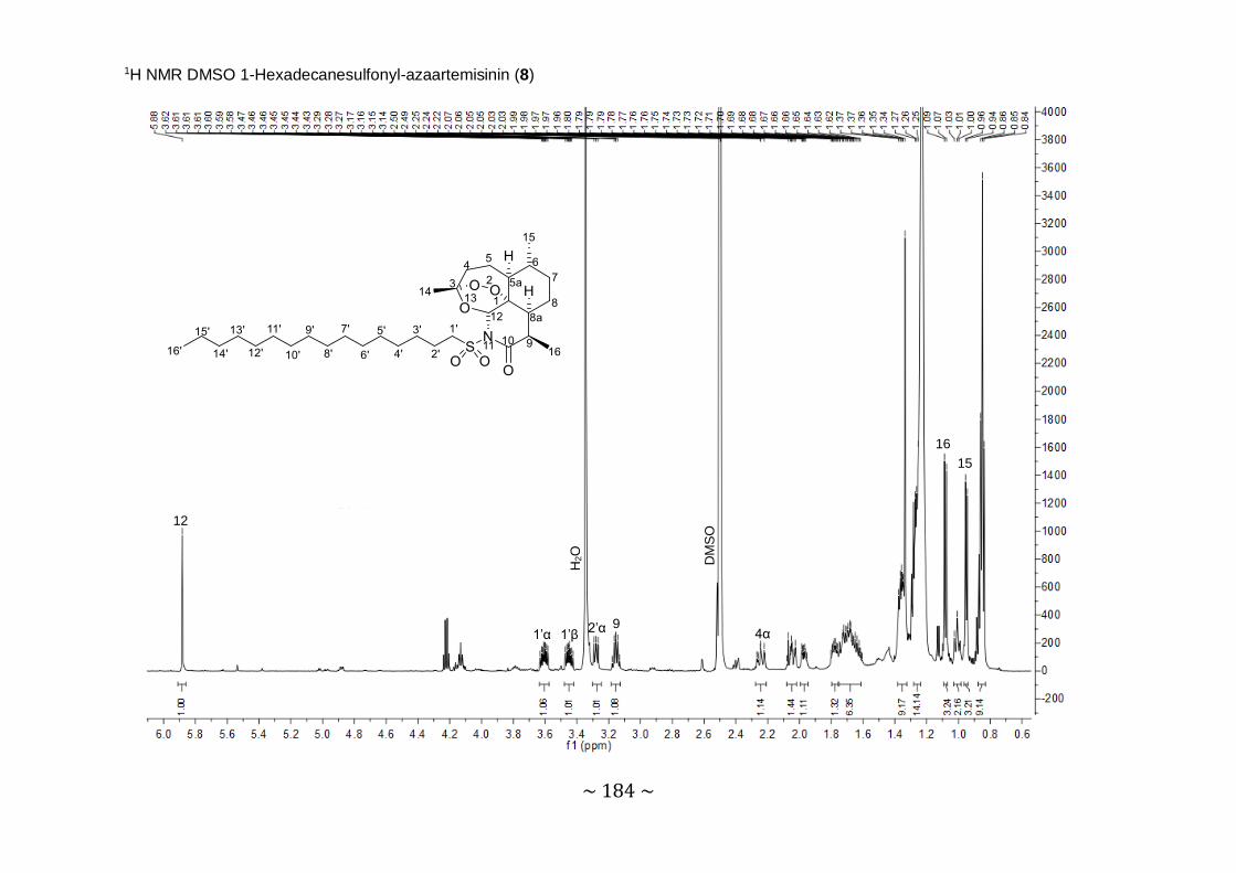
~ 184 ~
1H NMR DMSO 1-Hexadecanesulfonyl-azaartemisinin (8)
12
1’α 1’β
H2O
2’α 9
DM
SO
4α
15 16

~ 185 ~
13C NMR DMSO 1-Hexadecanesulfonyl-azaartemisinin (8)
10
3
12a 12
1’
5a 8a
DM
SO
9 3’ 14
4’ – 13’
15
16
16’
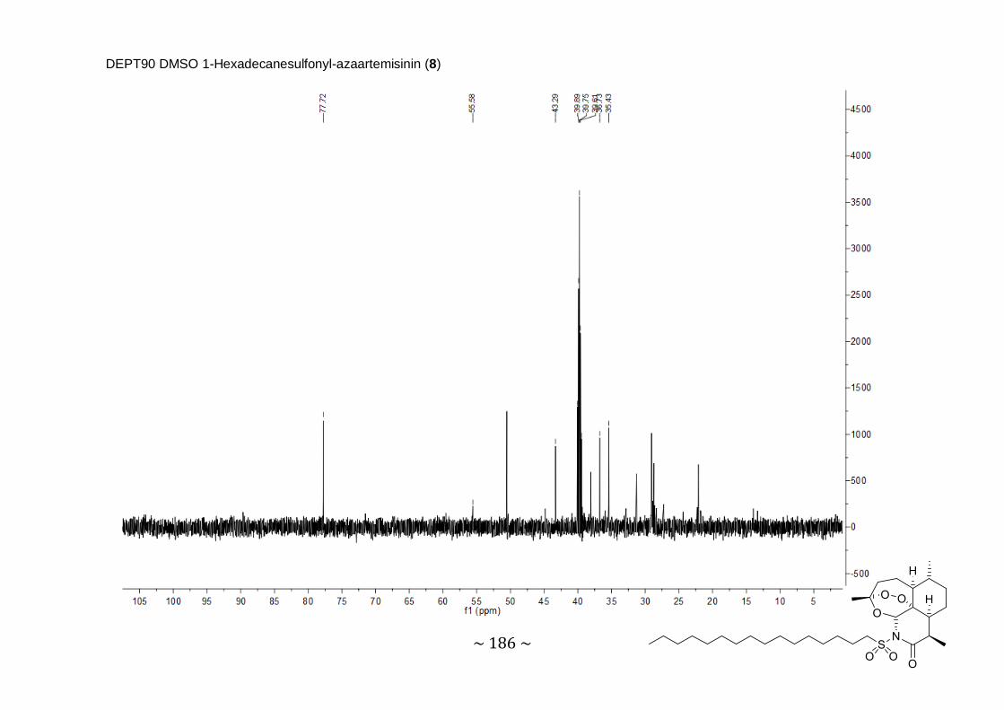
~ 186 ~
DEPT90 DMSO 1-Hexadecanesulfonyl-azaartemisinin (8)

~ 187 ~
DEPT135 DMSO 1-Hexadecanesulfonyl-azaartemisinin (8)

~ 188 ~
COSY DMSO 1-Hexadecanesulfonyl-azaartemisinin (8)
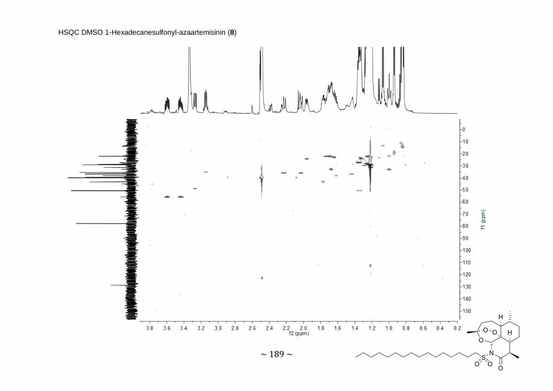
~ 189 ~
HSQC DMSO 1-Hexadecanesulfonyl-azaartemisinin (8)
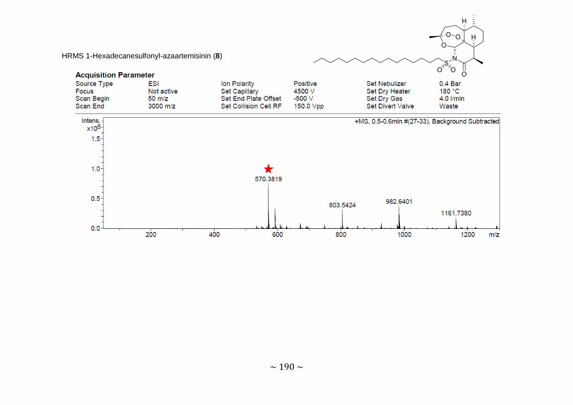
~ 190 ~
HRMS 1-Hexadecanesulfonyl-azaartemisinin (8)

~ 191 ~
IR 1-Hexadecanesulfonyl-azaartemisinin (8)

~ 192 ~
1H NMR CDCl3 Benzenesulfonyl-azaartemisinin (9)
12 2’ & 6’
4’
3’ & 5’
CDCl3
9 4α
8a 5a
14 16
15

~ 193 ~
13C NMR CDCl3 Benzenesulfonyl-azaartemisinin (9)
10 1’
12 4’
2’ &
6’
3’ &
5’
3 1
5a 8a
9
15 16
CDCl3

~ 194 ~
DEPT90 CDCl3 Benzenesulfonyl-azaartemisinin (9)

~ 195 ~
DEPT135 CDCl3 Benzenesulfonyl-azaartemisinin (9)

~ 196 ~
COSY CDCl3 Benzenesulfonyl-azaartemisinin (9)
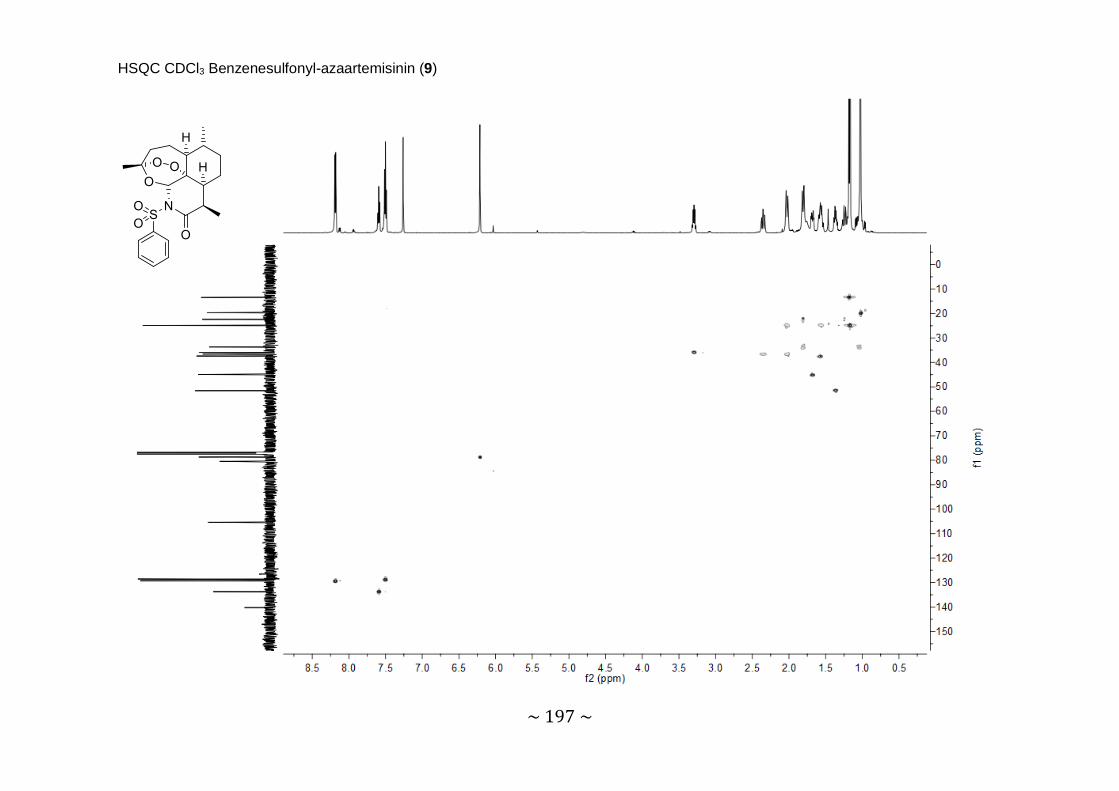
~ 197 ~
HSQC CDCl3 Benzenesulfonyl-azaartemisinin (9)

~ 198 ~
HRMS Benzenesulfonyl-azaartemisinin (9)
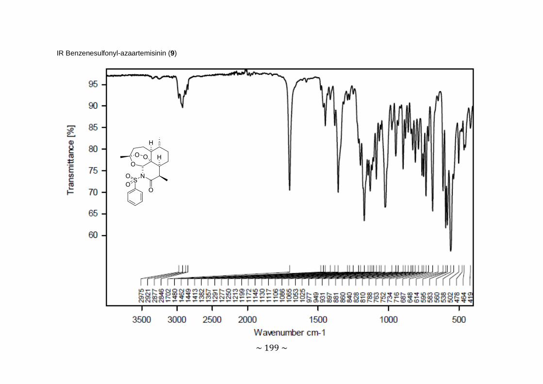
~ 199 ~
IR Benzenesulfonyl-azaartemisinin (9)

~ 200 ~
1H NMR CDCl3 3-(Trifluoromethyl)benzenesulfonyl-azaartemisinin (10)
12
6’
2’ 4’ 5’
CDCl3
MeOH
9 4α
8a
Hexane
16 14
15

~ 201 ~
13C NMR CDCl3 3-(Trifluoromethyl)benzenesulfonyl-azaartemisinin (10)
10 1’ 6’
5’ 3
2’
3’
12a
12
CDCl3
5a
MeO
H
8a
9
14
15 16
4’
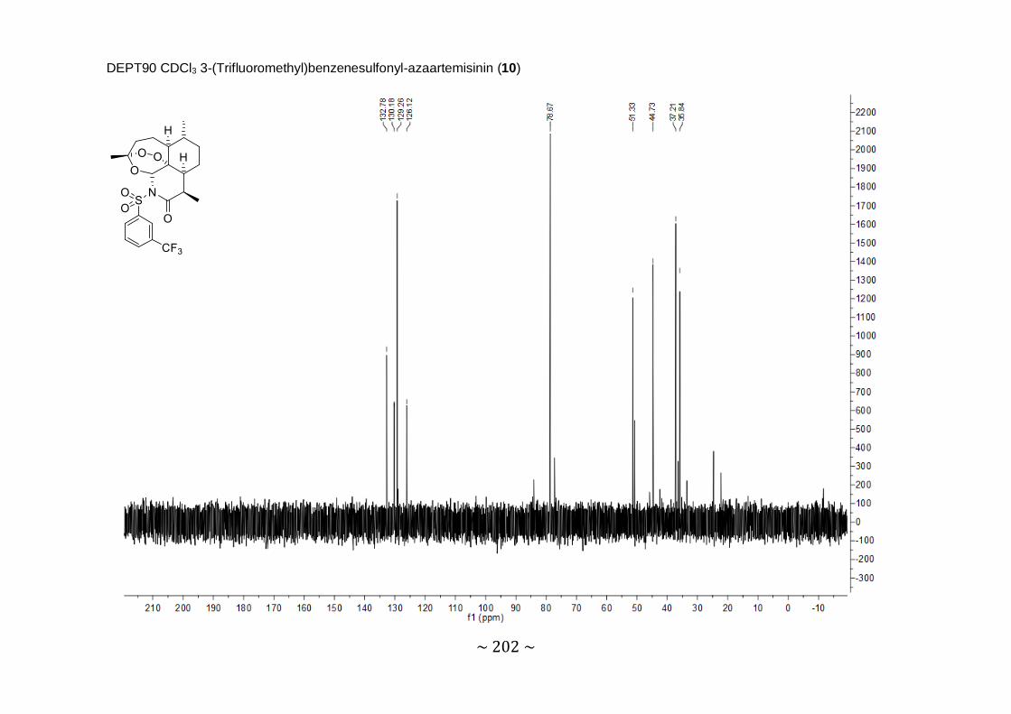
~ 202 ~
DEPT90 CDCl3 3-(Trifluoromethyl)benzenesulfonyl-azaartemisinin (10)

~ 203 ~
DEPT135 CDCl3 3-(Trifluoromethyl)benzenesulfonyl-azaartemisinin (10)

~ 204 ~
COSY CDCl3 3-(Trifluoromethyl)benzenesulfonyl-azaartemisinin (10)

~ 205 ~
HSQC CDCl3 3-(Trifluoromethyl)benzenesulfonyl-azaartemisinin (10)

~ 206 ~
HRMS 3-(Trifluoromethyl)benzenesulfonyl-azaartemisinin (10)
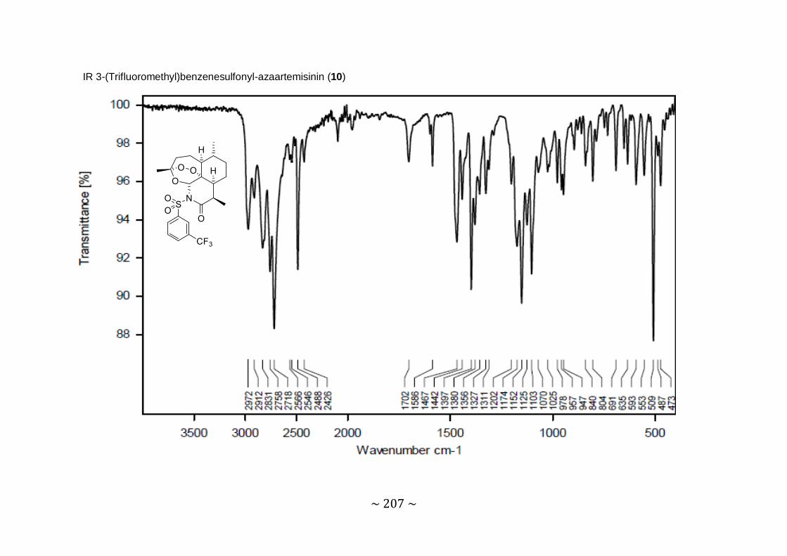
~ 207 ~
IR 3-(Trifluoromethyl)benzenesulfonyl-azaartemisinin (10)

~ 208 ~
1H NMR CDCl3 4-(Trifluoromethyl)benzenesulfonyl-azaartemisinin (11)
12
2’ & 6’ 3’ & 5’
CDCl3
9 4α
8a 5a
16 14
15
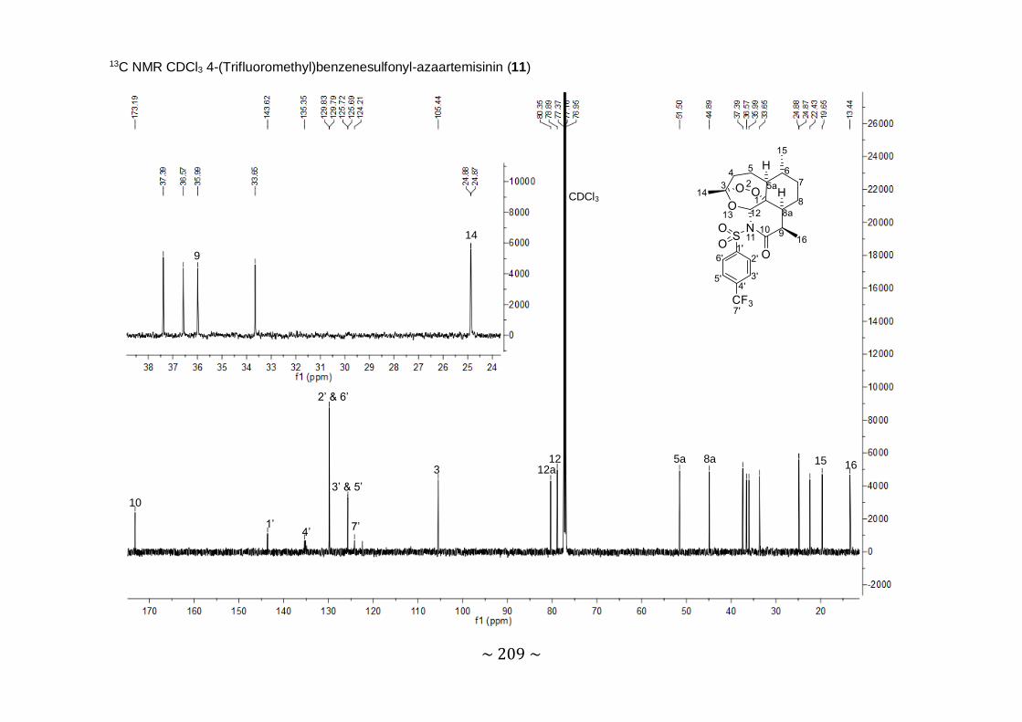
~ 209 ~
13C NMR CDCl3 4-(Trifluoromethyl)benzenesulfonyl-azaartemisinin (11)
10
1’ 4’
2’ & 6’
7’
3’ & 5’ 3 12a
12
CDCl3
5a 8a
9
14
15 16
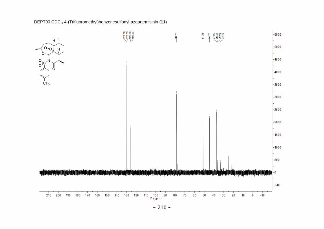
~ 210 ~
DEPT90 CDCl3 4-(Trifluoromethyl)benzenesulfonyl-azaartemisinin (11)
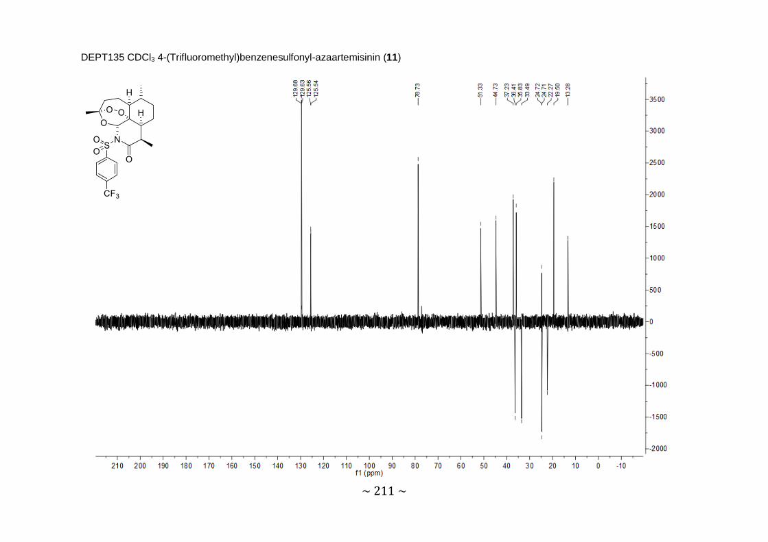
~ 211 ~
DEPT135 CDCl3 4-(Trifluoromethyl)benzenesulfonyl-azaartemisinin (11)

~ 212 ~
COSY CDCl3 4-(Trifluoromethyl)benzenesulfonyl-azaartemisinin (11)
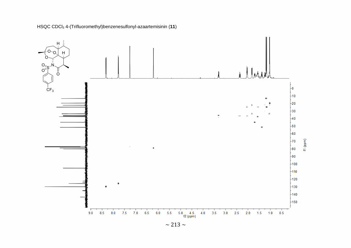
~ 213 ~
HSQC CDCl3 4-(Trifluoromethyl)benzenesulfonyl-azaartemisinin (11)

~ 214 ~
HRMS 4-(Trifluoromethyl)benzenesulfonyl-azaartemisinin (11)

~ 215 ~
IR 4-(Trifluoromethyl)benzenesulfonyl-azaartemisinin (11)

~ 216 ~
1H NMR CDCl3 4-Iodobenzenesulfonyl-azaartemisinin (12)
12
CDCl3
3’ &
5’
2’ &
6’
9 4α
8a H
2O
5a
14
16
15
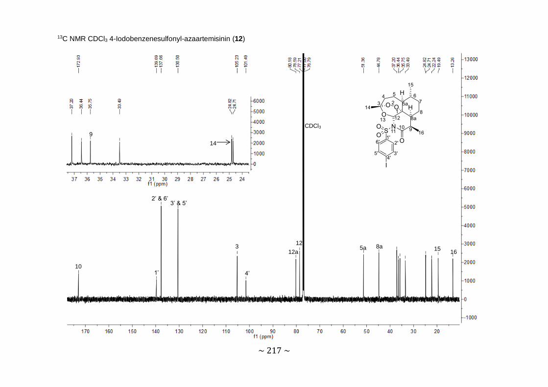
~ 217 ~
13C NMR CDCl3 4-Iodobenzenesulfonyl-azaartemisinin (12)
10 1’
2’ & 6’ 3’ & 5’
3
4’
12a 12
CDCl3
5a 8a
9 14
15 16

~ 218 ~
DEPT90 CDCl3 4-Iodobenzenesulfonyl-azaartemisinin (12)

~ 219 ~
DEPT135 CDCl3 4-Iodobenzenesulfonyl-azaartemisinin (12)

~ 220 ~
COSY CDCl3 4-Iodobenzenesulfonyl-azaartemisinin (12)

~ 221 ~
HSQC CDCl3 4-Iodobenzenesulfonyl-azaartemisinin (12)
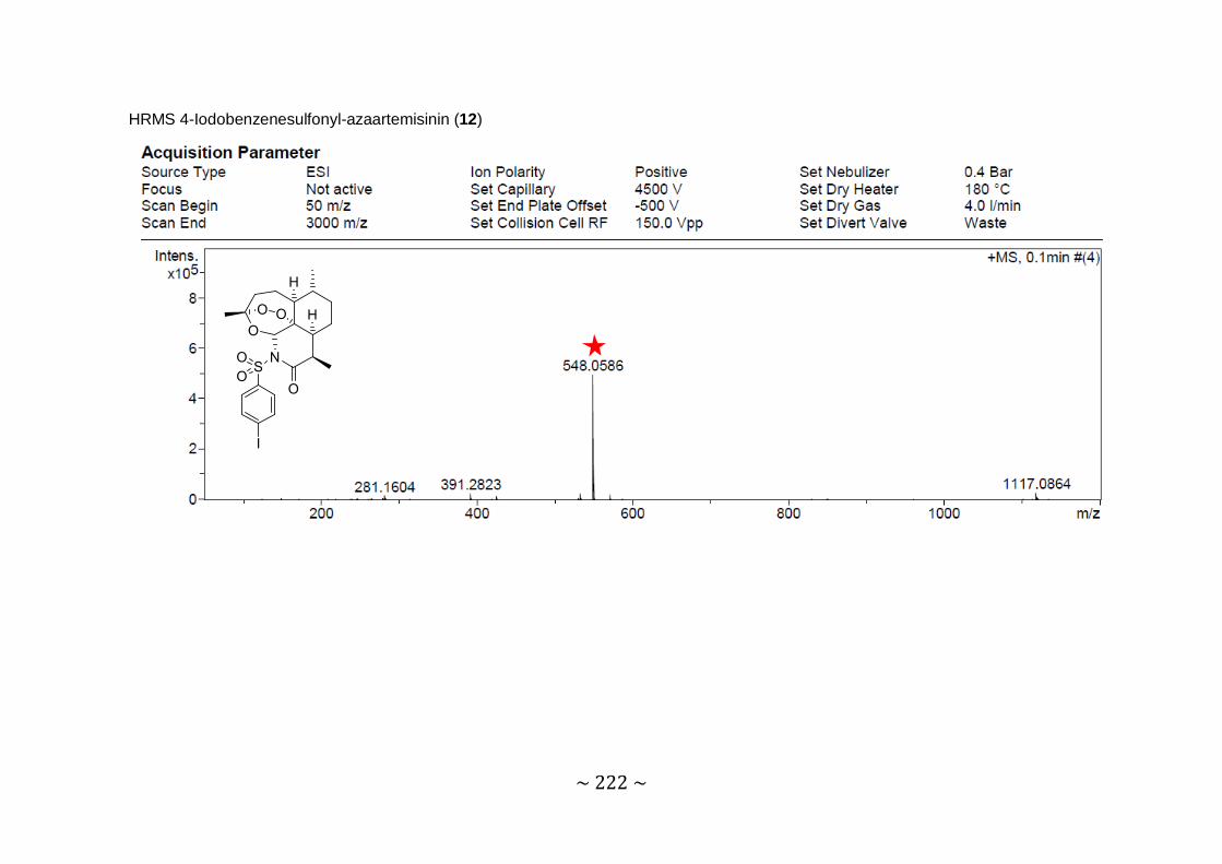
~ 222 ~
HRMS 4-Iodobenzenesulfonyl-azaartemisinin (12)

~ 223 ~
IR 4-Iodobenzenesulfonyl-azaartemisinin (12)

~ 224 ~
1H NMR CDCl3 4-Methoxybenzenesulfonyl-azaartemisinin (13)
12 2’ & 6’ CDCl3
3’ & 5’
8’
9 4α
8a
5a
14 16 15

~ 225 ~
13C NMR CDCl3 4-Methoxybenzenesulfonyl-azaartemisinin (13)
10 4’
1’, 2’ & 6’ 3’ & 5’
3 12a 12
CDCl3
8’ 5a
8a
9 14
15 16

~ 226 ~
DEPT90 CDCl3 4-Methoxybenzenesulfonyl-azaartemisinin (13)

~ 227 ~
DEPT135 CDCl3 4-Methoxybenzenesulfonyl-azaartemisinin (13)

~ 228 ~
COSY CDCl3 4-Methoxybenzenesulfonyl-azaartemisinin (13)
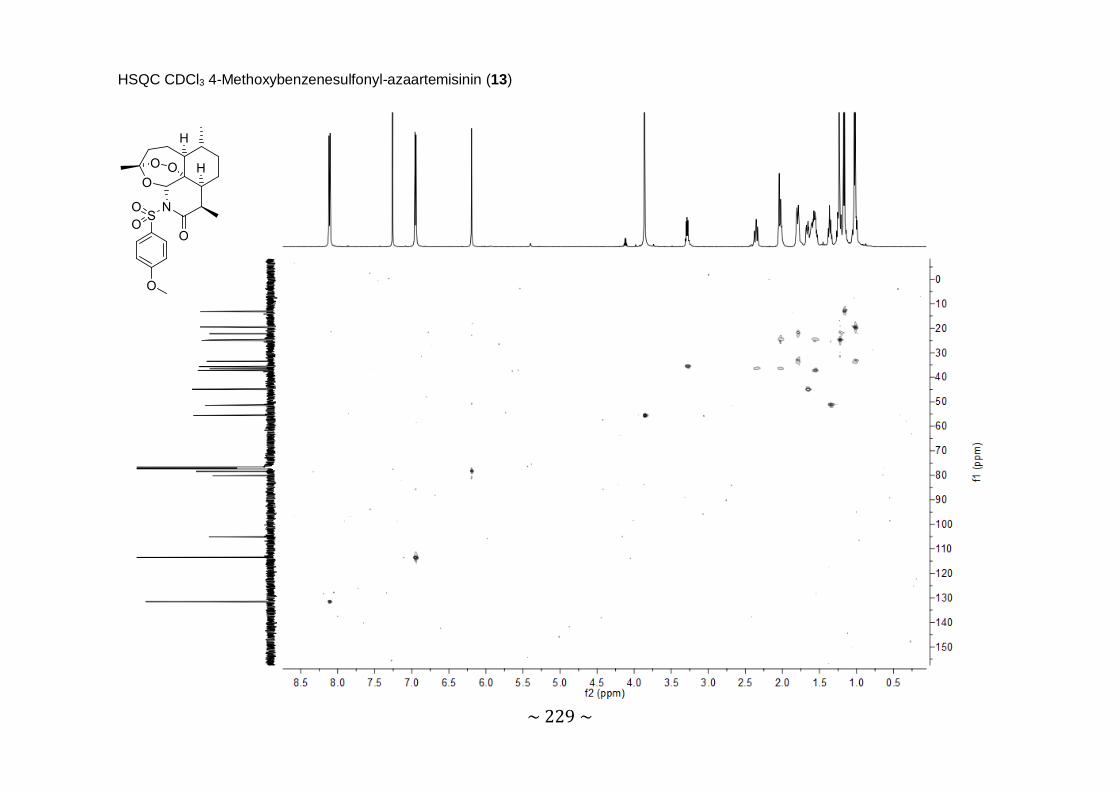
~ 229 ~
HSQC CDCl3 4-Methoxybenzenesulfonyl-azaartemisinin (13)
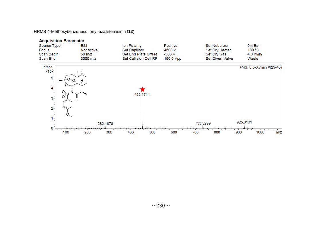
~ 230 ~
HRMS 4-Methoxybenzenesulfonyl-azaartemisinin (13)

~ 231 ~
HRMS 4-Methoxybenzenesulfonyl-azaartemisinin (13)

~ 232 ~
1H NMR CDCl3 4-Fluorobiphenyl-4-sulfonyl-azaartemisinin (14)
12 2’ & 6’ 3’ & 5’
8’ &
12’
CDCl3
9’ & 11’ MeOH
9 4α
8a
5a
14 16
15

~ 233 ~
13C NMR CDCl3 4-Fluorobiphenyl-4-sulfonyl-azaartemisinin (14)
10
10’
1’ 4’ 7’
2’ & 6’
3’ & 5’
9‘ & 11’ 3
8’ & 12’
12a 12
CDCl3
MeOH 5a
8a
9 14
15 16

~ 234 ~
DEPT90 CDCl3 4-Fluorobiphenyl-4-sulfonyl-azaartemisinin (14)

~ 235 ~
DEPT135 CDCl3 4-Fluorobiphenyl-4-sulfonyl-azaartemisinin (14)

~ 236 ~
COSY CDCl3 4-Fluorobiphenyl-4-sulfonyl-azaartemisinin (14)

~ 237 ~
HSQC CDCl3 4-Fluorobiphenyl-4-sulfonyl-azaartemisinin (14)

~ 238 ~
HRMS 4-Fluorobiphenyl-4-sulfonyl-azaartemisinin (14)

~ 239 ~
IR 4-Fluorobiphenyl-4-sulfonyl-azaartemisinin (14)

~ 240 ~
1H NMR CDCl3 Cyclopropanesulfonyl-azaartemisinin (15)
12
CDCl3
9 1’ 4α
8a
14 16
15

~ 241 ~
13C NMR CDCl3 Cyclopropanesulfonyl-azaartemisinin (15)
10
3
12a
12
CDCl3
5a
8a
9 14
15 16
3’
2’
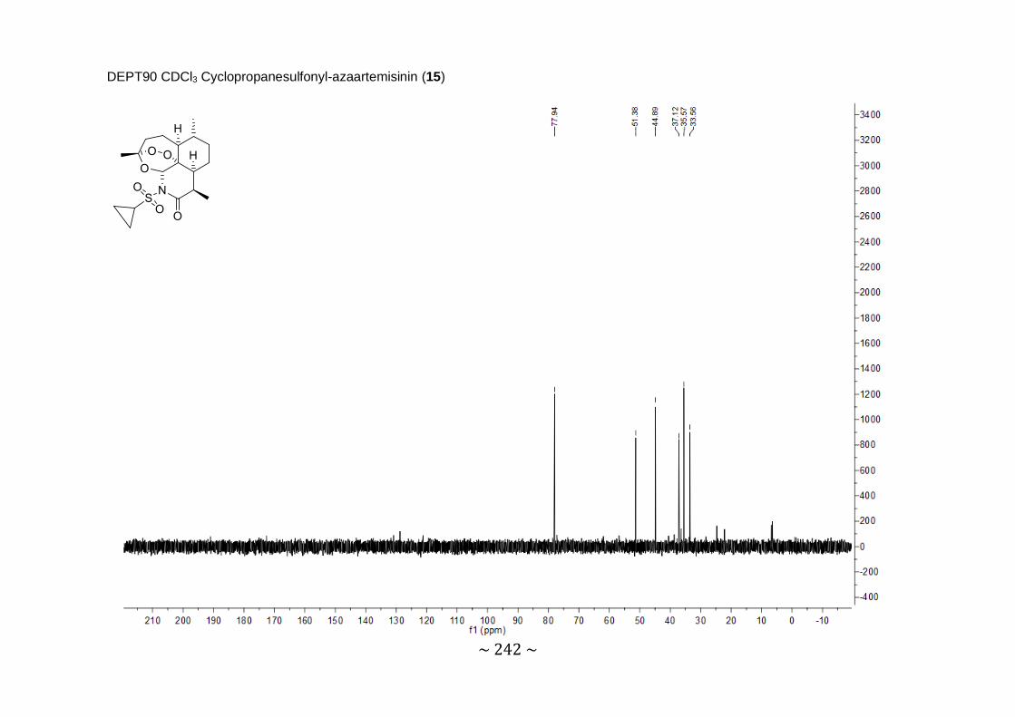
~ 242 ~
DEPT90 CDCl3 Cyclopropanesulfonyl-azaartemisinin (15)
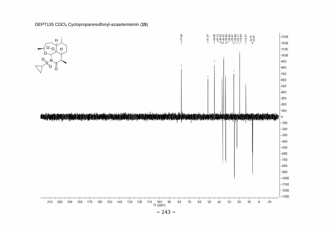
~ 243 ~
DEPT135 CDCl3 Cyclopropanesulfonyl-azaartemisinin (15)

~ 244 ~
COSY CDCl3 Cyclopropanesulfonyl-azaartemisinin (15)

~ 245 ~
HSQC CDCl3 Cyclopropanesulfonyl-azaartemisinin (15)

~ 246 ~
HRMS Cyclopropanesulfonyl-azaartemisinin (15)

~ 247 ~
IR Cyclopropanesulfonyl-azaartemisinin (15)

~ 248 ~
1H NMR CDCl3 2’-Thiophenesulfonylazaartemisinin (16)
12
2’ 4’ 3’
CDCl3
9 4α
8a 5a
16 14 15

~ 249 ~
13C NMR CDCl3 2’-Thiophenesulfonylazaartemisinin (16)
10 1’
2’ 4’
3’ 3 12a
12
CDCl3
5a 8a
9 5
14
15 16
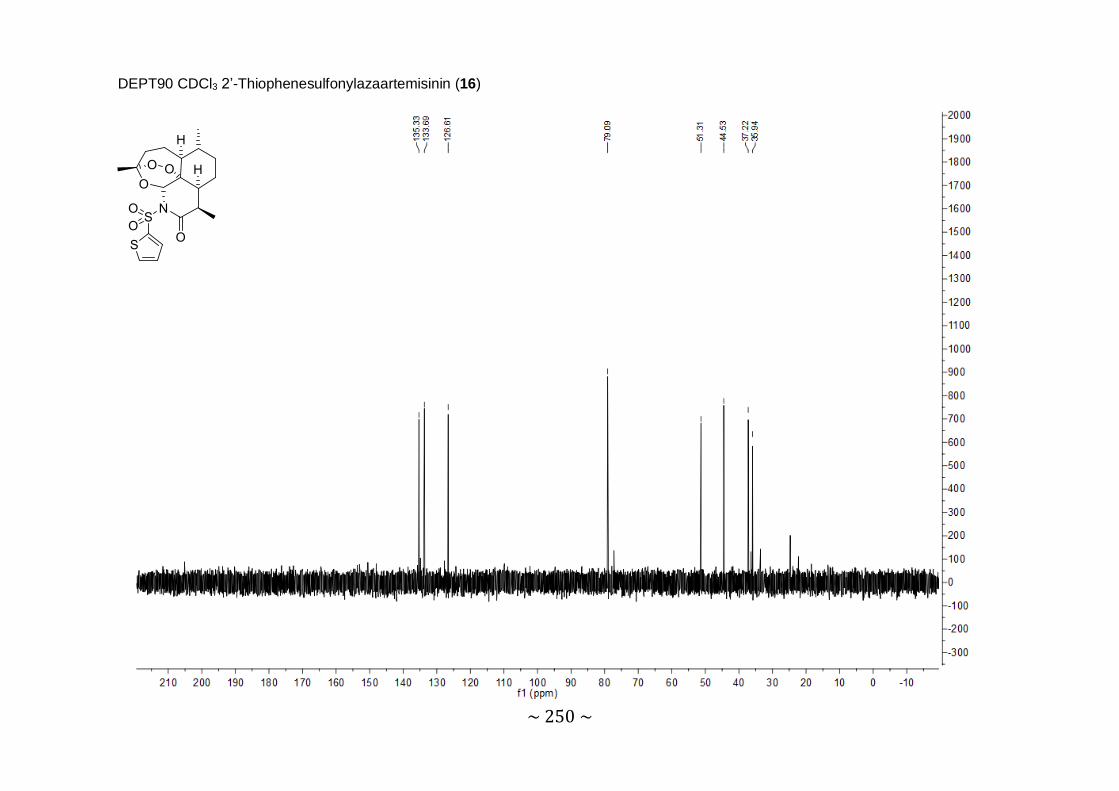
~ 250 ~
DEPT90 CDCl3 2’-Thiophenesulfonylazaartemisinin (16)
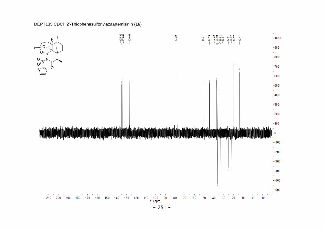
~ 251 ~
DEPT135 CDCl3 2’-Thiophenesulfonylazaartemisinin (16)

~ 252 ~
COSY CDCl3 2’-Thiophenesulfonylazaartemisinin (16)

~ 253 ~
HSQC CDCl3 2’-Thiophenesulfonylazaartemisinin (16)

~ 254 ~
HRMS 2’-Thiophenesulfonylazaartemisinin (16)

~ 255 ~
IR 2’-Thiophenesulfonylazaartemisinin (16)

~ 256 ~
1H NMR CDCl3 2,5-Dichlorothiophenesulfonyl-azaartemisinin (17)
2’
CDCl3
12
9 4α
EtOAc
8a 5a
14 16 15

~ 257 ~
14
5
9
3
3’ 1’ 10
2’
5’
16 15 8a
5a
CDCl3
13C NMR CDCl3 2,5-Dichlorothiophenesulfonyl-azaartemisinin (17)
12a
12

~ 258 ~
DEPT90 CDCl3 2,5-Dichlorothiophenesulfonyl-azaartemisinin (17)
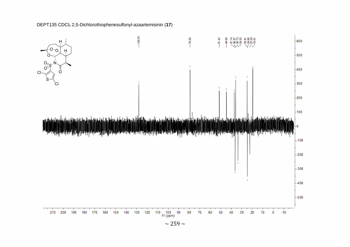
~ 259 ~
DEPT135 CDCl3 2,5-Dichlorothiophenesulfonyl-azaartemisinin (17)
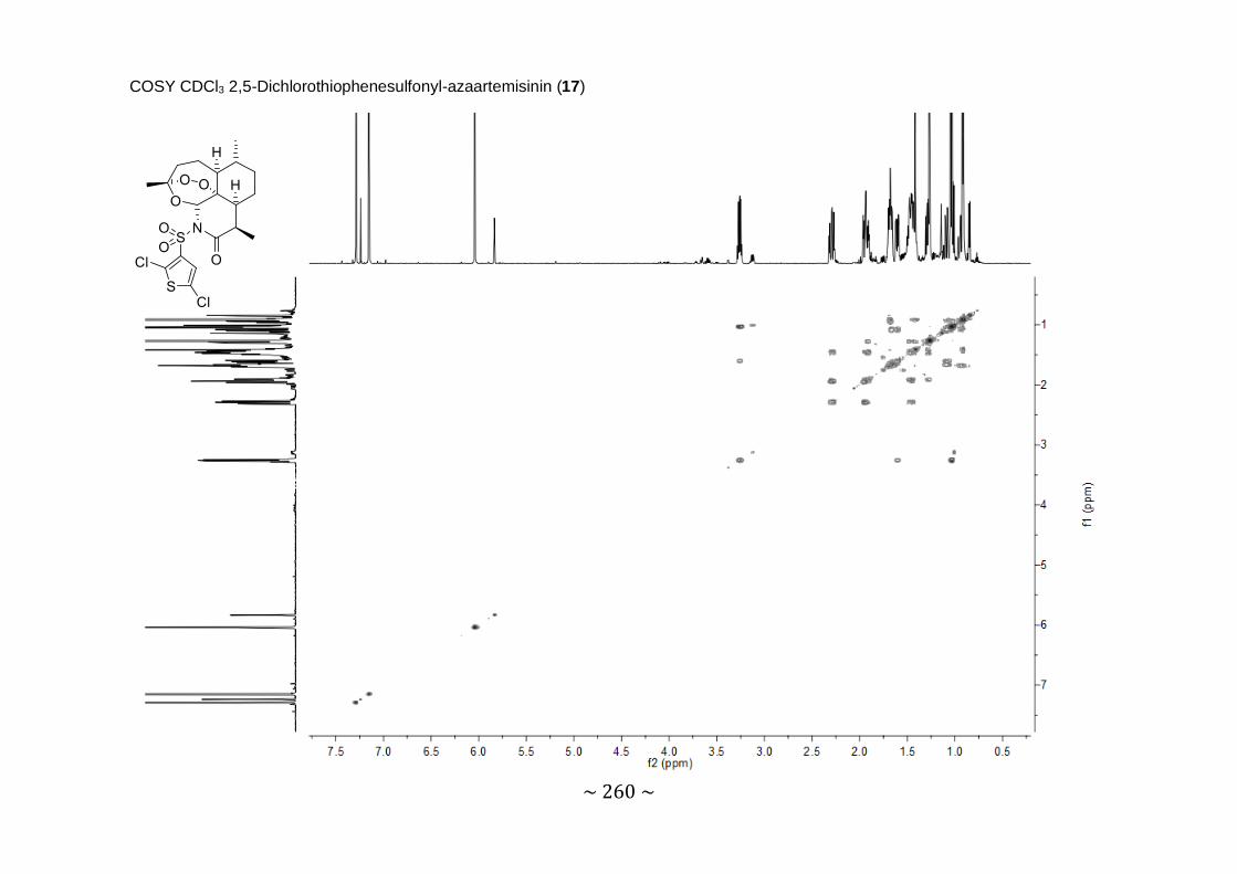
~ 260 ~
COSY CDCl3 2,5-Dichlorothiophenesulfonyl-azaartemisinin (17)

~ 261 ~
HSQC CDCl3 2,5-Dichlorothiophenesulfonyl-azaartemisinin (17)

~ 262 ~
HRMS 2,5-Dichlorothiophenesulfonyl-azaartemisinin (17)
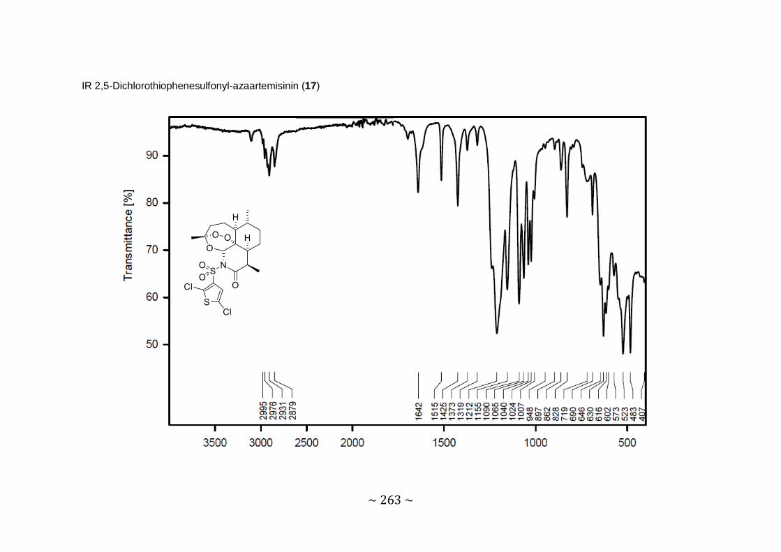
~ 263 ~
IR 2,5-Dichlorothiophenesulfonyl-azaartemisinin (17)
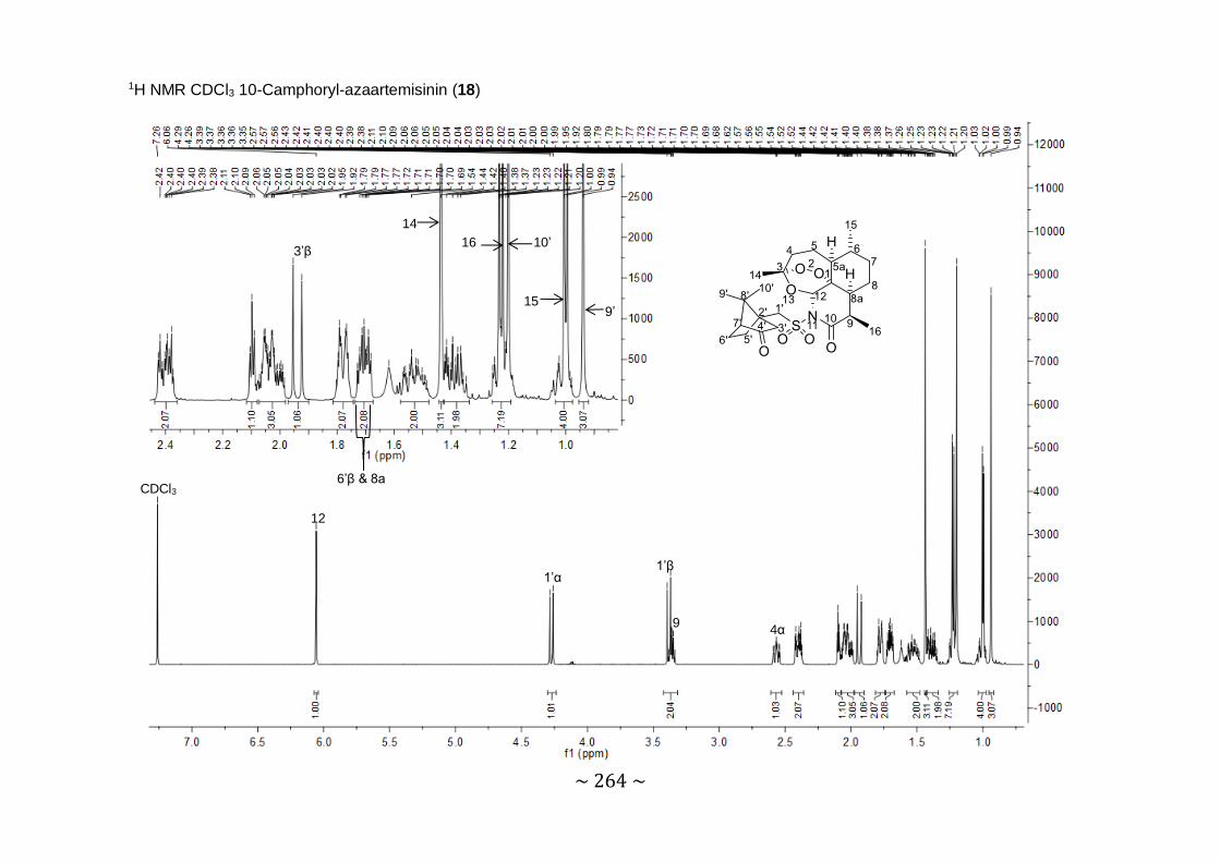
~ 264 ~
1H NMR CDCl3 10-Camphoryl-azaartemisinin (18)
12
CDCl3
1’α 1’β
9 4α
3’β
6’β & 8a
14 16 10’
15 9’
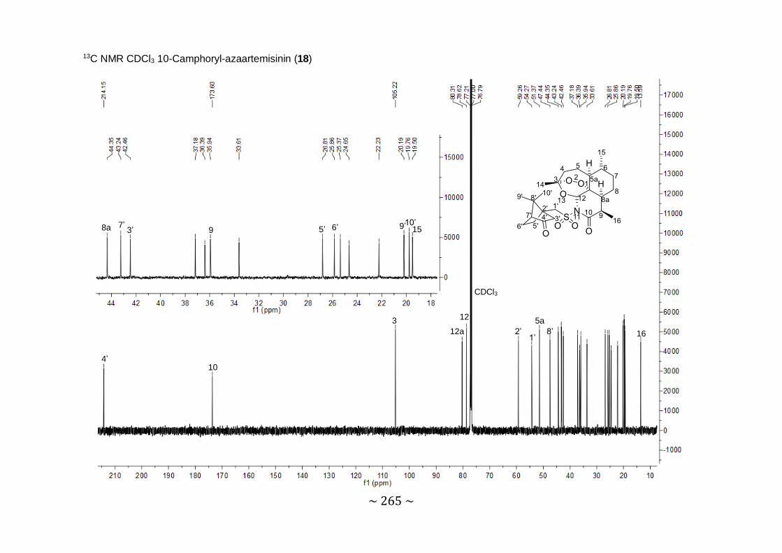
~ 265 ~
13C NMR CDCl3 10-Camphoryl-azaartemisinin (18)
12
4’ 10
3 12a
CDCl3
2’ 1’
5a 8’
8a 7’ 3’ 9 5’ 6’ 9’ 10’ 15
16

~ 266 ~
DEPT90 CDCl3 10-Camphoryl-azaartemisinin (18)

~ 267 ~
DEPT135 CDCl3 10-Camphoryl-azaartemisinin (18)

~ 268 ~
COSY CDCl3 10-Camphoryl-azaartemisinin (18)

~ 269 ~
HSQC CDCl3 10-Camphoryl-azaartemisinin (18)

~ 270 ~
HRMS 10-Camphoryl-azaartemisinin (18)
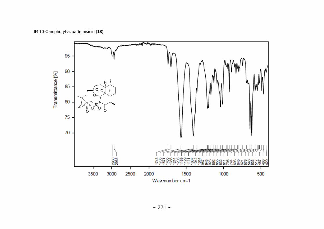
~ 271 ~
IR 10-Camphoryl-azaartemisinin (18)
Copyright © 2022 FDOKUMEN