Survival after chemotherapy and/or radiotherapy versus self-expanding metal stent insertion in the...
9
RESEARCH ARTICLE Open Access Survival after chemotherapy and/or radiotherapy versus self-expanding metal stent insertion in the setting of inoperable esophageal cancer: a case- control study George Sgourakis 1,2 , Ines Gockel 1,5* , Constantine Karaliotas 2 , Markus Moehler 3 , Carl Christoph Schimanski 3 , Heinz Schmidberger 4 and Theodor Junginger 1 Abstract Background: Our aim was to compare survival of the various treatment modality groups of chemotherapy and/or radiotherapy in relation to SEMS (self-expanding metal stents) in a retrospective case-control study. We have made the hypothesis that the administration of combined chemoradiotherapy improves survival in inoperable esophageal cancer patients. Methods: All patients were confirmed histologically as having surgically non- resectable esophageal carcinoma. Included were patients with squamous cell carcinoma, undifferentiated carcinoma as well as Siewert type I–but not type II - esophagogastric junctional adenocarcinoma. The decision to proceed with palliative treatments was taken within the context of a multidisciplinary team meeting and full expert review based on patient’s wish, co-morbid disease, clinical metastases, distant metastases, M1 nodal metastases, T4-tumor airway, aorta, main stem bronchi, cardiac invasion, and peritoneal disease. Patients not fit enough to tolerate a radical course of definitive chemo- and/or radiation therapy were referred for self-expanding metal stent insertion. Our approach to deal with potential confounders was to match subjects according to their clinical characteristics (contraindications for surgery) and tumor stage according to diagnostic work-up in four groups: SEMS group (A), Chemotherapy group (B), Radiotherapy group (C), and Chemoradiotherapy group (D). Results: Esophagectomy was contraindicated in 155 (35.5%) out of 437 patients presenting with esophageal cancer to the Department of General and Abdominal Surgery of the University Hospital of Mainz, Germany, between November 1997 and November 2007. There were 133 males and 22 females with a median age of 64.3 (43-88) years. Out of 155 patients, 123 were assigned to four groups: SEMS group (A) n = 26, Chemotherapy group (B) n = 12, Radiotherapy group (C) n = 23 and Chemoradiotherapy group (D) n = 62. Mean patient survival for the 4 groups was as follows: Group A: 6.92 ± 8.4 months; Group B: 7.75 ± 6.6 months; Group C: 8.56 ± 9.5 months, and Group D: 13.53 ± 14.7 months. Significant differences in overall survival were associated with tumor histology (P = 0.027), tumor localization (P = 0.019), and type of therapy (P = 0.005), respectively, in univariate analysis. Treatment modality (P = 0.043) was the only independent predictor of survival in multivariate analysis. The difference in overall survival between Group A and Group D was highly significant (P < 0.01) and in favor of Group D. As concerns Group D versus Group B and Group D versus Group C there was a trend towards a difference in overall survival in favor of Group D (P = 0.069 and P = 0.059, respectively). * Correspondence: [email protected] 1 Department of General and Abdominal Surgery, Johannes Gutenberg University-Hospital of Mainz, Mainz, Germany Full list of author information is available at the end of the article Sgourakis et al. BMC Cancer 2012, 12:70 http://www.biomedcentral.com/1471-2407/12/70 © 2012 Sgourakis et al; BioMed Central Ltd. This is an Open Access article distributed under the terms of the Creative Commons Attribution License (http://creativecommons.org/licenses/by/2.0), which permits unrestricted use, distribution, and reproduction in any medium, provided the original work is properly cited.
-
Upload
independent -
Category
Documents
-
view
0 -
download
0
Transcript of Survival after chemotherapy and/or radiotherapy versus self-expanding metal stent insertion in the...

RESEARCH ARTICLE Open Access
Survival after chemotherapy and/or radiotherapyversus self-expanding metal stent insertion in thesetting of inoperable esophageal cancer: a case-control studyGeorge Sgourakis1,2, Ines Gockel1,5*, Constantine Karaliotas2, Markus Moehler3, Carl Christoph Schimanski3,Heinz Schmidberger4 and Theodor Junginger1
Abstract
Background: Our aim was to compare survival of the various treatment modality groups of chemotherapy and/orradiotherapy in relation to SEMS (self-expanding metal stents) in a retrospective case-control study. We have madethe hypothesis that the administration of combined chemoradiotherapy improves survival in inoperableesophageal cancer patients.
Methods: All patients were confirmed histologically as having surgically non- resectable esophageal carcinoma.Included were patients with squamous cell carcinoma, undifferentiated carcinoma as well as Siewert type I–but nottype II - esophagogastric junctional adenocarcinoma. The decision to proceed with palliative treatments was takenwithin the context of a multidisciplinary team meeting and full expert review based on patient’s wish, co-morbiddisease, clinical metastases, distant metastases, M1 nodal metastases, T4-tumor airway, aorta, main stem bronchi,cardiac invasion, and peritoneal disease. Patients not fit enough to tolerate a radical course of definitive chemo-and/or radiation therapy were referred for self-expanding metal stent insertion. Our approach to deal withpotential confounders was to match subjects according to their clinical characteristics (contraindications forsurgery) and tumor stage according to diagnostic work-up in four groups: SEMS group (A), Chemotherapy group(B), Radiotherapy group (C), and Chemoradiotherapy group (D).
Results: Esophagectomy was contraindicated in 155 (35.5%) out of 437 patients presenting with esophagealcancer to the Department of General and Abdominal Surgery of the University Hospital of Mainz, Germany,between November 1997 and November 2007. There were 133 males and 22 females with a median age of 64.3(43-88) years. Out of 155 patients, 123 were assigned to four groups: SEMS group (A) n = 26, Chemotherapy group(B) n = 12, Radiotherapy group (C) n = 23 and Chemoradiotherapy group (D) n = 62. Mean patient survival for the4 groups was as follows: Group A: 6.92 ± 8.4 months; Group B: 7.75 ± 6.6 months; Group C: 8.56 ± 9.5 months, andGroup D: 13.53 ± 14.7 months. Significant differences in overall survival were associated with tumor histology (P =0.027), tumor localization (P = 0.019), and type of therapy (P = 0.005), respectively, in univariate analysis. Treatmentmodality (P = 0.043) was the only independent predictor of survival in multivariate analysis. The difference inoverall survival between Group A and Group D was highly significant (P < 0.01) and in favor of Group D. Asconcerns Group D versus Group B and Group D versus Group C there was a trend towards a difference in overallsurvival in favor of Group D (P = 0.069 and P = 0.059, respectively).
* Correspondence: [email protected] of General and Abdominal Surgery, Johannes GutenbergUniversity-Hospital of Mainz, Mainz, GermanyFull list of author information is available at the end of the article
Sgourakis et al. BMC Cancer 2012, 12:70http://www.biomedcentral.com/1471-2407/12/70
© 2012 Sgourakis et al; BioMed Central Ltd. This is an Open Access article distributed under the terms of the Creative CommonsAttribution License (http://creativecommons.org/licenses/by/2.0), which permits unrestricted use, distribution, and reproduction inany medium, provided the original work is properly cited.

Conclusions: The prognosis of inoperable esophageal cancer seems to be highly dependent on the suitability ofthe induction of patient-specific therapeutic measures and is significantly better, when chemoradiotherapy isapplied.
BackgroundAccurate information regarding the proportion ofpatients with esophageal cancer in whom surgery is con-traindicated is difficult to obtain. This largely reflectsvariations in the selection of patients for palliative treat-ment modalities. In the Western world, more than halfthe patients with esophageal cancer are not amenable tosurgery as they usually present with severe comorbidityand an advanced stage of disease [1].The choice of treatment must be tailored to the indi-
vidual and will depend on the location and stage of thetumor, as well as the overall health of the patient.Four RCT’s [2-5] and one meta-analysis [6] compared
brachytherapy, laser ablation therapy and argon beamcoagulation (APC) therapy with self-expanding metalstents within the context of esophageal cancer palliation.The aforementioned studies present symptomaticpatient relief as the primary outcome and patient survi-val as the secondary. Only one of the studies [3] pro-vides data for external beam radiation therapy, butpatients are collectively analyzed with those who under-went APC.It has also been suggested that combination chemora-
diotherapy may improve response rates and thus survi-val, although evidence is limited [7]. A study providing astraightforward comparison between chemotherapy and/or radiotherapy and SEMS is lacking.We have made the hypothesis that the administration
of combined chemoradiotherapy improves survival ininoperable esophageal cancer patients. Our aim was tospecify survival of the various treatment modalities inrelation to SEMS in a retrospective case-control study.
MethodsFrom November 1997 to November 2007, a total of 437patients presented to our institution with histologicallyproven esophageal carcinoma. Esophagectomy was con-traindicated in 155 (35.5%) patients (133 males, 22females) with a median age of 64.3 (43-88) years. Thisrepresents a group of individuals for whom a minimumof 4 years of follow-up data was possible.Reasons of incurability were distant metastases (n =
54; 34.8%), local tumor spread (n = 58; 37.4%) and pre-existent cardiopulmonary diseases (n = 26; 16.8%).Seventeen (11%) patients presented further reasons ofincurability. Of these, 5 patients refused surgery, and 5were excluded from surgery as they did not have anadequate substitute organ for reconstruction and
esophagoplasty. In 7 patients, the risk of esophagectomywas considered too high due to their poor generalhealth status.After histological confirmation, all patients underwent
a preoperative diagnostic work-up, in addition to com-puted tomography of the neck, thorax, and abdomen,which included endosonography of the esophagus, bar-ium swallow, transabdominal sonography of the abdo-men, as well as positron emission tomography (PET), aspreviously described [8]. A conventional X-ray examina-tion of the thorax and laboratory tests with the tumormarkers carcinoembryonic antigen (CEA), Ca 19-9, Ca72-4, and Alpha-Feto-Protein (AFP) were routinelyperformed.
Inclusion and exclusion criteriaAll patients were confirmed histologically as having eso-phageal carcinoma. Included were patients with squa-mous cell carcinoma (SCC), undifferentiated carcinoma(UDC), as well as Siewert type I–but not type II–eso-phagogastric junctional adenocarcinoma (ADC) [9].The decision to proceed with palliative treatments was
taken within the context of a multidisciplinary teammeeting and full expert review based on patient’s wish,co-morbid disease, distant metastases, M1 nodal metas-tases, and T4 tumor.Hematogenic and lymphogenic metastases were docu-
mented by computed tomography (CT) examination,PET scan and endosonographic ultrasound. In summary,a CT diagnosis of T4-tumor stage made an R0 resectionimpossible.Standard indicators defining a patient as medically
inoperable included baseline forced expiratory volumein the first second of expiration (FEV1) of less than 40%predicted, carbon monoxide diffusing capacity of lessthan 40% predicted, baseline hypoxemia or hypercapnia,severe pulmonary hypertension; diabetes mellitus withend-organ damage; severe cerebral, cardiovascular, orperipheral vascular disease; or severe, chronic heartdisease.
Multidisciplinary team strategyPatients not fit enough to tolerate a radical course ofdefinitive chemotherapy and/or radiation, or those whoneeded rapid relief of their dysphagia (swallowing maydeteriorate because of radiation induced edema andswelling of the tumor) were referred for self-expandingmetal stent (SEMS) insertion. Radiochemotherapy was
Sgourakis et al. BMC Cancer 2012, 12:70http://www.biomedcentral.com/1471-2407/12/70
Page 2 of 9
carried out according to the Herskovic protocol (fourcourses of combined fluorouracil and cisplatin plus5,000 cGy of radiation therapy) [10]. In brief, patientswithout distant metastases underwent external beamradiotherapy with a dose of 50-60 Gy and, wheneverpossible, an additional brachytherapy of up to 68 Gy. Inselected patients with previous cancer and consecutiveradiotherapy (e.g. oropharyngeal, laryngeal cancer, etc.),these doses had to be individually adapted. Patientsreceiving chemotherapy alone were given a combinationof fluorouracil and cisplatin, providing no contraindica-tions were present. Patient groups were not randomized.Locally inoperable squamous cell carcinoma was trea-
ted by chemoradiotherapy, radiation therapy alone, orpalliative chemotherapy in cases of distant metastases,whereas advanced or metastasized adenocarcinoma wastreated by chemotherapy or chemoradiotherapy.
Control for confounding variablesData of patients with esophageal carcinoma undergoingthe four main treatment modalities (SEMS, chemother-apy and/or radiotherapy) were matched according toclinical characteristics (contraindications for surgery)and tumor stage by diagnostic work-up.The confounding variables were chosen according to
the Standardization or Adjustment method: we appliedthe cumulative proportion surviving rates generated ineach of our four strata (stent, chemotherapy, radiother-apy, chemoradiotherapy) to the same “standard” theore-tical population. This standard population was createdso that the frequency of the confounder is identicalbetween each group of patients; this was achieved byapplying sequential filters to the Microsoft Exceldatabase.According to their treatment modality, patients were
assigned to four groups: SEMS group (A), Chemother-apy group (B), Radiotherapy group (C), and Chemora-diotherapy group (D).According to the study hypothesis, the control group
included patients in whom a self expandable stent hadbeen inserted.
DefinitionsIn patients with histologically proven esophageal carci-noma, the following prognostic variables wererecorded, American Society of Anesthesiologists (ASA)classification (I-IV) according to the anesthesiologyevaluation, body mass index (BMI) based on bodyweight and height in kg/m2, and the nutritional statusincluding tobacco and/or alcohol abuse. Tobaccoabuse was defined by the consumption of at least 5cigarettes a day over a period of > 1 year, whereasalcohol abuse was defined by the regular intake ofbeer, wine or hard drinks, at least every second day.
Among the comorbidities, cardiovascular risk factorswere defined as a history of coronary heart disease, ormyocardial infarction, arterial hypertension, valvulardisease (> II°), arrhythmia requiring therapy (> III°according to the Lown classification), heart failureNYHA (New York Heart Association) > grade II, andperipheral occlusive arterial disease (> IIb accordingto Fontaine). A history of chronic obstructive pulmon-ary disease (COPD), regular tobacco consumption,and/or the use of bronchospasmolytics were subsumedunder pulmonary diseases. The preoperative evalua-tion of the vital capacity (VC) and the forced expira-tory volume in 1 s (FEV1 = Tiffeneau test) served toensure a more accurate assessment. Preexisting cirrho-sis of the liver (≥CHILD-Pugh A) was defined as hepa-tic disease, and determined on the basis of themeasurement of serum albumin (g/dl), serum bilirubin(mg/dl), Quick value (%), and the presence of ascitesor encephalopathy. The evaluation of additional riskfactors included the prevalence of diabetes mellitus(insulin-dependent or requiring drug therapy), and thehistory of a secondary carcinoma.For a better comparison of staging procedures, the
esophagus was considered in thirds, according to theendoscopic location of the tumor: upper third: dentalfront to 20 cm; middle third: 20-30 cm; lower third: 30cm to the Z-line.The various study parameters were coded as follows:
Procedures: 1 = explorative laparotomy/laparoscopy, butno resection; 2 = none; 3 = primarily neoadjuvant inten-tion; 4 = surgical exploration, but no resection; 5 =explorative thoracotomy/thoracoscopy, but no resection.Tumor histology: 1 = squamous cell cancer; 2 = adeno-carcinoma; 3 = undifferentiated carcinoma. Contraindi-cation for surgery: 1 = metastases; 2 = local tumorspread; 3 = cardiopulmonary; 4 = pulmonary; 5 = car-diac; 6 = esophageal substitute not available; 7 = tumorspread and poor general condition; 8 = tumor spreadand cardial disease; 9 = patient refuses surgery; 10 =mucosal resection; 11 = metastases and poor generalcondition; 12 = patient refuses surgery and poor generalcondition. Reasons for non-surgical management: 1 =metastases; 2 = locally not resectable; 3 = cardiopulmon-ary contraindication; 4 = other. Metastasis site: 1 =liver; 2 = lung; 3 = lung + liver; 4 = M1 lymph nodes; 5= diffuse; 6 = peritoneal carcinosis; 7 = bone; 8 = brain;9 = skin. Type of metastases: 1 = hematogeneous; 2 =lymphatic. Treatment modality: 1 = stent; 2 = che-motherapy; 3 = radiation; 4 = radio-chemotherapy; 5 =tracheostomy; 6 = mucosectomy. For all clinical charac-teristics, the presence of the variables mentioned wasdeclared 1, and the absence 0.Approval for the study was obtained from the hospital
ethics committee.
Sgourakis et al. BMC Cancer 2012, 12:70http://www.biomedcentral.com/1471-2407/12/70
Page 3 of 9

Statistical analysisIn order to validate our patient selection with confound-ing variables, two different statistical tests were applied:1) Kruskal-Wallis ANOVA by Ranks test (Median test)was used in order to compare the clinico-pathologicalparameters (numerical) of patients among the variousgroups of therapy and compute post-hoc comparisonsof mean ranks of all pairs attributed to by groups and 2)Spearman’s correlations were also used to analyze thedistribution of the clinico-pathological parameters(coded in categories) separately in each group oftherapy.A multivariate analysis was performed under Cox’s
Proportional Hazard Model considering all factors thatgained statistical significance (P < 0.05) in univariateanalysis under the same model. Variables that reachedsignificance in the multivariate model were consideredas predictors of survival. In order to compare survivalbetween two samples, the Cox’s F-test was employed. Soas to validate our results in terms of cumulative propor-tion surviving, sample size calculation was performedwith a type I error rate (Alpha) 0.05 and power goal0.80 by applying the two-tailed Log-Rank test.Significance was considered at a level < 0.05. Statistical
release 7 (Statsoft, Tulsa, USA) was used for statisticalanalysis.
ResultsIn relation to all patients presenting with esophagealcancer from November 1997 to November 2007, theproportion of inoperable patients was higher in SCC’s(23.8%; n = 104/437) as compared to ADC’s (10.5%; n =46/437) (in addition 5 patients with UDC). Both tumorentities revealed different reasons of inoperability: Inpatients with SCC, local tumor spread predominatedwith 45.7%, whereas in the majority of patients withADC (58.7%), inoperability was due to hematogeneousmetastases. Cardiopulmonary diseases causing contrain-dication for surgery were equally distributed amongboth tumor entities (17.1% in patients with SCC and17.4% in patients with ADC).The course of the disease was ascertained in 152 out
of 155 (98%) patients by December 31st 2010; no datadocumenting the course were available in 3 patients atthat time. One hundred and forty-eight patients haddied from their malignancy; of the remaining 7 patients,4 were alive and 3 were lost from follow-up. All 4patients alive at the time of last follow-up had initiallypresented with a locally inoperable SCC and had under-gone radiochemotherapy.Six patients who underwent mucosectomy (n = 2) and
tracheostomy (n = 1) or were lost from follow-up (n =3) were not included in the analysis. The respectivenumbers of patients for SCC, ADC and UDC were 104,
46 and 5. According to the diagnostic work-up, tumorstage was IIA in 17 patients, IIB/III in 76 patients andIV in 54 patients. Contraindications for surgery werecardiopulmonary status in 26 patients, metastasis andtumor spread in 119, surgery refusal by 5, and nochance of esophageal substitution in five patients.Brachytherapy was administered to 2 patients in group
C and to 16 patients in group D. Seven patients ingroup B, 10 in group C, and 37 patients in group Dcompleted their allocated treatment modality.UDC patients were excluded since there were missing
data in 3 (tumor stage), while the remaining 2 patientswere excluded during the selection process.In applying the sequential filters for confounding vari-
ables, only 123 out of 155 patients were considered forthe analysis. We took care to include only patients with-out missing data, in terms of contraindications for sur-gery and TNM-stage by applying sequential filters to theMicrosoft Excel database (Figure 1). Contraindicationsfor surgery were divided into 3 categories: a) cardiopul-monary, b) metastasis/non-resectable and c) other.Patients were considered accordingly by an analogy 1/5/1 (paralleling the rates among the 155 patients) in eachof the 3 patient classes of the TNM-stage (IIA, IIB/III,IV) which were considered respectively by an analogy of2/9/7 (paralleling the rates among the 155 patients) (Fig-ure 1). These sequential steps reduced the total of 155patients by 8 patients (missing data), 19 patients by“TNM-stage” filtering, 2 patients by “contraindicationsfor surgery” filtering and 3 patients lost to follow up(Figure 1). The remaining 123 patients were assigned tofour groups: SEMS group (A) n = 26 (SCC:10/ADC:16),Chemotherapy group (B) n = 12 (SCC:10/ADC:2),Radiotherapy group (C) n = 23 (SCC:11/ADC:12), Che-moradiotherapy group (D) n = 62 (SCC:52/ADC:10).For every patient in group B, approximately 2 patientsin groups A and C, and 5 in group D were analyzed interms of survival.As concerns the allocation of clinico-pathological
characteristics among the 4 groups, all 21 parameterswere equally distributed in terms of age, tumor localiza-tion, type of metastasis, histology, and cardiopulmonarystatus (Table 1).Overall median and mean patient survival was 6
months and 10.89 ± 12.63 months, respectively. Therespective numbers for the 4 groups were as follows:Group A: 3/6.92 ± 8.4 months; Group B: 7/7.75 ± 6.6months; Group C: 4/8.56 ± 9.5 months and Group D: 8/13.53 ± 14.7 months. Overall survival for the 4 groups isdepicted in Table 2.Twenty-one variables were analyzed in univariate analy-
sis of survival: gender, age, BMI, vital capacity (VC), FEV1,cTNM-classification, ASA-classification, nutritional status,tobacco consumption, alcohol intake, diabetes, Child-Pugh
Sgourakis et al. BMC Cancer 2012, 12:70http://www.biomedcentral.com/1471-2407/12/70
Page 4 of 9

score, cardiopulmonary status, existence of a second carci-noma, histopathology, tumor localization, contraindicationfor surgery, cause of inoperability, site of metastasis andtype of metastasis. Significant differences in overall survi-val were associated with tumor histology (P = 0.027),tumor localization (P = 0.019), and type of therapy (P =0.005), respectively (Table 3).In multivariate Cox’s Proportional Hazard regression
analysis of survival, the model including the predictorsin univariate analysis gained statistical significance (P <0.001), but treatment modality (P = 0.043) was the onlyindependent predictor of survival (Table 4).With regard to the 12-month hazard rate/standard
error of hazard rate for the 4 treatment groups: GroupA = 0.14/0.07, Group B = 0.16/0.10, Group C = 0.15/0.06, and Group D = 0.04/0.01.The difference in overall survival between Group A
and Group D was highly significant (P < 0.01), and infavor of Group D. Comparing Group D versus Group Band Group D versus Group C, the difference in overallsurvival was marginally significant, and in favor ofGroup D (P = 0.069 and P = 0.059, respectively). Theother possible comparisons in overall survival betweenGroups did not reach statistical significance (Figure 2).
Aiming to establish a 2-year difference in overall sur-vival of 24.3% vs. 3.8% for groups D and A respectively,there was a need for 26 patients per treatment group.The respective number for group D vs. C was 23patients (24.3% vs. 0%), and for group D vs. B, it was 61patients (24.3% vs. 8.6%). All the aforementioned com-parisons of overall survival between groups fulfilled thesample size calculation, with the exception of one(group D: 62 patients vs. group B: 12 patients).In order to investigate the rationale of our multidisci-
plinary team strategy to assign more adenocarcinomapatients to chemotherapy alone (n = 12) than to che-moradiotherapy (n = 9), survival curves of these twogroups were compared. No statistical significant differ-ence in overall survival was observed (P = 0.589). Thesame applied to squamous cell carcinoma patientsundergoing chemoradiotherapy (n = 55) versus radio-therapy alone (n = 10) (P = 0.405).
DiscussionIn the setting of care in inoperable esophageal cancer,the combination of chemoradiotherapy was more effica-cious than any other treatment modality, and undoubt-edly superior to the use of SEMS in terms of patient
Figure 1 Only patients without missing data (in terms of the confounding variables) were included by applying sequential filters tothe Microsoft Excel database. Contraindications for surgery were divided into 3 categories: a) cardiopulmonary (CP), b) metastasis/non-resectable (M/NR) and c) other. Patients were considered accordingly by an analogy 1/5/1 (paralleling the rates among the 155 patients) in eachof the 3 patient classes of the TNM-stage (IIA, IIB/III, IV) which were considered respectively by an analogy of 2/9/7 (paralleling the rates amongthe 155 patients). These sequential steps reduced the total 155 patients by 8 patients (missing data), 19 patients by “TNM-stage” filtering, 2patients by “contraindications for surgery” filtering and by 3 patients lost to follow up. The remaining 123 patients were assigned to four groups.
Sgourakis et al. BMC Cancer 2012, 12:70http://www.biomedcentral.com/1471-2407/12/70
Page 5 of 9

survival. Only chemoradiation patient Group D had 4-and 5-year survivors.Though the clinico-pathological characteristics among
patients of the 4 groups were not significantly different,there were distinct differences observed in some. This ismainly attributed to the multidisciplinary team strategy:1) Patients not fit enough to tolerate a radical course of
definitive chemotherapy and/or radiation were referredfor self-expanding metal stent insertion, and 2) Patientswith locally inoperable squamous cell carcinoma weretreated by chemoradiotherapy, radiation therapy aloneor chemotherapy in cases of distant metastases, whereaspatients with advanced adenocarcinoma were treated bychemotherapy or chemoradiotherapy. Despite this
Table 1 Comparison of clinico-pathological characteristics among the 4 groups of treatment modality
Variable (continuous) Kruskal-Wallis ANOVA by Ranks test(Median test) Mean ± SD
P-value
Age A SEMSB ChemoC RadiationD C/R
69.57 ± 10.08 P = 0.017 (SEMS vs. C/R*P = 0.013)
67.00 ± 4.30
66.44 ± 8.57
61.05 ± 9.74
BMI 24.87 ± 4.27 P = 0.466
25.91 ± 5.65
25.70 ± 4.00
24.81 ± 5.11
Vital capacity (VC) 3.54 ± 1.08 P = 0.287
3.75 ± 0.93
2.75 ± 1.28
3.78 ± 0.95
FEV1 32.53 ± 33.53 P = 0.306
41.29 ± 33.42
38.86 ± 30.13
46.98 ± 28.11
Variable (categorical) Distribution of variable categories amonggroups of therapy
Spearman Rank Order Correlations P-value
cTNM-classification -0.124360 0.165
ASA-classification -0.017661 0.876
Nutrition 0.166002 0.244
Smoking 0.080980 0.387
Alcohol 0.082105 0.380
Child-Pugh score 0.033203 0.724
Pulmonary status 0.226517 0.013
Cardiovascular status -0.226854 0.013
Diabetes -0.043337 0.642
Secondary carcinoma 0.095611 0.286
Gender -0.146058 0.095
Histology -0.348704 0.001
Localization -0.259496 0.002
Contraindications forsurgery
0.032170 0.715
Causes ofinoperability
0.128822 0.142
Site of metastases -0.060909 0.566
Type of metastases 0.224707 0.032
SD: Standard deviation; SEMS: Self-expandable metallic stents; C/R*: Chemo-Radiotherapy; BMI: body mass index; ASA: American Society of Anesthesiologistsclassification; FEV1: Forced expiratory volume in 1 s
Sgourakis et al. BMC Cancer 2012, 12:70http://www.biomedcentral.com/1471-2407/12/70
Page 6 of 9
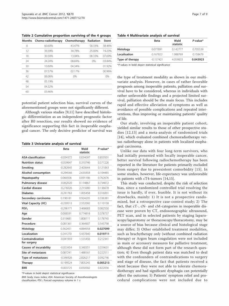
potential patient selection bias, survival curves of theaforementioned groups were not significantly different.Although various studies [9,11] have described histolo-
gic differentiation as an independent prognostic factorafter R0 resection, our results showed no evidence ofsignificance supporting this fact in inoperable esopha-geal cancer. The only decisive predictor of survival was
the type of treatment modality as shown in our multi-variate analysis. However, in cases of rather favorableprognosis among inoperable patients, palliation and sur-vival have to be considered, whereas in individuals withrather unfavorable findings and a projected limited sur-vival, palliation should be the main focus. This includesrapid and effective alleviation of symptoms as well asavoidance of possible complications and repeated inter-ventions, thus improving or maintaining patients’ qualityof life.Our study, involving an inoperable patient cohort,
yielded similar results to those of other prospective stu-dies [12,13] and a meta-analysis of randomized trials[14], which evaluated combined chemoradiotherapy ver-sus radiotherapy alone in patients with localized esopha-geal carcinoma.Unlike our data with four long-term survivors, who
had initially presented with locally inoperable cancer,better survival following radiochemotherapy has beenreported in the literature for patients primarily excludedfrom surgery due to preexistent comorbidity [15]. Insome studies, however, life-expectancy was unfavorablein patients with cT4-tumors [14,15].This study was conducted, despite the risk of selection
bias, since a randomized controlled trial resolving theissue is hardly, if ever, feasible. It is not without itsdrawbacks, mainly: 1) It is not a prospective rando-mized, but a retrospective case-control study; 2) Thefact, that cT-, cN- and cM-categories in inoperable dis-ease were proven by CT, endosonographic ultrasound,PET scan, and in selected patients by staging laparo-scopy/laparotomy or thoracoscopy/thoracotomy, may bea source of bias because clinical and histological stagemay differ; 3) Other established treatment modalities,such as brachytherapy only (without combined radiationtherapy) or Argon beam coagulation were not includedas main or accessory measures for palliative treatment,although these did not form part of the research ques-tion; 4) Even though patient data was matched to dealwith the confounders of contraindications to surgeryand stage of disease, the fact that patients received astent because they were not able to tolerate chemora-diotherapy and had significant dysphagia can potentiallyaffect the outcome; 5) Patients’ symptom relief and pro-cedural complications were not included due to
Table 2 Cumulative proportion surviving of the 4 groups
Months Chemo-radiotherapy Chemotherapy Radiation Stent
6 60.60% 43.47% 58.33% 38.46%
12 39.39% 34.78% 25.00% 19.23%
18 30.30% 13.04% 08.33% 07.69%
24 24.24% 08.69% 0% 03.84%
30 10.60% 04.34% 01.92%
36 07.57% 02.17% 00.96%
42 06.06% 0% 0%
48 05.19%
54 04.32%
60 03.46%
Table 3 Univariate analysis of survival
Beta Waldstatistic
P-value*
ASA-classification -0.034372 0.034097 0.853501
Nutrition status -0.059647 0.310746 0.577226
Smoking -0.282645 2.403644 0.121063
Alcohol consumption -0.294566 2.635858 0.104485
Hepatopathy 0.066506 0.091186 0.762676
Pulmonary disease -0.062020 0.114867 0.734672
Cardial disease -0.276026 2.215080 0.136678
Diabetes -0.241763 1.005458 0.316001
Secondary carcinoma 0.196181 0.924205 0.336381
Vital Capacity (VC) -0.239513 2.552092 0.110158
FEV1 -0.296171 3.468683 0.062550
Age 0.008591 0.774818 0.378737
Gender 0.319681 1.808111 0.178745
Procedure 0.081361 0.364907 0.545798
Histology 0.362451 4.884958 0.027099
Localization 0.241270 5.437840 0.019711
Contraindicationfor surgery
-0.041959 1.555456 0.212341
Causes of incurability -0.053454 0.340351 0.559631
Site of metastases 0.087241 1.488161 0.222511
Type of metastases -0.346926 2.826217 0.092746
Therapy -0.199524 7.805245 0.005213
BMI -0.003725 0.050582 0.822056
*P-values in bold depict statistical significance
BMI: body mass index; ASA: American Society of Anesthesiologistsclassification; FEV1: Forced expiratory volume in 1 s
Table 4 Multivariate analysis of survival
Beta Waldstatistic
P-value*
Histology 0.077891 0.142777 0.705539
Localisation 0.167822 1.988769 0.158479
Type of therapy -0.157421 4.059833 0.043923
*P-values in bold depict statistical significance
Sgourakis et al. BMC Cancer 2012, 12:70http://www.biomedcentral.com/1471-2407/12/70
Page 7 of 9

incomplete data, but this issue has already been resolvedby a recent meta-analysis [6].
ConclusionsIn summary, the prognosis of inoperable esophagealcancer seems to be highly dependent on the suitabilityof the induction of patient-specific therapeutic measuresand is better when chemoradiotherapy is applied,though this is not proven by randomized data in ourpatient cohort.
Author details1Department of General and Abdominal Surgery, Johannes GutenbergUniversity-Hospital of Mainz, Mainz, Germany. 22nd Surgical Department andSurgical Oncology Unit of “Korgialenio-Benakio”, Red Cross Hospital, Athens,Greece. 31st Medical Clinic and Policlinic, Johannes Gutenberg University-Hospital of Mainz, Mainz, Germany. 4Department of Radiooncology,Johannes Gutenberg University-Hospital of Mainz, Mainz, Germany.5Department of General and Abdominal Surgery, Johannes GutenbergUniversity-Hospital of Mainz, Langenbeckstr. 1, D-55131 Mainz, Germany.
Authors’ contributionsGS, IG, and TJ conceived of the study, analysed the results and drafted themanuscript. MM and CCS carried out the chemotherapy, and HS theirradiation of patients. GS, IG, CK, and TJ participated in the design of thestudy and performed the statistical analysis. All authors read and approvedthe final version of the manuscript.
Competing interestsThe authors declare that they have no competing interests.
Received: 10 September 2011 Accepted: 15 February 2012Published: 15 February 2012
References1. Tytgat GN, Bartelink H, Bernards R, et al: Cancer of the esophagus and
gastric cardia: recent advances. Dis Esophagus 2004, 17:10-26.2. Homs MY, Steyerberg EW, Eijkenboom WM, et al: Palliative treatment of
esophageal cancer with dysphagia: more favourable outcome fromsingle-dose internal brachytherapy than from the placement of a self-expanding stent; a multicenter randomised study. Ned Tijdschr Geneeskd2005, 149:2800-2806.
3. Shenfine J, McNamee P, Steen N, et al: A pragmatic randomisedcontrolled trial of the cost-effectiveness of palliative therapies forpatients with inoperable oesophageal cancer. Health Technol Assess 2005,9:1-121.
4. Bergquist H, Wenger U, Johnsson E, et al: Stent insertion or endoluminalbrachytherapy as palliation of patients with advanced cancer of theesophagus and gastroesophageal junction. Results of a randomized,controlled clinical trial. Dis Esophagus 2005, 18:131-139.
5. Dallal HJ, Smith GD, Grieve DC, et al: A randomized trial of thermalablative therapy versus expandable metal stents in the palliativetreatment of patients with esophageal carcinoma. Gastrointest Endosc2001, 54:549-557.
6. Sgourakis G, Gockel I, Radtke A, et al: The use of self-expanding stents inesophageal and gastroesophageal junction cancer palliation: a meta-analysis and meta-regression analysis of outcomes. Dig Dis Sci 2010,55:3018-3030.
7. Gockel I, Kneist W, Junginger T: Incurable esophageal cancer: patterns oftumor spread and therapeutic consequences. World J Surg 2006,30:183-190.
8. Kneist W, Schreckenberger M, Bartenstein P, et al: Prospective evaluationof positron emission tomography in the preoperative staging ofesophageal carcinoma. Arch Surg 2004, 139:1043-1049.
9. Siewert JR, Holscher AH, Becker K, Gossner W: Cardia cancer: attempt at atherapeutically relevant classification. Chirurg 1987, 58:25-32.
10. Herskovic A, Martz K, Al-Sarraf M, et al: Combined chemotherapy andradiotherapy compared with radiotherapy alone in patients with cancerof the esophagus. N Engl J Med 1992, 326:1593-1598.
11. Holscher AH, Bollschweiler E, Schneider PM, Siewert JR: Prognosis of earlyesophageal cancer. Comparison between adeno- and squamous cellcarcinoma. Cancer 1995, 76:178-186.
Figure 2 Cumulative proportion surviving for the patients of the 4 groups.
Sgourakis et al. BMC Cancer 2012, 12:70http://www.biomedcentral.com/1471-2407/12/70
Page 8 of 9

12. Cooper JS, Guo MD, Herskovic A, et al: Chemoradiotherapy of locallyadvanced esophageal cancer: long-term follow-up of a prospectiverandomized trial (RTOG 85-01). Radiation Therapy Oncology Group.JAMA 1999, 281:1623-1627.
13. Slabber CF, Nel JS, Schoeman L, et al: A randomized study of radiotherapyalone versus radiotherapy plus 5-fluorouracil and platinum in patientswith inoperable, locally advanced squamous cancer of the esophagus.Am J Clin Oncol 1998, 21:462-465.
14. Wong R, Malthaner R: Combined chemotherapy and radiotherapy(without surgery) compared with radiotherapy alone in localizedcarcinoma of the esophagus. Cochrane Database Syst Rev 2006, 1:CD002092.
15. Conroy T, Etienne PL, Adenis A, et al: Vinorelbine and cisplatin inmetastatic squamous cell carcinoma of the oesophagus: response,toxicity, quality of life and survival. European organisation for researchand treatment of cancer gastrointestinal tract cancer cooperative group.Ann Oncol 2002, 13:721-729.
Pre-publication historyThe pre-publication history for this paper can be accessed here:http://www.biomedcentral.com/1471-2407/12/70/prepub
doi:10.1186/1471-2407-12-70Cite this article as: Sgourakis et al.: Survival after chemotherapy and/orradiotherapy versus self-expanding metal stent insertion in the settingof inoperable esophageal cancer: a case-control study. BMC Cancer 201212:70.
Submit your next manuscript to BioMed Centraland take full advantage of:
• Convenient online submission
• Thorough peer review
• No space constraints or color figure charges
• Immediate publication on acceptance
• Inclusion in PubMed, CAS, Scopus and Google Scholar
• Research which is freely available for redistribution
Submit your manuscript at www.biomedcentral.com/submit
Sgourakis et al. BMC Cancer 2012, 12:70http://www.biomedcentral.com/1471-2407/12/70
Page 9 of 9