Surface structure, model and mechanism of an insect integument adapted to be damaged easily
11
BioMed Central Page 1 of 11 (page number not for citation purposes) Journal of Nanobiotechnology Open Access Research Surface structure, model and mechanism of an insect integument adapted to be damaged easily Jean-Luc Boevé* 1 , Véronique Ducarme 1,2 , Tanguy Mertens 3 , Philippe Bouillard 3 and Sergio Angeli 4,5 Address: 1 Department of Entomology, IRSNB-KBIN, Royal Belgian Institute of Natural Sciences, Rue Vautier 29, B-1000 Bruxelles, Belgium, 2 Present address: Unité d'écologie et de biogéographie, Croix du Sud, 4–5, B-1348 Louvain-la-Neuve, Belgium, 3 Unité de modélisation des structures et des matériaux, CP 194/5, Université Libre de Bruxelles, Avenue Roosevelt 50, B-1050 Bruxelles, Belgium, 4 Institut für Zoologie, Stephanstrasse 24, Justus-Liebig-Universität Giessen, D-35390 Giessen, Germany and 5 Present address: Institut für Forstzoologie und Waldschutz, Georg-August Universität Göttingen, Büsgenweg 3, D-37077 Göttingen, Germany Email: Jean-Luc Boevé* - [email protected]; Véronique Ducarme - [email protected]; Tanguy Mertens - [email protected]; Philippe Bouillard - [email protected]; Sergio Angeli - [email protected] * Corresponding author Abstract Background: Several sawfly larvae of the Tenthredinidae (Hymenoptera) are called easy bleeders because their whole body integument, except the head capsule, disrupts very easily at a given spot, under a slight mechanical stress at this spot. The exuding haemolymph droplet acts as a feeding deterrent towards invertebrate predators. The present study aimed to describe the cuticle surface, to consider it from a mechanistic point of view, and to discuss potential consequences of the integument surface in the predator-prey relationships. Results: The integument surface of sawfly larvae was investigated by light microscopy (LM) and scanning electron microscopy (SEM) which revealed that the cuticle of easy bleeders was densely covered by what we call "spider-like" microstructures. Such microstructures were not detected in non-easy bleeders. A model by finite elements of the cuticle layer was developed to get an insight into the potential function of the microstructures during easy bleeding. Cuticle parameters (i.e., size of the microstructures and thickness of the epi-versus procuticle) were measured on integument sections and used in the model. A shear force applied on the modelled cuticle surface led to higher stress values when microstructures were present, as compared to a plan surface. Furthermore, by measuring the diameter of a water droplet deposited on sawfly larvae, the integument of several sawfly species was determined as hydrophobic (e.g., more than Teflon ® ), which was related to the sawfly larvae's ability to bleed easily. Conclusion: Easy bleeders show spider-like microstructures on their cuticle surface. It is suggested that these microstructures may facilitate integument disruption as well as render the integument hydrophobic. This latter property would allow the exuding haemolymph to be maintained as a droplet at the integument surface. Published: 01 October 2004 Journal of Nanobiotechnology 2004, 2:10 doi:10.1186/1477-3155-2-10 Received: 28 May 2004 Accepted: 01 October 2004 This article is available from: http://www.jnanobiotechnology.com/content/2/1/10 © 2004 Boevé et al; licensee BioMed Central Ltd. This is an open-access article distributed under the terms of the Creative Commons Attribution License (http://creativecommons.org/licenses/by/2.0 ), which permits unrestricted use, distribution, and reproduction in any medium, provided the original work is properly cited.
Transcript of Surface structure, model and mechanism of an insect integument adapted to be damaged easily
BioMed CentralJournal of Nanobiotechnology
ss
Open AcceResearchSurface structure, model and mechanism of an insect integument adapted to be damaged easilyJean-Luc Boevé*1, Véronique Ducarme1,2, Tanguy Mertens3, Philippe Bouillard3 and Sergio Angeli4,5Address: 1Department of Entomology, IRSNB-KBIN, Royal Belgian Institute of Natural Sciences, Rue Vautier 29, B-1000 Bruxelles, Belgium, 2Present address: Unité d'écologie et de biogéographie, Croix du Sud, 4–5, B-1348 Louvain-la-Neuve, Belgium, 3Unité de modélisation des structures et des matériaux, CP 194/5, Université Libre de Bruxelles, Avenue Roosevelt 50, B-1050 Bruxelles, Belgium, 4Institut für Zoologie, Stephanstrasse 24, Justus-Liebig-Universität Giessen, D-35390 Giessen, Germany and 5Present address: Institut für Forstzoologie und Waldschutz, Georg-August Universität Göttingen, Büsgenweg 3, D-37077 Göttingen, Germany
Email: Jean-Luc Boevé* - [email protected]; Véronique Ducarme - [email protected]; Tanguy Mertens - [email protected]; Philippe Bouillard - [email protected]; Sergio Angeli - [email protected]
* Corresponding author
AbstractBackground: Several sawfly larvae of the Tenthredinidae (Hymenoptera) are called easy bleedersbecause their whole body integument, except the head capsule, disrupts very easily at a given spot,under a slight mechanical stress at this spot. The exuding haemolymph droplet acts as a feedingdeterrent towards invertebrate predators. The present study aimed to describe the cuticle surface,to consider it from a mechanistic point of view, and to discuss potential consequences of theintegument surface in the predator-prey relationships.
Results: The integument surface of sawfly larvae was investigated by light microscopy (LM) andscanning electron microscopy (SEM) which revealed that the cuticle of easy bleeders was denselycovered by what we call "spider-like" microstructures. Such microstructures were not detected innon-easy bleeders. A model by finite elements of the cuticle layer was developed to get an insightinto the potential function of the microstructures during easy bleeding. Cuticle parameters (i.e.,size of the microstructures and thickness of the epi-versus procuticle) were measured onintegument sections and used in the model. A shear force applied on the modelled cuticle surfaceled to higher stress values when microstructures were present, as compared to a plan surface.Furthermore, by measuring the diameter of a water droplet deposited on sawfly larvae, theintegument of several sawfly species was determined as hydrophobic (e.g., more than Teflon®),which was related to the sawfly larvae's ability to bleed easily.
Conclusion: Easy bleeders show spider-like microstructures on their cuticle surface. It issuggested that these microstructures may facilitate integument disruption as well as render theintegument hydrophobic. This latter property would allow the exuding haemolymph to bemaintained as a droplet at the integument surface.
Published: 01 October 2004
Journal of Nanobiotechnology 2004, 2:10 doi:10.1186/1477-3155-2-10
Received: 28 May 2004Accepted: 01 October 2004
This article is available from: http://www.jnanobiotechnology.com/content/2/1/10
© 2004 Boevé et al; licensee BioMed Central Ltd. This is an open-access article distributed under the terms of the Creative Commons Attribution License (http://creativecommons.org/licenses/by/2.0), which permits unrestricted use, distribution, and reproduction in any medium, provided the original work is properly cited.
Page 1 of 11(page number not for citation purposes)

Journal of Nanobiotechnology 2004, 2:10 http://www.jnanobiotechnology.com/content/2/1/10
BackgroundThe integument of insects is very often involved in defencestrategies towards predators and pathogenic agents [1,2].Generally it constitutes the first contact point in the inter-action between an insect and such natural enemies. Itoften offers an efficient protection as a physical barrierdue to its hardness, for instance, in adult beetles. At theopposite extreme, a low mechanical strength of the integ-ument can be implicated in insect defence strategies aswell. One example of this is the phenomenon of reflexbleeding that is known in several insect orders. The integ-ument presents a few localized weak points which can dis-rupt when the insect under disturbance will increase itsinternal hydraulic pressure, provoking the release of adroplet of distasteful haemolymph [e.g., [3]]. The phe-nomenon of easy bleeding is another type of adaptationused in defence, by where the whole body integument,except the head capsule, can disrupt easily at a given spotwhen this spot is subjected to mechanical stress [see defi-nition in [4]]. The phenomenon occurs in the larvae ofsome species belonging to sawflies (Hymenoptera, Sym-phyta, Tenthredinidae). Species that show easy bleedingnotably belong to genera such as Aneugmenus, Athalia,Monophadnus, Phymatocera and Rhadinoceraea. Recently,the mechanical strength of dissected pieces of larval integ-ument was measured in a calibrated manner. The forceneeded to damage the integument can vary in more thanone order of magnitude from one species to another [4].Easy bleeding differs from reflex bleeding in that, first,almost the whole body integument is potentially involvedin the phenomenon, and second, an external force is nec-essary to exhibit the phenomenon [4]. As soon as theintegument of an easy bleeder is damaged, a haemolymphdroplet exudes and can remain as such during severalminutes.
An ecological implication of easy bleeding is that theemission of a haemolymph droplet will deter an attackingpredator from killing and feeding on an easy bleeder.Indeed, the haemolymph is feeding deterrent towards for-aging ants and wasps [4-8]. Birds are other importantpredators of sawfly larvae [9], but to which easy bleedingseems less clearly effective [10]. Thus the ecological func-tion of easy bleeding is demonstrated as a chemicallymediated defence strategy directed especially towards for-aging invertebrate predators.
However, integument disruption remains puzzling from amorphological and mechanistic point of view. Thepresent study is based on a comparative analysis of the lar-val integument surface in several sawfly species, whichcomprise easy bleeders as well as non-easy bleeders. Wewanted to describe the geometry and to approach themechanical properties of the integument surface, and toconsider proximate, ecological implications.
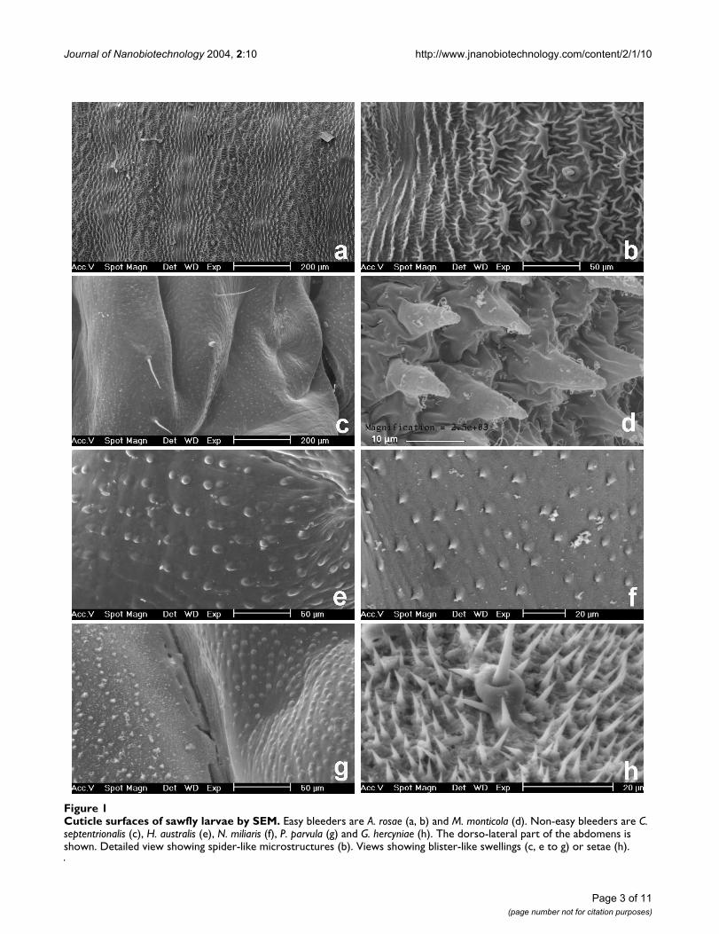
ResultsMicrostructures covering the cuticle surfaceThe larvae of sawfly species observed by SEM showedabove surface microstructures of their cuticle and whichare described below. These microstructures were strikinglymore complexly structured in easy bleeders than in non-easy bleeders (Fig. 1, 2) and this differing occurrenceamong sawfly species was significant (P = 0.0001, Fisherexact probability test, N = 24 species; Table 1).
In easy bleeders, the cuticle is covered with irregularlyshaped wart-like microstructures (verrucose). Their den-sity is approximately of 15 units per 0.01 mm2. They pos-sess fine ridges (carinulate) in a radiated way (Fig. 1b,2b,2d), hence the term "spider-like". The fine ridges (i.e.,the "legs" of the "spider") more or less imbricate inbetween those from adjacent microstructures, and theirwidth is approximately of 0.5 to 1.5 µm. The form of themicrostructure is generally circular (diameter excludingridges: 10 µm in A. padi), but can be elongated (length: 35µm in A. rosae). The ridges can be reduced (e.g., in A. padi).The height of microstructures was measured on LM viewsand reaches 23 µm (in P. aterrima). For further measure-ments by LM, see Table 2.
Compared to easy bleeders, the cuticle surface of non-easybleeders was much smoother. It only shows blister-likeswellings (pustulate) which have a diameter of 3–4 µm(e.g., in T. nigritus, H. australis, Nematus, Fig. 1e,1f), 6–7µm (e.g., in S. multifasciata, Fig. 2a) up to 12 µm (in H. tes-tudinea). In some genera such as Nematus and Craesus,each swelling shows a very small prickle (echinulate). Sev-eral swellings are sometimes aligned and then can bejoined, several together, to form a low ridge of approxi-mately 35 µm long (in C. septentrionalis).
Although E. ventralis and M. spinolae are easy bleeders, nospider-like microstructures were detected. Instead, smallridges with one or a few prickles, and small spines wereobserved, respectively. The larvae (alive) of these two spe-cies as well as T. scrophulariae are covered with a layer ofwaxy powder. Setae were observed instead of microstruc-tures in the outgroup species G. hercyniae (Fig. 1h).
Modelling the mechanical behaviour of the cuticleThe aim of modelling was to compare the repartition ofstresses of two cuticle configurations, as found in non-easy bleeders (M1) versus easy bleeders (M2), when asame loading is applied (Fig. 3a,3b). The maximum stressvalue (in compression and traction) is an indicator of pos-sible initiation of crack or damage (see Methods).
In Table 2, M1/1, M1/2 and M1/10 show the influence ofthe Young's modulus of the epicuticle, relative to the pro-cuticle, on the distribution of stresses in a cuticle patch.
Page 2 of 11(page number not for citation purposes)
Journal of Nanobiotechnology 2004, 2:10 http://www.jnanobiotechnology.com/content/2/1/10
Cuticle surfaces of sawfly larvae by SEMFigure 1Cuticle surfaces of sawfly larvae by SEM. Easy bleeders are A. rosae (a, b) and M. monticola (d). Non-easy bleeders are C. septentrionalis (c), H. australis (e), N. miliaris (f), P. parvula (g) and G. hercyniae (h). The dorso-lateral part of the abdomens is shown. Detailed view showing spider-like microstructures (b). Views showing blister-like swellings (c, e to g) or setae (h).
Page 3 of 11(page number not for citation purposes)

Journal of Nanobiotechnology 2004, 2:10 http://www.jnanobiotechnology.com/content/2/1/10
Cuticle surfaces of sawfly larvae by SEM and related integument sections by LMFigure 2Cuticle surfaces of sawfly larvae by SEM and related integument sections by LM. Non-easy bleeder is S. multifasciata (a, e). Easy bleeders are P. aterrima (b, f), A. padi (c, g), R. nodicornis (d) and R. bensoni (h). Views by SEM (a to d) show blister-like swellings (a) or spider-like microstructures (b to d). Views by LM (e to h) showing that, above a cellular layer, the cuticle com-prises a procuticle, in blue, whereas the epicuticle, in red (e, g), is not observed in some species (f, h).
Page 4 of 11(page number not for citation purposes)

Journal of Nanobiotechnology 2004, 2:10 http://www.jnanobiotechnology.com/content/2/1/10
When the Young's modulus of the epicuticle wasincreased, keeping the one of the procuticle constant, thestresses concentrated in the epicuticle and the maximalvalues increased (Fig. 3c to 3e). This occurred with bothload cases (i.e., normal and shear force).
With a normal force, M1 and M2 resulted in stress valueswhich were in the same range of magnitude (Table 2, Fig.3c to 3f). Thus from this load case it cannot be deducedthat one integument configuration will be damaged moreeasily than the other. In contrast, stress values obtainedwith a shear force were approximately three times higherin M2 than M1 (Table 2; Fig. 3g,3h). This suggests that an
integument with microstructures is more constrained andwill more easily reach the yield stress corresponding to thedamage of the cuticle. This conclusion was corroboratedby considering several single sawfly species. The maximalstress value in compression as well as in traction wasalways more extreme in five species of easy bleeders thanin five species of non-easy bleeders (Table 2, see shearforce). Note that for the easy bleeders M. monticola and P.aterrima, the apical part of the microstructure wasextremely minute. All stresses were concentrated in the tipof the microstructure, which lead to non-physical deflec-tions. The obtained values for these two species are prob-ably irrelevant in a comparison with other species.
Table 1: Easy bleeding, cuticle microstructures and hydrophobic property in sawfly larvae
Species Easy bleeding1 Microstructures2 Droplet3
2 µlDiameter3
4 µl
TENTHREDINIDAEAllantiinae
Athalia rosae (L.) EB + ? ? or 2.1 ± 0.0Blennocampinae
Eurhadinoceraea ventralis (Panzer) EB - · ·Monophadnus monticola (Hartig) EB + · ·Monophadnus spinolae (Klug) EB - · ·Phymatocera aterrima (Klug) EB + ? or 1.5 ± 0.0 ? or 2.0 ± 0.0Rhadinoceraea bensoni Beneš EB + · ·Rhadinoceraea micans (Klug) EB + ? ?Rhadinoceraea nodicornis Konow EB + ? or 1.6 ± 0.1 ? or 2.0 ± 0.1Tomostethus nigritus (Fabricius) N-EB - · ·
NematinaeCraesus alniastri (Sharfenberg) N-EB - 1.6 ± 0.0 2.1 ± 0.0Craesus septentrionalis (L.) N-EB - 1.7 ± 0.0 2.2 ± 0.1Hemichroa australis (Serville) N-EB* - 1.6 ± 0.1 2.2 ± 0.1Hemichroa crocea (Geoffr.) N-EB - · ·Hoplocampa testudinea (Klug) N-EB - · ·Nematus melanocephalus Hartig · - · ·Nematus miliaris (Panzer) N-EB* - · ·Nematus pavidus Serville N-EB* - · ·Pristiphora laricis (Hartig) N-EB - · ·Pristiphora testacea (Jurine) N-EB - 1.9 ± 0.0 2.6 ± 0.0Pseudodineura parvula (Klug) · - · ·
SelandriinaeAneugmenus padi (L.) EB + 1.7 ± 0.2 2.2 ± 0.2Strongylogaster mixta (Klug) N-EB - 1.7 ± 0.1 2.1 ± 0.1Strongylogaster multifasciata (Geoffr.) N-EB - 1.6 ± 0.1 2.1 ± 0.0
TenthredininaeTenthredo scrophulariae L. N-EB* - · ·
ARGIDAEArge sp. N-EB - · ·
DIPRIONIDAEGilpinia hercyniae (Hartig) N-EB - 1.6 ± 0.0 2.0 ± 0.0
1 Species was an easy bleeder (EB), or a non-easy bleeder (N-EB). Data from Boevé & Schaffner [4], except data from U Schaffner & JLB, unpublished results (*).2 Spider-like microstructures were present (+) or absent (-) by observations of the cuticle surface by SEM and/or of cuticle sections by LM.3 Cuticle was either too hydrophobic so that adherence of water droplet was impossible (?) or the diameter (mean ± SD, in mm) of a 2 and 4 µl droplet on the cuticle was measured. (·) Not tested.
Page 5 of 11(page number not for citation purposes)

Journal of Nanobiotechnology 2004, 2:10 http://www.jnanobiotechnology.com/content/2/1/10
Hydrophobic property of cuticle surfacesIt was difficult or even impossible to deposit a water drop-let on the larval body of some sawflies (Table 1). Whenthe pipette tip was brought close to the integument,almost within physical contact, the droplet was pushedaside against the tip border. By then retrieving the pipette,the droplet was again on its tip, not on the integument.This could happen for some of the individuals tested perspecies (Table 1).
In species where the integument was less hydrophobic,the diameter of the droplet on it ranged from 1.5 to 1.9mm (2 µl droplet) and from 2.0 to 2.6 mm (4 µl). Con-sidering this latter droplet size, a small diameter (2.0 mm)or an immeasurable diameter (see above) was associatedwith sawfly species which are easy bleeders, whereas alarger droplet diameter (> 2.0 mm) was associated withnon-easy bleeders (P = 0.045, Fisher exact probability test,N = 12 species, Table 1). Thus easy bleeders possess a
Table 2: Model input and output with force applied on cuticle of non-easy bleeders (A) and easy bleeders (B)
A
M1/1 M1/2 M1/10 Hc Pl Pt Sm Ts
W1 110 110 110 110 110 110 110 110H1 20 20 20 7 5 8 8 11H2 10 10 10 6 5 3 5 5E1 500 500 500 500 500 500 500 500E2 500 1000 5000 5000 5000 5000 5000 5000
F 1zMax 0.206 0.170 1.001 2.910 4.520 2.651 2.906 2.201Min -0.835 -1.003 -1.604 -4.167 -6.240 -6.482 -4.633 -3.772F 1xMax 1.553 1.614 1.794 2.545 2.956 3.337 2.714 2.569Min -0.363 -0.383 -0.447 -0.717 -0.860 -0.864 -0.764 -0.710
B
M2 Ar Mm Pa Rb Rn
W1 110 70 33.5 60 50 60H1 15 8 8 15 10 15H3 15 8 14 23 10 10D1 28 23 9 20 15 20D2 20 11 1 2 10 10S1 20 5 6 8 8 8P 1 3.306 80 100 4 4N µstr 1 1 5 1 1 1
F 1zMax 0.665 1.696 0.623 4.291 1.430 0.770Min -0.793 -2.560 -0.972 -7.286 -1.412 -2.134F 1xMax 4.788 7.554 35.820 174.600 12.840 7.632Min -1.476 -2.241 -1.782 -9.267 -3.805 -2.317
Model of non-easy bleeders was based on parameter values measured on LM and SEM views from H. crocea and N. pavidus together (M1/1, M1/2, M1/10), and from H. crocea (Hc), P. luridiventris (Pl), P. testacea (Pt), S. multifasciata (Sm) and T. scrophulariae (Ts). Different relative values of Young's modulus for procuticle (E1) and epicuticle (E2) were used in M1/1, M1/2, and M1/10.Model of easy bleeders was based on parameter values measured on LM and SEM views from P. aterrima and R. micans together (M2), and from A. rosae (Ar), M. monticola (Mm), P. aterrima (Pa), R. bensoni (Rb) and R. nodicornis (Rn). Parameter values, in µm, introduced in the model: width of the model sample (W1), height of procuticle layer (H1), height of epicuticle layer (H2), height of microstructure (H3), diameter at base of microstructure (D1), diameter at top of microstructure (D2), shortest distance between microstructures (S1). Number of microstructures set under pressure (N µstr). Pressure applied per microstructure (P).Stress values, obtained with a normal force (F 1z) or shear force (F 1x), are given as extreme values in traction (Max) and compression (Min).
Page 6 of 11(page number not for citation purposes)

Journal of Nanobiotechnology 2004, 2:10 http://www.jnanobiotechnology.com/content/2/1/10
Models of the cuticle of sawfly larvaeFigure 3Models of the cuticle of sawfly larvae. Model representing a non-easy bleeder (a, c to e, g) and an easy bleeder (b, f, h). View in perspective showing five microstructures (b) and the location of the applied force (a, b). Maximal stress distribution in a section through the cuticle (c to h). The ratio of Young's modulus for the procuticle to the one of the epicuticle is assumed to be 1/1 (c), 1/2 (d) and 1/10 (e). The applied force is normal (c to f) or sheared (g, h). The maximal value corresponds to the maximal stress of the principal stress 1 and the minimal value to the minimal stress of the principal stress 2. Only the distribu-tion of principal stress 1 is shown, while the maximal value is given in Table 2. Degrees of freedom = 120,553 (a, c to e, g), 40,701 (b, f, h).
a b
cd
e f
g h
Page 7 of 11(page number not for citation purposes)

Journal of Nanobiotechnology 2004, 2:10 http://www.jnanobiotechnology.com/content/2/1/10
hydrophobic and non-easy bleeders a rather hydrophilicintegument.
On inert surfaces the diameter of 2 and 4 µl droplets wasconstantly as follows: immeasurable (see above) and 2.2mm on Teflon®, 1.8 and 2.3 mm on Parafilm®, 1.8 and 2.3mm on polystyrene and 2.6 and 3.3 mm on glass, respec-tively. Thus even Teflon®, that is considered as highlyhydrophobic, led to a 4 µl droplet diameter which wascomparable to the one obtained on the (hydrophilic)integument of non-easy bleeders. The 2 µl droplet wasapparently light enough to impede its adhesion on theTeflon® surface, but not the larger droplet size tested.
DiscussionSeveral sawfly larvae showed a characteristic cuticle sur-face with spider-like microstructures and this was associ-ated with a low mechanical strength of their integument.For instance, these microstructures were present in theeasy bleeder Aneugmenus padi and absent in the non-easybleeders Strongylogaster spp. Both genera are closelyrelated since they belong to the same subfamily and havethe same host plant [11]. It is likely that the occurrence ofmicrostructures cannot be interpreted simply in terms ofa systematic arrangement of species and that they arerelated to the phenomenon itself of easy bleeding. The lar-val abdomen of several Dolerus (Tenthredinidae, Selandri-inae) species presents "meshes of microsculpture notsharply defined", with a dimension ranging from 20 to 40µm, and these microstructures are often fused [12]. Theymay constitute an intermediate state between thoseobserved by us on easy bleeders and non-easy bleeders,but being more physically comparable to those of non-easy bleeders by the absence of spider-like microstruc-tures. Particular microstructures are also observed at thecuticle surface of other arthropods than sawflies, such asin nymphs of bugs and ticks [13] and in adults of flies anddragonflies [14,15]. Their function is to allow by stretch-ing an increase of body volume during feeding, to allyflexibility with mechanical stability during highlyrepeated movements, etc., but their possible role in pro-moting a mechanical damage of the integument was notenvisaged so far [16].
The question arises to know whether in sawfly larvae ableto bleed easily the microstructures are directly involved inintegument disruption. We compared cuticle models ofnon-easy bleeders versus easy bleeders and applied a unitforce on it. Compared to the real-life, the model was sim-plified by considering a linear elastic behaviour of thecuticle (i.e., the stresses are proportional to the strains –Hooke's law), because we do not know the exact physicalproperties of the cuticle. Nevertheless, a comparison ofgeometrical parameters from easy bleeders versus non-easy bleeders revealed that by applying a shear force the
cuticle stresses both in compression and tension werehigher in the presence of microstructures (Table 2). Thissuggests that microstructures may directly contribute inthe damage of the integument. Yet, the breaking line of adamaged integument goes between the microstructures(SA, personal observation on the easy bleeder P. aterrima).This biological observation is in agreement with ourmodel results. The regions subject to high stresses are notrestricted to the zone of the microstructure, but extenddeeper into the cuticle mass (Fig. 3h). From this trend wemay extrapolate that if the shear force is enhanced, themicrostructure will not break off from the rest of the cuti-cle, but the fracture line will start at the base of a micro-structure and continue throughout the whole cuticlethickness. In other words, the integument will disrupt.This conclusion becomes even more relevant in the realis-tic situation where an attacking predator applies a more orless oblique force on the cuticle. Beside physical aspects,chemical ones also contribute in the mechanical proper-ties of an integument [16-20]. One of these properties,visco-elasticity, is determined in the abdominal integu-ment of the bug Rhodnius by the matrix protein(s) of theprocuticle with a reinforcing effect of chitin microfibriles.Differing chitin and protein patterns are observed in thecuticle when easy bleeders are compared to non-easybleeders (M. Spindler-Barth & SA, unpublished results).Ongoing research aims to investigate these physiologicalaspects as well as the healing process, and to link themwith the phenomenon of easy bleeding.
The cuticle surface of easy bleeders was highly hydropho-bic (Table 1) as compared to a well-known hydrophobicmaterial such as Teflon®. There is a trend for the integu-ment of easy bleeders (e.g., P. aterrima, Rhadinoceraea spp.,A. padi) to appear as mat, in contrast to the brilliant aspectin non-easy bleeders (e.g., Strongylogaster spp., Craesusspp.) (JLB, personal observations). We believe that thehydrophobic property is ecologically relevant duringpredator-prey interactions. When a predator, typically aninsect with biting-chewing mandibles [10], bites into theintegument of a sawfly larva at a given spot, the best forthe larva is to keep the deterrent haemolymph spatiallyconcentrated at this spot. A counter-example is that someinsects are known to have morphological devices of theintegument surface or wetting agents included in theirdefensive secretion, which help the secretion to spread out[21,22]. But such secretions are typically volatile and thedefence consists of keeping the aggressor at a distance. Themorphological devices and wetting agents modulate theevaporation of the secretion and, thereby, the effective-ness of a defence that acts by olfactory cues. In the case ofeasy bleeding, deterrent compounds dissolved in thehaemolymph need to contact the mouthparts of anaggressor, acting by gustatory cues. Moreover, easy bleed-ers should not spread out their hemolymph since they
Page 8 of 11(page number not for citation purposes)

Journal of Nanobiotechnology 2004, 2:10 http://www.jnanobiotechnology.com/content/2/1/10
would lose this valuable liquid. Remaining as a dropletand in contact with the larval haemocel, the droplet canbe sucked back by the larva into its body within a few min-utes, providing that the larva is not more disturbed [4].
A parallel can be drawn between the integument surface ofeasy bleeders and the one of several plant leaves. The lotusleaf led recently to the so-called Lotus-effect® [23]. Partic-ular physico-chemical properties of the leaf allow a self-cleaning by rain. This effect relies on a micro-structuredsurface and a coating of waxy crystals. Both characteristicscontribute in rendering the surface hydrophobic [23,24].The optimal configuration and size of the structures is acoarse structure of 10 to 50 µm and a finer one of 0.2 to 5µm [25]. This corresponds well to the case of the spider-like microstructures as found on the cuticle of easy bleed-ers. In the insect both these coarse and finer structures areprovided by the spider-like structures (Results), whereasin the plant each scale of structures is due to microstruc-tures and waxy crystals, respectively [26]. There are nowaxy crystals on the body surface of easy bleeders. A finelayer of waxy powder covers only some species of easybleeders as well as non-easy bleeders (see Results). Such awaxy powder consists mainly of hexacosan-1-ol in Erio-campa ovata [27], a non-easy bleeder [4] not studied in thepresent work. It is likely that in a majority of easy bleedersthe hydrophobic property relies especially or solely on thegeometry of the cuticle surface, by the occurrence ofmicrostructures.
ConclusionsWe suppose at least two types of functions in the occur-rence of spider-like microstructures, which we observedspecifically on the body surface of easy bleeders. Firstlythe damage provoked by a biting predator could be facili-tated. Secondly the integument of easy bleeders could berendered hydrophobic, which helps stop the emittedhaemolymph droplet from spreading out.
MethodsInsectsAll sawfly larvae (see Table 1) were collected in the field(Belgium, Germany, Switzerland), except A. rosae and G.hercyniae that came from indoor populations. The larvaewere identified according to Lorenz & Kraus [11]. The full-grown larval stage was used.
Observations by SEM and LMFixed larvae stored in ethanol were dried, coated withgold, and examined with a Philips XL-30 ESEM. Speci-mens were placed to observe the dorsal and lateral part ofthe abdomen. The terminology used in describing thecuticle surface refers to Harris [28].
Series of 7 µm thin cross sections were obtained from lar-vae by using classical histological techniques. They weredeparaffinized in xylene and rehydrated in severaldecreasing ethanol to water solutions, then stained by theAzan trichrome method [29] and observed by LM.
Model by finite elementsGeneral mechanical assumptionsThe general rigorous mechanical behaviour of the cuticleis complex. As a first attempt to understand the propertyof easy bleeding, it was assumed that a damage of theintegument is due to excessive stress under static loading.Since the cuticle of a larva is also geometrically complex inthree dimensions, no simplified laws, for instance,derived from the strength of material could be used. Theanalysis was therefore performed on solid configuration,discretized by a standard finite element method [30]. Itwas assumed that the stress-strain law is linear and iso-tropic (Hooke's law) and that the displacements andstrains are small. The geometrical dimensions of the cuti-cle are very small, at the microscale. It is known that forsuch a configuration, the assumption of continuum maynot be valid [31]. But, it is also known for standard mate-rials such as metals that the strength is generally underes-timated with continuum assumption. This is the reasonwhy the analysis performed in this paper was purely qual-itative and based on a comparison of the stress betweenthe geometry encountered in easy bleeders and non-easybleeders. Most of the results were interpreted on the prin-cipal stress: for 3D mechanical configurations, three direc-tions always exist for which the stress (and strain forisotropic laws) is maximum or minimum. According tothese directions, the shear stress is zero. It was then sup-posed that the maximum stress values (in traction or com-pression) cause the initiation of the cuticle damage.
Finite element modellingThe finite element analysis was performed using the gen-eral mechanical purpose software SAMCEF® version 9.1. Itwas assumed that the integument is made of the repeti-tion of reproducible patches in both x, y directions. Thus,only one patch has to be modelled by the appropriateboundary conditions representing this repetition (i.e., thedisplacements on each boundary are blocked in the direc-tion normal to this boundary). The geometry was discre-tized with 3D solid linear finite elements (prisms orbricks). The patch used to model non-easy bleeders wascomposed of two layers, procuticle and epicuticle, of dif-ferent properties in height and Young's modulus. It is sup-posed that generally the epicuticle of insects is pliant butnot extensible, stronger in compression than tension, andthat the epicuticle is less elastic than the procuticle [2].The patch used for easy bleeders contained five micro-structures and was homogenous. Indeed, generally no epi-cuticle is clearly detected in the cuticle of easy bleeders
Page 9 of 11(page number not for citation purposes)

Journal of Nanobiotechnology 2004, 2:10 http://www.jnanobiotechnology.com/content/2/1/10
observed by LM (e.g., Fig. 2f), an exception being A. padi(Fig. 2g).
LoadingThe loading was always divided into two load cases: aforce perpendicular to the patch surface (normal force)and parallel to it (shear force).
In a real situation, the mandibles of an attacking predator,typically a small arthropod, apply the loading [10]. Thediameter of a mandible's tip was measured on workers ofthe ant Myrmica rubra and reached 20 µm as the smallestvalue. In the model, the shape of the contact point madeby the mandible was a disc (radius = 10 µm) on which theforce was applied. This force was applied either on theupper centre of the epicuticle for non-easy bleeders (Fig.3a) or on the top of the central microstructure when con-sidering easy bleeders (Fig. 3b). As the radius of the upperpart of the microstructure changed from one species toanother, being generally lower than 10 µm, the appliedsurface force was adapted to obtain a same resultant forcefor each configuration.
The insect body contains a liquid, haemolymph. Thepatch, therefore, was modelled by applying a surface forceequilibrated with the loading of the predator.
Hydrophobic propertyThis property of the integument was estimated by a simplemethod that allowed the use of insects alive. The first stepwas to notice whether a 2 and 4 µl droplet of charcoal fil-tered water could adhere, gently depositing it with a 1–10µl pipette on the thoracic or abdominal integument of asawfly larva that was resting on a leaf of its host plant. Ifan adherence was possible, the diameter of the depositeddroplet was measured under a stereomicroscope withmicrometer. Six full-grown larvae were tested per species.As control the following substrates were tested in the samemanner: Teflon®, Parafilm®, polystyrene and glass. Onthese biological and inert surfaces, the droplet reaction(i.e., adherence capability and droplet diameter) was con-sidered to express the hydrophobic or hydrophilic prop-erty of the surface.
Authors' contributionsJLB collected and identified the insects, performed thetests on the hydrophobic property, measured the cuticleparameters for the model on LM views, and wrote themanuscript, except the parts about this model in Resultsand Methods. VD obtained most SEM views. TM and PBperformed the model by finite elements and wrote thetwo related parts in the manuscript. SA maintained indoorpopulations of two sawfly species and carried out andphotographed the integument sections used in LM and,thereby, in the model.
AcknowledgementsWe thank Johan Billen (Katholieke Universiteit Leuven) for his help in obtaining preliminary LM views, and Julien Cillis (IRSNB-KBIN) for his tech-nical assistance in SEM. Previous versions of the manuscript were kindly reviewed by Travis Turner, Caroline Müller and three anonymous referees. The research performed by JLB and SA was supported by the European Community's Improving Human Potential Programme under contract HPRN-CT-1999-00054, INCHECO.
References1. Whitman DW, Blum MS, Alsop DW: Allomones: chemicals for
defense. Insect Defenses: adaptive mechanisms and strategies of preyand predators Edited by: Evans DL, Schmidt JO. Albany: State Univ ofNew York Press; 1990:23-61.
2. St Leger RJ: Integument as a barrier to microbial infections.Physiology of the Insect Epidermis Edited by: Bennington K, RetnakaranA. Melbourne: CSIRO Publications; 1991:284-306.
3. Hollande AC: L'autohémorrhée ou le rejet du sang chez lesinsectes (toxicologie du sang). Arch Anat Microsc 1911,13:171-318.
4. Boevé J-L, Schaffner U: Why does the larval integument ofsome sawfly species disrupt so easily? The harmful haemol-ymph hypothesis. Oecologia 2003, 134:104-111.
5. Heads PA, Lawton JH: Bracken, ants and extrafloral nectaries.III. How insect herbivores avoid ant predation. Ecol Entomol1985, 10:29-42.
6. Schaffner U, Boevé J-L, Gfeller H, Schlunegger UP: Sequestration ofVeratrum alkaloids by specialist Rhadinoceraea nodicornisKonow (Hymenoptera, Tenthredinidae) and its ecoetholog-ical implications. J Chem Ecol 1994, 20:3233-3250.
7. Müller C, Boevé J-L, Brakefield P: Host plant derived feedingdeterrence towards ants in the turnip sawfly Athalia rosae.Entomol Exp Appl 2002, 104:153-157.
8. Müller C, Brakefield PM: Analysis of a chemical defense in sawflylarvae: easy bleeding targets predatory wasps in latesummer. J Chem Ecol 2003, 29:2683-2694.
9. Boevé J-L: Sawflies (Hymenoptera: Tenthredinidae). Encyclope-dia of Entomology Volume 3. Edited by: Capinera JL. Dordrecht: KluwerAcademic Publishers; 2004:1949-1954.
10. Boevé J-L, Müller C: Defence effectiveness of easy bleedingsawfly larvae towards invertebrate and avian predators.Chemoecology in press.
11. Lorenz H, Kraus M: Die Larvalsystematik der Blattwespen(Tenthredinoidea und Megalodontoidea). Berlin: AkademieVerlag; 1957.
12. Leblanc L, Goulet H: Description of larvae of eight nearctic spe-cies of Dolerus (Hymenoptera: Tenthredinidae) with focuson six Equisetum-feeding species from the Ottawa region. CanEntomol 1992, 124:999-1014.
13. Hackman RH: Expanding abdominal cuticle in the bug Rhodn-ius and in the tick Boophilus. J Insect Physiol 1975, 21:1613-1623.
14. Gorb SN: Armored cuticular membranes in Brachycera(Insecta, Diptera). J Morphol 1997, 234:213-222.
15. Gorb SN: Ultrastructure of the neck membrane in dragon-flies (Insecta, Odonata). J Zool, London 2000, 250:479-494.
16. Vincent JFV, Wegst UGK: Design and mechanical properties ofinsect cuticle. Arthr Struct Dev 2004, 33:187-199.
17. Hepburn HR, Chandler HD: Material properties of arthropodcuticles: the arthrodial membranes. J Comp Physiol B 1976,109:177-198.
18. Locke M: The cuticular pattern in an insect – the interseg-mental membranes. J Exp Biol 1960, 37:398-406.
19. Reynolds SE: The mechanical properties of the abdominalcuticle of Rhodnius larvae. J Exp Biol 1975, 62:69-80.
20. Vincent JFV: Morphology and design of the extensible interseg-mental membrane of the female migratory locust. Tissue &Cell 1981, 13:831-853.
21. Filshie BK, Waterhouse DF: The structure and development ofa surface pattern on the cuticle of the green vegetable bugNezara viridula. Tissue & Cell 1969, 1:367-385.
22. Dettner K: Solvent-dependent variability of effectiveness ofquinone-defensive systems of Oxytelinae beetles (Coleop-tera: Staphylinidae). Entomol Gener 1991, 15:275-292.
Page 10 of 11(page number not for citation purposes)
http://www.ncbi.nlm.nih.gov/entrez/query.fcgi?cmd=Retrieve&db=PubMed&dopt=Abstract&list_uids=1100734

Journal of Nanobiotechnology 2004, 2:10 http://www.jnanobiotechnology.com/content/2/1/10
Publish with BioMed Central and every scientist can read your work free of charge
"BioMed Central will be the most significant development for disseminating the results of biomedical research in our lifetime."
Sir Paul Nurse, Cancer Research UK
Your research papers will be:
available free of charge to the entire biomedical community
peer reviewed and published immediately upon acceptance
cited in PubMed and archived on PubMed Central
yours — you keep the copyright
Submit your manuscript here:http://www.biomedcentral.com/info/publishing_adv.asp
BioMedcentral
23. Dambacher GT: Der Lotus-Effect – heute und morgen. Kunstst-offe 2002, 92:65-66.
24. Patankar N: On the modelling of hydrophobic contact angleson rough surfaces. Langmuir 2003, 19:1249-1253.
25. The Lotus-effect® [http://www.botanik.uni-bonn.de/system/lotus/en/prinzip_html.html]
26. Barthlott W, Neinhuis C: Purity of the sacred lotus, or escapefrom contamination in biological surfaces. Planta 1997,202:1-8.
27. Percy JE, Blomquist GJ, MacDonald JA: The wax-secreting glandsof Eriocampa ovata L. (Hymenoptera: Tenthredinidae):ultrastructural observations and chemical composition ofthe wax. Can J Zool 1983, 61:1797-1804.
28. Harris RA: A glossary of surface sculpturing. Occasional Papers inEntomol 1979, 28:1-31.
29. Romeis B: Mikroskopische Technik. 17th edition. Edited by: BöckP. Münich: Urban and Schwarzenberg; 1989.
30. Zienkiewicz OC, Taylor RL: The Finite Element Method. TheBasis Volume 1. 5th edition. London: Butterworth-Heinemann; 2000.
31. Hutchinson JW: Plasticity at the micron scale. Intern J SolidsStructures 2000, 37:225-238.
Page 11 of 11(page number not for citation purposes)