Supplement 1: Full Guideline and Additional Tables*
64
© 2020 American College of Physicians Supplement 1: Full Guideline and Additional Tables* Faughnan ME, Mager JJ, Hetts S, et al. Second international guidelines for the diagnosis and management of hereditary hemorrhagic telangiectasia. Ann Intern Med. 8 September 2020. [Epub ahead of print]. doi:10.7326/M20-1443 Full Guideline and Additional Tables * This supplementary material was provided by the authors to give readers further details on their article. The material was reviewed but not copyedited.
-
Upload
khangminh22 -
Category
Documents
-
view
0 -
download
0
Transcript of Supplement 1: Full Guideline and Additional Tables*

© 2020 American College of Physicians
Supplement 1: Full Guideline and Additional Tables*
Faughnan ME, Mager JJ, Hetts S, et al. Second international guidelines for the diagnosis and management of hereditary hemorrhagic telangiectasia. Ann Intern Med. 8 September 2020. [Epub ahead of print]. doi:10.7326/M20-1443
Full Guideline and Additional Tables
* This supplementary material was provided by the authors to give readers further details ontheir article. The material was reviewed but not copyedited.
2
ONLINE SUPPLEMENT Second International Guidelines for the Diagnosis and Management of HHT Marie E. Faughnan MDMSc1,2, Johannes J. Mager MD PhD3, Steven Hetts MD4, Valerie A. Palda MD MSc5, Kelly Lang-Robertson6, Elisabetta Buscarini MD7, Erik Deslandres MD8,Raj S. Kasthuri MD9, Andrea Lausman MD10, David Poetker MD MA11, Felix Ratjen, MD12, Mark S. Chesnutt MD13, Marianne Clancy RDH MPA14, Kevin J. Whitehead MD15, Hanny Al-Samkari MD16, Murali Chakinala MD17, Miles Conrad MD18, Daniel Cortes BscPhm19, Claudia Crocione20, Jama Darling MD21, Els de Gussem MD22, Carol Derksen23, Sophie Dupuis-Girod MD PhD24, Patrick Foy MD25, Urban Geisthoff MD26, James R. Gossage MD27, Adrienne Hammill MD28, Ketil Heimdal, MD29, Katharine Henderson MS, CGC30, Vivek N. Iyer MD MPH31, Anette D. Kjeldsen, MD32, Masaki Komiyama MD33, Kevin Korenblatt MD34, Jamie McDonald MS CGC35, J. McMahon36, J. McWilliams MD37, Mary E. Meek MD38, Meir Mei-Zahav MD39, Scott Olitsky, MD MBA14, Sara Palmer, PhD40, Rose Pantalone RN1, Jay F. Piccirillo MD41, Beth Plahn RN MHA42, Mary E.M. Porteous MD43, Marco C. Post MD PhD44, Ivan Radovanovic MD45, Paul J. Rochon, MD46, Josanna Rodriguez-Lopez MD47, Carlo Sabba MD48, Marcelo Serra MD49, Claire Shovlin PhD MA50, Dennis Sprecher, MD51, Andrew J. White MD52, Ingrid Winship MBChB MD53, Roberto Zarrabeitia MD54.
Author affiliations:
1Toronto HHT Centre, Division of Respirology, Department of Medicine, St. Michael’s Hospital, University of Toronto, Toronto, ON, Canada 2Li Ka Shing Knowledge Institute, St. Michael’s Hospital, University of Toronto, Toronto, ON, Canada 3St. Antonius Hospital, Nieuwegein/Utrecht, The Netherlands 4Department of Neurointerventional Radiology, University of California San Francisco USA 5Department of Medicine and Institute of Health Policy, Management and Evaluation, University of Toronto, Toronto, ON, Canada 6Centre for Effective Practice, Toronto, ON, Canada 7UOC Gastroenterologia ed Endoscopia Digestiva, HHT Reference Center ERN, Ospedale Maggiore, ASST Crema, Italy 8Department of Gastroenterology, CHUM, Hotel Dieu, Montreal, QC Canada 9Division of Hematology/Oncology, University of North Carolina, Chapel Hill, Chapel Hill, North Carolina, USA 10Department of Obstetrics and Gynecology, University of Toronto, St. Michael’s Hospital, Toronto, ON, Canada 11Department of Otolaryngology, Froedtert and Medical College of Wisconsin, Milwaukee, WI, USA 12Division of Respiratory Medicine, Department of Pediatrics, Translational Medicine, Research Institute, The Hospital for Sick Children, University of Toronto, Toronto, ON Canada
3
13VA Portland Health Care System, HHT Center of Excellence, Dotter Department of Interventional Radiology, Oregon Health & Science University, USA 14Cure HHT, Monkton, Maryland, USA 15Department of Cardiovascular Medicine and Pediatric Cardiology, University of Utah Medical l Center, Salt Lake City, Utah, USA 16Division of Hematology, Massachusetts General Hospital, Harvard Medical School, Boston, Massachusetts, USA 17Department of Pulmonology and Critical Care, Washington University School of Medicine, St. Louis, MO, USA 18Department of Interventional Radiology University of California San Francisco USA 19Pharmacy Department, St. Michaels Hospital, Unity Health Toronto, Toronto, Canada 20HHT Europe, Rome, Italy 21Department of Hepatology, University of North Carolina, Chapel Hill, North Carolina, USA 22Department of Medicine, Section of Respirology, Grace Hospital, Winnipeg, MB Canada 23HHT Canada, Spruce Grove, Alberta, Canada 24Hospices Civils de Lyon, Femme-Mère-Enfant, Hospital 69677 BRON, France 25Department of Hematology, Froedtert and Medical College of Wisconsin, Milwaukee, WI USA 26Department of Otorhinolaryngology, Head and Neck Surgery, University Hospital of Marburg, Phillips University Marburg, Marburg Germany 27Augusta University, Augusta, GA, USA 28Division of Hematology, Cancer and Blood Diseases Institute, Cincinnati Children’s Hospital, and Department of Pediatrics, University of Cincinnati, Cincinnati, Ohio, USA 29Department of Genetics, Oslo University Hospital, RIkshopitalet, Oslo, Norway, 30Yale University School of Medicine, New Haven, CT, USA 31Division of Pulmonary and Critical Care Medicine, Mayo Clinic, Rochester, Minnesota, USA 32Department of Otorhinolaryngology Head and Neck Surgery, HHT-center OUH, Vascern member Odense University Hospital, Odense, Denmark 33Department of Neurointervention, Osaka City General Hospital, Osaka, Japan 34Department of Hepatology, Washington University School of Medicine, St. Louis, MO, USA 35Department of Pathology and Radiology, University of Utah Medical Center, Salt Lake City, Utah, USA 36Chester, New Jersey, USA 37Department of Interventional Radiology, University of California Los Angeles, California, USA 38Department of Interventional Radiology, University of Arkansas for Medical Sciences, Little Rock, Arkansas, USA 39Pulmonology Institute, Schneider Children’s Medical Center of Israel, Sackler School of Medicine, Tel Aviv University, Israel 40Baltimore, Maryland, USA 41Department of Otolaryngology-Head & Neck Surgery, Washington University School of Medicine, St. Louis, MO, USA
4
42Sioux Falls, South Dakota, USA 43Department of Genetics University of Edinburgh, Center of Molecular Medicine, Edinburgh, Scotland 44Department of Cardiology, St. Antonius Hospital, Nieuwegein/Utrecht and University Medical Center Utrecht, The Netherlands 45Department of Neurosurgery, University Health Network, Toronto Western Hospital, University of Toronto, Toronto, Canada 46Department of Interventional Radiology, University of Colorado Hospital, Aurora, CO, 47Department of Pulmonology, Massachusetts General Hospital, Boston, Massachusetts, USA 48Department of Internal Medicine, University of Bari, Bari, Italy 49Department of Internal Medicine, Hospital Italiano de Buenos Aires, Buenos Aires, Argentina 50Department of Pulmonology, Hammersmith Hospital, London, England 51Blue Bell, PA, USA 52Division of Pediatric Immunology and Rheumatology, Washington University School of Medicine, St. Louis, MO, USA 53Genomic Medicine, Royal Melbourne Hospital and University of Melbourne, Melbourne, Australia 54Servicio de Medicina Interna, Unidad HHT, Hospital Sierrallana (Servicio Cántabro de Salud), Torrelavega (Cantabria), Spain
Correspondence to: Dr. M.E. Faughnan, St. Michael’s Hospital, University of Toronto, St. Michael’s Hospital, 30 Bond St, Toronto, M5B-1W8, Canada; [email protected]
Funding Sources: The Christopher McMahon Family and Cure HHT. Financial support for MEF: Nelson Arthur Hyland Foundation, Li Ka Shing Knowledge Institute of St Michael’s Hospital. Role of Funding Sources: The funding sources had no role in the design, conduct or reporting of the study or in the decision to submit the results for publication. Although the funding sources were not directly involved in the generation of the recommendations, some of the participants in the guidelines process were also board members of Cure HHT, officers of Cure HHT or members of various Cure HHT committees.
Competing interests: VAP received an honorarium for moderating the HHT Guidelines Conference, DC received an honorarium for conference participation and KLR was compensated for conducting the literature search and evidence review; neither participated in voting.
Potential conflicts of interest were reported prior to the Guidelines Conference: All were classified as “no significant conflict”, as per process detailed in the methods.
Contributors: All of the authors contributed to the Guidelines development and the resulting manuscript.
5
Word Count: 13,347 Abbreviations: ACVRL1 = activin A receptor like type 1 AE = adverse event APC = argon plasma coagulation AV = arteriovenous AVF = arteriovenous fistula AVM(s) = arteriovenous malformation(s) CBC=complete blood count CE = capsule endoscopy CVM = capillary vascular malformation CO2 = carbon dioxide CT = computed tomography DVT = deep venous thrombosis EGD = esophagogastroduodenoscopy ENG = endoglin ENT = ear nose and throat ERCP= endoscopic retrograde cholangiopancreatography ESS= epistaxis severity score GI = gastrointestinal GWG = guidelines working group HHT = hereditary hemorrhagic telangiectasia HHT1= hereditary hemorrhagic telangiectasia type 1 HHT2= hereditary hemorrhagic telangiectasia type 2 HOCF = high-output cardiac failure IV = intravenous JP-HHT = juvenile polyposis-hereditary hemorrhagic telangiectasia overlap MCV = mean corpuscular volume MELD= model for end stage liver disease MR = magnetic resonance MRI = magnetic resonance imaging OLT = orthotopic liver transplant PaO2 = arterial partial pressure of oxygen QOL= quality of life RBC = red blood cell RCT = randomized controlled trial SMAD4= Mothers Against Decapentaplegic homolog 4 TTCE = transthoracic contrast echocardiography VEGF= vascular endothelial growth factor VMs = vascular malformations WHO = World Health Organization
6
Centers with recognized expertise in the diagnosis and management of HHT can be located at https://curehht.org/, the website for Cure HHT and vascern.eu, the website for the European Reference Network for Rare Vascular Diseases. ABSTRACT Description: HHT is an autosomal dominant disease with an estimated prevalence of 1 per 5,000, characterized by the presence of vascular malformations (VMs). These result in chronic bleeding, acute hemorrhage and complications from shunting through VMs. The goal of the Second International HHT Guidelines process was to develop evidence-based consensus guidelines for the management and prevention of HHT-related symptoms and complications.
Methods: The guidelines were developed using the AGREE-II framework and GRADE methodology. The Guidelines expert panel included expert physicians (clinical and genetic) in HHT from fifteen countries, guidelines methodologists, health care workers, health care administrators, patient advocacy representatives and people with HHT. During the pre-conference process, the expert panel generated clinically relevant questions in six priority topic areas. A systematic literature search was conducted in June 2019, and articles meeting a priori criteria were included to generate evidence tables which were used as the basis for recommendation development. The expert panel subsequently convened during a guidelines conference to conduct a structured consensus process, during which recommendations reaching >=80% consensus were discussed and approved.
Recommendations: The expert panel generated and approved six new recommendations for each of the six priority topic areas: Epistaxis, Gastrointestinal Bleeding, Anemia & Iron Deficiency, Liver VMs, Pediatric Care, Pregnancy & Delivery (36 total). The recommendations highlight new evidence in existing topics from the First International HHT Guidelines and provide guidance in three new areas: Anemia, Pediatrics and Pregnancy & Delivery. These recommendations should facilitate implementation of key components of HHT care into clinical practice.
Word count=249
Funding Sources: The Christopher McMahon Family and Cure HHT. Financial support for MEF: Nelson Arthur Hyland Foundation, Li Ka Shing Knowledge Institute of St Michael’s Hospital. Role of Funding Sources: The funding sources had no role in the design, conduct or reporting of the study or in the decision to submit the results for publication. Although the funding sources were not directly involved in the generation of the recommendations, some of the participants in the guidelines process were also board members of Cure HHT, officers of Cure HHT or members of various Cure HHT committees.
7
INTRODUCTION Hereditary hemorrhagic telangiectasia (HHT) is an autosomal dominant disease with an estimated prevalence of approximately 1 per 5,000(1). It is characterized by clinically significant vascular malformations (VMs) of skin and mucous membranes of the nose and gastrointestinal tract as well as the brain, lung and liver. HHT is under-diagnosed and there is often a long diagnostic delay (2). Making the diagnosis of HHT in a patient allows appropriate screening and preventive treatment to be undertaken in the patient and their affected family members. The most common symptom of HHT, epistaxis, has an age-related expression, as does the appearance of the typical telangiectasia (3). In 2000, consensus clinical diagnostic criteria known as the Curaçao Criteria were published(4) (Supplement Table 1), and upheld in the first International HHT Guidelines(5). Genetic testing for HHT diagnosis was also recommended in the first International HHT Guidelines, primarily for asymptomatic people from a family with known HHT, as detailed in Table. In 97% of patients with a definite clinical HHT diagnosis, a causative mutation is identified in one of these genes: Endoglin (ENG, HHT1), Activin-Receptor Like kinase-1 (ACVRL1, HHT2), and Mothers Against Decapentaplegic homolog 4 (SMAD4, JP-HHT) (6). The goal of this Second International HHT Guidelines process was to develop evidence-informed consensus guidelines regarding the diagnosis of HHT. the prevention of HHT-related complications and treatment of symptomatic disease in areas not previously addressed by guidelines and areas where significant new literature had been published. Several other recommendations from the first International HHT Guidelines were not re-assessed during this current process and remain currently recommended (Table). METHODS The Second International HHT Guidelines were developed using the AGREE-II framework and GRADE methodology. The international HHT community provided priority topics to be included or updated based on new evidence or topics not previously addressed. Recommendations not revisited, but still considered currently recommended, are detailed in Table. Topic groups were appointed for each of the six areas selected for update or new review. Topic groups identified key questions to guide the systematic search strategy of the literature. Six sets of search strategies were developed and executed between May and June 2019 in Ovid MEDLINE by a medical librarian (KLR) with input from the Chair, and through a series of pre-determined steps illustrated in Part 1 of Supplement 2, including double review of both abstracts and full text articles; 221 articles were summarized into evidence tables. The quality of included RCTs was assessed (Part 2 of Supplement 2) using the structured framework of the Cochrane Risk of Bias Tool(7). In the months preceding the conference, the six topic groups generated draft recommendations based on key questions and the evidence tables and consistent with GRADE(8) formatting for levels of evidence and strength of recommendation. Draft recommendations were distributed to all panel members 2 weeks before the consensus meeting. The Guidelines Working Group (GWG) convened at the Guidelines Conference in November 2019 in Toronto Canada to partake in a structured consensus process. The
8
GWG included clinical and genetic experts in all aspects of HHT from fifteen countries, guidelines methodologists, health care workers, health care administrators, HHT clinic staff, medical trainees, patient advocacy representatives, and patients with HHT. The GWG completed individual conflict of interest disclosures and potential conflicts were reviewed by the chair. The GWG was presented draft recommendations with supporting quality of evidence, voted anonymously on the wording/quality of evidence, was presented the draft strength of recommendation with justification by GRADE methodology, and then voted on the strength of recommendation. Consensus of 80% was required for the recommendation to be included in the Guidelines. A structured process was used to identify sources of disagreement for votes of less than 80% (see below). The recommendations were sent for external review to HHT experts and organizations; their comments were collated and addressed (Part 3 of Supplement 2). The funding sources had no role in the design, conduct or reporting of the Guidelines or in the decision to submit for publication. Consensus Process: At the beginning of the conference, recommendation development methods were reviewed and discussed with the attendees(panel). For each topic area, topic groups met and refined draft recommendations. For each topic group, the topic leader presented the draft recommendation and quality of evidence to the entire panel, with supporting details for clinical considerations, after which time was allowed for discussion. The panel then voted anonymously on the wording of the recommendation and quality of evidence, using a standard format for wording and the evidence levels HIGH-MODERATE-LOW-VERY LOW (consensus). The topic leader then presented the draft strength of recommendation with justification by GRADE methodology (quality of evidence, balance of benefits and harms, values and preferences, cost - not considered explicitly but discussed as relevant). The panel then voted on the strength of recommendation. Consensus of 80% had to be achieved to allow the recommendation to be included in the guideline. If the initial vote was less than 80% consensus, the recommendation was deferred to the second day of the conference for further discussion and revision. Subsequent voting had also to achieve 80% consensus for the recommendation to be included. In the event that the panel did not achieve 80% consensus for strength of recommendation, the alternate strength was voted upon (STRONG/WEAK). If consensus was still not achieved, discussion continued to clarify the panel’s views on which factors (quality of evidence, balance of benefits and harms, values and preferences, cost) were driving dissent. In this way, the panel made every effort to make explicit non-evidentiary factors influencing recommendation strength. After all recommendations were discussed and voted upon, the chair reviewed next steps, surveyed the panel regarding future research and guidelines priorities (Part 4 of Supplement 2) and the conference was adjourned. Patient Involvement: Patient representatives (patients with HHT, caregivers as well as representatives from Cure HHT and other patient advocacy organizations) were included at every step of the development process. Patient values were incorporated into the recommendations,
9
during discussion and voting. Patients voted anonymously on recommendations and participated as manuscript authors. Epistaxis Management
Background: Epistaxis is the most common symptom of HHT, developing in 90% of adults with the disease, affecting quality of life and often leading to iron deficiency and anemia. Typically, turbulent nasal airflow with breathing leads to mucosal dryness and bleeding from telangiectases of the nasal mucosa. As such, replacing lost moisture to help prevent the telangiectases from cracking and bleeding is a mainstay of epistaxis care. In a randomized clinical trial comparing topical therapies to saline as placebo, saline was found to significantly reduce the epistaxis severity score (ESS) at both 12 and 24 weeks after therapy(9). In many patients, additional therapies are often considered, when symptoms are persistent or severe, despite moisturization. Tranexamic acid is an oral antifibrinolytic agent that can stabilize clots by preventing premature clot lysis and has been shown to decrease intraoperative bleeding in other conditions. Two RCTs (Supplement Table 2) of oral tranexamic acid demonstrated a significant decrease in epistaxis severity(10, 11) with minimal adverse events. Neither study showed a significant improvement in hemoglobin but baseline levels were normal or nearly normal in both studies so the opportunity for improvement may have been small. Three studies in HHT have not found an increased risk of thrombosis with tranexamic acid(10, 11, 44), though there remains concern that this agent should be avoided in patients at high risk for thrombosis (e.g. patients with a history of arterial thrombosis or unprovoked venous thrombosis), in patients with atrial fibrillation and patients with thrombophilia or elevated factor VIII. Various ablative therapies have been studied in controlled and uncontrolled case series (Supplement Table 3). Lasers, including the Argon, potassium-titanyl-phosphate (KTP), and Nd-YAG lasers(15). Outcomes are variable, with at best temporary and partial improvement in epistaxis. However, side effects of laser treatments overall are relatively minor. Access can be limited by required laser safety precautions, local availability of specific lasers and costs. Sclerotherapy with foamed sodium tetradecyl sulfate to the
10
nasal cavity can be performed in the outpatient setting under local anesthesia. Three studies using foamed sodium tetradecyl sulfate, including one RCT, all from the same investigators, concluded that sclerotherapy was effective and safe(12, 13, 108). The investigators found that bleeding was substantially better controlled after sclerotherapy than standard therapy with minimal adverse effects. Though rare, potential side effects include septal perforation, transient dizziness, blurred vision and permanent blindness(108). The literature regarding radiofrequency and electrosurgery treatment for nasal telangiectatic lesions is scarce; there are only a few studies showing efficacy of treatment. Bipolar electrosurgery, is preferred over monopolar electrosurgery, given its lower risk for collateral damage, specifically septal perforation. Radiofrequency cauterizes the telangiectasias at a lower temperature than electrocautery and reduces the risk for collateral damage(14). Overall, there is evidence that ablative therapies can provide temporary and partial improvement in epistaxis, and that side effects are mostly minor. Severe epistaxis can be life threatening and devastating to QOL of HHT patients, and symptoms are often not adequately controlled with moisturization and ablative therapies. As such, systemic therapies and more invasive surgical management is often considered. Low level of evidence studies of antiangiogenic therapies are detailed in Supplement Table 3. Bevacizumab is a humanized recombinant monoclonal antibody that inhibits vascular endothelial growth factor (VEGF) and has been shown to be effective in several diseases characterized by increased angiogenesis. From 2006 through 2019 there have been 3 prospective(16-18) and 5 retrospective studies(19-23) that evaluated the use of intravenous bevacizumab in HHT in 5 or more patients with HHT-related bleeding (152 total patients, most with epistaxis). Objective improvements were noted in the majority of studies that reported on epistaxis severity, hemoglobin level, RBC transfusion, and/or quality of life (QOL). The most commonly reported adverse events (AE) include hypertension(19) and arthralgia(73). Some studies have noted problems with wound healing, sometimes serious(17, 23). Overall, the evidence supports the effectiveness of IV bevacizumab in reducing epistaxis severity and RBC need, and improving anemia. However, in the absence of RCT, the magnitude of benefit and long-term safety are unclear. Of note, RCTs of topical (nasal) bevacizumab(9, 109) and intranasal bevacizumab injections(110), have not shown any significant benefit (Supplement Table 2). Thalidomide and several of its analogs have been shown to downregulate VEGF levels in HHT patients(111) and improve blood vessel wall integrity(112). From 2007 through 2019 there have been 4 prospective(111-114) and 2 retrospective studies(115, 116) that evaluated the use of oral thalidomide in 5 or more patients with HHT-related epistaxis (67 total patients), detailed in Supplement Table 3. Objective improvements were noted in all but one study that reported on epistaxis severity, hemoglobin level, RBC transfusion, and/or QOL. Neuropathy is one of the most commonly reported side effects, often leading to discontinuation of the drug(73, 114, 115), and known teratogenicity precludes its use in women with child-bearing potential. Overall, low level evidence supports effectiveness of oral thalidomide in decreasing epistaxis severity and RBC need, and in improving
11
anemia. However, AEs are substantial and often a limiting factor with neuropathy persisting even after discontinuation of the drug in two thirds of patients(73, 115). Several other antiangiogenic agents are under investigation in the treatment of HHT related epistaxis. Pazopanib is a multikinase inhibitor that showed signs of efficacy in one small series(45). Pomalidomide is a thalidomide analog that appears to have a lesser incidence of neuropathy and is under study in a large RCT in HHT related bleeding. Doxycycline is an oral metalloproteinase inhibitor that may have downstream antiangiogenic effects and is under study in two small RCTs at present. The role of these agents in HHT related epistaxis will await additional studies. Invasive surgical procedures are also often considered when epistaxis is not adequately controlled with moisturization and ablative therapies. Low level of evidence studies of invasive surgical procedures, including septodermoplasty and nasal closure, are detailed in Supplement Table 3. The expert panel considered invasive surgical procedures as an equal option to the systemic therapies, and that this decision requires extensive consultation with the patient. In addition, comorbid disease, such as atrial fibrillation, can limit the use of prothrombotic drugs and require even aggressive anticoagulation or antiplatelet therapy instead. In these cases the invasive surgical measures(24-29) may be more appropriate as they could allow use of indicated anticoagulation or antiplatelet treatment. Several studies have evaluated septodermoplasty with the largest study(24) in which eighty-six percent of followed patients reported improved QOL, after mean follow-up of 3.75 years. Complications included worsening sinus infections (30%), decreased sense of smell, (58%) and frequent minor side effects, such as crusting and nasal airflow obstruction. Richer and colleagues(26) reported a series of 43 patients undergoing nasal closure, 83% reporting complete cessation of bleeding and no patients requesting reversal of the procedure. The largest study(28) includes 100 patients that underwent nasal closure with 50 of them having pre and post procedure data; ninety-four percent reported complete cessation of the bleeding. A number of surgical variations have been described for both nasal closure and septodermoplasty, though these have not been compared, and therefore clinical decision making should involve a rhinologic surgeon with expertise in these techniques. Recommendations: A1: The expert panel recommends that patients with HHT-related epistaxis use moisturizing topical therapies that humidify the nasal mucosa to reduce epistaxis. Quality of Evidence: Moderate (Agreement 98%) Topical saline has been shown to reduce epistaxis severity score, compared to baseline, in an RCT of multiple topical therapies (9)(Supplement Table 2). Strength of Recommendation: Strong (Agreement 100%) Clinical Considerations: Topical saline (spray or gel) is typically used twice daily. A2: The expert panel recommends that clinicians consider the use of oral tranexamic acid for the management of epistaxis that does not respond to moisturizing topical therapies. Quality of Evidence: High (Agreement 92%)

12
Two RCTs of oral tranexamic acid demonstrated a significant decrease in epistaxis severity(10, 11) with minimal adverse events (Supplement Table 2). Strength of Recommendation: Strong (Agreement 94%) Clinical Considerations: Prescribing and safety monitoring guidance for oral tranexamic acid is detailed in Supplement Table 4. A3: The expert panel recommends that clinicians should consider ablative therapies for nasal telangiectasias including laser treatment, radiofrequency, electrosurgery, and sclerotherapy in patients that have failed to respond to moisturizing topical therapies. Quality of Evidence: Moderate (Agreement 83%) One RCT demonstrated reduced ESS, with sclerotherapy(12). Multiple uncontrolled series of various ablative therapies demonstrated temporarily reduced epistaxis(13-15). (Supplement Tables 2,3) Strength of Recommendation: Weak (Agreement 94%) Clinical Considerations: Clinicians and patients should choose the specific ablative therapy based on local expertise, understanding that ablative therapy is a temporizing treatment for epistaxis and perforation of the nasal septum is a known complication of all techniques. A4: The expert panel recommends that clinicians consider the use of systemic antiangiogenic agents for the management of epistaxis that has failed to respond to moisturizing topical therapies, ablative therapies and/or tranexamic acid. Quality of Evidence: Moderate (Agreement 92%) Multiple uncontrolled series of intravenous (IV) bevacizumab have demonstrated reduced epistaxis, improved anemia, reduced transfusion requirements or improved QOL(16-23)( Supplement Table 3). Strength of Recommendation: Strong (Agreement 82%) Clinical Considerations: Prescribing and safety monitoring guidance for IV bevacizumab is detailed in Supplement Table 4. Oral thalidomide can also be considered, though side effects often limit long term use. Risks, and benefits of anti-angiogenic medications should be considered, as well as alternatives, such as septodermoplasty and nasal closure, in these patients. Shared decision making with patients is crucial. A5: The expert panel recommends that clinicians consider a septodermoplasty for patients whose epistaxis has failed to respond sufficiently to moisturizing topical therapies, ablative therapies, and/or tranexamic acid. Quality of Evidence: Low (Agreement 92%) Multiple uncontrolled series of septodermoplasty have demonstrated reduced epistaxis, improved anemia, reduced surgical re-intervention or improved QOL(24-29) (Supplement Table 3). Strength of Recommendation: Weak (Agreement 88%) Clinical considerations: Clinicians and patients should consider septodermoplasty when epistaxis affects QOL or is life-threatening, considering risks and benefits, as well as alternatives, such as nasal closure and anti-angiogenic medications. Shared decision making with patients is crucial.
13
A6: The expert panel recommends that clinicians consider a nasal closure for patients whose epistaxis has failed to respond sufficiently to moisturizing topical therapies, ablative therapies, and/or tranexamic acid. Quality of Evidence: Moderate (Agreement 86%) Multiple uncontrolled series of nasal closure have demonstrated reduced epistaxis, (26, 28) (Supplement Table 5) Strength of Recommendation: Strong (Agreement 82%) Clinical considerations: Clinicians and patients should consider nasal closure when epistaxis affects QOL or is life-threatening, considering risks and benefits, as well as alternatives, such as septodermoplasty and anti-angiogenic medications. Shared decision making with patients is crucial. Gastrointestinal Bleeding Management Background:
HHT-related GI bleeding develops in approximately 30% of HHT patients, typically manifesting in the 5th-6th decades(30, 31, 33, 40, 117, 118). Though most symptomatic patients have GI telangiectases in the stomach (46-75%) and the small bowel (56-91%), up to 30% also have telangiectases in the colon(30-33, 119). The prevalence of GI telangiectases and HHT-related GI bleeding increases with age, varying by the population studied (unselected HHT vs. those with suspected GI bleeding(30-33, 119)), and by genotype(120). The cardinal manifestation of GI tract involvement is anemia from occult GI bleeding. Clinically overt bleeding (melena, hematemesis) is less common. Anemia occurs in approximately half of HHT patients(32, 35, 36), with epistaxis often a significant contributor, and this anemia is severe in up to 25% of patients(35). Severe anemia has a considerable effect on QOL(37-40) and cardiovascular morbidity and mortality. Bleeding related complications are also the most common cause for hospitalization amongst HHT patients(41). Given the clinical impact of anemia, and the otherwise occult nature of the GI bleeding, the clinical assessment of the severity of HHT-related GI bleeding is based primarily on anemia severity and hematologic support required to maintain the target hemoglobin. Though some patients are clinically identified as having a “heavy burden” of GI telangiectases, to date endoscopic findings (number, size, distribution of telangiectases) have not correlated well with severity of anemia. Future studies are needed to determine if an endoscopic classification could replace or complement a classification scheme based on anemia severity. A severity classification is needed for HHT-related GI bleeding, as new systemic therapies reach clinical trials and clinical care. Esophagogastroduodenoscopy (EGD) remains the diagnostic gold standard for upper GI telangiectases. Capsule endoscopy (CE) has an excellent safety profile but lacks the capability of assessing the stomach(33, 34). Limited data are available comparing CE to EGD in the setting of HHT(30-32) (Supplement Table 5), but suggest the diagnostic yield for the small bowel is similar to EGD. As such, the role of CE remains complementary to
14
EGD when anemia remains unexplained by the severity of epistaxis and gastric involvement, or when the EGD is negative. Though Argon Plasma Coagulation (APC) is the first line of therapy for acutely bleeding GI vascular lesions(42, 43) in non-HHT patients, there are insufficient data supporting its systematic and repeated use in HHT. The rate of recurring lesions is high in non-HHT lesions but has not been studied in HHT. Complications of repeated treatments have not been assessed, and there is considerable variability in expertise among endoscopists(42). Coagulation of bleeding lesions with APC at diagnostic endoscopy is appropriate but repeated sessions should be limited to severe patients who continue to bleed despite systemic therapy. Small series have reported reduction in RBC transfusion requirement and improvement of hemoglobin after planned (capsule endoscopy driven) eradication of telangiectases with APC during double balloon enteroscopy(33, 121). Clinical trials are needed to explore the efficacy of other endoscopic therapeutics, such as Hemoclips, band ligation, Hybrid APC, etc., which may be particularly relevant for larger lesions that are felt to be at higher risk for severe bleeding. There are small case series and case reports regarding systemic therapies for HHT-related GI bleeding. Early studies and experience suggested benefit with hormonal therapy(122-125), though more recent studies suggest a better benefit-risk ratio for antifibrinolytics(44) and anti-angiogenic therapies including bevacizumab(19, 21, 23, 45)and thalidomide(73, 126), with the 4 studies meeting evidence criteria reported in Supplement Table 5. For mild to moderate GI bleeding, tranexamic acid may prove useful although its effect is probably weak, with studies showing improved nasal bleeding, but no significant improvement in anemia(44). For moderate to severe patients, who are transfusion or IV iron dependent, the use of IV bevacizumab (see also Epistaxis section for additional background details) has shown significant reduction of transfusion requirements in several uncontrolled case series, with a good safety profile(19, 21, 23). Recurrence of GI bleeding after initial response to IV bevacizumab “induction” therapy is common and there is experience with maintenance dosing; the potential long-term benefits as well as the optimal treatment regimen remain to be defined. Other anti-angiogenic drugs (pazopanib, pomalidomide, doxycycline), and specific estrogen receptor modulators (SERMs, such as tamoxifen, raloxifene, or bazedoxifene) may be useful agents(127-129) however evidence in HHT-related GI bleeding remains limited to small numbers of cases. Approximately 3% of HHT patients have SMAD4 mutation and overlap syndrome with juvenile polyposis syndrome(130). These patients are at high risk of colorectal cancer(131-133) and should be screened aggressively starting from age 15 years. HHT patients without Juvenile Polyposis have colorectal cancer risks similar to the general population and should be screened accordingly. Patients with SMAD4 mutation are also at risk for aortopathy and hyperlaxity and require appropriate screening(134).
Recommendations: B1: The expert panel recommends esophagogastroduodenoscopy as the first line diagnostic test for suspected HHT-related bleeding. Patients who meet colorectal cancer
15
screening criteria and patients with SMAD4-HHT (genetically proven or suspected) should also undergo colonoscopy. Quality of Evidence: Low (Agreement 82%) Several cross-sectional diagnostic yield studies demonstrated a high yield from esophagogastroduodenoscopy (EGD) for upper GI telangiectases in HHT patients with suspected GI bleeding (30-32)( Supplement Table 5). Strength of Recommendation: Strong (Agreement 94%) Clinical Considerations: Clinicians should consider performing EGD in experienced center, given potential unusual complications during EGD (such as massive epistaxis), and also be aware of precautions required for HHT patients with pulmonary AVMs (Table). In suspected or proven SMAD4-HHT, screening colonoscopy is recommended, starting at age 15 years, repeated every three years if no polyps are found OR every year along with EGD if colonic polyp(s) are found. Other HHT patients (non-SMAD4) should be screened for colon cancer as per general population guidelines.
B2: The expert panel recommends considering capsule endoscopy for suspected HHT-related bleeding, when esophagogastroduodenoscopy does not reveal significant HHT-related telangiectasia. Quality of Evidence: Low (Agreement 92%) Several cross-sectional diagnostic yield studies demonstrated a high yield from capsule endoscopy (CE), with excellent safety profile, for small bowel GI telangiectases in HHT patients with suspected GI bleeding(30-34) (Supplement Table 5). Strength of Recommendation: Strong (Agreement 88%) Clinical considerations: CE remains complementary to EGD when anemia is unexplained by the severity of epistaxis and gastric involvement, or when the EGD is negative. Despite recent progress, CE remains a costly, non-reusable technology with limited availability in many centers. It has also been demonstrated to inadequately evaluate the stomach, missing up to 50% of significant gastric lesions.
B3: The expert panel recommends that clinicians grade the severity of HHT-related GI bleeding and proposes the following framework: ● Mild HHT-related GI bleeding: Patient who meets their hemoglobin goals* with oral
iron replacement. ● Moderate HHT-related GI bleeding: Patient who meets their hemoglobin goals* with
IV iron treatment. ● Severe HHT-related GI bleeding: Patient who does not meet their hemoglobin goals*
despite adequate iron replacement or requires blood transfusions. * Hemoglobin goals should reflect age, gender, symptoms and comorbidities. Quality of Evidence: Low (expert consensus) (Agreement 96%) Case series describe a severity range for HHT-related GI bleeding, with secondary anemia, reduced QOL, blood transfusion requirements, hospitalization, morbidity and mortality(32, 35-41). Strength of Recommendation: Strong (Agreement 96%)
16
Clinical Considerations: Since no clear correlation exists between number, size, appearance, distribution of GI telangiectasia and the severity of HHT-related GI bleeding, the expert panel proposes the above classification, based on the severity of anemia, for grading patients with HHT-related GI bleeding, for future development. Hemoglobin goals, rather than hemoglobin levels, have been specified, to reflect the patient’s individual physiological needs. This classification is not proposed for the classification of the acutely anemic during the initial diagnostic phase, but rather for HHT patients who have had a significant period of iron therapy after diagnosis of HHT-related GI bleeding (three or more months). Need for regular, scheduled IV iron infusions define patients in the moderate (or severe) GI bleeding category. Thus, an isolated dose of IV iron in an otherwise “mild” patient would not qualify as moderate GI bleeding. B4: The expert panel recommends that endoscopic argon plasma coagulation be only used sparingly during endoscopy. Quality of Evidence: Low (expert consensus) (Agreement 88%) Expert consensus in HHT and case series in non-HHT patients demonstrate some benefit from endoscopic argon plasma coagulation (APC) (42, 43). Strength of Recommendation: Weak (Agreement 81%) Clinical considerations: Given the multiplicity and the diffuse distribution of lesions in HHT-related GI bleeding, the expert panel recommends that the use of APC should be limited, generally to the initial endoscopic evaluation, to address spontaneously bleeding lesions and a limited number (10 or less) of significant (1-3 mm) non-bleeding lesions. Repeated sessions of APC are discouraged to avoid repeated iatrogenic injury to the intestinal mucosa, with possible short- and long-term complications. However, APC, including via double balloon enteroscopy, can be considered as an adjunct to systemic therapies for severe HHT-related GI bleeding, in the partial or non-responder.
B5: The expert panel recommends that clinicians consider treatment of mild HHT-related GI bleeding with oral antifibrinolytics. Quality of Evidence: Low (Agreement 94%) One case series reported reduced need for endoscopic management in patients treated with oral tranexamic acid (44) (Supplement Table 6) with a good safety profile. Strength of Recommendation: Weak (Agreement 90%) Clinical Considerations: Prescribing and safety monitoring guidance for oral tranexamic acid is detailed in Supplement Table 4. B6: The expert panel recommends that clinicians consider treatment of moderate to severe HHT-related GI bleeding with intravenous bevacizumab or other systemic anti-angiogenic therapy. Quality of Evidence: Moderate (Agreement 94%)

17
Small uncontrolled series of systemic anti-angiogenic therapies have demonstrated improved anemia, reduced transfusion requirements or improved QOL (19, 21, 45)( Supplement Table 6) Strength of recommendation: Strong (Agreement 98%) Clinical Considerations: Prescribing and safety monitoring guidance for IV bevacizumab is detailed in Supplement Table 4.
18
Anemia and Anticoagulation Background: Iron Deficiency Anemia Anemia is a common complication in people with HHT, with an estimated prevalence of around 50%(36, 46). Anemia is typically diagnosed in adulthood and rarely in children with HHT(47). The primary etiology of anemia is iron deficiency secondary to chronic mucocutaneous bleeding (epistaxis and/or GI bleeding from telangiectases). The average age of onset of epistaxis is 12 years and epistaxis tends to worsen with age(36, 135). GI bleeding is less common than epistaxis, occurring in approximately 30% of older adults(118), and is not typically encountered in the pediatric population. Manifestations of anemia depend on its severity and can range from fatigue to exertional dyspnea and palpitations. Anemia results in high cardiac output and therefore exacerbates HHT-associated high cardiac output states most commonly encountered with significant liver VMs. Clinical features specific to iron deficiency anemia include a craving to eat certain substances, referred to as pica (typically ice but can include starches, clay, etc.)(136), and findings of angular cheilitis and koilonychia on physical examination(137). Iron deficiency can result in symptoms even in the absence of anemia, such as exercise limitation, fatigue, restless leg syndrome, hair loss, myalgias and decreased attention span(138-140). Correction of the iron deficiency leads to resolution of these symptoms. Screening for anemia typically involves the following laboratory tests: complete blood count (CBC), iron panel (serum iron, total iron binding capacity, transferrin saturation), and ferritin. A CBC alone could miss underlying iron deficiency without anemia. A low ferritin level is very sensitive and specific for iron deficiency(141, 142). However, as ferritin is an acute phase reactant, it can be normal or slightly elevated in patients with iron deficiency who have a coexisting inflammatory process(137). An iron panel will often help in discerning whether there is underlying iron deficiency in such cases. While a healthy and balanced diet (per WHO guidelines) is likely to provide the required daily allowance of iron, this will often be inadequate to replete total body iron stores in people with HHT who experience chronic bleeding and have developed iron deficiency either with or without anemia. The initial approach to treatment of iron deficiency in the HHT patients should be with oral iron replacement (with important and common exceptions discussed below). Oral iron preparations come in varying strengths, which are commercially listed in two ways: the total iron content and the amount of elemental iron. Of these, the elemental iron content is the measure of ‘absorbable iron’ and we therefore use elemental iron content in these guidelines. Published guidelines for treatment of iron deficiency anemia typically recommend oral replacement of 100-200 mg of elemental iron in three divided daily doses(48-50). Recent developments in the understanding of iron biology have suggested that lower doses of elemental iron replacement may be more effective. Moretti et al.(51) demonstrated that the levels of hepcidin increase acutely following intake of oral iron. This occurs with both higher amounts of elemental iron per dose as well as multiple daily doses of oral iron, and results in a decreased fractional absorption of iron from the GI tract(51). The optimal dose of daily elemental iron was

19
identified to be 40-80 mg per dose, with either once daily dosing or every-other-day dosing(52). The most common cause for poor adherence to oral iron replacement is GI intolerance (constipation, nausea, epigastric pain, diarrhea). This occurs more frequently with non-heme based oral iron preparations compared to heme-sourced iron, and is primarily related to the amount of elemental iron per dose(48, 53). If oral iron replacement is associated with constipation, the use of a daily stool softener or other such bowel regimen should be considered to help with adherence. Various factors can affect absorption of iron from the GI tract. Oral iron is best absorbed from an empty stomach in an acidic environment(143) so is frequently co-administered with Vitamin C. Oral iron can be taken with food if needed, such as in people with GI intolerance, however foods that can interfere with or inhibit iron absorption should be avoided, as well as tea, coffee and milk(144). Many medications and supplements can affect iron absorption, such as aluminum containing phosphate binders, antacids, H2-receptor antagonists, proton-pump inhibitors, calcium supplements, and cholestyramine; these should therefore not be taken at the same time as oral iron. Intravenous iron replacement should be considered in people with HHT who do not tolerate oral iron despite dosing and interval adjustments, in people in whom oral iron is ineffective in adequately treating iron deficiency anemia, and in people who do not absorb oral iron due to comorbid conditions (e.g. inflammatory bowel disease, people gastric bypass surgery, etc.). Intravenous iron can be considered over oral iron supplementation in the first line setting in patients who present with severe, symptomatic iron deficiency anemia, and where blood transfusion is considered inappropriate, because of the immediate availability of considerable amounts of iron for erythropoiesis with this approach compared to oral iron, particularly in the setting of coexisting chronic bleeding. In patients who have failed a brief trial of oral iron or in whom it is not expected to be effective, immediate initiation of intravenous iron is reasonable. Intravenous iron is generally well tolerated. Common side effects include nausea/vomiting/cramping, arthralgias, flushing, back pain, low blood pressure, headache, fever, and dark urine. These are dose related and typically short lived when they occur. Allergic/hypersensitivity reactions are rare and include bronchospasm, rash, itching, low blood pressure, and anaphylaxis. Transient but significant worsening of epistaxis following iron infusion has been reported(145, 146). Adverse effects can be minimized by slowing the rate of intravenous iron infusion. Premedication with a single dose of antihistamines and/or steroids can be helpful in patients with a history of or concern for adverse effects like myalgias after intravenous iron infusions(147). Intravenous iron should be avoided in the acute phase of infectious disease given concern over potentiating severity of infections. Dosing of intravenous iron is dependent on the severity of iron deficiency and the preparation of intravenous iron used. Not all intravenous iron preparations are available in every country and considerations such as distance from the clinic, availability, history of allergic reactions, cost and patient preference should factor into the decision regarding
20
choice of intravenous iron preparation. Unless chronic bleeding is successfully halted through systemic therapies and/or procedural interventions, repeated administrations of intravenous iron every few months is expected to prevent recurrence of iron deficiency. Transfusion of packed red blood cells (RBCs) is also required in some people with HHT, typically when the hemoglobin needs to be urgently raised(41), or when aggressive iron supplementation is not sufficient to compensate for rapid blood loss. The hemoglobin value below which transfusion of RBCs is typically recommended in the general population is 7 g/dL. This transfusion threshold is applicable to some people with HHT as well. In addition to acute, large volume blood loss, chronic recurrent bleeding can result in severe anemia requiring RBC transfusions. When HHT patients have comorbidities, such as severe cardiac disease or hypoxemia from pulmonary AVM-associated shunting, they may require maintenance of higher baseline hemoglobin levels to maintain their arterial oxygen content. A higher hemoglobin threshold (such as 8-9 g/dL) may also be considered in HHT patients with poorly controlled chronic and recurrent bleeding, or when there is a need to acutely increase hemoglobin levels to prevent complications related to decreased oxygen delivery, such as during pregnancy or prior to surgical procedures. It is important to consider alternate causes of anemia in people with HHT, when appropriate. In situations where anemia is normocytic or macrocytic (normal or high MCV), rather than the typical microcytic MCV seen in iron-deficiency, evaluation for an alternate etiology for anemia should be pursued. People with HHT can develop a folate deficiency as a result of chronically increased erythropoiesis due to chronic bleeding, or hemolysis(55). Finally, unrelated primary bone marrow processes, such as myelodysplasia, should also be considered in the evaluation of anemia that persists despite correction of iron deficiency, particularly in older patients. Anticoagulation and Antiplatelet Therapy in HHT Though HHT typically results in mucocutaneous bleeding and is recognized as a rare bleeding disorder by the Center for Disease Control, it is important to recognize that HHT does not protect against the development of thrombosis. On the contrary, people with HHT may be at increased risk for thrombotic complications, with one large series reporting a prevalence of thrombotic events at 6%, higher than that for the age matched general population(148, 149). Further, the risk for thrombosis was found to be independent of comorbidities and therapeutic approaches to mitigate bleeding, but interestingly correlated with presence of iron deficiency and elevated levels of circulating coagulation factor VIII(148). In addition, an increased risk for thrombotic stroke has also been observed by the same group(150). Given these considerations, people with HHT should receive appropriate pharmacological thromboprophylaxis during periods of increased risk as any other patient would (e.g. prolonged immobility, following major surgery or orthopedic surgery, etc.). This may prevent need for subsequent therapeutic anticoagulation, which would be associated with a higher risk for bleeding complications. Also, therapeutic anticoagulation and/or antiplatelet therapy should also not be automatically withheld in all people with HHT given concern over potential increase in bleeding risk. Both anticoagulation and use of antiplatelet therapy can be well tolerated by the majority of HHT patients(56, 57). However, the decision to pursue these therapies
21
will need to be considered on an individual basis, taking into account the personal severity of bleeding and anemia, patient acceptance of possible worsening of bleeds, and other comorbidities. While anticoagulation or antiplatelet therapy in isolation is encouraged when indicated, the bleeding risk with combining anticoagulation and antiplatelet therapy or with dual antiplatelet therapy in people with HHT is considered to be significant. Therefore, these combinations should be avoided if possible. Recommendations: C1: The expert panel recommends that the following HHT patients be tested for iron deficiency and anemia:
● All adults, regardless of symptoms ● All children with recurrent bleeding and/or symptoms of anemia
Quality of Evidence: High (Agreement 98%) Three case series have reported iron deficiency anemia as a common complication of HHT, typically in adults (36, 46, 47). Strength of the Recommendation: Strong (Agreement 96%) Clinical considerations: Testing typically includes complete blood count (CBC) and ferritin. If anemic but ferritin is not reduced, serum iron, total iron binding capacity, and transferrin saturation should be performed, and a hematology consultation should be considered. As severe epistaxis and/or GI bleeding is not routinely encountered in children with HHT, routine testing for iron deficiency and anemia is not deemed necessary in asymptomatic children with HHT. C2: The expert panel recommends iron replacement for treatment of iron deficiency and anemia as follows:
● Initial therapy with oral iron ● Intravenous iron replacement for patients in whom oral is not effective, not
absorbed or not tolerated, or presenting with severe anemia Quality of Evidence: Moderate (Agreement 88%) Evidence for iron replacement and initial dosing is based on case series in HHT and non-HHT iron deficiency anemia (48-53). Strength of the Recommendation: Strong (Agreement 100%) Clinical considerations: Iron replacement typically starts with once daily oral dosing of 35-65 mg of elemental iron, 2 hours before or 1 hour after meals. If this is not tolerated, every-other-day dosing of oral iron or an alternate oral iron preparation (such as a heme-iron preparation or a non-heme iron preparation with lower elemental iron content) can be attempted. If initial dosing is inadequate for correction of the iron deficiency, increasing the daily dose or twice daily dosing should be considered. The patient should be counseled about various dietary factors and medications which can affect iron absorption. In general, an interval of 2-12 hours between iron supplements and these medications is preferred (www.RXfiles.ca Drug Comparison Charts). Follow-up CBC, iron panel and/or ferritin 1 month after initiation of iron replacement is recommended to assess response. An increase in hemoglobin of at least 1.0 gram/dL is expected and, if not achieved, should be considered an inadequate response. When oral iron supplementation is pursued in
22
people with iron deficiency without anemia, improvement in ferritin and transferrin saturation is expected after 1 month. For intravenous iron, routine monitoring of CBC and ferritin is necessary and helpful in guiding prescription of dose intervals, understanding that ferritin levels may be unreliable for 2 weeks post-infusion. In patients with chronic, recurrent bleeding, regularly scheduled iron infusions, with interval adjusted based on follow-up bloodwork, may be considered to maintain iron stores and prevent the development of severe anemia. The dose of intravenous iron can be guided by the total iron deficit, which can be calculated using the Ganzoni formula(54). Alternatively, a total initial dose of 1 gram of intravenous iron can be provided, as a single infusion or in divided doses based on institutional protocols and preferences. Unless chronic bleeding is successfully halted through systemic therapies and/or procedural interventions, repeated administrations of intravenous iron every few months is expected to prevent recurrence of iron deficiency. A few considerations specific to the type of intravenous iron preparation warrant mention: a significantly higher incidence of hypophosphatemia (>20%) has been reported in patients receiving multiple doses of ferric carboxymaltose(151, 152); ferumoxytol can affect the quality of MRI imaging and therefore MRIs should be avoided for at least 4 weeks following infusion of ferumoxytol(153, 154). C3: The expert panel recommends RBC transfusions in the following settings:
● Hemodynamic instability/shock ● Comorbidities that require a higher hemoglobin target ● Need to increase the hemoglobin acutely, such as prior to surgery or during
pregnancy ● Inability to maintain an adequate hemoglobin despite frequent iron infusions
Quality of Evidence: Low (Agreement 92%) Expert consensus in HHT. Strength of the Recommendation: Strong (Agreement 96%) Clinical considerations: Hemoglobin targets and thresholds for RBC transfusion should be individualized in HHT, depending on patient symptoms, severity of ongoing HHT-related bleeding, response to other therapies and iron supplementation, the presence of comorbidities and the acuity of the care setting. C4: The expert panel recommends considering evaluation for additional causes of anemia in the setting of an inadequate response to iron replacement. Quality of Evidence: Low (Agreement 100%) One case series has reported folate deficiency and hemolysis as additional causes of anemia in HHT patients(55). Strength of the Recommendation: Strong (Agreement 100%) Clinical considerations: Evaluation should include measurement of folate, Vitamin B12, MCV, smear, reticulocyte counts, TSH and work-up for hemolysis, with referral to hematology in unresolved cases. C5: The expert panel recommends that HHT patients receive anticoagulation (prophylactic or therapeutic) or antiplatelet therapy when there is an indication, with
23
consideration of their individualized bleeding risks; bleeding in HHT is not an absolute contraindication for these therapies. Quality of Evidence: Low (Agreement 98%) Expert consensus in HHT and two case series demonstrated that anticoagulation or antiplatelet therapy is well tolerated by the majority of HHT patients(56, 57). Strength of the Recommendation: Strong (Agreement 98%) Clinical considerations: When anticoagulation is pursued, unfractionated heparin, low molecular weight heparin and vitamin K antagonists are preferred over direct-acting oral anticoagulants, which are less well tolerated in HHT(58).For HHT patients with atrial fibrillation who do not tolerate anticoagulation or are considered too high risk for anticoagulation can be considered for alternate approaches to decreasing cardioembolic risk, such as left atrial appendage closure(59). C6: The panel recommends avoiding the use of dual antiplatelet therapy and/or combination of antiplatelet therapy and anticoagulation, where possible, in patients with HHT. Quality of Evidence: Low (expert consensus) (Agreement 83%) Expert consensus in HHT. Strength of the Recommendation: Weak (Agreement 92%) Clinical considerations: If dual or combination therapies are required, duration of therapy should be minimized and patients should be monitored closely.
24
Liver VMs in HHT Background: Liver VMs occur in 41–74% of HHT patients(61, 155), occurring in all genotypes, but the clinical presentation is typically more severe in patients with ACVRL1 mutation (HHT2)(69, 120, 156). The mean age of patients at diagnosis of liver VMs is 48 years(61, 69, 120) with a female predominance of 4.5 to 1. Liver VMs in HHT typically present as diffuse small lesions throughout the liver, and rarely as discrete large AVMs. Three different and often concomitant types of intrahepatic shunting (hepatic artery to portal vein, hepatic artery to hepatic vein and/or portal vein to hepatic vein) can lead to different and potentially overlapping clinical features, including high-output cardiac failure (HOCF), portal hypertension, encephalopathy, biliary ischemia and mesenteric ischemia(60, 157). Liver VMs in HHT may be associated with either diffuse or partial hepatocellular regenerative activity(158); the prevalence of focal nodular hyperplasia in patients with HHT is 100-fold greater than in general population(159). HHT liver involvement is not associated with liver insufficiency(60, 157). Whereas only 8 to 14% of patients with liver VMs are symptomatic at baseline(61, 70, 155), prospective study has shown significant development of morbidity and mortality. The incidence of fatal outcome and of morbidity was 1.1% and 3.6% per person-years, respectively(69, 70). HOCF represents the predominant reported complication associated with HHT, but complicated portal hypertension occurs at a rate comparable to that of HOCF (1.4 and 1.2, per 100 person-years, respectively)(69). In patients with a high-output cardiac state due to liver VMs, the incidence of atrial fibrillation is1.6 per 100 person-years(46, 69). Much rarer presentations of liver VMs in HHT include encephalopathy, mesenteric angina and ischemic cholangitis that can cause bilomas or more ominously lead to a catastrophic complication termed “hepatic disintegration”(5, 60, 74, 160, 161).
The suspicion of liver involvement in HHT comes from history, physical examination, laboratory assessment of liver function tests, echocardiographic evaluation (with measurement of cardiac index and estimation of pulmonary hypertension)(162), and screening for signs, symptoms and biomarkers of heart failure. Anicteric cholestasis is observed in one third of patients with liver VMs, with a direct correlation with the severity of VMs and their complications(69-71). Doppler ultrasound has been proposed as the preferred first-line investigation for the assessment of liver VMs due to its safety, tolerability, low costs and accuracy for the detection of liver VMs(5, 60-64) and very good interobserver agreement for the presence/absence of liver VMs (Kappa = 0.85-0.93)(65). Doppler ultrasound also allows grading of severity of liver VMs (from 0.5 to 4) which correlates with patient outcome and has been shown to be a predictor of clinical outcome(69). Abdominal computed tomography (CT) with a standardized protocol (multiphasic contrast-enhanced) provides detailed anatomic assessment and has the potential for reproducible results across centers, with excellent accuracy(155) (Supplement Table 7). However, CT findings do not correlate however with liver VMs severity(163) or clinical presentation(66), although CT has been recommended previously when expertise in Doppler US is lacking for diagnosing liver VMs(60). Magnetic resonance imaging (MRI) of the liver provides excellent accuracy with both multiphase anatomic assessment and hemodynamic characterization of liver VMs(68). The abnormalities are better depicted on MR angiograms and dynamic MRI images, providing
25
a map of anomalous vessels and analysis of filling kinetics; MRI has been proven to be as accurate as CT for liver VMs, and involves no ionizing radiation(67). Moderate to good interobserver reproducibility for MR imaging has been demonstrated. In the case of pregnant patients, US is preferred to avoid ionizing radiation or gadolinium exposure to the fetus. We continue to recommend against liver biopsy, as we did in the first International HHT Guidelines(5) (Table), as a major and unnecessary bleeding risk. Echocardiographic evaluation is recommended at the time of liver VM diagnosis, to evaluate of the impact liver VMs on cardiac function and morphology, particularly cardiac index and pulmonary artery pressures, and to provide a baseline for comparisons over time(60, 164, 165). In those with signs or symptoms of heart failure and an intermediate or high probability of pulmonary hypertension, right-heart catheterization should be performed to accurately assess cardiac and pulmonary hemodynamics(60, 164, 165). Right heart catheterization is also essential for diagnosing different forms of pulmonary hypertension, for example pre-capillary pulmonary arterial hypertension characterized by high pulmonary vascular resistance and normal pulmonary artery wedge pressure which can be associated with HHT(166). In patients diagnosed with liver VMs, follow-up with ultrasound Doppler and echocardiography should help identify complications and disease progression. The assessment of prognosis of symptomatic liver VMs using available outcome predictors can assist in decision-making. Identified disease progression predictors include: stage 4 liver VMs at baseline and ACVRL1 mutation(69). Clinical factors that can be used to predict low, moderate and high risk categories for significant disease from liver VMs include: age at presentation >47 years, female gender, hemoglobin level at presentation < 8 g/dL (or < 5 mmol/L) and alkaline phosphatase level at presentation > 300 UI/L(70). A retrospective cohort (72) has demonstrated other worrisome features including mean pulmonary artery pressure (≥25 mmHg at catheterization), elevated bilirubin, weight loss, GI bleeding and any biliary ischemia, atrial fibrillation, high blood transfusion requirement, right upper quadrant pain, and sepsis.
Presently, no treatment is recommended for asymptomatic liver VMs. An intensive therapeutic approach, tailored to the type of complication present, is recommended for symptomatic liver involvement in HHT(60). Patients with HOCF should have care supervised by a specialist experienced in managing HOCF; treatments include aggressive treatment of anemia, salt restriction and the use of diuretics, as needed. Management of atrial fibrillation in HOCF follows the same principles as in the general population. Anticoagulation for stroke prevention should be considered based on individualized risk assessment, as discussed in the Anemia and Anticoagulation section. Patients with pulmonary hypertension should be evaluated and treated by a physician with expertise.
Antibiotic treatment is administered in HHT patients with liver VMs and cholangitis. Endoscopic retrograde cholangiopancreatography (ERCP) with stenting is not an option
26
as large duct obstruction is usually not present and ERCP may increase the risk of infection, in ischemic ducts. Necrotizing cholangitis with hepatic necrosis is an ominous complication of liver VMs, requiring emergent liver transplantation. Management of portal hypertension follows the same principles as in patients without HHT. The use of non-selective beta-blockers in patients with severe HOCF should be supervised by a cardiologist. Transjugular intrahepatic portosystemic shunt placement may worsen hyperdynamic circulation and precipitate cardiac failure. Management of encephalopathy follows the same principles as in patients without HHT who have cirrhosis, including the use of lactulose and rifaximin. The reported response to first-line treatment in patients with symptomatic liver VMs in HHT is complete in 63%, partial in 21% and absent (with progression to death) in 14%(69). These data support the recommendation to consider aggressive options only for otherwise intractable complications, after the assessment of response to first line treatment has been made, after 6-12 months(60). Outcomes of orthotopic liver transplantation (OLT) (Supplement Table 7 for liver VMs in HHT are excellent with 82-92% survival(74, 75). Liver VMs in HHT are included in MELD (Model for End Stage Liver Disease) exceptions: suggested MELD exception points for HHT include a score of 40 to patients with acute biliary necrosis and 22 to patients with HOCF(60). Potential morbidity and mortality rates associated with OLT are a cause for concern and the optimal timing for OLT in HHT with symptomatic liver involvement should be supported by predictors of outcome(69, 70, 72). Recurrence of liver VMs after OLT has been demonstrated in only a small number of cases, many years post-OLT, and has been asymptomatic(76). Other surgical or interventional options for treating complicated liver VMs such as hepatic embolization and/or banding of the hepatic arteries are associated with a high rate of serious complications including death and cholangiopathy and should be reserved as a last resort when medical therapies fail and OLT is not an option(5, 60, 167). There is growing evidence for the role of intravenous bevacizumab in patients with severe liver VMs (Supplement Table 8), primarily in those with HOCF(16). However, potential adverse events (AE) related to bevacizumab need careful consideration: in 69 HHT patients who received bevacizumab treatment for a total of 63.8 person-years treatment, an average AE incidence rate of 50 per 100 person-years, including 1 fatal event probably related to bevacizumab, have been described(73). Furthermore, rates of non or partial response to bevacizumab(16), and recurrence of symptoms/signs after drug withdrawal make this drug unsuitable to replace OLT for complicated liver VMs in HHT. Bevacizumab may offer a potential “bridging” role to OLT, and if a response is obtained with resolution/improvement of the liver VM complication, the option of OLT should be re-assessed. Bevacizumab complicates wound healing and transplant teams should closely coordinate with HHT providers so that bevacizumab can be stopped long enough prior to OLT to minimize complications, while still minimizing the time off of therapy. The optimal OLT window is likely between 2 and 6 months after the last dose of bevacizumab.
Recommendations
27
D1: The expert panel recommends that screening for liver VMs be offered to adults with definite or suspected HHT. Quality of Evidence: Low (Agreement 84%) Several cross-sectional diagnostic studies demonstrated high yield and accuracy of Doppler ultrasound, multiphase contrast computed tomography (CT) and magnetic resonance imaging (MRI) for detection of liver VMs (5, 60-68) (Supplement Table 7), with Doppler ultrasound severity grading predictive of outcomes(69). Anicteric cholestasis, reported in one third of liver VMs patients, correlated with severity of liver VMs and complications(69-71). Strength of Recommendation: Weak (Agreement 93%) Clinical considerations: The rationale for screening is based on the concept that awareness of liver VMs could improve subsequent patient management. In some cases, documenting presence of liver VMs can help to clarify the diagnosis of HHT by establishing an additional Curaçao criterion. The imaging test of choice for liver VM screening in HHT is the Doppler ultrasound due to its accuracy, safety, tolerability, low costs and operating characteristics. However, depending on local expertise and availability of Doppler ultrasound testing, as well as patient preference, patients may be screened clinically (history, physical and blood work) or alternative imaging may be considered, such as multiphase abdominal CT or MRI. D2: The expert panel recommends diagnostic testing for liver VMs in HHT patients with symptoms and/or signs suggestive of complicated liver VMs (including heart failure, pulmonary hypertension, abnormal cardiac biomarkers, abnormal liver function tests, abdominal pain, portal hypertension or encephalopathy), using Doppler ultrasound, multiphase contrast CT scan or contrast abdominal MRI for diagnostic assessment of liver VMs. Quality of Evidence: High (Agreement 98%) Several cross-sectional diagnostic studies demonstrated high yield and accuracy of Doppler ultrasound, multiphasic contrast CT and MRI for diagnosis of liver VMs (5, 60-68) (Supplement Table 7) Strength of Recommendation: Strong (Agreement 100%) Clinical considerations: The choice of imaging modality should be informed by the risk/benefit balance, local expertise and availability/cost. Contrast studies (CT and MRI) should be avoided if kidney dysfunction. Echocardiography provides additional information about the hemodynamic impact of liver VMs. These tests will be most informative when performed in a center with HHT expertise, in the context of a clinical assessment at an HHT Center of Excellence. D3: The expert panel recommends an intensive first-line management only for patients with complicated and/or symptomatic liver VMs, tailored to the type of liver VM complication(s). The expert panel recommends that HHT patients with high-output cardiac failure and pulmonary hypertension be co-managed by the HHT Center of Excellence AND an HHT cardiologist OR a pulmonary hypertension specialty clinic. Quality of Evidence: Moderate (Agreement 88%)
28
One large series demonstrated moderate response to first-line therapy, tailored to liver VM complication(69). Expert consensus supported the recommendation for specialized center management. Strength of Recommendation: Strong (Agreement 88%) Clinical considerations: Typically, patients with symptomatic liver VMs are managed by an expert team at an HHT Center of Excellence, with at least annual follow-up. D4: The expert panel recommends that clinicians estimate prognosis of liver VMs using available predictors, to identify patients in need of closer monitoring Quality of Evidence: Moderate (Agreement 89%) Three observational studies have identified clinical predictors of complications from liver VMs (69, 70, 72). Strength of Recommendation: Strong (Agreement 82%) Clinical considerations: Clinicians should plan monitoring for patients with liver VMs patients based on estimated prognosis. D5: The expert panel recommends considering intravenous bevacizumab for patients with symptomatic high-output cardiac failure due to liver VMs who have failed to respond sufficiently to first-line management. Quality of Evidence: Moderate (Agreement 98%) Small uncontrolled series of IV bevacizumab have demonstrated improved cardiac output or clinical symptoms in 80% of patients with severe liver VMs, primarily in those with HOCF(16) (Supplement Table 8). AE rate was reported at 50 per 100 person-years, including 1 fatal event probably related to bevacizumab(73). Strength of Recommendation: Strong (Agreement 98%) Prescribing and safety monitoring guidance for IV bevacizumab is detailed in Supplement Table 4. D6: The expert panel recommends referral for consideration of liver transplantation for patients with symptomatic complications of liver VMs, specifically refractory high-output cardiac failure, biliary ischemia or complicated portal hypertension. Quality of Evidence: Moderate (Agreement 83%) Small uncontrolled series of orthotopic liver transplantation (OLT) for liver VMs in HHT demonstrated excellent 5-10 year survival (82-92%) (74, 75) with asymptomatic rare and late recurrence of liver VMs after OLT (76). Strength of Recommendation: Strong (Agreement 92%) Clinical considerations: Timing for listing a symptomatic patient for OLT should be based on prognostic predictors and the severity of liver VMs complications, including pulmonary hypertension. Liver transplant can be undertaken in the presence of pulmonary hypertension if pulmonary vascular resistance, estimated by right heart catheterization, is < 3 Woods Units. Portal pressure measurement with hepatic venous pressure gradient is reserved for selected patients with complicated liver VMs when evaluated for OLT(60) Pediatric Care Background The previous guidelines regarding diagnosis and management of HHT(5) focused on screening and treatment of adults. While some manifestations such as telangiectasia and
29
epistaxis manifestations are age dependent and may be absent in young children with HHT, potentially serious and even life-threatening complications of visceral AVMs can occur at any age. Currently, the literature about diagnosis and management in children with HHT is limited, but protocols for screening and treatment of children with HHT have been developed in HHT centers around the world. Complications described in the literature are mostly due to pulmonary arteriovenous malformations (AVMs) and brain vascular malformations (VMs). Therefore, the focus of the pediatric HHT guidelines is on screening and management of pulmonary AVMs and brain VMs.
Since establishing the diagnosis of HHT based on clinical criteria is less reliable in children than in adults(168), a different approach is required in this age group, with genetic testing playing a more important role than in adults(77-79). HHT is an autosomal dominant disease with age-related but high penetrance; therefore, every child of a parent with HHT has a 50% chance of inheriting the disease. Genetic testing in children is usually performed in a stepwise approach in which the affected parent is tested first (see overall Background section). If a pathogenic variant has been identified in the index case or in other affected member of the family(5), genetic testing can be used to establish the diagnosis in children prior to screening for visceral AVMs. Equally important, genetic testing can identify non-affected children who can be released from follow-up.
The prevalence of pulmonary AVMs varies with the type of HHT: pulmonary AVMs are found in about 50% of patients with HHT1 and in about 10% of patients with HHT2(120, 156). While these estimates are based on studies in adults, data suggest that the prevalence of pulmonary AVM is comparable in children(80-83). This is supported by one study that found a similar prevalence of pulmonary AVM in children with HHT1 as in their parents suggesting that the vast majority of pulmonary AVMs are present early in life(84). This has important implications for screening as the yield in genetically confirmed cases is high. Pulmonary AVMs are found in children with all types of HHT and at any age. Pulmonary AVMs associated with low oxygen saturations (< 96% at sea level), as well as large pulmonary AVMs, can cause serious, sometimes life-threatening complications, including hemorrhage, brain abscess and stroke(82, 83, 85). For that reason, screening children with HHT or at risk for HHT is indicated after birth, or at the time of presentation. Two screening protocols have been studied in children (Supplement Table 9); at present both are seen as equivalent. The first screening approach (“Dutch protocol”) uses a conservative screening strategy of oximetry and chest X-ray. As small pulmonary AVMs cannot be excluded in this setting, procedural antibiotic prophylaxis is recommended to all subjects. Evidence from the Dutch cohort suggests that this protocol is sufficient to prevent pulmonary AVM related complications(90). Transthoracic contrast echocardiography (TTCE) is used in the second screening protocol and has a higher sensitivity as a screening test for pulmonary AVMs(86, 87). It requires an intravenous access and has not clearly been shown to detect additional pulmonary AVMs that would cause complications in childhood. TTCE has the advantage of being a non-radiating test. The use of a quantitative scoring system for analysis of TTCE can increase the specificity of the test and can be used to determine whether a CT-scan should be performed(88), as the diagnostic confirmatory test(88, 169).
30
Embolotherapy of pulmonary AVMs has a high success rate in children(85) (Supplement Table 10). There are however no data to suggest that small pulmonary AVMs associated with normal oxygen saturation need to be treated in children. In rare cases, larger pulmonary AVMs with normal saturation can occur and treatment can be considered, especially in the case of symptoms. Growth of pulmonary AVMs over time has also been documented in children(91); therefore follow-up of children is important to capture these changes.
Brain VM is a general term that encompasses three principal types of vascular lesions in HHT: nidus brain AVM, brain arteriovenous fistula (AVF), and capillary vascular malformation (CVM)(170). These vascular malformations are thought to have significantly different natural history risk for spontaneous brain hemorrhage, ranging from extremely low in CVM, to intermediate in brain AVM (as can be further risk-stratified with detailed angio-architectural information, see CR6 below), to high in AVF. Overall, brain VM are less common than pulmonary AVMs in HHT. The prevalence in children is not well defined; data from studies in adults suggest that brain VMs are found in 8-16% of patients with HHT1 and 1-2% of patients with HHT2(171-173), though the AVF type appears to be over-represented in children(92, 174). Brain VMs can be present from birth and there are often no warning signs or symptoms prior to hemorrhage of a brain VM(175, 176). Clinical symptoms are subtle or absent in children and case series from different centers have described brain hemorrhage in children prior to diagnosis or screening procedures(93, 94, 175). The purpose of imaging screening of children with HHT is to identify if a brain VM is present and, to the extent possible, differentiate between the three common subtypes of brain VM. The most sensitive and specific non-invasive imaging modality to identify brain VM is MRI(96-98).
Observational studies suggest that treatment of brain VM is successful and can prevent brain hemorrhage(92, 100) (Supplement Table 10). Brain VM with relatively high natural history risk for rupture include pial AVFs as well as nidus brain AVMs with specific angio-architectural features or evidence of prior hemorrhage(95, 101, 102, 170, 177). High risk features for future nidus brain AVM rupture, sometimes identifiable on MRI but more reliably identified on digital subtraction angiography (DSA), include but are not limited to: feeding artery aneurysms, nidus aneurysms, venous outflow stenoses, and deep venous drainage. Intra-lesional microhemorrhage seen on brain MRI is an independent risk factor for future nidus brain AVM rupture(102).
It is important to appreciate that while the recommendations below are based on consensus of experts in the field, different approaches regarding pre-symptomatic genetic testing and screening procedures are used in different countries. Whenever possible, these different strategies are mentioned in the recommendations.
Recommendations Pediatric Care Recommendations
31
E1: The expert panel advises that diagnostic genetic testing be offered for asymptomatic children of a parent with HHT. Quality of Evidence: High (Agreement 96%) Two cross-sectional diagnostic studies demonstrated that genetic testing can identify subclinical or pre-symptomatic disease in children of HHT families with known mutation (77-79). Strength of the Recommendation: Strong (Agreement 94%) Clinical considerations: An affected family member should be tested first to determine the causative mutation, prior to testing an asymptomatic child who does not meet the clinical diagnostic criteria for HHT (Curaçao criteria)(4). The established clinical diagnostic criteria (Curaçao criteria)(4) for HHT are less reliable in young children, because many symptoms of HHT have onset in late childhood or even adulthood (age related penetrance)(168). It is generally accepted that for children to have pre-symptomatic testing for a genetic condition, there should be a clinical benefit to this testing. The value of this testing may be viewed differently depending on the specifics of the routinely recommended organ screening protocol in a given country for children with HHT. The alternatives, pros and cons should be discussed especially with younger patients or – as appropriate – their parents to achieve the best result for the patient. E2: The expert panel recommends screening for pulmonary AVMs in asymptomatic children with HHT or at risk for HHT at the time of presentation / diagnosis. Quality of Evidence: Moderate (Agreement 94%) Several pediatric case series have demonstrated prevalence of pulmonary AVMs similar to adults and risk of life-threatening complications with good outcomes from embolization (80-85). Several series have reported two sensitive screening protocols in children(86-90) (Supplement Table 9). Strength of the Recommendation: Strong (Agreement 94%) Clinical considerations: Screening may be performed with either chest X-ray and pulse oximetry OR transthoracic contrast echocardiography (TTCE). Screening with CT is not recommended, though CT chest remains the confirmatory diagnostic test when screening tests are positive. E3: The expert panel recommends that large pulmonary AVMs and pulmonary AVMs associated with reduced oxygen saturation be treated in children to avoid serious complications. Quality of Evidence: Moderate (Agreement 98%) Case series demonstrated that children are at risk of serious complications from large pulmonary AVMs (or AVMs causing hypoxemia) (82, 83, 85), and embolization is safe and effective(85) (Supplement Table 10). Strength of the Recommendation: Strong (Agreement 98%) Clinical considerations: Pulmonary AVMs with feeding arteries ≥3 mm diameter are suitable for embolotherapy. Follow-up is indicated, to detect recanalization and reperfusion of treated AVMs and growth of small untreated AVMs. Specific protocols vary among centers (CT, oximetry or TTCE), as do intervals.
32
E4: The expert panel recommends repeating pulmonary AVM screening in asymptomatic children with HHT or at risk for HHT; typically at 5 year intervals. Quality of Evidence: Low (Agreement 92%) One case series demonstrated growth of pulmonary AVMs during childhood(91). Strength of the Recommendation: Strong (Agreement 86%) Clinical considerations: Typically, negative screening is repeated every 5 years. In children with indeterminate or borderline screening results, either based on imaging or oximetry, screening should be repeated sooner. E5: The expert panel recommends screening for brain VM in asymptomatic children with HHT, or at risk for HHT, at the time of presentation / diagnosis. Quality of Evidence: Low (Agreement 86%) Case series demonstrated risk of intracranial hemorrhage from brain VMs(92-95) in children, MRI as sensitive screening test(96-98), and benefits of surgical and endovascular management(99, 100), with also significant risk. Strength of the Recommendation: Strong (Agreement 86%) Clinical considerations: First-line screening is MRI (contrast enhanced more sensitive) to identify brain VM and determine subtype and risk factors for hemorrhage. This typically requires sedation or anesthesia in young children. The decision to treat versus observe is based on risk of treatment versus risk of hemorrhage. As such, the decision to screen the child should be a shared decision among clinicians, caregivers and the child (where possible). There are important differences in clinical practice across countries: from screening asymptomatic children with MRI in infancy, to no routine screening of asymptomatic children for brain VM. Patient representatives felt strongly that children should be screened for brain VMs citing anecdotal evidence of disastrous outcomes in unscreened patients. E6: The expert panel recommends that brain VMs with high risk features be treated. Quality of Evidence: Low (Agreement 100%) Case series demonstrated risk of intracranial hemorrhage from brain VMs(92-95), identified high risk features(95, 101, 102) benefits of surgical and endovascular management(99, 100), with also significant risk. Strength of the Recommendation: Strong (Agreement 98%) Clinical considerations: Given the need to balance natural history risk with treatment risk, children with HHT who have brain VM should be referred to a center with multidisciplinary expertise in neurovascular disease management. Treated brain VMs require close follow-up; the follow-up for small (untreated) brain VMs is not well defined.
Pregnancy and Delivery Background: A pregnant woman with HHT should be assessed for their risk of pregnancy and delivery related complications and have access to, as needed, to a multidisciplinary maternal-fetal medicine team that includes HHT experts. At the initial obstetrical visit, pregnant patients
33
should have a thorough review of their diagnosis history and past evaluations as well as recent status, symptoms and concerns. In addition, given that offspring are at 50% risk of inheriting the pathogenic mutation, pre-pregnancy consultation with an obstetrician is recommended, for consideration of options before and during and after pregnancy for genetic diagnosis.
The term “high-risk pregnancy” is a label used to describe situations in which a pregnant woman, her fetus, or both, are at higher risk when compared to a “typical” pregnancy for complications during pregnancy, labor & delivery or post-partum. Many pregnant women with HHT are labeled as “high-risk”, as there is 1% overall risk of complication in pregnancy in patients with HHT(103). However, it is possible to stratify this risk. Risk stratification can be based upon the results of a patient’s AVM screening and/or treatment. Unscreened patients and patients with known but untreated pulmonary AVMs of significant size (>2-3 mm) are at highest risk. The physiologic changes of pregnancy to the circulatory system include an increase in cardiac output by 30-50% and an increased blood volume by 40% by 28 weeks. Pregnancy also results in high progesterone levels, which may increase venous distensibility(178). This collective effect of these factors may result in enlargement and/or rupture of untreated pulmonary AVMs during pregnancy(104). Recent studies have estimated a risk of about 17% for non-fatal complications(107) and 2% for mortality(103). Hemothorax, hemoptysis, ischemic stroke, and pulmonary deterioration have all been reported(103, 104, 107). Pulmonary AVMs should be screened for and treated prior to pregnancy(107). If a HHT patient becomes pregnant and pulmonary AVMs have not been excluded, screening should be performed either with TTCE or with chest CT. TTCE using agitated saline is considered safe during pregnancy(179). Chest CT requires radiation, but the fetal dose is minimal(180) and can be delayed until after organogenesis as is discussed below. No IV contrast is required, and a low-dose non-contrast protocol is adequately sensitive for detecting and characterizing pulmonary AVMs.
If a pregnant patient with HHT is diagnosed with pulmonary AVMs, the decision to embolize and subject the fetus to ionizing radiation and periprocedural complications should be weighed against the risk of no treatment. The feeding artery size threshold at which to embolize asymptomatic pregnant patients has not been established but it should likely follow recommendations for the general population of 2-3 mm. Pregnant patients who are symptomatic from pulmonary AVM (e.g. hemorrhagic or neurologic complication), should undergo diagnostic CT and immediate treatment with embolization, regardless of gestational age.
In asymptomatic pregnant women, diagnostic chest CT imaging and treatment with embolization should be delayed until after organogenesis is complete (12 weeks). This timing is supported by the observation that 85-90% pulmonary AVM complications occur in the second or third trimesters(104, 105). Thus, screening and treatment of asymptomatic pulmonary AVMs should typically occur between 12-20 weeks of gestational age. The estimated fetal dose for a maternal chest CT is less than 0.5 mGy, and estimated fetal dose for pulmonary embolization is about 1-2mGy(106). Fetal radiation doses below 50mGy are considered negligible (The American College of Obstetricians and Gynecologists) and there are no known health effects associated with
34
fetal radiation at these levels of exposure. Considering the high risk of non-fatal pulmonary AVM related complications during pregnancy (17%)(107) and mortality (2%)(103), the benefit of embolization is favored over no treatment ,in most cases.
Pregnant women with HHT who screen negative for pulmonary AVMs have similar pregnancy risk as their non-HHT counterparts. After initial evaluation at a tertiary center, they may be advised that they are suitable candidates for management outside of tertiary level care with careful attention to known complications such as worsening epistaxis and anemia. Patients should be counselled that they are not at higher risk of miscarriage than the general population(107), outcomes are generally good, but they need to be educated regarding signs and symptoms of severe complications.
Given the absence of evidence that pregnancy increases the size of brain VMs or the likelihood of hemorrhage, a diagnosis of pregnancy is not an indication for screening for brain VMs. A retrospective series from 1995(104) did not include any cases of intracranial hemorrhage among 161 pregnancies in 47 affected women. A second cohort study from the same institution in 2008(103) (both retrospective and prospective) followed up on 484 pregnancies in 197 non screened HHT women. There was one case of subarachnoid hemorrhage during the second trimester of pregnancy and another case of hemorrhage in the third trimester due to a brain AVM (0.4% rate of bleeding). A third retrospective case series published in 2014(107) analyzed 244 pregnancies in 87 women with one case of intracranial hemorrhage (0.4%) in the postpartum period in a previously unscreened patient. These published risks of brain AVM hemorrhage during pregnancy appear similar to the hemorrhage rate of brain VMs in non-pregnant patients with HHT, which is estimated at 0.4-1.0% per year(177, 181). In cases of known, asymptomatic brain VMs, no intervention is typically required during pregnancy, due to the low risk of hemorrhage(182, 183). There is no conclusive evidence of an increased risk of first hemorrhage during pregnancy from brain VM(184). However, some higher-risk situations should be recognized, including patients with high-flow AV fistulae, patients with brain AVM and recent (< 2 years) clinical bleed, patients with brain AVM and history of bleeding during a previous pregnancy, and patients with complex brain VM with a neurosurgical opinion of higher bleeding risk. If a brain VM ruptures during pregnancy, the re-bleed rate in the 2nd/3rd trimester and postpartum and is high ~27-30%(185, 186). Mortality from a brain VM bleed in pregnancy is ~28%, which is higher than in the non-pregnant state(187). Even considering these higher-risk situations, there is no evidence justifying treating unruptured and asymptomatic brain VMs in a pregnant person, given the risks of radiosurgery, embolization and surgical resection, but a multi-disciplinary team should make decisions on a case by case basis as to whether any intervention is required. Pregnant women with a known brain VM may labor and attempt to undergo a spontaneous vaginal delivery. There are no reports of pregnant people with HHT having a brain VM bleed during labor. This supports the recommendations for vaginal delivery as is done in pregnant people with brain VMs who do not have HHT. There may be cases in which the opinion of the multidisciplinary team is that the patient should undergo a caesarean section. This might include patients presenting with brain VM symptoms in
35
pregnancy, or patients with prior hemorrhage from brain VMs. In all patients with brain VM, diligent management of blood pressure is imperative, to avoid swings in either direction. Modification of general anesthesia to avoid hypertension is prudent(187).
The prevalence of spinal VMs in the HHT population is very low, although higher than the general population. Routine screening for spinal VMs is not recommended due to the rarity of spinal VMs in the thoracolumbar spine in asymptomatic people with HHT. Pregnant women with HHT who have never had a spinal MRI should not have one just because pregnancy is diagnosed. Unenhanced MRI only excludes medium or large spinal VMs and gadolinium is contraindicated in pregnancy. Lomax et. al(187) mentions that pregnancy may exacerbate the symptoms of spinal VM. In a case of a known spinal VM, an anesthesiologist should be consulted to address anesthetic options on a case by case basis. The prevalence of spinal VMs in HHT is 0.5%. Spinal VMs are predominantly symptomatic in males and the pediatric population(188), are generally perimedullary (rarely in the dural space), and usually involve the thoracic spine, with a minority extending into the lumbar region (189). Since the majority of spinal VM in patients with HHT are located perimedullary, this should not affect epidural anesthesia.
There are two large studies of pregnancy in HHT and neither reported complications from epidural or spinal anesthesia(103, 107). In one study there were 92 spinal/epidurals in 185 deliveries, and in the other study, there were 484 pregnancies; no spinal hemorrhages were reported. Likewise, there are no case reports of patients with HHT, who are asymptomatic of spinal VM, developing complications from spinal VM secondary to spinal/epidural anesthesia. There is no evidence for routine screening, and no evidence to deny an unscreened pregnant person an epidural. Epidural anesthesia can safely be offered, and patients should be counseled that the risk of complication with an epidural is theoretical. It is prudent to have an epidural/spinal anesthetic performed by an experienced anesthetist.
Recommendations F1: The expert panel recommends that clinicians discuss pre-conception and pre-natal diagnostic options including pre-implantation genetic diagnosis with HHT affected individuals. Quality of Evidence: Very Low (Agreement 86%) Expert consensus in HHT. Strength of the Recommendation: Strong (Agreement 83%) Clinical Considerations: Once the causative familial mutation is identified in an affected parent, then it can be screened for in future off-spring. Available options, including pre-implantation, post-conception and post-delivery testing, vary internationally. The discussion will be influenced by local legislation pertaining to pre-implantation diagnosis and termination of pregnancy. ● Pre-implantation genetic diagnosis where there is the option to transfer non-
affected embryos. The course of action desired should be discussed as part of the pretest counselling.
● Post-conception options include Chorionic Villus Sampling (CVS) and Amniocentesis. These invasive diagnostic options carry a small risk of miscarriage
36
(1% and <0.5% respectively). Given the risks, a discussion about what path the pregnant person would take once results were available is imperative. If there is no consideration of termination of pregnancy based on the HHT status of the fetus, then these tests may be reserved for other indications, such as fetal anomalies or other screen positive results.
● Post-delivery: parents can be offered genetic testing on cord blood of the infant at time of delivery. While concerns exist for the testing of asymptomatic children for adult onset conditions for which there is no potential benefit of testing in childhood, childhood AVM screening is recommended in HHT (see pediatric section), with treatment in selected cases,
F2: The expert panel recommends testing with unenhanced MRI in pregnant women with symptoms suggestive of brain VMs. Quality of Evidence: Very Low (Agreement 98%) Expert consensus in HHT. Strength of the Recommendation: Strong (Agreement 92%) Clinical Considerations: MRI, without gadolinium, should be planned in second trimester, for symptomatic patients including patients with previous cerebral hemorrhage. Asymptomatic patients do not require routine screening during pregnancy.
F3: The expert panel recommends that pregnant women with HHT who have not been recently screened and/or treated for pulmonary AVM should be approached as follows: ● In asymptomatic patients, initial pulmonary AVM screening should be performed
using either agitated saline transthoracic contrast echocardiography (TTCE) or low-dose non-contrast chest CT, depending on local expertise. Chest CT, when performed, should be done early in the second trimester.
● In patients with symptoms suggestive of pulmonary AVM, diagnostic testing should be performed using low-dose non-contrast chest CT. This testing can be performed at any gestational age, as clinically indicated.
● Pulmonary AVMs should be treated starting in the second trimester unless otherwise clinically indicated.
Quality of Evidence: Moderate (Agreement 88%) Case series demonstrated elevated risk of complications from pulmonary AVMs during pregnancy(103-105), and low risk of imaging and embolization in second trimester(106). Strength of the Recommendation: Strong (Agreement 83%) Clinical Considerations: Technique for embolization in pregnant patients should include measures to reduce fetal radiation exposure, including avoidance of fluoroscopy over the abdomen and pelvis, use of pulsed or low-dose fluoroscopy mode, minimizing angiography runs, and use of tight collimation. For both CT and angiography, abdominal shielding is not helpful, and may in fact increase scattered radiation to the fetus.
F4: The expert panel recommends that pregnant women with HHT be managed at a tertiary care center by a multi-disciplinary team, if they have untreated pulmonary AVMs and/or brain VMs OR have not been recently screened for pulmonary AVMs. Quality of Evidence: Very Low (Agreement 94%)
37
Expert consensus in HHT. Strength of the Recommendation: Strong (Agreement 85%) Clinical Considerations: Pregnant women with untreated pulmonary AVMs or brain VMs, and those who have not been screened, should be considered high risk for hemorrhagic and neurologic complications, and be managed accordingly by a high-risk team with HHT expertise.
F5: The expert panel recommends not withholding an epidural because of a diagnosis of HHT, and that screening for spinal vascular malformations is not required. Quality of Evidence: Low (Agreement 98%) Two case series demonstrated no evidence of hemorrhagic complications from epidural or spinal anesthesia(103, 107). Strength of the Recommendation: Strong (Agreement 92%) Clinical Considerations: Patients should meet with an anesthetist during early third trimester to discuss anesthesia options. The risk of complications from spinal VM during epidural anesthesia are unsubstantiated and only theoretical. F6: The expert panel recommends that women with known, non-high risk brain VMs can labor and proceed with vaginal delivery. Patients may require an assisted second stage on a case by case basis. Quality of Evidence: Moderate (Agreement 94%) Two case series demonstrated no intracranial hemorrhage during delivery from brain VMs in HHT patients(103, 107). Strength of the Recommendation: Strong (Agreement 94%) Clinical Considerations. If a brain VM has not previously ruptured, patients may proceed with mode of delivery based on obstetrical indications and discussion with their obstetrical care provider. Vaginal delivery is not contra-indicated. Patients with “high risk” brain VMs should be considered for Cesarean section, OR epidural, to allow passive descent of the presenting part, with consideration for an assisted second stage. Diligent management of blood pressure is imperative, in these higher risk cases, and obtaining the opinion a multi-disciplinary neuro vascular team is prudent. Acknowledgements: Many thanks to Nicole Schaefer and Sandy Medeiros, for their contributions to Guidelines development and the Guidelines Conference.
38
References 1. Dakeishi M, Shioya T, Wada Y, Shindo T, Otaka K, Manabe M, et al. Genetic
epidemiology of hereditary hemorrhagic telangiectasia in a local community in the northern part of Japan. Hum Mutat. 2002;19(2):140-8.
2. Pierucci P, Lenato GM, Suppressa P, Lastella P, Triggiani V, Valerio R, et al. A long diagnostic delay in patients with Hereditary Haemorrhagic Telangiectasia: a questionnaire-based retrospective study. Orphanet J Rare Dis. 2012;7:33.
3. Porteous ME, Burn J, Proctor SJ. Hereditary haemorrhagic telangiectasia: a clinical analysis. J Med Genet. 1992;29(8):527-30.
4. Shovlin CL, Guttmacher AE, Buscarini E, Faughnan ME, Hyland RH, Westermann CJ, et al. Diagnostic criteria for hereditary hemorrhagic telangiectasia (Rendu-Osler-Weber syndrome). Am J Med Genet. 2000;91(1):66-7.
5. Faughnan ME, Palda VA, Garcia-Tsao G, Geisthoff UW, McDonald J, Proctor DD, et al. International guidelines for the diagnosis and management of hereditary haemorrhagic telangiectasia. J Med Genet. 2011;48(2):73-87.
6. McDonald J, Wooderchak-Donahue W, VanSant Webb C, Whitehead K, Stevenson DA, Bayrak-Toydemir P. Hereditary hemorrhagic telangiectasia: genetics and molecular diagnostics in a new era. Front Genet. 2015;6:1.
7. Chandler J CM, McKenzie J, Boutron I, Welch V (editors). Cochrane Methods. Cochrane Database of Systematic Reviews. 2016;10.
8. Balshem H, Helfand M, Schunemann HJ, Oxman AD, Kunz R, Brozek J, et al. GRADE guidelines: 3. Rating the quality of evidence. J Clin Epidemiol. 2011;64(4):401-6.
9. Whitehead KJ, Sautter NB, McWilliams JP, Chakinala MM, Merlo CA, Johnson MH, et al. Effect of Topical Intranasal Therapy on Epistaxis Frequency in Patients With Hereditary Hemorrhagic Telangiectasia: A Randomized Clinical Trial. JAMA. 2016;316(9):943-51.
10. Gaillard S, Dupuis-Girod S, Boutitie F, Riviere S, Moriniere S, Hatron PY, et al. Tranexamic acid for epistaxis in hereditary hemorrhagic telangiectasia patients: a European cross-over controlled trial in a rare disease. J Thromb Haemost. 2014;12(9):1494-502.
11. Geisthoff UW, Seyfert UT, Kubler M, Bieg B, Plinkert PK, Konig J. Treatment of epistaxis in hereditary hemorrhagic telangiectasia with tranexamic acid - a double-blind placebo-controlled cross-over phase IIIB study. Thromb Res. 2014;134(3):565-71.
12. Boyer H, Fernandes P, Le C, Yueh B. Prospective randomized trial of sclerotherapy vs standard treatment for epistaxis due to hereditary hemorrhagic telangiectasia. Int Forum Allergy Rhinol. 2015;5(5):435-40.
13. Boyer H, Fernandes P, Duran O, Hunter D, Goding G. Office-based sclerotherapy for recurrent epistaxis due to hereditary hemorrhagic telangiectasia: a pilot study. Int Forum Allergy Rhinol. 2011;1(4):319-23.
14. Rotenberg B, Noyek S, Chin CJ. Radiofrequency ablation for treatment of hereditary hemorrhagic telangiectasia lesions: "How I do it". Am J Rhinol Allergy. 2015;29(3):226-7.
15. Kuan EC, Peng KA, Thompson CF, Suh JD, Wang MB. Sinonasal quality of life outcomes following laser treatment of epistaxis related to hereditary hemorrhagic telangiectasia. Lasers Med Sci. 2017;32(3):527-31.
16. Dupuis-Girod S, Ginon I, Saurin JC, Marion D, Guillot E, Decullier E, et al. Bevacizumab in patients with hereditary hemorrhagic telangiectasia and severe hepatic vascular malformations and high cardiac output. JAMA. 2012;307(9):948-55.
17. Thompson AB, Ross DA, Berard P, Figueroa-Bodine J, Livada N, Richer SL. Very low dose bevacizumab for the treatment of epistaxis in patients with hereditary hemorrhagic telangiectasia. Allergy Rhinol (Providence). 2014;5(2):91-5.
39
18. Chavan A, Schumann-Binarsch S, Schmuck B, Oltmer F, Geisthoff U, Hoppe F, et al. Emerging role of bevacizumab in management of patients with symptomatic hepatic involvement in Hereditary Hemorrhagic Telangiectasia. Am J Hematol. 2017;92(11):E641-E4.
19. Iyer VN, Apala DR, Pannu BS, Kotecha A, Brinjikji W, Leise MD, et al. Intravenous Bevacizumab for Refractory Hereditary Hemorrhagic Telangiectasia-Related Epistaxis and Gastrointestinal Bleeding. Mayo Clin Proc. 2018;93(2):155-66.
20. Epperla N, Kapke JT, Karafin M, Friedman KD, Foy P. Effect of systemic bevacizumab in severe hereditary hemorrhagic telangiectasia associated with bleeding. Am J Hematol. 2016;91(6):E313-4.
21. Al-Samkari H, Kritharis A, Rodriguez-Lopez JM, Kuter DJ. Systemic bevacizumab for the treatment of chronic bleeding in hereditary haemorrhagic telangiectasia. J Intern Med. 2019;285(2):223-31.
22. Rosenberg T, Fialla AD, Kjeldsen J, Kjeldsen AD. Does severe bleeding in HHT patients respond to intravenous bevacizumab? Review of the literature and case series. Rhinology. 2019;57(4):242-51.
23. Guilhem A, Fargeton AE, Simon AC, Duffau P, Harle JR, Lavigne C, et al. Intra-venous bevacizumab in hereditary hemorrhagic telangiectasia (HHT): A retrospective study of 46 patients. PLoS One. 2017;12(11):e0188943.
24. Levine CG, Ross DA, Henderson KJ, Leder SB, White RI, Jr. Long-term complications of septal dermoplasty in patients with hereditary hemorrhagic telangiectasia. Otolaryngol Head Neck Surg. 2008;138(6):721-4.
25. Lesnik GT, Ross DA, Henderson KJ, Joe JK, Leder SB, White RI, Jr. Septectomy and septal dermoplasty for the treatment of severe transfusion-dependent epistaxis in patients with hereditary hemorrhagic telangiectasia and septal perforation. Am J Rhinol. 2007;21(3):312-5.
26. Richer SL, Geisthoff UW, Livada N, Ward PD, Johnson L, Mainka A, et al. The Young's procedure for severe epistaxis from hereditary hemorrhagic telangiectasia. Am J Rhinol Allergy. 2012;26(5):401-4.
27. Ulso C, Vase P, Stoksted P. Long-term results of dermatoplasty in the treatment of hereditary haemorrhagic telangiectasia. J Laryngol Otol. 1983;97(3):223-6.
28. Lund VJ, Darby Y, Rimmer J, Amin M, Husain S. Nasal closure for severe hereditary haemorrhagic telangiectasia in 100 patients. The Lund modification of the Young's procedure: a 22-year experience. Rhinology. 2017;55(2):135-41.
29. Rimmer J, Lund VJ. A modified technique for septodermoplasty in hereditary hemorrhagic telangiectasia. Laryngoscope. 2014;124(1):67-9.
30. van Tuyl SA, Letteboer TG, Rogge-Wolf C, Kuipers EJ, Snijder RJ, Westermann CJ, et al. Assessment of intestinal vascular malformations in patients with hereditary hemorrhagic teleangiectasia and anemia. Eur J Gastroenterol Hepatol. 2007;19(2):153-8.
31. Canzonieri C, Centenara L, Ornati F, Pagella F, Matti E, Alvisi C, et al. Endoscopic evaluation of gastrointestinal tract in patients with hereditary hemorrhagic telangiectasia and correlation with their genotypes. Genet Med. 2014;16(1):3-10.
32. Ingrosso M, Sabba C, Pisani A, Principi M, Gallitelli M, Cirulli A, et al. Evidence of small-bowel involvement in hereditary hemorrhagic telangiectasia: a capsule-endoscopic study. Endoscopy. 2004;36(12):1074-9.
33. Greve E, Moussata D, Gaudin JL, Lapalus MG, Giraud S, Dupuis-Girod S, et al. High diagnostic and clinical impact of small-bowel capsule endoscopy in patients with hereditary hemorrhagic telangiectasia with overt digestive bleeding and/or severe anemia. Gastrointest Endosc. 2010;71(4):760-7.
40
34. Nam SJ, Lee HS, Lim YJ. Evaluation of Gastric Disease with Capsule Endoscopy. Clin Endosc. 2018;51(4):323-8.
35. Kjeldsen AD, Vase P, Green A. Hereditary haemorrhagic telangiectasia: a population-based study of prevalence and mortality in Danish patients. J Intern Med. 1999;245(1):31-9.
36. Kasthuri RS, Montifar M, Nelson J, Kim H, Lawton MT, Faughnan ME, et al. Prevalence and predictors of anemia in hereditary hemorrhagic telangiectasia. Am J Hematol. 2017.
37. Pasculli G, Resta F, Guastamacchia E, Di Gennaro L, Suppressa P, Sabba C. Health-related quality of life in a rare disease: hereditary hemorrhagic telangiectasia (HHT) or Rendu-Osler-Weber disease. Qual Life Res. 2004;13(10):1715-23.
38. Geisthoff UW, Heckmann K, D'Amelio R, Grunewald S, Knobber D, Falkai P, et al. Health-related quality of life in hereditary hemorrhagic telangiectasia. Otolaryngol Head Neck Surg. 2007;136(5):726-33; discussion 34-5.
39. Donaldson JW, McKeever TM, Hall IP, Hubbard RB, Fogarty AW. Complications and mortality in hereditary hemorrhagic telangiectasia: A population-based study. Neurology. 2015;84(18):1886-93.
40. Iyer VN, Brinjikji W, Apala D, Pannu BS, Kotecha A, Leise MD, et al. Impact of Age on Outcomes in Hospitalized Patients with Hereditary Hemorrhagic Telangiectasia. Adv Hematol. 2018;2018:4798425.
41. Brinjikji W, Wood CP, Lanzino G, Cloft HJ, Misra S, Kallmes DF, et al. High Rates of Bleeding Complications among Hospitalized Patients with Hereditary Hemorrhagic Telangiectasia in the United States. Ann Am Thorac Soc. 2016;13(9):1505-11.
42. Becq A, Rahmi G, Perrod G, Cellier C. Hemorrhagic angiodysplasia of the digestive tract: pathogenesis, diagnosis, and management. Gastrointest Endosc. 2017;86(5):792-806.
43. Kwan V, Bourke MJ, Williams SJ, Gillespie PE, Murray MA, Kaffes AJ, et al. Argon plasma coagulation in the management of symptomatic gastrointestinal vascular lesions: experience in 100 consecutive patients with long-term follow-up. Am J Gastroenterol. 2006;101(1):58-63.
44. Zaffar N, Ravichakaravarthy T, Faughnan ME, Shehata N. The use of anti-fibrinolytic agents in patients with HHT: a retrospective survey. Ann Hematol. 2015;94(1):145-52.
45. Faughnan ME, Gossage JR, Chakinala MM, Oh SP, Kasthuri R, Hughes CCW, et al. Pazopanib may reduce bleeding in hereditary hemorrhagic telangiectasia. Angiogenesis. 2018.
46. Shovlin CL, Awan I, Cahilog Z, Abdulla FN, Guttmacher AE. Reported cardiac phenotypes in hereditary hemorrhagic telangiectasia emphasize burdens from arrhythmias, anemia and its treatments, but suggest reduced rates of myocardial infarction. Int J Cardiol. 2016;215:179-85.
47. Chaturvedi SS, N., Clancy, M.S., Kasthuri, R.S. Presentation and outcomes of venous thromboembolism in adults with HHT. Thromb Res. 2018 Sep;169:41-43. doi: 10.1016/j.thromres.2018.07.004. Epub 2018 Jul 4.
48. Hallberg L, Ryttinger L, Solvell L. Side-effects of oral iron therapy. A double-blind study of different iron compounds in tablet form. Acta Med Scand Suppl. 1966;459:3-10.
49. Finnamore H, Le Couteur J, Hickson M, Busbridge M, Whelan K, Shovlin CL. Hemorrhage-adjusted iron requirements, hematinics and hepcidin define hereditary hemorrhagic telangiectasia as a model of hemorrhagic iron deficiency. PLoS One. 2013;8(10):e76516.
50. Peyrin-Biroulet L, Williet N, Cacoub P. Guidelines on the diagnosis and treatment of iron deficiency across indications: a systematic review. Am J Clin Nutr. 2015;102(6):1585-94.
41
51. Moretti D, Goede JS, Zeder C, Jiskra M, Chatzinakou V, Tjalsma H, et al. Oral iron supplements increase hepcidin and decrease iron absorption from daily or twice-daily doses in iron-depleted young women. Blood. 2015;126(17):1981-9.
52. Stoffel NU, Cercamondi CI, Brittenham G, Zeder C, Geurts-Moespot AJ, Swinkels DW, et al. Iron absorption from oral iron supplements given on consecutive versus alternate days and as single morning doses versus twice-daily split dosing in iron-depleted women: two open-label, randomised controlled trials. Lancet Haematol. 2017;4(11):e524-e33.
53. DeLoughery TG. Safety of Oral and Intravenous Iron. Acta Haematol. 2019;142(1):8-12. 54. Ganzoni AM. [Intravenous iron-dextran: therapeutic and experimental possibilities].
Schweiz Med Wochenschr. 1970;100(7):301-3. 55. Thielemans L, Layton DM, Shovlin CL. Low serum haptoglobin and blood films suggest
intravascular hemolysis contributes to severe anemia in hereditary hemorrhagic telangiectasia. Haematologica. 2019;104(4):e127-e30.
56. Edwards CP, Shehata N, Faughnan ME. Hereditary hemorrhagic telangiectasia patients can tolerate anticoagulation. Ann Hematol. 2012;91(12):1959-68.
57. Devlin HL, Hosman AE, Shovlin CL. Antiplatelet and anticoagulant agents in hereditary hemorrhagic telangiectasia. N Engl J Med. 2013;368(9):876-8.
58. Shovlin CL, Millar CM, Droege F, Kjeldsen A, Manfredi G, Suppressa P, et al. Safety of direct oral anticoagulants in patients with hereditary hemorrhagic telangiectasia. Orphanet J Rare Dis. 2019;14(1):210.
59. Vorselaars VM, Velthuis S, Swaans MJ, Mager JJ, Snijder RJ, Rensing BJ, et al. Percutaneous left atrial appendage closure-An alternative strategy for anticoagulation in atrial fibrillation and hereditary hemorrhagic telangiectasia? Cardiovasc Diagn Ther. 2015;5(1):49-53.
60. European Association for the Study of the Liver. Electronic address eee. EASL Clinical Practice Guidelines: Vascular diseases of the liver. J Hepatol. 2016;64(1):179-202.
61. Buscarini E, Danesino C, Olivieri C, Lupinacci G, De Grazia F, Reduzzi L, et al. Doppler ultrasonographic grading of hepatic vascular malformations in hereditary hemorrhagic telangiectasia -- results of extensive screening. Ultraschall Med. 2004;25(5):348-55.
62. Caselitz M, Bahr MJ, Bleck JS, Chavan A, Manns MP, Wagner S, et al. Sonographic criteria for the diagnosis of hepatic involvement in hereditary hemorrhagic telangiectasia (HHT). Hepatology. 2003;37(5):1139-46.
63. Schelker RC, Barreiros AP, Hart C, Herr W, Jung EM. Macro- and microcirculation patterns of intrahepatic blood flow changes in patients with hereditary hemorrhagic telangiectasia. World J Gastroenterol. 2017;23(3):486-95.
64. Buonamico P, Suppressa P, Lenato GM, Pasculli G, D'Ovidio F, Memeo M, et al. Liver involvement in a large cohort of patients with hereditary hemorrhagic telangiectasia: echo-color-Doppler vs multislice computed tomography study. J Hepatol. 2008;48(5):811-20.
65. Buscarini E, Gebel M, Ocran K, Manfredi G, Del Vecchio Blanco G, Stefanov R, et al. Interobserver agreement in diagnosing liver involvement in hereditary hemorrhagic telangiectasia by Doppler ultrasound. Ultrasound Med Biol. 2008;34(5):718-25.
66. Wu JS, Saluja S, Garcia-Tsao G, Chong A, Henderson KJ, White RI, Jr. Liver involvement in hereditary hemorrhagic telangiectasia: CT and clinical findings do not correlate in symptomatic patients. AJR Am J Roentgenol. 2006;187(4):W399-405.
67. Scardapane A, Stabile Ianora A, Sabba C, Moschetta M, Suppressa P, Castorani L, et al. Dynamic 4D MR angiography versus multislice CT angiography in the evaluation of vascular hepatic involvement in hereditary haemorrhagic telangiectasia. Radiol Med. 2012;117(1):29-45.
42
68. Milot L, Kamaoui I, Gautier G, Pilleul F. Hereditary-hemorrhagic telangiectasia: one-step magnetic resonance examination in evaluation of liver involvement. Gastroenterol Clin Biol. 2008;32(8-9):677-85.
69. Buscarini E, Leandro G, Conte D, Danesino C, Daina E, Manfredi G, et al. Natural history and outcome of hepatic vascular malformations in a large cohort of patients with hereditary hemorrhagic teleangiectasia. Dig Dis Sci. 2011;56(7):2166-78.
70. Singh S, Swanson KL, Hathcock MA, Kremers WK, Pallanch JF, Krowka MJ, et al. Identifying the presence of clinically significant hepatic involvement in hereditary haemorrhagic telangiectasia using a simple clinical scoring index. J Hepatol. 2014;61(1):124-31.
71. Buscarini E, Buscarini L, Danesino C, Piantanida M, Civardi G, Quaretti P, et al. Hepatic vascular malformations in hereditary hemorrhagic telangiectasia: Doppler sonographic screening in a large family. J Hepatol. 1997;26(1):111-8.
72. Young LH, Henderson, K., Pollak, J.S., White, R.I. Jr, Ciarleglio, M.M., Deng, y., Garcia-Tsao, G. Predictors of death in patients with HHT, liver vascular malformations and smptomatic heart failure. Hematology Reports. 2013;5:6.
73. Buscarini E, Botella LM, Geisthoff U, Kjeldsen AD, Mager HJ, Pagella F, et al. Safety of thalidomide and bevacizumab in patients with hereditary hemorrhagic telangiectasia. Orphanet J Rare Dis. 2019;14(1):28.
74. Lerut J, Orlando G, Adam R, Sabba C, Pfitzmann R, Klempnauer J, et al. Liver transplantation for hereditary hemorrhagic telangiectasia: Report of the European liver transplant registry. Ann Surg. 2006;244(6):854-62; discussion 62-4.
75. Dupuis-Girod S, Chesnais AL, Ginon I, Dumortier J, Saurin JC, Finet G, et al. Long-term outcome of patients with hereditary hemorrhagic telangiectasia and severe hepatic involvement after orthotopic liver transplantation: a single-center study. Liver Transpl. 2010;16(3):340-7.
76. Dumortier J, Dupuis-Girod S, Valette PJ, Valent A, Guillaud O, Saurin JC, et al. Recurrence of Hereditary Hemorrhagic Telangiectasia After Liver Transplantation: Clinical Implications and Physiopathological Insights. Hepatology. 2019;69(5):2232-40.
77. Cymerman U, Vera S, Pece-Barbara N, Bourdeau A, White RI, Jr., Dunn J, et al. Identification of hereditary hemorrhagic telangiectasia type 1 in newborns by protein expression and mutation analysis of endoglin. Pediatr Res. 2000;47(1):24-35.
78. Giordano P, Nigro A, Lenato GM, Guanti G, Suppressa P, Lastella P, et al. Screening for children from families with Rendu-Osler-Weber disease: from geneticist to clinician. J Thromb Haemost. 2006;4(6):1237-45.
79. Bossler AD, Richards J, George C, Godmilow L, Ganguly A. Novel mutations in ENG and ACVRL1 identified in a series of 200 individuals undergoing clinical genetic testing for hereditary hemorrhagic telangiectasia (HHT): correlation of genotype with phenotype. Hum Mutat. 2006;27(7):667-75.
80. Al-Saleh S, Mei-Zahav M, Faughnan ME, MacLusky IB, Carpenter S, Letarte M, et al. Screening for pulmonary and cerebral arteriovenous malformations in children with hereditary haemorrhagic telangiectasia. Eur Respir J. 2009;34(4):875-81.
81. Curie A, Lesca G, Cottin V, Edery P, Bellon G, Faughnan ME, et al. Long-term follow-up in 12 children with pulmonary arteriovenous malformations: confirmation of hereditary hemorrhagic telangiectasia in all cases. J Pediatr. 2007;151(3):299-306.
82. Giordano P, Lenato GM, Suppressa P, Lastella P, Dicuonzo F, Chiumarulo L, et al. Hereditary hemorrhagic telangiectasia: arteriovenous malformations in children. J Pediatr. 2013;163(1):179-86 e1-3.
83. Gefen AM, White AJ. Asymptomatic pulmonary arteriovenous malformations in children with hereditary hemorrhagic telangiectasia. Pediatr Pulmonol. 2017;52(9):1194-7.
43
84. Latino GA, Al-Saleh S, Alharbi N, Edwards C, Faughnan ME, Ratjen F. Prevalence of pulmonary arteriovenous malformations in children versus adults with hereditary hemorrhagic telangiectasia. J Pediatr. 2013;163(1):282-4.
85. Faughnan ME, Thabet A, Mei-Zahav M, Colombo M, Maclusky I, Hyland RH, et al. Pulmonary arteriovenous malformations in children: outcomes of transcatheter embolotherapy. J Pediatr. 2004;145(6):826-31.
86. Al-Saleh S, Dragulescu A, Manson D, Golding F, Traubici J, Mei-Zahav M, et al. Utility of contrast echocardiography for pulmonary arteriovenous malformation screening in pediatric hereditary hemorrhagic telangiectasia. J Pediatr. 2012;160(6):1039-43 e1.
87. Karam C, Sellier J, Mansencal N, Fagnou C, Blivet S, Chinet T, et al. Reliability of contrast echocardiography to rule out pulmonary arteriovenous malformations and avoid CT irradiation in pediatric patients with hereditary hemorrhagic telangiectasia. Echocardiography. 2015;32(1):42-8.
88. Fernandopulle N, Mertens L, Klingel M, Manson D, Ratjen F. Echocardiography Grading for Pulmonary Arteriovenous Malformation Screening in Children with Hereditary Hemorrhagic Telangiectasia. J Pediatr. 2018;195:288-91 e1.
89. Westermann CJ, Rosina AF, De Vries V, de Coteau PA. The prevalence and manifestations of hereditary hemorrhagic telangiectasia in the Afro-Caribbean population of the Netherlands Antilles: a family screening. Am J Med Genet A. 2003;116A(4):324-8.
90. Hosman AE, de Gussem EM, Balemans WAF, Gauthier A, Westermann CJJ, Snijder RJ, et al. Screening children for pulmonary arteriovenous malformations: Evaluation of 18 years of experience. Pediatr Pulmonol. 2017;52(9):1206-11.
91. Ratjen A, Au J, Carpenter S, John P, Ratjen F. Growth of Pulmonary Arteriovenous Malformations in Pediatric Patients with Hereditary Hemorrhagic Telangiectasia. J Pediatr. 2019;208:279-81.
92. Krings T, Ozanne A, Chng SM, Alvarez H, Rodesch G, Lasjaunias PL. Neurovascular phenotypes in hereditary haemorrhagic telangiectasia patients according to age. Review of 50 consecutive patients aged 1 day-60 years. Neuroradiology. 2005;47(10):711-20.
93. Morgan T, McDonald J, Anderson C, Ismail M, Miller F, Mao R, et al. Intracranial hemorrhage in infants and children with hereditary hemorrhagic telangiectasia (Osler-Weber-Rendu syndrome). Pediatrics. 2002;109(1):E12.
94. Saleh M, Carter MT, Latino GA, Dirks P, Ratjen F. Brain arteriovenous malformations in patients with hereditary hemorrhagic telangiectasia: clinical presentation and anatomical distribution. Pediatr Neurol. 2013;49(6):445-50.
95. Hetts SW, Keenan K, Fullerton HJ, Young WL, English JD, Gupta N, et al. Pediatric intracranial nongalenic pial arteriovenous fistulas: clinical features, angioarchitecture, and outcomes. AJNR Am J Neuroradiol. 2012;33(9):1710-9.
96. Mori H, Aoki S, Okubo T, Hayashi N, Masumoto T, Yoshikawa T, et al. Two-dimensional thick-slice MR digital subtraction angiography in the assessment of small to medium-size intracranial arteriovenous malformations. Neuroradiology. 2003;45(1):27-33.
97. Mukherji SK, Quisling RG, Kubilis PS, Finn JP, Friedman WA. Intracranial arteriovenous malformations: quantitative analysis of magnitude contrast MR angiography versus gradient-echo MR imaging versus conventional angiography. Radiology. 1995;196(1):187-93.
98. Gauvrit JY, Oppenheim C, Nataf F, Naggara O, Trystram D, Munier T, et al. Three-dimensional dynamic magnetic resonance angiography for the evaluation of radiosurgically treated cerebral arteriovenous malformations. Eur Radiol. 2006;16(3):583-91.
99. Krings T, Chng SM, Ozanne A, Alvarez H, Rodesch G, Lasjaunias PL. Hereditary hemorrhagic telangiectasia in children: endovascular treatment of neurovascular malformations: results in 31 patients. Neuroradiology. 2005;47(12):946-54.
44
100. Meybodi AT, Kim H, Nelson J, Hetts SW, Krings T, terBrugge KG, et al. Surgical Treatment vs Nonsurgical Treatment for Brain Arteriovenous Malformations in Patients with Hereditary Hemorrhagic Telangiectasia: A Retrospective Multicenter Consortium Study. Neurosurgery. 2018;82(1):35-47.
101. Hetts SW, Cooke DL, Nelson J, Gupta N, Fullerton H, Amans MR, et al. Influence of patient age on angioarchitecture of brain arteriovenous malformations. AJNR Am J Neuroradiol. 2014;35(7):1376-80.
102. Guo Y, Saunders T, Su H, Kim H, Akkoc D, Saloner DA, et al. Silent intralesional microhemorrhage as a risk factor for brain arteriovenous malformation rupture. Stroke. 2012;43(5):1240-6.
103. Shovlin CL, Sodhi V, McCarthy A, Lasjaunias P, Jackson JE, Sheppard MN. Estimates of maternal risks of pregnancy for women with hereditary haemorrhagic telangiectasia (Osler-Weber-Rendu syndrome): suggested approach for obstetric services. BJOG. 2008;115(9):1108-15.
104. Shovlin CL, Winstock AR, Peters AM, Jackson JE, Hughes JM. Medical complications of pregnancy in hereditary haemorrhagic telangiectasia. QJM. 1995;88(12):879-87.
105. Ference BA, Shannon TM, White RI, Jr., Zawin M, Burdge CM. Life-threatening pulmonary hemorrhage with pulmonary arteriovenous malformations and hereditary hemorrhagic telangiectasia. Chest. 1994;106(5):1387-90.
106. Gershon AS, Faughnan ME, Chon KS, Pugash RA, Clark JA, Bohan MJ, et al. Transcatheter embolotherapy of maternal pulmonary arteriovenous malformations during pregnancy. Chest. 2001;119(2):470-7.
107. de Gussem EM, Lausman AY, Beder AJ, Edwards CP, Blanker MH, Terbrugge KG, et al. Outcomes of pregnancy in women with hereditary hemorrhagic telangiectasia. Obstet Gynecol. 2014;123(3):514-20.
108. Hanks JE, Hunter D, Goding GS, Jr., Boyer HC. Complications from office sclerotherapy for epistaxis due to hereditary hemorrhagic telangiectasia (HHT or Osler-Weber-Rendu). Int Forum Allergy Rhinol. 2014;4(5):422-7.
109. Dupuis-Girod S, Ambrun A, Decullier E, Fargeton AE, Roux A, Breant V, et al. Effect of Bevacizumab Nasal Spray on Epistaxis Duration in Hereditary Hemorrhagic Telangectasia: A Randomized Clinical Trial. JAMA. 2016;316(9):934-42.
110. Riss D, Burian M, Wolf A, Kranebitter V, Kaider A, Arnoldner C. Intranasal submucosal bevacizumab for epistaxis in hereditary hemorrhagic telangiectasia: a double-blind, randomized, placebo-controlled trial. Head Neck. 2015;37(6):783-7.
111. Peng HL, Yi YF, Zhou SK, Xie SS, Zhang GS. Thalidomide Effects in Patients with Hereditary Hemorrhagic Telangiectasia During Therapeutic Treatment and in Fli-EGFP Transgenic Zebrafish Model. Chin Med J (Engl). 2015;128(22):3050-4.
112. Lebrin F, Srun S, Raymond K, Martin S, van den Brink S, Freitas C, et al. Thalidomide stimulates vessel maturation and reduces epistaxis in individuals with hereditary hemorrhagic telangiectasia. Nat Med. 2010;16(4):420-8.
113. Fang J, Chen X, Zhu B, Ye H, Zhang W, Guan J, et al. Thalidomide for Epistaxis in Patients with Hereditary Hemorrhagic Telangiectasia: A Preliminary Study. Otolaryngol Head Neck Surg. 2017;157(2):217-21.
114. Invernizzi R, Quaglia F, Klersy C, Pagella F, Ornati F, Chu F, et al. Efficacy and safety of thalidomide for the treatment of severe recurrent epistaxis in hereditary haemorrhagic telangiectasia: results of a non-randomised, single-centre, phase 2 study. Lancet Haematol. 2015;2(11):e465-73.
115. Hosman A, Westermann CJ, Snijder R, Disch F, Mummery CL, Mager JJ. Follow-up of Thalidomide treatment in patients with Hereditary Haemorrhagic Telangiectasia. Rhinology. 2015;53(4):340-4.
45
116. Baysal M, Umit EG, Kirkizlar HO, Ozdover AC, Demir AM. Thalidomide for the Management of Bleeding Episodes in Patients with Hereditary Hemorrhagic Telangiectasia: Effects on Epistaxis Severity Score and Quality of Life. Turk J Haematol. 2019;36(1):43-7.
117. Sabba C, Pasculli G, Lenato GM, Suppressa P, Lastella P, Memeo M, et al. Hereditary hemorrhagic telangiectasia: clinical features in ENG and ALK1 mutation carriers. J Thromb Haemost. 2007;5(6):1149-57.
118. Kjeldsen AD, Kjeldsen J. Gastrointestinal bleeding in patients with hereditary hemorrhagic telangiectasia. Am J Gastroenterol. 2000;95(2):415-8.
119. Chamberlain SM, Patel J, Carter Balart J, Gossage JR, Jr., Sridhar S. Evaluation of patients with hereditary hemorrhagic telangiectasia with video capsule endoscopy: a single-center prospective study. Endoscopy. 2007;39(6):516-20.
120. Lesca G, Olivieri C, Burnichon N, Pagella F, Carette MF, Gilbert-Dussardier B, et al. Genotype-phenotype correlations in hereditary hemorrhagic telangiectasia: data from the French-Italian HHT network. Genet Med. 2007;9(1):14-22.
121. Chetcuti Zammit S, Sanders DS, McAlindon ME, Sidhu R. The Impact of Small Bowel Endoscopy in Patients with Hereditary Hemorrhagic Telangiectasia. Turk J Haematol. 2018;35(4):300-1.
122. Longacre AV, Gross CP, Gallitelli M, Henderson KJ, White RI, Jr., Proctor DD. Diagnosis and management of gastrointestinal bleeding in patients with hereditary hemorrhagic telangiectasia. Am J Gastroenterol. 2003;98(1):59-65.
123. Van Cutsem E. Georges Brohee Prize. Oestrogen-progesterone, a new therapy of bleeding gastrointestinal vascular malformations. Acta Gastroenterol Belg. 1993;56(1):2-10.
124. Haq AU, Glass J, Netchvolodoff CV, Bowen LM. Hereditary hemorrhagic telangiectasia and danazol. Ann Intern Med. 1988;109(2):171.
125. Yaniv E, Preis M, Hadar T, Shvero J, Haddad M. Antiestrogen therapy for hereditary hemorrhagic telangiectasia: a double-blind placebo-controlled clinical trial. Laryngoscope. 2009;119(2):284-8.
126. Wang XY, Chen Y, Du Q. Successful treatment of thalidomide for recurrent bleeding due to gastric angiodysplasia in hereditary hemorrhagic telangiectasia. Eur Rev Med Pharmacol Sci. 2013;17(8):1114-6.
127. Albiñana V, Recio-Poveda, L., Zarrabeitia, R., Botella, L.M. Current and emerging pharmacotherapies for hereditary hemorrhagic telangiectasia. Expert Opinion on Orphan Drugs. . 2017;Volume 5(8):665-75.
128. Ruiz-Llorente L, Gallardo-Vara E, Rossi E, Smadja DM, Botella LM, Bernabeu C. Endoglin and alk1 as therapeutic targets for hereditary hemorrhagic telangiectasia. Expert Opin Ther Targets. 2017;21(10):933-47.
129. Zacharski LR, Dunbar SD, Newsom WA, Jr. Hemostatic effects of tamoxifen in hereditary hemorrhagic telangiectasia. Thromb Haemost. 2001;85(2):371-2.
130. Gallione CJ, Repetto GM, Legius E, Rustgi AK, Schelley SL, Tejpar S, et al. A combined syndrome of juvenile polyposis and hereditary haemorrhagic telangiectasia associated with mutations in MADH4 (SMAD4). Lancet. 2004;363(9412):852-9.
131. Aretz S, Stienen D, Uhlhaas S, Stolte M, Entius MM, Loff S, et al. High proportion of large genomic deletions and a genotype phenotype update in 80 unrelated families with juvenile polyposis syndrome. J Med Genet. 2007;44(11):702-9.
132. Gallione C, Aylsworth AS, Beis J, Berk T, Bernhardt B, Clark RD, et al. Overlapping spectra of SMAD4 mutations in juvenile polyposis (JP) and JP-HHT syndrome. Am J Med Genet A. 2010;152A(2):333-9.
46
133. Schwenter F, Faughnan ME, Gradinger AB, Berk T, Gryfe R, Pollett A, et al. Juvenile polyposis, hereditary hemorrhagic telangiectasia, and early onset colorectal cancer in patients with SMAD4 mutation. J Gastroenterol. 2012;47(7):795-804.
134. Heald B, Rigelsky C, Moran R, LaGuardia L, O'Malley M, Burke CA, et al. Prevalence of thoracic aortopathy in patients with juvenile Polyposis Syndrome-Hereditary Hemorrhagic Telangiectasia due to SMAD4. Am J Med Genet A. 2015;167A(8):1758-62.
135. Plauchu H, de Chadarevian JP, Bideau A, Robert JM. Age-related clinical profile of hereditary hemorrhagic telangiectasia in an epidemiologically recruited population. Am J Med Genet. 1989;32(3):291-7.
136. Kettaneh A, Eclache V, Fain O, Sontag C, Uzan M, Carbillon L, et al. Pica and food craving in patients with iron-deficiency anemia: a case-control study in France. Am J Med. 2005;118(2):185-8.
137. Lopez A, Cacoub P, Macdougall IC, Peyrin-Biroulet L. Iron deficiency anaemia. Lancet. 2016;387(10021):907-16.
138. Vaucher P, Druais PL, Waldvogel S, Favrat B. Effect of iron supplementation on fatigue in nonanemic menstruating women with low ferritin: a randomized controlled trial. CMAJ. 2012;184(11):1247-54.
139. Trost LB, Bergfeld WF, Calogeras E. The diagnosis and treatment of iron deficiency and its potential relationship to hair loss. J Am Acad Dermatol. 2006;54(5):824-44.
140. Haas JD, Brownlie Tt. Iron deficiency and reduced work capacity: a critical review of the research to determine a causal relationship. J Nutr. 2001;131(2S-2):676S-88S; discussion 88S-90S.
141. Guyatt GH, Oxman AD, Ali M, Willan A, McIlroy W, Patterson C. Laboratory diagnosis of iron-deficiency anemia: an overview. J Gen Intern Med. 1992;7(2):145-53.
142. Mast AE, Blinder MA, Gronowski AM, Chumley C, Scott MG. Clinical utility of the soluble transferrin receptor and comparison with serum ferritin in several populations. Clin Chem. 1998;44(1):45-51.
143. Finnamore HE, Whelan K, Hickson M, Shovlin CL. Top dietary iron sources in the UK. Br J Gen Pract. 2014;64(621):172-3.
144. Nelson M, Poulter J. Impact of tea drinking on iron status in the UK: a review. J Hum Nutr Diet. 2004;17(1):43-54.
145. Shovlin CL, Gilson C, Busbridge M, Patel D, Shi C, Dina R, et al. Can Iron Treatments Aggravate Epistaxis in Some Patients With Hereditary Hemorrhagic Telangiectasia? Laryngoscope. 2016;126(11):2468-74.
146. Shovlin CL, Patel T, Jackson JE. Embolisation of PAVMs reported to improve nosebleeds by a subgroup of patients with hereditary haemorrhagic telangiectasia. ERJ Open Res. 2016;2(2).
147. Laman CA, Silverstein SB, Rodgers GM. Parenteral iron therapy: a single institution's experience over a 5-year period. J Natl Compr Canc Netw. 2005;3(6):791-5.
148. Livesey JA, Manning RA, Meek JH, Jackson JE, Kulinskaya E, Laffan MA, et al. Low serum iron levels are associated with elevated plasma levels of coagulation factor VIII and pulmonary emboli/deep venous thromboses in replicate cohorts of patients with hereditary haemorrhagic telangiectasia. Thorax. 2012;67(4):328-33.
149. Shovlin CL, Sulaiman NL, Govani FS, Jackson JE, Begbie ME. Elevated factor VIII in hereditary haemorrhagic telangiectasia (HHT): association with venous thromboembolism. Thromb Haemost. 2007;98(5):1031-9.
150. Shovlin CL. Circulatory contributors to the phenotype in hereditary hemorrhagic telangiectasia. Front Genet. 2015;6:101.
151. Adkinson NF, Strauss WE, Macdougall IC, Bernard KE, Auerbach M, Kaper RF, et al. Comparative safety of intravenous ferumoxytol versus ferric carboxymaltose in iron deficiency anemia: A randomized trial. Am J Hematol. 2018;93(5):683-90.
47
152. Wolf M, Chertow GM, Macdougall IC, Kaper R, Krop J, Strauss W. Randomized trial of intravenous iron-induced hypophosphatemia. JCI Insight. 2018;3(23).
153. Vasanawala SS, Nguyen KL, Hope MD, Bridges MD, Hope TA, Reeder SB, et al. Safety and technique of ferumoxytol administration for MRI. Magn Reson Med. 2016;75(5):2107-11.
154. Rostoker G, Cohen Y. Magnetic resonance imaging repercussions of intravenous iron products used for iron-deficiency anemia and dialysis-associated anemia. J Comput Assist Tomogr. 2014;38(6):843-4.
155. Memeo M, Stabile Ianora AA, Scardapane A, Buonamico P, Sabba C, Angelelli G. Hepatic involvement in hereditary hemorrhagic telangiectasia: CT findings. Abdom Imaging. 2004;29(2):211-20.
156. Letteboer TG, Mager JJ, Snijder RJ, Koeleman BP, Lindhout D, Ploos van Amstel JK, et al. Genotype-phenotype relationship in hereditary haemorrhagic telangiectasia. J Med Genet. 2006;43(4):371-7.
157. Garcia-Tsao G, Korzenik JR, Young L, Henderson KJ, Jain D, Byrd B, et al. Liver disease in patients with hereditary hemorrhagic telangiectasia. N Engl J Med. 2000;343(13):931-6.
158. Wanless IR, Gryfe A. Nodular transformation of the liver in hereditary hemorrhagic telangiectasia. Arch Pathol Lab Med. 1986;110(4):331-5.
159. Buscarini E, Danesino C, Plauchu H, de Fazio C, Olivieri C, Brambilla G, et al. High prevalence of hepatic focal nodular hyperplasia in subjects with hereditary hemorrhagic telangiectasia. Ultrasound Med Biol. 2004;30(9):1089-97.
160. Blewitt RW, Brown CM, Wyatt JI. The pathology of acute hepatic disintegration in hereditary haemorrhagic telangiectasia. Histopathology. 2003;42(3):265-9.
161. Dominguez IB, Annet L, Waignein F, Sempoux C, Geubel A. Extensive ischemic liver necrosis complicating hereditary hemorrhagic telangiectasia: a rare indication for liver transplantation. Liver Int. 2005;25(3):677-9.
162. Ginon I, Decullier E, Finet G, Cordier JF, Marion D, Saurin JC, et al. Hereditary hemorrhagic telangiectasia, liver vascular malformations and cardiac consequences. Eur J Intern Med. 2013;24(3):e35-9.
163. Cavel A, Bleuzen A, Bertrand P, Patat F, Cottier JP. Comparison between Doppler ultrasonography and multiphase multidetector-row computed tomography in the detection of liver involvement in Rendu-Osler disease: An analysis of 62 patients. Diagn Interv Imaging. 2016;97(4):451-9.
164. Vorselaars VM, Velthuis S, Snijder RJ, Vos JA, Mager JJ, Post MC. Pulmonary hypertension in hereditary haemorrhagic telangiectasia. World J Cardiol. 2015;7(5):230-7.
165. Gincul R, Lesca G, Gelas-Dore B, Rollin N, Barthelet M, Dupuis-Girod S, et al. Evaluation of previously nonscreened hereditary hemorrhagic telangiectasia patients shows frequent liver involvement and early cardiac consequences. Hepatology. 2008;48(5):1570-6.
166. Trembath RC, Thomson JR, Machado RD, Morgan NV, Atkinson C, Winship I, et al. Clinical and molecular genetic features of pulmonary hypertension in patients with hereditary hemorrhagic telangiectasia. N Engl J Med. 2001;345(5):325-34.
167. Chavan A, Luthe L, Gebel M, Barg-Hock H, Seifert H, Raab R, et al. Complications and clinical outcome of hepatic artery embolisation in patients with hereditary haemorrhagic telangiectasia. Eur Radiol. 2013;23(4):951-7.
168. Pahl KS, Choudhury A, Wusik K, Hammill A, White A, Henderson K, et al. Applicability of the Curacao Criteria for the Diagnosis of Hereditary Hemorrhagic Telangiectasia in the Pediatric Population. J Pediatr. 2018;197:207-13.
48
169. Inarejos Clemente EJ, Ratjen F, Manson DE. Utility of MDCT MIP Postprocessing Reconstruction Images in Children With Hereditary Hemorrhagic Telangiectasia. J Comput Assist Tomogr. 2016;40(3):375-9.
170. Krings T, Kim H, Power S, Nelson J, Faughnan ME, Young WL, et al. Neurovascular manifestations in hereditary hemorrhagic telangiectasia: imaging features and genotype-phenotype correlations. AJNR Am J Neuroradiol. 2015;36(5):863-70.
171. Bayrak-Toydemir P, McDonald J, Markewitz B, Lewin S, Miller F, Chou LS, et al. Genotype-phenotype correlation in hereditary hemorrhagic telangiectasia: mutations and manifestations. Am J Med Genet A. 2006;140(5):463-70.
172. Kjeldsen AD, Moller TR, Brusgaard K, Vase P, Andersen PE. Clinical symptoms according to genotype amongst patients with hereditary haemorrhagic telangiectasia. J Intern Med. 2005;258(4):349-55.
173. Velthuis S, Vorselaars VMM, van Gent MWF, Westermann CJJ, Snijder RJ, Mager JJ, et al. Role of transthoracic contrast echocardiography in the clinical diagnosis of hereditary hemorrhagic telangiectasia. Chest. 2013;144(6):1876-82.
174. Matsubara S, Mandzia JL, ter Brugge K, Willinsky RA, Faughnan ME. Angiographic and clinical characteristics of patients with cerebral arteriovenous malformations associated with hereditary hemorrhagic telangiectasia. AJNR Am J Neuroradiol. 2000;21(6):1016-20.
175. Easey AJ, Wallace GM, Hughes JM, Jackson JE, Taylor WJ, Shovlin CL. Should asymptomatic patients with hereditary haemorrhagic telangiectasia (HHT) be screened for cerebral vascular malformations? Data from 22,061 years of HHT patient life. J Neurol Neurosurg Psychiatry. 2003;74(6):743-8.
176. Ganesan V, Robertson F, Berg J. Neurovascular screening in hereditary haemorrhagic telangiectasia: dilemmas for the paediatric neuroscience community. Dev Med Child Neurol. 2013;55(5):405-7.
177. Kim H, Nelson J, Krings T, terBrugge KG, McCulloch CE, Lawton MT, et al. Hemorrhage rates from brain arteriovenous malformation in patients with hereditary hemorrhagic telangiectasia. Stroke. 2015;46(5):1362-4.
178. Cunningham FG. Williams obstetrics. 25th edition. ed. New York: McGraw-Hill; 2018. 179. Colletti PM, Lee KH, Elkayam U. Cardiovascular imaging of the pregnant patient. AJR
Am J Roentgenol. 2013;200(3):515-21. 180. McCollough CH, Schueler BA, Atwell TD, Braun NN, Regner DM, Brown DL, et al.
Radiation exposure and pregnancy: when should we be concerned? Radiographics. 2007;27(4):909-17; discussion 17-8.
181. Willemse RB, Mager JJ, Westermann CJ, Overtoom TT, Mauser H, Wolbers JG. Bleeding risk of cerebrovascular malformations in hereditary hemorrhagic telangiectasia. J Neurosurg. 2000;92(5):779-84.
182. Mohr JP, Overbey JR, von Kummer R, Stefani MA, Libman R, Stapf C, et al. Functional impairments for outcomes in a randomized trial of unruptured brain AVMs. Neurology. 2017;89(14):1499-506.
183. Lasjaunias P. Cerebromedullary arteriovenous locations in children and adults with HHT. Hematology Meeting Reports. 2007;1:43.
184. Davidoff CL, Lo Presti A, Rogers JM, Simons M, Assaad NNA, Stoodley MA, et al. Risk of First Hemorrhage of Brain Arteriovenous Malformations During Pregnancy: A Systematic Review of the Literature. Neurosurgery. 2019;85(5):E806-E14.
185. Gross BA, Du R. Hemorrhage from arteriovenous malformations during pregnancy. Neurosurgery. 2012;71(2):349-55; discussion 55-6.
186. Trivedi RA, Kirkpatrick PJ. Arteriovenous malformations of the cerebral circulation that rupture in pregnancy. J Obstet Gynaecol. 2003;23(5):484-9.

49
187. Lomax S, Edgcombe H. Anesthetic implications for the parturient with hereditary hemorrhagic telangiectasia. Can J Anaesth. 2009;56(5):374-84.
188. Eli I, Gamboa NT, Joyce EJ, Park MS, Taussky P, Schmidt RH, et al. Clinical presentation and treatment paradigms in patients with hereditary hemorrhagic telangiectasia and spinal vascular malformations. J Clin Neurosci. 2018;50:51-7.
189. Brinjikji W, Nasr DM, Cloft HJ, Iyer VN, Lanzino G. Spinal arteriovenous fistulae in patients with hereditary hemorrhagic telangiectasia: A case report and systematic review of the literature. Interv Neuroradiol. 2016;22(3):354-61.
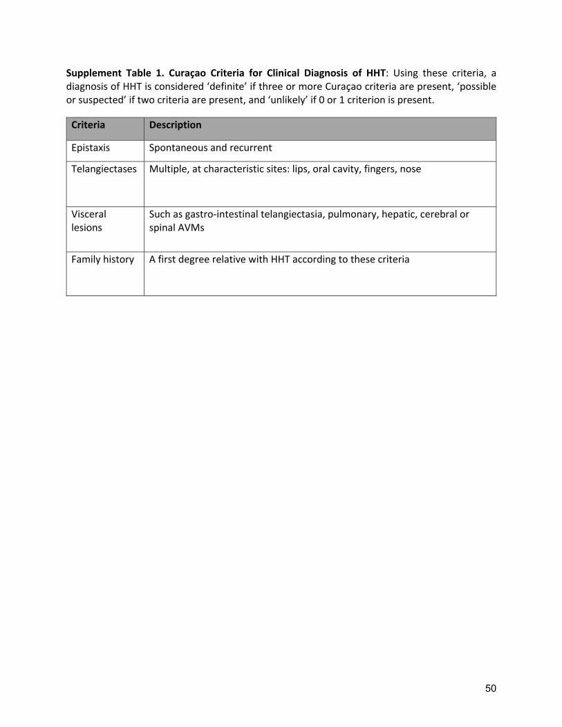
50
Supplement Table 1. Curaçao Criteria for Clinical Diagnosis of HHT: Using these criteria, a diagnosis of HHT is considered ‘definite’ if three or more Curaçao criteria are present, ‘possible or suspected’ if two criteria are present, and ‘unlikely’ if 0 or 1 criterion is present.
Criteria Description
Epistaxis Spontaneous and recurrent
Telangiectases Multiple, at characteristic sites: lips, oral cavity, fingers, nose
Visceral lesions
Such as gastro‐intestinal telangiectasia, pulmonary, hepatic, cerebral or spinal AVMs
Family history A first degree relative with HHT according to these criteria
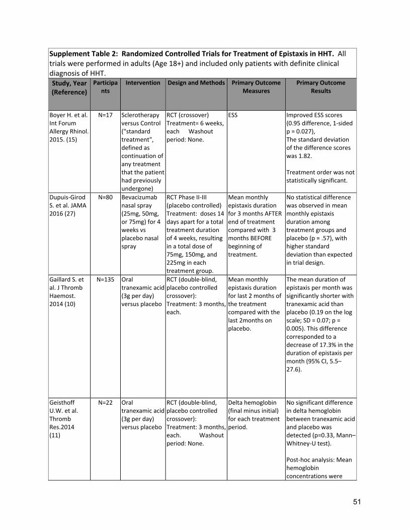
51
Supplement Table 2: Randomized Controlled Trials for Treatment of Epistaxis in HHT. All trials were performed in adults (Age 18+) and included only patients with definite clinical diagnosis of HHT. Study, Year (Reference)
Participants
Intervention Design and Methods Primary Outcome Measures
Primary Outcome Results
Boyer H. et al. Int Forum Allergy Rhinol. 2015. (15)
N=17 Sclerotherapy versus Control ("standard treatment", defined as continuation of any treatment that the patient had previously undergone)
RCT (crossover) Treatment= 6 weeks, each Washout period: None.
ESS Improved ESS scores (0.95 difference, 1‐sided p = 0.027), The standard deviation of the difference scores was 1.82. Treatment order was not statistically significant.
Dupuis‐Girod S. et al. JAMA 2016 (27)
N=80
Bevacizumab nasal spray (25mg, 50mg, or 75mg) for 4 weeks vs placebo nasal spray
RCT Phase II‐III (placebo controlled) Treatment: doses 14 days apart for a total treatment duration of 4 weeks, resulting in a total dose of 75mg, 150mg, and 225mg in each treatment group.
Mean monthly epistaxis duration for 3 months AFTER end of treatment compared with 3 months BEFORE beginning of treatment.
No statistical difference was observed in mean monthly epistaxis duration among treatment groups and placebo (p = .57), with higher standard deviation than expected in trial design.
Gaillard S. et al. J Thromb Haemost. 2014 (10)
N=135 Oral tranexamic acid (3g per day) versus placebo
RCT (double‐blind, placebo controlled crossover): Treatment: 3 months, each.
Mean monthly epistaxis duration for last 2 months of the treatment compared with the last 2months on placebo.
The mean duration of epistaxis per month was significantly shorter with tranexamic acid than placebo (0.19 on the log scale; SD = 0.07; p = 0.005). This difference corresponded to a decrease of 17.3% in the duration of epistaxis per month (95% CI, 5.5–27.6).
Geisthoff U.W. et al. Thromb Res.2014 (11)
N=22 Oral tranexamic acid (3g per day) versus placebo
RCT (double‐blind, placebo controlled crossover): Treatment: 3 months, each. Washout period: None.
Delta hemoglobin (final minus initial) for each treatment period.
No significant difference in delta hemoglobin between tranexamic acid and placebo was detected (p=0.33, Mann–Whitney‐U test). Post‐hoc analysis: Mean hemoglobin concentrations were
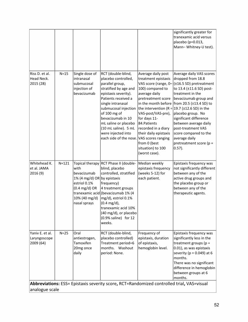
52
significantly greater for tranexamic acid versus placebo (p=0.013, Mann– Whitney‐U test).
Riss D. et al. Head Neck. 2015 (28)
N=15 Single dose of intranasal submucosal injection of bevacizumab
RCT (double‐blind, placebo controlled, parallel group, stratified by age and epistaxis severity). Patients received a single intranasal submucosal injection of 100 mg of bevacizumab in 10 mL saline or placebo (10 mL saline). 5 mL were injected into each side of the nose.
Average daily post treatment epistaxis VAS score (range, 0–100) compared to average daily pretreatment score in the month before the intervention (R = VAS‐post/VAS‐pre), for days 11‐84.Patients recorded in a diary their daily epistaxis VAS scores ranging from 0 (best situation) to 100 (worst case).
Average daily VAS scores dropped from 18.8 (±16.5 SD) pretreatment to 13.4 (±11.6 SD) post‐treatment in the bevacizumab group and from 20.5 (±13.4 SD) to 19.7 (±12.6 SD) in the placebo group. No significant difference between average daily post‐treatment VAS score compared to the average daily pretreatment score (p = 0.57).
Whitehead K. et al. JAMA 2016 (9)
N=121 Topical therapy with bevacizumab 1% (4 mg/d) OR estriol 0.1% (0.4 mg/d) OR tranexamic acid 10% (40 mg/d) nasal sprays
RCT Phase II (double‐blind, placebo controlled, stratified by epistaxis frequency) 4 treatment groups (bevacizumab 1% (4 mg/d), estriol 0.1% (0.4 mg/d), tranexamic acid 10% (40 mg/d), or placebo (0.9% saline) for 12 weeks.
Median weekly epistaxis frequency (weeks 5‐12) for each patient.
Epistaxis frequency was not significantly different between any of the active drug groups and the placebo group or between any of the therapeutic agents.
Yaniv E. et al. Laryngoscope 2009 (64)
N=25 Oral antiestrogen, Tamoxifen 20mg once daily
RCT (double‐blind, placebo controlled) Treatment period=6 months. Washout period: None.
Frequency of epistaxis, duration of epistaxis, hemoglobin level.
Epistaxis frequency was significantly less in the treatment groups (p = 0.01), as was epistaxis severity (p = 0.049) at 6 months. There was no significant difference in hemoglobin between groups at 6 months.
Abbreviations: ESS= Epistaxis severity score, RCT=Randomized controlled trial, VAS=visual analogue scale
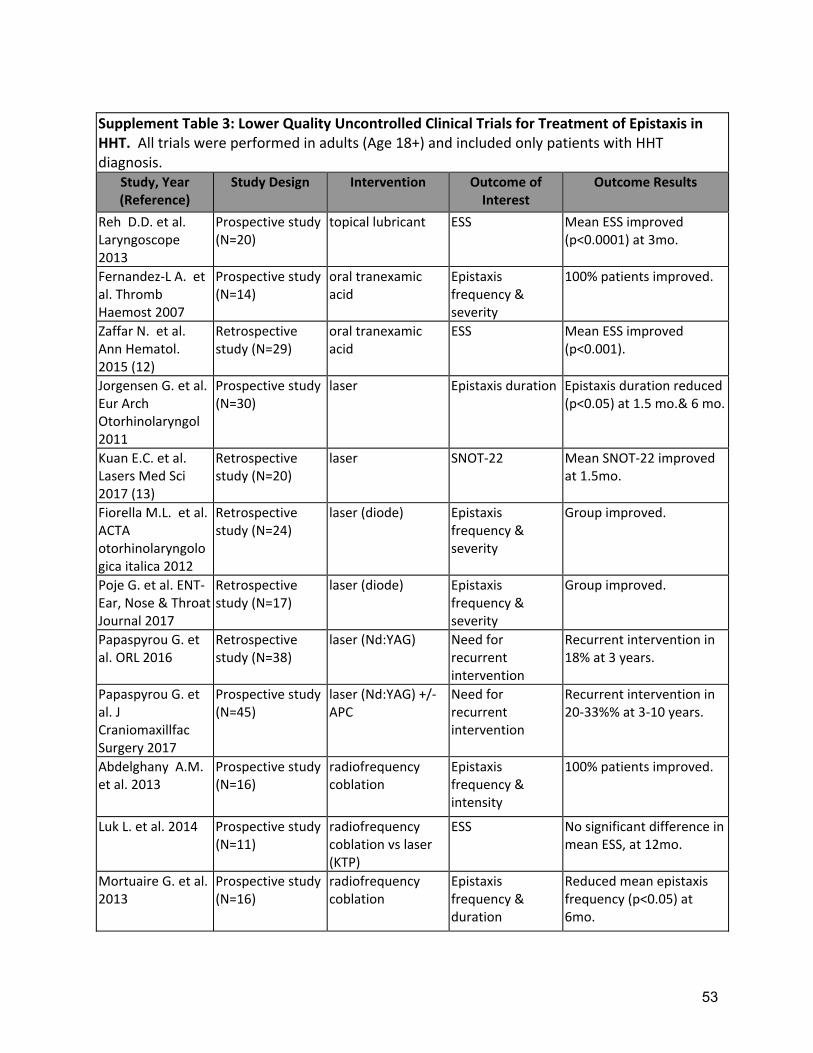
53
Supplement Table 3: Lower Quality Uncontrolled Clinical Trials for Treatment of Epistaxis in HHT. All trials were performed in adults (Age 18+) and included only patients with HHT diagnosis.
Study, Year (Reference)
Study Design Intervention Outcome of Interest
Outcome Results
Reh D.D. et al. Laryngoscope 2013
Prospective study (N=20)
topical lubricant ESS Mean ESS improved (p<0.0001) at 3mo.
Fernandez‐L A. et al. Thromb Haemost 2007
Prospective study (N=14)
oral tranexamic acid
Epistaxis frequency & severity
100% patients improved.
Zaffar N. et al. Ann Hematol. 2015 (12)
Retrospective study (N=29)
oral tranexamic acid
ESS Mean ESS improved (p<0.001).
Jorgensen G. et al. Eur Arch Otorhinolaryngol 2011
Prospective study (N=30)
laser Epistaxis duration Epistaxis duration reduced (p<0.05) at 1.5 mo.& 6 mo.
Kuan E.C. et al. Lasers Med Sci 2017 (13)
Retrospective study (N=20)
laser SNOT‐22 Mean SNOT‐22 improved at 1.5mo.
Fiorella M.L. et al. ACTA otorhinolaryngologica italica 2012
Retrospective study (N=24)
laser (diode) Epistaxis frequency & severity
Group improved.
Poje G. et al. ENT‐Ear, Nose & Throat Journal 2017
Retrospective study (N=17)
laser (diode) Epistaxis frequency & severity
Group improved.
Papaspyrou G. et al. ORL 2016
Retrospective study (N=38)
laser (Nd:YAG) Need for recurrent intervention
Recurrent intervention in 18% at 3 years.
Papaspyrou G. et al. J Craniomaxillfac Surgery 2017
Prospective study (N=45)
laser (Nd:YAG) +/‐ APC
Need for recurrent intervention
Recurrent intervention in 20‐33%% at 3‐10 years.
Abdelghany A.M. et al. 2013
Prospective study (N=16)
radiofrequency coblation
Epistaxis frequency & intensity
100% patients improved.
Luk L. et al. 2014 Prospective study (N=11)
radiofrequency coblation vs laser (KTP)
ESS No significant difference in mean ESS, at 12mo.
Mortuaire G. et al. 2013
Prospective study (N=16)
radiofrequency coblation
Epistaxis frequency & duration
Reduced mean epistaxis frequency (p<0.05) at 6mo.
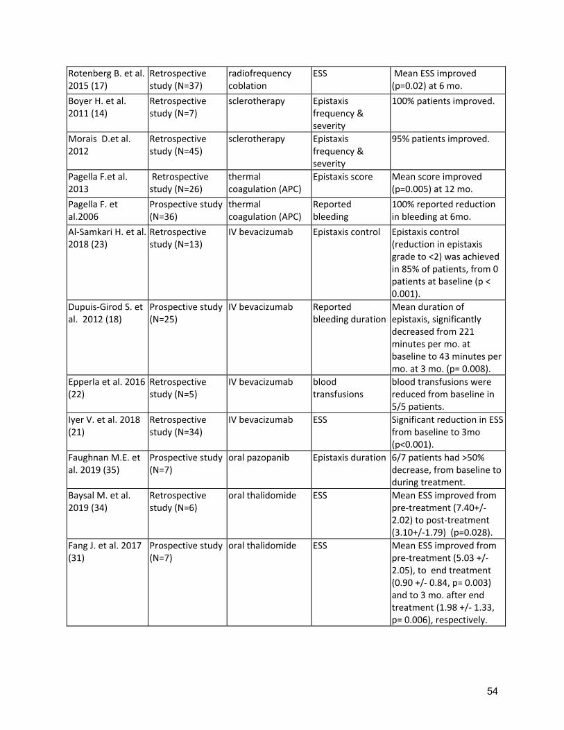
54
Rotenberg B. et al. 2015 (17)
Retrospective study (N=37)
radiofrequency coblation
ESS Mean ESS improved (p=0.02) at 6 mo.
Boyer H. et al. 2011 (14)
Retrospective study (N=7)
sclerotherapy Epistaxis frequency & severity
100% patients improved.
Morais D.et al. 2012
Retrospective study (N=45)
sclerotherapy Epistaxis frequency & severity
95% patients improved.
Pagella F.et al. 2013
Retrospective study (N=26)
thermal coagulation (APC)
Epistaxis score Mean score improved (p=0.005) at 12 mo.
Pagella F. et al.2006
Prospective study (N=36)
thermal coagulation (APC)
Reported bleeding
100% reported reduction in bleeding at 6mo.
Al‐Samkari H. et al. 2018 (23)
Retrospective study (N=13)
IV bevacizumab Epistaxis control Epistaxis control (reduction in epistaxis grade to <2) was achieved in 85% of patients, from 0 patients at baseline (p < 0.001).
Dupuis‐Girod S. et al. 2012 (18)
Prospective study (N=25)
IV bevacizumab Reported bleeding duration
Mean duration of epistaxis, significantly decreased from 221 minutes per mo. at baseline to 43 minutes per mo. at 3 mo. (p= 0.008).
Epperla et al. 2016 (22)
Retrospective study (N=5)
IV bevacizumab blood transfusions
blood transfusions were reduced from baseline in 5/5 patients.
Iyer V. et al. 2018 (21)
Retrospective study (N=34)
IV bevacizumab ESS Significant reduction in ESS from baseline to 3mo (p<0.001).
Faughnan M.E. et al. 2019 (35)
Prospective study (N=7)
oral pazopanib Epistaxis duration 6/7 patients had >50% decrease, from baseline to during treatment.
Baysal M. et al. 2019 (34)
Retrospective study (N=6)
oral thalidomide ESS Mean ESS improved from pre‐treatment (7.40+/‐2.02) to post‐treatment (3.10+/‐1.79) (p=0.028).
Fang J. et al. 2017 (31)
Prospective study (N=7)
oral thalidomide ESS Mean ESS improved from pre‐treatment (5.03 +/‐ 2.05), to end treatment (0.90 +/‐ 0.84, p= 0.003) and to 3 mo. after end treatment (1.98 +/‐ 1.33, p= 0.006), respectively.
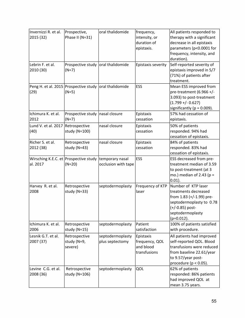
55
Invernizzi R. et al. 2015 (32)
Prospective, Phase II (N=31)
oral thalidomide frequency, intensity, or duration of epistaxis.
All patients responded to therapy with a significant decrease in all epistaxis parameters (p<0.0001 for frequency, intensity, and duration).
Lebrin F. et al. 2010 (30)
Prospective study (N=7)
oral thalidomide Epistaxis severity Self‐reported severity of epistaxis improved in 5/7 (71%) of patients after treatment.
Peng H. et al. 2015 (29)
Prospective study (N=5)
oral thalidomide ESS Mean ESS improved from pre‐treatment (6.966 +/‐ 3.093) to post‐treatment (1.799 +/‐ 0.627) significantly (p = 0.009).
Ichimura K. et al. 2012
Prospective study (N=7)
nasal closure Epistaxis cessation
57% had cessation of epistaxis.
Lund V. et al. 2017 (40)
Retrospective study (N=100)
nasal closure Epistaxis cessation
50% of patients responded. 94% had cessation of epistaxis.
Richer S. et al. 2012 (38)
Retrospective study (N=43)
nasal closure Epistaxis cessation
84% of patients responded. 83% had cessation of epistaxis.
Wirsching K.E.C. et al. 2017
Prospective study (N=20)
temporary nasal occlusion with tape
ESS ESS decreased from pre‐treatment median of 3.59 to post‐treatment (at 3 mo.) median of 2.43 (p = 0.01).
Harvey R. et al. 2008
Retrospective study (N=33)
septodermoplasty Frequency of KTP laser
Number of KTP laser treatments decreased from 1.83 (+/‐1.99) pre‐septodermoplasty to 0.78 (+/‐0.85) post‐septodermoplasty (p=0.012).
Ichimura K. et al. 2006
Retrospective study (N=15)
septodermoplasty Patient satisfaction
100% of patients satisfied with procedure.
Lesnik G.T. et al. 2007 (37)
Retrospective study (N=9, severe)
septodermoplasty plus septectomy
Epistaxis frequency, QOL and blood transfusions
All patients had improved self‐reported QOL. Blood transfusions were reduced from baseline 22.61/year to 9.57/year post‐procedure (p < 0.05).
Levine C.G. et al. 2008 (36)
Retrospective study (N=106)
septodermoplasty QOL 62% of patients responded: 86% patients had improved QOL at mean 3.75 years.

56
Rimmer J. et al. 2014 (41)
Prospective study (N=7)
septodermoplasty Epistaxis frequency & severity
100% of patients reported reduction in epistaxis frequency and severity.
Abbreviations: ESS= Epistaxis severity score, mo.=month, QOL=quality of life, SNOT=sinonasal outcome test‐22
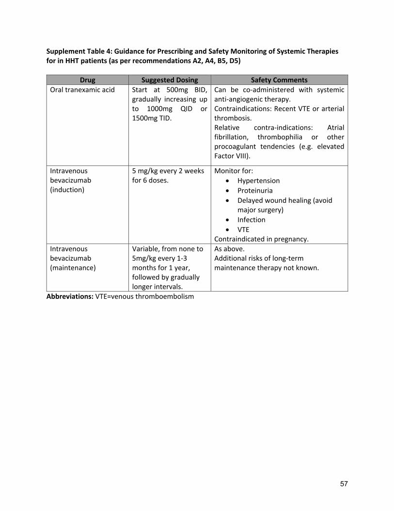
57
Supplement Table 4: Guidance for Prescribing and Safety Monitoring of Systemic Therapies for in HHT patients (as per recommendations A2, A4, B5, D5)
Drug Suggested Dosing Safety Comments
Oral tranexamic acid Start at 500mg BID, gradually increasing up to 1000mg QID or 1500mg TID.
Can be co‐administered with systemic anti‐angiogenic therapy. Contraindications: Recent VTE or arterial thrombosis. Relative contra‐indications: Atrial fibrillation, thrombophilia or other procoagulant tendencies (e.g. elevated Factor VIII).
Intravenous bevacizumab (induction)
5 mg/kg every 2 weeks for 6 doses.
Monitor for:
Hypertension
Proteinuria
Delayed wound healing (avoid major surgery)
Infection
VTE Contraindicated in pregnancy.
Intravenous bevacizumab (maintenance)
Variable, from none to 5mg/kg every 1‐3 months for 1 year, followed by gradually longer intervals.
As above. Additional risks of long‐term maintenance therapy not known.
Abbreviations: VTE=venous thromboembolism
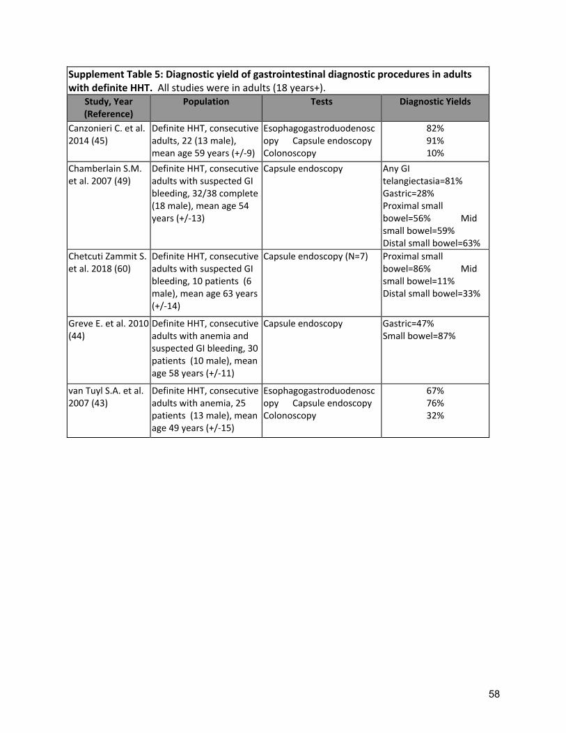
58
Supplement Table 5: Diagnostic yield of gastrointestinal diagnostic procedures in adults with definite HHT. All studies were in adults (18 years+).
Study, Year (Reference)
Population Tests Diagnostic Yields
Canzonieri C. et al. 2014 (45)
Definite HHT, consecutive adults, 22 (13 male), mean age 59 years (+/‐9)
Esophagogastroduodenoscopy Capsule endoscopy Colonoscopy
82% 91% 10%
Chamberlain S.M. et al. 2007 (49)
Definite HHT, consecutive adults with suspected GI bleeding, 32/38 complete (18 male), mean age 54 years (+/‐13)
Capsule endoscopy Any GI telangiectasia=81% Gastric=28% Proximal small bowel=56% Mid small bowel=59% Distal small bowel=63%
Chetcuti Zammit S. et al. 2018 (60)
Definite HHT, consecutive adults with suspected GI bleeding, 10 patients (6 male), mean age 63 years (+/‐14)
Capsule endoscopy (N=7) Proximal small bowel=86% Mid small bowel=11% Distal small bowel=33%
Greve E. et al. 2010 (44)
Definite HHT, consecutive adults with anemia and suspected GI bleeding, 30 patients (10 male), mean age 58 years (+/‐11)
Capsule endoscopy Gastric=47% Small bowel=87%
van Tuyl S.A. et al. 2007 (43)
Definite HHT, consecutive adults with anemia, 25 patients (13 male), mean age 49 years (+/‐15)
Esophagogastroduodenoscopy Capsule endoscopy Colonoscopy
67% 76% 32%
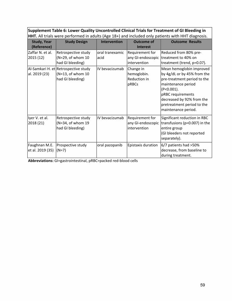
59
Supplement Table 6: Lower Quality Uncontrolled Clinical Trials for Treatment of GI Bleeding in HHT. All trials were performed in adults (Age 18+) and included only patients with HHT diagnosis. Study, Year (Reference)
Study Design Intervention Outcome of Interest
Outcome Results
Zaffar N. et al. 2015 (12)
Retrospective study (N=29, of whom 10 had GI bleeding)
oral tranexamic acid
Requirement for any GI‐endoscopic intervention
Reduced from 80% pre‐treatment to 40% on treatment (trend, p=0.07).
Al‐Samkari H. et al. 2019 (23)
Retrospective study (N=13, of whom 10 had GI bleeding)
IV bevacizumab Change in hemoglobin. Reduction in pRBCs
Mean hemoglobin improved by 4g/dL or by 45% from the pre‐treatment period to the maintenance period (P<0.001). pRBC requirements decreased by 92% from the pretreatment period to the maintenance period.
Iyer V. et al. 2018 (21)
Retrospective study (N=34, of whom 19 had GI bleeding)
IV bevacizumab Requirement for any GI‐endoscopic intervention
Significant reduction in RBC transfusions (p=0.007) in the entire group (GI bleeders not reported separately).
Faughnan M.E. et al. 2019 (35)
Prospective study (N=7)
oral pazopanib Epistaxis duration 6/7 patients had >50% decrease, from baseline to during treatment.
Abbreviations: GI=gastrointestinal, pRBC=packed red‐blood cells
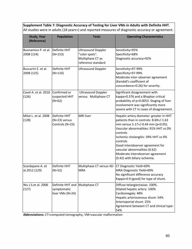
60
Supplement Table 7: Diagnostic Accuracy of Testing for Liver VMs in Adults with Definite HHT. All studies were in adults (18 years+) and reported measures of diagnostic accuracy or agreement.
Study, Year (Reference)
Population Tests Operating Characteristics
Buonamico P. et al. 2008 (124)
Definite HHT (N=153)
Ultrasound Doppler "color spots". Multiphase CT as reference standard
Sensitivity=95% Specificity=68% Diagnostic accuracy=92%
Buscarini E. et al. 2008 (125)
Definite HHT (N=110)
Ultrasound Doppler Sensitivity=97‐99% Specificity=97‐99% Moderate inter‐observer agreement (Kendall's coefficient of concordance=0.26) for severity.
Cavel A. et al. 2016 (126)
Confirmed or suspected HHT (N=62)
Ultrasound Doppler versus Multiphase CT
Significant disagreement with kappa=0.376 and a Bhapkar critical probability of p=0.0053. Staging of liver involvement was significantly more severe with CT in cases of disagreement.
Milot L. et al. 2008 (128)
Definite HHT (N=23) versus Controls (N=23)
MRI liver Hepatic artery diameter: greater in HHT patients than in controls: 8.69+/‐1.63 mm versus 5.17+/‐0.44 mm (p<0.05). Vascular abnormalities: 91% HHT vs 0% controls Ischemic cholangitis: 39% HHT vs 0% controls Good interobserver agreement for vascular abnormalities (0.62) Moderate interobserver agreement (0.42) with biliary ischemia.
Scardapane A. et al.2012 (129)
Definite HHT (N=52)
Multiphase CT versus 4D‐MRA
CT Diagnostic Yield=69% MRA Diagnostic Yield=69% No significant difference accuracy Kappa=0.9 (good) for type of shunt.
Wu J.S.et al. 2006 (127)
Definite HHT and symptomatic liver VMs (N=24)
Multiphase CT Diffuse telangiectasias: 100%. Dilated hepatic artery: 100% Cardiomegaly: 48% Hepatic arteriovenous shunt: 54% Arterioportal shunt: 25% Agreement between CT and clinical type: 54%
Abbreviations: CT=computed tomography, VM=vascular malformation
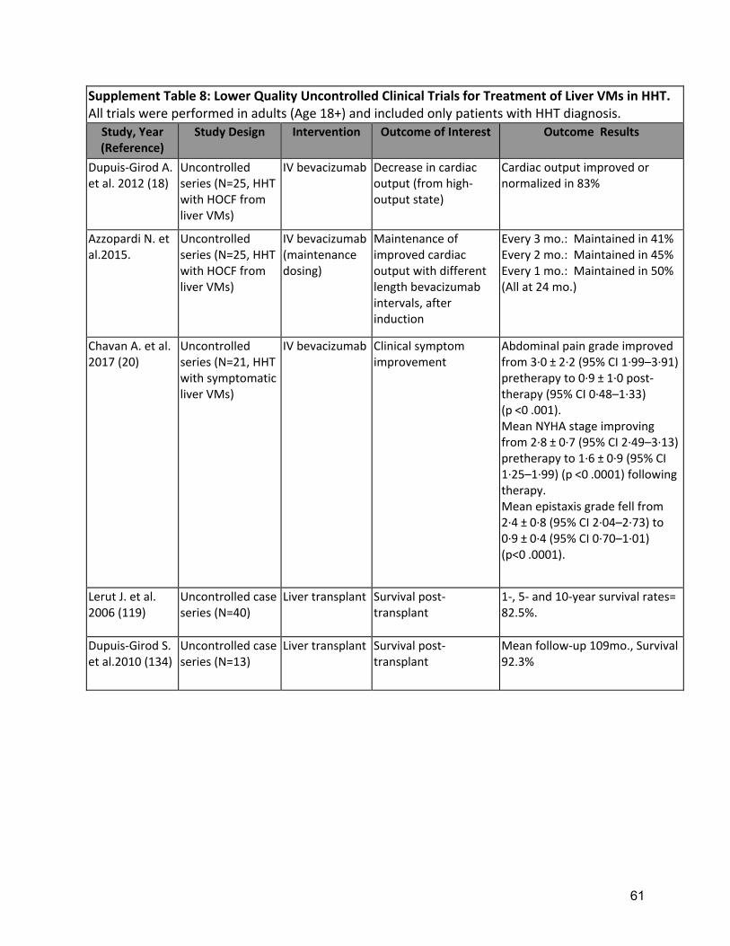
61
Supplement Table 8: Lower Quality Uncontrolled Clinical Trials for Treatment of Liver VMs in HHT. All trials were performed in adults (Age 18+) and included only patients with HHT diagnosis.
Study, Year (Reference)
Study Design Intervention Outcome of Interest Outcome Results
Dupuis‐Girod A. et al. 2012 (18)
Uncontrolled series (N=25, HHT with HOCF from liver VMs)
IV bevacizumab Decrease in cardiac output (from high‐output state)
Cardiac output improved or normalized in 83%
Azzopardi N. et al.2015.
Uncontrolled series (N=25, HHT with HOCF from liver VMs)
IV bevacizumab (maintenance dosing)
Maintenance of improved cardiac output with different length bevacizumab intervals, after induction
Every 3 mo.: Maintained in 41% Every 2 mo.: Maintained in 45% Every 1 mo.: Maintained in 50% (All at 24 mo.)
Chavan A. et al. 2017 (20)
Uncontrolled series (N=21, HHT with symptomatic liver VMs)
IV bevacizumab Clinical symptom improvement
Abdominal pain grade improved from 3∙0 ± 2∙2 (95% CI 1∙99–3∙91) pretherapy to 0∙9 ± 1∙0 post‐therapy (95% CI 0∙48–1∙33) (p <0 .001). Mean NYHA stage improving from 2∙8 ± 0∙7 (95% CI 2∙49–3∙13) pretherapy to 1∙6 ± 0∙9 (95% CI 1∙25–1∙99) (p <0 .0001) following therapy. Mean epistaxis grade fell from 2∙4 ± 0∙8 (95% CI 2∙04–2∙73) to 0∙9 ± 0∙4 (95% CI 0∙70–1∙01) (p<0 .0001).
Lerut J. et al. 2006 (119)
Uncontrolled case series (N=40)
Liver transplant Survival post‐transplant
1‐, 5‐ and 10‐year survival rates= 82.5%.
Dupuis‐Girod S. et al.2010 (134)
Uncontrolled case series (N=13)
Liver transplant Survival post‐transplant
Mean follow‐up 109mo., Survival 92.3%
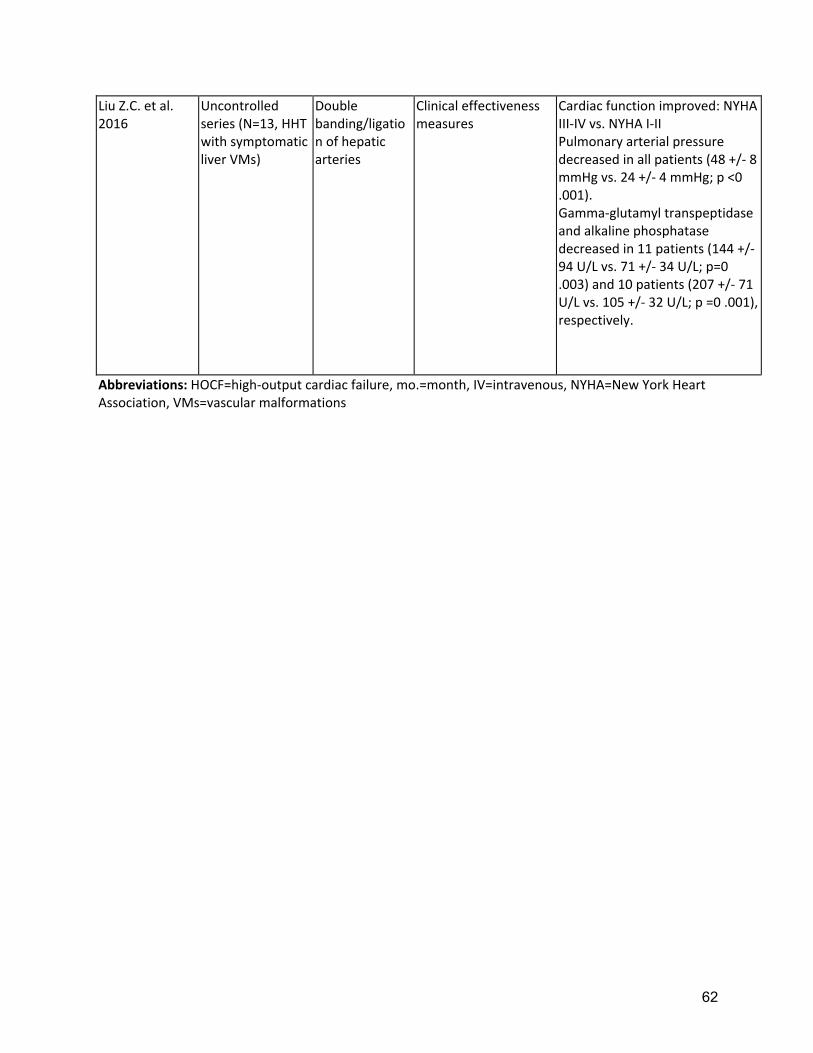
62
Liu Z.C. et al. 2016
Uncontrolled series (N=13, HHT with symptomatic liver VMs)
Double banding/ligation of hepatic arteries
Clinical effectiveness measures
Cardiac function improved: NYHA III‐IV vs. NYHA I‐II Pulmonary arterial pressure decreased in all patients (48 +/‐ 8 mmHg vs. 24 +/‐ 4 mmHg; p <0 .001). Gamma‐glutamyl transpeptidase and alkaline phosphatase decreased in 11 patients (144 +/‐ 94 U/L vs. 71 +/‐ 34 U/L; p=0 .003) and 10 patients (207 +/‐ 71 U/L vs. 105 +/‐ 32 U/L; p =0 .001), respectively.
Abbreviations: HOCF=high‐output cardiac failure, mo.=month, IV=intravenous, NYHA=New York Heart Association, VMs=vascular malformations
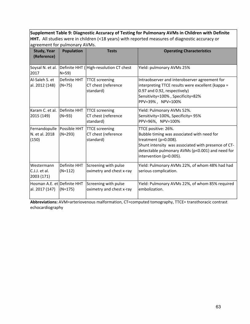
63
Supplement Table 9: Diagnostic Accuracy of Testing for Pulmonary AVMs in Children with Definite HHT. All studies were in children (<18 years) with reported measures of diagnostic accuracy or agreement for pulmonary AVMs. Study, Year (Reference)
Population Tests Operating Characteristics
Soysal N. et al. 2017
Definite HHT ( N=59)
High‐resolution CT chest Yield: pulmonary AVMs 25%
Al‐Saleh S. et al. 2012 (148)
Definite HHT (N=75)
TTCE screening CT chest (reference standard)
Intraobserver and interobserver agreement for interpreting TTCE results were excellent (kappa = 0.97 and 0.92, respectively) Sensitivity=100% , Specificity=82% PPV=39% , NPV=100%
Karam C. et al. 2015 (149)
Definite HHT (N=93)
TTCE screening CT chest (reference standard)
Yield: Pulmonary AVMs 52%. Sensitivity=100%, Specificity= 95% PPV=96%, NPV=100%
Fernandopulle N. et al. 2018 (150)
Possible HHT (N=293)
TTCE screening CT chest (reference standard)
TTCE positive: 26%. Bubble timing was associated with need for treatment (p=0.008). Shunt intensity was associated with presence of CT‐detectable pulmonary AVMs (p<0.001) and need for intervention (p=0.005).
Westermann C.J.J. et al. 2003 (171)
Definite HHT (N=112)
Screening with pulse oximetry and chest x‐ray
Yield: Pulmonary AVMs 22%, of whom 48% had had serious complication.
Hosman A.E. et al. 2017 (147)
Definite HHT (N=175)
Screening with pulse oximetry and chest x‐ray
Yield: Pulmonary AVMs 22%, of whom 85% required embolization.
Abbreviations: AVM=arteriovenous malformation, CT=computed tomography, TTCE= transthoracic contrast echocardiography

64
Supplement Table 10: Lower Quality Uncontrolled Clinical Trials for Treatment of Pulmonary AVMs and Brain VMs in HHT. All trials were performed in children (<18 years) and included only patients with HHT diagnosis.
Study, Year (Reference)
Study Design Intervention Outcome of Interest
Outcome Results
Faughnan M.E. et al. 2004 (146)
Definite HHT and pulmonary AVMs (N=42)
Transcatheter embolization of pulmonary AVMs
Reperfusion rate and safety
Reperfusion in 15% of embolized pulmonary AVMs . No serious or long‐term procedural complications.
Meybodi A.T. et al. 2018 (166)
Definite HHT and brain VMs (N=6 children treated)
Surgical management of brain VMs
Neurological outcomes
5/6 children: improved or stable mRS post‐op and 1/6 had temporarily worsened mRS post‐op.
Krings T. et al. 2005 (172)
Definite HHT and brain VMs (N=25 children treated, including 14 with brain AVFs)
Embolization Clinical outcomes 87% patients had stabilization of the disease, ameliorating the symptoms or even complete resolution.
Abbreviations: AVM=arteriovenous malformation, VM=vascular malformations, mRS=modified Rankin score