Study of the impact of TNF-α/TNFR2 signalling pathway ...
184
HAL Id: tel-03423533 https://tel.archives-ouvertes.fr/tel-03423533 Submitted on 10 Nov 2021 HAL is a multi-disciplinary open access archive for the deposit and dissemination of sci- entific research documents, whether they are pub- lished or not. The documents may come from teaching and research institutions in France or abroad, or from public or private research centers. L’archive ouverte pluridisciplinaire HAL, est destinée au dépôt et à la diffusion de documents scientifiques de niveau recherche, publiés ou non, émanant des établissements d’enseignement et de recherche français ou étrangers, des laboratoires publics ou privés. Study of the impact of TNF-α/TNFR2 signalling pathway blockade on the allogeneic antitumour response after hematopoietic stem cell allograft Audrey Moatti To cite this version: Audrey Moatti. Study of the impact of TNF-α/TNFR2 signalling pathway blockade on the allogeneic antitumour response after hematopoietic stem cell allograft. Cellular Biology. Université Paris-Est, 2020. English. NNT : 2020PESC0041. tel-03423533
-
Upload
khangminh22 -
Category
Documents
-
view
0 -
download
0
Transcript of Study of the impact of TNF-α/TNFR2 signalling pathway ...
HAL Id: tel-03423533https://tel.archives-ouvertes.fr/tel-03423533
Submitted on 10 Nov 2021
HAL is a multi-disciplinary open accessarchive for the deposit and dissemination of sci-entific research documents, whether they are pub-lished or not. The documents may come fromteaching and research institutions in France orabroad, or from public or private research centers.
L’archive ouverte pluridisciplinaire HAL, estdestinée au dépôt et à la diffusion de documentsscientifiques de niveau recherche, publiés ou non,émanant des établissements d’enseignement et derecherche français ou étrangers, des laboratoirespublics ou privés.
Study of the impact of TNF-α/TNFR2 signallingpathway blockade on the allogeneic antitumour response
after hematopoietic stem cell allograftAudrey Moatti
To cite this version:Audrey Moatti. Study of the impact of TNF-α/TNFR2 signalling pathway blockade on the allogeneicantitumour response after hematopoietic stem cell allograft. Cellular Biology. Université Paris-Est,2020. English. �NNT : 2020PESC0041�. �tel-03423533�
UNIVERSITE PARIS-EST École Doctorale Sciences de la Vie et de la Sante
These de doctorat
Biologie cellulaire et moléculaire Specialite Immunologie
Study of the impact of TNF-α/TNFR2
signalling pathway blockade on the allogeneic
antitumour response after hematopoietic
stem cell allograft
Étude de l’effet du blocage de la voie de signalisation du TNF-α/TNFR2 sur l’efficacité de la
réponse alloréactive anti-tumorale après greffe de cellules souches hématopoïétiques
Audrey Moatti
Soutenue le 25 septembre 2020
Jury :
Dr. Eliane PIAGGIO Rapporteur Pr. Frédéric BARON Rapporteur Pr. Alain FISCHER Examinateur Dr. David MICHONNEAU Examinateur Dr. Allan THIOLAT Encadrant Dr. Fadi ISSA Co-directeur de thèse Pr. José COHEN Directeur de thèse

2
Host laboratories
Main laboratory
INSERM U955 – Équipe COHEN
Institut Mondor de Recherche Biomédicale (IMRB)
Hôpital Henri Mondor
51 avenue du Maréchal de Lattre de Tassigny
94010 CRETEIL, FRANCE
Mobility laboratory
Transplant Research Immunology Group (TRIG)
Nuffield Dept of Surgical Sciences
John Radcliffe Hospital
Headley Way, Headington
OX3 9DU OXFORD, UK
3
Keywords
• Tumour Necrosis Factor Receptor 2 (TNFR2)
• Tumour Necrosis Factor alpha (TNF-α)
• Regulatory T cells (Treg)
• Hematopoietic stem cells allograft (alloHSCT)
• Immunotherapy
• Graft-versus-tumour effect (GvT)
4
Mots-clés
• Récepteur de type 2 au TNF- (TNFR2)
• TNF-α
• Lymphocytes T régulateurs (Treg)
• Greffe de cellules souches hématopoïétiques allogénique (alloCSH)
• Immunothérapie
• Effet du greffon contre la tumeur
5
Abstract
Allogeneic hematopoietic stem cell transplantation (alloHSCT) is a standard consolidation
therapy for high-risk hematologic malignancies associated with a lower relapse risk than
chemotherapy alone. If part of its efficacy is due to the high dose chemotherapy and/or
radiotherapy that represents the first-line treatment, a proper graft-versus-tumour (GVT)
effect has been long-time recognized. It relies on alloreactive donor cytotoxic T cells activation
in the recipient body, then capable of residual tumour cells elimination. Unfortunately,
underlying malignancy relapse remains the principal cause of mortality post-allotransplant.
Therapeutic options available until now have shown low efficacy to treat post-alloHSCT
relapses, often linked to a poor prognosis. Interestingly, removing ex vivo regulatory T cells
(Treg), that control alloreactive response, from the graft have proved to be efficient in raising
the conventional T cells efficacy.
If alloHSCT was the first immunotherapy available against cancer, promising new approaches
are now emerging that target immune checkpoints restraining anti-tumour effector
responses. It was demonstrated that tumour necrosis factor type 2 receptor (TNFR2) was
critical for the phenotypic stabilization and the suppressive function of human Treg. Our team
previously revealed that blocking the TNFR2 pathway led to a complete loss of Treg protective
function in a model of graft-versus-host disease prevention by Treg-based cell therapy.
Based on these results, we tested the possibility of amplifying an anti-tumour response by
blocking TNFR2 in a model of tumour relapse following alloHSCT. In experimental conditions
in which neither the donor T lymphocytes nor the anti-TNFR2 antibody per se has any effect
on tumour relapse separately, we observed that the co-administration of a sub-optimal
number of T cells associated with an anti-TNFR2 treatment could set off alloreactivity and
induce a subsequent dramatic anti-tumour effect. This was associated in the spleen with a
reduced Treg/T cell ratio and an impaired function of both CD4+ and CD8+ Treg. These results
validate TNFR2 as a new target molecule for the development of immunotherapies to treat
haematological malignancy relapse. They also open up new perspectives on the possibility of
amplifying, more widely in cancer, an anti-tumour response by directly targeting the Treg.

6
Résumé court
La transplantation de cellules souches hématopoïétiques (CSH) est une stratégie standard de
consolidation des cancers hématologiques agressifs, associée à un risque de rechute inférieur
à celui de la chimiothérapie seule. Une partie de son efficacité repose sur une première étape
de chimiothérapie et/ou de radiothérapie à fortes doses, mais le greffon lui-même induit un
effet anti-tumoral puissant dans un second temps. Ce dernier est provoqué par l’alloréactivité
des lymphocytes T cytotoxiques du donneur qui s’activent au contact des tissus du receveur
et deviennent capable de détruire les potentielles cellules tumorales résiduelles. Malgré cela,
la principale cause de mortalité post-allogreffe demeure la rechute tumorale. La survenue
d’une rechute est associée à un pronostic particulièrement sombre, d’autant plus que les
stratégies thérapeutiques employées jusqu’à ce jour ont montré une efficacité limitée. La
déplétion des lymphocytes T régulateurs (Treg) du greffon, cellules responsables du contrôle
de la réponse adaptative, s’est montrée efficace pour améliorer l’alloréactivité des T
conventionnels.
Historiquement, l’allogreffe fut la première immunothérapie disponible contre le cancer. Ces
dernières années, de nouvelles approches prometteuses émergent, en particulier les
thérapies ciblant les points de contrôle du système immunitaire. Le récepteur de type 2 au
TNFα (TNFR2) est l’un de ces freins de la réponse anti-tumorale de par son rôle critique dans
la stabilité phénotypique et la capacité suppressive des Treg. Notre équipe a récemment
démontré, dans un modèle murin de greffe de moelle osseuse, que le blocage de la voie de
signalisation du TNFR2 entravait l’effet préventif de la maladie du greffon contre l’hôte
exercée par une infusion de Treg.
A partir de ces résultats, nous avons testé la possibilité d’exacerber la réponse anti-tumorale
en inhibant la voie de signalisation du TNFR2. Dans un modèle mimant la rechute tumorale
post-allogreffe, ni l’infusion d’une faible quantité de lymphocytes T du donneur, ni l’injection
d’un anticorps bloquant le TNFR2 n’influencent la survenue d’une tumeur. Au contraire, la co-
administration d’une dose sous-optimale de lymphocytes T du donneur au traitement anti-
TNFR2 entraine un puissant effet anti-tumoral. L’amplification de la réponse allogénique dans
7
ces conditions est liée à une altération de la fonction des Treg CD4+ et CD8+, ainsi qu’à une
baisse du ratio Treg/T effecteurs. Ces résultats valident l’utilisation du TNFR2 comme nouvelle
cible immunothérapeutique dans le cadre des rechutes de cancer hématologiques. A plus
large échelle, une thérapie permettant de restreindre directement l’action suppressive des
Treg pour amplifier la réponse immunitaire anti-tumorale ouvrirait de nouvelles perspectives
de traitement en première ligne pour de nombreux types tumoraux.
8
Résumé long
L’allogreffe de cellules souches hématopoïétiques (alloHSCT), première
immunothérapie anti-tumorale initiée dans les années 1950, reste aujourd’hui encore le seul
recours thérapeutique pour des patients atteints d’hémopathies malignes chimio-résistantes.
Cette stratégie repose non seulement sur les cellules souches du donneur pour remplacer le
système hématopoïétique du patient, mais aussi sur l’efficacité des lymphocytes T matures
contenus dans le greffon. En effet, les T du donneur présents dans la greffe permettent
l’élimination de potentielles cellules tumorales résiduelles, assurant ainsi une efficacité anti-
tumorale durable (abrégé GVL, pour « garft-versus-leukemia effect »). Au contraire, une
alloréactivité insuffisante des T du donneur explique une partie des rechutes leucémiques
post-alloHSCT. Pour contourner ce problème, mon laboratoire d’accueil a conduit avec succès
le premier essai clinique de déplétion ex vivo des lymphocytes T régulateurs (Treg), cellules
qui freinent la réponse immunitaire, afin de réactiver une réponse anti-tumorale (Maury et
al., 2010, 2014). Depuis quelques années, plutôt que d’avoir recours à des approches de
thérapie cellulaire, mon laboratoire d’accueil s’intéresse aux facteurs de survie des Treg. Avant
mon arrivée, ils ont démontré que le blocage du récepteur au TNFα de type II (TNFR2)
fortement exprimé par les Treg murins et humains inhibe leur effet suppresseur in vivo dans
un modèle d’alloHSCT (Leclerc at al., 2016).
Faisant suite à ces travaux, l’objectif principal de ma thèse fut de réactiver l’alloréactivité en
bloquant la fonction des Treg via la voie TNFR2 et de provoquer ainsi un effet anti-tumoral
puissant dans des modèles de rechutes leucémiques post-greffe chez la souris. Une première
tâche a donc été de mettre en place un modèle préclinique de rechute tumorale post-
alloHSCT. Le deuxième objectif de ma thèse consiste à tester la possibilité de transférer ces
résultats chez l’homme, (i) en analysant l’expression du TNFR2 sur des prélèvements de
patients (ii) des modèles in vitro de cytotoxicité et (ii) des modèles in vivo de tumeurs du sang
humaines chez la souris immunodéficiente. Ce second axe a été très initié au court de ma
thèse, et a fait le fruit de plusieurs collaborations, dont une mobilité de 9 mois à Oxford au
sein du Wood Lab. Cependant nous nous sommes heurtés à des difficultés concernant la mise
en place de modèles permettant l’observation d’un effet de la « xénogreffe contre la tumeur »
9
comparable à celui observé chez les patients. Il fera ainsi l’objet de publications ultérieures et
ne sera pas présenté dans cette thèse par manque de résultats finalisés et pour plus de clarté,
la biologie de ces modèles nécessitant à elle seule un chapitre introductif extrêmement dense.
Concernant le premier objectif, le modèle murin de rechutes leucémiques post-greffe
a été mis en place avec succès au début de ma thèse. Dans la première série d'expériences,
nous avons essayé de définir le nombre maximum de cellules tumorales et de cellules T
nécessaires à injecter chez les souris receveuses afin de créer un modèle de rechute tumorale
en l'absence de GVHD. Nous avons fixé ces nombres à 2x10⁴ pour les cellules P815 et 106 pour
les lymphocytes T, et avons ainsi défini les conditions expérimentales appropriées imitant
celles des patients en rechute de leurs hémopathies malignes après alloHSCT. Les souris
recevant 106 lymphocytes T et traitées avec des anti-TNFR2 à J0, J2 et J4 ont développé une
GVHD comme en témoigne un faible taux de survie au jour 40 et un grade clinique élevé de
GVHD. Chez les souris non traitées, aucun signe de la maladie n'a jamais été détecté. Afin de
valider notre hypothèse, nous avons reproduit ces expériences en présence de cellules P815.
Là encore, l’administration de l’anticorps anti-TNFR2 administrés à J0, J2 et 4, ou uniquement
à 10, a entraîné des manifestations cliniques de GVHD, alors qu'aucun signe de GVHD n'a été
observé chez les souris non traitées. Des cellules P815 ont été détectées chez toutes les souris
qui n'avaient pas reçu de lymphocytes T et chez 64% des souris ayant reçu des lymphocytes T.
En revanche, l'administration d'anti-TNFR2 à J0 ou à J10 a entraîné une diminution
spectaculaire de l'incidence des tumeurs puisque des cellules P815 ont été détectées chez
20% et 12,5% des souris, respectivement. Nos travaux antérieurs ont démontré la possibilité
d’inhiber des Treg thérapeutiques, administrés à fortes doses dans un modèle de prévention
de la GVHD, par un traitement anti-TNFR2 in vivo. Dans cette étude, nous avons démontré
qu'un traitement anti-TNFR2 permet de déclencher directement l'alloréactivité dans un
contexte où les lymphocytes T perfusés seuls ne sont pas capables de le faire. Nous avons
également démontré que le traitement anti-TNFR2 peut médier un effet GVL puissant dans
un modèle expérimental approprié de rechute hématologique maligne après alloHSCT. Cette
approche thérapeutique est non seulement aussi efficace, mais aussi beaucoup plus simple et
polyvalente que notre premier essai clinique chez l'homme basé sur la déplétion de Treg ex
vivo. Ceci est attesté par la possibilité, comme démontré ici, de traiter de manière préventive

10
(à J0) ou curative (à J10) en donnant aux lymphocytes T la possibilité de médier l'effet GVL
avant toute intervention thérapeutique ultérieure.
Cliniquement parlant, le traitement anti-TNFR2 serait préférentiellement retardé jusqu'à ce
que la rechute leucémique soit confirmée. Nous avons donc choisi le douzième jour pour
rechercher les mécanismes sous-jacents impliqués dans le déclenchement de l'alloréactivité
et l'effet GVL consécutif chez les souris traitées à J10. Deux jours après le traitement, plusieurs
manifestations histologiques de la GVHD ont été observées au niveau de la peau, du foie, de
l'intestin grêle et du côlon de souris traitées versus non traitées conformément aux
observations cliniques, renforçant ainsi la fiabilité et la sensibilité de notre nouveau système
de notation clinique publié précédemment. Le pourcentage de lymphocytes T CD8+ a
augmenté conduisant à une diminution significative du rapport Treg / Teff, une modification
en faveur de l'effet GVL. Ceci est compatible avec la tendance à l'augmentation des nombres
de CD4 et CD8, et l'augmentation statistiquement significative du pourcentage de cellules T
KI-67+ CD8 en division, par rapport aux souris non traitées. Inversement, le pourcentage de
TNFR2 exprimé par les lymphocytes T est considérablement diminué sans abolir ni la capacité
du traitement anti-TNFR2 à déclencher l'alloréactivité, ni l'effet GVL associé.
Nous avons ensuite focalisé notre analyse sur les Treg (CD4+ Foxp3+), principale population
cellulaire ciblée de la stratégie thérapeutique anti-TNFR2. A J12, le pourcentage de Treg dans
les lymphocytes T CD4 + a légèrement diminué, et il est intéressant de noter que le niveau
d'expression de Foxp3 était significativement réduit lorsque l'interaction TNF/TNFR2 était
inhibée, ce qui est cohérent avec le lien récemment décrit entre TNFR2 et Foxp3. De plus, le
niveau d’expression de CTLA-4, TNFR2, et CD25 à la surface des Treg est considérablement
diminué, ainsi que la densité d’expression de ces deux derniers marqueurs. Des observations
similaires mais moins marquées ont été observées chez les souris à traitées à J0. Une
population de lymphocytes T CD8+ Foxp3+ avec des fonctions suppressives exercées tôt dans
la GVHD a été identifiée chez la souris et chez l'homme. De même, dans notre modèle, nous
avons observé une sous-population émergente de cellules T CD8+ Foxp3+ qui présentait une
expression élevée de TNFR2 ainsi que des molécules CD25 et CTLA-4. Après traitement anti-
TNFR2 le pourcentage de lymphocytes T CD8+Foxp3+ est légèrement diminué, mais surtout,
le pourcentage de lymphocytes T CD8+ Foxp3+ exprimant le TNFR2, CD25 et CTLA-4 apparaît
11
considérablement réduit. Henrich et al. ont découvert que les lymphocytes T CD8 + Foxp3 +
alloréactifs atténuent modérément la GVHD tout en épargnant l'effet GVL (Henrich et al.,
2016). Ici, nous avons démontré que réduire le nombre de ces cellules via un traitement anti-
TNFR2 mais surtout diminuer leur état d'activation augmentait l'alloréactivité sans abroger
l'effet GVL associé. En conclusion, nous avons montré que le blocage de la voie TNFR2 se
traduit par une augmentation rapide de l'alloréactivité, et par conséquent une augmentation
de l'effet GVL / GVT. Ceci s'explique principalement par la réduction spectaculaire de
l'activation de CD4+ et CD8+ Treg. Nous pensons que nos travaux valident, dans un modèle
préclinique pertinent, la possibilité de traiter la rechute leucémique post-greffe en bloquant
l'effet suppresseur des Treg. De plus il pourrait permettre un effet anti-tumoral direct lorsque
les cellules tumorales expriment le TNFR2 comme récemment proposé dans la littérature
(Torrey et al., 2019). Pour cela, nous encourageons fortement le développement rapide de
mAb bloquant le TNFR2 de qualité clinique humaine.
Dans ce but, la deuxième partie de mon projet visait à démontrer que ce type de
stratégie thérapeutique peut être appliquée chez l’homme, cet aspect ne sera pas détaillé
dans le corps de la thèse mais dans sa discussion. Il n’existe pas à ce jour de traitement de
grade clinique permettant de bloquer le TNFR2 humain, c’est pourquoi la première année de
mon doctorat a été consacrée au criblage de différents anticorps anti-hTNFR2. Un premier
candidat a été sélectionné après avoir démontré une efficacité sur la survie et la fonction des
Treg dans des protocoles d’expansion de Treg. Notamment sur une préparation de cellules
humaines enrichies en CD25+ activées, le blocage du TNFR2 avec cet anticorps a permis un
enrichissement en CD8+ effecteurs activés et une diminution du nombre de Treg. Afin de
tester son effet sur la fonction des Treg in vivo dans un modèle relevant, j’ai effectué une
mobilité de 9 mois dans le laboratoire du Pr Kathryn Wood à Oxford. Ce laboratoire a
démontré dans un modèle de souris immunodéficientes qu’une infusion de Treg humains est
capable de protéger d’un rejet de greffe de peau humaine par des PBMC humains allogéniques
(Issa et al., 2010). Pour différentes raisons inhérentes à la difficulté du modèle nous n’avons
pas observé d’effet du traitement dans le temps imparti à ma mobilité. Dans l’optique de
tester in vivo l’effet d’un anticorps bloquant le TNFR2 humain, j’ai également travaillé sur la
mise en place d’un modèle de maladie du greffon contre l’hôte xénogénique déclenchée par
l’injection de PBMC humains à des souris immunodéficientes. Les tests dans ce modèle de
12
l’effet de l’anticorps sur l’apparition de la maladie se sont en cours et seront poursuivis au
laboratoire. Nous avons également mis au point un modèle de développement tumoral chez
la souris immunodéficiente et quantifié, comme précédemment chez la souris
immunocompétente, la dose sub-optimale de lymphocytes T humains à injecter pour ne pas
rejeter une lignée leucémique humaine (RS4) administrée en même temps que les PBMC
humains. Ce modèle permettra prochainement de tester in vivo l’efficacité anti-tumorale du
candidat anticorps bloquant anti-TNFR2 humain.

12
Acknowledgements
ACKNOWLEDGEMENTS
14
Une thèse n’est pas un travail solitaire. Je tiens donc à exprimer ma profonde reconnaissance
à toutes les personnes qui ont contribué directement à ce travail, mais aussi à celles qui m’ont
soutenu au quotidien lors de la préparation de mon doctorat.
Tout d’abord, un grand merci à mes deux rapporteurs, le Docteur Eliane Piaggio et le
Professeur Frédéric Baron d’avoir accepté de prendre le temps d’évaluer le présent manuscrit,
ainsi qu’au Docteur David Michonneau de participer à mon jury en temps qu’examinateur. Je
remercie également le Professeur Alain Fischer de bien vouloir être le président de ce jury.
C’est pour moi un honneur de présenter mon travail et de pouvoir discuter avec des
immunologistes d’un tel niveau d’expertise sur la greffe de moelle osseuse, en recherche et
en pratique clinique.
A mon directeur de thèse, le Professeur José Cohen, j’adresse ma plus sincère et profonde
gratitude. José, tu m’as accordé ta confiance pour poursuivre un projet qui te tenait à cœur,
et tu m’as soutenu sans relâche dans les différentes étapes de ma thèse. Avec ton optimisme
à toute épreuve tu es capable de rendre possible l’impossible. J’espère que tu pourras
continuer à encadrer des doctorants malgré tes responsabilités grandissantes, ce fut en tout
cas pour moi un privilège d’être formée à la recherche à tes côtés. Merci pour tout José.
Merci à Allan pour l’énorme travail qu’il a accompli depuis le début de ce projet, qui fut aussi
le sien, mais également à Caroline qui m’a accompagnée sans cesse dans les diverses
expériences avec toujours une expertise incomparable. Merci à Anaïs qui fut ma première
étudiante, pour son apport conséquent au projet TNFR2 lors de son master 2, et son aide
précieuse au cours des expériences qui ont suivi. Je remercie le Docteur Frédéric Charlotte
pour l’analyse histologique de nos très nombreux prélèvements murins en un temps record,
ainsi que les « filles de la plateforme de cytométrie » pour leur aide et la bonne ambiance que
je trouvais toujours au cytomètre. Je remercie enfin chaleureusement tous les membres
passés du laboratoire et les nouveaux arrivants dans l’équipe « réunifiée », qui ont contribué
à mon travail par leurs conseils et en établissant une bonne ambiance de travail.
J’ai une pensée toute particulière pour ma partenaire de thèse, Chloé. Toujours là pour me
donner un coup de main, égayer mes journées, et me rappeler que manger c’est important.

ACKNOWLEDGEMENTS
15
Malgré mon départ, rien ne remplacera dans mon cœur ta dramatiquement mauvaise
imitation des accents belges, canadiens et allemands pendant la pesée des souris. A l’avenir
fais attention à ne plus laisser tomber de langes... Tu vas me manquer, mais tata Audrey ne
sera jamais loin. Finalement, une mention spéciale pour les membres de l’équipe 21 bis, et
surtout Julie qui fut une extraordinaire co-doctorante pendant 3 ans, je n’ai jamais rencontré
une personne dotée d’une telle volonté de bien faire et d’aider les autres, merci pour tout.
Carole, toujours sur tous les fronts, tu réussis avec brio à conjuguer dix vies parallèles (sans
parler de tes nombreux amants..). J’ai pour toi une admiration énorme, merci pour tous tes
conseils et ton écoute. Hamza, bravo d’avoir rendu le métro plus respirable : parler
d’expériences ratées et de cheeseburgers est toujours un excellent remède contre la ligne 8.
I was very fortunate to prepare this work, in two different laboratories. For providing me with
this amazing opportunity, my gratitude goes to Professor Katheryn Wood who opened the
doors of her lab in Oxford to me. Doctor Fadi Issa, who agreed to be my co-director was always
helpful and supportive to me, not only during my stay in England, but also for the rest of my
PhD. I cannot fail to say thank you to Doctor Joanna Hester, who kindly shared her time,
precious advice and huge Treg experience with me, and to Monica Dolton, the best problem
solver I will probably ever meet. I was lucky to work with brilliant students in the lab who were
always kindly helping me. To Masateru and Kento, I am so grateful for the huge work you did
in the animal house, in addition to yours, since I could not access it myself. A big thanks to
Rebeca for teaching me a lot of experimental methods, and for providing me with a “Spanish
family” outside the lab when my English was still experimental. Prateek I will remember you
for your amazing scientific energy, “bubbling” ideas and the countless snacks you fed me with.
Finally, thank you to the other TRIG students at the time Alaa, Kostas, Hisashi, Oliver, George
and my adorable little Banu. Then, to the countless people I have met outside the lab and
helped me survive the cold of England winter, thanks for your friendship. Zach, it was so much
fun crossing your path again, in England this time. I will see you in New Orleans for the next
Mardi Gras, I promise! A special thought to my invaluable (and numerous!) roommates:
Cristina, Dan, Karl, Bec, Abbie, Andrew, Pascal, Eva, Britt and Derek. I also was lucky to gain an
“English mum” in Oxford. Kate, you have been much more than a colleague for me, saving me
when I was homeless, explaining everything to me 10 times and sharing with me your
passions. I will always remember the taste of elderflower. Thank you so much for everything.
ACKNOWLEDGEMENTS
16
Je tiens à remercier également très chaleureusement mes colocataires parisiens, qui ont
contribué chacun à leur manière à embellir mon quotidien de thésarde. Julien et Antoine, vous
êtes partis il y a 10 minutes mais je me sens déjà très seule. Merci à notre « voisine », Maylis,
notamment de m’avoir accompagnée durant le peu de pratique sportive que j’ai pu avoir.
Merci aux anciens du master 1 de cancérologie qui me suivent et m’épaulent depuis mon
arrivée à Paris. Un grand merci également à tous ceux qui, hors labo, m’ont abreuvé de cas
cliniques et d’histoires médicales en tout genre, et tout particulièrement Guillaume, me
permettant de mieux comprendre la réalité clinique derrière mon sujet de recherche. Une
pensée très forte pour mes amies de toujours, Amandine et Hajiba pour leur soutien
indéfectible depuis notre rencontre, quoi que j’entreprenne. Enfin, Ana, merci de tout cœur
pour ton amitié inestimable. Je ne connais pas beaucoup d’amis (même imaginaires !) qui
auraient pris le temps de relire une thèse entière sans intérêt pour le contenu. Bien au-delà
de la thèse, tu es avec moi à chaque étape depuis des années je ne suis jamais vraiment seule.
Je garde en mémoire tous les cafés, films, promenades, carabineiros et paysages dramatiques
qu’on n’a pas pu manger, boire, voire ou faire ensemble et que je dois inventer. C’est
incroyable parce que même sans tout ça, tu es mon ombre et je serais toujours la tienne.
Je tiens enfin à remercier ma famille qui a fait de moi celle que je suis aujourd’hui. Je n’aurai
pas pu aller au bout de cette thèse sans la ténacité héritée de mon père, et l’ouverture d’esprit
que nous a inculqué ma mère. A mon petit frère Julien, sans qui mon enfance aurait été bien
morne, je tiens à souhaiter ici un excellent début de thèse. J’espère que ce sera pour toi une
expérience aussi riche qu’elle le fut pour moi. Chloé, toujours ensemble depuis ma naissance,
tu étais avec moi pendant la thèse également, merci pour tous les bons moments passés et
ceux à venir. J’attends maintenant avec impatience de rencontrer le nouveau petit membre
de la famille… et de savoir s’il s’appellera Auguste.
Un dernier changement de langue s’impose, en hommage à celle qui m’a soutenue en
permanence durant cette thèse. Nestas derradeiras liñas, quero expresar un inmenso
agradecemento á miña avoa. Ti, que me axudaches tanto, con todo o que puideches. Ti, que
es a muller máis capaz, máis valente e traballadora que coñezo. Así, ao teu xeito e sen
decatarte, ensinachesme unha morea de cousas tódolos días. Coídate moito avoiña, e lembra
que sempre serás un exemplo para min.
16
Table of contents
KEYWORDS ........................................................................................................................ 2
MOTS-CLES ........................................................................................................................ 4
ABSTRACT ......................................................................................................................... 5
RÉSUME COURT ................................................................................................................. 6
RÉSUME LONG................................................................................................................... 8
ACKNOWLEDGEMENTS .................................................................................................... 13
ABBREVIATIONS ............................................................................................................... 21
INTRODUCTION ............................................................................................................... 24
Avant-propos ........................................................................................................................................... 25
Allogeneic hematopoietic stem-cells transplantation ........................................................... 26
A. Discovery and principle ............................................................................................................................ 26
B. Clinical settings ......................................................................................................................................... 28
1. Overview .............................................................................................................................................. 28
2. Patient eligibility for alloHSCT ............................................................................................................. 28
a) Underlying disease impact ............................................................................................................. 29
b) Individual factors impact ................................................................................................................ 31
3. Sources of hematopoietic stem cells................................................................................................... 33
4. Donor selection ................................................................................................................................... 35
5. Preparative regimens .......................................................................................................................... 37
6. Post-alloHSCT monitoring ................................................................................................................... 40
a) Engraftment and chimerism ........................................................................................................... 40
b) Short- and long-term care .............................................................................................................. 41
7. Complications and management ........................................................................................................ 42
a) Relapses and post-alloHSCT secondary malignancies .................................................................... 45
18
b) Infectious risk .................................................................................................................................. 47
c) Graft-versus-host disease ............................................................................................................... 48
(1) Acute .......................................................................................................................................... 49
(2) Chronic ....................................................................................................................................... 52
(3) Preventive action against GvHD ................................................................................................ 54
(a) aGvHD risk factors ................................................................................................................. 54
(b) Current immunosuppressive prophylaxis ............................................................................. 56
(c) Partial T cell depletion strategies .......................................................................................... 57
Immunotherapeutic effect of alloHSCT ................................................................................ 59
A. GvT effect: Definition and clinical evidences ........................................................................................... 60
B. Establishment of the alloreactive response ............................................................................................. 62
C. Allogeneic effector phase......................................................................................................................... 64
1. T cells trafficking .................................................................................................................................. 64
2. Effector/memory profile ..................................................................................................................... 65
3. T helper subsets................................................................................................................................... 65
4. Antigen specificity ............................................................................................................................... 66
5. Cytotoxic mechanisms of tumour cell elimination.............................................................................. 66
D. Causes of relapse...................................................................................................................................... 68
a) Tumour cells intrinsic mechanisms of evasion ............................................................................... 68
b) Microenvironment-induced evasion .............................................................................................. 69
E. Clinical strategies for a better GvT effect ................................................................................................ 71
1. Toward a relapse prediction................................................................................................................ 71
2. Adoptive cell therapies ........................................................................................................................ 72
a) DLI ................................................................................................................................................... 72
b) Chimeric antigen receptor T cells ................................................................................................... 73
3. Anti-immune checkpoints ................................................................................................................... 74
The role of regulatory T cells in cancer ................................................................................. 76
A. Fresh perspectives in oncology ................................................................................................................ 76
B. Treg biology .............................................................................................................................................. 77
1. Discovery and key features ................................................................................................................. 77
2. Treg ontogeny and lineage plasticity .................................................................................................. 79
3. Phenotypic characterization................................................................................................................ 80
4. Mechanisms of suppression ................................................................................................................ 85
a) Described suppressing mechanisms ............................................................................................... 85
b) Dynamic aspects of Treg suppressive function .............................................................................. 88
5. CD8+ Treg resurgence ......................................................................................................................... 89
C. Treg dysregulation in cancer .................................................................................................................... 91
19
1. Treg association with cancer progression ........................................................................................... 91
a) State of the art in solid and blood cancers ..................................................................................... 91
b) Unravelling the literature divergences ........................................................................................... 93
2. Treg interactions with the TME ........................................................................................................... 96
a) Tumour infiltration and activation ................................................................................................. 96
b) Suppression of the anti-tumoral effector response ....................................................................... 97
3. Effect of the current immunotherapies on Treg ................................................................................. 97
D. Interfering with Treg immune suppression ........................................................................................... 101
1. Rational behind Treg blockade .......................................................................................................... 101
2. Indirect anti-Treg strategies .............................................................................................................. 101
a) Chemotherapeutic agents ............................................................................................................ 101
b) Induction of “suppression-proof” Teff ......................................................................................... 102
3. Treg-specific blockade ....................................................................................................................... 103
a) Freezing Treg migration ................................................................................................................ 104
b) Bypassing Treg differentiation ...................................................................................................... 104
c) IL-2/CD25-focused strategies ....................................................................................................... 106
d) Turning off Treg activation signals ............................................................................................... 107
e) Converting Treg into pro-inflammatory cells ............................................................................... 109
The TNF-α/TNFR2 pathway: a new ICP to target................................................................. 112
A. Historical point of view .......................................................................................................................... 113
B. Comprehensive overview of TNF- signalling ....................................................................................... 115
1. Actors involved and their expression pattern ................................................................................... 115
2. Signalling pathways ........................................................................................................................... 116
C. TNFR2 critical function in tolerance ....................................................................................................... 120
1. For immune homeostasis maintenance ............................................................................................ 120
2. Under inflammatory conditions ........................................................................................................ 122
D. TNF- implication in alloHSCT................................................................................................................ 125
E. TNF-α/TNFR2 role in malignancies ......................................................................................................... 128
1. In the TME ......................................................................................................................................... 128
2. For cancer cells .................................................................................................................................. 131
F. Specific TNFR2 pathway blockade in cancer .......................................................................................... 132
1. Rationale ............................................................................................................................................ 132
2. Pioneer approaches ........................................................................................................................... 132
THESIS OBJECTIVES ........................................................................................................ 135
RESULTS ........................................................................................................................ 138
20
DISCUSSION ................................................................................................................... 160
TNFR2 inhibition blocks tumour growth post-alloHSCT ....................................................... 161
A. Mouse model of post-alloHSCT relapse: advantages and limitations ................................................... 161
B. On the role of TNFR2 expression on tumour cells ................................................................................. 162
C. On the risk of GvHD induction after anti-TNFR2 treatment .................................................................. 162
TNFR2 blockade spurs the alloimmune response................................................................ 164
A. By helping effector response initiation .................................................................................................. 164
B. By inducing Treg stability and function decline ..................................................................................... 165
C. Integrative view of TNFR2 inhibition impact in alloHSCT ...................................................................... 167
TNFR2 is a legitimate new ICP target of the anti-cancer arsenal .......................................... 169
A. TNF- pathway as a promising anti-cancer agent once more ............................................................... 169
B. TNFR2 attractive pattern of expression ................................................................................................. 170
C. TNFR2 modulation of Teff/Treg equilibrium .......................................................................................... 171
D. Potential side effects for TNFR2 blockade ............................................................................................. 173
E. Activation versus TNFR2 blockade in tumoral context .......................................................................... 174
F. Hopes for combined therapies ............................................................................................................... 176
TRANSLATIONAL PERSPECTIVES ...................................................................................... 178
BIBLIOGRAPHY ............................................................................................................... 182
21
Abbreviations
ABM Agence de la biomédecine (French biomedical agency)
ADCC Antibody-dependent cell-mediated cytotoxicity
aGvHD Acute graft-versus-host disease
AICD Activation induced cell death
ALL Acute lymphoblastic leukaemia
alloHSCT Allogeneic hematopoietic stem-cells transplantation
AML Acute myeloblastic leukaemia
APC Antigen-presenting cell(s)
ATG Anti-thymocyte globulin
ATLL Adult T-cell leukaemia/lymphoma
CAR Chimeric antigen receptor T cells
CCL Chemokine (C–C motif) ligand
CCR Chemokine (C–C motif) receptor
cGvHD Chronic graft-versus-host disease
cHL Classical Hodgkin Lymphoma
CIBMTR Center for International Blood and Marrow Transplant Research
CMV Cytomegalovirus
CNI Calcineurin inhibitors
CR Complete remission
CSA Cyclosporin A
CTCL Cutaneous T-cell lymphoma
CTLA-4 Cytotoxic T-lymphocyte antigen 4
DAMP Damage-associated molecular patterns
DC Dendritic cells
DLI Donor lymphocyte infusion
EBMT European Society for Blood and Marrow Transplantation
EBV Epstein-Barr virus
FOXP3 Forkhead/winged helix family
22
G-CSF Granulocyte colony-stimulating factor
GvHD Graft-versus-host disease
GvL Graft-versus-leukaemia
GvT Graft-versus-tumour
HLA Human leukocyte antigen
HNSCC Head and neck squamous cell carcinoma
HSC Hematopoietic stem-cells
ICP Immune checkpoint
IDO Indolamine 2,3-dioxygenase
IKK IκB kinase
IL Interleukin
iTreg Induced Tregs
LAG3 Lymphocyte activation gene-3
LT-α Lymphotoxin-α
mAb Monoclonal Antibody
MAC Myeloablative conditioning
MDS Myelodysplastic syndrome
MHC Major histocompatibility complex
MMF Mycophenolate mofetil
MRD Measurable residual disease
MTX Methotrexate
NF-κB Nuclear factor-κB
NHL Non-Hodgkin lymphoma
NK Natural Killer
NMA Nonmyeloablative
NRM Non-relapse mortality
NRP1 Neuropilin-1
NSCLC Non-small cell lung cancer
nTreg Natural Tregs
OS Overall survival
PAMP Pathogen-associated molecular patterns
23
PB Peripheral blood
PBPC Peripheral blood progenitor cells
PR Partial remission
PT-Cy Post-transplant cyclophosphamide
PTLD Post-transplant lymphoproliferative disease
RIC Reduced-intensity conditioning
SIR Sirolimus
sTNF-α Soluble tumour necrosis factor alpha
TAA Tumour-associated antigens
TAC Tacrolimus
TAM Tumour-infiltrated macrophages
TBI Total body irradiation
TCR T cell receptor
Teff Effector T cells
TGF-β Transforming growth factor beta
Th Helper T cells
TIL Tumour-infiltrating lymphocytes
TME Tumour microenvironment
tmTNF-α Transmembrane tumour necrosis factor alpha
TNF-α Tumour necrosis factor alpha
TNFR1 Tumour necrosis factor receptor type 1
TNFR2 Tumour necrosis factor receptor type 2
TRADD TNFR1-associated death-domain
TRAF-2 TNFR-associated factor 2
Treg Regulatory T cells
TRM Transplant-related mortality
UCB Umbilical cord blood
VEGF Vascular endothelial growth factor

24
Introduction
INTRODUCTION Avant-propos
25
Avant-propos
Since the first description of tolerance in the context of anaphylaxis in 1911, this physiologic
mechanism has been found dysfunctional in several pathological contexts. In particular,
tumours use host tolerance for their own growth benefit. In that respect, a recent branch of
oncology aims to disturb this pro-tumoral mechanism by targeting tolerance’s actors as a new
therapeutic approach. On the other hand, in certain pathological situations tolerance is
lacking, either toward the self in autoimmunity/autoinflammation or toward the cells of
another individual during organ transplant rejection. The latter is distinctive because the
immune system is trained to recognize host cells through the expression of self-antigens,
including malignant cells, whereas it rejects alloantigens expressed by third party cells. This
very mechanism has been exploited for decades by the first anti-cancer immunotherapy in
history: allogeneic hematopoietic stem-cells transplantation (alloHSCT). AlloHSCT is used in a
myriad of other settings outside cancer, including non-malignant hemopathies but also certain
autoimmune diseases, and primary immunodeficiencies to mention a few. Our focus in this
introduction will remain on alloHSCT as an anticancer therapeutic strategy and its use against
severe and refractory hematologic malignancies. Because this therapy is rapidly evolving, the
first part intends to remind allograft history and establish a precise state of the art of its use
in patients, highlighting the current need to improve this therapy. If alloHSCT activity in France
and abroad has been growing exponentially thanks to various procedural improvements,
relapses occur in about half of allografted patients, with an often worsened aggressivity.
Chapter II is a brief overview of the parties involved in the antitumour effect, as well as their
brakes and the therapeutic strategies to bypass them. The regulatory subset among T cells
(Treg) responsible for tolerance maintenance in the body, is one of these brakes capable of
impairing alloreactivity efficacy, as well as tumour autologous tumour responses. Thus, the
following part presents the mechanism of action used by these cells and their connections
with carcinogenesis. Finally, the last section sheds light on the role of the TNF- type II
receptor (TNFR2) and its signalling pathway activation role on Treg in cancer patients.
Previously unrecognized, this receptor now emerges as a newly described target for anti-
cancer immune checkpoint therapies, as first-line treatment and for post-allograft relapses.
INTRODUCTION Allogeneic hematopoietic stem-cells transplantation
16
Allogeneic hematopoietic stem-cells transplantation
A. Discovery and principle
During its seventy years of investigation, the use of allogeneic hematopoietic stem-cells
transplantation as an anti-cancer therapy has evolved from a procedure with unmanageable
complications to a standard treatment for many haematological malignancies. The idea that
bone marrow infusion could protect against lethal radiations first emerged in the context of
the atomic bomb explosions (Jacobson, 1949). This mid-1950’s discovery triggered a new
wave of optimism for physicians as they could possibly raise the anti-cancer therapy dose for
their patients with severe haematological tumours at a toxic level for their bone marrow
content. A large number of animal studies, mentioned in later parts of this thesis, had began
to bring the concept to the clinic and continue today in an effort for refining this therapy
understanding and use (Boieri et al., 2016). The pioneer program of human bone-marrow
grafting was led by E.D. Thomas and colleagues in New York between 1955 and 1957 (Thomas
et al., 1957). The outcome of this first study was disappointing since the engraftment failed
for most of the enrolled patients and they died either from aplasia or their initial disease.
Although the results were not satisfactory, this clinical trial established the proof of principle
that hematopoietic stem cells injection was safe for patients when the graft was carefully
prepared. In 1959, they then used syngeneic graft after a total body irradiation (TBI) to treat
two patients suffering from acute lymphoblastic leukaemia (ALL) (Thomas et al., 1959). It was
the first time observing a haematological recovery despite of a supralethal irradiation.
Regrettably both patients relapsed from their initial disease highlighting the fact that even at
a high dose, the irradiation alone was ineffective to exhaustively destroy leukaemia cells.
Remarkably, in this article E.D. Thomas drew from Barnes work on mice to evoke a potential
immunological role of the graft to react against the tumour (Barnes et al., 1956). The results
of two meaningful studies were published the same year, but it would take until 1965 to see
a successful attempt of bone marrow transplantation in a leukaemia suffering patient showing
“chimerism, tolerance, and anti-leukemic effects” (Mathé et al., 1965; McGovern et al., 1959).
Although the transplant itself was successful, the patient eventually succumbed to a
complication later known as graft-versus-host disease (GvHD). Bortin in 1970, reviewed the
transplants carried out between 1958 and 1968 (Bortin, 1970). Out of 203 patients, 125
INTRODUCTION Allogeneic hematopoietic stem-cells transplantation
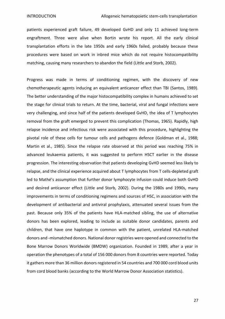
27
patients experienced graft failure, 49 developed GvHD and only 11 achieved long-term
engraftment. Three were alive when Bortin wrote his report. All the early clinical
transplantation efforts in the late 1950s and early 1960s failed, probably because these
procedures were based on work in inbred mice which do not require histocompatibility
matching, causing many researchers to abandon the field (Little and Storb, 2002).
Progress was made in terms of conditioning regimen, with the discovery of new
chemotherapeutic agents inducing an equivalent anticancer effect than TBI (Santos, 1989).
The better understanding of the major histocompatibility complex in humans achieved to set
the stage for clinical trials to return. At the time, bacterial, viral and fungal infections were
very challenging, and since half of the patients developed GvHD, the idea of T lymphocytes
removal from the graft emerged to prevent this complication (Thomas, 1965). Rapidly, high
relapse incidence and infectious risk were associated with this procedure, highlighting the
pivotal role of these cells for tumour cells and pathogens defence (Goldman et al., 1988;
Martin et al., 1985). Since the relapse rate observed at this period was reaching 75% in
advanced leukaemia patients, it was suggested to perform HSCT earlier in the disease
progression. The interesting observation that patients developing GvHD seemed less likely to
relapse, and the clinical experience acquired about T lymphocytes from T cells-depleted graft
led to Mathé’s assumption that further donor lymphocyte infusion could induce both GvHD
and desired anticancer effect (Little and Storb, 2002). During the 1980s and 1990s, many
improvements in terms of conditioning regimens and sources of HSC, in association with the
development of antibacterial and antiviral prophylaxis, attenuated several issues from the
past. Because only 35% of the patients have HLA-matched sibling, the use of alternative
donors has been explored, leading to include as suitable donor candidates, parents and
children, that have one haplotype in common with the patient, unrelated HLA-matched
donors and -mismatched donors. National donor registries were opened and connected to the
Bone Marrow Donors Worldwide (BMDW) organization. Founded in 1989, after a year in
operation the phenotypes of a total of 156 000 donors from 8 countries were reported. Today
it gathers more than 36 million donors registered in 54 countries and 700 000 cord blood units
from cord blood banks (according to the World Marrow Donor Association statistics).

INTRODUCTION Allogeneic hematopoietic stem-cells transplantation
28
B. Clinical settings
1. Overview
Bone marrow transplant-related discoveries over time have led to the current anti-cancer
immunotherapy used today in 37 different transplant centres all over France. Succinctly, this
therapy principle is to use a healthy source of hematopoietic progenitors to replace the
hematologic and immunologic system of a patient who suffers from bad prognostic blood
cancer. If the intended goal is always to exhaustively destroy tumour cells, alloHSCT is actually
a general term that refers to a two-step procedure whose application broadly differs between
countries and transplant centres (Lee et al., 2008).
The first step constitutes a preparation to enable the patient to receive the graft. His
hematopoietic system is partly destroyed, including its malignant cells, using what is named a
conditioning regimen. It can either be a chemotherapeutic agent, a radiation therapy, or else
a combination of the two. The recipient cells’ elimination creates space in his bone marrow
for the upcoming donor cells to migrate in and repopulate the hematopoietic niche. Hence,
the second phase consists of infusing the graft from a healthy donor. The patient’s medical
history and cancer type, aside from the chosen source of HSC and donor type mainly drive the
decision of using one particular conditioning treatment. These parameters, covered in detail
in the coming sections, will also influence the transplantation efficacy as well as its short- and
long-term toxicities.
2. Patient eligibility for alloHSCT
Considering one given patient, its eligibility for alloHSCT will depend on a broad range of
parameters regarding the patient himself or its malignancy type. The availability of a suitable
donor will also be clearly determining. Historically, bone-marrow transplant was mainly
indicated for patient with severe blood cancer, refractory to the usual therapies and in a fit
physical condition. Until now, no global consensus exists even if major improvements for
conditioning regimens had permitted to enlarge graft candidate profiles. The decision to graft
is often taken on a case-by-case basis after a collegial decision of the transplant centre
physicians. A pre-transplant study of the above-mentioned variables establishes a benefit-risk
assessment which is carefully explained and discussed with the patient.
INTRODUCTION Allogeneic hematopoietic stem-cells transplantation
29
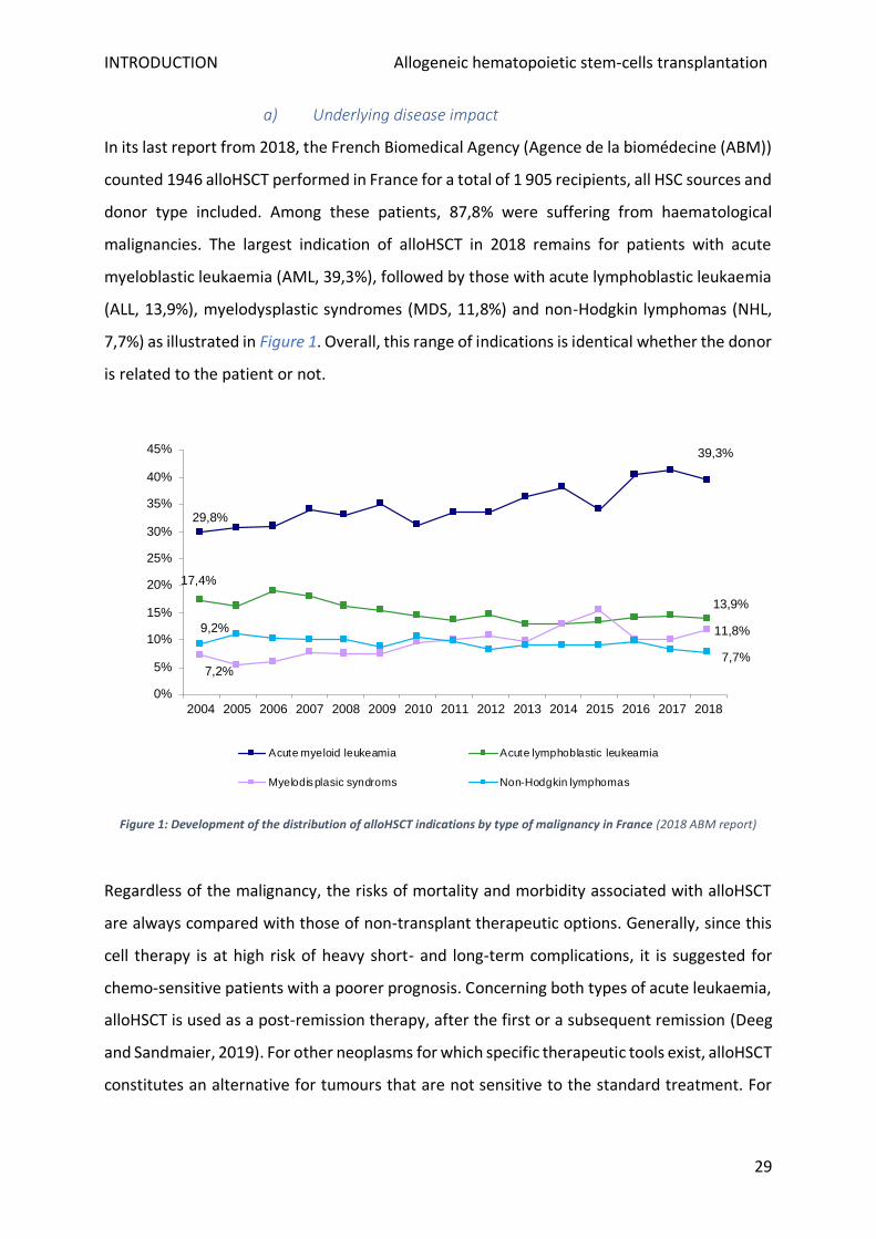
a) Underlying disease impact
In its last report from 2018, the French Biomedical Agency (Agence de la biomédecine (ABM))
counted 1946 alloHSCT performed in France for a total of 1 905 recipients, all HSC sources and
donor type included. Among these patients, 87,8% were suffering from haematological
malignancies. The largest indication of alloHSCT in 2018 remains for patients with acute
myeloblastic leukaemia (AML, 39,3%), followed by those with acute lymphoblastic leukaemia
(ALL, 13,9%), myelodysplastic syndromes (MDS, 11,8%) and non-Hodgkin lymphomas (NHL,
7,7%) as illustrated in Figure 1. Overall, this range of indications is identical whether the donor
is related to the patient or not.
Figure 1: Development of the distribution of alloHSCT indications by type of malignancy in France (2018 ABM report)
Regardless of the malignancy, the risks of mortality and morbidity associated with alloHSCT
are always compared with those of non-transplant therapeutic options. Generally, since this
cell therapy is at high risk of heavy short- and long-term complications, it is suggested for
chemo-sensitive patients with a poorer prognosis. Concerning both types of acute leukaemia,
alloHSCT is used as a post-remission therapy, after the first or a subsequent remission (Deeg
and Sandmaier, 2019). For other neoplasms for which specific therapeutic tools exist, alloHSCT
constitutes an alternative for tumours that are not sensitive to the standard treatment. For
29,8%
39,3%
17,4%
13,9%
7,2%
11,8%
0%
5%
10%
15%
20%
25%
30%
35%
40%
45%
2004 2005 2006 2007 2008 2009 2010 2011 2012 2013 2014 2015 2016 2017 2018
Acute myeloid leukeamia Acute lymphoblastic leukeamia
Myelodisplasic syndroms Non-Hodgkin lymphomas
7,7%
9,2%
INTRODUCTION Allogeneic hematopoietic stem-cells transplantation
30
example, for patients with chronic myeloid leukaemia that are resistant or intolerant to
tyrosine kinase inhibitors.
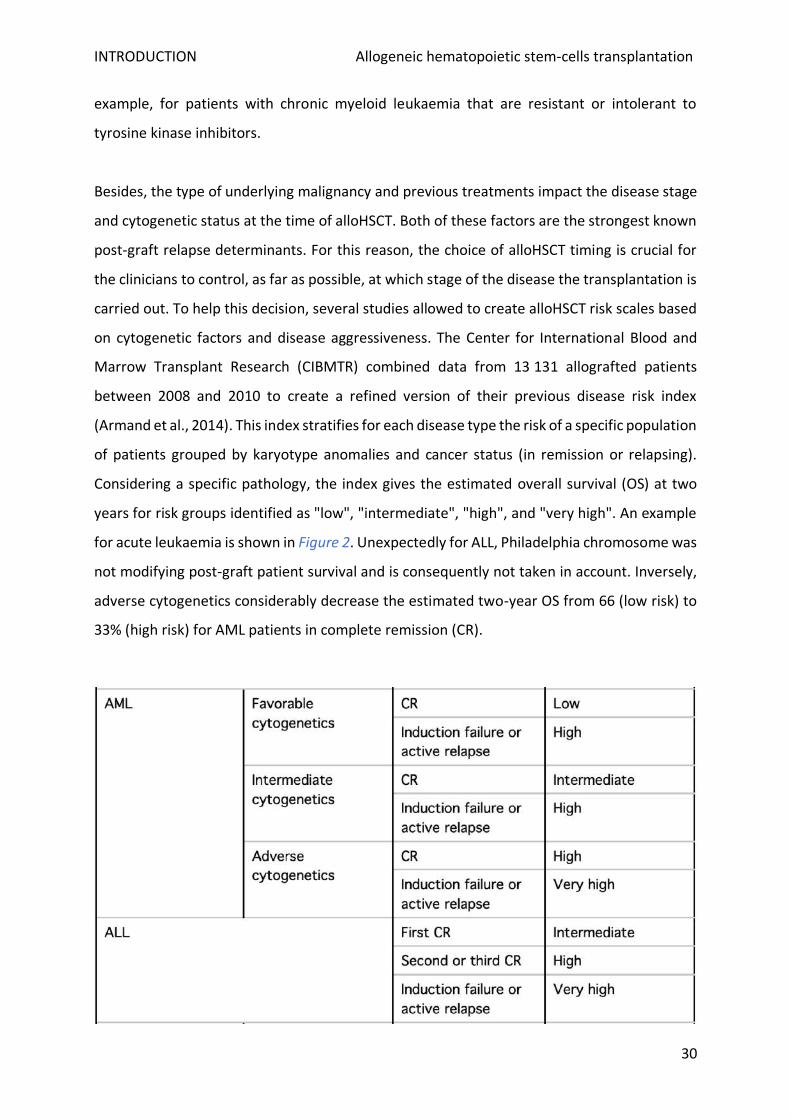
Besides, the type of underlying malignancy and previous treatments impact the disease stage
and cytogenetic status at the time of alloHSCT. Both of these factors are the strongest known
post-graft relapse determinants. For this reason, the choice of alloHSCT timing is crucial for
the clinicians to control, as far as possible, at which stage of the disease the transplantation is
carried out. To help this decision, several studies allowed to create alloHSCT risk scales based
on cytogenetic factors and disease aggressiveness. The Center for International Blood and
Marrow Transplant Research (CIBMTR) combined data from 13 131 allografted patients
between 2008 and 2010 to create a refined version of their previous disease risk index
(Armand et al., 2014). This index stratifies for each disease type the risk of a specific population
of patients grouped by karyotype anomalies and cancer status (in remission or relapsing).
Considering a specific pathology, the index gives the estimated overall survival (OS) at two
years for risk groups identified as "low", "intermediate", "high", and "very high". An example
for acute leukaemia is shown in Figure 2. Unexpectedly for ALL, Philadelphia chromosome was
not modifying post-graft patient survival and is consequently not taken in account. Inversely,
adverse cytogenetics considerably decrease the estimated two-year OS from 66 (low risk) to
33% (high risk) for AML patients in complete remission (CR).
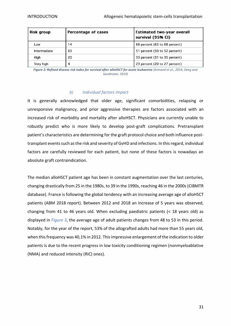
INTRODUCTION Allogeneic hematopoietic stem-cells transplantation
31
Figure 2: Refined disease risk index for survival after alloHSCT for acute leukaemia (Armand et al., 2014; Deeg and Sandmaier, 2019)
b) Individual factors impact
It is generally acknowledged that older age, significant comorbidities, relapsing or
unresponsive malignancy, and prior aggressive therapies are factors associated with an
increased risk of morbidity and mortality after alloHSCT. Physicians are currently unable to
robustly predict who is more likely to develop post-graft complications. Pretransplant
patient’s characteristics are determining for the graft protocol choice and both influence post-
transplant events such as the risk and severity of GvHD and infections. In this regard, individual
factors are carefully reviewed for each patient, but none of these factors is nowadays an
absolute graft contraindication.
The median alloHSCT patient age has been in constant augmentation over the last centuries,
changing drastically from 25 in the 1980s, to 39 in the 1990s, reaching 46 in the 2000s (CIBMTR
database). France is following the global tendency with an increasing average age of alloHSCT
patients (ABM 2018 report). Between 2012 and 2018 an increase of 5 years was observed,
changing from 41 to 46 years old. When excluding paediatric patients (< 18 years old) as
displayed in Figure 3, the average age of adult patients changes from 48 to 53 in this period.
Notably, for the year of the report, 53% of the allografted adults had more than 55 years old,
when this frequency was 40,1% in 2012. This impressive enlargement of the indication to older
patients is due to the recent progress in low toxicity conditioning regimen (nonmyeloablative
(NMA) and reduced intensity (RIC) ones).
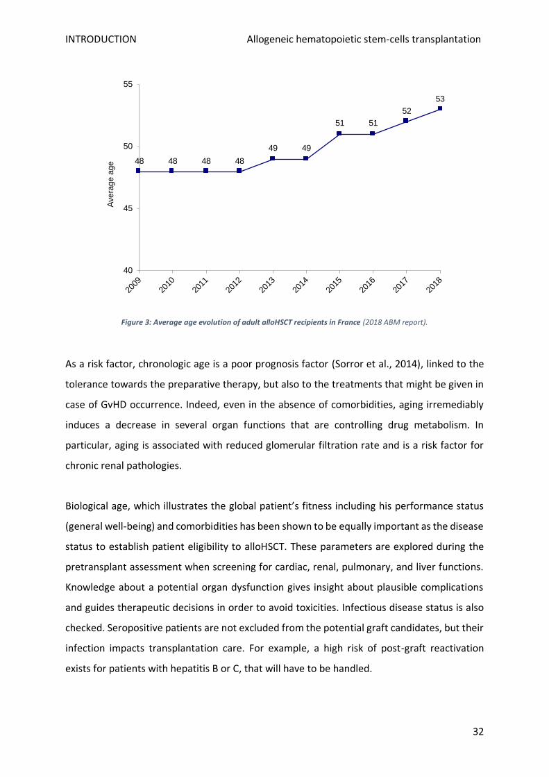
INTRODUCTION Allogeneic hematopoietic stem-cells transplantation
32
Figure 3: Average age evolution of adult alloHSCT recipients in France (2018 ABM report).
As a risk factor, chronologic age is a poor prognosis factor (Sorror et al., 2014), linked to the
tolerance towards the preparative therapy, but also to the treatments that might be given in
case of GvHD occurrence. Indeed, even in the absence of comorbidities, aging irremediably
induces a decrease in several organ functions that are controlling drug metabolism. In
particular, aging is associated with reduced glomerular filtration rate and is a risk factor for
chronic renal pathologies.
Biological age, which illustrates the global patient’s fitness including his performance status
(general well-being) and comorbidities has been shown to be equally important as the disease
status to establish patient eligibility to alloHSCT. These parameters are explored during the
pretransplant assessment when screening for cardiac, renal, pulmonary, and liver functions.
Knowledge about a potential organ dysfunction gives insight about plausible complications
and guides therapeutic decisions in order to avoid toxicities. Infectious disease status is also
checked. Seropositive patients are not excluded from the potential graft candidates, but their
infection impacts transplantation care. For example, a high risk of post-graft reactivation
exists for patients with hepatitis B or C, that will have to be handled.
48 48 48 48
49 49
51 51
52
53
40
45
50
55
2009
2010
2011
2012
2013
2014
2015
2016
2017
2018
Avera
ge a
ge
INTRODUCTION Allogeneic hematopoietic stem-cells transplantation
33
Nutritional status should be taken into account since weight might affect the drug required
dosage and distribution. Except for extreme exceptions, severe underweight and obesity are
not a cause of graft ineligibility, but it appears that these patients have a lower outcome post-
alloHSCT compared to those without weight imbalance (Nakao et al., 2014).
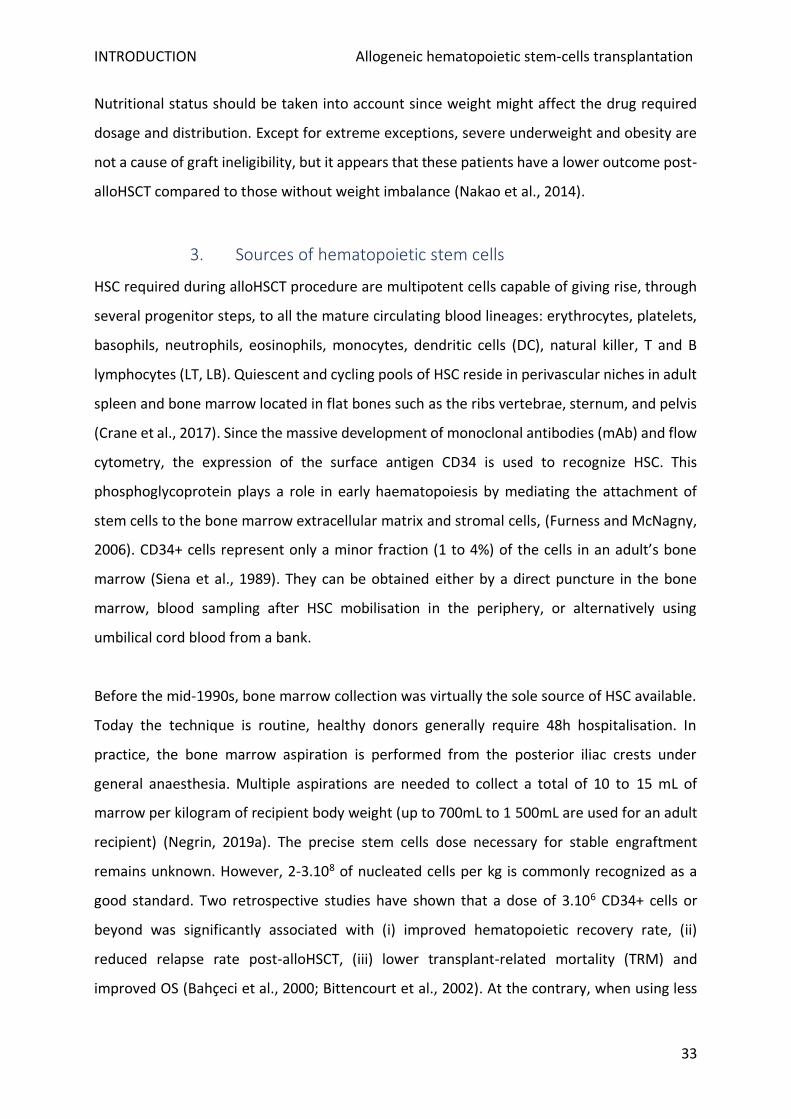
3. Sources of hematopoietic stem cells
HSC required during alloHSCT procedure are multipotent cells capable of giving rise, through
several progenitor steps, to all the mature circulating blood lineages: erythrocytes, platelets,
basophils, neutrophils, eosinophils, monocytes, dendritic cells (DC), natural killer, T and B
lymphocytes (LT, LB). Quiescent and cycling pools of HSC reside in perivascular niches in adult
spleen and bone marrow located in flat bones such as the ribs vertebrae, sternum, and pelvis
(Crane et al., 2017). Since the massive development of monoclonal antibodies (mAb) and flow
cytometry, the expression of the surface antigen CD34 is used to recognize HSC. This
phosphoglycoprotein plays a role in early haematopoiesis by mediating the attachment of
stem cells to the bone marrow extracellular matrix and stromal cells, (Furness and McNagny,
2006). CD34+ cells represent only a minor fraction (1 to 4%) of the cells in an adult’s bone
marrow (Siena et al., 1989). They can be obtained either by a direct puncture in the bone
marrow, blood sampling after HSC mobilisation in the periphery, or alternatively using
umbilical cord blood from a bank.
Before the mid-1990s, bone marrow collection was virtually the sole source of HSC available.
Today the technique is routine, healthy donors generally require 48h hospitalisation. In
practice, the bone marrow aspiration is performed from the posterior iliac crests under
general anaesthesia. Multiple aspirations are needed to collect a total of 10 to 15 mL of
marrow per kilogram of recipient body weight (up to 700mL to 1 500mL are used for an adult
recipient) (Negrin, 2019a). The precise stem cells dose necessary for stable engraftment
remains unknown. However, 2-3.108 of nucleated cells per kg is commonly recognized as a
good standard. Two retrospective studies have shown that a dose of 3.106 CD34+ cells or
beyond was significantly associated with (i) improved hematopoietic recovery rate, (ii)
reduced relapse rate post-alloHSCT, (iii) lower transplant-related mortality (TRM) and
improved OS (Bahçeci et al., 2000; Bittencourt et al., 2002). At the contrary, when using less

INTRODUCTION Allogeneic hematopoietic stem-cells transplantation
34
than 1,2.106 CD34+ cells, neutrophil and platelet engraftment is sub-optimal (Zubair et al.,
2004).
In 1971 the first evidence was found that colony-forming cells were present at an extremely
low level in human peripheral blood (McCredie et al., 1971). The authors described at the time
no reduction of these circulating cells number after repeated leukapheresis. However, it took
until the setup of mobilization protocols for peripheral blood to be successfully used as a
source of HSC for allogeneic transplantation. Using filgrastim (human recombinant
granulocyte colony-stimulating factor (G-CSF)), Schmitz et al. reported the feasibility of
unmanipulated allogeneic peripheral blood progenitor cells transplantation with long-term
engraftment and no detrimental GvHD (Schmitz et al., 1995). The administration during 4 or
5 days of this hematopoietic growth factor results in a drastic HSC mobilisation, that usually
allows to recover enough CD34+ cells with one apheresis session at day 5 (Negrin, 2019a). The
collection contains a mixture of HSC and progenitor cells referred to as peripheral blood
progenitor cells (PBPC), richer in CD34+ than bone marrow samples. Hematopoietic
reconstitution is faster after the infusion of mobilized PBPC than bone marrow: neutrophils
recover in 8 to 10 days, platelets in 10 to 12. However, a higher risk of overall and extensive
chronic GvHDs exist (Holtick et al., 2014). No difference has been highlighted in terms of
overall and leukaemia-free survival, global health, or late events (Friedrichs et al., 2010).
Umbilical cord blood (UCB) refers to the remaining blood in the umbilical cord and placenta
after birth that can be preserved in frozen banks. The use of cord blood as an alternative HSC
source has been initially described in 1995 for paediatric leukemic patients (Wagner et al.,
1995). First restricted to children because of the reduced graft material size, Laughlin et al.
reported in 2004 the successful alloHSCT of an adult cohort using unrelated donor UCB
(Laughlin et al., 2004). In order to provide a sufficient dose of HSC to engraft adults, the use
of two unrelated and partially matched cord blood units has revealed to be a possibility
(Barker et al., 2005). UCB is a rich source of immature HSC, ready-to-use, that offers a large
donor pool and is associated with less GvHD risk. However, the donor is unavailable in case of
further donor lymphocyte infusions (DLI) would be needed, and UCB utilization was associated
with risk of graft failure and delayed immune reconstitution (Smith and Wagner, 2009).
INTRODUCTION Allogeneic hematopoietic stem-cells transplantation
35
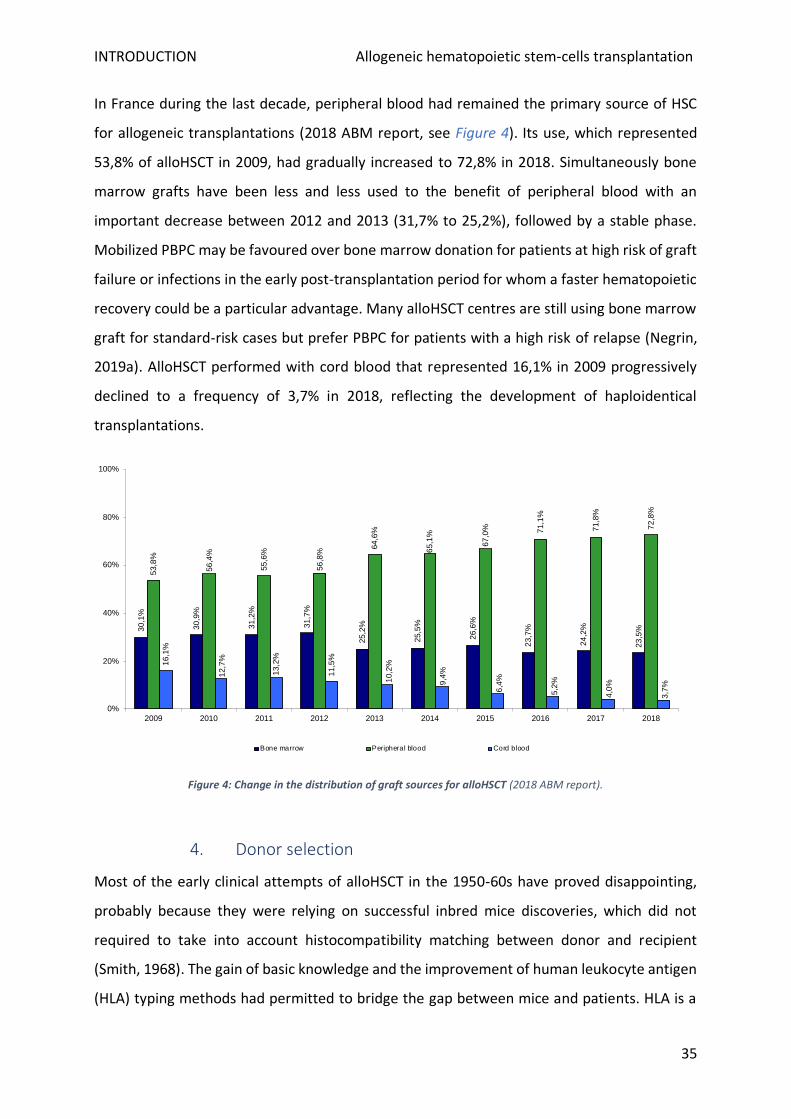
In France during the last decade, peripheral blood had remained the primary source of HSC
for allogeneic transplantations (2018 ABM report, see Figure 4). Its use, which represented
53,8% of alloHSCT in 2009, had gradually increased to 72,8% in 2018. Simultaneously bone
marrow grafts have been less and less used to the benefit of peripheral blood with an
important decrease between 2012 and 2013 (31,7% to 25,2%), followed by a stable phase.
Mobilized PBPC may be favoured over bone marrow donation for patients at high risk of graft
failure or infections in the early post-transplantation period for whom a faster hematopoietic
recovery could be a particular advantage. Many alloHSCT centres are still using bone marrow
graft for standard-risk cases but prefer PBPC for patients with a high risk of relapse (Negrin,
2019a). AlloHSCT performed with cord blood that represented 16,1% in 2009 progressively
declined to a frequency of 3,7% in 2018, reflecting the development of haploidentical
transplantations.
Figure 4: Change in the distribution of graft sources for alloHSCT (2018 ABM report).
4. Donor selection
Most of the early clinical attempts of alloHSCT in the 1950-60s have proved disappointing,
probably because they were relying on successful inbred mice discoveries, which did not
required to take into account histocompatibility matching between donor and recipient
(Smith, 1968). The gain of basic knowledge and the improvement of human leukocyte antigen
(HLA) typing methods had permitted to bridge the gap between mice and patients. HLA is a
30,1
%
30
,9%
31,2
%
31
,7%
25
,2%
25,5
%
26,6
%
23,7
%
24
,2%
23,5
%
53,8
%
56
,4%
55
,6%
56
,8%
64
,6%
65
,1%
67
,0% 71
,1%
71
,8%
72,8
%
16
,1%
12,7
%
13
,2%
11
,5%
10,2
%
9,4
%
6,4
%
5,2
%
4,0
%
3,7
%
0%
20%
40%
60%
80%
100%
2009 2010 2011 2012 2013 2014 2015 2016 2017 2018
Bone marrow Peripheral blood Cord blood

INTRODUCTION Allogeneic hematopoietic stem-cells transplantation
36
biologic system that provides millions of potential combinations, with more than 5 500 Class
I alleles (HLA-A, -B, -C) and over 1 600 class II alleles (HLA-DRB1 and DQB1). Molecular typing
that is now used for allele matching defines HLA genes by their sequence. A 10/10 HLA match
refers to HLA-A, -B, -C, -DRB1, and -DQB1 identical alleles for the pair donor-recipient. In
France, this number of antigens is generally used, but up to 12 HLA alleles can be checked
(adding HLA-DP1), and 8 being the norm in the US (ignoring DQB1).
When available, a matched sibling donor (genoidentical donor) is preferred over other options
because of a better clinical outcome including a reduced GvHD risk, secondarily because of
the speed of the search. If several are available, the choice is made considering other donor
parameters such as age, matched cytomegalovirus (CMV) status is preferred, for women the
absence of previous deliveries, and blood type. If no matching HLA sibling is found, an
unrelated donor is searched on the international registry which offers today more than 25
million donors registered on 79 countries. In the absence of a 10/10 donor match
(phenoidentical donor), an alternative source of HSC can be considered. This term refers to (i)
a phenoidentical donor with only one HLA incompatibility (9/10), (ii) an intrafamilial
haploidentical donor that carries half of the matched HLA genes (typically a parent or child),
or (iii) unrelated UCB. Without a genoidentical option, a search of HLA antibodies in the
recipient is indicated.
According to ABM’s data set, among the 1 946 allografts performed in 2018 in France the
frequency of related or unrelated donor utilization was similar (respectively 991 and 955,
Figure 5) when unrelated grafts were previously in majority. During the last five years,
haploidentical donors have been an expanding option (for 458 grafts in 2018, corresponding
to 46,2% of the related ones). The number of the matched related donor (HLA-identical) has
been decreasing also, probably consequently to the change in patients’ average age following
less aggressive regimens use since their siblings will have more donation contraindications.
Consequently, for these patients, a haploidentical donor is usually used. However, to date,
prospective study results have still not compared graft using different alternative donors.
Regarding unrelated donors, HSC from healthy adults is a growing choice for alloHSCT since
2009. Rather, grafts performed with UCB are in decreasing since 2013 (188 against 70 in 2018),
in parallel to the tripling number of haploidentical allografts between 2014 and 2018.
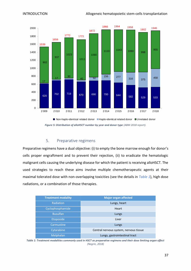
INTRODUCTION Allogeneic hematopoietic stem-cells transplantation
37
Figure 5: Distribution of alloHSCT number by year and donor type (ABM 2018 report).
5. Preparative regimens
Preparative regimens have a dual objective: (i) to empty the bone marrow enough for donor’s
cells proper engraftment and to prevent their rejection, (ii) to eradicate the hematologic
malignant cells causing the underlying disease for which the patient is receiving alloHSCT. The
used strategies to reach these aims involve multiple chemotherapeutic agents at their
maximal tolerated dose with non-overlapping toxicities (see the details in Table 1), high dose
radiations, or a combination of those therapies.
Treatment modality Major organ affected
Radiation Lungs, heart
Cyclophosphamide Heart
Busulfan Lungs
Etoposide Liver
Carmustine Lungs
Cytarabine Central nervous system, nervous tissue
Melphalan Lungs, gastrointestinal tract
Table 1: Treatment modalities commonly used in HSCT as preparative regimens and their dose limiting organ effect (Negrin, 2018)
620702 719 670 690 700 644
582 529 533
17
1730
40 92156 277
316 375458
902
9371023
1011
1090
1110 1043 1060 998955
0
200
400
600
800
1000
1200
1400
1600
1800
2000
2 009 2 010 2 011 2 012 2 013 2 014 2 015 2 016 2 017 2 018
Non-haplo-identical related donor Haplo-identical related donor Unrelated donor
1946
1539
1656
1772
1966
1721
1872
1964 19581902
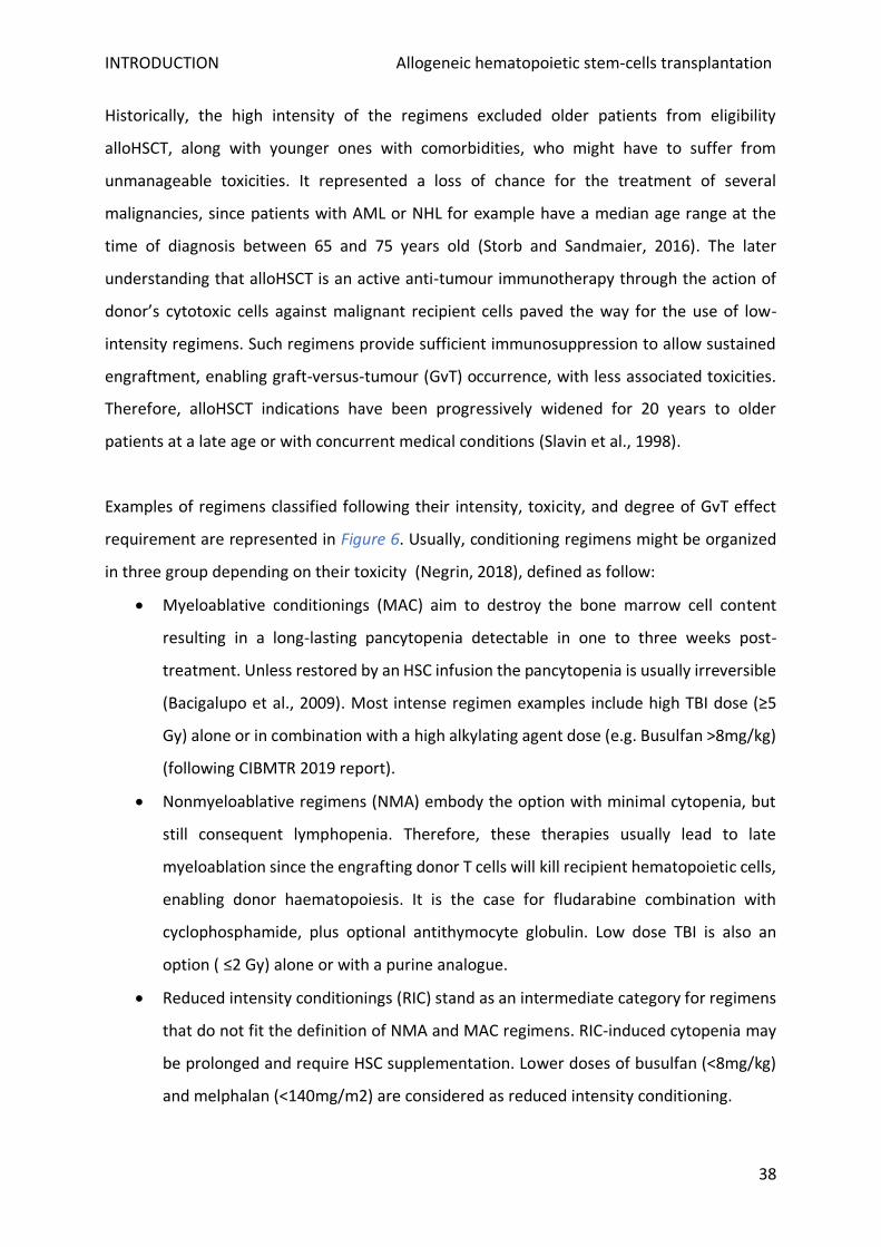
INTRODUCTION Allogeneic hematopoietic stem-cells transplantation
38
Historically, the high intensity of the regimens excluded older patients from eligibility
alloHSCT, along with younger ones with comorbidities, who might have to suffer from
unmanageable toxicities. It represented a loss of chance for the treatment of several
malignancies, since patients with AML or NHL for example have a median age range at the
time of diagnosis between 65 and 75 years old (Storb and Sandmaier, 2016). The later
understanding that alloHSCT is an active anti-tumour immunotherapy through the action of
donor’s cytotoxic cells against malignant recipient cells paved the way for the use of low-
intensity regimens. Such regimens provide sufficient immunosuppression to allow sustained
engraftment, enabling graft-versus-tumour (GvT) occurrence, with less associated toxicities.
Therefore, alloHSCT indications have been progressively widened for 20 years to older
patients at a late age or with concurrent medical conditions (Slavin et al., 1998).
Examples of regimens classified following their intensity, toxicity, and degree of GvT effect
requirement are represented in Figure 6. Usually, conditioning regimens might be organized
in three group depending on their toxicity (Negrin, 2018), defined as follow:
• Myeloablative conditionings (MAC) aim to destroy the bone marrow cell content
resulting in a long-lasting pancytopenia detectable in one to three weeks post-
treatment. Unless restored by an HSC infusion the pancytopenia is usually irreversible
(Bacigalupo et al., 2009). Most intense regimen examples include high TBI dose (≥5
Gy) alone or in combination with a high alkylating agent dose (e.g. Busulfan >8mg/kg)
(following CIBMTR 2019 report).
• Nonmyeloablative regimens (NMA) embody the option with minimal cytopenia, but
still consequent lymphopenia. Therefore, these therapies usually lead to late
myeloablation since the engrafting donor T cells will kill recipient hematopoietic cells,
enabling donor haematopoiesis. It is the case for fludarabine combination with
cyclophosphamide, plus optional antithymocyte globulin. Low dose TBI is also an
option ( ≤2 Gy) alone or with a purine analogue.
• Reduced intensity conditionings (RIC) stand as an intermediate category for regimens
that do not fit the definition of NMA and MAC regimens. RIC-induced cytopenia may
be prolonged and require HSC supplementation. Lower doses of busulfan (<8mg/kg)
and melphalan (<140mg/m2) are considered as reduced intensity conditioning.
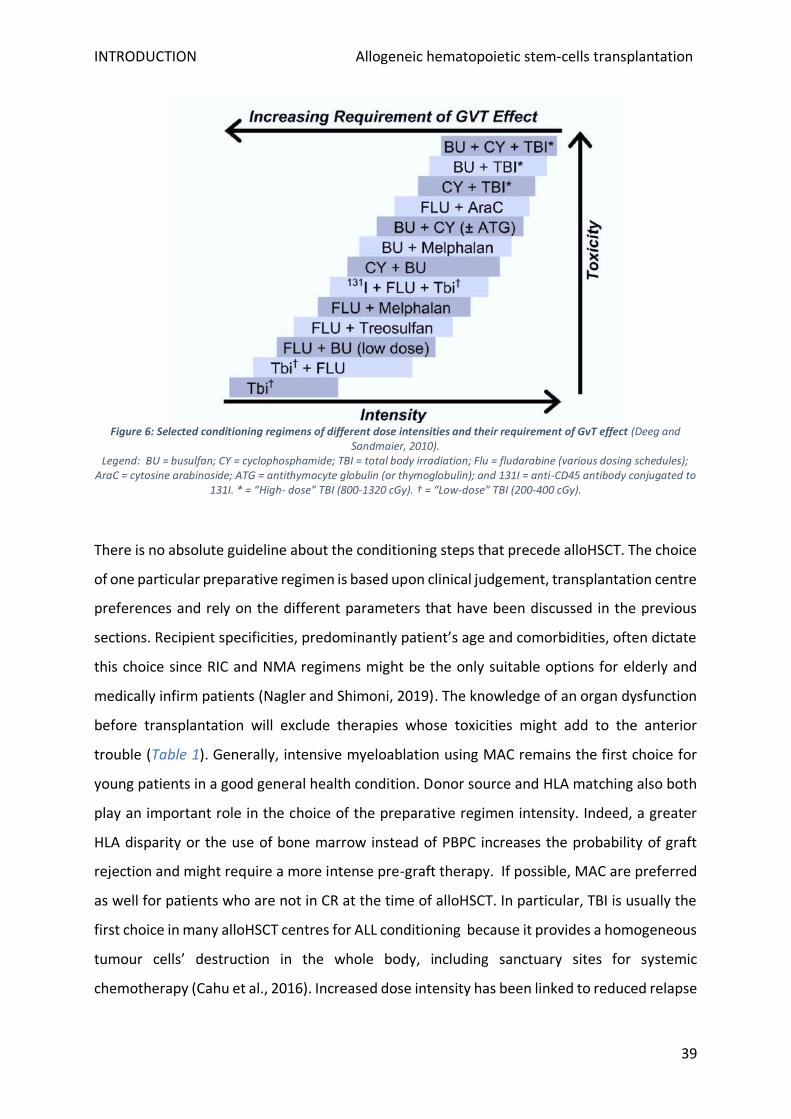
INTRODUCTION Allogeneic hematopoietic stem-cells transplantation
39
Figure 6: Selected conditioning regimens of different dose intensities and their requirement of GvT effect (Deeg and Sandmaier, 2010).
Legend: BU = busulfan; CY = cyclophosphamide; TBI = total body irradiation; Flu = fludarabine (various dosing schedules); AraC = cytosine arabinoside; ATG = antithymocyte globulin (or thymoglobulin); and 131I = anti-CD45 antibody conjugated to
131I. * = “High- dose” TBI (800-1320 cGy). † = “Low-dose” TBI (200-400 cGy).
There is no absolute guideline about the conditioning steps that precede alloHSCT. The choice
of one particular preparative regimen is based upon clinical judgement, transplantation centre
preferences and rely on the different parameters that have been discussed in the previous
sections. Recipient specificities, predominantly patient’s age and comorbidities, often dictate
this choice since RIC and NMA regimens might be the only suitable options for elderly and
medically infirm patients (Nagler and Shimoni, 2019). The knowledge of an organ dysfunction
before transplantation will exclude therapies whose toxicities might add to the anterior
trouble (Table 1). Generally, intensive myeloablation using MAC remains the first choice for
young patients in a good general health condition. Donor source and HLA matching also both
play an important role in the choice of the preparative regimen intensity. Indeed, a greater
HLA disparity or the use of bone marrow instead of PBPC increases the probability of graft
rejection and might require a more intense pre-graft therapy. If possible, MAC are preferred
as well for patients who are not in CR at the time of alloHSCT. In particular, TBI is usually the
first choice in many alloHSCT centres for ALL conditioning because it provides a homogeneous
tumour cells’ destruction in the whole body, including sanctuary sites for systemic
chemotherapy (Cahu et al., 2016). Increased dose intensity has been linked to reduced relapse
INTRODUCTION Allogeneic hematopoietic stem-cells transplantation
40
incidence but also with higher non-relapse mortality (NRM) (Aoudjhane et al., 2005). Although
lower intensity regimens usefully appear to trigger less toxicities, they have been also
associated with an increased risk of relapse.
6. Post-alloHSCT monitoring
a) Engraftment and chimerism
Once the HSC infusion has been performed, it becomes central to monitor if the newly-formed
cells emerging from the patient’s bone marrow after aplasia are from donor origin. The
genotype investigations of post-transplant haematopoiesis are referred to as chimerism
analysis. If a complete donor reconstitution was first believed necessary, it then appears that
the coexistence of both recipient and donor haematopoiesis might be compatible with stable
engraftment (Bader, 2019). The term complete chimerism refers to the first scenario, while
the second is called mixed chimerism. Post-alloHSCT chimerism is actually a dynamic
parameter, and a patient presenting no autologous cells at a certain timepoint might switch
to mixed chimerism later, and the contrary. Short tandem repeats PCR is currently the gold
standard method used to assess post-transplant chimerism (Thiede et al., 2001). It has
become possible to determine chimerism for different cell subpopulations, revealing that
patients may display mixed chimerism only in some particular cell lines, a phenomenon called
split chimerism. T lymphocytes origin is usually used to monitor the engraftment: chimerism
is considered as complete if CD3+ cells are from the donor for more than 95%. Under 5% of
donor CD3+ indicates a graft rejection. T cells evaluation timing depends on the myeloablative
level of the preparative regimen. In most French graft centres, usual timepoints are at day 30,
90, 180, and 360 (Dubois et al., 2017).
Chimerism is an informative parameter for alloreactivity and graft tolerance post-alloHSCT.
The persistency or the resurgence of recipient cells might correspond either to (i) preparative
regimen spared non-malignant cells, (ii) survival tumour cells, (iii) the result of a recipient
haematopoiesis reappearance, or else a combination of those three. Except for the patients
suffering from a malignancy without clear specifics markers of detection (typically MDS or
AML patients), more sensitive technics than chimerism are usually required to precisely
determine patient’s measurable residual disease (MRD). However, many studies have shown
that patients developing mixed chimerism had an increased risk of relapse, highlighting that
INTRODUCTION Allogeneic hematopoietic stem-cells transplantation
41
chimerism assessment can serve as a relapse prognosis factor. The restoration of an efficient
GvT effect has been proved to be feasible by converting mixed to complete chimerism using
DLI or interrupting immunosuppressive agents (Bader et al., 2004; Platzbecker et al., 2012). In
order to identify patients at risk of relapse and adjust immunotherapy strategies, chimerism
analysis needs to be frequent since the time-lapse between mixed chimerism onset and
cancer recurrence is often brief.
b) Short- and long-term care
Aside from the frequent chimerism monitoring, patients undergoing alloHSCT require
multidisciplinary controls to prevent or treat potential short-term and later complications. At
discharge, patients receive advice to adopt a lifestyle compatible with post-alloHSCT
condition, since they are immunocompromised, may have developed or will undergo later
treatment-related organ damages. Discharge is also the starting point of a long-term follow-
up plan, based on patient individual risk factors. General recommendations of post-transplant
monitoring at early and late timing are listed in Table 2. Unlisted specific measures will apply
for particular populations of patients, including patients with GvHD, ongoing significant
corticosteroid exposure, paediatric patients, and those who have received TBI. Succinctly,
each follow-up care plan takes in consideration the following parameters:
• the type of previous anti-tumoral therapies and conditioning regimen
• the potential comorbid medical conditions and organ dysfunctions
• the degree of cytopenia, occurrence of GvHD and associated treatments
AlloHSCT may be associated with late morbidity and survivors require life-long monitoring for
relapse of the underlying disease, management of transplantation-associated complications,
general medical and psychological care, accompanied by age- and gender-appropriate health
maintenance.
AlloHSCT recipients may remain immunocompromised far beyond 2 years post-transplant,
especially individuals with chronic GvHD. Therefore, these patients should also be routinely
revaccinated after transplant until they regain immune competence, in order to prevent
infectious complications for which a vaccine is available. International consensus guidelines
for vaccination schedule are available for physicians (Majhail et al., 2012).
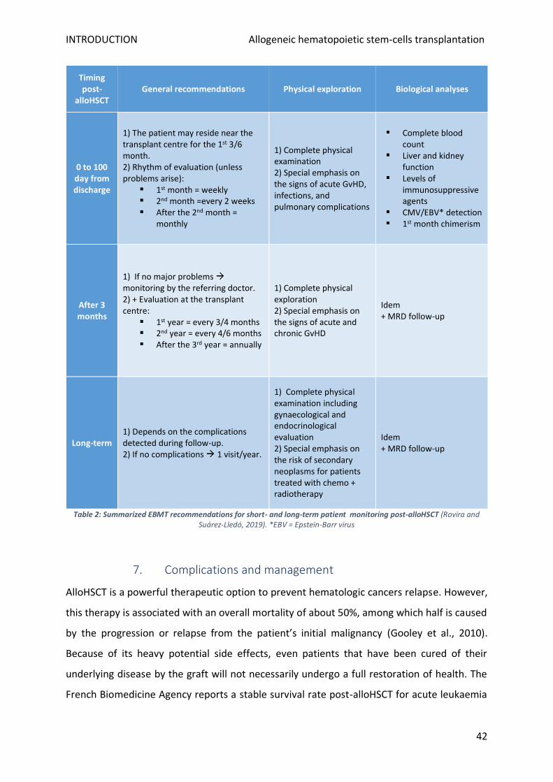
INTRODUCTION Allogeneic hematopoietic stem-cells transplantation
42
Timing post-
alloHSCT General recommendations Physical exploration Biological analyses
0 to 100 day from discharge
1) The patient may reside near the transplant centre for the 1st 3/6 month. 2) Rhythm of evaluation (unless problems arise):
1st month = weekly 2nd month =every 2 weeks After the 2nd month =
monthly
1) Complete physical examination 2) Special emphasis on the signs of acute GvHD, infections, and pulmonary complications
Complete blood count
Liver and kidney function
Levels of immunosuppressive agents
CMV/EBV* detection 1st month chimerism
After 3 months
1) If no major problems monitoring by the referring doctor. 2) + Evaluation at the transplant centre:
1st year = every 3/4 months 2nd year = every 4/6 months After the 3rd year = annually
1) Complete physical exploration 2) Special emphasis on the signs of acute and chronic GvHD
Idem + MRD follow-up
Long-term 1) Depends on the complications detected during follow-up. 2) If no complications 1 visit/year.
1) Complete physical examination including gynaecological and endocrinological evaluation 2) Special emphasis on the risk of secondary neoplasms for patients treated with chemo + radiotherapy
Idem + MRD follow-up
Table 2: Summarized EBMT recommendations for short- and long-term patient monitoring post-alloHSCT (Rovira and Suárez-Lledó, 2019). *EBV = Epstein-Barr virus
7. Complications and management
AlloHSCT is a powerful therapeutic option to prevent hematologic cancers relapse. However,
this therapy is associated with an overall mortality of about 50%, among which half is caused
by the progression or relapse from the patient’s initial malignancy (Gooley et al., 2010).
Because of its heavy potential side effects, even patients that have been cured of their
underlying disease by the graft will not necessarily undergo a full restoration of health. The
French Biomedicine Agency reports a stable survival rate post-alloHSCT for acute leukaemia
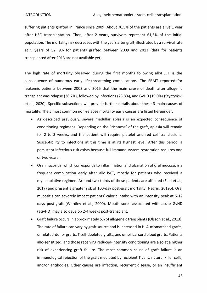
INTRODUCTION Allogeneic hematopoietic stem-cells transplantation
43
suffering patients grafted in France since 2009. About 70,5% of the patients are alive 1 year
after HSC transplantation. Then, after 2 years, survivors represent 61,5% of the initial
population. The mortality risk decreases with the years after graft, illustrated by a survival rate
at 5 years of 52, 9% for patients grafted between 2009 and 2013 (data for patients
transplanted after 2013 are not available yet).
The high rate of mortality observed during the first months following alloHSCT is the
consequence of numerous early life-threatening complications. The EBMT reported for
leukemic patients between 2002 and 2015 that the main cause of death after allogenic
transplant was relapse (38.7%), followed by infections (23.8%), and GvHD (19.0%) (Styczyński
et al., 2020). Specific subsections will provide further details about these 3 main causes of
mortality. The 5 most common non-relapse mortality early causes are listed hereunder:
• As described previously, severe medullar aplasia is an expected consequence of
conditioning regimens. Depending on the “richness” of the graft, aplasia will remain
for 2 to 3 weeks, and the patient will require platelet and red cell transfusions.
Susceptibility to infections at this time is at its highest level. After this period, a
persistent infectious risk exists because full immune system restoration requires one
or two years.
• Oral mucositis, which corresponds to inflammation and ulceration of oral mucosa, is a
frequent complication early after alloHSCT, mostly for patients who received a
myeloablative regimen. Around two-thirds of these patients are affected (Elad et al.,
2017) and present a greater risk of 100-day post-graft mortality (Negrin, 2019b). Oral
mucositis can severely impact patients’ caloric intake with an intensity peak at 6-12
days post-graft (Wardley et al., 2000). Mouth sores associated with acute GvHD
(aGvHD) may also develop 2-4 weeks post-transplant.
• Graft failure occurs in approximately 5% of allogeneic transplants (Olsson et al., 2013).
The rate of failure can vary by graft source and is increased in HLA-mismatched grafts,
unrelated-donor grafts, T cell-depleted grafts, and umbilical cord blood grafts. Patients
allo-sensitized, and those receiving reduced-intensity conditioning are also at a higher
risk of experiencing graft failure. The most common cause of graft failure is an
immunological rejection of the graft mediated by recipient T cells, natural killer cells,
and/or antibodies. Other causes are infection, recurrent disease, or an insufficient
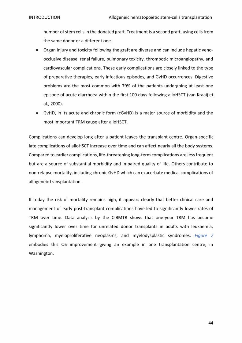
INTRODUCTION Allogeneic hematopoietic stem-cells transplantation
44
number of stem cells in the donated graft. Treatment is a second graft, using cells from
the same donor or a different one.
• Organ injury and toxicity following the graft are diverse and can include hepatic veno-
occlusive disease, renal failure, pulmonary toxicity, thrombotic microangiopathy, and
cardiovascular complications. These early complications are closely linked to the type
of preparative therapies, early infectious episodes, and GvHD occurrences. Digestive
problems are the most common with 79% of the patients undergoing at least one
episode of acute diarrhoea within the first 100 days following alloHSCT (van Kraaij et
al., 2000).
• GvHD, in its acute and chronic form (cGvHD) is a major source of morbidity and the
most important TRM cause after alloHSCT.
Complications can develop long after a patient leaves the transplant centre. Organ-specific
late complications of alloHSCT increase over time and can affect nearly all the body systems.
Compared to earlier complications, life-threatening long-term complications are less frequent
but are a source of substantial morbidity and impaired quality of life. Others contribute to
non-relapse mortality, including chronic GvHD which can exacerbate medical complications of
allogeneic transplantation.
If today the risk of mortality remains high, it appears clearly that better clinical care and
management of early post-transplant complications have led to significantly lower rates of
TRM over time. Data analysis by the CIBMTR shows that one-year TRM has become
significantly lower over time for unrelated donor transplants in adults with leukaemia,
lymphoma, myeloproliferative neoplasms, and myelodysplastic syndromes. Figure 7
embodies this OS improvement giving an example in one transplantation centre, in
Washington.
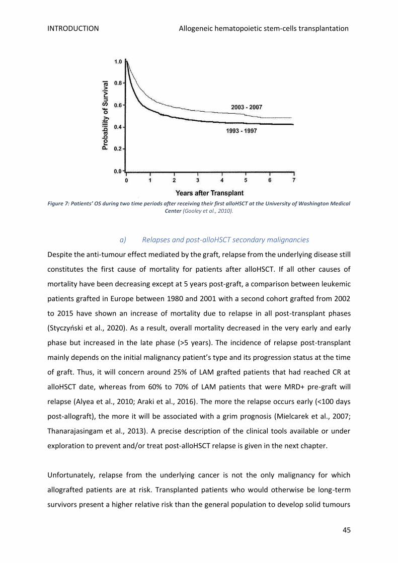
INTRODUCTION Allogeneic hematopoietic stem-cells transplantation
45
Figure 7: Patients’ OS during two time periods after receiving their first alloHSCT at the University of Washington Medical Center (Gooley et al., 2010).
a) Relapses and post-alloHSCT secondary malignancies
Despite the anti-tumour effect mediated by the graft, relapse from the underlying disease still
constitutes the first cause of mortality for patients after alloHSCT. If all other causes of
mortality have been decreasing except at 5 years post-graft, a comparison between leukemic
patients grafted in Europe between 1980 and 2001 with a second cohort grafted from 2002
to 2015 have shown an increase of mortality due to relapse in all post-transplant phases
(Styczyński et al., 2020). As a result, overall mortality decreased in the very early and early
phase but increased in the late phase (>5 years). The incidence of relapse post-transplant
mainly depends on the initial malignancy patient’s type and its progression status at the time
of graft. Thus, it will concern around 25% of LAM grafted patients that had reached CR at
alloHSCT date, whereas from 60% to 70% of LAM patients that were MRD+ pre-graft will
relapse (Alyea et al., 2010; Araki et al., 2016). The more the relapse occurs early (<100 days
post-allograft), the more it will be associated with a grim prognosis (Mielcarek et al., 2007;
Thanarajasingam et al., 2013). A precise description of the clinical tools available or under
exploration to prevent and/or treat post-alloHSCT relapse is given in the next chapter.
Unfortunately, relapse from the underlying cancer is not the only malignancy for which
allografted patients are at risk. Transplanted patients who would otherwise be long-term
survivors present a higher relative risk than the general population to develop solid tumours
INTRODUCTION Allogeneic hematopoietic stem-cells transplantation
46
of practically every organ, acute leukaemia, MDS and post-transplant lymphoproliferative
disease (PTLD) (Negrin, 2019). Solid and haematologic malignancies development occur late
in the post-transplant course, often after 3 years, and depend on a large range of patient
genetic and treatment-related factors, including irradiation dose for conditioning,
prophylactic immunosuppressive agents, viral infections, cGvHD occurrence, and patient age
(Rizzo et al., 2009). Solid tumours are relatively infrequent, a large study from the CIBMTR
reported new solid cancers at twice the expected rate in the general population, with an
increased incidence of 1% at 10 years, 2,2 at 15 and 3,3% at 30 (Curtis et al., 1997). Post-graft
leukaemia can be either from a recipient’s origin or acquired via a malignant donor clone (Sala
Torra and Loeb, 2011; Wiseman, 2011). Donor cell leukaemia is a rare complication associated
with a poor prognosis, with a possibly underestimated incidence since it is often difficult to
prove the donor origin of malignant cells. Thus, they could actually represent up to 5% of post-
transplant leukaemia classified as initial disease relapses (Tichelli, 2019). Screening and
preventive measures for secondary malignancies consist of risk awareness counselling and
clinical assessment annually, with age and gender-appropriate cancer routine surveillance. For
post-transplant blood malignancies, no standard treatment exists. Secondary solid cancer
should be treated as a de novo cancer of the same type and will share the same prognosis
range.
If data emerges about donor-derived leukaemia, for now, the vast majority of secondary
malignancies are host-derived (Alpár, 2011; Nakamizo et al., 2011). The one exception is PTLD,
which usually occurs in the first year of transplantation and is caused, for its conventional
form, by EBV+ donor cells. This EBV-related complication exists in both HSC and organ
transplantation, impacting 1% of alloHSCT patients at 10 years, with more than 80% incidence
the first year (Straathof et al., 2002). The most common symptoms of PTLD,
lymphadenopathy, and fever, are the consequence of EBV-induced transformation of B cells
in the setting of impaired anti-EBV cellular immunity due to iatrogenic immunosuppression
and resulting in an outgrowth of EBV-infected B cells (Styczynski and Giebel, 2019). Risk factors
for PTLD are proportional to the degree of T-cell impairment, with a high degree of
participation for GvHD prevention strategies that indiscriminately remove effector T cells. In
the meantime, EBV also plays an important role in cGvHD pathogenesis, and to a less extent
aGvHD, since B cells are directly involved in the pathophysiology of chronic GVHD.
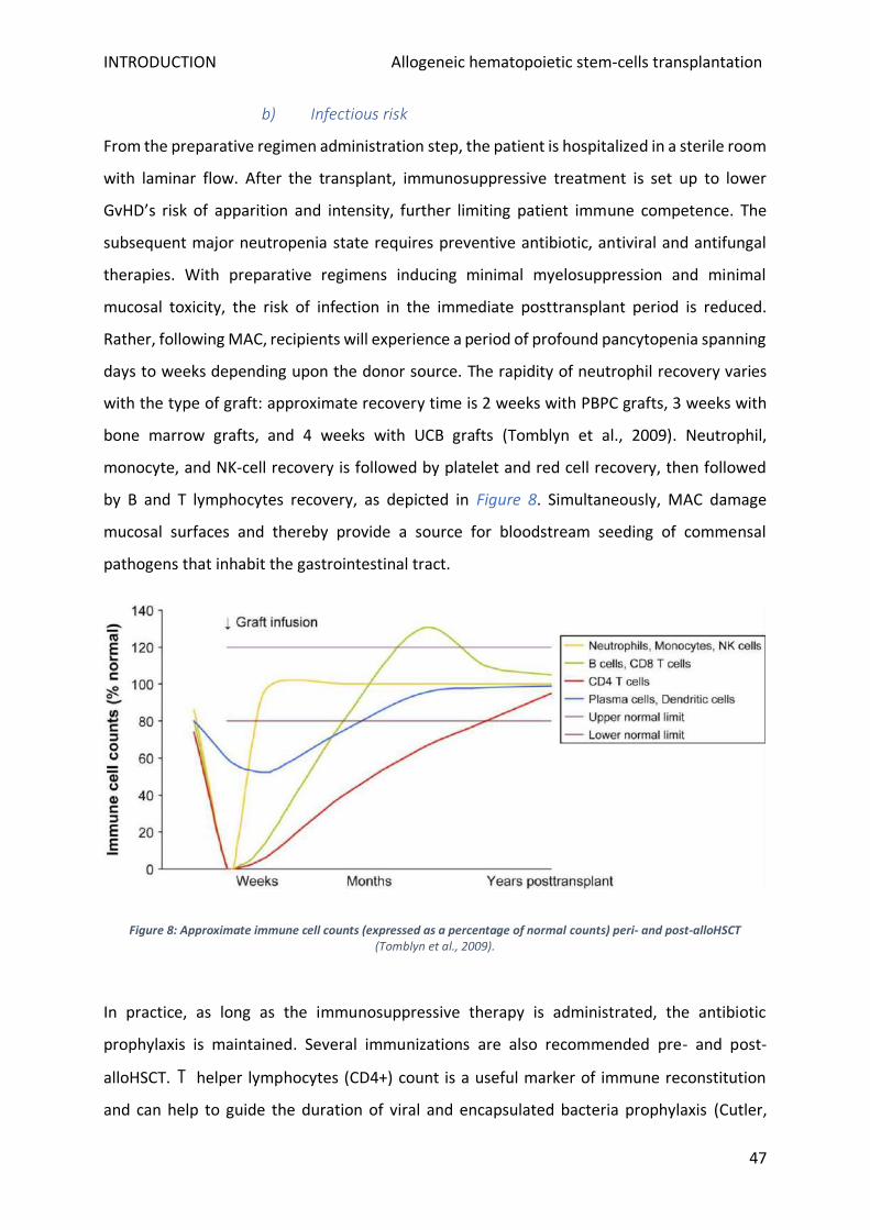
INTRODUCTION Allogeneic hematopoietic stem-cells transplantation
47
b) Infectious risk
From the preparative regimen administration step, the patient is hospitalized in a sterile room
with laminar flow. After the transplant, immunosuppressive treatment is set up to lower
GvHD’s risk of apparition and intensity, further limiting patient immune competence. The
subsequent major neutropenia state requires preventive antibiotic, antiviral and antifungal
therapies. With preparative regimens inducing minimal myelosuppression and minimal
mucosal toxicity, the risk of infection in the immediate posttransplant period is reduced.
Rather, following MAC, recipients will experience a period of profound pancytopenia spanning
days to weeks depending upon the donor source. The rapidity of neutrophil recovery varies
with the type of graft: approximate recovery time is 2 weeks with PBPC grafts, 3 weeks with
bone marrow grafts, and 4 weeks with UCB grafts (Tomblyn et al., 2009). Neutrophil,
monocyte, and NK-cell recovery is followed by platelet and red cell recovery, then followed
by B and T lymphocytes recovery, as depicted in Figure 8. Simultaneously, MAC damage
mucosal surfaces and thereby provide a source for bloodstream seeding of commensal
pathogens that inhabit the gastrointestinal tract.
Figure 8: Approximate immune cell counts (expressed as a percentage of normal counts) peri- and post-alloHSCT (Tomblyn et al., 2009).
In practice, as long as the immunosuppressive therapy is administrated, the antibiotic
prophylaxis is maintained. Several immunizations are also recommended pre- and post-
alloHSCT. T helper lymphocytes (CD4+) count is a useful marker of immune reconstitution
and can help to guide the duration of viral and encapsulated bacteria prophylaxis (Cutler,
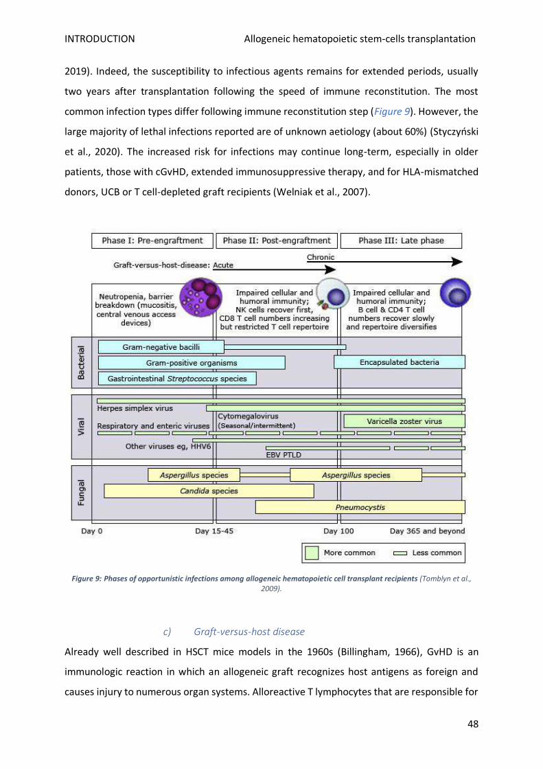
INTRODUCTION Allogeneic hematopoietic stem-cells transplantation
48
2019). Indeed, the susceptibility to infectious agents remains for extended periods, usually
two years after transplantation following the speed of immune reconstitution. The most
common infection types differ following immune reconstitution step (Figure 9). However, the
large majority of lethal infections reported are of unknown aetiology (about 60%) (Styczyński
et al., 2020). The increased risk for infections may continue long-term, especially in older
patients, those with cGvHD, extended immunosuppressive therapy, and for HLA-mismatched
donors, UCB or T cell-depleted graft recipients (Welniak et al., 2007).
Figure 9: Phases of opportunistic infections among allogeneic hematopoietic cell transplant recipients (Tomblyn et al., 2009).
c) Graft-versus-host disease
Already well described in HSCT mice models in the 1960s (Billingham, 1966), GvHD is an
immunologic reaction in which an allogeneic graft recognizes host antigens as foreign and
causes injury to numerous organ systems. Alloreactive T lymphocytes that are responsible for

INTRODUCTION Allogeneic hematopoietic stem-cells transplantation
49
the beneficial GvT effect are also the main actors of damaging the recipient’s tissues during
GvHD. Thus, this ambivalent role of T cells after alloHSCT is the reason why attempts of
preventing or treating this life-threatening complication without impairing GvT therapeutic
effect have been rather unsuccessful. An overview of both GvT effect and GvHD immune
mechanisms will be given in the Chapter I.
GvHD can be acute or chronic, which differ based on time of onset, immunological causes, and
clinical manifestations. Initially, the distinction was solely based on the time of occurrence,
respectively before or after day 100 post-HSC infusion. However, this definition has proved
unsatisfactory since aGvHD symptoms could appear beyond day 100 after RIC regimens
and/or after further DLI which are usually given after day 100. In 2005, an NIH consensus
workgroup established a new classification reported in Table 3, which is still current (Filipovich
et al., 2005). Hence, patients can be subject to recurrent forms of aGvHD, or present an
overlap syndrome with both acute and chronic GvHD features.
Subtype Day after HSCT Features of
aGvHD
Features of
cGvHD
Acute
GvHD
Classic <100 days Yes No
Persistent,
recurrent, or late onset >100 days Yes No
Chronic
GvHD
Classic No time limit No Yes
Overlap
syndrome No time limit Yes Yes
Table 3: Current classification of acute and chronic GvHD (Filipovich et al., 2005).
(1) Acute
GvHD in its acute form is characterized by one or more of the following features: an
erythematous skin reaction, cholestatic liver disease, and gastrointestinal dysfunction (Table
4). A retrospective analysis of allografted patients at GvHD onset has shown that 81% had a
skin rash, 50% had liver impairment and 54% had gut dysfunction (Martin et al., 1990).
Because alloHSCT recipients are subject to various other complications than GvHD, the
diagnosis is often uneasy, which can have consequences on the appropriate treatment choice.
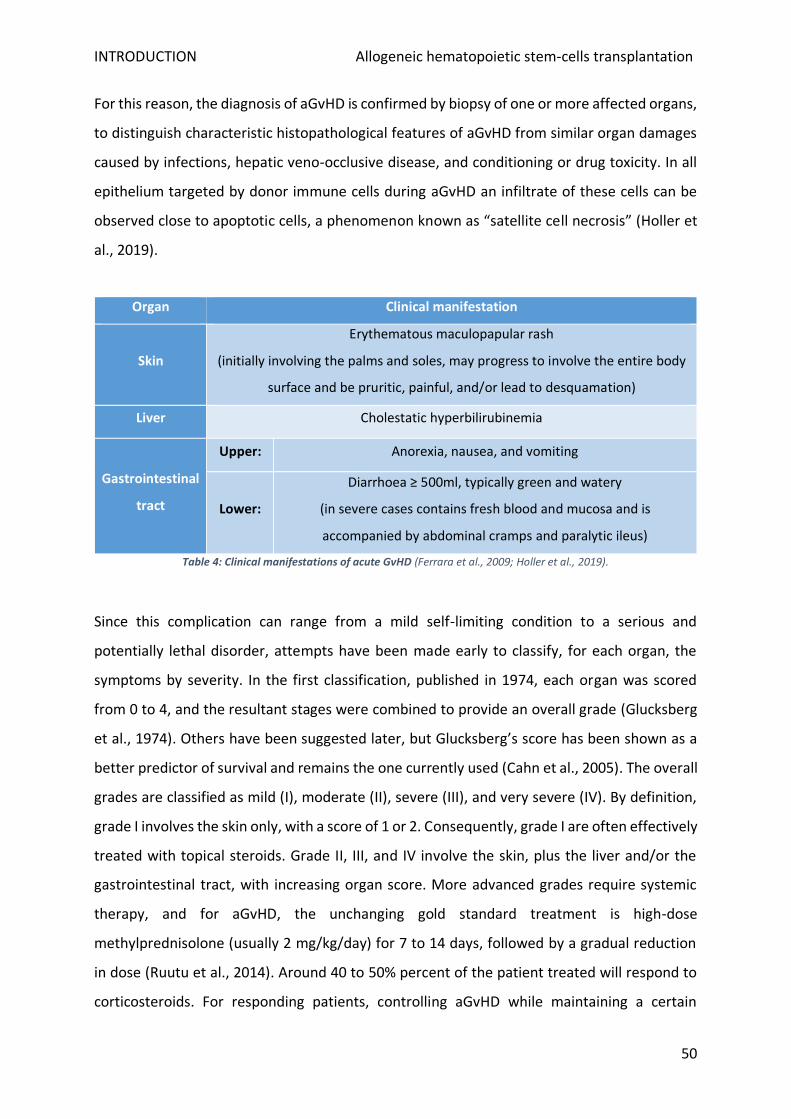
INTRODUCTION Allogeneic hematopoietic stem-cells transplantation
50
For this reason, the diagnosis of aGvHD is confirmed by biopsy of one or more affected organs,
to distinguish characteristic histopathological features of aGvHD from similar organ damages
caused by infections, hepatic veno-occlusive disease, and conditioning or drug toxicity. In all
epithelium targeted by donor immune cells during aGvHD an infiltrate of these cells can be
observed close to apoptotic cells, a phenomenon known as “satellite cell necrosis” (Holler et
al., 2019).
Organ Clinical manifestation
Skin
Erythematous maculopapular rash
(initially involving the palms and soles, may progress to involve the entire body
surface and be pruritic, painful, and/or lead to desquamation)
Liver Cholestatic hyperbilirubinemia
Gastrointestinal
tract
Upper: Anorexia, nausea, and vomiting
Lower:
Diarrhoea ≥ 500ml, typically green and watery
(in severe cases contains fresh blood and mucosa and is
accompanied by abdominal cramps and paralytic ileus)
Table 4: Clinical manifestations of acute GvHD (Ferrara et al., 2009; Holler et al., 2019).
Since this complication can range from a mild self-limiting condition to a serious and
potentially lethal disorder, attempts have been made early to classify, for each organ, the
symptoms by severity. In the first classification, published in 1974, each organ was scored
from 0 to 4, and the resultant stages were combined to provide an overall grade (Glucksberg
et al., 1974). Others have been suggested later, but Glucksberg’s score has been shown as a
better predictor of survival and remains the one currently used (Cahn et al., 2005). The overall
grades are classified as mild (I), moderate (II), severe (III), and very severe (IV). By definition,
grade I involves the skin only, with a score of 1 or 2. Consequently, grade I are often effectively
treated with topical steroids. Grade II, III, and IV involve the skin, plus the liver and/or the
gastrointestinal tract, with increasing organ score. More advanced grades require systemic
therapy, and for aGvHD, the unchanging gold standard treatment is high-dose
methylprednisolone (usually 2 mg/kg/day) for 7 to 14 days, followed by a gradual reduction
in dose (Ruutu et al., 2014). Around 40 to 50% percent of the patient treated will respond to
corticosteroids. For responding patients, controlling aGvHD while maintaining a certain
INTRODUCTION Allogeneic hematopoietic stem-cells transplantation
51
degree of immunocompetence against pathogen and potential residual tumour cells is always
challenging. Severe forms of aGvHD, that present a diminished chance of response to the
treatment, are associated with a bleak prognosis, with 25% long term survival for grade III and
5% for grade IV. So far, no single agent has shown better results when combined with
corticosteroids for first-line treatment (Martin et al., 1990; Rashidi et al., 2016).
Failure to respond to standard steroid doses or recurrence following initial dose reduction
(steroid dependence) will require second-line therapy. None have shown convincing long-
term efficacy. The most frequent choice involves one or several mAb directed against T cells
extracellular molecules, or anti-thymocyte globulin. mAb commonly used include
alemtuzumab which targets the pan-T lymphocytes marker CD52, inolimomab and daclizumab
directed against IL-2 receptor alpha subunit presents at the surface of activated T cells, and
etanercept and infliximab that recognize tumour necrosis factor alpha (TNFα). These therapies
are usually efficient to control aGvHD in a short term, but durable effects are relatively
infrequent, and the outcome of refractory aGvHD is poor with approximately 80% mortality,
especially if the lower GI tract is involved. A better outcome has been reported in a multicentre
study, with less associated toxicity, using frequent extracorporeal photopheresis (Jagasia et
al., 2013).
Other treatments have emerged during the last decade with promising results for severe
steroid-refractory aGvHD, in particular drugs acting on a downstream actor of extracellular
receptors targeted by current immunosuppressive drugs. These new targets include Janus-
activated-kinase (JAK) 1 and 2 which are intracellular signal transducer, under several cytokine
receptors, and Aurora kinase downstream the co-stimulation receptor CD28 (Hill et al., 2018).
Another type of strategy consists of using the immunosuppressive function of anti-
inflammatory grafted cell types including mesenchymal stromal cells infusion (Munneke et al.,
2016) and regulatory T cells (Treg) stimulation using α1 antitrypsin, which besides of its proper
anti-inflammatory effect stimulate these lymphocytes (Magenau et al., 2018; Marcondes et
al., 2016). Lastly, new concepts have also been proposed recently, Wu and Reddy have
highlighted the importance played by tissue-intrinsic factors in mediating aGvHD severity,
independently of the immune system role, opening a new therapeutic window (Wu and
Reddy, 2017). Thus, the difficulties to treat steroid-resistant aGvHD underline that steroid
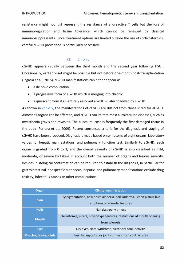
INTRODUCTION Allogeneic hematopoietic stem-cells transplantation
52
resistance might not just represent the resistance of alloreactive T cells but the loss of
immunoregulation and tissue tolerance, which cannot be renewed by classical
immunosuppressants. Since treatment options are limited outside the use of corticosteroids,
careful aGvHD prevention is particularly necessary.
(2) Chronic
cGvHD appears usually between the third month and the second year following HSCT.
Occasionally, earlier onset might be possible but not before one-month post-transplantation
(Jagasia et al., 2015). cGvHD manifestations can either appear as:
• a de novo complication,
• a progressive form of aGvHD which is merging into chronic,
• a quiescent form if an entirely resolved aGvHD is later followed by cGvHD.
As shown in Table 5, the manifestations of cGvHD are distinct from those listed for aGvHD.
Almost all organs can be affected, and cGvHD can imitate most autoimmune diseases, such as
myasthenia gravis and myositis. The buccal mucosa is frequently the first damaged tissue in
the body (Ferrara et al., 2009). Recent consensus criteria for the diagnosis and staging of
cGvHD have been proposed. Diagnosis is made based on symptoms of eight organs, laboratory
values for hepatic manifestations, and pulmonary function test. Similarly to aGvHD, each
organ is graded from 0 to 3, and the overall severity of cGvHD is also classified as mild,
moderate, or severe by taking in account both the number of organs and lesions severity.
Besides, histological confirmation can be required to establish the diagnosis, in particular for
gastrointestinal, nonspecific cutaneous, hepatic, and pulmonary manifestations exclude drug
toxicity, infectious causes or other complications.
Organ Clinical manifestation
Skin Dyspigmentation, new onset alopecia, poikiloderma, lichen planus-like
eruptions or sclerotic features
Nails Nail dystrophy or loss
Mouth Xerostomia, ulcers, lichen-type features, restrictions of mouth opening
from sclerosis
Eyes Dry eyes, sicca syndrome, cicatricial conjunctivitis
Muscles, fascia, joints Fasciitis, myositis, or joint stiffness from contractures
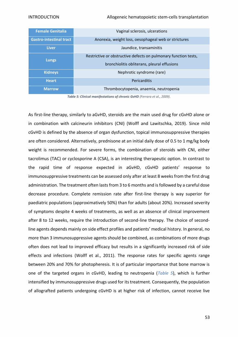
INTRODUCTION Allogeneic hematopoietic stem-cells transplantation
53
Female Genitalia Vaginal sclerosis, ulcerations
Gastro-intestinal tract Anorexia, weight loss, oesophageal web or strictures
Liver Jaundice, transaminitis
Lungs Restrictive or obstructive defects on pulmonary function tests,
bronchiolitis obliterans, pleural effusions
Kidneys Nephrotic syndrome (rare)
Heart Pericarditis
Marrow Thrombocytopenia, anaemia, neutropenia
Table 5: Clinical manifestations of chronic GvHD (Ferrara et al., 2009).
As first-line therapy, similarly to aGvHD, steroids are the main used drug for cGvHD alone or
in combination with calcineurin inhibitors (CNI) (Wolff and Lawitschka, 2019). Since mild
cGvHD is defined by the absence of organ dysfunction, topical immunosuppressive therapies
are often considered. Alternatively, prednisone at an initial daily dose of 0.5 to 1 mg/kg body
weight is recommended. For severe forms, the combination of steroids with CNI, either
tacrolimus (TAC) or cyclosporine A (CSA), is an interesting therapeutic option. In contrast to
the rapid time of response expected in aGvHD, cGvHD patients’ response to
immunosuppressive treatments can be assessed only after at least 8 weeks from the first drug
administration. The treatment often lasts from 3 to 6 months and is followed by a careful dose
decrease procedure. Complete remission rate after first-line therapy is way superior for
paediatric populations (approximatively 50%) than for adults (about 20%). Increased severity
of symptoms despite 4 weeks of treatments, as well as an absence of clinical improvement
after 8 to 12 weeks, require the introduction of second-line therapy. The choice of second-
line agents depends mainly on side effect profiles and patients’ medical history. In general, no
more than 3 immunosuppressive agents should be combined, as combinations of more drugs
often does not lead to improved efficacy but results in a significantly increased risk of side
effects and infections (Wolff et al., 2011). The response rates for specific agents range
between 20% and 70% for photopheresis. It is of particular importance that bone marrow is
one of the targeted organs in cGvHD, leading to neutropenia (Table 5), which is further
intensified by immunosuppressive drugs used for its treatment. Consequently, the population
of allografted patients undergoing cGvHD is at higher risk of infection, cannot receive live
INTRODUCTION Allogeneic hematopoietic stem-cells transplantation
54
vaccines, and often require supplemental intravenous immunoglobulin (for patients with IgG
<400 mg/dL).
cGvHD is the most relevant cause of late non-relapse morbidity following alloHSCT and
subsequent mortality of approximately 25%, (Grube et al., 2016). The CIBMTR reported an
incidence of cGvHD approaching 50% among allografted patients and its increase during the
last two decades. Rising cGvHD incidence can be explained by the increasing patient age
(incidence of 30% for children, 60% for adults) and more frequent use of unrelated and/or
mismatched donors, PBPC, and RIC regimens (Arai et al., 2015; Baird et al., 2010). By far,
history of aGvHD remains the greatest risk factor for chronic form apparition (Carlens et al.,
1998). As a result, strategies to prevent aGvHD may play a key role in preventing a subsequent
apparition of cGvHD, even if cGvHD is not simply the end-stage of aGvHD. It is crucial to
highlight the somehow protective effect of cGvHD regarding the resurgence rate of patients’
underlying malignancy. For this reason, the OS of patients developing mild cGvHD is
paradoxically better compared to patients without cGvHD. Even the OS of patients with
moderate cGvHD is not different from patients without cGvHD, as the slightly increased
mortality associated with this complication is counterbalanced by lower disease-associated
mortality (Kuzmina et al., 2012).
(3) Preventive action against GvHD
In spite of 50 years of research efforts, GvHD remains the most challenging complication after
alloHSCT for physicians to manage. From 30 to 50% of the patients will develop aGvHD
depending on his clinical history, including 15% with a severe form (grades III and IV). This
section focuses on the options available to prevent the development of aGvHD since, as stated
above, the main risk factor for developing cGvHD is the previous development of the acute
form of the disease.
(a) aGvHD risk factors
Development of GvHD after the graft is profoundly linked to the transplant features choice
exposed above, namely donor type, preparative regimen, and HSC source. Risk factors for
grade II to IV include graft originated from HLA-matched unrelated donors and HLA-
mismatched related or unrelated donors, all compared to HLA-matched related donors
INTRODUCTION Allogeneic hematopoietic stem-cells transplantation
55
(Kuzmina et al., 2012). The use of a female donor for a male recipient is also associated with
an increased risk. In terms of conditioning regimens, the use of TBI leads to a superior risk of
aGvHD in comparison with other conditioning therapies.
Post-alloHSCT outcomes is also known as highly impacted by the graft source and its cellular
content. Interestingly, grafting with growth factor–mobilized blood cells showed no
statistically significant associations with risk of grades II to IV aGvHD despite the presence of
up to a log more CD3+ T cells in a mobilized PBPC graft than in a bone marrow one (Rutella et
al., 1999). It appears that mobilization methods polarize helper T cells (Th) and dendritic cells
(DC) in a Th2/DC2 phenotype. Concerning bone marrow grafts, the ones who contain a
number above the median of invariant natural killer (NK) T cells, plasmacytoid DC and naïve
CD4+ and CD8+ T cells have been associated a greater OS at three years (Waller et al., 2014).
Graft rejection and GvHD related deaths were inferior for patients receiving grafts with more
plasmacytoid DC. Another team reported that a high donor Treg content was associated with
a lower risk of aGvHD following HLA-matched alloHSCT (Rezvani et al., 2006). However, this
last point is controversial in human since different Treg identification method lead to
divergent results (reviewed in Cohen and Boyer, 2006). In the case of UCB, cells’ graft
particular naïve immunological profile permits a reduced incidence and severity of paediatric
a- and cGvHD in HLA-matched sibling transplants compared with historically matched HLA-
matched sibling marrow transplants (Rocha et al., 2000). The different methods of graft
manipulation to avoid GvHD development that have been performed are described below, in
part (c).
More recently, refined clinical risk scores and novel biomarkers have been reported, which
predicts aGvHD response to initial therapy, and associated survival or TRM (MacMillan et al.,
2015). The strength of these biomarkers for early identification of high-risk patients at day 7
after HSCT or GvHD onset has been proved in large multicentre consortia and needs now
confirmation by trials on biomarker-guided treatment strategies (Hartwell et al., 2017; Levine
et al., 2015; Vander Lugt et al., 2013). Biomarkers of cGvHD are also currently explored but
will require validation before clinical use. These efforts to better identify the patients at risk
to develop GvHD are necessary to reproductively adapt the use of actual and future pre-
emptive treatment strategies.
INTRODUCTION Allogeneic hematopoietic stem-cells transplantation
56
(b) Current immunosuppressive prophylaxis
Actual preventive strategies against aGvHD differ between countries and depend mainly on
the patient’s preparative regimen type and donor. After MAC regimen, the gold standard for
aGvHD prophylaxis in Europe remains the combination of two immunosuppressive drugs:
short term methotrexate (MTX) and CSA (Michonneau and Socié, 2019). Better efficacy of this
dual therapy, compared to each molecule alone, was confirmed in the 1980s by two phase III
randomized trials, followed by a twenty-year follow-up report (Sorror et al., 2005; Storb et al.,
1986). If this treatment is associated with an unquestionable benefit, it goes together with
toxic effects, further increased by drug interactions, such as renal failure, arterial
hypertension, or raised infection susceptibility.
Two decades after CSA has begun to be used by clinicians, tacrolimus, a new CNI made its
apparition during the 1990s. TAC in conjunction with MTX was tested as a new aGvHD
prophylactic therapy in two randomized phase III trials after MAC with genoidentical and
phenoidentical grafts (Nash et al., 2000; Ratanatharathorn et al., 1998). No superiority in
terms of survival rate was observed in both trials for this new association, compare to
MTX+CSA. However, they have shown a decreased incidence of grade II to IV aGvHD, and this
option has been retained mainly by the US transplantation centres. Both CSA and TAC inhibit
effector T cells activation by targeting the nuclear factor of activated T-cell (NFAT) family of
transcription factors, which are downstream regulators of interleukin-2 (IL-2) transcription.
However, in the meantime, the decrease of IL-2 level also impacts anti-inflammatory Treg
function and survival.
For RIC and NMA regimens, CSA or TAC can be used alone or in combination with
mycophenolate mofetil (MMF) (Zeiser and Blazar, 2017). This association is widely used in this
setting, but no randomized prospective trial has tested its superiority with the classic
prophylaxis. Sirolimus (SIR) is a mammalian target of rapamycin (mTOR) inhibitor, that in
contrast with MMF, has been tested in combination with TAC and compared with TAC+MTX
in a phase III randomized study showing equivalent efficacy, but differences in toxicity (Törlén
et al., 2016). Unlike CNI, SIR is acting mainly on conventional T cells activation, without
impairing as much Treg function, because conventional T cells are depending more on the
protein kinase B/Akt pathway.
INTRODUCTION Allogeneic hematopoietic stem-cells transplantation
57
(c) Partial T cell depletion strategies
Besides classical immunosuppressive molecules in current use, an alternative approach to
GvHD prophylaxis is to remove donor T cells either ex vivo prior to the infusion or in vivo
before and/or after infusion using polyclonal anti-thymocyte globulin (ATG) or mAb.
Alternatively, a similar effect can be achieved by positive selection of CD34+ stem cells. Since
T cell depletion techniques may result in a drastic drop of a- and cGvHD risk, they have been
widespread in the 1980s and 1990s. Unfortunately, they were rapidly also identified as
contributing to an increased risk of graft failure, infections, and underlying disease relapse
(Horowitz et al., 1990; Marmont et al., 1991). As a result, they became restricted to situations
with increased risk of developing GvHD, such as mismatched and haploidentical transplants
where the risk of death from GvHD outweighs the risk of later disease recurrence. For alloHSCT
using unrelated donors, polyclonal ATG has become a major player since almost two decades,
supported by four prospective phase III randomized trials showing preventive effects mainly
on cGvHD (Finke et al., 2009; Kröger et al., 2016; Soiffer et al., 2017; Walker et al., 2016). In
the same indication, alemtuzumab although efficacious in preventing aGvHD has never been
tested prospectively in a randomized phase III trial and has almost exclusively been used in
the UK. Other studies have explored alternative methods including the infusion of an
expanded population of Treg at the time of stem cell infusion (Brunstein et al., 2011; Ianni et
al., 2011; Trzonkowski et al., 2009), and partial T cell depletion of alloreactive T cell subsets
such as α/β T cells, and in vitro or in vivo activated cells. The most successful one at the time
is post-transplant cyclophosphamide (PT-Cy) that target activated T cells.
As illustrated previously in Figure 5 for France, there has been during the past few years a
recent development in the use of haploidentical donors worldwide. While the initial attempts
to prevent GvHD were to use a huge dose of CD34+ selected cells, the appearance of PT-Cy
has truly revolutionized haploidentical grafts use, allowing alloHSCT access to more graft-
candidates. Cy administered in two doses scheduled soon after transplantation (at day 3 and
4), and followed by TAC+MMF, depletes highly proliferating alloreactive conventional T cells
while helping to preserve Treg (Luznik et al., 2012). HSC contains an important quantity of
aldehyde dehydrogenase and can consequently convert the 4-hydroxycyclophosphamide into
a non-alkylating metabolite. Specific toxicity associated with Cy includes haemorrhagic cystitis
and rare but potentially serious early cardiologic dysfunction. The incidence of aGvHD remains
INTRODUCTION Allogeneic hematopoietic stem-cells transplantation
58
significant (around 1 among 3 patients), but there is now growing evidence that PT-Cy might
be associated with a low rate of chronic GvHD (Fuchs, 2017). PT-Cy is now proposed for other
alloHSCT settings, including transplantation from an unrelated donor and genoidentical
sibling, setting in which a randomized phase III trial is ongoing to compare PT-Cy alone versus
MTX+CSA usual prophylaxis (Kanakry et al., 2014; Luznik et al., 2012).
In spite of the above sections, new therapies and prophylactic regimens are needed since
severe GvHD rates remain high. The better comprehension of alloreactive T cell activation and
migration to the targeted organ is now allowing new therapeutic hopes, of treatments
showing promising effects in pre-clinical models to reach the patients’ bedside in the
upcoming years.
INTRODUCTION Immunotherapeutic effect of alloHSCT
58
Immunotherapeutic effect of alloHSCT
AlloHSCT activity in France and abroad has been growing exponentially thanks to various
procedural improvements, simultaneously with the enlargement of its indication landscape to
several high-risk haematological malignancies (ABM and EBMT 2019 reports). In this setting,
it remains the most curative option for refractory diseases, despite a highly reduced
effectiveness in comparison to its use as consolidation therapy in diseases in complete
remission at the time of transplant (Biernacki et al., 2020). In around half of the grafted
patients, this heavy therapy is still insufficient to avoid a relapse from their underlying
malignancy, with an often worsened aggressivity. For now, only a narrow range of therapeutic
tools has proved useful in this situation, to potentialize the graft antitumour effect. This
second chapter ambitions to provide the immunobiological foundations underlying the GvT
effect, the reasons behind its potential failure, and the existent and emerging strategies to
face this clinical situation.
INTRODUCTION Immunotherapeutic effect of alloHSCT
60
A. GvT effect: Definition and clinical evidences
Two main elements in alloHSCT account for protection from relapse: the pre-graft preparative
regimen and the presence of donor T cells naturally contained inside the graft. Conditioning
primarily mediates relapse protection in the early phase after transplantation, from 0 to 12
months, while the effect of donor T cells through the GvT effect occurs usually after the first
year (Biernacki et al., 2020). As previously mentioned (Chapter I.B.5. Figure 6), depending on
the conditioning intensity the requirement in terms of GvT strength differs. It is of particular
importance for low-intensity options NMA and RIC regimens, whereas conditioning and GvT
effect both contribute to relapse protection when using intensive myeloablative regimens
(Inamoto et al., 2011; Scott et al., 2017).
GvT has been first recognized in the pioneer allograft experiment sets performed by Barnes
et al., they reported that leukemic mice treated with a subtherapeutic dose of radiation and
a syngeneic (identical twin) graft transplant were more likely to relapse than mice given an
allogeneic stem cell transplant (Barnes et al., 1956). They hypothesized that the allogeneic
graft contained cells with immune reactivity necessary for eradicating residual leukaemia cells.
They also noted that recipients of allogeneic grafts, though less likely to relapse, died of a
“wasting syndrome” now recognized as GvHD. Thus, in addition to describing GvT, these
experiments highlighted for the first time the intricate relationship between GvT and GvHD.
Later on, the importance of donor T cells in mediating GvT was originally inferred from clinical
data demonstrating increased relapse risk with extensive ex vivo T cell depletion from donor
grafts before infusion into patients (Horowitz et al., 1990; Marmont et al., 1991). Another
important aspect in this demonstration has been the observation of a higher relapse rate in
syngeneic graft recipient compared to matched-HLA allogeneic ones, indicating that even in
well-matched individuals, subtle degrees of mismatch can induce the beneficial effect of GVT
(Weiden et al., 1979). The discovery of the polymorphic genes that encode minor
histocompatibility antigens permitted to explain the observations about HLA-identical-sibling.
T-cell responses to minor histocompatibility antigens are responsible for the antileukemic
activity, but also cause GvHD (Bleakley and Riddell, 2004). NK cells do have a role to play in
INTRODUCTION Immunotherapeutic effect of alloHSCT
61
GvT effect and GvHD in HLA-mismatched grafts, but their action is out of scope of this thesis
(Hu and Liu, 2017).
INTRODUCTION Immunotherapeutic effect of alloHSCT
62
B. Establishment of the alloreactive response
Conditioning regimen is not only responsible for the early antitumour action by directly
depleting healthy as well as malignant hematopoietic cells, but also is crucial to initiate the
long-term GvT effect. In consequence, the delayed transfer of alloreactive T cells in chimaera
mice does not induce GvHD and results in reduced GvT effect (Chakraverty, 2006, 2008; Li et
al., 2015). Indeed, chemotherapeutic agents and radiations used in alloHSCT preparative
regimens aim to be mainly toxic for the hematopoietic compartment while sparing the
damages on other tissues, but they still generate DNA strand breaks and reactive oxygen
species in the host tissues (Kim et al., 2014; Mauch et al., 1995). In fine, it induces epithelium
and mesenchymal cell death by necrosis or apoptosis (Sonis et al., 2004; Blijlevens and Sonis,
2007). These tissue damages will generate danger signals emanating from both the host
(damage-associated molecular patterns (DAMP)) and its commensal flora (pathogen-
associated molecular patterns (PAMP)), contributing to the activation of local antigen-
presenting cell (APC) that will, in turn, activate alloreactive T cells (Brubaker et al., 2015; Jones
et al., 1971). DAMP include a myriad of pro-inflammatory cytokines, such as TNF-, and
chemokines, which together can increase the expression of adhesion molecules, HLA antigens
and costimulatory molecules on host APC (Ferrara et al., 2009). An important aspect of this
process is the implication, not only of the donor APC derived from HSC but also those from
the host, able to present host HLA and minor antigens to alloreactive donor T cells. This
coexistence is transitory since recipient APC will be progressively killed by donor T cells and
replaced by bone marrow-emerging donor APC, able to present recipient antigens using cross-
presentation (Shlomchik, 2007; Wang et al., 2011). Therefore, donor APC are not required for
GvHD initiation, but they cause its maintenance and are mandatory for a long-term GvT effect
(Matte et al., 2004; Reddy et al., 2005). Many studies have attempted to identify the critical
APC subsets involved, predicated on the notion that deletion may prevent GVHD, however it
appears that if one subset is sufficient to induce GvHD, none is necessary (Koyama and Hill,
2016).

INTRODUCTION Immunotherapeutic effect of alloHSCT
63
INTRODUCTION Immunotherapeutic effect of alloHSCT
64
C. Allogeneic effector phase
Subsequently to the conditioning regimen, the release of several signals of danger by host
tissues and microbiota promotes APC maturation in peripheral tissues, followed by their
migration in secondary lymphoid organs and Peyer patches (Ferrara et al., 2009). During the
first 12 hours post-allograft, most part of the alloreactive T cell pool is posted in these
secondary organs, where the stimulation by activated APC occurs and results in a massive
proliferation (Panoskaltsis-Mortari et al., 2004). They reach the spleen at day two and the
migration to target organs starts at day 5 or 6 (Beilhack et al., 2005).
1. T cells trafficking
The effector phase covers the freshly divided alloreactive T cell clones’ migration and their
differentiation into effector T cells (Teff) able to kill remaining patient’s tumour cells, or in
case of aGVHD occurrence, to destroy target tissues. Thus, aGVHD according to Ferrara can
be viewed as “an exaggerated but normal inflammatory mechanisms mediated by donor
lymphocytes infused into the recipient where they function appropriately, given the foreign
environment they encounter” (Ferrara et al., 2009). Indeed, in the context of alloHSCT, donor
T cells are specific for antigens that are ubiquitously expressed in the host’s body. T cell
migration seems to depend on the APC that activates it, the APC’s origin will determine which
chemokine receptor the T cell will express, defining is tissular drop point (Bouazzaoui et al.,
2009); Kim et al., 2003). This last point and the interactions between donor T cells and tissue-
resident activated APC may explain, in part, the clinical picture of aGvHD regarding
conditioning damages preferential target (Sadeghi et al., 2013). Regarding the GvT effect, the
mechanisms of alloreactive T cells migration inside the body in relation to malignant cells’
position are unclear, and whether the alloreactive response distribution is homogeneous in
all tissues or focused on cancer cells location is not fully solved. Michonneau et al. recently
reported the existence of a gradient of donor T cells cytotoxic capacity, as a function of the
considered tissue, due to the expression of programmed cell death protein (PD-1) ligands in
the microenvironment (Michonneau et al., 2016). This observation suggests an anatomical
segregation of cytotoxic T cells, that might let open tissue niches for cancer cells to escape
GvT action while justifying further GvHD sites of predilection.
INTRODUCTION Immunotherapeutic effect of alloHSCT
65
2. Effector/memory profile
After alloHSCT, donor T cells display diverse phenotypes that correlate with their function.
Alloreactive T cells co-exist with non-host-reactive donor T cells which are beneficial for
peripheral T-cell reconstitution, and both subsets diverge in phenotype and division rate
(Maury et al., 2001). Additionally, alloreactive T cells phenotype appear to be different
depending on their capacity to induce GvHD or GvT. Indeed, GvHD induction can be limited by
depletion of the naïve T cell population from the graft in mice, a strategy that seems to be
confirmed in patients, without impairing GvT effect (Bleakley et al., 2014, 2015). In contrast,
T cells responsible for the antitumour effect appear to come from the CD4+ and CD8+ central
memory and the effector memory populations (Dutt et al., 2011; Zhang et al., 2004; Zheng et
al., 2008, 2009, 2014). Furthermore, central memory T cells show a higher migration potential
than effector memory cells in the secondary lymphoid organs resulting in a better antitumour
efficacy (Klebanoff et al., 2005).
3. T helper subsets
In the same vein, the profile of donor T cells differentiation in Th1/2/17 has been deeply
explored trying to detect a difference between GvHD and GvL main actors. Historically, GvHD
have been thought to be driven only by Th1 differentiation profile, among others because of
the very high expression of TNF- and IFN-γ in target organs (Antin and Ferrara, 1992; Hill et
al., 1997). At the contrary, Th2 cells were for long considered as a protective population, and
the blocking of Th2 differentiation has been strongly associated with GvHD amplification
(Fowler et al., 1994, Tawara et al., 2008). However, the reality is less straightforward since
both Th1 and Th2 are required for the appearance of lesions in the gut for instance, suggesting
that both populations potentially have different functions depending on the tissue considered
(Nikolic et al., 2000). Regarding Th profiles involved in the antitumour action post-allograft,
less data is available for now. Th1 cells are also the principal mediator of the GvT effect, and
Th2 CD4+ do not seem necessary for its application, while cytotoxic CD8+ type 2 could have a
role (Yang et al., 2002). Lastly, deficiency in Th17 cells induce a diminution of Th1 cells to the
benefit of Treg cells, resulting in a milder GvHD, due in part to the reduced migration of T cells
toward the gut (Hanash et al., 2011). Notably, this subset does not appear to have a role in
the tumour eradication.
INTRODUCTION Immunotherapeutic effect of alloHSCT
66
4. Antigen specificity
One main difference between GvHD and GvT are the targeted cells. In theory, only the T cell
population implied in the GvT effect recognize tumour-associated antigens (TAA), when both
GvHD and GvT actors are able to recognize HLA-related antigens, causing cancer cells’
elimination as well as GvHD lesions. For this reason, TAA recognition has been thought to be
an interesting feature to modulate GvT independently from GvHD induction in healthy tissues.
However, if the presence of T cell clones specific for TAA post-alloHSCT has been associated
with a reduction of the relapse rate, data suggest that GvT effect is mainly mediated by HLA
incompatibilities or minor antigens at the surface of malignant cells (Fontaine et al., 2001;
Kapp et al., 2009). Interestingly, it was demonstrated that a unique minor incompatibility was
sufficient to induce a potent GvT effect, without triggering GvHD presumably because of a
higher sensitivity of hematopoietic cells to the T cell cytolytic action. This particular
observation has later led to the hypothesis that the pattern of tissular destruction and the
efficacy of tumour elimination could reflect tissue sensitiveness toward donor allogeneic
response (Vincent et al., 2011).
5. Cytotoxic mechanisms of tumour cell elimination
First, a direct interaction between cancer cells and donor T cells is necessary for the GvT effect
to occur. Indeed, the expression by the tumour cells of the alloantigen, for major and minor
HLA incompatibilities, is needed for CD4+ and CD8+ to set-up the anti-tumour response
(Reddy et al., 2005; Matte-Martone et al., 2008). The effector response uses the
perforin/granzyme B signalling pathway, whereas the Fas/Fas-L interaction does not appear
to play a role, in contrast with its role during GvHD (Tsukada et al., 1999; Schmaltz et al., 2001).
Soluble factors have also been implied in GvT. Importantly, using anti-TNF− after allograft
could reduce GvHD while maintaining an apparent GvT function, although transitory with the
observation of early relapses (Korngold et al., 2003; Tsukada et al., 1999). Another study has
demonstrated that TNF- production by T cells was required for a potent GvT effect (Schmaltz
et al., 2003). No increase of relapse has been reported until now when using anti-TNF-
treatment in patients (Alousi et al., 2009; Gatza et al., 2014).
INTRODUCTION Immunotherapeutic effect of alloHSCT
67
Then, if there is a common ground for GvT and GvHD in terms of antigen specificity and some
effector capacity such as perforin/granzyme B pathway or TNF- production by donor T cells,
some distinct features of allogeneic anti-tumour response exist in comparison with GvHD
mechanisms. For instance, Fas/Fas-L pathway seems restricted to GvHD. Also, if TNF- is
produced in any case, TRAIL (for TNF-related apoptosis-inducing factor) which belongs to the
TNF receptor family, is required for a potent GvT effect but does not seems to take part in
GvHD associated-processes (Schmaltz et al., 2002). Accordingly, since TRAIL overexpression
on donor T cells induces a GvL effect but no GvHD, it has been considered as an interesting
potential target to boost antitumour response without damaging healthy cells (Ghosh et al.,
2013).
INTRODUCTION Immunotherapeutic effect of alloHSCT
68
D. Causes of relapse
Recipients may sustain remissions that last years after alloHSCT but then relapse, consistent
with the existence but ultimate failure of GvT activity (Horowitz et al., 2018). In solid cancers,
the prolonged immune pressure selectively allows immune-resistant subclones to survive, a
process refers to as immunoediting (Schreiber et al., 2011). A growing body of literature
reports such a mechanism of GvT failure due to an intrinsic mechanism of residual leukemic
cells, which adapt and ultimately evade T cell control (Falkenburg and Jedema, 2017; Zeiser
and Vago, 2019). Alternatively, tumour escape can be caused by an increasing functional
impairment of donor T cell over time as a consequence of their environment. These two
possibilities will be addressed individually but are not mutually exclusive.
a) Tumour cells intrinsic mechanisms of evasion
An example of immunoediting is provided by the high rate of extramedullary relapses,
including in immune sanctuaries such as the central nervous system, after alloHSCT but even
more frequently after DLI, suggesting a reaction from the tumour cells to more immune
pressure (Harris et al., 2013).
At the cell scale, cancer cells are known to escape immunosurveillance through HLA genomic
loss or expression downregulation. HLA class II, but not class I, expression downregulation,
with a 3 to 12 time reduction of the RNA product, is observed in relapses after HLA-matched
and mismatched alloHSCT (Christopher et al., 2018). It is interesting to note that only class II
HLA is impacted by these downmodulations, supporting the idea that CD4+ T cells play a major
role in the GvT effect. IFN- is known to upregulate HLA expression on cells and seems to be
able to restore class II expression on post-alloHSCT leukemic cells in vitro, suggesting systemic
IFN-γ administration may help maintain GvL for that particular point (Toffalori et al., 2019).
However, cancer cells can alternatively upregulate the expression of molecules that can
directly weaken the immune system, among which PD-L1 able to trigger T cells exhaustion is
directly induced by IFN-γ exposure (Choi et al., 2012a; Vago and Dazzi, 2019).
One particular example of strategies employed by cancer cells to avoid immune system watch
is the genomic loss of the mismatched HLA haplotype is frequently documented in leukaemia
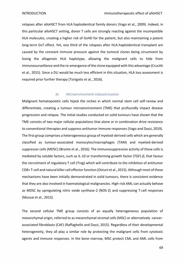
INTRODUCTION Immunotherapeutic effect of alloHSCT
69
relapses after alloHSCT from HLA haploidentical family donors (Vago et al., 2009). Indeed, in
this particular alloHSCT setting, donor T cells are strongly reacting against the incompatible
HLA molecules, creating a higher risk of GvHD for the patient, but also maintaining a potent
long-term GvT effect. Yet, one third of the relapses after HLA-haploidentical transplant are
caused by the constant immune pressure against the tumoral clones being circumvent by
losing the allogeneic HLA haplotype, allowing the malignant cells to hide from
immunosurveillance and the re-emergence of the clone equipped with this advantage (Crucitti
et al., 2015). Since a DLI would be much less efficient in this situation, HLA loss assessment is
required prior further therapy (Tsirigotis et al., 2016).
b) Microenvironment-induced evasion
Malignant hematopoietic cells hijack the niches in which normal stem cell self-renew and
differentiate, creating a tumour microenvironment (TME) that profoundly impact disease
progression and relapse. The initial studies conducted on solid tumours have shown that the
TME consists of two major cellular populations that alone or in combination drive resistance
to conventional therapies and suppress antitumor immune responses (Vago and Dazzi, 2019).
The first group comprises a heterogeneous group of myeloid-derived cells which are generally
classified as tumour-associated monocytes/macrophages (TAM) and myeloid-derived
suppressor cells (MDSC) (Bronte et al., 2016). The immunosuppressive activity of these cells is
mediated by soluble factors, such as IL-10 or transforming growth factor (TGF)-β, that favour
the recruitment of regulatory T cell (Treg) which will contribute to the inhibition of antitumor
CD8+ T-cell and natural killer cell effector function (Ostuni et al., 2015). Although most of these
mechanisms have been initially demonstrated in solid tumours, there is consistent evidence
that they are also involved in haematological malignancies. High-risk AML can actually behave
as MDSC by upregulating nitric oxide synthase-2 (NOS-2) and suppressing T-cell responses
(Mussai et al., 2013).
The second cellular TME group consists of an equally heterogeneous population of
mesenchymal origin, referred to as mesenchymal stromal cells (MSC) or alternatively cancer-
associated fibroblasts (CAF) (Raffaghello and Dazzi, 2015). Regardless of their developmental
heterogeneity, they all play a similar role by protecting the malignant cells from cytotoxic
agents and immune responses. In the bone marrow, MSC protect CML and AML cells from
INTRODUCTION Immunotherapeutic effect of alloHSCT
70
chemotherapeutic agents such as imatinib and Ara-C (Vianello F et al., 2010). This protection
is exerted in a non-antigen-specific fashion and rely mainly on the release of indoleamine 2-3
dioxygenase-1 (IDO-1), which consumes the essential amino acid tryptophan, but several
additional immunosuppressive mechanisms have been described (Jones et al., 2007).
However, more recent data have highlighted the important contribution of tissue-resident
monocytes/macrophages in delivering a sustainable immunosuppressive effect (Cheung and
Dazzi, 2018).
Finally, the role of Treg in generating immune resistance has been much discussed and
suggested as a mean to protect against GvHD (Cohen and Boyer, 2006). While there is plenty
of data indicating how these cells exert a very negative impact on the outcome of solid
tumours, data in preclinical models of allogeneic HSCT have suggested that Treg may
selectively inhibit GvHD without compromising GvL (Edinger et al., 2003). However, increased
frequencies of CD4+CD25hi Treg correlate with disease relapse after allogeneic stem cell
transplantation for CML, suggesting that Treg do play a role in tumour escape observed in
patients, and could be of interest to boost the GvT effect (Nadal et al., 2007).
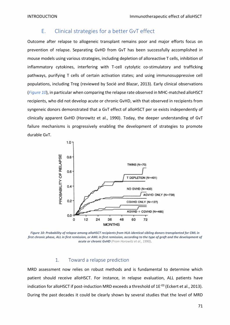
INTRODUCTION Immunotherapeutic effect of alloHSCT
71
E. Clinical strategies for a better GvT effect
Outcome after relapse to allogeneic transplant remains poor and major efforts focus on
prevention of relapse. Separating GvHD from GvT has been successfully accomplished in
mouse models using various strategies, including depletion of alloreactive T cells, inhibition of
inflammatory cytokines, interfering with T-cell cytolytic co-stimulatory and trafficking
pathways, purifying T cells of certain activation states; and using immunosuppressive cell
populations, including Treg (reviewed by Socié and Blazar, 2013). Early clinical observations
(Figure 10), in particular when comparing the relapse rate observed in MHC-matched alloHSCT
recipients, who did not develop acute or chronic GvHD, with that observed in recipients from
syngeneic donors demonstrated that a GvT effect of alloHSCT per se exists independently of
clinically apparent GvHD (Horowitz et al., 1990). Today, the deeper understanding of GvT
failure mechanisms is progressively enabling the development of strategies to promote
durable GvT.
Figure 10: Probability of relapse among alloHSCT recipients from HLA-identical sibling donors transplanted for CML in first chronic phase, ALL in first remission, or AML in first remission, according to the type of graft and the development of
acute or chronic GvHD (From Horowitz et al., 1990).
1. Toward a relapse prediction
MRD assessment now relies on robust methods and is fundamental to determine which
patient should receive alloHSCT. For instance, in relapse evaluation, ALL patients have
indication for alloHSCT if post-induction MRD exceeds a threshold of 1E-03 (Eckert et al., 2013).
During the past decades it could be clearly shown by several studies that the level of MRD
INTRODUCTION Immunotherapeutic effect of alloHSCT
72
immediately prior to transplant does have a clear prognostic impact on post-HSCT outcome
(Knechtli et al., 1998). Retrospective studies in children with relapsed ALL revealed an
important cut-off for post-HSCT outcome. Patients who received transplantation with an MRD
important load had a by far inferior prognosis than patients with lower MRD loads before
transplant (Bader et al., 2015). Based on these findings, several studies are now underway
investigating strategies to improve outcome in these ultrahigh-risk patients. Adaption of
transplant approaches might allow successful transplantation(Leung et al., 2012). Almost half
of the patients with high levels of MRD before trans- plantation achieved molecular remission
by day +100 (Spinelli et al., 2015). This finding indicates that MRD detection post-transplant
provides additional value to the MRD assessment prior to transplantation. It could be
demonstrated in prospective clinical studies that the close monitoring of MRD by different
approaches allows the prediction of relapse and may therefore form the basis of different
intervention strategies making use of leukaemia-specific targeted therapy (Bader et al., 2015;
Balduzzi et al., 2014). Future perspectives will focus on MRD-guided intervention to prevent
overt relapse (Rettinger et al., 2017).
2. Adoptive cell therapies
a) DLI
In 1990, Kolb et al. were the first reporting sustained remission after DLI in patients with CML
who relapsed after alloHSCT (Kolb et al., 1990). Since then, this adoptive cell therapy had
become the first resort for relapsing patients post-allograft without prior GvHD occurrences
(de Lima et al., 2014). Interestingly, MRD-directed DLI have been proved to significantly
decrease the relapse rate without aggravating GvHD (Yan et al., 2012). Recently, different
modified DLI protocols have been suggested, aiming to improve its efficacy or reduce the risks
of associated GvHD. Chang et. al utilised G-CSF-mobilized peripheral blood stem cell instead
of a steady lymphocyte infusion because of this cytokine’s ability to polarize Th1 to Th2
phenotype and induce hyporesponsiveness of T cells (Chang and Huang, 2013). This protocol
also included the introduction of short-term immunosuppressive agents (CSA/MTX), to further
decrease the incidence of GVHD. The feasibility and efficacy of this approach were confirmed
either for treatment or prevention of relapse after haploidentical HSCT.
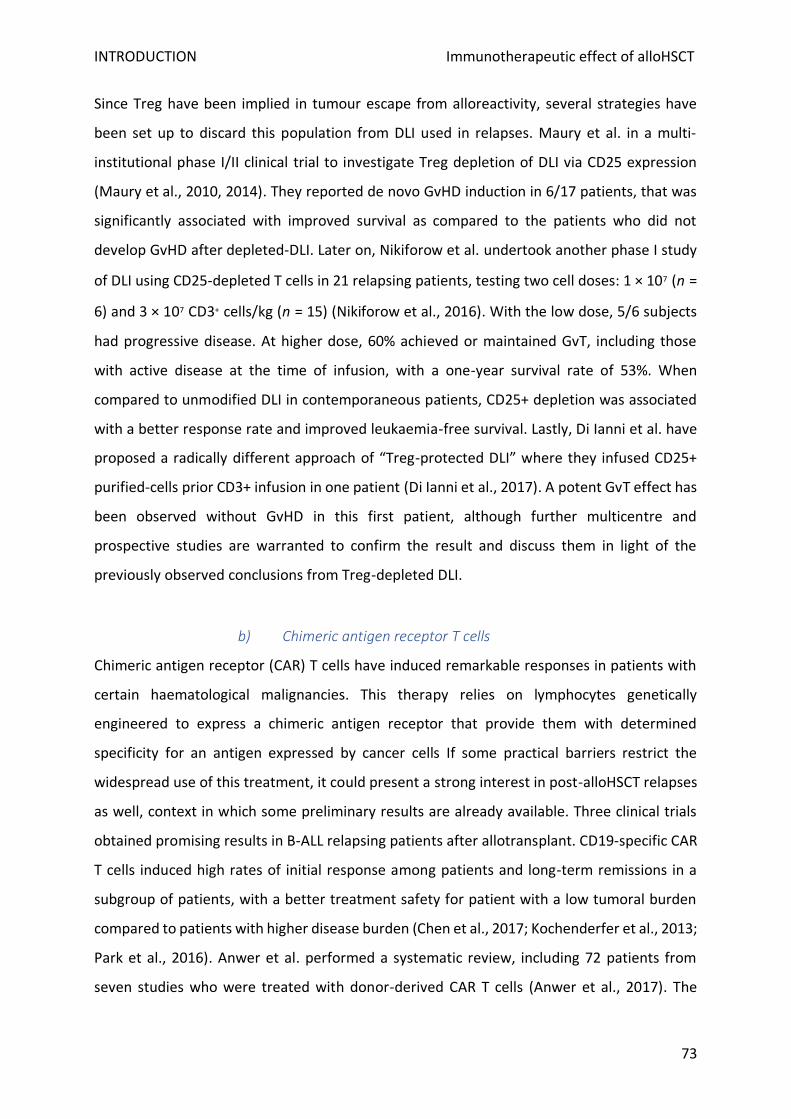
INTRODUCTION Immunotherapeutic effect of alloHSCT
73
Since Treg have been implied in tumour escape from alloreactivity, several strategies have
been set up to discard this population from DLI used in relapses. Maury et al. in a multi‐
institutional phase I/II clinical trial to investigate Treg depletion of DLI via CD25 expression
(Maury et al., 2010, 2014). They reported de novo GvHD induction in 6/17 patients, that was
significantly associated with improved survival as compared to the patients who did not
develop GvHD after depleted‐DLI. Later on, Nikiforow et al. undertook another phase I study
of DLI using CD25-depleted T cells in 21 relapsing patients, testing two cell doses: 1 × 10⁷ (n =
6) and 3 × 10⁷ CD3+ cells/kg (n = 15) (Nikiforow et al., 2016). With the low dose, 5/6 subjects
had progressive disease. At higher dose, 60% achieved or maintained GvT, including those
with active disease at the time of infusion, with a one-year survival rate of 53%. When
compared to unmodified DLI in contemporaneous patients, CD25+ depletion was associated
with a better response rate and improved leukaemia-free survival. Lastly, Di Ianni et al. have
proposed a radically different approach of “Treg-protected DLI” where they infused CD25+
purified-cells prior CD3+ infusion in one patient (Di Ianni et al., 2017). A potent GvT effect has
been observed without GvHD in this first patient, although further multicentre and
prospective studies are warranted to confirm the result and discuss them in light of the
previously observed conclusions from Treg-depleted DLI.
b) Chimeric antigen receptor T cells
Chimeric antigen receptor (CAR) T cells have induced remarkable responses in patients with
certain haematological malignancies. This therapy relies on lymphocytes genetically
engineered to express a chimeric antigen receptor that provide them with determined
specificity for an antigen expressed by cancer cells If some practical barriers restrict the
widespread use of this treatment, it could present a strong interest in post-alloHSCT relapses
as well, context in which some preliminary results are already available. Three clinical trials
obtained promising results in B-ALL relapsing patients after allotransplant. CD19-specific CAR
T cells induced high rates of initial response among patients and long-term remissions in a
subgroup of patients, with a better treatment safety for patient with a low tumoral burden
compared to patients with higher disease burden (Chen et al., 2017; Kochenderfer et al., 2013;
Park et al., 2016). Anwer et al. performed a systematic review, including 72 patients from
seven studies who were treated with donor-derived CAR T cells (Anwer et al., 2017). The
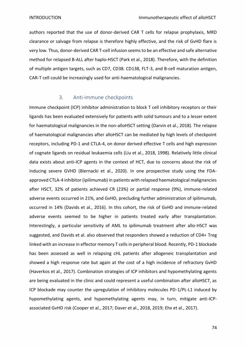
INTRODUCTION Immunotherapeutic effect of alloHSCT
74
authors reported that the use of donor-derived CAR T cells for relapse prophylaxis, MRD
clearance or salvage from relapse is therefore highly effective, and the risk of GvHD flare is
very low. Thus, donor-derived CAR T-cell infusion seems to be an effective and safe alternative
method for relapsed B-ALL after haplo-HSCT (Park et al., 2018). Therefore, with the definition
of multiple antigen targets, such as CD7, CD38. CD138, FLT-3, and B-cell maturation antigen,
CAR-T cell could be increasingly used for anti-haematological malignancies.
3. Anti-immune checkpoints
Immune checkpoint (ICP) inhibitor administration to block T cell inhibitory receptors or their
ligands has been evaluated extensively for patients with solid tumours and to a lesser extent
for haematological malignancies in the non-alloHSCT setting (Darvin et al., 2018). The relapse
of haematological malignancies after alloHSCT can be mediated by high levels of checkpoint
receptors, including PD-1 and CTLA-4, on donor derived effective T cells and high expression
of cognate ligands on residual leukaemia cells (Liu et al., 2018, 1998). Relatively little clinical
data exists about anti-ICP agents in the context of HCT, due to concerns about the risk of
inducing severe GVHD (Biernacki et al., 2020). In one prospective study using the FDA-
approved CTLA-4 inhibitor (ipilimumab) in patients with relapsed haematological malignancies
after HSCT, 32% of patients achieved CR (23%) or partial response (9%), immune-related
adverse events occurred in 21%, and GvHD, precluding further administration of ipilimumab,
occurred in 14% (Davids et al., 2016). In this cohort, the risk of GvHD and immune-related
adverse events seemed to be higher in patients treated early after transplantation.
Interestingly, a particular sensitivity of AML to ipilimumab treatment after allo-HSCT was
suggested, and Davids et al. also observed that responders showed a reduction of CD4+ Treg
linked with an increase in effector memory T cells in peripheral blood. Recently, PD-1 blockade
has been assessed as well in relapsing cHL patients after allogeneic transplantation and
showed a high response rate but again at the cost of a high incidence of refractory GvHD
(Haverkos et al., 2017). Combination strategies of ICP inhibitors and hypomethylating agents
are being evaluated in the clinic and could represent a useful combination after alloHSCT, as
ICP blockade may counter the upregulation of inhibitory molecules PD-1/PL-L1 induced by
hypomethylating agents, and hypomethylating agents may, in turn, mitigate anti-ICP-
associated GvHD risk (Cooper et al., 2017; Daver et al., 2018, 2019; Ehx et al., 2017).
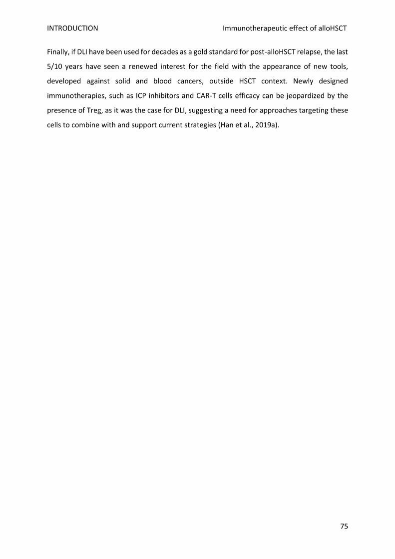
INTRODUCTION Immunotherapeutic effect of alloHSCT
75
Finally, if DLI have been used for decades as a gold standard for post-alloHSCT relapse, the last
5/10 years have seen a renewed interest for the field with the appearance of new tools,
developed against solid and blood cancers, outside HSCT context. Newly designed
immunotherapies, such as ICP inhibitors and CAR-T cells efficacy can be jeopardized by the
presence of Treg, as it was the case for DLI, suggesting a need for approaches targeting these
cells to combine with and support current strategies (Han et al., 2019a).
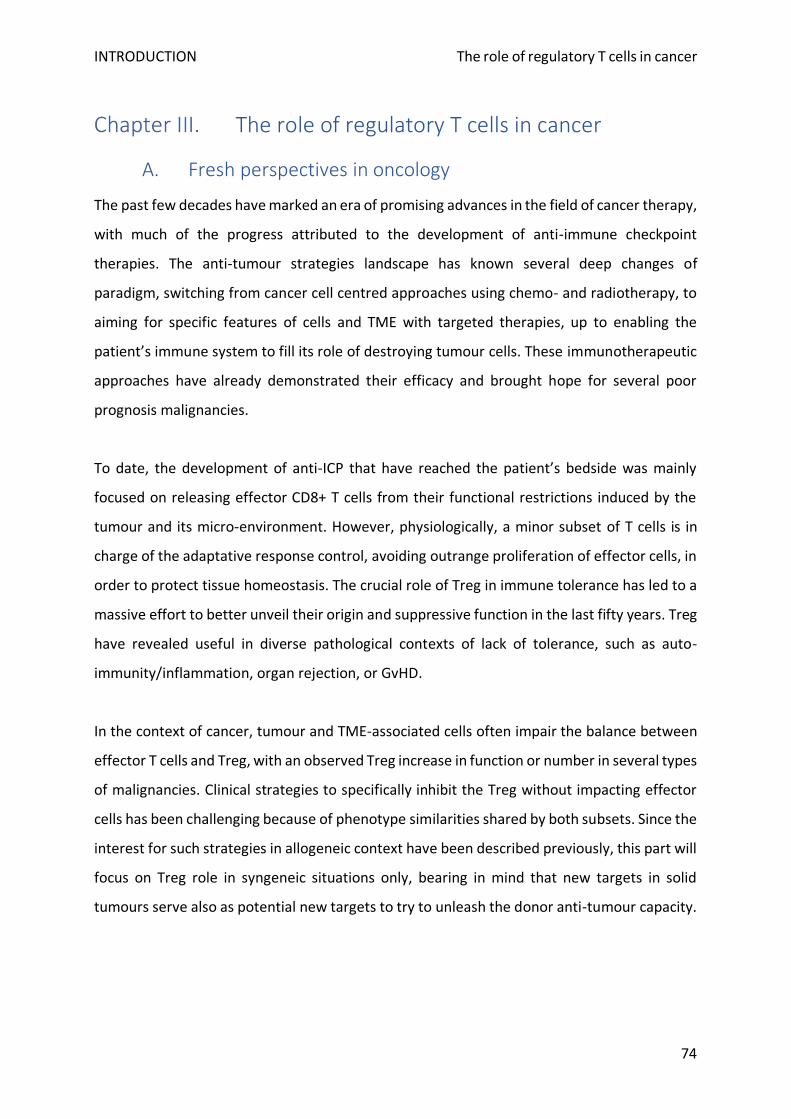
INTRODUCTION The role of regulatory T cells in cancer
74
The role of regulatory T cells in cancer
A. Fresh perspectives in oncology
The past few decades have marked an era of promising advances in the field of cancer therapy,
with much of the progress attributed to the development of anti-immune checkpoint
therapies. The anti-tumour strategies landscape has known several deep changes of
paradigm, switching from cancer cell centred approaches using chemo- and radiotherapy, to
aiming for specific features of cells and TME with targeted therapies, up to enabling the
patient’s immune system to fill its role of destroying tumour cells. These immunotherapeutic
approaches have already demonstrated their efficacy and brought hope for several poor
prognosis malignancies.
To date, the development of anti-ICP that have reached the patient’s bedside was mainly
focused on releasing effector CD8+ T cells from their functional restrictions induced by the
tumour and its micro-environment. However, physiologically, a minor subset of T cells is in
charge of the adaptative response control, avoiding outrange proliferation of effector cells, in
order to protect tissue homeostasis. The crucial role of Treg in immune tolerance has led to a
massive effort to better unveil their origin and suppressive function in the last fifty years. Treg
have revealed useful in diverse pathological contexts of lack of tolerance, such as auto-
immunity/inflammation, organ rejection, or GvHD.
In the context of cancer, tumour and TME-associated cells often impair the balance between
effector T cells and Treg, with an observed Treg increase in function or number in several types
of malignancies. Clinical strategies to specifically inhibit the Treg without impacting effector
cells has been challenging because of phenotype similarities shared by both subsets. Since the
interest for such strategies in allogeneic context have been described previously, this part will
focus on Treg role in syngeneic situations only, bearing in mind that new targets in solid
tumours serve also as potential new targets to try to unleash the donor anti-tumour capacity.
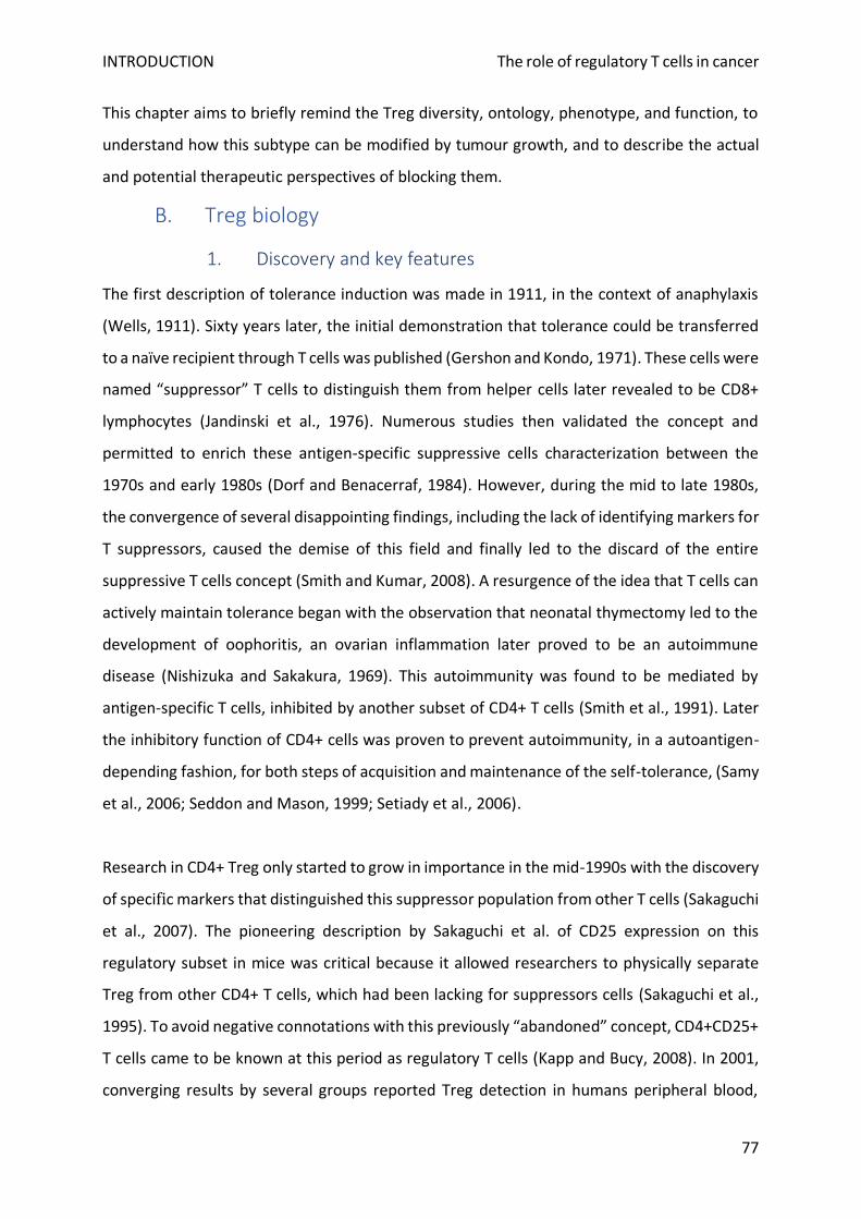
INTRODUCTION The role of regulatory T cells in cancer
77
This chapter aims to briefly remind the Treg diversity, ontology, phenotype, and function, to
understand how this subtype can be modified by tumour growth, and to describe the actual
and potential therapeutic perspectives of blocking them.
B. Treg biology
1. Discovery and key features
The first description of tolerance induction was made in 1911, in the context of anaphylaxis
(Wells, 1911). Sixty years later, the initial demonstration that tolerance could be transferred
to a naïve recipient through T cells was published (Gershon and Kondo, 1971). These cells were
named “suppressor” T cells to distinguish them from helper cells later revealed to be CD8+
lymphocytes (Jandinski et al., 1976). Numerous studies then validated the concept and
permitted to enrich these antigen-specific suppressive cells characterization between the
1970s and early 1980s (Dorf and Benacerraf, 1984). However, during the mid to late 1980s,
the convergence of several disappointing findings, including the lack of identifying markers for
T suppressors, caused the demise of this field and finally led to the discard of the entire
suppressive T cells concept (Smith and Kumar, 2008). A resurgence of the idea that T cells can
actively maintain tolerance began with the observation that neonatal thymectomy led to the
development of oophoritis, an ovarian inflammation later proved to be an autoimmune
disease (Nishizuka and Sakakura, 1969). This autoimmunity was found to be mediated by
antigen-specific T cells, inhibited by another subset of CD4+ T cells (Smith et al., 1991). Later
the inhibitory function of CD4+ cells was proven to prevent autoimmunity, in a autoantigen-
depending fashion, for both steps of acquisition and maintenance of the self-tolerance, (Samy
et al., 2006; Seddon and Mason, 1999; Setiady et al., 2006).
Research in CD4+ Treg only started to grow in importance in the mid-1990s with the discovery
of specific markers that distinguished this suppressor population from other T cells (Sakaguchi
et al., 2007). The pioneering description by Sakaguchi et al. of CD25 expression on this
regulatory subset in mice was critical because it allowed researchers to physically separate
Treg from other CD4+ T cells, which had been lacking for suppressors cells (Sakaguchi et al.,
1995). To avoid negative connotations with this previously “abandoned” concept, CD4+CD25+
T cells came to be known at this period as regulatory T cells (Kapp and Bucy, 2008). In 2001,
converging results by several groups reported Treg detection in humans peripheral blood,
INTRODUCTION The role of regulatory T cells in cancer
78
among the CD4+CD25high T cell population (Baecher-Allan et al., 2001; Dieckmann et al., 2001;
Jonuleit et al., 2001; Levings et al., 2001; Stephens et al., 2001; Taams et al., 2001).
Constitutive expression of CD25 is restricted to Treg in naïve mice while in humans, only cells
that highly express CD25 are designated Treg since activated Teff acquire intermediate levels
of this molecule. Besides, CD25 not only serves as a useful marker for Treg identification but
its function of high-affinity IL-2 receptor is also major for expansion, survival, maintenance,
and Treg suppressive capacity (Chinen et al., 2016).
Although CD25 has been crucial in identifying Treg, its expression is not Treg-restricted,
making necessary the search for a more specific Treg marker. As for CD8+ Treg previously, no
Treg lineage marker could be found that was only associated with Treg. The identification of
the forkhead/winged-helix family (FOXP3) transcription factor as a key switch that drives
differentiation of naive T cells into the Treg lineage and maintains their suppressive function
was fundamental for this research area (Brunkow et al., 2001; Khattri et al., 2003; Schubert et
al., 2001). Up to 90% of the Treg subset in mice is FOXP3+, while naive and activated
conventional T cells do not reach detectable levels of FOXP3 expression (Fontenot et al., 2003;
Hori et al., 2003). The vast majority of human CD4+CD25high Treg also express FOXP3 (Roncador
et al., 2005; Yagi et al., 2004), similarly to mice. Conversely, human conventional T cells
transiently express intermediate levels of FOXP3 upon activation, the same way than CD25,
thus establishing limits to FOXP3 specificity for human Treg (Pillai et al., 2007). Nevertheless,
FOXP3 loss of function in humans causes the failure of Treg differentiation, leading to an
autoimmune lymphoproliferative disease, fatal without a donor marrow graft, called immune
dysregulation, polyendocrinopathy, enteropathy, X-linked (IPEX) (Verbsky and Chatila, 2013).
The same syndrome, referred to as “scurfy”, is described in mice deficient in FOXP3 (Lin et al.,
2005, 2007; Wildin et al., 2001). The scurfy phenotype was shown to be reversible with a
restoration of the Treg compartment.
The identification of the FOXP3 gene helped to investigate the characterization and function
of Treg due to its role as a ‘master regulator’. Development of mAb detecting the intranuclear
form allowed Treg identification by flow cytometry and construction of FOXP3-GFP reporter
mice allowed viable Treg cells to be FACS-sorted. Standard Treg assays using sorted GFP+ cells
were responsible for the one regulatory activity within the CD4+CD25+ T cells subset, unlike
INTRODUCTION The role of regulatory T cells in cancer
79
GFP- cells with an equivalent phenotype (Fontenot et al., 2005; Wan and Flavell, 2005).
Importantly, numerous non-FOXP3+ regulatory T subsets exist and exhibit suppressive
functions but do not require a stable FOXP3 expression (or only a transitory one), including
Type 1 regulatory T cells (Tr1), Th3 cells, and double-negative regulatory cells (Shevach, 2006).
Keeping that in mind, this chapter will centre solely on FOXP3+ Treg populations because they
have been more intensively investigated in the field of cancer, and organ/bone marrow
transplantation. Since most of the studies used as references are focused on CD4+FOXP3+,
they will be referred to as “Treg” and CD8+FOXP3+ as “CD8+ Treg”.
2. Treg ontogeny and lineage plasticity
Treg are classically divided into two subsets depending on their origin: thymic-derived
“natural” Treg (nTreg) and peripherally-induced Treg (iTreg). nTreg represent 5 to 10% of the
total CD4+ lymphocytes, and constitutively express high levels of FOXP3 and CD25 (Piccirillo
and Thornton, 2004). During thymic selection, nTreg are positively selected thymocytes with
relatively high avidity for self-antigens. The interaction between the TCR of a thymocyte and
the MHC class II on a stromal thymic cell surface, loaded with self-antigen, appears to provide
the signal for a nTreg fate (Jordan et al., 2001). Hence, nTreg engagement seems to be
essentially cytokine independent. By opposition, iTreg derive from conventionally thymus-
selected naïve CD4+ cells, that will gain FOXP3 and CD25 expression following ad hoc antigenic
stimulation in a defined cytokine environment (Bilate et al., 2016). Considering the nature of
iTreg differentiation induced by non-self-antigens and a particular TCR signalling combined
with specialized immunoregulatory cytokines such as TGF-β, IL-2, and IL-4, these cells are
assumed to essentially play a role in maintaining mucosal tolerance. iTreg may, therefore,
control immune responses to commensal antigens and prevent allergic-type reactions
(Romano et al., 2017).
Taken collectively, studies about Treg induction have brought the idea that most of the naïve
CD4+ T cells can potentially become Treg when stimulated in suitable conditions (Kapp and
Bucy, 2008). The differences in phenotype stability observed between natural and iTreg
subsets may be a reflection of the diversity of FOXP3 expression levels caused by chronic in
vivo stimulation by autoantigens versus acute activation by exogenous antigens. The recent
INTRODUCTION The role of regulatory T cells in cancer
80
description of the Treg-specific demethylated region (TSDR) within the FOXP3 gene locus in
both mice and humans permitted new insight in the FOXP3 role in the Treg phenotype
initiation and maintenance (Floess et al., 2007). Stable expression of the FOXP3 gene appears
to be correlated with the demethylation of this evolutionarily conserved cytosine guanine
dinucleotide(CpG)-rich element to generate a permanent suppressive cell lineage. Variability
in FOXP3 gene expression not only acts on Treg phenotype initiation but also determines the
different levels of Treg suppressive abilities. Hence, the demethylation status of the TSDR also
correlates in both mice and humans with suppressive function maintenance, likewise,
methylation blockade induces FOXP3 expression and can raise CD4+ cells suppressive activity
(Baron et al., 2007; Polansky et al., 2008).
3. Phenotypic characterization
If the Treg population was first described using CD25, and then better-characterized thanks to
FOXP3 discovery, their phenotype features have been completed by a growing number of
surface and intracellular markers, related to Treg development, migration, activation state,
and function. New surface markers were even more required for human Treg study since CD25
expression was not allowing a clear differentiation between Treg and conventional CD4+ cells,
and FOXP3 could not serve as a cell sorting marker because of its intracellular localisation. This
matter has been improved greatly with the observation that the IL-7 receptor α-chain, CD127,
was only weakly expressed at Treg surface, contrary to the non-regulatory CD4+ population
(Liu et al., 2006; Seddiki et al., 2006). CD127 is now routinely used to recognize human Treg,
defined as CD4+CD25+/hiCD127-/low, with some restrictions, since 10% of the CD25+FOXP3+
subset actually expresses CD127 (Yu et al., 2012). Besides, 15% of the CD25+CD127-
population do not express FOXP3, and conventional T cells show low expression of this
molecule in a part of the general population, and in the context of chronic infection or HIV
(Alves et al., 2008; Del Pozo-Balado Mdel et al., 2010; Santegoets et al., 2015).
Twenty years ago, two other molecules, that play a crucial role in Treg function have been
described: CTLA-4 (CD152) (Read, Malmstrom et al. 2000, Salomon, Lenschow et al. 2000,
Takahashi, Tagami et al. 2000) and GITR (CD357) (McHugh, Whitters et al. 2002, Shimizu,
Yamazaki et al. 2002). CTLA-4 is not only involved in Treg suppressive function, as detailed
later but also in Treg homeostasis maintenance in a steady-state (Linsley, Brady et al. 1991
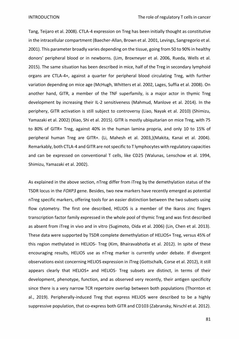
INTRODUCTION The role of regulatory T cells in cancer
81
Tang, Teijaro et al. 2008). CTLA-4 expression on Treg has been initially thought as constitutive
in the intracellular compartment (Baecher-Allan, Brown et al. 2001, Levings, Sangregorio et al.
2001). This parameter broadly varies depending on the tissue, going from 50 to 90% in healthy
donors’ peripheral blood or in newborns. (Lim, Broxmeyer et al. 2006, Rueda, Wells et al.
2015). The same situation has been described in mice, half of the Treg in secondary lymphoid
organs are CTLA-4+, against a quarter for peripheral blood circulating Treg, with further
variation depending on mice age (McHugh, Whitters et al. 2002, Lages, Suffia et al. 2008). On
another hand, GITR, a member of the TNF superfamily, is a major actor in thymic Treg
development by increasing their IL-2 sensitiveness (Mahmud, Manlove et al. 2014). In the
periphery, GITR activation is still subject to controversy (Liao, Nayak et al. 2010) (Shimizu,
Yamazaki et al. 2002) (Xiao, Shi et al. 2015). GITR is mostly ubiquitarian on mice Treg, with 75
to 80% of GITR+ Treg, against 40% in the human lamina propria, and only 10 to 15% of
peripheral human Treg are GITR+. (Li, Mahesh et al. 2003,)(Makita, Kanai et al. 2004).
Remarkably, both CTLA-4 and GITR are not specific to T lymphocytes with regulatory capacities
and can be expressed on conventional T cells, like CD25 (Walunas, Lenschow et al. 1994,
Shimizu, Yamazaki et al. 2002).
As explained in the above section, nTreg differ from iTreg by the demethylation status of the
TSDR locus in the FOXP3 gene. Besides, two new markers have recently emerged as potential
nTreg specific markers, offering tools for an easier distinction between the two subsets using
flow cytometry. The first one described, HELIOS is a member of the Ikaros zinc fingers
transcription factor family expressed in the whole pool of thymic Treg and was first described
as absent from iTreg in vivo and in vitro (Sugimoto, Oida et al. 2006) (Lin, Chen et al. 2013).
These data were supported by TSDR complete demethylation of HELIOS+ Treg, versus 45% of
this region methylated in HELIOS- Treg (Kim, Bhairavabhotla et al. 2012). In spite of these
encouraging results, HELIOS use as nTreg marker is currently under debate. If divergent
observations exist concerning HELIOS expression in iTreg (Gottschalk, Corse et al. 2012), it still
appears clearly that HELIOS+ and HELIOS- Treg subsets are distinct, in terms of their
development, phenotype, function, and as observed very recently, their antigen specificity
since there is a very narrow TCR repertoire overlap between both populations (Thornton et
al., 2019). Peripherally-induced Treg that express HELIOS were described to be a highly
suppressive population, that co-express both GITR and CD103 (Zabransky, Nirschl et al. 2012).
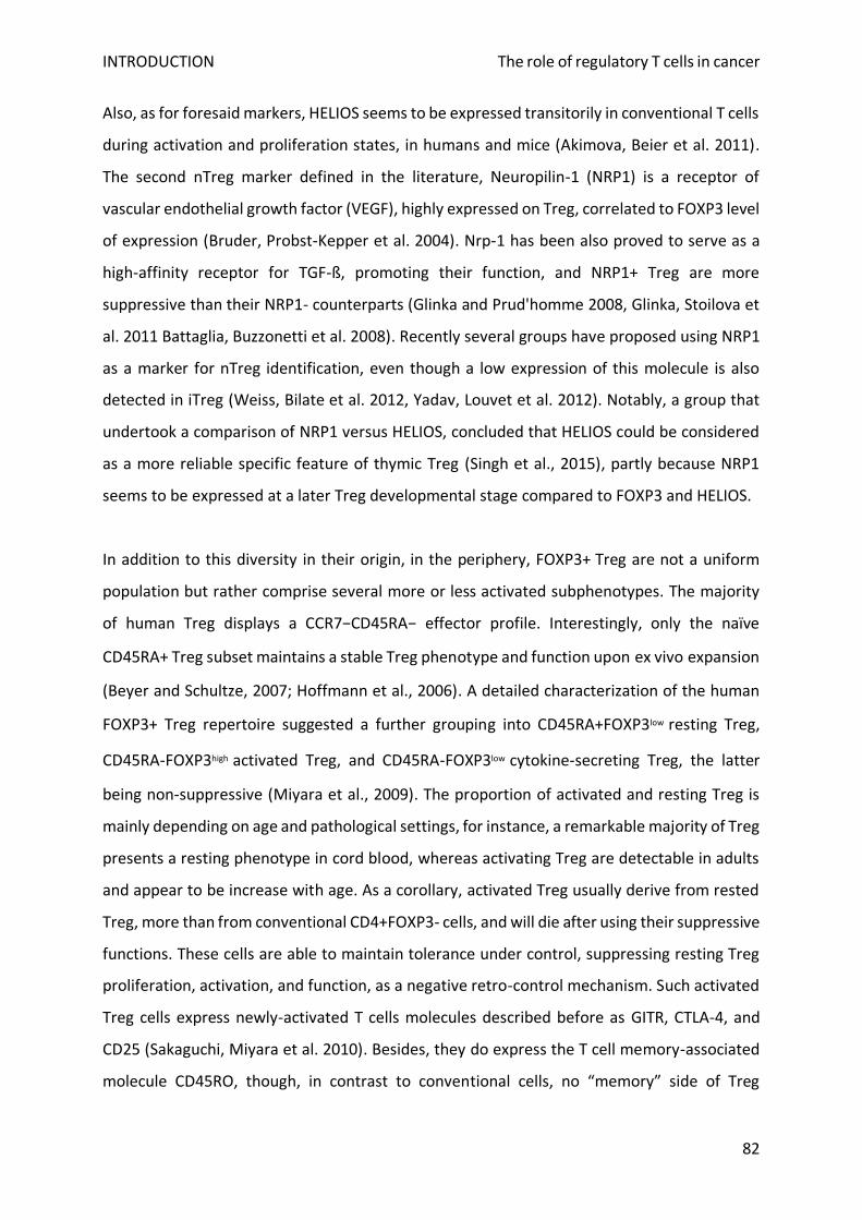
INTRODUCTION The role of regulatory T cells in cancer
82
Also, as for foresaid markers, HELIOS seems to be expressed transitorily in conventional T cells
during activation and proliferation states, in humans and mice (Akimova, Beier et al. 2011).
The second nTreg marker defined in the literature, Neuropilin-1 (NRP1) is a receptor of
vascular endothelial growth factor (VEGF), highly expressed on Treg, correlated to FOXP3 level
of expression (Bruder, Probst-Kepper et al. 2004). Nrp-1 has been also proved to serve as a
high-affinity receptor for TGF-ß, promoting their function, and NRP1+ Treg are more
suppressive than their NRP1- counterparts (Glinka and Prud'homme 2008, Glinka, Stoilova et
al. 2011 Battaglia, Buzzonetti et al. 2008). Recently several groups have proposed using NRP1
as a marker for nTreg identification, even though a low expression of this molecule is also
detected in iTreg (Weiss, Bilate et al. 2012, Yadav, Louvet et al. 2012). Notably, a group that
undertook a comparison of NRP1 versus HELIOS, concluded that HELIOS could be considered
as a more reliable specific feature of thymic Treg (Singh et al., 2015), partly because NRP1
seems to be expressed at a later Treg developmental stage compared to FOXP3 and HELIOS.
In addition to this diversity in their origin, in the periphery, FOXP3+ Treg are not a uniform
population but rather comprise several more or less activated subphenotypes. The majority
of human Treg displays a CCR7−CD45RA− effector profile. Interestingly, only the naïve
CD45RA+ Treg subset maintains a stable Treg phenotype and function upon ex vivo expansion
(Beyer and Schultze, 2007; Hoffmann et al., 2006). A detailed characterization of the human
FOXP3+ Treg repertoire suggested a further grouping into CD45RA+FOXP3low resting Treg,
CD45RA-FOXP3high activated Treg, and CD45RA-FOXP3low cytokine-secreting Treg, the latter
being non-suppressive (Miyara et al., 2009). The proportion of activated and resting Treg is
mainly depending on age and pathological settings, for instance, a remarkable majority of Treg
presents a resting phenotype in cord blood, whereas activating Treg are detectable in adults
and appear to be increase with age. As a corollary, activated Treg usually derive from rested
Treg, more than from conventional CD4+FOXP3- cells, and will die after using their suppressive
functions. These cells are able to maintain tolerance under control, suppressing resting Treg
proliferation, activation, and function, as a negative retro-control mechanism. Such activated
Treg cells express newly-activated T cells molecules described before as GITR, CTLA-4, and
CD25 (Sakaguchi, Miyara et al. 2010). Besides, they do express the T cell memory-associated
molecule CD45RO, though, in contrast to conventional cells, no “memory” side of Treg
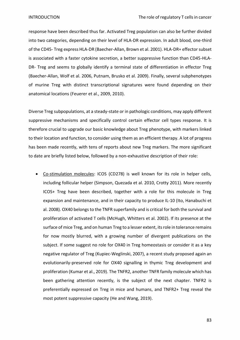
INTRODUCTION The role of regulatory T cells in cancer
83
response have been described thus far. Activated Treg population can also be further divided
into two categories, depending on their level of HLA-DR expression. In adult blood, one-third
of the CD45- Treg express HLA-DR (Baecher-Allan, Brown et al. 2001). HLA-DR+ effector subset
is associated with a faster cytokine secretion, a better suppressive function than CD45-HLA-
DR- Treg and seems to globally identify a terminal state of differentiation in effector Treg
(Baecher-Allan, Wolf et al. 2006, Putnam, Brusko et al. 2009). Finally, several subphenotypes
of murine Treg with distinct transcriptional signatures were found depending on their
anatomical locations (Feuerer et al., 2009, 2010).
Diverse Treg subpopulations, at a steady-state or in pathologic conditions, may apply different
suppressive mechanisms and specifically control certain effector cell types response. It is
therefore crucial to upgrade our basic knowledge about Treg phenotype, with markers linked
to their location and function, to consider using them as an efficient therapy. A lot of progress
has been made recently, with tens of reports about new Treg markers. The more significant
to date are briefly listed below, followed by a non-exhaustive description of their role:
• Co-stimulation molecules: ICOS (CD278) is well known for its role in helper cells,
including follicular helper (Simpson, Quezada et al. 2010, Crotty 2011). More recently
ICOS+ Treg have been described, together with a role for this molecule in Treg
expansion and maintenance, and in their capacity to produce IL-10 (Ito, Hanabuchi et
al. 2008). OX40 belongs to the TNFR superfamily and is critical for both the survival and
proliferation of activated T cells (McHugh, Whitters et al. 2002). If its presence at the
surface of mice Treg, and on human Treg to a lesser extent, its role in tolerance remains
for now mostly blurred, with a growing number of divergent publications on the
subject. If some suggest no role for OX40 in Treg homeostasis or consider it as a key
negative regulator of Treg (Kupiec-Weglinski, 2007), a recent study proposed again an
evolutionarily-preserved role for OX40 signalling in thymic Treg development and
proliferation (Kumar et al., 2019). The TNFR2, another TNFR family molecule which has
been gathering attention recently, is the subject of the next chapter. TNFR2 is
preferentially expressed on Treg in mice and humans, and TNFR2+ Treg reveal the
most potent suppressive capacity (He and Wang, 2019).
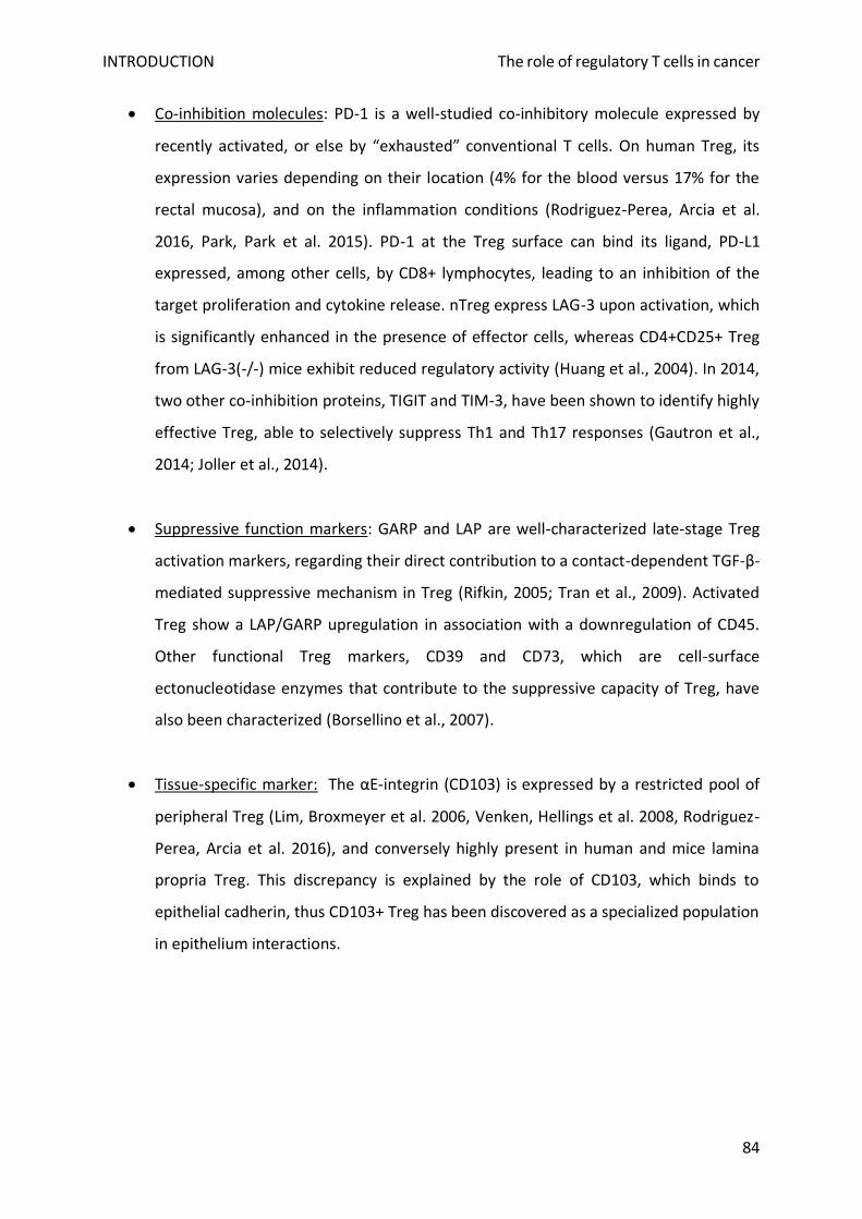
INTRODUCTION The role of regulatory T cells in cancer
84
• Co-inhibition molecules: PD-1 is a well-studied co-inhibitory molecule expressed by
recently activated, or else by “exhausted” conventional T cells. On human Treg, its
expression varies depending on their location (4% for the blood versus 17% for the
rectal mucosa), and on the inflammation conditions (Rodriguez-Perea, Arcia et al.
2016, Park, Park et al. 2015). PD-1 at the Treg surface can bind its ligand, PD-L1
expressed, among other cells, by CD8+ lymphocytes, leading to an inhibition of the
target proliferation and cytokine release. nTreg express LAG-3 upon activation, which
is significantly enhanced in the presence of effector cells, whereas CD4+CD25+ Treg
from LAG-3(-/-) mice exhibit reduced regulatory activity (Huang et al., 2004). In 2014,
two other co-inhibition proteins, TIGIT and TIM-3, have been shown to identify highly
effective Treg, able to selectively suppress Th1 and Th17 responses (Gautron et al.,
2014; Joller et al., 2014).
• Suppressive function markers: GARP and LAP are well-characterized late-stage Treg
activation markers, regarding their direct contribution to a contact-dependent TGF-β-
mediated suppressive mechanism in Treg (Rifkin, 2005; Tran et al., 2009). Activated
Treg show a LAP/GARP upregulation in association with a downregulation of CD45.
Other functional Treg markers, CD39 and CD73, which are cell-surface
ectonucleotidase enzymes that contribute to the suppressive capacity of Treg, have
also been characterized (Borsellino et al., 2007).
• Tissue-specific marker: The αE-integrin (CD103) is expressed by a restricted pool of
peripheral Treg (Lim, Broxmeyer et al. 2006, Venken, Hellings et al. 2008, Rodriguez-
Perea, Arcia et al. 2016), and conversely highly present in human and mice lamina
propria Treg. This discrepancy is explained by the role of CD103, which binds to
epithelial cadherin, thus CD103+ Treg has been discovered as a specialized population
in epithelium interactions.
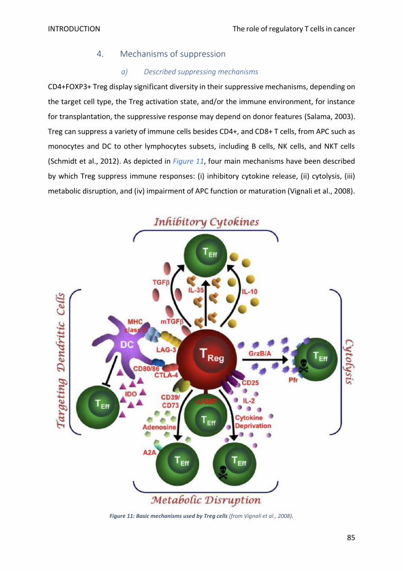
INTRODUCTION The role of regulatory T cells in cancer
85
4. Mechanisms of suppression
a) Described suppressing mechanisms
CD4+FOXP3+ Treg display significant diversity in their suppressive mechanisms, depending on
the target cell type, the Treg activation state, and/or the immune environment, for instance
for transplantation, the suppressive response may depend on donor features (Salama, 2003).
Treg can suppress a variety of immune cells besides CD4+, and CD8+ T cells, from APC such as
monocytes and DC to other lymphocytes subsets, including B cells, NK cells, and NKT cells
(Schmidt et al., 2012). As depicted in Figure 11, four main mechanisms have been described
by which Treg suppress immune responses: (i) inhibitory cytokine release, (ii) cytolysis, (iii)
metabolic disruption, and (iv) impairment of APC function or maturation (Vignali et al., 2008).
Figure 11: Basic mechanisms used by Treg cells (from Vignali et al., 2008).
INTRODUCTION The role of regulatory T cells in cancer
86
(i) Major regulatory cytokines secreted by Treg include TGF-β, IL-10, and IL-35. IL-10 and TGF-
β have been proved necessary for Treg protection in many pathologic settings, initially in
colitis using inflammatory bowel disease models for both cytokines (Asseman et al., 1999;
Fahlén et al., 2005). Although their importance as suppressive mediators is undisputed, their
contribution to the function of thymus-derived nTreg is still a matter of debate (Shevach,
2006), partly due to the historical perception that Treg suppress in a contact-dependent
manner (Takahashi et al., 1998). However, membrane-bound TGF-β was shown to be required
in Treg-mediated contact-dependent suppression (Nakamura et al., 2001). More recently, IL-
35 has been described as necessary for a maximal suppression activity of Treg (Collison et al.,
2010). Thus, the pool of anti-inflammatory cytokines released by Treg is involved in both Treg
maintenance through de novo Treg induction and support of FOXP3 expression, and
direct/indirect suppression of effector T cell through signalling blockade and regulation of IFN-
γ (Schmidt et al., 2012). Lastly, they also induce the generation of suppressive cytokines by
APC, that combined with the one they produce leads to the modulation of the behaviour of
neighbouring effector cells (Morlacchi et al., 2011).
(ii) Treg can exert suppression through secretion of granzymes A and B, inducing Teff
programmed cell death (Gondek et al., 2005; Grossman et al., 2004). Expression of Fas ligand
on the surface of Treg has also been shown to induce Fas-dependent apoptosis in
conventional T cells (Strauss et al., 2009). Interestingly, cytotoxic Treg are also able to lyse Ag-
presenting B cells in an epitope-specific manner, causing the down-regulation of bystander T
cells proliferation (Janssens et al., 2003).
(iii) Metabolic disruption is another potent mechanism of suppression. Several studies have
demonstrated that Treg actively deplete IL-2 from their microenvironment, due to the density
of high-affinity IL-2 receptor CD25 on their surface (McNally et al., 2011; Pandiyan et al., 2007;
Thornton and Shevach, 1998). Deprived of their main mitogen and activation stimulus, Teff
are impaired in their proliferation and function. Treg also express preferentially the
ectoenzymes CD39 and CD73, which respectively catalyse the reaction ATP AMP+ADP, and
metabolize AMP into adenosine (Deaglio et al., 2007; Regateiro et al., 2013). Both create
unsuitable surroundings for Teff since they are dependent upon ATP sources in their
INTRODUCTION The role of regulatory T cells in cancer
87
environment, and conversely lead to anergy caused by adenosine excess. Adenosine has an
opposite effect on Treg due to the expression of a different version of its receptor,
contributing to this cell type to their suppressive function (Bodor et al., 2012; Bopp et al.,
2007). Last, upon contact with nTreg cells, cAMP is transferred into activated helper T cells
target and/or APC via gap junctions blocking their function and IL-2 secretion.
(iv) Contact-dependent mechanisms of Treg-mediated suppression include the engagement
of inhibitory co-receptors expressed on the surface of Treg with their ligands on Teff or APCs.
Probably the most well-defined of these co-inhibitory receptors is CTLA-4, which binds to
CD80/86 on DC, transmitting intracellular signals that tolerise these APC (Read et al., 2000;
Walker, 2013). Ligation of CD80/CD86 by CTLA-4 results in an impaired maturation, the
secretion of immunoregulatory molecules such as indolamine 2,3-dioxygenase (IDO), the
downregulation of CD80/86 expression, and importantly a binding competition with the co-
activator CD28. Although CD80/86 is mainly expressed by APCs, activated and alloreactive T
cells might express these molecules, providing a further target for CTLA-4 in the context of
GvHD (Taylor et al., 2004). Similarly, LAG3 is also able to inhibit DC maturation upon ligation
of MHC class II (Huang et al., 2004).
Few is known about immune cell dynamics and contact in vivo, but advances have been made
about Treg specificity in term of Teff suppression. The discovery that Treg could inhibit the
responses of T cells specific for unrelated antigens if both the Treg and the Teff epitopes were
expressed by the same APC has now been validated several times, both in vivo and in vitro
(Kapp and Bucy, 2008; Kapp et al., 2007). Interestingly, a study in non-obese diabetic (NOD)
mice, reported no detectable stable associations between Treg and Th cells during active
suppression. Treg cells were found as directly interacting with DC presenting islet antigen.
These persistent Treg-DC contacts reduced the Th-DC time of interaction and preceded the
inhibition of the targeted Th cell activation by DC, underlying further APC’ central function to
Treg suppressive capacity in vivo (Tang et al., 2006a). This kind of sequential interaction model
has been long-time known for cytotoxic T cells activation by T helper via DC (Ridge et al., 1998).

INTRODUCTION The role of regulatory T cells in cancer
88
b) Dynamic aspects of Treg suppressive function
The exact suppressive mechanisms employed by Treg are most likely context-dependent,
impacted by their type, status of activation, anatomical location, and their surrounding
environment including the number and type of Teff cells in the vicinity, and the level of
inflammation. Tang and Bluestone have proposed a three-tiered model to explain how Treg
may maintain tissue homeostasis (Tang and Bluestone, 2008). In the steady-state, TGF-β and
CTLA-4 appear to be the main actor of immune tolerance maintenance by Treg. This phase is
referred to by the authors as “homeostatic control”. During “damage control”, at the site of
inflammation, activated Treg will use more diverse and rapidly mobilised anti-inflammatory
mediators, including IDO, IL-10, and adenosine. Cytolytic mechanisms are associated with Treg
chronic activation and seem to be reserved for persistent inflammation cases. Finally,
suppressive mechanisms of “infectious tolerance” take place after the resolution of an
immune response. The tolerogenic microenvironment established by Treg is maintained
through several positive- feedback mechanisms even after inflammation has been turned off.
Several of the Treg-induced mediators, including IL-10, TGFβ, IDO, and adenosine, are not only
actively inhibiting Teff response but also play a role in these positive feedback-loops for Treg
function, and/or induction via APC tolerization. Another interesting point is the discovery that
Teff cells own an active role in potentiating Treg-mediated suppression, in particular through
the IL-35 secretion pathway (Vignali et al., 2008). Since the genetic profiles of activated Treg
in the presence and absence of Teff are divergent, it might be possible that this function-boost
role concern also other regulatory proteins. Molecular agents that mediate this Treg
enhancement are unknown, but IL-2 might be a good candidate for this function (Thornton et
al., 2004).
In a different register, Treg have not only been labelled as guardians of tissue homeostasis but
also appear to directly play a role in parenchymal tissue repair via their interaction with local
non-immunological cells, for instance in the muscle (Burzyn et al., 2013). The description of
such non-immunological Treg functions may help to hasten resolution of inflammation
required after tissue damage. Although, since a damaged tissue is a source of inflammatory
mediators and potentially immunogenic neo-antigens, tissue homeostasis Treg function is also
directly linked to tolerance.
INTRODUCTION The role of regulatory T cells in cancer
89
5. CD8+ Treg resurgence
Even if tolerance research has been mostly centred on CD4+ Treg since the pioneer description
by Sakaguchi’s team, some groups were still trying to better elucidate CD8+ suppressor cells’
role at the end of the 1990s. The observation was made that they could be activated in an
antigen-dependant way if they were surrounded by TGF-ß (Rich et al., 1995; Wilbanks et al.,
1992). A few years later, CD8+ antigen-specific suppressor cells were described in the blood
of cardiac transplanted patients undergoing stable engraftment (Ciubotariu et al., 2001; Liu et
al., 1998). These results have raised again the interest in this temporary neglected population.
In the particular setting of heart graft, CD8+ Treg showed an up-regulation of FOXP3
expression, both in rat models and humans (Liu et al., 2004; Suciu-Foca et al., 2005).
CD8+CD25+FOXP3+ have then been reported as induced in human cells in vivo and in vitro by
TGF-ß activation, or after cells treatment with a modified non-depleting anti-CD3 antibody
(Bisikirska et al., 2005; Kapp et al., 2007). Altogether these results indicate that FOXP3 could
serve as a critical regulator of CD8+ cell induction and suppressive function, as described for
CD4+ Treg. A comparison of FOXP3 degree of necessity for the function of CD4+ and CD8+
Treg, using FOXP3-GFP knock-in mice, has been further confirmed that FOXP3 expression in
CD8+ regulate their suppressive activity (Kapp and Bucy, 2008). Both CD8+ and CD4+ Treg
showed also similar expression levels of FOXP3 and CTLA-4, which represent their most
characteristic markers (Scotto et al., 2004). The biggest difference between CD4+ and CD8+
Treg resides in the expression of CD28. CD4+ Treg express a higher level of CD28, which is
required for their interaction with B7 molecules (Tang et al., 2003). For CD8+ T cells, the
expression of CD28 is partially dispensable due to their reduced production of IL-2 (Hoyer et
al., 2007a, 2007b; Tai et al., 2005). Also, similarly to nTreg CD4+, thymus-derived CD8+ Treg
have been described, with a CD28- phenotype in mice and humans, as for CD4+ nTreg they
inhibit the immune response in an antigen non-specific and not-MHC-restricted way
(Vuddamalay et al., 2016). Natural CD8+ Treg were reported to have a
CD8+CD25+FOXP3+CTLA-4+GITR+ phenotype, suggesting a suppression using CTLA-4 (Cosmi
et al., 2003). A myriad of phenotypes has been observed for CD8+ Treg in the periphery, and
in pathological settings (indexed by Ligocki and Niederkorn, 2015)
INTRODUCTION The role of regulatory T cells in cancer
90
In vitro and in vivo experimental systems investigating polyclonal populations of CD8+ Treg
have led to the description of distinct mechanisms of immune regulation, most of them being
similar to CD4+ Treg (Smith and Kumar, 2008). Direct killing of the target cell by perforin or
Fas/FasL pathway (Correale and Villa, 2008), and negative signalling directly on the target or
on APCs in PD-1 and CTLA-4 dependant manners have been well described (Chang et al., 2002;
Izawa et al., 2007; Probst et al., 2005). CD8+ Treg capacity of secreting soluble factors, such as
immunosuppressive cytokines is still under debate. TGF-ß first seemed not to participate in
CD8+ mechanisms, IL-10 might and TRAIL have just been described (Griffith et al., 2007;
Poussier et al., 2002; Tang et al., 2006b). In heart transplant, IFN-γ, IDO, and fibroleukin-2
support CD8+ Treg suppression of CD4+ Teff with which there are in contact (Li et al., 2010).
In addition to suppressing effector T cells, CD8+FOXP3+ Treg are capable of inducing the de
novo generation of CD4+FOXP3+ Treg by a process that is contact-dependent and also
requires the production of soluble TGF-β (Lerret et al., 2012). As for CD4+ Treg, it is not clear
whether the explanation for multiple mechanisms could mean the existence of separate
subsets of CD8+ Treg that use different mechanisms of suppression, or if there is one
homogeneous CD8+ Treg population using different suppressor mechanisms depending on the
context of the immune reaction, with different behaviours in vitro versus in vivo. Interestingly,
the conventional T cells targeted by CD4+ and CD8+ Treg seems to belong in two distinct
populations. CD8+ Treg might target activated lymphocytes when nonspecific CD4+ are mostly
specific of T cell priming suppression (Smith and Kumar, 2008). Thus, it appears that
complementary regulatory mechanisms act during different phases of the immune response,
using distinct Treg populations.
INTRODUCTION The role of regulatory T cells in cancer
91
C. Treg dysregulation in cancer
Treg are the central actors of self-tolerance maintenance in all areas of the body, enabling
maternofoetal tolerance during pregnancy, assuring constant protection against autoimmune
diseases development, allergies, or infection-induced organ pathologies, and also are the
guardians of post-transplant regulation of immune reactions, preventing rejection in solid
organ transplant and GvHD after HSCT (Sakaguchi, 2004). However, Treg has been proved to
dampen effector response in certain pathologic conditions, including anti-infectious
responses, and anti-tumoral defence (Curiel, 2008; Wohlfert and Belkaid, 2008; Zou, 2006). A
better understanding of Treg dysregulation in cancers during the last two decades has opened
a new therapeutic window in the field of anti-tumoral immunotherapies.
1. Treg association with cancer progression
a) State of the art in solid and blood cancers
Several human carcinomas have been associated with a high infiltrate of Treg, including
pancreas, lung, liver, gastrointestinal tract, breast, ovary, and head and neck cancers
(reviewed by Tanaka and Sakaguchi, 2017). Additionally, a published data meta-analysis has
described for melanoma, cervix, breast, and kidney tumours a negative association between
patients’ survival and high FOXP3+ infiltration (Shang et al., 2015). Previous reports have also
established a correlation between a diminished ratio of infiltrating CD8+ lymphocytes to
FOXP3+ Treg, and a poor prognosis for patients with breast, ovarian, gastric and colorectal
cancers (Bates et al., 2006; Curiel et al., 2004; Sasada et al., 2003; Sato et al., 2005; Sinicrope
et al., 2009). For ovarian carcinoma, Curiel et al. were able to subsequently propose that the
level of infiltration of CD4+CD25+FOXP3+ cells at each stage of the disease could serve as a
good indicator of disease progression and survival prediction (Curiel et al., 2004). Yet
surprisingly, numerous other studies have reported a correlation between high Treg tumour
infiltration and a favourable prognosis for the patients suffering from breast, ovarian,
colorectal, and head and neck carcinomas, contrasting with above-mentioned publications
(Badoual et al., 2006; Correale et al., 2010; Frey et al., 2010; Leffers et al., 2008; Salama et al.,
2009; West et al., 2013; Yeong et al., 2017).
INTRODUCTION The role of regulatory T cells in cancer
92
In the case of haematological malignancies, fewer publications are available, but similarly to
solid neoplasms, divergent observations have been made. Concerning acute leukaemia, a
recent meta-analysis reported an increased frequency of Treg using 13 studies for B-ALL, T-
ALL, and AML, both in adults and children, using either bone marrow samples, peripheral
blood, or both (Niedźwiecki et al., 2019). In some subtypes, the difference of percentages may
also condition a response to chemotherapy and thus the prognosis of the disease. As for solid
cancers, it is still unknown whether the increased percentage of Treg is a cause or an effect of
acute leukaemia. Notably, in AML an elevated percentage of Treg is observed after completing
treatment and even achieving a remission (Szczepanski et al., 2009). Treg frequencies are
higher in chronic lymphoid leukaemia patients than in controls and correlate with disease
status (D’Arena et al., 2011). Similarly, Treg may play a role in the maintenance of chronic
myeloid leukaemia, and the decrease of their levels in patients with complete molecular
remission suggests that Treg might have a clinical value in evaluating the effects of therapy
(Zahran et al., 2014). In monoclonal gammopathies of undetermined significance and multiple
myeloma, the proportion of activated Treg in CD4+ T cells was also found significantly higher
in patients than healthy volunteers in both peripheral blood and bone marrow (Wang et al.,
2018b). The results are more heterogeneous in lymphomas and seem to depend on the
considered subtype. A group suggested that low infiltration of FOXP3+ cells in conjunction
with high infiltration of TIA-1+ cells (cytolytic cells) in classical Hodgkin lymphoma (cHL) may
represent biological markers predicting disease progression and an unfavourable outcome
(Alvaro et al., 2005). Accordingly, another group reported that a FOXP3 to Granzyme B ratio
of 1 or less was associated with poor free failure survival and OS (Kelley et al., 2007). In NHL,
Treg cells were increased markedly in peripheral blood versus healthy controls, regardless of
the NHL subtype, and correlated with disease stage and serum lactate dehydrogenase, a
described NHL prognosis factor (Mittal et al., 2008). Among NHL types, higher Treg numbers
were proved to predict improved survival in patients with follicular lymphoma, while a marked
reduction in Tregs is observed on transformation to diffuse large B-cell lymphomas (Carreras
et al., 2006). Similar findings were made for cutaneous T-cell lymphoma (CTCL) most common
subtypes, observing that an increased number of FOXP3+ Treg was associated with improved
survival in both mycosis fungoides and unspecified CTCL (Gjerdrum et al., 2007).
INTRODUCTION The role of regulatory T cells in cancer
93
b) Unravelling the literature divergences
While cytotoxic tumour-infiltrating lymphocytes (TIL) are associated with favourable clinical
outcomes in various tumour settings, the relationship between FOXP3+ TIL and prognosis
seems less clear. This controversy about Treg’s role in malignancies may arise from different
causes, including (i) the intrinsic heterogeneity of the tumour cells/immune infiltration for a
given cancer type or between histopathological subtypes, (ii) the divergence of method and
markers used to identify Treg depending on the considered study. In this regard, Sakaguchi’s
group suggested that these contradictory results may be rooted in an “improper
interpretation of the heterogeneous FOXP3+ cells as a single population of Treg cells”, when
this population includes, besides functional Treg, a subset of non-suppressive cells (Tanaka
and Sakaguchi, 2017). Following this hypothesis, they have proved that two CD4+FOXP3+ T
cell subpopulations distinctly control the prognosis of colorectal cancers, the FOXP3+ non-
Treg inflammatory subset being associated with a better prognosis than the regulatory one
(Saito et al., 2016). In another hand, the transitory FOXP3 expression in neo-activated human
Teff might also be adding a level of complexity in these studies’ interpretation. Surprisingly,
FOXP3 has been detected also on myeloblasts and macrophages (Chen et al., 2006;
Szczepanski et al., 2009). For these reasons, proving the suppressive function of a hypothetical
regulatory population is essential, and would probably help to clear the contradictions, even
if it is often uneasy to do (Han et al., 2019a). As a matter of fact, some groups have already
been able to confirm the regulatory function of FOXP3+ TIL, assessing their TSDR status,
and/or using ex vivo suppression assays in melanoma, head and neck carcinoma, and ovarian
carcinoma (Curiel et al., 2004; Strauss et al., 2007; Toker et al., 2018).
Since the composition of tumoral infiltrate may be assessed only after surgical excision, Treg
in the periphery might alternatively serve as an attractive potential biomarker for cancer
patients monitoring. Early evidence depicted an increased pool of CD4+CD25+ Treg in the
peripheral blood of cancer patients with potent immunosuppressive features (Wolf et al.,
2003). Examples of Treg serving as markers include the results of a pancreatic cancer study
where peripheral blood Treg level predicted the tumour response to chemotherapy (Liu et al.,
2017a), and another study where Treg levels changed markedly in rectal cancer patients after
preoperative radiotherapy (Napolitano et al., 2015). Thus, interesting data could emerge from
easy-to-access circulating patients’ Treg with an appropriate flow cytometry marker choice.
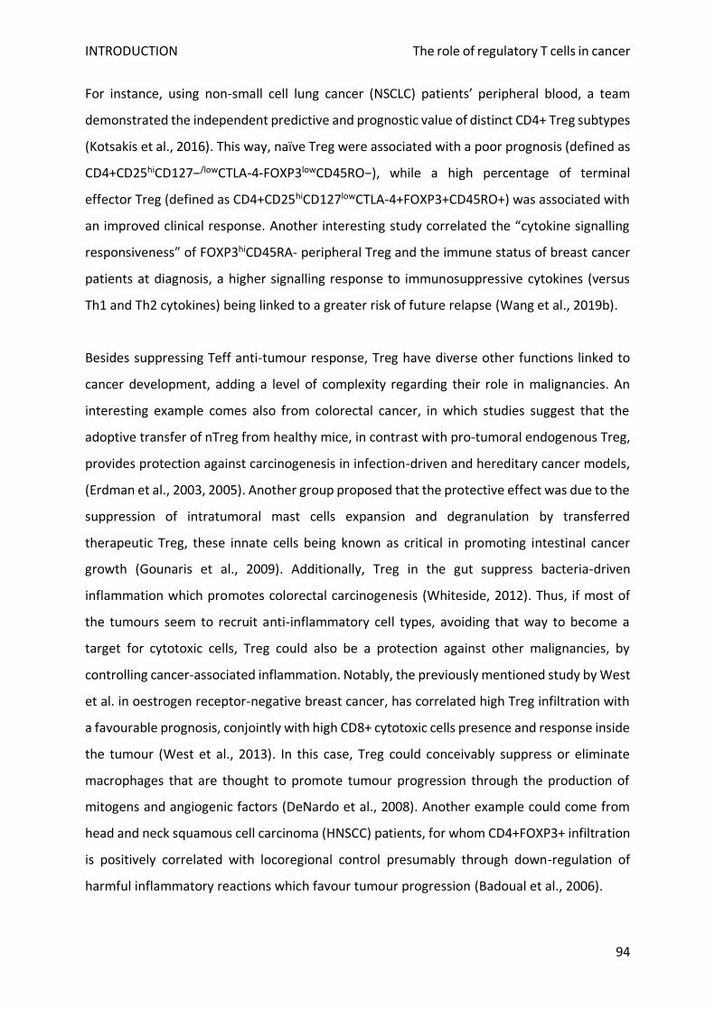
INTRODUCTION The role of regulatory T cells in cancer
94
For instance, using non-small cell lung cancer (NSCLC) patients’ peripheral blood, a team
demonstrated the independent predictive and prognostic value of distinct CD4+ Treg subtypes
(Kotsakis et al., 2016). This way, naïve Treg were associated with a poor prognosis (defined as
CD4+CD25hiCD127−/lowCTLA-4-FOXP3lowCD45RO−), while a high percentage of terminal
effector Treg (defined as CD4+CD25hiCD127lowCTLA-4+FOXP3+CD45RO+) was associated with
an improved clinical response. Another interesting study correlated the “cytokine signalling
responsiveness” of FOXP3hiCD45RA- peripheral Treg and the immune status of breast cancer
patients at diagnosis, a higher signalling response to immunosuppressive cytokines (versus
Th1 and Th2 cytokines) being linked to a greater risk of future relapse (Wang et al., 2019b).
Besides suppressing Teff anti-tumour response, Treg have diverse other functions linked to
cancer development, adding a level of complexity regarding their role in malignancies. An
interesting example comes also from colorectal cancer, in which studies suggest that the
adoptive transfer of nTreg from healthy mice, in contrast with pro-tumoral endogenous Treg,
provides protection against carcinogenesis in infection-driven and hereditary cancer models,
(Erdman et al., 2003, 2005). Another group proposed that the protective effect was due to the
suppression of intratumoral mast cells expansion and degranulation by transferred
therapeutic Treg, these innate cells being known as critical in promoting intestinal cancer
growth (Gounaris et al., 2009). Additionally, Treg in the gut suppress bacteria-driven
inflammation which promotes colorectal carcinogenesis (Whiteside, 2012). Thus, if most of
the tumours seem to recruit anti-inflammatory cell types, avoiding that way to become a
target for cytotoxic cells, Treg could also be a protection against other malignancies, by
controlling cancer-associated inflammation. Notably, the previously mentioned study by West
et al. in oestrogen receptor-negative breast cancer, has correlated high Treg infiltration with
a favourable prognosis, conjointly with high CD8+ cytotoxic cells presence and response inside
the tumour (West et al., 2013). In this case, Treg could conceivably suppress or eliminate
macrophages that are thought to promote tumour progression through the production of
mitogens and angiogenic factors (DeNardo et al., 2008). Another example could come from
head and neck squamous cell carcinoma (HNSCC) patients, for whom CD4+FOXP3+ infiltration
is positively correlated with locoregional control presumably through down-regulation of
harmful inflammatory reactions which favour tumour progression (Badoual et al., 2006).
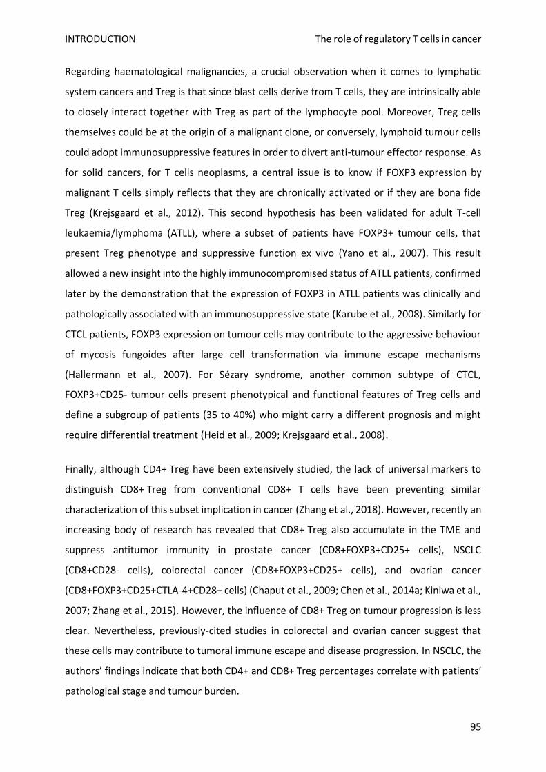
INTRODUCTION The role of regulatory T cells in cancer
95
Regarding haematological malignancies, a crucial observation when it comes to lymphatic
system cancers and Treg is that since blast cells derive from T cells, they are intrinsically able
to closely interact together with Treg as part of the lymphocyte pool. Moreover, Treg cells
themselves could be at the origin of a malignant clone, or conversely, lymphoid tumour cells
could adopt immunosuppressive features in order to divert anti-tumour effector response. As
for solid cancers, for T cells neoplasms, a central issue is to know if FOXP3 expression by
malignant T cells simply reflects that they are chronically activated or if they are bona fide
Treg (Krejsgaard et al., 2012). This second hypothesis has been validated for adult T-cell
leukaemia/lymphoma (ATLL), where a subset of patients have FOXP3+ tumour cells, that
present Treg phenotype and suppressive function ex vivo (Yano et al., 2007). This result
allowed a new insight into the highly immunocompromised status of ATLL patients, confirmed
later by the demonstration that the expression of FOXP3 in ATLL patients was clinically and
pathologically associated with an immunosuppressive state (Karube et al., 2008). Similarly for
CTCL patients, FOXP3 expression on tumour cells may contribute to the aggressive behaviour
of mycosis fungoides after large cell transformation via immune escape mechanisms
(Hallermann et al., 2007). For Sézary syndrome, another common subtype of CTCL,
FOXP3+CD25- tumour cells present phenotypical and functional features of Treg cells and
define a subgroup of patients (35 to 40%) who might carry a different prognosis and might
require differential treatment (Heid et al., 2009; Krejsgaard et al., 2008).
Finally, although CD4+ Treg have been extensively studied, the lack of universal markers to
distinguish CD8+ Treg from conventional CD8+ T cells have been preventing similar
characterization of this subset implication in cancer (Zhang et al., 2018). However, recently an
increasing body of research has revealed that CD8+ Treg also accumulate in the TME and
suppress antitumor immunity in prostate cancer (CD8+FOXP3+CD25+ cells), NSCLC
(CD8+CD28- cells), colorectal cancer (CD8+FOXP3+CD25+ cells), and ovarian cancer
(CD8+FOXP3+CD25+CTLA-4+CD28− cells) (Chaput et al., 2009; Chen et al., 2014a; Kiniwa et al.,
2007; Zhang et al., 2015). However, the influence of CD8+ Treg on tumour progression is less
clear. Nevertheless, previously-cited studies in colorectal and ovarian cancer suggest that
these cells may contribute to tumoral immune escape and disease progression. In NSCLC, the
authors’ findings indicate that both CD4+ and CD8+ Treg percentages correlate with patients’
pathological stage and tumour burden.
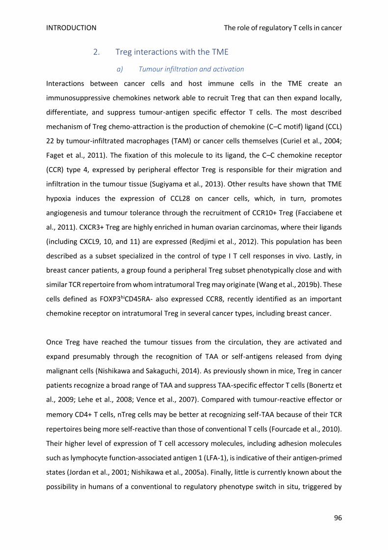
INTRODUCTION The role of regulatory T cells in cancer
96
2. Treg interactions with the TME
a) Tumour infiltration and activation
Interactions between cancer cells and host immune cells in the TME create an
immunosuppressive chemokines network able to recruit Treg that can then expand locally,
differentiate, and suppress tumour-antigen specific effector T cells. The most described
mechanism of Treg chemo-attraction is the production of chemokine (C–C motif) ligand (CCL)
22 by tumour-infiltrated macrophages (TAM) or cancer cells themselves (Curiel et al., 2004;
Faget et al., 2011). The fixation of this molecule to its ligand, the C–C chemokine receptor
(CCR) type 4, expressed by peripheral effector Treg is responsible for their migration and
infiltration in the tumour tissue (Sugiyama et al., 2013). Other results have shown that TME
hypoxia induces the expression of CCL28 on cancer cells, which, in turn, promotes
angiogenesis and tumour tolerance through the recruitment of CCR10+ Treg (Facciabene et
al., 2011). CXCR3+ Treg are highly enriched in human ovarian carcinomas, where their ligands
(including CXCL9, 10, and 11) are expressed (Redjimi et al., 2012). This population has been
described as a subset specialized in the control of type I T cell responses in vivo. Lastly, in
breast cancer patients, a group found a peripheral Treg subset phenotypically close and with
similar TCR repertoire from whom intratumoral Treg may originate (Wang et al., 2019b). These
cells defined as FOXP3hiCD45RA- also expressed CCR8, recently identified as an important
chemokine receptor on intratumoral Treg in several cancer types, including breast cancer.
Once Treg have reached the tumour tissues from the circulation, they are activated and
expand presumably through the recognition of TAA or self-antigens released from dying
malignant cells (Nishikawa and Sakaguchi, 2014). As previously shown in mice, Treg in cancer
patients recognize a broad range of TAA and suppress TAA-specific effector T cells (Bonertz et
al., 2009; Lehe et al., 2008; Vence et al., 2007). Compared with tumour-reactive effector or
memory CD4+ T cells, nTreg cells may be better at recognizing self-TAA because of their TCR
repertoires being more self-reactive than those of conventional T cells (Fourcade et al., 2010).
Their higher level of expression of T cell accessory molecules, including adhesion molecules
such as lymphocyte function-associated antigen 1 (LFA-1), is indicative of their antigen-primed
states (Jordan et al., 2001; Nishikawa et al., 2005a). Finally, little is currently known about the
possibility in humans of a conventional to regulatory phenotype switch in situ, triggered by
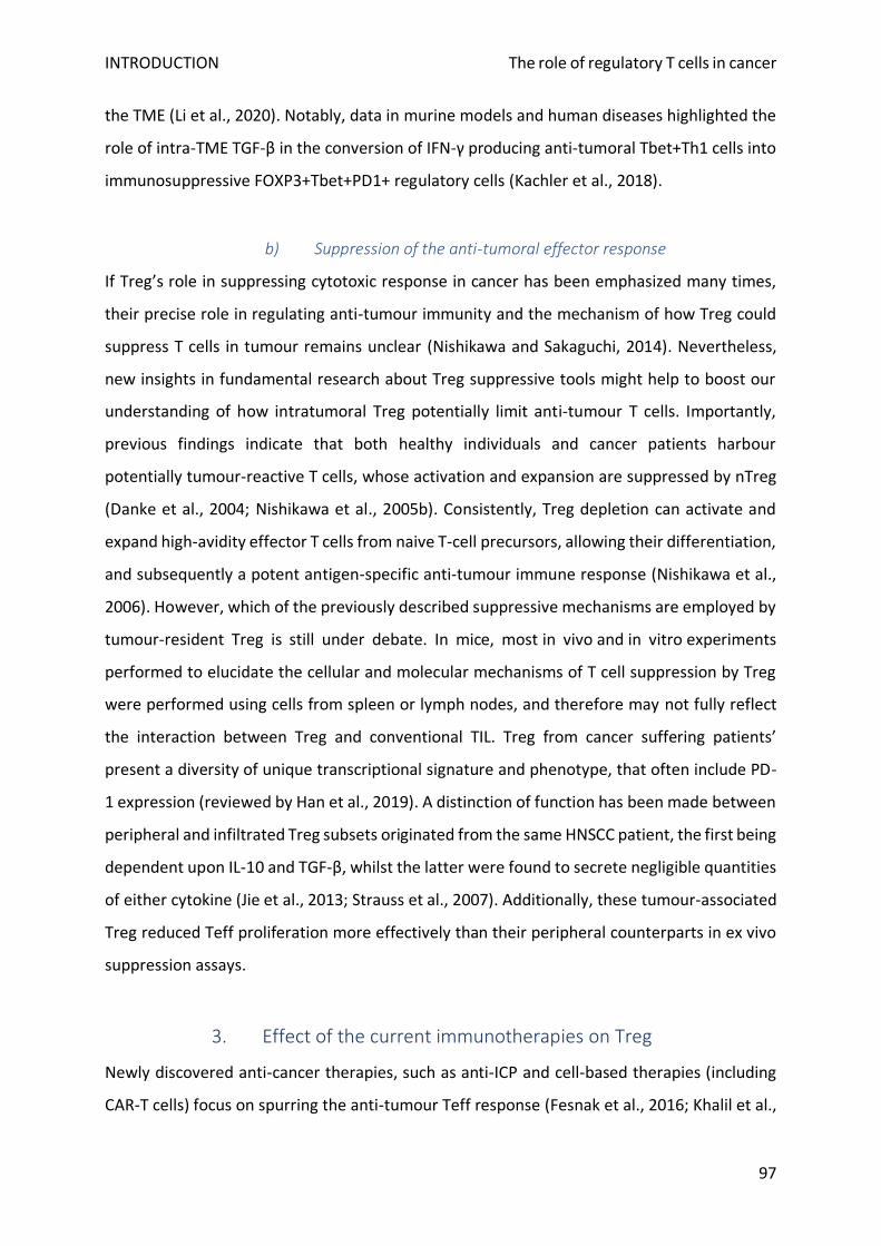
INTRODUCTION The role of regulatory T cells in cancer
97
the TME (Li et al., 2020). Notably, data in murine models and human diseases highlighted the
role of intra-TME TGF-β in the conversion of IFN-γ producing anti-tumoral Tbet+Th1 cells into
immunosuppressive FOXP3+Tbet+PD1+ regulatory cells (Kachler et al., 2018).
b) Suppression of the anti-tumoral effector response
If Treg’s role in suppressing cytotoxic response in cancer has been emphasized many times,
their precise role in regulating anti-tumour immunity and the mechanism of how Treg could
suppress T cells in tumour remains unclear (Nishikawa and Sakaguchi, 2014). Nevertheless,
new insights in fundamental research about Treg suppressive tools might help to boost our
understanding of how intratumoral Treg potentially limit anti-tumour T cells. Importantly,
previous findings indicate that both healthy individuals and cancer patients harbour
potentially tumour-reactive T cells, whose activation and expansion are suppressed by nTreg
(Danke et al., 2004; Nishikawa et al., 2005b). Consistently, Treg depletion can activate and
expand high-avidity effector T cells from naive T-cell precursors, allowing their differentiation,
and subsequently a potent antigen-specific anti-tumour immune response (Nishikawa et al.,
2006). However, which of the previously described suppressive mechanisms are employed by
tumour-resident Treg is still under debate. In mice, most in vivo and in vitro experiments
performed to elucidate the cellular and molecular mechanisms of T cell suppression by Treg
were performed using cells from spleen or lymph nodes, and therefore may not fully reflect
the interaction between Treg and conventional TIL. Treg from cancer suffering patients’
present a diversity of unique transcriptional signature and phenotype, that often include PD-
1 expression (reviewed by Han et al., 2019). A distinction of function has been made between
peripheral and infiltrated Treg subsets originated from the same HNSCC patient, the first being
dependent upon IL-10 and TGF-β, whilst the latter were found to secrete negligible quantities
of either cytokine (Jie et al., 2013; Strauss et al., 2007). Additionally, these tumour-associated
Treg reduced Teff proliferation more effectively than their peripheral counterparts in ex vivo
suppression assays.
3. Effect of the current immunotherapies on Treg
Newly discovered anti-cancer therapies, such as anti-ICP and cell-based therapies (including
CAR-T cells) focus on spurring the anti-tumour Teff response (Fesnak et al., 2016; Khalil et al.,
INTRODUCTION The role of regulatory T cells in cancer
98
2016; Topalian et al., 2015). If these strategies already demonstrated a sustained beneficial
effect in certain malignancies, a substantial proportion of the treated patients are not
responding. Treg-mediated anti-tumour response suppression is one potential mechanism
that may reduce the efficacy of the immunotherapies currently available to treat cancer,
alongside with other immunosuppressive cell populations (TAM, MDSC) (Binnewies et al.,
2018). On the other side, some immunotherapeutic agents seem to differentially impair Treg
number and/or function, either directly or indirectly through TME modifications (Han et al.,
2019a). Thus, an ongoing effort of research is trying to define the effects of recent anti-cancer
immunotherapies on Treg, and vice versa, to try to improve the response rate, yet most of the
questions are currently unanswered.
Regarding the crucial role of CTLA-4 expression for Treg functions, the participation of Treg
modulation in anti-CTLA-4 clinical potency has been extensively investigated and is still
controversial (Tang et al., 2018). In particular, the debate focuses on whether the Teff or Treg
compartment is the primary target of ipilimumab, the only approved mAb against CTLA-4 to
date. In mice, it is established that CTLA-4 blockade results in a drastic reduction of
FOXP3+CTLA-4+ TIL, in an Fc-γ receptor-dependent manner (Bulliard et al., 2013; Selby et al.,
2013; Simpson et al., 2013). Arce Vargas et al. recently reached the same conclusion using
ipilimumab in a mouse model expressing human Fc-γR. Besides in patients with advanced
melanoma, response to ipilimumab was associated with a specific alloform of activating Fc-γR
conferring higher binding affinity to IgG1 (Arce Vargas et al., 2018). If initially, the
demonstration has been made that both Teff and Treg were required for maximal
enhancement of anti‐tumour immunity using human-CTLA-4 expressing mice (Peggs et al.,
2009), in 2018 a group asked for a reassessment of CTLA-4 blockade mechanisms of action.
Using CTLA-4 human/mouse heterozygous mice, they have shown that anti-B7 antibodies that
effectively block CD4+ T cell activation and de novo CD8+ T cell priming in lymphoid organs do
not negatively affect the immunotherapeutic effect of ipilimumab. Given these results, they
suggested that CTLA-4 blockade causes tumour rejection by mechanisms that are dependent
on the host FcR but independent of checkpoint inhibition (Du et al., 2018; Tang et al., 2018).
When scrutinizing the literature, the reports about ipilimumab effect on patients’ Treg also
present contradictions. Different results have pointed out the decreased frequency of
tumour-infiltrating Treg in ipilimumab-treated patients for bladder cancer and advanced
INTRODUCTION The role of regulatory T cells in cancer
99
melanoma (Liakou et al., 2008; Romano et al., 2015; Simeone et al., 2014). On the other hand,
a report published last year found no significant change or depletion of FOXP3+ infiltrating
cells, while ipilimumab did augment the quantity of intratumoral CD4+ and CD8+ lymphocytes
quantified in melanoma, prostate cancers, and bladder cancers (Sharma et al., 2019).
The three antibodies targeting the PD-1/PD-L1 signalling pathway have received their
marketing authorization later, hence the number of clinical studies documenting quantitative
or qualitative Treg changes post-treatment are limited (Alsaab et al., 2017). Importantly, like
CTLA-4, PD-1 can be expressed on Treg, notably in cancer, implying a potential direct action
of anti-PD-1 antibodies on these cells (De Simone et al., 2016; Francisco et al., 2010; Plitas et
al., 2016; Toker et al., 2018). Murine results suggest the absence of implication for PD-1 on
nTreg, whilst PD-1 appears to be necessary for Treg induction from naïve CD4+ cells in line
with the observation that PD-L1 blockade interferes with the induction and maintenance of
iTreg (Chen et al., 2014b; Francisco et al., 2009). Notably, PD-1 inhibition synergizes with other
therapeutic strategies reducing Treg quantity in mice (Arce Vargas et al., 2017; Dodagatta-
Marri et al., 2019; Oweida et al., 2018). In humans, a study that analysed biopsies from
patients under pembrolizumab found changes in MDSC, but not in Treg frequencies (Ribas et
al., 2016). Additionally, in vitro analyses have shown that pembrolizumab mediates its
immunostimulatory effects via the release of effector T cells from suppression only, and does
not modulate Treg functions (Toor et al., 2018, 2019). Later, in metastatic melanoma, a group
noted for non-relapsing patients a paradoxical increase in circulating Treg proportion and
phosphoSTAT3-mediated proliferation, both positively correlated with patient survival 13
weeks after PD-1 blockade using nivolumab (Woods et al., 2018). Nevertheless, they also
reported a reduction in Treg suppressive-capacity in vivo, and in vitro as in two different
studies, one using another anti-PD-1 on melanoma patients cells (Wang et al., 2009), and the
other with nivolumab on healthy volunteer cells (Wang et al., 2014).
Beyond current therapies, promising ICP candidates to target are already screened for their
interaction with Treg. It include LAG-3, a protein highly expressed on tumour-infiltrated Treg
in cancer patients (Huang et al., 2004). Its blockade alone or in combination with PD-1/PD-L1
blockade lead to an inhibition of Treg function in patients (Solinas et al., 2019). TIM-3, another
signature molecule of conventional T cells exhaustion under investigation in cancer, is

INTRODUCTION The role of regulatory T cells in cancer
100
expressed by a small fraction of peripheral Treg cells, and a large proportion of intratumoral
Treg that present advanced suppressive capacities (Yano et al., 2007).
A quite promising observation about Treg after mAb infusion has been made in leukemic
patients treated with blinatumomab. This bispecific construct, combined with chemotherapy,
permits a haematological remission in around 50% of resistant B-ALL patients (Farhadfar and
Litzow, 2016). Duell et al. were able to prove that Treg are the best clinical outcome predictors
for this treatment, with blinatumomab responders showing lower levels of Treg in peripheral
blood than non-responders (Duell et al., 2017). Anterior reports in paediatric leukaemia
patients demonstrated that a chain of immunological phenomena follows the injection of
blinatumomab, causing Treg activation and in turn a rapid release of IL-10 (von Stackelberg et
al., 2016). This triggers a drop in CD8+ proliferation, consequently reducing the effectiveness
of the anti-tumour response (Vedi and Ziegler, 2014). Thus, taken together these results (i)
identified blinatumomab responders by quantifying peripheral Treg, and (ii) allowed to
highlight the need of a specific anti-Treg strategy in order to enlarge the efficacy of this mAb
to current refractory patients (Niedźwiecki et al., 2019).
To date, two CAR T-cell therapies have been approved very recently in Europe and the USA,
as well as several other countries, for the treatment of leukaemia and lymphoma (Elsallab et
al., 2020). Some groups have already tried to understand the relation between Treg and these
adoptive T cells after the infusion. In a clinical trial, CD19-targeted third-generation CAR-T cells
used against B-cell lymphoma and leukaemia did not modify the frequency of Treg (Enblad et
al., 2018). Interestingly, prior lymphodepletion using chemotherapeutic agents that
transiently reduce the frequency of Treg were able to improve the persistence of several CD19
CAR-T cells constructs as well as therapeutic outcomes (Park et al., 2016). Then, if the direct
effect of Treg on CAR-T cells remains unknown, this observation could foster a therapeutic
approach combining CAR-T cells use and Treg-impairing agents, as for previous
immunotherapies.
INTRODUCTION The role of regulatory T cells in cancer
101
D. Interfering with Treg immune suppression
1. Rational behind Treg blockade
Treg inherent function to suppress conventional T cell response can be acutely detrimental
for the immune control of cancer initiation, growth, and dissemination. Besides, Treg are likely
to interfere with Teff-centred immunotherapies. If they can serve as prognostic factors, it also
appears necessary to learn how to block this heterogeneous population in patients to
overcome its immunosuppressive effects. Initially, mice models allowed to observe a
beneficial issue of Treg depletion on anti-tumour response potency, using FOXP3DTR mice or
antibodies targeting receptors highly expressed on Treg cells, such as CD25, GITR, and folate
receptor 4 (Arce Vargas et al., 2017; Coe et al., 2010; Klages et al., 2010; Yamaguchi et al.,
2007). On the contrary, joint infusion of Treg and CD8+ conventional T cells prevents the
adoptive cell therapy efficacy against murine melanoma (Antony et al., 2005). Based on these
arguments, several cancer immunotherapies targeting Treg directly are currently being tested
in patients. In addition, therapies already in use have a documented an impairing effect on
Treg, either directly as previously mentioned for ipilimumab, or indirectly as for some
chemotherapeutic agents.
2. Indirect anti-Treg strategies
a) Chemotherapeutic agents
Interestingly, cytostatic treatments can reduce the quantity of Treg and synergize with
immunomodulatory drugs to enhance antitumor response. Antineoplastic drugs such as
cyclophosphamide and fludarabine can selectively affect Treg, presumably because nTreg are
physiologically in a more proliferative state than other T cells via recognizing self-antigens or
commensal microbes (Beyer et al., 2005; Walter et al., 2012). Low-dose cyclophosphamide,
significantly reduced Treg while maintaining an elevated level of CD8+ T cells in the tumour,
leading to enhanced antitumor immunity (Ghiringhelli et al., 2004; Ladoire et al., 2008; Scurr
et al., 2017; Wada et al., 2009; Walter et al., 2012). Conversely, two other groups have
reported contradicting data where cyclophosphamide administration does not represent an
optimal therapy to eliminate Treg or to increase CD8+ response (Audia et al., 2007; Sevko et
al., 2013). Despite this, additional studies showed that cyclophosphamide effect on Treg could
be further improved in its selectivity and efficacy through combination with multipeptide
INTRODUCTION The role of regulatory T cells in cancer
102
vaccination, OX40 agonist, or anti-PD-1 (Hirschhorn-Cymerman et al., 2009; Mkrtichyan et al.,
2011; Walter et al., 2012). Several other FDA-approved anti-cancer agents including three
tyrosine kinase inhibitors also reduced the levels of intratumoral Treg (Desar et al., 2011; Finke
et al., 2013; Hirschhorn-Cymerman et al., 2009; Larmonier et al., 2008).
b) Induction of “suppression-proof” Teff
It is crucial to note that if the recent approaches mainly aim to hamper Treg actions,
alternative strategies could search to trigger T cells’ resistance to Treg suppression. It could
be particularly relevant when trying to enhance adoptive T cell therapies such as tumour
specific TCR cells or CAR-T cells. Recently, a variety of molecular pathways and cellular
mechanisms that govern T cell activation have been implicated in T cell resistance, including
surface receptors such as Toll-like receptors or TNF family members (OX40, 4-1BB, GITR),
intracellular molecules like Cbl-b and TRAF-6, and various T cell stimulatory cytokines
(reviewed by Han et al., 2019).
One interesting potential signalling to target, is the TCR molecular pathway, including its
negative regulators. The E3 ubiquitin ligase Cbl-b plays an important role in regulating diverse
arms of the TCR signalling pathways, promoting T cell inhibition (Paolino and Penninger,
2010). Cbl-b deficiency in mice augments anti-tumour T cell responses in both genetically
engineered and transplanted tumour models (Chiang et al., 2007; Loeser et al., 2007; Paolino
et al., 2011; Stromnes et al., 2010). Cbl-b deficient CD8+ and CD4+ T cells are refractory to
Treg-mediated suppression in vitro (Adams et al., 2010; Gruber et al., 2013). In this setting,
one proposed mechanism by which conventional T cells resist Treg includes insensitivity to
TGF-β receptor signalling.
On the other hand, T cells stimulated with appropriate cytokines may be equally susceptible
to Treg cell-mediated suppression, but by increasing proliferation and quantity of T cells, the
suppressive effect of Treg may become less apparent or overwhelmed. Historically, this fine
balance between regulatory and effector functions was observed with IL-2, a cytokine able to
enhance T cell proliferation, despite stimulating conjointly Treg (Takahashi et al., 1998;
Thornton and Shevach, 1998). This phenomenon is already observed in some clinical settings
using IL-2. For instance, high dose IL-2 is part of the protocol for adoptive TIL therapy against
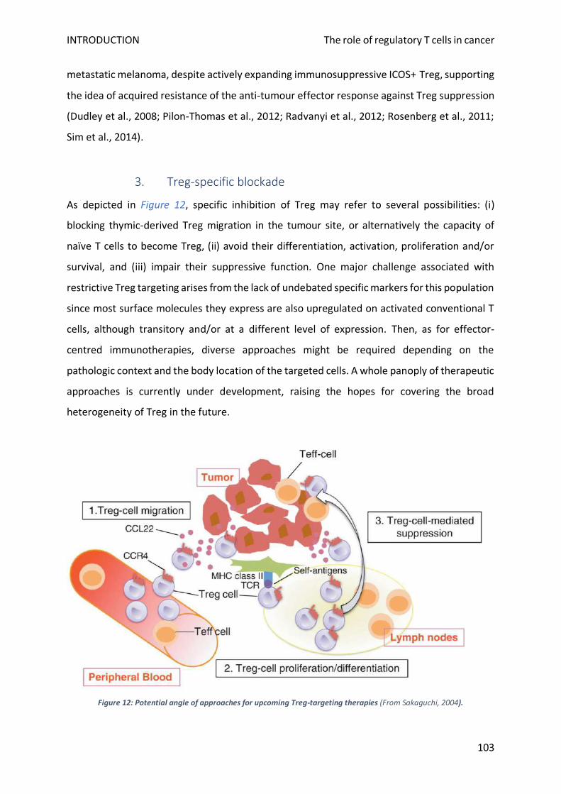
INTRODUCTION The role of regulatory T cells in cancer
103
metastatic melanoma, despite actively expanding immunosuppressive ICOS+ Treg, supporting
the idea of acquired resistance of the anti-tumour effector response against Treg suppression
(Dudley et al., 2008; Pilon-Thomas et al., 2012; Radvanyi et al., 2012; Rosenberg et al., 2011;
Sim et al., 2014).
3. Treg-specific blockade
As depicted in Figure 12, specific inhibition of Treg may refer to several possibilities: (i)
blocking thymic-derived Treg migration in the tumour site, or alternatively the capacity of
naïve T cells to become Treg, (ii) avoid their differentiation, activation, proliferation and/or
survival, and (iii) impair their suppressive function. One major challenge associated with
restrictive Treg targeting arises from the lack of undebated specific markers for this population
since most surface molecules they express are also upregulated on activated conventional T
cells, although transitory and/or at a different level of expression. Then, as for effector-
centred immunotherapies, diverse approaches might be required depending on the
pathologic context and the body location of the targeted cells. A whole panoply of therapeutic
approaches is currently under development, raising the hopes for covering the broad
heterogeneity of Treg in the future.
Figure 12: Potential angle of approaches for upcoming Treg-targeting therapies (From Sakaguchi, 2004).
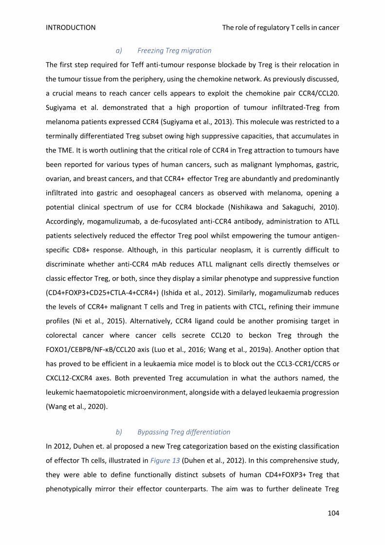
INTRODUCTION The role of regulatory T cells in cancer
104
a) Freezing Treg migration
The first step required for Teff anti-tumour response blockade by Treg is their relocation in
the tumour tissue from the periphery, using the chemokine network. As previously discussed,
a crucial means to reach cancer cells appears to exploit the chemokine pair CCR4/CCL20.
Sugiyama et al. demonstrated that a high proportion of tumour infiltrated-Treg from
melanoma patients expressed CCR4 (Sugiyama et al., 2013). This molecule was restricted to a
terminally differentiated Treg subset owing high suppressive capacities, that accumulates in
the TME. It is worth outlining that the critical role of CCR4 in Treg attraction to tumours have
been reported for various types of human cancers, such as malignant lymphomas, gastric,
ovarian, and breast cancers, and that CCR4+ effector Treg are abundantly and predominantly
infiltrated into gastric and oesophageal cancers as observed with melanoma, opening a
potential clinical spectrum of use for CCR4 blockade (Nishikawa and Sakaguchi, 2010).
Accordingly, mogamulizumab, a de-fucosylated anti-CCR4 antibody, administration to ATLL
patients selectively reduced the effector Treg pool whilst empowering the tumour antigen-
specific CD8+ response. Although, in this particular neoplasm, it is currently difficult to
discriminate whether anti-CCR4 mAb reduces ATLL malignant cells directly themselves or
classic effector Treg, or both, since they display a similar phenotype and suppressive function
(CD4+FOXP3+CD25+CTLA-4+CCR4+) (Ishida et al., 2012). Similarly, mogamulizumab reduces
the levels of CCR4+ malignant T cells and Treg in patients with CTCL, refining their immune
profiles (Ni et al., 2015). Alternatively, CCR4 ligand could be another promising target in
colorectal cancer where cancer cells secrete CCL20 to beckon Treg through the
FOXO1/CEBPB/NF-κB/CCL20 axis (Luo et al., 2016; Wang et al., 2019a). Another option that
has proved to be efficient in a leukaemia mice model is to block out the CCL3-CCR1/CCR5 or
CXCL12-CXCR4 axes. Both prevented Treg accumulation in what the authors named, the
leukemic haematopoietic microenvironment, alongside with a delayed leukaemia progression
(Wang et al., 2020).
b) Bypassing Treg differentiation
In 2012, Duhen et. al proposed a new Treg categorization based on the existing classification
of effector Th cells, illustrated in Figure 13 (Duhen et al., 2012). In this comprehensive study,
they were able to define functionally distinct subsets of human CD4+FOXP3+ Treg that
phenotypically mirror their effector counterparts. The aim was to further delineate Treg
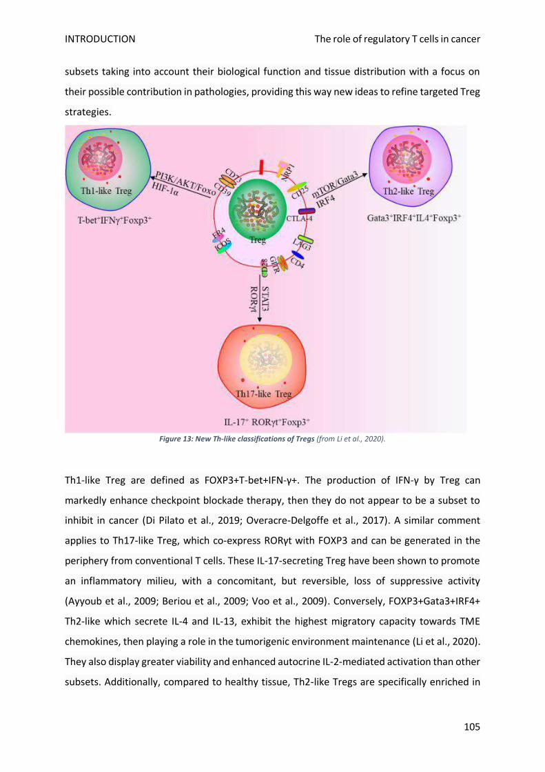
INTRODUCTION The role of regulatory T cells in cancer
105
subsets taking into account their biological function and tissue distribution with a focus on
their possible contribution in pathologies, providing this way new ideas to refine targeted Treg
strategies.
Figure 13: New Th-like classifications of Tregs (from Li et al., 2020).
Th1-like Treg are defined as FOXP3+T-bet+IFN-γ+. The production of IFN-γ by Treg can
markedly enhance checkpoint blockade therapy, then they do not appear to be a subset to
inhibit in cancer (Di Pilato et al., 2019; Overacre-Delgoffe et al., 2017). A similar comment
applies to Th17-like Treg, which co-express RORγt with FOXP3 and can be generated in the
periphery from conventional T cells. These IL-17-secreting Treg have been shown to promote
an inflammatory milieu, with a concomitant, but reversible, loss of suppressive activity
(Ayyoub et al., 2009; Beriou et al., 2009; Voo et al., 2009). Conversely, FOXP3+Gata3+IRF4+
Th2-like which secrete IL-4 and IL-13, exhibit the highest migratory capacity towards TME
chemokines, then playing a role in the tumorigenic environment maintenance (Li et al., 2020).
They also display greater viability and enhanced autocrine IL-2-mediated activation than other
subsets. Additionally, compared to healthy tissue, Th2-like Tregs are specifically enriched in
INTRODUCTION The role of regulatory T cells in cancer
106
malignant tissues from patients with melanoma and colorectal cancer (Halim et al., 2017).
Compared to the two others, this subset appears to represent the main Treg population
involved in cancer immunology, a piece of information which provides clues for novel
therapeutic strategies. Importantly, Th2-like programming can be induced by IL-4R signalling
that promotes Gata3 expression (Krishnamoorthy et al., 2012; Noval Rivas et al., 2015). It
could explain the opposite findings that have been observed in the past when using IL-4 as an
anti-tumour agent (Li et al., 2009). The dogma is changing since recently the capacity of IL-4
to improve tolerance in patients presenting autoimmunity has been demonstrated (Yang et
al., 2017).
c) IL-2/CD25-focused strategies
Because Treg were originally identified as CD4+CD25+ T cells, numerous studies have explored
the effects of targeting this receptor or its associated pro-survival cytokine, IL-2. The depletion
of CD25+ Treg population via targeted antibody therapy enhanced the anti-tumour immunity
and progressively reduced tumour volume in murine cancer models (Onizuka et al., 1999;
Shimizu et al., 1999). However, when applied to patients this strategy has raised a number of
concerns based on inconsistent in vivo responses, toxicities, and lack of specificity.
A recombinant protein composed of IL‐2 and the active domain of the diphtheria toxin, named
denileukin diftitox was developed to deplete Treg. Clinical experience using denileukin diftitox
delivered mixed results due to varying clinical responses, similarly to the previously observed
effects of anti-CD25 in mice (Foss, 2006). The treatment of patients with renal cell carcinoma
effectively relieved inhibition by Treg to promote anti-tumour immunity, whereas the
opposite trend was observed in metastatic melanoma (Attia et al., 2005; Dannull et al., 2005).
Tumour heterogeneity, the existence of CD25− Treg and CD25 expression on other immune
cells, such as T, B, and NK lymphocytes, may explain seemingly opposite outcomes in this
particular approach, that was discontinued in 2014 (Brisslert et al., 2006; Leong et al., 2014).
Another strategy, in use for decades, relies on differential IL-2 dose to target either Treg or
Teff depending on the pathological context. In contrast with the disappointing results of low-
dose IL-2 treatment, high-dose IL-2 administration in metastatic melanoma and renal cell
carcinoma patients yield complete tumour regression in a fraction of patients while extending
disease-free intervals in many other patients (Klapper et al., 2008). However, several
INTRODUCTION The role of regulatory T cells in cancer
107
drawbacks intrinsic to IL2 hamper its efficacy and broader application including its high
toxicity, short circulatory half-life, and ability to induce immunosuppressive responses
through Treg expansion (Tang and Harding, 2019).
The results obtained with CD25-targeted antibodies also remain controversial. A phase-I/II
study with CD25-blocking mAb daclizumab in combination with DC vaccine has shown a
transient but complete depletion of CD25hi cells in melanoma patients (Jacobs et al., 2010;
Mitchell et al., 2011; Vries et al., 2011). Despite its preliminary efficacy, many questions still
need to be addressed before developing a successful therapeutic strategy exploiting CD25
blockade with limitations that include a lack of specificity alongside with severe and various
side effects such as cytokine release syndrome or infections (Martinelli et al., 2019; Verma et
al., 2019). However, in breast cancer patients, the combination of daclizumab and vaccination
produced favourable clinical responses seemingly without severe side effects (Rech et al.,
2012).
CD25 is highly expressed on Treg, while Teff can transiently gain CD25 expression after TCR
stimulation. It has been generally acknowledged that the dual roles played by IL-2 in activating
both Treg and Teff impede a safe and efficient clinical use of the blockade of CD25 and IL-2
(Ross and Cantrell, 2018). However, the potential anti-CD25/IL-2 strategies has been recently
demonstrated by numerous pre-clinical results. Creative strategies to surmount the high
toxicities, short half-life, and pleiotropic effects of IL-2 continue to be developed including
novel mutational, pegylation, and tumour targeting approaches (Tang and Harding, 2019). IL-
2 may also enhance the efficacy of checkpoint inhibitors and CAR-T-based therapies (Sun et
al., 2019). Regarding CD25 targeting mAb, Fc-optimized antibodies against CD25 have been
effectively designed (Arce Vargas et al., 2017). Innovative approaches include the conjugation
of anti-CD25 with a photoactivatable dye sensitive to near-infrared light, which permits its
localisation inside the tumour, using infrared irradiation, and the specific reduction of
intratumoral Treg cells (Sato et al., 2016).
d) Turning off Treg activation signals
Beyond CD25 as a target molecule, Treg constitutively express receptors such as GITR, CTLA-
4, and folate receptor 4. In the TME, Treg further upregulate a large number of receptors
INTRODUCTION The role of regulatory T cells in cancer
108
including ICOS, OX40, GITR, TIGIT, PD-1, and CTLA-4 (Liu et al., 2016). Apart from CTLA-4 and
PD-1, already available in several malignancies’ therapeutic arsenal, the rest of these
molecules are currently tested in clinical studies. As ICP were the subject of an anterior
section, this subpart will focus only on co-stimulatory molecules.
GITR is expressed in resting CD4+ and CD8+ lymphocytes and increased upon T-cell activation
(Shevach and Stephens, 2006). On the Treg surface, GITR is constitutively expressed at a high
density, where its fixation by agonistic mAb results in Treg suppressive capacity reduction
(Ephrem et al., 2013; Kanamaru et al., 2004; McHugh et al., 2002). The results of a first-in-
human trial with an anti-GITR mAb, TRX518, have been reported last year (Killock, 2019). In
this phase I study, TRX518 monotherapy at various doses in 43 patients with advanced-stage
solid tumours revealed an effective depletion of peripheral blood Treg. Interestingly, this
effect in the circulation was in apparent direct correlation with Treg depletion in the tumour,
suggesting an interesting biomarker for GITR targeting strategies. However, blood CD8+ or
CD4+ Teff and NK were not notably modulated and, disappointingly, no objective clinical
responses were observed. Notably, exhausted PD-1+ lymphocytes were found to persist in
patient tumour specimens with the highest levels of Treg depletion. In mice models where
this resistance to GITR agonism has been also observed, T cells’ efficacy via PD-1 inhibition
rescued tumours’ responsiveness to the treatment (Killock, 2019). Indeed, the authors have
initiated a trial using TRX518 in combination with PD-1 blockade, with promising clinical
responses already observed, including for refractory patients to prior anti-PD-1 therapy alone
(Zappasodi et al., 2019).
In line with GITR pre-clinical results, OX40, another co-stimulatory molecule expressed
transiently on activated T-cells but constitutively on Treg has been shown to reduce Treg-
mediated immunosuppression when targeted with an intratumoral injection of agonistic mAb
(Croft, 2003; Piconese et al., 2008). The results of a phase I clinical trial have confirmed that
OX40 is a potent immune-stimulating target in metastatic cancer patients. Injection of an OX-
40 agonist increased CD4 and CD8 Teff proliferation and the response to tumour-specific
antigens, enhancing both humoral and cellular immunity in cancer treatment (Curti et al.,
2013). In analogy to the mouse tumour models, anti-OX40 mAb may further benefit from
combination strategies (Shrimali et al., 2017). Hence, several phase I and II trials are ongoing
INTRODUCTION The role of regulatory T cells in cancer
109
using five different OX-40-targeting molecules, in monotherapy or combined with other
immunotherapies or chemotherapeutic agents (Aspeslagh et al., 2016).
Lastly, data indicate that the ICOS pathway, functionally related to CD28 co-stimulation, plays
a critical role in effective responses to anti-CTLA-4 and perhaps other anti-ICP agents (Solinas
et al., 2020). Thus, ICOS agonists are usually administered in concomitance with anti-CTLA-4
or anti-PD-1 mAb, for their ability to synergistically inhibit the suppressive activity of Treg and
to potentiate the antitumor activity of CD4+ and CD8+ Teff (Yap et al., 2018). On the contrary,
ICOS antagonistic mAb mainly deplete Treg by stimulating the antibody-dependent cell-
mediated cytotoxicity (ADCC) mediated by NK cells (Le et al., 2016; Mo et al., 2017). Diverse
agonists and antagonists are under evaluation in clinical trials, in monotherapy or synergy with
other agents targeting Treg.
e) Converting Treg into pro-inflammatory cells
One remarkable aspect of Treg, aside from their overriding heterogeneity, is their well-
established plasticity of phenotype and function depending on their cytokine
microenvironment (Sakaguchi et al., 2013; Sawant and Vignali, 2014). Although poorly
understood at the moment, T cell stimulatory cytokines may mediate FOXP3 downregulation,
convert naïve Treg to “effector-like” Th1/17, or directly turn Treg into conventional T cells
(Dominguez-Villar et al., 2011; Hori, 2011; Komatsu et al., 2009; McClymont et al., 2011).
Preliminary approaches have been designed to take advantage of this enticing Treg feature,
targeting transcription factors as well as epigenetic modifiers to shift intratumoral Treg
differentiation program or to “re-program” them to help the fight against cancer cells (Han et
al., 2019b). These recent strategies currently remain at the state of fundamental and pre-
clinical data.
One axis of research is focused on naïve Treg commitment into Th1 differentiation program,
because, as mentioned, this subset is associated with better immunotherapy efficacy. The
main cue driving the differentiation of Treg toward a Th1-like pro-inflammatory phenotype is
the presence of IL-12, which via the PI3K/AKT/Foxo1/3 signalling cascade is the major pathway
involved in IFN-γ secretion by human Treg (Kitz et al., 2016). Foxo1 is the key transcriptional
regulator that prevents IFN-γ expression, by direct fixation to the INFG gene upstream region.
INTRODUCTION The role of regulatory T cells in cancer
110
Accordingly, the Treg-specific deletion of Foxo1 leads to an increase in IFN-γ+ Treg (Ouyang et
al., 2010, 2012). Another factor that prevents INFG gene expression upregulation in Treg, the
E3 ubiquitin ligase VHL, is capable of regulating the hypoxia sensor HIF-1α through the
proteasome, maintaining this way Treg stability and suppressive capacity (Lee et al., 2015).
VHL-deficient Treg are converted into Th1-like Treg, display FOXP3 downregulation and
excessive IFN-γ production, induced by HIF-1α direct positive transcriptional regulation of the
IFNG gene promoter.
Other approaches are aiming to convert Treg cells into immune-stimulatory cells using
epigenetic modifications to disrupt their lineage. For instance, the activity of the histone
H3K27 methyltransferase enhancer of zeste homolog 2 (EZH2), which is boosted in human and
murine tumour-infiltrating Treg, can be targeted to promote the conversion of Treg into IFN-
γ producing effector cells, able to remodel the TME and enhance anti-tumour immunity (Wang
et al., 2018a). Several other epigenetic modifiers such as Bromodomain and Extra-Terminal
(BET) family proteins and histone acetyltransferase p300 can also be targeted to disrupt Treg
function and improve anti-tumour immune response (Adeegbe et al., 2017; Liu et al., 2013).
If they seem efficient, these proteins also possess other biological functions, then their
molecular targeting may induce diverse off-target effects (Han et al., 2019a).
Finally, iTreg are, by definition, associated with stronger phenotype plasticity than nTreg
(Adeegbe and Nishikawa, 2013). Nevertheless, it appears that nTreg do not undergo a strict
lineage commitment, as previously hypothesised, since targeting neuropilin-1 induces IFN-γ
production and “Treg functional fragility”, unlocking, in turn, the anti-tumour immunity
(Overacre-Delgoffe et al., 2017). If the literature diverges about HELIOS use as a marker for
Treg origin, this transcription factor undeniably plays a capital part in their lineage
engagement. Tumour-infiltrating Treg nearly uniformly express HELIOS, and at higher levels
than circulating Treg (Kelley et al., 2007). Kim and colleagues showed that the prophylactical
engagement of GITR using an agonistic mAb decreased HELIOS expression restrictively in
intratumoral Treg, favoured their conversion to Teff specifically, and substantially impaired
tumour growth (Nakagawa et al., 2016). HELIOS may thus be a legitimate target to restrain
Treg stability with as well an advantageous limited expression in other cell types that would

INTRODUCTION The role of regulatory T cells in cancer
111
suggest minimal off-target effects (Chougnet and Hildeman, 2016). However, no specific tool
has been described yet that exclusively blocks HELIOS activity.
INTRODUCTION The TNF-α/TNFR2 pathway: a new ICP to target
112
The TNF-α/TNFR2 pathway: a new ICP to target
Despite these emerging approaches to block or deplete Treg, limitations still exist, including
the lack of a Treg-specific biomarker and potential induction of autoimmunity as a
consequence of systemic Treg impairment (Kim et al., 2009b; Stephens et al., 2001).
Additionally, Treg depletion can be followed by their rapid reconstitution, potentially resulting
in a higher Treg frequency in comparison to their level prior depletion (Berod et al., 2014;
Mahnke et al., 2007). Therefore, developing new tools to circumvent tolerance toward
malignant cells still looms large.
In that respect, the type II receptor to TNF-α represents another hope in targeting Treg,
including in tumoral context (Cohen and Wood, 2017). To a larger extent, the particular
pattern of expression of this molecule on the immune system actors, preferentially presents
on tolerogenic cells, could permit a tuned modulation of both innate and adaptive responses
in diverse pathogenic contexts (Wajant and Beilhack, 2019).
After a brief recall of TNF-α historical relationship to anti-cancer therapy, this chapter aims to
first expose TNF-α ambivalent role in both immunosuppression and inflammation in relation
to which of its receptor is involved. The second half will depict the most recent developments
that have led to consider TNFR2 as a new brake impeding anti-tumour immune response
through its beneficial role on Treg, and therefore as an alternative target for cancer
immunotherapy.
INTRODUCTION The TNF-α/TNFR2 pathway: a new ICP to target
113
A. Historical point of view
TNF-α was discovered in 1975 as an endotoxin-inducible molecule that caused necrosis of
cancer cells in vitro - earning its tumour necrosis designation - and acted in synergy with
interferons (Bloksma et al., 1983; Carswell et al., 1975; Williamson et al., 1983). Soon, TNF-α
was shown to be expressed by monocytes/macrophages and activated T cells, distinctly from
another cytotoxic cytokine previously described, the lymphotoxin-α (LTα) (English et al., 1991;
Kelker et al., 1985; Turner and Feldmann, 1988). The interest for this cytokine grew
exponentially because of its ability to control tumour growth, not only in vitro but also in vivo
(Haranaka et al., 1984; Matthews, 1979; Prince et al., 1982). Early reports demonstrated that
monocyte-expressed TNF-α was capable of selective tumour cytotoxicity, and experimentally
induced murine tumours could be treated in vivo either by recombinant TNF directly, and/or
by LPS (then known as endotoxin), found to be a biological inducer of TNF-α (Ziegler-Heitbrock
et al., 1986). At the time these studies convincingly proposed TNF-α as a potent tumoricidal,
working independently from the tumour type, while displaying an apparent safety in murine
malignancy models (Sedger and McDermott, 2014). If its effect was certainly due in part to
direct tumoral necrosis induction, another side of its action that relied on tumour-associated
capillary injury was omitted (Haranaka et al., 1984, 1987).
Fostered by its preclinical efficacy, recombinant TNF-α monotherapy was quickly shifted
toward phase I clinical trials. Early observations were made of a panoply of severe acute
toxicities, including fevers, early confusions, nausea/vomiting, tachycardia or hypotension
(Creaven et al., 1987; Feinberg et al., 1988; Schwartz et al., 1989; Zamkoff et al., 1989). If a
limited number of patients did exhibit an effect on tumour growth, it was only transient, and
most eventually succumbed to their neoplasm, partly because of the advanced state of
recruited patients’ malignancy. Later attempts to administrate TNF-α in combination with IL-
2 proved to be evenly inefficient (Negrier et al., 1992). In the end, none of the 36 phase I trials
(a half of combined approaches) and 10 phase II studies did significantly improve the picture
(Roberts et al., 2011).
Although the results of these trials extinguished the hopes of a potent anti-tumour strategy,
the broad biological roles of TNF-α were also revealed through the side effects observed. In
INTRODUCTION The TNF-α/TNFR2 pathway: a new ICP to target
114
the same period, the first evidence that neutralizing antibodies to TNF-α protected mice
against lethal TNF-α-mediated endotoxemia opened a fresh perspective for TNF-α based-
therapy, beyond its original indication in cancer (Beutler et al., 1985). Rapidly, the crucial role
of TNF-α in inflammation was better described as both independent and dependant on its
ability to induce expression of IL-6 (Shalaby et al., 1989; Zhang et al., 1988). Further
confirmation for the pivotal role of TNF-α in inflammation came from the observation of
inherited auto-inflammatory syndromes caused by diverse mutations in TNF-α receptor type
I (TNFR1) (Kimberley et al., 2007). Known as TNF-α-receptor-associated periodic syndrome
(TRAPS), this TNF-α/TNFR1 signalling pathway over-activation lead predominantly to
unexplained episodes of fever and inflammation during which TNF-α, IL-6, and IL-1β were
described as central (McDermott et al., 1999; Savic et al., 2012).
Finally, in opposition to its initial use, clinical success using TNF-α has emerged from its
neutralization to inhibit inflammation. Several TNF-specific mAb and recombinant fusion
proteins have been produced, and other TNF-blockade strategies are currently under
evaluation in clinical trials. Six dispose of the approval in different autoimmune conditions,
such as Crohn’s disease, diverse arthritis-related pathologies, and psoriasis (Sedger and
McDermott, 2014).
INTRODUCTION The TNF-α/TNFR2 pathway: a new ICP to target
115
B. Comprehensive overview of TNF- signalling
1. Actors involved and their expression pattern
Today TNF- is considered as one of the most pleiotropic cytokines described in mammals,
with roles spanning virtually every biological system beyond its activity in immune system
physiology. Indeed, this transmembrane protein is expressed by immune cells such as
monocytes/macrophages (including microglia in the nervous system), B cells, activated T and
NK cells, but also by a diverse array of non-immune cells such as fibroblasts, keratinocytes,
astrocytes, endothelial, epithelial cells, but also cancer cells (Sedger and McDermott, 2014).
TNF- production is highly inducible, up to 10 000 fold, thanks to a mRNA production
transcriptionally up-regulated by nuclear factor-κB (NF-κB), c-Jun, activator protein-1 (AP1)
and nuclear factor associated with activated T cells (NFAT) which bind the promoter region of
the TNF- gene (Varfolomeev and Vucic, 2018). Besides, TNF- expression depends on
ERK1/2, p38-MAPK and c-Jun kinase (JNK) signalling at the post-transcriptional level, which
act by modulation of mRNA stability and translation efficacy (Falvo et al., 2010).
Transmembrane TNF- (tmTNF-) assembles into a homotrimer that is cleaved by the matrix
metalloproteases TNF-α-converting enzyme (TACE/ADAM17) releasing a soluble form of the
TNF- (sTNF-) homotrimer, responsible for TNF-α endocrine function (Black et al., 1997;
Kriegler et al., 1988; Moss et al., 1997).
Both forms can bind to structurally related but functionally distinct receptors: TNFR1 (p55/60)
that binds to tmTNF- as well as sTNF-, and TNFR2 (p75/80) that display a higher affinity and
is more robustly activated by tmTNF- than sTNF- (Grell et al., 1995). While TNFR1 is
ubiquitously expressed in almost any cell type at a low level, TNFR2 expression is finely
regulated, limited on several immune system cells, but also plays an important role on
vasculature cells, muscle and brain tissues (Faustman and Davis, 2010; Fischer et al., 2011;
Pegoretti et al., 2018; Puimège et al., 2014). Although sometimes several thousand molecules
can be detected, TNFR1 expression levels are typically below 1 000 molecules per cell,
especially in T cells, limiting its detection with flow cytometry but not excluding functionally
relevant TNFR1 molecule numbers (Medvedev et al., 1996). TNFR2 expression varies more
and can reach more than 105 molecules per cell in tumour cell lines. However, the expression
levels of TNFR proteins can be regulated by cytokines, especially by interferons , which
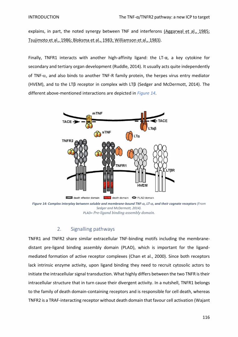
INTRODUCTION The TNF-α/TNFR2 pathway: a new ICP to target
116
explains, in part, the noted synergy between TNF and interferons (Aggarwal et al., 1985;
Tsujimoto et al., 1986; Bloksma et al., 1983; Williamson et al., 1983).
Finally, TNFR1 interacts with another high-affinity ligand: the LT-α, a key cytokine for
secondary and tertiary organ development (Ruddle, 2014). It usually acts quite independently
of TNF-, and also binds to another TNF-R family protein, the herpes virus entry mediator
(HVEM), and to the LTβ receptor in complex with LTβ (Sedger and McDermott, 2014). The
different above-mentioned interactions are depicted in Figure 14.
Figure 14: Complex interplay between soluble and membrane-bound TNF-, LT-, and their cognate receptors (From Sedger and McDermott, 2014).
PLAD= Pre-ligand binding assembly domain.
2. Signalling pathways
TNFR1 and TNFR2 share similar extracellular TNF-binding motifs including the membrane-
distant pre-ligand binding assembly domain (PLAD), which is important for the ligand-
mediated formation of active receptor complexes (Chan et al., 2000). Since both receptors
lack intrinsic enzyme activity, upon ligand binding they need to recruit cytosolic actors to
initiate the intracellular signal transduction. What highly differs between the two TNFR is their
intracellular structure that in turn cause their divergent activity. In a nutshell, TNFR1 belongs
to the family of death domain-containing receptors and is responsible for cell death, whereas
TNFR2 is a TRAF-interacting receptor without death domain that favour cell activation (Wajant
INTRODUCTION The TNF-α/TNFR2 pathway: a new ICP to target
117
et al., 2003). The reality is way more complex for each receptor, added to an interplay that
exist between the two receptors intracellular pathways, reflecting the rich range of TNF
abilities. The signalling outlines are listed below and represented in Figure 15.
TNFR1 death domain preferentially interacts with the adaptor protein named TNFR1-
associated death-domain (TRADD) protein (Dempsey et al., 2003). TRADD further recruits
another two adaptor proteins, receptor-interacting protein kinase 1 (RIPK1) and TNFR-
associated factor (TRAF) 2, thus forming an enzymatic complex signalosome also known as
signalling complex I. One of the main targets of the complex I is the enzyme complex called
IκB kinase (IKK). Phosphorylation of IKK leads in turn to the canonical activation of the
transcription factor NF-κB as well as members of the family of MAP kinases such as JNK and
p38 MAPK (Brinkman et al., 1999; Natoli et al., 1997). The respective target genes of these
proteins allow the organism to respond effectively to environmental changes. The classical
NF-κB pathway is activated by a variety of inflammatory signals, resulting in coordinate
expression of multiple inflammatory and innate immune genes (Liu et al., 2017b). JNK
pathway and p38 signalling are strongly activated by surrounding stress and inflammatory
cytokines. They contribute to a broad range of reaction, including inflammation, apoptosis,
cell activation/migration/differentiation, cell cycle regulation, cytokine production, and
metabolism (Morrison, 2012). Alternatively, the signalling complex I can also be internalized
and then be converted to a death-inducing signalling complex, so-called pro-apoptotic
complex II, by adaptor protein Fas-associated protein with death domain (FADD). The complex
II includes the procaspase 8 which is activated by autocatalytic cleavage, initiating
downstream caspase cascades and ultimately inducing cell apoptosis (Micheau and Tschopp,
2003; Schneider-Brachert et al., 2004). In case caspase 8 is absent or inactivated, kinase-active
RIPK1 recruits and activates RIPK3, resulting in the formation of the necrosome and further
execution of necroptosis via membrane permeabilization (Grootjans et al., 2017; Vanden
Berghe et al., 2014). Caspases and their inhibitors are pivotal elements for deciding the cell’s
fate after TNF-/TNFR1 triggering, alongside with the engagement of NF-κB-mediated anti-
apoptotic signalling pathways, able to delay the time of death (Schliemann M et al., 2011).
INTRODUCTION The TNF-α/TNFR2 pathway: a new ICP to target
118
In contrast to the well-characterized TNFR1 signalling pathways and their physiologic
relevance, TNFR2-mediated signalling pathways and in particular, their role in TNF biology
were uncovered much later. Because of the lack of death domain, TNFR2 is unable to recruit
TRADD protein, but can weakly bound with TRAF2 without intermediary instead (Rothe et al.,
1995). In these conditions, TRAF2 induces the noncanonical NF-κB pathway, through the
activation of the NF-κB-inducing kinase (NIK), which further leads to the phosphorylation of
IKKα and the processing of p100, a crucial step in the nuclear translocation of p52/RelB (Borghi
et al., 2018). Alternative NF-κB pathway acts quite distinctly from the classical one, for
instance by being dispensable for the initial activation of naive T cells through TCR signalling
but crucial for the in vivo generation and maintenance of effector and memory T cells (Sun,
2017). However, upon binding to TRAF2, TNFR2 can also recruit cIAP1/2 proteins that are
involved in the TNFR1-mediated NF-κB activation, creating a crosstalk between TNFR
pathways. TNFR2 pathway activation consumes the cytosolic pool of TRAF2-cIAP1/2 complex,
limiting its availability for other receptors, including TNFR1. Due to the relevance of TRAF2
and cIAPs for preventing apoptosis and necroptosis in the context of TNFR1 signalling, TNFR2-
mediated deprivation of these molecules is able to enhance TNFR1-induced cell death in
macrophages (Siegmund et al., 2016). Lastly, another notable adaptor protein called
endothelial/epithelial protein tyrosine kinase (Etk/BMX) has been described, that can interact
TNFR2 in a ligand-independent manner (Pan et al., 2002). TNFR2-mediated Etk
phosphorylation is able to partially activate the growth factor receptor VEGFR2, resulting in
cell survival and proliferation through PI3K/Akt activation (Fischer et al., 2015; Ortí-Casañ et
al., 2019).
Remarkably, an amplification loop has been stated for TNF- pathway (Zuckerman et al.,
1991). Since TNF- binding to TNFR activates the MAP kinase signalling cascades and
transcription factors of the NF-κB family, it induces its own transcription in cells that exhibit
its receptors (Wajant and Beilhack, 2019). Furthermore, TNFR-binding can induce reverse
signalling into the tmTNF- producing cells (Tartaglia et al., 1993). The signal transduction
results in the activation of NF-κB, this time within the TNF--producing cell, leading again to
more TNF- transcription (Watts et al., 1999; Xin et al., 2006).
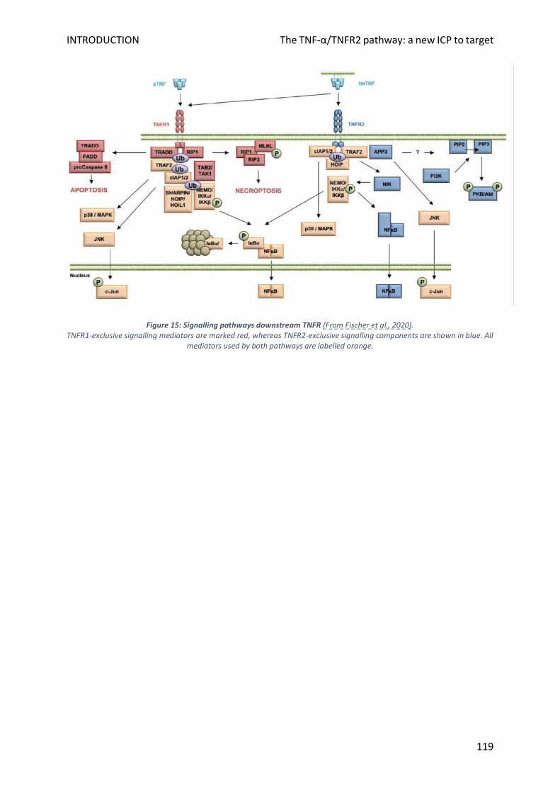
INTRODUCTION The TNF-α/TNFR2 pathway: a new ICP to target
119
Figure 15: Signalling pathways downstream TNFR (From Fischer et al., 2020). TNFR1-exclusive signalling mediators are marked red, whereas TNFR2-exclusive signalling components are shown in blue. All
mediators used by both pathways are labelled orange.
INTRODUCTION The TNF-α/TNFR2 pathway: a new ICP to target
120
C. TNFR2 critical function in tolerance
1. For immune homeostasis maintenance
Described TNF--mediated proinflammatory functions are in majority mediated via TNFR1
expression in a multitude of effector immune cells (Mehta et al., 2018). TNFR2 expression is
predominantly found on myeloid cells, and on activated conventional T cells in which it acts
as a co-stimulatory molecule, in a unique, non-redundant manner compared to CD28 co-
stimulation (Aspalter et al., 2003; Reiner, 2007). TNFR2 deficiency in knockout mice impairs
the proliferative ability of conventional CD4+ and CD8+ cells and decrease their production of
TNF-, IFN-γ and IL‐2 expression in response to TCR stimulation (Kim and Teh, 2001; Kim et
al., 2006, 2008). Intriguingly, TNF- produced by activated Teff under inflammatory conditions
was proved necessary for the stimulation of expansion and the enhancement of Treg
suppressive function (Chen et al., 2007; Grinberg-Bleyer et al., 2010; Leclerc et al., 2016).
TNFR2 is not only essential for optimal Teff proliferation and activation but also for the
induction of activation-induced cell death (AICD) that terminates the proliferative response, a
process dependent on the TNFR2 downstream actor TRAF2 (Twu et al., 2011). Consistently,
TNFR2−/− CD8+ T lymphocytes exhibit high resistance to AICD, leading to a worsened colonic
inflammation (Punit et al., 2015). Thus, TNFR2 signalling is responsible for dual effects in T
cells, since its activation stimulates the effector response set-up while permitting its
subsequent regulation through (i) the death of effector cells and (ii) the stimulation of Treg
subpopulation that also help to terminate the immune reaction.
Although TNFR1 expression is not different between Treg and non-Treg immune cells, human
Treg constitutively express high levels of TNFR2 compared to conventional in a steady-state
(Fischer et al., 2020). Hence, using a target-agnostic phage display screening approach on
human Treg to find antibody mimetics with preferential binding to Treg rather than Teff, has
surprisingly led to only finding candidates that specifically bind to TNFR2 (Williams et al.,
2016). Therefore, in addition to being a Treg marker, several studies conducted by Chen et al.
highlighted that TNFR2 expression by Treg has important functional implications and defines
the maximally suppressive subset of mouse and human Treg (Chen and Oppenheim, 2011a,
2011b; Chen et al., 2008, 2010a, 2010b) In particular, it was shown that the expression level
of TNFR2 is correlated to the suppressive potential of nTreg (Chen et al., 2007), indicating that
INTRODUCTION The TNF-α/TNFR2 pathway: a new ICP to target
121
the most potent suppressors are highly susceptible to TNFR2 activation. These findings have
been confirmed by many other groups since these pioneer findings (Ablamunits et al., 2010;
Atretkhany et al., 2018; Grinberg-Bleyer et al., 2010; Housley et al., 2011; Kleijwegt et al.,
2010; Okubo et al., 2013). Although, if membrane-bound TNFR2 form can be either
immunosuppressive or immunostimulatory depending on the cell type that expresses it, the
function of the soluble form of TNFR2 seems consistently immunosuppressive (van Mierlo et
al., 2008). Activated Treg can release high amounts of sTNFR2, which, at least partially,
represents another immunosuppressive mechanism of Treg. Interestingly, it seems that these
features are not restricted to CD4+ Treg since the most potent CD8+ suppressors are also
characterized by the expression of TNFR2, although exploratory studies are required to
confirm such results about this unrecognized regulatory subset (Ablamunits et al., 2010;
Horwitz et al., 2013).
It is well recognized now that TNFR2 contributes as well to the expansion of nTreg in vitro and
in vivo (Chen et al., 2007, 2008; Chopra et al., 2016; Fischer et al., 2017, 2018, 2019a, 2019b;
Okubo et al., 2013; Padutsch et al., 2019). Interestingly, it has been shown that T cell-specific
TRAF3−/− mice produce twice the normal number of TNFR2-expressing Treg (McPherson et al.,
2012). TRAF3 in T cells is necessary for the classical NF-κB signalling pathway and the
prevention of the alternative pathway. Thus, this observation suggests particular importance
for non-conventional NF-κB pathway in the generation of this highly suppressive regulatory
subset. Recently, TNFR2 ligation has been confirmed to enhance cell proliferation through the
non-canonical NF-κB pathway in human Treg, enhancing IL-2-induced cell proliferation (Wang
et al., 2018c). However, in mice Treg, the activation of p38 MAPK via the classic NF-κB signal
appears to also be important for TNFR2-induced proliferation (He et al., 2018). In addition, a
range of information about TNFR2 role in Treg expansion and phenotype stability emanates
from the field of adoptive Treg cell therapy to help tolerance in the context of autoimmunity,
organ rejection or GvHD. Using a TNFR2 agonistic antibody in standard ex vivo Treg expansion
protocols conferred improved suppressive activity while reducing Treg heterogeneity (Okubo
et al., 2013). Furthermore, using the TNFR2-specific mAb MR2-1 as an agonist, TNFR2
signalling promoted the expansion of low purity MACS-isolated Treg preparations to
homogenous Treg populations, stable under further restimulation (He et al., 2016). These
studies, in accordance with in vitro and in vivo above-mentioned data, demonstrate the role

INTRODUCTION The TNF-α/TNFR2 pathway: a new ICP to target
122
of TNFR2 on Treg potency and encourage its utilization to improve ex vivo Treg expansion
methods for clinical applications.
2. Under inflammatory conditions
Interestingly, TNFR2 polymorphisms can be found in patients with several inflammatory and
auto-immune diseases, cementing the idea of its pivotal link to human immune tolerance. The
substitution of one arginine by methionine in TNFR2 gene at exon 5 (M196R) correlates with
high concentrations of soluble TNFR2 in rheumatoid arthritis and osteoarthritis patients and
is associated with increased susceptibility to develop rheumatoid arthritis (Oregón-Romero et
al., 2006; Song et al., 2014). Similar observations were made for the same polymorphism in
lupus, and for another (587G) in ulcerative colitis (Horiuchi et al., 2007; Pierik et al., 2004).
Regarding TNFR2 function in Treg and conventional T cells in tissue homeostasis, these
observations raise the question of TNFR2 role in the interplay between inflammatory and
regulatory pathways in human immune pathologies.
Ex vivo, TNFR2 activation is able to maintain a stable Treg phenotype. Accordingly, in vivo,
several results have demonstrated that sTNF- preserved or even increased FOXP3 expression
alongside with murine and human Treg suppressive capacity in an inflammatory environment
(Chen et al., 2007; Okubo et al., 2013; Zaragoza et al., 2016). Using a TNFR2 agonist only
prevented the loss of FOXP3 expression, whereas the sustained hypomethylation of the TSDR
locus required both rapamycin and TNFR2 agonist, suggesting that the stabilization of FOXP3
expression in vivo requires both mTOR and NF-κB signalling pathways (He et al., 2016).
However, contradictory data have been published, providing evidence for negative effect of
TNF- on Treg function by reducing FOXP3 expression or enhancing its dephosphorylation,
reversible under anti-TNF- treatment in individuals with rheumatoid arthritis (Nie et al.,
2013; Valencia et al., 2006). Thus, the effect of TNF- on Treg suppressive function during
inflammatory challenge remains controversial. Of particular interest, one study conducted by
Ehrenstein’s group did shed light on the beneficial effect of tmTNF- on Treg numbers during
chronic inflammation. They observed that a therapeutic anti-TNF- mAb (adalimumab),
paradoxically promoted the interaction between monocytes and Treg isolated from patients
with rheumatoid arthritis. Adalimumab bound to tmTNF- on monocytes and unexpectedly
INTRODUCTION The TNF-α/TNFR2 pathway: a new ICP to target
123
enhanced its expression and fixation to TNFR2 expressed on Treg surface, expanding
functional FOXP3+ Treg equipped to suppress Th17 cells (Nguyen and Ehrenstein, 2016). In
the same vein, a recent publication suggested that the expansion and activation of TNFR2-
expressing Treg may be one of the mechanisms by which anti-TNF- agents control
inflammation in rheumatoid arthritis, with an observed rise of TNFR2+ Treg in patients 3
months after treatment (Santinon et al., 2020). Conversely, there were no differences
compared to baseline for spondylarthritis patients. Thus, some therapeutic antibodies
thought to act by simply blocking their target can trigger the immunosuppressive properties
of TNF-, when other molecules may not. Viewed as a whole, these data might suggest that
the role of TNF-/TNFR2 (i) may depend on the pathology considered and the immune actors
involved, (ii) could fluctuate as a function of the pool of sTNF- compared to tmTNF-.
TNFR2’s implication in human diseases spreads beyond autoimmunity, reflecting the
pleiotropic function of its ligand. Activation by TNFR1 results in the allergic inflammatory
responses while TNFR2 plays a role in the immune tolerance to allergens, with a preferential
expression by highly suppressive and replicating Treg (Ahmad et al., 2018). In pathogen-driven
inflammation, Treg TNFR2+ were found to play a major role in CD4+ T-cell impairment during
sepsis, while the inhibition of tmTNF-α shedding by a specific antibody afforded protection
against septic shock (Gaborit et al., 2020; Li et al., 2019). In cardiac function recovery, TNFR2,
not TNFR1, could enhance mesenchymal stem cell-mediated cardiac protection following
acute ischemia (Kelly et al., 2010). Opposing functions for TNFR1 and TNFR2 have been
depicted in many neurological pathologic settings, in inflammatory diseases, infectious or not,
degenerative diseases, and chronic neuropathic pain (Fischer et al., 2020). Next to its immune
regulatory role, TNFR2 critically contributes to neuronal survival and regeneration, in contrast
to TNFR1, which promotes neuronal tissue destruction (Ortí-Casañ et al., 2019). These diverse
pathological settings have in common the central role of TNF- in their physiopathological
mechanisms. Thus, the specific modulation of TNFR2 rather than TNF- modulation has been
proposed by the different up-cited authors, opening a broad range of therapeutic
opportunities for such reagents. Furthermore, the previously unknown protective and anti-
inflammatory effects of TNF- via TNFR2 binding may explain why TNF- inhibitors failed to
be effective in diseases such as heart failure or multiple sclerosis, where TNF- has been
INTRODUCTION The TNF-α/TNFR2 pathway: a new ICP to target
124
strongly implicated as a driving force (Levine et al., 1990; The Lenercept Multiple Sclerosis
Study Group, 2011).
Remarkably, CD4+ and CD8+ are not the only populations to benefit from TNFR2 signalling to
enhance their suppressive function. Under inflammatory conditions MDSC accumulation and
suppressive function have been described as dependent of their TNFR2 expression and its
signalling through tmTNF- binding (Ham et al., 2015; Hu et al., 2014; Polz et al., 2014; Zhao
et al., 2012). Lastly, the TNF-/TNFR2 signalling pathway has just been shown to be a key
regulatory factor for mesenchymal stem cell, neural and endothelial progenitor cell
immunosuppressive effect (Beldi et al., 2020; Naserian et al., 2020; Shamdani et al., 2020).
Altogether, these observations promote the existence of a TNFR2-dependant network of
immunosuppressive cells ignored until today, raising the hope of progressively unraveling
apparent inconsistencies surrounding TNF- since its discovery.
INTRODUCTION The TNF-α/TNFR2 pathway: a new ICP to target
125
D. TNF- implication in alloHSCT
TNF- plays a pivotal role in all the steps after alloHSCT, from the initial massive release of
this cytokine by host macrophages right away after the conditioning therapy, to a potential
GvHD occurrence (Markey et al., 2013; Xun et al., 1994). In 1987, based on the notion that
excessive release of pro-inflammatory cytokines could cause GvHD, a polyclonal anti-TNF-
antibody was tested in a mouse model and happened to reduce mortality (Clark et al., 2004;
Piguet et al., 1987). Indeed, during the effector phase, TNF- is able to provide costimulatory
signals to naïve CD4+ and CD8+ T lymphocytes through TNFR2 (Aspalter et al., 2003; Kafrouni
et al., 2003; Kim and Teh, 2001). This cytokine together with IL-1β also enhances TNF-
expression by freshly activated alloreactive T cells constituting a feed-forward-loop of TNF-
release (Hill et al., 1999a; Holler et al., 1990, 1993). After their migration toward GvHD target
tissues, infiltrating donor T cells also release TNF- upon allorecognition, causing epithelial
damage predominantly in the gastrointestinal tract and skin (Bäuerlein et al., 2013; Beilhack
et al., 2005, 2008; Chakraverty et al., 2006; Hattori et al., 1998; Paris et al., 2001; Piguet et al.,
1987). Interestingly, intestinal epithelial cells are particularly susceptible to TNF--induced
apoptosis due to their expression of a tissue damage-induced receptor of the TNFR family
(Fn14), providing a hypothesis to explain why the intestinal tract is a primary target for GvHD
tissue damage (Chopra et al., 2015).
Genetic polymorphisms in the TNF- cytokine gene and TNFR genes, in both donor and host,
have been studied in clinical populations. Results of these studies confirm that high donor
TNF- production and increased recipient responses to this cytokine via receptor signalling
contribute to GvHD risk (Markey et al., 2013; Wilson et al., 1997). The importance of the TNF-
signalling pathway has also been confirmed clinically by the strong correlation between the
amount of systemic TNF- release (>100 pg/mL) in the first 3 months after alloHSCT and
aGvHD occurrence, as well as other complications (Holler et al., 1990, 1993). Furthermore a
strong increase of soluble TNFR1, 7 days after alloHSCT, correlated with GvHD incidence,
severity and patient survival (Choi et al., 2008; Kitko et al., 2008). These results lead to the
integration of sTNFR1, together with IL-2-receptor-alpha, IL-8, and hepatocyte growth factor,
into a proposed serum biomarker panel for GvHD diagnosis and prediction of survival
(Paczesny et al., 2009).
INTRODUCTION The TNF-α/TNFR2 pathway: a new ICP to target
126
Based on these data, several clinical studies were initiated to test TNF- inhibitors for the
treatment of aGvHD or to prevent the onset of aGvHD. Infliximab, a TNF- blocking antibody
failed in clinical trials, in both approaches, and might increase bacterial and fungal infections
(Couriel et al., 2004; Hamadani et al., 2008). Etanercept, an Fc fusion protein of the TNFR2
ectodomain, in combination with high-dose steroids showed initially promising response rates
in GvHD patients, but no improved OS or prophylactic benefits (Busca et al., 2007; Choi et al.,
2012b; Park et al., 2014). In paediatric steroid-resistant aGvHD patients, a recent non-
randomized phase II trial suggested a high response rate and improved OS using etanercept,
with better effects obtained in cutaneous and gastrointestinal aGvHD, accordingly to TNF-
tissular pattern after alloHSCT (Faraci et al., 2019).
If the detrimental effects of TNF- on GvHD pathogenesis provided a clear rationale to test
TNF- inhibitors in alloHSCT, for now, this strategy has not met all expectations. Furthermore,
TNF- blockade prevented aGvHD in several mouse models but may also affect the GVL effect.
Indeed, TNFR1-deficient donor CD8 T cells resulted in an increased leukaemia relapse after
alloHSCT (Hill et al., 1999b; Piguet et al., 1987; Teshima et al., 2002; Xun et al., 1994).
Additionally, it has become clear only very recently that TNF- also triggers anti-inflammatory
feedback loops by stimulation of expansion and function of tolerogenic TNFR2+ cells.
Interestingly, the same polymorphism found in rheumatoid arthritis and lupus patients,
M196R, when detected in alloHSCT donors is correlated with an increased incidence of severe
GvHD (Ishikawa et al., 2002). Thus, in light of the refined knowledge about TNF- opposite
role in the immune system, fresh pre-clinical studies have tested the option to preferentially
target TNFR2 pathway in the control of GVHD. The idea here is to activate specifically TNFR2
in vivo to induce tolerance via Treg activation after alloHSCT, without triggering TNFR1-
associated cell death, as an alternative to cell therapy approaches using tedious ex vivo Treg
GMP-expansion procedures.
First, Chopra et. al reported the development of a novel strategy for inhibiting GvHD based on
expanding recipient Treg in vivo before alloHSCT using a selective TNFR2 agonist (Chopra et
al., 2016). In this model, they observed a significant reduction in GvHD severity and
subsequent survival prolongation. Importantly, the use of the TNFR2 agonist had no
detrimental effect on the ability of donor T cells present in the graft to mediate GVT or to
INTRODUCTION The TNF-α/TNFR2 pathway: a new ICP to target
127
eliminate pathogens. In a second study, the authors demonstrated that the activation and
suppressive activity of Treg were increased in mice undergoing aGvHD, in correlation with the
high levels of TNF-α present at the same time in the serum (Pierini et al., 2016). TNF-α while
inducing Treg proliferation in vivo also limited the capacity of conventional CD4+ and CD8+ T
cells to proliferate and differentiate, thus reducing their capacity to trigger GvHD. The last
study also supported the concept that Treg are dependent on TNF- and TNFR2 for their
ability to control GvHD. Indeed, blocking the TNF-/TNFR2 pathway increased the clinical
GvHD score, GvHD-related mortality, led to a gain of activation in donor T cells and a down-
modulation of FOXP3 and activation markers expression on Treg (Leclerc et al., 2016).
Collectively, these results conclusively demonstrate that Treg control of GVHD relies on (i)
TNF- produced by effector T cells and (ii) expression of TNFR2 by Treg. This data provides a
strong beam of arguments to propose that TNFR2 activation in GvHD patients may be more
beneficial than current anti-TNF- strategies. Fundamentally, these findings also expand the
understanding of Treg action, by delineating a negative feedback loop that matches the
magnitude of Treg action to donor T cell activity in alloHSCT conditions.
INTRODUCTION The TNF-α/TNFR2 pathway: a new ICP to target
128
E. TNF-α/TNFR2 role in malignancies
1. In the TME
If tolerogenic cells do not have a monopoly on TNFR2 expression in the steady-state, quite a
few studies suggest that TNFR2+ Treg with high suppressive capacities are strongly
represented in cancer patients TME, and sometimes peripheral blood, comparably to mice
models’ findings (Chen et al., 2008). The first remarkable observation about TNFR2 in the
context of cancer is the decrease in tumour growth and metastasis described in TNFR2
knockout mice, despite TNFR2 costimulatory function on Teff (Chopra et al., 2013; Ham et al.,
2015; Sasi et al., 2012). TNFR2‐deficient mice had reduced infiltration and induction of MDSC
coinciding with a diminution in Treg number inside the tumours, confirming TNFR2’s
requirement for these tolerogenic cells participation in the TME (Zhao et al., 2012).
Furthermore, if the beneficial effects of TNF- on CD8+ Teff are mainly mediated through
TNFR2 (Calzascia et al., 2007; Zheng et al., 1995), Chen et al. have shown that the upregulation
of TNFR2 on intratumoral Treg enable them to overcome the greater resistance to suppression
of intratumoral TNFR2+ conventional T cells (Chen et al., 2010b). Altogether these data put
forward the idea of a diverted TNFR2 role in tumours, to the profit of a dominant
immunosuppressive TME, notwithstanding its activation/anti-apoptotic function relevance on
T cells in a steady-state.
Regarding cancer patients, there is now compelling evidence that TNFR2+ Treg accumulate in
TIL (for Sézary Syndrome and cervical cancer) and tumour ascites in ovarian cancer. This
population is also increased in AML patients’ peripheral blood (PB) as well as in those from
hepatocellular carcinoma, lung and cervical cancer patients (He et al., 2019a). Remarkably, in
peripheral blood samples from lung cancer patients where TNFR2+ Treg were also detected,
the expression of TNFR2 appeared to correlate better than CD25 expression and CD127
absence with FOXP3 expression (Yan et al., 2015). Additionally, TNFR2+ Treg were associated
with lymphatic invasion, distant metastasis and more advanced clinical stage of lung cancer
patients. This observation and others highlight the pivotal role of TNFR2 expression in Treg in
the context of human cancer, reaching mice model conclusions. An updated list of the
different studies available at this subject, in mice and humans, and their outlines are provided
in Table 6. About TNFR2+ MDSC, their presence has been noted in plasmacytoma,
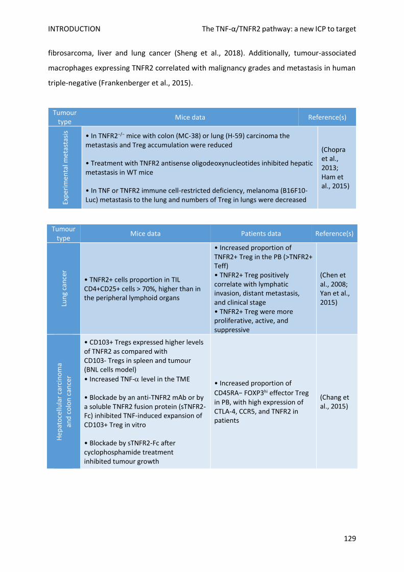
INTRODUCTION The TNF-α/TNFR2 pathway: a new ICP to target
129
fibrosarcoma, liver and lung cancer (Sheng et al., 2018). Additionally, tumour-associated
macrophages expressing TNFR2 correlated with malignancy grades and metastasis in human
triple-negative (Frankenberger et al., 2015).
Tumour type
Mice data Reference(s)
Exp
erim
enta
l met
asta
sis
• In TNFR2−/− mice with colon (MC-38) or lung (H-59) carcinoma the metastasis and Treg accumulation were reduced • Treatment with TNFR2 antisense oligodeoxynucleotides inhibited hepatic metastasis in WT mice • In TNF or TNFR2 immune cell-restricted deficiency, melanoma (B16F10-Luc) metastasis to the lung and numbers of Treg in lungs were decreased
(Chopra et al., 2013; Ham et al., 2015)
Tumour type
Mice data Patients data Reference(s)
Lun
g ca
nce
r
• TNFR2+ cells proportion in TIL CD4+CD25+ cells > 70%, higher than in the peripheral lymphoid organs
• Increased proportion of TNFR2+ Treg in the PB (>TNFR2+ Teff) • TNFR2+ Treg positively correlate with lymphatic invasion, distant metastasis, and clinical stage • TNFR2+ Treg were more proliferative, active, and suppressive
(Chen et al., 2008; Yan et al., 2015)
Hep
ato
cellu
lar
carc
ino
ma
an
d c
olo
n c
ance
r
• CD103+ Tregs expressed higher levels of TNFR2 as compared with CD103- Tregs in spleen and tumour (BNL cells model)
• Increased TNF- level in the TME • Blockade by an anti-TNFR2 mAb or by a soluble TNFR2 fusion protein (sTNFR2-Fc) inhibited TNF-induced expansion of CD103+ Treg in vitro • Blockade by sTNFR2-Fc after cyclophosphamide treatment inhibited tumour growth
• Increased proportion of
CD45RA− FOXP3hi effector Treg in PB, with high expression of CTLA-4, CCR5, and TNFR2 in patients
(Chang et al., 2015)
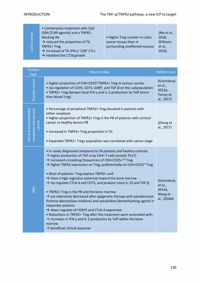
INTRODUCTION The TNF-α/TNFR2 pathway: a new ICP to target
130
Co
lon
can
cer
• Combination treatment with CpG ODN (TLR9 agonist) and a TNFR2-blocking Ab:
reduced the proportion of TIL TNFR2+ Treg
increased of TIL IFN-γ+ CD8+ CTLs inhibited the CT26 growth
• Higher Treg number in colon cancer tissues than in surrounding unaffected mucosa
(Nie et al., 2018; Williams et al., 2016)
Tumour type
Patients data Reference(s)
Ova
rian
can
cer
• Higher proportion of CD4+CD25hiTNFR2+ Treg in tumour ascites • Up-regulation of CD39, CD73, GARP, and TGF-β on this subpopulation • TNFR2+ Treg dampen local IFN-γ and IL-2 production by Teff (more than blood Treg)
(Govindaraj et al., 2013a; Torrey et al., 2017)
Cer
vica
l in
trae
pit
hel
ial
neo
pla
sia
and
cer
vica
l ca
nce
r
• Percentage of peripheral TNFR2+ Treg elevated in patients with either neoplasm • Higher proportion of TNFR2+ Treg in the PB of patients with cervical cancer vs healthy donors PB • Increased in TNFR2+ Treg proportion in TIL
• Expanded TNFR2+ Tregs population was correlated with cancer stage
(Zhang et al., 2017)
AM
L
• In newly diagnosed compared to CR patients and healthy controls: higher production of TNF-α by CD4+ T cells (mostly Th17) increased circulating frequencies of CD4+CD25+/hi Treg higher TNFR2 expression on Treg, preferentially on CD4+CD25hi Treg
• Most of patients’ Treg express TNFR2+ and:
Have a high migration potential toward the bone marrow Up-regulate CTLA-4 and CD73, and produce more IL-10 and TGF-β
• TNFR2+ Treg in the PB and the bone marrow
are selectively decreased after epigenetic therapy with panobinostat (histone deacetylase inhibitor) and azacytidine (demethylating agent) in responder patients
down-regulate of FOXP3 and CTLA-4 expression • Reductions in TNFR2+ Treg after this treatment were associated with:
increases in IFN-γ and IL-2 production by Teff within the bone marrow
beneficial clinical response
(Govindaraj et al., 2014a; Wang et al., 2018d)
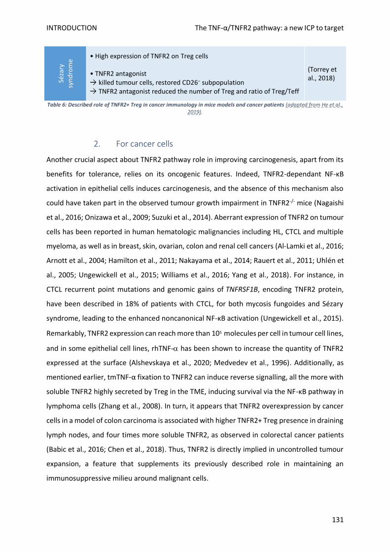
INTRODUCTION The TNF-α/TNFR2 pathway: a new ICP to target
131
Séza
ry
syn
dro
me
• High expression of TNFR2 on Treg cells • TNFR2 antagonist
killed tumour cells, restored CD26− subpopulation TNFR2 antagonist reduced the number of Treg and ratio of Treg/Teff
(Torrey et al., 2018)
Table 6: Described role of TNFR2+ Treg in cancer immunology in mice models and cancer patients (adapted from He et al., 2019).
2. For cancer cells
Another crucial aspect about TNFR2 pathway role in improving carcinogenesis, apart from its
benefits for tolerance, relies on its oncogenic features. Indeed, TNFR2-dependant NF-κB
activation in epithelial cells induces carcinogenesis, and the absence of this mechanism also
could have taken part in the observed tumour growth impairment in TNFR2-/- mice (Nagaishi
et al., 2016; Onizawa et al., 2009; Suzuki et al., 2014). Aberrant expression of TNFR2 on tumour
cells has been reported in human hematologic malignancies including HL, CTCL and multiple
myeloma, as well as in breast, skin, ovarian, colon and renal cell cancers (Al-Lamki et al., 2016;
Arnott et al., 2004; Hamilton et al., 2011; Nakayama et al., 2014; Rauert et al., 2011; Uhlén et
al., 2005; Ungewickell et al., 2015; Williams et al., 2016; Yang et al., 2018). For instance, in
CTCL recurrent point mutations and genomic gains of TNFRSF1B, encoding TNFR2 protein,
have been described in 18% of patients with CTCL, for both mycosis fungoides and Sézary
syndrome, leading to the enhanced noncanonical NF-κB activation (Ungewickell et al., 2015).
Remarkably, TNFR2 expression can reach more than 105 molecules per cell in tumour cell lines,
and in some epithelial cell lines, rhTNF- has been shown to increase the quantity of TNFR2
expressed at the surface (Alshevskaya et al., 2020; Medvedev et al., 1996). Additionally, as
mentioned earlier, tmTNF-α fixation to TNFR2 can induce reverse signalling, all the more with
soluble TNFR2 highly secreted by Treg in the TME, inducing survival via the NF-κB pathway in
lymphoma cells (Zhang et al., 2008). In turn, it appears that TNFR2 overexpression by cancer
cells in a model of colon carcinoma is associated with higher TNFR2+ Treg presence in draining
lymph nodes, and four times more soluble TNFR2, as observed in colorectal cancer patients
(Babic et al., 2016; Chen et al., 2018). Thus, TNFR2 is directly implied in uncontrolled tumour
expansion, a feature that supplements its previously described role in maintaining an
immunosuppressive milieu around malignant cells.
INTRODUCTION The TNF-α/TNFR2 pathway: a new ICP to target
132
F. Specific TNFR2 pathway blockade in cancer
1. Rationale
Although the qualification of “tumour necrosis” has withstood the test of time, the reality is
less straightforward. Due to its plethora of functions, TNF- through its two receptors is
responsible for divergent actions in the context of cancer (Montfort et al., 2019). If its function
through TNFR1 effectively favours cell death occurrence in cancer cells while promoting T cells
pro-inflammatory response via NF-ⲕB signalling, TNFR2 activation on tolerogenic cells
recruited by the tumour could be detrimental for anti-cancer responses. Taking in account this
refined view of TNF- functions, several studies have focused on sensitizing cancer cells to
TNFR1-induced apoptosis, for instance by inhibiting survival signals such as NF-κB in combined
therapy with TNF- (Wang and Lin, 2008). However, these approaches are, as the historical
attempts to use TNF-, not specific to cancer cells, with a high risk of off-target. A more
specific approach that emerges is to block TNFR2 to focus the therapy on immunosuppressive
cells that accumulate during carcinogenesis, hoping to (i) get rid of the detrimental tolerogenic
TME, including infiltrated TNFR2+ Treg to consequently awake the anti-tumour response, (ii)
while redirecting TNF- to TNFR1 expressed at the surface of immune effector cells,
promoting the inflammatory response. Furthermore, many tumours appear to start
expressing TNFR2 during their transformation process or originate from cells that express it in
the case of immune cells-derived neoplasm, offering a chance to directly impair tumour
evolution by blocking an oncogene. Finally, arguments exist for this strategy to be applied in
both solid cancers and haematological malignancies in first-line therapy, as well as after
allograft failure to cure the patient, a setting of particular interest because of the central TNF-
role after that procedure.
2. Pioneer approaches
Based on these compelling assertions, several studies have addressed the feasibility of
therapeutic TNFR2 blockade in animal cancer models (summarized in Table 6). Faustman’s
group has developed two dominant hTNFR2 antagonist mAb, which lock TNFR2 receptor in
the form of antiparallel dimmers, preventing further TNF- binding (Torrey et al., 2017).
Consequently, using these compounds, even in the presence of TNF-, could kill patients’ Treg
isolated from ovarian cancer ascites more potently than those from healthy donors,
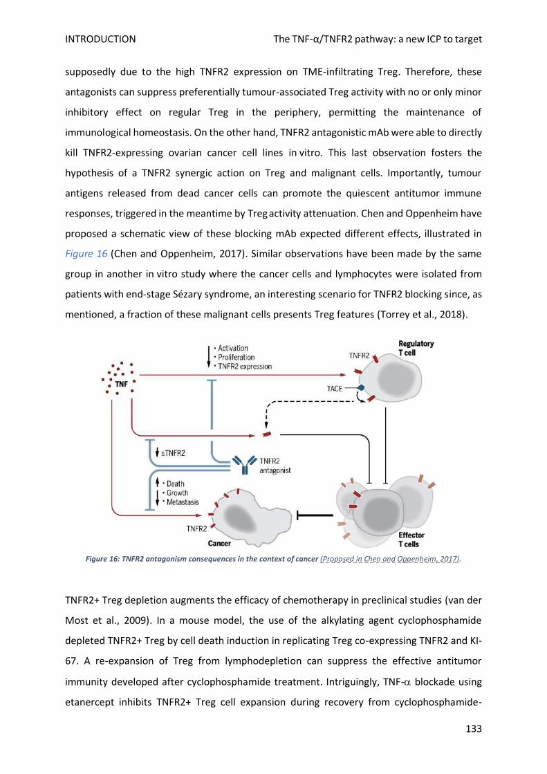
INTRODUCTION The TNF-α/TNFR2 pathway: a new ICP to target
133
supposedly due to the high TNFR2 expression on TME-infiltrating Treg. Therefore, these
antagonists can suppress preferentially tumour-associated Treg activity with no or only minor
inhibitory effect on regular Treg in the periphery, permitting the maintenance of
immunological homeostasis. On the other hand, TNFR2 antagonistic mAb were able to directly
kill TNFR2-expressing ovarian cancer cell lines in vitro. This last observation fosters the
hypothesis of a TNFR2 synergic action on Treg and malignant cells. Importantly, tumour
antigens released from dead cancer cells can promote the quiescent antitumor immune
responses, triggered in the meantime by Treg activity attenuation. Chen and Oppenheim have
proposed a schematic view of these blocking mAb expected different effects, illustrated in
Figure 16 (Chen and Oppenheim, 2017). Similar observations have been made by the same
group in another in vitro study where the cancer cells and lymphocytes were isolated from
patients with end-stage Sézary syndrome, an interesting scenario for TNFR2 blocking since, as
mentioned, a fraction of these malignant cells presents Treg features (Torrey et al., 2018).
Figure 16: TNFR2 antagonism consequences in the context of cancer (Proposed in Chen and Oppenheim, 2017).
TNFR2+ Treg depletion augments the efficacy of chemotherapy in preclinical studies (van der
Most et al., 2009). In a mouse model, the use of the alkylating agent cyclophosphamide
depleted TNFR2+ Treg by cell death induction in replicating Treg co-expressing TNFR2 and KI-
67. A re-expansion of Treg from lymphodepletion can suppress the effective antitumor
immunity developed after cyclophosphamide treatment. Intriguingly, TNF- blockade using
etanercept inhibits TNFR2+ Treg cell expansion during recovery from cyclophosphamide-
INTRODUCTION The TNF-α/TNFR2 pathway: a new ICP to target
134
induced lymphodepletion and markedly inhibits the growth of established CT26 tumours in
mice, without affecting CD8+ T cell activation (Chang et al., 2015). In the same colon cancer
model, as well as in 4T1 breast cancers, the combination of a TNFR2-blocking mAb with an
immune stimulator (toll-like receptor agonist) markedly enhanced the antitumor efficacy of
immunotherapy through reducing the number of tumour-infiltrating TNFR2+ Treg and
increasing the number of IFN-γ-producing CD8+ cells (Nie et al., 2018). Notably, the
antagonistic TNFR2 antibody TR75-54.7 inhibited the growth of mammary carcinoma more
efficiently than a CD25 antagonist. In addition, some pharmacological agents regulate the
expression of TNF- and/or of its receptors. For instance, thalidomide and its analogues
prevent the surface expression of TNFR2 on activated T cells, which is associated with the
inhibition of TNFR2 protein trafficking to the cell membrane (Marriott et al., 2002). Treating
AML patients with azacitidine and lenalidomide, a thalidomide derivative can reduce TNFR2
expression on T cells as well as TNFR2+ Treg in vivo, leading to enhanced effector immune
function (Govindaraj et al., 2014b). Moreover, the need for new blocking molecules usable in
patients may be fulfilled soon since the TNF-/TNFR2 crystal structure is now available (Mukai
et al., 2010), revealing the specific binding pattern between TNF- and TNFR2. In virtual
screening, a library of compounds has been examined to predict their binding poses and
affinities (Shaikh et al., 2018). Compounds that resemble the binding pose to the native ligand
with better binding affinity will be selected as candidates, together with blocking mAb
candidates, for further research and development in the drug discovery pipeline.
Altogether these preliminary results prove in a convincing way the benefits and apparent
safety of TNFR2 blockade in cancer. Although, a potential safety concern emanates from the
inducible expression on Teff upon TCR stimulation. Also, costimulation through TNFR2 on Teff
could empower their ability to resist Treg-mediated suppression in tumours. Nevertheless,
since Treg in the TME seems to persistently express higher levels of TNFR2 than Teff, the
assumption that this treatment should have a more profound impact on Treg than on Teff
sounds realistic. Then, the net outcome of TNFR2 antagonism could favour Teff activation and
expansion, triggering the set-up of an effective antitumor immune response. This hypothesis
will require further testing in a suitable mouse tumour model before future studies in human
patients, with a careful watch on TNFR2 inhibition impact on effector response at each step.

133
Thesis
objectives
136
The modern era of anti-cancer strategies has seen a change of paradigm, with a drastic target
evolution, from killing the tumour cells to enabling the patient’s immune system to do so.
Today, immunotherapy is sparking an extraordinary level of energy and enthusiasm among
the scientific community and healthcare industry due to its potential outstanding effect in
solid and hematologic malignancies, including post-alloHSCT relapses. In this last case, the
historic and still relevant immunotherapeutic approach, DLI, is directly centred on the boost
of effector alloimmune response. Similarly, recently approved blinatumomab, ICP mAb, or
adoptive CAR-T cells approach were developed in the perspective of enabling patient’s Teff to
recognize and eradicate malignant cells. An alternative view emerging for a decade, proposes
to tackle another immune brake, by targeting Treg suppressive action on the anti-tumoral
response. A myriad of potential therapeutic approaches have been described in animal
models or are currently being assessed in clinical trials, with limited success for now.
In 2010 our laboratory successfully completed the first clinical trial relying on ex-vivo Treg
removal from DLI to trigger a powerful GVT effect in relapsing patients post-alloHSCT (Maury
et al., 2010, 2014). These results have served as a foundation stone to justify the use of anti-
Treg therapies to boost the alloimmune response to the tumour. In this context, TNF-/TNFR2
signalling pathway is one promising candidate due to (i) TNFR2 critical role on Treg phenotypic
stabilization and suppressive capacity and (ii) TNF- abundancy at each stage of the allogeneic
immune response in the context of alloHSCT (Chen et al., 2008, 2010a, 2013; Clark, 2007;
Grinberg-Bleyer et al., 2010; Okubo et al., 2016; Zaragoza et al., 2016). Our team previously
revealed that blocking the TNFR2 pathway led to a complete loss of Treg protective function
in a model of GvHD prevention by Treg-based cell therapy (Leclerc et al., 2016). Meanwhile,
other groups have demonstrated that TNFR2 activation could conversely reduce the immune
response to control life-threatening GvHD (Chopra et al., 2016; Pierini et al., 2016).
If these works have then established the proof-of-principle that TNFR2 modulation in vivo is a
good target in the context of alloHSCT, no demonstration existed yet of its potency to bolster
anti-tumour response in vivo. Thus, this has been the global purpose of this project,
fractionated in three complementary and successive objectives:
137
1) To develop a pre-clinical model that mimics a post-alloHSCT relapse.
2) To establish in this setting if the mAb-mediated blockade of TNFR2 could protect the
treated-mice from developing a haematological malignancy.
3) To gain finer comprehension, in vivo, of the impact of the treatment on (i) the organs
targeted by alloreactivity (ii) the immune system components from the donor and the
recipient, with special emphasis on effector and regulatory T cells.
In a distinct axis, efforts were made to transpose these results to human cells, both in vitro
and using diverse humanized mice models. The preliminary unpublished data obtained on this
subject are presented in the perspective section of this manuscript, alongside with others
required key steps that will lead toward the development of clinical-grade TNFR2-targeted
therapies.

138
Results
RESULTS
139
“TNFR2 blockade on regulatory T-cells unleashes anti-tumor immune response after
hematopoietic stem cell transplantation.”
Manuscript under evaluation in Blood.
AlloHSCT remains the most curative option for refractory and severe haematological
malignancies. In around half of the grafted patients, this heavy therapy is still insufficient to
avoid a relapse from their underlying malignancy, with an often worsened aggressivity. Until
now, only a narrow range of therapeutic tools has proved useful in this situation, DLI being
the main recourse.
In this study, we took advantage of the well-established knowledge that Treg are a pivotal
brake for the anti-tumour response, including in alloreactive conditions where they can lessen
DLI efficacy. To block their activity and trigger a potent alloreactivity to eradicate malignant
cells, we relied on the high expression of TNFR2 described on Treg, a beneficial actor for Treg
function in the steady-state and various inflammatory conditions that involve TNF-. Using a
mouse model to mimic the lack of Teff efficacy in relapsing patients, we established the proof
of principle that blocking the TNFR2 signalling pathway leads to tangible tumoricidal action,
associated with a heightened Teff/Treg ratio in comparison to untreated animals. This clinical
benefit presumably emanates from the downregulation of FOXP3 expression and other
activation markers in CD4+ Treg naturally present within the graft. We also made the first
demonstration, in our knowledge, of a similar destabilisation of CD8+ Treg suppressive
phenotype after TNFR2 blockade, suggesting a dual interest for the anti-TNFR2 treatment on
tolerogenic populations implied in the relapse.
In summary, this article offers new avenues for Treg-based immunomodulation through TNF-
/TNFR2 interaction that represent a novel anti-ICP therapy to set off a potent GVL effect
after alloHSCT. Our results represent further increments to the actual translational research
landscape that promotes TNFR2 modulation in accordance to the clinical context, including
possible application in first-line therapy for various types of cancers.
RESULTS
140
TNFR2 blockade on regulatory T-cells unleashes anti-tumor immune response after
hematopoietic stem cell transplantation
Audrey Moatti,1 Anaïs Debesset,1 Caroline Pilon,1,2 Asma Beldi-Ferchiou,1,3 Mathieu
Leclerc,1,4 Frédéric Charlotte,5 Pauline Cadot6, Sébastien Maury,1,2,4 Allan Thiolat,1* José L
Cohen1,2*
1. Univ Paris Est Créteil, INSERM, IMRB, F-94010 Créteil, France ;
2. AP-HP, Groupe hospitalo-universitaire Chenevier Mondor, Centre d’investigation clinique
Biothérapie, F-94010 Creteil, France ;
3. AP-HP, Groupe hospitalo-universitaire Chenevier Mondor, Service d’immunologie
biologique, F-94010 Creteil, France ;
4. AP-HP, Groupe hospitalo-universitaire Chenevier Mondor, Service d’hématologie Clinique,
F-94010 Creteil, France ;
5. AP-HP, GHU Pitié Salpêtrière, Service d’anatomopathologie, F-75013, Paris, France ;
6. AP-HP, Groupe hospitalo-universitaire Chenevier Mondor, Service d’oncologie-
radiothérapie, F-94010 Creteil, France.
* AT and JLC co-senior authors
Correspondence : José L. Cohen: Hôpital Henri Mondor, Centre d’investigation clinique
Biotherapie, 53 Av du Mal de Lattre de Tassigny, 94010 Créteil France.
Tel: ++ 33(0) 1 49 81 44 75; e-mail: [email protected]
Running title: Anti-TNFR2 treatment induces GVT effect
Word count: 1195 words
RESULTS
141
Key points
1. TNF/TNFR2 interaction represents a novel immune checkpoint therapy to modulate
alloreactivity after alloSCT
2. Blocking TNFR2 pathway increases GVL effect of insufficient effector donor T-cells
RESULTS
142
Abstract
Targeting immune checkpoints that inhibit anti-tumor immune responses is emerging as a
new solid therapeutic approach in cancer. We recently revealed that blocking TNFR2 pathway
induces a complete loss of regulatory T-cells (Treg) protective function in a model of graft-
versus-host disease prevention relying on Treg-based cell therapy. Here, we tested the
possibility of amplifying anti-tumor response by targeting TNFR2 in a model of tumor relapse
following hematopoietic stem cell transplantation. This corresponds to a clinical situation
where the different existing therapeutic options have low efficacy. In experimental conditions
in which neither donor T-lymphocytes nor TNFR2-blocking antibody per se have any effect on
tumor relapse separately, we observed that the co-administration of a sub-optimal number
of T-cells associated with anti-TNFR2 treatment could set off alloreactivity and subsequently
induce a significant anti-tumor effect. This was associated with a reduced Treg/T-cell ratio and
a reduced percentage of activated CD4 and CD8 Tregs. These results highlight TNFR2 as new
target molecule for the development of immunotherapies to treat blood malignancy relapse.
They also open the door for new perspectives on the possibility of amplifying, more widely in
solid cancer, anti-tumor responses by directly targeting Tregs through TNFR2.
RESULTS
143
Introduction
In allogeneic hematopoietic stem cells transplantation (alloSCT), it is now clear that regulatory
T-cells (Treg) have a key role in the fine tuning of immune responses1-3. Cell therapy using Treg
infusions to prevent graft-versus-host disease (GVHD) showed very promising results in
clinics4-7. Converselly, in 2010, we initially performed an ex vivo Treg depletion from donor
lymphocyte infusions (DLI) in order to enhance the graft-versus-leukemia/graft-versus-tumor
(GVL/GVT) effect in patients who relapsed after alloSCT8,9. The initial clinical trial suggested
that Treg depletion could set off alloreactivity and its associated GVL/GVT effect. A
confirmatory, randomized, double-blind trial, following the same approach, is currently
ongoing (DLI-Boost, ClinicalTrials.gov, NCT03236129).
It was previously demonstrated that Tumor Necrosis Factor Receptor-type 2 (TNFR2) was
critical to promote human Treg stability and suppressive functions10,11. Alike, we have recently
revealed, for the first time, complete Treg dependency on TNF to maintain suppressive
capacity in vivo. The protective effect of therapeutic Tregs against GVHD was completely
abolished when mice were treated with TNFR2-blocking mAb at time of alloSCT. We
conclusively demonstrated in the formentioned setting that disease control by Treg depends
on TNF produced by donor T-cells, and on expression of TNFR2 on injected therapeutic Treg12.
Herein, we hypothesized that blocking TNF/TNFR2 pathway would give space for a powerful
and amplified GVL/GVT effect to emerge, which could be useful to treat blood malignancy
relapse after alloSCT. For this, we have developed a dedicated experimental model in mice in
which the number of donor T-cells was insufficient to mediate a GVL/GVT effect. The in-vivo
administration of anti-TNFR2 treatment set off strong alloreactivity associated with a potent
GVT effect.
RESULTS
144
Methods
Post-SCT tumor relapse models and analyses
All the GVHD and tumor relapse experiments were adapted from protocols previously
published13,14 and precisely described in the supplemental method section. GVHD clinical
scoring was established as recently described15. Briefly, 500 μg of TNFR2 blocking mAb (TR75-
54.7 Bio X Cell, West Lebanon, New Hampshire, USA) was administered IP to grafted mice on
days 0, 2, and 4, or alternatively day 10. Mice were sacrificed at day 12 to perform
histopathological examination of target organs of GVHD, and spleens were collected for
immune cell analysis.
RESULTS
145
Results and Discussion In the first series of experiments, we tried to define the maximum number of tumor cells and
T-cells needed to be infused in recipient mice in order to create a model of tumor relapse in
the absence of GVHD. We fixed these numbers at 2x10⁴ for P815 cells and 106 for T-cells, and
thus defined the appropriate experimental conditions mimicking those of patients who had
relapse of their hematologic malignancies following alloSCT (Figure 1A). Mice receiving 106 T-
cells and treated with anti-TNFR2 at D0, D2, and D4 developed GVHD as evidenced by low
survival rate at day 40 and elevated clinical grade of GVHD. In untreated mice, no signs of the
disease were ever detected (Figure 1B). In order to validate our hypothesis, we reproduced
these experiments in the presence of P815 cells. Here again, anti-TNFR2 administered at D0,
D2 and 4, or alternately given only at 10, resulted in clinical GVHD manifestations, whereas no
GVHD signs were observed in untreated mice (Figure 1C). P815 cells were detected in all mice
which had no T-cells infusion and in 12 out of 19 (64%) 106 T-cells-infused mice. In contrast,
administering anti-TNFR2 at D0 or at D10 resulted in a dramatic decrease in tumor incidence
since P815 cells were detected in 4 out of 20 (20%) and in 1 out of 8 (12.5%) mice, respectively.
Our previous work demonstrated the possibility to deactivate therapeutic Tregs, given in high
doses in a model of GVHD prevention, by in vivo anti-TNFR2 treatment12. In this study, we
have demonstrated that anti-TNFR2 treatment allows to directly set off alloreactivity in a
context where infused T-cells alone are not able to. We also put forwards the proof of concept
that anti-TNFR2 treatment can mediate a potent GVL/GVT effect in an appropriate
experimental model of hematological malignancy relapse after alloSCT. This therapeutic
approach is not only as effective as but also much simpler and more versatile than our first in-
human clinical trial based on ex vivo Treg depletion. This is attested by the possibility, as
demonstrated here, to treat in a preventive (at D0) or a curative (at D10) manner by giving T-
RESULTS
146
cells the chance to mediate GVL/GVT effect before any further therapeutic intervention.
Clinically speaking, anti-TNFR2 treatment would be preferentially delayed till leukemic relapse
was confirmed. We thus chose day 12 to look for the underlying mechanisms implied in
triggering alloreactivity and the subsequent GVL/GVT effect in mice treated at D10. Two days
after the treatment, several histological manifestations of GVHD were observed in the skin,
liver, small intestine, and colon of treated versus untreated mice (Supplemental figure 1) in
accordance with clinical observations, and thus reinforcing the reliability and the sensitivity of
our previously published new clinical grading system15. The percentage of CD8+ T-cells
increased leading to a significant decrease of Treg/Teff ratio (Figure 2A), a modification in
favor of GVL/GVT effect. This is compatible with the trend of increase in CD4 and CD8
numbers, and the statistically significant increase in the percentage of dividing KI-67+ CD8 T-
cells, compared with untreated mice. Conversely, the percentage of TNFR2 expressing T-cells
dramatically decreased (supplemental figure 2) without abolishing neither the capacity of
anti-TNFR2 treatment to set off alloreactivity nor the associated GVL/GVT effect.
We then focused our analysis on CD4+Foxp3+ Treg, the main targeted cell population of anti-
TNFR2 therapeutic strategy. At D12, the percentage of Treg in CD4+ T-cells slightly decreased,
and interestingly the expression level of Foxp3 was significantly reduced when TNF/TNFR2
interaction was inhibited, which is consistent with recently described link between TNFR2 and
Foxp316. Additionally, the percentage of TNFR2, CD25, and CTLA4 expressing Tregs
dramatically decreased, as well as the expression levels of TNFR2 and CD25 (Figure 2B). Similar
but less marked observations were recorded upon treating mice at D0 (supplemental figure
3).
RESULTS
147
A population of CD8+Foxp3+ T-cells with suppressive functions released early in GVHD was
identified in mice17 and in humans18,19. Alike, in our model, we observed an emerging
CD8+Foxp3+ T-cell subpopulation which exhibited elevated expression of TNFR2 (89.51% +/-
2.12) in addition to CD25, and CTLA4. Anti-TNFR2 treatment decreased the percentage of
CD8+Foxp3+ T-cells, but above all, it dramatically reduced the percentage of TNFR2, CD25, and
CTLA4-expressing CD8+Foxp3+ T-cells (Figure 2C). Henrich et al found that alloreactive
CD8+Foxp3+ T-cells moderately attenuate GVHD while sparing GVL effect20. Here, we
demonstrated that decreasing part of these cells via anti-TNFR2 treatment but, above all,
decreasing their state of activation increased alloreactivity without abrogating the associated
GVL effect.
In conclusion, we showed that blocking TNFR2 pathway results in a rapid increase in
alloreactivity, and consequently in boosting GVL/GVT effect. This is explained mainly by the
dramatic reduction of both CD4 and CD8 Foxp3 Treg activation. We believe that our work
validates in a relevant preclinical model, the possibility of treating post-SCT hematological
malignancy relapse by blocking Treg suppressive effect, in addition to a potential direct anti-
tumor effect of anti-TNFR2 when tumor cells express TNFR2 as recently proposed21. For such
we strongly encourage the rapid development of human clinical grade TNFR2 blocking mAb.
RESULTS
148
Acknowledgements
Audrey Moatti and Anaïs Debesset received each a PhD grant from Université Paris-Est-Créteil
(UPEC). This work was supported and funded mainly by Fédération Leucémie Espoir and partly
by a grant from the French Ministry of Health (PRTK-2018). We are grateful for IMRB for the
access to their animal facility team and to the flow cytometry platform team who made means
available for us.
Authorship Contributions
AM, AD, CP, PC, and AT performed experiments, FC performed histology on target organs of
GVHD and analyzed the data. ML and SM analyzed the data. AM, CP, AT, and JLC designed the
study and analyzed the data. AT, AM, and JLC wrote the manuscript.
Disclosure of Conflicts of Interest
José L Cohen has worked as an advisor for HIFIBio Therapeutics
RESULTS
149
References
1. Cohen JL, Trenado A, Vasey D, Klatzmann D, Salomon BL. CD4(+)CD25(+)
immunoregulatory T Cells: new therapeutics for graft-versus-host disease. J Exp Med.
2002;196(3):401-406.
2. Hoffmann P, Ermann J, Edinger M, Fathman CG, Strober S. Donor-type CD4(+)CD25(+)
regulatory T cells suppress lethal acute graft-versus-host disease after allogeneic bone
marrow transplantation. J Exp Med. 2002;196(3):389-399.
3. Taylor PA, Lees CJ, Blazar BR. The infusion of ex vivo activated and expanded
CD4(+)CD25(+) immune regulatory cells inhibits graft-versus-host disease lethality. Blood.
2002;99(10):3493-3499.
4. Di Ianni M, Falzetti F, Carotti A, et al. Tregs prevent GVHD and promote immune
reconstitution in HLA-haploidentical transplantation. Blood. 2011;117(14):3921-3928.
5. Martelli MF, Di Ianni M, Ruggeri L, et al. HLA-haploidentical transplantation with
regulatory and conventional T-cell adoptive immunotherapy prevents acute leukemia relapse.
Blood. 2014;124(4):638-644.
6. Brunstein CG, Miller JS, Cao Q, et al. Infusion of ex vivo expanded T regulatory cells in
adults transplanted with umbilical cord blood: safety profile and detection kinetics. Blood.
2011;117(3):1061-1070.
7. Brunstein CG, Miller JS, McKenna DH, et al. Umbilical cord blood-derived T regulatory
cells to prevent GVHD: kinetics, toxicity profile, and clinical effect. Blood. 2016;127(8):1044-
1051.
RESULTS
150
8. Maury S, Lemoine FM, Hicheri Y, et al. CD4+CD25+ regulatory T cell depletion improves
the graft-versus-tumor effect of donor lymphocytes after allogeneic hematopoietic stem cell
transplantation. Sci Transl Med. 2010;2(41):41ra52.
9. Maury S, Rosenzwajg M, Redjoul R, et al. Lymphodepletion followed by infusion of
suicide gene-transduced donor lymphocytes to safely enhance their antitumor effect: a phase
I/II study. Leukemia. 2014;28(12):2406-2410.
10. Zaragoza B, Chen X, Oppenheim JJ, et al. Suppressive activity of human regulatory T
cells is maintained in the presence of TNF. Nat Med. 2016;22(1):16-17.
11. Chen X, Wu X, Zhou Q, Howard OM, Netea MG, Oppenheim JJ. TNFR2 is critical for the
stabilization of the CD4+Foxp3+ regulatory T. cell phenotype in the inflammatory
environment. J Immunol. 2013;190(3):1076-1084.
12. Leclerc M, Naserian S, Pilon C, et al. Control of GVHD by regulatory T cells depends on
TNF produced by T cells and TNFR2 expressed by regulatory T cells. Blood. 2016;128(12):1651-
1659.
13. Trenado A, Charlotte F, Fisson S, et al. Recipient-type specific CD4+CD25+ regulatory T
cells favor immune reconstitution and control graft-versus-host disease while maintaining
graft-versus-leukemia. J Clin Invest. 2003;112(11):1688-1696.
14. Litvinova E, Maury S, Boyer O, et al. Graft-versus-leukemia effect after suicide-gene-
mediated control of graft-versus-host disease. Blood. 2002;100(6):2020-2025.
15. Naserian S, Leclerc M, Thiolat A, et al. Simple, Reproducible, and Efficient Clinical
Grading System for Murine Models of Acute Graft-versus-Host Disease. Front Immunol.
2018;9:10.
16. Tseng WY, Huang YS, Clanchy F, et al. TNF receptor 2 signaling prevents DNA
methylation at the. Proc Natl Acad Sci U S A. 2019;116(43):21666-21672.
RESULTS
151
17. Beres AJ, Haribhai D, Chadwick AC, Gonyo PJ, Williams CB, Drobyski WR. CD8+ Foxp3+
regulatory T cells are induced during graft-versus-host disease and mitigate disease severity.
J Immunol. 2012;189(1):464-474.
18. Zheng J, Liu Y, Liu M, et al. Human CD8+ regulatory T cells inhibit GVHD and preserve
general immunity in humanized mice. Sci Transl Med. 2013;5(168):168ra169.
19. Bézie S, Usal C, Guillonneau C. In Vitro and In Vivo Assessment of T, B and Myeloid Cells
Suppressive Activity and Humoral Responses from Transplant Recipients. J Vis Exp. 2017(126).
20. Heinrichs J, Bastian D, Veerapathran A, Anasetti C, Betts B, Yu XZ. Regulatory T-Cell
Therapy for Graft-versus-host Disease. J Immunol Res Ther. 2016;1(1):1-14.
21. Torrey H, Khodadoust M, Tran L, et al. Targeted killing of TNFR2-expressing tumor cells
and T. Leukemia. 2019;33(5):1206-1218.
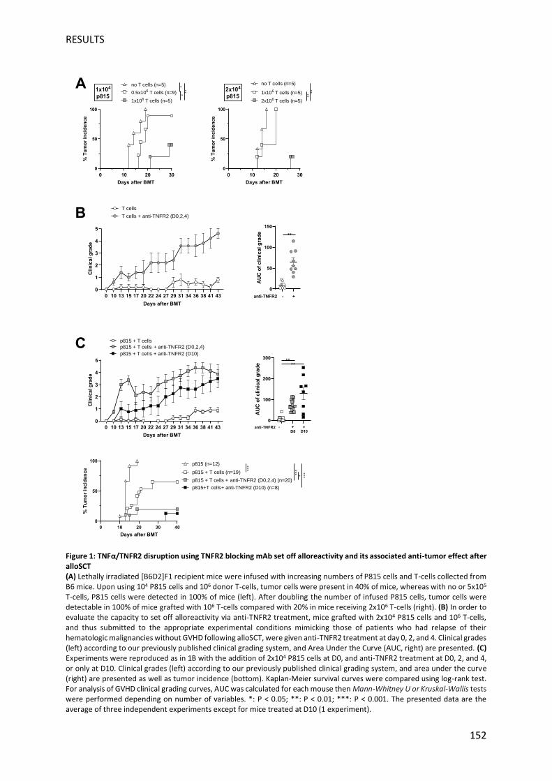
RESULTS
152
Figure 1: TNFα/TNFR2 disruption using TNFR2 blocking mAb set off alloreactivity and its associated anti-tumor effect after alloSCT (A) Lethally irradiated [B6D2]F1 recipient mice were infused with increasing numbers of P815 cells and T-cells collected from B6 mice. Upon using 104 P815 cells and 106 donor T-cells, tumor cells were present in 40% of mice, whereas with no or 5x10⁵ T-cells, P815 cells were detected in 100% of mice (left). After doubling the number of infused P815 cells, tumor cells were detectable in 100% of mice grafted with 106 T-cells compared with 20% in mice receiving 2x10⁶ T-cells (right). (B) In order to evaluate the capacity to set off alloreactivity via anti-TNFR2 treatment, mice grafted with 2x10⁴ P815 cells and 106 T-cells, and thus submitted to the appropriate experimental conditions mimicking those of patients who had relapse of their hematologic malignancies without GVHD following alloSCT, were given anti-TNFR2 treatment at day 0, 2, and 4. Clinical grades (left) according to our previously published clinical grading system, and Area Under the Curve (AUC, right) are presented. (C) Experiments were reproduced as in 1B with the addition of 2x10⁴ P815 cells at D0, and anti-TNFR2 treatment at D0, 2, and 4, or only at D10. Clinical grades (left) according to our previously published clinical grading system, and area under the curve (right) are presented as well as tumor incidence (bottom). Kaplan-Meier survival curves were compared using log-rank test. For analysis of GVHD clinical grading curves, AUC was calculated for each mouse then Mann-Whitney U or Kruskal-Wallis tests were performed depending on number of variables. *: P < 0.05; **: P < 0.01; ***: P < 0.001. The presented data are the average of three independent experiments except for mice treated at D10 (1 experiment).
0
50
100
150
AU
Co
fclin
icalg
rad
e
anti-TNFR2 - +
**
0 10 13 15 17 20 22 24 27 29 31 34 36 38 41 43
0
1
2
3
4
5
Days after BMT
Cli
nic
alg
rad
e
T cells
T cells + anti-TNFR2 (D0,2,4)
0 10 20 30 40
0
50
100
Days after BMT
%T
um
or
incid
en
ce
p815 (n=12)
p815 + T cells (n=19)
p815 + T cells + anti-TNFR2 (D0,2,4) (n=20)
*** *** *
p815+T cells+ anti-TNFR2 (D10) (n=8)
***
0
100
200
300
AU
Co
fclin
ica
lg
rad
e
anti-TNFR2 +-D0 D10
**
+
**
0 10 13 15 17 20 22 24 27 29 31 34 36 38 41 43
0
1
2
3
4
5
Days after BMT
Clin
ica
lg
rad
e
p815 + T cells
p815 + T cells + anti-TNFR2 (D0,2,4)
p815 + T cel ls + anti-TNFR2 (D10)
0 10 20 30
0
50
100
Days after BMT
%T
um
or
incid
en
ce
no T cells (n=5)
0.5x10⁶ T cells (n=9)
1x10⁶ T cells (n=5)
**
**1x10⁴
p815
0 10 20 30
0
50
100
Days after BMT
%T
um
or
incid
en
ce
no T cells (n=5)
1x10⁶ T cells (n=5)
2x10⁶ T cells (n=5)
****2x10⁴
p815
A
B
C
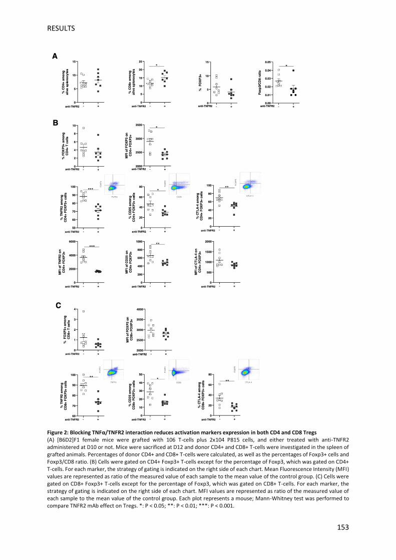
RESULTS
153
Figure 2: Blocking TNFα/TNFR2 interaction reduces activation markers expression in both CD4 and CD8 Tregs (A) [B6D2]F1 female mice were grafted with 106 T-cells plus 2x10⁴ P815 cells, and either treated with anti-TNFR2 administered at D10 or not. Mice were sacrificed at D12 and donor CD4+ and CD8+ T-cells were investigated in the spleen of grafted animals. Percentages of donor CD4+ and CD8+ T-cells were calculated, as well as the percentages of Foxp3+ cells and Foxp3/CD8 ratio. (B) Cells were gated on CD4+ Foxp3+ T-cells except for the percentage of Foxp3, which was gated on CD4+ T-cells. For each marker, the strategy of gating is indicated on the right side of each chart. Mean Fluorescence Intensity (MFI) values are represented as ratio of the measured value of each sample to the mean value of the control group. (C) Cells were gated on CD8+ Foxp3+ T-cells except for the percentage of Foxp3, which was gated on CD8+ T-cells. For each marker, the strategy of gating is indicated on the right side of each chart. MFI values are represented as ratio of the measured value of each sample to the mean value of the control group. Each plot represents a mouse; Mann-Whitney test was performed to compare TNFR2 mAb effect on Tregs. *: P < 0.05; **: P < 0.01; ***: P < 0.001.
RESULTS
154
Supplemental methods:
Mice: Female C57BL/6J (B6, H-2b) and female (B6xDBA2) F1 ([B6D2]F1, H-2bxd) mice of 10 to
12 weeks of age were obtained from Charles River Laboratories (France). Experimental
procedures were specifically approved by the local ethical committee (N°APAFIS#11511-
2017092610086943).
GVHD and tumor relapse models: Ten- to 12-week-old recipient [B6D2]F1 female mice
received 10 Gy irradiation (X-ray) followed by retro-orbital infusion of 5 x 10⁶ bone marrow
(BM) cells and 1 x 10⁶ CD3+ T-cells. BM and T-cell suspensions were prepared using leg bones
and lymph node, respectively, as previously described12. All infused cells were isolated from
female C57BL/6 mice. For tumor relapse models, we injected 1 or 2x10⁴ mastocytoma cells
P815-GFP (gift from Dr G Marodon) IV in the retro orbital sinus at time of BMT. After semi-
allogeneic BM transplantation (BMT), GVHD and tumor incidence were evaluated three times
per weeks. Clinical GVHD was evaluated as previously described15. Each of the five following
parameters was scored 0 (if absent) or 1 (if present): weight loss >10% of initial weight,
hunched posture, skin lesion, dull fur, and diarrhea. Dead mice received a total score of five.
Mice were considered positives for tumor when a visible mass appeared near the eye and/or
if P815 cells were detected in peripheral blood using GFP expression and surface expression
of H-2Kd antigens (recipient type).
Flow cytometry
Spleens were harvested at day 12 and stained with the antibodies listed in Supplementary
Table 1. Non-specific binding was blocked using anti-CD16/CD32 (Miltenyi Biotec). For
intracellular staining, cells were fixed and permeabilized with fixation/permeabilization
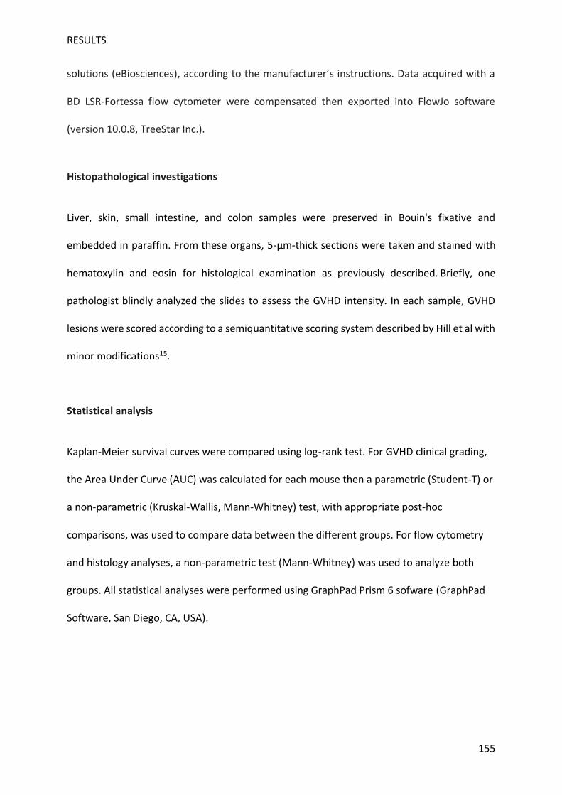
RESULTS
155
solutions (eBiosciences), according to the manufacturer’s instructions. Data acquired with a
BD LSR-Fortessa flow cytometer were compensated then exported into FlowJo software
(version 10.0.8, TreeStar Inc.).
Histopathological investigations
Liver, skin, small intestine, and colon samples were preserved in Bouin's fixative and
embedded in paraffin. From these organs, 5-µm-thick sections were taken and stained with
hematoxylin and eosin for histological examination as previously described. Briefly, one
pathologist blindly analyzed the slides to assess the GVHD intensity. In each sample, GVHD
lesions were scored according to a semiquantitative scoring system described by Hill et al with
minor modifications15.
Statistical analysis
Kaplan-Meier survival curves were compared using log-rank test. For GVHD clinical grading,
the Area Under Curve (AUC) was calculated for each mouse then a parametric (Student-T) or
a non-parametric (Kruskal-Wallis, Mann-Whitney) test, with appropriate post-hoc
comparisons, was used to compare data between the different groups. For flow cytometry
and histology analyses, a non-parametric test (Mann-Whitney) was used to analyze both
groups. All statistical analyses were performed using GraphPad Prism 6 sofware (GraphPad
Software, San Diego, CA, USA).
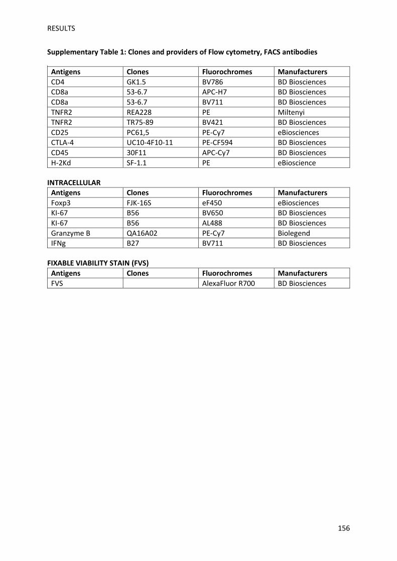
RESULTS
156
Supplementary Table 1: Clones and providers of Flow cytometry, FACS antibodies
Antigens Clones Fluorochromes Manufacturers
CD4 GK1.5 BV786 BD Biosciences CD8a 53-6.7 APC-H7 BD Biosciences
CD8a 53-6.7 BV711 BD Biosciences
TNFR2 REA228 PE Miltenyi TNFR2 TR75-89 BV421 BD Biosciences
CD25 PC61,5 PE-Cy7 eBiosciences
CTLA-4 UC10-4F10-11 PE-CF594 BD Biosciences
CD45 30F11 APC-Cy7 BD Biosciences
H-2Kd SF-1.1 PE eBioscience INTRACELLULAR
Antigens Clones Fluorochromes Manufacturers
Foxp3 FJK-16S eF450 eBiosciences KI-67 B56 BV650 BD Biosciences
KI-67 B56 AL488 BD Biosciences
Granzyme B QA16A02 PE-Cy7 Biolegend IFNg B27 BV711 BD Biosciences
FIXABLE VIABILITY STAIN (FVS)
Antigens Clones Fluorochromes Manufacturers
FVS AlexaFluor R700 BD Biosciences
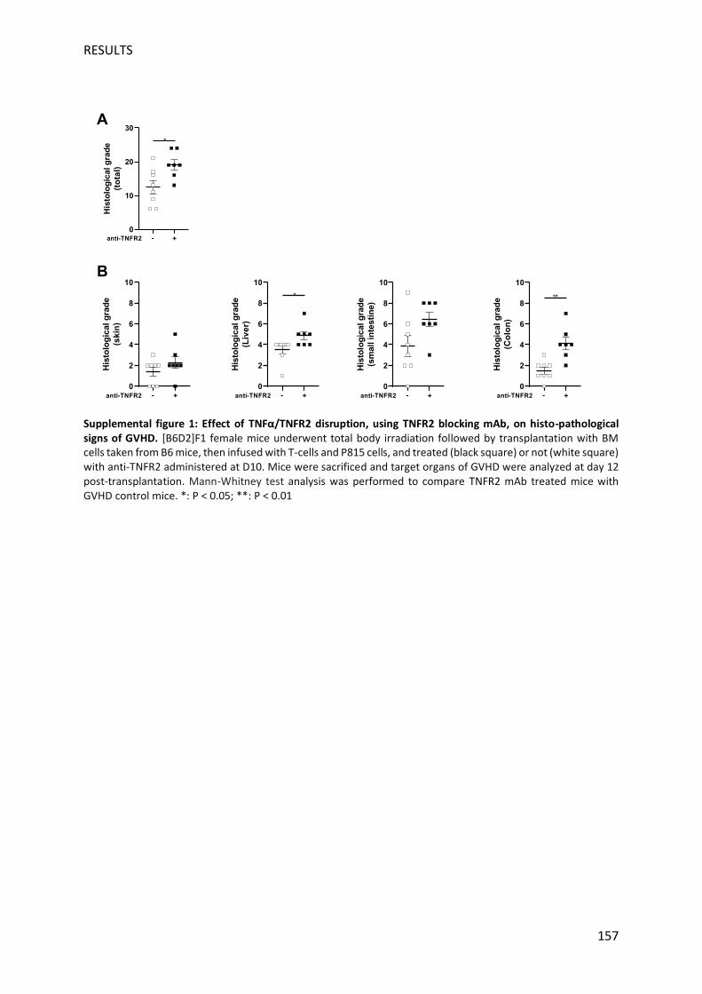
RESULTS
157
Supplemental figure 1: Effect of TNFα/TNFR2 disruption, using TNFR2 blocking mAb, on histo-pathological signs of GVHD. [B6D2]F1 female mice underwent total body irradiation followed by transplantation with BM cells taken from B6 mice, then infused with T-cells and P815 cells, and treated (black square) or not (white square) with anti-TNFR2 administered at D10. Mice were sacrificed and target organs of GVHD were analyzed at day 12 post-transplantation. Mann-Whitney test analysis was performed to compare TNFR2 mAb treated mice with GVHD control mice. *: P < 0.05; **: P < 0.01
0
10
20
30
His
tolo
gic
alg
rad
e
(to
tal)
anti-TNFR2 - +
*
0
2
4
6
8
10
His
tolo
gic
alg
rad
e
(sk
in)
anti-TNFR2 - +
0
2
4
6
8
10
His
tolo
gic
alg
rad
e
(Liv
er)
*
anti-TNFR2 - +
0
2
4
6
8
10
His
tolo
gic
alg
rad
e
(sm
all
inte
sti
ne)
anti-TNFR2 - +
0
2
4
6
8
10
His
tolo
gic
alg
rad
e
(Co
lon
)
**
anti-TNFR2 - +
A
B
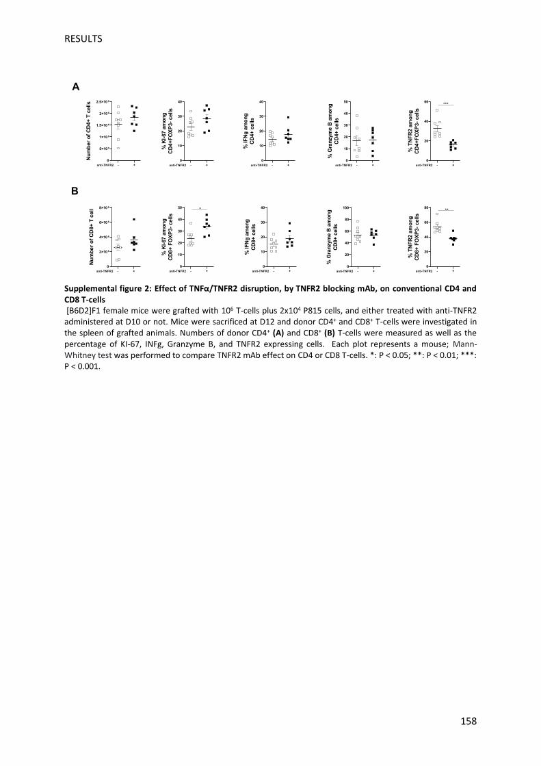
RESULTS
158
Supplemental figure 2: Effect of TNFα/TNFR2 disruption, by TNFR2 blocking mAb, on conventional CD4 and CD8 T-cells [B6D2]F1 female mice were grafted with 106 T-cells plus 2x10⁴ P815 cells, and either treated with anti-TNFR2 administered at D10 or not. Mice were sacrificed at D12 and donor CD4+ and CD8+ T-cells were investigated in the spleen of grafted animals. Numbers of donor CD4+ (A) and CD8+ (B) T-cells were measured as well as the percentage of KI-67, INFg, Granzyme B, and TNFR2 expressing cells. Each plot represents a mouse; Mann-Whitney test was performed to compare TNFR2 mAb effect on CD4 or CD8 T-cells. *: P < 0.05; **: P < 0.01; ***: P < 0.001.
0
10
20
30
40
50
%K
I-67
am
on
g
CD
8+
FO
XP
3-
ce
lls
*
anti-TNFR2 +-
0
10
20
30
40
%K
I-67
am
on
g
CD
4+
FO
XP
3-
cells
anti-TNFR2 +-
0
20
40
60
80
%T
NF
R2
am
on
g
CD
8+
FO
XP
3-
ce
lls
**
anti-TNFR2 +-
0
20
40
60
%T
NF
R2
am
on
g
CD
4+
FO
XP
3-
cells
anti-TNFR2 +-
***
0
10
20
30
40
%IF
Ng
am
on
g
CD
8+
cells
anti-TNFR2 +-
0
10
20
30
40
%IF
Ng
am
on
g
CD
4+
cells
anti-TNFR2 +-
0
5×10 5
1×10 6
1.5×10 6
2×10 6
2.5×10 6
Nu
mb
er
of
CD
4+
Tcells
anti-TNFR2 +-
0
2×10 6
4×10 6
6×10 6
8×10 6
Nu
mb
er
of
CD
8+
Tcell
anti-TNFR2 +-
Aa
B
0
20
40
60
80
100
%G
ran
zym
eB
am
on
g
CD
8+
cells
anti-TNFR2 +-
0
10
20
30
40
50
%G
ran
zym
eB
am
on
g
CD
4+
cells
anti-TNFR2 +-
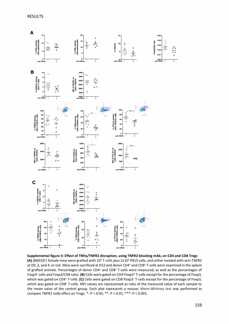
RESULTS
159
Supplemental figure 3: Effect of TNFα/TNFR2 disruption, using TNFR2 blocking mAb, on CD4 and CD8 Tregs (A) [B6D2]F1 female mice were grafted with 106 T-cells plus 2x10⁴ P815 cells, and either treated with anti-TNFR2 at D0, 2, and 4, or not. Mice were sacrificed at D12 and donor CD4+ and CD8+ T-cells were examined in the spleen of grafted animals. Percentages of donor CD4+ and CD8+ T-cells were measured, as well as the percentages of Foxp3+ cells and Foxp3/CD8 ratio. (B) Cells were gated on CD4+Foxp3+ T-cells except for the percentage of Foxp3, which was gated on CD4+ T-cells. (C) Cells were gated on CD8+Foxp3+ T-cells except for the percentage of Foxp3, which was gated on CD8+ T-cells. MFI values are represented as ratio of the measured value of each sample to the mean value of the control group. Each plot represents a mouse; Mann-Whitney test was performed to compare TNFR2 mAb effect on Tregs. *: P < 0.05; **: P < 0.01; ***: P < 0.001.

158
Discussion
DISCUSSION TNFR2 inhibition blocks tumour growth post-alloHSCT
159
TNFR2 inhibition blocks tumour growth post-
alloHSCT
A. Mouse model of post-alloHSCT relapse: advantages and
limitations
The results of our previous study, performed using a model of GvHD, revealed the necessity
of TNFR2 pathway for Treg function to protect the mice against an overactivated alloreactive
response against host tissues (Leclerc et al., 2016). To test whether we could conversely use
this Treg feature to promote alloreactivity in a context where it is lacking, as needed for
relapsing patients post-alloHSCT, we first searched to establish a model consistent with this
clinical situation. In our established parent-into-F1 graft model, using TBI at a lethal dose as a
conditioning regimen, a fixed donor quantity of bone marrow fully prevents recipient death.
Then, injected intravenously with the bone marrow, lymph node infusion from the donor
serves as a source of alloreactive naïve T cells, like DLI for relapsing patients, and a low dose
of P815 cells (host-derived mastocytoma) correspond to the residual disease that remains in
the case of insufficient donor antitumour immune response post-alloHSCT. Without T cells
injection, P815 cells show an aggressive proliferation rhythm, inducing a tumour incidence of
100% before day 20 post-graft. Then, we defined a suboptimal T cell dose contained in the
injected lymph node, in such a way that the tumour establishment, albeit delayed, reached
around 50% at day 20. This situation reflects the existence of a large pool of non-responding
patients when using DLI or other anti-cancer therapies. Secondly, in this operational relapse
model, we tested two therapeutic schemes using a TNFR2 blocking mAb. Three intraperitoneal
injections at day 0, 2 and 4 followed the therapeutic strategy already used in the GvHD study
and corresponded to a prophylactic strategy to prevent cancer cells proliferation and
dissemination. Alternatively, a unique injection at day 10 (1/3 of the previous total dose) was
used to test the possibility to halt the ongoing cancer progression, since the first tumour was
detectable in the control group at this time.
Beyond well-described drawbacks when using alloHSCT rodent models, such as the lack of
microbiota diversity, the sole use of purified bone marrow as HSC source or irradiation as
DISCUSSION TNFR2 inhibition blocks tumour growth post-alloHSCT
162
conditioning, or else conducting experimentations on young adult animals only, conclusions
in one model may not translate into other immunologically distinct models or the clinic (Socié
and Blazar, 2013). Indeed, different strain combinations have different Treg content and Th
profile, which can balance the dominance of CD4+ or CD8+ Teff for instance. Furthermore,
GvT modelling in mice is limited by the use of a cell lines repertoire, that often does not
correlate well to de novo malignant cells because of their long-time use causing important
genetic drifts. However, studies using transformed or mutated primary hematopoietic cells
given to the host are infrequent and difficult to perform. For these reasons, we plan to
reproduce the GvT experiments using a distinct graft combination and cell line. The set-up of
this model is ongoing, using B6xBALB/c mice as host and B6 mice as donors, tested against a
B cell lymphoma (A20), a cell line that does not express TNFR2.
B. On the role of TNFR2 expression on tumour cells
In our model, TNFR2 mAb injection drastically reduced mastocytoma appearance. Both early
and delayed treatment delivered similar results, suggesting that TNFR2 blockade could have
an anti-tumour effect, (i) in a preventive manner for undetectable malignancies or minimal
tumour burden detected in patients before cancer resurgence, and (ii) as a curative strategy
for already emerging haematologic neoplasm. In spite of the high TNFR2 expression detected
on the P815 cell line in vitro, no direct impact of TNFR2 blockade was observed in the absence
of donor T cells injection, suggesting that the observed tumour cell killing is mainly immune-
mediated in these conditions (data not shown). However, the maintenance of TNFR2
expression in vivo has not been tested. Indeed, the pattern of expression of TNFR1 and 2 at
some tumour cell line surface has been shown to variate greatly as a function of TNF-, then
the possibility of a TNFR2 downregulation should be tested in vitro and validated in vivo for
P815 as well (Alshevskaya et al., 2020). Another possibility is that TNFR2 survival pathway is
not necessary for P815 cells, because of redundant strong potential survival signals that could
compensate for the loss of NF-κB activation via TNF-/TNFR2.
C. On the risk of GvHD induction after anti-TNFR2 treatment
As a reflection of a strengthened alloreactivity, anti-TNFR2 mAb treatment induced
observable characteristic aGvHD lesions a few days after treatment initiation, with and in
DISCUSSION TNFR2 inhibition blocks tumour growth post-alloHSCT
163
absence of P815 cells, in comparison to the untreated control group. In a second experiment
set where the mice were euthanized at day 12 (referred to as short-time experiment), the
histological grade of aGvHD target tissues was significantly higher for the treated group when
assessed in the liver and colon. These observations provide a strong clue that TNFR2 blockade
did increase alloreactivity in aGvHD target organs, as a consequence of donor T cells
activation, expansion and proliferation. Indeed, if for a while the medical and scientific
community have searched to dissociate the beneficial GvT effect from the life-threatening
GvHD side effect, the clinical reality is less straightforward. Alloreactive T cells, the main actors
responsible for GvHD, are also those who play a critical role in the antitumour effect, as
supported by the observed increased risk of leukaemia relapse when they are reduced or
absent as occurs with autografts, syngeneic twin grafts or with T cell-depleted alloHSCT
(Horowitz et al., 1990). In a clinical trial conducted by our group, ex vivo Treg depletion from
DLI has led to the first GvHD occurrences in relapsing patients (Maury et al., 2010). If GvHD
lesions were reversible using adapted patient care, the GvT effect potently protected some
patients from a potential cancer resurgence. In consequence, rather than distinguish “good”
and “bad” allograft actions, it seems that the future clinic approaches could be shifting toward
personalized medicine strategies, relying on a fine alloreactivity tuning to either increase the
GvT effect or control GvHD after alloHSCT, as required by each patient individually. In the case
of our study, no GvHD prophylaxis is usually used in GvT mice models in contrast with patients’
situation. It could be interesting to test the reversibility of the alloreactivity escalation induced
by our treatment.
DISCUSSION TNFR2 blockade spurs the alloimmune response
159
TNFR2 blockade spurs the alloimmune response
A. By helping effector response initiation
The alloreactivity initiation observed after TNFR2 blockade at day 10 was associated in our
model with different modifications affecting T cell populations in the spleen at day 12. In the
short-term experimental setting, anti-TNFR2 mAb-receiving group presented (i) a superior
CD8+ lymphocytes frequency and proliferative features and (ii) no change in CD4+
populations, including Treg, in terms of frequency but a drastic impairment of Treg phenotype
when compared to untreated mice. No difference was found in terms of CD8+ cytotoxic
capacity in treated mice. Because P815 cells express MHC class I, but no class II and are
resistant to Fas-induced apoptosis, the main pathway of rejection when using this lineage is
through perforin/granzyme secretion by CD8+ cells (Hill et al., 1997; Rebel et al., 1999;
Teshima et al., 1999). A potential explanation in our model for the unchanged granzyme B
expression in CD8+ cells could be the short timeframe (48 hours) between the treatment and
the analysis of the spleen content. A large majority of the CD8+ lymphocytes display an
effector phenotype in this allogeneic setting, with more than 80% CD44+CD62L- cells among
the CD8+ population in the spleen at day 12 for both control and treated mice. However, only
the treated group developed GvHD symptoms starting at day 13 in our long-lasting
experiment. Presumably, the higher expression of KI-67 on CD8+ cells from treated animals
reflects a superior clonal expansion process that follows TCR triggering when the TNFR2 is
blocked, despite the costimulatory activity described for this receptor. Finally, the temporal
window that we selected gave us a glimpse on the cytotoxic response set up, that probably
precedes the effective effector phase responsible for the preventive action against the tumour
observed in the long-term experiments.
Importantly, Chen et al. reported in breast and lung carcinoma that intratumoral Teff
expressing elevated levels of TNFR2 acquired the capacity to resist peripheral Treg
suppression, but remained susceptible to inhibition by more suppressive tumour-infiltrating
Treg, expressing higher levels of TNFR2 (Chen et al., 2010b). Going a step further, we
demonstrate in a post-alloHSCT relapse model that the blockade of TNFR2 on T cells, a
receptor that has been shown to operate as a non-redundant costimulatory signal in Teff
DISCUSSION TNFR2 blockade spurs the alloimmune response
165
(Aspalter et al., 2003), does not induce CD4+ proliferation, rather allows a higher CD8+
expansion. Thus, the pattern of TNFR2+ expression on the different immune cell populations
involved in the pathology considered could greatly change the outcome of TNFR2 modulation.
After the allograft, in our murine model, TNFR2+ immune actors’ distribution seems
favourable to the effector response when shutting down this signalling pathway. This last
point will have to be carefully assessed in exploratory phenotyping of TNFR2+ cells from
relapsing post-alloHSCT patients before planning to bring TNFR2 blockade to clinical trials.
Whether the increased frequency of CD8+ in the spleen can be observed in the blood could
not be assessed with precision because of the profound lymphopenia caused by TBI
conditioning. However, further experiments using pooled blood from several mice from the
same group would help determine if it could serve as an early biomarker of TNFR2 blockade
response.
B. By inducing Treg stability and function decline
Day+2 after TNFR2 blockade seems to be premature to observe important changes in
conventional T cells phenotype. Conversely, it appears that the major differences after
treatment concern the pool of Treg, not in terms of their quantity but in phenotype. Indeed,
TNFR2 treatment only dimly reduced the percentage of FOXP3+ cells among CD4+
lymphocytes, a decrease that was not significant at day 12 in comparison with untreated
controls. In contrast, the level of intracellular FOXP3 in Treg from mice receiving anti-TNFR2
mAb injections dropped homogeneously compared to control mice. This observation confirms
in vivo the link between TNFR2 signalling pathway and FOXP3 in Treg, consistently with in vitro
descriptions made by others (Chen et al., 2007; Okubo et al., 2013; Zaragoza et al., 2016).
Further evaluation of TSDR methylation profile ex vivo would be valuable to determine how
definitive is the Treg destabilisation we observe. In accordance with FOXP3 rarefaction inside
Treg, the two extracellular proteins that represent the “core module” of Treg, CD25 and CTLA-
4, were reduced both in frequency and density of expression at Treg surface (significantly,
except for the observed reduction in CTLA-4 MFI). In regular Treg, both are upregulated by
FOXP3-dependant transcriptional program, and are directly responsible for Treg suppressive
capacity, by depriving Teff from IL-2 through CD25, and costimulation blockade for CTLA-4
(Wing and Sakaguchi, 2012). Other Treg markers, such as PD-1 that also have a role for Treg
DISCUSSION TNFR2 blockade spurs the alloimmune response
166
function, or the transcription factor HELIOS associated with Treg stability under inflammation
that can positively regulate FOXP3 expression, were not impacted by TNFR2 blockade in our
model (data not shown) (Kim et al., 2015; Sebastian et al., 2016). Thus, the impact on CD25
and CTLA-4 we observe might be the indirect consequence of a loss in regulatory transcription
program orchestrated by FOXP3, as a result of TNFR2 signalling pathway blockade. To go
further in our understanding of TNFR2 inhibition consequences on Treg, we plan on a short-
term basis to assess and compare the transcriptomic landscape between mice splenocytes
from treated and untreated groups.
In parallel to these significant changes striking CD4+ Treg, we also observed, for the first time
to our knowledge, a decline in CD8+ Treg function after TNFR2 inhibition. This regulatory
subset has been described in allotransplantation models, among splenocytes and infiltrated
in GvHD target tissues, starting at day 5 with a peak of detection at day 10 post-graft (Beres
et al., 2012). CD8+ iTreg were sufficient to prevent GvHD-associated mortality in the complete
absence of CD4+ Treg, suggesting a non-redundant role for this population in alloreactive
response control. In our model, at day 12, untreated mice showed a high frequency of TNFR2
in CD8+ Treg, as in CD4+ Treg (mean ≈90% in both subsets), but far exceeding the conventional
FOXP3- cells TNFR2 expression (53 and 32% in FOXP3-CD8+ and FOXP3-CD4+ respectively). As
for their CD4+ counterpart, phenotype modifications in treated mice affected negatively CD25
and CTLA-4 expression on CD8+ Treg, whereas no significant differences were observed in PD-
1 and HELIOS frequencies. The co-expression of TNFR2 with FOXP3, CTLA-4, PD-1 and PD-L1
in CD8+ cells have been reported in a xenoGvHD model (Horwitz et al., 2013). Interestingly, in
this study, the suppressive activity of CD8+ Treg in vitro correlated better with TNFR2 and PD-
L1 than FOXP3. Furthermore, blocking studies suggested that TNF- enhances PD-L1
expression and the suppressive activity of the CD8 Treg generated in their model. Then,
regarding its effect on CTLA-4 and CD25 and in accordance with this previous work, TNFR2
blockade could be the most suitable tool described to impair CD8+ Treg function in allogeneic
settings. Also, to better understand how TNFR2 blockade affects CD8+ Treg suppressive
function, and following these anterior findings, PD-L1 expression study could be of greater
interest than PD-1. Similarly, another study, in line with the previous one, showed that the
induction by anti-CD3 mAb of CD8+ Treg expressing CD25, CTLA-4, FOXP3, and TNFR2 required
TNF- and signalling through the NF-κB cascade (Ablamunits et al., 2010). Importantly, they

DISCUSSION TNFR2 blockade spurs the alloimmune response
167
also report that the combined expression of TNFR2 and CD25 identified a potent
subpopulation of CD8+ Treg. In experiments where infusions of ex vivo expanded CD8+ Treg
were used to attenuate aGvHD, the reduction of alloreactive T cell proliferation was strongly
CTLA-4-dependent (Zheng et al., 2013). These data suggest that our observations of a
diminished TNFR2, CD25 and CTLA-4 co-expression on CD8+FOXP3+ may reflect a weakened
alloreactive T cells suppression, a hypothesis that we could consider verifying ex vivo in
suppression assays.
C. Integrative view of TNFR2 inhibition impact in alloHSCT
Whether these qualitative modifications on CD4+ and CD8+ Treg are a direct consequence of
the mAb binding to TNFR2 expressed on the Treg surface remains to be determined, and the
same applies for CD8+ Teff enhanced proliferation capacity. However, regarding the literature
about TNFR2’s pivotal role in Treg function, we can propose a general scheme in our model of
how TNFR2 blockade modifies Treg/Teff balance to result in a potent tumoricidal action. First,
a strong beam of arguments suggests that TNFR2 inhibition using a mAb destabilises CD4+ and
CD8+ Treg function, with a clear reduction in TNFR2, CTLA-4 and CD25 expressing populations
from these two regulatory subsets. For CD4+ Treg, this effect is presumably the result of
FOXP3 transcriptional activity positive regulation by TNFR2, while for CD8+ Treg the literature
and our study do not suggest a strong implication of the “Treg master gene” in the phenotype
we observed. The diminution of the most potent subsets of Treg appears to be a reasonable
explanation for the higher frequency and proliferation of CD8+ cells seen at day 12 under
TNFR2 blockade, which in turn underlie the better antitumour alloreactivity leading to P815
rejection and the observation of GvHD signs in the gut and liver. Importantly, the up-
mentioned sequence of events resulting in the therapeutic effect of anti-TNFR2 mAb after
alloHSCT, apply mostly for one injection at day 10. With the early (day 0, 2, 4) scheme of
injection the results are quite the same for both timings regarding anti-tumour efficacy and
CD4+ Treg phenotype, a non-significant tendency was observed in terms of CD8+ Treg the
reduction in function-related proteins. The ratio of FOXP3+/CD8+ cells was only slightly
inferior compared to controls whereas this difference is significant with a treatment at day
10. A potential explanation for the differential results with preventive and curative treatment
schemes could be that in our model, induced subsets of Treg appear several days after the
graft. Indeed, CD8+ Treg are an induced population only, which are not detected in the graft
DISCUSSION TNFR2 blockade spurs the alloimmune response
168
prior to infusion. Their detection before day 5 was possible in the spleen in a GvHD model, but
their appearance in our model might be delayed due to the low-dose of T cells we inject (Beres
et al., 2012). Their function is not redundant with CD4+ Treg, then their activity spared by the
early treatment scheme could explain the less striking effect observed in CD8+ Teff
population. Same goes for CD4+ Treg, iTreg quantity grow in the first 20 days post-graft in
reaction to alloreactivity. Then, iTreg absence at the time of the early treatment could explain
the absence of difference at day 12 in FOXP3 density of expression, observed with the late
treatment administration, since these induced cells are more prompt to lose FOXP3 strong
expression than nTreg. Like induced Treg, other TNFR2+ populations, are potentially emerging
after donor cells exposition to recipient alloantigen, being the target of the mAb we injected,
this point will require more analyse timepoints in the future. For instance, NK TNFR2+ cells
have been reported in certain situations (Xu et al., 2007).
Finally, one of the more striking consequences witnessed after TNFR2 mAb injection in our
post-alloHSCT model was the dramatic decrease in TNFR2 expression on all the studied T cell
populations, regulatory and conventional. This effect does not seem to be an artefact due to
steric hindrance of the therapeutic mAb on the site of detection by flow cytometry mAb (data
not shown). Because TNF- by itself can up-regulate TNFR2 expression on T cells, interruption
of TNF-/TNFR2 interaction thus presumably down-regulates TNFR2 surface expression on T
cells, explaining the expression drop we detect. Interestingly, the TNFR2 antagonist described
in Torrey et al.’s study appeared to stabilize the surface expression of TNFR2, inhibiting the
release of immunomodulatory sTNFR2 from Treg (Torrey et al., 2017). This last mechanism
explains a part of their mAb immunomodulatory role and should be assessed for our mAb as
well, by sTNFR2 dosage in mice sera, to understand whether the remaining TNFR2 we observe
at the surface is less frequently cleaved after treatment.

DISCUSSION TNFR2 is a legitimate new ICP target of the anti-cancer arsenal
169
TNFR2 is a legitimate new ICP target of the anti-
cancer arsenal
A. TNF- pathway as a promising anti-cancer agent once more
In a study conducted in 1999, Ferrara’s group first described, using a P815-based GvT model,
that sTNF- blockade resulted in increased leukaemia mortality (Hill et al., 1999b, 2000). This
study also demonstrated a reduced capacity to induce GvHD when using donor T cells
deficient for TNFR1 and warned about the crucial role of TNF-/TNFR1 signalling for the
generation/maintenance of cytotoxic T cells and subsequent GvT activity. At the time, in
reaction to the failure of T cell-depleted graft strategies, the scientific and medical community
has proposed as prevention of aGvHD to retain mature T cells in the bone marrow graft while
disrupting the amplification of inflammatory cytokine. Accordingly, the first clinical trials using
anti-TNF- for GvHD prophylaxis were then ongoing, although this approach would not
provide full satisfaction either in the future. Twenty years later, with the growing body of
evidence that tolerogenic and pro-inflammatory aspects of TNF- could be dissociated via the
distinct targeting of its two receptors, we and others propose a therapeutic alternative to TNF-
targeting by blocking TNFR-2 while retaining TNFR1 anti-tumoral purposes. The dual role of
TNF- in inflammation and tolerance can probably explain most of the erratic literature about
this cytokine in alloHSCT mice models, using several conditioning regimens, various TNF-
neutralizing treatments, and multiple mouse strain/leukaemia combinations (Borsotti et al.,
2007; Hill et al., 1999b; Korngold et al., 2003; Matte et al., 2004; Schmaltz et al., 2003). Some
of these studies reported that sTNF- makes an important contribution to tumour clearance
in accordance with Ferrara’s results, while others have concluded that TNF- is dispensable
for a maximal antileukemic response (Hill et al., 1999b; Korngold et al., 2003; Schmaltz et al.,
2003). Our recent results fall within these attempts of understanding better the role of TNF-
in the balance between GvT and GvHD post-alloHSCT. The improved alloreactivity we
observed proves that blocking TNFR2 is sufficient to trigger a potent anti-immune response,
presumably by impairing tolerance, but it could also potentially permit a higher availability of
the TNF- pool for TNFR1 fixation. The potential induction of TNFR1-dependent anti-tumour
immunity as an indirect effect of TNFR2 inhibition remains to be explored.
DISCUSSION TNFR2 is a legitimate new ICP target of the anti-cancer arsenal
170
Besides alloHSCT, TNF- has found its place as a therapeutic target in autoimmune disease
where anti-TNF- treatments have been revolutionary in several pathologies. If the efficacy
of anti-TNF treatments is not to demonstrate, it remains a rather unspecific approach that
does not only affect autoreactive actors. Indeed, as a reminder for its name, TNF- inhibition
can result in rare but severe occurrences of haematological malignancies treatment-induced
(Hansel et al., 2010). The perceived increase in these haematological malignancies has led to
include in the WHO classification of tumours the category “iatrogenic immunodeficiency-
associated lymphoproliferative disease”, and the risk of virally transformed tumours is closely
watched as well (Campo et al., 2011). If the risk of anti-TNF--induced cancer for autoimmune
patients is under debate, it still provides a good example of a situation where promoting
tolerance via TNFR2 activation would presumably be a better strategy than blocking TNF-.
Again, the incredibly wide range of physiologic functions dependent on TNF-/TNFR1 in all
the body systems, including cancer immunosurveillance, render this pathway more tricky to
target (Faustman and Davis, 2010, 2013).
As for solid and hematologic malignancies, the unspecific broad effects of TNF- infusion in
patients, initially led to abandon the focus on this signalling pathway as first-line therapy
against neoplasms. However, in light of the accumulation of pre-clinical data about TNFR2
role, and numerous clinical reports associating TNFR2+ Treg and tumour cells in patients, it is
now clear that a TNFR2-centred approach could be an interesting strategy for cancer patients,
outside the context of post-alloHSCT relapse. The advantages and limitations of such a
strategy are discussed in the following sections.
B. TNFR2 attractive pattern of expression
In contrast with its type I counterpart, TNFR2 is not ubiquitously expressed but rather confined
in expression to immune system actors. Among these, a high density of TNFR1 and TNFR2 is
observed on monocytes, macrophages, and DC, and both pathways in these cells are
interconnected, acting in favour of their activation, proliferation and survival (Maney et al.,
2014; Rossol et al., 2007; Wajant and Siegmund, 2019). MDSC, an inflammation-induced
population, seems to be the one myeloid population that specifically requires TNFR2 for its
induction and suppressive function (Polz et al., 2014). In a steady-state, TNFR2 expression on
DISCUSSION TNFR2 is a legitimate new ICP target of the anti-cancer arsenal
171
lymphoid cells is nearly restricted to CD4+ Treg, in both humans and mice. In the context of
cancer, TNFR2+ CD4+ Treg seems to be increased in patients’ tumour infiltrate and/or
peripheral blood (He and Wang, 2019). Treg from the CD8+ cells’ subset has been described
in cancer models, and recently CD8+ Treg have been found to also express TNFR2+. Besides,
we demonstrated that CD8+TNFR2+ Treg, as well as CD4+TNFR2+ Treg, depend on this
receptor signalling for their suppressive function against alloantigen specific T cells. Aside
from these three subsets, which rely on their TNFR2 expression to suppress anti-tumour
immunity, TNFR2 is also an oncogene expressed by many carcinomas and hematologic
malignancies including T cell/Treg-derived neoplasms (Torrey et al., 2018). This last
information led the scientific community working on the subject to propose that TNFR2
blockade in cancer patients could “kill two birds with one stone” meaning it would boost
antitumor immune responses while directly killing tumour cells (Chen and Oppenheim, 2017;
Torrey et al., 2017). One really interesting feature of TNFR2 is its restricted perimeter of
expression, raising the hope of limited side effects under therapeutic utilization, contrarily to
TNF- or TNFR1 targeting. Because of the high toxicities associated with currently use anti-
ICP, there is an ongoing need for new cancer immunotherapies that have promising activity
but are also well tolerated. However, apart from the immune compartment, TNFR2 is
expressed and upregulated in pathologic situations such as ischemia, in endothelium cells, and
in the neural tissue at the surface of local macrophages (microglia), but also other non-
neuronal cells (astrocytes) (Fischer et al., 2020). Whether TNFR2 blockade could have an off-
target effect in these two tissues or non-desired immune effects through myeloid cells will
need to be carefully monitored.
C. TNFR2 modulation of Teff/Treg equilibrium
A crucial point about the TNFR2 pattern of expression is its change under inflammation.
Similarly to CD25, conventional T cells upregulate TNFR2 under TCR activation (Govindaraj et
al., 2013b). This asks the question of a potential deleterious effect on the adaptive response
after TNFR2 blockade, and more specifically on the cytotoxic anti-tumour CD8+ response. To
answer this question, it is necessary to first understand well the role of TNFR2 in Teff during a
resolvable immune response. Firstly, if TNFR2 stimulation in Teff correlates with a high
proliferative capacity and effector cytokine-producing capability, TNFR2 deficient mice still
DISCUSSION TNFR2 is a legitimate new ICP target of the anti-cancer arsenal
172
display a normal T-cell development (Erickson et al., 1994). Also, the time of exposure to TNF-
is deeply shaping the consequences of TNFR2 activation on Teff. A comprehensive scheme
for TNF-/TNFR2 role in Teff during the natural resolution of inflammation by Treg can be
proposed as follow (Chen and Oppenheim, 2011c):
1) In the steady-state, the equilibrium Treg and Teff activation preserves immune
homeostasis.
2) In the early stage of inflammation, activated Teff up‐regulate their TNFR2
expression under TNF- exposure, increasing their capacity to resist Treg‐
mediated inhibition, and therefore mount an effective immune response. Also
a slower Treg response than Teff to TNF- results in delayed
immunosuppressive feedback effects (Chen et al., 2013).
3) In the later stage of inflammation, chronic exposure to TNF- lead to impaired
production of effector cytokines caused by Treg competition with Teff for co‐
stimulatory TNF‐/TNFR2 action (Aspalter et al., 2003; Clark et al., 2005).
Importantly, TNFR2 also seems to be necessary for CD8+ T cells sensibility to
AICD (Kim et al., 2009a). In the meantime, Treg TNFR2-dependent stimulation
enhances their suppressive activity, resulting in the resolution of inflammatory
responses and restoration of immune homeostasis.
TNFR2 pattern of expression suggest that TNF- is able to induce a potent immune reaction
through monocytes/macrophages activation, and T cells co-stimulation. In the meantime, its
activation launches a “delayed immune response arrest” through its expression on tolerogenic
cells. Then, in non-physiological settings, including alloHSCT or cancer, this pattern of
expression changes, and the fine dual function balance is lost. During carcinogenesis, tumour
cells induce many modifications in the soluble milieu, including in TNF- levels, because of its
production by TME-cells such as infiltrated T cells or macrophages, and by the tumour itself
(Josephs et al., 2018). They also recruit immunosuppressive cells that further upregulate their
TNFR2 expression under TNF- exposure. This disturbs the equilibrium of TNF-/TNFR2
expression pattern, presumably putting cancer development in the third situation previously
described, i.e. a Teff function impairment due to chronic exposure of both Treg and Teff to
TNF-. In agreement with this assumption, a study have shown that the enhanced protection

DISCUSSION TNFR2 is a legitimate new ICP target of the anti-cancer arsenal
173
against lymphoma in TNFR2−/− mice resulted from the recruitment, activation and AICD
resistance of TAA specific memory TNFR2−/− CD8 T cells and their prolonged survival at the
tumour site (Kim et al., 2009a). Altogether these data point in the direction of beneficial action
of TNFR2 blockade on Teff under chronic TNF- exposure, despite the co-stimulatory role of
TNFR2 in brief inflammation. However, a number of information is still lacking to untangle
what could be the real consequences on immunity when using this strategy as a therapeutic
tool.
D. Potential side effects for TNFR2 blockade
Anti-CTLA-4 and anti-PD-1/PDL-1 mAb have shown high antitumour efficacy in responder
patients. However, a major drawback associated with the use of anti-ICP agents is the
apparition of severe autoimmunity/autoinflammatory symptoms (Young et al., 2018). Indeed,
these therapeutic tools are able to “reawaken” patients’ effector response against tumour
cells which express both TAA and self-antigens (Shimizu et al., 1999). Then, the over-activation
of Teff against the self can lead to diverse organ lesions. Besides, these antibodies by binding
off-targets also induce deleterious autoimmune effects such as diabetes mellitus after PD-L1
treatment, due to PD-L1 expression on pancreatic ß-cells. Since these chronic affections
deeply affect patients’ quality of life, treatment-safety is a major consideration for the new
generation of immunotherapies to come.
In our hands, when used after alloHSCT, blocking mAb against TNFR2 induces a change of
phenotype in Treg, and presumably impair their suppressive capacity, but do not depletes
them. Our results show a drastic decrease of TNFR2+ on Treg, but not of FOXP3+ frequency
modification after treatment, suggesting a downregulation or superior cleavage of TNFR2 at
Treg surface. Thus, the treatment induces the disappearance of the target population (Treg
TNFR2+) but quantitatively spares the pool of Treg in the spleen. The preservation of a viable
Treg pool, although less functionally potent, could better prevent autoimmunity when
compared to Treg complete depletion. In addition, the re-expression of TNFR2 by Treg after
treatment cannot be excluded. Also, these observations are in line with the fact that TNFR2-/-
mice do not develop autoimmunity, suggesting also that the restriction of TNFR2 on a Treg
subset in the steady-state does not impair the capacity of other subpopulations to maintain
DISCUSSION TNFR2 is a legitimate new ICP target of the anti-cancer arsenal
174
an immune balance (Vanamee and Faustman, 2017). In cancers, an advantage of TNFR2 is its
higher expression in several tumour-infiltrated Treg compared to Treg in the circulation. The
dominant TNFR2 antagonists from Torrey et al. study preferentially suppressed tumour-
associated Treg activity but have no or only minor inhibitory effects on regular Treg in the
periphery, which play a crucial role in the maintenance of immunological homeostasis (Torrey
et al., 2017). Neither our team nor Faustman’s group recorded evidence yet regarding the
transient or long-term nature of the effect observed on Treg after TNFR2 blockade. In a
nutshell, targeting TNFR2 presents a few features that allow hopes about its capacity to be
well tolerated. Nevertheless, the risk of autoimmunity when blocking TNFR2 is considerable,
as for other new anti-ICP candidates, and difficult to evaluate outside proper clinical trials.
E. Activation versus TNFR2 blockade in tumoral context
A recent study conducted by a pharmaceutical industry evaluated the activity of murine and
human anti-TNFR2 antibodies for cancer therapy (Tam et al., 2019). They described the
mechanism of action of a novel mouse TNFR2 antibody (Y9) that appears to act through Fc-
dependent agonism on conventional T cells, responsible for its antitumor activity in preclinical
mouse models. In the several subcutaneous tumour models they used, with both hematologic
and carcinomas cell lines, they observed no significant Treg depletion or dysfunction. A second
pharmaceutical group screened for some TNFR2 binders, a subset of which were found to
agonise the receptor leading to NF-κB pathway signalling in vitro (Williams et al., 2016). In
CT26 syngeneic tumours, their TNFR2-specific agonists inhibited tumour growth, enhanced
tumour infiltration by CD8+ T cells and increased CD8+ T cell IFN-γ synthesis. In the light of the
previous subsection, these results are not antithetical to Faustman’s results in ovarian cancer
and Sézary syndrome using a TNFR2 antagonist neither ours in post-alloHSCT relapse. Indeed,
in the TME are gathered antitumour cells, that can benefit from TNF/TNFR2 pathway
signalling for their activation, as cited they are mainly macrophages and conventional T cells.
In a milieu under constant TNF- saturation, because Teff are susceptible to Treg exhaustion
again after long TNF- exposure, a strategy to target immunosuppressive actors by blocking
TNFR2 is probably the better angle of approach. However, what about a tumour context
where no TNFR2 signal is delivered because TNF- is rather lacking? Then, in theory, providing
a TNFR2 signal to proinflammatory TME-cells, as well as to Treg could permit, at least
DISCUSSION TNFR2 is a legitimate new ICP target of the anti-cancer arsenal
175
transiently to trigger a potent anti-tumour Teff response, resistant to Treg suppression, while
preserving tolerance (refers to the previous subsection). Another goal that seems to be
potentially reachable by both TNFR2 agonists and antagonists, is the direct impact of the
treatment on tumour cells. If TNFR2 blockade can divest cancer cells from a survival pathway,
agonists molecules may cause a domino effect while activating TNFR2, that could sensitize
malignant cells to TNFR1-induced cell death by depriving the cells in cIAP/TRAF (Siegmund et
al., 2016). Lastly, in our post-alloHSCT model, NK and T cells are mostly responsible for
allogeneic tumour rejection, and even if they were affected by the treatment, an effect on
myeloid cells could probably not be assessed in this kind of pathologic model. On the contrary,
in solid tumours at least, among TNFR2+ cells in the TME, TNFR2 agonism could have a positive
action on infiltrated macrophages. In William et al. and Tam et al. study, such an effect has
not been observed, but in an anterior study, TNFR2 expressed on host innate immune cells
was proved sufficient to mediate the antitumor effect of TNF- probably by inhibition of
tumour angiogenesis (Zhao et al., 2007). This observation could be another interesting aspect
for TNFR2 agonism in cancer, though for now more data are required about TNFR2 and
myeloid cells.
However, due to its plethora of functions, TNF- through its two receptors is also responsible
for divergent actions in the context of cancer (Montfort et al., 2019). Far from what was
initially thought about its antitumour effect, a growing body of clinical data has been
supporting the concept that chronic inflammation promotes tumour development and
progression. As a major proinflammatory cytokine, reports have involved TNF- in all aspects
of carcinogenesis, from the cellular transformation to survival, proliferation, invasion,
angiogenesis, and metastasis (Wang and Lin, 2008). Hence, in phase I and II clinical trials, two
anti-TNF- (infliximab and etanercept) achieved prolonged disease stabilization in patients
with metastatic breast cancer, recurrent ovarian cancer, or immunotherapy-resistant or
refractory renal cell carcinoma (Harrison et al., 2007; Madhusudan et al., 2004, 2005). In the
specific situation where carcinogenesis is proved to be inflammation-driven, including through
TNF- proinflammatory effect, Treg have been demonstrated to have a paradoxical protective
effect. Thus, a strategy to block TNFR2 could be counterproductive by inhibiting Treg anti-
inflammatory functions in this particular case. In contrast, the utilisation of an agonist to
preserve Treg while activating Teff, and macrophages could be more appropriate. Quite a few
DISCUSSION TNFR2 is a legitimate new ICP target of the anti-cancer arsenal
176
examples of tumours where Treg might be protective can be cited, included colorectal
carcinoma in which gut microbiota driven-inflammation helps tumour growth (Erdman et al.,
2003, 2005; Gounaris et al., 2009; Whiteside, 2012). In oestrogen receptor-negative breast
patients, high Treg infiltration is associated with a favourable prognosis, conjointly with high
CD8+ cytotoxic cells presence and response inside the tumour, and in HNSCC patients,
CD4+FOXP3+ infiltration positively correlate locoregional control through down-regulation of
the harmful inflammatory reactions which favour tumour progression (Badoual et al., 2006;
West et al., 2013).
To conclude, it certainly is important to stress the inclusion of this kind of approach in the
concept of personalized medicine. A myriad of tools will undoubtedly be available in the near
future and will allow us to take more and more in account the cancer type, immune status and
cancer patients’ particularities. TNFR2 could be beneficial in several scenarios, both its
activation and blockade could be usefully anti-cancer therapy, but probably for distinct
therapeutic situations. Regarding our current knowledge, their use in therapy will have to be
pathological context-dependent, with the TNF- level in the tumour surroundings and the
time of exposure to this cytokine (depending on the tumour type and disease stage) being
potentially the most crucial factors to take into account when using such agents. The future
preclinical experiments should probably work towards comparing agonist and antagonist
effects in the same model, to determine if the previous assumptions are accurate.
Importantly, TNFR2 is not the only target-molecule to offer this dual modulation possibility. A
whole class of new therapeutics focuses on costimulatory molecules preferentially expressed
on Treg such as GITR, ICOS, OX40, 4-1BB, and DR3, creating a new potential drug class of
“checkpoint stimulators”, completing the current ICP blockade options available (Chen and
Oppenheim, 2017).
F. Hopes for combined therapies
The clinical use of anti-ICP has shown limitations in term of responder frequency for the same
application. Anti–PD-1 combinations with chemotherapy or other immunotherapies, such as
anti–CTLA-4, have been able to improve efficacy, but often at the expense of substantial
increases in toxicities compared to anti–PD-1 alone (Paz-Ares et al., 2018; Weber et al., 2016).

DISCUSSION TNFR2 is a legitimate new ICP target of the anti-cancer arsenal
177
Since Treg could be a brake that limits the action of these molecules, depletion of Treg or
reduction of Treg suppressive activity are two strategies that could enhance currently
available treatments. Tumour killing using chemotherapeutic drugs, irradiation, or anti-ICP
may release self-antigens and TAA and cause local inflammation; and this may recruit and
activate Treg in tumour tissues, consequently hampering ensuing antitumour immune
responses. Then, to strongly activate Teff, an anti-Treg strategy should be used prior to other
pro-Teff immunological treatments, to avoid any interference from Treg recruitment.
If combinational strategies could improve the therapeutic efficacy of one agent, it may also
enlarge the range of indications compared to monotherapies. For instance, to solve the issue
of non-infiltrated/cold tumours where immunotherapy alone are inefficient since antitumour
immune cells are not able to migrate and reach the tumour cells (Bonaventura et al., 2019).
Combined approaches are being designed to normalize vascularisation, while potentializing
Teff response in cold tumours. In this particular setting, TNFR2 is a highly interesting candidate
to use with an angiogenesis modulator. Indeed, its expression on endothelial cells makes it
essential for tumour angiogenesis in highly vascularized murine lung tumour xenografts (Sasi
et al., 2012). Tumour growth was inhibited in TNFR2-/- mice in correlation with decreases in
VEGF expression and capillary density, as well as bone marrow-derived endothelial progenitor
cells incorporation into the functional capillary network. Our group is currently trying to use
an angiogenic normalizer in association with TNFR2 blockade, to potentialize immune cells
infiltration while avoiding Treg suppression in a model of pancreatic cancer. In summary, in
monotherapy three aspects of TNFR2 blocking strategy make it a good antitumour strategy in
murine models: (i) the reduction of Treg function, (ii) the direct blockade of tumour cells
survival/proliferation, (iii) the neo-angiogenesis activity mediated by cancer cells. In combined
therapy each of these aspects can be supplemented by another strategy: (i) anti-ICP, (ii)
chemotherapy or radiations, (iii) vascular normalization therapies.
176
Translational
perspectives
PERSPECTIVES
177
From the clinical utilities point of view, monotherapy and combination of TNFR2 modulation
with immune checkpoint inhibitors seems to be an attractive approach in reshaping modern
cancer immunotherapy. Targeting TNFR2 in cancer patients now requires adapted mAb or
small molecules, agonists or antagonists. Defining the right tool to transfer murine findings to
patients is a promising but also a challenging task. Two groups have made particular advances
in this respect:
• Faustman’s group has developed an antagonist candidate that has proved efficient in
inhibiting ascite Treg in ovarian cancer and from Sézary patients (Torrey et al., 2017,
2018).
• Tam et al. screened for agonists antibodies anti-human and -mice TNFR2. They
reported an antitumour effect in several cancer types using the murine version, and
found corresponding activity of human agonist TNFR2 antibodies to use in patients
(Tam et al., 2019).
If the development of good-manufacturing practice reagents to modulate TNFR2 in patients
seems to be on the good track, efficient an way to prove that mice results can be transposed
to strategies for patients is currently less easy. First, many aspects of human immunology are
not perfectly reflected in mouse models, in particular, human Tregs appear to differ in several
fundamental properties from their mouse counterparts (Rodríguez‐Perea et al., 2016). In the
steady state, TNFR2 pattern on lymphoid cells is not different in mice and healthy donors, with
a restricted expression on Treg. However, under in vitro high TCR stimulation using anti-
CD3/CD28 beads, the pattern is substantially modified and most polyclonally activated T cells
upregulate TNFR2 at a high density, and the same goes using allogeneic stimulation (data not
shown). This does not seem to reflect TNFR2 pattern of expression that has been observed in
cancer settings, or in relapsing post-allograft patients as shown in Figure 17, where most of
the Teff remain TNFR2- as in mice models (He et al., 2019a; Torrey et al., 2017, 2018). Then,
in vitro study to test anti-TNFR2 tools are complicated as modelling T cell exhaustion or mid-
activation of the TCR for example are a complex matter. Besides, most of the available
information concerns isolated Treg, and is thus mostly independent of TNF- exposure.
Further attempts are needed, including to better define the intracellular events that follow
agonist or antagonist candidate fixation on human Treg and Teff.
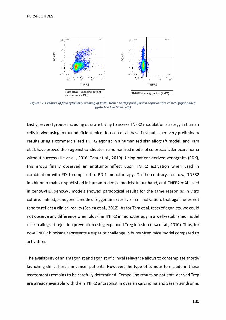
PERSPECTIVES
180
Figure 17: Example of flow cytometry staining of PBMC from one (left panel) and its appropriate control (right panel) (gated on live CD3+ cells)
Lastly, several groups including ours are trying to assess TNFR2 modulation strategy in human
cells in vivo using immunodeficient mice. Joosten et al. have first published very preliminary
results using a commercialized TNFR2 agonist in a humanized skin allograft model, and Tam
et al. have proved their agonist candidate in a humanized model of colorectal adenocarcinoma
without success (He et al., 2016; Tam et al., 2019). Using patient-derived xenografts (PDX),
this group finally observed an antitumor effect upon TNFR2 activation when used in
combination with PD-1 compared to PD-1 monotherapy. On the contrary, for now, TNFR2
inhibition remains unpublished in humanized mice models. In our hand, anti-TNFR2 mAb used
in xenoGvHD, xenoGvL models showed paradoxical results for the same reason as in vitro
culture. Indeed, xenogeneic models trigger an excessive T cell activation, that again does not
tend to reflect a clinical reality (Scalea et al., 2012). As for Tam et al. tests of agonists, we could
not observe any difference when blocking TNFR2 in monotherapy in a well-established model
of skin allograft rejection prevention using expanded Treg infusion (Issa et al., 2010). Thus, for
now TNFR2 blockade represents a superior challenge in humanized mice model compared to
activation.
The availability of an antagonist and agonist of clinical relevance allows to contemplate shortly
launching clinical trials in cancer patients. However, the type of tumour to include in these
assessments remains to be carefully determined. Compelling results on patients-derived Treg
are already available with the hTNFR2 antagonist in ovarian carcinoma and Sézary syndrome.
7.41 0.061
1.3191.2
0-103
103
104
105
TNFR2
0
-103
103
104
105
FO
XP
3
1.32 5.47
36.356.9
0-103
103
104
105
TNFR2
0
-103
103
104
105
FO
XP
3
Post-HSCT relapsing patient
(will recieve a DLI)TNFR2 staining control (FMO)
PERSPECTIVES
181
The presence of TNFR2+ Treg has also been reported in numerous other types of malignancies,
allowing to guide the cohort choice. Regarding post-allograft relapses, the reproducibility of
our results in a second model, humanised or not, is necessary to prove TNFR2 inhibition effect
on tumoral growth. The lack of data concerning TNFR2 expression in relapsing patients will
also need to be overcome before considering safely starting a clinical trial.

177
Bibliography

BIBLIOGRAPHY
183