Experimental validation of 5 in-silico predicted glioma biomarkers
Upload
independentCategory
view
3download
0
BioMed CentralMolecular Cancer
ss
Open AcceResearchDifferential expression and role of p21cip/waf1 and p27kip1 in TNF-α-induced inhibition of proliferation in human glioma cellsPabbisetty Sudheer Kumar, Anjali Shiras, Gowry Das, Jayashree C Jagtap, Vandna Prasad and Padma Shastry*Address: National Centre for Cell Science, NCCS, Ganeshkhind, Pune 411 007, India
Email: Pabbisetty Sudheer Kumar - [email protected]; Anjali Shiras - [email protected]; Gowry Das - [email protected]; Jayashree C Jagtap - [email protected]; Vandna Prasad - [email protected]; Padma Shastry* - [email protected]
* Corresponding author
AbstractBackground: The role of TNF-α in affecting the fate of tumors is controversial, while somestudies have reported apoptotic or necrotic effects of TNF-α, others provide evidence thatendogenous TNF-α promotes growth and development of tumors. Understanding themechanism(s) of TNF-α mediated growth arrest will be important in unraveling the contributionof tissue associated macrophages in tumor resistance. The aim of this study was to investigate therole of Cyclin Dependent Kinase Inhibitors (CDKI) – p21cip/waf1 and p27kip1 in TNF-α mediatedresponses in context with p53 and activation of NF-κB and Akt pathways. The study was done withhuman glioma cell lines -LN-18 and LN-229 cells, using monolayer cultures and MulticellularSpheroids (MCS) as in vitro models.
Results: TNF-α induced inhibition of proliferation and enhanced the expression of p21cip/waf1 andp27kip1 in LN-18 cells. p21 was induced on exposure to TNF-α, localized exclusively in the nucleusand functioned as an inhibitor of cell cycle but not as an antiapoptotic protein. In contrast, p27 wasconstitutively expressed, localized predominantly in the cytoplasm and was not involved in arrestof proliferation. Our data using IκBα mutant LN-18 cells and PI3K/Akt inhibitor-LY294002 revealedthat the expression of p21 is regulated by NF-κB. Loss of IκBα function in LN-229 cells (p53positive) did not influence TNF-α induced accumulation of pp53 (Ser-20 p53) suggesting that p53was not down stream of NF-κB. Spheroidogenesis enhanced p27 expression and p21 induced byTNF-α was significantly increased in the MCS compared to monolayers.
Conclusion: This study demarcates the functional roles for CDKIs-p21cip/waf1 and p27kip1 duringTNF-α stimulated responses in LN-18 glioma cells. Our findings provide evidence that TNF-α-induced p21 might be regulated by NF-κB or p53 independently. p21 functions as an inhibitor ofcell proliferation and does not have a direct role in rendering the cells resistant to TNF-α mediatedcytotoxicity.
Published: 12 June 2007
Molecular Cancer 2007, 6:42 doi:10.1186/1476-4598-6-42
Received: 14 February 2007Accepted: 12 June 2007
This article is available from: http://www.molecular-cancer.com/content/6/1/42
© 2007 Kumar et al; licensee BioMed Central Ltd. This is an Open Access article distributed under the terms of the Creative Commons Attribution License (http://creativecommons.org/licenses/by/2.0), which permits unrestricted use, distribution, and reproduction in any medium, provided the original work is properly cited.
Page 1 of 12(page number not for citation purposes)
Molecular Cancer 2007, 6:42 http://www.molecular-cancer.com/content/6/1/42
BackgroundGliomas are the most common malignant brain tumors inadults [1]. The median survival of the patients is less thantwo years and the prognosis remains poor despite exten-sive research and advances in radiation therapy and chem-otherapeutic regimes. The factors responsible for theaggressive behavior of gliomas include marked local inva-sive growth, neovascularization and evasion to immuneresponses [2]. Macrophages comprise an important com-ponent of the immune system against tumors. In gliomas,macrophages are recruited and remain at the site of tumorconstituting a major proportion of the tumor mass [3].The ability of gliomas to escape the host immune systemis thought to contribute to the malignant behavior ofthese tumors, raising speculation about the role of thesetumor infiltrating macrophages in progression and aggres-siveness of gliomas [3]. Secretion of cytokines, particularlyTNF-α, by activated macrophages is known to affect sur-vival, growth and proliferation of the tumor [4]. TNF-αmediates apoptotic or necrotic effects in many tumorsdepending on cell types, and causes growth inhibition in40% of tumor cell lines [5-7]. Reports on the effects ofTNF-α on glioma growth and progression are contradic-tory. TNF-α-induced responses range from cytotoxicity tocytostatic effects [8-10]. Earlier in vivo studies with tumorsindicated that TNF-α was a death inducer and hence usedin clinical trials for treatment of cancers [11]. Howeverrecent reports provide evidence that endogenous TNF-αpromotes growth and development of tumors [12,13]. Ingliomas, TNF-α is shown to reduce growth and prolongsurvival by enhancing macrophage recruitment andmicrocyst formation [14]. Understanding the mecha-nism(s) of TNF-α mediated growth arrest will be impor-tant in unraveling the contribution of tissue associatedmacrophages in tumor resistance in gliomas.
The progression of cells through cell cycle depends on theactivation of cyclins and cyclin dependent kinases – CDKswhich function together in the G1 phase for initiating Sand progression to G2/M phases. The activity of the com-plexes is regulated by two families of cyclin dependentkinase inhibitors (CDKIs)-including INK4 proteins thatbind only to CDK4/CDK6 and are specific for G1 phaseand the cip/kip proteins which include p21cip/waf1 and p27kip1 and are not specific for any particular cell cycle phase[15,16]. p21 is an universal inhibitor of cell cycle progres-sion and arrests in G1 as well as G2 phases [17]. p21 neg-atively regulates cell cycle progression by inhibiting CDK2and CDK4 and blocks DNA replication by binding toPCNA [18]. p21 is also implicated in terminal differentia-tion, replicative senescence and protection from p53dependent and p53-independent apoptosis [19,20]. p27arrests at G1 phase of the cell cycle by contact inhibitionand controls the G1/S transition by inhibiting the activityof a wide variety of cyclin/CDK complexes [21]. It is
expressed in many tumors and may be partly responsiblefor growth arrest in tumor cells [22,23]. Though muta-tions in p27 are rare, a negative correlation between p27expression and tumor progression is documented inmany cancers and loss of p27 in tumors is associated withpoor prognosis [24].
Elucidation of cellular and molecular mechanismsinvolved in survival/death signaling in response to chem-otherapeutic agents has been a focus of recent studies forunderstanding drug resistance in tumors. However, suchstudies have been hampered by limitations in the conven-tional in vitro models as they do not reflect the complexi-ties of solid tumors. Multicellular spheroids (MCS)closely resemble the in vivo situation with regard to cellshape and environment, which in turn can affect the geneexpression and behavior of the cells [25]. The biologicalsignificance and clinical relevance of MCS have been welldocumented and these experimental systems have beenused for studying micro-environmental effects on basicmechanisms, such as regulation of proliferation, metabo-lism, differentiation, cell death, invasion, angiogenesisand immune response [26-28]. Though the MCS showgreater resistance compared to the two dimensional-mon-olayer cultures, the mechanisms that confer greater resist-ance to radiotherapy, chemotherapy in the MCS are notwell understood [29]. In this study, we analyzed the effectof TNF-α on proliferation and survival of human gliomacells and investigated the role of CDKIs-p21cip/waf1 andp27kip1 in context with p53 and activation of NF-κB andAkt pathways using monolayer cultures and MCS as invitro models.
ResultsTNF-α-induced inhibition of proliferation is mediated by TNFR1Initial experiments were done to assess the effect of TNF-α on cell growth in LN-18 cells. Monolayers and sphe-roids derived from LN-18 cells were treated with TNF-αand cell proliferation was measured by tritiated thymidineincorporation assay. TNF-α (2.5–100 ng/ml) inducedinhibition of proliferation (25%) even at the lowest con-centration, with at least two fold greater inhibition (65%)in spheroids at concentrations ≥10 ng/ml (Fig. 1A). TNF-α-induced signaling are mediated via distinct cell surfacereceptors, TNFR1 (~55 kDa) and TNFR2 (~75 kDa) [30].The expression of these receptors in LN-18 cells was stud-ied by RT-PCR and flowcytometry analysis. The RT-PCRdata and the histoplots show that LN-18 cells express highlevels of TNFR1 compared to TNFR2. This was in contrastwith another human glioma cell line-U87MG, whichshowed high expression of both the receptors (Fig. 1B and1C). To investigate the receptor mediated signaling, cellswere pre-incubated with neutralizing antibodies toTNFR1 and TNFR2 (50 µg/ml) and subsequently treated
Page 2 of 12(page number not for citation purposes)
Molecular Cancer 2007, 6:42 http://www.molecular-cancer.com/content/6/1/42
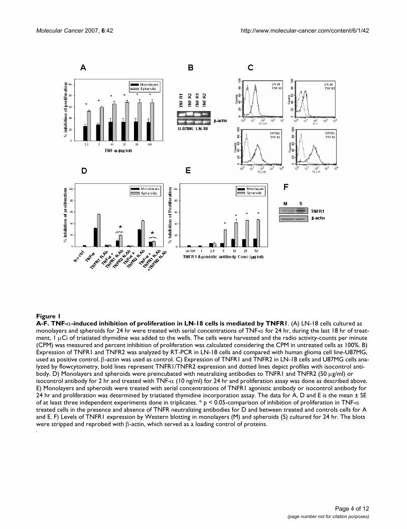
with TNF-α for 24 hr. Pretreatment with neutralizing anti-bodies to TNFR1 but not TNFR2 reversed the inhibitionsignificantly (Fig. 1D), suggesting that TNF-α stimulatedresponses were mediated by TNFR1. TNFR1 agonistic anti-body (1–50 µg/ml) induced inhibition of proliferation upto 50% at concentrations ≥10 µg/ml in the MCS, while theinhibition was only 15% in the monolayers (Fig. 1E). Asthe inhibition with TNF-α and TNFR1 agonistic antibodywas significantly higher in the spheroids, we determinedthe levels of TNFR1 in the two culture models by Westernblotting and found that the expression of TNFR1 wasmany fold higher in the spheroids (Fig. 1F). These experi-ments demonstrated that TNF-α-induced inhibition ofproliferation was significantly higher in spheroids com-pared to monolayers in LN-18 cells and the effect wasmediated by TNFR1.
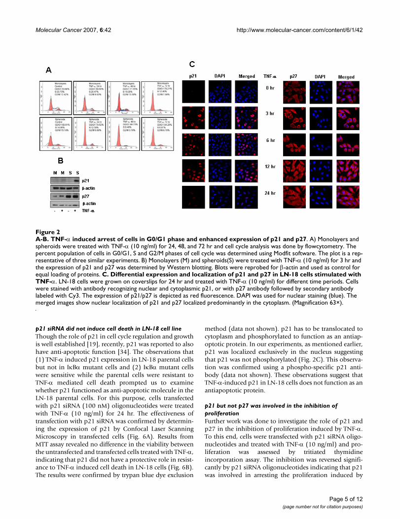
TNF-α up regulates p21 and p27 expressionThe inhibition in cell growth induced by TNF-α was stud-ied by cell cycle analysis. LN-18 cells were treated withTNF-α up to 72 hr period and analyzed for DNA contentby flow-cytometry at 24 hr intervals. Spheroids culturedfor 24 hr had higher population of G0/G1 cells comparedto the corresponding monolayer. TNF-α treatment signif-icantly increased the cell population in G0/G1 cells inboth the models at all the time points but to a greaterextent in spheroids compared to monolayer (Fig. 2A)
Cyclin dependent kinase inhibitors (CDKI) p21cip/waf1 andp27 kip1 are important in TNF-α mediated signaling [31].We studied the effect of TNF-α on the expression of theseCDKI proteins in the two culture models of LN-18 cells byimmunoblotting. As shown in Figure 2B, the expressionof p21 was below the level of detection in the both the cul-ture systems. On stimulation with TNF-α, the level inmonolayers was marginally increased while the expres-sion was strongly enhanced in the spheroids. Our dataalso revealed that LN-18 cells constitutively expressed p27and the levels increased drastically with spheroidogenesis.Stimulation with TNF-α resulted in further upregulationof p27 expression in both the models (Fig. 2B). We furtherperformed time course experiments by Confocal Laserscanning Microscopy (CLSM) to study the expression andlocalization of p21 and p27 in response to TNF-α. Wefound that p21 was not detectable in the control cells con-firming the data from immunoblotting experiments. p21was induced from 3 hr post-treatment with TNF-α withfurther increase in the number of p21 positive cells at 6 hr.The level decreased at 12 hr and complete loss of expres-sion was observed at 24 hr. Using an antibody that recog-nizes p21 in the nucleus as well as cytoplasm it was foundthat p21 was restricted to the nucleus (Fig. 2C). The cellswere highly positive for p27 (>95%) with predominantlycytoplasmic staining though nuclear staining was also
seen in some cells. TNF-α treatment marginally enhancedthe p27 expression from 3 hr onwards (Fig. 2C).
TNF-α-induced p21 is regulated by NF-κB and p53 independentlyNF-κB pathway is a one of the major mediators in TNF-αinduced effects. As our preliminary experiments indicatedthat p21 was induced on stimulation with TNF-α in LN-18 cells, we examined the role of NF-κB pathway in theregulation of p21 using a stable IκBα mutant LN-18 cellline. Characterization of the IκBα mutant cell line con-firmed that the cells were sensitive to TNF-α mediated celldeath. Figure 3A and 3B shows the response of the IκBαmutant cell line to TNF-α determined by MTT assay andPARP cleavage. Western blotting and immunofluores-cence analysis revealed that p21 was not detected in theIκBα mutant LN-18 cells and was not induced on stimu-lation with TNF-α (Fig. 3C, and 3D) suggesting that p21expression might be regulated by the NF-κB pathway. Onthe other hand, the expression of p27 was not altered inIκBα mutant LN-18 cells (Fig. 3D and Fig. 2B), implyingthat p27 expression was not under the control of NF-κB.
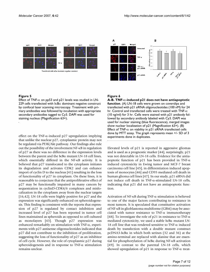
p21 is regulated mainly via tumor suppressor protein-p53. Recently, p52/p100 NF-κB was described to regulatep53 function and hence influence the p53-regulated deci-sion-making following DNA damage, a process involvingp21 [32]. As LN-18 cells express mutant p53 protein, toexamine if NF-κB influenced the p21 expression directlyor whether it was via p53, experiments were performedusing LN-229, a human glioma cell line expressing wild-type p53 [33]. Cells were exposed to TNF-α for 6 hr andanalyzed for the expression of phosphorylated ser20-p53(pp53) and p21. Stimulation with TNF-α led to enhancedaccumulation of phosphorylated p53 in the nucleus,interestingly, the untreated control cells also displayeddetectable levels of pp53. In contrast, the expression ofnuclear p21 decreased in TNF-α treated cells compared tocontrols (Fig. 4A). To delineate the pathways regulatingp21, the effect of TNF-α on pp53 and p21 was studied inLN-229 cells transfected with IκBα dominant negativeconstruct. The loss of IκBα activity in the transfected cellswas evident by the inhibition of p65 translocation tonucleus on stimulation with TNF-α compared withuntransfected LN-229 cells (Fig. 4B). To address the ques-tion whether loss of NF-κB activation in these cells mightinfluence the levels of pp53 and p21, transfected cellsexposed to TNF-α for 6 hr were stained with antibodies toSer20-p53 and p21. As shown in figure 5, there was no sig-nificant difference in the accumulation of pp53 or nuclearp21 between the transfected and untransfected cells.These findings led us to conclude that p53 might not func-tion downstream of NF-κB and secondly, TNF-α-inducedp21 can be regulated by p53 and NF-κB independently.
Page 3 of 12(page number not for citation purposes)
Molecular Cancer 2007, 6:42 http://www.molecular-cancer.com/content/6/1/42
Page 4 of 12(page number not for citation purposes)
A-F. TNF-α-induced inhibition of proliferation in LN-18 cells is mediated by TNFR1Figure 1A-F. TNF-α-induced inhibition of proliferation in LN-18 cells is mediated by TNFR1. (A) LN-18 cells cultured as monolayers and spheroids for 24 hr were treated with serial concentrations of TNF-α for 24 hr, during the last 18 hr of treat-ment, 1 µCi of triatiated thymidine was added to the wells. The cells were harvested and the radio activity-counts per minute (CPM) was measured and percent inhibition of proliferation was calculated considering the CPM in untreated cells as 100%. B) Expression of TNFR1 and TNFR2 was analyzed by RT-PCR in LN-18 cells and compared with human glioma cell line-U87MG, used as positive control. β-actin was used as control. C) Expression of TNFR1 and TNFR2 in LN-18 cells and U87MG cells ana-lyzed by flowcytometry, bold lines represent TNFR1/TNFR2 expression and dotted lines depict profiles with isocontrol anti-body. D) Monolayers and spheroids were preincubated with neutralizing antibodies to TNFR1 and TNFR2 (50 µg/ml) or isocontrol antibody for 2 hr and treated with TNF-α (10 ng/ml) for 24 hr and proliferation assay was done as described above. E) Monolayers and spheroids were treated with serial concentrations of TNFR1 agonistic antibody or isocontrol antibody for 24 hr and proliferation was determined by triatiated thymidine incorporation assay. The data for A, D and E is the mean ± SE of at least three independent experiments done in triplicates. * p < 0.05-comparison of inhibition of proliferation in TNF-α treated cells in the presence and absence of TNFR neutralizing antibodies for D and between treated and controls cells for A and E. F) Levels of TNFR1 expression by Western blotting in monolayers (M) and spheroids (S) cultured for 24 hr. The blots were stripped and reprobed with β-actin, which served as a loading control of proteins.
Molecular Cancer 2007, 6:42 http://www.molecular-cancer.com/content/6/1/42
p21 siRNA did not induce cell death in LN-18 cell lineThough the role of p21 in cell cycle regulation and growthis well established [19], recently, p21 was reported to alsohave anti-apoptotic function [34]. The observations that(1) TNF-α induced p21 expression in LN-18 parental cellsbut not in IκBα mutant cells and (2) IκBα mutant cellswere sensitive while the parental cells were resistant toTNF-α mediated cell death prompted us to examinewhether p21 functioned as anti-apoptotic molecule in theLN-18 parental cells. For this purpose, cells transfectedwith p21 siRNA (100 nM) oligonucleotides were treatedwith TNF-α (10 ng/ml) for 24 hr. The effectiveness oftransfection with p21 siRNA was confirmed by determin-ing the expression of p21 by Confocal Laser ScanningMicroscopy in transfected cells (Fig. 6A). Results fromMTT assay revealed no difference in the viability betweenthe untransfected and transfected cells treated with TNF-α,indicating that p21 did not have a protective role in resist-ance to TNF-α induced cell death in LN-18 cells (Fig. 6B).The results were confirmed by trypan blue dye exclusion
method (data not shown). p21 has to be translocated tocytoplasm and phosphorylated to function as an antiap-optotic protein. In our experiments, as mentioned earlier,p21 was localized exclusively in the nucleus suggestingthat p21 was not phosphorylated (Fig. 2C). This observa-tion was confirmed using a phospho-specific p21 anti-body (data not shown). These observations suggest thatTNF-α-induced p21 in LN-18 cells does not function as anantiapoptotic protein.
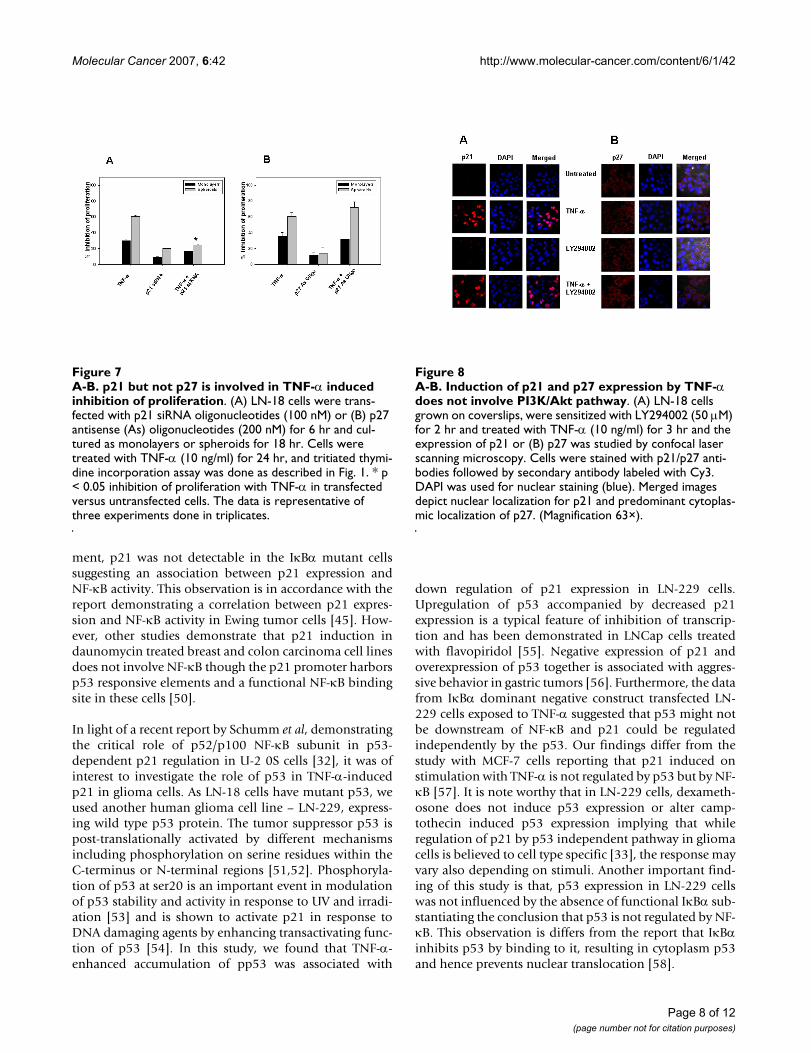
p21 but not p27 was involved in the inhibition of proliferationFurther work was done to investigate the role of p21 andp27 in the inhibition of proliferation induced by TNF-α.To this end, cells were transfected with p21 siRNA oligo-nucleotides and treated with TNF-α (10 ng/ml) and pro-liferation was assessed by tritiated thymidineincorporation assay. The inhibition was reversed signifi-cantly by p21 siRNA oligonucleotides indicating that p21was involved in arresting the proliferation induced by
A-B. TNF-α induced arrest of cells in G0/G1 phase and enhanced expression of p21 and p27Figure 2A-B. TNF-α induced arrest of cells in G0/G1 phase and enhanced expression of p21 and p27. A) Monolayers and spheroids were treated with TNF-α (10 ng/ml) for 24, 48, and 72 hr and cell cycle analysis was done by flowcytometry. The percent population of cells in G0/G1, S and G2/M phases of cell cycle was determined using Modfit software. The plot is a rep-resentative of three similar experiments. B) Monolayers (M) and spheroids(S) were treated with TNF-α (10 ng/ml) for 3 hr and the expression of p21 and p27 was determined by Western blotting. Blots were reprobed for β-actin and used as control for equal loading of proteins. C. Differential expression and localization of p21 and p27 in LN-18 cells stimulated with TNF-α. LN-18 cells were grown on coverslips for 24 hr and treated with TNF-α (10 ng/ml) for different time periods. Cells were stained with antibody recognizing nuclear and cytoplasmic p21, or with p27 antibody followed by secondary antibody labeled with Cy3. The expression of p21/p27 is depicted as red fluorescence. DAPI was used for nuclear staining (blue). The merged images show nuclear localization of p21 and p27 localized predominantly in the cytoplasm. (Magnification 63×).
Page 5 of 12(page number not for citation purposes)

Molecular Cancer 2007, 6:42 http://www.molecular-cancer.com/content/6/1/42
TNF-α (Fig. 7A). Similar experiments done with LN-18cells transfected with p27 antisense oligonucleotides (200nM) revealed no difference in the inhibition between con-trols and transfected cells treated with TNF-α (Fig. 7B).Together, these results show that p21 did not function asan antiapoptotic protein and secondly p21 but not p27played a crucial role in the arrest of proliferation inducedby TNF-α.
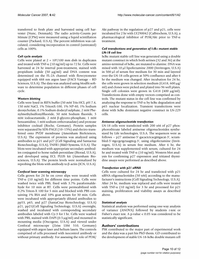
The CDKIs, p21 and p27 are reported to be also regulatedby PI3K/Akt pathway [35-38], we therefore examined therole of PI3K/Akt pathway by treating LN-18 cells sensi-tized with LY294002, a pharmacological inhibitor ofPI3K/Akt pathway with TNF-α. The inhibitor had no effecton the fluorescence intensity or the percent positivity ofp21 or p27 positive cells in TNF-α stimulated cells, sug-gesting that PI3K/Akt pathway was not involved in regula-tion of these CDKIs in LN-18 cells (Fig. 8A and 8B).
DiscussionIn this report, we established that though p21cip/waf1 andp27kip1 are upregulated in response to TNF-α, p21 but notp27 is involved in the arrest of proliferation in TNF-αstimulated LN-18 glioma cells. TNF-α-induced inhibitionof proliferation was mediated via TNFR1. This observa-tion is significant as most aggressive gliomas express highlevels of TNFR1. We also demonstrated that p21 and p27differed in their localization and expression levels in theexperimental models represented by monolayers andspheroids. While p27 was constitutively present, p21 wasinduced on stimulation with TNF-α and the upregulationin the spheroids was markedly higher compared to mon-olayers.
p27 is expressed abundantly in human malignant gliomasand is associated with better prognosis [39,40]. p27 isnormally a nuclear protein but cytoplasmic localizationhas been demonstrated in breast and colon cancers andcorrelates with poor survival in adenocarcinoma [23].Relocalization of p27 in the cytoplasm is the result ofphosphorylation of p27 nuclear localization domaininduced by Akt kinase [36] and the presence of cytoplas-mic p27 correlates with activated Akt in breast tumors[41]. It is likely that such a correlation exists in gliomasalso as high basal levels and activated Akt were detected inLN-18 cells (our unpublished data) and the cells werehighly positive for p27 with predominant cytoplasmiclocalization. Interestingly, PI3K/Akt inhibitor had no
A-B. Effect of TNF-α on expression of pp53 and p65 in LN-229 cells (p53 positive)Figure 4A-B. Effect of TNF-α on expression of pp53 and p65 in LN-229 cells (p53 positive). A) LN-229 cells were exposed to TNF-α for 6 hr and stained with antibodies to phosphorylated Ser-20 p53 and p21. B) LN-229 cells were transfected with IκBα dominant negative construct and the expression of nuclear p65 was determined after exposure to TNF-α for 6 hr.
A-D. Effect of TNF-α on cell viability and expression of p21 and p27 in IκBα mutant LN-18 cellsFigure 3A-D. Effect of TNF-α on cell viability and expression of p21 and p27 in IκBα mutant LN-18 cells. (A) Wild and mutant IκBα LN-18 cells were treated with TNF-α (10 ng/ml) for 12 hr and cell viability was assessed by MTT assay. The percent cell death was determined considering viability of control cells as 100%. (B) IκBα mutant LN-18 cells were treated with TNF-α (10 ng/ml) for different time points and cleavage of PARP (89 kD fragment) was determined by Western blotting. C) IκBα mutant LN-18 cells grown on coverslips were treated with TNF-α (10 ng/ml) for 3 hr and stained with p21 followed by secondary antibody labeled with Cy3 and DAPI was used for staining nucleus (Magnifica-tion 63×). D) Expression of p21 and p27 determined by Western blotting in monolayers (M) and spheroids (S) derived from IκBα mutant LN-18 cells treated with TNF-α for 3 hr. The blots were stripped and reprobed with β-actin, which served as control for equal loading of proteins.
Page 6 of 12(page number not for citation purposes)
Molecular Cancer 2007, 6:42 http://www.molecular-cancer.com/content/6/1/42
effect on the TNF-α-induced p27 upregulation implyingthat unlike the nuclear p27, cytoplasmic protein may notbe regulated via PI3K/Akt pathway. Our findings also ruleout the possibility of the involvement NF-κB in regulationof p27 as there was no difference in the expression levelsbetween the parent and the IκBα mutant LN-18 cell lines,which essentially differed in the NF-κB activity. It isbelieved that p27 translocated to the cytoplasm initiatesits degradation and activates CDK2 and can enhanceimport of cyclin D to the nucleus [41] resulting in the lossof functionality of p27 in cytoplasm. On these lines, it isreasonable to conjecture that the antiproliferative effect ofp27 may be functionally impaired in many cancers bysequestration in cyclinD-CDK4/6 complexes and mislo-calization in the cytoplasm away from the nuclear targets[16,42]. LN-18 cells were highly positive for p27 and theexpression was significantly enhanced on spheroidogene-sis. This finding is consistent with the reports that expres-sion of p27 is regulated by contact inhibition andincreased level of p27 has been reported in tumor celllines maintained as spheroids as opposed to cell culturedas monolayers [43]. Though p27 expression wasenhanced remarkably on stimulation with TNF-α, experi-ments with p27-antisense oligonucleotides indicated thatp27 did not contribute to the inhibition of proliferation,suggesting the loss of functionality of p27 as an inhibitorof cell cycle. However, the role of cytoplasmic p27 duringspheroidogenesis and in response to TNF-α stimulationremains unclear.
Elevated levels of p21 is reported in aggressive gliomasand is used as a prognostic marker [44], surprisingly, p21was not detectable in LN-18 cells. Evidence for the antia-poptotic function of p21 has been provided in TNF-αinduced cytotoxicity in Ewing tumor and MCF-7 breastcarcinoma cell line [45], in differentiation induced apop-tosis of monocytes [46] and CD95 mediated cell death inhuman glioma cell lines [47]. In our study, p21 siRNA didnot induce cell death in TNF-α-stimulated LN-18 cells,indicating that p21 did not have an antiapoptotic func-tion.
Activation of NF-κB during TNF-α stimulation is believedto one of the major factors contributing to resistance inmost tumors. It is speculated that constitutive activationof NF-κB in glioblastoma multiforme (GBM) may be asso-ciated with tumor resistance to TNF-α immunotherapy[48]. To investigate the role of p21 in resistance to TNF-αmediated cytotoxicity, we used a stable IκBα mutant LN-18 cell line that was rendered sensitive to TNF-α induceddeath by transfection with a double mutant constructpcDNA3-IκBα in which both serines (32 and 36) at theamino-terminal are replaced with alanine that are essen-tial for phosphorylation of IκBα during NF-κB activation[49]. In contrast to the parental LN-18 cells, whichshowed upregulation of p21 in response to TNF-α treat-
A-B. TNF-α-induced p21 does not have antiapoptotic func-tionFigure 6A-B. TNF-α-induced p21 does not have antiapoptotic function. (A) LN-18 cells were grown on coverslips and transfected with p21 siRNA oligonucleotides (100 nM) for 24 hr. Control and transfected cells were treated with TNF-α (10 ng/ml) for 3 hr. Cells were stained with p21 antibody fol-lowed by secondary antibody labeled with Cy3. DAPI was used for nuclear staining (blue fluorescence), merged images show nuclear localization of p21 (Magnification 63×). (B) Effect of TNF-α on viability in p21 siRNA transfected cells done by MTT assay. The graph represents mean +/- SD of 3 experiments done in duplicates.
Effect of TNF-α on pp53 and p21 levels was studied in LN-229 cells transfected with IκBα dominant negative construct by confocal laser scanning microscopyFigure 5Effect of TNF-α on pp53 and p21 levels was studied in LN-229 cells transfected with IκBα dominant negative construct by confocal laser scanning microscopy. Treatment with pri-mary antibodies was followed by incubation with appropriate secondary antibodies tagged to Cy3. DAPI was used for staining nucleus (Magnification 63×).
Page 7 of 12(page number not for citation purposes)
Molecular Cancer 2007, 6:42 http://www.molecular-cancer.com/content/6/1/42
ment, p21 was not detectable in the IκBα mutant cellssuggesting an association between p21 expression andNF-κB activity. This observation is in accordance with thereport demonstrating a correlation between p21 expres-sion and NF-κB activity in Ewing tumor cells [45]. How-ever, other studies demonstrate that p21 induction indaunomycin treated breast and colon carcinoma cell linesdoes not involve NF-κB though the p21 promoter harborsp53 responsive elements and a functional NF-κB bindingsite in these cells [50].
In light of a recent report by Schumm et al, demonstratingthe critical role of p52/p100 NF-κB subunit in p53-dependent p21 regulation in U-2 0S cells [32], it was ofinterest to investigate the role of p53 in TNF-α-inducedp21 in glioma cells. As LN-18 cells have mutant p53, weused another human glioma cell line – LN-229, express-ing wild type p53 protein. The tumor suppressor p53 ispost-translationally activated by different mechanismsincluding phosphorylation on serine residues within theC-terminus or N-terminal regions [51,52]. Phosphoryla-tion of p53 at ser20 is an important event in modulationof p53 stability and activity in response to UV and irradi-ation [53] and is shown to activate p21 in response toDNA damaging agents by enhancing transactivating func-tion of p53 [54]. In this study, we found that TNF-α-enhanced accumulation of pp53 was associated with
down regulation of p21 expression in LN-229 cells.Upregulation of p53 accompanied by decreased p21expression is a typical feature of inhibition of transcrip-tion and has been demonstrated in LNCap cells treatedwith flavopiridol [55]. Negative expression of p21 andoverexpression of p53 together is associated with aggres-sive behavior in gastric tumors [56]. Furthermore, the datafrom IκBα dominant negative construct transfected LN-229 cells exposed to TNF-α suggested that p53 might notbe downstream of NF-κB and p21 could be regulatedindependently by the p53. Our findings differ from thestudy with MCF-7 cells reporting that p21 induced onstimulation with TNF-α is not regulated by p53 but by NF-κB [57]. It is note worthy that in LN-229 cells, dexameth-osone does not induce p53 expression or alter camp-tothecin induced p53 expression implying that whileregulation of p21 by p53 independent pathway in gliomacells is believed to cell type specific [33], the response mayvary also depending on stimuli. Another important find-ing of this study is that, p53 expression in LN-229 cellswas not influenced by the absence of functional IκBα sub-stantiating the conclusion that p53 is not regulated by NF-κB. This observation is differs from the report that IκBαinhibits p53 by binding to it, resulting in cytoplasm p53and hence prevents nuclear translocation [58].
A-B. Induction of p21 and p27 expression by TNF-α does not involve PI3K/Akt pathwayFigure 8A-B. Induction of p21 and p27 expression by TNF-α does not involve PI3K/Akt pathway. (A) LN-18 cells grown on coverslips, were sensitized with LY294002 (50 µM) for 2 hr and treated with TNF-α (10 ng/ml) for 3 hr and the expression of p21 or (B) p27 was studied by confocal laser scanning microscopy. Cells were stained with p21/p27 anti-bodies followed by secondary antibody labeled with Cy3. DAPI was used for nuclear staining (blue). Merged images depict nuclear localization for p21 and predominant cytoplas-mic localization of p27. (Magnification 63×).
A-B. p21 but not p27 is involved in TNF-α induced inhibition of proliferationFigure 7A-B. p21 but not p27 is involved in TNF-α induced inhibition of proliferation. (A) LN-18 cells were trans-fected with p21 siRNA oligonucleotides (100 nM) or (B) p27 antisense (As) oligonucleotides (200 nM) for 6 hr and cul-tured as monolayers or spheroids for 18 hr. Cells were treated with TNF-α (10 ng/ml) for 24 hr, and tritiated thymi-dine incorporation assay was done as described in Fig. 1. * p < 0.05 inhibition of proliferation with TNF-α in transfected versus untransfected cells. The data is representative of three experiments done in triplicates.
Page 8 of 12(page number not for citation purposes)

Molecular Cancer 2007, 6:42 http://www.molecular-cancer.com/content/6/1/42
Recent studies suggest an antiapoptotic function for p21[45]. To exert protective effect against damage induced byapoptotic agents, it is essential that p21 is phosphorylatedand is localized in the cytoplasm [20]. We found thatTNF-α-induced p21 was restricted to the nucleus suggest-ing that it might not function as an antiapoptotic protein.Our conclusion that TNF-α-induced p21 was not phos-phorylated and was restricted to nuclear localization isbased on our findings using two different antibodies toexamine the phosphorylation status of p21, an antibodythat was immunoreactive with nuclear as well as cytoplas-mic p21 and a second antibody recognizing exclusivelyphosphorylated p21 (details in materials and methods).Though the molecular mechanisms in compartmentaliza-tion of p21 are not clear, it is reported that Akt might reg-ulate the subcellular changes [37]. Phosphotidylinositol3-kinase (PI3K) and its downstream target, the Akt/PKBserine threonine kinase [59] are activated on TNF-TNFRbinding. Akt activity is elevated in most GBM cells partic-ularly those with mutant form of PTEN and is importantin glioma formation and progression [60]. Activation ofAkt is paralleled with increased p21 stability and corre-lated with resistance to taxol-induced apoptosis in gliob-lastoma cell lines [38]. High grade gliomas are known toexpress high levels of Akt [52] and enhanced Akt activityis demonstrated in spheroids from Ewing tumors [53]. Weobserved increased levels and activity of Akt in spheroidsof LN-18 cells (our unpublished data). These findings andour data depicting increased expression of TNF-α-inducedp21 in spheroids compared to monolayer cells promptedus to examine the involvement of Akt in the regulation ofp21. We found that PI3K/Akt inhibitor-LY290042 did notaffect the expression of p21 suggesting that it was not reg-ulated by this pathway. Experiments with p21 siRNAreversed the inhibition of proliferation confirming its roleas a regulator of cell cycle progression. These results led usto conclude that p21 functioned as an inhibitor of prolif-eration but not as an antiapoptotic protein in TNF-αmediated response in LN-18 cells. Furthermore, our stud-ies with IκBα mutant LN-18 cells and pharmacologicalinhibitor-LY290042 revealed that p21 expression was reg-ulated by NF-κB and not Akt pathway.
In conclusion, this study demarcates the functions ofCDKIs-p21cip/waf1 and p27kip1 during TNF-α stimulatedresponses in LN-18 glioma cells. p21 induced on stimula-tion with TNF-α may be regulated by NF-κB or p53, prob-ably depending on the status of p53. Our findings do notsupport a role for p27 in regulation of proliferation or ren-dering resistance in TNF-α induced cytotoxicity, whichcould be attributed to its cytoplasmic localization. How-ever questions concerning the relevance of enhanced cyto-plasmic p27 expression during spheroidogenesis and inresponse to TNF-α stimulation remain to be answered.
The findings in this study assume importance from thepoint of TNF-α-TNFR1 interaction in the in vivo milieu ingliomas wherein TNF-α is secreted by the tumor infiltrat-ing macrophages and TNF receptors are abundantlyexpressed by the tumor cells. The stimulation leads to acti-vation of various pathways including NF-κB pathwaywhich contributes to chemoresistance. The resistance maybe further enhanced by induction of p21 by TNF-α in NF-κB dependent manner leading to the arrest of cells in theG1 phase. The regulation of p21 by p53 and NF-κB in wildtype p53 and mutant p53 tumors in response to TNF-αdeserves further in-depth studies Additionally, heightenedinduction of p21 to TNF-α stimulation in MCS represent-ing the cells in low proliferating phase underscore theMCS as more realistic model for studying the molecularmechanisms in resistance to chemotherapeutic agents insolid tumors.
MethodsCell cultures and generation of multicellular spheroidsThe human glioma cell lines LN-18, LN-229 and U87MGwere procured from American Type Culture Collection(ATCC, U.S.A). Cells were cultured in Dulbecco' s modi-fied eagle's medium (DMEM) supplemented with 1.5 gm/L sodium bicarbonate, 4 mM glutamine and 5%-10%fetal bovine serum (Gibco, U.S.A) at 37°C in an atmos-phere of 5% CO2/95% air. Multicellular spheroids weregenerated by the liquid overlay technique as describedpreviously [61] with a slight modification. All experi-ments were done using 24 hr cultures. Single cell suspen-sions were prepared from spheroids by dissociation with0.25% trypsin in PBS at 37°C for 30 min.
Cell viability assayThe effect of TNF-α on cell viability was assessed by MTT(3-(4, 5-Dimethyl-2-thiazolyl)-2, 5-diphenyl-2H-tetrazol-lium bromide) assay. Cells were treated with TNF-α (1–100 ng/ml) for 24 hr. During the last 4 hr of incubation,10 µl of MTT (5 mg/ml) (Amersham Biosciences, U.S.A)were added. The crystals were dissolved in 10%SDS-0.01N HCl and absorbance was measured at 570 nm withreference to 650 nm. The percent viability was calculatedconsidering controls as 100%. Cell death was also con-firmed by trypan blue dye exclusion method.
Tritiated thymidine incorporation assayCell proliferation was measured by tritiated thymidineincorporation assay. Cells were grown as monolayers orspheroids in 96 well plates and treated with serial concen-trations of TNF-α (1 to 100 ng/ml – Peprotech, U.S.A) for24 hr, during the last 18 hr before termination of theassay, 1 µCi tritiated thymidine (Specific activity 240Gbq/mmole, from Board of Radiation and Isotope Tech-nology-BRIT, India) were added to wells. Monolayer cellswere trypsinized in the same plate while spheroids were
Page 9 of 12(page number not for citation purposes)
Molecular Cancer 2007, 6:42 http://www.molecular-cancer.com/content/6/1/42
transferred to fresh plate and harvested using cell har-vester (Nunc, Denmark). The radio activity-Counts perMinute (CPM) were measured using a liquid scintillationcounter (Packard, U.S.A). The percent inhibition was cal-culated, considering incorporation in control (untreated)cells as 100%.
Cell cycle analysisCells were plated at 2 × 10⁶/100 mm dish in duplicatesand treated with TNF-α (10 ng/ml) up to 72 hr. Cells wereharvested at 24 hr interval post-treatment, stained withpropidium iodide (50 µg/ml) and DNA content wasdetermined on the FL-2A channel with flowcytometerequipped with 488 nm argon laser (FACS Vantage – BDSciences, U.S.A). The data was analyzed using Modfit soft-ware to determine population in different phases of cellcycle.
Western blottingCells were lysed in RIPA buffer (50 mM Tris-HCl, pH 7.4,150 mM NaCl, 1% TritonX-100, 1% NP-40, 1% Sodiumdeoxycholate, 0.1% Sodium dodecyl sulphate, 2 mM Phe-nylmethylsulfonylfluoride, 50 mM Sodium fluoride, 5mM iodoacetamide, 2 mM β-glycero-phosphate, 1 mMbenzamidine, 1 mM sodium orthovanadate) and proteaseinhibitor cocktail (Roche, Germany). Protein sampleswere separated by SDS-PAGE (10–15%) and electro-trans-ferred onto PVDF membrane (Amersham BioSciences,U.S.A). The expression of proteins was analyzed usingantibodies to p21 and p27 (Cell Signaling and SantacruzBiotechnology, U.S.A), TNFR1 (R&D Systems, U.S.A). Theblots were incubated with appropriate secondary antibod-ies conjugated to horse radish peroxidase (Biorad, U.S.A),and developed using ECL PLUS kit (Amersham Bio-sciences, U.S.A). The protein levels were normalized byreprobing the blots with antibody to β-actin (ICN, U.S.A).
Confocal laser scanning microscopyCells grown for 24 hr on cover slips were treated withTNF-α (10 ng/ml) for different time points. Cells werewashed twice with PBS, fixed with 3.7% paraformalde-hyde for 10 min at RT. Cells were permeabilized with0.2% Triton-X 100 for 5 min and blocked with PBS con-taining 1% BSA and 10% goat serum for 30 min. Cellswere incubated with appropriately diluted antibodies topp53, p65, and p27 (SantaCruz Biotechnology, U.S.A)p21, pp21(Cell Signaling Technology, U.S.A) overnight,washed and incubated with corresponding secondaryantibodies labeled with Cy-3 for I hr. Cells were washedwith PBS, stained with DAPI (0.5 µg/ml) and mounted inmounting media (Oncogene, U.S.A) and viewed underConfocal microscope (Zeiss LSM 510, Germany)equipped with argon laser and helium lasers. The controlscomprised of cells processed with isocontrol antibody orwithout primary antibody. For assessing the role of PI3K/
Akt pathway in the regulation of p27 and p21, cells wereincubated for 2 hr with LY290042 (Calbiochem, U.S.A), apharmacological inhibitor of PI3K/Akt prior to TNF-αtreatment.
Cell transfections and generation of IκBα mutant stable LN-18 cell lineIκBα mutant stable cell line was generated using a doublemutant construct in which both serines (32 and 36) at theamino-terminal of IκBα, are mutated to alanine. DNA wasmixed with 10 µl lipofectamine 2000 (Invitrogen, U.S.A)in 500 µl of serum free medium for 30 min and layeredover the LN-18 cells grown at 50% confluence and after 6hr the medium was changed. After incubation for 24 hr,the cells were grown in selection medium (G418, 600 µg/ml) and clones were picked and plated into 96 well plates.Single cell colonies were grown in G418 (400 µg/ml).Transfections done with empty vectors comprised as con-trols. The mutant status in the cell line was confirmed byanalyzing the response to TNF-α by IκBα degradation andp65 nuclear localization. Transient transfections weredone with IκBα dominant negative construct in LN-229cells.
Antisense oligonucleotide transfectionLN-18 cells were transfected with 200 nM of p27 phos-phorothioate labeled antisense oligonucleotides synthe-sized by Life technologies, U.S.A. The sequences were asfollows – p27 antisense-5'-gacactctcacgtttgacat-3', scram-bled-5'-ttgccgctgctaagttcg-3', using oligofectamine (Invit-rogen, U.S.A) in serum free medium. After 6 hr, themedium was supplemented with serum, cultured for 24hr and treated with TNF-α (10 ng/ml). Western blot anal-ysis for confirming p27 expression and tritiated thymi-dine assays were performed as described above.
Transfection with p21 siRNACells were cultured for 24 hr and transfected with p21siRNA oligonucleotides (50 nM) according to the manu-facturer's instructions (Cell Signaling Technology, U.S.A).After 24 hr, medium was replaced and cells were treatedwith TNF-α (10 ng/ml) for 3 hr and processed for p21staining, proliferation and viability assays as describedabove.
Statistical analysisStatistical analysis was performed using one-way analysisof variance (ANOVA) followed by students t-test orFisher's exact test. A p-value < 0.05 was considered to bestatistically significant.
Authors' contributionsPSK contributed to the major part of experimental workand the data was a part his PhD thesis. GD contributed tothe development of stable LN-18-IκBα double mutant cell
Page 10 of 12(page number not for citation purposes)
Molecular Cancer 2007, 6:42 http://www.molecular-cancer.com/content/6/1/42
lines. JCJ and VP helped in flowcytometry and confocalmicroscopy studies. AS contributed to stimulating discus-sions and critical comments on the article. PS is the prin-cipal investigator, responsible for concept of the projectand designing the experiments.
AcknowledgementsWe are thankful to Dr. Sudhir Krishna, NCBS, for providing IκBα double mutant construct, and Dr. Karunagaran Devarajan, Rajiv Gandhi Centre for Biotechnology, for providing IκBα dominant negative construct. The authors acknowledge the technical help by Ms. Ashwini Atre in confocal microscopy and Ms. Hemangini Shikhave for flowcytometry. The study was partly funded by Indian Council of Medical (ICMR). P. Sudheer Kumar was supported by ICMR fellowship.
References1. Kleihues P, Louis DN, Scheithauer BW, Rorke LB, Reifenberger G,
Burger PC, Cavenee WK: The WHO classification of tumors ofthe nervous system. J Neuropathol Exp Neurol 2002, 61:215-225.
2. Weller M, Fontana A: The failure of current immunotherapyfor malignant glioma. Tumor-derived TGF-beta, T-cellapoptosis, and the immune privilege of the brain. Brain ResBrain Res Rev 1995, 21:128-151.
3. Badie B, Schartner J: Role of microglia in glioma biology. MicroscRes Tech 2001, 54:106-113.
4. Ledgerwood EC, Pober JS, Bradley JR: Recent advances in themolecular basis of TNF signal transduction. Lab Invest 1999,79:1041-1050.
5. Goeddel DV, Aggarwal BB, Gray PW, Leung DW, Nedwin GE, Pal-ladino MA, Patton JS, Pennica D, Shepard HM, Sugarman BJ: Tumornecrosis factors: gene structure and biological activities. ColdSpring Harb Symp Quant Biol 1986, 51(Pt 1):597-609.
6. Sugarman BJ, Aggarwal BB, Hass PE, Figari IS, Palladino MA Jr, ShepardHM: Recombinant human tumor necrosis factor-alpha:effects on proliferation of normal and transformed cells invitro. Science 1985, 230:943-945.
7. Danforth DN Jr, Sgagias MK: Tumour necrosis factor-alphamodulates oestradiol responsiveness of MCF-7 breast cancercells in vitro. J Endocrinol 1993, 138:517-528.
8. Ruggiero V, Latham K, Baglioni C: Cytostatic and cytotoxic activ-ity of tumor necrosis factor on human cancer cells. J Immunol1987, 138:2711-2717.
9. Cai Z, Korner M, Tarantino N, Chouaib S: IkappaB alpha overex-pression in human breast carcinoma MCF7 cells inhibitsnuclear factor-kappaB activation but not tumor necrosis fac-tor-alpha-induced apoptosis. J Biol Chem 1997, 272:96-101.
10. Jeoung DI, Tang B, Sonenberg M: Effects of tumor necrosis fac-tor-alpha on antimitogenicity and cell cycle-related proteinsin MCF-7 cells. J Biol Chem 1995, 270:18367-18373.
11. Wiedenmann B, Reichardt P, Rath U, Theilmann L, Schule B, Ho AD,Schlick E, Kempeni J, Hunstein W, Kommerell B: Phase-I trial ofintravenous continuous infusion of tumor necrosis factor inadvanced metastatic carcinomas. J Cancer Res Clin Oncol 1989,115:189-192.
12. Moore PA, Belvedere O, Orr A, Pieri K, LaFleur DW, Feng P, et al.:BLyS: member of the tumor necrosis factor family and Blymphocyte stimulator. Science 1999, 285:260-263.
13. Wilson J, Balkwill F: The role of cytokines in the epithelial can-cer microenvironment. Semin Cancer Biol 2002, 12:113-120.
14. Villeneuve J, Tremblay P, Vallieres L: Tumor necrosis factorreduces brain tumor growth by enhancing macrophagerecruitment and microcyst formation. Cancer Res 2005,65:3928-3936.
15. Morgan DO: Principles of CDK regulation. Nature 1995,374:131-134.
16. Sherr CJ, Roberts JM: CDK inhibitors: positive and negativeregulators of G1-phase progression. Genes Dev 1999,13:1501-1512.
17. Cayrol C, Knibiehler M, Ducommun B: p21 binding to PCNAcauses G1 and G2 cell cycle arrest in p53-deficient cells. Onco-gene 1998, 16:311-320.
18. Gartel AL: The conflicting roles of the cdk inhibitor p21(CIP1/WAF1) in apoptosis. Leuk Res 2005, 29:1237-1238.
19. Gartel AL, Radhakrishnan SK: Lost in transcription: p21 repres-sion, mechanisms, and consequences. Cancer Res 2005,65:3980-3985.
20. Gartel AL, Serfas MS, Gartel M, Goufman E, Wu GS, el-Deiry WS,Gartel AL, Serfas MS, Gartel M, Goufman E, Wu GS, el-Deiry WS,Tyner AL: p21 (WAF1/CIP1) expression is induced in newlynondividing cells in diverse epithelia and during differentia-tion of the Caco-2 intestinal cell line. Exp Cell Res 1996,227:171-181.
21. Polyak K, Kato JY, Solomon MJ, Sherr CJ, Massague J, Roberts JM, KoffA: p27Kip1, a cyclin-Cdk inhibitor, links transforming growthfactor-beta and contact inhibition to cell cycle arrest. GenesDev 1994, 8:9-22.
22. Barnes A, Pinder SE, Bell JA, Paish EC, Wencyk PM, Robertson JF,Elston CW, Ellis IO: Expression of p27kip1 in breast cancer andits prognostic significance. J Pathol 2003, 201:451-459.
23. Slingerland J, Pagano M: Regulation of the cdk inhibitor p27 andits deregulation in cancer. J Cell Physiol 2000, 183:10-17.
24. Sherr CJ: Cancer cell cycles. Science 1996, 274:1672-1677.25. Hamilton G: Multicellular spheroids as an in vitro tumor
model. Cancer Lett 1998, 131:29-34.26. Kunz-Schughart LA, Kreutz M, Knuechel R: Multicellular sphe-
roids: a three-dimensional in vitro culture system to studytumour biology. Int J Exp Pathol 1998, 79:1-23.
27. Mueller-Klieser W: Three-dimensional cell cultures: frommolecular mechanisms to clinical applications. Am J Physiol1997, 273:C1109-C1123.
28. Sutherland RM, Rasey JS, Hill RP: Tumor biology. Am J Clin Oncol1988, 11:253-274.
29. Drewinko B, Patchen M, Yang LY, Barlogie B: Differential killingefficacy of twenty antitumor drugs on proliferating and non-proliferating human tumor cells. Cancer Res 1981,41:2328-2333.
30. Vandenabeele P, Declercq W, Beyaert R, Fiers W: Two tumournecrosis factor receptors: structure and function. Trends CellBiol 1995, 5:392-399.
31. Yu C, Takeda M, Soliven B: Regulation of cell cycle proteins byTNF-alpha and TGF-beta in cells of oligodendroglial lineage.J Neuroimmunol 2000, 108:2-10.
32. Schumm K, Rocha S, Caamano R, Perkins ND: Regulation of p53tumour suppressor target gene expression by the p52 NF-κBsubunit. EMBO J 2006, 25:4820-4832.
33. Naumann U, Durka S, Weller M: Dexamethasone-mediated pro-tection from drug cytotoxicity: association with p21WAF1/CIP1
protein accumulation? Oncogene 1998, 17:1567-1575.34. Gartel AL, Tyner AL: The role of the cyclin-dependent kinase
inhibitor p21 in apoptosis. Mol Cancer Ther 2002, 1:639-649.35. Blain SW, Massague J: Breast cancer banishes p27 from nucleus.
Nat Med 2002, 8:1076-1078.36. Viglietto G, Motti ML, Bruni P, Melillo RM, D'Alessio A, Califano D,
Vinci F, Chiappetta G, Tsichlis P, Bellacosa A, Fusco A, Santoro M:Cytoplasmic relocalization and inhibition of the cyclin-dependent kinase inhibitor p27(Kip1) by PKB/Akt-mediatedphosphorylation in breast cancer. Nat Med 2002, 8:1136-1144.
37. Zhou BP, Liao Y, Xia W, Spohn B, Lee MH, Hung MC: Cytoplasmiclocalization of p21Cip1/WAF1 by Akt-induced phosphoryla-tion in HER-2/neu-overexpressing cells. Nat Cell Biol 2001,3:245-252.
38. Li Y, Dowbenko D, Lasky LA: AKT/PKB phosphorylation ofp21Cip/WAF1 enhances protein stability of p21Cip/WAF1and promotes cell survival. J Biol Chem 2002, 277:11352-11361.
39. Spirin KS, Simpson JF, Takeuchi S, Kawamata N, Miller CW, KoefflerHP: p27/Kip1 mutation found in breast cancer. Cancer Res1996, 56:2400-2404.
40. Alleyne CH Jr, He J, Yang J, Hunter SB, Cotsonis G, James CD, OlsonJJ: Analysis of cyclin dependent kinase inhibitors in malignantastrocytomas. Int J Oncol 1999, 14:1111-1116.
41. Cheng M, Olivier P, Diehl JA, Fero M, Roussel MF, Roberts JM, SherrCJ: The p21(Cip1) and p27(Kip1) CDK 'inhibitors' are essen-tial activators of cyclin D-dependent kinases in murinefibroblasts. EMBO J 1999, 18:1571-1583.
42. Jirstrom K, Ringberg A, Ferno M, Anagnostaki L, Landberg G: Tissuemicroarray analyses of G1/S-regulatory proteins in ductalcarcinoma in situ of the breast indicate that low cyclin D1 is
Page 11 of 12(page number not for citation purposes)
Molecular Cancer 2007, 6:42 http://www.molecular-cancer.com/content/6/1/42
Publish with BioMed Central and every scientist can read your work free of charge
"BioMed Central will be the most significant development for disseminating the results of biomedical research in our lifetime."
Sir Paul Nurse, Cancer Research UK
Your research papers will be:
available free of charge to the entire biomedical community
peer reviewed and published immediately upon acceptance
cited in PubMed and archived on PubMed Central
yours — you keep the copyright
Submit your manuscript here:http://www.biomedcentral.com/info/publishing_adv.asp
BioMedcentral
associated with local recurrence. Br J Cancer 2003,89:1920-1926.
43. St CB, Florenes VA, Rak JW, Flanagan M, Bhattacharya N, SlingerlandJM, Kerbel RS: Impact of the cyclin-dependent kinase inhibitorp27Kip1 on resistance of tumor cells to anticancer agents.Nat Med 1996, 2:1204-1210.
44. Jung JM, Bruner JM, Ruan S, Langford LA, Kyritsis AP, Kobayashi T,Levin VA, Zhang W: Increased levels of p21WAF1/Cip1 inhuman brain tumors. Oncogene 1995, 11:2021-2028.
45. Javelaud D, Wietzerbin J, Delattre O, Besancon F: Induction ofp21Waf1/Cip1 by TNFalpha requires NF-kappaB activityand antagonizes apoptosis in Ewing tumor cells. Oncogene2000, 19:61-68.
46. Pennington KN, Taylor JA, Bren GD, Paya CV: IkappaB kinase-dependent chronic activation of NF-kappaB is necessary forp21(WAF1/Cip1) inhibition of differentiation-induced apop-tosis of monocytes. Mol Cell Biol 2001, 21:1930-1941.
47. Glaser T, Wagenknecht B, Weller M: Identification of p21 as atarget of cycloheximide-mediated facilitation of CD95-medi-ated apoptosis in human malignant glioma cells. Oncogene2001, 20:4757-4767.
48. Yamamoto M, Fukushima T, Hayashi S, Ikeda K, Tsugu H, Kimura H,Soma G, Tomonaga M: Correlation of the expression of nuclearfactor-kappa B, tumor necrosis factor receptor type 1(TNFR 1) and c-Myc with the clinical course in the treatmentof malignant astrocytomas with recombinant mutanthuman tumor necrosis factor-alpha (TNF-SAM2). AnticancerRes 2000, 20:611-618.
49. Anto RJ, Venkatraman M, Karunagaran D: Inhibition of NF-kappaBsensitizes A431 cells to epidermal growth factor-inducedapoptosis, whereas its activation by ectopic expression ofRelA confers resistance. J Biol Chem 2003, 278:25490-25498.
50. Hellin AC, tires-Alj M, Verlaet M, Benoit V, Gielen J, Bours V, MervilleMP: Roles of nuclear factor-kappaB, p53, and p21/WAF1 indaunomycin-induced cell cycle arrest and apoptosis. J Pharma-col Exp Ther 2000, 295:870-878.
51. Sheau-Yann Shieh, Yoichi Taya, Carol Prives: DNA damage-induc-ible phosphorylation of p53 at N-terminal sites including anovel site, Ser20, requires tetramerization. EMBO J 1999,18:1815-1823.
52. Steegenga WT, van der Eb AJ, Jochemsen AG: How Phosphoryla-tion Regulates the Activity of p53. J Mol Biol 1996, 263:103-113.
53. Tamar Unger, Tamar Juven-Gershon, Eli Moallem, Michael Berger,Ronit Vogt Sionov, Guillermina Lozano, Moshe Oren, Ygal Haupt:Critical role for Ser20 of human p53 in the negative regula-tion of p53 by Mdm2. EMBO J 1999, 18:1805-1814.
54. James Jabbur R, Peng Huang, Wei Zhang: DNA damage-inducedphosphorylation of p53 at serine 20 correlates with p21 andMdm-2 induction in vivo. Oncogene 2000, 19:6203-6208.
55. Zoya Demidenko N, Mikhail Blagosklonny V: Flavopiridol Inducesp53 via Initial Inhibition of Mdm2 and p21 and, Independ-ently of p53, Sensitizes Apoptosis-Reluctant Cells to TumorNecrosis Factor. Cancer Res 2004, 64:3653-3669.
56. Xie HL, Su Q, He XS, Liang XQ, Zhou JG, Song Y, Li YQ: Expressionof p21WAF1 and p53 and polymorphism of p21WAF1 genein gastric carcinoma. World J Gastroenterol 2004, 10:1125-1131.
57. Drané P, Leblanc V, Miro-Mur F, Saffroy R, Debuire B, May E: Accu-mulation of an inactive form of p53 protein in cells treatedwith TNFα. Cell Death Differ 2002, 9:527-537.
58. Nan-Shan Chang: The Non-ankyrin C Terminus of IκBα Physi-cally Interacts with p53 in Vivo and Dissociates in Responseto Apoptotic Stress, Hypoxia, DNA Damage, and Trans-forming Growth Factor-β1-mediated Growth Suppression. JBiol Chem 2002, 277:10323-10331.
59. Ozes ON, Mayo LD, Gustin JA, Pfeffer SR, Pfeffer LM, Donner DB:NF-kappaB activation by tumour necrosis factor requiresthe Akt serine-threonine kinase. Nature 1999, 401:82-85.
60. Haas-Kogan D, Shalev N, Wong M, Mills G, Yount G, Stokoe D: Pro-tein kinase B (PKB/Akt) activity is elevated in glioblastomacells due to mutation of the tumor suppressor PTEN/MMAC. Curr Biol 1998, 8:1195-1198.
61. LaRue KE, Bradbury EM, Freyer JP: Differential regulation of cyc-lin-dependent kinase inhibitors in monolayer and spheroidcultures of tumorigenic and nontumorigenic fibroblasts. Can-cer Res 1998, 58:1305-1314.
Page 12 of 12(page number not for citation purposes)
Copyright © 2022 FDOKUMEN