Studies on the mechanism of the lung toxicity of paraquat: Comparison of tissue distribution and...
11
TOXICOLOGY AND APPLIED PHARMACOLOGY 28,216-226 (1974) Studies on the Mechanism of the Lung Toxicity of Paraquat: Comparison of Tissue Distribution and Some Biochemical Parameters in Rats and Rabbits KENNETH F. ILETT,’ BITTEN STRIPP, RAYMOND H. MENARD,~ WATSON D. REID~ AND JAMESR. GILLETTE Laboratory of Chemical Pharmacology, National Heart and Lung Institute, Bethesda, Maryland 20014 Received Jury 20,1973: accepted September 29,1973 Studies on the Mechanism of the Lung Toxicity of Paraquat : Comparison of Tissue Distribution and Some Biochemical Parameters in Rats and Rab- bits. ILETT, K. F., STRIPP, B., MENARD, R. H., REID, W. D. AND GILLETTE, J. R. (1974). Toxicol. Appl. Pharmacol. 28, 216-226. After iv injection of [14C]paraquat (20 mg/kg) tissue localization was preferential in lungs of rats as well as rabbits although the latter did not show any histopathologic or biochemical signs of lung damage. No preferential subcellular localiza- tion of [14C]paraquat wasfound in lungs of either species,but all subcellular levels decreasedmore rapidly in the rabbit than in the rat. [14C]Paraquat was not covalently bound to tissue macromolecules. In vitro measurements of lipid peroxidation, HzOz formation and lung lysosomal stability failed to account adequately for the lung damage in the rat. Paraquat (l,l’-dimethyl-4,4’-dipyridylium), a herbicide widely used in agriculture, causes lung damage in man (Bullivant, 1966; Malone et al., 1971, Fisher et al., 1971) and in several species of laboratory animals (Clark et al., 1966). The damage is mani- fested at first by hemorrhage and edema and then, at later stages, by consolidation of the lung and the development of fibrosis. In most instances, death is caused by pul- monary impairment and characteristically occurs between several days and 3 weeks after administration or ingestion ofparaquat. Treatment of paraquat poisoning in man usually consists of measures such as induced vomiting, gastric lavage, adsorption of the herbicide onto bentonite or Fullers Earth, hemodialysis, peritoneal dialysis, steroid and/or immunosuppressive therapy (Fennelly etal., 1971; Browne, 1971, Fisheretal., 1971; GrundiesetaZ., 1971). However, such treatment is generally ineffective and the true fatality rate is 33-50 %. We have attempted to define more precisely the biochemical mechanism of the lung toxicity caused by paraquat in the hope that such information could be applied to the treatment of paraquat poisoning in man. 1 Present Address: Department of Pharmacology, University of WesternAustralia, Nedlands, 6009, Western Australia. 2 Supported by NHLI Fellowship F02 HE51972-01 and by Hoffman-La Roche, Nutley, New Jersey. 3 Present address: Reston-Georgetown Medical Center, Hunter’s Woods Plaza, Reston, Virginia. Copyright 0 1974 by Academic Press, Inc. 216 All rights of reproduction in any form reserved. Printed in Great Britain
-
Upload
independent -
Category
Documents
-
view
0 -
download
0
Transcript of Studies on the mechanism of the lung toxicity of paraquat: Comparison of tissue distribution and...

TOXICOLOGY AND APPLIED PHARMACOLOGY 28,216-226 (1974)
Studies on the Mechanism of the Lung Toxicity of Paraquat:
Comparison of Tissue Distribution and Some Biochemical
Parameters in Rats and Rabbits
KENNETH F. ILETT,’ BITTEN STRIPP, RAYMOND H. MENARD,~ WATSON D. REID~ AND JAMES R. GILLETTE
Laboratory of Chemical Pharmacology, National Heart and Lung Institute, Bethesda, Maryland 20014
Received Jury 20,1973: accepted September 29,1973
Studies on the Mechanism of the Lung Toxicity of Paraquat : Comparison of Tissue Distribution and Some Biochemical Parameters in Rats and Rab- bits. ILETT, K. F., STRIPP, B., MENARD, R. H., REID, W. D. AND GILLETTE, J. R. (1974). Toxicol. Appl. Pharmacol. 28, 216-226. After iv injection of [14C]paraquat (20 mg/kg) tissue localization was preferential in lungs of rats as well as rabbits although the latter did not show any histopathologic or biochemical signs of lung damage. No preferential subcellular localiza- tion of [14C]paraquat was found in lungs of either species, but all subcellular levels decreased more rapidly in the rabbit than in the rat. [14C]Paraquat was not covalently bound to tissue macromolecules. In vitro measurements of lipid peroxidation, HzOz formation and lung lysosomal stability failed to account adequately for the lung damage in the rat.
Paraquat (l,l’-dimethyl-4,4’-dipyridylium), a herbicide widely used in agriculture, causes lung damage in man (Bullivant, 1966; Malone et al., 1971, Fisher et al., 1971) and in several species of laboratory animals (Clark et al., 1966). The damage is mani- fested at first by hemorrhage and edema and then, at later stages, by consolidation of the lung and the development of fibrosis. In most instances, death is caused by pul- monary impairment and characteristically occurs between several days and 3 weeks after administration or ingestion ofparaquat.
Treatment of paraquat poisoning in man usually consists of measures such as induced vomiting, gastric lavage, adsorption of the herbicide onto bentonite or Fullers Earth, hemodialysis, peritoneal dialysis, steroid and/or immunosuppressive therapy (Fennelly etal., 1971; Browne, 1971, Fisheretal., 1971; GrundiesetaZ., 1971). However, such treatment is generally ineffective and the true fatality rate is 33-50 %.
We have attempted to define more precisely the biochemical mechanism of the lung toxicity caused by paraquat in the hope that such information could be applied to the treatment of paraquat poisoning in man.
1 Present Address: Department of Pharmacology, University of Western Australia, Nedlands, 6009, Western Australia.
2 Supported by NHLI Fellowship F02 HE51972-01 and by Hoffman-La Roche, Nutley, New Jersey.
3 Present address: Reston-Georgetown Medical Center, Hunter’s Woods Plaza, Reston, Virginia. Copyright 0 1974 by Academic Press, Inc. 216 All rights of reproduction in any form reserved. Printed in Great Britain

PAZAQUAT ON RAT AND RABBIT LUNG 217
METHODS
Animals. Sprague-Dawley rats (male, 160-200 g; Hormone Assay Labs., Chicago, Illinois) and rabbits (male; 2-3 kg; NIH strain) were maintained on standard Purina diets with free access to water.
Treatments. Paraquat [as the dichloride salt (methyl viologen), Sigma Chemical Co.] was administered to rats (20 mg/kg iv) or to rabbits (lo-80 mg/kg iv) as a single dose. Animals were sacrificed under pentobarbital sodium anesthesia (40 mg/kg ip) at daily intervals during the following week.
Histopathologic studies were performed on slices (3 mm thick) of lung, that had been fixed in buffered formalin. Paraffin sections (8pm) were prepared by standard techniques and stained with periodic acid-Schiff reagent and hematoxylin (Culling, 1963). For the measurement of tissue concentrations of paraquat, [N-methyl 14C]paraquat dichloride (Amersham Searle Corp., 15.5 or 36 $i/pmol) was administered to rats or rabbits (20 mg/kg iv) and the animals were sacrificed after 6,24 and 65 hr and 7 days.
isolation of microsomalprotein. Liver or lung tissue was removed, rinsed in 0.9Tb saline solution and homogenized with 3 volumes or Tris-KC1 buffer (0.02 M Tris, 1 .15 “/< KCl; pH 7.4) with a high-speed blade homogenizer (IKA Ultra-turrax TP18/2). The homogenates were centrifuged at 9000 g for 20 min and the resulting supernatants centrifuged at 105,000 g for 60 min. The microsomal pellets were resuspended in Tris-KC1 buffer in a volume corresponding to two times the weight of the liver and the protein concentration were determined by the method of Gornall et al. (1949).
Measurement of cytochrome P-450, NADPH-cytochrome c reductase and the rate of oxidation of NADPH. The amounts of cytochrome P-450 in liver or lung microsomes were determined in an Aminco Chance dual wavelength spectrophotometer as described by Omura and Sato (1964). The activities of NADPH-cytochrome c reductase were measured according to Phillips and Langdon (1962) and rates of NADPH oxidation were measured as described by Gigon et al. (1969).
Measurement oj total metabolism of [14C]bromobenzene and conversion of methanol toformaldehyde. Release of hydrogen peroxide in vitro was measured by the microsomal conversion of methanol to formaldehyde. Each reaction mixture (1.0 ml) contained either 4 mg of lung microsomal protein or 2 mg of liver microsomal protein, 1800 units4 of catalase (Sigma Chemical Co.), paraquat (1O-4 M), Tris buffer 50 mM, pH 7.4 and an NADPH-generating system as previously described (Stripp et al., 1971). Methanol (final concentration 0.25 M) was added to start the reaction. The mixtures were incubated for 15 min at 37°C. The reaction was stopped by the sequential addition of 0.3 ml of an 8.9 % w/v ZnSO, solution and 0.7 ml of a freshly prepared mixture (3 : 1) of saturated Ba(OH), and saturated sodium borate solutions. Reaction mixtures were then centrifuged at 2000 g for 15 min, and formaldehyde in the supernatant was deter- mined by the method of Nash (1953).
The metabolism of [14C]bromobenzene (Mallinckrodt Chemical Co.; 3.87 or 6.14 @/umol) by microsomal suspensions prepared from lung and liver was determined as previously described (Reid et al., 1973).
Measurement of tissue concentrations of [14C]paraquat. [14C]Paraquat was isolated from tissues by a modification of the ion-exchange chromatographic method described
4 One unit will decompose 1 prnol of HzOz/min at WC, pH 7.0.
218 ILETT ET AL.
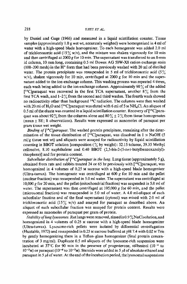
by Daniel and Gage (1966) and measured in a liquid scintillation counter. Tissue samples (approximately 1 .O g wet wt, accurately weighed) were homogenized in 4 ml of water with a high-speed blade homogenizer. To each homogenate was added 2.0 ml of trichloroacetic acid (15 ‘A w/v), and the mixture was shaken vigorously for 10 min and then centrifuged at 2000 g for 10 min. The supernatant was transferred to an II-mm id column, 10 mm long, containing 0.5 ml Dowex AG 5OW-X8 cation exchange resin (100-200 mesh) in the H+ form that had been previously washed with 20 ml of distilled water. The protein precipitate was resuspended in 5 ml of trichloroacetic acid (5 % w/v), shaken vigorously for 10 min, centrifuged at 2000 g for 10 min and the super- natant added to the ion-exchange column. This washing process was repeated 4 times, each wash being added to the ion-exchange column. Approximately 90 % of the added [14C]paraquat was recovered in the first TCA supernatant, another 8% from the first TCA wash, and l-2 % from the second and third washes. The fourth wash showed no radioactivity other than background 14C radiation. The columns were then washed with 20 ml of Hz0 and [14C]paraquat was eluted with 6 ml of 5 M NH,Cl. An aliquot of 0.5 ml of the eluate was counted in a liquid scintillation counter. Recovery of [14C]para- quat was about 92 % from the columns alone and 80 % + 2 % from tissue homogenates (mean + SE; 8 observations). Results were expressed as nanomoles of paraquat per gram tissue wet weight.
Binding of [14Cjpuruquat. The washed protein precipitate, remaining after the deter- mination of the tissue distribution of [14C]paraquat, was dissolved in 1 N NaOH (5 ml/g tissue wet wt) and aliquots were assayed for radioactivity by liquid scintillation counting in BBOT solution [composition (% by weight) : 52.15 toluene, 39.35 Methyl cellosolve, 8.10 naphthalene and 0.40 BBOT (2,5-bis-2-(5-tert-butylbenzoxazolyl)- thiophene)] and for protein content.
Subcellular distribution of [14CJparaquat in the lung. Lung tissue (approximately 5 g), obtained from rats and rabbits treated 24 or 65 hr previously with [14C]paraquat, was homogenized in 4 volumes of 0.25 M sucrose with a high-speed blade homogenizer (Ultra-turrax). The homogenate was centrifuged at 600 g for 10 min and the pellet (nuclear fraction) was resuspended in 5.0 ml water. The supernatant was centrifuged at 10,000 g for 20 min, and the pellet (mitochondrial fraction) was suspended in 5.0 ml of water. The supernatant was then centrifuged at 105,000 g for 60 min, and the pellet (microsomal fraction) was resuspended in 5.0 ml of water. A 4.0 ml-aliquot of each subcellular fraction and of the final supernatant (cytosol) was mixed with 2.0 ml of trichloroacetic acid (15 % w/v) and assayed for paraquat as described above. An aliquot of each subcellular fraction was assayed for protein content. Results were expressed as nanomoles of paraquat per gram of protein.
Stability of lung lysosomes. Rat lungs were removed, rinsed in0.9 %NaCl solution, and homogenized in 4 volumes of 0.25 M sucrose with a high-speed blade homogenizer (Ultra-turrax). Lysosome-rich pellets were isolated by differential centrifugation (Huxtable, 1972) and resuspended in 0.25 M sucrose buffered at pH 7.4 with 0.02 M Tris by gently homogenizing them in a Teflon-glass homogenizer (final protein concen- tration of 3 mg/ml). Duplicate 0.5 ml aliquots of the lysosome-rich suspension were incubated at 37°C for 90 min in the presence of progesterone, stilbestrol (10d4 to 10w3m) or paraquat (10m6 to 10-3~). Steroids were added in 5 ~1 of absolute ethanol and paraquat in 5 ~1 of water. At the end of the incubation period, the lysosomal suspensions

PARAQUAT ON RAT AND RABBIT LUNG ‘19
were centrifuged at 20,000 g for 10 min; duplicate aliquots (0.1-0.2 ml) of the clear supernatant were assayed for acid phosphatase as measured by the hydrolysis of p-nitrophenol phosphate (Sigma Tech. Bull. No. 104) and for&glucuronidase according to the method of Fishman (1965).
Measurement of Catalase. The lungs and livers of rats and rabbits were excised after perfusion with 0.9 % NaCl solution to remove as much blood as possible. A 5.0 % wiv aqueous homogenate was prepared with a high-speed blade homogenizer (Ultra- turrax). Catalase activity was determined by the modification of the method of Beers and Sizer (1952) as described by Ganschow and Schimke (1968).
Measurement of lipid peroxidation. The in vitro production of malonaldehyde by rat lung microsomes was measured in a 3.0-ml incubation system containing 12.5 mM nicotinamide, 12 PM FeCl,, 4 mM ADP, 0.45 mM NADPH, all of which are known to provide optimal conditions for lipid peroxidation (Schacter et al., 1972), paraquat (low5 to 10-3~) and 4.0 mg microsomal protein in a 0.05 M Tris buffer at pH 7.4. The samples were incubated at 37°C for 20 min in a Dubnoff metabolic shaker, and the reactions were terminated by the addition of 1 ml of a 30% w/v trichloroacetic acid solution. Malonaldehyde was determined as described by Ernster and Nordenbrand (1967). The Student’s t test was used to make comparison between control and treated animals.
RESULTS AND DISCUSSION
Our initial histopathologic studies showed the expected lung damage in the rat (Clark et al., 1966) and have confirmed the absence of lung damage in the rabbit (Butler and Kleinerman, 1971). In the rat, a single dose of paraquat (20 mg/kg iv) caused a reproducible pattern of lung damage. Hemorrhage and edema were noted 24 hr after administration. Large areas of consolidation were observed between 48 and 72 hr: some evidence of fibrosis was found between 7 and 10 days. At this dose about 95 ‘,; of the rats survived for 2 weeks. The other 5 % usually died 3 or 4 days after paraquat administration. The administration of paraquat (10,40,80 mg/kg iv) to rabbits did not produce the characteristic lung damage seen in rats and many other species. Mortality, however, was high in rabbits that received 40 or 80 mg/kg, and death usually occurred within the first 24 hr. Paraquat-induced lung damage in rats is accompanied by de- creases in cytochrome P-450 concentrations and in the total metabolism of [14C]bromo- benzene by lung microsomes (Table I). These decreases occurred 24 and 72 hr after administration of paraquat (20 mg/kg iv). In the rabbit, however, paraquat had no effect on cytochrome P-450 concentration in lung microsomes or on total in vitro metabolism of [14C]bromobenzene (Table 1). Moreover, 48 hr after paraquat adminis- tration to rats, NADPH oxidation was almost completely blocked in the lung micro- somes, but NADPH-cytochrome c reductase activity was virtually unchanged (Table 2). In contrast, paraquat administration had little if any effect on these activities in liver. We hoped to explain the mechanism of paraquat toxicity by more closely investigating these species differences in biochemical and tissue lesions.
The highest tissue concentrations of paraquat administered (20 mg/kg, 30-50 &i/animal) were in the lungs of both species (Table 3), in agreement with the recently published study in rats by Sharp et al., (1972). Although paraquat showed a preferential

220 ILETT ET AL.
localization in rabbit lung, especially at an early time point, the concentration of paraquat decreased much more rapidly in the rabbit lung than in the rat lung. Thus, the toxicity of paraquat in the rat lung seems to parallel the prolonged retention of paraquat in the lung of this species.
TABLE 1 CONCENTRATION OF CYTOCHROME P-450 IN LUNG MICRO~~MES AND TOTAL IN VITRO META- BOLISM OF [14C]B~~~~~~~~~~ BY LUNG MICRO.WMES AT VARIOUS TIME INTERVALS AFTER
TREATMENT OF RATS AND RABBITS WITH PARAQUAT DICHLORIDE (20 mg/kg IV)
Cytochrome P-450 [fz,,,,,,/mg Total metabolism of [r4C]bromo- protein benzene (pmol/mg/min)
Time intervals Rat” Rabbit” Rat” Rabbit”
Control 0.041+ 0.005 0.067 + 0.014 25.0 &- 3.6 256.8 + 13.2 24 hr 0.026 + 0.004b 0.053 + 0.014 23.5 f 2.3 - 48 hr - - - 251.1 + 14.8 72 hr 0.015 + 0.005b 0.063 + 0.004 13.9 + 1.7” -
0 Results are expressed as mean f SE of determinations on microsomes from each of 4 animals. b p < 0.01 with respect to controls. c p < 0.05 with respect to controls.
TABLE 2 NADPH-CYTOCHROME c REDUCTASE A~TIVI~ AND RATE OF NADPH OXIDATION IN MICRO- SOMES PREPARED FROM THE LUNGS AND LIVERS OF RATS, 48 HR AFTER TREATMENT WITH
PARAQUAT DICHLORIDE (20 mg/kg IV)
NADPH cytochrome c reductase Rate of oxidation of NADPH (nmol/mg proteinlmin) (nmol/mg protein/min)
Group’ Lws Liver Lung Liver
Control Exp I 57.0 187 1.3 9.7 Exp II 60.0 183 2.2 9.1
Paraquat Exp I 46.0 140 0.10 7.9 treated Exp II 43.0 156 0.30 10.0
a Results are the means of duplicate determinations on pooled microsomal protein from the lungs or livers of 4rats.
TABLE 3 DISTRIBUTION OF[‘~C]PARAQUAT DICHLORIDE (nmol/g TISSUE) IN RAT AND RABBIT
Rat”(S) Rabbit” (3)
Tissue 6Hr 1 Day 7 Days 6Hr 1 Day 7
Brain 0.37 310.13 0.87 & 0.04 0.53 f 0.08 0.90 f 0.09 0.49 + 0.08 ND Heart 0.60 zk 0.23 1.13 + 0.21 0.12 + 0.02 3.04 f 0.02 1.52 + 0.83 0.11 + 0.02 Kidney 6.36 f 0.43 1.93 f 0.15 0.07 f 0.01 19.80 + 2.85 5.25 + 0.98 0.11 k 0.03 Liver 3.44 rt 0.84 0.90 + 0.47 0.07 + 0.01 3.56 f 0.26 1.59 f 0.13 0.18 + 0.01 Lung lg.05 rt 2.30 11.36 + 0.22 0.81 + 0.12 25.39 + 4.30 7.91 f 2.33 0.07 + 0.01 Plasma 0.10 If: 0.06 ND” ND 1.19 z!z 0.12 0.28 + 0.04 ND
u Results are expressed as mean + SE. The numbers in parentheses are the number of animals in each group.
b ND = not detectable.

PARAQUATONRATANDRABBITLUNG 221
Since the lungs contained highest concentrations of paraquat, we measured the subcellular distribution of paraquat in this tissue 24 and 65 hr after the administration of the toxicant. Table 4 shows that after 65 hr paraquat was evenly distributed in all subcellular fractions of the lungs of both rats and rabbits. Concentrations of paraquat in all subcellular fractions, however, decreased more rapidly in the rabbit lung than in the rat lung. Several other compounds that produce tissue necrosis, such as bromo- benzene (Brodie et al., 1971; Ilett et al., 1973; Reid ef al., 1973; Reid, 1973a,b) form a covalent bond with tissue macromolecules. However, we found no evidence that paraquat becomes covalently bound. After thorough washing of the precipitates with dilute trichloroacetic acid, only insignificant amounts of radiolabeled material could be detected in protein from brain, heart, kidney, liver, lung or plasma.
TABLE 4
SUBCELLULARDISTRIBUTIONOF['~C]PARAQUATINRATANDRABBITLUNG
Subcellular fraction
Rat lung” (nmol/g protein)
24 Hr 65 Hr
Rabbit lungb (nmol/g protein) --
24 Hr 65 Hr
Nuclear 49.22 rt 7.53 41.13 f 6.91 52.36 f 7.22 15.35 k 2.71 Mitochondriat 36.85 + 4.97 41.54 + 6.78 35.29 + 5.09 15.25 2 1.84 Microsomal 62.54 + 4.31 58.48 + 1.29 36.21 rt 4.24 20.96 + 4.91 Cytosol 35.16 + 3.11 46.04 f 8.10 76.24 t 11.97 18.02 &- 2.54
a Results are expressed as mean of 4 animals + SE. b Results are expressed as mean of 3 animals f SE.
One proposed mechanism of action for paraquat is a diminution of alveolar sur- factant material in the lung as suggested by Manktelow (1967), Fisher and Clements (1969) and Robertson et al. (1971). However, Fletcher and Wyatt (1970) found that the phospholipid content of rat lung was unchanged after pretreatment with paraquat and have shown more recently (Fletcher and Wyatt, 1972) that neither the amount nor the rate of destruction of dipalmitoyl lecithin, the major phospholipid in lung, was altered after paraquat treatment.
The question of whether paraquat itself or a metabolite is responsible for the lung toxicity in animals remains to be resolved. Most studies have indicated that paraquat is not metabolized (Conning et al., 1969), but Molnar and Hayes (1971) claim to have isolated a metabolite of paraquat from the rat lung. We found no evidence, however, of N-demethylation of [14C]paraquat 516 by 9000 g supernatants of liver or lung from either rats or rabbits although these tissues possessed measurable N-demethylase activity when [N-methyP4C]codeine or ethyl morphine were used as substrates.6
Lysosomal instability has been considered to be an important factor in the toxicity of drugs and other compounds (Davies and Allison, 1972). Progesterone and stilbestrol (5 x 10e4 M), which are known to decrease lysosomal stability (Allison, 1968), caused an increased release of acid phosphatase and @glucuronidase, but paraquat had no
’ N Demethylation was assessed by measuring [14C]-formaldehyde as a dimedon derivative as described by Taylor and Weissbach (1965).
6 Unpublished results (Ilett, K. F., Stripp, B. and Menard, R. H.).

222 ILETT ET AL.
effect on lysosomal stability at low concentrations (10V6 M). In fact, at high concentra- tions (lo-” to 10e3 M) it stabilized the lysosomal membrane and decreased the release of lysosomal enzymes (Table 5). Thus, paraquat itself apparently did not decrease lysoso- ma1 stability, but the possibility still existed that a free radical could be formed which
TABLE 5
IN VITRO STABILITY OF RAT LUNG LYSO~~MES IN THE PRESENCE OF VARIOUS CONCENTRATIONS OF PARAQUAT DICHLORIDE, PROGESTERONE AND STTLBE~TROL
Concentration
Release of lysosomal enzymes” (units/ml incubate/hr)
Compound added 04 Acid Phosphatase j&Glucuronidase
None - 0.91 18.5 Paraquat 1o-3 0.43 10.0
lo+ 0.65 11.5 10-s 0.75 12.7 10-6 0.90 16.1
Progesterone 5 x 1o-4 1.29 65.9 Stilbestrol 5 x 1o-4 1.44 79.5
a Results are the means of duplicate determinations on pooled lysosomal fractions prepared from the lungs of 20 rats and typical of 3 experiments.
might damage the membrane. Thus, in plants, the herbicidal activity of paraquat has been ascribed to the accumulation of H,Oz during cyclic reduction and oxidation of paraquat (Calderbank, 1968). Therefore, we considered that a similar process could occur in animal cells and thereby lead to cellular damage perhaps by causing a decrease in the stability of the lysosomal membrane. Such a process probably would be accom- panied by enhanced NADPH oxidation. Gage (1968) showed that paraquat stimulates the rate of oxidation of NADPH in mitochondrial and microsomal fractions of rat liver. Using microsomal fractions, we have confirmed these findings, and, in addition, we have shown that paraquat stimulates NADPH oxidation in the rabbit liver and in the lungs of both rat and rabbit (Table 6). The stimulation of NADPH oxidation was not
TABLE 6
EFFECT OF IN VITRO ADDITION OF PARAQUAT DICHLORIDE ON THE RATE OF OXIDATION OF NADPH IN MICROSCWAL SUSPENSIONS FROM RATS OR
RABBITS’
Concentration of paraquat
(M)
Rat Rabbit
Liver Lung Liver Lung
0 4.26 0.85 4.03 2.82 1o-5 5.60 1.13 5.65 3.71 1o-4 12.50 2.50 14.92 10.08 1o-3 37.4 7.65 37.90 26.21
a Results (expressed as nanomoles per milligram protein per minute) are the means of duplicate determinations on pooled tissues from 4 animals and typical of 2 experiments.

PARAQUAT ON RAT AND RABBIT LUNG 223
blocked by carbon monoxide; paraquat, therefore, may act as an uncoupler in the electron transport chain receiving electrons from cytochrome c reductase. This deduc- tion is supported by the fact that paraquat (10e5 to 10-j M) added in vitro was able to inhibit the microsomal metabolism of [14C]bromobenzene in both species (Table 7), although paraquat did not produce either a type I or type II spectral change (Remmer et al., 1966) when added to microsomal suspensions (unpublished observations).
TABLE 7 EFFECT OF THE IN VITRO ADDITION OF PARAQUAT ON THE MICROXIMAL METABOLISM OF [14C]-
BROMOBENZENE IN RAT AND RABBIT
Microsomal preparation IC50”
(M)
Rat lung 0.8 x 1O-4 Rat liver 0.9 x 1o-4 Rabbit lung 5.8 x lo-“ Rabbit liver 2.4 x 1O-4
a The IC50 is the concentration of paraquat dichloride that causes a 50% inhibition of the in vitro metabolism of [‘4C]bromobenzene. This was determined from a plot of percent inhibition of bromo- benzene metabolism versus concentration of paraquat (3 points).
Presumably the paraquat-dependent NADPH oxidation should result in the forma- tion of H,O,. We investigated this possibility in vitro by adding paraquat to liver and lung microsomal suspensions from rats and rabbits and measuring the formation of HzOz, determined indirectly by measuring the conversion of methanol to formaldehyde. Paraquat in a concentration of 10m4 M greatly enhanced the conversion of methanol to formaldehyde, indicating increased production of H,02, but at 10e5 M it had very little effect and at 10e6 M none at all (Table 8). Contrary to our expectations, moreover, the rate of H,O, formation was higher in the rabbit than in the rat lung.
TABLE 8
EFFECT OF PARAQUAT DICHLORIDE ON THE RATE OF CONVERSION OF METHANOL TO FORMALDEHYDE BY LUNG AND LIVER MICROSOMES~
Rate of formation of formaldehyde (nmol/mg protein/min)
Concentration of paraquat
Rat Rabbit -
64) Liver Lung Liver Lung
0 17.0 1.22 12.41 3.21 1o-4 55.0 3.40 76.91 26.77 1o-5 22.2 1.52 lo-+ 17.3 1.18
a Results are means of triplicate determination on pooled tissues from 20 rats or 4 rabbits and typical of 2 experiments.

224 ILETT ET AL.
Nevertheless, it still seemed possible that a higher steady state concentration of H,Oz could exist in rat lung if the catalase levels were lower in rat lung than in rabbit lung. As shown in Table 9, however, the catalase activities in perfused lung were similar in both species although they were significantly less in the rabbit liver than in the rat liver. In addition, measurements of the hemoglobin content of the tissue homo- genates indicated that the small amount of blood remaining after perfusion could not contribute significantly to the activity of catalase measured in these tissues.
TABLE 9 CATALASEACTIVITYINBLOODAND TISSUES
Species Liver Lung Blood
Rat” 69.25 f 1.21 3.88 + 0.17 18.46 f 1.11 Rabbitb 26.53 rt 1.21’ 4.30 + 0.53 25.27 * 1.5gd
0 Values are the mean of 4 animals + SE expressed as units x 10m3 per gram tissue or milliliter of blood.
b Values are the mean of 3 animals f SE. =p < 0.001 with respect to rat liver. d p < 0.05 with respect to rat blood.
Gage (1968) suggested that the free radicals which are produced in the tissues during the cyclic reduction and oxidation of paraquat might cause lipid peroxidation and result in tissue damage. However, our results show that paraquat added in vitro is a potent inhibitor of lipid peroxidation (Fig. 1). The inability of paraquat to stimulate
I - ._~_ .L 1~ I---.- _. .-.- 10-5 IO-' 16'
MOLAR CONCENTRATION OF PARAOUAT DICHLORIDE
FIG. 1. Effect of paraquat dichloride (10m5 to 10T3~) on the in vitro production of malonaldehyde by microsomes from rat lung. Each point represents the mean of triplicate determinations.
PARAQUAT ON RAT AND RABBIT LUNG 225
lipid peroxidation was also noted when paraquat was added under nonoptimal con- ditions, that is, in presence of NADPH alone.
The results of this study show that unlike bromobenzene, paraquat does not become covalently bound to lung macromolecules. Moreover, it seems unlikely that paraquat evokes its toxicity by leading to the formation of hydrogen peroxide and free radicals. Although the total concentration of paraquat in lung tissue in vivo at 6 hr seems to approach the concentration required to increase NADPH oxidation and hydrogen peroxide in vitro (compare Tables 3, 6, and 8), the free concentration in lung tissue is undoubtedly much lower. Indeed it seems likely that the free concentration in lung cells would be no higher than that found in the plasma (Table 3, IO-’ M), which is 2-3 orders of magnitude less than that required in vitro to cause hydrogen peroxide formation (Tables 6 and 8). Furthermore, the effects of paraquat on rabbit lung in vitro are the same as or greater than those on the rat lung; yet, lung necrosis does not occur in the rabbit. Thus, the mechanism of paraquat toxicity remains obscure.
ACKNOWLEDGMENTS We gratefully acknowledge the expert technical assistance of Mr. John George.
REFERENCES ALLISON, A. (1968). The role of lysosomes in the action of drugs and hormones. In Adc.
Chemother. 3,253-302. BEERS, R. R. AND SIZER, I. W. (1952). A spectrophotometric method for measuring the break-
down of hydrogen peroxide bycatalase. .I. Biof. Chem. 195, 133-140. BRODIE, B. B., REID, W. D., CHO, A. K., SIPES, 1. G., KRISHNA, G. AND GILLETTE, J. R. Possible
mechansim of liver necrosis caused by aromatic organic compounds. Proc. Nat. Acad. Sci. U.S. 68,16&164.
BROWNE, T. D. (1971). Treatment of paraquat ingestion. Brit. Med. J. 3, 580. BULLIVANT, C. M. (1966). Accidental poisoning by paraquat: Report of two cases in man.
Brit. Med.J. 1, 1272-1273. BUTLER, C. AND KLEINERMAN, J. (1971). Paraquat in the rabbit. Brit. J. Ind. Med. 28,67-71. CALDERBANK, A. (1968). The bipyridylium herbicides. Aduan. Pest Contr. Res. 8, 127-235. CLARK, D. G., MCELLIGO~, T. F. AND HURST, E. W. (1966). The toxicity of paraquat. Brit. J.
Znd. Med. 23,126-132. CONNING, D. M., FLETCHER, K. AND SWAN, A. A. B. (1969). Paraquat and related bipyridyls.
Brit. Med. Bull. 25,245-249. CULLING, C. F. A. (1963). Handbook of Histological Techniques, 2nd ed., pp. 25-229. Butter-
worth, Washington. DANIEL, J. W. AND GAGE, J. C. (1966). Absorption and excretion of diquat and paraquat in
rats. Brit. J. Znd. Med. 23, 133-136. DAVIES, P. AND ALLISON, A. C. (1972). The significance of the lysosomes in toxicology. Crit.
Rer. Toxicol. 1,283-323. ERNSTER, L. AND NORDENBRAND, K. (1967). Methods Enzymof. 10,574-580. FENNELLY, J. J., FITZGERALD, M. X. AND FITZGERALD, 0. (1971). Recovery from severe
paraquat poisoning following forced diuresis and immunosuppressive therapy. J. Zr. Med. Ass. 64,69-71.
FISHER, H. K. AND CLEMENTS, J. A. (1969). Effects of the herbicide paraquat on mechanical properties of rat lungs. Fed. Proc., Fed. Amer. Sot. Exp. Biol. 28,526.
FISHER, H. K., HUMHRIES, M. AND BAILS, R. (1971). Paraquat poisoning. Recovery from renal and pulmonary damage. Ann. Intern. Med. 75,73 l-736.
FISHMAN, W. H. (1965). Beta-glucuronidase. In : Methods qfEnzymatic Analysis (H. Bergmeyer. ed.). pp. 869-874. Academic Press, New York.

226 ILETT ET AL.
FLETCHER, K. AND WYATT, I. (1970). The composition of lung lipids after poisoning with paraquat. &it. J. Exp. Path. 51,6&l-610.
FLETCHER, K. AND WYAX-T, I. (1972). The action of paraquat on the incorporation of palmitic acid into dipalmitoyl lecithin in mouse lungs. Brit. J. Exp. Pathol. 53, 225-230.
GAGE, J. C. (1968). The action of paraquat and diquat on the respiration of liver cell fractions. Biochem. J. 109,757-761.
GANSCHOW, R. AND SCHIMKE, R. T. (1968). Genetic control of catalase in inbred mice. In: Regulatory Mechanisms for protein Synthesis in Mammalian Cells (A. San Pietro, M. R. Lamborg and F. T. KeMey, eds.), pp. 377-397. Academic Press, New York.
GIGON, P. L., GRAM, T. E. AND GILLETTE, J. R. (1969). Studies on the rate of reduction of hepatic microsomal cytochrome P-450 by reduced nicotinamide adenine dinucleotide phosphate: Effect of drug substrates. Mol. Pharmacol. 5, 109-118.
GORNALL, A. G., BARDAWILL, C. J. AND DAVID, M. M. (1949). Determination of serum proteins by means of the biuret reaction. J. Biol. Chem. 177,751-766.
GRUNDIES, H., KOLMAR, D. AND BENNHOLD, I. (1971). Paraquat intoxication, Case report with special reference to hemodialysis. Deut. Med. Wochenschr. g&588-589.
HUXTABLE, C. R. (1972). The effect of ingestion of swainsoniagalegifolia on the liver lysosomes of the guinea pig. Aust. J. Exp. Biol. Med. Sci. 50, 109-l 18.
ILETT, K. F., REID, W. D., SIPES, I. G. AND KRISHNA, G. (1973). Chloroform toxicity in mice: Correlation of renal and hepatic necrosis with covalent binding of metabolites to tissue macromolecules. Exp. Mol. Pathol. 19,215-229.
MALONE, J. D., CARMODY, M. AND KEOGH, B. (1971). Paraquat poisoning. A review of nineteen cases. J. Zr. Med. Ass. 64, 59-68.
MANKTELOW, B. W. (1967). The loss of pulmonary surfactant in paraquat poisoning: A mode for the study of the respiratory distress syndrome. Brit. J. Exp. Pathol. 48,366-369.
MOLNAR, I. G. AND HAYES, W. J., JR. (1971). Distribution and metabolism of paraquat in the rat. Toxicol. Appl. Pharmacol. 19,405.
NASH, I. (1953). The calorimetric estimation of formaldehyde by means of the Hantzsch reaction. Biochem. J. 55,416421.
OMURA, T. AND SATO, R. (1964). The carbon monoxide binding pigment of liver microsomes. I. Evidence for its hemoprotein nature. J. Biol. Chem. 239,2370-2378.
PHILLIPS, A. AND LANGDON, R. (1962). Hepatic triphosphopyridine nucleotide cytochrome c reductase: Isolation, characterization and kinetic studies. J. Biol. Chem. 237, 2652-2660.
REID, W. D. (1973a). Relationship between tissue necrosis and covalent binding of toxic metabolites of halogenated aromatic hydrocarbons. In: Pharmacology and the future of Man. Proc. Znt. Congress Pharmacol., 5th, 1972, Vol. 2, pp. 62-74. S. Karger, Basel.
REID, W. D. (1973b). Mechanism of renal necrosis induced by bromobenzene or chloro- benzene, Exp. MoI. Pathol. 19,197-214.
REID, W. D., ILETT, K. F., GLICK, J. M AND KRISHNA, G (1973) Metabolism and covalent binding of aromatic hydrocarbons in the lung: Relationship to experimental broncheolar necrosis. Amer Rev Resp Diseases 107, 539-551
REMMER, H., SCHENKMAN, J. B., ESTABROOK, R. W., SASAME, H. A., GILLETTE, J. R., NARASIM- HULU, S., COOPER, D. Y. AND ROSENTHAL, 0. W. (1966). Drug interaction with hepatic microsomal cytochrome. Mol. Pharmacol. 2,187-190.
ROBERTSON, B., EINHORNING, G., IVEMARK, B., MALMQVIST, E. AND MODEE, J. (1971). Experi- mental respiratory distress induced by parayuat. J. Pathol. 103,239-244.
SCHACTER, B. A., MARVER, H. S. AND MEYER, U. A. (1972). Hemoprotein catabolism during stimulation of microsomal lipid peroxidation. Biochim. Biophys. Acta 279, 221-227.
SHARP, C. W., OTTOLENGHI, A. AND POSNER, H. S. (1972). Correlation of paraquat toxicity with tissue concentrations and weight loss of the rat. Toxicol. Appl. Pharmacol. 22,241-251.
STRIPP, B.. HAMRICH M. E., ZAMPAGLIONE, N. G. AND GILLETTE, J. R. (1971). The effect of spironolactone on drug metabolism by hepatic microsomes. 1. Pharmacol. Exp. Ther. 176, 766-771.
TAYLOR, R. T. AND WEISSBACH, H. (1965). Radioactive assay for serine transhydroxymethy- lase. Anal. Biochem. 13. 8Cb84.