
Structure determination of symmetric homo-oligomers by a complete search of symmetry configuration...
17
Structure Determination of Symmetric Homo-Oligomers by a Complete Search of Symmetry Configuration Space, Using NMR Restraints and van der Waals Packing Shobha Potluri, 1 Anthony K. Yan, 1 James J. Chou, 2 Bruce R. Donald, 1,3,4 * and Chris Bailey-Kellogg 1 * 1 Department of Computer Science, Dartmouth College, Hanover, New Hampshire 03755 2 Department of Biological Chemistry and Molecular Pharmacology, Harvard Medical School, Boston, Massachusetts 02115 3 Department of Chemistry, Dartmouth College, Hanover, New Hampshire 03755 4 Department of Biological Sciences, Dartmouth College, Hanover, New Hampshire 03755 ABSTRACT Structural studies of symmetric homo-oligomers provide mechanistic insights into their roles in essential biological processes, in- cluding cell signaling and cellular regulation. This paper presents a novel algorithm for homo-oligo- meric structure determination, given the subunit structure, that is both complete, in that it evalu- ates all possible conformations, and data-driven, in that it evaluates conformations separately for consistency with experimental data and for qual- ity of packing. Completeness ensures that the algorithm does not miss the native conformation, and being data-driven enables it to assess the structural precision possible from data alone. Our algorithm performs a branch-and-bound search in the symmetry configuration space, the space of symmetry axis parameters (positions and orienta- tions) defining all possible C n homo-oligomeric complexes for a given subunit structure. It elimi- nates those symmetry axes inconsistent with in- tersubunit nuclear Overhauser effect (NOE) dis- tance restraints and then identifies conformations representing any consistent, well-packed struc- ture to within a user-defined similarity level. For the human phospholamban pentamer in dodecylphosphocholine micelles, using the struc- ture of one subunit determined from a subset of the experimental NMR data, our algorithm identi- fies a diverse set of complex structures consistent with the nine intersubunit NOE restraints. The distribution of determined structures provides an objective characterization of structural uncer- tainty: backbone RMSD to the previously deter- mined structure ranges from 1.07 to 8.85 A ˚ , and variance in backbone atomic coordinates is an av- erage of 12.32 A ˚ 2 . Incorporating vdW packing reduces structural diversity to a maximum back- bone RMSD of 6.24 A ˚ and an average backbone variance of 6.80 A ˚ 2 . By comparing data consis- tency and packing quality under different assump- tions of oligomeric number, our algorithm identi- fies the pentamer as the most likely oligomeric state of phospholamban, demonstrating that it is possible to determine the oligomeric number directly from NMR data. Additional tests on a number of homo-oligomers, from dimer to hep- tamer, similarly demonstrate the power of our method to provide unbiased determination and evaluation of homo-oligomeric complex structures. Proteins 2006;65:203–219. V V C 2006 Wiley-Liss, Inc. Key words: Nuclear magnetic resonance (NMR); Structure determination; Symmetric homo-oligomer; Membrane protein; Configuration space INTRODUCTION Symmetric homo-oligomers play pivotal roles in complex biological processes, including ion transport and regula- tion, signal transduction, and transcriptional regulation. We are particularly interested in phospholamban, a sym- metric homopentameric membrane protein that regulates the calcium levels between cytoplasm and sarcoplasmic reticulum, and hence aids in muscle contraction and relaxation. 1 Ion conductance studies 2 also suggest that phospholamban might have a separate role as an ion channel. To understand the dual function of phospholam- ban and other such symmetric homo-oligomers, we have developed a combined experimental–computational ap- *Correspondence to: Bruce R. Donald, 6211 Sudikoff Laboratory, Hanover, NH 03755. E-mail: [email protected] or Chris Bailey-Kellogg, 6211 Sudikoff Laboratory, Hanover, NH 03755. E-mail: [email protected] The Supplementary Material referred to in this article can be found at http://www.interscience.wiley.com/jpages/0887-3585/suppmat/ Grant sponsor: National Institutes of Health; Grant number: R01 GM 65982; Grant sponsor: National Science Foundation; Grant num- bers: EIA-9802068, EIA-0305444; Grant sponsor: National Science Foundation; Grant numbers: IIS-0444544, IIS-0502801. Grant spon- sor: Smith Family award. Received 22 December 2005; Revised 2 May 2006; Accepted 15 May 2006 Published online 8 August 2006 in Wiley InterScience (www. interscience.wiley.com). DOI: 10.1002/prot.21091 Abbreviations: NMR, nuclear magnetic resonance; NOE, nuclear Overhauser effect; RMSD, root-mean-square deviation; SCS, sym- metry configuration space; S 2 , space of orientations represented on a two-sphere; vdW, van der Waals; WPS, well-packed satisfying. V V C 2006 WILEY-LISS, INC. PROTEINS: Structure, Function, and Bioinformatics 65:203–219 (2006)
-
Upload
independent -
Category
Documents
-
view
0 -
download
0
Transcript of Structure determination of symmetric homo-oligomers by a complete search of symmetry configuration...
Structure Determination of Symmetric Homo-Oligomersby a Complete Search of Symmetry Configuration Space,Using NMR Restraints and van der Waals Packing
Shobha Potluri,1 Anthony K. Yan,1 James J. Chou,2 Bruce R. Donald,1,3,4* and Chris Bailey-Kellogg1*1Department of Computer Science, Dartmouth College, Hanover, New Hampshire 037552Department of Biological Chemistry and Molecular Pharmacology, Harvard Medical School, Boston, Massachusetts 021153Department of Chemistry, Dartmouth College, Hanover, New Hampshire 037554Department of Biological Sciences, Dartmouth College, Hanover, New Hampshire 03755
ABSTRACT Structural studies of symmetrichomo-oligomers provide mechanistic insights intotheir roles in essential biological processes, in-cluding cell signaling and cellular regulation. Thispaper presents a novel algorithm for homo-oligo-meric structure determination, given the subunitstructure, that is both complete, in that it evalu-ates all possible conformations, and data-driven,in that it evaluates conformations separately forconsistency with experimental data and for qual-ity of packing. Completeness ensures that thealgorithm does not miss the native conformation,and being data-driven enables it to assess thestructural precision possible from data alone. Ouralgorithm performs a branch-and-bound search inthe symmetry configuration space, the space ofsymmetry axis parameters (positions and orienta-tions) defining all possible Cn homo-oligomericcomplexes for a given subunit structure. It elimi-nates those symmetry axes inconsistent with in-tersubunit nuclear Overhauser effect (NOE) dis-tance restraints and then identifies conformationsrepresenting any consistent, well-packed struc-ture to within a user-defined similarity level.For the human phospholamban pentamer in
dodecylphosphocholine micelles, using the struc-ture of one subunit determined from a subset ofthe experimental NMR data, our algorithm identi-fies a diverse set of complex structures consistentwith the nine intersubunit NOE restraints. Thedistribution of determined structures provides anobjective characterization of structural uncer-tainty: backbone RMSD to the previously deter-mined structure ranges from 1.07 to 8.85 A, andvariance in backbone atomic coordinates is an av-erage of 12.32 A2. Incorporating vdW packingreduces structural diversity to a maximum back-bone RMSD of 6.24 A and an average backbonevariance of 6.80 A2. By comparing data consis-tency and packing quality under different assump-tions of oligomeric number, our algorithm identi-fies the pentamer as the most likely oligomericstate of phospholamban, demonstrating that it ispossible to determine the oligomeric number
directly from NMR data. Additional tests on anumber of homo-oligomers, from dimer to hep-tamer, similarly demonstrate the power of ourmethod to provide unbiased determination andevaluation of homo-oligomeric complex structures.Proteins 2006;65:203–219. VVC 2006 Wiley-Liss, Inc.
Key words: Nuclear magnetic resonance (NMR);Structure determination; Symmetrichomo-oligomer; Membrane protein;Configuration space
INTRODUCTION
Symmetric homo-oligomers play pivotal roles in complexbiological processes, including ion transport and regula-tion, signal transduction, and transcriptional regulation.We are particularly interested in phospholamban, a sym-metric homopentameric membrane protein that regulatesthe calcium levels between cytoplasm and sarcoplasmicreticulum, and hence aids in muscle contraction andrelaxation.1 Ion conductance studies2 also suggest thatphospholamban might have a separate role as an ionchannel. To understand the dual function of phospholam-ban and other such symmetric homo-oligomers, we havedeveloped a combined experimental–computational ap-
*Correspondence to: Bruce R. Donald, 6211 Sudikoff Laboratory,Hanover, NH 03755. E-mail: [email protected] orChris Bailey-Kellogg, 6211 Sudikoff Laboratory, Hanover, NH 03755.E-mail: [email protected]
The Supplementary Material referred to in this article can be foundat http://www.interscience.wiley.com/jpages/0887-3585/suppmat/
Grant sponsor: National Institutes of Health; Grant number: R01GM 65982; Grant sponsor: National Science Foundation; Grant num-bers: EIA-9802068, EIA-0305444; Grant sponsor: National ScienceFoundation; Grant numbers: IIS-0444544, IIS-0502801. Grant spon-sor: Smith Family award.
Received 22 December 2005; Revised 2 May 2006; Accepted 15May 2006
Published online 8 August 2006 in Wiley InterScience (www.interscience.wiley.com). DOI: 10.1002/prot.21091
Abbreviations: NMR, nuclear magnetic resonance; NOE, nuclearOverhauser effect; RMSD, root-mean-square deviation; SCS, sym-metry configuration space; S2, space of orientations represented ona two-sphere; vdW, van der Waals; WPS, well-packed satisfying.
VVC 2006 WILEY-LISS, INC.
PROTEINS: Structure, Function, and Bioinformatics 65:203–219 (2006)
proach that, given the subunit structure, determines thecomplex structure from a combination of sparse intersubu-nit nuclear Overhauser effect (NOE) distance restraintsand van der Waals (vdW) packing. Our approach is com-plete, in that we test all possible conformations, and isdata-driven, in that we first test conformations for consis-tency with data and only then evaluate each of the con-sistent conformations for vdW packing. Completeness in astructure determination method is a key requirement,since it ensures that no conformation consistent with thedata is missed. This avoids any bias in the search, as wellas any potential for becoming trapped in local minima,both of which are problems inherent in energy minimiza-tion-based approaches. The data-driven nature of ourmethod allows us to independently quantify the amountof structural constraint provided by data alone, versusboth data and packing. This avoids over-reliance on sub-jective choices of parameters for energy minimization, andconsequent false precision in determined structures. Ourmethod also allows us to determine the oligomeric numberof the complex. To our knowledge, this is the firstapproach that determines the oligomeric number of a
symmetric homo-oligomer from intersubunit NOE dis-tance restraints. The confidence in the determined oligo-meric number depends on the information content in theavailable data.
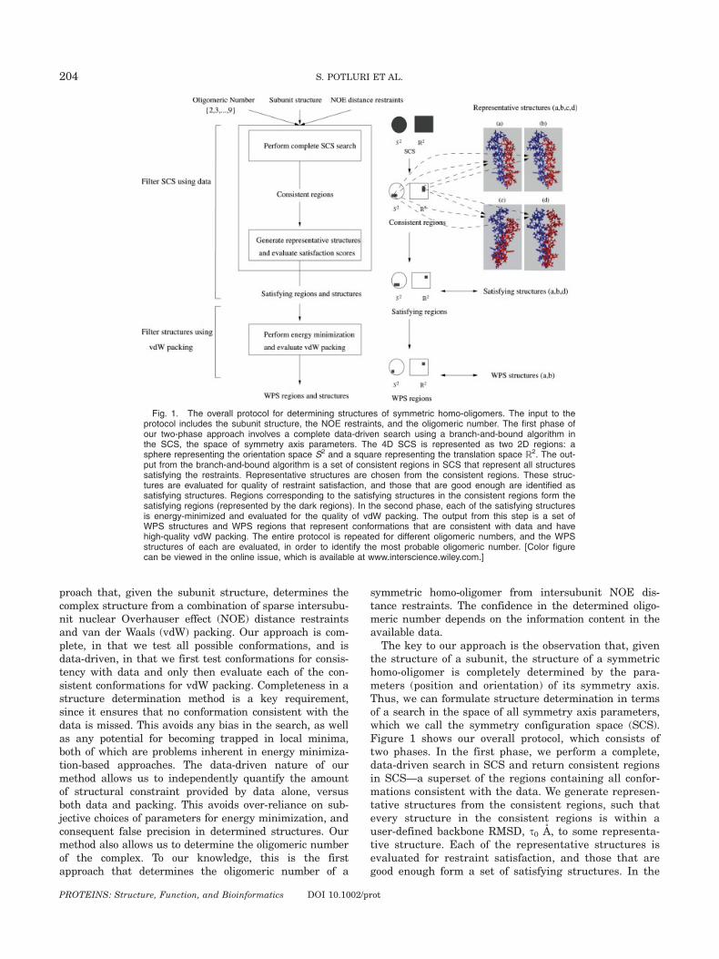
The key to our approach is the observation that, giventhe structure of a subunit, the structure of a symmetrichomo-oligomer is completely determined by the para-meters (position and orientation) of its symmetry axis.Thus, we can formulate structure determination in termsof a search in the space of all symmetry axis parameters,which we call the symmetry configuration space (SCS).Figure 1 shows our overall protocol, which consists oftwo phases. In the first phase, we perform a complete,data-driven search in SCS and return consistent regionsin SCS—a superset of the regions containing all confor-mations consistent with the data. We generate represen-tative structures from the consistent regions, such thatevery structure in the consistent regions is within auser-defined backbone RMSD, s0 A, to some representa-tive structure. Each of the representative structures isevaluated for restraint satisfaction, and those that aregood enough form a set of satisfying structures. In the
Fig. 1. The overall protocol for determining structures of symmetric homo-oligomers. The input to theprotocol includes the subunit structure, the NOE restraints, and the oligomeric number. The first phase ofour two-phase approach involves a complete data-driven search using a branch-and-bound algorithm inthe SCS, the space of symmetry axis parameters. The 4D SCS is represented as two 2D regions: asphere representing the orientation space S2 and a square representing the translation space R2. The out-put from the branch-and-bound algorithm is a set of consistent regions in SCS that represent all structuressatisfying the restraints. Representative structures are chosen from the consistent regions. These struc-tures are evaluated for quality of restraint satisfaction, and those that are good enough are identified assatisfying structures. Regions corresponding to the satisfying structures in the consistent regions form thesatisfying regions (represented by the dark regions). In the second phase, each of the satisfying structuresis energy-minimized and evaluated for the quality of vdW packing. The output from this step is a set ofWPS structures and WPS regions that represent conformations that are consistent with data and havehigh-quality vdW packing. The entire protocol is repeated for different oligomeric numbers, and the WPSstructures of each are evaluated, in order to identify the most probable oligomeric number. [Color figurecan be viewed in the online issue, which is available at www.interscience.wiley.com.]
204 S. POTLURI ET AL.
PROTEINS: Structure, Function, and Bioinformatics DOI 10.1002/prot
second phase, each of the satisfying structures is eval-uated for high-quality vdW packing. We ultimatelyreturn a set of well-packed satisfying (WPS) structuresthat are consistent with data and have high-quality vdWpacking. We quantify the uncertainty in determinedstructures in terms of the size of the regions in SCS andthe variations in atomic coordinates in the structures.The difference in uncertainty between the satisfyingstructures and the WPS structures illustrates the rela-tive precision that is possible from data alone, versusdata and packing together.Our approach allows us to test whether the available
data suffices to determine the oligomeric number withhigh confidence. For each possible oligomeric number, wedetermine a set of WPS structures. We place higher con-fidence in the oligomeric number that has WPS structurewith better vdW packing. Thus we determine the oligo-meric number using the NMR data and vdW packing.Our approach provides for an independent verification ofthe oligomeric state, which is typically determined usingexperiments, such as chemical cross-linking, followed bySDS-PAGE, or by equilibrium sedimentation.Traditional protocols (e.g., Refs. 3, 4) for structure
determination of protein complexes from NMR data usesimulated annealing and molecular dynamics. A variantof this protocol was used to determine the structure ofthe phospholamban pentamer.1 However, the simulatedannealing/molecular dynamics mechanism is not com-plete and could get trapped in local minima. Further-more, the precision in the determined structure isstrongly affected by temperature. Our approach avoidstemperature dependence and local minima problems,and does not suffer from ‘‘false precision’’ in characteriz-ing the diversity of determined structures.Previous computational techniques5 for predicting the
structures of complexed proteins fall into two categories:homology modeling and docking. One can confidently pre-dict protein interactions by homology modeling6,7 only ifthe structure of a homologous complex is available.8 Dock-ing strategies usually involve a two-stage approach: gen-erate a set of possible docked structures, and then scorethem. The possible structures are typically generated bysampling the space of rotations and translations of thedocked subunit with respect to the fixed subunit.9–15
Alternatively, geometric hashing16 samples conformationsthat are consistent with shape complementarity. Scoringis usually based on shape and chemical complementar-ity.17–20 Docking-based methods are immediately applica-ble to homo-oligomers, by docking two subunits of thehomo-oligomer. Pierce and Weng,21 Duhovny et al.,22 andComeau and Camacho23 have all used docking-basedapproaches, followed by filtering based on symmetry andscoring by minimizing energy functions, to predict struc-tures of homo-oligomers.Several minimalist experiments, though not compre-
hensive enough to determine structures by themselves,have been used in conjunction with docking approachesto score predicted models.24–27 Proximity information be-tween adjacent units in a complex can be obtained from
experiments such as mutagenesis,28–30 hydrogen-deute-rium exchange,26,31 chemical cross-linking,32–34 fluores-cence energy transfer,35,36 Fourier transform infra-redspectroscopy,37 and sparse intermolecular NOE data.38
Existing docking-based approaches are not complete,because they generate possible docked structures bysampling, and might miss conformations close to thenative structure. This is evident from our tests on phos-pholamban, when we used several available docking-based methods21–23 to predict the complex structurefrom the subunit structure. A significant number of themodels provided by these approaches satisfy none ofthe experimental intersubunit NOE restraints deter-mined by Oxenoid and Chou.1 The backbone RMSD ofthe model that best satisfies the experimental re-straints from each of the approaches in Refs. 21–23 to areference structure from Ref. 1 are, respectively, 3.25,2.52, and 10.19 A (data not shown). The high RMSDvalue for the last approach is due to a parallel arrange-ment of the helices in the subunits, whereas phospho-lamban in reality has a left-handed twist. Ourapproach identifies structures with a left-handed twistand that have RMSD as low as 1.07 A to the referencestructure.
Docking-based approaches also focus on putting to-gether a single pair of subunits without any considera-tion of the effect on the other subunits. Our search inthe SCS takes advantage of the ‘‘closed-ring’’ constraintof a symmetric homo-oligomer. Wang et al.38 propose abranch-and-bound algorithm to compute rigid body trans-formations satisfying potentially ambiguous intersubunitdistance restraints. In contrast, our algorithm uses theoligomeric number to enforce an a priori symmetry con-straint. In this sense, it is analogous to the manner inwhich noncrystallographic symmetry is handled in molec-ular replacement for X-ray crystallography.39 By formu-lating the structure determination problem in the SCSrather than in the space of atom positions, we are able toexploit directly the kinematics of the ‘‘closed-ring’’ con-straint, and thereby derive an analytical bound for prun-ing, which is tighter and more accurate than previousrandomized numerical techniques.38
In contrast to previous techniques, our method sepa-rately quantifies the amount of structural constraintprovided by data alone, versus data and packing. Forphospholamban, we show that the average backbonevariance in the set of satisfying structures is 12.32 A2,but is reduced to 6.80 A2 after incorporating packing.Our approach also allows us to characterize informationcontent as a function of the number of independentrestraints. Using simulated restraints for influenza hae-magglutinin, we found that, as the number of independ-ent restraints increases from 8 to 64, the satisfyingregion in the SCS determined by our approach shrinks,and the average variance in atomic positions decreasesfrom 0.9 to 0.2 A2. We also show, by applying ourapproach to different test cases, that both NOE dataand vdW packing are required to determine the oligo-meric number.
205 STRUCTURE DETERMINATION OF SYMMETRIC HOMO-OLIGOMERS
PROTEINS: Structure, Function, and Bioinformatics DOI 10.1002/prot
METHODS
We determine the 3D structure of symmetric homo-oligomers given a set of NOE restraints, the subunitstructure (i.e., the ‘‘bound’’ structure of a monomerwithin the complex), and the oligomeric number. We as-sume that the NOE restraints are correct and the back-bone symmetry is exact, and thus consider invalid anyconformation violating even one restraint. Our approachdoes allow for asymmetry and flexibility in the sidechains. Experimentally, it is possible to determine thebound subunit structure prior to computing the oligo-meric assembly. Phospholamban is one example of a sys-tem satisfying these assumptions. Nine intersubunitNOE restraints were obtained1 by using a mixture of la-beled and unlabeled subunits and filtering NOE signalsappropriately. Only those restraints with no chemicalshift degeneracy were used. The subunit structure wasdetermined by a simulated annealing protocol, usingintramolecular distance restraints from NOEs, backboneorientation restraints from residual dipolar couplings,and side-chain v1/v2 restraints from three-bond scalarcouplings.1
As shown in Figure 1, the inputs to our approach area set of intersubunit NOE restraints, the subunit struc-ture, and the oligomeric number. Given the structure ofa subunit, identifying the position (t [ R2) and orienta-tion (a [ S2) of the symmetry axis determines the struc-ture of a homo-oligomer with Cn symmetry. Geometri-cally, the axis of symmetry is a line parallel to the unitvector a, which intersects the xy-plane at the point t.For a given axis, rotating the subunit n times by theangle of symmetry (3608/n) around the symmetry axisyields the structure. Each possible conformation of thesymmetric homo-oligomer is represented by a point inthe 4D SCS, S2 3 R2. Hence, a search in SCS allows usto identify all WPS conformations. Each region in theSCS represents a set of symmetry axes, and each in-dividual axis represents a unique conformation of thehomo-oligomer. We perform a complete data-drivensearch in the 4D space of symmetry axis parameters,and prune out regions representing conformations thatare inconsistent with the data. We ultimately returnregions in SCS, the consistent regions, which contain allconformations that are consistent with the data. SCS istoo large to search naıvely or exhaustively. Therefore,we have developed a novel branch-and-bound algorithmthat is efficient and provably conservative, in that itexamines and conservatively eliminates nonsatisfyingregions. Without this algorithm, a complete, data-drivensearch would not be computationally feasible. We nextchoose representative structures from the consistentregions, such that every conformation in the consistentregions is within an RMSD of s0 A to at least one repre-sentative structure. Owing to the conservative boundsused in our search, the representative structures mightcontain conformations that are inconsistent with thedata. The set of satisfying structures includes only thoserepresentative structures with restraint satisfactionscores below a chosen threshold. We then evaluate each
of the satisfying structures by energy-minimizing andscoring them, based on vdW packing. The set of WPSstructures includes those energy-minimized satisfyingstructures with vdW packing scores below a chosenthreshold. The remainder of this section describes eachstep in detail.
Complete Search of SCS
We must identify all possible symmetry axis parame-ters, (a,t) [ (S2 3 R2), such that corresponding struc-tures satisfy all the restraints. Here, a represents theorientation of the axis and t represents the relative posi-tion between the axis and one of the subunits. An exactalgebraic formulation for identifying all possible valuesof (a,t) is possible, but it would yield high-degree polyno-mials that are expensive to solve exactly. Hence, we de-velop here a branch-and-bound algorithm that searchesthe SCS and conservatively eliminates regions thatprobably cannot satisfy all the restraints. The branch-and-bound approach facilitates a complete search overall possible conformations and ultimately returns con-sistent regions in SCS.
As Figure 2 shows, the branch-and-bound search fol-lows a tree structure, and performs a recursive searchthrough regions in SCS. Each node in the tree is a SCSgrid cell—a 4D hypercuboid defined by values represent-ing extrema along each of the four dimensions. At eachnode of the branch-and-bound search, we test whetherany point in the grid cell represents a consistent confor-mation. If such a point possibly exists, we branch andpartition the cell into smaller subcells. We continuebranching, until we can either eliminate or accept eachgrid cell. We eliminate a cell by computing bounds onthe conformations it represents and determining thatthey all violate at least one restraint or contain signifi-cant steric clashes. We conservatively accept a grid cellas part of the consistent regions, when all the structuresit represents either provably satisfy all the restraints orare within an RMSD of s0 A of each other, and wheneach restraint is satisfied by at least one conformationrepresented by the cell.
Bounding
We evaluate a grid cell for potential steric clash onlywhen the cell is ‘‘small’’ enough (�58 in S2 and �0.5 A inR2). A grid cell is pruned only when there is a severesteric clash (see Results section) between atoms in the16 structures represented by the corners of the 4D gridcell. This soft pruning allows for conformations with afew steric clashes in side chains that can be overcomethrough energy minimization performed later.
To test whether we can eliminate a grid cell G due torestraint violation, we independently consider each re-straint, jjp � q0jj � d, where p and q0 are positions ofatoms in adjacent subunits in clockwise order. Let posi-tion q correspond to q0 in the same subunit to which pbelongs. We then compute Gq, the set of all possible
206 S. POTLURI ET AL.
PROTEINS: Structure, Function, and Bioinformatics DOI 10.1002/prot
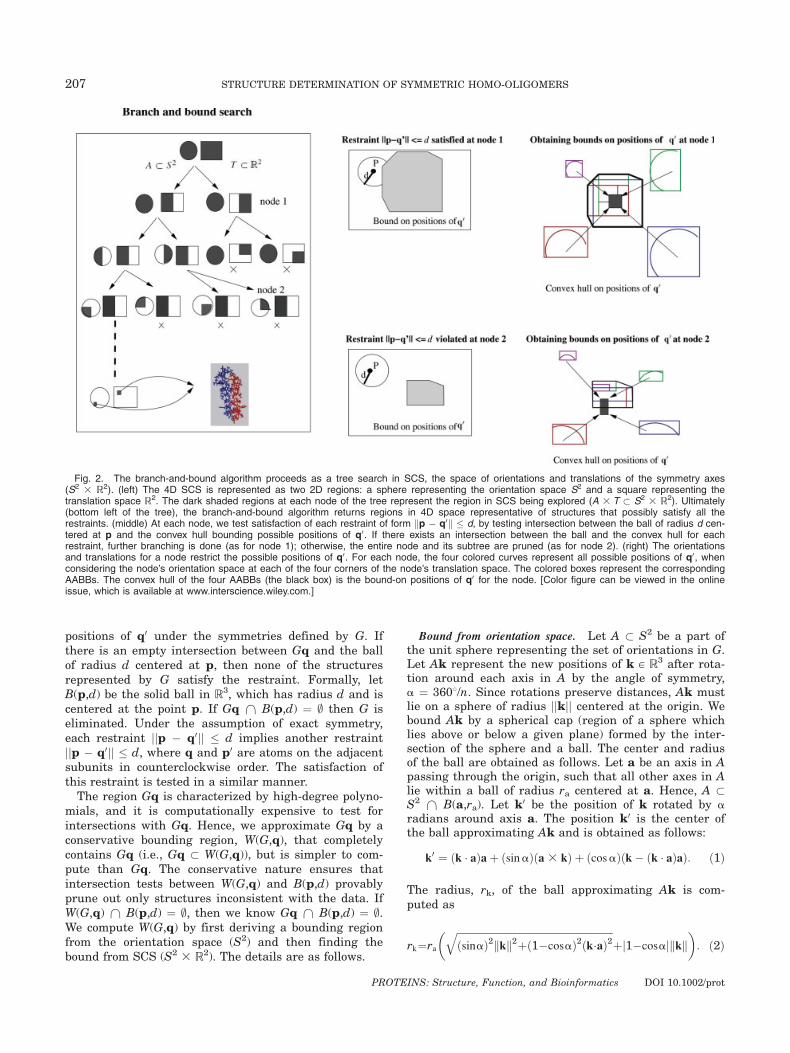
positions of q0 under the symmetries defined by G. Ifthere is an empty intersection between Gq and the ballof radius d centered at p, then none of the structuresrepresented by G satisfy the restraint. Formally, letB(p,d) be the solid ball in R3, which has radius d and iscentered at the point p. If Gq \ B(p,d) ¼ ; then G iseliminated. Under the assumption of exact symmetry,each restraint jjp � q0jj � d implies another restraintjjp � q0jj � d, where q and p0 are atoms on the adjacentsubunits in counterclockwise order. The satisfaction ofthis restraint is tested in a similar manner.The region Gq is characterized by high-degree polyno-
mials, and it is computationally expensive to test forintersections with Gq. Hence, we approximate Gq by aconservative bounding region, W(G,q), that completelycontains Gq (i.e., Gq � W(G,q)), but is simpler to com-pute than Gq. The conservative nature ensures thatintersection tests between W(G,q) and B(p,d) provablyprune out only structures inconsistent with the data. IfW(G,q) \ B(p,d) ¼ ;, then we know Gq \ B(p,d) ¼ ;.We compute W(G,q) by first deriving a bounding regionfrom the orientation space (S2) and then finding thebound from SCS (S2 3 R2). The details are as follows.
Bound from orientation space. Let A � S2 be a part ofthe unit sphere representing the set of orientations in G.Let Ak represent the new positions of k [ R3 after rota-tion around each axis in A by the angle of symmetry,a ¼ 3608/n. Since rotations preserve distances, Ak mustlie on a sphere of radius jjkjj centered at the origin. Webound Ak by a spherical cap (region of a sphere whichlies above or below a given plane) formed by the inter-section of the sphere and a ball. The center and radiusof the ball are obtained as follows. Let a be an axis in Apassing through the origin, such that all other axes in Alie within a ball of radius ra centered at a. Hence, A �S2 \ B(a,ra). Let k0 be the position of k rotated by aradians around axis a. The position k0 is the center ofthe ball approximating Ak and is obtained as follows:
k0 ¼ ðk � aÞaþ ðsinaÞða 3 kÞ þ ðcosaÞðk� ðk � aÞaÞ: ð1Þ
The radius, rk, of the ball approximating Ak is com-puted as
rk¼ra
� ffiffiffiffiffiffiffiffiffiffiffiffiffiffiffiffiffiffiffiffiffiffiffiffiffiffiffiffiffiffiffiffiffiffiffiffiffiffiffiffiffiffiffiffiffiffiffiffiffiffiffiffiffiffiffiffiffiffiffiffiffiffiffiðsinaÞ2kkk2þð1�cosaÞ2ðk�aÞ2
qþj1�cosajkkk
�: ð2Þ
Fig. 2. The branch-and-bound algorithm proceeds as a tree search in SCS, the space of orientations and translations of the symmetry axes(S2 3 R2). (left) The 4D SCS is represented as two 2D regions: a sphere representing the orientation space S2 and a square representing thetranslation space R2. The dark shaded regions at each node of the tree represent the region in SCS being explored (A 3 T � S2 3 R2). Ultimately(bottom left of the tree), the branch-and-bound algorithm returns regions in 4D space representative of structures that possibly satisfy all therestraints. (middle) At each node, we test satisfaction of each restraint of form kp � q0k � d, by testing intersection between the ball of radius d cen-tered at p and the convex hull bounding possible positions of q0. If there exists an intersection between the ball and the convex hull for eachrestraint, further branching is done (as for node 1); otherwise, the entire node and its subtree are pruned (as for node 2). (right) The orientationsand translations for a node restrict the possible positions of q0. For each node, the four colored curves represent all possible positions of q0, whenconsidering the node’s orientation space at each of the four corners of the node’s translation space. The colored boxes represent the correspondingAABBs. The convex hull of the four AABBs (the black box) is the bound-on positions of q0 for the node. [Color figure can be viewed in the onlineissue, which is available at www.interscience.wiley.com.]
207 STRUCTURE DETERMINATION OF SYMMETRIC HOMO-OLIGOMERS
PROTEINS: Structure, Function, and Bioinformatics DOI 10.1002/prot
Finally, we define our bounding region to be the spheri-cal cap C ¼ B(k0,rk) \ S(0,jjkjj), where S(0,jjkjj) denotesthe sphere of radius jjkjj that is centered at the point 0,the origin. By construction, Ak � C. We approximate Cwith a bounding box that is aligned along the x-, y-, andz-axes—an axis-aligned bounding box (AABB), which werefer to as V(A,k). We use V(A,k) to help us perform aquick test for intersection. If this quick test is passed,we then perform a second, more careful and expensivetest for intersection, using a tighter bounding region forAk. This tighter bound is the smallest AABB that con-tains Ak. The dimensions of the box are found by per-forming a numerical global minimization (and maximi-zation) on the x-, y-, and z-coordinates of k0. The globaloptimization is done by gridding A and starting a gradi-ent descent from each of the grid points. The details ofboth our intersection tests, and the Eqs. (1) and (2) areprovided in the Supplementary Material.
Bound from SCS. Let T � R2 and A � S2 denote thesets of translations and orientations in G. The region Gqrepresents the positions of q on rotation around eachaxis in A 3 T by the angle of symmetry a. To bound Gqand determine W(G,q), we need to find the orientation-based bound (as mentioned earlier) for each translationt [ T. We choose our bounding region W(G,q) as anapproximation of the convex hull of Gq. Using the prop-erties of convex hulls and the fact that T is convex, weare able to prove that the convex hull of Gq is deter-mined by just the corners of T (see Supplementary Ma-terial for proof). Let H(U) be the convex hull of U � R3.It can be shown that H(Gq) ¼ H(|4
i¼1{Aqti þ ti}) whereti represents the four corners of T and qti denotes theposition (q � ti). We use our bounds on regions of Ak,the AABB V(A,k), to bound Aqti. We then bound Gq byfinding the convex hull of the AABBs at the corners ofT. This convex hull is our bounding region W(G,q). Itcan be proven that W(G,q) is a conservative boundingregion for Gq (see Supplementary Material). That is,Gq � W(G,q). Hence, testing satisfaction of a restraintjjp � q0jj � d requires testing for the intersection of theconvex hull of the AABBs at the corners of T (which is abounding region for Gq), with a solid ball centered at pwith radius d. We test the intersection between a balland a convex polyhedron, using the method described inRef. 40. For details on the bounding regions and pruningcriteria, please see the Supplementary Material.
Branching
In partitioning a grid cell into subcells, we seek todivide the cell into two regions along the dimension thatwill cause one of the restraints to be violated by all theconformations represented by one of the subcells. Thiskind of a division will allow us to efficiently eliminate thesubcell. We use the following heuristic to achieve this. Foreach restraint jjp � q0jj � d, we compute q0 for each ofthe corners of the grid cell. We then identify the dimen-sion that has the largest difference in jjp � q0jj distancesfor its pair of corners. We partition along that dimension.
Determining Satisfying Structures
Our algorithm guarantees that the consistent regionsit returns represent every conformation of the symmet-ric homo-oligomer that is consistent with the data.Because we prune conservatively, they might also repre-sent additional structures inconsistent with the data. Toidentify the most consistent structures and evaluatepacking quality, we generate representative structuresfrom the consistent regions. Choosing good representa-tives helps ensure that we do not miss native structures(as can occur with sampling-based docking approaches;see Introduction section). We choose the set of represen-tative structures as follows. A grid cell is accepted aspart of the consistent regions only when all structures itrepresents are within s0 A of each other. We first obtainthe structures from the centroids of the grid cells in theconsistent regions. We then cluster these structuresusing an agglomerative complete linkage hierarchicalclustering41 that allows two structures to be within acluster only if their backbone RMSD is within s0 A. Thecentroids of the clusters then form a set of representa-tive structures. This procedure ensures that every struc-ture of the consistent region is within s0 A of at leastone representative structure, and hence ensures thatthe representative structures form a good representationof the consistent regions.
Some of representative structures might be inconsis-tent with data, because of the conservative bounds usedwhen pruning regions. We define the satisfaction scorefor each structure as the sum over the violated NOErestraints of the difference in expected and observeddistance.
The set of satisfying structures are those representa-tive structures with satisfaction scores below a threshold(chosen as 1 A). Note that each satisfying structure rep-resents a set of grid cells in the consistent regions. Theunion of all such grid cells forms the satisfying regions.
Determining WPS Structures
Having obtained the set of satisfying structures, wenow evaluate each of them for packing quality. We firstenergy-minimize them with the LBFGS conjugate-gradi-ent minimization algorithm (10,000 minimization steps)in CNS.42 The energy function being minimized includesthe NOE restraint energy terms as well as the modelingenergy terms of vdW (6-12 Lennard-Jones potential),bond length, bond angle, dihedral, and improper ener-gies.42 We harmonically restrain the backbone and theNOE restraints to ensure that we maintain the symme-try and satisfy the restraints. The minimization ac-counts for flexibility and asymmetry in the side chainsand should help obtain conformations of the side chainsthat aid in good vdW packing.
We define the packing score of an energy-minimizedstructure as the difference between the vdW energy of asubunit in the structure when it is in the complex,and the vdW energy of the subunit when it is not in thecomplex.
208 S. POTLURI ET AL.
PROTEINS: Structure, Function, and Bioinformatics DOI 10.1002/prot
We then define the set of WPS structures as those sat-isfying structures with packing scores below a threshold,chosen here as 0 kcal/mol, since well-modeled structuresshould have negative packing scores. We refer to theunion of grid cells in the consistent regions correspond-ing to the WPS structures as the WPS regions.
Evaluating Uncertainty
We evaluate uncertainty in two ways. One way is bycalculating the uncertainty in the conformations as thevariance in the position of each atom across the set ofsatisfying (or WPS) structures. The second way is byquantifying uncertainty in SCS as the spread in theareas of the satisfying (or WPS) regions in the transla-tion space and the orientation space. The 4D volume ofthe satisfying (or WPS) regions also indicates uncer-tainty. By comparing these measures for satisfying ver-sus WPS structures and regions, we evaluate the con-straint on structure provided by data alone versus theconstraint provided by both data and packing together.
Handling Ambiguity
The spectral overlap inherent in NMR spectra for sym-metric homo-oligomers leads to intersubunit distancerestraints that have a two-fold ambiguity. By two-foldambiguity, we mean that the relative order of the re-straints (whether they have a clockwise or a counter-clockwise orientation) is not apparent. Each restraint hastwo possible orientations. For instance, when a intersubu-nit restraint is observed between residues 32 and 38, therestraint could be between residue 32 on the first subunitand residue 38 on the adjacent subunit in clockwise orderor between residue 32 on the first subunit and residue 38on the adjacent subunit in counterclockwise order.We handle this two-fold ambiguity by extending our
branch-and-bound search of SCS. If we have r res-traints, each with two orientations, then there are 2r�1
possible combinations (the first orientation assignmentcan be made arbitrarily, since the measurements arerelative). However, the structure of our search allowsus to avoid testing all the combinations explicitly.Recall that, for each grid cell, we test each of therestraints individually using our conservative bounds(Bounding section). Now we simply individually testeach of the ‘‘oriented restraints’’ (i.e., a restraint witheither a clockwise or counterclockwise orientation).This yields 1 þ 2(r � 1) independent tests, rather thanthe exponential blow-up required to consider all combi-nations. If an oriented restraint is violated, then allcombinations including it are implicitly eliminatedfrom further consideration. If both orientations for anyrestraint are eliminated for some grid cell, then thecell is completely pruned. Otherwise, we continuebranching as usual. At the end of the search, we canenumerate the combinations represented in the satisfy-ing regions.
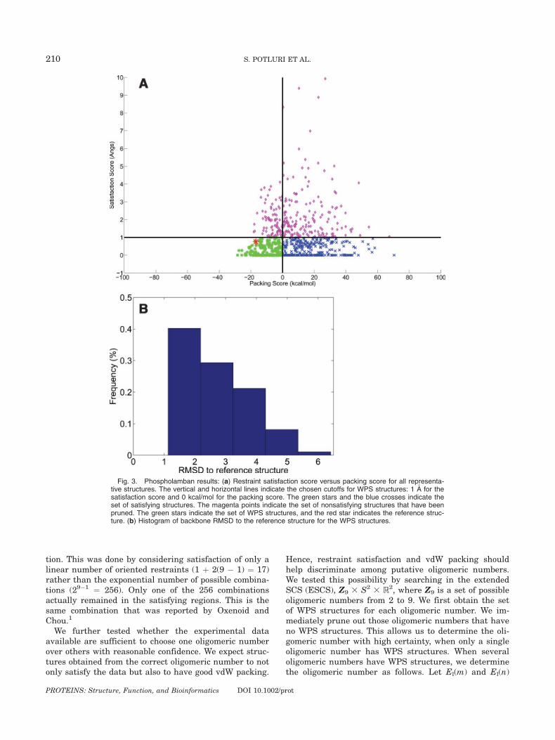
RESULTSResults on Phospholamban
We tested our protocol on phospholamban, using thesubunit structure and nine experimental NOE restraintsfrom the Chou lab. We chose as the reference structure,the best representative conformer (as indicated in Ref.1) among the deposited 20 NMR structures (PDB id:1ZLL). For all our test cases, we chose the structuralsimilarity threshold, s0, to be 1 A. Also, we declare astructure to have a steric clash when there are at leastfive pairs of atoms, such that each pair is separated byless than 1.5 A. Energy minimization of structuresallows side-chain flexibility such that one to two stericclashes can be eliminated. Our analysis is conserva-tive and declares a clash only when at least five atomscollide.
Figure 3(a) plots the packing scores versus the satis-faction scores for phospholamban. The set of satisfyingstructures has an overall range of 1.07–8.85 A backboneRMSD to the reference structure. This range indicatesthe diversity in structures possible, using just the nineexperimental intersubunit NOE restraints. The averagevariance in the positions of the atoms in the set of satis-fying structures is 12.32 A2. The area of the translationspace in the satisfying regions is �290 A2 and that ofthe orientation space is �0.40 radian2. The volume ofthe 4D region is �1.24 A2 radian2. These values indicatethe constraint on structure provided by the data alone.
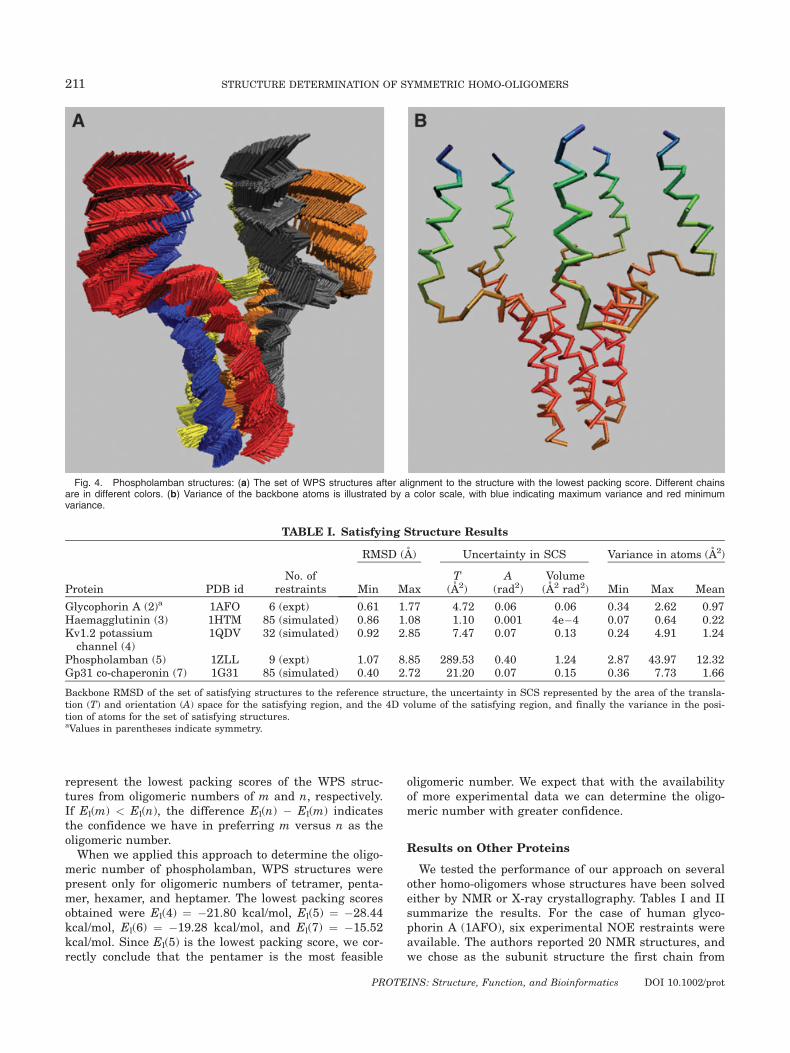
Figure 3(a) also shows the set of WPS structures (ingreen). The reference structure has a satisfaction scoreof around 0.8 A and packing score of �17 kcal/mol, andit lies in the WPS region. Figure 3(b) shows the back-bone RMSD of the reference structure to each of theWPS structures. Incorporating packing quality reducesthe maximum RMSD to the reference structure from8.85 to 6.24 A. The area of the translation space reducesfrom 290 to �135 A2 and that of the orientation spacereduces from 0.40 to �0.23 radian2. The volume of the4D space reduces from 1.24 A2 radian2 in the set of sat-isfying structures to �0.51 A2 radian2. All these valuesindicate the additional constraint that packing qualityimposes on the structure of phospholamban. The aver-age variance in the positions of the atoms is reducedfrom 12.32 to 6.80 A. Figure 4(a) illustrates all the WPSstructures, and Figure 4(b) illustrates the variance ofeach of the backbone atoms. The figures show that thereis more uncertainty in the amphipathic helices than inthe transmembrane helices. The average variance in theamphipathic helices is 10.75 A2, whereas that inthe transmembrane helices is 2.96 A2. This is becausethe experimental restraints are between residues in thetransmembrane helices. This agrees with the observa-tion in Ref. 1 that the amphipathic helices are less welldetermined. From this we conclude that we need morerestraints in the amphipathic helices to determine thestructure with greater precision.
We resolved the ambiguity in the nine intersubunitrestraints, as described in the Handling Ambiguity sec-
209 STRUCTURE DETERMINATION OF SYMMETRIC HOMO-OLIGOMERS
PROTEINS: Structure, Function, and Bioinformatics DOI 10.1002/prot
tion. This was done by considering satisfaction of only alinear number of oriented restraints (1 þ 2(9 � 1) ¼ 17)rather than the exponential number of possible combina-tions (29�1 ¼ 256). Only one of the 256 combinationsactually remained in the satisfying regions. This is thesame combination that was reported by Oxenoid andChou.1
We further tested whether the experimental dataavailable are sufficient to choose one oligomeric numberover others with reasonable confidence. We expect struc-tures obtained from the correct oligomeric number to notonly satisfy the data but also to have good vdW packing.
Hence, restraint satisfaction and vdW packing shouldhelp discriminate among putative oligomeric numbers.We tested this possibility by searching in the extendedSCS (ESCS), Z9 3 S2 3 R2, where Z9 is a set of possibleoligomeric numbers from 2 to 9. We first obtain the setof WPS structures for each oligomeric number. We im-mediately prune out those oligomeric numbers that haveno WPS structures. This allows us to determine the oli-gomeric number with high certainty, when only a singleoligomeric number has WPS structures. When severaloligomeric numbers have WPS structures, we determinethe oligomeric number as follows. Let El(m) and El(n)
Fig. 3. Phospholamban results: (a) Restraint satisfaction score versus packing score for all representa-tive structures. The vertical and horizontal lines indicate the chosen cutoffs for WPS structures: 1 A for thesatisfaction score and 0 kcal/mol for the packing score. The green stars and the blue crosses indicate theset of satisfying structures. The magenta points indicate the set of nonsatisfying structures that have beenpruned. The green stars indicate the set of WPS structures, and the red star indicates the reference struc-ture. (b) Histogram of backbone RMSD to the reference structure for the WPS structures.
210 S. POTLURI ET AL.
PROTEINS: Structure, Function, and Bioinformatics DOI 10.1002/prot
represent the lowest packing scores of the WPS struc-tures from oligomeric numbers of m and n, respectively.If El(m) < El(n), the difference El(n) � El(m) indicatesthe confidence we have in preferring m versus n as theoligomeric number.When we applied this approach to determine the oligo-
meric number of phospholamban, WPS structures werepresent only for oligomeric numbers of tetramer, penta-mer, hexamer, and heptamer. The lowest packing scoresobtained were El(4) ¼ �21.80 kcal/mol, El(5) ¼ �28.44kcal/mol, El(6) ¼ �19.28 kcal/mol, and El(7) ¼ �15.52kcal/mol. Since El(5) is the lowest packing score, we cor-rectly conclude that the pentamer is the most feasible
oligomeric number. We expect that with the availabilityof more experimental data we can determine the oligo-meric number with greater confidence.
Results on Other Proteins
We tested the performance of our approach on severalother homo-oligomers whose structures have been solvedeither by NMR or X-ray crystallography. Tables I and IIsummarize the results. For the case of human glyco-phorin A (1AFO), six experimental NOE restraints wereavailable. The authors reported 20 NMR structures, andwe chose as the subunit structure the first chain from
Fig. 4. Phospholamban structures: (a) The set of WPS structures after alignment to the structure with the lowest packing score. Different chainsare in different colors. (b) Variance of the backbone atoms is illustrated by a color scale, with blue indicating maximum variance and red minimumvariance.
TABLE I. Satisfying Structure Results
Protein PDB idNo. of
restraints
RMSD (A) Uncertainty in SCS Variance in atoms (A2)
Min MaxT
(A2)A
(rad2)Volume(A2 rad2) Min Max Mean
Glycophorin A (2)a 1AFO 6 (expt) 0.61 1.77 4.72 0.06 0.06 0.34 2.62 0.97Haemagglutinin (3) 1HTM 85 (simulated) 0.86 1.08 1.10 0.001 4e�4 0.07 0.64 0.22Kv1.2 potassiumchannel (4)
1QDV 32 (simulated) 0.92 2.85 7.47 0.07 0.13 0.24 4.91 1.24
Phospholamban (5) 1ZLL 9 (expt) 1.07 8.85 289.53 0.40 1.24 2.87 43.97 12.32Gp31 co-chaperonin (7) 1G31 85 (simulated) 0.40 2.72 21.20 0.07 0.15 0.36 7.73 1.66
Backbone RMSD of the set of satisfying structures to the reference structure, the uncertainty in SCS represented by the area of the transla-tion (T) and orientation (A) space for the satisfying region, and the 4D volume of the satisfying region, and finally the variance in the posi-tion of atoms for the set of satisfying structures.aValues in parentheses indicate symmetry.
211 STRUCTURE DETERMINATION OF SYMMETRIC HOMO-OLIGOMERS
PROTEINS: Structure, Function, and Bioinformatics DOI 10.1002/prot
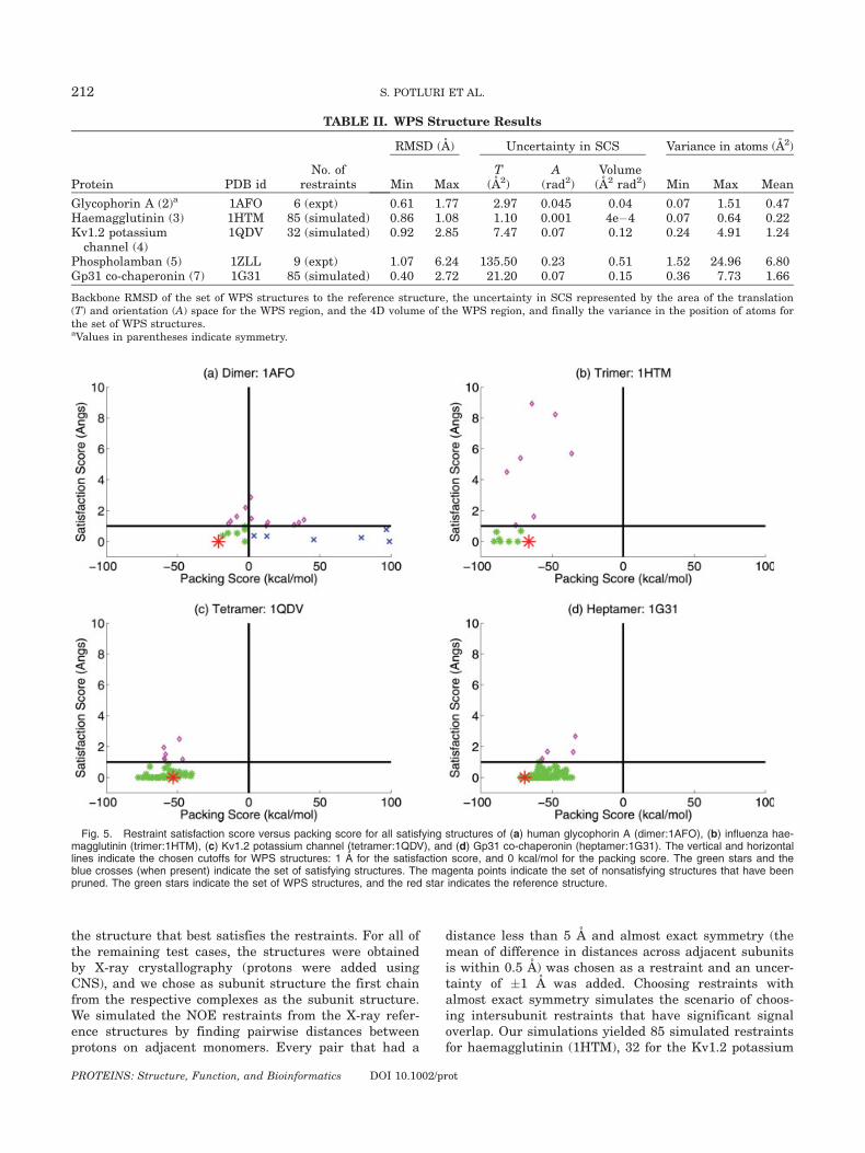
the structure that best satisfies the restraints. For all ofthe remaining test cases, the structures were obtainedby X-ray crystallography (protons were added usingCNS), and we chose as subunit structure the first chainfrom the respective complexes as the subunit structure.We simulated the NOE restraints from the X-ray refer-ence structures by finding pairwise distances betweenprotons on adjacent monomers. Every pair that had a
distance less than 5 A and almost exact symmetry (themean of difference in distances across adjacent subunitsis within 0.5 A) was chosen as a restraint and an uncer-tainty of �1 A was added. Choosing restraints withalmost exact symmetry simulates the scenario of choos-ing intersubunit restraints that have significant signaloverlap. Our simulations yielded 85 simulated restraintsfor haemagglutinin (1HTM), 32 for the Kv1.2 potassium
Fig. 5. Restraint satisfaction score versus packing score for all satisfying structures of (a) human glycophorin A (dimer:1AFO), (b) influenza hae-magglutinin (trimer:1HTM), (c) Kv1.2 potassium channel (tetramer:1QDV), and (d) Gp31 co-chaperonin (heptamer:1G31). The vertical and horizontallines indicate the chosen cutoffs for WPS structures: 1 A for the satisfaction score, and 0 kcal/mol for the packing score. The green stars and theblue crosses (when present) indicate the set of satisfying structures. The magenta points indicate the set of nonsatisfying structures that have beenpruned. The green stars indicate the set of WPS structures, and the red star indicates the reference structure.
TABLE II. WPS Structure Results
Protein PDB idNo. of
restraints
RMSD (A) Uncertainty in SCS Variance in atoms (A2)
Min MaxT
(A2)A
(rad2)Volume(A2 rad2) Min Max Mean
Glycophorin A (2)a 1AFO 6 (expt) 0.61 1.77 2.97 0.045 0.04 0.07 1.51 0.47Haemagglutinin (3) 1HTM 85 (simulated) 0.86 1.08 1.10 0.001 4e�4 0.07 0.64 0.22Kv1.2 potassiumchannel (4)
1QDV 32 (simulated) 0.92 2.85 7.47 0.07 0.12 0.24 4.91 1.24
Phospholamban (5) 1ZLL 9 (expt) 1.07 6.24 135.50 0.23 0.51 1.52 24.96 6.80Gp31 co-chaperonin (7) 1G31 85 (simulated) 0.40 2.72 21.20 0.07 0.15 0.36 7.73 1.66
Backbone RMSD of the set of WPS structures to the reference structure, the uncertainty in SCS represented by the area of the translation(T) and orientation (A) space for the WPS region, and the 4D volume of the WPS region, and finally the variance in the position of atoms forthe set of WPS structures.aValues in parentheses indicate symmetry.
212 S. POTLURI ET AL.
PROTEINS: Structure, Function, and Bioinformatics DOI 10.1002/prot
channel (1QDV), and 85 for the Gp31 co-chaperonin(1G31).Figure 5 shows for each test case a plot of packing
scores versus satisfaction scores. Table I shows theresults on the satisfying regions and satisfying struc-tures, and Table II shows the results on WPS regionsand WPS structures. The tables and figure clearly showthat except for human glycophorin A, the remaining testcases have identical sets of satisfying structures andWPS structures. Further, the spread in the satisfyingregion and the WPS region is almost the same. The rea-son for this similarity might be because we have usedall possible restraints (85, 32, and 85) for these testcases. This use of all restraints causes almost all the sat-isfying structures returned by the branch-and-bound
algorithm to have high-quality vdW packing, and henceto belong to the set of WPS structures.
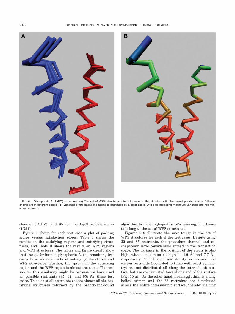
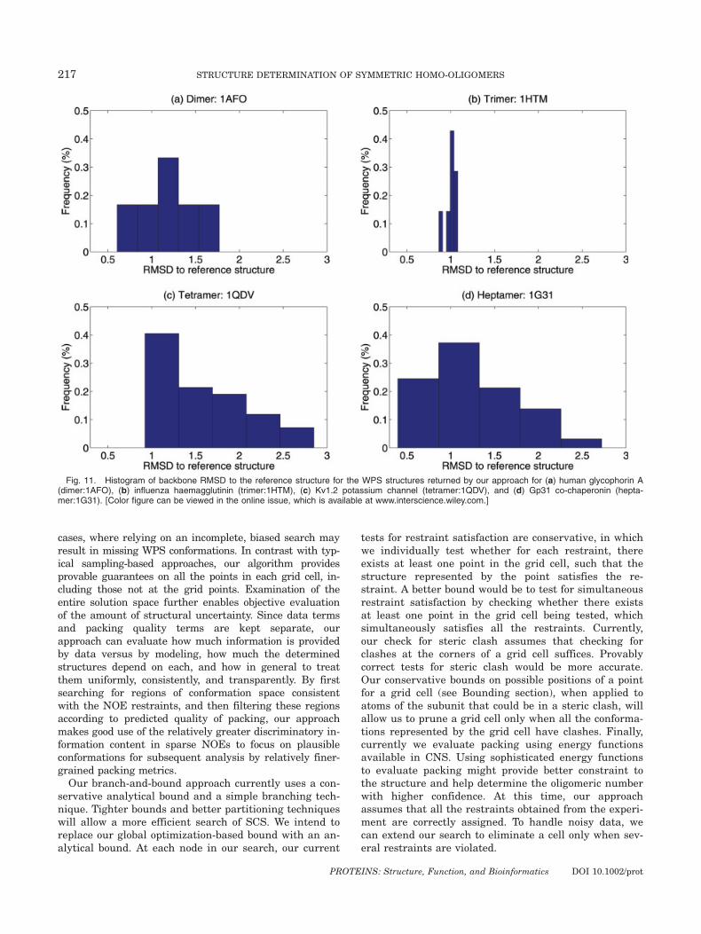
Figures 6–9 illustrate the uncertainty in the set ofWPS structures for each of the test cases. Despite using32 and 85 restraints, the potassium channel and co-chaperonin have considerable spread in the translationspace. The variance in the position of the atoms is alsohigh, with a maximum as high as 4.9 A2 and 7.7 A2,respectively. The higher uncertainty is because thechosen restraints (restricted to those with exact symme-try) are not distributed all along the intersubunit sur-face, but are concentrated toward one end of the surface[Fig. 10(a)]. On the other hand, haemagglutinin is a longhelical trimer, and the 85 restraints are distributedacross the entire intersubunit surface, thereby yielding
Fig. 6. Glycophorin A (1AFO) structures: (a) The set of WPS structures after alignment to the structure with the lowest packing score. Differentchains are in different colors. (b) Variance of the backbone atoms is illustrated by a color scale, with blue indicating maximum variance and red min-imum variance.
213 STRUCTURE DETERMINATION OF SYMMETRIC HOMO-OLIGOMERS
PROTEINS: Structure, Function, and Bioinformatics DOI 10.1002/prot
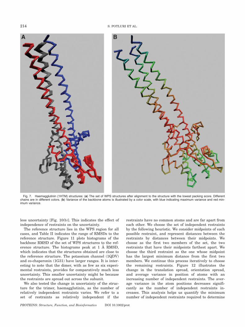
less uncertainty [Fig. 10(b)]. This indicates the effect ofindependence of restraints on the uncertainty.The reference structure lies in the WPS region for all
cases, and Table II indicates the range of RMSDs to thereference structure. Figure 11 plots histograms of thebackbone RMSD of the set of WPS structures to the ref-erence structure. The histograms peak at 1 A RMSD,which indicates that the structures obtained are close tothe reference structure. The potassium channel (1QDV)and co-chaperonin (1G31) have larger ranges. It is inter-esting to note that the dimer, with as few as six experi-mental restraints, provides for comparatively much lessuncertainty. This smaller uncertainty might be becausethe restraints are spread out across the subunit.We also tested the change in uncertainty of the struc-
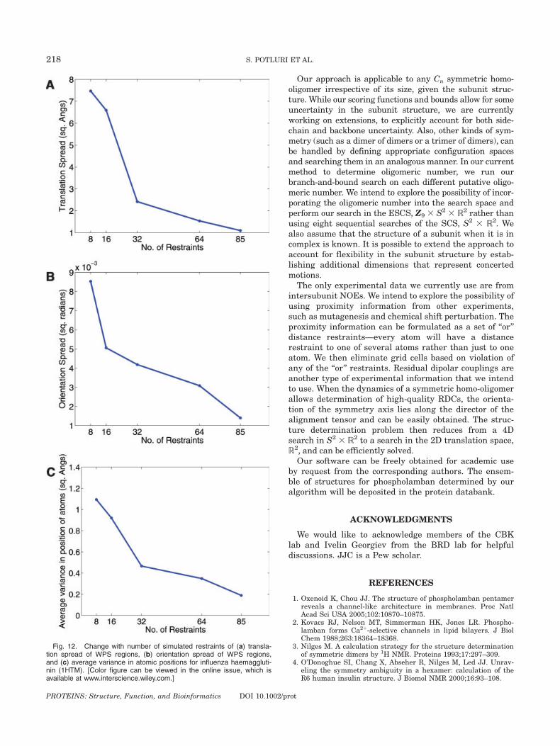
ture for the trimer, haemagglutinin, as the number ofrelatively independent restraints varies. We refer to aset of restraints as relatively independent if the
restraints have no common atoms and are far apart fromeach other. We choose the set of independent restraintsby the following heuristic. We consider midpoints of eachpossible restraint, and represent distances between therestraints by distances between their midpoints. Wechoose as the first two members of the set, the tworestraints that have their midpoints farthest apart. Wechoose the third restraint as the one whose midpointhas the largest minimum distance from the first twomembers. We continue this process iteratively to choosethe remaining restraints. Figure 12 illustrates thechange in the translation spread, orientation spread,and average variance in position of atoms with anincreasing number of independent restraints. The aver-age variance in the atom positions decreases signifi-cantly as the number of independent restraints in-creases. This analysis helps us quantify the minimumnumber of independent restraints required to determine
Fig. 7. Haemagglutinin (1HTM) structures: (a) The set of WPS structures after alignment to the structure with the lowest packing score. Differentchains are in different colors. (b) Variance of the backbone atoms is illustrated by a color scale, with blue indicating maximum variance and red min-imum variance.
214 S. POTLURI ET AL.
PROTEINS: Structure, Function, and Bioinformatics DOI 10.1002/prot
the structure of the homo-oligomer up to a specified pre-cision. For example, if we are willing to tolerate �1 A2
uncertainty in the positions of the atoms, about eight in-dependent restraints will be sufficient. On the otherhand, if we want high precision and are not willing totolerate uncertainty above 0.3 A2 in the positions ofatoms, we need as many as 64 independent restraints.
We also applied our method for determination of oligo-meric number (Results on Phosphalamban) to the testcases. For glycophorin A, haemagglutinin, and the Kv1.2potassium channel, we could determine the oligomericnumber with high certainty, because the set of WPSstructures was empty for oligomeric numbers other thanthe correct one. Glycophorin A is especially interesting,
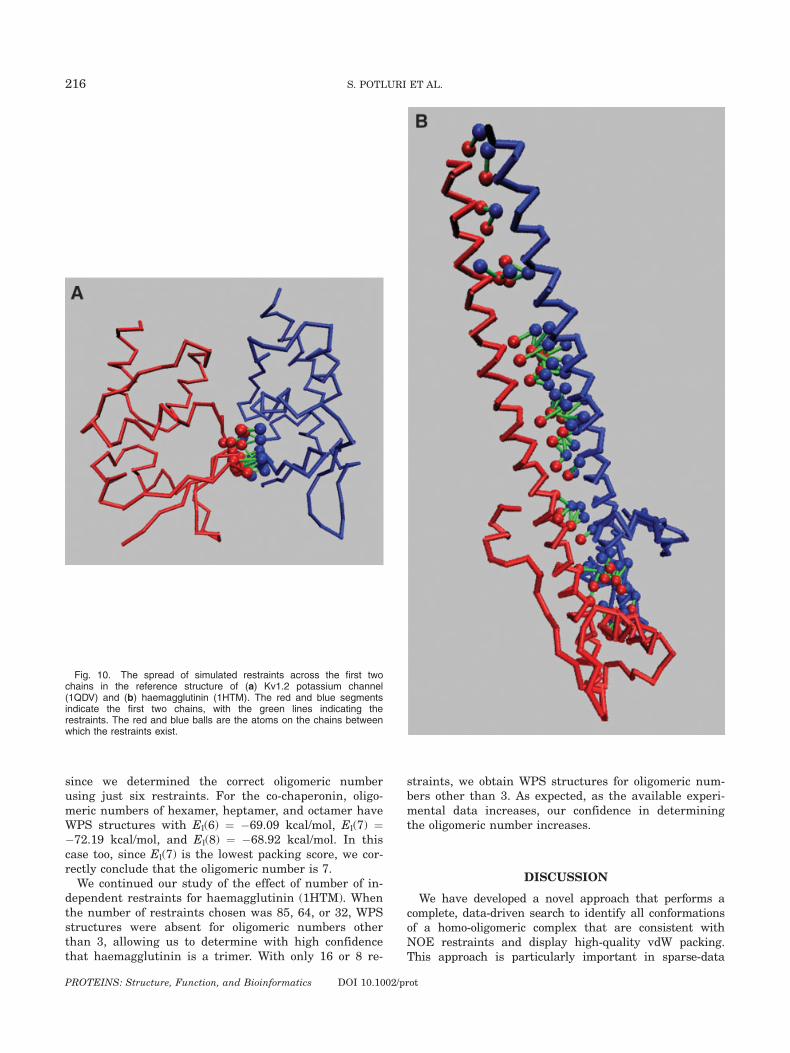
Fig. 9. Gp31 co-chaperonin (1G31) structures: (a) The set of WPS structures after alignment to the structure with the lowest packing score.Different chains are in different colors. (b) Variance of the backbone atoms is illustrated by a color scale, with blue indicating maximum variance andred minimum variance.
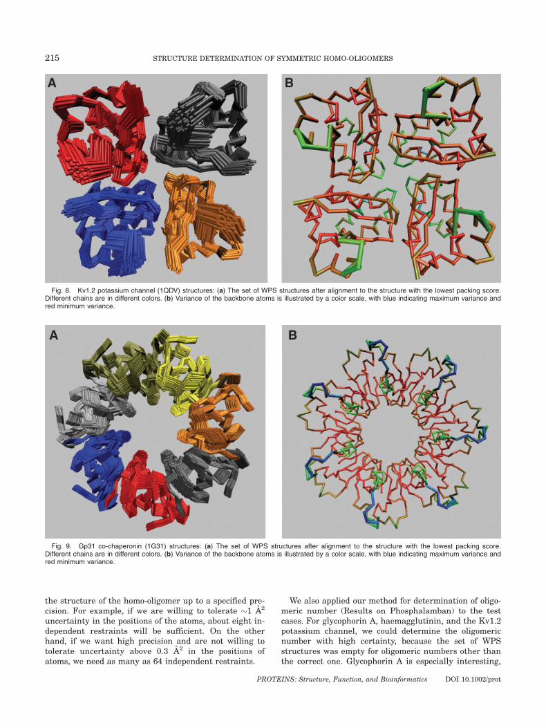
Fig. 8. Kv1.2 potassium channel (1QDV) structures: (a) The set of WPS structures after alignment to the structure with the lowest packing score.Different chains are in different colors. (b) Variance of the backbone atoms is illustrated by a color scale, with blue indicating maximum variance andred minimum variance.
215 STRUCTURE DETERMINATION OF SYMMETRIC HOMO-OLIGOMERS
PROTEINS: Structure, Function, and Bioinformatics DOI 10.1002/prot
since we determined the correct oligomeric numberusing just six restraints. For the co-chaperonin, oligo-meric numbers of hexamer, heptamer, and octamer haveWPS structures with El(6) ¼ �69.09 kcal/mol, El(7) ¼�72.19 kcal/mol, and El(8) ¼ �68.92 kcal/mol. In thiscase too, since El(7) is the lowest packing score, we cor-rectly conclude that the oligomeric number is 7.We continued our study of the effect of number of in-
dependent restraints for haemagglutinin (1HTM). Whenthe number of restraints chosen was 85, 64, or 32, WPSstructures were absent for oligomeric numbers otherthan 3, allowing us to determine with high confidencethat haemagglutinin is a trimer. With only 16 or 8 re-
straints, we obtain WPS structures for oligomeric num-bers other than 3. As expected, as the available experi-mental data increases, our confidence in determiningthe oligomeric number increases.
DISCUSSION
We have developed a novel approach that performs acomplete, data-driven search to identify all conformationsof a homo-oligomeric complex that are consistent withNOE restraints and display high-quality vdW packing.This approach is particularly important in sparse-data
Fig. 10. The spread of simulated restraints across the first twochains in the reference structure of (a) Kv1.2 potassium channel(1QDV) and (b) haemagglutinin (1HTM). The red and blue segmentsindicate the first two chains, with the green lines indicating therestraints. The red and blue balls are the atoms on the chains betweenwhich the restraints exist.
216 S. POTLURI ET AL.
PROTEINS: Structure, Function, and Bioinformatics DOI 10.1002/prot
cases, where relying on an incomplete, biased search mayresult in missing WPS conformations. In contrast with typ-ical sampling-based approaches, our algorithm providesprovable guarantees on all the points in each grid cell, in-cluding those not at the grid points. Examination of theentire solution space further enables objective evaluationof the amount of structural uncertainty. Since data termsand packing quality terms are kept separate, ourapproach can evaluate how much information is providedby data versus by modeling, how much the determinedstructures depend on each, and how in general to treatthem uniformly, consistently, and transparently. By firstsearching for regions of conformation space consistentwith the NOE restraints, and then filtering these regionsaccording to predicted quality of packing, our approachmakes good use of the relatively greater discriminatory in-formation content in sparse NOEs to focus on plausibleconformations for subsequent analysis by relatively finer-grained packing metrics.Our branch-and-bound approach currently uses a con-
servative analytical bound and a simple branching tech-nique. Tighter bounds and better partitioning techniqueswill allow a more efficient search of SCS. We intend toreplace our global optimization-based bound with an an-alytical bound. At each node in our search, our current
tests for restraint satisfaction are conservative, in whichwe individually test whether for each restraint, thereexists at least one point in the grid cell, such that thestructure represented by the point satisfies the re-straint. A better bound would be to test for simultaneousrestraint satisfaction by checking whether there existsat least one point in the grid cell being tested, whichsimultaneously satisfies all the restraints. Currently,our check for steric clash assumes that checking forclashes at the corners of a grid cell suffices. Provablycorrect tests for steric clash would be more accurate.Our conservative bounds on possible positions of a pointfor a grid cell (see Bounding section), when applied toatoms of the subunit that could be in a steric clash, willallow us to prune a grid cell only when all the conforma-tions represented by the grid cell have clashes. Finally,currently we evaluate packing using energy functionsavailable in CNS. Using sophisticated energy functionsto evaluate packing might provide better constraint tothe structure and help determine the oligomeric numberwith higher confidence. At this time, our approachassumes that all the restraints obtained from the experi-ment are correctly assigned. To handle noisy data, wecan extend our search to eliminate a cell only when sev-eral restraints are violated.
Fig. 11. Histogram of backbone RMSD to the reference structure for the WPS structures returned by our approach for (a) human glycophorin A(dimer:1AFO), (b) influenza haemagglutinin (trimer:1HTM), (c) Kv1.2 potassium channel (tetramer:1QDV), and (d) Gp31 co-chaperonin (hepta-mer:1G31). [Color figure can be viewed in the online issue, which is available at www.interscience.wiley.com.]
217 STRUCTURE DETERMINATION OF SYMMETRIC HOMO-OLIGOMERS
PROTEINS: Structure, Function, and Bioinformatics DOI 10.1002/prot
Our approach is applicable to any Cn symmetric homo-oligomer irrespective of its size, given the subunit struc-ture. While our scoring functions and bounds allow for someuncertainty in the subunit structure, we are currentlyworking on extensions, to explicitly account for both side-chain and backbone uncertainty. Also, other kinds of sym-metry (such as a dimer of dimers or a trimer of dimers), canbe handled by defining appropriate configuration spacesand searching them in an analogous manner. In our currentmethod to determine oligomeric number, we run ourbranch-and-bound search on each different putative oligo-meric number. We intend to explore the possibility of incor-porating the oligomeric number into the search space andperform our search in the ESCS, Z9 3 S2 3 R2 rather thanusing eight sequential searches of the SCS, S2 3 R2. Wealso assume that the structure of a subunit when it is incomplex is known. It is possible to extend the approach toaccount for flexibility in the subunit structure by estab-lishing additional dimensions that represent concertedmotions.
The only experimental data we currently use are fromintersubunit NOEs. We intend to explore the possibility ofusing proximity information from other experiments,such as mutagenesis and chemical shift perturbation. Theproximity information can be formulated as a set of ‘‘or’’distance restraints—every atom will have a distancerestraint to one of several atoms rather than just to oneatom. We then eliminate grid cells based on violation ofany of the ‘‘or’’ restraints. Residual dipolar couplings areanother type of experimental information that we intendto use. When the dynamics of a symmetric homo-oligomerallows determination of high-quality RDCs, the orienta-tion of the symmetry axis lies along the director of thealignment tensor and can be easily obtained. The struc-ture determination problem then reduces from a 4Dsearch in S2 3 R2 to a search in the 2D translation space,R2, and can be efficiently solved.
Our software can be freely obtained for academic useby request from the corresponding authors. The ensem-ble of structures for phospholamban determined by ouralgorithm will be deposited in the protein databank.
ACKNOWLEDGMENTS
We would like to acknowledge members of the CBKlab and Ivelin Georgiev from the BRD lab for helpfuldiscussions. JJC is a Pew scholar.
REFERENCES
1. Oxenoid K, Chou JJ. The structure of phospholamban pentamerreveals a channel-like architecture in membranes. Proc NatlAcad Sci USA 2005;102:10870–10875.
2. Kovacs RJ, Nelson MT, Simmerman HK, Jones LR. Phospho-lamban forms Ca2þ-selective channels in lipid bilayers. J BiolChem 1988;263:18364–18368.
3. Nilges M. A calculation strategy for the structure determinationof symmetric dimers by 1H NMR. Proteins 1993;17:297–309.
4. O’Donoghue SI, Chang X, Abseher R, Nilges M, Led JJ. Unrav-eling the symmetry ambiguity in a hexamer: calculation of theR6 human insulin structure. J Biomol NMR 2000;16:93–108.
Fig. 12. Change with number of simulated restraints of (a) transla-tion spread of WPS regions, (b) orientation spread of WPS regions,and (c) average variance in atomic positions for influenza haemaggluti-nin (1HTM). [Color figure can be viewed in the online issue, which isavailable at www.interscience.wiley.com.]
218 S. POTLURI ET AL.
PROTEINS: Structure, Function, and Bioinformatics DOI 10.1002/prot
5. Russell RB, Alber F, Aloy P, Davis FP, Korkin D, Pichaud M,Topf M, Sali A. A structural perspective on protein–proteininteractions. Curr Opin Struct Biol 2004;14:313–324.
6. Aloy P, Bottcher B, Ceulemans H, Leutwein C, Mellwig C, FischerS, Gavin AC, Bork P, Superti-Furga G, Serrano L. Structure-based assembly of protein complexes in yeast. Science 2004;303:2026–2029.
7. Aloy P, Russell RB. The third dimension for protein interactionsand complexes. Trends Biochem Sci 2002;27:633–638.
8. Marti-Renom MA, Stuart AC, Fiser A, Sanchez R, Melo F, Sali A.Comparative protein structure modeling of genes and genomes.Annu Rev Biophys Biomol Struct 2000;29:291–325.
9. Ritchie DW, Kemp GJ. Protein docking using spherical polarFourier correlations. Proteins 2000;39:178–194.
10. Gabb HA, Jackson RM, Sternberg MJ. Modeling protein dockingusing shape complementarity, electrostatics and biochemical in-formation. J Mol Biol 1997;272:106–120.
11. Vakser IA. Protein docking for low-resolution structures. ProteinEng 1995;8:371–377.
12. Fernandez-Recio J, Totrov M, Abagyan R. Soft protein–proteindocking in internal coordinates. Protein Sci 2002;11:280–291.
13. Mandell JG, Roberts VA, Pique ME, Kotlovyi V, Mitchell JC,Nelson E, Tsigelny I, Eyck LFT. Protein docking using continuumelectrostatics and geometric fit. Protein Eng 2001;14:105–113.
14. Gray JJ, Moughon S, Wang C, Schueler-Furman O, Kuhlman B,Rohl CA, Baker D. Protein–protein docking with simultaneousoptimization of rigid-body displacement and side-chain confor-mations. J Mol Biol 2003;331:281–299.
15. Chen R, Li L, Weng Z. ZDOCK: an initial-stage protein-dockingalgorithm. Proteins 2003;52:80–87.
16. Norel R, Petrey D, Wolfson HJ, Nussinov R. Examination ofshape complementarity in docking of unbound proteins. Proteins1999;36:307–317.
17. Duhovny D, Inbar Y, Polak V, Shatsky M, Halperin I, Benya-mini A, Barzilai A, Dror O, Haspel N, Nussinov R. Taking ge-ometry to its edge: fast unbound rigid (and hinge-bent) docking.Proteins 2003;52:107–112.
18. Smith GR, Sternberg MJ. Prediction of protein–protein interac-tions by docking methods. Curr Opin Struct Biol 2002;12:28–35.
19. Janin J, Henrick K, Moult J, Eyck LT, Sternberg MJ, Vajda S,Vakser I, Wodak SJ. CAPRI: a critical assessment of predictedinteractions. Proteins 2003;52:2–9.
20. Gray JJ, Moughon SE, Kortemme T, Schueler-Furman O,Misura KM, Morozov A, Baker D. Protein–protein dockingpredictions for the CAPRI experiment. Proteins 2003;52:118–122.
21. Pierce B, Weng Z. M-ZDOCK: a grid-based approach for Cn sym-metric multimer docking. Bioinformatics 2005;21:1472–1476.
22. Duhovny D, Nussinov R, Wolfson HJ. Efficient unbound dockingof rigid molecules. In Proceedings of the 2nd Workshop on Algo-rithms in Bioinformatics (WABI), Rome, Italy, Sep 17–21, 2002. Lec-ture Notes in Computer Science, Vol. 2452, (Guigo R, Gusfield D,editors). Berlin: Springer-Verlag, 2002. pp 185–200.
23. Comeau SR, Camacho CJ. Predicting oligomeric assemblies: N-mers a primer. J Struct Biol 2005;150:233–244.
24. Dobrodumov A, Gronenborn AM. Filtering and selection ofstructural models: combining docking and NMR. Proteins 2003;53:18–32.
25. Dominguez C, Boelens R, Bonvin AM. HADDOCK: a protein–protein docking approach based on biochemical or biophysicalinformation. J Am Chem Soc 2003;125:1731–1737.
26. Anand GS, Law D, Mandell JG, Snead AN, Tsigelny I, Taylor SS,Eyck LFT, Komives EA. Identification of the protein kinase A reg-ulatory RIa-catalytic subunit interface by amide H/2H exchangeand protein docking. Proc Natl Acad Sci USA 2003; 100:13264–13269.
27. Morillas M, Gomez-Puertas P, Rubi B, Clotet J, Arino J, ValenciaA, Hegardt FG, Serra D, Asins G. Structural model of a malonyl-CoA-binding site of carnitine octanoyltransferase and carnitinepalmitoyltransferase I: mutational analysis of a malonyl-CoA af-finity domain. J Biol Chem 2002;277:11473–11480.
28. Cunningham BC, Jhurani P, Ng P, Wells JA. Receptor and anti-body epitopes in human growth hormone identified by homolog-scanning mutagenesis. Science 1989;243:1330–1336.
29. Adams PD, Engelman DM, Brunger AT. Improved prediction forthe structure of the dimeric transmembrane domain of glyco-phorin A obtained through global searching. Proteins 1996;26:257–261.
30. Adams PD, Arkin IT, Engelman DM, Brunger AT. Computationalsearching and mutagenesis suggest a structure for the pentamerictransmembrane domain of phospholamban. Nat Struct Biol 1995;2:154–162.
31. Lanman J, Lam TT, Barnes S, Sakalian M, Emmett MR, MarshallAG, Prevelige PE, Jr. Identification of novel interactions in HIV-1capsid protein assembly by high-resolution mass spectrometry.J Mol Biol 2003;325:759–772.
32. Serino G, Su H, Peng Z, Tsuge T, Wei N, Gu H, Deng XW. Char-acterization of the last subunit of the arabidopsis COP9 signalo-some: implications for the overall structure and origin of thecomplex. Plant Cell 2003;15:719–731.
33. Back JW, de Jong L, Muijsers AO, de Koster CG. Chemicalcross-linking and mass spectrometry for protein structural mod-eling. J Mol Biol 2003;331:303–313.
34. Trester-Zedlitz M, Kamada K, Burley SK, Fenyo D, Chait BT,Muir TW. A modular cross-linking approach for exploring pro-tein interactions. J Am Chem Soc 2003;125:2416–2425.
35. Truong K, Ikura M. The use of FRET imaging microscopy todetect protein–protein interactions and protein conformationalchanges in vivo. Curr Opin Struct Biol 2001;11:573–578.
36. Yan Y, Marriott G. Analysis of protein interactions usingfluorescence technologies. Curr Opin Chem Biol 2003;7:635–640.
37. Kariakin A, Davydov D, Peterson JA, Jung C. A new approachto the study of protein–protein interaction by FTIR: complexformation between cytochrome P450BM-3 heme domain andFMN reductase domain. Biochemistry 2002;41:13514–13525.
38. Wang CE, Perez TL, Tidor B. AmbiPack: a systematic algorithmfor packing of macromolecular structures with ambiguous dis-tance constraints. Proteins: Struct Funct Genet 1998;32:26–42.
39. Lilien RH, Bailey-Kellogg C, Anderson AA, Donald BR. A sub-group algorithm to identify cross-rotation peaks consistent withnon-crystallographic symmetry. Acta Crystallogr D Biol Crystal-logr 2004;60:1057–1067.
40. Ericson C. Real-time collision detection. San Francisco: MorganKaufmann, 2005.
41. Han J, Micheline K. Data mining concepts and techniques. SanFrancisco: Morgan Kaufmann, 2001.
42. Brunger AT, Adams PD, Clore GM, DeLano WL, Gros P, Grosse-Kunstleve RW, Jiang JS, Kuszewski J, Nilges M, Pannu NS,Read RJ, Rice LM, Simonson T, Warren GL. Crystallographyand NMR system: a new software suite for macromolecular struc-ture determination. Acta Crystallogr D Biol Crystallogr 1998;54:905–921.
219 STRUCTURE DETERMINATION OF SYMMETRIC HOMO-OLIGOMERS
PROTEINS: Structure, Function, and Bioinformatics DOI 10.1002/prot