
Structure and vibrational spectra of fac - Re I ( CO ) 3 + complex with...
6
Structure and vibrational spectra of fac-Re I ðCOÞ þ 3 complex with N-methyl-2-pyridinecarbothioamide L. Fuks a, * , E. Gniazdowska a, * , J. Mieczkowski b , J. Narbutt a , W. Starosta a , M. Zasepa a a Institute of Nuclear Chemistry and Technology, Dorodna 16, 03-195 Warsaw, Poland b Department of Chemistry, Warsaw University, Pasteura 1, 02-09 Warsaw, Poland Received 14 July 2004; accepted 10 September 2004 Abstract Syntheses of a bidentate ligand L = N-methyl-2-pyridinecarbothioamide and its tricarbonylchlororhenium(I) complex, [fac- Re(CO) 3 LCl], are described. Physico-chemical characteristics of the novel species are presented. The molecular structures of both complex and ligand have been established by means of X-ray single crystal diffraction, and confirmed by FT-IR spectroscopy. UV–Vis absorption spectra of the complex have shown that it is stable in aqueous solutions at least for several days. Partition coef- ficients of the ligand and the complex in the iso-octanol–water systems have been determined. Lipophilicity of both is moderate: log(P) 1.1. Ó 2004 Elsevier B.V. All rights reserved. Keywords: Tricarbonylrhenium(I); N-methyl-2-pyridinecarbothioamide; Structural studies; Lipophilicity 1. Introduction Two rhenium radionuclides, 186 Re and 188 Re, are con- sidered as candidates for radiotherapeutical applications because of their favorable nuclear properties, in particu- lar the latter, which can be obtained from the 188 W/ 188 Re generator system [1–3] available in the increasing number of nuclear medicine departments and clinics. Especially the 188 Re radioisotope is suitable for preparation of radi- opharmaceuticals for therapeutic applications, due to its favorable nuclear properties. This nuclide decays through the emission of a b particle (E b = 2.2 MeV, T 1/2 = 16.9 h), which has energy appropriate for penetrat- ing and destroying abnormal tissues, and of c-rays (E c = 155 keV), which can be efficiently used for imaging and calculations of radiation doses. Rhenium eluted from the generator in the form of stable perrhenate anions ð 188 ReO 4 Þ does not form therapeutically active com- plexes. Compounds in lower oxidation states, in turn, are relatively easily re-oxidized back to perrhenate. Due to this fact, the rhenium radiopharmaceuticals are diffi- cult to prepare under clinical conditions. The major advantage of [Re(OH 2 ) 3 (CO) 3 ] + emerges from high stability of the [fac-Re(CO) 3 ] + moiety in water and its ability to exchange labile solvent ligands, H 2 O. Moreover, the d 6 electronic configuration of the M(I) center in the octahedral field makes the complexes kinetically inert. The discovery of simple preparative procedure for production of [fac- 188 Re(CO) 3 (H 2 O) 3 ] + (1) as a precursor of radiopharmaceuticals [4–7] has opened novel routes in labelling biomolecules [8]. How- ever, even relatively simple derivatives of 1 may be of interest for radiopharmacy. Interaction of hydrophilic 0022-328X/$ - see front matter Ó 2004 Elsevier B.V. All rights reserved. doi:10.1016/j.jorganchem.2004.09.040 * Corresponding authors. Tel.: +48 22 811 27 35; fax: +48 22 811 15 32. E-mail addresses: [email protected], [email protected] (L. Fuks), [email protected] (E. Gniazdowska). Journal of Organometallic Chemistry 689 (2004) 4751–4756 www.elsevier.com/locate/jorganchem
-
Upload
independent -
Category
Documents
-
view
1 -
download
0
Transcript of Structure and vibrational spectra of fac - Re I ( CO ) 3 + complex with...

Journal of Organometallic Chemistry 689 (2004) 4751–4756
www.elsevier.com/locate/jorganchem
Structure and vibrational spectra of fac-ReIðCOÞþ3 complex withN-methyl-2-pyridinecarbothioamide
L. Fuks a,*, E. Gniazdowska a,*, J. Mieczkowski b, J. Narbutt a,W. Starosta a, M. Zasepa a
a Institute of Nuclear Chemistry and Technology, Dorodna 16, 03-195 Warsaw, Polandb Department of Chemistry, Warsaw University, Pasteura 1, 02-09 Warsaw, Poland
Received 14 July 2004; accepted 10 September 2004
Abstract
Syntheses of a bidentate ligand L = N-methyl-2-pyridinecarbothioamide and its tricarbonylchlororhenium(I) complex, [fac-
Re(CO)3LCl], are described. Physico-chemical characteristics of the novel species are presented. The molecular structures of both
complex and ligand have been established by means of X-ray single crystal diffraction, and confirmed by FT-IR spectroscopy.
UV–Vis absorption spectra of the complex have shown that it is stable in aqueous solutions at least for several days. Partition coef-
ficients of the ligand and the complex in the iso-octanol–water systems have been determined. Lipophilicity of both is moderate:
log(P) � 1.1.
� 2004 Elsevier B.V. All rights reserved.
Keywords: Tricarbonylrhenium(I); N-methyl-2-pyridinecarbothioamide; Structural studies; Lipophilicity
1. Introduction
Two rhenium radionuclides, 186Re and 188Re, are con-sidered as candidates for radiotherapeutical applications
because of their favorable nuclear properties, in particu-
lar the latter, which can be obtained from the 188W/188Re
generator system [1–3] available in the increasing number
of nuclear medicine departments and clinics. Especially
the 188Re radioisotope is suitable for preparation of radi-
opharmaceuticals for therapeutic applications, due to its
favorable nuclear properties. This nuclide decaysthrough the emission of a b� particle (Eb = 2.2 MeV,
T1/2 = 16.9 h), which has energy appropriate for penetrat-
ing and destroying abnormal tissues, and of c-rays
0022-328X/$ - see front matter � 2004 Elsevier B.V. All rights reserved.
doi:10.1016/j.jorganchem.2004.09.040
* Corresponding authors. Tel.: +48 22 811 27 35; fax: +48 22 811 15
32.
E-mail addresses: [email protected], [email protected]
(L. Fuks), [email protected] (E. Gniazdowska).
(Ec = 155 keV), which can be efficiently used for imaging
and calculations of radiation doses. Rhenium eluted from
the generator in the form of stable perrhenate anionsð188ReO�
4 Þ does not form therapeutically active com-
plexes. Compounds in lower oxidation states, in turn,
are relatively easily re-oxidized back to perrhenate. Due
to this fact, the rhenium radiopharmaceuticals are diffi-
cult to prepare under clinical conditions.
The major advantage of [Re(OH2)3(CO)3]+ emerges
from high stability of the [fac-Re(CO)3]+ moiety in
water and its ability to exchange labile solvent ligands,H2O. Moreover, the d6 electronic configuration of the
M(I) center in the octahedral field makes the complexes
kinetically inert. The discovery of simple preparative
procedure for production of [fac-188Re(CO)3(H2O)3]+
(1) as a precursor of radiopharmaceuticals [4–7] has
opened novel routes in labelling biomolecules [8]. How-
ever, even relatively simple derivatives of 1 may be of
interest for radiopharmacy. Interaction of hydrophilic

4752 L. Fuks et al. / Journal of Organometallic Chemistry 689 (2004) 4751–4756
complexes [188Re(CO)3(H2O)L] (2) (L = bidentate ligand)
with plasma components, presumably due to exchange
of the remaining water molecule in 2 for peptides [8]
leads to supposing that more lipophilic species 2 would
easily cross the brain-blood barrier or blood cell mem-
brane to be trapped in the cell where the peptide concen-tration is high.
The aim of our work was to synthesize and physico-
chemically characterize novel tricarbonylrhenium(I)
complexes with N,S-bidentate ligands – derivatives of
thiopicolinic acid amide. Due to the presence of two soft
donor atoms in the ligand molecules, sulfur and pyridi-
nic nitrogen, the Re(I) chelates were expected to be
sufficiently stable. In the presented paper N2-methyl-2-pyridinecarbothioamide (3) and tricarbonyl(N2-methyl-
2-pyridinecarbothioamide)chlororhenium(I) (4) were
synthesized and studied as the first compounds in the
series.
2. Experimental
2.1. Synthesis of N2-methyl-2-pyridinecarbothioamide,
C7H8N2S (3)
The ligand 3 was obtained according to the general
procedure described in [9]. Sulphur (6.4 g, 0.2 mol)
and N-methyformamide (11.8 g, 0.2 mol.) were added
to 50 cm3 of freshly distilled a-picoline. After heating
the above mixture under reflux for 30 h, the excess ofsolvent was evaporated under reduced pressure. Crude
product was dissolved in chloroform, purified on a silica
gel column (Merck, 200–300 mesh) by elution with chlo-
roform, and twice re-crystallized from ethyl acetate. 12.3 g
(40.4% yield) of pure 3 was obtained.
M.p.: 75–79 �C; solubility in water (determined by
HPLC): 3 · 10�5 mol/l. Elemental analysis Calc. (for
C7H8N2S, M = 152): 55.26%C, 5.26%H, 18.42%N.Found: 55.15%C, 5.33%H, 18.30%N. 1H NMR (d,CDCl3): 10.244 (1H, m); 8.674 (1H, dd, J1 = 10 Hz,
J2 = 7.9 Hz); 8.457 (1H, dt J = 4 Hz); 7.802 (1H, dt,
J1 = 1.8 Hz, J2 = 7.8 Hz); 7.407 (1H, dq, J1 = 1.4 Hz,
J2 = 4.8 Hz, J3 = 7.8 Hz); 3.392, 3.365 (3H, 2 · s). 13C
NMR (d, CDCl3): 191.632; 150.951; 146.887; 137.163;
125.974; 124.459; 32.601.
2.2. Synthesis of tricarbonyl(N2-methyl-2-pyridinecarbo-
thioamide)chlororhenium(I) (4)
[Re(CO)3LCl] (4), where L = (3) was obtained in two
different ways (according the procedures given in [10,11]
and [12], respectively):
1. 19.5 mg (0.128 mmol) of (3) was dissolved in 1 cm3 ofmethanol. Then 56 mg (0.088 mmol) of the (Et4N)2-
Re(CO)3Cl3 (synthesized in the Paul Scherrer Insti-
tute Villigen, Switzerland by low pressure procedure)
was added and the color has changed immediately
from yellow to purple. The solution was stirred for
20 min, until a red precipitate appeared, and then
placed in a refrigerator for 2 days. The red crystals
obtained were vacuum-dried. TLC (ethyl acetate/n-hexane; 30:70): Rf = 0.62.
2. Re(CO)5Cl (Aldrich, 126 mg, 0.333 mmol) and 3 (54
mg, 0.354 mmol) were dissolved in 13 cm3 deaerated
(argon) THF, and stirred for 24 h.. The orange
residue that appear after solvent evaporation was
dissolved in 15 cm3 n-pentane/dichloromethane mix-
ture (1:1), stirred for 12 h, filtered, and dissolved in
a small amount of methanol. TLC (ethyl acetate/n-hexane, 30/70): Rf = 0.64. Crystals suitable for
X-ray diffraction analysis were obtained after evapo-
ration of methanol and slow crystallization from
methanol/dichloromethane mixture (1:1).
M.p.: 279 �C (decomp.); solubility in water (deter-
mined by HPLC): about 4 · 10�2 mol/l. For the IR
spectra and crystallographic data – see below.Solvents for synthesis, crystallization, and TLC (p.a.,
POCh, Gliwice, Poland) were used without further
purification.
3. Vibrational spectroscopy and X-ray diffraction studies
IR spectra of 3 and 4 either in solid pellets in KBr(about 1% of the samples; 4.000–400 cm�1) or in THF
solutions (ATR – 4.000–650 cm�1) were recorded using
ATI-Mattson Genesis and Bruker Equinox 55 FT-IR
spectrophotometers.
X-ray reflections of (4) were measured at room tem-
perature using the KUMA KM4 four circle diffractom-
eter operating in x � 2h mode. Crystal of (3) after
picking up from the mother liquid at room temperatureappeared to be unstable upon action of the X-ray and
became to be amorphous within few hours. Going down
with the temperature of crystal to 100 K radically de-
creased the decay rate and enabled us to collect a
number of reflections sufficient for determining crystal
structure of (3).
Three standard reflections were monitored every 200
reflections. Unit cell dimensions and standard deviationswere obtained by least-squares fit to 25 reflections
(15� < 2h < 30�). Reflections were processed using pro-
file analysis and corrected for Lorentz factor and polar-
ization effect. Re(I) (heavy atom) was located by
Patterson�s method while the positions of other non-
hydrogen atoms were determined in the course of suc-
cessive refinement using SHELXLSSHELXLS program [13]. Final
refinement on F2 by full-matrix least squares methodwas done on positional parameters of all atoms, aniso-
tropic temperature factors of all non H-atoms and

-0.2
3
1
2
200 800400 600
Abs
Wavelength [nm]
Fig. 1. UV–Vis spectra of the ligand (3, solid line) and the title
complex (4, dotted line) registered as the iso-octanol solutions.
L. Fuks et al. / Journal of Organometallic Chemistry 689 (2004) 4751–4756 4753
isotropic temperature factors of hydrogen atoms.
Weighting scheme was used in the form: w ¼1=½r2ðF 2
oÞþ ðA� P Þ2 þ B� P �, where P ¼ ½MaxðF 2o; 0Þþ
2F 2c �=3. The parameters A and B are listed in Table 1.
Calculations were carried out using SHELXLSHELXL97 program
[14,15]. Summary of the experimental details is pre-sented in Table 1.
3.1. Determination of partition coefficients
Partition coefficients, Pn, defined as the ratio of molar
concentrations of n in the organic (iso-octanol) and
aqueous (water or aqueous 0.9% NaCl solution) phases
at equilibrium serve as a measure of lipophilicity of 3and 4,
Pn ¼½n�org½n�aq
¼½n�org
½n�0org � ½n�org;
where, n stands for 3 or 4, ½n�0org is the initial concentra-
tion of n in the organic phase, and [n]org and [n]aq are the
equilibrium concentrations of n in the organic and aque-
ous phases, respectively. The iso-octanol (water satu-
rated) solutions were saturated with either (3) or (4)
by overnight stirring, then shaken with water or saline
until the equilibrium was reached. After centrifugation
(4000 rpm for 15 min) the concentration of (3) or (4)in the organic layer was spectrophotometrically deter-
mined: by UV–Vis and FT-IR, respectively. The concen-
trations of (3) were determined at four wavelengths: 260,
270, 280 and 300 nm (Fig. 1) both in the initial iso-oct-
anol solution and in the organic phase at equilibrium
Table 1
Summary of the structure refinement parameters
Ligand (3) Complex (4)
Temperature (K) 100(2) 293(2)
Wavelength (A) 0.71073 0.71073
Crystal system Triclinic Triclinic
Space group P�1 P�1Calculated density 1.405 2.302
l(Mo Ka) 0.37 9.55
F(000) 480 428
Crystal size (mm) 0.l3 · 0.14 · 0.38 0.10 · 0.12 · 0.30
Max 2h for data collection 56.26 60.12
Index range �7 6 h 6 0 �11 6 h 6 0
�12 6 k 6 11 �11 6 k 6 11
�26 6 l 6 25 �15 6 l 6 15
Number of measured reflections 5702 4104
Number of unique reflections
with Fo > 4r(Fo)
1846 3838
Rint 0.0278 0.0174
Method of structure solution Direct method Direct method
Method of structure refinement Full-matrix least-squares on F2
Number of parameters refined 330 168
Goodness-of-fit on F2 1.315 1.125
Final R1 [Fo > 4r(Fo)] 0.0796 0.0484
Final wR2 index 0.3501 0.1301
Largest difference peak and hole 1.18, �0.62 3.91, �2.79
Weight parameters (A, B) 0.1058, 4.40 0.1022, 0.34
Mean shift/esd 0.031 0.007
with water or saline (Vorg:Vaq = 1:1). In turn, the relative
concentrations of (4) in the iso-octanol solutions were
determined using two distinct bands assigned to the
coordinated CO molecules: 2015 and 1913 cm�1. A lin-
ear dependence of the absorbance on the concentrationof (4) up to the saturated solution has been observed.
The equilibrium concentration of (4) in the aqueous
phase was determined as a difference between its concen-
trations in the initial and equilibrium organic phases.
Due to the high P4 values (P4 > 10) in order to minimize
experimental errors the Vorg:Vaq = 1:10 ratio was used in
the partition experiments with (4).
4. Results and discussion
The infrared spectra of solid 3 or 4 are shown in
Fig. 2. The detailed assignment of the peaks was made
according to [11] and [13], as well as those cited therein.
The main FT-IR bands of 3 or 4, m(cm�1) are listed in
Table 2 together the proposed assignments. All the mainbands of 3 can be found in both spectra. Two character-
istic peaks of the C–O vibrations clearly appear in the
spectrum of 4 (the complex) and confirm the existence
of the entire rhenium–tricarbonyl core. The ATR spec-
tra of THF solutions of 3 and 4 do not significantly dif-
fer from those of the solid species.
Both the ligand (3) and the complex (4) exhibit two
characteristic bands in the range of 1570–1430 cm�1,assigned to m(CN) vibrations [16–18]. These bands
determine the N–C [N(2)–C(7)thiocarbonyl] bond or-
der, which is intermediate between the single bond,
of characteristic frequency of 1350–1250 cm�1, and
the double bond of 1690–1640 cm�1 [19,20]. The
appearance of the IR signals in the intermediate re-
gion indicates that amongst the possible resonance
structures, e.g.,
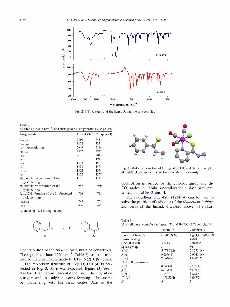
Fig. 3. Molecular structure of the ligand (3, left) and the title complex
(4, right). (Hydrogen atoms in 4 are not shown for clarity).
Table 3
Unit cell parameters for the ligand (3) and Re(CO)3LCl complex (4)
Ligand (3) Complex (4)
Empirical formula C14H16N4S2 C10H7ClN2O3ReS
Formula weight 456.89
Crystal system 304.43 Triclinic
Space group P�1 P�1a (A) 5.9768(12) 7.8270(16)
b (A) 9.239(18) 7.9740(16)
c (A) 20.626(4) 10.903(2)
Unit cell dimensions
a (�) 88.48(4) 77.35(4)
b (�) 89.19(4) 84.39(4)
c (�) 1.44(4) 88.12(4)
V (A3) 1079.32(4) 660.7(2)
Z 3 2
4000 3500 3000 2000 1500 1000 50020
40
60
80
100
0
20
40
60
80
100
Ligand
wavenumbers, cm-1
Complex
tran
smis
sion
, %
Fig. 2. FT-IR spectra of the ligand 3, and the title complex 4.
Table 2
Selected IR bands (cm�1) and their possible assignments (KBr pellets)
Assignments Ligand (3) Complex (4)
mNH3,as3450 3451
mNH3,sym3272 3251
mCH (aromatic ring) 3049 3114
mCH3,as2922 2927
mCO – 2015
mCO – 1913
mCN 1533 1567
mCN 1438 1478
mC@S 1352 1374
mCN 1272 1257
A1 cumulative vibration of the
pyridine ring
1101 1111
B1 cumulative vibration of the
pyridine ring
957 960
dC–H (2H vibration of the 2-substituted
pyridine ring)
796 783
dN–C@S 743 731
mC–S 436 487
m, stretching; d, bending modes.
4754 L. Fuks et al. / Journal of Organometallic Chemistry 689 (2004) 4751–4756
NNH
S
NN
S
a contribution of the thioenol form must be considered.
The signals at about 1250 cm�1 (Table 2) can be attrib-
uted to the presumably single N–CH3 [N(2)–C(8)] bond.
The molecular structure of Re(CO)3LCl (4) is pre-
sented in Fig. 3. As it was expected, ligand (3) coor-
dinates the cation bidentately: via the pyridinenitrogen and the sulphur atoms forming a five-mem-
ber plane ring with the metal center. Axis of the
octahedron is formed by the chloride anion and the
CO molecule. Main crystallographic data are pre-
sented in Tables 3 and 4.
The crystallographic data (Table 4) can be used to
solve the problem of existence of the thioketo and thioe-
nol forms of the ligand, discussed above. The short

Table 4
Selected bond lengths (pm) and angles (�) in the ligand (3) and
Re(CO)3LCl complex (4)
Ligand (3) Complex (4)
Bond lengths
S1–C7 167.3(8) C6–N1 136.1(9)
C7–N2 129.(10) C6–C7 147.1(9)
N2–C8 145.7(22) S1–C7 166.9(7)
C6–C7 150.7(12) C7–N2 132.7(9)
N1–C6 132.4(11) N2–C8 145.6(12)
Re–C1 189.2(8)
Re–C2 192.7(9)
Re–C3 190.1(8)
Re–N1 219.2(6)
Re–S 244.4(2)
Re–C1 247.6(2)
Angles
N2–C7–C6 115.1(7) N1–Re–S1 79.02(17)
N2–C7–S1 123.1(6) N1–C6–C7 116.3(6)
S1–C7–C6 121.8(6) C7–S1–Re 101.0(2)
C7–C6–N1 114.3(7) C6–C7–S1 121.5(5)
C6–N1–Re 121.7(5)
N2–C7–C6 117.3(6)
N2–C7–S1 121.2(6)
L. Fuks et al. / Journal of Organometallic Chemistry 689 (2004) 4751–4756 4755
distance between C(7) and N(2) atoms, of ca. 130 pm
observed in both free and coordinated ligand (3), com-
pared to ca. 150 pm for the single C–N and ca. 120–
130 pm for the double C@N bond [21], supports the
conclusion based on the IR spectra, that the conribution
of the thioenol form of the ligand is significant.
An important feature of drugs, which determines
their biodistribution, is lipophilicility. Diffusion proc-esses in the human body are controlled by the
difference in the composition of the intra- and extra-
cellular liquids. In the present work, partition coeffi-
cients of (3) and (4) have been determined between
iso-octanol and an aqueous phase being the pure
water or 0.9% of NaCl aqueous solution, respectively.
Pure water in the first system corresponds to the intra-
cellular liquid, whereas 0.9% NaCl 1 in the second tothe extracellular liquid [22,23]. It has been found, that
for both (3) and (4) the Pn values are not affected
(within the experimental error) by the given difference
in the composition of the aqueous phase: log P3 =
1.14 ± 0.17 and 1.11 ± 0.08, while log P4 = 1.01 ±
0.08 and 1.11 ± 0.11, for iso-octanol–water and iso-
octanol–0.9% NaCl, respectively.
Stability of 4 in aerated aqueous solution towardsoxidation has been studied by registering its UV–Vis
spectrum (Fig. 1) vs. time. Within 48 h (which corre-
sponds to ca. three half-life periods of 188Re) no distinct
change in the spectrum has been detected.
1 Commercial peritoneal injection solution produced by Baxter
Terpol, Ltd. (Sieradz, Poland) contains 154 mmol dm�3 of natrium
and 154 mmol dm�3 of chloride ions (pH 6.7).
5. Conclusions
A novel soft donor N,S-bidentate ligand, L = N2-me-
thyl-2-pyridinecarbothioamide (3), has been synthesized.
Its tricarbonylchlororhenium(I) complex, Re(CO)3(L)Cl
(4), can easily be prepared from pentacarbonylrhe-nium(I) chloride. Structural studies of 4 confirm that lig-
and (3) coordinates the Re(I) cation bidentatly by the S
and N atoms. The existence of 3, both free and coordi-
nated,in the thioenol form has been concluded from the
IR spectra and structural data. The complex (4) is stable
in aerated solutions for at least 2 days.
6. Supplementary material
Crystallographic data (excluding structural factors)
for the structures reported in this paper have been
deposited with the Cambridge Crystallographic Data
Centre and allocated the Deposition Nos. CCDC
242919 andCCDC 224857 for 3 and 4, respectively.
Acknowledgements
The work was supported by the Research Contract
No. 4 TO9A 11023 from the State Committee for Scien-
tific Research (KBN; Poland). We thank Mrs. Jowita
Orska (INCT) for her assistance in determining the sol-
ubility of the investigated species.
References
[1] Q. Liang, G.J. Ehrhardt, A.R. Ketring, R. Miller, Radiochim.
Acta 79 (1997) 137.
[2] H. Kamioki, S. Mirzadeh, R.M. Lambrecht, F.F. Knapp Jr., K.
Dadachova, Radiochim. Acta 65 (1994) 39.
[3] F.F. Knapp Jr., A.P. Callahan, A.L. Beets, S. Mirzadeh, B.T.
Hsieh, Appl. Radiat. Isotopes 45 (1994) 1123.
[4] R. Alberto, R. Schibli, A. Egli, P.A. Schubiger, W.A. Herrmann,
G. Artus, U. Abram, T.A. Kaden, J. Organomet. Chem. 493
(1995) 119.
[5] R. AlbertoTopics in Current Chemistry, 176, Springer, Berlin,
Heidelberg, 1996, p. 149.
[6] R. Schibli, R. Alberto, U. Abram, S. Abram, A. Egli, P.A.
Schubiger, T.A. Kaden, Inorg. Chem. 37 (1998) 3509.
[7] R. Alberto, R. Schibli, R. Waibel, U. Abram, A.P. Schubiger,
Coord. Chem. Rev. 190–192 (1999) 901.
[8] R. Schibli, R. La Bella, R. Alberto, E. Garcia-Garayoa, K.
Ortner, U. Abram, P.A. Schubiger, Bioconjug. Chem. 11 (2000)
345.
[9] Neuere Methoden der Praparativen Organischen Chemie, Band
III, Verlag Chemie, 1961, p. 30.
[10] L. Fuks, E. Gniazdowska, Unpublished data obtained during
the scientific visit in the Paul Scherrer Institute and in the
Institute of the Bioinorganic Chemistry, Zurich University,
Switzerland, 2002.
[11] R. Schibli, Normaldrucksynthese von (TcCl3(CO)3)2� und
(ReCl3(CO)3)2� und ihr Substitutionsverhalten im Hinblick auf

4756 L. Fuks et al. / Journal of Organometallic Chemistry 689 (2004) 4751–4756
Anwendungen in der Nuklermedizin, PhD Thesis, Paul Scherrer
Institut, Switzerland, 1996.
[12] F.S.Schoster,S.K.Zeisler, J.Radioanal.Nucl.Chem. 220 (1997) 149.
[13] C. Adams, V. Barone, J. Chem. Phys. 108 (1998) 66.
[14] G.M. Sheldrick, SHELXLSHELXL 93 Program for the Refinement of
Crystal Structure, University of Gottingen, Germany, 1993.
[15] A.J.C. Wilson (Ed.), International Tables for Crystallography, C,
Kluwer, Dordrecht, 1992.
[16] S. Wajda, K. Drabent, Bull. Acad. Polon. Sci., Ser. Sci. Chim. 25
(1977) 963.
[17] K. Nakamoto, J. Fujita, R.A. Condrote, Y. Morimoto, J. Chem.
Phys. 39 (1963) 423.
[18] G. Durgaprasad, D.N. Sathyanarayana, C.C. Patel, Can. J.
Chem. 47 (1969) 631.
[19] A.W. Herlimger, S.N. Wenhold, T.V. Long, J. Am. Chem. Soc.
92 (1970) 6474.
[20] J.J. Criado, J.A. Lopez Aria, B. Marcias, L.R. Fernandez Lago,
Inorg. Chim. Acta 193 (1992) 229.
[21] (a) R.J. Gillespie, in: Molecular Geometry, Van Norstrand, NY,
1972, pp. 18–21;
(b) R.J. Gillespie, P.A. Popelier, in: Chemical Bonding and
Molecular Geometry, Oxford University Press, NY, Oxford, 2001,
pp. 25–38.
[22] W. Skwara, Private communication on the concentration of
sodium and chloride ions in the bi-distilled water produced from
the INCT deep water draw-out source.
[23] (a) S.J. Lippard, J.M. Berg, Principles of Bioinorganic Chemistry
(Polish edition), PWN, Warsaw, 1998, p. 151;
(b) Z. Jakubowski, J. Kabata, L. Kalinowski, M. Szczepanska-
Konkel, S. Angielski, Medicinal Analysis in Everyday Practice –
Reference Values and their Interpretation (in Polish), MakMed
Editory, Gdansk, 1994, pp. 56, 286.
![Vibrational spectra, NBO analysis, HOMO–LUMO and first hyperpolarizability of 2-{[(2-Methylprop-2-en-1-yl)oxy]methyl}-6-phenyl-2,3,4,5-tetrahydro-1,2,4-triazine-3,5-dione, a potential](https://static.fdokumen.com/doc/165x107/6324d4c6c9c7f5721c01c4b7/vibrational-spectra-nbo-analysis-homolumo-and-first-hyperpolarizability-of.jpg)


![Vibrational spectroscopic (FT-IR and FT-Raman) studies, HOMO-LUMO, NBO analysis and MEP of 6-methyl-1-({[(2E)-2-methyl-3-phenyl-prop-2-en-1-yl]oxy}methyl)-1,2,3,4-tetra-hydroquinazoline-2,4-dione,](https://static.fdokumen.com/doc/165x107/633494f441100cab3c07ce05/vibrational-spectroscopic-ft-ir-and-ft-raman-studies-homo-lumo-nbo-analysis.jpg)