Structure and support induced structure disruption of soft nanoparticles obtained from hydroxylated...
7
Structure and support induced structure disruption of soft nanoparticles obtained from hydroxylated fatty acids J. A. Heredia-Guerrero, a M. A. San-Miguel, b M. Luna, c E. Dom ınguez, d A. Heredia e and J. J. Ben ıtez * a Received 25th December 2010, Accepted 15th February 2011 DOI: 10.1039/c0sm01545h Soft and spherical nanoparticles, named as cutinsomes, have been prepared from concentrated 9(10),16- dihydroxypalmitic acid (diHPA) in aqueous solution. After isolation, cutinsomes have been chemically and structurally characterized by ATR-FTIR, TEM and dynamic atomic force microscopy (dynamic AFM). The nanoparticle can be described as a lipidic, liquid-like and mostly esterified core surrounded by a polar shell of carboxylate/carboxylic acid molecules. Molecular dynamic (MD) simulations have been used to support this model. The structural stability of soft cutinsomes has been tested by deposition on both non-polar (HOPG) and polar (mica) flat substrates. It has been found that the magnitude of the interaction between the polar shell of cutinsomes and the support determines their structure conservation or its spreading or rupture and spill out of the liquid-like content. The structural consistence of these nanoparticles as a function of the polarity of substrate is of interest in elucidating the formation mechanism of cutin, the most abundant biopolyester in nature and a very interesting biomaterial to be mimetized. 1. Introduction Long chain polyhydroxy fatty acids are molecules of particular interest because they are the constituents of plant barrier poly- mers such as cutin and suberin. These biopolymers are extremely abundant in nature and play a very important role in protecting higher plants against physical, chemical and biological impacts as well as in regulating water loss. 1 Despite such ubiquitous presence in the biosphere, their chemical composition is rather uniform. For instance, depolymerization products of bio- polyester cutin are almost exclusively composed of derivatives of the C 16 and C 18 carboxylic acids. 2,3 Among these derivatives, the 9(10),16-dihydroxypalmitic acid (diHPA) is the most abundant monomer and it is considered as the building unit of cutin, the lipid polyester that constitutes the major part of plant cuticle. The specific interactions between long chain polyhydroxy fatty acid molecules have been studied in detail by preparing self- assembled layers from solution on a flat support like mica. 4–7 Results obtained clearly indicate that hydroxyl groups rule intermolecular interactions. Thus, hydroxyl groups in mid-chain positions reinforce the lateral interaction between packed mole- cules but a hydroxyl in terminal position is necessary to trigger the multilayer growth. Particularly, and if compared with related molecules, diHPA monomers lead to a very effective 2D growth covering the mica support as well as the simultaneous develop- ment of multilayers. Besides, spontaneous self-esterification within the layers has been detected. 6 These findings have brought the attention to the self-assembly route as a plausible mechanism for the biosynthesis of cutin at the wall of epidermal cells in plants. Such basic information about self-assembly of polyhydroxy fatty acids has been obtained using solutions containing chlo- roform or light alcohols as solvents. However, any hypothesis about self-assembly in vivo has to involve an aqueous medium. In weak acid aqueous medium, polyhydroxy fatty acids give rise to nanometre sized lipid spherical particles. 8 It has also been demonstrated that these nanoparticles (cutinsomes) are involved in the genesis of cutin of young fruits during development. 9 The soft structure of cutinsomes requests special care and the employment of high sensitivity and non-destructive analysis techniques. Among them, dynamic atomic force microscopy (dynamic AFM) provides a very high topographic resolution with negligible sample perturbation. This AFM mode is based on the change in oscillation amplitude of the cantilever due to the effect of attractive non-linear van der Waals forces. 10–12 The main difference with respect to intermittent contact (tapping) is that parameters can be adjusted for non-contact operation: the tip does not usually reach the adhesive or repulsive contact with the surface. Therefore, while keeping the resolution power of tapping mode, the possibility of sample damage is further reduced. This a Instituto de Ciencia de Materiales de Sevilla, Centro Mixto CSIC-Universidad de Sevilla, Avda, Americo Vespuccio 49, 41092 Seville, Spain. E-mail: [email protected] b Departamento de Qu ımica-F ısica, Facultad de Qu ımica, Universidad de Sevilla, 41012 Seville, Spain c Instituto de Microelectr onica de Madrid (IMM-CSIC), 28760 Tres Cantos, Madrid, Spain d Estaci on Experimental ‘‘La Mayora’’ (CSIC), Algarrobo-Costa, 29750 M alaga, Spain e Departamento de Biolog ıa Molecular y Bioqu ımica, Universidad de M alaga, 29071 M alaga, Spain This journal is ª The Royal Society of Chemistry 2011 Soft Matter , 2011, 7, 4357–4363 | 4357 Dynamic Article Links C < Soft Matter Cite this: Soft Matter , 2011, 7, 4357 www.rsc.org/softmatter PAPER Downloaded by CNM. InstitutoMicroelectrónica MAD. (IMM-CNM) on 31 August 2011 Published on 10 March 2011 on http://pubs.rsc.org | doi:10.1039/C0SM01545H View Online
-
Upload
independent -
Category
Documents
-
view
0 -
download
0
Transcript of Structure and support induced structure disruption of soft nanoparticles obtained from hydroxylated...
Dynamic Article LinksC<Soft Matter
Cite this: Soft Matter, 2011, 7, 4357
www.rsc.org/softmatter PAPER
Dow
nloa
ded
by C
NM
. Ins
titut
oMic
roel
ectr
ónic
a M
AD
. (I
MM
-CN
M)
on 3
1 A
ugus
t 201
1Pu
blis
hed
on 1
0 M
arch
201
1 on
http
://pu
bs.r
sc.o
rg |
doi:1
0.10
39/C
0SM
0154
5HView Online
Structure and support induced structure disruption of soft nanoparticlesobtained from hydroxylated fatty acids
J. A. Heredia-Guerrero,a M. A. San-Miguel,b M. Luna,c E. Dom�ınguez,d A. Herediae and J. J. Ben�ıtez*a
Received 25th December 2010, Accepted 15th February 2011
DOI: 10.1039/c0sm01545h
Soft and spherical nanoparticles, named as cutinsomes, have been prepared from concentrated 9(10),16-
dihydroxypalmitic acid (diHPA) in aqueous solution. After isolation, cutinsomes have been chemically
and structurally characterized by ATR-FTIR, TEM and dynamic atomic force microscopy (dynamic
AFM). The nanoparticle can be described as a lipidic, liquid-like and mostly esterified core surrounded
by a polar shell of carboxylate/carboxylic acid molecules. Molecular dynamic (MD) simulations have
been used to support this model. The structural stability of soft cutinsomes has been tested by
deposition on both non-polar (HOPG) and polar (mica) flat substrates. It has been found that the
magnitude of the interaction between the polar shell of cutinsomes and the support determines their
structure conservation or its spreading or rupture and spill out of the liquid-like content. The structural
consistence of these nanoparticles as a function of the polarity of substrate is of interest in elucidating
the formation mechanism of cutin, the most abundant biopolyester in nature and a very interesting
biomaterial to be mimetized.
1. Introduction
Long chain polyhydroxy fatty acids are molecules of particular
interest because they are the constituents of plant barrier poly-
mers such as cutin and suberin. These biopolymers are extremely
abundant in nature and play a very important role in protecting
higher plants against physical, chemical and biological impacts
as well as in regulating water loss.1 Despite such ubiquitous
presence in the biosphere, their chemical composition is rather
uniform. For instance, depolymerization products of bio-
polyester cutin are almost exclusively composed of derivatives of
the C16 and C18 carboxylic acids.2,3 Among these derivatives, the
9(10),16-dihydroxypalmitic acid (diHPA) is the most abundant
monomer and it is considered as the building unit of cutin, the
lipid polyester that constitutes the major part of plant cuticle.
The specific interactions between long chain polyhydroxy fatty
acid molecules have been studied in detail by preparing self-
assembled layers from solution on a flat support like mica.4–7
Results obtained clearly indicate that hydroxyl groups rule
intermolecular interactions. Thus, hydroxyl groups in mid-chain
aInstituto de Ciencia de Materiales de Sevilla, Centro MixtoCSIC-Universidad de Sevilla, Avda, Americo Vespuccio 49, 41092Seville, Spain. E-mail: [email protected] de Qu�ımica-F�ısica, Facultad de Qu�ımica, Universidad deSevilla, 41012 Seville, SpaincInstituto de Microelectr�onica de Madrid (IMM-CSIC), 28760 TresCantos, Madrid, SpaindEstaci�on Experimental ‘‘La Mayora’’ (CSIC), Algarrobo-Costa, 29750M�alaga, SpaineDepartamento de Biolog�ıa Molecular y Bioqu�ımica, Universidad deM�alaga, 29071 M�alaga, Spain
This journal is ª The Royal Society of Chemistry 2011
positions reinforce the lateral interaction between packed mole-
cules but a hydroxyl in terminal position is necessary to trigger
the multilayer growth. Particularly, and if compared with related
molecules, diHPA monomers lead to a very effective 2D growth
covering the mica support as well as the simultaneous develop-
ment of multilayers. Besides, spontaneous self-esterification
within the layers has been detected.6 These findings have brought
the attention to the self-assembly route as a plausible mechanism
for the biosynthesis of cutin at the wall of epidermal cells in
plants.
Such basic information about self-assembly of polyhydroxy
fatty acids has been obtained using solutions containing chlo-
roform or light alcohols as solvents. However, any hypothesis
about self-assembly in vivo has to involve an aqueous medium. In
weak acid aqueous medium, polyhydroxy fatty acids give rise to
nanometre sized lipid spherical particles.8 It has also been
demonstrated that these nanoparticles (cutinsomes) are involved
in the genesis of cutin of young fruits during development. 9 The
soft structure of cutinsomes requests special care and the
employment of high sensitivity and non-destructive analysis
techniques. Among them, dynamic atomic force microscopy
(dynamic AFM) provides a very high topographic resolution
with negligible sample perturbation. This AFM mode is based on
the change in oscillation amplitude of the cantilever due to the
effect of attractive non-linear van der Waals forces.10–12 The main
difference with respect to intermittent contact (tapping) is that
parameters can be adjusted for non-contact operation: the tip
does not usually reach the adhesive or repulsive contact with the
surface. Therefore, while keeping the resolution power of tapping
mode, the possibility of sample damage is further reduced. This
Soft Matter, 2011, 7, 4357–4363 | 4357
Dow
nloa
ded
by C
NM
. Ins
titut
oMic
roel
ectr
ónic
a M
AD
. (I
MM
-CN
M)
on 3
1 A
ugus
t 201
1Pu
blis
hed
on 1
0 M
arch
201
1 on
http
://pu
bs.r
sc.o
rg |
doi:1
0.10
39/C
0SM
0154
5HView Online
technique has been extensively used to image the topography of
delicate samples such as physisorbed supramolecular three
dimensional architectures13 or even liquid droplets10 and films.14
In this article, structural data obtained from TEM and dynamic
AFM are also supported with molecular dynamic (MD) simu-
lations.
From a biological point of view, the possibility of biopolyester
cutin formation in nature through cutinsomes packing, aggre-
gation and fusion at the epidermis of plant cell walls is very
plausible. This hypothesis drives the analysis to the nanoparticle
self-assembly rather than to the discrete molecular packing. In
this sense, the physical and chemical properties of the support are
very important factors in controlling the mechanism of cutin-
somes accretion. For this reason, this study comprises two model
supports such as HOPG (non-polar, hydrophobic) and mica
(polar, hydrophilic). The aim is to obtain general conclusions
that can later be transferred to the actual and particular condi-
tions acting at the vegetal epidermal cell walls.
2. Experimental section
2.1. Lipid nanoparticle preparation
9(10),16-Dihydroxypalmitic acid (diHPA) is not a commercial
product and has been obtained by depolymerization in aqueous
solution of NaOH 1 M of plant cutin from enzymatically iso-
lated cuticles of tomato fruits following a procedure already
reported.15 Cutinsomes were prepared starting with filtered
10 mM diHPA aqueous alkaline solutions at pH 12.5 and
reducing the pH to 6.8 with small amounts of HCl.16 The
solution becomes then opalescent and nanoparticles were
obtained after centrifugation at 19 000g. The product is washed
several times with distilled water and filtered. Dispersions are
prepared using 1.5 mg of cutinsomes in 1.5 mL of distilled water
and sonicated for 1 h.
2.2. ATR-FTIR
ATR-FTIR spectra of samples were obtained from pellets of
purified cutinsomes using an ATR accessory (MIRacle ATR,
PIKE Technologies, USA) coupled to FTIR spectrometer (FT/
IR-4100, JASCO, Spain). All spectra were recorded in the 4000
to 600 cm�1 range at 4 cm�1 resolution and 250 scans were
accumulated.
2.3. TEM
For TEM analysis, a small drop of dispersion of cutinsomes in
water was deposited on a copper grid. Nanoparticles were
stained with an aqueous uranyl acetate solution 1% (w/v). The
grids were analyzed at 100 kV with a Philips CM 100 electron
microscope.
2.4. MD simulations
Molecular dynamic simulations were performed with
DL_POLY17 using the NVT ensemble with a Nos�e–Hoover
thermostat18 at 300 K. The initial velocities were set up from
a Boltzmann distribution. The equations of motion were inte-
grated by using the velocity-Verlet algorithm with a time step of
4358 | Soft Matter, 2011, 7, 4357–4363
1 fs. Simulations were extended up to 4 ns. The interatomic
potential parameters were taken from literature and have been
used already in previous studies.6,7 A united atom model was
employed for –CH3 and –CH2– groups and all the other atoms in
hydroxyl and carboxylic groups were represented explicitly. A
cutoff radius of 15 �A was used for the Lennard-Jones (6–12)
potential terms. The electrostatic interactions were treated using
the method of Hautman and Klein which is an adaptation of the
Ewald method for systems which are periodic in two dimensions
only.19 Molecules were deposited on a substrate represented as
a flat surface and the interaction between each particle and the
surface was computed according to:
VðrÞ ¼ C12
ðz� z0Þ12� C3
ðz� z0Þ3
where Cna(s3n)1/2, z is the distance to the surface and z0 is a limit
approach distance for each center. 3 and s are Lennard-Jones
potential parameters and they were calculated using the Lorentz–
Berthelot combining rules. Cn parameters for CH2 and O parti-
cles were taken and adapted from the literature.20
2.5. Cutinsome deposition for dynamic AFM
Two preparation methods: (i) drop vaporization and (ii) spin
coating, have been used. In both cases, a droplet (25 mL) of the
aqueous dispersion of cutinsomes was deposited on the freshly
cleaved substrate (HOPG, ZYB quality, or mica, muscovite). In
drop vaporization method, water was allowed being evaporated
slowly by keeping the sample at 6 �C for 24 h. In the spin coating
method, the samples were spun at 40 rps for 30 s.
2.6. Dynamic AFM operation
The AFM is a Cervantes model from Nanotec Electr�onica
(Spain) operated at room conditions (20–25 �C and 35–40% RH).
Images were obtained and processed using the WSxM software.21
Nanosensors Si3N4 rectangular cantilevers with nominal force
constant of 2.8 N m�1 and resonance frequency around 80 kHz
were used.
Parameters such as the cantilever driving oscillation ampli-
tude, force constant, resonance frequency and oscillation
amplitude set point have to be carefully selected and optimized
for the operation out of contact.22 Out of contact conditions can
be obtained at typically 1–5 nm from the surface.23–25
3. Results and discussion
3.1. Infrared spectroscopy characterization of cutinsomes
Fig. 1 shows the infrared spectra of cutinsomes obtained from
diHPA. A broad medium intensity band at 3327 cm�1 is indica-
tive of the stretching vibration of hydrogen bonded hydroxyl
groups. The frequency value is in the range of intermolecular
hydrogen bonds that give rise to polymeric associations26,27 and
therefore suggests a strong crosslinking between the alkyl chains.
A residual phase with a more relaxed hydrogen bonding is
revealed by the shoulder at 3460 cm�1.
Strong asymmetrical and symmetrical stretching vibrations of
methylene (–CH2–) groups are located at 2924 and 2851 cm�1,
respectively. The position of these bands is a good parameter to
This journal is ª The Royal Society of Chemistry 2011
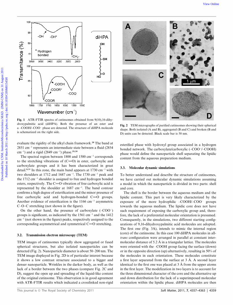
Fig. 1 ATR-FTIR spectra of cutinsomes obtained from 9(10),16-dihy-
droxypalmitic acid (diHPA). Both the presence of an ester and
a –COOH/–COO� phase are detected. The structure of diHPA molecule
is schematized on the right side.
Fig. 2 TEM micrographs of purified cutinsomes showing their spherical
shape. Both isolated (A and B), aggregated (B and C) and broken (B and
D) units can be detected. Black scale bar is 50 nm.
Dow
nloa
ded
by C
NM
. Ins
titut
oMic
roel
ectr
ónic
a M
AD
. (I
MM
-CN
M)
on 3
1 A
ugus
t 201
1Pu
blis
hed
on 1
0 M
arch
201
1 on
http
://pu
bs.r
sc.o
rg |
doi:1
0.10
39/C
0SM
0154
5HView Online
evaluate the rigidity of the alkyl chain framework.28 The band at
2851 cm�1 represents an intermediate state between a fluid (2854
cm�1) and a rigid (2849 cm�1) phase.29,30
The spectral region between 1800 and 1500 cm�1 corresponds
to the stretching vibrations of (C]O) in ester, carboxylic and
carboxylate groups and it has been characterized in great
detail.8,31 In this zone, the main band appears at 1730 cm�1 with
two shoulders at 1712 and 1687 cm�1. The 1730 cm�1 peak and
the 1712 cm�1 shoulder is assigned to free and hydrogen bonded
esters, respectively. The C]O vibration of free carboxylic acid is
represented by the shoulder at 1687 cm�1. The band contour
confirms a high degree of esterification and the minor presence of
free carboxylic acid and hydrogen-bonded C]O groups.
Another evidence of esterification is the 1166 cm�1 asymmetric
C–O–C stretching (not shown in the figure).
On the other hand, the presence of carboxylate (–COO�)
groups is significant, as indicated by the 1561 cm�1 and the 1412
cm�1 (not shown in the figure) peaks, respectively assigned to the
corresponding asymmetrical and symmetrical C]O stretching.
3.2. Transmission electron microscopy (TEM)
TEM images of cutinsomes typically show aggregated or fused
spherical structures, but also isolated nanoparticles can be
detected (Fig. 2). Nanoparticle diameter is about 50–200 nm. The
TEM image displayed in Fig. 2D is of particular interest because
it shows a low contrast structure associated to a bigger and
denser nanoparticle. Wrinkles in the darker body, as well as the
lack of a border between the two phases (compare Fig. 2C and
D), suggest the open up and spreading of the liquid-like content
of the original cutinsome. This observation is in good agreement
with ATR-FTIR results which indicated a crosslinked non-rigid
This journal is ª The Royal Society of Chemistry 2011
esterified phase with hydroxyl group associated in a hydrogen
bonded network. The carboxylate/carboxylic (–COO�/–COOH)
phase would define the nanoparticle shell separating the lipidic
content from the aqueous preparation medium.
3.3. Molecular dynamic simulations
To better understand and describe the structure of cutinsomes,
we have carried out molecular dynamic simulations assuming
a model in which the nanoparticle is divided in two parts: shell
and core.
The shell is the border between the aqueous medium and the
lipidic content. This part is very likely characterized by the
exposure of the more hydrophilic –COOH/–COO� groups
towards the aqueous medium. The lipidic core does not have
such requirement of exposing the carboxylic group and, there-
fore, the lack of a preferential molecular orientation is presumed.
Consequently, in the simulations, two different starting config-
urations of 9,16-dihydroxypalmitic acid molecules are adopted.
The first one (Fig. 3A), intends to mimic the internal region
(core) of the cutinsome. In this case 100 diHPA molecules in all-
trans configuration were arranged in parallel at constant inter-
molecular distance of 5.2 �A in a triangular lattice. The molecules
were oriented with the –COOH group facing the surface (down)
or in the opposite direction (up) alternatively, resulting in 50% of
the molecules in each orientation. These molecules constitute
a first layer separated from the surface at 3 �A. A second layer
identical to the first one was placed at 3 �A from the upper atoms
in the first layer. The modelization in two layers is to account for
the three dimensional character of the core and the alternative up
and down distribution for the lack of a superimposed molecular
orientation within the lipidic phase. diHPA molecules are then
Soft Matter, 2011, 7, 4357–4363 | 4359
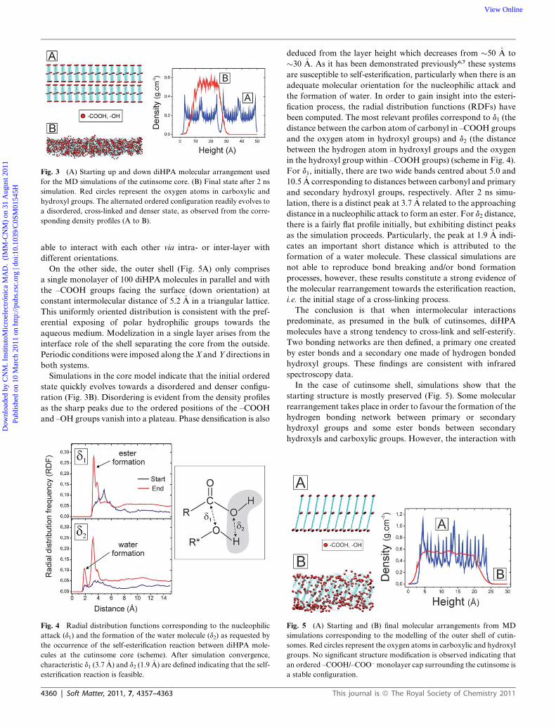
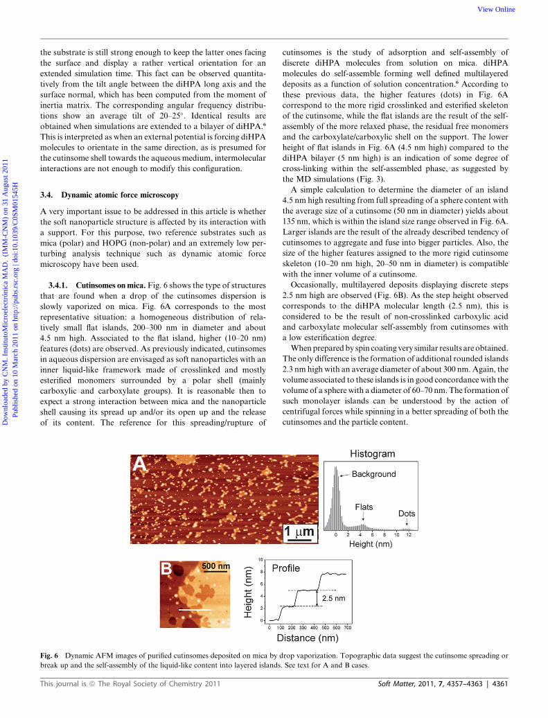
Fig. 3 (A) Starting up and down diHPA molecular arrangement used
for the MD simulations of the cutinsome core. (B) Final state after 2 ns
simulation. Red circles represent the oxygen atoms in carboxylic and
hydroxyl groups. The alternated ordered configuration readily evolves to
a disordered, cross-linked and denser state, as observed from the corre-
sponding density profiles (A to B).
Dow
nloa
ded
by C
NM
. Ins
titut
oMic
roel
ectr
ónic
a M
AD
. (I
MM
-CN
M)
on 3
1 A
ugus
t 201
1Pu
blis
hed
on 1
0 M
arch
201
1 on
http
://pu
bs.r
sc.o
rg |
doi:1
0.10
39/C
0SM
0154
5HView Online
able to interact with each other via intra- or inter-layer with
different orientations.
On the other side, the outer shell (Fig. 5A) only comprises
a single monolayer of 100 diHPA molecules in parallel and with
the –COOH groups facing the surface (down orientation) at
constant intermolecular distance of 5.2 �A in a triangular lattice.
This uniformly oriented distribution is consistent with the pref-
erential exposing of polar hydrophilic groups towards the
aqueous medium. Modelization in a single layer arises from the
interface role of the shell separating the core from the outside.
Periodic conditions were imposed along the X and Y directions in
both systems.
Simulations in the core model indicate that the initial ordered
state quickly evolves towards a disordered and denser configu-
ration (Fig. 3B). Disordering is evident from the density profiles
as the sharp peaks due to the ordered positions of the –COOH
and –OH groups vanish into a plateau. Phase densification is also
Fig. 4 Radial distribution functions corresponding to the nucleophilic
attack (d1) and the formation of the water molecule (d2) as requested by
the occurrence of the self-esterification reaction between diHPA mole-
cules at the cutinsome core (scheme). After simulation convergence,
characteristic d1 (3.7 �A) and d2 (1.9 �A) are defined indicating that the self-
esterification reaction is feasible.
4360 | Soft Matter, 2011, 7, 4357–4363
deduced from the layer height which decreases from �50 �A to
�30 �A. As it has been demonstrated previously6,7 these systems
are susceptible to self-esterification, particularly when there is an
adequate molecular orientation for the nucleophilic attack and
the formation of water. In order to gain insight into the esteri-
fication process, the radial distribution functions (RDFs) have
been computed. The most relevant profiles correspond to d1 (the
distance between the carbon atom of carbonyl in –COOH groups
and the oxygen atom in hydroxyl groups) and d2 (the distance
between the hydrogen atom in hydroxyl groups and the oxygen
in the hydroxyl group within –COOH groups) (scheme in Fig. 4).
For d1, initially, there are two wide bands centred about 5.0 and
10.5 �A corresponding to distances between carbonyl and primary
and secondary hydroxyl groups, respectively. After 2 ns simu-
lation, there is a distinct peak at 3.7 �A related to the approaching
distance in a nucleophilic attack to form an ester. For d2 distance,
there is a fairly flat profile initially, but exhibiting distinct peaks
as the simulation proceeds. Particularly, the peak at 1.9 �A indi-
cates an important short distance which is attributed to the
formation of a water molecule. These classical simulations are
not able to reproduce bond breaking and/or bond formation
processes, however, these results constitute a strong evidence of
the molecular rearrangement towards the esterification reaction,
i.e. the initial stage of a cross-linking process.
The conclusion is that when intermolecular interactions
predominate, as presumed in the bulk of cutinsomes, diHPA
molecules have a strong tendency to cross-link and self-esterify.
Two bonding networks are then defined, a primary one created
by ester bonds and a secondary one made of hydrogen bonded
hydroxyl groups. These findings are consistent with infrared
spectroscopy data.
In the case of cutinsome shell, simulations show that the
starting structure is mostly preserved (Fig. 5). Some molecular
rearrangement takes place in order to favour the formation of the
hydrogen bonding network between primary or secondary
hydroxyl groups and some ester bonds between secondary
hydroxyls and carboxylic groups. However, the interaction with
Fig. 5 (A) Starting and (B) final molecular arrangements from MD
simulations corresponding to the modelling of the outer shell of cutin-
somes. Red circles represent the oxygen atoms in carboxylic and hydroxyl
groups. No significant structure modification is observed indicating that
an ordered –COOH/–COO� monolayer cap surrounding the cutinsome is
a stable configuration.
This journal is ª The Royal Society of Chemistry 2011
Dow
nloa
ded
by C
NM
. Ins
titut
oMic
roel
ectr
ónic
a M
AD
. (I
MM
-CN
M)
on 3
1 A
ugus
t 201
1Pu
blis
hed
on 1
0 M
arch
201
1 on
http
://pu
bs.r
sc.o
rg |
doi:1
0.10
39/C
0SM
0154
5HView Online
the substrate is still strong enough to keep the latter ones facing
the surface and display a rather vertical orientation for an
extended simulation time. This fact can be observed quantita-
tively from the tilt angle between the diHPA long axis and the
surface normal, which has been computed from the moment of
inertia matrix. The corresponding angular frequency distribu-
tions show an average tilt of 20–25�. Identical results are
obtained when simulations are extended to a bilayer of diHPA.6
This is interpreted as when an external potential is forcing diHPA
molecules to orientate in the same direction, as is presumed for
the cutinsome shell towards the aqueous medium, intermolecular
interactions are not enough to modify this configuration.
3.4. Dynamic atomic force microscopy
A very important issue to be addressed in this article is whether
the soft nanoparticle structure is affected by its interaction with
a support. For this purpose, two reference substrates such as
mica (polar) and HOPG (non-polar) and an extremely low per-
turbing analysis technique such as dynamic atomic force
microscopy have been used.
3.4.1. Cutinsomes on mica. Fig. 6 shows the type of structures
that are found when a drop of the cutinsomes dispersion is
slowly vaporized on mica. Fig. 6A corresponds to the most
representative situation: a homogeneous distribution of rela-
tively small flat islands, 200–300 nm in diameter and about
4.5 nm high. Associated to the flat island, higher (10–20 nm)
features (dots) are observed. As previously indicated, cutinsomes
in aqueous dispersion are envisaged as soft nanoparticles with an
inner liquid-like framework made of crosslinked and mostly
esterified monomers surrounded by a polar shell (mainly
carboxylic and carboxylate groups). It is reasonable then to
expect a strong interaction between mica and the nanoparticle
shell causing its spread up and/or its open up and the release
of its content. The reference for this spreading/rupture of
Fig. 6 Dynamic AFM images of purified cutinsomes deposited on mica by d
break up and the self-assembly of the liquid-like content into layered islands
This journal is ª The Royal Society of Chemistry 2011
cutinsomes is the study of adsorption and self-assembly of
discrete diHPA molecules from solution on mica. diHPA
molecules do self-assemble forming well defined multilayered
deposits as a function of solution concentration.6 According to
these previous data, the higher features (dots) in Fig. 6A
correspond to the more rigid crosslinked and esterified skeleton
of the cutinsome, while the flat islands are the result of the self-
assembly of the more relaxed phase, the residual free monomers
and the carboxylate/carboxylic shell on the support. The lower
height of flat islands in Fig. 6A (4.5 nm high) compared to the
diHPA bilayer (5 nm high) is an indication of some degree of
cross-linking within the self-assembled phase, as suggested by
the MD simulations (Fig. 3).
A simple calculation to determine the diameter of an island
4.5 nm high resulting from full spreading of a sphere content with
the average size of a cutinsome (50 nm in diameter) yields about
135 nm, which is within the island size range observed in Fig. 6A.
Larger islands are the result of the already described tendency of
cutinsomes to aggregate and fuse into bigger particles. Also, the
size of the higher features assigned to the more rigid cutinsome
skeleton (10–20 nm high, 20–50 nm in diameter) is compatible
with the inner volume of a cutinsome.
Occasionally, multilayered deposits displaying discrete steps
2.5 nm high are observed (Fig. 6B). As the step height observed
corresponds to the diHPA molecular length (2.5 nm), this is
considered to be the result of non-crosslinked carboxylic acid
and carboxylate molecular self-assembly from cutinsomes with
a low esterification degree.
When prepared by spin coating very similar results are obtained.
The only difference is the formation of additional rounded islands
2.3 nm high with an average diameter of about 300 nm. Again, the
volume associated to these islands is in good concordance with the
volume of a sphere with a diameter of 60–70 nm. The formation of
such monolayer islands can be understood by the action of
centrifugal forces while spinning in a better spreading of both the
cutinsomes and the particle content.
rop vaporization. Topographic data suggest the cutinsome spreading or
. See text for A and B cases.
Soft Matter, 2011, 7, 4357–4363 | 4361
Dow
nloa
ded
by C
NM
. Ins
titut
oMic
roel
ectr
ónic
a M
AD
. (I
MM
-CN
M)
on 3
1 A
ugus
t 201
1Pu
blis
hed
on 1
0 M
arch
201
1 on
http
://pu
bs.r
sc.o
rg |
doi:1
0.10
39/C
0SM
0154
5HView Online
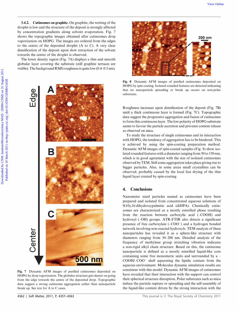
3.4.2. Cutinsomes on graphite. On graphite, the wetting of the
droplet is low and the structure of the deposit is strongly affected
by concentration gradients along solvent evaporation. Fig. 7
shows the topographic images obtained after cutinsomes drop
vaporization on HOPG. The images are ordered from the edges
to the centre of the deposited droplet (A to C). A very clear
densification of the deposit upon slow retraction of the solvent
towards the centre of the droplet is observed.
The lower density region (Fig. 7A) displays a thin and smooth
globular layer covering the substrate (still graphite terraces are
visible). The background RMS roughness is quite low (0.4–0.5 nm).
Fig. 7 Dynamic AFM images of purified cutinsomes deposited on
HOPG by drop vaporization. The globular structure gets denser on going
from the edge towards the centre of the deposited drop. Topographic
data suggest a strong cutinsome aggregation rather than nanoparticle
break up. See text for A to C cases.
Fig. 8 Dynamic AFM images of purified cutinsomes deposited on
HOPG by spin coating. Isolated rounded features are detected indicating
that no nanoparticle spreading or break up occurs on non-polar
substrates.
4362 | Soft Matter, 2011, 7, 4357–4363
Roughness increases upon densification of the deposit (Fig. 7B)
until a thick continuous layer is formed (Fig. 7C). Topographic
data suggest the progressive aggregation and fusion of cutinsomes
to form this continuous layer. The low polarity of HOPG substrate
seems to favour the particle accretion and prevents content release
as observed on mica.
To study the structure of single cutinsomes and its interaction
with HOPG, the tendency of aggregation has to be hindered. This
is achieved by using the spin-coating preparation method.
Dynamic AFM images of spin-coated samples (Fig. 8) show iso-
lated rounded features with a diameter ranging from 50 to 150 nm,
which is in good agreement with the size of isolated cutinsomes
observed by TEM. Still some aggregation takes place giving rise to
bigger particles. Also, in some areas small crystallites can be
observed, probably caused by the local fast drying of the thin
liquid layer created by spin-coating.
4. Conclusions
Nanometer sized particles named as cutinsomes have been
prepared and isolated from concentrated aqueous solutions of
9(10),16-dihydroxypalmitic acid (diHPA). Chemically cutin-
somes are characterized as a mostly esterified phase resulting
from the reaction between carboxylic acid (–COOH) and
hydroxyl (–OH) groups. ATR-FTIR also detects a significant
presence of free carboxylate (–COO�) and a hydrogen bonded
network involving non-reacted hydroxyls. TEM analysis of these
nanoparticles has revealed it as a sphere-like structure with
diameters ranging from 50–200 nm. Detailed analysis of the
frequency of methylene group stretching vibration indicates
a non-rigid alkyl chain structure. Based on this, the cutinsome
nanoparticle is defined as a mostly esterified liquid-like core
containing some free monomeric units and surrounded by a –
COOH/–COO� shell separating the lipidic content from the
aqueous environment. Molecular dynamic simulation results are
consistent with this model. Dynamic AFM images of cutinsomes
have revealed that their interaction with the support can control
their spherical structure disruption. Polar substrates such as mica
induce the particle rupture or spreading and the self-assembly of
the liquid-like content driven by the strong interaction with the
This journal is ª The Royal Society of Chemistry 2011
Dow
nloa
ded
by C
NM
. Ins
titut
oMic
roel
ectr
ónic
a M
AD
. (I
MM
-CN
M)
on 3
1 A
ugus
t 201
1Pu
blis
hed
on 1
0 M
arch
201
1 on
http
://pu
bs.r
sc.o
rg |
doi:1
0.10
39/C
0SM
0154
5HView Online
–COOH/–COO� shell. However, non-polar substrates such as
HOPG preserve the nanoparticle structure.
The structure and capacity of cutinsomes to aggregate and
undergo self-esterification is of particular interest because they
have been proposed as the building units of cutin, the most
abundant polyester in nature and a very interesting material to be
mimetized as a bioplastic.30 The capability of the support in
preserving the integrity of such nanoparticles is an important
factor in elucidating the transport mechanism across the plant
cell walls in the biosynthetic pathway of cutin.
Acknowledgements
This research work has been funded by the Consejer�ıa de
Innovaci�on Ciencia y Empresa (Junta de Andaluc�ıa) through the
TEP-02550 project and the EPSRC and BBSRC (via the Bion-
anotechnology IRC).
References
1 K. Roberts, Handbook of Plant Science, Wiley, Chichester, 2007, vol. 1.2 A. Heredia, Biochim. Biophys. Acta, Gen. Subj., 2003, 1620, 1.3 M. Riederer and C. Muller, The Biology of Plant Cuticle, Blackwell,
London, 2005.4 J. J. Ben�ıtez, J. A. Heredia-Guerrero and A. Heredia, J. Phys. Chem.
C, 2007, 111, 9465.5 J. J. Ben�ıtez, J. A. Heredia-Guerrero, F. Serrano and A. Heredia, J.
Phys. Chem. C, 2008, 112, 16968.6 J. A. Heredia-Guerrero, M. A. San-Miguel, M. S. P. Sansom,
A. Heredia and J. J. Ben�ıtez, Langmuir, 2009, 25, 6869.7 J. A. Heredia-Guerrero, M. A. San-Miguel, M. S. P. Sansom,
A. Heredia and J. J. Ben�ıtez, Phys. Chem. Chem. Phys., 2010, 12, 10423.8 (a) J. A. Heredia-Guerrero, J. J. Ben�ıtez and A. Heredia, BioEssays,
2008, 30, 273; (b) A. Heredia, J. A. Heredia-Guerrero,E. Dom�ınguez and J. J. Ben�ıtez, Biointerphases, 2008, 4, 1.
9 E. Dom�ınguez, J. A. Heredia-Guerrero, J. J. Ben�ıtez and A. Heredia,Mol. BioSyst., 2010, 6, 948.
This journal is ª The Royal Society of Chemistry 2011
10 B. Anczykowski, D. Kr€uger, K. L. Babcock and H. Fuchs,Ultramicroscopy, 1996, 66, 251.
11 A. K€uhle, A. H. Sørensen and J. Bohr, J. Appl. Phys., 1997, 81, 6562.12 M. Luna, J. Colchero and A. M. Bar�o, J. Phys. Chem. B, 1999, 103,
9576.13 M. Fuss, M. Luna, D. Alc�antara, J. M. de la Fuente, P. M. Enr�ıquez-
Navas, J. Angulo, S. Penad�es and F. Briones, J. Phys. Chem. B, 2008,112, 11595.
14 A. Gil, J. Colchero, M. Luna, J. G�omez-Herrero and A. M. Bar�o,Langmuir, 2000, 16, 5086.
15 P. Luque, S. Bruque and A. Heredia, Arch. Biochem. Biophys., 1995,317, 417.
16 C. L. Apel, D. W. Deamer and M. N. Mautner, Biochim. Biophys.Acta, Gen. Subj., 2002, 1559, 1.
17 W. Smith, C. W. Yong and P. M. Rodger, Mol. Simul., 2002, 28, 385.18 (a) S. Nose, Mol. Phys., 1984, 52, 255; (b) S. Nose, J. Chem. Phys.,
1984, 81, 511; (c) available at www.ccp5.ac.uk/DL_POLY.19 J. Hautman and M. L. Klein, Mol. Phys., 1992, 75, 379.20 (a) J. Hautman and M. L. Klein, J. Chem. Phys., 1989, 91, 4994; (b)
R. P. S. Fartaria, F. F. M. Freitas and F. M. S. S. Fernandes, J.Electroanal. Chem., 2005, 574, 321; (c) R. P. S. Fartaria,F. F. M. Freitas and F. M. S. S. Fernandes, J. Braz. Chem. Soc.,2004, 15, 224.
21 I. Horcas, R. Fern�andez, J. M. G�omez-Rodr�ıguez, J. Colchero,J. G�omez-Herrero and A. M. Bar�o, Rev. Sci. Instrum., 2007, 78, 1.
22 M. Luna, J. Colchero, J. G�omez-Herrero and A. M. Bar�o, Appl. Surf.Sci., 2000, 157, 285.
23 M. Luna, J. Colchero and A. M. Bar�o, Appl. Phys. Lett., 1998, 72,3461.
24 P. J. de Pablo, J. Colchero, M. Luna, J. G�omez-Herrero andA. M. Bar�o, Phys. Rev. B: Condens. Matter, 2000, 61, 14179.
25 M. K€ober, E. Sahag�un, M. Fuss, F. Briones, M. Luna and J. J. S�aenz,Phys. Status Solidi RRL, 2008, 2, 138.
26 L. J. Bellamy, The Infrared Spectra of Complex Molecules, Chapmanand Hall, London, 1975, vol. 1.
27 L. P. Kuhn, J. Am. Chem. Soc., 1952, 74, 2492.28 V. Velkova and M. Lafleur, Chem. Phys. Lipids, 2002, 117, 63.29 J. P. Douliez and A. Heredia, Biomacromolecules, 2005, 6, 30.30 J. A. Heredia-Guerrero, A. Heredia, R. Garc�ıa-Segura and
J. J. Ben�ıtez, Polymer, 2009, 50, 5633.31 J. A. Heredia-Guerrero, E. Dom�ınguez, M. Luna, J. J. Ben�ıtez and
A. Heredia, Chem. Phys. Lipids, 2010, 163, 329.
Soft Matter, 2011, 7, 4357–4363 | 4363