Structure and sequence of a UDP glucose pyrophosphorylase gene of Dictyostelium discoideum
16
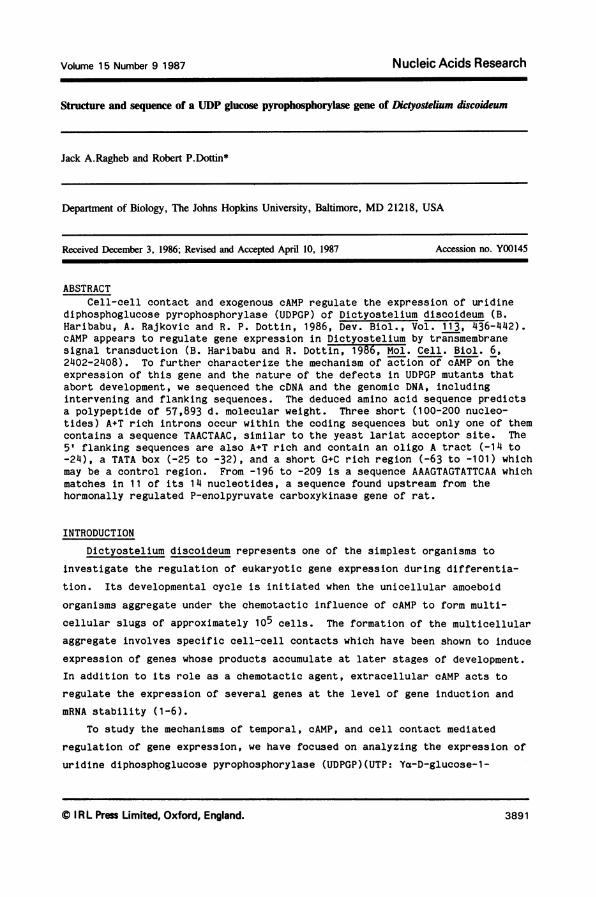
Volume 15 Number 9 1987 Nucleic Acids Research Structure and sequence of a UDP glucose pyrophosphorylase gene of Dictyostelium discoideum Jack A.Ragheb and Robert P.Dottin* Department of Biology, The Johns Hopkins University, Baltimore, MD 21218, USA Received December 3, 1986; Revised and Accepted April 10, 1987 Accession no. Y00145 ABSTRACT Cell-cell contact and exogenous cAMP regulate the expression of uridine diphosphoglucose pyrophosphorylase (UDPGP) of Dictyostelium discoideum (B. Haribabu, A. Rajkovic and R. P. Dottin, 1986, Bev. Biol., Vol. 113, 436-442). cAMP appears to regulate gene expression in Dictyostelium by transmembrane signal transduction (B. Haribabu and R. Dottin, 1986, Mol. Cell. Biol. 6, 2402-2408). To further characterize the mechanism of action of cAMP on the expression of this gene and the nature of the defects in UDPGP mutants that abort development, we sequenced the cDNA and the genomic DNA, including intervening and flanking sequences. The deduced amino acid sequence predicts a polypeptide of 57,893 d. molecular weight. Three short (100-200 nucleo- tides) A+T rich introns occur within the coding sequences but only one of them contains a sequence TAACTAAC, similar to the yeast lariat acceptor site. The 5' flanking sequences are also A+T rich and contain an oligo A tract (-14 to -24), a TATA box (-25 to -32), and a short G+C rich region (-63 to -101) which may be a control region. From -196 to -209 is a sequence AAAGTAGTATTCAA which matches in 11 of its 14 nucleotides, a sequence found upstream from the hormonally regulated P-enolpyruvate carboxykinase gene of rat. INTRODUCTION Dictyostelium discoideum represents one of the simplest organisms to investigate the regulation of eukaryotic gene expression during differentia- tion. Its developmental cycle is initiated when the unicellular amoeboid organisms aggregate under the chemotactic influence of cAMP to form multi- cellular slugs of approximately 105 cells. The formation of the multicellular aggregate involves specific cell-cell contacts which have been shown to induce expression of genes whose products accumulate at later stages of development. In addition to its role as a chemotactic agent, extracellular cAMP acts to regulate the expression of several genes at the level of gene induction and mRNA stability (1-6). To study the mechanisms of temporal, cAMP, and cell contact mediated regulation of gene expression, we have focused on analyzing the expression of uridine diphosphoglucose pyrophosphorylase (UDPGP)(UTP: Ya-D-glucose-1- © I RL Press Limited, Oxford, England. Nucleic Acids Research Volume 15 Number 9 1987 389 1
-
Upload
independent -
Category
Documents
-
view
0 -
download
0
Transcript of Structure and sequence of a UDP glucose pyrophosphorylase gene of Dictyostelium discoideum
Volume 15 Number 9 1987 Nucleic Acids Research
Structure and sequence of a UDP glucose pyrophosphorylase gene of Dictyostelium discoideum
Jack A.Ragheb and Robert P.Dottin*
Department of Biology, The Johns Hopkins University, Baltimore, MD 21218, USA
Received December 3, 1986; Revised and Accepted April 10, 1987 Accession no. Y00145
ABSTRACTCell-cell contact and exogenous cAMP regulate the expression of uridine
diphosphoglucose pyrophosphorylase (UDPGP) of Dictyostelium discoideum (B.Haribabu, A. Rajkovic and R. P. Dottin, 1986, Bev. Biol., Vol. 113, 436-442).cAMP appears to regulate gene expression in Dictyostelium by transmembranesignal transduction (B. Haribabu and R. Dottin, 1986, Mol. Cell. Biol. 6,2402-2408). To further characterize the mechanism of action of cAMP on theexpression of this gene and the nature of the defects in UDPGP mutants thatabort development, we sequenced the cDNA and the genomic DNA, includingintervening and flanking sequences. The deduced amino acid sequence predictsa polypeptide of 57,893 d. molecular weight. Three short (100-200 nucleo-tides) A+T rich introns occur within the coding sequences but only one of themcontains a sequence TAACTAAC, similar to the yeast lariat acceptor site. The5' flanking sequences are also A+T rich and contain an oligo A tract (-14 to-24), a TATA box (-25 to -32), and a short G+C rich region (-63 to -101) whichmay be a control region. From -196 to -209 is a sequence AAAGTAGTATTCAA whichmatches in 11 of its 14 nucleotides, a sequence found upstream from thehormonally regulated P-enolpyruvate carboxykinase gene of rat.
INTRODUCTION
Dictyostelium discoideum represents one of the simplest organisms to
investigate the regulation of eukaryotic gene expression during differentia-
tion. Its developmental cycle is initiated when the unicellular amoeboid
organisms aggregate under the chemotactic influence of cAMP to form multi-
cellular slugs of approximately 105 cells. The formation of the multicellular
aggregate involves specific cell-cell contacts which have been shown to induce
expression of genes whose products accumulate at later stages of development.
In addition to its role as a chemotactic agent, extracellular cAMP acts to
regulate the expression of several genes at the level of gene induction and
mRNA stability (1-6).To study the mechanisms of temporal, cAMP, and cell contact mediated
regulation of gene expression, we have focused on analyzing the expression of
uridine diphosphoglucose pyrophosphorylase (UDPGP)(UTP: Ya-D-glucose-1-
© I RL Press Limited, Oxford, England.
Nucleic Acids ResearchVolume 15 Number 9 1987
389 1
Nucleic Acids Research
phosphate uridyltransferase, EC 2.7.7.9), a well studied, enzyme in
Dictyostelium. We chose this enzyme because UDPGP mutants in Dictyostelium
discoideum abort the developmental cycle, suggesting that the enzyme is
essential for the completion of development (7,8). It has long been known
that cell contact plays a role in the regulation of specific activity of this
enzyme (5). Although many other genes are regulated by cell contact and cAMP
in Dictyostelium and several have been cloned, few have been identified.
Discoidin is the only one for which mutants may have been isolated. We have
previously shown that there are two mRNAs (UDPGP1,2) that encode several
differentially regulated isoforms of the enzyme (9,10). By using the cDNA as
a probe, we have also shown by Northern blot analysis that the ten fold rise
in enzyme specific activity observed during development can be accounted for
by the increase in UDPGP1 mRNA levels (10). We have recently demonstrated that
both cell-cell contact and cAMP coordinately regulate the accumulation of the
two UDPGP mRNAs (11). Disaggregation of slugs results in a rapid loss of
UDPGP mRNAs. Addition of cAMP to disaggregated cells elevates the level of
UDPGP mRNAs, although exogenous cAMP does not restore UDPGP mRNAs to their
original level. Other factors may be required for maximal expression of the
UDPGP1 gene. Unaggregated single cells, starved and shaken rapidly in
suspension, do not accumulate UDPGP mRNAs. However, addition of cAMP to these
cells caused UDPGP mRNAs to accumulate, suggesting that the requirement for
cell-cell contact could be bypassed in part by cAMP addition. Furthermore, we
have recently shown that exogenous cAMP analogues induce UDPGP1 expression and
that of other genes in the same relative order of potency that the analogues
bind to the cell surface cAMP receptor (12). This result suggests that the
induction of UDPGP1 and other Dictyostelium genes by exogenous cAMP is
mediated through the cell surface receptor, which may exert its effect on gene
expression by transmembrane signal transduction using second messenger(s)(12,13). The nature of the second messenger(s) is unknown. The central role of
UDPGP in Dictyostelium development, its importance as a marker for development
and now for signal transduction, and the availabilty of mutations affecting
UDPGP expression, make it an important gene to characterize at the nucleotide
sequence level.
As a first step towards utilizing the available mutants in elucidating the
mechanisms involved in the temporal and cAMP regulated expression of this
gene, we isolated and characterized the UDPGP1 gene and determined the
nucleotide sequence of both the UDPGP1 genomic and cDNA clones. The results
show the presence of A+T rich flanking sequences and three A+T rich introns.
3892

Nucleic Acids Research
In the 5' flanking sequence there is a short G+C rich region which may have
regulatory significance. A short 14 nucleotide sequence from -196 to -209
matches 11 nucleotides of a putative consensus cAMP response sequence in the
hormone regulated P-enolpyruvate carboxykinase gene of rat (14). Since
extracellular cAMP appears to behave as a hormone in Dictyostelium,
similarities between peptide hormone action in higher eukaryotes and exogenous
cAMP action in Dictyostelium are intriguing.
MATERIALS AND METHODS
DNA and RNA Isolation
Dictyostelium discoideum strain AX3 was used for all experiments.
Conditions for growth and differentiation as well as the isolation of RNA are
as described previously (8-10). To prepare nuclear DNA, cells were washed in
0.2% NaCl at 40C, resupended in ethidium bromide (300 ig/ml, 2.5 x 10⁵
cells/ml) and lysed with 1.45% Triton X100 and Chemusol NP12 (Rhone-Poulenc).Nuclei were washed in one volume of the same solution. The nuclear pellet was
resuspended at 2 x 10⁶ nuclei/ml in NB [Tris*Cl (25 mM), Mg acetate (5 mM),EDTA (0.5 mM), sucrose (5%) pH 7.6). EDTA was added to 250 mM at room
temperature. Nuclei were gently lysed at 550 with 5% sarkosyl, and 1 gm/ml
CsCl was added immediately. The refractive index was adjusted to 1.3965 and
the nuclear DNA was banded by centrifugation at 40 K rpm for 40 hr in a
Beckman Ty65 rotor.
Dictyostelium DNA Cloning Procedures
Restriction enzymes were purchased from BRL and New England Biolabs. DNA
ligase, T4 polynucleotide kinase, DNA polymerase, and its Klenow fragment were
from BRL. DNase I, calf intestinal alkaline phosphatase and AMV reverse
transcriptase were from Boehringer Mannheim. Radioactive nucleotides were
from either Amersham Corp. or New England Nuclear. The M13 cloning and
sequencing reagents were from either P-L Biochemicals or Bethesda Research
Laboratories. The cloning methods used were basically as described by
Maniatis et al. (15). One genomic library of Dictyostelium nuclear DNA in
lambda was constructed and generously made available by Dr. Daphne Blumberg.
The library was constructed using Dictyostelium DNA which was partially
digested with Eco RI and size fractionated on a sucrose gradient. The DNA
fragments were ligated into Xgt wes-XB in place of the stuffer fragment of
that vector. A second genomic library was constructed in the plasmid vector
pUC12 by ligating a Bgl II, Hind III total double digest of genomic DNA to
pUC12 that had been digested with Bam HI and Hind III. To enrich for clones
3893
Nucleic Acids Research
containing the UDPGP1 5' genomic fragment, the ligation products were then
digested with Xho I and Pst I before being transfected into a recA host.
Plaque and Colony Screening, and DNA Blot Hybridization
Hybridization probes (107 cpm//g) were usually prepared by nick
translation of isolated fragments from cDNA subelones (16). Occasionally
probes were made by synthesizing the complementary strand of a recombinant M13
template. In these cases, the synthesis conditions used were the same as for
dideoxy sequencing except that the ddNTPs were omitted, the cold dNTPs were
used at 0.1 mM, and the reaction contained 100 jCi of [a-32PIdATP. The
labelled double-stranded region of interest was excised with the appropriate
restriction enzymes and subsequently purified on an agarose gel.
Lambda plaques were transferred to either nitrocellulose (Schleicher &
Schuell or Millipore Corp.) or nylon membranes (Biodyne) and screened by the
method of Benton and Davis (17). Colonies were transferred to nylon membranes
while still small (<1 mm) and then amplified on plates containing chlor-
amphenicol (250 ig/ml) overnight. The screening was performed basically as
described by Maniatis et al. (15). Restriction digests were fractionated by
electrophoresis on agarose gels, transferred to nitrocellulose or nylon
membranes, and hybridized to 32P-labeled DNA probes as described by Southern
(18).
DNA Sequencing and Primer Extension
DNA sequencing was usually carried out by the chain termination method of
Sanger et al. (19,20) using the M13 vectors mp8,9,18, and 19. We used the
chemical cleavage method of Maxam and Gilbert in cases where dideoxy
sequencing reactions did not work because the highly A+T rich DNA caused
nonspecific termination at oligo A or oligo T tracts. Ten nanograms of a [32PI5' end labeled (106 cpm) synthetic oligonucleotide (5'-GATCCAGTTGATTGT-3'),
complementary to a sequence in the UDPGP1 mRNA 49 nucleotides downstream from
the initiating ATG, was annealed to 50 pg of total RNA from cells developed
for 18 hr. The annealing reaction was performed in 10 4l of 10 mM Tris, pH
8.3, at 370C, 2 mM MgCl2 by heating at 750C for 15 min and then cooling slowly
to room temperature in a 13 x 100 mm test tube filled with water initially at
750C. This reaction was incubated with 12.5 units of AMV reverse transcrip-
tase (1000 U/ml) with or without Actinomfcin D (25 ug/ml) in 4 mM DTT, 10 mM
MgCl2, 50 mM Tris, pH 8.3 at 370C for 60 min at 420C in the presence of 0.5 mM
dNTPs.
3894
Nucleic Acids Research
RESULTS
Structure and Sequence of the UDPGP1 cDNA
We have previously reported the cloning of the UDPGP1 cDNA by dG-dC
tailing into the Pst I site of pBR322 (10). A restriction map of the cDNA was
constructed and fragments of it were subcloned into M13 vectors for sequencing
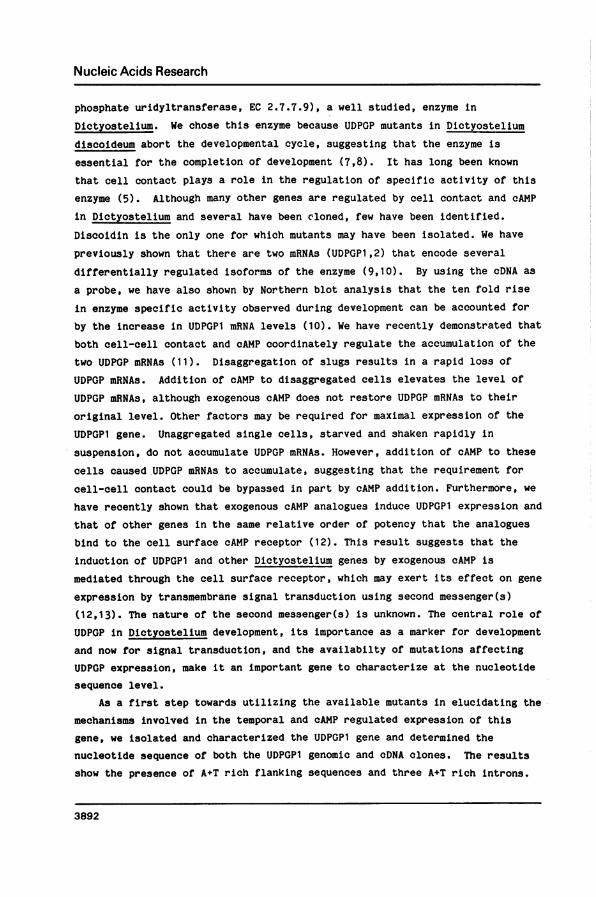
by the dideoxy method of Sanger et al. (19,20). The restriction map and the
sequencing strategy employed are shown in Fig.1. The direction of transcrip-
tion had been determined by hybrid arrested translation using single strands
of UDPGP1 cDNA (10) and was confirmed by hybridization of single-stranded
probes to northern blots (data not shown).
The cDNA, excluding the tails, is 1,583 nucleotides long (Fig. 2). It
contains a single open reading frame which is 1,525 nucleotides long and
5' 3'
SEQUENCING -------STRATEGY -
UDPGP cDNA ' -MAPR(Kb)rR YBw 1W ScP
GENOMIC ,, 0.3 0.2 0.24 9,0.18'.MAP "
/ 0.5 0.2 0.35 : 0.29H R Sc DRDSi R R VB B HRV Sc B R
SEQUENCING -, _:, _STRATEGY - -
A3
19// PUC125 PUCI23
MPR900
Fig. 1. Structure of the UDPGP1 cDNA and genomic DNA. A restriction map of the1.65 Kb cDNA insert in the Pst I site of pBR322 is shown. The cDNA map isaligned with the genomic restriction map, and some of the correspondingrestriction sites in the two DNAs are indicated with dotted lines. The genomicmap spans approximately 13 Kb. Restriction sites are designated as follows: B,Bgl II; D, Dra I; H, Hind III; N, Nsi I; P, Pst I; R, Eco RI; Rs, Rsa I; S,Sau3A I; Sc, Sca I; V, Eco RV. The sequencing strategy utilized for the twoDNAs is indicated. Arrows indicate the direction of the sequencing reactionand the amount of information obtained from a given clone or restrictionfragment. The 5' and 3' notations indicate the direction of transcription. Thegenomic segments contained within the various subclones are indicated in thebottom of the figure. The displayed map distances are in Kbp. Only thoseRsa I, Sau3A I, and Dra I sites pertinent to the sequencing strategy areshown.
3895

Nucleic Acids Research
V Sau 3A IATG ACA GAT ACA CCA ACA TCA AAA GCA ACA GTT GAA AGA CCA AAA TTA CAA TCA ACT GGA TCA TTA CAT ACT TTAMET Thr Asp Thr Ala Thr Ser Lys Ala Thr Val Glu Arg Pro Lys Lou Gln Ser Thr Gly Ser Lou His Ser LouDra. ITTT AAA CAT OTT GAT TTA TTT TCA CAC AAT CAT GAA GAA TTA TAT CCA CCA CTT CAA CAT GCT GCA ACA TtT GCAPhe Lys Asp Val Asp Lou Phe Ser Glu Asn Asp Glu Clu Lou Tyr Pro Pro Lou Gln His Gly Ala Arg Phe Ala
GCA CCA ATT GAA CAT ACT ACA TTA TTA GCA TTC COT ATC AAA CCA GAT CAA CTT AAA GCA TTC CAA AAA CAA AGAAla Pro Ile Glu Asp Ser Thr Lou Lou Ala Lou Gly MET Lys Pro Asp Glu Lou Lys Ala Phe Gln Lys Gln ArgNsi I
CkT GCC1 AC ATT AAC AAC GAT CAA ATT TAC ACT CAT CAA ATT AAA ATT CCA AAT AAA ACT CAA ATC CTA GAT TATHis Ala Tyr Ilo Asn Lys Asp Gln I1 Tyr Thr Asp Clu Il Lys Il Pro Amn Lys Thr Clu MET Val Asp Tyr
CAT CAA CTT CAT TTA OTC TCA CCA ATT CAC CAA TCA AAT OCT TCC ACA TTA TTA AAT AAA TTA GTT GTA ATT AAAHis Gln Lou His Lou Vol Sor Pro Ile Asp GCl Sor Asn Ala Ser Arg Lou Lou Asn Lys Leu Vol Vol Ile Lys
Rsa ITTA AAT CGT GCT CTT CGT AAT ACT ATG COT TCT AAA ACT GCT AAA AGC ACA ATG GAA ATA OCT CCA CGT CTT ACTLou Asn Cly Cly Lou Oly Asn Sor MET Gly Cys Lys Thr Ala Lys Sor Thr MET Clu Ile Ala Pro Gly Vol Thr
TTT TTA CAT ATG GCA OTT OCT CAT ATT CAA CAA ATT AAT CAA OAT TAT AAT OTT GAT CTC CCA TTG CTT ATT ATGPhe Lou Asp MET Ala Vol Ala His Ilo Glu Gln Il Aso Cln Asp Tyr Asn Vol Asp Vol Pro Lou Vol Ile MET
Eco RIAAT TCT TAT AAA ACT CAT AAT GAA ACT AAT AAG OTT ATT GAA AAG TAT AAA ACT CAT AAA CTT ACT ATT AAA ACTAsn Ser Tyr Lys Thr His Aso Glu Thr Asn Lys Val Ie Clu Lys Tyr Lys Thr His Lys Vol Ser h1o Lys Thr
TTC CAA CAA TCA ATG TTC CCA AAG ATC TAT AAA GAT ACA TTA AAT TTA GTA CCA AAA CCA AAT ACA CCA ATG AATPhe Cln Gla Sor MET Phe Pro Lys MET Tyr Lys Asp Thr Lou Asn Lou Vol Pro Lys Pro Asn Thr Pro MET Asn
Eco R0 Bgl II!CCA AAG GAA TCOG TAT CCA CCA GGT TCA GGT CAT ATC TTT AGA TCA CTC CAA AGA TCT CGT TTO ATT CAT GAA TTTPro Lys Glu Trp Tyr Pro Pro Cly Sor Gly Asp . Phe Arg Ser Lou Cln Arg Ser Cly Lou Ile Asp Glu Phe
TTA GCT GCT GCT AAA GAA TAT ATT TTC ATT TCA AAT GTT GAA AAT TTA GCT TCA ATA ATT GAT CTT CAG CTA TTALou Ala Ala Gly Lys Glu Tyr Ile Ph Ile Ser Asn Val Glu Asn Lou Cly Sor Ile Ile Asp Lou Cln Vol Lou
AAT CAT ATT CAT TTC CAA AAC ATT GAA TTT OCT TTA GAA GTC ACA AAT CCT ATT AAT ACT CAT TCA ACT GCT GCTAsn His Ile His Lou Gln Lys Ile Glu Phe Gly Lou Clu Vol Thr Asn Arg Ile Asn Thr Asp Ser Thr GCy Cly
B1 IIATT TTA ATG TCA TAT AAA CAT AAA CTT CAT CTT TTG GAA TTA TCT CAA GTT AAA CCA CAG AAA TTA A TTIle Lou MET Ser Tyr Lys Asp Lys Lou His Lou Lou Clu Lou Sor Glo Val Lys Pro Glu Lys Lou Lys Ilo Phe
AAA GAT TTT AAA CTT TGG AAT ACA AAT AAT ATT TOG GTT AAT TTG AAA TCA GTT TCA AAT TTA ATT AAA GAA CATLys Asp Ph. Lys Lou Trp Asn Thr Asn Asn l. Trp Vol Asn Lou Lys Sor Vol Ser Asn Lou Ile Lys Glu Asp
AAA TTA GAT TTA CAT TGG ATT GTT AAT TAT CCA CTT GAA AAT CAT AAA GCA ATG GTA CAA TTA CAA ACA CCA GCALys Lou Asp Lou Asp Trp Ile Vol Asn Tyr Pro Lou Clu Asn His Lys Ala MET Vol Gln Lou Clu Thr Pro ;la
Eco RI EcoRVGGT ATG COT ATT CAA AAT TTT AAG AAT TCA OTT GCA ATT TTT CTA CCA CGT OAT AGA TAT CGT CCA ATT AAA TCAGly MET Cly I.e Cln Asn Phe Lys Asn Ser Vol Ala Ile Pho Vol Pro Arg Asp Arg Tyr Arg Pro Ile Lys Sor
ACA ACT CAA TTA TTC GTT GCA CAA TCA AAT ATT TTC CAA TTT OAT CAT CCT CAA GTT AAA TTA AAT TCA AAG AGAThr Sor GCl Lou Lou Vol Ala Gln Ser Asn Ile Ph. Gln Phe Asp His Cly Gln Vol Lys Lou Asn Ser Lys Arg
CAA CGT CAA CAT GTA CCA CTT OTT AAA TTG CGT GAA GAA TTT TCA ACA GTT TCA CAT TAT CAA AAC AGA TTT AAAClu Cly Glb Asp Vol Pro Lou Vol Lys Lou Gly Glu Glu Ph. Ser Thr Vol Sor Asp Tyr Glu Lys Arg Ph. Lys
TCA ATT CCA CAT TTA TTG GAA TTG OAT CAT CTT ACT OTT TCT COT CAT GTT TAC TTT GOT TCA AGA ATT ACT CTTSor Il. Pro Asp Lou Lou Clu Lou Asp His Lou Thr Val Sor Gly Asp Vol Tyr Ph. Oly Ser Arg Ile Thr Lou
AAA GGT ACA GTC ATT ATT GTA GCT AAT CAT CGT GAA COT OTT CAT ATT CCA OAT CGT GTG GTT TTA CAA AAT AAALys Cly Thr Vol Ile Ile Val Ala Asn His Cly Clu Arg Vol Asp I1e Pro Asp Gly Val Val Lou Clu Asn Lys
Sca ICTA CTT TCT GCC ACT CTT AGA ATT TTG CAT CAT TAA att cta ctc a00 aaa tta att ggt coo aaa 000 a00 00aVatOu Ser Cly Thr Lou Arg Ile Leu Asp His
000 a0a a00 000 a00 0a0 0
Fig. 2. Nucleic acid sequence of the UDPGP1 cDNA and deduced amino acidsequence. The 1,583 bp cDNA sequence, excluding the dG-dC tails is shown. Itbegins with the first nucleotide to the right of the open triangle and endswith the terminus of the poly A tract. Only the sequence of the RNA sensestrand is shown. Those restriction sites determined by restric'ion analysisare shown above the sequence and the recognition sequence underlined. Thededuced amino acid sequence is shown immediately below the nuc'Leic acidsequence. The identity of the four N terminal-most amino acids (the initiatingMet, Thr, Asp, Thr) was deduced from the genomic DNA sequence. The termina-tion codon is indicated by an asterisk below it. Solid triangles indicate theposition of the intervening sequences in the genomic DNA.
3896
Nucleic Acids Research
terminates with a TAA codon. However, it lacks an initiating ATG and a 5'
untranslated region. The 3' untranslated region is 25 nucleotides long and is
followed by a 31 nucleotide long polyA tract. A canonical polyadenylation
signal, AATAAA, is not found upstream from the polyA tract. A sequence, AAAAAA,
which differs from it by a single nucleotide, is located 11 nucleotides 5' to
the beginning of the polyA tract.
The portion of the open reading frame that is present in the cDNA is
sufficient to encode a 57,373 d. polypeptide, which is close to the publishedmolecular weight of UDPGP1, suggesting that only a small portion of the NH2
terminal coding sequences are absent from the cDNA. Several interesting
structural features of the polypeptide are revealed by analysis of the deduced
amino acid sequence. Among these is the pattern of Lys usage: Twenty nine of
forty five Lys residues occur in pairs or triplets with the structure Lys-(X),_--Lys-(X)1_3-Lys. In one region of the polypetide Lys residues constitute 5/14amino acids, and in a second region 5/12 amino acids. There are 24 Pro
residues, 6 of which occur within a 15 amino acid stretch in approximately the
middle of the protein. There is only one Cys residue present in the predicted
amino acid sequence, thus excluding the possibility of any intramolecular
disulfide bridges in the UDPGP1 polypeptide.
Isolation and Structure of UDPGP1 Genomic Sequences
Using the UDPGP1 cDNA and subclones of cDNA fragments as hybridization
probes, a restriction map of the gene was constructed by hybridization to
Southern blots of genomic DNA (Fig. 1). Our data showed that a single copy of
the gene hybridized to this cDNA and spanned three Eco RI fragments; 2.5, 0.9,and 9.5 Kb in size. Probes from the 5' end of the cDNA hybridized only to the
2.5 Kb genomic DNA fragment while probes from the 3' end of the cDNA
hybridized only to the 9.5 Kb genomic fragment. The presence of a Hind III
site in the genomic DNA that was absent in the cDNA suggested that the UDPGP1
gene contained an intervening sequence.
Using the UDPGP1 cDNA as a probe, a Agt wes-AB genomic library of nuclear
DNA was screened by the method of Benton and Davis (17). After several rounds
of screening, a clone, designated A3, was isolated and found to contain the
0.9 and 9.5 Kb Eco RI genomic fragments that constitute the central portion
and the 3' end of the gene respectively (see Fig. 1). The 2.5 Kb Eco RI
fragment containing the 5' end of the gene was subsequently isolated as part
of a 2.9 Kb Hind III-Bgl II fragment from a genomic library constructed in the
plasmid vector pUC12. This clone was designated pUC125.The 0.9 Kb Eco RI fragment from X3 was subcloned into the M13 vector mp8.
3897

Nucleic Acids Research
This clone was designated mpR900. A 2.5 Kb portion of the 9.5 Kb Eco RI
fragment, which contained all the 3' end sequences complementary to the cDNA,
was subcloned into pUC12 as part of a 2.7 Kb Hind III-Bgl II fragment that
overlaps at its 5' end sequences contained in the 0.9 Kb central Eco RI
fragment. This clone was designated pUC123 (Fig. 1).
Restriction analysis of the genomic subclones and the cDNA revealed the
presence of three small 100-200 bp introns within the translated regions of
the UDPGP1 gene. These introns are located between the Nsi I and 5' Eco RI
sites, the two central Bgl II sites, and the Bgl II and 3' Eco RI sites. DNA
sequencing confirmed the presence of these introns.
Genomic sequences were determined by a combination of the dideoxy chain
termination method and the chemical cleavage method of Maxam and Gilbert (21).
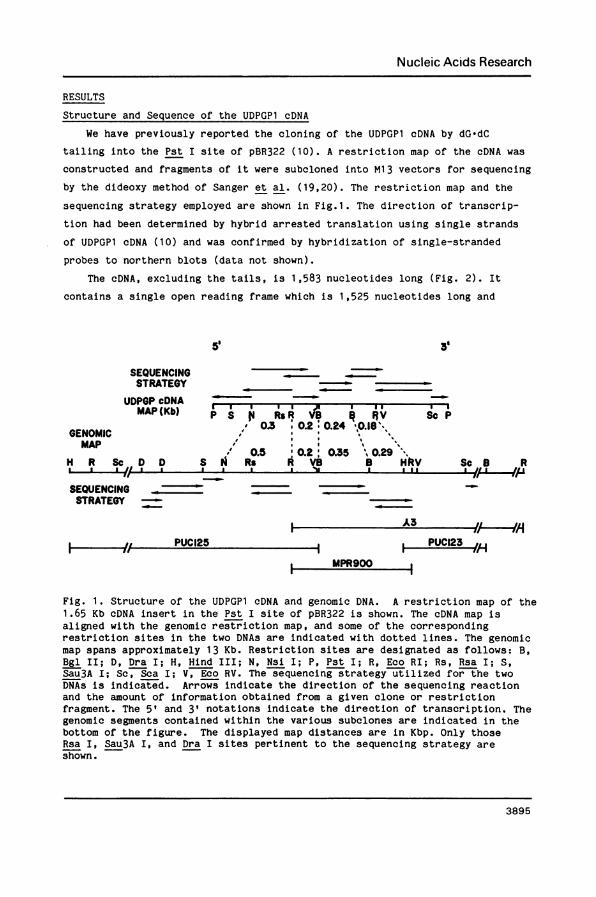
The sequencing strategy is shown in Fig 1. The sequence composition of all
three introns is >90% A+T, and the introns obey the GT/AG rule. The region of
conservation of the donor sequence in the introns is actually somewhat longer,
the hexanucleotide GTAAGT being present at the beginning of all three introns.
The second intron contains a sequence, TAACTAAC, which is strikingly similar
to the highly conserved TACTAAC box found in yeast introns (22) and is present36 nucleotides before the splice site in the same relative position within
that UDPGP1 intron as the conserved sequence found in yeast. This sequence is
known to be involved in lariat formation during RNA splicing in yeast.
Interestingly, the terminal AG dinucleotide of the third intron is the
internal AG of the genomic Hind III site (AAGCTT), and thus the splicing event
per se accounts for the absence of the Hind III site within the cDNA (Fig. 3).The first intron is located between codons 160 and 161, while the second
intron splits codon 273 between the first and second nucleotide, and the third
intron lies between codons 361 and 362 (Fig. 2). Partial sequencing of the
pUC123 subclone demonstrated that there are no introns present in the gene
downstream from the 3' Sca I site.
Partial sequencing of pUC125 provided the identity of the three N
terminal-most amino acids and the initiating ATG. This ATG is preceded by
thirteen A nucleotides, which is a pattern that has been observed with several
other Dictyostelium translational start sites (23-26). A sequence, TATAAAAA,
conforming to the consensus TATA box in Dictyostelium (TATAAATA)(25) was
found about 80 nucleotides upstream from the initiating ATG and, as we shall
show, between -25 and -32 from the start of transcription. For more than
300 bp upstream from the site of transcriptional initiation, the sequence is
only 10% G+C, except for a region between -63 and -101 bp which is 45% G+C.
3898

Nucleic Acids Research
FLANKING 5' SEQUENCE-320 . -300 . -280 . -260 . -24b
tttttttttttttttttttttttcaaaaaaaaataaaaaaaatttaatttaatttatataaatetcacattacattttttttttatttatcatttgtta-220 . -200 . -180 . -160 . -140
tataaaataccacaa aagtagtattcastatattaaatttattttaaacttgtattattatttttatattagttttttttttttttttatttttattt-120 . -1 -80 . -60 . -40
ttattttttttttaatttttttttaactccgccaatcatcattatcatcatcatcaccatcacaattattataataaaaaatattcatatatatatapa-20 . +1 . +20 +40 +50
aaaaaaaaaaaaacaatattaataattttaactaattaatttattattataaaaagaaaataaaaaaaaaaaaaaATG ACA GAT ACAAA A Met Thr Asp Thr
FLANKING 31 SEQUENCE+1960 +1980 . +2000 +2020
CAT TAAattctactcaaaaaattaattggtcaaaaaaaaaaaaaaaaaaaaaaaaaaaaaaaaaaaaaaHis *
INTRON I SEQUENCE+520 +540 +560 +580 +600
GAAtaaagtagtaaaattttttaaaataataataattattattaataatattttcttttggtattaatttatttattttttattttecacttttttccaGlu
+620 . +640 +660 . +680 +700ttttttecatttttaattttttttttttttttttttaatttttttttttaatttttttttttatattaaaasttttaatataRCAA
Gln
INTRON II SEQUENCE+1040 +1060 +1080 . +1100 +1120
CTT CgtaagttgtttttttaaaatttgaaaaaaaaataaataaaaataaaaataactaacacaaataaaataaattattatttttattattattaLeu ........
+11 34tAg AG GTA
Gln Val
INTRON III SEQUENCE+1400 +1420 +1440 . +1460 . +1480
CCAEtaagtaatttttttttttttttttttttttttttttttttttatattatttatttattaattaattaataattttttatttatttaaattttPro
+1500t;atttttaattttccaaagCTT
Leu
Fig. 3. Genomic DNA Sequence. Translated sequences are shown in upper caseletters. The G+C rich region in the 5' flanking sequence is underlined. Theso-called cAMP responsive sequence AAAGTAGTATTCAA is boxed. The location ofthe TATAAAAA sequence is underscored by a dashed line. The sites oftranscriptional initiation are indicated by an arrow beneath the sequence. Thefirst nucleotide of the shortest transcript is arbitrarily designated as the+1 nucleotide. The initiating ATG and the three N terminal-most amino acidsare shown. The C terminal amino acid and termination codons are shown. Thefirst poly A tract downstream from the termination codon corresponds to thepoly A tract in the cDNA. The sequence of all three introns and theirflanking codons are shown. The conserved sequences at the termini of theintrons are underlined. The TAACTAAC box within the second intron isunderscored with a dotted line.
Most of the G+C rich region consists a trimer ATC, which is repeated seven
times in the sequence. Another noteworthy feature of this region is the
sequence asymmetry of the strands; with the exception of one nucleotide, all
of the G nucleotides are located on one strand and all the C nucleotides are
on the other strand (Fig. 3). The considerably higher G+C content of this
region and its unusual structure may signify that it has a role in gene
regulation. A second sequence AAAGTAGTATTCAA (boxed) matches in 11 of its 14
nucleotides the sequence AAAGTTTAGTCAA in the hormone regulated rat PEP
carboxykinase gene (l14) which is postulated to be a cAMP responsive sequence
3899
Nucleic Acids Research
A TA G G C C a b
3'
AT
>AT _
>STTA>A .
CG't
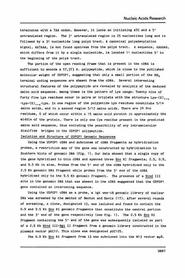
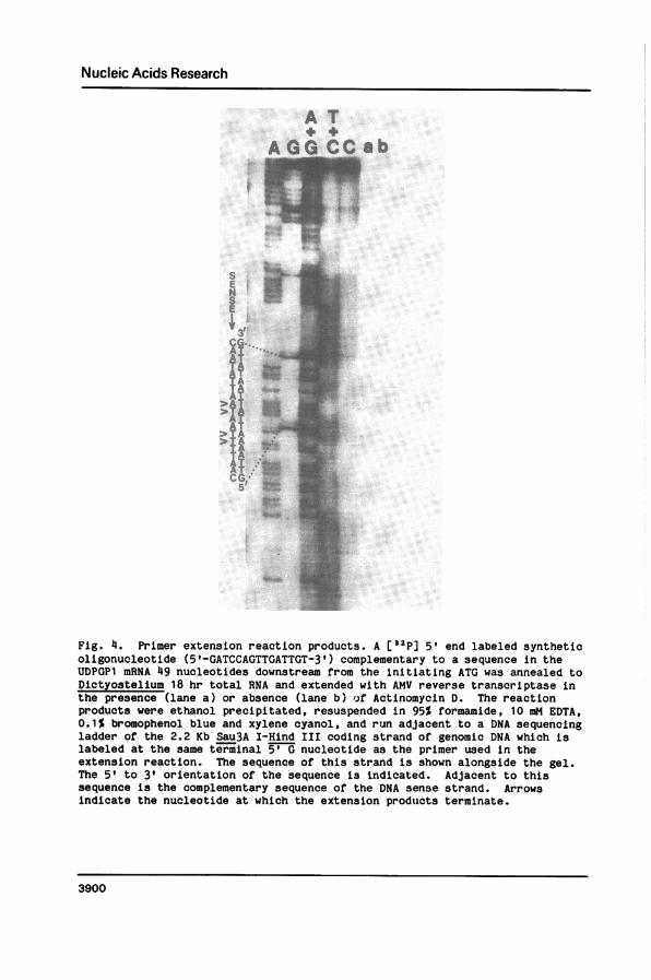
Fig. 4. Primer extension reaction products. A [32p] 5' end labeled syntheticoligonucleotide (5'-GATCCAGTTGATTGT-3') complementary to a sequence in theUDPGP1 mRNA 49 nucleotides downstream from the initiating ATG was annealed toDictyostelium 18 hr total RNA and extended with AMV reverse transcriptase inthe presence (lane a) or absence (lane b) of Actinomycin D. The reactionproducts were ethanol precipitated, resuspended in 95% formamide, 10 mM EDTA,0.1% bromophenol blue and xylene cyanol, and run adjacent to a DNA sequencingladder of the 2.2 Kb Sau3A I-Hind III coding strand of genomic DNA which islabeled at the same terminal 5' G nucleotide as the primer used in theextension reaction. The sequence of this strand is shown alongside the gel.The 5' to 3' orientation of the sequence is indicated. Adjacent to thissequence is the complementary sequence of the DNA sense strand. Arrowsindicate the nucleotide at which the extension products terminate.
3900
1:

Nucleic Acids Research
only on the basis of its homology to sequences upstream from other cAMP
regulated genes in prokaryotes and eukaryotes.
A 38 nucleotide poly A tract encoded in the genome occurs 25 nucleotides
downstream from the translation termination site. The 3' end of the cDNA and
genomic DNA are co-linear (Fig. 3).
Mapping the Site of Transcriptional Initiation
The DNA primer used for primer extension was a 5' end labeled synthetic
pentadecanucleotide complementary to sequences near the 5' end of the mRNA
beginning at the first Sau3A I site in the cDNA (Fig. 4). The template was
total RNA from cells developed for 18 hrs. The reaction products were
denatured and electrophoresed on a 7 M urea gel adjacent to a Maxam-Gilbert
sequencing ladder of the 2.2 Kb Sau3A I-Hind III coding strand of genomic DNA
which is 5' end labeled at the Sau3A I site, i.e. at the same position as the
primer used in the extension reaction. This strategy allows direct determina-
tion of the complementary nucleotide sequence at which the mRNA is initiated.
A doublet was observed corresponding to two T residues 49 and 50 nucleotides
from the ATG (Fig. 4). This observation maps the major initiation product of
transcription to a position about 49 nucleotides upstream from the A in the
initiating ATG (Fig. 3). A fainter doublet occurs 53 and 54 nucleotides from
the A in ATG. Si nuclease analysis was also performed to map the site of
transcriptional initiation. In general, the S1 nuclease resistant fragments
were somewhat smaller than the products of the primer extension experiment
(result not shown). The size of the resistant fragments was found to be
inversely proportional to the temperature at which the Si reaction was
performed. This is presumably a consequence of breathing of the RNA-DNA
hybrid at the A+T rich end of the mRNA.
DISCUSSION
UDPGP is a well studied enzyme that has long been used by developmental
biologists as a marker for differentiation in Dictyostelium. The enzyme is
essential for development and the UDPGP mutants that have been isolated all
abort the developmental cycle at the same stage. We have recently shown that
the expression of the UDPGP gene is regulated by cAMP through signal
transduction (12,29). Here, we have described the cloning and sequencing of a
cDNA complementary to the UDPGP1 mRNA, and the genomic DNA that encodes this
transcript and its flanking sequences. These sequences will be useful in
characterizing the UDPGP mutants and elucidating the mechanisms involved in
3901
Nucleic Acids Research
the regulation of UDPGP1 gene expression. The cDNA contains a single open
reading frame that is sufficient to encode nearly the entire UDPGP1
polypeptide. Interesting structural features of the polypeptide were
predicted from the deduced amino acid sequence. The polypeptide contains 49
Lys residues which are distributed in clusters made up of simple repeating
units. The presence of a cluster of Pro residues in the middle of the
polypeptide suggests that the protein may be divided into two large domains
joined by a Pro rich, random coil joining segment.
Comparison of the two nucleic acids revealed the presence of three introns
within the coding sequences of the genomic DNA. Like other introns sequenced
from Dictyostelium they are 90% A+T, and are relatively short when compared to
other eukaryotic introns. They all obey the GT/AG rule for intervening
sequences, and actually contain a slightly longer conserved sequence, GTAAGT,
at the 5' splice site. This level of conservation is unprecedented in
Dictyostelium, but its functional or regulatory significance is unknown. The
site at which lariat formation occurs is a highly conserved sequence (TACTAAC)in yeast (22). A variant of that consensus sequence is present in only one of
the three introns (intron II) of this Dictyostelium gene. The presence of the
initial GT and terminal AG dinucleotides suggest that intervening sequences
may be processed from the UDPGP1 mRNA via a lariat intermediate, as has been
shown in other eukaryotes.
By utilizing the method of primer extension, we mapped the site of
transcriptional initiation to approximately 50 nucleotides upstream from the
initiating ATG. Although S1 nuclease mapping resulted in slightly smaller
fragments the results are consistent with the conclusions drawn from the
primer extension experiments.
Primer extension of the UDPGP1 mRNA produced four products. A possible,
though unsubstantiated, explanation for such microheterogeneity in the case of
the UDPGP1 mRNA can be seen upon examination of the sequences immediatelyupstream from the consensus TATA box. Since only the ATA at the second,
third, and fourth positions of the TATA consensus sequence are invariant in
Dictyostelium (25), it is possible to actually derive three additional TATA
boxes from the sequence immediately upstream from the consensus TATA box. If
this were the case, the four TATA boxes would overlap one another, each being
staggered by two nucleotides from the other (TATATATATAAAAA). Since the
position of the TATA box presumably determines the site of transcriptionalinitiation in eukaryotes, this duplication of TATA boxes could account for the
microheterogeneity seen at the 5' end of the UDPGP mRNA. Interestingly, the
3902
Nucleic Acids Research
most prominent of the primer extension products is the smallest one, which
implies that the shortest UDPGP1 mRNA is the most abundant mRNA species. The
transcriptional initiation of this message is presumably directed from the 3'
most TATA box, i.e. the one that most closely matches the concensus sequence.
Dictyostelium genes in which a canonical polyadenylation signal cannot be
readily identified have been reported previously (27). In the case of the
Dictyostelium ras gene, as with the UDPGP1 gene, the absence of a canonical
polyadenylation signal is accompanied by the presence of a poly A tract within
the genome immediately downstream from the 3' untranslated region. The
sequence, AAAAAA, is located where one would expect a polyadenylation signal
to be found, but differs from the canonical sequence by a single nucleotide.
Whether the poly A tail on the mRNA transcribed from these genes is derived
from the encoded sequence in the genome, added post-transcriptionally, or both
will require further investigation.
Unlike all other Dictyostelium genes that have been sequenced (23-26), the
UDPGP1 gene does not have an oligo T stretch in the RNA sense strand of the
gene just upstream from the site of transcriptional initiation. Rather, it
has an oligo A stretch on that strand and of course an oligo T stretch on the
complementary strand, suggesting that if the oligo T is important its
orientation is not. The implication is that if the oligo T sequences do play
a role in promoter function, they are capable of functioning bidirectionally.
This hypothesis awaits testing in a Dictyostelium DNA transfection system. In
addition to having a TATA box and an "oligo T element" that are not separated
from one another, the UDPGP1 gene also differs from other known Dictyostelium
genes in not having G residues flanking its TATA box. In fact, there are
only six G residues in the 300 nucleotides that have been sequenced upstream
from the TATA box, and the first of these occurs about 65 nucleotides upstreamfrom it. A consensus TATA box (TATAAAAA) is found immediately after the oligoA tract between -25 and -32 (25,26). In addition, there is no CAAAT sequence
in the 5' untranslated region of the mRNA. Flanking sequences upstream from
the gene, like the introns, are 90% A+T. The presence of a G+C rich domain at
-63 to -100 within this 90% A+T region is intriguing. Since UDPGP is
developmentally regulated and its expression is modulated by cAMP, this G+C
rich sequence may represent one of several possible regulatory regions that
are presumably upstream from the gene. A G+C rich sequence upstream from the
cysteine proteinase II gene of Dictyostelium has been identified by Datta et
al. (28). There is evidence that this sequence coincides with a nuclease
hypersensitive site (Pavlovic and Parish, personal communication).
3903
Nucleic Acids Research
Our previous experiments suggested that extracellular cAMP regulates gene
expression by binding to a cell surface receptor and inducing the synthesis of
an intracellular second messenger. Based on the pharmacological specificity
of the target receptor molecule which is involved in gene expression, we and
other workers concluded that the cAMP dependent protein kinase is not the
direct target for extracellular cAMP (12,13). The nature of the second
messenger(s) remain unknown. We have recently proposed a model to account for
our current observations (29).
Peptide hormones are thought to regulate gene expression in higher
eukaryotes by inducing the synthesis of intracellular second messengers such
as cAMP. Hanson and Reich have independenlty suggested that intracellular
cAMP may act directly via a "CAP-like" DNA binding protein to activate
transcription (30,31). They have identified sequences upstream from several
hormonally regulated genes that are partially homologous to the E. coli DNA
CAP binding site. The functions of these sequences are being elucidated in
other laboratories. The sequence identified upstream of the rat PEP
carboxykinase gene by Hanson is conserved in the UDPGP1 gene and is located at
a similar position (30). Though UDPGP1 is regulated by extracellular cAMP and
it appears unlikely that intracellular cAMP is also involved in regulating its
expression (13). Whether that sequence is required for cAMP regulation
expression in rat PEP CK gene is also obscure. Therefore, the observation is
interesting, but its significance remains to be determined by mutational
analysis. The availability of mutants altered in the cAMP stimulus/responsesystem, and a well-characterized receptor system should make Dictyostelium a
useful model for studying how extracellular molecules regulate gene expression
via transmembrane signal transduction.
ACKNOWLEDGEMENTS
We thank Dr. Daphne Blumberg for providing assistance in screening the
lambda genomic library. We appreciate the help of Dorothy Regula and Jerry
Keightley in preparing the manuscript. This work was submitted by J.A.R. in
partial fulfillment of the requirements for a Ph.D. This work was supported by
NIH GM2730. This publication is number XXXX from the Department of Biology.
*To whom reprint requests should be sent
REFERENCES1. Blumberg, D. D., Margolskee, J. P., Barklis, S. N. Chung, S. N.
Cohen, N. S. and Lodish, H. F. (1982) Specific cell-cell contacts are
3904

Nucleic Acids Research
essential for induction of gene expression during differentiation ofDictyostelium discoideum. Proc. Natl. Acad. Sci. U.S.A. 79:127-131.
2. Chisholm, T., Barklis, E. and Lodish, H. F. (1984) Mechanism of sequentialinduction of cell-type specific mRNAs in Dictyostelium differentiation.Nature (London) 310:67-69.
3. Chung, S., Landfear, S. N., Blumberg, D. D. Cohen, N. S. and Lodish, H.F. (1981) Synthesis and stability of developmentally regulatedDictyostelium mRNAs are affected by cell-cell contact and cAMP. Cell24:785-797.
4. Mehdy, M. C., Ratner, D. and Firtel, R. A. (1983) Induction and modulationof cell type specific gene expression in Dictyostelium. Cell 32:763-771.
5. Newell, P. C., Longlancit, M. and Sussman, M. (1971) Control of enzymesynthesis by cellular interaction during development of the cellularslime mold Dictyostelium discoideum. J. Mol. Biol. 58:541-554.
6. Williams, J. G., Lloyd, M. M. and Devine, J. M. (1979) Characterizationand transcription analysis of a cloned sequence derived from a majordevelopmentally regulated mRNA of D. discoideum. Cell 17:903-913.
7. Diamond, R. L., Farnsworth, P. A. and Loomis, W. F. (1976) Isolation andcharacterization of mutants affecting UDPG pyrophosphorylase activity inDictyostelium discoideum. Dev. Biol. 50:169-181.
8. Sussman, M., and Osborn, M. H. (1964) UDP-Galactose polysaccharidetransferase in the cellular slime mold, Dictyostelium discoideum:Appearance and disappearance of activity during cell differentiation.Proc. Natl. Acad. Sci. USA 52:81-87.
9. Fishel, B. R., Manrow, R. E. and Dottin, R. P. (1982) Developmentalregulation of multiple forms of UDP glucose pyrophosphorylase ofDictyostelium. Dev. Biol. 92:175-187.
10. Fishel, B. R., Ragheb, J. A., Rajkovic, A., Haribabu, B. Schweinfest, C.W. and Dottin, R. P. (1985) Molecular Cloning of a cDNA Complementary toa UDP-Glucose Pyrophosphorylase mRNA of Dictyostelium discoideum. Dev.Biol. 110:369-381.
11. Haribabu, B., Rajkovic, A. and Dottin, R. P. (1985) Cell-Cell contact andcAMP regulate the expression of a UDP glucose pyrophosphorylase gene ofDictyostelium discoideum. Dev. Biol. 113:436-442.
12. Haribabu, B. and Dottin, R. (1986) Pharmacological characterization ofcyclic AMP receptors mediating gene regulation in Dictyosteliumdiscoideum. Mol. Cell. Biol. 6:2402-2408.
13. Oyama, M. and Blumberg, D. (1986) Interaction of cAMP with the cellsurface receptor induces cell-type-specific mRNA accumulation inDictyostelium discoideum. Proc. Natl. Acad. Sci. U.S.A. 83:4819-4823.
14. Wynshaw-Boris, A., Lugo, T., Short, J., Fournier, R. and Hanson, R.(1984) Identification of a cAMP regulatory region in the gene for ratcystolic phosphoenolpyruvate carboxykinase (GTP). J. Biol. Chem.259:12161-12169.
15. Maniatis, T., Fritsch, E. and Sambrook, J. (1982) Molecular cloning. ColdSpring Harbor Laboratory, Cold Spring Harbor, N.Y.
16. Rigby, P., Dieckmann, M., Rhodes, C. and Berg, P. (1977) Labellingdeoxyribonucleic acid to high specific activity in vitro by nicktranslation with DNA polymerase I. J. Mol. Biol. 113:237-251.
17. Benton, N., and Davis, R. W. (1977) Screening Xgt recombinant clones byhybridization to single plaques in situ. Science 196:180-183.
18. Southern, E. (1975) Detection of specific sequences among DNA fragmentsseparated by gel electrophoresis. J. Mol. Biol. 98:503.
19. Sanger, F., Nichen, S. and Colsen, A. (1977) DNA sequencing withchain-terminating inhibitors. Proc. Natl. Acad. Sci. USA 74:5463-5467.
3905

Nucleic Acids Research
20. Kwiatkowski, R. W., Schweinfest, C. W. and Dottin, R. P. (1984) Molecularcloning and the complete nucleotide sequence of the creatine kinase-McDNA from chicken. Nucleic Acids Res. 12:6925-69344.
21. Maxam, A., and Gilbert, W. (1980) Sequencing end-labeled DNA withbase-specific chemical cleavages. Methods Enzymol. 65:1499-560.
22. Rymond, B. C. and Rosbash, M. (1985) Cleavage of 5' splice site and lariatformation are independent of 3' splice site in yeast mRNA sp3licing.Nature (London) 317:735.
23. Barklis, E., Pontius, B., Barfield, K. and Lodish, H. F. (1985) Structureof the promoter of the Dictyostelium discoideum prespore EB4 gene. Mol.Cell. Biol. 5:11465-11472.
24. Barklis, E., Pontius, B. and Lodish, H. F. (1985) Structure of theDictyostelium discoideum prestalk Dll gene and protein. -Mol. Cell. Biol.5:11473-11479.
25. Kimmel, A. and Firtel, R. (1982) The organization and expression of theDictyosteium genome, p.233-324. In W.F. Loomis (ed.), The development ofDictyosteium discoideum. Academic Press, Inc. New York.
26. Kimmel, A. and Firtel, R. (1983) Sequence organization in Dictyostelium:Unique structure at the 5' ends of protein coding genes. Nucleic AcidsRes. 11:541-552.
27. Reymond, C., Gomer, R., Mehdy, M. and Firtel, R. (1985) Developmentalregulation of a Dictyostelium gene encoding a protein homologous tomammalian ras protein. Cell 39:141.
28. Datta, S., Gomer, R. and Firtel, R. (1986) Spatial and temporalregulation of a foreign gene by a prestalk-specific promoter intransformed Dictyostelium discoideum. Mol. Cell. Biol. 6:811-820.
29. Haribabu, B., Ragheb, J. and Dottin, R. (1986) ICN-UCLA Symposia Mol.Cell. Biol. New Series, Vol. 51, Editors Firtel, R. and Davidson, E.Alan Liss, Inc., New York, N. Y.
30. Wynshaw-Boris, A., Lugo, T., Short, T., Fournier, R. and Hanson, R.(1984) Identification of a cAMP regulatory region in the gene for ratcyutosolic phosphoenolpyruvate carboxykinase (GTP). J. Biol. Chem. 259:12161-1 2169.
31. Nagamine, Y. and Reich, E. (1985) Gene expression and cAMP. Proc. Natl.Acad. Sci., U.S.A. 82:4606-4610.
3906