Extrachromosomal replication of shuttle vectors in Dictyostelium discoideum
Upload
independentCategory
view
2download
0
J. Biosci., Vol. 17, Number 4, December 1992, pp 353–394. © Printed in India. The determination of spatial pattern in Dictyostelium discoideum
VIDYANAND NANJUNDIAH* and SHWETA SARAN Developmental Biology and Genetics Laboratory, Indian Institute of Science, Bangalore 560 012, India and *Jawaharlal Nehru Centre for Advanced Scientific Research, Bangalore 560 012, India MS received 8 July 1992; revised 12 October 1992 Abstract. Free-living amoebae of the cellular slime mould Dictyostelium discoideum aggregate when starved and give rise to a long and thin multicellular structure, the slug. The slug resembles a metazoan embryo, and as with other embryos it is possible to specify a fate map. In the case of Dictyostelium discoideum the map is especially simple: cells in the anterior fifth of the slug die and form a stalk while the majority of those in the posterior differentiate into spores. The genesis of this anterior-posterior distinction is the subject of our review. In particular, we ask: what are the relative roles of individual pre-aggregative predispositions and post-aggregative position in determining cell fate? We review the literature on the subject and conclude that both factors are important. Variations in nutritional status, or in cell cycle phase at starvation, can bias the probability that an amoeba differentiates into a stalk cell or a spore. On the other hand, isolates, or slug fragments, consisting of only prestalk cells or only prespore cells can regulate so as to result in a normal range of both cell types. We identify three levels of control, each being responsible for guiding patterning in normal development: (i) 'coin tossing', whereby a cell autonomously exhibits a preference for developing along either the stalk or the spore pathway with relative probabilities that can be influenced by the environment; (ii) 'chemical kinetics', whereby prestalk and prespore cells originate from undifferentiated amoebae on a probabilistic basis but, having originated, interact (e.g. via positive and negative feedbacks), and the interaction influences the possibility of conversion of one cell type into the other; and (iii) 'positional information', in which the spatial distribution of morphogens in the slug influences the pathway of differentiation. In the case of possibilities (i) and (ii), sorting out of like cell types leads to the final spatial pattern. In the case of possibility (iii), the pattern arises in situ. Keywords. Slime mould; Dictyostelium; development; pattern formation.
1. Introduction 1.1 Development in Dictyostelium discoideum A characteristic feature of multicellular development is that the final state of differentiation of contiguous groups of cells can be foretold from a knowledge of their location in the embryo. It is this feature that makes it possible to prepare fate maps (Wilson 1925). Whether location and fate are merely correlated, or whether one is the cause of the other, is a long-standing problem in developmental biology. In other words, does the fate of a cell depend on its position, or does position reflect prior differentiative tendencies? The answer, which varies both from organism to
353

354 Vidyanand Nanjundiah and Shweta Saran organism and, within the same organism, from one tissue to another, must depend on the mechanisms for spatial and temporal control of cell differentiation operating within particular groups of cells.
The cellular slime mould Dictyostelium discoideum is an excellent system for studying the relation of cell position to cell fate. Development in D. discoideum results in only two terminal cell types, but the processes of morphogenesis and pattern formation occur as in many higher organisms (Bonner 1967; Williams et al 1989b; Inouye 1992). Cellular slime mould amoebae remain solitary, growing and dividing by mitosis, until food is depleted. Starvation — and, in the case of D. discoideum, attainment of a critical cell density (Jain et al 1992) — triggers the onset of the social phase. Even though the multicellular state comes about by the aggregation of spatially separated cells and not by repeated cell divisions, development proceeds thereafter much as in any metazoan embryo. Since cell division stops before aggregation starts, the total number of cells-remains constant during development. This makes the slime moulds ideal for studying the cellular basis of morphogenesis and spatial patterning, especially in terms of the role played by intercellular communication in these phenomena (Bonner 1967; Loomis 1982).
On starvation, slime mould cells aggregate at common collecting points by Chemotaxis towards each other (Bonner 1947; Shaffer 1957); anywhere from 102 to 105 cells can be in a single multicellular structure. In the case of D. discoideum the chemoattractant is cyclic AMP (cAMP; Konijn et al 1967). Interaction of cAMP with cell-surface receptors activates intracellular signal transduction pathways involving a cAMP amplification and relay loop and, independently, to directed cell movement (Devreotes 1982; Gerisch 1987). Over a longer time scale, cAMP stimulation leads to the induction of gene products required for aggregation and post-aggregative development (Janssens and van Haastert 1987; Firtel et al 1989). A loose aggregate, which soon displays a nipple-shaped tip at its centre, is formed within about 10 h of the onset of aggregation. By 15–16 h, a long and thin migrating slug or pseudoplasmodium develops. The slug culminates to form a mature fruiting body consisting of a spore mass or sorus (approximately 80% of the cells) supported by an erect stalk and a basal disc, the last two made up of dead cells (see figure 1 for a sketch of the life cycle). Corresponding to these terminally differentiated types, one speaks of prestalk and prespore cells when referring to the fate of undifferentiated amoebae. 1.2 Fate map and determination of cell types There are three distinct regions in the fate map of the D. discoideum slug. The
anterior (roughly 15%) of the slug is made up of prestalk cells and the posterior (about 80%), of prespore cells (Raper 1940; Loomis 1982). A small minority of non-prespore cells is scattered in the posterior region of the slug; because they share many features in common with anterior prestalk cells, these cells are referred to as anterior-like (Sternfeld and David 1981). Cells located in the posteriormost part of the slug, the rear-guard cells, die (as all prestalk cells do); they eventually make up the basal disc of the fruiting body (Bonner 1944, 1957). As shown by monitoring cell movements, there is a regular exchange of anterior prestalk cells, anterior-like cells and rear-guard cells (Kakutani and Takeuchi 1987). Thus, despite their different

The determination of spatial pattern in Dictyostelium discoideum 355 locations, these cells can be said to belong to the same cell type. Until recently, it was believed that there was little interconversion between prestalk and prespore cells (Devine and Loomis 1985), but a study by Sternfeld (1992) points to a complex sequence of conversions, including the transformation of some prespore cells to anterior-like cells. It must be mentioned that whatever interchanges there are between the two main cell types are unlikely to be significant in terms of affecting relative numbers: by grafting stained anterior portions (stained by growing cells on Serratia) of slugs onto unstained posteriors and vice versa, Raper (1940) was able to show that the dividing line between the two portions held firm over 6 to 12 h of migration [in a similar experiment Bonner (1952), using neutral red as the stain, found that the colouring became uniform after a day or two, but ascribed this to diffusion of the dye]. Within the general class of prestalk cells, there are subclasses that can be distinguished by their responses to factors that influence stalk cell differentiation (Sobolewski and Weeks 1988) and by their expression of stage- and
Figure 1. Life cycle of D. discoideum; schematic depiction, not to scale, (a) Amoeba that has emerged from a spore by germination; (b) group of amoebae that have arisen by cell division (not necessarily from a single amoeba); (c) amoebae that have been starved for some hours (the break between b and c indicates starvation and the cessation of growth and cell division); (d) aggregating amoebae; (e) tipped aggregate (early); (f) completed aggregate; (g) migratory slug; (h) early culminant; (i) fruiting body; (j) spores. Under standard laboratory conditions single cells divide every 3–4 h; the course from starvation to fruiting body formation can take about 20 h.

356 Vidyanand Nanjundiah and Shweta Saran cell type-specific genes. The spatial and temporal distribution of activity of these genes suggests that they play distinct roles during the morphogenetic steps involved in terminal differentiation (Jermyn et al 1989; Jermyn and Williams 1991; Williams et al 1989 a, b). Hayashi and Takeuchi (1981) reported that the sorus of the differentiated fruiting body included some undifferentiated amoebae, and Sternfeld and David (1982) showed that these amoebae were anterior-like cells that had sorted towards the slug anterior during culmination. However, we (V Nanjundiah and A Bhogle, unpublished) have found that the number of amoebae forming an aggregate corresponds exactly to the sum total of differentiated stalk and spore cells; at least this is so in the case of aggregates that are small enough for cell numbers to be counted. Figure 2 shows the locations of the presumptive cell types in the slug.
Conventionally, 'differentiation' is equated with cell- or tissue-specific gene expression (even though, in principle, differences in cellular phenotype within a tissue are not inconsistent with identical patterns of gene expression). Studies on many systems have shown that beyond a particular stage of development, distinct groups of cells manifest an irreversible commitment to differentiate in specific directions. This is described by saying that a particular group of cells is determined to form a certain terminal type. The distinction between determination and differentiation is not absolute, since the commitment to a given pathway of gene expression is probably based on the prior activity of other genes. In other words the determination to 'do' something in future can depend on, and indeed be equivalent to, a present state of differentiation. It has long been known that in D. discoideum (i) isolated prestalk or prespore slug fragments can show partial conversion to the other type (Raper 1940; Sakai 1973) and (ii) a pre-existing prestalk or prespore state can be changed by environmental stimuli (e.g. temperature; see Bonner and Slifkin 1949; Lokeshwar 1983; Maeda 1984). More pertinently, an irrevocable decision to form stalk or spore is made essentially simultaneously with actual terminal differentiation (Morrissey et al 1984). Considerations such as these led Bonner (1982) to question whether the concept of determination is useful in this system. However, it is known that if development is allowed to continue undisturbed, certain properties (e.g. the pattern of staining with vital dyes) enable one to anticipate whether an amoeba will differentiate into a stalk cell or a spore: in fact this constitutes the basis of the standard expressions 'prestalk' and 'prespore' (a usage that implicitly presupposes determination in the conventional sense). It follows that a significant decision has been made by — or imposed on — the individual amoeba regarding its fate well before terminal differentiation sets in. As suggested by a reviewer, we could refer to this decision as a 'predisposition' or a 'commitment'. However, in our opinion these terms are too tentative. We feel that 'predisposition' or 'commitment' would be appropriate in a situation wherein (for example) cells located at different positions within a tissue differ in respect of levels of a particular morphogen but exhibit identical patterns of gene expression. As we discuss later, what one is dealing with here, quite often, is a combination of phenotypic variation and differential gene expression. Recent experiments with reporter genes indicate that prestalk and prespore cells can be detected far earlier than had been believed hitherto. In view of this, we feel that 'determination' is not an inappropriate term to describe the state of a cell, granted that we are using the word in a new sense. To be sure, determination in D. discoideum is more labile than

The determination of spatial pattern in Dictyostelium discoideum 357
Figure 2. Slug (bottom) and fruiting body (top) showing the location of presumptive celltypes; adapted from Raper (1940), Bonner (1957), Sternfeld and David (1982) and Jermyn et al (1989). The anterior of the slug is to the right. Rear guard cells go to form the basal disc, at the very bottom of the stalk; prestalk Β (Pst B) cells end up slightly higher in the stalk and may also contribute to the basal disc; the main body of the stalk is made up of Pst Β and prestalk 0 cells. Here we have assumed on grounds of plausibility that Pst Β cells are predominantly the precursors of the upper portion of the stalk and prestalk 0 cells, of the lower portion. Anterior-like cells end up by. cupping the spore mass (sorus) at its bottom and also, to some extent, at its top. Prestalk A (Pst A) cells differentiate to form the topmost portion of the stalk. Apart from the spore cells (which derive from prespores) and perhaps some undifferentiated amoebae, the cells in the fruiting body are dead. Note that prestalk 0 is a 'default' category and implies only that those cells are neither Pst A nor Pst B, though it cannot be excluded that they belong to the Pst A class (see Jermyn and Williams, 1991). The possibility of a further subdivision of the prestalk and prespore classes cannot be excluded.
In most animal systems in agreement with accepted convention elsewhere, interconversion of determined cell types can be described as transdetermination. 1.3 Development of spatial pattern There events occur at, or slightly before, the slug stage of development: prestalk and prespore cells are determined, they become spatially segregated, and their relative

358 Vidyanand Nanjundiah and Shweta Saran numbers attain a fixed ratio (Bonner 1967; MacWilliams and Bonner 1979; Takeuchi et al 1982). These three features are mutationally separable. Mutants, as well as developmental aberrations, can display cell-type determination and normal proportions in the slug but abnormal segregation patterns (Ishida 1980; Oyama et al 1983), or determination and normal segregation but an abnormal ratio of cell types (MacWilliams 1982; Morrissey 1982). Even in the case of the wild type, amoebae can differentiate into prestalk and prespore cells and can be formed in submerged aggregates or in monolayers without normal spatial patterning (Garrod and Forman 1977; Sternfeld and Bonner 1981; Gross et al 1983). Thus, though normally they are correlated, determination, spatial segregation and proportioning can be specified independently.
As we have mentioned earlier, the environment can have a strong influence on development in D. discoideum; an example is provided by the influence of oxygen. Sternfeld and Bonner (1977) showed that if made to develop in roller tubes in an oxygen-rich atmosphere, submerged amoebae formed clumps in which — to begin with — prestalk cells constituted the outer shell. This shell of prestalk tissue persisted as the clump elongated; subsequently an asymmetric, and therefore polarized, distribution of prestalk and prespore cells was formed along the long axis of the clump. Eventually these cells formed mature stalk and spore cells. Sternfeld and David (1981, 1982) demonstrated that prestalk cells oriented towards oxygen gradients and that there was an increased proportion of anterior tissue in submerged aggregates. This was ascribed to an elevated oxygen concentration: Sternfeld and Bonner (1977) had shown that the proportion of anterior tissue in slugs could be increased by raising the oxygen level [this finding was confirmed by Sternfeld (1988), who went on to suggest that the influence of oxygen on tissue proportions could be useful in identifying proportion-regulating morphogens].
It would appear that an undifferentiated amoeba can adopt two strategies in deciding between becoming a prestalk and becoming a prespore cell (Bonner 1967; MacWilliams and Bonner 1979). (i) If the cell population is functionally inhomogeneous at the time of aggregation, an individual cell can make use of the inhomogeneity to preferentially follow either the stalk or the spore pathway. A cell could exercise this preference on its own, i.e. without needing to interact with other cells, but may also have its autonomous tendencies reinforced and stabilized by interactions with other cells. For most types of cellular inhomogeneity at aggregation, the initial spatial distribution of prestalk and prespore cells is overwhelmingly likely to be random. Therefore, if the two cell types are to end up in appropriate locations relative to one another, it is evident that similar types must sort out following aggregation (Bonner 1959). (ii) On the other hand, if preaggregation cells are not sufficiently inhomogeneous (with respect to certain critical variables), the spatial pattern of determined cell types in the slug can be based on a group decision taken by the cell mass as a whole. One way of doing this is to have cell fate depend on relative position along the anterior-posterior axis of the slug. Loosely referred to as a 'positional information' mechanism (Wolpert 1971), this must involve intercellular communication and cooperative behaviour. In contrast, for predetermination and sorting out to work it is sufficient that cells (belonging to the two classes) recognize themselves as belonging to a distinct class and behave appropriately in an independent fashion. Bonner (1992) has recently discussed the problem in terms of signalling within a cell versus signalling between

The determination of spatial pattern in Dictyostelium discoideum 359 cells, and, as will emerge in the course of this review, this is the essential distinction between the two points of view. The issue has often been presented in either-or terms: either the cells that find themselves in the anterior of the slug become prestalks and those that find themselves in the posterior, prespores (by and large), or prestalks and prespores differentiate in scattered locations and then come to occupy the anterior and posterior positions respectively. In this article we attempt to assess the relative merits of these two seemingly distinct hypotheses for the determination of prestalk and prespore cells. We argue that the real situation reflects contributions from both effects, with cell fate influencing position and position in turn affecting cell fate. 1.4 Evolution of spatial pattern It is fascinating to speculate on the ultimate origins of spatial patterning and cell- type differentiation in D. discoideum. As we have seen, broadly speaking, some cells adopt the prestalk pathway (for the moment we club together anterior-like and rear guard cells under this head) and others adopt the prespore pathway. Finally one has stalk cells, which are dead, and spore cells, from each of which emerges an amoeba. One believes that the stalk derives from prestalk cells (identifiable as such, let us assume, independently of the fate map) and the spores from prespore cells. In the evolutionary context, the existence of prestalk and prespore cells — and later, of stalk and spore—can be thought of as a form of social behaviour, or division of labour. Seen thus, the central question is: how can it be in the genetic interest of some amoebae to die and in so dying sacrifice themselves for the sake of those that form spores? Once one has a satisfactory answer to this, a host of subsidiary questions follow, and these pertain to the consequence (s) for fitness of the details of development: to take one example, what are the reasons for the occurrence and spatial arrangement of the various types of prestalk cells within the slug in D. discoideum? It is not our intention to discuss evolutionary aspects in this review [as so often in slime mould biology, here too the essence of the problem was delineated first by Bonner (1957, 1967, 1982)]. Instead, we restrict ourselves to stating the two questions that experiment must answer before a satisfactory analysis of the evolution of social behaviour in slime moulds can be undertaken. Firstly, what is the degree of genetic relatedness, in the wild, between the amoebae that make up an aggregate (or fruiting body)? If, as in most laboratory conditions, the amoebae constitute a clone, their genetic relatedness is 100%. In that case to explain the apparent self-sacrifice by stalk cells it is sufficient to establish that such behaviour optimizes the reproductive success of the original amoeba (Nanjundiah 1985). However, this argument will not do if aggregates are made up of more than one clone, because such a condition can be unstable: any genotype can 'cheat' and exploit the rest by biasing its own developmental strategy to favour spore formation more than the average genotype in the aggregate does. The implication is that in order to ensure reliability, the actual mode of development must incorporate an optimal balance of costs and benefits, or in the language of game theory, an evolutionarily stable strategy (Armstrong 1984; Matsuda and Harada 1990). This leads us to the second question to be answered by experiment: what are the developmental choices open to a starved amoeba, and does an amoeba that enters

360 Vidyanand Nanjundiah and Shweta Saran an aggregate and gets into the prestalk or prespore pathway do so because that is the best of all possible alternatives? In particular, does a prestalk cell possess some chance of not forming part of the stalk, but of surviving and passing on its genes, a chance that would be far smaller were it to refuse to cooperate with prespore cells in building a communal structure? One sees here the importance of examining how general the phenomenon, mentioned earlier is, that some anterior-like cells survive as undifferentiated amoebae (Hayashi and Takeuchi 1981; Sternfeld and David 1982).
As we intend to consider possible models for the spatial determination of cells types, some comments are in order concerning the relation of these models to the evolution of the life cycle. It is a matter of experience that the understanding of physical systems proceeds via the construction of 'minimal models'. Experiments purposely designed to eliminate possible external influences one by one, perform an important role in the formulation of the minimal model: the aim of an experiment is to exclude from consideration what is inessential. In evolutionary biology on the other hand, the most useful models are those that are generated by the inclusion of all possible factors, not by elimination. If a system 'works' when subject to a particular set of influences, it is wise to assume that those influences constitute a part of the overall set of contributing factors operating in nature (unless, needless to say, there are sound reasons for believing that the influences cannot operate). These remarks are based on the consideration that evolution by natural selection must involve a great deal, of fine-tuning. It is implicit in this concept that living systems—especially relatively primitive systems with a high degree of develop- mental plasticity such as the cellular slime moulds (Bonner 1967)—have evolved a network of what might be thought of as multiple insurance: if one route to attaining reproductive success is not available, another is tried. 1.5 A preview of what is to come The rest of this article is structured as follows. We start with a listing of experimental results that support the hypothesis that cell-type determination is a function of the (relative) position of a cell in the multicellular aggregate. This leads us to a discussion of morphogens, that is, endogenously generated chemical signals that convey positional information. Next, we go through the reasons for taking seriously the possibility of predetermination of cell types; by implication, correct relative positions would then be not the cause, but instead the consequence of the co-segregation of similar cell types. We end by trying to organize our ideas in terms of three conceptually distinct schemes. We refer to these schemes by the terms 'coin tossing', meaning a purely stochastic, cell-autonomous mechanism for determination; 'chemical kinetics', which is characterized by conversion of cell types (as in a reversible chemical reaction)—with the plausible assumption that rates of interconversion are limited by feedbacks; and 'positional information', in which spatially varying concentrations of extracellular morphogens are responsible for generating differences between otherwise identical amoebae. Our chief conclusion will be that D. discoideum makes use of all three schemes for the generation of spatial pattern during normal development.
Some of what we shall say can be found in earlier reviews (Loomis 1975;

The determination of spatial pattern in Dictyostelium discoideum 361 Takeuchi et al 1977; MacWilliams and Bonner 1979; Morrissey 1982; Schaap 1986; Williams et al 1989b; Inouye 1992); a clear elucidation of the conceptual issues is contained in a book and a paper by Bonner (1967, 1982). Meinhardt (1983) has shown in a mathematical model based on short-range activation and long-range inhibition that predetermination, positive and negative feedbacks, and sorting out can combine to yield the normal pattern. In a computer simulation, Sekimura and Kobuchi (1986) have explored the roles of differential Chemotaxis and differential cell adhesion as possible determinants of pattern; and Inouye (1990) has outlined a theoretical, but qualitative, model of patterning with close attention to feedbacks and the role of morphogens. 2. Position-dependent determination 2.1 Motivation The principal reason for thinking that cell types in D. discoideum might be specified in a position-dependent manner is that the slug is analogous to a regulative embryo: when isolated, both prestalk and prespore fragments exhibit transdeter- mination and restore the missing cell type in approximately normal proportions (Raper 1940; Gregg 1965; Sakai 1973). At the same time, an analysis of differentiation in aggregates of different sizes (Bonner 1957; Stenhouse and Williams 1977, 1981; Williams et al 1981) shows that cell-type proportions are not strictly size-invariant. Fruiting bodies become more 'spory' with increasing size, the fraction of spores levelling off after the total cell number reaches a few hundred (Hashimoto et al 1988; V Nanjundiah and A Bhogle, unpublished). The fact that the ratio of cell types varies systematically with overall size implies the need of a mechanism to sense the total number of cells in an aggregate. Equivalently, some measure of the number of cells might be sensed, for example size (linear dimension), as in the related slime mould Polysphondylium violaceum (Spiegel and Cox 1980). Assuming an appropriate means of sensing, one can imagine that the final ratio of cell types is determined by intercellular communication leading to a cell sensing its relative position and behaving accordingly.
A large number of differences are known to exist between the front and the back of the slug. Of these, some are graded; that is, they vary smoothly from anterior to posterior. In theory, any of them could be the position-related signal that is appropriately interpreted to give rise to a prestalk-prespore distinction. However, many more differences between the front and back are non-graded, meaning that they vary abruptly; the obvious possibility is that they are consequences of a determined prestalk or prespore state. Examples are differences between front and back in the ability to be stained by vital dyes (Bonner 1952), in the ability to utilize glucose (Bonner et al 1984), and in the levels of many enzymes (Miller et al 1969; Oohata 1983; Schaller et al 1984). Vital dyes like neutral red and nile blue produce a uniform staining pattern early in migration but soon thereafter a dark/light anterior/posterior pattern emerges (Raper 1940; Bonner 1952; Francis and O'Day 1971; Sternfeld and David 1981). The enzyme UDP-galactose polysaccharide galactosyltransferase is located in posterior cells of the slug (Newell et al 1971) and the product of its activity, a mucopolysaccharide, is found in prespore vesicles
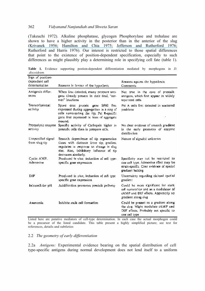
362 Vidyanand Nanjundiah and Shweta Saran (Takeuchi 1972). Alkaline phosphatase, glycogen Phosphorylase and trehalase are shown to have a higher activity in the posterior than in the anterior of the slug (Krivanek 1956; Hamilton and Chia 1975; Jefferson and Rutherford 1976; Rutherford and Harris 1976). Our interest is restricted to those spatial differences that point to the existence of position-dependent specification, especially to such differences as might plausibly play a determining role in specifying cell fate (table 1). Table 1. Evidence supporting postion-dependent differentiation mediated by morphogens in D. discoideum.
Listed here are putative mediators of cell-type determination. In each case the actual morphogen could be a precursor of the listed candidate. This table present a highly simplified picture; see text for references, details and subtleties 2.2 The geometry of early differentiation 2.2a Antigens: Experimental evidence bearing on the spatial distribution of cell type-specific antigens during normal development does not lend itself to a uniform

The determination of spatial pattern in Dictyostelium discoideum 363 interpretation. Using a monoclonal antibody, Noce and Takeuchi (1985) showed that a prestalk antigen initially appears at random locations in the aggregate and that the cells containing it then sort out to their final positions. However, the number of cells containing this antigen decreases during development (Takeuchi et al 1986), and it is not known whether the decrease is position-specific. Evidence for positional control of pattern formation was claimed by Krefft et al (1984). They analysed the appearance of two different prespore markers: a cell-surface antigen recognized by the MUD-1 monoclonal antibody (Krefft et al 1983) and a prespore vesicle antigen detected in prespore cells using antispore antiserum (Takeuchi 1963). The spatial distribution of both markers was consistent with the hypothesis that at the earliest stage at which prespore cells could be identified, they were already in contiguous positions in their final locations in the tipped aggregate. In other studies with prespore antibodies also, it was found that right from their initial appearance the antigens appeared in a contiguous and correctly positioned group of cells (Takeuchi et al 1986). This was taken to indicate that prespore cell differentiation occurred in response to positionally localized morphogenetic signals. (Obviously, one can never rule out the possibility of an earlier determinative event followed by sorting out and the development of the appropriate antigen in situ. If we end up saying that position-dependent determination really occurs, it will be because of the combined weight of evidence from many experiments, not because it is logically necessary.) It would appear on the basis of the above that the determination of prespore cells, but not that of prestalk cells, is position-dependent. Thus, in the first instance, an undetermined amoeba would have a high probability of becoming a prespore cell only if it found itself within the appropriate region of the multicellular aggregate. For an amoeba to become a prestalk cell, though, intracellular signals (whose possible nature we consider later) would be more important than position. It should be kept in mind that this apparent difference in mechanisms for prestalk and prespore cell distribution could be due to the particular antigens tested. Supporting this, the spatial pattern of gene expression in transformants shows that positional cues can influence the determination of prestalk cells (see below). 2.2b Gene expression experiments using reporter genes: Using cell-autonomous genetic markers, Williams et al (1989a, b) traced the origins of prespore cells and two types of prestalk cells (PstA and PstB) during slug formation. The distribution of cell types seen in the aggregated, mound stage is topologically quite different from that in the slug. PstB cells appear in the base of the aggregate mound and prespore cells in the central region above, while PstA cells are first detectable in scattered positions but later migrate towards the tip of the mound as it forms. Subsequently, in the slug, pDd56-lacZ transformants (containing a PstB-specific promoter) display a central funnel of β-galactosidase-expressing cells in the anterior zone; the funnel does not normally extend to the extreme tip. PstA cells surround PstB cells in the slug anterior. PstA and PstB cell differentiation occurs prior to the appearance of the tip, but once the tip is formed PstA cells move towards it. These findings imply that both cell sorting and positional cues contribute to patterning.
Esch and Firtel (1991) analysed cells expressing a fusion construct (Dd-ras) of the D. discoideum ras gene with the lacZ gene of E. coli. They showed that Dd-ras is expressed in a randomly scattered subpopulation of cells in the early aggregate. As development proceeds, the expression becomes most intense at the tip, in other

364 Vidyanand Nanjundiah and Shweta Saran Words in the prestalk region. Clearly, this suggests sorting. Haberstroh and Firtel (1990) cloned the gene encoding the spore coat protein SP60 and analysed its expression during development. The SP60-lacZ fusion protein is almost exclusively expressed in prespore cells. The staining pattern seen while amoebae are still moving into the aggregate indicates that the first cells expressing SP60-lacZ are already arranged in a ring, that is, form part of a contiguous group. Several hours later (at the slug stage), SP60-lacZ staining is evident in more than 80% of the cells. This could (but, in analogy to the case of the prespore antigens, need not) imply that prespore cells are recruited in a prespore zone. One can hypothesize how such a zone might be defined. For example, once the tip is formed it can act as the source of a diffusible chemical signal. The distances from the tip within which the strength of the signal is neither too high nor too low will define a zone, and amoebae entering the zone could stand a high chance of becoming prespores. Taken together, these results suggest that spatial information may be essential in specifying prespore cells but not so in the case of all prestalk cells, a conclusion consistent with the experiments using antibody staining cited earlier. 2.2c Proteases: Morphogenesis and differentiation in D. discoideum are responses to starvation, implying that only internal energy stores are available to fuel the processes; one of the means of utilizing internal energy is by protein degradation (Gregg et al 1954). Fong and Rutherford (1978) showed that the activity of the proteolytic enzyme cathepsin Β was significantly higher in prestalk cells than in prespore cells. Subsequently, Fong and Bonner (1979) showed that protein degradation is essential for normal differentiation. Several protease inhibitors block differentiation and in many cases this inhibition is reversible. Chloroquine, which inhibits slime mould cathepsin Β activity in vivo, was shown to interfere with development by blocking fruiting body formation; the inhibition was reversed by addition of amino acids. Also, glutathione and the microbial protease inhibitors antipain and leupeptin delay fruiting body formation. Gomer et al (1986) analysed a developmentally regulated prestalk specific gene encoding a protein whose sequence resembles that of the lysosomal cysteine proteinases cathepsin Η and Β. Prestalk cathepsin is distributed in the anterior one-tenth of migrating slugs as well as on the periphery of the posterior surface, that is, in the region of the rear guard cells. The fact that a protease is differentially distributed between prespore and prestalk cells suggests a role for it in differentiation. The possibility that proteolysis might play a role in the control of spatial pattern is interesting in the light of the claim that the end product of proteolytic activity, ammonia, is a candidate morphogen (see later). 2.3 The slug tip Position-dependent differentiation requires landmarks with respect to which cells can sense their position within the mass (Wolpert 1971). The obvious landmarks in the D discoideum slug are the anterior and posterior boundaries. Of these, little is known about the possible role of the posterior boundary. The anterior boundary, on the other hand, demarcates a structure of undoubted importance, the slug tip The tip, comprising a nipple-shaped group of cells, is formed at a late stage of aggregation, is present throughout subsequent stages, and has complex properties
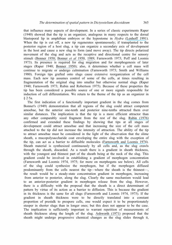
The determination of spatial pattern in Dictyostelium discoideum 365 that influence many aspects of development. In a series of classic experiments Raper (1940) showed that the tip is an organizer, analogous in many respects to the dorsal blastoporal lip in amphibian embryos or the hypostome in Hydra (Lenhoff 1991). When the tip is cut off, a new tip regenerates spontaneously; if transplanted to the posterior region of a host slug, a tip can organize a secondary axis of development in the host and cause a new slug to form (and move away). The tip directs polarized movement of the slug and acts as the receptive and directional centre for sensory stimuli (Bonner 1950; Bonner et al 1950, 1989; Farnsworth 1973; Poff and Loomis 1973). Its presence is required for slug migration and for morphogenesis of later stages (Raper 1940; Bonner 1950); also, it determines whether a cell mass will continue to migrate or undergo culmination (Farnsworth 1973; Smith and Williams 1980). Foreign tips grafted onto slugs cause extensive reorganization of the cell mass. Each new tip assumes control of some of the cells, at times resulting in fragmentation of the original slug into smaller but otherwise normal slugs (Raper 1940; Farnsworth 1973; Rubin and Robertson 1975). Because of these properties the tip has been considered a possible source of one or more signals responsible for induction of cell differentiation. We return to the theme of the tip as an organizer in § 2·5a.
The first indication of a functionally important gradient in the slug comes from Bonner's (1949) demonstration that all regions of the slug could attract competent amoebae, but the anterior one-tenth and posterior nine-tenths attracted them over similar distances. The implication is that the tip is a more efficient attractor than any other comparably sized fragment from the rest of the slug. Rubin (1976) confirmed and extended these findings by showing that tips at all stages of development could attract amoebae and that increasing the size of the cell mass attached to the tip did not increase the intensity of attraction. The ability of the tip to attract amoebae must be considered in the light of the observation that the slime sheath, a mucopolysaccharide coat enveloping the entire slug with the exception of the tip, can act as a barrier to diffusible molecules (Farnsworth and Loomis 1974). Sheath material is synthesized continuously by all cells and, as the slug crawls through the sheath, discarded. As a result there is a gradient in sheath thickness, with the youngest and thinnest part of the sheath being at the neck of the slug. This gradient could be involved in establishing a gradient of morphogen concentration (Farnsworth and Loomis 1974, 1975; for more on morphogens see below). All cells of the slug could synthesize the morphogen, but if the morphogen diffused predominantly from the cells nearest the tip—where the sheath is the thinnest—, the result would be a steady-state concentration gradient in morphogen, increasing from anterior to posterior, along the slug. Clearly the same mechanism would lead to an anterior-posterior gradient in morphogen release from the slug. However, there is a difficulty with the proposal that the sheath is a direct determinant of pattern by virtue of its action as a barrier to diffusion. This is because the gradient in its thickness is the same for all slugs (Farnsworth and Loomis 1974, 1975). If the gradient in sheath thickness were to be directly translated into a constant proportion of prestalk to prespore cells, one would expect it to be proportionately steeper in shorter slugs than in longer ones; but this does not appear to be the case. The implication is sufficiently important to warrant repetition of measurements of sheath thickness along the length of the slug. Ashworth (1971) proposed that the sheath might undergo progressive chemical changes as the slug slides through it,

366 Vidyanand Nanjundiah and Shweta Saran and that this aging of the sheath (with the degree of aging varying along the slug length) could provide a mechanism for unspecified positional cues.
By transplanting foreign tips onto tipless slugs, Durston (1974) and Lokeshwar and Nanjundiah (1983) found evidence for an anterior-posterior inhibitory gradient, A transplanted tip is maximally inhibited, meaning least successful in organizing the host slug, when the site of transplantation is closest to the the anterior end of the host. There is a striking correlation between the time needed to regenerate a new tip (following removal of the existing tip) and the position at which the regeneration is induced (Lokeshwar and Nanjundiah 1981). When a slug is cut transversely, the time taken by the posterior fragment to regenerate a new tip increases linearly as the position of the cut is moved from anterior to posterior. This time is independent of slug size, slug age or absolute distance from the anterior margin to the cut surface, and depends only on the relative length of the removed anterior fragment, When a similar experiment is carried out on slugs that have already been transected once, the time needed for secondary tip regeneration exhibits regulation, Specifically: a posterior fragment is first removed from a slug (the 'old' slug; the remaining anterior portion can be thought of as constituting a 'new' slug). In the 'new' slug, a second cut removes part of its anterior. The observation is that if the two cuts follow in quick succession, the time needed for a new tip to regenerate at the site of the second cut corresponds to its position relative to the length of the 'old' slug. However, if the second cut is made at various times after the first, the regeneration time gradually increases. This continues until it attains a value that is appropriate to the position of the cut with respect to the length of the 'new' slug (Lokeshwar and Nanjundiah 1983). One might say that the cells at the cut surface gradually erase the memory of their previous (relative) location and replace it by a knowledge of their new location. The process of accomodation to a change in slug size is, comparatively speaking, slow. It takes between 100 and 120 min for cells to sense that they have been reassigned from a location of 25% (reckoned as distance from the front relative to overall slug length) to a location at 50%, whereas tip regeneration times at corresponding locations vary from about 40 min (25%) to 80 min (50%). On the other hand, compared to the time needed for transdetermination of cell type in anterior slug fragments [anywhere from 3 h to 7 h; Sakai (1973)], accommodation is a fast process. These time scales are suggestive of a series of interrelated processes involving, firstly, tip regeneration; next, the setting up of various gradients deriving from the tip; and finally, transdetermination, with all three processes depending on intercellular communication. Results of similar experiments carried out on chimaeric slugs suggest that the mode of communication is cell-to-cell relay of an oscillatory signal (Lokeshwar and Nanjundiah 1985). It is tempting to hypothesize that similar processes regulate cell determination during normal development as well. However, until the putative signal is identified, this remains hypothetical. In slug fragments, intracellular reduction of nitro-blue tetrazolium yields a tip-specific stain and shows that the stain takes longer to appear in posterior isolates than in anterior ones (Mine and Takeuchi 1967); this finding is in accord with the data on tip regeneration times. 2.4 The pattern of cell-type interconversion in isolates We have already referred to Raper's (1940) finding that both anterior and posterior

The determination of spatial pattern in Dictyostelium discoideum 367 slug fragments can, when isolated, form normally proportioned fruiting bodies. The implications of this finding were proved by Bonner et al (1955) and Sakai (1973) when they showed, using a combination of histochcmical staining and antibodies, that cell type conversion took place within such fragments. Interestingly, conversion — or even de-determination—does not occur in isolated prestalk or prespore amoebae (Gregg 1971). Therefore, apart from other things, the phenomenon implies the existence of mutually reinforcing interactions within cells of the same type—what has been termed, in a different context, a community effect (Gurdon 1988). Observations on anterior slug isolates suggest that prestalk-to- prespore conversion takes place only in the posterior part of the isolate (Bonner et al 1955; Takeuchi et al 1982). Correspondingly, experiments using a biochemical stain for ..prespore cells showed that in posterior, predominantly prespore slug fragments, prespore-to-prestalk conversion was restricted to the anterior margin (Bonner et al 1955). On the other hand, when a vital stain specific for prestalk cells was used, conversion seemed to occur in diverse locations (Takeuchi et al 1982). Sternfeld and David (1982) have shown that when a slug tip is removed, anterior- like cells situated in the posterior portion migrate to the cut surface; they suggest that it is these cells that constitute the new tip. By implication, the origin of the tip must be ascribed to predetermination — at least in regenerating slugs. This would fit with the findings of Takeuchi et al (1982). In contrast, the observations cited above of Bonner et al (1955) regarding transdetermination in posterior slug fragments, and of Mine and Takeuchi (1967) pertaining to the kinetics of transdetermination, or of Lokeshwar and Nanjundiah (1983), that tip regeneration times increase as the size of the posterior fragment decreases, suggest a purely positional basis for tip regeneration. The experiments of Gregg and Karp (1978) strengthen the view in favour of a positional mechanism and underscore the importance of settling this point. They found that after vegetative amoebae were pulse-labelled with [3H]fucose, radioactivity in the slugs (that arose following starvation) was restricted to prespore cells. In other experiments, unlabelled migrating slugs were transected, and prestalk and prespore isolates were labelled separately. In prestalk isolates, [3H]fucose was incorporated within 10 min in the base of the isolate; similarly, in prespore isolates, the indication was that the cells at the margin of transection were the first to get converted to prestalk (needless to say, monitoring the disappearance of a histochemical stain, or the appearance of unlabelled cells in a labelled background, is difficult). Clearly, one needs to examine whether the migration of anterior-like cells is preceded by the appearance of prestalk cells at the cut surface, say via prespore-to-prestalk conversion, at the front of the posterior fragment.
By and large, prestalk and prespore cells do not interconvert within slugs that are formed in the normal course of development, but do so within slug fragments consisting mainly of one cell type. This means that in the-normal situation there must be factors capable of preventing such conversion. Using labelled prestalk cells and unlabelled prespore cells, Akiyama and Inouye (1987) have shown that both on an agar surface and in suspension, cell type conversion from prestalk to prespore occurs only when the prespore proportion is lower than normal. In their experiments the fraction of prestalk cells converted to prespore decreased as the initial prespore proportion was increased. Thus prespore cells tend to inhibit the tendency of prestalk cells to become prespore, and this can happen even in the

368 Vidyanand Nanjundiah and Shweta Saran absence of a normal spatial pattern of cell types. One interpretation of these findings would be that prespore cells release a substance that lowers the rate of prestalk-to-prespore conversion. Alternatively, prestalk cells might enhance prestalk-to-prespore conversion by producing an activator which in turn could be inactivated by prespore cells. Factors known to increase the proportion of stalk cells—for example differentiation inducing factor (DIF), weak acids such as propionate (Gross et al 1983) and products of cyclic AMP hydrolysis such as adenosine (Weijer et al 1984a) — could be candidates for the endogenous inhibitor of prestalk-to-prespore conversion (Inouye 1989), though a recent report by Inouye indicates that DIF is not involved (Inouye 1991). Irrespective of the nature of the factor (s), the pattern of transdetermination in slug isolates suggests that intercellular signall- ing enables the cells in a slug to become determined, or reinforces a state of determination, in a position-dependent manner also during normal development. 2.5 Morphogens A cell can sense its position relative to other cells if it can associate position with the concentration of a chemical species whose level varies in space (as we have mentioned earlier, positional sensing does not require the physical transport of any substance; see Goodwin and Cohen 1969). The chemical is supposed to elicit cellular responses in a concentration-dependent, and therefore position-dependent, manner. Because of its role in guiding morphogenesis, such a chemical is called a morphogen. Morphogens have been claimed to exist in D. discoideum, but as will be seen presently, in no case has it been convincingly demonstrated that a candidate chemical satisfies all requirements for it to be called a morphogen. These are (i) differentiation-inducing ability, (ii) endogenous production and (iii) correct spatial distribution. In the case of some chemical and ionic species, there is evidence that they might mediate intercellular communication. The evidence is of two sorts, Firstly, they are found outside cells and their levels exhibit sustained oscillations in shaken cell suspensions; this is true of cAMP, H+(pH), K+, Ca++, and possibly others (Nanjundiah and Wurster 1989). The fact that the oscillations continue for many cycles without any discernible damping points to the existence of a synchronizer (Nanjundiah 1986); and if there is a synchronizer, cells can make use of it in order to communicate with one another.
Another line of evidence pointing to a morphogenetic role is more direct: certain endogenously produced chemicals can induce cell type-specific determination. On the basis of the latter criterion, one can think of cAMP, adenosine, ammonia or Η+ (pH) and DIF as possible morphogens in D. discoideum. There is clear evidence that D. discoideum cells are sensitive to reiterated, in fact to periodic, stimuli of cAMP (Darmon et al 1975; Gerisch et al 1975; Nanjundiah 1989). Unfortunately, in spite of this, few workers have taken into account the possibility that it may be necessary to apply other candidate morphogens too in the form of oscillatory impulses if one wishes, to understand their natural effects (for an exception see Wurster and Kay 1990). Perhaps the fact that the morphology of the slug could not be simpler from a theoretical point of view, with apparently just one spatial dimension being relevant, has also played a role in encouraging the construction of position-dependent, morphogen gradient-based models for patterning in D. discoideum. A simple minded

The determination of spatial pattern in Dictyostelium discoideum 369 view, long current, has been that a single anterior-to-posterior morphogenetic gradient might be enough to set up a distinction between prestalk and prespore genes in the slug. In this connection, we mention that if more than one morphogen is present, spatial distribution can be more complex. For example, Odell and Bonner (1986) make a case for two mutually orthogonal gradients being responsible for slug movements: one anterior to posterior and the other from the core of the slug, idealized as a cylinder, to the periphery. 2.5a cAMP and adenosine: Bonner (1970) showed that millimolar concentrations of cAMP could induce starved D. discoideum cells to become mature stalk cells without normal morphogenesis. Since cAMP continues to be present during post-aggregative stages of development (Malkinson and Ashworth 1972; Pan et al 1974), it was taken to be the natural inducer of stalk cell differentiation. It has subsequently been confirmed that cAMP is also required for prespore differentiation (Kay 1982; Okamoto 1986; Wang et al 1988). Berks and Kay (1988) suggested that cAMP at physiological doses was an inhibitor of stalk cell differentiation, but subsequent work has shown that the earlier observation was specific to a particular stalk protein (Berks and Kay 1990; see below). Riley et al (1989) showed that terminal differentiation of spores is favoured by high levels of intracellular cAMP. Cells can be made to agglutinate by shaking them in liquid culture, and when this is done prestalk and prespore cells are formed; under these conditions a periodic application of cAMP pulses advances the time of appearance of prespores by 3–4 h (Forman and Garrod 1977b). Stable expression of prestalk/prespore differences needs cell contact and the continued presence of cAMP (and possibly other factors; Lodish et al 1982).
What about the spatial distribution of cAMP? The earliest, albeit circumstantial, evidence pertaining to this has already been mentioned: Bonner's (1949) finding that anterior slug fragments attract chemotactic amoebae more strongly than posterior fragments. Supporting this finding, Brenner (1977), on making measurements after fragmenting the slug transversely, reported a weak' gradient in total cAMP levels with the high point at the anterior end. On the other hand, in a similar study Lokeshwar (1983) did not find any significant variation in total cAMP levels within the slug. Using a sensitive microdissection technique, Rutherford and colleagues have made direct measurements of cAMP levels and of enzymes involved in cAMP production and breakdown; on the basis of these experiments, they tried to infer the distribution of cAMP in vivo. They found that there is a peak of cAMP during culmination, the last phase of morphogenesis, which coincides with the stage of terminal differentiation. Further, in individuals at this stage, cAMP is localized in a gradient within the spore mass, with the highest level at the base; there is no significant difference in levels between differentiated stalk and prespore cells (Merkle et al 1984). Adenylate cyclase activity is localized in the prespore region, is absent in the stalk, and displays a gradient similar to that of cAMP (Merkle and Rutherford 1984). The same study showed that there were no significant spatial differences in adenylate cyclase activities within the slug. Brown and Rutherford (1980) had found earlier that at culmination cAMP phosphodiesterase activity was strongly localised in stalk cells, with the highest activity in the base of the stalk. These results have been interpreted as implying a steady-state gradient of cAMP within the culminating mass, with a source located at the base of the spores and a sink at

370 Vidyanand Nanjundiah and Shweta Saran the base of the stalk (Merkle and Rutherford 1984). In relation to these findings it must be stated that the relevance (for determination of spatial pattern) of cAMP measurements made after terminal differentiation has occurred is not clear. Otte et al (1986) measured production and turnover of extracellular cAMP signals by prestalk and prespore cells within the migrating slug. They found that the two cell types had similar basal levels of adenylate cyclase activity and were about equally capable of relaying cAMP. However, intact prestalk cells had a three-fold higher binding capacity for cAMP and a three-fold higher level of cell-surface phosphodiesterase activity than prespore cells. Interpreted in the simplest possible terms, this would suggest a spatial gradient of cAMP in the slug, with levels in the anterior (prestalk) region lower than in the posterior (prespore) region. Note that this refers to the gradient within the slug and does not contradict the findings of Bonner (1949) and Rubin (1976) pertaining to differential chemotactic attraction, As indicated earlier, the gradient in slime sheath thickness reconciles the two sets of results. There is another interesting aspect to the more rapid turnover of cAMP by prestalk cells. Because of this, if cAMP release continues to be oscillatory in the slug, one might expect — depending on the detailed explanation for cAMP oscillations — a higher oscillation frequency in prestalk cells than in prespore cells. This was found to be the case by Weijer et al (1984a). They went on to suggest that the ability of the tip to generate cAMP signals at a higher frequency than other cells, and thereby to function as a pacemaker, was responsible for its being able to organize the slug mass. Durston and Vork (1979) had shown earlier that prestalk cells were preferentially attracted by oscillatory cAMP signals originating from the slug tip. It is interesting to speculate whether spontaneous oscillation frequencies, chemotactic responses and prestalk-prespore tendencies might be related in a cause-and-effect fashion. Here we draw attention to the possibility that something other than cAMP may also be involved in oscillatory signal communication within the slug (Nanjundiah and Wurster 1989).
Many experiments point to a morphogenetic role for adenosine, but the nature of the role remains uncertain. Adenosine inhibits prestalk to prespore conversion in roller-tube cultures (probably by reducing cell responsiveness to cAMP; Weijer and Durston 1985), and an enzymatically induced decrease in its level in the slug causes prespore cells to appear in the prestalk zone (Schaap and Wang 1986; Wang et al 1988). The observation that adenosine can potentiate the cAMP-induced expression of prestalk-specific mRNAs and, at a hundred-fold higher concentration, can inhibit prespore gene expression (in strain NC4; Spek et al 1988), is consistent with this finding. On the other hand, working with strain V12M2, Berks and Kay (1988) found that adenosine inhibited gene expression only at very high concentrations (2 mM) and that too in both cell types. There is indirect evidence for a spatial gradient of adenosine in late multicellular stages. Armant et al (1980) have shown that 5' AMP nucleotidase activity is essentially restricted to prestalk cells adjacent to the prespore region. This, and the fact that the enzyme appears to function extracellularly (cytochemical staining was found only on the plasma membrane), is consistent with its role as an inhibitor of prestalk-to-prespore conversion. However, adenosine is unlikely to be involved in the inhibition by prespore cells of cell type conversion in suspension cultures because it is produced more by prestalk cells than by prespores (Wang et-al 1988) and has no effect on the conversion of prestalks in vitro (Inouye 1988b).

The determination of spatial pattern in Dictyostelium discoideum 371 2,5b DIF: DIF, initially identified as an inducer of stalk differentiation (Town et al 1976) has since then been considered as a potential prestalk-specific morphogen (though there are also indications that it has a role to play during aggregation; Wurster and Kay 1990). It consists of five related molecular species, with more than 90% of the activity associated with just one of the five, DIF-1 (Kay et al 1983; Brookman et al 1987). In what follows, by 'DIF' we shall mean DIF-1 unless stated otherwise. Whereas differentiation along the spore pathway requires cAMP only (Kay et al 1979: Kay 1982), prestalk differentiation requires DIF in addition to cAMP (Town et al 1976; Williams et al 1987). By also acting as an inhibitor of prespore determination, it appears that DIF regulates the choice between the stalk and spore pathways (Kay and Jermyn 1983). DIF is unable to induce conversion of prestalk cells to stalk cells, even at high concentrations, nor does it induce migrating slugs to fruit (Inouye 1988b). Berks and Kay (1990) have made an interesting case for a combinatorial control of cell determination by cAMP and DIF acting in concert. They measured the transcriptional response of cells in shaken suspensions at different developmental stages and to various signal molecules. The finding was that while the appearance of PstA-specific mRNA was stimulated by cAMP and DIF, that of PstB-specific mRNA was stimulated by DIF but inhibited by cAMP. In the case of a prespore-specific mRNA, cAMP was a stimulant and DIF an inhibitor. In this connection, the finding that DIF can inhibit cAMP signalling in early developmental stages (Wang et al 1986; Wurster and Kay 1990) is of interest.
Coming to spatial distribution, expression of the pDd56 gene, which defines cells as PstB, is dependent upon the presence of DIF (Jermyn et al 1987). Since PstB cells initially appear to differentiate at the base of the tight aggregate, the base would seem to be a region of high DIF concentration from the earliest stages of morphogenesis. Measurements of total DIF from slug fragments showed that the specific activity exhibited a gradient reverse to that expected on the basis of its morphogenetic effect, being higher in the prespore zone than in the prestalk zone (Brookman et al 1987). One way of accounting for the reverse gradient would be to say that DIF is produced by prespore cells precisely to maintain the appropriate fraction of cells in the prestalk state. However, the amount of DIF needed to suppress the conversion of prestalk cells to prespore appears to be much larger than the amount produced (Brookman et al 1982). This may indicate that in addition to DIF prespore cells release other substances that also help to prevent the transdetermination of prestalk cells (Inouye 1991). Another explanation that has been offered for the reverse gradient of active DIF is that DIF is a consumed substrate in a reaction-diffusion mechanism for morphogenetic signalling (Brookman et al 1987). The idea seems to be that all cells produce a freely diffusible substrate (DIF), which is converted in an autocatalytic fashion into an inducer of the prestalk state by a localized subset of cells, in this case the anterior cells. Continuing with the idea, DIF would become depleted in the anterior, and the hypothetical activator derived from DIF would be restricted to the prestalk zone. DIF-1 has been shown to induce its own breakdown (Insall et al 1992) via a dechlorinase, and it has been claimed that those (randomly distributed) cells in which the enzyme is highly active become prestalk. Following this, a selective migration of prestalk cells towards the tip could provide a spatial DIF-1 gradient of the sort observed by Brookman et al (1987). We wish to draw attention
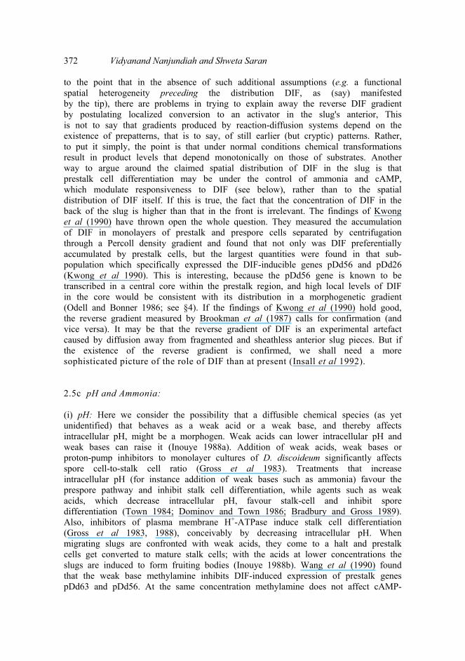
372 Vidyanand Nanjundiah and Shweta Saran to the point that in the absence of such additional assumptions (e.g. a functional spatial heterogeneity preceding the distribution DIF, as (say) manifested by the tip), there are problems in trying to explain away the reverse DIF gradient by postulating localized conversion to an activator in the slug's anterior, This is not to say that gradients produced by reaction-diffusion systems depend on the existence of prepatterns, that is to say, of still earlier (but cryptic) patterns. Rather, to put it simply, the point is that under normal conditions chemical transformations result in product levels that depend monotonically on those of substrates. Another way to argue around the claimed spatial distribution of DIF in the slug is that prestalk cell differentiation may be under the control of ammonia and cAMP, which modulate responsiveness to DIF (see below), rather than to the spatial distribution of DIF itself. If this is true, the fact that the concentration of DIF in the back of the slug is higher than that in the front is irrelevant. The findings of Kwong et al (1990) have thrown open the whole question. They measured the accumulation of DIF in monolayers of prestalk and prespore cells separated by centrifugation through a Percoll density gradient and found that not only was DIF preferentially accumulated by prestalk cells, but the largest quantities were found in that sub- population which specifically expressed the DIF-inducible genes pDd56 and pDd26 (Kwong et al 1990). This is interesting, because the pDd56 gene is known to be transcribed in a central core within the prestalk region, and high local levels of DIF in the core would be consistent with its distribution in a morphogenetic gradient (Odell and Bonner 1986; see §4). If the findings of Kwong et al (1990) hold good, the reverse gradient measured by Brookman et al (1987) calls for confirmation (and vice versa). It may be that the reverse gradient of DIF is an experimental artefact caused by diffusion away from fragmented and sheathless anterior slug pieces. But if the existence of the reverse gradient is confirmed, we shall need a more sophisticated picture of the role of DIF than at present (Insall et al 1992). 2.5c pH and Ammonia: (i) pH: Here we consider the possibility that a diffusible chemical species (as yet unidentified) that behaves as a weak acid or a weak base, and thereby affects intracellular pH, might be a morphogen. Weak acids can lower intracellular pH and weak bases can raise it (Inouye 1988a). Addition of weak acids, weak bases or proton-pump inhibitors to monolayer cultures of D. discoideum significantly affects spore cell-to-stalk cell ratio (Gross et al 1983). Treatments that increase intracellular pH (for instance addition of weak bases such as ammonia) favour the prespore pathway and inhibit stalk cell differentiation, while agents such as weak acids, which decrease intracellular pH, favour stalk-cell and inhibit spore differentiation (Town 1984; Dominov and Town 1986; Bradbury and Gross 1989). Also, inhibitors of plasma membrane Η+-ATPase induce stalk cell differentiation (Gross et al 1983, 1988), conceivably by decreasing intracellular pH. When migrating slugs are confronted with weak acids, they come to a halt and prestalk cells get converted to mature stalk cells; with the acids at lower concentrations the slugs are induced to form fruiting bodies (Inouye 1988b). Wang et al (1990) found that the weak base methylamine inhibits DIF-induced expression of prestalk genes pDd63 and pDd56. At the same concentration methylamine does not affect cAMP-

The determination of spatial pattern in Dictyostelium discoideum 373 induced expression of the prespore gene D19 (Van Lookeren Campagne et al 1989), so the inhibitory effect on stalk gene expression is not due to a general inhibition of transcription. In sum, a lowering of intracellular pH seems to favour the prestalk state. However, in his observations on slugs cultured under fixed oxygen concentrations, Sternfeld (1988) concluded from the blanching of the neutral red stain during the course of development that there were alterations in intracellular pH but tissue proportions were unaffected.
In contrast to the studies on effect of adding weak acids or bases, measurement of intracellular pH during normal development has yielded conflicting results. Three different groups of investigators (Jentoft and Town 1985; Kay et al 1986; Satre et al 1986; Town et al 1987) have reported that there is no change of cytoplasmic pH during development. Ratner (1986) detected no difference in intracellular pH between prestalk and prespore cell types but Aerts (1988) claimed that changes in intracellular pH are involved in prespore cell regulation in D. discoideum. Aerts (1988) made intracellular pH estimates by two null point methods and observed a a higher average pH in prespore cells (the average cell pH in populations with approximately 80% prespore cells was approximately 0·2 units higher than that in populations with about 50% prespore cells). In the same study, he also showed that cell-type regulation in suspension (roller tube) cultures could be abolished by preventing the pH change that normally accompanied such regulation. Furukawa et al’s (1990) measurements revealed a transient intracellular acidification in amoebae during the first few hours of development, prior to the preaggregative phase; eventually the pH returned to neutral. Under conditions in which it induces prespore gene expression, cAMP produces a gradual increase in intracellular pH (Van Lookeren Campagne et al 1989), but bypassing cAMP showed that the pH increase by itself cannot be responsible for prespore induction. Similarly, the prestalk-inducing effect of DIF is not mediated through intracellular pH changes (Inouye 1988a). Inouye also found that prespore cells have higher intracellular pH and higher resistance to acid load than prestalk cells (Inouye 1985, 1988a), and that decrease of intracellular pH leads to maturation of prestalk cells in vivo (Inouye 1988b). Furukawa et al (1990) were unable to detect any gradient in intra- cellular pH along the slug. On the other hand, Lokeshwar (1983) found that after staining amoebae with neutral red the entire slug fluoresced even though, as expected, only the anterior portion was coloured red. This suggests that a pH gradient does exist. In a critical assessment of the literature on pH measurements in D. discoideum, Inouye points to the use of variant techniques as one reason why different workers do not agree (Inouye 1988a). It appears that at present, while the fact that weak acids and bases can affect cell-type determination by affecting intracellular pH is acceptable, the role of pH as a controlling agent during normal development is still unclear. (ii) Ammonia: Ammonia is produced by the starved cells of the slug (Gregg et al 1954; White and Sussman 1961; Wright et al 1977; Walsh and Wright 1978). It inhibits fruiting-body formation, and its removal induces migrating slugs to fruit (Schindler and Sussman 1977a). Addition of ammonia to monolayer cultures inhibits stalk cell formation but not prespore and spore differentiation (Gross et al 1983; Neave et al 1983); in suspension cultures, ammonia in the presence of cAMP can stabilize aspects of post-aggregative differentiation that are believed normally to depend on the integrity of the aggregate (Oyama et al 1988). It

374 Vidyanand Nanjundiah and Shweta Saran has been hypothesized that local ammonia depletion occurs during normal development at the apex and base of the culminating structure during early fruiting body formation (Sussman and Schindler 1978). Stimulatory effects of either ammonia depletion or carbon dioxide-induced acid load on stalk cell differentiation in migratory slugs were shown by Inouye (1988b), but it is not clear whether in such experiments there is an induction of stalk cell differentiation as such or whether the effect is indirect and due to the slug-to-fruiting body switch.
Ammonia may also be involved in cell type regulation in the slug. A class of mutants that are hypersensitive to the inhibitory effects of ammonia during culmination (Newell and Ross 1982) show a reduced ratio of prestalk to prespore cells in the migratory slug (MacWilliams and David 1984). Ammonia inhibits extracellular accumulation of cAMP without increasing the rate of its hydrolysis (Riley and Barclay 1990), suggesting that it inhibits cAMP secretion. By reducing cAMP release (Schindler and Sussman 1977b; Williams et al 1984), ammonia can act as a reversible modulator of cAMP relay. Thus ammonia, as a candidate morphogen, could favour spore formation by promoting intracellular accumulation of cAMP or maintenance of high intracellular cAMP levels. While studying cAMP-induced differentiation in monolayers of sporogenous mutants, Gross et al (1983) showed that ammonia could promote spore formation and Bradbury and Gross (1989) demonstrated a preferential accumulation of prespore enzymes upon exposure to ammonia. Also, for DIF to induce stalk cell differentiation under normal conditions, endogenously produced ammonia must be removed (Wang and Schaap 1989). In other words, the morphogenetic role of ammonia could be realized in combination with other morphogens such as cAMP and DIF. What we have said so far suggests a role for ammonia as a promoter of the prespore pathway, but direct monitoring of gene expression by Berks and Kay (1988) indicated only non- specific inhibition, and that at very high concentrations (20 mM NH4C1). Using shaken cell suspensions, Oyama and Blumberg (1986) found that ammonia was required for accumulation of both prespore and prestalk mRNAs; similarly, Oyama et al (1988) found that ammonia could induce both prespore-specific and non specific enzymes. The reasons behind the discrepancy may have to do with differences in genotype and experimental conditions. For the moment, we note that ammonia remains an attractive candidate for a prespore-specific morphogen. It could play a role either as a weak base, or as a mediator of cAMP and DIF effects. As a volatile substance, its ability to diffuse rapidly, especially from the slug anterior means that it can be present as a gradient in the slug (Inouye 1990). 3. Predetermination and sorting The alternative to a position-dependent mechanism is that the fate of a cell is predetermined in the aggregate (or earlier); the cells then sort out to produce the anterior-posterior pattern observed in the slug. Sorting of like types is known to occur when cells within the slug are displaced relative to each other (Bonner 1952; Takeuchi 1969; Matsukuma and Durston 1979; Sternfeld and David 1981, 1982), but this may be indicative of a 'memory' effect consequent to position-dependent determination. We ask whether a similar process is involved in establishing the normal pattern. Clearly the issue is one of timing: does cell determination precede

The determination of spatial pattern in Dictyostelium discoideum 375 the segregated distribution of cell types in the slug, or does it occur only after positions along the anterior-posterior axis become established?
D. discoideum cells show several predispositions of their fate before acquiring their eventual locations in the slug. For example, in an electron microscopic study Schaap et al (1982) showed that before and during aggregation a randomly distributed group of cells became electron dense. These cells became prespores while the electronlucent cells became prestalks. Cell size and cell density could mark an early prestalk/prespore difference. Both preaggregation and aggregation stage cells can be separated into two classes in density gradients (Takeuchi 1969; Bonner et al 1971; Maeda and Maeda 1974). After differential labelling and mixing, these classes sort out depending on their densities. Takeuchi (1969) and Bonner et al (1971) showed that when the cells were recombined after centrifugation in a dextrin solution, the heavier cells preferentially occupied the anterior (prestalk) region of slugs. Maeda and Maeda (1974) verified the heterogeneity of preaggregation-stage cells with respect to relative density and its role in sorting but by their method (isopycnic centrifugation in a urografin gradient) the lighter cells were the ones that ended up in the anterior of the slug. A similar observation was made using diverse methods by Schaap et al (1982), Ratner and Borth (1983) and Weijer et al (1984b). The contradictory results may be due to different effects of the two media on the amoebae. Leach et al (1973) observed that amoebae grown in an axenic medium preferentially adopted the spore pathway when mixed with amoebae grown on bacteria. As axenically grown cells are significantly bigger than those grown on bacteria, this would again indicate that the larger cells become prespores. On the other hand, measurements by Bonner and Frascella (1953) and Bonner (1959), and our observations during the course of fluorescence-activated cell sorting studies (Shweta Saran and V Nanjundiah, unpublished), suggest that prestalk cells are larger than prespore cells. The matter awaits resolution. From studies on the sizes of cells and nuclei Bonner and Frascella (1953) and Bonner et al (1955) concluded that the size distribution among cells grown in a common (bacterial) environment is continuous and not bimodal, so that if size per se plays a role in normal development, either there must be a threshold involved or cells must be able to sense relative size. Prestalk preference in preaggregation cells is associated with other properties as well. For example, future prestalk cells exhibit a relatively high
cell-surface hydrophobicity (Sharpe and Watts 1985) and a relatively high level of a
prestalk antigen (Noce and Takeuchi 1985). The use of shaking or rolling cultures to produce aggregates of starved amoebae
under submerged conditions provides an alternative approach to the problem of sorting (Takeuchi 1969; Forman and Garrod 1977a, b; Garrod and Forman 1977; Sternfeld and Bonner 1977; Sternfeld and David 1979, 1981; Tasaka and Takeuchi 1979, 1981). D. discoideum cells display prestalk/prespore staining differences when allowed to form clumps in suspensions shaken at low speed (when shaken at high speeds, the cells do not form clumps and differentiation does not occur). If transferred to non-nutrient agar, the clumps develop into normal fruiting bodies. Thus, under suspension-culture conditions, prestalk and prespore cells are formed even though there is no morphologically distinct tip or any other evidence of polarity. Showing a behaviour consistent with cell-autonomous determination, the prespore cells are distributed at random (Takeuchi et al 1988). These observations suggest that (i) differences can emerge within identically raised amoebae in the

376 Vidyanand Nanjundiah and Shweta Saran absence of the normal spatial morphology and (ii) these differences are correlated with future fate. At the very least, this makes it clear that the normal geometry of aggregation, and so the normal spatial distribution of possible morphogens, is not a prerequisite for the origin of all cell-type differences.
What causes dissimilar cell types to segregate, and when do they do so? Investigation of the timing of cell sorting has shown that normally this occurs during late aggregation and formation of the slug, roughly between 12 and 15 h of development (Bonner and Adams 1958; Durston and Vork 1979; Tasaka and Takeuchi 1979; Wang et al 1988; Tirlapur et al 1991). Three mechanisms have been invoked to explain cell sorting: Chemotaxis, orientation towards oxygen gradients, and differential cell adhesion. We have already alluded to the possible role of oxygen. Chemotaxis of prestalk cells towards cAMP produced by the tip has been observed or inferred in a number of cases (Nestle and Sussman 1972; Maeda and Maeda 1974; Matsukama and Durston 1979; Sternfeld and David 1981, 1982; Inouye and Takeuchi 1982a, b; Kopachik 1982). Sorting could be caused by differential Chemotaxis of the two cell types (Durston and Vork 1977; Inouye and Takeuchi 1982a, b). As in some tissue-culture systems, differences with respect to cell adhesion are probably also involved in differential sorting behaviour (Yabuno 1971; Siu et al 1983; see Sternfeld 1979 in support of the idea that this can also explain the sorting-out of different species in mixed aggregates). It has been suggested that differences in adhesiveness between prestalk and prespore cells can facilitate cell sorting (Lam et al 1981), possibly this can happen in combination with differential Chemotaxis or through different mobilities in the two cell types.
The question remains: by what means could functional differences appear in a population of genetically identical cells subject to identical environmental influences? In looking for an answer it is important to realize that experiments using phenotypically (or otherwise) distinguishable cells usually tell us what conditions are suficient for sorting to occur, not what conditions are necessary. Keeping this in mind, we proceed to highlight three factors implicated in the origin of the prestalk/prespore distinction in a cell-autonomous manner (table 2).
Table 2. Possible bases for predetermination in D. discoideum.
Note that there is no evidence that these are primary causes; the data only indicate a correlation between these and cell type in the slug or fruiting body. The assumption is that the properties listed here, together with sorting out, lead to the correct spatial pattern. See text for details and references.

The determination of spatial pattern in Dictyostelium discoideum 377 3.1 Nutritional state At starvation, when axenically grown cells fed on 86 mM glucose (G + ) are mixed with cells raised in the absence of glucose (G–), the G– cells tend to become prestalk cells while the G+ cells preferentially become prespore cells (Leach et al 1973). Cells grown on bacteria exhibit prestalk tendencies when mixed with G + cells or G– cells. Noce and Takeuchi (1985) extended this finding by showing that other metabolizable sugars could substitute for glucose. Before aggregation the cells were mixed at random, so that relative position within the aggregation field could not be responsible for the pattern of sorting. These results can be interpreted in two ways. The determination of both cell types can be independently influenced by the level of sugar; or, one type can be directly affected and in turn it can influence the other. The observation that G– cells by themselves give rise to 'stalky' fruiting bodies, and G+ cells to 'spory' ones (Garrod and Ashworth 1973), indicates that the level of sugar can bias the probability that an individual amoeba eventually becomes stalk or spore. 3.2 Cell cycle On the basis of cell counts and [3H ] thymidine incorporation, it has been estimated that growing cells spend approximately 10% of their time in Gl, 40% in S and 50% in G2 phase. Mitosis accounts for approximately 2% of the total cycle; Gl is the most variable phase, sometimes being absent entirely (Katz and Bourguignon 1974; McDonald and Durston 1984). In the light of what is to follow, we note that all amoebae come to be at same stage of the cell cycle eventually (Katz and Bourguignon 1974).
In cell mixtures, 'early' (E) cells, which are at the S phase or early G2 phase when starved, preferentially occupy the slug anterior. On the other hand, 'late' (L) cells, meaning those in mid-G2 through mitosis at the time of starvation, sort out to the posterior, prespore region (McDonald and Durston 1984; Weijer et al 1984b). Confirming this, Gomer and Firtel (1987) found that in low-density monolayers with few or no cell contacts, the propensity of a single amoeba to differentiate into either prespore or prestalk is autonomous to the amoeba: cells that divide between approximately 90 min before and 40 min after starvation, and so presumably belonging to the Ε class, tend to become prestalks, whereas cells dividing at other times tend to become prespores. The relation between cell-cycle phase at starvation and position in the slug has been explored in a number of experiments (it is possible that during normal development the cell-cycle phase primarily influences positioning in the slug and thereby, only secondarily, the direction of differentiation). Plasma membrane proteins may differ through the cell cycle, causing differences in sorting behaviour. Gomer and Firtel (1987) suggested that during aggregation, the cells predisposed on the basis of their cell cycle phase at starvation to become prestalk cells might also be the earliest to initiate the signalling system based on cAMP relay. This suggestion fits with the finding of McDonald (1986) that Ε cells were more likely to initiate centres than L cells. Wang et al (1988) extended this finding by investigating the expression of the cAMP chemotactic system during development of synchronized Ε or L amoebae and

378 Vidyanand Nanjundiah and Shweta Saran showing that Ε cells bound cAMP, had cell surface cAMP phosphodiesterase activity, and could relay cAMP signals, significantly earlier and to higher levels than L cells. They inferred that Ε cells are prestalk sorters because they are the first to initiate aggregation centres and respond most effectively with Chemotaxis and signal relay. A cell cycle-imposed bias for differentiation pathways is also indicated by the experiments of Weijer et al (1984b), who found that Ε cells formed slugs with 53% prestalk cells while slugs formed by L cells contained only 12% prestalk cells. In a recent study (Maeda et al 1989) no correlation was apparent between cell cycle phase and possible 'stalkiness' or 'sporiness' of fruiting bodies developed from synchronized cells. Nevertheless, in the same study Maeda et al did find a cell cycle phase-dependent sorting tendency in mixtures of amoebae in different phases. However, they observed that late G2-phase amoebae preferentially became prestalks in mixtures with mid G2-phase ones. In the light of the studies cited above, this finding is puzzling. Other factors that have been observed to affect prespore-prestalk ratios, such as nutritional status, might either modulate the initial cell cycle-dependent choice or the subsequent maintenance of that choice within the aggregate. 3.3 Calcium Studies on a variety of systems point to a role for intracellular Ca++ in transduction of extracellular signals, cell-to-cell communication and differentiation. Our interest here pertains to the possible correlation of Ca++ with early prestalk/prespore determination. Using a combination of atomic absorption spectroscopy and alizarin red staining, Maeda and Maeda (1973) concluded that total calcium was higher in the prestalk region than in the prespore region. From fluorimetric measurements on dissociated amoebae, Abe and Maeda (1989) claimed that this also held true for cytoplasmic calcium, and our preliminary experiments (unpublished) support this claim. However, Schlatterer (1990) has reported methodological difficulties with the technique (fluorescence labelling using the Ca+ +-sensitive dyes Fura-2 AM and Indo-1), so final confirmation is still awaited. Tirlapur et al (1991) used Chlortetracycline (CTC) fluorescence to show that there is a difference in sequestered Ca++ within the slug, with prestalk cells having significantly higher levels than prespores. In the same study, by labelling either the centre or the periphery of an early aggregate with CTC, it was possible to infer that cells containing high levels of sequestered Ca++ were distributed at random in the aggregate. Beginning with appearance of the aggregate tip, and even more so after completion of aggregation, cells of the same type (with respect to sequestered Ca + +) sorted out. We have recently extended these observations and obtained evidence for the existence of two functional classes of amoebae at the vegetative stage itself (ms in preparation). Freshly starved, CTC-labelled amoebae display a bimodal distribution of fluorescence emission. Cells from the extremes of the two modes can be mixed, whereupon they aggregate and develop a prestalk/prespore CTC fluorescence pattern identical with that seen in normally developing slugs labelled with CTC. In other words the cells that exhibit high levels of fluorescence become prestalk and those that exhibit low levels, prespores. The implication is that already at the vegetative stage, a high level of sequestered Ca++ is an indicator of

The determination of spatial pattern in Dictyostelium discoideum 379 future cell death (meaning differentiation into stalk or basal disc). In support of this view, there is some indication that calcium can induce stalk cell differentiation (Maeda 1970) and inhibit prespore differentiation (Abe and Maeda 1991). If confirmed, this would suggest that Ca++ levels can bias the probability of differentiation of vegetative, solitary cells along one pathway versus the other. 4. Summing up Three classes of proximal causes are commonly invoked in order to account for the development of spatial patterning in higher organisms. Traditionally, these comprise (i) maternal influences, as transmitted through the cytoplasm of the unfertilized egg, and (ii) intercellular interactions amongst the cells of the embryo (Slack 1991). Today to these one adds (iii) imprinting, meaning a reversible genetic modification induced by passage through the germ line of one sex or the other and expressed in the zygote (Jablonka and Lamb 1989). In a broader context, one can classify the causes into those depending on historical factors (cytoplasmic influences, imprinting) and those depending on the sociology of the multicellular embryo (intercellular interactions). Experience shows that both sets of factors are needed for a complete explanation of how any embryo develops. As we have seen, in the case of D. discoideum too it appears that the problem of the spatial distribution of prestalk and prespore cells can be resolved only if one takes into account both the history of the individual amoebae comprising an aggregate and the sociology of the aggregate. In terms of the alternatives we have considered, this means that if our intention is to understand pattern formation in detail, we cannot make an exclusive choice between position-independent determination followed by sorting, on the one hand, and position-dependent determination on the other. In focusing our attention on these alternatives, we do not wish to downplay the importance of more complicated, and perhaps more realistic, possibilities: for example, sorting out and (positive and negative) feedbacks together leading to the appearance of a stable spatial pattern. The problem is that such possibilities do not eliminate an element of predetermination (based on which subsequent processes follow), so that we come back to position-independent determination as the original cause. The observations of Sternfeld (1992) suggest that sorting of determined cell types within the slug may not be a once-for-all affair, and more to the point, that some prespore cells convert to prestalk. However, as we have mentioned, by and large it is a fact that cellular position in the slug is strongly correlated with genetic and phenotypic markers at stages preceding slug formation and with the terminal phenotype of the cell. This would seem to indicate that cell type conversion in the slug plays at best a minor role in the determination of pattern. In any case, pedagogically it is useful to consider the two extreme hypotheses of positional dependence and sorting, if only to satisfy ourselves that neither hypothesis is complete in the sense of being capable of explaining all experimental findings.
We have seen that both prestalk and prespore fragments of slugs can regulate and restore the normal pattern (Raper 1940); in other words predetermination, if it exists, is not irreversible. Also, self-generated extracellular factors (for example cAMP, DIF and ammonia) can lead to certain cells adopting the prestalk pathway and others becoming prespores. However, differentiation, and even correct

380 Vidyanand Nanjundiah and Shweta Saran proportioning, can occur under conditions that prevent the operation of normal spatial cues (for example in submerged agglomerates; Forman and Garrod 1977b; Sternfeld and David 1981), but then the spatial pattern is not similar to that seen in the slug (though there are resemblances; Takeuchi 1991). As we have discussed, aggregation-competent cells can be expected to differ with respect to more than one trait, and, in the case of the phase of the cell cycle at which an amoeba is starved or the level of sequestered Ca+ + in freshly starved amoebae, differences between cells are reflected as positional differences in the slug (McDonald and Durston 1984; Shweta Saran and V Nanjundiah, in preparation). If neither hypothesis can stand on its own, we need to consider whether both predispositions (at the cellular level) and positional cues (deriving from multicellular communication) play a role in the determination of spatial pattern, with their relative importance depending on the degree of functional inhomogeneity that exists in preaggregation cells. To the extent that such inhomogeneity is present, it can cause cells to sort out; positional effects can then come into play and reinforce the bias provided by preaggregation-stage differences. The necessity for inhomogeneities to be functional is tricky but important, because the question is not whether preaggregation amoebae are heterogeneous but rather whether some aspect of that heterogeneity serves as a cue for cell determination in normal development.
A factor that must be kept in mind if one wishes to consider the situation that might obtain in nature is that within the same contiguous group of amoebae some will exhaust the available supply of food significantly earlier than others. The ability to respond to cAMP by Chemotaxis appears before the ability to relay a cAMP signal, which in turn appears before the ability to produce an oscillatory cAMP signal autonomously (Robertson et al 1972). Also, adhesiveness of cells increases with time after starvation (Garrod 1972). Given these facts, there will be major, albeit smoothly varying, differences between groups of cells in the same aggregate. In consequence it is probable that on account of some of these, not only will dissimilar cells tend to sort out from one another (as already discussed), but in addition, secondary differences of direct significance for the prestalk/prespore distinction will come into being. One possibility is that extracellular cAMP- dependent patterns of cell type-specific gene expression will be set up in some cells before in others. In short, there are reasons for believing that D. discoideum amoebae developing in the wild have available, prior to the formation of the slug, information that can be used to bias the direction in which a given amoeba will differentiate. This reasoning is supported by McDonald's (1984) finding that when amoebae starved for 4 h were mixed with those starved for 8 h and the two made to co aggregate, the older (8 h) cells sorted to the anterior, prestalk region and the younger ones to the posterior, prespore region.
Even under laboratory conditions, the earliest morphological evidence of inhomogeneity in a cell population is the aggregate centre. The ability to initiate aggregation by developing into a source of attractant — a centre — is not restricted to any special sub-class of amoebae (Shaffer 1962). However, preaggregation differences in nutritional status, intracellular levels of sequestered calcium, or cell- cycle phase may bias the probability that a cell or cell group becomes a centre. Direct evidence for this is available only for cell-cycle phase (McDonald 1986), but indirect evidence suggests the truth of the conjecture in the other two cases as well (Garrod and Ash worth 1973 in the case of nutritional status; Shweta Saran and V

The determination of spatial pattern in Dictyostelium discoidewn 381 Nanjundiah, in preparation, in the case of sequestered calcium). In the absence of such bias, it seems likely that in D. discoideum an aggregate is initiated on the basis of local density fluctuations, with the centre tending to appear wherever cells are clumped more than usual (Keller and Segel 1970; Nanjundiah 1973). This conclusion is strengthened by the recent finding (Jain et al 1992) that a secreted factor enables starved amoebae to sense how crowded they are, and to develop further only if the cell density is sufficiently high. Once there is an aggregate centre, there are many ways in which it can lead to spatial segregation of future cell types. If there are pre-existing inhomogeneities, segregation could be on the basis of differential Chemotaxis to cAMP or differences in cell-cell adhesion (Matsukama and Durston 1979). If there are no functionally significant inhomogeneities, the aggregate centre can act as a signal source and cause differences to develop between those cells close to itself and those at the periphery. That this is not implausible is suggested by the observation of Haberstroh and Firtel (1990) that the expression of the gene encoding the spore coat protein SP60 is first detected in a distal ring of cells surrounding the centre of the late aggregate. The aggregate centre is generally believed to be the forerunner of the slug tip; though Williams et al (1989b) have hypothesized that tip formation might be a consequence of cell sorting. In their studies prestalk differentiation occurred prior to tip formation and PstA cells began migrating towards the tip after it had formed. While assessing the implication of this observation one must consider the possibility that the biochemical equivalent of a tip may start functioning before a tip is visible morphologically (Paterno and O'Day 1981). If preaggregation differences are absent or minimal — say all cells are of the same nutritional background, or are at the same cell cycle phase when starved, or have about the same level of sequestered calcium—, cells will have to depend on post-aggregative positional cues.
We now indicate, in outline, our picture of how the spatial determination of cell types might actually occur in D. discoideum (figure 3). We do so in the form of qualitative descriptions of three non-exclusive models (Nanjundiah and Lokeshwar 1984). For reasons stated in the section on evolutionary considerations, our contention is that while none of these models is sufficient by itself, each reflects a set of actual cellular behaviours. One can think of each model as being a different strategy that the system is capable of utilising. If asked what 'really happens' under normal conditions, our answer would be that all the three strategies listed below are used. In other words, all of them are assumed to represent aspects of the real situation. We assume that amoebae are raised in a common medium, starved simultaneously, and evenly spread on a non-nutrient substrate. To begin we consider the question of how cell fate might be biased in the absence of position- based cues. We offer two possibilities.
The first is a purely stochastic (as opposed to deterministic) model, and we call it coin tossing. It postulates that there is a certain probability p with which a cell adopts the prespore pathway and, correspondingly, a probability 1–p that it does not (and so becomes a prestalk, anterior-like or basal-disc cell). We picture each cell tossing a coin, as it were, and following a course of differentiation depending on how the coin lands. The value of p is assumed to be dependent on the genotype, environment, and what one might call the historical experience of the individual amoeba (for instance its cell cycle phase when food ran out). The probability p is assumed to be a cell-autonomous parameter, meaning that its value for any cell is

382 Vidyanand Nanjundiah and Shweta Saran
Figure 3. Scheme of possible transitions (long arrows) between cell type and influences bearing on those transitions (see Bonner et al 1955), The small arrows imply an activatory influence and small lines ending in a bar imply an inhibitory influence. With the exception perhaps of oxygen, the activatory and inhibitory metabolites originate from the cells themselves. As discussed in the text, experimental results pertaining to some of the influences are equivocal. They have nevertheless been shown here for illustrative purposes. The figure suggests that (a) cAMP, DIF and the other factors modulate the rates of certain processes (transitions) and that (b) these processes can take place even in the absence of these factors. The absence of modulation would imply that 'coin tossing' or 'chemical kinetics' (without feedback) operates, (a) may be correct, but (b) is not necessarily correct because the factors are cellular in origin.
independent of the decision taken by other cells. According to the laws of statistics, if these assumptions are correct one would expect to find that in an aggregate of η cells, on average np cells become spores, the actual numbers in different aggregates following a binomial frequency distribution. This is a testable proposition. Apart from fulfilling the requirement of a fixed mean spore cell-to-stalk cell ratio (this being part of the assumptions of the model), we have a mathematical prediction of the manner in which cell numbers in spore and stalk fractions vary about the mean ratio under conditions in which cell-cell interactions are insignificant. Are there reasons for taking this model seriously? Yes, if in the experiments in which amoebae are spread at very low densities (say at 103 cells/cm2 or thereabouts) and made to differentiate by the addition of various factors (cAMP, DIF), individual cells do not interact with one another. Besides, if it is confirmed that amoebae grown in the absence of glucose, or amoebae starved when they are in the early S phase of their cell cycle, have disproportionately 'stalky' fruiting bodies (Garrod and Ashworth 1973; McDonald and Durston 1984), it would mean that the probability that an individual cell ends up as a stalk or a spore can be influenced by its previous history. Gomer and Firtel (1987) observed that at low densities a starved amoeba becomes a spore or stalk cell depending on when it last divided in relation to the onset of starvation. This finding suggests both the importance of history as a determinant of the parameter p and the dispensability of intercellular interactions. Nevertheless, none of the above cases constitutes conclusive evidence in favour of the 'coin-tossing' model. Such evidence can be obtained only from a careful

The determination of spatial pattern in Dictyostelium discoideum 383 Statistical analysis of observations made on genuinely non-interacting amoebae. If the model holds up, one will be confronted with the interesting question of what the intracellular (genetic and biochemical) basis of 'coin-tossing' is. To speculate on the biology of the process: it could be that amoebae that are starved when within (say) the first 20% of the cell cycle tend to become prestalks, and that those finding themselves within the remaining 80% of the cell cycle when starved tend to become prespores. In the absence of other factors, this would automatically account for the average proportion of prestalk cells being 20%. We note that stochastic models for differentiation have been found useful in other contexts (Nakahata et al 1982).
We refer to the second model for determination of cell fate in the absence of positional cues by the phrase mass action. The name is based on an analogy with a reversible, mass action based, chemical reaction of the type , except that in the slime mould case a better representation would be
with X standing for an undifferentiated amoeba, A for a prespore cell and B for a prestalk cell. This model is qualitatively correct: we know that the various transitions represented by it occur, and—in analogy with chemical equilibrium—the model predicts a fixed proportion of cell types. As it stands the model may not be correct in detail, because it also predicts a continuous interconversion of prestalk and prespore cells (but see Sternfeld 1992). However, once we take into account feedbacks, for example the demonstrated ability of prespore cells to inhibit prestalk- to-prespore conversion (Akiyama and Inouye 1987), and the ability of prestalk cellsto inhibit prestalk-to-prespore conversion by producing DIF-1 dechlorinase (Insall et al 1992), it can be ensured that cell types remain stable, at least once correct proportions are reached. In contrast to the 'coin-tossing' model, which is based on the single parameter P, this model has several independent parameters—the probability of prestalk-to-prespore conversion (and vice versa), feedback coefficients, and so on. Again, the question of biological interest is to examine how the values of these parameters get established. The statistics of the simplest 'chemical-kinetics' model with no feedbacks is identical with that of the 'coin-tossing' model, meaning that a Statistical analysis of the distribution of differentiated cell types cannot be used to distinguish between the two (though they can be distinguished on the basis of their biology, of course). To put it roughly, this is because both depend on intracellular properties alone and are based on random events whose descriptions as stochastic processes are not very different. Once feedbacks, and so intercellular interactions, are introduced, the two models differ even in the statistical distributions they predict and so can be tested by measuring proportions of cell types. The models considered so far involve cell-type determination without reference to pattern. As we have seen, sorting out is needed to impose pattern on the aggregate.
Our final model has to do with extracellular distributions of endogenously produced chemicals, putative morphogens, that can reinforce the autonomous tendencies just sketched. Even in the absence of intracellular cues, morphogens can direct cells to follow particular pathways. This we shall call the 'positional information' model (we reiterate that for conveying positional information it is not essential for a chemical species to be the signal; see Wolpert 1971). How strong

384 Vidyanand Νanjundiah and Shweta Saran is the evidence in support of the possibility that a smoothly varying extracellular gradient of a morphogen is responsible for the distinction between the spatial domains of prestalk and prespore cells? At first sight, cAMP and DIF seem to be possible candidates for such morphogens, and indeed so they may be; but, as discussed earlier, one needs further corroborative evidence in favour of a morphogen role for either substance. We have already mentioned the case of the cAMP-inducible SP60 gene expressed at the late-aggregate stage in a doughnut- shaped ring of cells around the tip (Haberstroh and Firtel 1990), which suggests that the spatial domain of its expression might be demarcated by a threshold level of a morphogen emanating from the tip. In the same study, Haberstroh and Firtel performed serial deletions of regions upstream of the SP60 coding sequence and discovered that the spatial extent of the staining pattern in D. discoideum transformants containing the SP60-lacZ fusions was, correspondingly, serially restricted. This observation was taken to indicate that there could be a spatial gradient of morphogen within the prespore zone, with cellular position (defined by the level of morphogen sensed by the cell) and promoter strength jointly affecting the level of gene expression.
In discussing morphogens, it is important to keep in mind a number of facts. For one thing, the spatial pattern of cell types is not, strictly speaking, a simple anterior-posterior difference. This means that a step function-like or one dimensional distribution of a single morphogen will not suffice. Anterior-like cells are interspersed in the posterior, prespore zone of the slug and rear-guard cells are situated at the end opposite to the bulk of the prestalk cells. Also, there may be differences among cells of the same type even within the prestalk (Bühl and MacWilliams 1991) and prespore zones. When we classify cell types according to patterns of gene expression, matters become worse: there are at least two types of prestalk cells, PstB cells are confined to a cylindrical core within the prestalk region (Jermyn et al 1989), and there are cells in the prestalk zone expressing a prespore marker (Harwood et al 1991). Further, some prespore cells can convert to anterior- like cells, move towards the tip, become part of the PstA component, go on to translocate to the core and become PstB, and then move all the way back and become rear-guard cells (Sternfeld 1992); in other words there is a dynamic component to sorting out. Clearly, to ascribe the detailed spatial array of cell types to spatially varying differences in steady-state morphogen levels seems to be pointless; at the least this would demand a tortuous geometry of morphogen distribution, not easy to conceive of. In a different context, Odell and Bonner (1986) have constructed a reaction-diffusion scheme with two morphogens, one with an anterior-to-posterior gradient and the other with a cylindrical core-to-periphery gradient; this is already closer to the sort of distribution one would like to see. It seems that in D. discoideum a morphogen can at best provide broad guidelines to a cell ('front', 'middle' or 'end' in the slug), not detailed instructions. Another aspect one must recall is that very likely there are many morphogens, and their interactions could generate a richer variety of spatial patterns than would be possible with a single chemical. Proportioning and terminal differentiation are sensitive to many intrinsic and extrinsic factors that alter intracellular cAMP levels, suggesting that both prestalk and prespore pathways respond to a network of interacting regulators, say interacting by combinatorics (Berks and Kay 1990), rather than to a master control mechanism. DIF, essential for terminal

The determination of spatial pattern in Dictyostelium discoideum 385 differentiation of stalk cells, regulates only a subset of prestalk-specific genes (Berks and Kay 1990). Likewise, intracellular cAMP might regulate only a subset of prespore specific genes (if regulation is positive) or prestalk genes (if regulation is negative).
What would happen if positional cues were prevented from operating? Presumably cell-autonomous tendencies, or predispositions, or both, would still function in the absence of positional cues (Sobolewski et al 1983) and would lead to differentiation and correct proportioning in a random spatial pattern (Oyama et al 1983). On the other hand, if, for whatever reason, cell-to-cell differences in initial predispositions could be suppressed, the other two factors—cell-autonomous decisions and positional cues — could come into play and specify spatial pattern. Purely positional cues might be responsible for specifying pattern in a variant of D. mucoroides (Bonner et al 1985). In this variant, spores germinate spontaneously and the resulting amoebae aggregate and differentiate without the normally obligatory prior phase of feeding; this sequence can be carried through many cycles. It would seem that the amoebae that participate in the aggregation process must be getting progressively smaller and increasingly 'homogenized' as they proceed from one life cycle to the next, and therefore must entirely be dependent on mutual signalling and on positional cues for cell determination as well as patterning. (We have tried to repeat this experiment with D. discoideum, germination being induced by heat shock, and have succeeded in carrying amoebae through two life cycles.) On the other hand, it may be that the internal cellular biochemistry responsible for 'coin tossing' or 'chemical kinetics' also operates in spores or freshly germinated amoebae. If so, germinating spores are not equipotent in terms of their differentiative tendencies, an unexpected and startling possibility. Acknowledgements
Shweta Saran is the recipient of a post doctoral fellowship from the Department of Biotechnology, New Delhi. This work was supported by a grant from the Jawaharlal Nehru Centre for Advanced Scientific Research, Bangalore. We thank the referees of the original version of the paper for several constructive suggestions. References Abe Τ and Maeda Υ 1989 The prestalk/prespore differentiation and polarized cell movement in
Dictyostelium discoideum slugs: A possible involvement of the intracellular Ca 2+ concentration; Protoplasma 151 175–178
Abe Τ and Maeda Υ 1991 Cellular differentiation in submerged monolayers of Dictyostelium discoideum: Possible functions of cytoplasmic Ca++ and DIF; Dev. Growth Differ. 33 469–478
Aerts R J 1988 Changes in cytoplasmic pH are involved in the cell type regulation of Dictyostelium; Cell Differ. 23 125–132
Akiyama Υ and Inouye Κ 1987 Cell type conversion in normally proportioned and prestalk enriched populations of slug cells in Dictyostelium discoideum; Differentiation 35 83–87
Armant D R, Stetler D A and Rutherford C L 1980 Cell surface localization of 5' AMP nucleotidase in prestalk cells of Dictyostelium discoideum; J. Cell Sci. 45 119–129
Armstrong D Ρ 1984 Why don't cellular slime molds cheat?; J. Theor. Biol. 109 271–283 Ashworth J Μ 1971 Cell development in the cellular slime mould Dictyostelium discoideum; Symp. Soc.
Exp. Biol. 25 27–28

386 Vidyanand Nanjundiah and Shweta Saran Berks Μ and Kay R R 1988 Cyclic AMP is an inhibitor of stalk cell differentiation in Dictyostelium
discoideum; Dev. Biol. 126 108–114 Berks Μ and Kay R R 1990 Combinatorial control of cell differentiation by cyclic AMP and DIF-1
during development of Dictyostelium discoideum; Development 110 977–984 Bonner J Τ 1944 A descriptive study on the development of the slime mold Dictyostelium discoideum;
Am. J. Bot. 31 175–182 Bonner J Τ 1947 Evidence for the formation of cell aggregates by Chemotaxis in the development of the
slime mold Dictyostelium discoideium; J. Exp. Zool. 106 1–26 Bonner J Τ 1949 The demonstration of acrasin in the later stages of development of the slime mold
Dictyostelium discoideum; J. Exp. Zool. 110 259–271 Bonner J Τ 1950 Observations on polarity in the slime molds Dictyostelium discoideum; Biol. Bull. 99
143–151 Bonner J Τ 1952 The pattern of differentiation in amoeboid slime molds; Am. Nat. 86 79–89 Bonner J Τ 1957 A theory of the control of differentiation in the cellular slime molds; Q. Rev. Biol. 32
232–246 Bonner J Τ 1959 Evidence for the sorting out of cells in the development of cellular slime molds; Proc.
Natl. Acad. Sci. USA 45 379–384 Bonner J Τ 1967 The cellular slime molds 2nd edition (Princeton: Princeton Univ. Press) Bonner J Τ 1970 Induction of stalk cell differentiation by cAMP in the cellular slime mold Dictyostelium
discoideum; Proc. Natl. Acad. Sci. USA 65 110–113 Bonner J Τ 1982 Evolutionary strategies and developmental constraints in the cellular slime molds; Am.
Nat. 119 530–552 Bonner J Τ 1992 The fate of the cell is the function of the position and vice-versa; J. Biosci. 17 95–114 Bonner J Τ and Adams Μ S 1958 Cell mixtures of different species and strains of cellular slime moulds; J. Emhryol. Exp. Morphol. 6 346–356 Bonner J Τ and Frascella Ε Β 1953 Variations in cell size during the development of the slime mold
Dictyostelium discoideum; Biol. Bull. 104 297–300 Bonner J Τ and Slifkin 1949 A study of the control of differentiation in the cellular slime mold
Dictyostelium discoideum; Am. J. Bot. 36 727–734 Bonner J T, Chiquoine A D and Kolderie Μ Q 1955 A histochemical study of differentiation in the
cellular slime molds; J. Exp. Zool. 130 133–157 Bonner J T, Har D and Suthers Η Β 1989 Ammonia and thermotaxis: Further evidence for a central role of ammonia in the directed cell mass movements of Dictyostelium discoideum; Proc. Natl. Acad. Sci.
USA 86 2733–2736 Bonner J T, Sieja Τ W and Hall Ε Μ 1971 Further evidence for the sorting out of cells in the
differentiation of the cellular slime mould, Dictyostelium discoideum; J. Embryol. Exp. Morphol. 25 457–465
Bonner J T, Sundeen C J and Suthers Η Β 1984 Patterns of glucose utilization and protein synthesis in the development of Dictyostelium discoideum; Differentiation 26 103–106
Bonner J Τ, Clarke W W Jr, Neely C L Jr and Slifkin Μ Κ 1950 The orientation of light and the extremely sensitive orientation to temperature gradients in the slime mould Dictyostelium discoideum; J. Cell Comp. Physiol. 36 149–158
Bonner J Τ, Joyner Β D, Moore A A, Suthers Η Β and Swanson J A 1985 Successive asexual life cycles of starved amoebae in the cellular slime mould, Dictyostelium mucoroides var. stoloniferum; J. Cell Sci. 76 23–30
Bradbury J Μ and Gross J D 1989 The effect of ammonia on cell type specific enzyme accumulation in Dictyostelium discoideum; Cell Differ. Dev. 27 121–128
Brenner Μ 1977 Cyclic AMP gradient in migrating pseudoplasmodia of the cellular slime mold Dictyostelium discoideum; J. Biol. Chem. 252 4073–4077
Brookman J T, Jermyn Κ A and Kay R R 1987 Nature and distribution of the morphogen D IF in the Dictyostelium slug; Development 100 119–124
Brookman J T, Town C D, Jermyn Κ A and Kay R R 1982 Developmental regulation of a stalk cell differentiation inducing factor in Dictyostelium discoideum; Dev. Biol. 91 191–196
Brown S S and Rutherford C L 1980 Localization of cyclic nucleotide phosphodiesterase in the multicellular stages of Dictyostelium; Differentiation 16 173–183
Bühl Β and Mac Williams Η Κ 1991 Cell sorting within the prestalk zone of Dictyostelium discoideum; Differentiation 46 147–152

The determination of spatial pattern in Dictyostelium discoideum 387
Darmon M, Brachet Ρ and Pereira da Silva L H 1975 Chemotactic signals induce cell differentiation in
Dictyostelium discoideum; Proc. Natl. Acad. Sci. U S A 72 3163–3166 Devine Κ Μ and Loomis W F 1985 Molecular characterization of anterior-like cells in Dictyostelium
discoideum; Dev. Biol. 107 364–372 Devreotes Ρ 1982 Chemotaxis; in The development of Dictyostelium discoideum (ed.) Loomis W F (New
York: Academic Press) pp 117–168 Dominov J A and Town C D 1986 Regulation of stalk and spore antigen expression in monolayer
cultures of Dictyostelium discoideum by pH; J. Embryol. Exp. Morphol. 96 131–150 Durston A J 1974 Pacemaker activity during aggregation in Dictyostelium discoideum; Dev. Biol. 37 225-
235 Durston A J and Vork F 1977 The control of morphogenesis and pattern in Dictyostelium discoideum; in
Development and differentiation of the cellular slime moulds (eds) Ρ Cappuccinelli and J Μ Ashworth (Amsterdam: Elsevier North Holland Biomedical Press) pp 1–16
Durston A J and Vork F 1979 A cinematographical study of the development of vitally stained Dictyostelium discoideum; J. Cell Sci. 36 261–279
Esch R Κ and Firtel R A 1991 cAMP and cell sorting control the spatial expression of a developmentally essential cell type specific ras gene in Dictyostelium; Genes Dev. 5 9–21
Farnsworth Ρ 1973 Morphogenesis in the cellular slime mould Dictyostelium discoideum: The formation and regulation of aggregate types and the specification of development axes; J. Embryol. Exp. Morphol. 29 253–266
Farnsworth Ρ A and Loomis W F 1974 A barrier to diffusion in pseudoplasmodium of Dictyostelium, discoideum; Dev. Biol. 41 77–83
Farnsworth Ρ A and Loomis W F 1975 A gradient in the thickness of the surface sheath in psuedoplasmodium in Dictyostelium discoideum; Dev. Biol. 46 349–357
Firtel R A, Van Haastert J M, Kimmel A R and Devreotes Ρ Ν 1989 G-protein linked signal transduction pathways in development: Dictyostelium as an experimental system; Cell 58 235-239
Fong D and Bonner J Τ 1979 Proteases in cellular slime mould development: Evidence for their involvement; Proc. Natl. Acad. Sci. USA 78 6481–6485
Fong D and Rutherford C L 1978 Protease activity during cell differentiation of the cellular slime mould; J. Bacteriol. 134 521-527
Forman D and Garrod D R 1977a Pattern formation in Dictyostelium discoideum I. Development of prespore cells and its relationship to the pattern in fruiting body; J. Embryol. Exp. Morphol 40 215- 228
Forman D and Garrod D R 1977b Pattern formation in Dictyostelium discoideum II. Differentiation and pattern formation in nonpolar aggregation; J. Embryol. Exp. Morphol. 40 229-243
Francis D W and Ο 'Day D Η 1971 Sorting out in pseudoplasmodia of Dictyostelium discoideum; J. Exp. Zool. 176 265–272
Furukawa R, Wampler J Ε and Fecheimer Μ 1990 Cytoplasmic pH of Dictyostelium discoideum amoebae during early development: identification of two cell subpopulations before the aggregation stage; J. Cell Biol. 110 1947-1954
Garrod D R 1972 Acquisition of cohesiveness by slime mould cells prior to morphogenesis; Exp, Cell Res. 72 588-591
Garrod D R and Ashworth J Μ 1973 Effect of growth conditions on development of the cellular slime mould Dictyostelium discoideum; J. Embryol. Exp. Morphol. 28 463–479
Garrod D R and Forman D 1977 Pattern formation in the absence of polarity in Dictyostelium discoideum; Nature (London) 165 144-146
Gerisch G 1987 Cyclic AMP and other signals controlling cell development and differentiation in Dictyostelium discoideum; Annu. Rev. Biochem. 56 853–879
Gerisch G, Fromm Η, Huesgen A and Wick U 1975 Control of cell contact sites by cyclic AMP pulses in differentiating Dictyostelium cells; Nature (London) 255 547–549
Gomer R Η and Firtel R A 1987 Cell autonomous determination of cell type choice in Dictyostelium development by cell cycle phase; Science 237 758–762
Gomer R H. Datta S and Firtel R A 1986 Cellular and sub-cellular distribution of a cyclic AMP regulated prestalk protein and prespore protein in Dictyostelium discoideum: a study on the anatomy of prestalk and prespore cells; J. Cell Biol. 103 1999–2015
Goodwin Β C and Cohen Μ Η 1969 A phase shift model for the spatial and temporal organization of developing systems; J. Theor. Biol. 25 49–107

388 Vidyanand Nanjundiah and Shweta Saran Gregg J Η 1965 Regulation in the cellular slime molds; Dev. Biol. 12 377–393 Gregg J Η 1971 Developmental potential of isolated Dictyostelium myxamoebae; Dev. Biol. 26 478–485 Gregg J Η and Karp G C 1978 Patterns of cell differentiation revealed by L-[3H] Fucose incorporation
in Dictyostelium discoideum; Exp. Cell Res. 112 31-46 Gregg J H, Mackney A L and Krivanek S Ο 1954 Nitrogen metabolism of the slime mold Dictyostelium
discoideum during growth and morphogenesis; Biol. Bull. 107 226–235 Gross J D, Peacey Μ J and Stradmann R Ρ 1988 Plasma membrane proton pump inhibition and stalk
cell differentiation in Dictyostelium discoideum; Differentiation 38 91–98 Gross J D, Bradbury J, Kay R R and Peacey Μ J 1983 Intracellular pH and the control of cell
differentiation in Dictyostelium discoideum; Nature (London) 303 244–245 Gurdon J Β 1988 A community effect in animal development; Nature (London) 336 772–774 Haberstroh L and Firtel R A 1990 A spatial gradient of expression of a cAMP – regulated prespore
cell – type specific gene in Dictyostelium discoideum; Genes Dev. 4 5996–6125 Hamilton I D and Chia W Κ 1975 Enzyme activity changes during cyclic AMP induced stalk cell
differentiation in P4, a variant of Dictyostelium discoideum; J. Gen. Microbiol. 91 295–306 Harwood A J, Early A E, Jermyn Κ A and Williams J 1991 Unexpected localization of cells expressing a
prespore marker of Dictyostelium discoideum; Differentiation 46 7–13 Hashimoto Y, Nakamura Τ and Yamada Τ 1988 Studies on tiny fruiting bodies of the cellular slime
mold Dictyostelium discoideum; Cytologiu 53 337–340 Hayashi Μ and Takeuchi I 1981 Differentiation of various cell types during fruiting body formation of
Dictyostelium discoideum; Dev. Growth Differ. 23 533–542 Inouye Κ 1985 Measurements of intracellular pH and its relevance to cell differentiation in Dictyostelium
discoideum; J. Cell Sci. 76 235–246 Inouye Κ 1988a Difference in cytoplasmic pH and the sensitivity to acid load between prespore cells and
prestalk cells of Dictyostelium; J. Cell Sci. 91 109–115 Inouye Κ 1988b Induction by acid load of the maturation of prestalk cells in Dictyostelium discoideum;
Development 104 669–681 Inouye Κ 1989 Control of cell type proportions by a secreted factor in Dictyostelium discoideum;
Development 107 605–609 Inouye Κ 1990 Control Mechanism of Stalk Formation in the Cellular Slime Mould Dictyostelium
discoideum; Forma 5 119–134 Inouye Κ 1991 Further studies on cell type conversion inhibiting factor of Dictyostelium discoideum
(abstract presented at the International Cellular Slime Mould Meeting, Vancouver, Canada) Inouye Κ 1992 Patterning in the cellular slime moulds; J. Biosci. 17 115–127 Inouye Κ and Takeuchi I 1982a Correlations between prestalk prespore tendencies and cAMP related
activities in Dictyostelium discoideum; Exp. Cell Res. 138 311–318 Inouye Κ and Takeuchi I 1982b Correlation between prestalk-prespore tendencies and cAMP related
activities in Dictyostelium discoideum; J. Cell Sci. 41 5 3 –64 Insall R, Naylor Ο and Kay R R 1992 DIF-1 induces its own breakdown in Dictyostelium; E M BO J. 11
2849–2854 Ishida S 1980 The effect of cAMP on differentiation of a mutant Dictyostelium discoideum capable of
developing without morphogenesis; Dev. Growth Differ. 22 781–788 Jablonka Ε and Lamb Μ J 1989 The inheritance of acquired epigenetic variations; J. Theor. Biol. 139
69–83 Jain R, Yuen I S, Taphouse C R and Gomer R Η 1992 Α density sensing factor controls development in
Dictyostelium; Genes Dev. 6 390–400 Jansens Ρ Μ W and Van Haastert Ρ J Μ 1987 Molecular basis of transmembrane signal transduction in
Dictyostelium discoideum; Microbiol. Rev. 51 396–418 Jefferson Β L and Rutherford C L 1976 A stalk specific localization of trehalose activity in Dictyostelium
discoideum; Exp. Cell Res. 103 127–134 Jantoft J Ε and Town C D 1985 Intracellular pH in Dictyostelium discoideum: A 31 P nuclear magnetic
resonance study; J. Cell Biol. 101 778–784 Jermyn Κ A, Berks M, Kay R R and Williams J G 1987 Two distinct classes of prestalk enriched mRNA
sequences in Dictyostelium discoideum; Development 100 745–755 Jermyn Κ A, Duffy Κ Τ and Willams J G 1989 A new anatomy of the prestalk zone of Dictyostelium;
Nature (London) 303 242–244 Jermyn Κ A and Williams J G 1991 An analysis of culmination in Dictyostelium using prestalk and
stalk-specific cell autonomous markers; Development 111 779–787

The determination of spatial pattern in Dictyostelium discoideum 389
Kakutani Τ and Takeuchi I 1987 Characterization of anterior-like cells in Dictyostelium discoideum as
analysed by their movement; Dev. Biol. 115 439-445 Katz Ε R and Bourguignon L Υ W 1974 The cell cycle and its relationship to aggregation in the cellular
slime mould Dictynstelium discoideum; Dev. Biol. 36 82–87 Kay R R 1982 cAMP and spore differentiation in Dictyostelium discoideum; Proc. Natl. Acad. Sci. USA
79 3228–3231 Kay R R and Jermyn Κ A 1983 A possible morphogen controlling differentiation in Dictyostelium
discoideum; Nature (London) 303 242–244 Kay R R, Dhokia Β and Jermyn Κ A 1983 Purification of stalk-cell-inducing morphogen from
Dictyostelium discoideum; Eur. J. Biochem. 136 51–56 Kay R R, Gadian G D and Williams S R 1986 Intracellular pH in Dictyostelium: A 31P nuclear magnetic
resonance study of its regulation and possible role in controlling cell differentiation; J. Cell Sci. 83 165–179
Kay R R, Town C D and Gross J D 1979 Cell differentiation in Dictyostelium discoideum; Differentiation 13 7–14
Keller Ε F and Segel L A 1970 Conflict between positive and negative feedback as an explanation for the initiation of aggregation in slime mould amoebae; Nature (London) 77 1365–1366
Konijn Τ Μ, Van der Meene J G C, Bonner J Τ and Barkley D S 1967 The acrasin activity of cAMP; Proc. Natl. Acad. Sci. USA 58 1152–1154
Kopachik W 1982 Size regulation in Dictyostelium; J. Embryol. Exp. Morphol. 68 23–35 Kreft M, Voet L, Mairhofer Η and Williams Κ L 1983 Analysis of proportion regulation in slugs in
Dictyostelium discoideum using a monoclonal antibody and a FACS-IV; Exp. Cell Res. 147 235–239 Kreft Μ, Voet L, Gregg J H, Mairhofer Η and Williams Κ L 1984 Evidence that positional information
is used to establish the prestalk-prespore pattern in Dictyostelium discoideum aggregates; EMBO. J. 3 201–206
Krivanek J Ο 1956 Alkaline phosphatase activity in the developing slime mold Dictyostelium discoideum Raper; J. Exp. Zool. 133 459–480
Kwong L, Xie Y, Daniel J, Robbins S Μ and Weeks G 1990 A Dictyostelium morphogen that is essential for stalk cell formation is generated by a subpopulation of prestalk cells; Development 110 303–310
Lam Τ Υ, Pickering G, Geltosky J and Siu C Η 1981 Differential cell cohesiveness expressed by prespore and prestalk cells of Dictyostelium discoideum; Differentiation 20 22–28
Leach C K, Ashworth J Μ and Garrod D R 1973 Cell sorting out during the differentiation if mixtures of metabolically distinct populations of Dictyostelium discoideum; J. Embryol. Exp. Morphol. 29 647– 661
Lenhoff H Μ 1991 Ethel Browne, Hans Spemann and the Discovery of the Organizer Phenomenon; Biol. Bull. 181 72–80
Lodish Η F, Blumberg D D, Chisholm R, Chung R, Coloma A, Landfear S, Barklis E, Lefebvre P, Zuker C and Mangiarotti G 1982 Control of gene expression; in The development of Dictyostelium discoideum (ed.) W F Loomis (New York: Academic Press) pp 325–352
Lokeshwar Β L 1983 A quantitative study of spatial patterning in the cellular slime mold Dictyostelium discoideum, Ph.D, thesis Indian Institute of Science, Bangalore.
Lokeshwar Β L and Nanjundiah V 1981 The scale invariance of spatial patterning in a developing system; Wilhelm Roux' Arch. Dev. Biol. 190 361–364
Lokeshwar Β L and Nanjundiah V 1983 Tip regeneration and positional information in the slug of Dictyostelium discoideum; J. Embryol. Exp. Morphol. 36 261–271
Lokeshwar Β L and Nanjundiah V 1985 Intercellular communication in the multicellular stage of Dictyostelium discoideum; Differentiation 30 15–20
Loomis W F 1975 Dictyostelium discoideum: A developmental system (New York: Academic Press) Ldomis W F (ed.) 1982 Development of Dictyostelium discoideum (New York: Academic Press) MacWilliams Η Κ 1982 Transplantation experiments and pattern mutants in cellular slime mould slugs;
Symp. Soc. Dev. Biol. 40 463–483 MacWilliams Η Κ and Bonner J Τ 1979 The prestalk-prespore pattern in cellular slime moulds;
Differentiation 14 1–22 MacWilliams Η Κ and David C Ν 1984 Pattern formation in Dictyostelium; Microbial development (eds)
R Losick and L Shapiro (Cold Spring Harbor: Cold Spring Harbor Press) Vol. 255, pp 255-274 Maeda Υ 1970 Influence of ionic conditions on cell differentiation and morphogenesis of the cellular
slime moulds; Dev. Growth Differ. 12 217-227 Maeda Μ 1984 Control of cellular differentiation by temperature in the cellular slime mould D.

390 Vidyanand Nanjundiah and Shweta Saran
discoideum; J. Cell Sci. 69 159–165 Maeda Υ and Maeda Μ 1973 The calcium content of the cellular slime mould Dictyostelium discoideum
during development; Exp. Cell Res. 82 125–130 Maeda Υ and Maeda Μ 1974 Heterogeneity of the cell population of the cellular slime mould
Dictyostelium discoideum before aggregation, and its relation to the subsequent locations of the cells; Exp. Cell Res. 84 88–94
Maeda Y, Ohmori T, Abe T, Abe F and Amagai A 1989 Transition of starving Dictyostelium cells to differentiation phase at a particular position of the cell cycle; Differentiation 41 169–175
Malkinson Α Μ and Ashworth J Μ 1972 Extracellular concentration of cAMP during axenic growth of myxamoebae of the cellular slime mould D. discoideum; Biochem. J. 121 611–612
Matsuda Η and Harada Υ 1990 Evolutionarily stable stalk to spore ratio in cellular slime molds and the law of equalization in net incomes; J. Theor. Biol. 147 329–344
Matsukama S and Durston A J 1979 Chemotactic cell sorting in Dictyostelium discoideum; J. Embryol. Exp. Morphol. 50 243··251
McDonald S A 1984 Developmental age-related cell sorting in Dictyostelium discoideum; Roux' Arch, Dev. Biol. 194 50··52
McDonald S A 1986 Cell cycle regulation of centre initiation in Dictyostelium discoideum; Dev. Biol. 117 546–549
McDonald S A and Durston A J 1984 The cell-cycle and sorting in Dictyostelium discoideum; J. Cell Sci. 66 195–204
Meinhardt Μ 1983 A model for the prestalk-prespore patterning in the slug of the slime mold Dictyostelium discoideum; Differentiation 24 191–202
Merkle R Κ and Rutherford C L 1984 Localization of adenylate cyclase during development of Dictyostelium discoideum; Differentiation 26 23–29
Merkle R Κ, Cooper Κ Κ and Rutherford C L 1984 Localization and levels of cyclic AMP during development of Dictyostelium discoideum; Cell Differ. 14 257–266
Miller Ζ I, Quance J and Ashworth J Μ 1969 Biochemical and cytological heterogeneity of the differentiating cells of the cellular slime moulds D. discoideum; Biochem. J. 114 815–818
Mine Η and Takeuchi I 1967 Tetrazolium reduction in slime mould development; Annu. Rep. Biol. Works Fac. Sci. Osaka Univ. 15 97–111
Morrissey J Η 1982 Cell proportioning and pattern formation; in The development of Dictyostelium discoideum (ed.) W F Loomis (New York: Academic Press) pp 411–449
Morrissey J H, Devine Κ Μ and Loomis W F 1984 The timing of cell type differentiation in D. discoideum; Dev. Biol 103 414–424
Nakahata T, Gross A J and Ogawa Μ 1982 A stochastic model of self renewal and commitment to differentiation of the primitive hemopoietic stem cells in culture; J. Cell. Physiol. 113 455–458
Nanjundiah V 1973 Chemotaxis, signal relaying and aggregation morphology; J. Theor. Biol. 42 63–105 Nanjundiah V 1985 The evolution of communication and social behaviour in Dictyostelium discoideum;
Proc. Indian Acad. Sci. (Anim. Sci.) 94 639–653 Nanjundiah V 1986 How rapidly do uncoupled oscillators desynchronize?; J. Theor. Biol. 121 375-379 Nanjundiah V 1989 Periodic stimuli are more successful than randomly spaced ones for inducing
development in Dictyostelium discoideum; Biosci. Rep. 8 571-577 Nanjundiah V and Lokeshwar Β L 1984 Biological Patterns and the problem of regulation; in The living
state (ed.) R Κ Mishra (New Delhi: Wiley Eastern) pp 308–322 Nanjundiah V and Wurster Β 1989 Is there a cyclic AMP independent oscillator in Dictyostelium
discoideum?; in Cell to cell signalling: from experiments to theoretical models (ed.) Α Goldbeter (London: Academic Press) pp 489–502
Neave N, Sobolewski A and Weeks G 1983 The effect of ammonia on stalk formation in submerged monolayers of Dictyostelium discoideum; Cell Differ. 13 301–307
Nestle Μ and Sussman Μ 1972 The effect of cyclic AMP on morphogenesis and enzyme accumulation in Dictyostelium discoideum; Dev. Biol. 28 545–554
Newell Ρ C and Ross F Μ 1982 Inhibition by adenosine of aggregation centre initiation and cAMP binding in Dictyostelium; J. Gen. Microbiol. 128 2715-2724
Newell Ρ C, Longlands Μ and Sussman Μ 1971 Control of enzyme synthesis by cellular interactions during development of cellular slime mould Dictyostelium discoideum; J. Mol. Biol. 58 541–554
Noce Τ and Takeuchi I 1985 Prestalk-prespore differentiation tendency of Dictyostelium discoideum as detected by a stalk-specific monoclonal antiody; Dev. Biol. 109 157-164

The determination of spatial pattern in Dictyostelium discoideum 391 Odell G'M and Bonner S Τ 1986 How the Dictyostelium discoideum grex crawls; Philos. Trans. R. Soc.
London B312 487–525 Okamoto Κ 1986 Continuous requirement of cAMP for prespore differentiation in Dictyostelium
discoideum; FEMS Lett. 37 383–385 Oohata A 1983 A prestalk cell specific acid phosphatase in Dictyostelium discoideum; J. Embryol. Exp.
Μorphol. 74 311–319 Otte A P, Plomp Μ J E, Arents J C, Janssens Ρ Μ W and Van Driel R 1986 Production and turnover of
cAMP signals by prestalk and prespore cells of Dictyostelium discoideum during cell aggregation; Differentiation 32 185–191
Oyama, Μ and Blumberg D D 1986 Cyclic AMP and NH3/NH4+ both regulate cell-type-specific mRNA
accumulation in the cellular slime mold Dictyostelium discoideum; Dev. Biol. 117 557–566 Oyama M, Okamoto Κ and Takeuchi I 1983 Proportion regulation without pattern formation in
Dictyostelium discoideum; J. Embryol. Exp. Μorphol. 75 293–301 Oyama Μ, Kubohara Υ, Oohata A A and Okamoto Κ 1988 Role of cAMP and ammonia in induction
and maintenance of post aggregative differentiation in a suspension culture of D. discoideum; Differentiation 32 1–16
Pan Ρ, Bonner J Τ, Wedner Η J and Parker C W 1974 Immunofluorescence evidence for the distribution of cAMP in cells and cell masses in cellular slime moulds; Proc. Natl. Acad. Sci. USA 71 1623-1625
Patcrno G D and OTJay D Η 1981 Cellular differentiation and pattern formation in the absence of morphogenesis in the cellular slime mould Polysphondylium pallidum: evidence for a biochemical tip (organizer) in submerged aggregates; Can. J. Microbiol. 27 924–936
Poff Κ L and Loomis W F 1973 Control of phototactic migration in Dictyostelium discoideum; Exp. Cell Ros. 82 236–240
Raper Κ Β 1940 Pseudoplasmodium formation and organization in Dictyostelium discoideum; J. Elisha Mitchell Sci. Soc. 56 241–282
Ratner D I 1986 Equivalence of intracellular pH of differentiating Dictyostelium cell types; Nature (London) 321 180–182
Ratner D I and Borth W 1983 Comparison of differentiating Dictyostelium discoideum cell types separated by an improved method of density gradient centrifugation; Exp. Cell Res. 143 1–13
Riley Β Β and Barclay S L 1990 Ammonia promotes accumulation of intracellular cAMP in differentiating amoebae of Dictyostelium discoideum; Development 109 715–722
Riley Β Β, Jensen Β R and Barclay S L 1989 Conditions that elevate intracellular cyclic AMP levels promote spore formation in Dictyostelium; Differentiation 41 5–13
Robertson A, Drage D J and Cohen Μ Η 1972 Control of aggregation in Dictyostelium discoideum by an external periodic pulse of cyclic AMP; Science 175 333–335
Rubin J 1976 The signal from fruiting body and conus tips of Dictyostelium discoideum; J. Embryol. Exp. Μorphol. 36 261–271
Rubin J and Robertson A 1975 The tip of the Dictyostelium discoideum psuedoplasmodium as an organizer; J. Embryol. Exp. Morphol. 36 261–271
Rutherford C L and Harris J F 1976 Identification of glycogen Phosphorylase in specific cell types during differentiation of Dictyostelium discoideum; Arch. Biochem. Biophys. 175 453–462
Sakai Υ 1973 Cell type conversion in isolated prestalk and prespore fragments of the cellular slime mould Dictyostelium discoideum; Dev. Growth Differ. 15 11–19
Satre M, Klein G and Martin J Β 1986 Intracellular pH control in Dictyostelium discoideum: a 31 P-NMR analysis; Biochimie 68 1253–1261
Schaap Ρ 1986 Regulation of size and pattern in the cellular slime moulds; Differentiation 33 1-16 Schaap Ρ and Wang Μ 1986 Interaction between adenosine and oscillatory cAMP signalling regulation
size and pattern in Dictyostelium discoideum; Cell 45 137–144 Schaap Ρ, Van der Molen and Konijn Τ Μ 1982 Early recognition of prespore differentiation in
Dictyostelium discoideum and its significance for models on pattern formation; Differentiation 22 1-5 Schalter Κ L, Leichtling Β Η, Majerfeld Ι Η, Wofendin, C, Spitz E, Kakinuma S and Rickenberg Η V 1984 Differential cellular distribution of cAMP dependent protein kinase during development of D. discoideum; Proc. Natl. Acad. Sci. USA 81 2127–2131
Schindler J and Sussman Μ 1977a Ammonia determines the choice of morphogenetic pathways in Dictyostelium discoideum; J. Mol. Biol. 28 545-554
Schindler J and Sussman Μ 1977b Effect of NH., in cAMP associated activities and extracellular cAMP production in D. discoideum; Biochem. Biophys. Res. Commun.79 611-617

392 Vidyanand Nanjundiah and Shweta Saran Schlatterer C 1990 Zur Rolle von Guaninnukleotiden und Calcium Während Chemotaktischen Stimulation
von Dictyostelium discoideum, Ph. D thesis, University of Konstanz, Germany Sekimura Τ and Kobuchi Υ 1986 A spatial pattern formation model for Dictyostelium discoideum; J.
Theor. Biol. 122 325–338 Shafer Β Μ 1957 Aspects of aggregation in cellular slime moulds. I. Orientation and Chemotaxis; Am.
Nat. 91 19–35 Shafer Β Μ 1962 The Acrasina; in Advance in morphogenesis, (eds) Μ Abercrombie and J Brachet (New
York: Academic Press) Vols 2 and 3 pp 109–182 and 301–322 Sharpe Ρ Τ and Watts J D 1985 Use of aqueous two phase partition to detect cell surface changes
during growth of Dictyostelium discoideum; J. Cell Sci. 75 339–346 Siu C, Des Roches Β and Lam Τ Υ 1983 Involvement of a cell surface gycoprotein in the cell-sorting
process of Dictyostelium discoideum; Proc. Natl. Acad. Sci. USA 80 6596–6600 Slack J Μ W 1991 From egg to embryo 2nd edition (Cambridge: Cambridge Univ. Press) Smith Ε and Williams Κ L 1980 Evidence for tip control of the slug/fruit switch in slugs of Dictyostelium
discoideum; J. Embryol. Exp. Morphol. 57 233–240 Sobolewski A and Weeks G 1988 The requirement for DIF for prestalk and stalk cell formation in D.
discoideum: a comparison of in vivo and in vitro differentiation conditions: Dev. Biol. 127 296–303 Sobolewski A, Neave Ν and Weeks G 1983 The induction of stalk cell differentiation in submerged
monolayers of Dictyostelium discoideum; Differentiation 25 93–100 Spek W, Drunen Κ V, Ejik R V and Schaap Ρ 1988 Opposite effects on two types of cAMP induced
gene expression in Dictyostelium indicate the involvement of atleast two different intracellular pathways for the transduction of cAMP signals; FEBS Lett. 2 231–234
Spiegel F W and Cox Ε C 1980 A one dimensional pattern in the cellular slime mould Polyspondylium pallidum; Nature (London) 286 806–807
Stenhouse F Ο and Williams Κ L 1977 Patterning in Dictyostelium discoideum: the proportions of the three differentiated cell types (spore, stalk, and basal disc) in the fruiting body; Dev. Biol. 59 140-152
Stenhouse F Ο and Williams Κ L 1981 Investigation of cell patterning in the asexual fruiting bodies of Dictyostelium discoideum using haploid and isogenic diploid strains; Differentiation 74 268-271
Sternfeld J 1979 Evidence for differential cellular adhesion as the mechanism of sorting-out of various cellular slime mold species; J. Emhryol. Exp. Morphol. 53 163–178
Sternfeld J 1988 Proportion regulation in Dictyostelium is altered by oxygen; Differentiation 37 173-179 Sternfeld J 1992 A study of' PstB cells during Dictyostelium migration and culmination reveals a
unidirectional cell type conversion; Wilhelm Roux's Arch. Dev. Biol, (in press) Sternfeld J and Bonner J Τ 1977 Cell differentiation in Dictyostelium under submerged conditions; Proc.
Natl. Acad. Sci. USA 74 268-271 Sternfeld J and Bonner J Τ 1981 Cell sorting during pattern formation in Dictyostelium; Differentiation
20 10–21 Sternfeld J and David C Ν 1979 Ammonia plus another factor are necessary for differentiation in
submerged clumps of Dictyostelium; J. Cell Sci. 38 181–191 Sternfeld J and David C Ν 1981 Cell-sorting during pattern formation in Dictyostelium; Differentiation
20 10–21 Sternfeld J and David C Ν 1982 Fate and regulation of anterior-like cells in Dictyostelium slugs: Dev.
Biol. 93 111–118 Sussman Μ and Schindler J 1978 A possible mechanism of morphogenetic regulation in Dictyostelium
discoideum; Differentiation 10 1–5 Takeuchi I 1963 Immunochemical and immunohistochemical studies on the development of the cellular
slime mold Dictyostelium mucoroides; Dev. Biol. 8 1–26 Takeuchi I 1969 Establishment of polar organization during slime mold development; in Nucleic acid
metabolism: cell differentiation and cancer growth (eds) Ε F Cowdry and S Seno (Oxford: Pergamon Press) pp 297–304
Takeuchi I 1972 Differentiation and dedifferentiation in cellular slime mould; in Aspects of Cellular and Molecular Physiology (ed.) Κ Hamaguchi (Tokyo: Tokyo University Press) pp 217–236
Takeuchi I 1991 Cell sorting and pattern formation in Dictyostelium discoideum; in Cell-cell interactions in early development, (ed.) J Gerhart (New York: Wiley-Liss) pp 249–259
Takeuchi I, Hayashi Μ and Tasaka Μ 1977 Cell differentiation and pattern formation in Dictyostelium; in Development and differentiation in the cellular slime mould (eds) Ρ Cappuccinelli and J Μ Ashworth (New York: Elsevier/North Holland) pp 1–16

The determination of spatial pattern in Dictyostelium discoideum 393 Takeuchi I, Noce Τ and Tasaka Μ 1986 Prestalk and prespore differentiation during development of
Dictyostelium discoideum; Curr. Top. Dev. Biol. 20 243–256 Takeuchi I, Tasaka Μ, Oyama Μ, Yamamoto A and Amagai A 1982 Pattern formation in the
development of Dictyostelium discoideum; in Embryonic development (eds) Μ Μ Burger and R Weber (New York: Alan R Liss) pp 283–294
Takeuchi I, Kakutani Τ and Tasaka Μ 1988 Cell behaviour during formation of prestalk/prespore pattern in submerged agglomerates of Dictyostelium discoideum; Dev. Genet. 9 607–614
Tasaka Μ and Takeuchi I 1979 Sorting out behaviour of disaggregated cells in the absence of morphogenesis in Dictyostelium discoideum; J. Embryol. Exp. Morphol. 49 89–102
Tasaka Μ and Takeuchi I 1981 Role of cell sorting in pattern formation in Dictyostelium discoideum; Differentiation 18 191–196
Tirlapur U, Gross J and Nanjundiah V 1991 Spatial variation of intracellular calcium along the Dictyostelium discoideum slug; Differentiation 48 137–146
Town C D 1984 Differentiation of Dictyostelium discoideum in monolayer cultures and its modification by ionic conditions; Differentiation 27 29–35
Town C D, Gross J D and Kay RR 1976 Cell differentiation without morphogenesis in Dictyostelium discoideum; Nature (London) 262 717–719
Town C D, Dominov J A, Karpinskin Β A and Jentoft J Ε 1987 Relationships between extracellular pH, intracellular pH, and gene expression in Dictyostelium discoideum; Dev. Biol. 122 354–362
Van Lookeren Campagne Μ Μ, Aerts R J, Spek W, Firtel R A F and Schaap Ρ 1989 Cyclic AMP induced elevation of intracellular pH precedes, but does not mediate, the induction of prespore differentiation in Dictyostelium discoideum; Development 105 401–406
Walsh J and Wright Β Ε 1978 Kinetics of net degradation during development and in Dictyostelium discoideum; J. Gen Microbiol. 108 57–62
Wang Μ and Schaap Ρ 1989 Ammonia depletion and DIF trigger stalk cell differentiation in intact Dictyostelium discoideum slugs; Development 105 569–574
Wang M, Van Haastert Ρ J Μ and Schaap Ρ 1986 Multiple effects of differentiation inducing factor on prespore differentiation and cyclic-AMP signal transduction in Dictyostelium; Differentiation 33 24– 28
Wang M, Van Driel R and Schaap Ρ 1988 Cyclic AMP-phosphodiesterase induces dedifferentiation of prespore cells in Dictyostelium discoideum slugs: evidence that cyclic AMP is the morphogenetic signal for prespore differentiation; Development 103 611–618
Wang M, Roelfsma J H, Williams J G and Schaap Ρ 1990 Cytoplasmic acidification facilitates but does not mediate DIF-induced prestalk gene expression in Dictyostelium discoideum; Dev. Biol. 140 182– 188
Weijer C J and Durston A J 1985 Influence of cyclic AMP and hydrolysis products on cell type regulation in Dictyostelium discoideum; J. Embryol. Exp. Morphol. 86 19–37
Weijer C J, McDonald S A and Durston A J 1984a A frequency difference in optical density oscillations of early Dictyostelium discoideum density classes and its implications for development; Differentiation 28 9–12
Weijer C J, McDonald S A and Durston A J 1984b Separation of Dictyostelium discoideum cells into density classes throughout their development and their relationship to the later cell types; Differentiation 2813 –23
White G J and Sussman Μ 1961 Metabolism of major cell components during slime mould morphogenesis; Biochem. Biophys. Acta 53 285–293
Williams G Β, Elder Ε Μ and Sussman Μ 1984 Modulation of the cAMP relay in Dictyostelium discoideum by ammonia and other metabolites: Possible morphogenetic consequences; Dev. Biol. 105 377–388
Williams J G, Jermyn Κ A and Duffy Κ Τ 1989b Formation and anatomy of the prestalk zone of Dictyostelium; Development (Suppl.) 91–97
Williams J G, Ceccarelli A, McRobbie S, Mahbubani H, Kay R R, Early A, Berks Μ and Jermyn Κ Α 1987 Direct induction of Dictyostelium prestalk gene expression by DIF provides evidence that DIF is a morphogen; Cell 49 185–192
Williams J G, Duffy Κ Τ, Lane D P, McRobbie S J, Traynor D, Kay R R and Jermyn Κ A 1989a Origins of prestalk and prespore cells in Dictyostelium development; Cell 59 1157–1163
Williams Κ L, Fisher Ρ R, MacWilliams Η Κ, Bonner J Τ 1981 Cell patterning in Dictyostelium discoideum; Differentiation 18 61–63

394 Vidyanand Nanjundiah and Shweta Saran Wilson Ε Β 1925 The cell in development and heredity 3rd edition (New York: MacMillan) Wolpert L 1971 Positional information and pattern formation; Curr. Top. Dev. Biol 6 183–224 Wright Β Ε, Tai A and Killick Κ A 1977 Fourth expansion and glucose perturbation of the
Dictyostelium kinetic model; Eur. J. Biochem. 74 217–225 Wurster Β and Kay R R 1990 New roles for DIF? Effects on early development in Dictyostelium; Dev.
Biol. 140 189–195 Yabuno Κ 1971 Changes in cellular adhesiveness during the development of the slime mold
Dictyostelium discoideum; Dev. Growth Differ. 13 181–190
Copyright © 2022 FDOKUMEN