Natural variation in CDC28 underlies morphological phenotypes in an environmental yeast isolate

AUTHOR'S PROOF!
UNCORRECTEDPROOF
1
23 APPLIED MICROBIAL AND CELL PHYSIOLOGY
4 Structural diversity and possible functional roles of free fatty5 acids of the novel soil isolate Streptomyces sp. NP10
7 Tatjana Ilic-Tomic & Marija S. Genčić & Milena Z. Živković &
8 Branka Vasiljevic & Lidija Djokic & Jasmina Nikodinovic-Runic &
9 Niko S. Radulović
10
11
12 Received: 1 December 2014 /Revised: 22 December 2014 /Accepted: 24 December 201413 # Springer-Verlag Berlin Heidelberg 2015
14 Abstract Herein, a novel soil bacterium Streptomyces sp.15 NP10 able to grow outside usual streptomycetes optimum16 conditions (e.g., at 4 °C, pH 9 and high NaCl concentrationQ1 ),17 exhibiting atypical hemolytic, DNAse, and cellulolytic activ-18 ities, is described. This strain produces and excretes into the19 growth medium large amounts of free long-chain fatty acids20 (FAs). A concurrent lipidomics study revealed a large struc-21 tural diversity of FAs with over 50 different n- and branched-22 chain, (un)saturated, and cyclopropane FAs (C7–C30) pro-23 duced by this strain. Two of these, i-17:0cy9-10 and a-24 18:0cy9-10, represent new natural products and the first ever25 identified branched cyclopropane FAs. Both free and bound26 lipid profiles of Streptomyces sp. NP10 were dominated by27 saturated branched chain FAs (i-14:0, a-15:0, and i-16:0).28 Although these free FAs showed only a moderate antimicro-29 bial activity, our results suggest that they could have an30 ecophysiological role in interspecies signaling with another31 soil microorganism Pseudomonas aeruginosa. This work rep-32 resents the first comprehensive report on the structural diver-33 sity and complexity of the free FA pool in Streptomyces. A34 naturally occurring streptomycete, such as Streptomyces sp.35 NP10, which secretes significant amounts of free long-chain
36FAs (non-cytotoxic) into the medium, could be useful in37microbial biodiesel production.
38Keywords Streptomyces sp. NP10 . Lipidomics . Free fatty39acids . Branched cyclopropane fatty acids . Ecophysiological40role . Microbial biodiesel production
41Introduction
42Members of the genus Streptomyces are renowned for their43morphological complexity as well as their capacity to produce44an unrivaled range of important metabolites, including half of45all known clinically used antibiotics, as well as a number of46anticancer, immunosuppressive, anthelmintic, and antifungal47agents (Hopwood 1988; van Wezel and McDowall 2011).48Streptomyces spp. commonly inhabit soil, one of the most49microbially diverse environments on earth (Hibbing et al.502010). In order to survive in such a complex ecosystem,51streptomycetes, as non-motile organisms, had to adopt and52find a way to face with innumerable stresses (chemical, phys-53ical, and/or biological) that could occur. It is established that54streptomycetes are particularly vulnerable to competition from55other soil microorganisms during the transition phase in colo-56nial development when the growth of the vegetativemycelium57is slowing as a result of nutrient exhaustion and the aerial58mycelium is about to develop at the expense of nutrients59released by breakdown of the vegetative hyphae (Hopwood601988; Challis and Hopwood 2003). Under this selective pres-61sure, streptomycetes typically stimulate the production of62chemically diverse metabolites. Thus, it is believed that the63ecological function of many Streptomycesmetabolites is in the64defense of the food source when other soil microorganisms65threaten it, when they act as antibiotics that thwart the growth
Tatjana Ilic-Tomic and Marija S. Genčić contributed equally to this work.
Electronic supplementary material The online version of this article(doi:10.1007/s00253-014-6364-5) contains supplementary material,which is available to authorized users.
T. Ilic-Tomic : B. Vasiljevic : L. Djokic : J. Nikodinovic-RunicInstitute of Molecular Genetics and Genetic Engineering, Universityof Belgrade, Vojvode Stepe 444a, P.O. Box 23, Belgrade 11010,Serbia
M. S. Genčić :M. Z. Živković :N. S. Radulović (*)Department of Chemistry, Faculty of Science and Mathematics,University of Niš, Višegradska 33, Niš 18000, Serbiae-mail: [email protected]
Appl Microbiol BiotechnolDOI 10.1007/s00253-014-6364-5
JrnlID 253_ArtID 6364_Proof# 1 - 17/01/2015

AUTHOR'S PROOF!
UNCORRECTEDPROOF
66 of microorganisms competing for their niche (Challis and67 Hopwood 2003; van Wezel and McDowall 2011).68 Fatty acids (FAs) are omnipresent molecules normally69 found bound to other compounds such as glycerol, sugars,70 or phosphate head groups to form lipids that are compo-71 nents of cell structures (e.g., membranes) and/or serve for72 energy storage. Free FAs have roles in host defenses of73 many multicellular organisms, including mammals, plants,74 mollusks, seaweeds, and amphibians against potential75 pathogenic or opportunistic microorganisms and could be76 biosynthesized de novo or released from lipids, typically77 by enzyme action (Desbois and Smith 2010; Chan and78 Vogel 2010). Free FAs are considered as very attractive79 antibacterial agents for various applications in medicine80 (especially for treating skin infections), agriculture, food81 preserva t ion , and formula t ion of cosmet ics or82 nutraceuticals, mainly because, in addition to a broad spec-83 trum of activity, non-specific mode of action and lower84 probability toward development of inducible resistance85 than with conventional antibiotics, they are available from86 natural sources and generally regarded as safe and non-87 toxic for human (Drake et al. 2008; Desbois and Smith88 2010). Furthermore, in the last decade, there is an increas-89 ing number of studies dealing with the reduction of bio-90 diesel production costs by using less expensive feedstocks91 with high content of free FAs like non-edible oils, animal92 fats and oils, recycled or waste oils, and byproducts of the93 refining vegetable oils (Veljković et al. 2006).94 Membrane lipid homeostasis is essential for bacterial sur-95 vival and adaptation to different environmental conditions.96 Bacteria have evolved mechanisms to control the formation97 of new FAs and modify the structure of existing FAs, and98 these allow bacteria to adjust membrane viscosity to match99 environmental requirements (Zhang and Rock 2008). The100 ability of bacteria to modify their membrane composition in101 response to environmental changes, such as in temperature,102 osmolarity, salinity, and pH, was determined early in the study103 of bacterial lipid metabolism. FA biosynthesis in Streptomyces104 spp. relies exclusively on FAS type II that are discrete105 monofunctional enzymes contributing FAs not only to build-106 ing membrane phospholipids but to storage triacylglycerides107 (Arabolaza et al. 2008; Gago et al. 2011). It is established that108 a number of functionally differentiated FAS variants have109 evolved by slight variations in the FAS pathways to produce110 a wide range of natural compounds, such as bioactive111 polyketides (PKS), in streptomycetes (Hertweck 2009; Gago112 et al. 2011; Florova et al. 2002). Having in mind the impor-113 tance of the genus Streptomyces, the FA biosynthetic genes114 and their transcriptional control had received surprisingly little115 attention compared to other bacteria (Arabolaza et al. 2010;116 Gago et al. 2011). On the other hand, in-depth structural117 diversity of FA pool in these organisms has not been ad-118 dressed up to date.
119The present study describes the isolation, characterization,120and identification of a novel Streptomyces strain designated as121NP10 that produces considerable amounts of n- and branched-122free FAs. In order to ascertain the possible physiological role123of these metabolites, free FAs were assayed for antimicrobial124activity against a panel of microorganisms and the effect on125quorum sensing in Pseudomonas aeruginosa. The unique126nature of NP10 strain was investigated with regards to high127adaptability to harsh environmental conditions (temperature,128pH, salt concentration, nutrients). Moreover, other129Streptomyces strains were screened for their capability to130produce free FAs. To the best of our knowledge, this is the131first report on Streptomyces strain that could accumulate and132excrete free FAs as a natural response to ecological competi-133tion and the first comprehensive assessment of FAs pool from134an aerobic soil microorganism.
135Materials and methods
136Isolation, characterization, and maintenance of the bacterial137isolate
138A novel actinomycetes strain was isolated from a soil sample139taken underneath a decaying wood from village Čumić,140Central Serbia as previously described (Djokic et al. 2011).141Genomic DNA from NP10 strain was extracted (Kapa142Express Extract Kit, Kapa Biosystems, Wilmington, MA,143USA) and 16S ribosomal DNA (rDNA) gene was amplified144by polymerase chain reaction (PCR) using universal bacterial145primers 27f and 1492r (Lane 1991). The obtained PCR prod-146uct was sequenced using Applied Biosystems 3130 Genetic147Analyzer (Foster City, CA, USA). Sequences were analyzed148and assembled by with SeqMan Pro software (DNASTAR149Inc., Madison, WI, USA). The BLASTN program (NCBI,150http://www.ncbi.nlm.nih.gov; Altschul et al. 1997) and the151Seqmatch tool of the Ribosomal Database Project-II Release1529.4 (RDP-II; http;//rdp.cme.msu.edu; Cole et al. 2009) were153used for sequences similarity searches. Alignment of NP10154strain sequence and homologous sequences taken from RDP155was performed with Clustal W 2.0 algorithm (Larkin et al.1562007). Phylogenetic tree of strain NP10 was constructed by157the maximum-likelihood algorithm using Jukes-Cantor dis-158tance correction and Bootstrap resampling method, all includ-159ed in MEGA6 package (Tamura et al. 2013). The tree was160rooted using 16S rDNA sequence of Streptomyces lividans161NRRL B-12275T as an out-group.162Strain NP10 was deposited at the Institute of Soil Science163(Belgrade, Serbia) culture collection ISS WDCM375 under164accession number ISS613, and the 16S rRNA gene sequence165was deposited in GeneBank (JQ288108).166Spore suspension of Streptomyces sp. NP10 was prepared167in 20 % glycerol (Kieser et al. 2000), maintained at −80 °C,
Appl Microbiol Biotechnol
JrnlID 253_ArtID 6364_Proof# 1 - 17/01/2015
AUTHOR'S PROOF!
UNCORRECTEDPROOF
168 and used for the inoculation of cultures for further experi-169 ments. Starter culture was grown by inoculating spores170 (20 μL) into 100-mL flasks containing 20 mL vegetative171 medium (maltose 15 g L−1, tryptic soy broth 8 g L−1, yeast172 extract 4 g L−1, CaCO3 2 g L
−1) and incubated during 48 h in a173 shaking incubator set at 30 °C and 180 rpm. These starter174 cultures were used for the inoculation of different media175 (0.4 %, v/v) in Erlenmeyer flasks containing coiled stainless176 steel wires for better aeration and incubated in the dark at177 30 °C and 180 rpm in a shaking incubator. Cultures were also178 grown at temperature range of 4–42 °C and pH range of 2–10.179 Cellulolytic activity of the isolate was assayed on the plates180 containing carboxymethyl cellulose (CMC) according to pre-181 viously published method (Kasana et al. 2008) while hemo-182 lytic activity was assessed using commercial blood agar plates183 (Becton, Dickinson and Company, Franklin Lakes, NJ, USA).
184 Preparation of crude and FAs enriched extracts from NP10185 culture
186 Crude culture extracts of Streptomyces sp. NP10 were pre-187 pared by growing the strain in 400 mL of MSY medium188 (maltose 30 g L−1, tryptic soy broth 8 g L−1, yeast extract189 4 g L−1, CaCO3 2 g L−1, NaNO3 3 g L−1, MnSO4·7H2O190 0.6 g L−1, ZnSO4 0.005 g L−1, FeSO4·7H2O 0.3 g L−1,191 CoCl2·7H2O 5 mg L−1) for 6 days at 30 °C shaking at192 180 rpm.193 Total cultures (mycelium and medium broth) were ex-194 tracted with an equal volume of ethyl acetate or hexane/195 chloroform mixture (4:1, v/v) for the preparation of crude196 and FA-enriched extracts, respectively. Crude culture ex-197 tracts were fractionated using flash chromatography that198 employed sil ica gel 60 (230–400 mesh; Merck,199 Darmstadt, Germany), while collected fractions were an-200 alyzed by thin-layer chromatography using aluminum-201 backed plates with a 0.25-mm silica layer (Kieselgel202 60 F254; Merck, Darmstadt, Germany) and i-16:0 as stan-203 dard and by UV–vis spectral analysis. The following204 solvent system was used for fractionation: n-hexane and205 ethyl acetate (7:3, v/v, 100 mL), n-hexane and ethyl ace-206 tate (1:1, 100 mL), ethyl acetate and methanol (7:3,207 100 mL), followed by ethyl acetate and methanol (1:1,208 100 mL). Appropriate fractions were collected, organic209 phase was removed under reduced pressure, and fractions210 were weighted.
211 In vitro bioactivity assays
212 For in vitro assays, dried ethyl acetate extracts or the chro-213 matographic fraction 11 (FR11) containing free fatty acids214 were weighted and redissolved in DMSO, allowing for the215 different concentrations/amounts to be used in the tests.
216Antimicrobial testing was carried out using standard217disc diffusion (200 μg per disc) and microdilution (Zgoda218and Porter 2001) assays on a panel of organisms obtained219from the American Type Culture Collection (ATCC). They220included the following: Micrococcus luteus ATCC 379,221Bacillus subtilis ATCC 6633, Enterococcus faecalis222ATCC 29212, Staphylococcus aureus ATCC 25923,223Klebsiella pneumoniae ATCC 13883, P. aeruginosa224PAO1 ATCC 27853, Candida albicans ATCC 10231, and225C. albicans ATCC 10259. Strains of Saccharomyces226cerevisiae FAV20 and FAS20 designed for detection of227immunosuppressive activity (Skoko et al. 2005) were also228included.229To test the effect of Streptomyces sp. NP10 extracts on230pyocyanine production in P. aeruginosa PAO1, modified231protocol of O ’Loughlin et al . (2013) was used.232P. aeruginosa PAO1 was grown in Kings A medium (1.5 %233glycerol, 20 g L−1 peptone, 1.64 g L−1 MgCl2, 10 g L−1
234K2SO4) at 37 °C for 24 h. Overnight, P. aeruginosa PAO1235culture was diluted 1:1000 into a 5-mL Kings A medium and236after addition of 50 or 100 μg mL−1 extracts from237Streptomyces sp. NP10, 50 or 100 μL mL−1 overnight238Streptomyces sp. NP10 culture or an equivalent amount of239dimethyl sulfoxide, cultured for another 24 h at 37 °C with240shaking. Culture aliquot (1 mL) was harvested at 14,000 rpm241for 20 min and supernatant was analyzed for pyocyanin on a242UV–vis spectrophotometer ultrospec 3300pro (Amersham243Biosciences, Piscataway, NJ, USA) at 695 nm. All experi-244ments were performed in triplicate and repeated at least three245times.246In vitro cytotoxicity assay was performed on MRC5 and247B12 cell line (human lung fibroblast and melanoma, obtained248from ATCC) in Gibco® RPMI-1640 medium (Life249Technologies Corporation, Carlsbad, CA, USA) supplement-250ed with 100 μg mL−1 streptomycin, 100 U mL−1 penicillin,251and 10 % fetal bovine serum (FBS). Both cell lines cells were252treated with increasing concentrations (1 ng mL−1 to2531 mg mL−1) of Streptomyces sp. NP10 crude or FAs contain-254ing extracts for 24 h, and cytotoxicity was determined using255MTT reduction assay (Hansen et al. 1989). Hemolytic effect256of the extracts on sheep erythrocytes was evaluated using the257method described by Suthindhiran and Kannabiran (2009).258Sheep red blood cells in 0.1 M phosphate buffered saline pH2597.4 (1 %v/v, Torlak, Belgrade, Serbia) were treated in260hexaplicate with 1, 10, or 100 μg mL−1 of NP10 extracts at26137 °C for 1 h. Hemoglobin absorbance was measured at262405 nm on Labsystem Multiskan EX plate reader (MTX Lab263Systems Inc., Vienna, VA, USA). The Q2hemolysis percentage264was calculated using the following equation: hemolysis (%)=265100[(Abs405 nm (treated)−Abs405 nm (non-treated)/(Abs405 nm
266(0.1 % Triton X-100 lysed)−Abs405 nm (non-treated)].267Commercial i-16:0 (Sigma Aldrich, St. Louis, MO, USA)268was included as control in bioactivity assays.
Appl Microbiol Biotechnol
JrnlID 253_ArtID 6364_Proof# 1 - 17/01/2015

AUTHOR'S PROOF!
UNCORRECTEDPROOF
269 Gas chromatography–mass spectrometry (GC-MS)
270 The GC-MS analyses were performed in triplicate on a271 Hewlett–Packard 6890 N gas chromatograph equipped with272 a f u s e d s i l i c a c a p i l l a r y c o l umn DB - 5 ( 5 %273 phenylmethylsiloxane, 30 m×0.25 mm, film thickness274 0.25 μm; Agilent Technologies, Santa Clara, CA, USA) and275 coupled with a 5975B mass selective detector from the same276 company. The injector and interface were operated at 250 and277 330 °C, respectively. The oven temperature was raised from278 70 to 315 °C at a heating rate of 5 °C min−1 and then279 isothermally held for 10 min. Helium at 1.0 mL min−1 was280 used as a carrier gas. The samples, 5 μL of the corresponding281 solutions, were injected in a pulsed splitless mode (the flow282 was 1.5 mL min−1 for the first 0.5 min and then set to283 1.0 mL min−1 throughout the remainder of the analysis). The284 mass selective detector was operated at the ionization energy285 of 70 eV, in the 35–650 amu range, with a scanning speed of286 0.34 s. The percentage composition was computed from the287 GC peak areas without the use of correction factors.
288 Nuclear magnetic resonance (NMR)
289 Proton (1H) and carbon (13C) NMR spectra were recorded on290 a Bruker Avance III 400 spectrometer (Bruker Corporation,291 Fällanden, Switzerland) operating at 400 and 100 MHz, re-292 spectively. 2D experiments: 1H–1H correlation spectroscopy293 (1H–1H COSY), nuclear Overhauser effect spectroscopy294 (NOESY), heteronuclear single-quantum correlation spectros-295 copy (HSQC), and heteronuclear multiple-bond correlation296 spectroscopy (HMBC) were run on the same instrument with297 the usual pulse sequences. All NMR spectra were measured at298 25 °C in deu te ra ted ch lo ro fo rm (CDCl3 ) wi th299 tetramethylsilane (TMS) as an internal standard. Chemical300 shifts are reported in ppm (δ) and referenced to TMS (δH=301 0 ppm) in 1H NMR spectra, or to residual CHCl3 (δH=302 7.25 ppm) and 13CDCl3 (δC=77 ppm) in heteronuclear 2D303 spectra. Scalar couplings are reported in hertz (Hz).
304 Liquid chromatography–time of flight mass spectrometry305 (LC-TOF/MS)
306 The LC-TOF/MS analysis was achieved on a Agilent 1200307 Series HPLC instrument (Agilent Technologies, Santa Clara,308 CA, USA) equippedwith an autosampler, Zorbax Eclipse Plus309 C18 column (150×4.6 mm, i.d.; 1.8 μm) and a diode-array310 detector, and coupled with a 6210 TOF LC/MS system of the311 same company. The mobile phase was a gradient prepared312 from 0.2 % formic acid in water (A) and acetonitrile (B),313 according to the following program: 0–1.5 min, 5 % B; 1.5–314 26 min 5–95 % B; 26–35 min 95 % B; 35–36 min 95–5 % B;315 36–41 min 5 % B. ESI-MS spectra were recorded in the range
316m/z 100–3200 in positive ion mode, with 4000 V ion source317potential and 140 Vof fragmentor potential.
318Qualitative analysis of fatty acid methyl esters (FAMEs)
319Qualitative analysis of FAMEs was based on at least two of320the following three means: positive matches of linear retention321index (RI) values and mass spectra with those in the literature322and GC co-injection with an authentic sample (Table 1).323Authentic samples of methyl esters of FAs available in our324laboratory (7:0–22:0, i-16:0, 11:1ω1, 16:1ω7c, 18:1ω9c, and32518:2ω6c) were prepared by synthesis performed in GC vials326via addition of ethereal solution of CH2N2. Cyclopropane327FAMEs standards were prepared from corresponding328monoenoic FAMEs with CH2N2 in the presence of329Pd(PhCN)2Cl2 as a catalyst (Gangadhar et al.1988). Epoxy330FAMEs standards were obtained from corresponding mono-331and dienoic FAMEs using m-chloroperoxybenzoic acid in332CHCl3 at room temperature (Aerts and Jacobs 2004). In order333to prepare standards of vicinal dihydroxy FAME (9,10-diOH-33418:0), the corresponding epoxy FAME was hydrolyzed in335tetrahydrofuran:H2O:0.5 % aq. HClO4 (3:1:1) at room tem-336perature (Moghaddam et al. 1996). Ethyl esters were synthe-337s ized by a Steg l ich procedure , u t i l i z ing N ,N ′ -338dicyclohexylcarbodiimide and 4-dimethylaminopyridine339(Radulović et al. 2012).
340Quantitative analysis of FAMEs
341The quantification of FAMEs in chromatographic fractions342and whole cell extracts was carried out by peak-area integra-343tion. Authentic standards of methyl esters of i-16:0, 16:0,34416:1ω7c, and 17:0cy9-10 were injected at seven different345concentra t ions (1 , 2 .5, 10, 25, 100, 2500, and3461000 μg mL−1) in order to build up seven-point GC-MS347calibration curves for certain FAMEs class by plotting com-348pound concentration versus peak area (C=f (A)). Each sample349was analyzed for three consecutive runs.
350Dimethyldisulfide (DMDS) derivatization
351The samples of chromatographic fractions and whole cell352extracts obtained after derivatization with CH2N2 were dis-353solved in DMDS (0.25 mL per mg of sample) and a solution354(0.05 mL per mg of sample) of I2 in diethyl ether (Et2O;35560 mg mL−1) was added. The mixture was stirred at room356temperature overnight. Then Et2O (5 mL per mg of sample)357was added, and the obtained mixture was washed with 10 %358aq. Na2S2O3, dried over anhydrous Na2SO4, and evaporated359to dryness. The residue was taken up in fresh Et2O (0.5 mL)360and directly analyzed by GC-MS.
Appl Microbiol Biotechnol
JrnlID 253_ArtID 6364_Proof# 1 - 17/01/2015

AUTHOR'S PROOF!
UNCORRECTEDPROOF
t1:1Q3 Table 1 Free FA composition of Streptomyces sp. NP10
t1:2 RIa Compound Designation Class FR 11b Method of identificationc
t1:3 1122 Methyl octanoate 8:0 N 0.022d RI, MS, CoI
t1:4 1186 Methyl 7-methyloctanoate i-9:0 I tr RI, MS
t1:5 1222 Methyl nonanoate 9:0 N 0.050 RI, MS, CoI
t1:6 1287 Methyl 8-methylnonanoate i-10:0 I 0.025 RI, MS
t1:7 1323 Methyl decanoate 10:0 N 0.050 RI, MS, CoI
t1:8 1388 Methyl 9-methyldecanoate i-11:0 I tr RI, MS
t1:9 1396 Methyl 8-methyldecanoate a-11:0 A tr RI, MS
t1:10 1404 Methyl 10-undecenoate 11:1ω1 U 0.026 RI, MS, CoI
t1:11 1424 Methyl undecanoate 11:0 N 0.029 RI, MS, CoI
t1:12 1488 Methyl 10-methylundecanoate i-12:0 I 0.045 RI, MS
t1:13 1524 Methyl dodecanoate 12:0 N 0.170 RI, MS, CoI
t1:14 1588 Methyl 11-methyldodecanoate i-13:0 I 0.060 RI, MS
t1:15 1596 Methyl 10-methyldodecanoate a-13:0 A 0.081 RI, MS
t1:16 1598 Methyl (Z)-4-tridecenoate 13:1ω9c U 0.033 MS
t1:17 1624 Methyl tridecanoate 13:0 N 0.043 RI, MS, CoI
t1:18 1688 Methyl 12-methyltridecanoate i-14:0 I 0.906 RI, MS
t1:19 1699 Methyl (Z)-9-tetradecenoate 14:1ω5c U 0.026 RI, MS, DMDS
t1:20 1724 Methyl tetradecanoate 14:0 N 0.189 RI, MS, CoI
t1:21 1753 Ethyl 12-methyltridecanoate E 0.035 RI, MS
t1:22 1788 Methyl 13-methyltetradecanoate i-15:0 I 0.499 RI, MS
t1:23 1796 Methyl 12-methyltetradecanoate a-15:0 A 1.114 RI, MS
t1:24 1824 Methyl pentadecanoate 15:0 N 0.206 RI, MS, CoI
t1:25 1853 Ethyl 13-methyltetradecanoate E 0.033 RI, MS
t1:26 1861 Ethyl 12-methyltetradecanoate E tr RI, MS
t1:27 1863 Methyl (Z)-14-methylpentadec-9-enoate i-16:1ω6c U 0.105 RI, MS, DMDS
t1:28 1888 Methyl 14-methylpentadecanoate i-16:0 I 1.390 RI, MS, CoI
t1:29 1899 Methyl (Z)-9-hexadecenoate 16:1ω7c U 0.292 RI, MS, CoI, DMDS
t1:30 1924 Methyl hexadecanoate 16:0 N 1.264 RI, MS, CoI
t1:31 1953 Ethyl 14-methylpentadecanoate E 0.054 RI, MS, CoI
t1:32 1966 Methyl 8-(2-(4-methylpentyl)cyclopropyl)octanoate i-17:0cy9-10 CP 0.189 RI, MS
t1:33 1971 Methyl (Z)-14-methylhexadec-9-enoate a-17:1ω7c U tr RI, MS, DMDS
t1:34 1988 Methyl 15-methylhexadecanoate i-17:0 I 0.301 RI, MS
t1:35 1989 Ethyl hexadecanoate E tr RI, MS, CoI
t1:36 1996 Methyl 14-methylhexadecanoate a-17:0 A 0.459 RI, MS
t1:37 2002 Methyl 8-(2-hexylcyclopropyl)octanoate 17:0cy9-10 CP 0.372 RI, MS, CoI
t1:38 2024 Methyl heptadecanoate 17:0 N tr RI, MS, CoI
t1:39 2063 Methyl (Z)-16-methylheptadec-9-enoate i-18:1ω8c U 0.107 RI, MS, DMDS
t1:40 2074 Methyl 8-(2-(4-methylhexyl)cyclopropyl)octanoate a-18:0cy9-10 CP 0.087 RI, MS
t1:41 2088 Methyl 16-methylheptadecanoate i-18:0 I 0.053 RI, MS
t1:42 2089 Methyl (Z,Z)-9,12-octadecadienoate 18:2ω6c U 0.067 RI, MS, CoI
t1:43 2099 Methyl (Z)-9-octadecenoate 18:1ω9c U 0.181 RI, MS, CoI, DMDS
t1:44 2124 Methyl octadecanoate 18:0 N 0.017 RI, MS, CoI
t1:45 2171 Methyl (Z)-16-methyloctadec-9-enoate a-19:1ω9c U tr RI, MS
t1:46 2188 Methyl 17-methyloctadecanoate i-19:0 I tr RI, MS
t1:47 2195 Ethyl octadecanoate E tr RI, MS, CoI
t1:48 2196 Methyl 16-methyloctadecanoate a-19:0 A 0.024 RI, MS
t1:49 2202 Methyl 8-(2-octylcyclopropyl)octanoate 19:0cy9-10 CP 0.038 RI, MS, CoI
t1:50 2224 Methyl nonadecanoate 19:0 N 0.021 RI, MS, CoI
t1:51 2288 Methyl 18-methylnonadecanoate i-20:0 I tr RI, MS
Appl Microbiol Biotechnol
JrnlID 253_ArtID 6364_Proof# 1 - 17/01/2015

AUTHOR'S PROOF!
UNCORRECTEDPROOF
361 Alkaline transesterification using solution of sodium362 methoxide in methanol (CH3ONa/CH3OH)
363 A solution of CH3ONa was prepared by dissolving 50 mg364 of metallic sodium in anhydrous CH3OH (5 mL), which365 was then cooled to room temperature. A portion of the366 whole cell extract (ca. 20 mg) dissolved in CH3OH was367 added with stirring to this solution, brought to reflux and368 quenched with excess ice-water. This was followed by369 immediate extraction of the reaction mixture with Et2O,370 the organic layers were combined, dried over anhydrous371 MgSO4, and concentrated under reduced pressure. The372 obtained oily residue was dissolved in Et2O (0.5 mL) and373 analyzed by GC-MS.374
375Results
376Description of the soil isolate Streptomyces sp. NP10
377An aerobic, rapidly growing, and sporulating (24–30 h at37830 °C) bacterium was isolated from the soil sample taken379underneath a decaying wood (village Čumić, Serbia) and380designated NP10 (Fig. 1). Surface-grown culture developed381light yellow oily droplets on the surface of mature white spore382chains (Fig. 1a), while no exogenous pigment was developed383even under prolonged incubation. On the basis on 16S rDNA384sequence analysis, NP10 was affiliated with genus385Streptomyces (Fig. 1b). Based on the database search386(Ribosomal Database Project, http://rdp.cme.msu.edu/
t1:52 Table 1 (continued)
RIa Compound Designation Class FR 11b Method of identificationc
t1:53 2324 Methyl eicosanoate 20:0 N 0.022 RI, MS, CoI
t1:54 2424 Methyl heneicosanoate 21:0 N 0.017 RI, MS, CoI
t1:55 2534 Methyl docosanoate 22:0 N 0.018 RI, MS, CoI
t1:56 2624 Methyl tricosanoate 23:0 N tr RI, MS
t1:57 2724 Methyl tetracosanoate 24:0 N 0.017 RI, MS
t1:58 2824 Methyl pentacosanoate 25:0 N tr RI, MS
t1:59 2924 Methyl hexacosanoate 26:0 N tr RI, MS
t1:60 3124 Methyl octacosanoate 28:0 N tr RI, MS
t1:61 Total 8.899 (56)
t1:62 Saturated fatty acid methyl esters
t1:63 Normal chain (N) 2.134 (20)e
t1:64 Even-numbered 1.768 (11)
t1:65 Odd-numbered 0.366 (9)
t1:66 Iso (I) 3.279(12)
t1:67 Even-numbered 2.419 (6)
t1:68 Odd-numbered 0.860 (6)
t1:69 Anteiso (A) 1.678 (5)
t1:70 Even-numbered n.d.
t1:71 Odd-numbered 1.678 (5)
t1:72 Unsaturated fatty acid methyl esters (U) 0.817 (10)
t1:73 Normal chain 0.625 (6)
t1:74 iso 0.192(2)
t1:75 anteiso tr (2)
t1:76 Cyclopropane fatty acid methyl esters (CP) 0.599 (3)
t1:77 Saturated fatty acid ethyl esters (E) 0.122 (6)
tr trace (<0.007 μg mL−1 ), n.d.not detecteda RI—retention indices on a DB-5 column calculated against a series of co-injected n-alkanes (C6–C34)b FR11—a chromatographic fraction (no. 11) of the total solvent extract containing FAsc RI—constituent identified by retention index matching, MS—constituent identified by mass spectra comparison, CoI—the identity of the constituentwas additionally confirmed by co-injection of an authentic sample, DMDS—position of double bond was confirmed from the fragmentation patterns ofthe corresponding DMDS adductsd Concentration is expressed as μg per mL of brothe Number in brackets represents the number of compounds belonging to that class
Appl Microbiol Biotechnol
JrnlID 253_ArtID 6364_Proof# 1 - 17/01/2015

AUTHOR'S PROOF!
UNCORRECTEDPROOF
387 index.jsp) of rDNA sequence the largest overlap (with prob-388 ability coefficient of 0.997) was with Streptomyces badius389 NRRL B-2567T and with Streptomyces rubiginosohelvolus390 NBRC 12912T. These results are in accordance with the391 search results of BLAST NCBI nucleotide collection (http://392 blast.ncbi.nlm.nih.gov/Blast.cgi).Q4 After bootstrapping, the393 phylogenetic tree of Streptomyces sp. NP10 was clustered394 with S. badius, S. rubiginosohelvolus, and Streptomyces395 tanashiensis (Fig. 1b).396 Streptomyces sp. NP10 utilized a wide range of sugars397 including glucose, maltose, mannitol, glycerol, xylose and398 could not utilize sucrose as a sole source of carbon. Tests for399 starch, gelatin, and urea hydrolysis showed positive results,400 but nitrate reduction, H2S, and indole production showed401 negative results. Biochemical tests revealed the presence of402 DNAse and hemolysin in this strain (Table S1 in the403 Supplementary Material). High cellulolytic and hemolytic404 activity was also detected in the solid medium (Fig. S1a, b405 in the Supplementary Material).
406The strain has a very high growth rate and a short life cycle407for a streptomycete. The optimum growth temperature was40828–30 °C, while surprisingly this strain could grow and spor-409ulate well at 4–8 °C (10–14 days period required from spore410germination to formation of colonies with mature spores) and411could also grow at 37 °C, but not at 42 °C. Streptomyces sp.412NP10 required no NaCl for growth, while it showed413halotolerance of up to 12 % NaCl in the medium (Fig. S1c414in the Supplementary Material). The optimum pH for415Streptomyces sp. NP10 growth was 6–7, while it could also416grow slower at pH 9. It can be concluded that Streptomyces sp.417NP10 strain is remarkably tolerant to osmotic, cold and heat,418and pH stress compared with other streptomycetes. In addi-419tion, cellulolytic activity makes it biotechnologically relevant.420Crude culture extracts of this strain showed antimicrobial421activity in disc-diffusion assay (200 μg per disc) against422S. cerevisiae FAV20, S. cerevisiae FAS20, C. albicans,423E. faecalis, S. aureus, B. subtilis, and M. luteus (Fig. S2a in424the Supplementary Material), while mild cytotoxic effect
Fig. 1 a The isolate NP10 withwhite aerial mycelium and whitespores developing oily-likedroplets on the surface of theculture grown on casein starchagar. b Maximum-likelihoodphylogenetic tree based on 16SrRNA gene sequences showingthe phylogenetic relationship ofisolate Streptomyces sp. NP10(designated in bold) and closelyrelated strains, usingStreptomyces lividans NRRLB-12275T as an out-group.Bootstrap values at branch pointsare expressed as a percentage of1000 replications. GeneBankaccession numbers are inbrackets. The scale bar represents0.005 substitutions per nucleotideposition
Appl Microbiol Biotechnol
JrnlID 253_ArtID 6364_Proof# 1 - 17/01/2015

AUTHOR'S PROOF!
UNCORRECTEDPROOF
425 against human fibroblasts and melanoma cell lines was exhib-426 ited only at high concentration of 1 mg mL−1 (Fig. S2b in the427 Supplementary Material).
428 Chemical characterization of antimicrobial fractions429 of Streptomyces sp. NP10 culture extract
430 Bioassay-guided fractionation of the ethyl acetate whole cul-431 ture extract of the new Streptomyces strain NP10 pointed to432 fraction 11 (FR 11), eluted with a mixture of hexane/ethyl433 acetate (1:1) from the SiO2 column, as active against434 C. albicans and E. faecalis. LC-TOF/MS analysis revealed435 that this fraction represented a mixture of several related436 compounds dominated by component(s) for which this high-437 resolution mass spectrometry technique predicted an empiri-438 cal formula of C16H31O2 for [M–H]− ion (at m/z 255.2329)439 indicating that it was a free saturated FAwith 16 carbon atoms440 (Mohn et al. 2009). Moreover, the presence of a carboxylic441 group was confirmed by the signal at δ 180.15 ppm in 13C442 NMR spectrum (Fig. 2b). The triplet at δ 2.345 ppm (J=443 7.5 Hz) and quintet at δ 1.630 ppm (J=7.5 Hz) were also444 readily assigned to methylene protons in α- and β-position to445 the carboxyl acid group based on chemical shift value, split-446 ting pattern, and the value of coupling constant (Fig. 2a).447 HSQC spectrum pointed to the existence of four different448 types of (terminal) methyl groups (Fig. 2c).Q5 Although the449 signals of these methyl groups overlapped in 1H NMR spec-450 trum (Fig. 2a), the doublet at δ 0.861 ppm (J=6.6 Hz) coupled451 with the septet at δ 1.513 ppm (J=6.6 Hz) could be straight-452 forwardly assigned to an isopropyl spin system (Vyssotski453 et al. 2012). This was further confirmed by appropriate454 cross-peak correlations in 1H–1H COSY and HMBC spectra.455 Protons of methyl group that resonated at 0.880 ppm showed456 connectivity in HMBC spectrum with two methylene carbons457 (DEPT experiment), at δ 22.714 and 31.949 ppm, suggesting458 the existence of “CH3-CH2-CH2-” structural moiety (Bengsch459 et al. 1986). Assignments of certain structural fragments of460 compounds from FR11 to appropriate signals in 1H- and 13C-461 NMR spectra, based on 2D homo- and heteronuclear-NMR462 experiments and available literature data, are shown in Fig. 2.463 A portion of FR11 was then treated with an ethereal solu-464 tion of CH2N2 in order to convert these free FAs to corre-465 sponding methyl esters, thus allowing their further qualitative466 and quantitative analyses by GC-MS. The GC-MS analysis467 enabled detection and identification of more than 55 compo-468 nents (Table 1, Fig. 3a) belonging tomethyl (or ethyl) esters of469 normal and branched (iso- or/and anteiso-)-chain saturated,470 unsaturated, and cyclopropane FAs. This rather diverse and471 complex free FA profile of Streptomyces sp. NP10 was dom-472 inated by saturated iso- and anteiso-FAs with i-14:0473 (0.906 μg mL−1), a-15:0 (1.114 μg mL−1), and i-16:0474 (1.390 μg mL−1) as the most abundant ones. These branched475 chain FAs were followed by normal chain isomers among
476which the highest content had 16:0 with 1.264 μg mL−1 that477was ranked second in the overall composition.
478In vitro activity of the free FAs and the extract of Streptomyces479sp. NP10 culture
480Although the crude Streptomyces sp. NP10 culture extract and481FR11 (FAs containing fraction) exhibited antibacterial effects482in disc diffusion assays, minimum inhibitory concentrations483(MIC) in liquid cultures were rather high and comparable to484that of commercial i-16:0. MICs of FAs extract were485500 μg mL−1 for C. albicans and E. fecaliswhile other strains486were completely inhibited only at concentrations higher than4871 mg mL−1. Interestingly, although no effect on the growth of488P. aeruginosa PAO1was observed at tested concentrations, an489obvious lack of green color typical for untreated P. aeruginosa490PAO1 cultures was noticed. This suggested that Streptomyces491sp. NP10 FAs extracts had the ability to modulate pyocyanin492production in P. aeruginosa PAO1 so this activity was exam-493ined further. Indeed, Streptomyces sp. NP10 crude extract, as494well as FAs extracts reduced pyocyanin production in495P. aeruginosa PAO1 strain between 10 and 30 % (Fig. 4a).496The most pronounced effect had FAs extract at 100 μg mL−1
497while live cells of Streptomyces sp. NP10 caused stimulatory498effect on pyocyanin production (Fig. 4a). Q7Therefore, it is499possible that FAs produced by strain Streptomyces sp. NP10500have ecophysiological role in interspecies signaling and501communication.502Streptomyces sp. NP10 crude culture extract caused 25 %503hemolysis at 100 μg mL−1, while FAs extracts as well as i-50416:0 exhibited almost no hemolytic activity at same concen-505tration (Fig. 4b). Hemolytic effect of the crude extracts can be506due to the presence of other compounds. Furthermore, FAs507exhibited onlymild cytotoxicity at very high concentrations of5081 mg mL−1 (results not shown) which is in a good agreement509with literature data that usually report stimulatory and protec-510tive effects of different FAs at lower concentrations (Mei et al.5112011). Saturated and unsaturated FAs differentially regulate512apoptosis in various experimental systems in which saturated513FAswere determined to be the more toxic lipid species (Ricchi514et al. 2009; Mei et al. 2011).
515Bound FA profile of Streptomyces sp. NP10
516In bacterial cells, FAs occur mainly in the cell membranes as517the acyl constituents of phospholipids (Kaneda 1991), al-518though they could be occasionally found in the free form as519minor components of neutral membrane lipids. Cellular520(bound) FA analysis is regarded as a very useful and indis-521pensable method, both in taxonomic studies and in identifica-522tion of new bacterial species (Vandamme et al. 1996).523Therefore, the bound FAs of new Streptomyces strain NP10524were also converted to methyl esters by alkaline
Appl Microbiol Biotechnol
JrnlID 253_ArtID 6364_Proof# 1 - 17/01/2015

AUTHOR'S PROOF!
UNCORRECTEDPROOF
525 transesterification (CH3ONa/CH3OH; Radulović et al. 2012)526 and analyzed by GC-MS. Forty-five FAMEs were identified527 and they are listed in Table S2 in the Supplementary Material.528 Bound FA profile, like the free FA ones, was dominated by i-529 14:0 (11.2 %), a-15:0 (16.3 %), i-16:0 (17.7 %), and 16:0530 (14.3 %) (Fig. 5a, b).
531 Identification methods and diversity of free FAs found532 in Streptomyces sp. NP10 strain
533 The free FAs from the novel strain Streptomyces sp. NP10534 were identified by GC-MS analysis as the corresponding
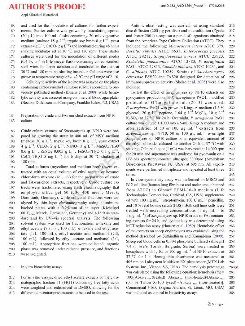
535methyl esters obtained after derivatization with CH2N2. All536analyzed total ion chromatograms contained several series of537FAMEs showing regularities in their GC retention behavior538(constant retention index difference of ca. 100 units, Fig. 3)539and possessing analogous mass spectra. Full details of the540structural elucidation are presented in Supplementary541Material. The identification of saturated normal chain and542branched (iso- and anteiso-) FAMEs was based on a combi-543nation of data coming from their mass spectra (Dickschat et al.5442011; Boon et al. 1977), NMR data (Fig. 2, Biermann and545Metzger 2004) and gas chromatographic retention behavior546(Fig. 3, Radulović et al. 2012). In addition to the mentioned
Fig. 2 Distinguishing structural fragments inferred from characteristic signals in a 1H-NMR, b 13C-NMR, and c, d HSQC spectrum of fraction 11
Appl Microbiol Biotechnol
JrnlID 253_ArtID 6364_Proof# 1 - 17/01/2015

AUTHOR'S PROOF!
UNCORRECTEDPROOF
547 spectral and retention data, the structure of monounsaturated548 normal and branched FAMEs, more specifically the double549 bond position, was inferred from the MS fragmentation pat-550 terns of the corresponding DMDS adducts (Fig. 3, Rontani551 et al. 2009; Dickschat et al. 2005). The cis configuration of the552 double bonds in all detected monoenoic FAs was determined
553from the absolute value of the 13C chemical shifts of allylic554carbons (Santos and Graça 2014). The identification was555corroborated wherever possible by a subsequent GC-MS anal-556ysis of authentic standards.557In order to clearly corroborate the presence of the558cyclopropyl group in a number of detected FAMEs, as well
Fig. 3Q6 a Total ion chromatogram of fraction 11 of Streptomyces sp. NP10culture extract derivatized with CH2N2. b Total ion chromatogram afterderivatization with DMDS with selected ion currant at m/z 217, mass
spectrum and characteristic fragmentation pattern of DMDS adduct of16:1ω7c
Appl Microbiol Biotechnol
JrnlID 253_ArtID 6364_Proof# 1 - 17/01/2015

AUTHOR'S PROOF!
UNCORRECTEDPROOF
559 as to assign its stereochemistry, the 1H NMR spectrum, as well560 as other 1D and 2D NMR spectra, of non-derivatized FR11561 were once closely inspected. Three high-field signals in the 1H562 NMR spectrum, mutually coupled in homonuclear 2D spectra,563 were indicative of the presence of a cis 1,2-disubstituted564 cyclopropane ring in the detected cyclopropane FAs565 (Macmillan and Molinski 2005; Knothe 2006). For com-566 pounds eluting at RI 2002 and 2202, it was assumed, based567 on comparisons of retention indices with literature values, that568 these are normal chain homologues 17:0cy9-10 and 19:0cy9-569 10, respectively (Zouari et al. 2011). Once again, their identity570 was undoubtedly verified by co-injection of authentic samples571 obtained by cyclopropanation of methyl esters of 16:1ω7c572 and 18:1ω9c, respectively, using CH2N2 in the presence of573 Pd(PhCN)2Cl2 as the catalyst (Gangadhar et al. 1988), where-574 as the position of branching methyl group in i-17:0cy9-10575 (RI=1966) and a-18:0cy9-10 (RI=2074) was inferred from576 the corresponding ΔRI values.
577Finally, a group of minor compounds exhibiting mass578spectra with two significant fragment ions at m/z 88 as the579base peak and atm/z 101 pointed either to α-methyl-branched580FAMEs or fatty acid ethyl esters (FAEEs). The possibility of581classifying these compounds in three series according to their582RI values, as well as the presence of [M–28]+ ion (loss of583CH2=CH2), arisen by Mc Lafferty rearrangement at the alk-584oxy branch of molecular ion, in their mass spectra (Gross5852004), prevailed on the side of ethyl esters of n, iso-, and586anteiso-FAs (i-14:0, i-15:0, a-15:0, i:16:0, 16:0, and 18:0).587This assumption was confirmed by co-injection of authentic588samples.589Several minor oxygenated FAs: 3-OH-8:0, 3-OH-10:0,59010-oxo-18:0 were identified as well. The identification was591made easier by the fact that the introduction of hydroxy,592oxo, or epoxy functionality leads to very distinguishable593fragmentation ions in mass spectra that also defines their594position in the alkyl chain (e.g., 3-hydroxy-FAs were dis-595tinguished by the base peak at m/z 103 produced by a596characteristic cleavage α to the carbon with the hydroxyl597group, while intensive ion at m/z 155 was diagnostic for598oxirane ring in positions 9 and 10; Ryhage and Stenhagen5991960). Of course, wherever it was possible, the initial600identification was confirmed by co-injection of a synthetic601standard.
602Optimization of conditions for production of free FAs603by Streptomyces sp. NP10
604Firstly, different solvents were examined in order to optimize605the extraction of free FAs produced by the strain Streptomyces606sp. NP10. The mixture of hexane/CHCl3 (4:1) was found to be607the most suitable, so it was used for as the extraction medium608in all further experiments (Fig. S4a, b in the Supplementary609Material). In order to determine whether this strain accumu-610lates or excretes these metabolites, mycelium and culture611media were separated by filtration and extracted independent-612ly. The analysis revealed that significantly higher amounts of613free FAs were present in the medium than in the mycelium614(Fig. 5c, d). Then, time dependence of the production of free615FAs by strain Streptomyces sp. NP10 was monitored and the616maximal production (amount excreted to culture media) was617observed at the sixth day of cultivation (Fig. 5e, f). In all618previously described experiments, the strain was cultured on619a complex medium rich in maltose (designated as MSY), and,620finally, the influence of different growth media (maltose-soy621flower (MSY), defined minimal R2m and minimal supple-622mented with yeast extract R2YE, respectively, media compo-623sitions are given in the Supplementary Material) on the pro-624duction of free FAs, was studied. It was found that the culti-625vation on a complex medium rich in maltose (designated as626MSY) as the carbon source was especially favorable for free627FA production (Fig. S5a, b in the Supplementary Material).
Fig. 4 In vitro activity of Streptomyces sp. NP10 extracts a onpyocyanine production in Pseudomonas aeruginosa PAO1 and bhemolytic effect. Crude culture extracts and i-16:0 acid standard wereadded at 100 μg mL−1, while FAs extracts were added at concentrations50 and 100 μg mL−1. * signifies statistically relevant difference
Appl Microbiol Biotechnol
JrnlID 253_ArtID 6364_Proof# 1 - 17/01/2015

AUTHOR'S PROOF!
UNCORRECTEDPROOF
Fig. 5 Bound and free FAprofiles of Streptomyces sp.NP10: a distribution of the majorFAs and b certain classes of FAs.cDistribution ofmajor free FAs inStreptomyces sp. NP10 culturebetween medium and myceliumand d distribution of certainclasses of FAs. Time dependenceof production and e distribution ofmain free FAs by Streptomycessp. NP10 and f distribution ofcertain classes of FAs. N normalchain, I iso, A anteiso, Uunsaturated, and CPcyclopropane FAs
Appl Microbiol Biotechnol
JrnlID 253_ArtID 6364_Proof# 1 - 17/01/2015

AUTHOR'S PROOF!
UNCORRECTEDPROOF
628 Furthermore, growing on this medium, the strain was able629 to produce free FA even at 4 °C (Fig. S5a, b in the630 Supplementary Material). Cultivation at this low temper-631 ature resulted in a decrease of the total amount of pro-632 duced free FAs, especially those of iso- and anteiso-chain.633 Thus, free FA profile at 4 °C was dominated with 16:0634 and 18:0, and intriguingly, a-17:0 was detected only in635 trace amounts (Fig. S5a, b in the Supplementary636 Material).
637 Taxonomic significance of free FAs
638 Previous findings that free FAs could be found in small639 amounts in cell membranes of some other Streptomyces sp.640 (Metz et al. 1988; Hoischen et al. 1997) led us to examine if641 this increased production and secretion of free FAs is a unique642 feature of the particular strain Streptomyces sp. NP10 or it is643 perhaps a characteristic of all members of Streptomyces genus644 previously unpublished. Thus, we studied whether three other
645Streptomyces species available from our microbiological col-646lection (both laboratory control strains and soil isolates) have647the ability to produce free FAs. The analysis showed that all648assayed strains: S. lividans NRRL B-12275, Streptomyces649durmitorensis MS405, and Streptomyces sp. NP2-ISS618650synthesized free FAs and that their free FAs profiles (domi-651nated by a-15:0 and i-16:0) were similar to that described for652Streptomyces sp. NP10 (Fig. 6a, b, Table S3 in the653Supplementary Material). The most notable differences were654in the increased amount of unsaturated FAs synthesized (es-655pecially i-16:1ω6c, 16:1ω7c, and a-17:1ω7c) by S. lividans656and of cyclopropane FAs (i-17:0cy9-10 and 17:0cy9-10)657found in Streptomyces sp. NP2, as well as in the slightly658decreased production of normal chain FAs (particularly65916:0) by the strain S. durmitorensis MS405 (Fig. 6a, b).660Moreover, the total amount of produced free FAs varied661among the analyzed strains from 2.427 μg mg−1 of dry my-662celium in the case of S. durmitorensis to 10.021 μg mg−1 in663the case of Streptomyces sp. NP10. 664
Fig. 6 Free fatty acid profiles offour different Streptomycesstrains. a Distribution of mainFAs. b Distribution of certainclasses of FAs. N normal chain, Iiso, A anteiso, U unsaturated, andCP cyclopropane FAs
Appl Microbiol Biotechnol
JrnlID 253_ArtID 6364_Proof# 1 - 17/01/2015

AUTHOR'S PROOF!
UNCORRECTEDPROOF
t2:1 Table 2 Structure, designation, and occurrence in the nature of unusual FAs found in Streptomyces sp. NP10
Fatty acid structure Designation Occurrence in the nature
Not found
Not found
-
-Cellular lipids of methanotrophic soil columnb
-Cellular fatty acid of Streptomyces griseusc
- Cellular lipids of biofilter sample of the animal rendering
plantd, e
- Cellular lipids of the sulfate-reducing bacteria
Desulfosarcina variabilise, f
- In free form in Streptomyces sp. A251g
- Cellular lipids of the sulfate-reducing bacteria
Desulfovibvio desulfuvicanse, h
- Cellular lipids of the Legionella micdadei and L. micdadei
Bari 2/158e, i
- Cellular fatty acid of the sponge Agelas conifera from the
Colombian Caribbeane, j
-Cellular fatty acid of Streptomyces griseusc
- Cellular fatty acid of the endemic fresh-water sponges
Baiealospongia baeillifera and Baicalospongia intermedia
(Lake Baikal)e, k
- Cellular fatty acid of the endemic sponge Lubomirskia
baicalensis (Lake Baikal) and its amphipod crustacean
parasite Brandtia (Spinacanthus) parasiticae, l
- In free form and as component of triacylglycerols of the
leaf beetle Chrysomela vigintipunctata (Scopoli)e, m
- Cellular lipids of the sulfate-reducing bacteria
Desulfosarcina variabilise, n
- Lipids in sediment samples from the Lusatian mining
districte, o
- Cellular lipids of the sponge Hymeniacidon sanguinea
from the Black Seap
- In total lipids from the green above-ground parts of alpine
plant Primula macrophyllae, q
- Cellular lipids of the microcosms samples freshly obtained
sediment of a monitoring well from a petroleum
hydrocarbon-contaminated aquifere, r
Lipids of surface sediments from Corner Inlet, Victoriaa
Appl Microbiol Biotechnol
JrnlID 253_ArtID 6364_Proof# 1 - 17/01/2015

AUTHOR'S PROOF!
UNCORRECTEDPROOF
665 Discussion
666 Although various Streptomyces spp. have been proven to be a667 prolific source of antibiotics and other useful metabolites, their668 involvement in the turnover of organic matter and xenobiotic669 compounds is only gaining more attention, which makes670 continuous search and isolation for novel species of this genus671 highly relevant. NP10 isolate reported in this study encom-672 passes quite a few desirable traits that make it suitable for673 further biotechnological applications such as biofuels produc-674 tion (cellulolytic activity accompanied with FA accumulation675 in the medium). Cellulolytic Streptomyces strains have been676 encountered in the past while, only recently, a phylogenetical-677 ly linked ability to degrade lignocellulose was described in678 Streptomyces associated with herbivorous insects (Book et al.
6792014). On the other hand, hemolytic activity is usually not680associated with Streptomyces spp., with some rare examples681reported (Suthindhiran and Kannabiran 2009), while682halotolerance is usually associated with Streptomyces isolated683from desert soils or salty waters as a consequence of their684adaptation to the saline environment (Thumar et al. 2010;685Bhave et al. 2013). Halotolerant and alkaliphilic686S. aburaviensis was isolated from the saline desert of Kutch687in India; however, it required 5–10%NaCl in the medium and688pH 9 for the optimal growth (Thumar et al. 2010). Under689varying cultivation conditions (incubation time, nutritive me-690dium, temperature, etc.), Streptomyces NP10 was found to691produce and excrete into the growth medium considerably692large amounts of free long-chain FAs (C7–C28). The maxi-693mum production of free FAs by this strain should be expected
t2:1 Table 2 (continued)
Fatty acid structure Designation Occurrence in the nature
- Hesperopeuce mertensiana (Pinaceae) seed lipidss
-Not found in any bacteria species
-In total lipids of leaves of Salvia nemorosat
- In total lipids of sericea lespedeza and bermudagrass
hays u
a Nichols et al. 1985bNichols et al. 1986c Suutari and Laakso 1993dKnief et al. 2003eGeometry of double bond was not specifiedf Rütters et al. 2001g Zheng et al. 2010h Taylor and Parkes 1983iMoss and Lambert-Fair 1990j Duque et al. 1993kDembitsky et al. 1993l Dembitsky et al. 1994mNikolova et al. 2000n Rütters et al. 2001o Poerschmann et al. 2012p Christie et al. 1994q Tsydendambaev et al. 2004r Pelz et al. 2001s Destaillats et al. 2002t Agar et al. 2008u Lee et al. 2012
Appl Microbiol Biotechnol
JrnlID 253_ArtID 6364_Proof# 1 - 17/01/2015

AUTHOR'S PROOF!
UNCORRECTEDPROOF
694 at the sixth day of cultivation in a complex medium rich in695 maltose at 28–30 °C. A detailed lipidomics study (consisting696 of a chromatographic isolation, NMR measurements, deriva-697 tizations, chemical transformations, and GC-MS co-injec-698 tions), that followed, enabled the identification of over 50699 different FAs of n-, iso-, and anteiso-chains including both700 saturated, unsaturated, and cyclopropane acids, among which701 i-14:0, a-15:0, i-16:0, and 16:0 were found to be the most702 abundant ones.703 The qualitative and quantitative composition of the total704 cellular FAs (Fig. 5a, b) of this novel isolate was consistent705 with its affiliation to the genus Streptomyces where most706 species contained iso- and anteiso-branched chain FAs (pri-707 marily a-15:0 and i-16:0) as the major bound FAs (Saddler708 et al. 1987). The main difference in bound and free FA profiles709 of this new strain was the occurrence of oxygenated FAs:710 18:0ep9-10c, 18:0di-ep9-10:12-13 (two isomers), and 9,10-711 di-OH-19:1 (two isomers) as minor component of total cellu-712 lar FAs (Table S-2 in the Supplementary Material). To the best713 of our knowledge, the identified epoxy FAs have not been714 previously found as components of any microorganism. It has715 been debated that some epoxy and dihydroxy FAs reported716 from various organisms may be artifacts arisen from either717 spontaneous or enzymatic (ep)oxidation (and hydrolysis) dur-718 ing sample preparation (Bernard 2014). Generally, streptomy-719 cetes, with the exception of some fast-growing thermophilic720 strains, grow very slowly even at their optimal temperature721 (usually 28 to 30 °C; Chen and Qin 2011). In view of this, the722 observed ability of this novel Streptomyces strain to have723 noticeable growth rate at low temperature (4 °C) seems rather724 remarkable. This atypical feature could be possibly attributed725 to the very high content of iso- and anteiso-FAs (43.5and726 24.3 %, respectively) in its membrane since it is well727 established that a major aspect of the cryotolerant physiology728 of some bacterial species, such as Listeria, is the predomi-729 nance of low freezing-point branched-chain FAs in the cell730 membrane, which permits the maintenance of membrane731 function at low temperatures (Mastronicolis et al. 2005). We732 have also found that the cultivation at this low temperature733 resulted in a decrease of the total amount of produced free734 FAs, especially those of iso- and anteiso- chain (Fig. S5a, b in735 the Supplementary Material). Thus, the observed change in736 free FAs profile accompanying the change in temperature737 could be explained by the fact that at lower temperatures the738 incorporation of branched-chain FAs into the phospholipids of739 cell membrane was possibly intensified. Considering that FA740 synthesis in bacteria is mostly dedicated to membrane main-741 tenance and having in mind that membrane is the first in742 contact with environmental stressors, it can be concluded that743 the remarkable tolerance of Streptomyces sp. NP10 strain to744 osmotic, cold and heat, and pH stress compared with other745 streptomycetes may be due to the unusual ability to746 biosynthesize FAs.
747We have examined FA accumulation/excretion of748Streptomyces sp. NP10 in regards to other possible ecophys-749iological roles such as a defense mechanism, as well as bac-750terial cell signaling. While FA extracts showed moderate751antimicrobial properties, it significantly reduced pyocyanin752production in P. aeruginosa PAO1 (Fig. 4a). P. aeruginosa753regulates pyocyanin production using an intercellular commu-754nication mechanism called quorum sensing, which is a chem-755ical communication process that bacteria use to regulate col-756lective behaviors and in P. aeruginosa it is linked to pathoge-757nicity and biofilm formation (Morkunas et al. 2012;758O’Loughlin et al. 2013). Not very much is known about759Streptomyces–Pseudomonas interspecies communication;760however, it is well established that 10:1ω8c can cause disper-761sion of P. aeruginosa PAO1, Candida and some other micro-762bial biofilms (Davies and Marques 2009).763As previously mentioned, free FAs have been previously764reported as minor components of neutral lipids of cell mem-765branes of few bacterial species like Bacillus spp. (Clejan et al.7661986), Aquaspirillum magnetotacticum (Gorby et al. 1988),767Streptomyces hygroscopicus (Hoischen et al. 1997) and768Streptomyces avermitilis (Metz et al. 1988). However, these769data are very scarce except for the strain S. avermitilis, where FA770chain length (C15–C17) was determined and the strain771Streptomyces sp. A251, when percentage (up to 8 %) and772identity of two fatty acids in the neutral lipid fraction were773determined (Zheng et al. 2010). Very recently, an oleaginous774bacterium belonging to the family Erysipelotrichaceae was de-775scribed that produces saturated long-chain free FAs (14:0, 16:0,77618:0, and 20:0) and accumulates them as intracellular droplets in777its cytoplasm. It is supposed that FAs accumulation in this strain778is a result of an imbalance between excess membrane FA bio-779synthesis due to homeoviscous adaptation to environmental780stress and limited β-oxidation activity due to anaerobic growth781involving lactic acid fermentation (Katayama et al. 2014).782Therefore, it seems that even the branched FAs that have783been ordinarily found as a part of the membrane phospho-784lipids, like i-14:0, a-15:0, i-16:0, and etc., were detected in this785study for the first time in free form in any bacteria species.786Moreover, so far, ethyl esters of these common bacterial FAs,787with exception of 16:0 and 18:0, were not described as me-788tabolites produced by any microorganism. In particular, ethyl789esters of i-12:0 and i-14:0 represent new natural products as790they have not been found up to now in samples of natural791origin, those of i-15:0, a-15:0, and i-16:0 were previously792identified just as components of odorous secretions of the793Tasmanian short-beaked echidna (Harris et al. 2012), while794the derivative of i-10:0 was only detected in volatiles of fresh795Thai green chili (Srisajjalertwaja et al. 2012).796Among the overall 59 identified free FAs (Table 1 and797Table S3 in the Supplementary Material), two of them, i-79817:0cy9-10 and a-18:0cy9-10, represent new natural products799and the first ever found branched cyclopropane FAs.
Appl Microbiol Biotechnol
JrnlID 253_ArtID 6364_Proof# 1 - 17/01/2015
AUTHOR'S PROOF!
UNCORRECTEDPROOF
800 Furthermore, branched chain monoenoic FAs, i-16:1ω6c, a-801 17:1ω7c, i-18:1ω8c, and a-19:1ω9c, and medium chain802 monoenoic FA, 13:1ω9c, have rather limited occurrence in803 nature (Table 2). Acid i-16:1ω6c was the only so far fully804 structurally characterized free FAs from any Streptomyces805 species. This FA and a-17:1ω7c were found only on few806 previous occasions as a part of cellular lipids of bacterial807 species like sulfate-reducing bacteria belonging to the genus808 Desulfosarcina, as well as Streptomyces griseus and/or809 Legionella micdadei (Table 2). The i-18:1ω8c and 13:1ω9c810 have not been so far identified, neither in free of bound form,811 in any microorganism, while there is just one previous report812 on the occurrence of a-19:1ω9c as a minor component of813 Hesperopeuce mertensiana seed triacylglicerols (Table 2).814 The 11:1ω1, although detected in other organisms were here-815 in found for the first time to be produced by bacteria, while816 some of the identified FAs, like i-22:0, 3-OH-8:0, 3-OH-10:0,817 and 10-oxo-18:0, represented new metabolites for genus818 Streptomyces.819 In recent years there is a growing interest toward the820 optimization of production of low-cost biofuels (FAs, al-821 cohols, olefins, hydrocarbons, etc.) by engineered micro-822 organisms due to the continual increase in the world’s823 energy demands and the scarcity of fossil fuel supplies.824 Since microorganisms are essentially capable of synthesiz-825 ing FAs, the FA production from microbial biomass is a826 promising and attractive alternative for traditional chemi-827 cal synthesis routes (Steen et al. 2010). Although constant828 progress has been made in metabolic engineering strategies829 that are being used to improve strains performances the830 lipids of interest are usually accumulated within the micro-831 bial cells. Thus, it is considered that the use of microor-832 ganism that could secrete lipids into the culture media833 would be an effective way of simplifying the downstream834 processing and reducing production costs (Meng et al.835 2013). Considering this, it seems that observed secretion836 of free FAs of this new Streptomyces strain into environ-837 ment is quite unique and possible very applicable feature in838 the research field of biofuel production.
839 Acknowledgments The authors acknowledge the Ministry of Educa-840 tion, Science and Technological Development of Serbia for the financial841 support (projects 172061 and 173048).
842843 Conflict of interest The authors declare that they have no conflicts of844 interest.
845 ReferencesQ8 846
847 Aerts HAJ, Jacobs PA (2004) Epoxide yield determination of oils and848 fatty acid methyl esters using 1H NMR. J Am Oil Chem Soc 81:849 841–846. doi:10.1007/s11746-004-0989-1
850Agar G, Adiguzel A, Baris O, Gulluce M, Sahin F (2008) Phenotypic and851genetic variation of some Salvia species grown in eastern Anatolia852region of Turkey. Asian J Chem 20:3935–3944853Altschul SF, Madden TL, Schaffer AA, Zhang J, Zhang Z, Miller W,854Lipman DJ (1997) Gapped BLAST and PSI-BLAST: a new gener-855ation of protein database search programs. Nucleic Acids Res 25:8563389–3402. doi:10.1093/nar/25.17.3389857Arabolaza A, Rodriguez E, Altabe S, Alvarez H, Gramajo H (2008)858Multiple pathways for triacylglycerol biosynthesis in Streptomyces859coelicolor. Appl Environ Microbiol 74:2573–2582. doi:10.1128/860AEM. 02638-07861Arabolaza A, D’Angelo M, Comba S, Gramajo H (2010) FasR, a novel862class of transcriptional regulator, governs the activation of fatty acid863biosynthesis genes in Streptomyces coelicolor. Mol Microbiol 78:86447–63. doi:10.1111/j.1365-2958.2010.07274.x865Bengsch E, Perly B, Deleuze C, Valero A (1986) A general rule for the866assignment of the carbon-13 NMR peaks in fatty acid chains. J867Magn Reson 68:1–13. doi:10.1016/0022-2364(86)90311-2868Bernard M (2014) Molecular cloning and functional characterization of869genes involved in the biosynthesis of polyunsaturated fatty acids in870oat (Avena sativa L.). Master of Science Dissertation, University of871Saskatchewan, Canada872Bhave SV, Shanbhag PV, Sonawane SK, Parab RR, Mahajan GB (2013)873Isolation and characterization of halotolerant Streptomyces874radiopugnans from Antarctica soil. Lett Appl Microbiol 56:348–875355. doi:10.1111/lam.12054876Biermann U, Metzger JO (2004) Alkylation of alkenes: ethylaluminum877sesquichloride-mediated hydro-alkyl additions with alkyl878chloroformates and di-tert-butylpyrocarbonate. J Am Chem Soc879126:10319–10330. doi:10.1021/ja048904y880Book AJ, Lewin GR, McDonald BR, Takasuka TE, Doering DT, Adams881AS, Blodgett JA, Clardy J, Raffa KF, Fox BG, Currie CR (2014)882Cellulolytic Streptomyces strains associated with herbivorous in-883sects share a phylogenetically linked capacity to degrade lignocel-884lulose. Appl Environ Microbiol 80:4692–4701. doi:10.1128/AEM.88501133-14886Boon JJ, van de Graaf B, Schuyl PJW, de Lange F, de Leeuw JW (1977)887The mass spectrometry of iso and anteiso monoenoic fatty acids.888Lipids 12:717–721. doi:10.1007/BF02570901889Challis GL, Hopwood DA (2003) Synergy and contingency as driving890forces for the evolution of multiple secondary metabolite production891by Streptomyces species. Proc Natl Acad Sci U S A 100(Suppl 2):89214555–14561. doi:10.1073/pnas.1934677100893Chan DI, Vogel HJ (2010) Current understanding of fatty acid biosyn-894thesis and the acyl carrier protein. Biochem J 430:1–19. doi:10.8951042/BJ20100462896Chen W, Qin Z (2011) Development of a gene cloning system in a fast-897growing and moderately thermophilic Streptomyces species and898heterologous expression of Streptomyces antibiotic biosynthetic899gene clusters. BMC Microbiol 11:243–252. doi:10.1186/1471-9002180-11-243901Christie WW, Brechany EY, Marekov IN, Stefanov KL, Andreev SN902(1994) The fatty acids of the sponge Hymeniacidon sanguinea from903the Black Sea. Comp Biochem Physiol B 109:245–252. doi:10.9041016/0305-0491(94)90008-6905Clejan S, Krulwich TA, Mondrus KR, Seto-Young D (1986) Membrane906lipid composition of obligately and facultatively alkalophilic strains907of Bacillus spp. J Bacteriol 168:334–340908Cole J, Wang Q, Cardenas E, Fish J, Chai B, Farris R, Kulam-Syed-909Mohideen A, McGarrell D, Marsh T, Garrity G, Tiedje J (2009) The910ribosomal database project: improved alignments and new tools for911rRNA analysis. Nucleic Acids Res 37:141–145. doi:10.1093/nar/912gkn879913Davies DG,Marques CN (2009) A fatty acid messenger is responsible for914inducing dispersion in microbial biofilms. J Bacteriol 191:1393–9151403. doi:10.1128/JB.01214-08
Appl Microbiol Biotechnol
JrnlID 253_ArtID 6364_Proof# 1 - 17/01/2015

AUTHOR'S PROOF!
UNCORRECTEDPROOF
916 Dembitsky VM, Rezanka T, Kashin AG (1993) Comparative study of the917 endemic freshwater fauna of Lake Baikal—II. Unusual lipid com-918 position of two sponge species Baicalospongia bacillifera and919 Baicalospongia intermedia (family Lubomirskiidae, class920 Demospongiae). Comp Biochem Physiol B 106:825–831. doi:10.921 1016/0305-0491(93)90037-6922 Dembitsky VM, Rezanka T, Kashin AG (1994) Comparative study of the923 endemic freshwater fauna of Lake Baikal—VI. Unusual fatty acid924 and lipid composition of the endemic sponge Lubomirskia925 baicalensis and its amphipod crustacean parasite Brandtia926 (Spinacanthus) parasitica. Comp Biochem Physiol B 109:415–927 426. doi:10.1016/0305-0491(94)90024-8928 Desbois AP, Smith VJ (2010) Antibacterial free fatty acids: activities,929 mechanisms of action and biotechnological potential. Appl930 Microbiol Biotechnol 85:1629–1642. doi:10.1007/s00253-009-931 2355-3932 Destaillats F, Wolff RL, Angers P (2002) Saturated and unsaturated933 anteiso-C19 acids in the seed lipids fromHesperopeuce mertensiana934 (Pinaceae). Lipids 37:325–328. doi:10.1007/s11745-002-0898-y935 Dickschat JS, Bode HB, Kroppenstedt RM, Müller R, Schulz S (2005)936 Biosynthesis of iso-fatty acids in myxobacteria. Org Biomol Chem937 3:2824–2831. doi:10.1039/B504889C938 Dickschat JS, Bruns H, Riclea R (2011) Novel fatty acid methyl esters939 from the actinomyceteMicromonospora aurantiaca. Beilstein J Org940 Chem 7:1697–1712. doi:10.3762/bjoc.7.200941 Djokic L, Narancic T, Nikodinovic-Runic J, Savic M, Vasiljevic B (2011)942 Isolation and characterization of four novel Gram-positive bacteria943 associated with the rhizosphere of two endemorelict plants capable944 of degrading a broad range of aromatic substrates. Appl Microbiol945 Biotechnol 91:1227–1238. doi:10.1007/s00253-011-3426-9946 Drake DR, Brogden KA, Dawson DV, Wertz PW (2008) Thematic947 review series: skin lipids. Antimicrobial lipids at the skin surface. J948 Lipid Res 49:4–11. doi:10.1194/jlr. R700016-JLR200949 Duque C, CepedaN,Martínez A (1993) The steryl ester and phospholipid950 fatty acids of the sponge Agelas conifera from the Colombian951 Caribbean. Lipids 28:767–769. doi:10.1007/BF02536002952 Florova G, Kazanina G, Reynolds KA (2002) Enzymes involved in fatty953 acid and polyketide biosynthesis in Streptomyces glaucescens: role954 of FabH and FabD and their acyl carrier protein specificity.955 Biochemistry 41:10462–10471. doi:10.1021/bi0258804956 Gago G, Diacovich L, Arabolaza A, Tsai S-C, Gramajo H (2011) Fatty957 acid biosynthesis in actinomycetes. FEMS Microbiol Rev 35:475–958 497. doi:10.1111/j.1574-6976.2010.00259.x959 Gangadhar A, Subbarao R, Lakshminarayana G (1988)960 Cyclopropanation of unsaturated fatty acid methyl esters using961 diazomethane and palladium(II) acetate. J Am Oil Chem Soc 65:962 601–606. doi:10.1007/BF02540687963 Gorby YA, Beveridge TJ, Blakemore RP (1988) Characterization of the964 bacterial magnetosome membrane. J Bacteriol 170:834–841965 Gross JH (2004) Mass spectrometry. A textbook. Springer, Berlin-966 Heidelberg967 Hansen MB, Nielsen SE, Berg K (1989) Re-examination and further968 development of a precise and rapid dye method for measuring cell969 growth/cell kill. J Immunol Methods 119:203–210. doi:10.1016/970 0022-1759(89)90397-9971 Harris RL, Davies NW, Nicol SC (2012) Chemical composition of972 odorous secretions in the Tasmanian short-beaked echidna973 (Tachyglossus aculeatus setosus). Chem Senses 37:819–836. doi:974 10.1093/chemse/bjs066975 Hertweck C (2009) The biosynthetic logic of polyketide diversity. Angew976 Chem Int Ed 48:4688–4716. doi:10.1002/anie.200806121977 Hibbing ME, Fuqua C, Parsek MR, Peterson SB (2010) Bacterial com-978 petition: surviving and thriving in the microbial jungle. Nat Rev979 Microbiol 8:15–25. doi:10.1038/nrmicro2259980 Hoischen C, Gura K, Luge C, Gumpert J (1997) Lipid and fatty acid981 composition of cytoplasmic membranes from Streptomyces
982hygroscopicus and its stable protoplast-type L form. J Bacteriol983179:3430–3436984Hopwood DA (1988) The Leeuwenhoek lecture, 1987. Towards an985understanding of gene switching in Streptomyces, the basis of spor-986ulation and antibiotic production. Proc R Soc Lond B Biol 235:121–987138. doi:10.1098/rspb.1988.0067988Kaneda T (1991) Iso- and anteiso-fatty acids in bacteria: biosynthesis,989function, and taxonomic significance. Microbiol Rev 55:288–302990Kasana RC, Salwan R, Dhar H, Dutt S, Gulati A (2008) A rapid and easy991method for the detection of microbial cellulases on agar plates using992Gram’s iodine. Curr Microbiol 57:503–507. doi:10.1007/s00284-993008-9276-8994Katayama T, KannoM,Morita N, Hori T, Narihiro T,Mitani Y, Kamagata995Y (2014) An oleaginous bacterium that intrinsically accumulates996long-chain free fatty acids in its cytoplasm. Appl Environ Microbiol99780:1126–1131. doi:10.1128/AEM. 03056-13998Kieser T, Bibb MJ, Buttner MJ, Chater KF, Hopwood DA (2000)999Practical Streptomyces genetics. The John Innes Foundation,1000Norwich1001Knief C, Altendorf K, Lipski A (2003) Linking autotrophic activity in1002environmental samples with specific bacterial taxa by detection of100313C-labelled fatty acids. Environ Microbiol 5:1155–1167. doi:10.10041046/j.1462-2920.2003.00510.x1005Knothe G (2006) NMR characterization of dihydrosterculic acid and its1006methyl ester. Lipids 41:393–396. doi:10.1007/s11745-006-5110-x1007Lane DJ (1991) 16S/23S rRNA sequencing. In: Stackebrandt E,1008Goodfellow MM (eds) Nucleic acid techniques in bacterial system-1009atic. Wiley, Chichester, pp 115–1751010Larkin MA, Blackshields G, Brown NP, Chenna R, McGettigan PA,1011McWilliam H, Valentin F, Wallace IM, Wilm A, Lopez R,1012Thompson JD, Gibson TJ, Higgins DG (2007) Clustal W and1013Clustal X version 2.0. Bioinformatics 23:2947–2948. doi:10.1093/1014bioinformatics/btm4041015Lee JH, Vanguru M, Moore DA, Kannan G, Terrill TH, Kouakou B1016(2012) Flavor compounds and quality parameters of chevon as1017influenced by Sericea Lespedeza hay. J Agric Food Chem 60:10183934–3939. doi:10.1021/jf20501251019Macmillan JB, Molinski TF (2005) Majusculoic acid, a brominated1020cyclopropyl fatty acid from a marine cyanobacterial mat assem-1021blage. J Nat Prod 68:604–606. doi:10.1021/np049596k1022Mastronicolis SK, Arvanitis N, Karaliota A, Litos C, Stavroulakis G,1023Moustaka H, Tsakirakisa A, Heropoulos G (2005) Cold dependence1024of fatty acid profile of different lipid structures of Listeria1025monocytogenes. Food Microbiol 22:213–219. doi:10.1016/j.fm.10262004.08.0021027Mei S, Ni H-M, Manley S, Bockus A, Kassel KM, Luyendyk JP, Copple1028BL, Ding WX (2011) Differential roles of unsaturated and saturated1029fatty acids on autophagy and apoptosis in hepatocytes. J Pharmacol1030Exp Ther 339:487–498. doi:10.1124/jpet.111.1843411031Meng X, Shang H, Zheng Y, Zhang Z (2013) Free fatty acid secretion by1032an engineered strain of Escherichia coli. Biotechnol Lett 35:2099–10332103. doi:10.1007/s10529-013-1305-41034Metz PA, Omstead DR, Kaplan L, Liesch JM, Stearns RA, Van-1035Denheuvel WJ (1988) Characterization of a lipid-rich fraction syn-1036thesized by Streptomyces avermitilis. J Chromatogr 441:31–44. doi:103710.1016/S0021-9673(01)84652-51038Moghaddam MF, Motoba K, Borhan B, Pinot F, Hammock BD (1996)1039Novel metabolic pathways for linoleic and arachidonic acid metab-1040olism. Biochim Biophys Acta 1290:327–339. doi:10.1016/0304-10414165(96)00037-21042Mohn T, Plitzko I, Hamburger M (2009) A comprehensive metabolite1043profiling of Isatis tinctoria leaf extracts. Phytochemistry 70:924–1044934. doi:10.1016/j.phytochem.2009.04.0191045Morkunas B, Galloway WR, Wright M, Ibbeson BM, Hodgkinson JT,1046O’Connell KM, Bartolucci N, Della Valle M, Welch M, Spring DR1047(2012) Inhibition of the production of the Pseudomonas aeruginosa
Appl Microbiol Biotechnol
JrnlID 253_ArtID 6364_Proof# 1 - 17/01/2015

AUTHOR'S PROOF!
UNCORRECTEDPROOF
1048 virulence factor pyocyanin in wild-type cells by quorum sensing1049 autoinducer-mimics. Org Biomol Chem 10:8452–8464. doi:10.1050 1039/C2OB26501J1051 Moss CW, Lambert-Fair MA (1990) Reevaluation of the cellular fatty1052 acid composition of Legionella micdadei Bari 2/158. J Clin1053 Microbiol 28:3891054 Nichols PD, Shaw PM, Johns RB (1985) Determination of the double1055 bond position and geometry in monoenoic fatty acids from complex1056 microbial and environmental samples by capillary GC-MS of their1057 Diels-Alder adducts. J Microbiol Meth 3:311–319. doi:10.1016/1058 0167-7012(85)90013-21059 Nichols PD, Guckert JB, White DC (1986) Determination of monoun-1060 saturated fatty acid double-bond position and geometry for micro-1061 bial monocultures and complex consortia by capillary GC-MS of1062 their dimethyl disulphide adducts. J Microbiol Meth 5:49–55. doi:1063 10.1016/0167-7012(86)90023-01064 Nikolova N, Rezanka T, Nikolova-Damyanova B (2000) Fatty acid1065 profiles of main lipid classes in adult Chrysomela vigintipunctata1066 (Scopoli) (Coleopterai: Chrysomelidae). Z Naturforsch C 55:661–1067 6661068 O’Loughlin CT, Miller LC, Siryaporn A, Drescher K, Semmelhack MF,1069 Bassler BL (2013) A quorum-sensing inhibitor blocks1070 Pseudomonas aeruginosa virulence and biofilm formation. Proc1071 Natl Acad Sci U S A 110:17981–17986. doi:10.1073/pnas.1072 13169811101073 Pelz O, Chatzinotas A, Zarda-Hess A, Abraham W-R, Zeyer J (2001)1074 Tracing toluene-assimilating sulfate-reducing bacteria using 13C-1075 incorporation in fatty acids and whole-cell hybridization. FEMS1076 Microbiol Ecol 38:123–131. doi:10.1111/j.1574-6941.2001.1077 tb00890.x1078 Poerschmann J, Koschorreck M, Górecki T (2012) Organic matter in1079 sediments of an acidic mining lake as assessed by lipid analysis. Part1080 I: fatty acids. Sci Total Environ 414:614–623. doi:10.1016/j.1081 scitotenv.2011.10.0151082 RadulovićN, DenićM, Stojanović-Radić Z, Skropeta D (2012) Fatty and1083 volatile oils of the gypsywort Lycopus europaeus L. and the1084 Gaussian-like distribution of its wax alkanes. J Am Oil Chem Soc1085 89:2165–2185. doi:10.1007/s11746-012-2118-71086 Ricchi M, Odoardi MR, Carulli L, Anzivino C, Ballestri S, Pinetti A,1087 Fantoni LI, Marra F, Bertolotti M, Banni S, Lonardo A, Carulli N,1088 Loria P (2009) Differential effect of oleic and palmitic acid on lipid1089 accumulation and apoptosis in cultured hepatocytes. J Gastroenterol1090 Hepatol 24:830–840. doi:10.1111/j.1440-1746.2008.05733.x1091 Rontani J-F, Zabeti N, Aubert C (2009) Double bond migration to1092 methylidene positions during electron ionization mass spectrometry1093 of branched monounsaturated fatty acid derivatives. J Am SocMass1094 Spectrom 20:1997–2005. doi:10.1016/j.jasms.2009.07.0201095 Rütters H, Sass H, Cypionka H, Rullkötter J (2001) Monoalkylether1096 phospholipids in the sulfate-reducing bacteria Desulfosarcina1097 variabilis and Desulforhabdus amnigenus. Arch Microbiol 176:1098 35–442. doi:10.1007/s0020301003431099 Ryhage R, Stenhagen E (1960) Mass spectrometric studies. VI. Methyl1100 esters of normal chain oxo-, hydroxy-, methoxy- and epoxy-acids.1101 Ark Kemi 15:545–5741102 Saddler GS, O’Donnell AG, Goodfellow M, Minnikin DE (1987)1103 SIMCA pattern recognition in the analysis of streptomycete fatty1104 acids. J Gen Microbiol 133:1137–1147. doi:10.1099/00221287-1105 133-5-11371106 Santos S, Graça J (2014) Stereochemistry of C18 monounsaturated cork1107 suberin acids determined by spectroscopic techniques including 1H-1108 NMR multiplet analysis of olefinic protons. Phytochem Anal 25:1109 192–200. doi:10.1002/pca.2491
1110SkokoN, Vujovic J, SavicM, Papic N, Vasiljevic V, Ljubijankic G (2005)1111Construction of Saccharomyces cerevisiae strain FAV20 useful in1112detection of immunosuppressants produced by soil actinomycete. J1113Microbiol Meth 61:137–140. doi:10.1016/j.mimet.2004.11.0071114Srisajjalertwaja S, Apichartsrangkoon A, Chaikham P, Chakrabandhu Y,1115Pathomrungsiyounggul P, Leksawasdi N, Supraditareporn W, Hirun1116S (2012) Color, capsaicin and volatile components of baked Thai1117green chili (Capsicum annuum Linn. var. Jak Ka Pat). J Agric Sci 4:111875–84. doi:10.5539/jas.v4n12p751119Steen EJ, Kang Y, Bokinsky G, Hu Z, Schirmer A, McClure A, del1120Cardayre SB, Keasling JD (2010) Microbial production of fatty-1121acid-derived fuels and chemicals from plant biomass. Nature 463:1122559–562. doi:10.1038/nature087211123SuthindhiranK,KannabiranK (2009) Hemolytic activity of Streptomyces1124VITSDK1 spp. isolated from marine sediments in Southern India. J1125Mycol Med 19:77–86. doi:10.1016/j.mycmed.2009.01.0011126Suutari M, Laakso S (1993) Signature GLC-MS ions in identification of1127Δ5- and Δ9-unsaturated iso- and anteiso-branched fatty acids. J1128Microbiol Meth 17:39–48. doi:10.1016/0167-7012(93)90077-U1129Tamura K, Stecher G, Peterson D, Filipski A, Kumar S (2013) MEGA6:1130molecular evolutionary genetics analysis version 6.0. Mol Biol Evol113130:2725–2729. doi:10.1093/molbev/mst1971132Taylor J, Parkes J (1983) The cellular fatty acids of the sulphate-reducing1133bacteria, Desulfobacter sp., Desulfobulbus sp. and Desulfovibvio1134desulfuvicans. J Gen Microbiol 129:3303–3309. doi:10.1099/113500221287-129-11-33031136Thumar JT, Dhulia K, Singh SP (2010) Isolation and partial purification1137of an antimicrobial agent from halotolerant alkaliphilic Streptomyces1138aburaviensis strain Kut-8. World J Microbiol Biotechnol 26:2081–11392087. doi:10.1007/s11274-010-0394-71140Tsydendambaev WD, Christie WW, Brechany EY, Vereshchagin AG1141(2004) Identification of unusual fatty acids of four alpine plant1142species from the Pamirs. Phytochemistry 65:2695–2703. doi:10.11431016/j.phytochem.2004.08.0211144van Wezel GP, McDowall KJ (2011) The regulation of the secondary1145metabolism of Streptomyces: new links and experimental advances.1146Nat Prod Rep 28:1311–1333. doi:10.1039/C1NP00003A1147Vandamme P, Pot B, Gillis M, de Vos P, Kersters K, Swings J (1996)1148Polyphasic taxonomy, a consensus approach to bacterial systemat-1149ics. Microbiol Rev 60:407–4381150VeljkovićVB, Lakićević SH, StamenkovićOS, Todorović ZB, LazićML1151(2006) Biodiesel production from tobacco (Nicotiana tabacum L.)1152seed oil with a high content of free fatty acids. Fuel 85:2671–2675.1153doi:10.1016/j.fuel.2006.04.0151154Vyssotski M, Ryan J, Lagutin K, Wong H, Morgan X, Stott M (2012) A1155novel fatty acid, 12,17-dimethyloctadecanoic acid, from the extrem-1156ophile Thermogemmatispora sp. (strain T81). Lipids 47:601–611.1157doi:10.1007/s11745-012-3668-z1158Zgoda JR, Porter JR (2001) A convenient microdilution method for1159screening natural products against bacteria and fungi. Pharm Biol116039:221–225. doi:10.1076/phbi.39.3.221.59341161Zhang Y-M, Rock CO (2008) Membrane lipid homeostasis in bacteria.1162Nat Rev Microbiol 6:222–232. doi:10.1038/nrmicro18391163Zheng CJ, Sohn MJ, Chi SW, Kim WG (2010) Methyl-branched fatty1164acids, inhibitors of enoyl-ACP reductase with antibacterial activity1165from Streptomyces sp. A251. J Microbiol Biotechnol 20:875–880.1166doi:10.4014/jmb.1001.010041167Zouari N, Fakhfakh N, Zouari S, Sellami M, AbidM, AyadiMA, Zaidi S,1168Neffati M (2011) Volatile and lipid analyses by gas chromatography/1169mass spectrometry and nutraceutical potential of edible wild Malva1170aegyptiaca L. (Malvaceae). Int J Food Sci Nutr 62:600–608. doi:10.11713109/09637486.2011.564157
1172
Appl Microbiol Biotechnol
JrnlID 253_ArtID 6364_Proof# 1 - 17/01/2015
Copyright © 2022 FDOKUMEN