Structural and functional response of toad urinary bladder to LiCl
12
Kidney International, Vol. 24 (1983), pp. 719—730 Structural and functional response of toad urinary bladder to LiC1 EMMA FERNANDEZ-REPOLLET, ANN LEFURGEY, MARCOS A. HARDY, and C. CRAIG TISHER Division of Nephrology, Department of Medicine, University of Florida, Gainesville, Florida, and the Division of Nephrology, University of Miami, Miami, Florida Structural and functional response of toad urinary bladder to LiCI. The physiological and morphological response of toad urinary bladder was examined during mucosal exposure of LiCI both with and without vasopressin (VP). With 20 or 100 mU/mi of VP in the serosal bath there was a decrease in ij,, between the first and second VP stimulation in LiCI-treated bladders (VP20, —14 6%; VP100, —16 5%) that was not different from that observed without LiC1 (VP20, —8 3%, P = NS). However, with I mU/mi of VP, a significant decrease in J.,, was evident in LiCl-treated (—30 10%) versus control sacs (+6 8%; P < 0.02). At all VP concentrations tested, a significant decrease in SCC and PD was observed between the first stimulation without LiCl and the second stimulation with LiCI. Both osmotic (Pr) and diffusional water perme- ability (Pd) were increased significantly with 11 mrvt LiCI only, while neither basal nor VP-stimulated urea permeability (Pa) was affected. Morphological changes paralleled the physiological alterations induced by LiCI. These data demonstrate that LiC1 interferes with the osmotic response of the toad bladder to low concentrations of VP, and increases both P1 and Pd while leaving P unaffected. These findings coupled with the cell swelling and intracellular vacuolization suggest the presence of a defect in transepithelial water movement somewhere beyond the apical membrane of the granular cell exposed to LiCI. Réponse structurelle et fonctionnelle de Ia vessie de crapaud au L1CI. La réponse physiologique et morphologique de Ia vessie de crapaud a été examinée pendant exposition de Ia muqueuse a du LiCI en presence ou en l'absence de vasopressine (VP). Pour 20 ou 100 mU/mI de VP dans le bain séreux, ii y avait une diminution de J entre Ia premiere et Ia seconde stimulation par VP dans les vessies traitées par le LiCI (VP20, —14 6%; VP1, —16 5%), qui n'étaient pas différentes de celles observées sans LiCI (VP20, —8 3%; P = NS). Cependant, avec 1 mU/mI de VP, une diminution significative de J, était évidente dans les sacs traités au LiCI (—30 10%) par rapport aux sacs contrôles (+6 8%; P < 0,02). Pour toutes les concentrations de VP testées, une diminution significative du SCC et de PD a été observée entre La premiere stimulation sans LiCl, et Ia seconde stimulation avec LiCI. Les perméabilites osmotiques (P1) et diffusionnelles (Pd) a l'eau Ctaient augmentées significativement avec Ii ms'i de LiC! seulement tandis que Ia perméabilite a l'uree basale ou stimulée par Ia VP (Pa) n'était pas affectée. Des modifications morphologiques allaient de pair avec les alterations physiologiques induites par le LiCI. Ces données demon- trent que LiCI interfere avec Ia réponse osmotique de Ia vessie de crapaud pour de faibles concentrations de VP, augmente P1 et Pd, mais laisse P inchange. Ces résultats, couples avec le gonflement cellulaire et Ia vacuolisation intracellulaire suggèrent Ia presence d'un défaut du mouvement transépithélial d'eau quelque part au delà de Ia membrane apicale de Ia cellule granulaire exposee au L1CI. conflicting results have been obtained in toad urinary bladder epithelium, a model of the collecting tubule in the mammalian nephron. Singer, Rotenberg, and Puschett [4] and Singer and Franko [5] demonstrated a significant inhibition of VP-induced Jv after exposure of the mucosal surface to LiC!. In contrast, Bentley and Wasserman [6] failed to find such an inhibitory effect at either similar or higher mucosal concentrations of LiCI. Finally, Harris and Jenner [7] reported a significant inhibition of the hydro-osmotic effect of VP after serosal exposure of toad urinary bladder to LiCl. In view of these conflicting data, the effects of LiCl on VP-induced Jv were re-examined and corre- lated with the morphological changes observed in toad urinary bladder epithelium. In addition, the effects of mucosal LiCI on osmotic (P1) and diffusional (Pd) water permeability and diffu- sional urea permeability (Pa) were also evaluated. Methods The toad, Bufo marinus, was used in all experiments. Ani- mals were sacrificed by double pithing and their paired urinary hemibladders were excised and rinsed in aerated amphibian Ringer's solution with the following composition in mmoles/ liter: sodium chloride, 111; potassium chloride, 3.4; sodium bicarbonate, 4.0; calcium chloride, 2.7, and dextrose, 5.0. The pH of the solution in equilibrium with room air was 7.8 to 8.1, and the osmolality was 226 to 232 mOsm/kg H20. A slightly different composition of amphibian Ringer's solution was em- ployed in those studies examining osmotic permeability (P1), diffusional water permeability (Pd), and urea permeability (Pa). The second solution had the following composition in mmoles/ liter; sodium chloride, 110; potassium chloride, 5; Tris-HCI, 5; calcium chloride, 1; and glucose, 5. The pH of the solution was 8.0 to 8.1, and the osmolality was 233 mOsm/kg H20. Tissue preparation. For all experiments in which both physi- ologic and morphologic observations were made, each hemi- bladder was mounted as a sac on the end of a glass or plastic cannula with the mucosal surface facing inward [81 unless otherwise specified. Physiologic studies. To investigate the effects of LiCI on VP- stimulated iv, sacs from 16 hemibladders were filled with Several studies have evaluated the effects of LiCI salt on VP- induced osmotic water flow (iv) in toad urinary bladder. Although an inhibitory effect on iv has been demonstrated consistently in experimental animals [1—3] and humans [1, 4], 719 Received for publication November 12, 1982 and in revised form May 9, 1983 © 1983 by the International Society of Nephrology
Transcript of Structural and functional response of toad urinary bladder to LiCl

Kidney International, Vol. 24 (1983), pp. 719—730
Structural and functional response of toadurinary bladder to LiC1
EMMA FERNANDEZ-REPOLLET, ANN LEFURGEY, MARCOS A. HARDY, and C. CRAIG TISHER
Division of Nephrology, Department of Medicine, University of Florida, Gainesville, Florida, and the Division of Nephrology,University of Miami, Miami, Florida
Structural and functional response of toad urinary bladder to LiCI. Thephysiological and morphological response of toad urinary bladder wasexamined during mucosal exposure of LiCI both with and withoutvasopressin (VP). With 20 or 100 mU/mi of VP in the serosal bath therewas a decrease in ij,, between the first and second VP stimulation inLiCI-treated bladders (VP20, —14 6%; VP100, —16 5%) that was notdifferent from that observed without LiC1 (VP20, —8 3%, P = NS).However, with I mU/mi of VP, a significant decrease in J.,, was evidentin LiCl-treated (—30 10%) versus control sacs (+6 8%; P < 0.02).At all VP concentrations tested, a significant decrease in SCC and PDwas observed between the first stimulation without LiCl and the secondstimulation with LiCI. Both osmotic (Pr) and diffusional water perme-ability (Pd) were increased significantly with 11 mrvt LiCI only, whileneither basal nor VP-stimulated urea permeability (Pa) was affected.Morphological changes paralleled the physiological alterations inducedby LiCI. These data demonstrate that LiC1 interferes with the osmoticresponse of the toad bladder to low concentrations of VP, and increasesboth P1 and Pd while leaving P unaffected. These findings coupled withthe cell swelling and intracellular vacuolization suggest the presence ofa defect in transepithelial water movement somewhere beyond theapical membrane of the granular cell exposed to LiCI.
Réponse structurelle et fonctionnelle de Ia vessie de crapaud au L1CI.La réponse physiologique et morphologique de Ia vessie de crapaud aété examinée pendant exposition de Ia muqueuse a du LiCI en presenceou en l'absence de vasopressine (VP). Pour 20 ou 100 mU/mI de VPdans le bain séreux, ii y avait une diminution de J entre Ia premiere etIa seconde stimulation par VP dans les vessies traitées par le LiCI(VP20, —14 6%; VP1, —16 5%), qui n'étaient pas différentes decelles observées sans LiCI (VP20, —8 3%;P = NS). Cependant, avec1 mU/mI de VP, une diminution significative de J, était évidente dansles sacs traités au LiCI (—30 10%) par rapport aux sacs contrôles (+6
8%; P < 0,02). Pour toutes les concentrations de VP testées, unediminution significative du SCC et de PD a été observée entre Lapremiere stimulation sans LiCl, et Ia seconde stimulation avec LiCI.Les perméabilites osmotiques (P1) et diffusionnelles (Pd) a l'eau Ctaientaugmentées significativement avec Ii ms'i de LiC! seulement tandis queIa perméabilite a l'uree basale ou stimulée par Ia VP (Pa) n'était pasaffectée. Des modifications morphologiques allaient de pair avec lesalterations physiologiques induites par le LiCI. Ces données demon-trent que LiCI interfere avec Ia réponse osmotique de Ia vessie decrapaud pour de faibles concentrations de VP, augmente P1 et Pd, maislaisse P inchange. Ces résultats, couples avec le gonflement cellulaireet Ia vacuolisation intracellulaire suggèrent Ia presence d'un défaut dumouvement transépithélial d'eau quelque part au delà de Ia membraneapicale de Ia cellule granulaire exposee au L1CI.
conflicting results have been obtained in toad urinary bladderepithelium, a model of the collecting tubule in the mammaliannephron. Singer, Rotenberg, and Puschett [4] and Singer andFranko [5] demonstrated a significant inhibition of VP-inducedJv after exposure of the mucosal surface to LiC!. In contrast,Bentley and Wasserman [6] failed to find such an inhibitoryeffect at either similar or higher mucosal concentrations of LiCI.Finally, Harris and Jenner [7] reported a significant inhibition ofthe hydro-osmotic effect of VP after serosal exposure of toadurinary bladder to LiCl. In view of these conflicting data, theeffects of LiCl on VP-induced Jv were re-examined and corre-lated with the morphological changes observed in toad urinarybladder epithelium. In addition, the effects of mucosal LiCI onosmotic (P1) and diffusional (Pd) water permeability and diffu-sional urea permeability (Pa) were also evaluated.
Methods
The toad, Bufo marinus, was used in all experiments. Ani-mals were sacrificed by double pithing and their paired urinaryhemibladders were excised and rinsed in aerated amphibianRinger's solution with the following composition in mmoles/liter: sodium chloride, 111; potassium chloride, 3.4; sodiumbicarbonate, 4.0; calcium chloride, 2.7, and dextrose, 5.0. ThepH of the solution in equilibrium with room air was 7.8 to 8.1,and the osmolality was 226 to 232 mOsm/kg H20. A slightlydifferent composition of amphibian Ringer's solution was em-ployed in those studies examining osmotic permeability (P1),diffusional water permeability (Pd), and urea permeability (Pa).The second solution had the following composition in mmoles/liter; sodium chloride, 110; potassium chloride, 5; Tris-HCI, 5;calcium chloride, 1; and glucose, 5. The pH of the solution was8.0 to 8.1, and the osmolality was 233 mOsm/kg H20.
Tissue preparation. For all experiments in which both physi-ologic and morphologic observations were made, each hemi-bladder was mounted as a sac on the end of a glass or plasticcannula with the mucosal surface facing inward [81 unlessotherwise specified.
Physiologic studies. To investigate the effects of LiCI on VP-stimulated iv, sacs from 16 hemibladders were filled with
Several studies have evaluated the effects of LiCI salt on VP-induced osmotic water flow (iv) in toad urinary bladder.Although an inhibitory effect on iv has been demonstratedconsistently in experimental animals [1—3] and humans [1, 4],
719
Received for publication November 12, 1982and in revised form May 9, 1983
© 1983 by the International Society of Nephrology

720 Fernandez-Repollet et al
approximately 2 ml of amphibian Ringer's solution diluted 1:3.5with distilled water (osmolality: 65 mOsm/kg H20) and im-mersed in a bath of 20 ml of aerated Ringer's solution (osmolali-ty: 226 to 232 mOsm/kg H20). Potential difference (PD), shortcircuit current (SCC), and resistance (R) were determined every30 mill during a 90-mm equilibration period (Basal I) [9]. Sacswith a PD less than 20 mV were discarded. Jv was determinedgravimetrically at 10- or 30-mm intervals during the same period[8]. Thereafter, all sacs were challenged with 20 mU/mi of VP(Pitressin, Parke-Davis Co., Detroit, Michigan) (Challenge I)added to the serosal bath; Jv was again determined gravimetri-cally after either 10 or 30 mm of VP stimulation. After initialexposure to VP, all sacs were transferred to fresh Ringer'ssolution without VP and an additional exchange was performed30 mm later. Jv, PD, SCC, and R were measured 30 and 60 mmafter VP removal. After the 60-mm rinse, the mucosal solutionof each experimental sac (N = 8) was discarded and replacedwith 2 ml of Ringer's solution containing 11 m LiCl (osmolali-ty: 65 mOsm/kg H20). The mucosal solution of the correspond-ing control sacs (N = 8) was exchanged for amphibian Ringer'ssolution (osmolality: 65 mOsm/kg H20). PD, SCC, and R weremeasured in all sacs 30 and 60 mm after the addition of LiCI tothe experimental sacs (Basal II). Jv was measured at 10- or 30-mm intervals. At the end of this period, all sacs were exposed asecond time to 20 mU/mi of VP (Challenge II) and Jv wasmeasured at 10- or 30-mm intervals up to 30 mm.
The same experimental protocol was followed in two addi-tional studies with the exception that the concentration of VPwas either increased to 100 mU/mI (N = 10 pairs) or decreasedto 1 mU/mI (N = 9 pairs). LiCl concentration was measured byatomic absorption spectrophotometry in Ringer's solutions andin mucosal samples obtained from experimental and controlsacs at the end of each experiment.
In an additional experiment the osmotic permeability (Pf) wasdetermined at 4.3 atm as described previously [101 in thepresence of LiCl. The coefficient of osmotic permeability, Pf, injim/sec was derived from the formula:
Pf = L RT/VW
where L is the hydraulic conductivity, R and T have their usualmeaning and V is the partial molar volume of water. The areaof the hemibladders was calculated by weight assuming aperfect sphere. Diffusional water permeability (Pd) was deter-mined in everted hemibladders as previously described [101with the mucosal bath stirred at 650 rpm. Urea permeability wasmeasured by adding '4C-urea (I Ci/ml) to the mucosal bathand sampling both serosal and mucosal baths after 30 mm.Radioactive samples were added to 10 ml of Aquasol (NewEngland Nuclear, Boston, Massachusetts) for scintillationcounting in a 1215 Rackbeta (LKB Instruments, Rockville,Maryland). Standard corrections for efficiency and quenchingwere used. Coefficients of diffusional water permeability (Pd) insm/sec and urea permeability (Pa) in nm/sec were computedfrom the Fick equation:
P = j*M—S/. (2)
where J1Ms is the mucosa-to-serosa radioisotopic flux, isthe mean concentration gradient of the radioisotope and A is thesurface area derived from the volume.
Morphologic studies. Tissue was prepared for light microsco-py and transmission (TEM) and scanning electron microscopy(SEM) immediately after completion of the physiologic obser-vations. Individual sacs were fixed for 30 mm by immersion in a1.3% giutaraldehyde solution containing 0.05 M sodium cacody-late (pH 7.4; osmolality, 220 to 230 mOsm/kg H20), theosmolality of which matched that of the serosal bathing solu-tion. After fixation the tissue was rinsed in the buffer ofidentical osmolality and stored at 4°C before processing. Tissuefor light microscopy and TEM was postfixed for 1 hr in 1%0s04 buffered with s-collidine (osmolality: 190 mOsm/kg H20),dehydrated through a graded series of ethanols, and infiltratedand embedded in epoxy resin (Epon) [Ill. Survey sections 1 iin thickness were cut and stained with toludine blue for lightmicroscopy. Thin sections for TEM were cut with diamondknives, doubly stained with uranyl acetate and lead citrate andexamined and photographed with an electron microscope (ZeissbA, Zeiss, West Germany). Tissue for SEM underwent dehy-dration and critical-point drying as described previously [121.Specimens were mounted on aluminum stubs before coating toa thickness of approximately 100 A in a Hummer V sputtercoater (Technics, Inc., Alexandria, Virginia) with gold-palladi-um. All specimens were examined and photographed with an'Autoscan" scanning electron microscope (ETEC Corp., Hay-ward, California) operating at 10 kV. To ensure unbiasedinterpretation, each sac preparation was coded so that morpho-logic examination could be performed without knowledge of theexperimental condition.
To provide a semiquantitative assessment of the light micro-scopic findings, two random tissue sections from five or sixbladders in each of the seven experimental conditions wereevaluated for the presence of intercellular space expansion, cellswelling, and intracellular vacuoles using a 0 to 4 gradingsystem. All evaluations were performed without knowledge ofthe experimental condition.
Calculations. The percent change (%) in Jv, PD, and SCCbetween the first (VP1) and second (VP2) vasopressin stimula-tion in control and experimental sac preparations was evaluatedas follows:
Control (L%) =
(X VP2 — X Ringer's) — (X VP1 — X Basal)>< 100
(X VP1 — X Basal)
Experimental (%) =
(X VP2 — X LiCI) — (X VP1 — X Basal) x 100(X VP1 — X Basal)
(3)
(4)
where X represents the corresponding value for Jv, PD, orSCC. The surface area was calculated assuming the hemiblad-ders were spheres where the volume equals 4/3irr3 and thesurface area equals 4irr2; r is the radius in centimeters.
Statistical analyses. Data were analyzed by paired andunpaired Student's t tests [13]. For examination of differencesbetween percent changes which did not conform to a normaldistribution, Mann-Whitney and Wilcoxon rank sum tests wereused [13]. Differences with a probability level of less than 0.05were considered statistically significant.

Response of toad urinary bladder to LiCI 721
Results
Physiologic observationsThe effect of 11 mivi LiC1 on Jv in the presence of 1 mU/mi of
VP is depicted in Figure 1. During basal conditions no signifi-cant difference in iv was observed between the control (0.050.01 1.d min cm2) and the experimental (0.03 0.01
min cm2) groups. However, Jv was enhanced afterexposure to 1 mU/mi of VP in both the control (1.62 0.19Ll min cm2,P<0.OOl)andtheexperimental(1.58 0.13
min cm, P < 0.001) groups. Water flow returned tobasal levels 60 mm after removal of the hormone. Subsequentexposure to 11 mM LiCI slightly increased iv, but the differencedid not achieve statistical significance. However, after expo-sure to 1 mU/mI of VP, jv was significantly inhibited in thepresence of 11 mtvi LiCI when compared to values achieved inthecontrolgroup (1.32 0.15 1.d min cm2 vs. 1.75 0.15
min cm2, P < 0.02). Furthermore, the percentagedecrease in Jv from the first to the second VP stimulation inLiCl-treated hemibladders was (—) 30.2 10.0%, a valuesignificantly different from the (+) 6.0 8.0% increase ob-served in the control preparations (P < 0.01). The initialmucosal LiC1 concentration was 12.9 0.1 m, while experi-mental sacs had a final mucosal LiCI concentration of 13.80.1 m'vi.
Figure 2 illustrates the effect of II m'vi LiCI on J in thepresence of 20 mU/mi of VP. Again, no significant difference inJv was observed between the control and the experimentalgroups during basal conditions. The increase in Jv in both thecontrol (2.20 0.15 tl min' cm, P < 0.001) and theexperimental (2.20 0.14 sl min cm, P < 0.001) sacswith VP returned to basal levels 60 mm after VP removal.Exposure to 11 m'vi LiCl had no measurable effect on Jv. Nodifference was evident in the response of the control and theLiCl-treated preparations to the first and second challengeswith VP f% = (—) 8.3 2.7 vs. (—) 13.1 5.7; P, NS]. Theinitial mucosal LiCl concentration was 11.4 0.8 m. Experi-mental sacs had a final concentration of 13.2 0.5 mivi.
The effects of 11 mM LiC1 on VP-stimulated iv were alsoevaluated at a VP concentration of 100 mU/mi. The enhance-ment in iv (2.46 0.14 l min cm2) was significantly
greater than that induced by 20 mU/mI of VP (2.21 0.13/Ll min cm2, P < 0.01) in the presence of the sameosmotic gradient (Fig. 3). As in the previous experiments, ItmM LiC1 did not alter basal Jv. In the presence of 11 mrvi LiCI,no significant difference in Jv was observed between sacsexposed to 100 mU/mi versus 20 mU/mI of VP. Additionally,the percentage decrease in Jv from the first to the secondstimulation with VP in sacs exposed to 100 mU/mi of thehormone [% = (—) 15.6 5.11 was not different from that ofsacs exposed to 20 mU/mI of the hormone [% = (—) 15.65.71. In the initial amphibian Ringer's solution the LiC1 concen-tration was 11.4 1.0 mt. Sacs treated with LiCI in thepresence of 100 mU/mi of VP had a final LiCI concentration of14.8 0.6 m, not significantly different from 14.1 0.5 mtvi
measured in the presence of 20 mU/mi of VP.
Effects of mucosal LiC1 on VP-stimulated PD, SCC, and R(Table 1)
At a VP concentration of I mU/mI, 11 mrvi LiCl significantlyinhibited the characteristic increase in PD and SCC observed
2.5 —
2.0 —
EY 1.5 —
::
Basal VP1 Rinse Ringer's LiCI VP2
Fig. 1. Effects of mucosal LiCl( II mM) on J,. in the presence of ImU/mi of VP. All values represent mean SEM.
II
1N=9N=9 I
2.5 -
2.0 -
1.5 -
1.0 -:.
0.5 -N=8 N=8
Basal VP1 Rinse Ringer's LiCI VP2
Fig. 2. Effects of mucosal LiCl ( 11 m) on J, in the presence of 20mU/mi of VP. All values represent mean SEM.
3.0
2.5L
i::1.0-
0.5 -N=10 N=10
Basal VP1 Rinse LiCl VP2
Fig. 3. Effects of mucosal LiCl( 11 mi) on VP-induced J. All valuesrepresent mean SEM. Vasopressin (VP) quantities are: VP20 = 20mU ml and VP1 = 100 mU ml.

722 Fernandez-Repollet ef a!
Table 1. Changes in VP-induced PD and SCC in control and LiCl-treated toad urinary bladders
Change, %"
VP1 vs. LiCI + VP1
PDSCC
+17.6 14.7+ 18.4 18.1
—46.0 I1.l—47.2 l2.3L
VP20 vs. LiCI + VP20
PDSCC
+28.1 10.6+30.0 18.4
—14.4 18.4h—50.5 134h
PDSCC
LiCI + VP20
—23.8 3.8—45.0 7.0
vs. LiCl + VP100
—18.5 8.9 NS—51.7 6.6 NS
Abbreviations: NS, not significant; PD, potential difference; SCC,short circuit current; VP, vasopressin.
"The value is expressed as the percent difference in PD or SCCbetween the first and the second stimulation with VP. All valuesrepresent mean SEM.
P < 0.05.
Table 2. Measurements of J, Pf, Pd, and P, in hemibladders treatedwith LiCI or LiC1 and VP"
Jil/cm2/hr
Pjxmlsec
Pdjxm/sec
Pnm/sec
Control 4.1 2.1 3.5 1.8 0.9 0.1 14 4vs.
LiCI, 11 ms 7.1 2.2N=12
P <0.001
6.3 1.9N=12
P< 0.001
1.8 0.2N= 8
P< 0.001
16 5N=11
P> 0.2
VP, 20 mU/mI 231 17 204 15 6.5 0.2 372 21vs.
LiCI and VP 228 11N= 8P> 0.2
201 10N= 8P>0.2
6.5 0.3N= 6P>0.2
330 44N=l8P>0.05
Abbreviations: J,, osmotic water flow; P, osmotic permeability; Pd,diffusional water permeability; P, urea permeability; VP, vasopressin.
a The values are mean SEM.
Fig. 4. Photomicrographs of toad bladder epit helium. A Bathed in Ringer's solution in the presence of an osmotic gradient (dilute mucosa). Nointercellular spaces or intracellular vacuoles are evident. B Exposed to mucosal II m'vi L1CI-Ringer's solution in the presence of an osmoticgradient. Again no intercellular spaces or intracellular vacuoles are seen. A slight swelling of granular cells is observed. (x 1100)
with VP stimulation. However, transepithelial electrical resist-ance remained unchanged (570 70 -cm2 vs. 670 123 [-cm2). In the control group PD and SCC were significantly
higher after the second challenge with VP and no significantdifference in resistance was detected (520 63 fl-cm2 vs. 590123 fl-cm2). Baseline values for PD, SCC, and R were not
a-,-

Response of toad urinary bladder to LiCI 723
Fig. 5. Photomicrographs of toad bladder. A Incubated with / m U/mi of VP in the presence of an osmotic gradient without LiCI. Dilatedintercellular spaces (arrows) were visible. B Incubated under similar conditions as Figure 5A, but with mucosal solution replaced with 11 mi LiCI-Ringers. Intracellular vacuolization and cell swelling were more prominent than in cells exposed to VP alone. (xl 100)
significantly different between control (1 mU/mI VP: PD = 456 mV; SCC = 47 8 pAmp; R = 1033 35 fl-cm2) andexperimental preparations (1 mU/mI VP + 11 mrvi L1CI: PD =40 S mV; SCC = 41 7 tAmp; R = 1144 238 fl-cm2). Inaddition, no significant difference was detected between thesurface area of control (10.6 0.7 cm2) and experimentalhemibladders (11.5 0.6 cm2).
With 20 mU/mI of VP, 11 mivi LiCI significantly inhibited PDand SCC compared to the first challenge with VP measured inthe absence of LiCI. Following VP stimulation transepithelialelectrical resistance in the LiCI-treated hemibladders was ele-vated when compared to that observed with the first VPexposure (VP1: 950 148 fl-cm2; VP2. 1350 219 fl-cm2; P <0.05). In controls PD and SCC were enhanced and remainedunaltered after the second VP stimulation. Mucosal 11 mrvi LiCIalso inhibited PD and SCC of hemibladders challenged with 100mU/ml of VP. In addition, R was increased significantly in LiCI-treated sacs during the second stimulation with both 100 mU/mi(750 146 fl-cm2 vs. 1160 190 fl-cm2, P < 0.01) and 20mU/mI (900 278 fl-cm2 vs. 1240 190 fl-cm2; P < 0.05) ofVP. Baseline values for PD, SCC, and R were not different insacs exposed to 20 mU/mi of VP (PD =54 7 mV; SCC = 406 Amp; R = 1475 163 fl-cm2) vs. 20 mU/mI + 11 LiCI (PD =
52 8 mV; SCC = 39 6 pAmp; R = 1340 250 fl-cm2).Comparable baseline values were recorded in sacs exposed to100 mU/mI + 11 ms'i LiCI (PD = 65 7 mV; SCC = 47 7j.tAmp; R 1256 103 fl-cm2). The average surface area of thehemibladders in each group was not different (VP20 =8.4 0.6cm2; VP20 + 11 mivi LiCI = 7.9 0.5 cm2; and VP100 + 11 mMLiC1 = 9.0 0.3 cm2).
Effects of mucosal LiC1 on osmotic permeability anddiffusional water and urea permeability (Table 2)
jv, Pf, and d increased significantly over control values inthe presence of 11 m'vi LiCI applied to the mucosal surface inthe absence of VP, but P was unaffected. On the other hand,no significant differences in jv, Pf, Pd, or P. were detectedbetween control sacs treated with 20 mU/mi of VP only andexperimental hemibladders exposed to both VP and LiCI. Theresults suggest that LiC1 at a concentration of 11 mM is capableof increasing both Pf and Pd when applied to the mucosalsurface of the toad urinary bladder.
Morphologic observationsWith light microscopy the appearance of toad bladder epithe-
hum fixed under control conditions (Fig. 4A) and after exposure
SaS40.
ft —.

724 Fernandez-Repollet et al
Fig. 6. Photomicrographs of toad bladder epithelium. A Fixed after exposure to 20 mU/mi of VP in the presence of an osmotic gradient.Intercellular spaces (arrows) are markedly dilated and swelling of the granular cells is also observed. B Incubated in 20 mU/mi of VP in thepresence ofmucosal /1 mrt L1C1 and an osmotic gradient. Intracellular vacuolization is striking, while intercellular space dilatation and granularcell swelling are still evident. (xllOO)
Table 3. Semiquantitative assessment of light microscopic histology
IntercellularExperimental
condition Naspace
dilatationIntracellularvacuolization
Cellswelling
Control (5) 0 0 0llmMLiCl (6) 0 0 +VP, 1 mU/mI (6) ++ + ++11 mM LiCl+ VP1 (6) ++ ++ ++VP, 20 mU/mi (6) ++ ++11 mrvi LiCI + VP20 (6) + + + + + + + +II mM LiCI+ VP1® (6) ++ ++++ +++
a The number in parenthesis indicates the number of hemibladdersexamined.
to 11 mM LiCI in Ringer's solution on the mucosal surface (Fig.4B) was nearly identical. There was no intracellular vacuoliza-tion of the granular cells or enlargement of intercellular spacesafter exposure to LiCI. A modest swelling of granular cells,however, was observed consistently in all bladders exposed toLiCI.
Exposure of I mU/mi of VP in the presence of an osmoticgradient resulted in swelling of the granular cells, discreet
dilatation of the intercellular spaces, and occasional intracellu-lar vacuolization (Fig. 5A). These changes were consistent withthe increase in J over control values that were measured inhemibladders exposed to VP. Figure 5B illustrates the appear-ance of epithelium fixed after incubation with 11 mivi LiCI-Ringer's solution applied to the mucosal surface before stimula-tion with 1 mU/ml of VP. In comparison with epitheliumexposed only to VP, the granular cells contained numerousintracellular vacuoles. Figure 6A illustrates the appearance oftissue exposed to 20 mU/mI of VP in the presence of an osmoticgradient. As anticipated, intercellular space enlargement andcell swelling were prominent and similar to tissue exposed to ImU/mi of VP under the same physiological conditions. With theaddition of 11 mit LiCI to the mucosal surface before stimula-tion with 20 mU/mi of VP, swelling of granular cells wasincreased and intracellular vacuolization was clearly moreprominent (Fig. 6B). Intracellular vacuolization continued toincrease in those preparations exposed to 100 mU/mI of VP inthe presence of 11 mM LiCI. These findings are consistent withthe inability of LiCI to inhibit the stimulation in Jv induced byeither 20 or 100 mU/mI of VP. The results are summarized inTable 3 where the semiquantitative evaluation of the lightmicroscopic findings is shown.
-
"—4.
.1—
A
p01
--4 lt)4t 1_as

Response of toad urinary bladder to LiCI 725
Fig. 7. Transmission electron micrographs of toad bladder. A Incubated with 20 mU/mi of VP in the presence of an Osmotic gradient without LiC1.There is uniform swelling of the granular cells (G) and dilatation of the intercellular spaces (IS). Figure 7 continues on the following pages.(x 10,476)
Examination of tissue with TEM confirmed the initial impres-sian gained by light microscopy that the large intraepithelialvacuoles observed in the presence of LICI, VP, and an osmoticgradient were intracellular in character and did not representexpansion of the intercellular space (Figs. 7A to C).
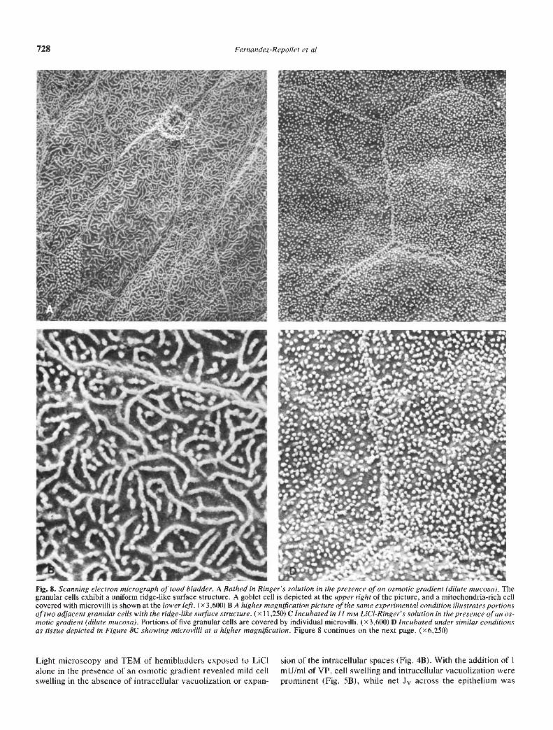
The characteristic appearance of toad urinary bladder bathedin amphibian Ringer's solution in the absence of LiCI and VP isdepicted by SEM in Figures 8A and B. All granular cells arecovered by microplicae or ridges of uniform appearance in thiscontrol preparation. With the addition of 11 mrvi LC1 to the
-. '?..-;..
-ct4 --
aA.-
I';. -'-- —.
At 'I —

726 Fernandez-Repollet et a!
B Incubated under similar conditions as 7A, but with mucosal solution replaced with ii msi Lid-Ringers. Cell swelling (lower right) andintracellular vacuolization are prominent, especially in the granular cells. Intercellular spaces (arrows) are also dilated. (x6,887)
mucosal bathing solution, there was complete transformation ofthe ridges to individual microvilli in all cells in every bladderexamined (Figs. 8C and D). The response to LiCI was identicalto that observed following the addition of VP to the serosalbathing solution (Fig. 8E). Thus, coincident with an increase inboth Pd and Pf, but with no change in P, there was atransformation of ridges to individual microvilli.
Discussion
This study evaluated the effects of LiC1 on Jv in toad urinarybladder epithelium. Exposure of the mucosal surface to 11 mtLiC1 did not inhibit VP-stimulated Jv at a hormone concentra-tion of 20 or 100 mU/mi. However, when the concentration ofVP was reduced to 1 mU/mI, mucosal exposure to LiCIsignificantly inhibited Jv. To minimize variability among differ-ent tissue preparations, an experimental protocol was devisedto measure VP-induced Jv in the same sac preparation in both
the presence and absence of LiCl. The use of this moresensitive experimental protocol might explain, at least in part,the difference between our results and those of Singer andFranko 5] who reported an inhibitory effect of 11 ma't LiC1 onthe hydro-osmotic response induced by 100 mU/ml of VP.Although there were other differences in the experimentaldesign such as a shorter period of LiCI exposure and thepresence of a greater osmotic gradient, it seems unlikely thatthese differences contributed significantly to the difference inresults. In addition, in the present study comparison of peakwater flow rates measured at 10-mm intervals instead of totalwater flow rates measured 30 mm after VP exposure also failedto reveal an inhibitory effect of LiCl on Jv as previouslysuggested by the same group of investigators [5]. In our studypeak Jv occurred 10 to 20 mm after VP administration in allbladders independent of the presence or absence of LiC1. Nosignificant difference in VP-induced peak Jv was detected
.•- - • -qi 1r -, V
1* v9S
---"4
L-. f-b •
--4b;:'t •.J' r-- -;•-• - — 4 •• •
• - - •. -
- • •':•)--.
I
—• I
V
- -5-
Yr
r

Response of toad urinary bladder to L1CI 727
C Incubated with 100 ,nU/ml of VP in the presence of a gradient and mucosal 11 mr.s LiCI-Ringers, Cell swelling and intracellular vacuolization areespecially prominent. (x5,l41)
between those bladders exposed to LiCI and 20 or 100 mU/mi ofVP (—19.3 5.8% and —17.1 5.5%, respectively) and thoseexposed to VP alone (—11.4 6.1%; P NS).
It is well known that the transport processes described in thetoad urinary bladder resemble those occurring in the mammali-an distal nephron. Therefore, it is not surprising that several invivo studies have demonstrated that LiCI impairs the respon-siveness of the collecting duct to VP in experimental animals [1,7, 14, 151 and humans [1,4, 16]. Hochman and Gutman [15] andJenner and McNeil [2] have shown that LiCI antagonized theresponse of the rat collecting duct to VP at a wide range ofhormone concentrations. However, examination of these datareveals that the inhibition by LiC1 of the usual increase in urineosmolality [15] and decrease in urine volume [2, 15] that isassociated with VP administration was greater in magnitude atlow concentrations of the hormone. These in vivo observationscompare quite favorably with our finding of an inhibitory effect
of LiC1 on Jv in bladders exposed to 1 mU/mi of VP, but thefailure to observe a similar effect with 20 and 100 mU/mI of VP.
The demonstration by SEM that LiCl exposure of the muco-sal surface of the hemibiadder was associated with the transfor-mation of microplicae to individual microvilli suggested thatLiC1 alone was capable of increasing the water permeability ofthe apical membrane of the granular cell. This hypothesis wasbased, in part, on the results of earlier studies from ourlaboratory in which VP, cAMP, and serosal hypertonicity allcaused a similar transformation, the former two in both thepresence and absence of an osmotic gradient [12, 17]. All threeconditions increase the water permeability of the apical mem-brane of the granular cell. To test this hypothesis further, Pf, Pd,and P. were measured with LiCI alone and with LiC1 and VP.Values for both Pf and Pd were doubled in comparison withcontrol hemibladders when 11 min LiCI was substituted forNaCI in the mucosal bath, while no change was found in Pu.
1.
1*
•1
I 1f
-
'c -H
3.
-.'c
1s
t.
.C
IL..
a-
-5/
'Is
I. -a
728 Ferncindez-Repollet et al
Fig. 8. Scanning electron micrograph of toad bladder. A Bathed in Ringer's solution in the presence of an osmotic gradient (dilute mucosa). Thegranular cells exhibit a uniform ridge-like surface structure. A goblet cell is depicted at the upper right of the picture, and a mitochondria-rich cellcovered with microvilli is shown at the lower left. (x 3,600) B A higher magnification picture of the same experimental condition illustrates portionsof two adjacent granular cells with the ridge-like surface structure. (X 11 ,250) C Incubated in 11 mM LiCI-Ringer's solution in the presence of an os-motic gradient (dilute mucosa). Portions of five granular cells are covered by individual microvilli. (x 3,600) D Incubated under similar conditionsas tissue depicted in Figure SC showing microvilli at a higher magnification. Figure 8 continues on the next page. (x6,250)
Light microscopy and TEM of hemibladders exposed to LICI sion of the intracellular spaces (Fig. 4B). With the addition of 1alone in the presence of an osmotic gradient revealed mild cell mU/mI of VP, cell swelling and intracellular vacuolization wereswelling in the absence of intracellular vacuolization or expan- prominent (Fig. 5B), while net Jv across the epithelium was

Response of toad urinary bladder to LjCl 729
Fig. 8 E Incubated in Ringer's solution in the presence of an osmoticgradient and I mU/mi of VP. Portions of three adjacent granular cellsare covered by individual microvilli identical in appearance to thoseobserved after exposure to mucosal LiCI. (x 10,900)
impaired. These are the morphological features one wouldanticipate if LiCI as well as VP was capable of increasing thewater permeability of the apical plasmalemma, but the exit stepfor water removal from the cell somewhere beyond the apicalcell membrane was impaired. At higher doses of VP in thepresence of LiC1, intracellular vacuolization was still present,but no significant effect on transepithelial water flow wasdetected. The larger doses of VP were somehow able toovercome the inhibitory effects of LiC1 on VP-induced transepi-thelial water movement. In addition, while LiCl alone resultedin a modest degree of cell swelling in the presence of an osmoticgradient, the addition of VP to the serosal bath was critical forthe development of large intracellular vacuoles. Thus, whileLiCI is capable of increasing both Pf and Pd, the magnitude ofthe permeability changes and, hence, the increase in J wasinsufficient to accentuate the altered pathway(s) of water move-ment across the epithelium occurring in the presence of LiCl.These findings are similar to those reported in toad urinarybladders pretreated with cytochalasin B before exposure to VPin the presence of an osmotic gradient [18]. Employing SEM inthis experimental setting, LeFurgey, Dratwa, and Tisher [18]observed transformation of microplicae to microvilli, whileTEM revealed the presence of large intracellular vacuoles inassociation with significant impairment in Jv. Similar structuraland functional results were reported by Abramow and Dratwa[19] with in vitro perfusion of rabbit collecting ducts pretreatedwith cytochalasin B and exposed to VP in the presence of anosmotic gradient. In contrast with the present studies, however,the large dilated intracellular vacuoles appeared to contribute tothe blunted response to VP in the cytochalasin B studies [18—201.
LiCI decreased sodium transport significantly as determinedby measurement of SCC and the PD in both the presence andabsence of VP. The sodium transport data are consistent withthe earlier studies of Singer and Franko [51 who also demon-strated that the effects of lithium on SCC could be reversed byamiloride. Since the decrease in PD and SCC during basalconditions was of the same order of magnitude, no significanteffect on R was observed in LiCI-treated bladders. AlthoughVP-stimulated SCC and PD were significantly inhibited by LiCIat all hormone concentrations tested, an increase in R wasobserved only in the presence of LiC1 and the higher VPconcentrations of 20 and 100 mU/mi. The precise mechanismresponsible for the inhibition of sodium transport and thenatriferic response to VP by LiCI remains unclear. The findingsmay reflect competition between lithium and sodium ions foressential transport sites at the epithelial cell membrane becauseof their basic structural similarity. As a consequence sodiumentry into the cell is diminished. Evidence supporting thisexplanation emerges from studies in frog skin [2 1—231 and frog[24, 25] and toad [5, 26, 27] urinary bladder that strongly suggestthat lithium ions use the same transport pathway as sodium ionsto gain access across the mucosal membrane. On the otherhand, the reduction in sodium transport observed in the pres-ence of lithium might be the result of a direct effect of the ion onthe sodium "pump." In this regard it has been demonstratedthat lithium ions are poorly transported by the sodium "pump"localized in the basolateral membrane, thus accounting for theinhibition of net sodium transport induced by lithium [281.
In summary, our findings demonstrate that mucosal LiCIenhances transepithelial water movement in toad urinary blad-der epithelium as indicated by a significant increase in basal Jv,Pf, and Pd. SEM revealed that the increase in water permeabili-ty induced by LiC1 was associated with transformation of thetypical ridge-like surface pattern of granular cells to elongatedmicrovilli. This morphological response is similar to that whichhas been noted during stimulation of transepithelial watertransport with VP, cyclic-AMP, and serosal hypertonicity [17,29, 30]. Our data also indicate that the inhibitory effect ofmucosal LiCI on VP-stimulated Jv is evident only at lowconcentrations of the hormone. LiC1 did not alter either basal orVP-stimulated P, thus providing additional evidence that ureaand water cross the apical membrane of the granular cell viaseparate pathways. Finally, in agreement with previous reports[4—6] a consistent inhibition of sodium transport (SCC) by LiC1was observed regardless of the presence or absence of VP.
AcknowledgmentsThis work was supported in part by United States Public Health
Service grants AM 28330 and AM 26569. Dr. A. LeFurgey wassupported through United States Public Health Service National Re-search Service Award, T 32 GM 07403. The current address for Dr. E.Fernandez-Repollet is the Department of Pharmacology, School ofMedicine, University of Puerto Rico, and for Dr. A. LeFurgey, it is theDepartment of Physiology, Duke University, Durham, North Carolina.Mrs. H. Parks, Mrs. K. Blake, and Mrs. N. Dehgan gave technicalassistance. Ms. E. Clausnitzer prepared the illustrations and Ms. J.Harris provided secretarial support.
Reprint requests to Dr. C. C. Tisher, Division of Nephrology, Box J-224, JHMHC, University of Florida, Gainesville, Florida 32610, USA
References
1. FORREST iN, COHAN AD, TORRETTI J, HIMMELHOCK JM, EP-sTEIN FH: On the mechanism of lithium-induced nephrogenic

730 Fernandez-Repollet et a!
diabetes insipidus in man and the rat. J Clin Invest 53:11 15—i 123,1974
2. JENNER LA, MACNEIL S: The effects of lithium ions on theantidiuretic action of vasopressin in the rat. BrJ Pharmacol 55:527—534, 1975
3. MARTINEZ-MALDONADO M, STAVROULAKI-TSAPARA A, TSAPARAN, SuE! WN, EKNOYAN G: Renal effects of lithium administrationin rats: Alterations in water and electrolyte metabolism and theresponse to vasopressin and cyclic-adenosine monophosphate dur-ing prolonged administration. J Lab Clin Med 86:445—461, 1975
4. SINGER I, ROTENBERG D, PUSCHETT JB: Lithium-induced nephro-genic diabetes insipidus: In vivo and in vitro studies. J Gun Invest51:1081—1091, 1972
5. SINGER I, FRANKO EA: Lithium-induced ADH resistance in toadurinary bladders. Kidney mt 3:15 1—159, 1973
6. BENTLEY PJ, WASSERMAN A: The effects of lithium on the perme-ability of an epitheliai membrane in the toad urinary bladder.Biochern Biophys Ada 266:285—292, 1972
7. HARRIS CA, JENNER FA: Some aspects of the inhibition of theaction of antidiuretic hormone by lithium ions in the rat kidney andbladder of the toad Bufo marinus, Br J Pharmacol 44:223—232, 1972
8. BENTLEY PJ: The effects of neurohypophysial extracts on watertransfer across the wall of the isolated urinary bladder of the toad,Bufo marinus. J Endocrinol 17:201—209, 1958
9. USSING HH, ZERAHN K: Active transport of sodium as the sourceof electric current in the short-circuited isolated frog skin. ActaPhysiolScand 23:110—127, 1951
10. HARDY MA: Requirement of Na and K for the action ofantidiuretic hormone on water permeability. Am J Physiol234:F476—F480, 1982
11. LUFT JH: Improvements in epoxy resin embedding methods. JBiophys Biochem Cytol 9:409—414, 1961
12. LEFURGEY A, TISHER CC: Time course of vasopressin-inducedformation of microvilli in granular cells of toad urinary bladder. JMembrBiol 61:13—19, 1981
13. SNEDECOR OW, COCHRAN WG: Statistical Methods (6th ed).Ames, Iowa, Iowa State University, 1967, pp. 111—121
14. CHRISTENSEN S, GEISLER A: Effect of antidiuretic drugs in ratswith lithium-induced polyuria. Acta Pharmacol Toxicol (Copenh)38:81—89, 1976
15. HOCHMAN S, GUTMAN Y: Lithium: ADH antagonism and ADHindependent action in rats with diabetes insipidus. EurJ Pharmacol28:100—107, 1974
16. BAYLIS PH, HEATH DA: Water disturbances in patients treatedwith oral lithium carbonate. Ann Intern Med 88:607—609, 1978
17. DRATWA M, LEFURGEY A, TISHER CC: Effect of vasopressin andserosal hypertonicity on toad urinary bladder. Kidney mt 16:695—703, 1979
18. LEFURGEY A, DRATWA M, TISHER CC: Effects of colchicine andcytochalasin B on vasopressin- and cylic adenosine monophos-phate-induced changes in toad urinary bladder. Lab Invest 45:308—315, 1981
19. ABRAMOW M, DRATWA M: Studies with cytochalasin B in theisolated rabbit collecting tubule: possible role of microfilaments invasopressin action. Colloq Inst Nat Sante Rech Med 30:133, 1974
20. HARDY MA, DIBONA DR: Microfilaments and the hydroosmoticaction of vasopressin in toad urinary bladder. Am J Physiol243:C200—C204, 1982
21. BIBER TUL, CURRAN PF: Dircct measurement of uptake of sodiumat the outer surface of the frog skin. J Gen Physiol 56:83—99,1970
22. DORGE A, NAGEL W: Effect of amiloride on sodium transport in thefrog skin. II. Sodium transport pooi and unidirectional fluxes.Pfluegers Arch 321:91—101, 1970
23, LEBLANC G: The mechanism of lithium accumulation in the isolatedfrog skin epithelium. Pfluegers Arch 337:1—18, 1972
24. BENTLEY PJ: Amiloride: A potent inhibitor of sodium transportacross the toad bladder. J Physiol (Lond) 195:317—330, 1968
25. EHRLICH EN, CRABBE J: The mechanism of action of amiprami-zide. Pfluegers Arch 302:79—96, 1968
26. HERRERA FC: Inhibition of lithium transport across toad urinarybladder by amiloride. Am J Physiol 222:499—502, 1972
27. HERRERA FC, EGEA R, HERRERA AM: Movement of lithium acrosstoad urinary bladder. Am J Physiol 220:1501—1508, 1971
28. DICK DAT, NAYLER GJ, DICK EG: Effects of lithium on sodiumtransport across membranes, in Lithium in Medical Practice, editedby JOHNSON FN, JOHNSON S, Baltimore, University Park Press,1978, p. 173
29. MILLS JW, MALICK LE: Mucosal surface morphology of the toadurinary bladder: Scanning electron microscope study of the natri-feric and hydrosmotic response to vasopressin. J Cell Biol 77:598—610, 1978
30. SPINELLI F, GROSS A, DESOUSA RC: The hydrosmotic effect ofvasopressin: A scanning electron-microscope study. J Membr Biol23:139—156, 1975