Structural and Functional Characterization on the Interaction of Yeast TFIID Subunit TAF1 with...
12
Review Structural and functional characterisation of cardiac fibroblasts Patrizia Camelliti a , Thomas K. Borg b , Peter Kohl a, * a University Laboratory of Physiology, Oxford, OX1 3PT, UK b Department of Cell and Developmental Biology and Anatomy, School of Medicine, University of South Carolina, Columbia, SC, United States Received 15 June 2004; received in revised form 20 August 2004; accepted 31 August 2004 Available online 18 October 2004 Time for primary review 27 days Abstract Cardiac fibroblasts form one of the largest cell populations, in terms of cell numbers, in the heart. They contribute to structural, biochemical, mechanical and electrical properties of the myocardium. Nonetheless, they are often disregarded by in vivo and in vitro studies into cardiac function. This review summarizes our understanding of fibroblast origin and identity, their structural organization and role in myocardial architecture, as well as functional aspects related to cell signalling and electro-mechanical function in the heart. D 2004 European Society of Cardiology. Published by Elsevier B.V. All rights reserved. Keywords: Connective tissue; Myocardium; Remodelling; Gap junction; Heart 1. Introduction Cardiac myocytes occupy approximately 75% of normal myocardial tissue volume, but they account for only 30– 40% of cell numbers [1]. The majority of the remaining cells are non-myocytes, predominantly fibroblasts. Other cell types, such as endothelial or vascular smooth muscle cells, represent comparatively small populations [2]. Fibroblasts are found throughout the cardiac tissue, surrounding myocytes and bridging dthe voidsT between myocardial tissue layers, so that, in essence, every cardiomyocyte is closely related to a fibroblast in normal cardiac tissue (Fig. 1). Pathological states are frequently associated with myocardial remodelling involving fibrosis. This is observed in ischemic and rheumatic heart disease, inflammation, hypertrophy, and infarction. The growth in fibrous tissue content is based on the maintained proliferative potential of fibroblasts (largely absent in myocytes of the adult heart [3]), and the synthesis of extra-cellular matrix (ECM) proteins, predominantly by fibroblasts [4]. Fibroblasts contribute to cardiac development, myocar- dial structure, cell signalling, and electro-mechanical func- tion in healthy and diseased myocardium [4–6]. In the following sections, we summarise current insight into cardiac fibroblast origin and identity, their structural and functional contribution to cardiac function, and highlight emerging hypotheses and targets for further research. 2. What is a fibroblast? 2.1. Terminology and scope Fibroblasts are obligatory components of the ECM. They are widely distributed in most vertebrate organisms, and associated with the various forms of connective tissue [7]. Fibroblasts are traditionally defined as cells of mesen- chymal origin that produce interstitial collagen (in contrast to myocytes that form collagen type IV as part of their basement membrane, fibroblasts also produce types I, III and VI). Collagen synthesis or deposition is, however, not usually assessed in the context of fibroblast identification (i.e. via in situ hybridization, or via immuno-cytochemical localization). Instead, cell classifi- 0008-6363/$ - see front matter D 2004 European Society of Cardiology. Published by Elsevier B.V. All rights reserved. doi:10.1016/j.cardiores.2004.08.020 * Corresponding author. Fax: +44 1865 272554. E-mail address: [email protected] (P. Kohl). Cardiovascular Research 65 (2005) 40 – 51 www.elsevier.com/locate/cardiores by guest on April 20, 2013 http://cardiovascres.oxfordjournals.org/ Downloaded from
Transcript of Structural and Functional Characterization on the Interaction of Yeast TFIID Subunit TAF1 with...
www.elsevier.com/locate/cardiores
Cardiovascular Research
D
Review
Structural and functional characterisation of cardiac fibroblasts
Patrizia Camellitia, Thomas K. Borgb, Peter Kohla,*
aUniversity Laboratory of Physiology, Oxford, OX1 3PT, UKbDepartment of Cell and Developmental Biology and Anatomy, School of Medicine, University of South Carolina, Columbia, SC, United States
Received 15 June 2004; received in revised form 20 August 2004; accepted 31 August 2004
Available online 18 October 2004
Time for primary review 27 days
http://cardiovascres.oxfordjournow
nloaded from
Abstract
Cardiac fibroblasts form one of the largest cell populations, in terms of cell numbers, in the heart. They contribute to structural,
biochemical, mechanical and electrical properties of the myocardium. Nonetheless, they are often disregarded by in vivo and in vitro studies
into cardiac function. This review summarizes our understanding of fibroblast origin and identity, their structural organization and role in
myocardial architecture, as well as functional aspects related to cell signalling and electro-mechanical function in the heart.
D 2004 European Society of Cardiology. Published by Elsevier B.V. All rights reserved.
Keywords: Connective tissue; Myocardium; Remodelling; Gap junction; Heart
by guest on April 20, 2013
als.org/
1. Introduction
Cardiac myocytes occupy approximately 75% of normal
myocardial tissue volume, but they account for only 30–
40% of cell numbers [1]. The majority of the remaining cells
are non-myocytes, predominantly fibroblasts. Other cell
types, such as endothelial or vascular smooth muscle cells,
represent comparatively small populations [2].
Fibroblasts are found throughout the cardiac tissue,
surrounding myocytes and bridging dthe voidsT between
myocardial tissue layers, so that, in essence, every
cardiomyocyte is closely related to a fibroblast in normal
cardiac tissue (Fig. 1). Pathological states are frequently
associated with myocardial remodelling involving fibrosis.
This is observed in ischemic and rheumatic heart disease,
inflammation, hypertrophy, and infarction. The growth in
fibrous tissue content is based on the maintained
proliferative potential of fibroblasts (largely absent in
myocytes of the adult heart [3]), and the synthesis of
extra-cellular matrix (ECM) proteins, predominantly by
fibroblasts [4].
0008-6363/$ - see front matter D 2004 European Society of Cardiology. Publish
doi:10.1016/j.cardiores.2004.08.020
* Corresponding author. Fax: +44 1865 272554.
E-mail address: [email protected] (P. Kohl).
Fibroblasts contribute to cardiac development, myocar-
dial structure, cell signalling, and electro-mechanical func-
tion in healthy and diseased myocardium [4–6]. In the
following sections, we summarise current insight into
cardiac fibroblast origin and identity, their structural and
functional contribution to cardiac function, and highlight
emerging hypotheses and targets for further research.
2. What is a fibroblast?
2.1. Terminology and scope
Fibroblasts are obligatory components of the ECM.
They are widely distributed in most vertebrate organisms,
and associated with the various forms of connective tissue
[7].
Fibroblasts are traditionally defined as cells of mesen-
chymal origin that produce interstitial collagen (in
contrast to myocytes that form collagen type IV as part
of their basement membrane, fibroblasts also produce
types I, III and VI). Collagen synthesis or deposition is,
however, not usually assessed in the context of fibroblast
identification (i.e. via in situ hybridization, or via
immuno-cytochemical localization). Instead, cell classifi-
65 (2005) 40–51
ed by Elsevier B.V. All rights reserved.

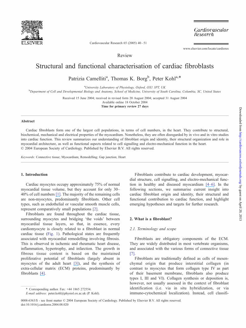
Fig. 1. Cardiac tissue structure overview. (A) Gross histological view (transversal cross-section) of rabbit ventricular myocardium, stained with Hematoxylin
and Eosin, shows layered sheets of cardiac muscle cells (pink), separated by dvoidsT that are filled with (un-labelled) non-myocytes. (B) Larger magnification
view of ventricular myocardium (confocal microscopy tissue cross-section), immuno-stained with anti-myomesin to mark myocytes (red), anti-vimentin to
mark fibroblasts (blue), anti-connexin43 (bright green dots) and with DAPI (4V-6-Diamidino-2-phenylindole) to label nuclei (pale yellow–green patches),
showing the dense network of fibroblasts that surrounds myocyte clusters of 2–4 cells. Scale bars in A and B: 5 mm and 20 Am, respectively.
P. Camelliti et al. / Cardiovascular Research 65 (2005) 40–51 41
by guest on April 20, 2013
http://cardiovascres.oxfordjournals.org/D
ownloaded from
cation is often based on morphological characteristics and/
or proliferative potential.
These dsubstitute identifiersT can vary widely with
location and metabolic activity of individual cells, organs,
or organisms. In general, though, fibroblasts lack a base-
ment membrane and tend to have multiple processes or
sheet-like extensions. They contain an oval nucleus (with 1
or 2 nucleoli), extensive rough endoplasmic reticulum, a
prominent Golgi apparatus, and abundant cytoplasmic
granular material.
The lack of a truly specific marker has long been a
limiting factor in studying fibroblasts in vivo. A useful label
for cardiac fibroblasts are anti-vimentin antibodies that react
with the abundant intermediate filaments of fibroblasts [8].
This marker also labels other cell types, for example
vascular endothelial cells and neurones, that contain
intermediate filaments. Given the characteristic cyto-mor-
phological differences between these cell types, however,
anti-vimentin has been a suitable tool for reliable identi-
fication of cardiac fibroblasts.
Fig. 2. Fibroblast myocyte interrelation in cardiac tissue. (A) Fibroblasts labelle
mouse ventricular tissue (red label: connexin43). (B) Fibroblasts labelled with a
(green) in sheep ventricular tissue (red: connexin43). Note the principal similarity
A more specific marker for cardiac fibroblasts is the
collagen receptor Discoidin Domain Receptor 2 (DDR2
[9]). DDR1 and DDR2 represent a relatively novel family of
collagen specific receptor tyrosine kinases [10,11]. Receptor
tyrosine kinases are a family of proteins involved in the
conversion of extracellular stimuli into cellular responses
[12]. These receptors mediate a variety of cell functions,
including growth, migration, morphology and differentia-
tion. The tissue distribution of DDR1 and DDR2 varies (and
can be mutually exclusive [13]), and DDR2 expression has
been detected in both rat and mouse heart [14]. Originally
defined as a collagen receptor on mesenchymal cells, DDR2
has also been found on leukocytes, as well as in tumours,
but not on cardiomyocytes or cardiac endothelial and
smooth muscle cells [9]. Interestingly, DDR2 gives a
labelling pattern that is very similar to that obtained using
vimentin antibodies (Fig. 2).
It is important to note that fibroblasts are principally
motile cells that contain actin (mainly a-smooth muscle
actin) and myosin, so that their identification as dcells
d with anti-DDR2 (blue) separate phalloidin-labelled myocytes (green) in
nti-vimentin (blue) form layers between anti-myomesin labelled myocytes
of cell type interrelation identified by the two antibodies. Scale bar: 20 Am.
P. Camelliti et al. / Cardiovascular Research 65 (2005) 40–5142
void of contractile proteinsT may cause false-negative
findings.
In addition, the term dmyofibroblastT—if applied solely
on the basis of actin or myosin presence—may be more
misleading than helpful. Fibroblasts are pleiomorphic, and
their actin or myosin content and arrangement are affected
by the environment, in particular mechanical parameters. An
increased contractile filament content does not necessarily
transform a fibroblast into a new cell type, but may merely
represent a distinctive phenotype. In the absence of a clear
and consistent definition, myofibroblasts will therefore not
be addressed as a separate entity in this review.
by guest on April 20, 2013
http://cardiovascres.oxfordjournals.org/D
ownloaded from
3. Origin of cardiac fibroblasts
3.1. Physiology
The mesenchymal cells that form the cardiac fibroblast
population are believed to be derived from two principal
sources: (1) the pro-epicardial organ, and (2) the epithelial–
mesenchymal transformation during the formation of
cardiac valves [15,16]. Other sources, such as the develop-
ing bone marrow and neural crest, differentiation from the
vascular walls, or circulating progenitor cells, are conceiv-
able, but we are far from an understanding of their relevance
in the healthy heart.
Fibroblast content increases with normal development
and aging [2,17]. During early human development,
myocyte and connective tissue cell numbers increase at a
similar rate, from about 0.5�109 at 28 weeks of foetal
development to 2–3�109 several weeks post partum.
Thereafter, myocyte cell numbers remain stable, while the
connective tissue cell count rises with cardiac weight to
~7�109 at 2 months of age [2]. This is mirrored by an
increase in the volume fraction of connective tissue, which
reaches about 5–6% in normal adult myocardium, but may
exceed 50% of the adult human sino-atrial node (SAN)
[18,19].
Similar dynamics are observed in other mammals, such
as rat and hamster, where little connective tissue is observed
in the early embryonic heart [20,21]. Most of the connective
tissue is involved, at that stage, with the formation of the
cardiac skeleton and the various valvular structures. The
three-dimensional collagen network, composed of the
epimysium, perimysium and endomysium, begins to form
in late foetal development. It is largely laid down during
neonatal growth [22], accompanied by rapid proliferation of
fibroblasts and substantial deposition of collagen [23].
Following neonatal development, fibroblast cell division
returns to a very low level, unless stimulated by either
physiological or pathological signals.
Thus, fibroblasts form a majority cell population in the
normal adult heart (up to two-thirds), which is largely
interspersed in the collagen network. Their physiological
origin remains to be elucidated more comprehensively.
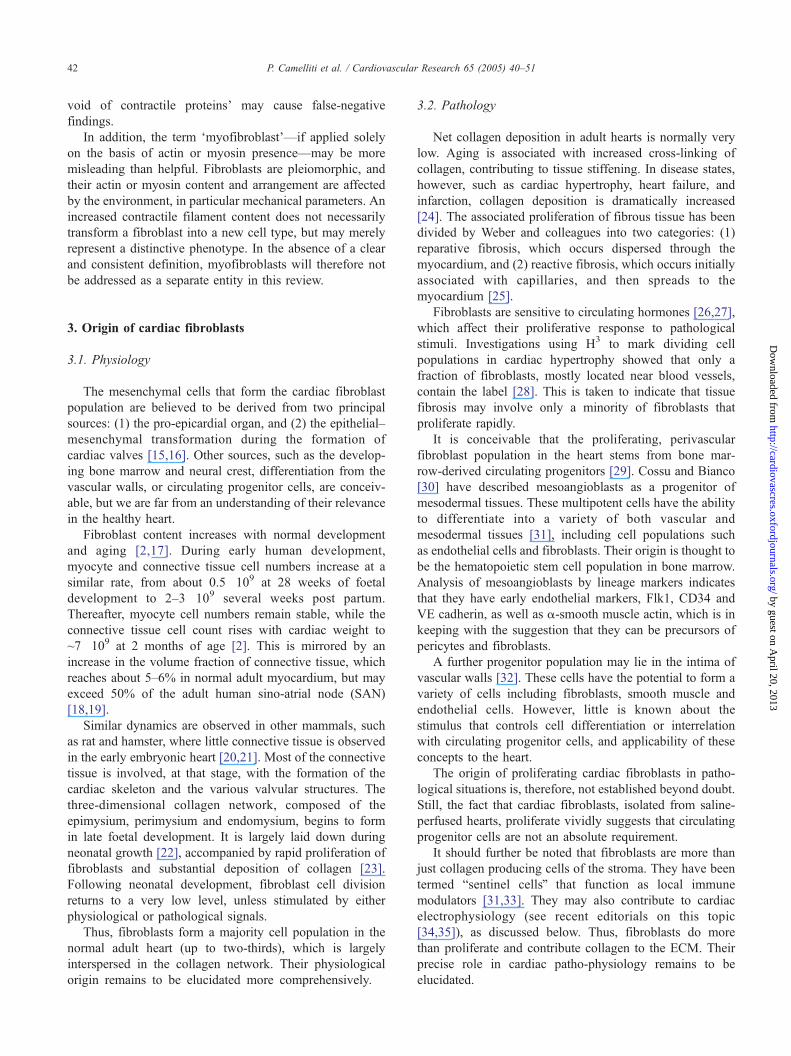
3.2. Pathology
Net collagen deposition in adult hearts is normally very
low. Aging is associated with increased cross-linking of
collagen, contributing to tissue stiffening. In disease states,
however, such as cardiac hypertrophy, heart failure, and
infarction, collagen deposition is dramatically increased
[24]. The associated proliferation of fibrous tissue has been
divided by Weber and colleagues into two categories: (1)
reparative fibrosis, which occurs dispersed through the
myocardium, and (2) reactive fibrosis, which occurs initially
associated with capillaries, and then spreads to the
myocardium [25].
Fibroblasts are sensitive to circulating hormones [26,27],
which affect their proliferative response to pathological
stimuli. Investigations using H3 to mark dividing cell
populations in cardiac hypertrophy showed that only a
fraction of fibroblasts, mostly located near blood vessels,
contain the label [28]. This is taken to indicate that tissue
fibrosis may involve only a minority of fibroblasts that
proliferate rapidly.
It is conceivable that the proliferating, perivascular
fibroblast population in the heart stems from bone mar-
row-derived circulating progenitors [29]. Cossu and Bianco
[30] have described mesoangioblasts as a progenitor of
mesodermal tissues. These multipotent cells have the ability
to differentiate into a variety of both vascular and
mesodermal tissues [31], including cell populations such
as endothelial cells and fibroblasts. Their origin is thought to
be the hematopoietic stem cell population in bone marrow.
Analysis of mesoangioblasts by lineage markers indicates
that they have early endothelial markers, Flk1, CD34 and
VE cadherin, as well as a-smooth muscle actin, which is in
keeping with the suggestion that they can be precursors of
pericytes and fibroblasts.
A further progenitor population may lie in the intima of
vascular walls [32]. These cells have the potential to form a
variety of cells including fibroblasts, smooth muscle and
endothelial cells. However, little is known about the
stimulus that controls cell differentiation or interrelation
with circulating progenitor cells, and applicability of these
concepts to the heart.
The origin of proliferating cardiac fibroblasts in patho-
logical situations is, therefore, not established beyond doubt.
Still, the fact that cardiac fibroblasts, isolated from saline-
perfused hearts, proliferate vividly suggests that circulating
progenitor cells are not an absolute requirement.
It should further be noted that fibroblasts are more than
just collagen producing cells of the stroma. They have been
termed bsentinel cellsQ that function as local immune
modulators [31,33]. They may also contribute to cardiac
electrophysiology (see recent editorials on this topic
[34,35]), as discussed below. Thus, fibroblasts do more
than proliferate and contribute collagen to the ECM. Their
precise role in cardiac patho-physiology remains to be
elucidated.

P. Camelliti et al. / Cardiovascular Research 65 (2005) 40–51 43
by guest on April
http://cardiovascres.oxfordjournals.org/D
ownloaded from
4. Fibroblast structural organization
4.1. Physiology
There are pronounced regional differences in the
organization and content of connective tissue in the heart.
Ventricular myocardium is arranged in highly oriented
layers that are about two to four cells thick. These layers
are embedded into a dense connective tissue network (Fig.
3A), and are interconnected by branches of myocytes
[36,37] and fibroblasts [38].
SAN pacemaker tissue has a higher relative fibroblast
content than ventricle, occupying some 45% to 75% of SAN
volume in man [18,19]. Cells in the SAN, in particular in its
central region, are less regularly organised than in ven-
tricular tissue (Fig. 3B). Fibroblasts are found either
interspersed with pacemaker myocytes, or forming islands
consisting largely of connective tissue only. The age-related
increase in SAN connective tissue coincides, in man, with a
significant reduction in myocyte content [18,19]. These
changes have been suggested to contribute to SAN
dysfunction in the elderly [18].
In all cardiac tissue areas, fibroblasts form a complex
3-D network within the connective tissue matrix that they
occupy [8,9]. There are abundant anatomical contacts,
including extensive membrane appositions between
neighbouring fibroblasts and myocytes. These have
recently been shown to be site of homogeneous and
heterogeneous gap-junctional coupling in rabbit right
atrium [8].
4.2. Pathology
Fibroblast proliferation and tissue remodelling are
features of many of cardiac pathologies, and their detailed
discussion would go beyond the scope of this paper.
Fig. 3. Fibroblast organization in rabbit normal ventricular tissue (A) and ra
elongated cells) and myocytes (anti-myomesin, striated cells) identifies the spatia
almost exclusively of fibroblasts, while in normal ventricle, fibroblasts and myoc
bars: 20 Am.
As an illustration of some key structural re-arrangements
during pathological remodelling, we present findings from a
recent study of sheep coronary occlusion, where fibroblast
infiltration of the damaged tissue occurred within hours of
myocardial infarction. Fibroblast density increased with
time post-infarction, reaching a maximum after 1 week,
followed by a subsequent decrease. Interestingly, fibroblast
content increases even in remote tissue regions that are not
directly affected by the infarct. Fig. 4 illustrates the
characteristic appearance of viable tissue remote from the
infarct zone, with a highly organized myocyte and fibroblast
pattern (Fig. 4A), compared to the infarct border with a mix
of disrupted and healthy myocytes and fibroblasts (Fig. 4B),
and the central infarct zone that is densely packed with
fibroblasts (Fig. 4C) at 1 week after infarction [39].
In general, pathologies cause fibroblast proliferation and
mobilisation, supporting their spread into apparently unaf-
fected tissue, as well as fibroblast phenotype changes (as
reviewed elsewhere [5,25,40]). The potential contribution of
fibroblasts to pathologically altered cardiac function is
discussed below.
4.3. In vitro models
Standard in vitro cardiac cell culture models do not
reproduce the in vivo structural organization of cardiac
fibroblasts, or their interrelation with myocytes. Most
cardiac cell cultures actually try to actively restrict fibroblast
content.
Using advanced micro-structuring techniques it is,
however, possible to create more in vivo-like models of
cardiac tissue [41]. In particular, by growing cells on
spatially restricted patterns, myocytes and fibroblasts can be
prompted to acquire more in vivo-like phenotypes and
spatial interrelation. Thus, using micro-fluidic deposition of
ECM proteins to guide cell attachment on elastic mem-
bbit SAN (B). Labelling for fibroblasts (anti-vimentin, brightly stained
l interrelation of cell types. Note that the SAN contains regions consisting
ytes are interspersed in a regular pattern. F: fibroblast; M: myocyte. Scale
20, 2013

Fig. 4. Fibroblast organization in sheep normal ventricular myocardium (A), infarct border zone (B) and centre (C), 1 week after infarction. Fibroblasts were
stained with anti-vimentin (F, bright elongated cells) and myocytes with anti-myomesin (M, striated cells). Scale bars: 20 Am.
P. Camelliti et al. / Cardiovascular Research 65 (2005) 40–5144
http://cardiovascres.oxfordjournaD
ownloaded from
branes, structured co-cultures of cardiomyocytes and fibro-
blasts have recently replicated important features of cardiac
tissue-architecture [42]. Strands of aligned myocytes show
cross striation that is din registerT among neighbouring cells.
These strands can be surrounded by parallel threads of
fibroblasts, with abundant contacts between the cell types
[38] (Fig. 5A), as seen in vivo.
More complex, three-dimensional cultures can be
devised by the culturing of myocytes on spatially aligned
collagen threads [43], where multiple layers of myocytes
and fibroblasts organize in an in vivo-like manner (Fig. 5B).
These advanced in vitro models are still being fully
characterized, but it is already evident that they will offer
improved tools for studying cardiac fibroblast function in
normal and pathological conditions.
by guest on April 20, 2
ls.org/
5. Fibroblast function
Fibroblasts contribute to structural, biochemical,
mechanical and electrical properties of the myocardium
[4,8,39,40,44]. The following sections will address relevant
aspects of their function.
Fig. 5. In vitro cardiac cell culture models. (A) Structured co-cultures of neonatal r
and myocytes (striated cells, labelled with anti-myomesin antibodies). (B) Three-d
cells) and fibroblasts (DDR2-stained, green). Fibroblast show long processes that
bar: 20 Am.
5.1. Structural and biochemical function
Fibroblasts are involved in the maintenance of myocar-
dial tissue structure, including ECM homeostasis and
production of factors involved in maintaining a balance
between synthesis and degradation of connective tissue
components, for example cytokines, growth factors and
matrix metalloproteinases (MMP).
In cardiovascular diseases, fibroblasts play a central and
dynamic role in the myocardial remodelling process, which
includes hypertrophy of cardiomyocytes, migration and
proliferation of fibroblasts, and changes in the extent and
composition of the cardiac ECM [5,40]. Excessive fibroblast
proliferation and increase in ECM protein content (fibrosis)
induce myocardial stiffening—an important patho-physio-
logical facet of cardiac dysfunction [45,46]. Fibrotic tissue
remodelling is associated with increased expression of
MMP and humoral factors, such as transforming growth
factor TGF-h, angiotensin II, endothelin-1 and tumour
necrosis factor-a.
MMP, by degrading interstitial fibrillar collagen, act as
key players in the ECM remodelling process after myocar-
dial infarction, and in infarct healing [47]. In the early phase
at fibroblasts (bright elongated cells, labelled with anti-vimentin antibodies)
imensional co-cultures of neonatal myocytes (phalloidin-stained red striated
envelope the myocytes, similar to in vivo cardiac tissue organization. Scale
013
P. Camelliti et al. / Cardiovascular Research 65 (2005) 40–51 45
by guest on April 20, 2013
http://cardiovascres.oxfordjournals.org/D
ownloaded from
of myocardial repair, MMP disrupt the collagen ECM
network. This enhances inflammatory cell infiltration that,
by producing cytokines, promotes subsequent fibroblast
migration, proliferation and differentiation, followed by
deposition of new ECM and scar formation. Increased
expression and activation of MMP leads to excessive ECM
degradation, impairment of infarct healing and potentially
cardiac rupture [47].
MMP are not exclusively expressed by fibroblasts, but
also other cardiac cells (like myocytes and endothelial cells)
and by inflammatory cells. They are also involved in the
regulation of cell growth and migration, cell survival/death
and angiogenesis. The details of MMP regulation in normal
and pathological conditions go beyond the scope of this
review, and we refer the reader to previous publications
[47,48].
Angiotensin II, TGF-h and tumour necrosis factor-a are
involved in autocrine and paracrine regulation of myocyte
hypertrophy, fibroblast proliferation and ECM protein turn-
over [40]. Angiotensin II further stimulates collagen gene
expression and collagen synthesis, and it reduces collagen
degradation (by attenuating MMP activity in cardiac
fibroblasts) [45], while endothelin-1 induces hypertrophy
in myocytes and stimulates collagen synthesis [40].
Myocardial tissue remodelling may also be promoted by
chronic adrenergic stimulation, which is an important
feature of heart failure. This has been shown to affect not
only myocyte hypertrophy and cell death [49], but also to
increase proliferation of cultured human cardiac fibroblasts
via secretion of autocrine factors [50].
Interestingly, statins, normally prescribed for lowering
cholesterol, have recently been shown to directly inhibit
fibroblast proliferation, an effect that may contribute to the
prevention of adverse cardiac remodelling [51].
Thus, fibroblast activity may give rise to, at times
unexpected, effects on cardiac structure and function. Given
their susceptibility to a large range of humoral factors, they
may foster a novel target for pharmacological interventions.
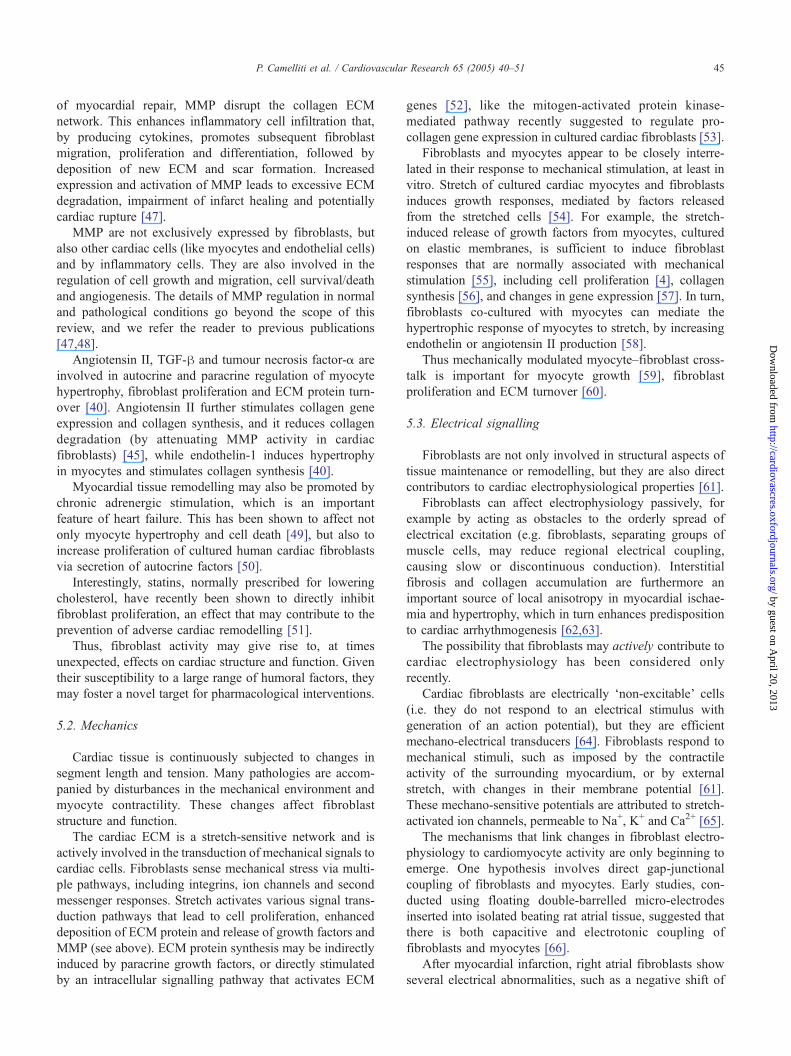
5.2. Mechanics
Cardiac tissue is continuously subjected to changes in
segment length and tension. Many pathologies are accom-
panied by disturbances in the mechanical environment and
myocyte contractility. These changes affect fibroblast
structure and function.
The cardiac ECM is a stretch-sensitive network and is
actively involved in the transduction of mechanical signals to
cardiac cells. Fibroblasts sense mechanical stress via multi-
ple pathways, including integrins, ion channels and second
messenger responses. Stretch activates various signal trans-
duction pathways that lead to cell proliferation, enhanced
deposition of ECM protein and release of growth factors and
MMP (see above). ECM protein synthesis may be indirectly
induced by paracrine growth factors, or directly stimulated
by an intracellular signalling pathway that activates ECM
genes [52], like the mitogen-activated protein kinase-
mediated pathway recently suggested to regulate pro-
collagen gene expression in cultured cardiac fibroblasts [53].
Fibroblasts and myocytes appear to be closely interre-
lated in their response to mechanical stimulation, at least in
vitro. Stretch of cultured cardiac myocytes and fibroblasts
induces growth responses, mediated by factors released
from the stretched cells [54]. For example, the stretch-
induced release of growth factors from myocytes, cultured
on elastic membranes, is sufficient to induce fibroblast
responses that are normally associated with mechanical
stimulation [55], including cell proliferation [4], collagen
synthesis [56], and changes in gene expression [57]. In turn,
fibroblasts co-cultured with myocytes can mediate the
hypertrophic response of myocytes to stretch, by increasing
endothelin or angiotensin II production [58].
Thus mechanically modulated myocyte–fibroblast cross-
talk is important for myocyte growth [59], fibroblast
proliferation and ECM turnover [60].
5.3. Electrical signalling
Fibroblasts are not only involved in structural aspects of
tissue maintenance or remodelling, but they are also direct
contributors to cardiac electrophysiological properties [61].
Fibroblasts can affect electrophysiology passively, for
example by acting as obstacles to the orderly spread of
electrical excitation (e.g. fibroblasts, separating groups of
muscle cells, may reduce regional electrical coupling,
causing slow or discontinuous conduction). Interstitial
fibrosis and collagen accumulation are furthermore an
important source of local anisotropy in myocardial ischae-
mia and hypertrophy, which in turn enhances predisposition
to cardiac arrhythmogenesis [62,63].
The possibility that fibroblasts may actively contribute to
cardiac electrophysiology has been considered only
recently.
Cardiac fibroblasts are electrically dnon-excitableT cells(i.e. they do not respond to an electrical stimulus with
generation of an action potential), but they are efficient
mechano-electrical transducers [64]. Fibroblasts respond to
mechanical stimuli, such as imposed by the contractile
activity of the surrounding myocardium, or by external
stretch, with changes in their membrane potential [61].
These mechano-sensitive potentials are attributed to stretch-
activated ion channels, permeable to Na+, K+ and Ca2+ [65].
The mechanisms that link changes in fibroblast electro-
physiology to cardiomyocyte activity are only beginning to
emerge. One hypothesis involves direct gap-junctional
coupling of fibroblasts and myocytes. Early studies, con-
ducted using floating double-barrelled micro-electrodes
inserted into isolated beating rat atrial tissue, suggested that
there is both capacitive and electrotonic coupling of
fibroblasts and myocytes [66].
After myocardial infarction, right atrial fibroblasts show
several electrical abnormalities, such as a negative shift of
P. Camelliti et al. / Cardiovascular Research 65 (2005) 40–5146
their resting membrane potential, an increase in membrane
resistance, altered mechanically induced potentials, and
enhanced sensitivity to mechanical stress [61]. These altered
electrical properties of fibroblasts could, if electrically
coupled to myocytes, lead to changes in pacemaker activity
[67], such as the depression of heart rate observed in this
model [61]. Furthermore, heterogeneous coupling of fibro-
blasts and myocytes in the infarct border zone would affect
the spread of excitation in such areas and could contribute to
the highly irregular and arrhythmogenic electrical properties
of cardiac scar tissue [6,68].
Thus, fibroblasts may affect electrical signalling in the
heart passively and, if electrically coupled to cardiomyo-
cytes, actively. The extent of gap-junctional coupling of
cardiac fibroblasts is discussed next.
http://cardD
ownloaded from
6. Fibroblast gap-junctional coupling
6.1. Fibroblast–fibroblast coupling
To date, fibroblast gap junction coupling has mainly been
studied in other organs, including skin, kidney, periodontal
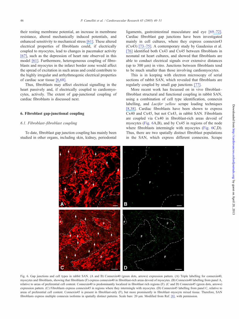
Fig. 6. Gap junctions and cell types in rabbit SAN. (A and B) Connexin40 (gr
myocytes and fibroblasts, showing that fibroblasts (F) express connexin40 in fibrob
relative to areas of preferential cell content. Connexin40 is predominantly localize
expression pattern. (C) Fibroblasts express connexin45 in regions where they inte
areas of preferential cell content. Connexin45 is present in fibroblast-only (F), b
fibroblasts express multiple connexin isoforms in spatially distinct patterns. Scale
ligaments, gastrointestinal musculature and eye [69–72].
Cardiac fibroblast gap junctions have been investigated
mainly in cell cultures, where they express connexin43
(Cx43) [73–75]. A contemporary study by Gaudesius et al.
[76] identified both Cx43 and Cx45 between fibroblasts in
neonatal rat heart cultures, and showed that fibroblasts are
able to conduct electrical signals over extensive distances
(up to 300 Am) in vitro. Junctions between fibroblasts tend
to be much smaller than those involving cardiomyocytes.
This is in keeping with electron microscopy of serial
sections of rabbit SAN, which revealed that fibroblasts are
regularly coupled by small gap junctions [77].
More recent work has focussed on in vivo fibroblast–
fibroblast structural and functional coupling in rabbit SAN,
using a combination of cell type identification, connexin
labelling, and Lucifer yellow scrape loading techniques
[8,38]. Cardiac fibroblasts have been shown to express
Cx40 and Cx45, but not Cx43, in rabbit SAN. Fibroblasts
are coupled via Cx40 in fibroblast-rich areas devoid of
myocytes (Fig. 6A,B), and by Cx45 in regions of the node
where fibroblasts intermingle with myocytes (Fig. 6C,D).
Thus, there are two spatially distinct fibroblast populations
in the SAN, which express different connexins. Scrape
een dots, arrows) expression pattern. (A) Triple labelling for connexin40,
last-rich areas devoid of myocytes. (B) Connexin40 labelling from panel A,
d in fibroblast rich regions (F). (C and D) Connexin45 (green dots, arrows)
rmingle with myocytes. (D) Connexin45 labelling from panel C, relative to
ut more prominently in fibroblast–myocyte mixed tissue. Therefore, SAN
bars: 20 Am. Modified from Ref. [8], with permission.
by guest on April 20, 2013
iovascres.oxfordjournals.org/
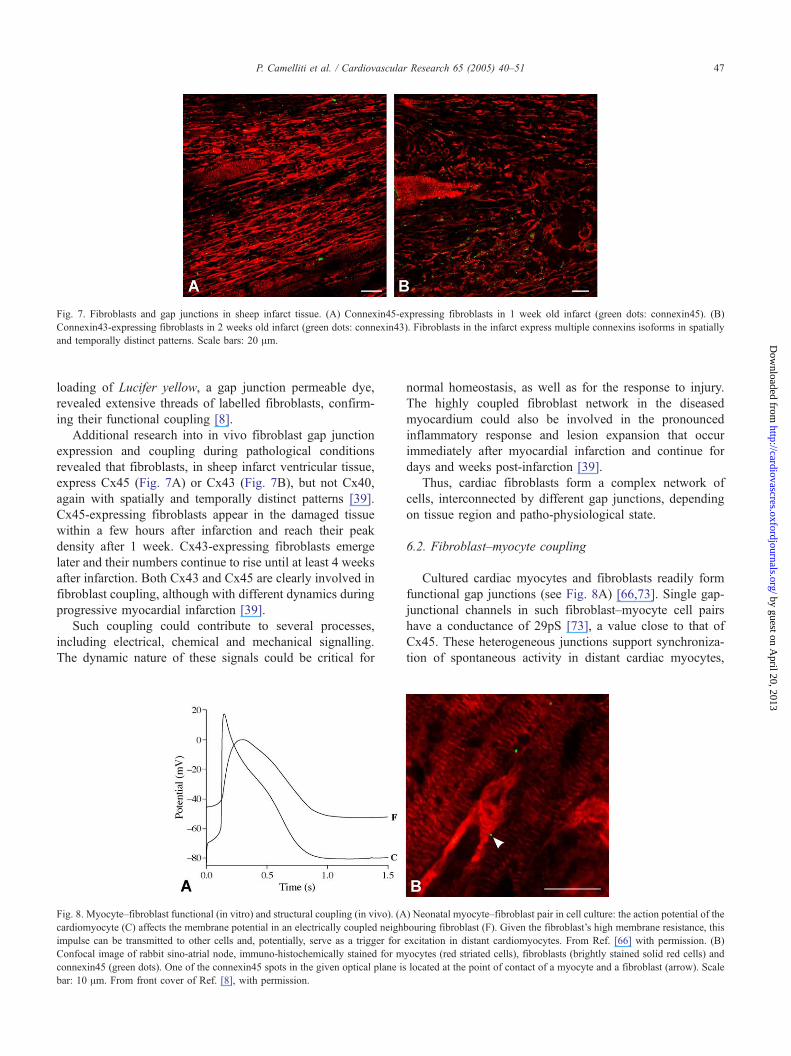
Fig. 7. Fibroblasts and gap junctions in sheep infarct tissue. (A) Connexin45-expressing fibroblasts in 1 week old infarct (green dots: connexin45). (B)
Connexin43-expressing fibroblasts in 2 weeks old infarct (green dots: connexin43). Fibroblasts in the infarct express multiple connexins isoforms in spatially
and temporally distinct patterns. Scale bars: 20 Am.
P. Camelliti et al. / Cardiovascular Research 65 (2005) 40–51 47
by guest on April
http://cardiovascres.oxfordjournals.org/D
ownloaded from
loading of Lucifer yellow, a gap junction permeable dye,
revealed extensive threads of labelled fibroblasts, confirm-
ing their functional coupling [8].
Additional research into in vivo fibroblast gap junction
expression and coupling during pathological conditions
revealed that fibroblasts, in sheep infarct ventricular tissue,
express Cx45 (Fig. 7A) or Cx43 (Fig. 7B), but not Cx40,
again with spatially and temporally distinct patterns [39].
Cx45-expressing fibroblasts appear in the damaged tissue
within a few hours after infarction and reach their peak
density after 1 week. Cx43-expressing fibroblasts emerge
later and their numbers continue to rise until at least 4 weeks
after infarction. Both Cx43 and Cx45 are clearly involved in
fibroblast coupling, although with different dynamics during
progressive myocardial infarction [39].
Such coupling could contribute to several processes,
including electrical, chemical and mechanical signalling.
The dynamic nature of these signals could be critical for
Fig. 8. Myocyte–fibroblast functional (in vitro) and structural coupling (in vivo). (A
cardiomyocyte (C) affects the membrane potential in an electrically coupled neigh
impulse can be transmitted to other cells and, potentially, serve as a trigger for
Confocal image of rabbit sino-atrial node, immuno-histochemically stained for m
connexin45 (green dots). One of the connexin45 spots in the given optical plane is
bar: 10 Am. From front cover of Ref. [8], with permission.
normal homeostasis, as well as for the response to injury.
The highly coupled fibroblast network in the diseased
myocardium could also be involved in the pronounced
inflammatory response and lesion expansion that occur
immediately after myocardial infarction and continue for
days and weeks post-infarction [39].
Thus, cardiac fibroblasts form a complex network of
cells, interconnected by different gap junctions, depending
on tissue region and patho-physiological state.
6.2. Fibroblast–myocyte coupling
Cultured cardiac myocytes and fibroblasts readily form
functional gap junctions (see Fig. 8A) [66,73]. Single gap-
junctional channels in such fibroblast–myocyte cell pairs
have a conductance of 29pS [73], a value close to that of
Cx45. These heterogeneous junctions support synchroniza-
tion of spontaneous activity in distant cardiac myocytes,
) Neonatal myocyte–fibroblast pair in cell culture: the action potential of the
bouring fibroblast (F). Given the fibroblast’s high membrane resistance, this
excitation in distant cardiomyocytes. From Ref. [66] with permission. (B)
yocytes (red striated cells), fibroblasts (brightly stained solid red cells) and
located at the point of contact of a myocyte and a fibroblast (arrow). Scale
20, 2013

P. Camelliti et al. / Cardiovascular Research 65 (2005) 40–5148
by guest on April 20, 2013
http://cardiovascres.oxfordjournals.org/D
ownloaded from
interconnected by fibroblasts only [76,78]. This behaviour is
promoted by the fact that fibroblasts have a very high
membrane resistance, making them excellent long-distance
conductors [35].
In addition, cultured cardiac fibroblasts may act as a
dcurrent sinkT for connected myocytes. Thus, transfection of
fibroblasts with the voltage-sensitive potassium channel
Kv1.3 changes cardiomyocyte excitability in neonatal rat
heart culture, illustrating the pronounced effect of electri-
cally coupled fibroblasts on cardiomyocyte electrophysiol-
ogy in vitro [79].
Immuno-histochemical identification of connexins in co-
cultures of cardiac fibroblasts and myocytes revealed Cx43
and Cx45 at points of heterogeneous cell contact. These
junctions are generally considerably smaller than cardio-
myocyte nexus [76].
In vitro evidence therefore suggests that myocytes and
fibroblasts are able to form functional gap junctions, which
could underlie the ability of fibroblasts to serve as
conductors for electrical excitation and to affect myocyte
electrophysiology.
Functional evidence of myocyte–fibroblast coupling in
vivo is more difficult to obtain, since cell types need to be
distinguished on the basis of electrophysiological recordings
(no optical control). This is easy for cardiomyocytes, and in
the case of fibroblasts that are not (or only very weakly)
coupled to adjacent cardiomyocytes. Unfortunately, these
fibroblasts do not form the object of interest in the given
context. Fibroblasts that are electrically well-coupled to
cardiomyocytes mimic the electrophysiological behaviour
of the latter cells (because of the high membrane resistance
of fibroblasts, see Fig. 8A for illustration). Myocyte-coupled
fibroblasts are, thus, difficult to identify by electrophysio-
logical means [66], and related studies have remained
inconclusive regarding the presence of heterogeneous
fibroblast myocyte coupling in vivo.
Investigations of native tissue by transmission electron
microscopy (TEM) yielded only one tiny bgap junction-like
contactQ between a myocyte and a fibroblast in rabbit SAN
[77], while extended areas of close myocyte–fibroblast
membrane appositions were regularly observed. It is
possible that tissue preparation for TEM (involving fixation,
dehydration, transversal cutting of thin sections, etc.) does
not favour the identification of the comparably small
heterogeneous connections, which may furthermore lack
some elements of mechanical and structural support
commonly present in myocyte nexus. Also, heterogeneous
cell coupling could occur via dispersed gap-junctional
channels that do not form a sufficiently electron-dense
substrate for TEM detection (as reported for pig coronary
arteries [80]).
More recently, the structural and functional interrelation
of fibroblasts and myocytes was studied in native tissue,
using immuno-histochemical techniques. Identification of
gap-junctional coupling was combined with positive iden-
tification of coupled cell-types in rabbit SAN. Preparations
were cut in the plane of the node to expose large areas of
intact tissue. This revealed that fibroblast–myocyte coupling
occurs in regions of the SAN where both cell types express
Cx45. Here, 10% of the total Cx45 was located at points of
fibroblast–myocyte contact (Fig. 8B). Interestingly, it is
assumed that immuno-histochemically identifiable gap
junctions should contain 75 to 100 individual channels—
an aggregation of electron-dense material that one would
normally expect to be able to identify in TEM. The
discrepancy in the related observations is, at present,
unresolved. The functionality of the heterogeneous gap
junctions in rabbit SAN was confirmed via Lucifer yellow
dye transfer, which revealed coupling of heterogeneous
fibroblast/myocyte cells strands [8].
Thus, gap-junctional coupling in the heart is not
restricted to cardiomyocytes. Fibroblasts show homo- and
heterogeneous coupling, which would allow them to play an
active role in cardiac electrophysiology.
7. Relevance and potential
Cardiac fibroblasts are important determinants of both
structure and function of the myocardium. They contribute
to structural, biochemical, mechanical and electrical char-
acteristics of cardiac function [4,5,44].
Connective tissue and fibroblasts form a structural
support-framework for myocytes and blood vessels, guiding
cardiac tissue cytoarchitecture. In the normal myocardium,
fibroblasts are largely quiescent cells, responsible for
homeostasis of ECM. In diseased myocardium, fibroblasts
are major players of cardiac remodelling, including fibro-
blast proliferation, migration, increased ECM turnover and
enhanced release of humoral factors [44]. Release of
cytokines and growth factors has both auto- and paracrine
effects on the activity and phenotype of myocytes and
fibroblasts.
In normal myocardium, fibroblasts form a highly coupled
network [8] that may contribute to metabolite regulation,
waste removal and biochemical signalling, similar perhaps
to astrocytes in the nervous system [81]. Fibroblasts also
form a coupled network in infarct tissue [39], where they
may play a role in nutrient transfer, metabolite regulation,
inflammatory responses, infarct lesion spread, and myocar-
dial remodelling [82]. Direct fibroblast–fibroblast and
fibroblast–myocyte coupling may provide a substrate for a
bystander effect, where fibroblasts pass information, includ-
ing ddeath signalsT, from infarcted myocytes to the
surrounding tissue, again potentially similar to the spread
of neuronal damage via coupled astrocytes [83].
Connective tissue and fibroblasts have been generally
considered as an obstacle to electrical excitation, contribu-
ting to discontinuous conduction and arrhythmogenesis
[62,84]. Recent evidence suggests, however, that fibroblasts
may, in addition, act as a substrate for electrical coupling of
cardiomyocytes.

P. Camelliti et al. / Cardiovascular Research 65 (2005) 40–51 49
by guest on April 20, 201
http://cardiovascres.oxfordjournals.org/D
ownloaded from
In cell culture, fibroblasts are known to form gap
junctions with myocytes [73,74], influencing their electro-
physiology and providing a substrate for electrical con-
duction between separated myocytes over extended
distances [76]. In vivo, fibroblasts are structurally and
functionally coupled to myocytes via gap junctions, at least
in the rabbit SAN [8]. Fibroblasts could, therefore, be
involved in short-range impulse conduction in the heart, by
interconnecting groups of SAN cells, or potentially layers of
myocardial tissue in the ventricular wall. Furthermore,
fibroblasts may contribute to long-range conduction, such
as in the case of synchronization of electrical activity in
recipient and donor myocardium across transplantation scar
tissue, or by connecting islands of surviving myocytes
inside an infarct with surrounding healthy myocardium. As
an in-between effect, fibroblast–myocyte coupling in patho-
physiological conditions may cause electrotonic depression
of viable tissue by the bulk of fibroblasts of the ischaemic
zone (fibroblasts acting as a current sink), contributing to
slowed conduction or block of excitation [85].
Computational models predict that, in addition to effects
on excitation and conduction [6], fibroblast mechano-
sensitivity may provide an alternative mechano-electric
transducer and contribute to cardiac mechano-electric feed-
back [6,68]. Being located outside the contractile machinery
(myocytes), fibroblasts would offer a different modality of
mechano-receptive signals and, by direct gap-junctional
coupling with other fibroblasts and/or adjacent myocytes,
have the potential to affect electrophysiology both in the
context of physiological feedback or patho-physiological
responses, including arrhythmogenesis [85].
In conclusion, fibroblasts are perhaps the most under-
estimated cell population in the heart. They contribute to
cardiac development, structure, and function. They are
sources and targets of bio-chemical and electro-mechanical
signalling pathways. Future research focussing on cardiac
fibroblasts will be required to improve our understanding of
cardiac function in normal and patho-physiological states.
3
Acknowledgments
We are grateful to Karen E. Porter for helpful comments
on the manuscript. Part of the study was conducted at the
Auckland University Biomedical Imaging Research Unit.
Supported by the UK Medical Research Council (#49498)
and the UK Biotechnology and Biological Sciences
Research Council (#18561). PK is a Royal Society Research
Fellow.
References
[1] Vliegen HW, van der Laarse A, Cornelisse CJ, Eulderink F.
Myocardial changes in pressure overload-induced left ventricular
hypertrophy. A study on tissue composition, polyploidization and
multinucleation. Eur Heart J 1991;12:488–94.
[2] Adler CP, Ringlage WP, Bfhm N. DNS-Gehalt und Zellzahl in Herz
und Leber von Kindern. Pathol Res Pract 1981;172:25–41.
[3] Beltrami AP, Urbanek K, Kajstura J, Yan SM, Finato N, Bussani R,
et al. Evidence that human cardiac myocytes divide after myocardial
infarction. N Engl J Med 2001;344:1750–7.
[4] MacKenna D, Summerour SR, Villarreal FJ. Role of mechanical
factors in modulating cardiac fibroblast function and extracellular
matrix synthesis. Cardiovasc Res 2000;46:257–63.
[5] Sun Y, Kiani MF, Postlethwaite AE, Weber KT. Infarct scar as living
tissue. Basic Res Cardiol 2002;97:343–7.
[6] Kohl P, Noble D. Mechanosensitive connective tissue: potential
influence on heart rhythm. Cardiovasc Res 1996;32:62–8.
[7] Ross MH, Romrell LJ, Kaye GI. Histology. 3rd ed. Philadelphia,
London7 Williams and Wilkins; 1995.
[8] Camelliti P, Green CR, LeGrice I, Kohl P. Fibroblast network in rabbit
sinoatrial node: structural and functional identification of homoge-
neous and heterogeneous cell coupling. Circ Res 2004;94:828–35.
[9] Goldsmith EC, Hoffman A, Morales MO, Potts JD, Price RL,
McFadden A, et al. Organization of fibroblasts in the heart. Dev
Dyn 2004;230:787–94.
[10] Shrivastava A, Radziejewski C, Campbell E, Kovac L, McGlynn
M, Ryan TE. An orphan receptor tyrosine kinase family whose
members serve as nonintegrin collagen receptors. Mol Cell
1997;1:25–34.
[11] Vogel W, Gish GD, Alves F, Pawson T. The discoidin domain receptor
tyrosine kinases are activated by collagen. Mol Cell 1997;1:13–23.
[12] Schlessinger J. Direct binding and activation of receptor tyrosine
kinases by collagen. Cell 1997;91:869–72.
[13] Johnson JD, Edman JC, Rutter WJ. A receptor tyrosine kinase found
in breast carcinoma cells has an extracellular discoidin I-like domain.
Proc Natl Acad Sci U S A 1993;90:5677–81.
[14] Lai C, Lemke G. An extended family of protein-tyrosine kinase genes
differentially expressed in the vertebrate nervous system. Neuron
1991;6:691–704.
[15] Perez-Pomares JM, Carmona R, Gonzalez-Iriarte M, Atencia G,
Wessels A, Munoz-Chapuli R. Origin of coronary endothelial cells
from epicardial mesothelium in avian embryos. Int J Dev Biol
2002;46:1005–13.
[16] Potts JD, Runyan RB. Epithelial–mesenchymal cell transformation in
the embryonic heart can be mediated, in part, by transforming growth
factor beta. Dev Biol 1989;134:392–401.
[17] Anversa P, Hiler B, Ricci R, Guideri G, Olivetti G. Myocyte cell loss
and myocyte hypertrophy in the aging rat heart. J Am Coll Cardiol
1986;8:1441–8.
[18] Shiraishi I, Takamatsu T, Minamikawa T, Onouchi Z, Fujita S.
Quantitative histological analysis of the human sinoatrial node during
growth and aging. Circulation 1992;85:2176–84.
[19] Davies MJ, Pomerance A. Quantitative study of ageing changes in
the human sinoatrial node and internodal tracts. Br Heart J 1972;
34:150–2.
[20] Borg TK. Development of the connective tissue network in the
neonatal hamster heart. Am J Anat 1982;165:435–43.
[21] Borg TK, Gay RE, Johnson LD. Changes in the distribution of
fibronectin and collagen during development of the neonatal rat heart.
Collagen Relat Res 1982;2:211–8.
[22] Borg TK, Caulfield JB. Collagen in the heart. Tex Rep Biol Med
1979;39:321–33.
[23] Bing OH, Ngo HQ, Humphries DE, Robinson KG, Lucey EC, Carver
W, et al. Localization of alpha1(I) collagen mRNA in myocardium
from the spontaneously hypertensive rat during the transition from
compensated hypertrophy to failure. J Mol Cell Cardiol
1997;29:2335–44.
[24] Kusachi S, Ninomiya Y. Myocardial infarction and cardiac fibro-
genesis. Fibrogenesis, cellular and molecular basis. Razzaque Eurekah
Press; 2003.
[25] Weber KT. Cardiac interstitium in health and disease: the fibrillar
collagen network. J Am Coll Cardiol 1989;13:1637–52.
P. Camelliti et al. / Cardiovascular Research 65 (2005) 40–5150
by guest on April 20, 2013
http://cardiovascres.oxfordjournals.org/D
ownloaded from
[26] Griffin M, Lee HW, Zhao L, Eghbali-Webb M. Gender-related
differences in proliferative response of cardiac fibroblasts to hypoxia:
effects of estrogen. Mol Cell Biochem 2000;215:21–30.
[27] Brilla CG, Maisch B, Zhou G, Weber KT. Hormonal regulation of
cardiac fibroblast function. Eur Heart J 1995;16(Suppl. C):45–50.
[28] Bishop JE, Laurent GJ. Collagen turnover and its regulation in the
normal and hypertrophying heart. Eur Heart J 1995;16(Suppl.
C):38–44.
[29] Abe R, Donnelly SC, Peng T, Bucala R, Metz CN. Peripheral blood
fibrocytes: differentiation pathway and migration to wound sites. J
Immunol 2001;166:7556–62.
[30] Cossu G, Bianco P. Mesoangioblasts-vascular progenitors for
extravascular mesodermal tissues. Curr Opin Genet Dev 2003;13:
537–42.
[31] Smith RS, Smith TJ, Blieden TM, Phipps RP. Fibroblasts as sentinel
cells. Synthesis of chemokines and regulation of inflammation. Am J
Pathol 1997;151:317–22.
[32] Sartore S, Chiavegato A, Faggin E, Franch R, Puato M, Ausoni S,
et al. Contribution of adventitial fibroblasts to neointima formation
and vascular remodeling: from innocent bystander to active
participant. Circ Res 2001; 89:1111–21.
[33] Silzle T, Randolph GJ, Kreutz M, Kunz-Schughart LA. The fibroblast:
sentinel cell and local immune modulator in tumor tissue. Int J Cancer
2004;108:173–80.
[34] Rudy Y. Conductive bridges in cardiac tissue: a beneficial role or an
arrhythmogenic substrate? Circ Res 2004;94:709–11.
[35] Kohl P. Heterogeneous cell coupling in the heart: an electrophysio-
logical role for fibroblasts. Circ Res 2003;93:381–3.
[36] Sommer JR, Scherer B. Geometry of cell and bundle appositions in
cardiac muscle: light microscopy. Am J Physiol 1985;248:H792–803.
[37] LeGrice IJ, Smaill BH, Chai LZ, Edgar SG, Gavin JB, Hunter PJ.
Laminar structure of the heart: ventricular myocyte arrangement and
connective tissue architecture in the dog. Am J Physiol
1995;269:H571–82.
[38] Camelliti P, McCulloch AD, Kohl P. Micro-structured co-cultures of
cardiac myocytes and fibroblasts: a two-dimensional in vitro model of
cardiac tissue. Microsc Microanal 2004 [in press].
[39] Camelliti P, Devlin GP, Matthews KG, Kohl P, Green CR. Spatially
and temporally distinct expression of fibroblast connexins after sheep
ventricular infarction. Cardiovasc Res 2004;62:415–25.
[40] Sun Y, Weber KT. Infarct scar: a dynamic tissue. Cardiovasc Res
2000;46:250–6.
[41] Rohr S. Patterned growth of heart cells in culture. Circ Res
1991;68:115–6.
[42] Gopalan SM, Flaim C, Bhatia SN, Hoshijima M, Knoell R, Chien KR,
et al. Anisotropic stretch-induced hypertrophy in neonatal ventricular
myocytes micropatterned on deformable elastomers. Biotechnol
Bioeng 2003;81:578–87.
[43] Simpson DG, Terracio L, Terracio M, Price RL, Turner DC, Borg TK.
Modulation of cardiac myocyte phenotype in vitro by the composition
and orientation of the extracellular matrix. J Cell Physiol
1994;161:89–105.
[44] Long CS, Brown RD. The cardiac fibroblast, another therapeutic
target for mending the broken heart? J Mol Cell Cardiol 2002;34:
1273–8.
[45] Zannad F, Dousset B, Alla F. Treatment of congestive heart failure:
interfering the aldosterone–cardiac extracellular matrix relationship.
Hypertension 2001;38:1227–32.
[46] Weber KT, Brilla CG. Pathological hypertrophy and cardiac inter-
stitium. Fibrosis and renin–angiotensin–aldosterone system. Circula-
tion 1991;83:1849–65.
[47] Tao ZY, Cavasin MA, Yang F, Liu YH, Yang XP. Temporal changes in
matrix metalloproteinase expression and inflammatory response
associated with cardiac rupture after myocardial infarction in mice.
Life Sci 2004;74:1561–72.
[48] Lindsey ML. MMP induction and inhibition in myocardial infarction.
Heart Fail Rev 2004;9:7–19.
[49] Singh K, Communal C, Sawyer DB, Colucci WS. Adrenergic
regulation of myocardial apoptosis. Cardiovasc Res 2000;45:713–9.
[50] Turner NA, Porter KE, Smith WH, White HL, Ball SG, Balmforth AJ.
Chronic beta2-adrenergic receptor stimulation increases proliferation
of human cardiac fibroblasts via an autocrine mechanism. Cardiovasc
Res 2003;57:784–92.
[51] Porter KE, Turner NA, O’Regan DJ, Balmforth AJ, Ball SG.
Simvastatin reduces human atrial myofibroblast proliferation inde-
pendently of cholesterol lowering via inhibition of RhoA. Cardiovasc
Res 2004;61:745–55.
[52] Chiquet M, Renedo AS, Huber F, Fluck M. How do fibroblasts
translate mechanical signals into changes in extracellular matrix
production? Matrix Biol 2003;22:73–80.
[53] Papakrivopoulou J, Lindahl GE, Bishop JE, Laurent GJ. Differential
roles of extracellular signal-regulated kinase 1/2 and p38MAPK in
mechanical load-induced procollagen alpha1(I) gene expression in
cardiac fibroblasts. Cardiovasc Res 2004;61:736–44.
[54] Ruwhof C, van Wamel AE, van der Valk LJ, Schrier PI, van der
Laarse A. Direct, autocrine and paracrine effects of cyclic stretch on
growth of myocytes and fibroblasts isolated from neonatal rat
ventricles. Arch Physiol Biochem 2001;109:10–7.
[55] Clarke MS, Caldwell RW, Chiao H, Miyake K, McNeil PL.
Contraction-induced cell wounding and release of fibroblast growth
factor in heart. Circ Res 1995;76:927–34.
[56] Carver W, Nagpal ML, Nachtigal M, Borg TK, Terracio L.
Collagen expression in mechanically stimulated cardiac fibroblasts.
Circ Res 1991;69: 116–22.
[57] van Wamel JE, Ruwhof C, van der Valk-Kokshoorn EJ, Schrier PI,
van der Laarse A. Rapid gene transcription induced by stretch in
cardiac myocytes and fibroblasts and their paracrine influence on
stationary myocytes and fibroblasts. Pflugers Arch 2000;439:781–8.
[58] Harada M, Itoh H, Nakagawa O, Ogawa Y, Miyamoto Y,
Kuwahara K, et al. Significance of ventricular myocytes and
nonmyocytes interaction during cardiocyte hypertrophy: evidence
for endothelin-1 as a paracrine hypertrophic factor from cardiac
nonmyocytes. Circulation 1997; 96:3737–44.
[59] Sil P, Sen S. Angiotensin II and myocyte growth: role of fibroblasts.
Hypertension 1997;30:209–16.
[60] Pathak M, Sarkar S, Vellaichamy E, Sen S. Role of myocytes in
myocardial collagen production. Hypertension 2001;37:833–40.
[61] Kamkin A, Kiseleva I, Isenberg G, Wagner KD, Gunther J, Theres H,
et al. Cardiac fibroblasts and the mechano-electric feedback mecha-
nism in healthy and diseased hearts. Prog Biophys Mol Biol
2003;82:111–20.
[62] Spach MS, Boineau JP. Microfibrosis produces electrical load
variations due to loss of side-to-side cell connections: a major
mechanism of structural heart disease arrhythmias. Pacing Clin
Electrophysiol 1997;20:397–413.
[63] Wolk R, Cobbe SM, Hicks MN, Kane KA. Functional, structural, and
dynamic basis of electrical heterogeneity in healthy and diseased
cardiac muscle: implications for arrhythmogenesis and anti-arrhyth-
mic drug therapy. Pharmacol Ther 1999;84:207–31.
[64] Kohl P, Kamkin AG, Kiseleva IS, Streubel T. Mechanosensitive cells
in the atrium of frog heart. Exp Physiol 1992;77:213–6.
[65] Hu H, Sachs F. Stretch-activated ion channels in the heart. J Mol Cell
Cardiol 1997;29:1511–23.
[66] Kohl P, Kamkin AG, Kiseleva IS, Noble D. Mechanosensitive
fibroblasts in the sino-atrial node region of rat heart: interaction with
cardiomyocytes and possible role. Exp Physiol 1994;79:943–56.
[67] Kiseleva I, Kamkin A, Kohl P, Lab MJ. Calcium and mechanically
induced potentials in fibroblasts of rat atrium. Cardiovasc Res
1996;32:98–111.
[68] Kohl P, Varghese A, Dekanski J, Noble D, Winslow RL. Computa-
tional study of the impact of cardiac mechanosensitive fibroblasts on
heart rhythm. Exp Clin Cardiol 1996;1:80–6.
[69] Hillis GS, Duthie LA, Brown PAJ, Simpson JG, MacLeod AM, Haites
NE. Upregulation and co-localization of connexin 43 and cellular
P. Camelliti et al. / Cardiovascular Research 65 (2005) 40–51 51
http://cardiovaD
ownloaded from
adhesion molecules in inflammatory renal disease. J Pathol
1997;182:373–9.
[70] Abdullah KM, Luthra G, Bilski JJ, Abdullah SA, Reynolds LP,
Redmer DA, et al. Cell-to-cell communication and expression of gap
junctional proteins in human diabetic and nondiabetic skin fibroblasts:
effects of basic fibroblast growth factor. Endocrine 1999;10:35–41.
[71] Spanakis SG, Petridou S. Functional gap junctions in corneal
fibroblasts and myofibroblasts. Investig Ophthalmol Vis Sci
1998;39:1320–8.
[72] Yamaoka Y, Sawa Y, Ebata N, Ibuki N, Yoshida S, Kawasaki T.
Double expression of connexin 43 and 32 in human periodontal
ligament fibroblasts. Tissue Cell 2000;32:328–35.
[73] Rook MB, Jongsma HJ, de Jonge B. Single channel currents of homo-
and heterologous gap junctions between cardiac fibroblasts and
myocytes. Pflqgers Arch 1989;414:95–8.
[74] Rook MB, van Ginneken ACG, De Jonge B, El Aoumari A, Gros D,
Jongsma HJ. Differences in gap junction channels between cardiac
myocytes, fibroblasts, and heterologous pairs. Am J Physiol
1992;263:C959–77.
[75] Doble BW, Kardami E. Basic fibroblast growth factor stimulates
connexin-43 expression and intercellular communication of cardiac
fibroblasts. Mol Cell Biochem 1995;143:81–7.
[76] Gaudesius G, Miragoli M, Thomas SP, Rohr S. Coupling of cardiac
electrical activity over extended distances by fibroblasts of cardiac
origin. Circ Res 2003;93:421–8.
[77] De Maziere AM, van Ginneken AC, Wilders R, Jongsma HJ, Bouman
LN. Spatial and functional relationship between myocytes and
fibroblasts in the rabbit sinoatrial node. J Mol Cell Cardiol
1992;24:567–78.
[78] Goshima K. Formation of nexuses and electrotonic transmission
between myocardial and FL cells in monolayer culture. Exp Cell Res
1970;63:124–30.
[79] Feld Y, Melamed-Frank M, Kehat I, Tal D, Marom S, Gepstein L.
Electrophysiological modulation of cardiomyocytic tissue by trans-
fected fibroblasts expressing potassium channels: a novel strategy to
manipulate excitability. Circulation 2002;105:522–9.
[80] Beny JL, Connat JL. An electron-microscopic study of smooth muscle
cell dye coupling in the pig coronary arteries. Role of gap junctions.
Circ Res 1992;70:49–55.
[81] Mugnaini E. Cell junctions of astrocytes, ependyma, and related cells
in the mammalian central nervous system, with emphasis on the
hypothesis of a generalized functional syncytium of supporting cells.
In: Fedoroff S, Vernadakis A, editors. Astrocytes. New York7 New
York Academic Press; 1986. p. 327–71.
[82] Garcia-Dorado D, Rodriguez-Sinovas A, Ruiz-Meana M. Gap
junction-mediated spread of cell injury and death during myocardial
ischemia–reperfusion. Cardiovasc Res 2004;61:386–401.
[83] Lin JH, Weigel H, Cotrina ML, Liu S, Bueno E, Hansen AJ, et al.
Gap-junction-mediated propagation and amplification of cell injury.
Nat Neurosci 1998;1: 494–500.
[84] Kawara T, Derksen R, de Groot JR, Coronel R, Tasseron S,
Linnenbank AC, et al. Activation delay after premature
stimulation in chronically diseased human myocardium relates
to the architecture of interstitial fibrosis. Circulation 2001;104:
3069–75.
[85] Kohl P, Hunter P, Noble D. Stretch-induced changes in heart rate and
rhythm: clinical observations, experiments and mathematical models.
Prog Biophys Mol Biol 1999;71:91–138.
scr
by guest on April 20, 2013es.oxfordjournals.org/