Functional Characterization of OsLti6a Using Yeast Heterologous Expression
Upload
khangminh22Category
view
0download
0
Dissertation zur Erlangung des Doktorgrades
der Fakultat fur Chemie und Pharmazie
der Ludwigs-Maximilians Universitat Munchen
Structural and Functional Characterization
of the
Yeast Ski2-Ski3-Ski8 Complex
Felix Johannes Halbach
aus
Prien am Chiemsee in Deutschland
2013
Erklarung
Diese Dissertation wurde im Sinne von §7 der Promotionsordnung vom 28. November
2011 von Frau Prof. Dr. Elena Conti betreut.
Eidesstattliche Versicherung
Diese Dissertation wurde eigenstandig und ohne unerlaubte Hilfe erarbeitet.
Munchen, den 31. Januar 2013
........................................
(Unterschrift des Autors)
Dissertation eingereicht am 29. Januar 2013
Erstgutachter: Prof. Dr. Elena Conti
Zweitgutachter: Prof. Dr. Karl-Peter Hopfner
Mundliche Prufung am 9. April 2013


Contents
Summary xiii
1 Preface 1
2 Introduction 3
2.1 mRNA degradation in eukaryotes . . . . . . . . . . . . . . . . . . . . . . . 3
2.1.1 Canonical mRNA decay is initiated by the deadenylation machinery
and driven by exonucleases . . . . . . . . . . . . . . . . . . . . . . . 4
2.1.2 Alternative decay pathways . . . . . . . . . . . . . . . . . . . . . . 6
2.1.3 Relative contributions of 5′ and 3′ decay routes . . . . . . . . . . . 6
2.2 The exosome is the major eukaryotic 3′ to 5′ ribonuclease complex . . . . . 7
2.2.1 Functions of the eukaryotic exosome: From maturation to degradation 7
2.2.2 The architecture of the core exosome and its conservation through
all domains of life . . . . . . . . . . . . . . . . . . . . . . . . . . . . 9
2.2.3 Rrp44 and Rrp6 confer activity to the eukaryotic exosome . . . . . 11
2.2.4 The inactive eukaryotic exosome core retains functionality . . . . . 12
2.3 Exosome cofactors . . . . . . . . . . . . . . . . . . . . . . . . . . . . . . . 14
2.3.1 General cofactors of the exosome . . . . . . . . . . . . . . . . . . . 14
2.3.2 Specific exosome cofactors . . . . . . . . . . . . . . . . . . . . . . . 17
2.4 The SKI complex is a general cofactor of the cytoplasmic exosome . . . . . 18
2.4.1 Functions of the Ski2-Ski3-Ski8 complex . . . . . . . . . . . . . . . 18
2.4.2 Towards the architecture of the S. cerevisiae Ski2-Ski3-Ski8 complex 20
2.4.3 Conservation of the Ski2-Ski3-Ski8 complex and Ski7 in higher eu-
karyotes . . . . . . . . . . . . . . . . . . . . . . . . . . . . . . . . . 25
2.5 Scope of this work . . . . . . . . . . . . . . . . . . . . . . . . . . . . . . . 26
vi Contents
3 Results 27
3.1 Crystal Structure of the S. cerevisiae Ski2 helicase . . . . . . . . . . . . . . 27
3.2 The Ski complex: Crystal structure and substrate channeling to the exosome 42
4 Discussion 65
4.1 Ski2 contains a conserved DExH box core and a variable insertion domain . 65
4.2 The structure of the SKI complex: a framework for Ski2 to function in
concert with the exosome . . . . . . . . . . . . . . . . . . . . . . . . . . . . 67
4.3 The Ski3 TPR scaffold organizes the SKI complex . . . . . . . . . . . . . . 68
4.4 Ski8 is a versatile adapter protein with multiple roles . . . . . . . . . . . . 71
4.5 The SKI complex regulates exosome function . . . . . . . . . . . . . . . . . 76
4.6 Major conclusions . . . . . . . . . . . . . . . . . . . . . . . . . . . . . . . . 80
5 Outlook 81
A Appendix 83
Bibliography 95
Acknowledgements 110
Abbreviations
ADP = adenosine diphosphate.
AMPPNP = adenosine 5′ -(β,γ-imido)triphosphate.
ARE = AU-rich elements.
ATP = adenosine triphosphate.
ATPase = ATP hydrolase.
CBC = cap-binding complex.
CUT = cryptic unstable transcript.
DNA = deoxyribonucleic acid.
DSB = double-strand break.
EMSA = electrophoretic mobility shift assay.
GTPase = GTP hydrolase.
HLH = helix-loop-helix.
HRDC = helicase RNase D C-terminal domain.
mRNA = messenger RNA.
NGD = no-go decay.
NMD = nonsense-mediated decay.
NSD = non-stop decay.
nt = nucleotides.
Pab1 = poly(A)-binding protein.
PAP = poly(A) polymerase.
PCR = polymerase chain reaction.
PIN = PiLT protein N-terminus.
PNPase = polynucleotide phosphorylase.
poly(A) = polyadenylate.
r.m.s.d. = root mean square deviation.
RISC = RNAi-induced silencing complex.
viii Glossary
RNA = ribonucleic acid.
RNAi = RNA interference.
RNase = ribonuclease.
RNP = ribonucleoprotein.
rRNA = ribosomal RNA.
SAD = single wavelength anomalous diffraction.
SF2 = superfamily II.
SKI = Superkiller.
snoRNA = small nucleolar RNA.
snRNA = small nuclear RNA.
topo VI-A = subunit A from archaeal topoisomerase VI.
TPR = tetratricopeptide repeat.
TRAMP = Trf4/5-Air1/2-Mtr4 polyadenylation complex.
tRNA = transfer RNA.
WD40 = Trp-Asp 40.
WH = winged helix.
List of Figures
Introduction
2.1 General pathways of mRNA decay . . . . . . . . . . . . . . . . . . . . . 5
2.2 Functions of the RNA exosome . . . . . . . . . . . . . . . . . . . . . . . 8
2.3 The molecular architecture of exosome-like complexes . . . . . . . . . . 10
2.4 RNA channeling by the exosome . . . . . . . . . . . . . . . . . . . . . . 13
2.5 Domain structure of the Ski2, Ski3, Ski8 and Ski7 proteins . . . . . . . . 21
2.6 Molecular architecture of DExH box helicases . . . . . . . . . . . . . . . 22
2.7 The crystal structure of yeast Ski8 . . . . . . . . . . . . . . . . . . . . . 23
Results
3.2.1 The crystal structure of S. cerevisiae Ski2∆N . . . . . . . . . . . . . . . 30
3.2.2 A comparison of the Ski2 and Mtr4 insertion domains . . . . . . . . . . 32
3.2.3 RNA-binding properties of the Ski2 insertion domain . . . . . . . . . . . 34
3.2.4 The Ski2 insertion is dispensable Ski2-Ski3-Ski8 complex formation . . . 35
3.2.S1 Structural features of the Ski2 insertion domain . . . . . . . . . . . . . 39
3.2.S2 A sequence alignment of Ski2 . . . . . . . . . . . . . . . . . . . . . . . 40
3.3.1 The biochemical properties of the Ski2-Ski3-Ski8 complex . . . . . . . . 44
3.2.2 Structure of the S. cerevisiae Ski2∆insert-Ski3-Ski8 complex . . . . . . . . 46
3.2.3 Subunit interactions of the Ski3 TPR scaffold . . . . . . . . . . . . . . . 47
3.2.4 Ski8 and Spo11 share a Ski3-binding motif . . . . . . . . . . . . . . . . 49
3.2.5 RNA-binding path and regulation in the Ski complex . . . . . . . . . . . 51
3.2.6 RNA channeling between Ski2-Ski3-Ski8 complex and the exosome . . . 52
3.2.S1 Structure determination of the Ski2∆insert-Ski3-Ski8 complex . . . . . . . 59
3.2.S2 Structural properties of the Ski3 N-terminus . . . . . . . . . . . . . . . . 60
x List of Figures
3.2.S3 Conservation of the Ski2 binding sites in the Ski3 scaffold . . . . . . . . 61
3.2.S4 A Q-R-x-x-F/Y motif in Ski8 and Spo11 mediates binding to Ski3 . . . 62
3.2.S5 Properties of the Ski2 RG-Loop segment and the Ski3 N-terminal arm . 63
3.2.S6 Interactions and activities of the Ski2-Ski3-Ski8 complex . . . . . . . . . 64
Discussion
4.1 Overview of solved crystal structures . . . . . . . . . . . . . . . . . . . . 74
4.2 Versatility in subunit binding modes of the Ski3 TPR scaffold . . . . . . 76
4.3 A structural model of the Spo11-Ski8 complex . . . . . . . . . . . . . . 80
4.4 A model for the exosome activation by the Ski2-Ski3-Ski8 complex . . . 84
List of Tables
Introduction
2.1 Cofactors of the yeast RNA exosome . . . . . . . . . . . . . . . . . . . . . 15
Results
3.1.1 Data collection statistics of the Ski2∆N structure . . . . . . . . . . . . . . 31
3.2.1 Data collection statistics of the Ski2∆insert-Ski3-Ski8 structure . . . . . . . . 45
Appendix
A.1 List of constructs . . . . . . . . . . . . . . . . . . . . . . . . . . . . . . . . 84
A.2 List of PCR primers . . . . . . . . . . . . . . . . . . . . . . . . . . . . . . 90

xii List of Tables
Summary
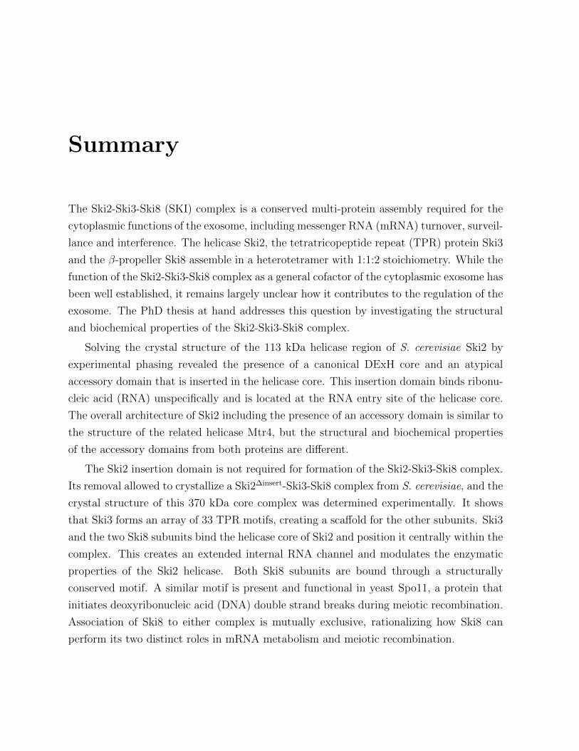
The Ski2-Ski3-Ski8 (SKI) complex is a conserved multi-protein assembly required for the
cytoplasmic functions of the exosome, including messenger RNA (mRNA) turnover, surveil-
lance and interference. The helicase Ski2, the tetratricopeptide repeat (TPR) protein Ski3
and the β-propeller Ski8 assemble in a heterotetramer with 1:1:2 stoichiometry. While the
function of the Ski2-Ski3-Ski8 complex as a general cofactor of the cytoplasmic exosome has
been well established, it remains largely unclear how it contributes to the regulation of the
exosome. The PhD thesis at hand addresses this question by investigating the structural
and biochemical properties of the Ski2-Ski3-Ski8 complex.
Solving the crystal structure of the 113 kDa helicase region of S. cerevisiae Ski2 by
experimental phasing revealed the presence of a canonical DExH core and an atypical
accessory domain that is inserted in the helicase core. This insertion domain binds ribonu-
cleic acid (RNA) unspecifically and is located at the RNA entry site of the helicase core.
The overall architecture of Ski2 including the presence of an accessory domain is similar to
the structure of the related helicase Mtr4, but the structural and biochemical properties
of the accessory domains from both proteins are different.
The Ski2 insertion domain is not required for formation of the Ski2-Ski3-Ski8 complex.
Its removal allowed to crystallize a Ski2∆insert-Ski3-Ski8 complex from S. cerevisiae, and the
crystal structure of this 370 kDa core complex was determined experimentally. It shows
that Ski3 forms an array of 33 TPR motifs, creating a scaffold for the other subunits. Ski3
and the two Ski8 subunits bind the helicase core of Ski2 and position it centrally within the
complex. This creates an extended internal RNA channel and modulates the enzymatic
properties of the Ski2 helicase. Both Ski8 subunits are bound through a structurally
conserved motif. A similar motif is present and functional in yeast Spo11, a protein that
initiates deoxyribonucleic acid (DNA) double strand breaks during meiotic recombination.
Association of Ski8 to either complex is mutually exclusive, rationalizing how Ski8 can
perform its two distinct roles in mRNA metabolism and meiotic recombination.

xiv Summary
Biochemical studies suggest that the SKI complex can thread RNAs directly to the exo-
some, coupling the helicase and the exoribonuclease through a continuous channel of 43-44
nucleotides length. Finally, an internal regulatory mechanism in the Ski2-Ski3-Ski8 com-
plex was identified. Both the Ski2-insertion domain and the Ski3 N-terminus cooperate
to inhibit ATPase and helicase activity of Ski2 when bound in the SKI complex. Thus,
the SKI complex regulates exosome activity in two ways. First by a direct substrate chan-
neling mechanism to the exosome that connects helicase and nuclease activities of both
complexes which may activate the exosome towards certain substrates. Second, by an
inhibitory mechanism that regulates substrate access to the helicase complex, which is a
prerequisite for controlling the exosome’s substrate specificity.
This doctoral thesis provides the first structural description of the entire yeast SKI
complex and identifies two mechanisms that may contribute to regulation of the activity
of the cytoplasmic exosome.
1 Preface
My doctoral work led to publication of two research articles1 2. Since both manuscripts
are coherent and represent the main body of work undertaken in the course of my Ph.D.
project, this thesis is written in cumulative style. The first chapter contains an introduction
to the biological background and the current state of the research. The second chapter
includes the classical “Results” and “Material and methods” sections in form of the original
manuscripts. A third and last chapter features a comprehensive discussion that integrates
the main aspects from both publications.
1The crystal structure of S. cerevisiae Ski2, a DExH helicase associated with the cytoplasmic functionsof the exosome. F. Halbach, M. Rode and E. Conti RNA, 2012, 18(1), 124-34
2The yeast Ski complex: crystal structure and substrate channeling to the RNA exosome. F. Halbach,P. Reichelt, M. Rode, E. Conti Cell, 2013, 154, 814-26

2 1. Preface
2 Introduction
2.1 mRNA degradation in eukaryotes
The balance between constructive and destructive events is a crucial concept found in many
biological systems at any level. In the metabolism of RNA, transcription and degradation
are the competing key events that determine the cellular level of a given transcript. Thus,
adjusting the turnover rate for a given transcript allows the cell to regulate the activity
of its gene product. Other means exist to control gene expression, among these post-
translational mechanisms. Nevertheless, degradation of mRNA provides the conceptually
simplest and most direct way to regulate the expression level of a particular gene since it
counteracts directly on transcription.
Degradation of mRNA is not only a means for the regulation of gene expression but
also helps to ensure the fidelity of transcription. For instance, cellular quality control
mechanisms identify improperly matured or otherwise erroneous transcripts and destine
them for degradation. Finally, degradation is the last step in chemical recycling of RNA,
a process that replenishes the cellular pools of nucleotides. Examples for this process
include breakdown of splicing by-products or cleaved, inactive transcripts produced by
RNA interference (RNAi) or other regulatory RNA mechanisms.
Most enzymes involved in the processes of RNA catabolism (e.g. nucleases, helicases
or RNA-binding proteins) are found in all domains of life. However, the specific pathways
of degradation and particularly their regulation can vary substantially. Nevertheless, in
eukaryotes two conserved canonical mRNA decay routes have evolved through which the
vast majority of all transcripts are degraded.

4 2. Introduction
2.1.1 Canonical mRNA decay is initiated by the deadenylation
machinery and driven by exonucleases
As soon as a given gene is transcribed in the nucleus, its mRNA is being processed and
spliced. During these processes, various protein factors are deposited on the nucleic acid
and the resulting protein-RNA complex is termed ribonucleoprotein (RNP). While these
factors can be part of the processing machinery, they also facilitate the export of the RNP
from the nucleus as well as cytoplasmic quality control mechanisms. Once the transcript
arrives in the cytoplasm, it faces two fundamental fates: translation or decay. From the
body of research conducted during the past decades it has now become clear that the ends
of an RNA molecule hold the two key determinants that govern the translation-vs-decay
decision: the 7-methylguanosine cap on the 5′ end and the polyadenylate (poly(A)) tail on
the 3′ end (Fig. 2.1).
Both molecular features have their cognate receptors. In the nucleus, the cap structure
of pre-mRNAs is bound by the cap-binding complex (CBC) that is formed by Cbp80 and
Cbp20 (Izaurralde et al., 1994). After export to the nucleus, the cytoplasmic eIF4F complex
binds to the cap of error-proofed mRNAs and thus replaces the CBC. eIF4F consists of
the cap-binding protein eIF4E, the scaffold protein eIF4G and the RNA helicase eIF4A
(reviewed in Richter and Sonenberg, 2005). Formation of the eIF4F complex enhances the
affinity of eIF4E for the 7-methylguanosine cap and forms a scaffold for other translational
factors (see below).
In yeast1 , the poly(A) tail comprises about 70 nucleotides (nt) (Manley and Takagaki,
1996; Keller and Minvielle-Sebastia, 1997) that are decorated by the poly(A)-binding pro-
tein (Pab1) (Kuhn and Wahle, 2004). The 5′ cap and the 3′ poly(A) tail act as protective
features and control translation and decay differentially. They promote translation, mainly
by recruitment of the eIF4E to the 40S pre-initiation complex (Kessler and Sachs, 1998;
Tarun and Sachs, 1995; Wells et al., 1998; Tarun and Sachs, 1996; Tarun et al., 1997). Con-
versely, they inhibit degradation by blocking access of exonucleases to the 5′ and 3′ ends
(Hsu and Stevens, 1993; Muhlrad et al., 1994, 1995; Anderson and Parker, 1998), and this
effect is apparently potentiated by circularization of the transcript through interaction of
Pab1 and eIF4G (Kessler and Sachs, 1998; Tarun and Sachs, 1995, 1996; Wells et al., 1998).
1The yeast S. cerevisiae is the to-date best studied model system for mRNA degradation. This PhDproject has thus focused on the S. cerevisiae proteins, and the introduction at hand is limited to the yeastsystem.
2.1 mRNA degradation in eukaryotes 5
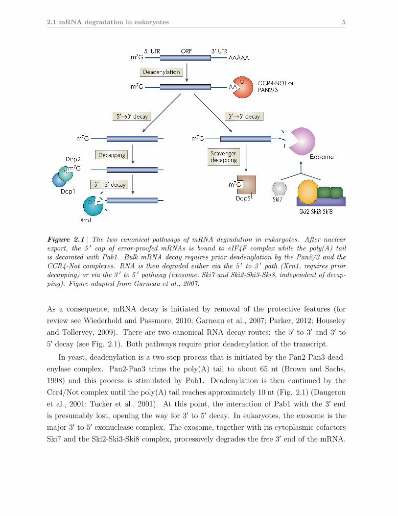
Figure 2.1 | The two canonical pathways of mRNA degradation in eukaryotes. After nuclearexport, the 5 ′ cap of error-proofed mRNAs is bound to eIF4F complex while the poly(A) tailis decorated with Pab1. Bulk mRNA decay requires prior deadenylation by the Pan2/3 and theCCR4-Not complexes. RNA is then degraded either via the 5 ′ to 3 ′ path (Xrn1, requires priordecapping) or via the 3 ′ to 5 ′ pathway (exosome, Ski7 and Ski2-Ski3-Ski8, independent of decap-ping). Figure adapted from Garneau et al., 2007.
As a consequence, mRNA decay is initiated by removal of the protective features (for
review see Wiederhold and Passmore, 2010; Garneau et al., 2007; Parker, 2012; Houseley
and Tollervey, 2009). There are two canonical RNA decay routes: the 5′ to 3′ and 3′ to
5′ decay (see Fig. 2.1). Both pathways require prior deadenylation of the transcript.
In yeast, deadenylation is a two-step process that is initiated by the Pan2-Pan3 dead-
enylase complex. Pan2-Pan3 trims the poly(A) tail to about 65 nt (Brown and Sachs,
1998) and this process is stimulated by Pab1. Deadenylation is then continued by the
Ccr4/Not complex until the poly(A) tail reaches approximately 10 nt (Fig. 2.1) (Daugeron
et al., 2001; Tucker et al., 2001). At this point, the interaction of Pab1 with the 3′ end
is presumably lost, opening the way for 3′ to 5′ decay. In eukaryotes, the exosome is the
major 3′ to 5′ exonuclease complex. The exosome, together with its cytoplasmic cofactors
Ski7 and the Ski2-Ski3-Ski8 complex, processively degrades the free 3′ end of the mRNA.
6 2. Introduction
The remaining 5′ cap structure is then degraded by the scavenger decapping enzyme DcpS
(Liu et al., 2002). The second decay pathway operates from the 5′ end of the transcript and
thus requires prior decapping in addition to deadenylation (Fig. 2.1). Decapping is effected
by the Dcp1-Dcp2 complex which is regulated by a host of factors including the activators
Lsm1-7 complex and Edc3 (reviewed in Franks and Lykke-Andersen, 2008; Simon et al.,
2006). Removal of the 7-methylguanosine cap leaves the mRNA with a 5′ phosphate which
is the preferred substrate of Xrn1, the major cytoplasmic 5′ to 3′ exonuclease (Larimer and
Stevens, 1990).
2.1.2 Alternative decay pathways
Specialized pathways exist that lead to degradation but bypass prior deadenylation and/or
decapping of the transcript. For instance, certain classes of transcripts recruit decap-
ping enhancers (e.g. the Rpb28/Dcp3 system, (Kshirsagar and Parker, 2004; Badis et al.,
2004)). These proteins promote decapping even though the poly(A) tail has not been
shortened. This enables the Xrn1 pathway to degrade the transcript via the free 5′ end.
Other examples include the endonucleolytic cleavage of capped and polyadenylated mR-
NAs, for instance through the RNAi machinery. The 5′ and 3′ ends of the resulting 3′ and
5′ fragments, respectively, are are not protected and thus accessible to the the Xrn1 or
exosome/Ski7/Ski2-Ski3-Ski8 pathways. Such bypass-mechanisms are also employed by
certain mRNA quality control pathways like no-go decay and nonstop decay (see section
2.4.1).
2.1.3 Relative contributions of 5′ and 3′ decay routes
The Xrn1 (5′ to 3′) and exosome/Ski7/Ski2-Ski3-Ski8 (3′ to 5′) decay routes are not es-
sential in yeast. Synthetic lethality only results when components of both pathways are
deleted (Johnson and Kolodner, 1995; Anderson and Parker, 1998; van Hoof et al., 2000b),
indicating that the two pathways operate redundantly. Currently it is still a matter of de-
bate which pathway constitutes the major route for mRNA decay. Yeast strains deleted for
either of the two pathways show a slow-growing phenotype only when the decapping/Xrn1
route is targeted (Beelman et al., 1996; Dunckley and Parker, 1999; Giaever et al., 2002;
Anderson and Parker, 1998), suggesting that the 5′ to 3′ route prevails. On the other
hand, more recent transcriptome-wide RNA profiling studies suggest that the impact of
these deletions is less pronounced as thought in the first place (Houalla et al., 2006; He
2.2 The exosome is the major eukaryotic 3′ to 5′ ribonuclease complex 7
et al., 2003). Ultimately, the decay route for a given transcript may depend on many
factors like growth phase, nature of the studied transcripts etc., making it difficult (and
possibly unnecessary) to discriminate major and minor pathways in a general fashion.
While both pathways may be similar in terms of quantitative contribution to overall decay,
from a conceptual point of view the exosome-mediated 3′ to 5′ decay is set apart: the
exosome is not only involved in degradation but also functions in processing and matura-
tion of certain RNA precursors. This means that, depending on the given substrate, the
exosome can either totally degrade an RNA substrate or partially trim it an apparently
very controlled fashion (see also section 2.2.1).
2.2 The exosome is the major eukaryotic 3′ to 5′ ribo-
nuclease complex
The exosome is a multi-subunit ribonuclease complex that was first identified from tandem
affinity-purification experiments in budding yeast (Mitchell et al., 1997). It is a processive
3′ exonuclease, i.e. it removes nucleotides one after the other from the 3′ end of the RNA
without dissociating from the substrate (Dziembowski et al., 2007; Liu et al., 2006). Sub-
sequent work identified homologous complexes in other eukaryotes (including plants) and
in archaea, highlighting its universally conserved role in RNA catabolism. Since then, bio-
chemical and structural work has shaped our understanding of the molecular architecture
of exosome complexes and their enzymatic function. Genetic experiments delineated many
of the pathways and protein factors that deliver substrates to the exosome. In parallel,
an ever-growing number of substrates for the exosome is being identified, particularly by
systems-wide approaches.
2.2.1 Functions of the eukaryotic exosome: From maturation to
degradation
The exosome operates both in the cytoplasm and the nucleus of eukaryotic cells. It pro-
cesses a set of substrates that is remarkably broad, including RNAs produced by each of
the three major RNA polymerases. Nonetheless, the exosome displays differential activity
towards these substrates: first, it can fully degrade a given substrate to remove it com-
pletely from the cellular pool. Second, it can partially trim the 3′ end, a process important
for maturation of certain RNA precursors.
8 2. Introduction
mRNA Turnover
mRNA
Quality
control
Cytoplasm
Exo9 + Rrp44
Nucleus
Processing
Degradation
mRNAs (NGD)
mRNAs
mRNAs (NSD)
mRNAs (NMD)
Hbs1, Dom34Mpp6
Mpp6
Nrd1-N
ab3
Nrd1-N
ab3
rRNA
generalcofactors
specific cofactors
substratesspecific cofactors
substrates generalcofactors
Trf4/5,
Air1/2
Mtr4
Rrp6,
Rrp47
aberrant rRNA, snRNA,
snoRNA, tRNA, mRNA etc.
snoRNA,
snRNA
CUTs,
PROMPTs
Ski7
Ski2-Ski3-Ski8
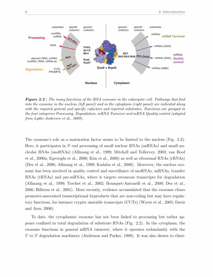
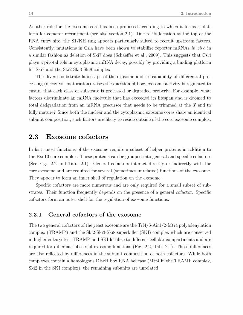
Figure 2.2 | The many functions of the RNA exosome in the eukaryotic cell. Pathways that feedinto the exosome in the nucleus (left panel) and in the cytoplasm (right panel) are indicated alongwith the required general and specific cofactors and reported substrates. Functions are grouped inthe four categories Processing, Degradation, mRNA Turnover and mRNA Quality control (adaptedfrom Lykke-Andersen et al., 2009).
The exosome’s role as a maturation factor seems to be limited to the nucleus (Fig. 2.2).
Here, it participates in 3′ end processing of small nuclear RNAs (snRNAs) and small nu-
cleolar RNAs (snoRNAs) (Allmang et al., 1999; Mitchell and Tollervey, 2003; van Hoof
et al., 2000a; Egecioglu et al., 2006; Kim et al., 2006) as well as ribosomal RNAs (rRNAs)
(Dez et al., 2006; Allmang et al., 1999; Kadaba et al., 2006). Moreover, the nuclear exo-
some has been involved in quality control and surveillance of snoRNAs, snRNAs, transfer
RNAs (tRNAs) and pre-mRNAs, where it targets erroneous transcripts for degradation
(Allmang et al., 1999; Torchet et al., 2002; Bousquet-Antonelli et al., 2000; Dez et al.,
2006; Hilleren et al., 2001). More recently, evidence accumulated that the exosome clears
promoter-associated transcriptional byproducts that are non-coding but may have regula-
tory functions, for instance cryptic unstable transcripts (CUTs) (Wyers et al., 2005; Davis
and Ares, 2006).
To date, the cytoplasmic exosome has not been linked to processing but rather ap-
pears confined to total degradation of substrate RNAs (Fig. 2.2). In the cytoplasm, the
exosome functions in general mRNA turnover, where it operates redundantly with the
5′ to 3′ degradation machinery (Anderson and Parker, 1998). It was also shown to elimi-
2.2 The exosome is the major eukaryotic 3′ to 5′ ribonuclease complex 9
nate 5′ fragments generated by the RNAi-induced silencing complex (RISC) during RNAi
(Orban and Izaurralde, 2005). In mammalian cells, mRNAs containing AU-rich elements
(ARE) are recruited to the exosome via dedicated ARE-binding proteins, resulting in rapid
decay of substrates (Chen et al., 2001).
Besides its role in mRNA turnover, the exosome serves as endpoint for several cyto-
plasmic quality control pathways. Transcripts containing a premature termination codon
are cotranslationally targeted by the nonsense-mediated decay (NMD) pathway and subse-
quently degraded by the cytoplasmic exosome (Mitchell and Tollervey, 2003). Transcripts
that lack a stop codon altogether were shown to be eliminated in an exosome-dependent
fashion (van Hoof et al., 2002). This pathway is referred to as non-stop decay (NSD).
Another quality control pathway termed no-go decay (NGD) has been described that tar-
gets mRNAs that are stalled on the translating ribosome. Stalled transcripts are cleaved
endonucleolytically which presumably releases them from the ribosome and generates free
5′ fragments that are cleared by the cytoplasmic exosome (Doma and Parker, 2006).
2.2.2 The architecture of the core exosome and its conservation
through all domains of life
Structure and activity of the archaeal exosome
The first insights into the molecular architecture of the exosome came from crystal struc-
tures of the archaeal complexes (Buttner et al., 2005; Lorentzen and Conti, 2005; Lorentzen
et al., 2007). The archaeal exosome is built from the three subunits Rrp41, Rrp42 and
Rrp4 (or Csl4) (Fig. 2.3B, central panel). Rrp41 and Rrp42 are homologous to bacterial ri-
bonuclease (RNase) PH, a phosphorolytic ribonuclease. Three Rrp41-Rrp42 hetero-dimers
assemble into a six-membered ring (termed the RNase PH ring). Rrp4 contains an N-
terminal S1-homology domain and a C-terminal K-homology (KH) domain. A trimeric
ring of Rrp4 (termed the S1/KH cap) assembles on top of the RNase PH ring, creating the
9-subunit archaeal exosome (Fig. 2.3A, central panel). Rrp4 can be substituted by Csl4
which contains an S1 domain and a zinc-knuckle domain. In vivo, the S1/KH cap is ho-
momeric for Rrp4 or Csl or contains a combination of both (Evguenieva-Hackenberg et al.,
2003), and in vitro Rrp4 and Csl4 confer different substrate specificities to the archaeal
exosome (Roppelt et al., 2010).
Biochemical studies showed that Rrp41 is the active subunit. It displays processive,
phosphorolytic ribonuclease activity towards single stranded RNA (Buttner et al., 2005;
10 2. Introduction
Lorentzen et al., 2005) but is unable to degrade through secondary structure elements like
hairpins (Lorentzen and Conti, 2005). Rrp42 contributes to RNA-binding and to formation
of the active site and it is required for nuclease activity of the complex (Lorentzen et al.,
2005; Buttner et al., 2005). The S1/KH cap has been shown provide additional RNA
binding sites to the complex that may help to regulate substrate access to the active sites.
The structures of the archaeal exosome revealed a surprisingly high similarity with
bacterial polynucleotide phosphorylase (PNPase), an phosphorolytic RNase complex that
forms part of the degradosome (Symmons et al., 2000). PNPase contains two RNase
PH cassettes and C-terminal S1/KH domain, and this polypeptide assembles into a ho-
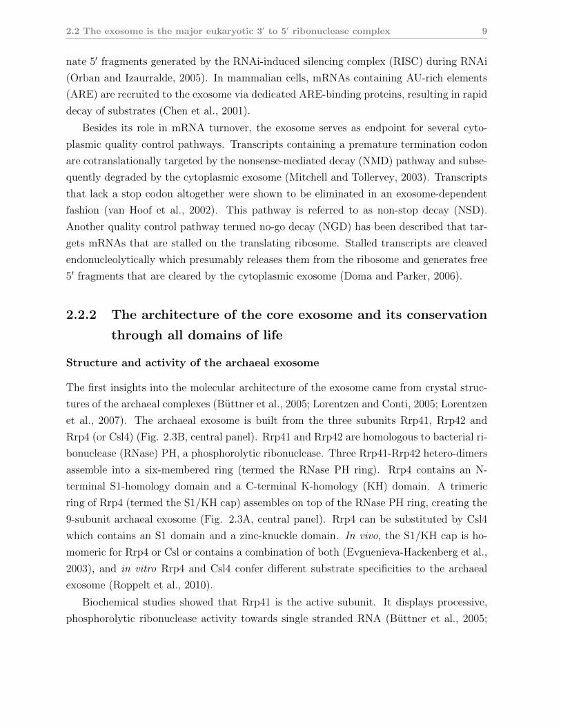
Figure 2.3 | The molecular architecture of exosome-like complexes is conserved throughout evo-lution. (A) Structures of bacterial PNPase (Symmons et al., 2000), archaeal 9-subunit exosome(Lorentzen et al., 2007) and human 9-subunit exosome (Liu et al., 2006) are shown in the left,middle and right panels, respectively. RNase PH (domain 1), Rrp41 and Rrp41-like proteinsare colored in red or shades thereof. RNase PH (domain 2), Rrp42 and Rrp42-like proteins inyellow or shades of yellow. S1/ KH domains or S1/KH containing proteins in shades of blue.(B) The domain structures of the proteins corresponding to the structure in each panel are showncolor-coded as above.
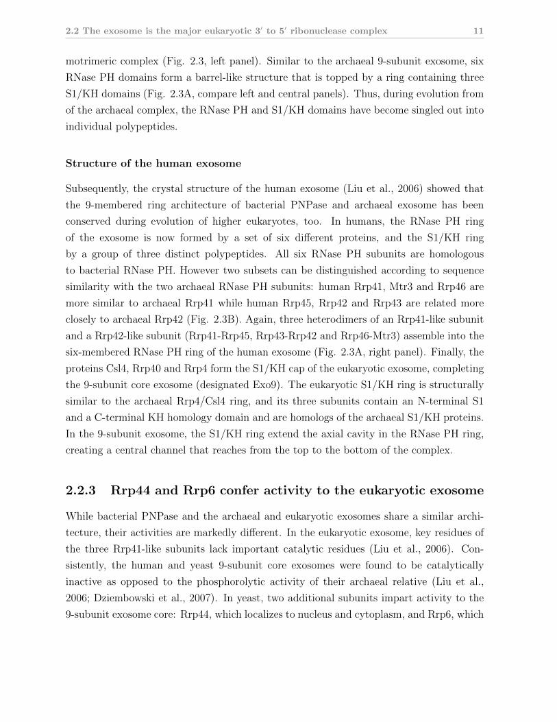
2.2 The exosome is the major eukaryotic 3′ to 5′ ribonuclease complex 11
motrimeric complex (Fig. 2.3, left panel). Similar to the archaeal 9-subunit exosome, six
RNase PH domains form a barrel-like structure that is topped by a ring containing three
S1/KH domains (Fig. 2.3A, compare left and central panels). Thus, during evolution from
of the archaeal complex, the RNase PH and S1/KH domains have become singled out into
individual polypeptides.
Structure of the human exosome
Subsequently, the crystal structure of the human exosome (Liu et al., 2006) showed that
the 9-membered ring architecture of bacterial PNPase and archaeal exosome has been
conserved during evolution of higher eukaryotes, too. In humans, the RNase PH ring
of the exosome is now formed by a set of six different proteins, and the S1/KH ring
by a group of three distinct polypeptides. All six RNase PH subunits are homologous
to bacterial RNase PH. However two subsets can be distinguished according to sequence
similarity with the two archaeal RNase PH subunits: human Rrp41, Mtr3 and Rrp46 are
more similar to archaeal Rrp41 while human Rrp45, Rrp42 and Rrp43 are related more
closely to archaeal Rrp42 (Fig. 2.3B). Again, three heterodimers of an Rrp41-like subunit
and a Rrp42-like subunit (Rrp41-Rrp45, Rrp43-Rrp42 and Rrp46-Mtr3) assemble into the
six-membered RNase PH ring of the human exosome (Fig. 2.3A, right panel). Finally, the
proteins Csl4, Rrp40 and Rrp4 form the S1/KH cap of the eukaryotic exosome, completing
the 9-subunit core exosome (designated Exo9). The eukaryotic S1/KH ring is structurally
similar to the archaeal Rrp4/Csl4 ring, and its three subunits contain an N-terminal S1
and a C-terminal KH homology domain and are homologs of the archaeal S1/KH proteins.
In the 9-subunit exosome, the S1/KH ring extend the axial cavity in the RNase PH ring,
creating a central channel that reaches from the top to the bottom of the complex.
2.2.3 Rrp44 and Rrp6 confer activity to the eukaryotic exosome
While bacterial PNPase and the archaeal and eukaryotic exosomes share a similar archi-
tecture, their activities are markedly different. In the eukaryotic exosome, key residues of
the three Rrp41-like subunits lack important catalytic residues (Liu et al., 2006). Con-
sistently, the human and yeast 9-subunit core exosomes were found to be catalytically
inactive as opposed to the phosphorolytic activity of their archaeal relative (Liu et al.,
2006; Dziembowski et al., 2007). In yeast, two additional subunits impart activity to the
9-subunit exosome core: Rrp44, which localizes to nucleus and cytoplasm, and Rrp6, which
12 2. Introduction
is exclusively found in the nucleus.
Rrp44 associates with the core exosome, resulting in the 10-subunit exosome (desig-
nated Exo10) that is identical in nucleus and cytoplasm. Rrp44 contains two distinct
hydrolytic RNase activities. In the N-terminus, a PiLT protein N-terminus (PIN) domain
confers endonuclease activity (Schneider et al., 2009; Schaeffer et al., 2009; Lebreton et al.,
2008). In the C-terminus, an RNase II domain confers processive 3′ to 5′ exonuclease
activity (Dziembowski et al., 2007; Liu et al., 2006). Structural studies have shown that
Rrp44 associates via its PIN domain to the Rrp41-Rrp45 dimer (Bonneau et al., 2009),
resulting in a location at the base of the RNase PH ring. In this conformation, both the
PIN domain and RNase II active sites are accessible from the central channel of the core
exosome.
In the yeast nucleus, Exo10 associates with another RNase, Rrp6, to create the 11-
subunit exosome (Exo11). Rrp6 belongs to the RNase D family and degrades ribonucleic
acids in a distributive, hydrolytic fashion from the 3′ to the 5′ end (Midtgaard et al.,
2006; Zuo and Deutscher, 2001; Phillips and Butler, 2003). Rrp6 contains an N-terminal
PMC2NT domain that mediates binding to Rrp47, followed by the RNase D catalytic
core and two helicase RNase D C-terminal domains (HRDCs) (Midtgaard et al., 2006).
The last HRDC domain mediates binding to Exo10 in vivo (Callahan and Butler, 2008).
While Rrp6 remains attached to Exo10, recent experiments suggest that it can also operate
independently of the Exo10 core (Callahan and Butler, 2008).
2.2.4 The inactive eukaryotic exosome core retains functionality
Even though the eukaryotic core exosome lost its activity during evolution, it retained an
architecture remarkably similar to the archaeal exosome and bacterial PNPase. Moreover,
in yeast all nine subunits of the exosome core are essential (Mitchell et al., 1997). This
suggests that the exosome core kept certain functions distinct from nuclease activity. These
functions must be closely linked to structural features that were conserved during evolution.
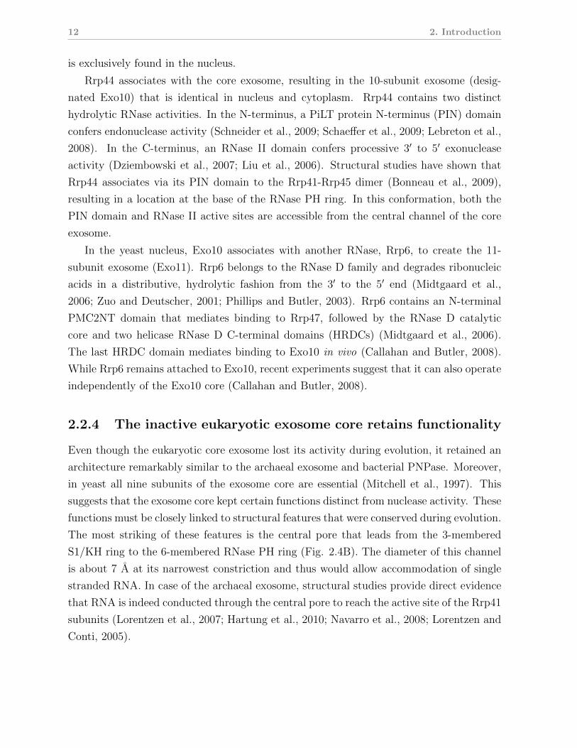
The most striking of these features is the central pore that leads from the 3-membered
S1/KH ring to the 6-membered RNase PH ring (Fig. 2.4B). The diameter of this channel
is about 7 A at its narrowest constriction and thus would allow accommodation of single
stranded RNA. In case of the archaeal exosome, structural studies provide direct evidence
that RNA is indeed conducted through the central pore to reach the active site of the Rrp41
subunits (Lorentzen et al., 2007; Hartung et al., 2010; Navarro et al., 2008; Lorentzen and
Conti, 2005).
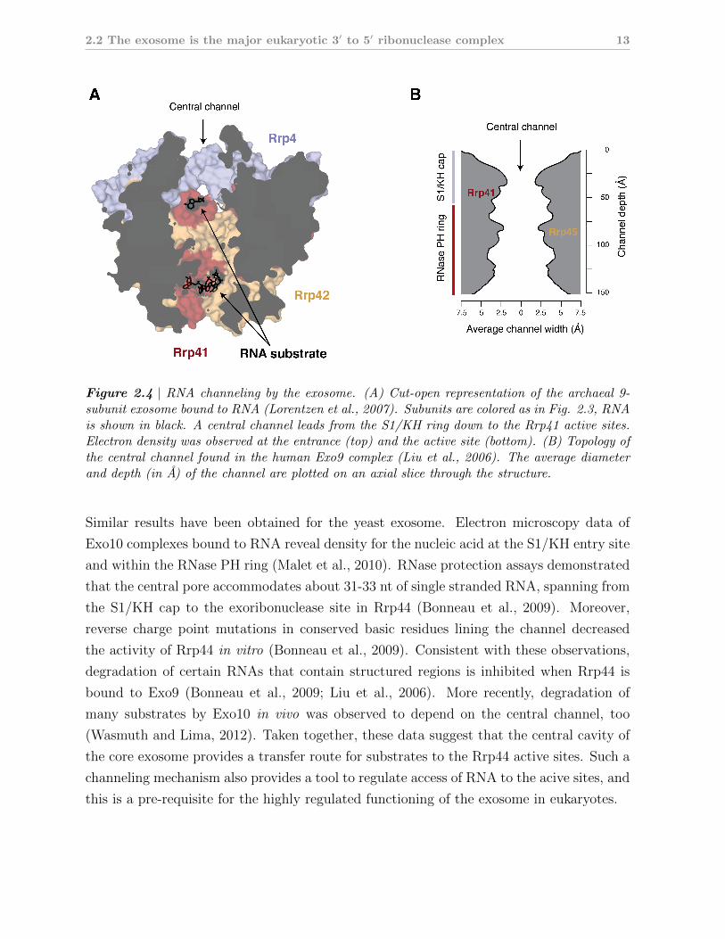
2.2 The exosome is the major eukaryotic 3′ to 5′ ribonuclease complex 13
Figure 2.4 | RNA channeling by the exosome. (A) Cut-open representation of the archaeal 9-subunit exosome bound to RNA (Lorentzen et al., 2007). Subunits are colored as in Fig. 2.3, RNAis shown in black. A central channel leads from the S1/KH ring down to the Rrp41 active sites.Electron density was observed at the entrance (top) and the active site (bottom). (B) Topology ofthe central channel found in the human Exo9 complex (Liu et al., 2006). The average diameterand depth (in A) of the channel are plotted on an axial slice through the structure.
Similar results have been obtained for the yeast exosome. Electron microscopy data of
Exo10 complexes bound to RNA reveal density for the nucleic acid at the S1/KH entry site
and within the RNase PH ring (Malet et al., 2010). RNase protection assays demonstrated
that the central pore accommodates about 31-33 nt of single stranded RNA, spanning from
the S1/KH cap to the exoribonuclease site in Rrp44 (Bonneau et al., 2009). Moreover,
reverse charge point mutations in conserved basic residues lining the channel decreased
the activity of Rrp44 in vitro (Bonneau et al., 2009). Consistent with these observations,
degradation of certain RNAs that contain structured regions is inhibited when Rrp44 is
bound to Exo9 (Bonneau et al., 2009; Liu et al., 2006). More recently, degradation of
many substrates by Exo10 in vivo was observed to depend on the central channel, too
(Wasmuth and Lima, 2012). Taken together, these data suggest that the central cavity of
the core exosome provides a transfer route for substrates to the Rrp44 active sites. Such a
channeling mechanism also provides a tool to regulate access of RNA to the acive sites, and
this is a pre-requisite for the highly regulated functioning of the exosome in eukaryotes.
14 2. Introduction
Another role for the exosome core has been proposed according to which it forms a plat-
form for cofactor recruitment (see also section 2.1). Due to its location at the top of the
RNA entry site, the S1/KH ring appears particularly suited to recruit upstream factors.
Consistently, mutations in Csl4 have been shown to stabilize reporter mRNAs in vivo in
a similar fashion as deletion of Ski7 does (Schaeffer et al., 2009). This suggests that Csl4
plays a pivotal role in cytoplasmic mRNA decay, possibly by providing a binding platform
for Ski7 and the Ski2-Ski3-Ski8 complex.
The diverse substrate landscape of the exosome and its capability of differential pro-
cessing (decay vs. maturation) raises the question of how exosome activity is regulated to
ensure that each class of substrate is processed or degraded properly. For example, what
factors discriminate an mRNA molecule that has exceeded its lifespan and is doomed to
total dedgradation from an mRNA precursor that needs to be trimmed at the 3′ end to
fully mature? Since both the nuclear and the cytoplasmic exosome cores share an identical
subunit composition, such factors are likely to reside outside of the core exosome complex.
2.3 Exosome cofactors
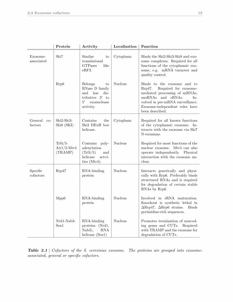
In fact, most functions of the exosome require a subset of helper proteins in addition to
the Exo10 core complex. These proteins can be grouped into general and specific cofactors
(See Fig. 2.2 and Tab. 2.1). General cofactors interact directly or indirectly with the
core exosome and are required for several (sometimes unrelated) functions of the exosome.
They appear to form an inner shell of regulation on the exosome.
Specific cofactors are more numerous and are only required for a small subset of sub-
strates. Their function frequently depends on the presence of a general cofactor. Specific
cofactors form an outer shell for the regulation of exosome functions.
2.3.1 General cofactors of the exosome
The two general cofactors of the yeast exosome are the Trf4/5-Air1/2-Mtr4 polyadenylation
complex (TRAMP) and the Ski2-Ski3-Ski8 superkiller (SKI) complex which are conserved
in higher eukaryotes. TRAMP and SKI localize to different cellular compartments and are
required for different subsets of exosome functions (Fig. 2.2, Tab. 2.1). These differences
are also reflected by differences in the subunit composition of both cofactors. While both
complexes contain a homologous DExH box RNA helicase (Mtr4 in the TRAMP complex,
Ski2 in the SKI complex), the remaining subunits are unrelated.
2.3 Exosome cofactors 15
Protein Activity Localization Function
Exosome-associated
Ski7 Similar totranslationalGTPases likeeRF3.
Cytoplasm Binds the Ski2-Ski3-Ski8 and exo-some complexes. Required for allfunctions of the cytoplasmic exo-some, e.g. mRNA turnover andquality control.
Rrp6 Belongs toRNase D familyand has dis-tributive 3′ to5′ exonucleaseactivity.
Nucleus Binds to the exosome and toRrp47. Required for exosome-mediated processing of snRNAs,snoRNAs and rRNAs. In-volved in pre-mRNA surveillance.Exosome-independent roles havebeen described.
General co-factors
Ski2-Ski3-Ski8 (SKI)
Contains theSki2 DExH boxhelicase.
Cytoplasm Required for all known functionsof the cytoplasmic exosome. In-teracts with the exosome via Ski7N-terminus.
Trf4/5-Air1/2-Mtr4(TRAMP)
Contains poly-adenylation(Trf4/5) andhelicase actvi-ties (Mtr4).
Nucleus Required for most functions of thenuclear exosome. Mtr4 can alsooperate independently. Physicalinteraction with the exosome un-clear.
Specificcofactors
Rrp47 RNA-bindingprotein
Nucleus Interacts genetically and physi-cally with Rrp6. Preferably bindsstructured RNAs and is requiredfor degradation of certain stableRNAs by Rrp6.
Mpp6 RNA-bindingprotein
Nucleus Involved in rRNA maturation.Knockout is synthetic lethal in∆Rrp47, ∆Rrp6 strains. Bindspyrimidine-rich sequences.
Nrd1-Nab3-Sen1
RNA-bindingproteins (Nrd1,Nab3), RNAhelicase (Sen1)
Nucleus Promotes termination of noncod-ing genes and CUTs. Requiredwith TRAMP and the exosome fordegradation of CUTs.
Table 2.1 | Cofactors of the S. cerevisiae exosome. The proteins are grouped into exosome-associated, general or specific cofactors.
16 2. Introduction
RNA helicases as cofactors for exosome-like complexes
Ski2 and Mtr4 are paralogs and share 29 % sequence identity and 45 % similarity. Both
enzymes belong to the superfamily II (SF2) of helicases and utilize energy from adenosine
triphosphate (ATP)-hydrolysis to unwind RNA duplexes (for review see Pyle, 2008; Sin-
gleton et al., 2007; Jankowsky and Fairman, 2007; Lohman et al., 2008). While to date
no helicase has been associated with the function of the archaeal exosome, such a link has
been found for the bacterial degradosome. The degradosome is a major prokaryotic RNase
complex that is formed by PNPase, the archetype of the exosome, as well as RNase E and
enolase (Carpousis, 2007). A fourth subunit was found to be RhlB, an ATP-dependent SF2
RNA helicase (Py et al., 1996). Degradation of certain structured RNA substrates requires
ATP in vitro (Py et al., 1996), and several studies showed that degradosome-mediated RNA
decay depends on RhlB in vivo (Bernstein et al., 2002, 2004; Khemici et al., 2005; Khemici
and Carpousis, 2004).
These observations suggest that RhlB locally unwinds secondary structure elements in
substrate RNAs to facilitate their degradation by the bacterial degradosome. The presence
of an ATP-dependent RNA helicase in each of the general cofactor complexes of the eu-
karyotic exosome led to the extrapolation of this working hypothesis to the function of the
TRAMP and SKI complexes in concert with the eukaryotic exosome. It is thought that
both general cofactors harness the helicase activity (Ski2 or Mtr4) to assist degradation of
challenging substrates.
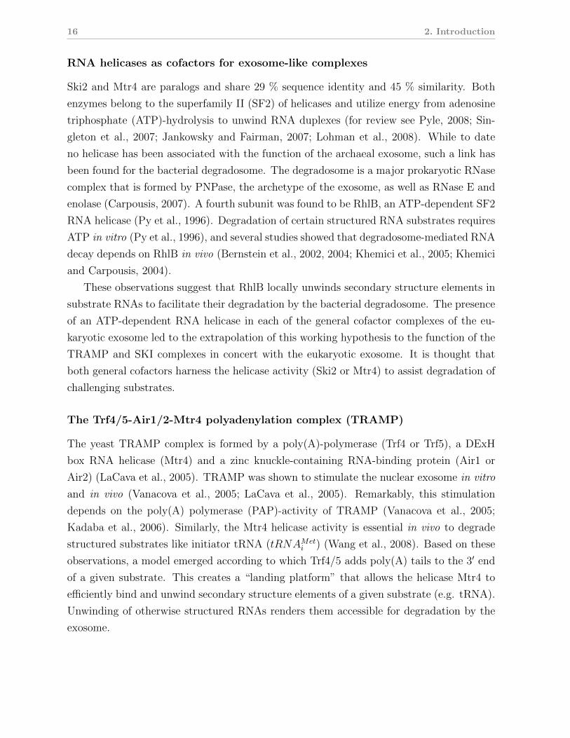
The Trf4/5-Air1/2-Mtr4 polyadenylation complex (TRAMP)
The yeast TRAMP complex is formed by a poly(A)-polymerase (Trf4 or Trf5), a DExH
box RNA helicase (Mtr4) and a zinc knuckle-containing RNA-binding protein (Air1 or
Air2) (LaCava et al., 2005). TRAMP was shown to stimulate the nuclear exosome in vitro
and in vivo (Vanacova et al., 2005; LaCava et al., 2005). Remarkably, this stimulation
depends on the poly(A) polymerase (PAP)-activity of TRAMP (Vanacova et al., 2005;
Kadaba et al., 2006). Similarly, the Mtr4 helicase activity is essential in vivo to degrade
structured substrates like initiator tRNA (tRNAMeti ) (Wang et al., 2008). Based on these
observations, a model emerged according to which Trf4/5 adds poly(A) tails to the 3′ end
of a given substrate. This creates a “landing platform” that allows the helicase Mtr4 to
efficiently bind and unwind secondary structure elements of a given substrate (e.g. tRNA).
Unwinding of otherwise structured RNAs renders them accessible for degradation by the
exosome.

2.3 Exosome cofactors 17
Efforts to pin down the mechanism of TRAMP-mediated exosome activation on the molec-
ular level are still ongoing. Recent advances include the crystal structure of the Trf5 cat-
alytic domain bound to Air2, which suggests that the C-terminal zinc knuckles of Air2p
mediate binding to the polymerase rather than being RNA-binding motifs (Hamill et al.,
2010). Structural and biochemical studies on Mtr4 have confirmed its identity and activity
as a DExH box helicase (Bernstein et al., 2008; Weir et al., 2010). Mtr4 was also found to
contain an usual accessory domain that emerges from the helicase core. It has structural
similarity to ribosomal KOW domains that are known to bind structured RNAs. Con-
sistently, the Mtr4 KOW domain exhibits affinity towards structured RNAs (Weir et al.,
2010) and is required for rRNA processing in vivo (Jackson et al., 2010).
The Ski2-Ski3-Ski8 complex (SKI)
In the yeast cytoplasm, the proteins Ski2, Ski3, Ski8 as well as Ski7 have been found to be
general cofactors for the exosome. Ski2 is a putative DExH box type RNA helicase that was
shown to form a complex with Ski3 and Ski8 in vivo (Brown et al., 2000). This Ski2-Ski3-
Ski8 was further shown to interact with the exosome via the eRF3-homolog Ski7 (Araki
et al., 2001; Wang et al., 2005). The Ski2-Ski3-Ski8 complex is required for all cytoplasmic
functions of the exosome. Because Ski2 is closely related to Mtr4, its helicase activity
is assumed to contribute to exosome activation similarly to Mtr4 within the TRAMP
complex. However, experimental evidence for such a mechanism is still lacking. A detailed
introduction to the Ski2-Ski3-Ski8 complex is given in the section 2.4.
2.3.2 Specific exosome cofactors
Several specific exosome cofactors have been described (Tab. 2.1). Frequently, these factors
are nuclear-specific RNA-binding proteins that operate in concert with other cofactors.
For instance, the nuclear Rrp47 is a partner protein of Rrp6 with apparent specificity for
structured RNAs (Stead et al., 2007; Mitchell et al., 2003). It is indispensable for certain
aspects of Rrp6/exosome-mediated processing of stable RNAs like snRNAs and snoRNAs
(Mitchell et al., 2003; Costello et al., 2011). Rrp47 has been proposed to act as a chaperone
that stabilizes interactions of Rrp6 with its cognate substrates, thus enhancing substrate
specificity and efficacy of degradation (Stead et al., 2007).
Another nuclear exosome cofactor, Mpp6, appears somewhat similar to Rrp47 in that
it is an RNA-binding protein that is synthetic lethal in an Rrp6 deletion background

18 2. Introduction
(Milligan et al., 2008). In contrast to Rrp47, Mpp6 preferentially binds pyrimidine-rich
sequences (Milligan et al., 2008). Mpp6 co-purifies with exosome-containing complexes
(Krogan et al., 2006) and plays a role in exosome-mediated decay of noncoding RNAs
(Milligan et al., 2008), but additional data are needed to define its role more precisely.
Yet another annotated exosome cofactor is the nuclear Nrd1-Nab3-Sen1 complex, which
is recruited by RNA polymerase II C-terminal domain to terminate CUTs (Carroll et al.,
2007; Vasiljeva et al., 2008). CUTs are then degraded by the nuclear exosome in a TRAMP-
dependent fashion that also requires Nrd1-Nab3-Sen1 (Arigo et al., 2006; Thiebaut et al.,
2006), suggesting that this cofactor complex orchestrates transcription termination and
degradation for certain transcript classes.
2.4 The SKI complex is a general cofactor of the cy-
toplasmic exosome
The SKI genes were originally identified from mutations in S. cerevisiae strains that were
infected by the double-stranded Killer virus. These mutations raised the levels of viral
RNA species which exacerbated the killer phenotype of infected cells (hence the name Su-
perkiller) (Toh et al., 1978; Ridley et al., 1984). The phenotypes could later be mapped
to a set of seven genes which were identified as Ski2, Ski3 and Ski8 (Widner and Wick-
ner, 1993; Rhee et al., 1989; Matsumoto et al., 1993) as well as Ski7 (Benard et al., 1999),
Ski1/Xrn1 (Larimer and Stevens, 1990), Ski4/Csl4 (van Hoof et al., 2000b) and Ski6/Rrp41
(Benard et al., 1998). Subsequent studies suggested that those proteins acted by repressing
expression of viral poly(A) RNA (Widner and Wickner, 1993). Eventually, translational re-
pression was found to be independent of the presence of viral RNA (Johnson and Kolodner,
1995), indicating a general role of the Ski proteins in mRNA catabolism.
2.4.1 Functions of the Ski2-Ski3-Ski8 complex
Cytoplasmic 3′ to 5′ mRNA turnover requires the Ski proteins and the exosome
Deletion of either of the SKI2, SKI3, SKI7 or SKI8 genes was found to block 3′ to
5′ degradation (Anderson and Parker, 1998; van Hoof et al., 2000b), and conditional knock-
outs of SKI6/RRP41 and RRP4 produced similar phenotypes, suggesting that these six
proteins operate along the same pathway. Since Rrp41 and Rrp4 had been previously re-
ported as subunits of the exosome complex (Mitchell et al., 1997), this results annotated

2.4 The SKI complex is a general cofactor of the cytoplasmic exosome 19
the exosome as the catalytic component of the 3′ to 5′ degradation pathway and identified
the Ski proteins as required cofactors.
Ski2, Ski3 and Ski8 from a stable complex in vivo in the yeast cytoplasm (Brown et al.,
2000; Synowsky and Heck, 2008). SKI7, the last of the original Superkiller genes, encodes
a homolog of translational guanosine triphosphate hydrolases (GTPases) like eRF3 and
EF1-α (Benard et al., 1999; Atkinson et al., 2008). Its N-terminal region was shown to
co-immunoprecipitate with the Ski2-Ski3-Ski8 complex and with exosome subunits (pre-
sumably Rrp4 and Csl4) (Wang et al., 2005; Araki et al., 2001; van Hoof et al., 2002).
Deletion of the Ski7 N-terminus in vivo interferes with cytoplasmic 3′ to 5′ decay. In con-
trast, the C-terminal GTPase domain of Ski7 is dispensable for exosome-mediate mRNA
turnover (Araki et al., 2001; van Hoof et al., 2000b).
The Ski2-Ski3-Ski8 complex and Ski7 are essential for cytoplasmic mRNA qual-
ity control
Apart from its role in mRNA turnover, the Ski2-Ski3-Ski8 complex is required for at least
three distinct mRNA quality control pathways that feed into the cytoplasmic exosome
(Fig. 2.2). First, NMD targets transcripts with premature stop codons through concerted
action of the Upf1-2-3 complex and stalled ribosomes (reviewed in Chang et al., 2007;
Conti and Izaurralde, 2005). Recruitment of other surveillance factors eventually releases
the stalled mRNA which is subsequently degraded from the 5′ end (Xrn1) and the 3′ end
(exosome/Ski7/Ski2-Ski3-Ski8) (Takahashi et al., 2003; Mitchell et al., 2003).
Second, transcripts that cause the ribosome to stall, e.g. due to unresolvable secondary
structure, are eliminated by NGD. Two dedicated translation factors, Dom34 and Hbs1,
are crucial to this pathway. Dom34 (homologous to eRF1) mimicks tRNA (Lee et al., 2007)
and binds along with the translational GTPase Hbs1 in the A-site of the stalled ribosome
(Becker et al., 2011; Chen et al., 2010). While the endonuclease activity that releases the
stalled transcript remains elusive, the exosome and Ski proteins were found responsible for
the degradation of the resulting 5′ fragment and Xrn1 for elimination of the corresponding
3′ fragment (Doma and Parker, 2006).
Third, transcripts that lack a stop codon altogether are targeted by NSD. NSD sub-
strates are readily degraded by the exosome in a Ski2-Ski3-Ski8- and Ski7-dependent fash-
ion (Frischmeyer et al., 2002; van Hoof et al., 2002). This pathway depends on the Ski7
N-terminal region but also requires the C-terminal GTPase domain (Frischmeyer et al.,
2002). This prompted a model according to which ribosomes that are stalled on read-
20 2. Introduction
through messages recruit Ski7 (possibly together with an eRF1-homolog) and thus induce
exosome- and Ski2-Ski3-Ski8-dependent mRNA decay. In contrast to NMD and NGD, the
5′ to 3′ degradation machinery is dispensable for NSD (Frischmeyer et al., 2002). To date
all known exosome-mediated 3′ to 5′ decay routes in the cytoplasm require Ski7 as well as
the Ski2-Ski3-Ski8 complex.
Consistently with their cytoplasmic localization, mutations in the Ski proteins do no
affect functions of the nuclear exosome like rRNA processing (van Hoof et al., 2000b;
Anderson and Parker, 1998). Taken together, these observations prompted the concept
that Ski2-Ski3-Ski8 in concert with Ski7 is a general cofactor of the cytoplasmic exosome.
2.4.2 Towards the architecture of the S. cerevisiae Ski2-Ski3-
Ski8 complex
Ski2 is a putative ATP-dependent RNA helicase of the DExH-box family
Ski2 has eluded biochemical and structural characterization so far, and knowledge about its
function mainly comes from extrapolation of data concerning related helicases like yeast
Mtr4 or the archaeal Hel308. Ski2 is a SF2 helicase, hallmark of which are two RecA-
like domains that contain a set of at least seven conserved motifs that mediate binding
to nucleotides or RNA (Fig. 2.5, for review see Pyle, 2008; Jankowsky and Fairman,
2007). While all SF2 enzymes bind nucleic acids and hydrolyze ATP, their molecular
functionality can vary greatly in terms of processivity, directionality and unwinding activity
(translocation-dependent or not). Ski2 and Mtr4 are most closely related to each other,
and together with Hel308 they have traditionally been classified as members of the family
of DExH-box RNA helicases (Pyle, 2008), bearing the eponymous Asp-Glu-X-His motif (X
being any amino acid) within the first RecA domain.
X-ray structures of A. fulgidus Hel308 (Buttner et al., 2007) and of yeast Mtr4 (Jackson
et al., 2010; Weir et al., 2010) have revealed the common architecture of the DExH-box
family (Fig. 2.6). These structures confirmed the canonical RecA domains and identified
a C-terminal helical domain formed by a winged helix (WH) and the so-called ratchet
domain (Buttner et al., 2007). The helical domain packs against both RecA-like domains
opposite of their RNA-binding motifs. This arrangement creates a funnel through with
the nucleic acid is threaded during translocation (Fig. 2.6). A conserved β-hairpin wedges
between guide and passenger strand, and translocation on the guide strand presumably
induces unwinding of double stranded nucleic acids. ATP-dependent duplex unwinding
2.4 The SKI complex is a general cofactor of the cytoplasmic exosome 21
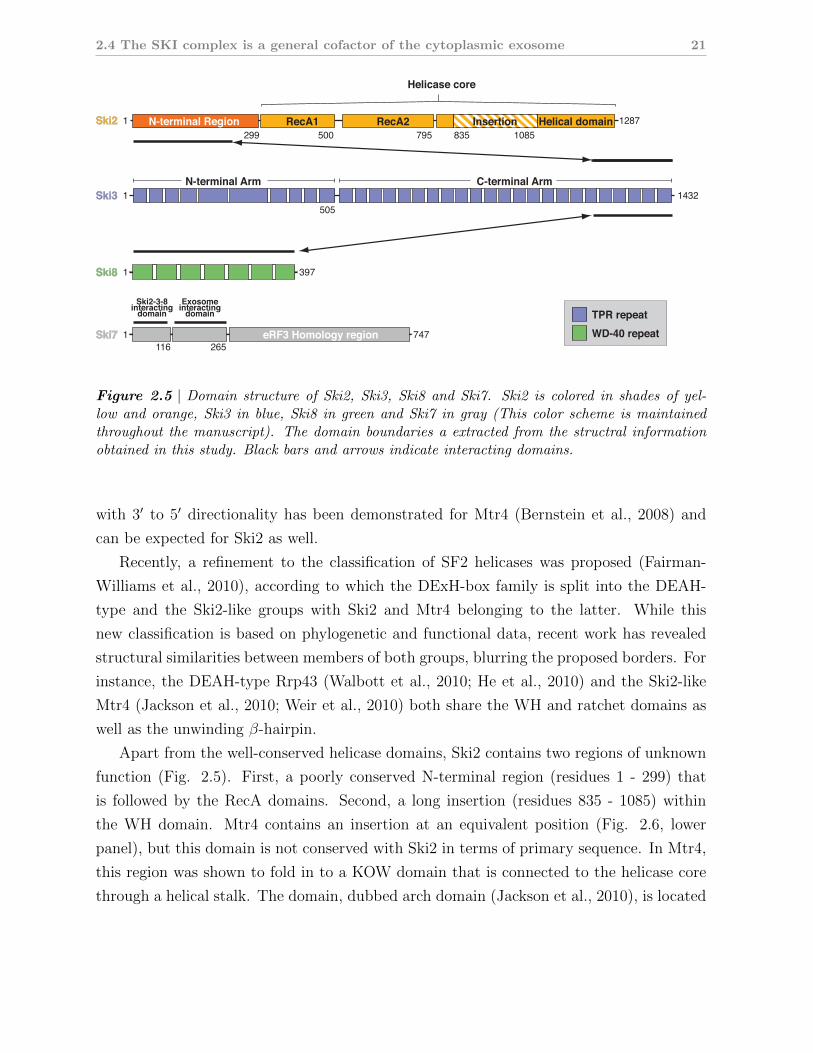
Ski8 3971
Ski7 7471
Ski3
5051
N-terminal Arm C-terminal Arm
TPR repeat
WD-40 repeat
Ski2
299
116 265
500 795 835 1085N-terminal Region
eRF3 Homology region
Helicase core
RecA1 RecA2 Helical domainInsertion 1287
1432
1
Exosomeinteracting
domain
Ski2-3-8interacting
domain
Figure 2.5 | Domain structure of Ski2, Ski3, Ski8 and Ski7. Ski2 is colored in shades of yel-low and orange, Ski3 in blue, Ski8 in green and Ski7 in gray (This color scheme is maintainedthroughout the manuscript). The domain boundaries a extracted from the structral informationobtained in this study. Black bars and arrows indicate interacting domains.
with 3′ to 5′ directionality has been demonstrated for Mtr4 (Bernstein et al., 2008) and
can be expected for Ski2 as well.
Recently, a refinement to the classification of SF2 helicases was proposed (Fairman-
Williams et al., 2010), according to which the DExH-box family is split into the DEAH-
type and the Ski2-like groups with Ski2 and Mtr4 belonging to the latter. While this
new classification is based on phylogenetic and functional data, recent work has revealed
structural similarities between members of both groups, blurring the proposed borders. For
instance, the DEAH-type Rrp43 (Walbott et al., 2010; He et al., 2010) and the Ski2-like
Mtr4 (Jackson et al., 2010; Weir et al., 2010) both share the WH and ratchet domains as
well as the unwinding β-hairpin.
Apart from the well-conserved helicase domains, Ski2 contains two regions of unknown
function (Fig. 2.5). First, a poorly conserved N-terminal region (residues 1 - 299) that
is followed by the RecA domains. Second, a long insertion (residues 835 - 1085) within
the WH domain. Mtr4 contains an insertion at an equivalent position (Fig. 2.6, lower
panel), but this domain is not conserved with Ski2 in terms of primary sequence. In Mtr4,
this region was shown to fold in to a KOW domain that is connected to the helicase core
through a helical stalk. The domain, dubbed arch domain (Jackson et al., 2010), is located

22 2. Introduction
Figure 2.6 | The molecular architecture of DExH-box helicases. The upper panel shows thecrystal structure of A. fulgidus Hel308 bound to a partially unwound DNA duplex (shown inblack) (Buttner et al., 2007). Both RecA domains are colored in yellow, the WH domain in darkyellow and the ratchet domain in orange. The WH and ratchet domains form a helical domainthat packs against both RecA domains. A C-terminally inserted helix-loop-helix (HLH) motif iscolored in red and an unwinding β-hairpin in magenta. The lower panel displays the crystalstructure of S. cerevisiae Mtr4 (Weir et al., 2010). The RecA, WH and ratchet domains areconserved with A.f. Hel308 and are colored accordingly. The inserted KOW domain is shown inred.
above the RNA entry site into the helicase core (Weir et al., 2010). It binds structured
RNAs in vitro (Weir et al., 2010) and is required for 5.8S rRNA processing in vivo (Jackson
et al., 2010). Given that the insertion in Ski2 and the KOW domain in Mtr4 occur are
well conserved positions in the protein, it can be speculated that Ski2 contains a similar
domain.

2.4 The SKI complex is a general cofactor of the cytoplasmic exosome 23
Ski3 and Ski8 are predicted to be structural proteins
Ski3 is a large protein (1432 residues) that is predicted to contain several tetratricopeptide
(TPR) motifs (Fig. 2.5). However, number and position of the TPRs vary according to
the algorithms used for prediction. In general, a single TPR motif is formed by two helices
that pack against each other in an antiparallel fashion (Hirano et al., 1990; Sikorski et al.,
1990; Das et al., 1998). TPRs typically occur as arrays of several contiguous motifs. Since
the individual repeats within an array are rotated by about 60◦, the resulting solenoid
has a superhelical shape. TPRs are known as protein-protein interaction motifs, and to
date no other ligands than (poly-)peptides have been identified (for review see Zeytuni and
Zarivach, 2012). TPR proteins are thus thought of as typical scaffold proteins that organize
large protein complexes. Indeed, this role has been highlighted by several structures of
TPR-mediated protein assemblies (Zhang et al., 2010; Lapouge et al., 2000; Paczkowski
et al., 2012) and has been proposed for Ski3, too.
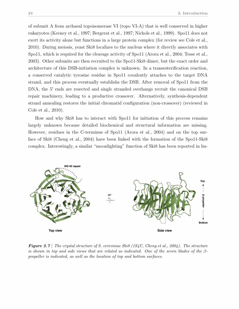
The previously determined crystal structure of Ski8 (Madrona and Wilson, 2004; Cheng
et al., 2004) shows that it contains seven Trp-Asp 40 (WD40) repeats that fold into a seven-
bladed β-propeller (Figs. 2.5 and 2.7). The seven blades form a disc-like structure with
top and bottom surfaces that are connected by a narrow constriction. While β-propeller
proteins sometimes display intrinsic enzymatic activities (e.g. as hydrolases or reductases),
they act more frequently as ligand-binding domains or mediators of protein-protein inter-
actions (reviewed in Chen et al., 2011). As no enzymatic activity had been demonstrated
for Ski8, a structural role for Ski8 has been proposed in the context of the Ski2-Ski3-Ski8
complex. In line with this hypothesis, mutations on a conserved hydrophobic cage at
the top surface of Ski8 have been shown to interfere with binding of Ski8 in vivo (Cheng
et al., 2004), pointing to a potential interface within the Ski2-Ski3-Ski8 complex. Mass-
spectrometry analysis of endogenous samples from yeast indicate a 1:1:2 stoichiometry for
Ski2-Ski3-Ski8 complex with two copies of the Ski8 subunit (Synowsky and Heck, 2008).
Nevertheless, the function of Ski8 within the complex as well as the necessity for two copies
of this subunit remain unclear.
Ski8 is also part of a meiotic DNA recombination complex
In yeast, Ski8 has a second role apart from its function in mRNA degradation. During
meiosis, DNA double-strand break (DSB) formation is the first step in crossing over of
homologous chromatids, a process that eventually leads to genetic diversification. The
catalytic activity of DSB initiation has been pinned down to the protein Spo11, a relative
24 2. Introduction
of subunit A from archaeal topoisomerase VI (topo VI-A) that is well conserved in higher
eukaryotes (Keeney et al., 1997; Bergerat et al., 1997; Nichols et al., 1999). Spo11 does not
exert its activity alone but functions in a large protein complex (for review see Cole et al.,
2010). During meiosis, yeast Ski8 localizes to the nucleus where it directly associates with
Spo11, which is required for the cleavage activity of Spo11 (Arora et al., 2004; Tesse et al.,
2003). Other subunits are then recruited to the Spo11-Ski8 dimer, but the exact order and
architecture of this DSB-initiation complex is unknown. In a transesterification reaction,
a conserved catalytic tyrosine residue in Spo11 covalently attaches to the target DNA
strand, and this process eventually establishs the DSB. After removal of Spo11 from the
DNA, the 5′ ends are resected and single stranded overhangs recruit the canonical DSB
repair machinery, leading to a productive crossover. Alternatively, synthesis-dependent
strand annealing restores the initial chromatid configuration (non-crossover) (reviewed in
Cole et al., 2010).
How and why Ski8 has to interact with Spo11 for initiation of this process remains
largely unknown because detailed biochemical and structural information are missing.
However, residues in the C-terminus of Spo11 (Arora et al., 2004) and on the top sur-
face of Ski8 (Cheng et al., 2004) have been linked with the formation of the Spo11-Ski8
complex. Interestingly, a similar “moonlighting” function of Ski8 has been reported in hu-
Figure 2.7 | The crystal structure of S. cerevisiae Ski8 (1S4U, Cheng et al., 2004). The structureis shown in top and side views that are related as indicated. One of the seven blades of the β-propeller is indicated, as well as the location of top and bottom surfaces.
2.4 The SKI complex is a general cofactor of the cytoplasmic exosome 25
mans. Here, the Ski8 homolog Wdr61 was shown to also participate in the Paf1 complex
that links transcription elongation with histone modification (Zhu et al., 2005).
Domain interactions within the Ski2-Ski3-Ski8 complex and with Ski7
While structural and biochemical characterization of the Ski2-Ski3-Ski8 complex is limited
to the crystal structure of Ski8, some information concerning the complex architecture
is available from yeast-two-hybrid and co-immunoprecipitation experiments (see also Fig.
2.5) (Wang et al., 2005; Brown et al., 2000). First, the interaction of Ski2 and Ski8 depends
on the presence of Ski3, consistent with the notion of Ski3 as a scaffold for the complex.
Second, the N-terminus of Ski2 (residues 1 - 279) is required and sufficient for interaction
with Ski3 in vivo (Wang et al., 2005). Third, the C-terminus of Ski3 (residues 1206 - 1432)
mediates binding to Ski8 (Wang et al., 2005). This agrees well with the observation that a
conserved hydrophobic patch on the top surface of Ski8 (Fig. 2.7) is required for interaction
with Ski3 in vivo (Cheng et al., 2004). Co-immuniprecipitation experiments indicate that
Ski7 interacts directly with Ski3 but not with Ski2 (Wang et al., 2005). The interaction
site resides in the Ski7 N-terminus (residues 1 - 96), while the Ski7 sub-N-terminus (80 -
264) mediates binding to the exosome (Araki et al., 2001).
2.4.3 Conservation of the Ski2-Ski3-Ski8 complex and Ski7 in
higher eukaryotes
The Ski2-Ski3-Ski8 complex subunits are conserved in higher eukaryotes. For instance, ho-
mologs of the yeast genes SKI2 (twister/SKI2), SKI3 (CG8777 / SKI3) and SKI8 (CG3909)
are present in flies (Seago et al., 2001; Orban and Izaurralde, 2005). These homologs ap-
pear to be functional, too. For example, the 5′ fragments generated by the RNAi silencing
mechanism in D. melanogaster are degraded by the exosome, and this pathways requries
the SKI2, SKI3 and SKI8 gene products (Orban and Izaurralde, 2005). These results in-
dicate that Drosophila SKI2, SKI3 and SKI8 are in fact orthologs of their yeast relatives,
and that the function of the Ski2-Ski3-Ski8 complex in mRNA decay may be conserved in
higher eukaryotes.
In humans, homologs of SKI2 (SKI2W), SKI3 (TTC37) and SKI8 (WDR61) exist
(Dangel et al., 1995; Lee et al., 1995), and recombinant Ski2w protein shows ATP hydrolase
(ATPase) activity in an RNA-dependent manner (Dangel et al., 1995). Another study
shows that Ski2w, Ttc37, and Wdr61 proteins form a stable complex (designated the
26 2. Introduction
“hSki” complex) in vivo in the cytoplasm and nucleus of human cells, and that the human
Ski8 homolog Wdr61 also forms part of the Paf1 complex (Zhu et al., 2005), a multi-
subunit assembly that orchestrates histone modification with transcription elongation (for
review see Tomson and Arndt, 2012). The same study also speculates that the Paf1
complex physically interacts with the hSki complex and thus links transcription elongation
to mRNA surveillance (Zhu et al., 2005).
In contrast to the core Ski complex proteins, a Ski7 homolog has not been identified
to date in higher eukaryotes. In fact, phylogenetic studies have identified Ski7 only in
the genus Saccharomyces where it appears to have emerged by a gene duplication event
from the eRF3/Hbs1 family (Atkinson et al., 2008). How the Ski2-Ski3-Ski8 complex is
recruited to the exosome in species lacking Ski7 remains unclear.
Collectively, these results from fly and human show that the Ski2-Ski3-Ski8 complex has
been conserved in higher eukaryotes. They further suggest that, despite the loss of Ski7,
the Ski2-Ski3-Ski8 complex most likely has maintained its function in mRNA turnover and
quality control during evolution and possibly gained functional plasticity.
2.5 Scope of this work
The requirement of the Ski proteins for exosome-mediated degradation of mRNAs in the
cytoplasm has been demonstrated. However, a thorough molecular understanding of the
Ski2-Ski3-Ski8 complex as well as its mode of activation of the exosome remain elusive.
No biochemical and structural information is available except for the crystal structure of
Ski8 (Cheng et al., 2004; Madrona and Wilson, 2004). The notion that Ski2 activates the
exosome by ATP-dependent remodeling of structured RNAs or RNPs lacks experimental
support and remains a hypothesis.
Thus, the present work aims to contribute to the understanding of the molecular biology
of the Ski2-Ski3-Ski8 complex in exosome-mediated mRNA decay, using a structural and
biochemical approach. Particular questions to be addressed are: What is the role of the
individual subunits within the complex? Is the helicase activity of Ski2 regulated through
its partner proteins? Can we gain mechanistic insights into interactions of RNA with the
Ski2-Ski3-Ski8 complex, Ski7 and the exosome, and can these insights help to understand
how the Ski proteins activate the cytoplasmic exosome?
3 Results
3.1 Crystal Structure of the S. cerevisiae Ski2 heli-
case
This article was published in 2012 in RNA (Issue 18(1), pages 124-134). The supple-
mental material is attached at the end of the article (pages 39-40). Figures and tables
of the manuscript are referred to by “3.1.X”, where X follows the numbering within the
manuscript.
28 3. Results
The crystal structure of S. cerevisiae Ski2, a DExH helicaseassociated with the cytoplasmic functions of the exosome
FELIX HALBACH, MICHAELA RODE, and ELENA CONTI1
Department of Structural Cell Biology, Max-Planck-Institute of Biochemistry, D-82152 Martinsried, Germany
ABSTRACT
Ski2 is a cytoplasmic RNA helicase that functions together with the exosome in the turnover and quality control of mRNAs. Ski2is conserved in eukaryotes and is related to the helicase Mtr4, a cofactor of the nuclear exosome involved in the processing andquality control of a variety of structured RNAs. We have determined the 2.4 A resolution crystal structure of the 113 kDahelicase region of Saccharomyces cerevisiae Ski2. The structure shows that Ski2 has an overall architecture similar to that ofMtr4, with a core DExH region and an extended insertion domain. The insertion is not required for the formation of the Ski2–Ski3–Ski8 complex, but is instead an RNA-binding domain. While this is reminiscent of the Mtr4 insertion, there are specificstructural and biochemical differences between the two helicases. The insertion of yeast Mtr4 consists of a b-barrel domain thatis flexibly attached to a helical stalk, contains a KOW signature motif, and binds in vitro-transcribed tRNAi
Met, but not single-stranded RNA. The b-barrel domain of yeast Ski2 does not contain a KOW motif and is tightly packed against the helical stalk,forming a single structural unit maintained by a zinc-binding site. Biochemically, the Ski2 insertion has broad substratespecificity, binding both single-stranded and double-stranded RNAs. We speculate that the Ski2 and Mtr4 insertion domainshave evolved with different properties tailored to the type of transcripts that are the substrates of the cytoplasmic and nuclearexosome.
Keywords: RNA degradation; exosome; helicase; structure
INTRODUCTION
The exosome is a conserved and essential macromolecularcomplex that degrades RNA substrates processively fromthe 39 end (Mitchell et al. 1997). In the eukaryotic nucleus,the exosome is involved in the maturation of ribosomalRNAs, small nuclear RNAs, and small nucleolar RNAs(Allmang et al. 1999; van Hoof et al. 2000a; Houalla et al.2006). It functions in the turnover of pre-mRNAs andcryptic unstable transcripts (Bousquet-Antonelli et al. 2000;Hilleren et al. 2001; Wyers et al. 2005). It is also required inquality-control mechanisms that target aberrant nuclearRNAs such as hypomodified tRNAi
Met (Kadaba et al. 2004;Vanacova et al. 2005). In the cytoplasm, the exosome isinvolved in bulk mRNA turnover (Anderson and Parker1998; van Hoof et al. 2000b) and also participates insurveillance pathways for the degradation of aberrantmRNAs that contain a premature stop codon (Lejeune
et al. 2003; Mitchell and Tollervey 2003; Takahashi et al.2003; Gatfield and Izaurralde 2004) or lack one altogether(van Hoof et al. 2002).
The 10-subunit core of the eukaryotic exosome is identicalin the nuclear and cytoplasmic compartments (for review,see Lorentzen et al. 2008a; Lykke-Andersen et al. 2009). Ninesubunits form a barrel-like structure (Exo-9) with a prom-inent central channel (Liu et al. 2006). The structure of Exo-9 is similar to that of the archaeal exosome and bacterialPNPase, but lacks the catalytic activity that is characteristicof these prokaryotic complexes (Buttner et al. 2005;Lorentzen et al. 2005, 2007). The nuclease activity of thecore exosome complex is conferred by the tenth subunit,Rrp44 (Liu et al. 2006; Dziembowski et al. 2007; Lebretonet al. 2008; Schaeffer et al. 2009; Schneider et al. 2009). BothRrp44 and the catalytically inactive Exo-9 subunits areessential in yeast. The Exo-9 subcomplex modulates theactivity of Rrp44 (Liu et al. 2006; Dziembowski et al.2007; Lorentzen et al. 2008b) and binds RNA substrates,guiding them through the central channel to reach theexoribonuclease active site (Bonneau et al. 2009). The Exo-9structure is also thought to recruit peripheral factors, such asthe nuclear ribonuclease Rrp6 (Liu et al. 2006; Cristodero
1Corresponding author.E-mail [email protected] published online ahead of print. Article and publication date are
at http://www.rnajournal.org/cgi/doi/10.1261/rna.029553.111.
124 RNA (2012), 18:124–134. Published by Cold Spring Harbor Laboratory Press. Copyright ! 2012 RNA Society.
Cold Spring Harbor Laboratory Press on December 13, 2011 - Published by rnajournal.cshlp.orgDownloaded from
3.1 Crystal Structure of the S. cerevisiae Ski2 helicase 29
et al. 2008) and the cytoplasmic protein Ski7 in yeast (Arakiet al. 2001; Dziembowski et al. 2007).
These peripheral factors associate with the core exosometo form an outer shell that is compartment specific (forreview, see Lebreton and Seraphin 2008). In the nucleus,the exosome functions together with the helicase Mtr4,which associates with a poly(A) polymerase (Trf4/Trf5)and a zinc finger protein (Air1/Air2) to form the TRAMPcomplex (LaCava et al. 2005). In the cytoplasm, the exosomefunctions together with the Ski complex (Anderson andParker 1998). The Ski (Superkiller) proteins were originallyidentified from recessive mutations that exacerbated the‘‘killer’’ phenotype, that is, the ability of yeast strainscontaining a dsRNA virus to produce a toxin that killsother strains (Toh-E and Wickner 1979; Ridley et al. 1984;Johnson and Kolodner 1995). These studies showed thatthe Superkiller mutations resided in a helicase (Ski2), a tetra-tricopeptide-repeat (TPR) protein (Ski3), and a WD40 pro-tein (Ski8) in addition to the cytoplasmic 59–39 exoribonu-clease (Xrn1). The Ski2, Ski3, and Ski8 proteins were laterfound to associate in a complex in vivo (Brown et al. 2000).The Ski complex has been implicated in many 39–59cytoplasmic degradation pathways mediated by the exo-some, including normal RNA turnover (Anderson andParker 1998; van Hoof et al. 2000b; Araki et al. 2001),nonsense-mediated decay (Mitchell and Tollervey 2003),nonstop decay (van Hoof et al. 2002), and RNA interfer-ence (Orban and Izaurralde 2005). The Ski and exosomecomplexes interact not only genetically, but also physicallyvia the yeast Ski7 protein (Araki et al. 2001).
The presence of a helicase in both the Ski and TRAMPcomplexes is intriguing. These exosome-associated heli-cases are thought to contribute to substrate recognition, tounwind secondary structure elements in the nucleic acids,or to remove bound proteins, and eventually to presentfavorable single-stranded RNA substrates to the exosome(Lebreton and Seraphin 2008; Houseley and Tollervey2009). The parallel between the nuclear and cytoplasmicregulators of the exosome is further compounded by thefact that Ski2 and Mtr4 share significant sequence similarity(z35% sequence identity in the predicted helicase region).Previous structural work has shown that Mtr4 has a helicasecore similar to that found in other members of the DExHfamily (Jackson et al. 2010; Weir et al. 2010), including thearchaeal DNA helicase Hel308 (Buttner et al. 2007) and thesplicing helicase Prp43 (He et al. 2010; Walbott et al. 2010).In addition, Mtr4 features a 200 aa insertion that containsa helical stalk and a b-barrel domain. The latter is structur-ally and functionally similar to KOW domains, which wereshown to bind structured RNAs in ribosomal proteins(Kyrpides et al. 1996; Selmer et al. 2006; Zhang et al.2009). Consistently, the KOW domain of yeast Mtr4 isrequired for 5.8S rRNA processing in vivo (Jackson et al.2010) and binds transcribed tRNAi
Met in vitro (Weiret al. 2010). Thus, the specific structural features of Mtr4
are in line with its biological functions in ribosomal RNAprocessing and quality control (de la Cruz et al. 1998; vanHoof et al. 2000a). Sequence alignments and secondarystructure predictions suggest that Ski2 has a helicase andan insertion domain similar to those in Mtr4. However,the potential RNA substrates that Ski2 encounters in thecytoplasm are different from those that are recognized byMtr4 in the nucleus. This raises the question as to whetherSki2 has specific features as compared with Mtr4. To addressthis question, we have analyzed the structural and bio-chemical properties of Ski2 from Saccharomyces cerevisiae.
RESULTS AND DISCUSSION
Structure determination of the 113 kDa helicaseregion of yeast Ski2
To initiate the biochemical and structural characterizationof Ski2, we expressed and purified the full-length (f.l.) S.cerevisiae ortholog (residues 1–1287) from insect cells. Ski2is predicted to have a low-complexity N-terminal regionthat has previously been shown to interact with Ski3 byyeast two-hybrid and coimmunoprecipitation analyses(Wang et al. 2005). Limited proteolysis of f.l. Ski2 incombination with N-terminal sequencing and mass spec-trometry indicated the presence of a protease-resistant frag-ment of z113 kDa that lacked the N-terminal 295 residuesand included the predicted helicase region (data not shown).We therefore engineered a construct of yeast Ski2 encom-passing residues 296–1287 (designated Ski2-DN) (Fig. 1A).Yeast Ski2-DN was obtained from expression in insectcells and crystallized in the presence of adenosine 59-(b,g-imido)triphosphate (AMPPNP), a nonhydrolyzableATP analog.
The crystal structure of Ski2-DN was solved to 2.4 Aresolution by the multiwavelength anomalous diffraction(MAD) method using a gold derivative (see Table 1 for datacollection and refinement statistics). The DExH helicasecore of Ski2-DN could be built and refined almost in itsentirety (with the exception of a disordered region betweenresidues 542 and 606) and included electron density for anAMPPNP molecule. An additional domain could be iden-tified that was inserted between residues 830 and 1086,consisting of a helical part and a globular region. However,the electron density for the globular domain, in particular,was weak and did not allow us to trace the polypeptidechain with an unambiguous amino acid register. Thestructure of Ski2-DN bound to AMPPNP was refined to 2.4A resolution, with Rwork of 23.8%, Rfree of 27.5%, and goodstereochemistry (Table 1). We proceeded by engineeringand crystallizing the region corresponding to the inserteddomain (residues 835–1085, designated Ski2-insert). Thestructure of Ski2-insert was determined de novo by thesingle anomalous diffraction (SAD) method using a crystalderivatized with gold. Crystals of Ski2-insert contained five
Ski2 crystal structure
www.rnajournal.org 125
Cold Spring Harbor Laboratory Press on December 13, 2011 - Published by rnajournal.cshlp.orgDownloaded from
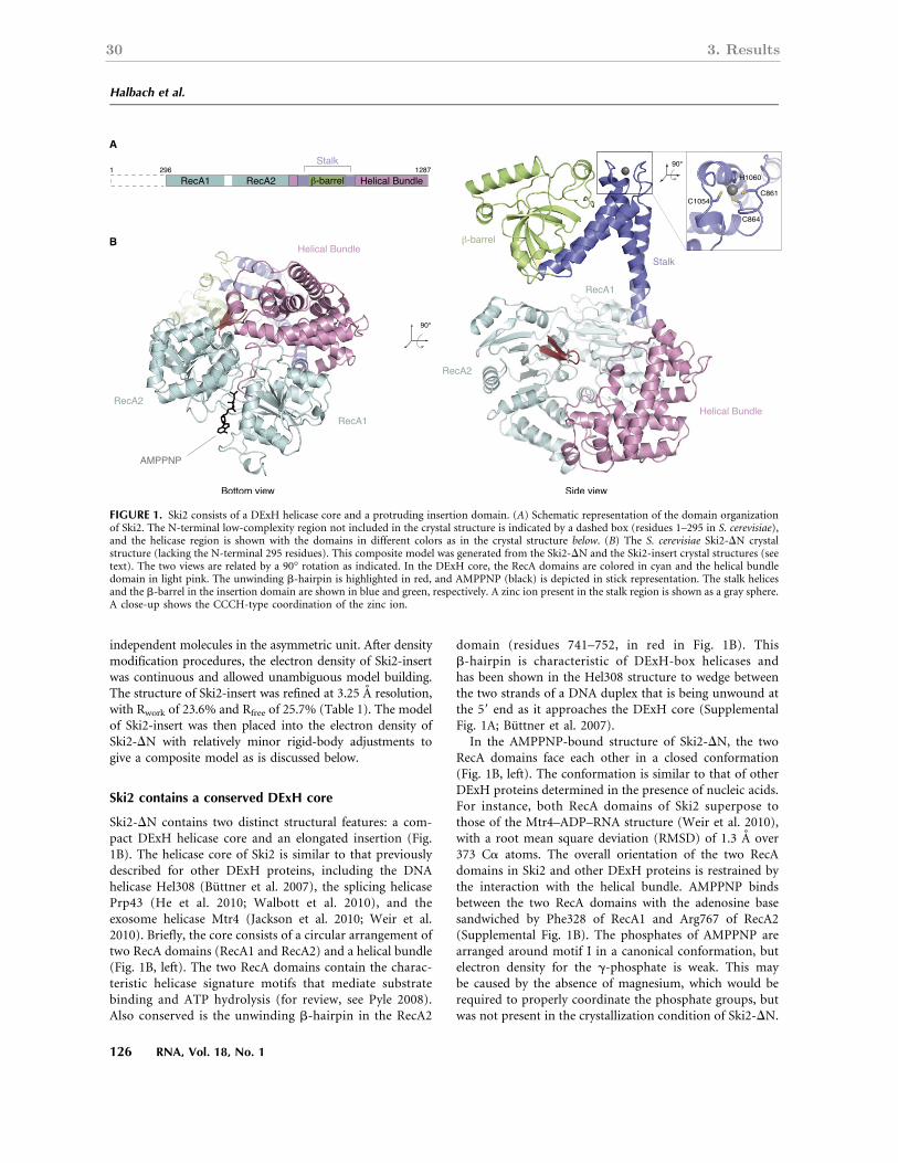
30 3. Results
independent molecules in the asymmetric unit. After densitymodification procedures, the electron density of Ski2-insertwas continuous and allowed unambiguous model building.The structure of Ski2-insert was refined at 3.25 A resolution,with Rwork of 23.6% and Rfree of 25.7% (Table 1). The modelof Ski2-insert was then placed into the electron density ofSki2-DN with relatively minor rigid-body adjustments togive a composite model as is discussed below.
Ski2 contains a conserved DExH core
Ski2-DN contains two distinct structural features: a com-pact DExH helicase core and an elongated insertion (Fig.1B). The helicase core of Ski2 is similar to that previouslydescribed for other DExH proteins, including the DNAhelicase Hel308 (Buttner et al. 2007), the splicing helicasePrp43 (He et al. 2010; Walbott et al. 2010), and theexosome helicase Mtr4 (Jackson et al. 2010; Weir et al.2010). Briefly, the core consists of a circular arrangement oftwo RecA domains (RecA1 and RecA2) and a helical bundle(Fig. 1B, left). The two RecA domains contain the charac-teristic helicase signature motifs that mediate substratebinding and ATP hydrolysis (for review, see Pyle 2008).Also conserved is the unwinding b-hairpin in the RecA2
domain (residues 741–752, in red in Fig. 1B). Thisb-hairpin is characteristic of DExH-box helicases andhas been shown in the Hel308 structure to wedge betweenthe two strands of a DNA duplex that is being unwound atthe 59 end as it approaches the DExH core (SupplementalFig. 1A; Buttner et al. 2007).
In the AMPPNP-bound structure of Ski2-DN, the twoRecA domains face each other in a closed conformation(Fig. 1B, left). The conformation is similar to that of otherDExH proteins determined in the presence of nucleic acids.For instance, both RecA domains of Ski2 superpose tothose of the Mtr4–ADP–RNA structure (Weir et al. 2010),with a root mean square deviation (RMSD) of 1.3 A over373 Ca atoms. The overall orientation of the two RecAdomains in Ski2 and other DExH proteins is restrained bythe interaction with the helical bundle. AMPPNP bindsbetween the two RecA domains with the adenosine basesandwiched by Phe328 of RecA1 and Arg767 of RecA2(Supplemental Fig. 1B). The phosphates of AMPPNP arearranged around motif I in a canonical conformation, butelectron density for the g-phosphate is weak. This maybe caused by the absence of magnesium, which would berequired to properly coordinate the phosphate groups, butwas not present in the crystallization condition of Ski2-DN.
FIGURE 1. Ski2 consists of a DExH helicase core and a protruding insertion domain. (A) Schematic representation of the domain organizationof Ski2. The N-terminal low-complexity region not included in the crystal structure is indicated by a dashed box (residues 1–295 in S. cerevisiae),and the helicase region is shown with the domains in different colors as in the crystal structure below. (B) The S. cerevisiae Ski2-DN crystalstructure (lacking the N-terminal 295 residues). This composite model was generated from the Ski2-DN and the Ski2-insert crystal structures (seetext). The two views are related by a 90° rotation as indicated. In the DExH core, the RecA domains are colored in cyan and the helical bundledomain in light pink. The unwinding b-hairpin is highlighted in red, and AMPPNP (black) is depicted in stick representation. The stalk helicesand the b-barrel in the insertion domain are shown in blue and green, respectively. A zinc ion present in the stalk region is shown as a gray sphere.A close-up shows the CCCH-type coordination of the zinc ion.
Halbach et al.
126 RNA, Vol. 18, No. 1
Cold Spring Harbor Laboratory Press on December 13, 2011 - Published by rnajournal.cshlp.orgDownloaded from

3.1 Crystal Structure of the S. cerevisiae Ski2 helicase 31
An insertion domain protrudes from the Ski2DExH core
The DExH core of Ski2 has an insertion of about 250residues that occurs in the middle of the helical bundledomain (Fig. 1A). The insertion forms an elongated struc-ture that protrudes from the core, extending byz50 A fromthe unwinding b-hairpin that marks the position of the 59end of RNA bound to the DExH core (Fig. 1B, right;Supplemental Fig. 1A). The insertion folds into two pairs ofantiparallel helices (a1–a4 and a2–a3), which connect toa distal b-barrel domain (Fig. 2A). The insertion is a ratherflexible feature of the structure. First, the crystallographictemperature factors increase significantly as comparedwith the core (60 A2 for the DExH core, 120 A2 for helicesa1–a4, 145 A2 for helices a2–a3, and 130 A2 for thebarrel). Second, we observe domain movements whencomparing the insertion domain in the two structures that
we determined: Superposition of the helices a1–a4 in theSki2-DN and Ski2-insert structures results in a rotation ofz15° in the position of helices a2–a3 and the b-barrel(Supplemental Fig. 1C). Despite the flexibility, the elec-tron density is well defined in the structure of Ski2-insert(Supplemental Fig. 1D).
In contrast to the DExH core, the insertion is poorlyconserved even among yeast species (Supplemental Fig. 2).Yet, it is a characteristic feature of exosome helicases: inMtr4 an insertion occurs at an equivalent position in thesequence and with a comparable topology of secondarystructure elements (Fig. 2A,B). The structure of the helicalstalk of Ski2 is similar to that of Mtr4 in that it forms anL-like shape with the a1–a4 and a2–a3 pairs of helicesroughly perpendicular to each other. In the case of yeastSki2, the two pairs of helices are connected by a zinc-binding CCCH-type motif (Fig. 1B, right). Although nozinc was present in the purification or crystallization
TABLE 1. Crystallographic data collection, phasing, and refinement statistics for the Ski2-DN and Ski2-insert structures
Ski2-DN (4A4Z) Ski2-insert (4A4K)
Crystal (Data set) Native Au (peak) Au (inflection) Au (remote) Native Au (peak)
Space group P212121 C2Cell dimensions
a, b, c (A) 82.8,118.6,129.5
84.6, 119.8, 129.7 230.5, 123.1,153.2
229.2, 124.9,149.4
a, b, g (°) 90, 90, 90 90, 90, 90 90, 131.1, 90 90, 130.7, 90Molecules/asymmetricunit
1 5
Data collectionWavelength (A) 0.988 1.037 1.040 0.995 0.999 1.040Resolution (A) 119–2.40 120–2.80 130–2.80 130–2.80 96–3.25 96–4.2
(2.53–2.4) (2.95–2.8) (2.95–2.8) (2.95–2.8) (3.43–3.25) (4.43–4.20Rsym 6.0 (60.0) 7.6 (58.6) 7.7 (68.8) 11.2 (102) 9.3 (66.1) 8.5 (76.9)I/sI 17.0 (3.1) 16.5 (3.0) 16.7 (2.8) 19.2 (3.1) 8.4 (1.5) 14.8 (3.3)Completeness (%) 99.9 (100) 100 (100) 100 (100) 100 (100) 99.6 (99.9) 99.9 (100)Multiplicity 5.5 3.8 3.8 7.8 2.9 4.5
PhasingPhasing Power 0.3 0.3 0.3 n.aMean figure of merit 0.37 0.29
RefinementResolution 39–2.40 57–3.25No. unique reflection 50,434 50,773Rwork/Rfree (%) 23.8/27.5 23.6/25.7Real space correlationcoefficient
0.73 0.76
B-factorsProtein 66.5 113.1Solvent 48.3 89.7
StereochemistryRMSD bond lengths (A) 0.003 0.002RMSD bond angles (°) 0.64 0.36Ramachandran outliers (%) 0.2 0.0Ramachandran favored (%) 96.1 97.0
The highest resolution shell is shown in parenthesis. The figure of merit numbers are given as a mean value over all resolution shells. The real-space correlation coefficient was calculated for the final refined model against a simulated annealing composite omit map, and thestereochemistry of the refined models was analyzed with the MolProbity webserver (Chen et al. 2010).
Ski2 crystal structure
www.rnajournal.org 127
Cold Spring Harbor Laboratory Press on December 13, 2011 - Published by rnajournal.cshlp.orgDownloaded from
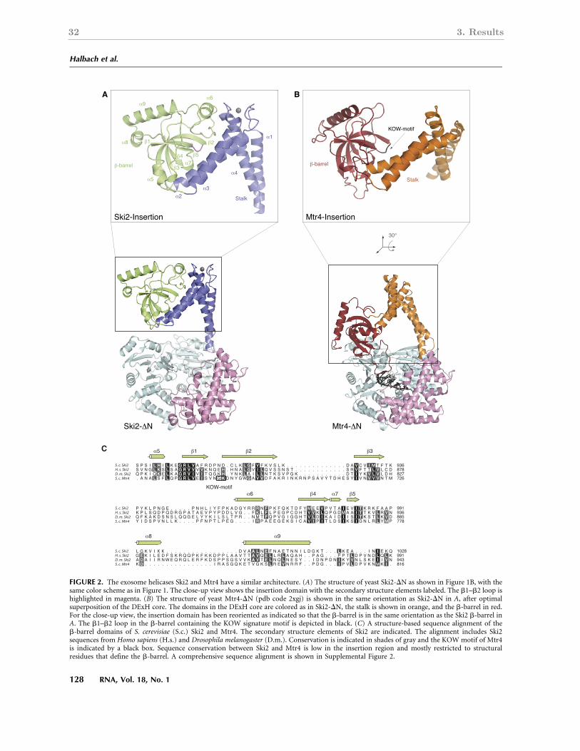
32 3. Results
FIGURE 2. The exosome helicases Ski2 and Mtr4 have a similar architecture. (A) The structure of yeast Ski2-DN as shown in Figure 1B, with thesame color scheme as in Figure 1. The close-up view shows the insertion domain with the secondary structure elements labeled. The b1–b2 loop ishighlighted in magenta. (B) The structure of yeast Mtr4-DN (pdb code 2xgj) is shown in the same orientation as Ski2-DN in A, after optimalsuperposition of the DExH core. The domains in the DExH core are colored as in Ski2-DN, the stalk is shown in orange, and the b-barrel in red.For the close-up view, the insertion domain has been reoriented as indicated so that the b-barrel is in the same orientation as the Ski2 b-barrel inA. The b1–b2 loop in the b-barrel containing the KOW signature motif is depicted in black. (C) A structure-based sequence alignment of theb-barrel domains of S. cerevisiae (S.c.) Ski2 and Mtr4. The secondary structure elements of Ski2 are indicated. The alignment includes Ski2sequences from Homo sapiens (H.s.) and Drosophila melanogaster (D.m.). Conservation is indicated in shades of gray and the KOW motif of Mtr4is indicated by a black box. Sequence conservation between Ski2 and Mtr4 is low in the insertion region and mostly restricted to structuralresidues that define the b-barrel. A comprehensive sequence alignment is shown in Supplemental Figure 2.
Halbach et al.
128 RNA, Vol. 18, No. 1
Cold Spring Harbor Laboratory Press on December 13, 2011 - Published by rnajournal.cshlp.orgDownloaded from
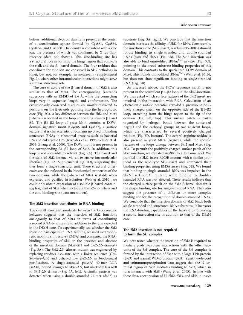
3.1 Crystal Structure of the S. cerevisiae Ski2 helicase 33
buffers, additional electron density is present at the centerof a coordination sphere formed by Cys861, Cys864,Cys1054, and His1060. The density is consistent with a zincion, the presence of which was confirmed by X-ray fluo-rescence (data not shown). This zinc-binding site hasa structural role in forming the hinge region that connectsthe stalk and the b!barrel domain. The four residues thatcoordinate the zinc ion are conserved in Ski2 orthologs infungi, but not, for example, in metazoans (SupplementalFig. 2), where other intramolecular interactions might servea similar structural role.
The core structure of the b-barrel domain of Ski2 is alsosimilar to that of Mtr4. The corresponding b-strandssuperpose with an RMSD of 2.6 A, while the connectingloops vary in sequence, length, and conformation. Theevolutionarily conserved residues are mostly restricted topositions on the b-strands pointing into the hydrophobiccore (Fig. 2C). A key difference between the Ski2 and Mtr4b-barrels is located in the loop connecting strands b1 andb2. The b1–b2 loop of yeast Mtr4 contains a KOW-domain signature motif (Gly686 and Lys687), a surfacefeature that is characteristic of domains involved in bindingstructured RNAs in ribosomal proteins such as bacterialL24 and eukaryotic L26 (Kyrpides et al. 1996; Selmer et al.2006; Zhang et al. 2009). The KOW motif is not present inthe corresponding b1–b2 loop of Ski2. In addition, thisloop is not accessible to solvent (Fig. 2A). The barrel andthe stalk of Ski2 interact via an extensive intramolecularinterface (Fig. 2A; Supplemental Fig. 1D), suggesting thatthey form a single structural unit. These structural differ-ences are also reflected in the biochemical properties of thetwo domains: while the b-barrel of Mtr4 is stable whenexpressed and purified in isolation (Weir et al. 2010), wecould only obtain expression of a soluble b-barrel-contain-ing fragment of Ski2 when including the a2–a3 helices andthe zinc-binding site (data not shown).
The Ski2 insertion contributes to RNA binding
The overall structural similarity between the two exosomehelicases suggests that the insertion of Ski2 functionsanalogously to that of Mtr4 in terms of contributinga second RNA-binding site in addition to the one expectedin the DExH core. To experimentally test whether the Ski2insertion participates in RNA binding, we used electropho-retic mobility shift assays (EMSA) and compared the RNA-binding properties of Ski2 in the presence and absenceof the insertion domain (Ski2-DN and Ski2-DN-Dinsert)(Fig. 3A). The Ski2-DN-Dinsert mutant was engineered byreplacing residues 835–1085 with a linker sequence (Gly–Ser–Arg–Gly) and behaved like Ski2-DN in biochemicalpurifications. A single-stranded poly(A) 40-mer RNA(ssA40) bound strongly to Ski2-DN, but markedly less wellto Ski2-DN-Dinsert (Fig. 3A, left). A similar pattern wasdetected when using a double-stranded 27-mer (ds27) as
substrate (Fig. 3A, right). We conclude that the insertiondomain increases the affinity of Ski2 for RNA. Consistently,the insertion alone (Ski2-insert, residues 835–1085) showedrobust binding to single-stranded and double-strandedRNAs (ss40 and ds27) (Fig. 3B). The Ski2 insertion wasalso able to bind unmodified tRNAi
Met in vitro (Fig. 3C),pointing to the broad substrate-binding properties of thisdomain. This contrasts to the specialized KOW domain ofMtr4, which binds unmodified tRNAi
Met (Weir et al. 2010),but does not show significant binding to single-strandedRNA (Fig. 3B).
As discussed above, the KOW sequence motif is notpresent in the equivalent b1–b2 loop in the Ski2-insertion.We thus asked which surface features of the Ski2 insert areinvolved in the interaction with RNA. Calculation of anelectrostatic surface potential revealed a prominent posi-tively charged patch on the opposite side of the b1–b2loop, stretching from the hinge region to the tip of thedomain (Fig. 3D, top). This surface patch is partlyorganized by hydrogen bonds between the conservedArg903 and the carbonyl groups of two adjacent loops,which are characterized by several positively chargedresidues (Fig. 3D, bottom). The central arginine residue isalso present in yeast Mtr4 (Arg678), while the specificfeatures of the loops diverge between Ski2 and Mtr4 (Fig.2C). To perturb the positively charged surface patch of theSki2 insertion, we mutated Arg903 to a glutamic acid. Wepurified the Ski2-insert R903E mutant with a similar pro-tocol as the wild-type Ski2-insert and compared theirbinding properties using EMSA assays (Fig. 3E). We foundthat binding to single-stranded RNA was impaired in theSki2-insert R903E mutant, while binding to double-stranded RNA was not affected. These results indicate thatthe charged surface patch on the Ski2 b-barrel domain isthe major binding site for single-stranded RNA. They alsosuggest the presence of a different or more complexbinding site for the recognition of double-stranded RNAs.We conclude that the insertion domain of Ski2 binds bothsingle-stranded and structured RNA substrates. It increasesthe RNA-binding capabilities of the helicase by providinga second interaction site in addition to that of the DExHcore.
The Ski2 insertion is not requiredto form the Ski complex
We next tested whether the insertion of Ski2 is required tomediate protein–protein interactions with the other sub-units of the Ski complex. The core of the Ski complex isformed by the interaction of Ski2 with a large TPR protein(Ski3) and a small WD40 protein (Ski8). Yeast two-hybridand coimmunoprecipitation data suggest that the N-ter-minal region of Ski2 mediates binding to Ski3, which inturn interacts with Ski8 (Wang et al. 2005). In line withthese data, coexpression of f.l. Ski2, Ski3, and Ski8 in insect
Ski2 crystal structure
www.rnajournal.org 129
Cold Spring Harbor Laboratory Press on December 13, 2011 - Published by rnajournal.cshlp.orgDownloaded from
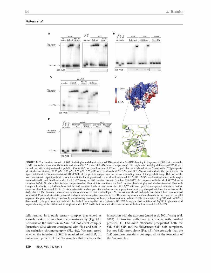
34 3. Results
cells resulted in a stable ternary complex that eluted asa single peak in size-exclusion chromatography (Fig. 4A).Removal of the insertion in Ski2 did not affect complexformation: Ski2-Dinsert comigrated with Ski3 and Ski8 insize-exclusion chromatography (Fig. 4A). We next testedwhether the insertion of Ski2 is required to bind Ski7, anouter-layer protein of the Ski complex that mediates the
interaction with the exosome (Araki et al. 2001; Wang et al.2005). In in-vitro pull-down experiments with purifiedproteins, f.l. GST–Ski7 efficiently precipitated both theSki2–Ski3–Ski8 and the Ski2Dinsert–Ski3–Ski8 complexes,but not Ski2-insert alone (Fig. 4B). We conclude that theSki2 insertion domain is not required for the formation ofthe Ski complex.
FIGURE 3. The insertion domain of Ski2 binds single- and double-stranded RNA substrates. (A) RNA binding to fragments of Ski2 that contain theDExH core with and without the insertion domain (Ski2-DN and Ski2-DN-Dinsert, respectively). Electrophoretic mobility shift assays (EMSA) werecarried out with a single-stranded poly(A) 40-mer (left) or double-stranded 27-mer (right) that were labeled at the 59 end with [32P]phosphate.Identical concentrations (0.25 mM, 0.75 mM, 2.25 mM, 6.75 mM) were used for both Ski2-DN and Ski2-DN-Dinsert and all other proteins in thisfigure. (Bottom) A Coomassie-stained SDS-PAGE of the protein sample used in the corresponding lanes of the gel-shift assay. Deletion of theinsertion domain significantly decreases the affinity for single-stranded and double-stranded RNA. (B) EMSAs as described above with single-stranded (ssA40) and double-stranded RNA (ds27) using the Ski2 insertion domain (residues 835–1085). As compared with the Mtr4 KOW domain(residues 667–818), which fails to bind single-stranded RNA at this condition, the Ski2 insertion binds single- and double-stranded RNA withcomparable affinity. (C) EMSAs show that the Ski2 insertion binds in vitro-transcribed tRNAi
Met with an apparently comparable affinity to that forsingle- or double-stranded RNA. (D) An electrostatic surface potential analysis reveals a prominent positively charged patch on the surface of theSki2 b-barrel. The domain is shown in a similar orientation to that used in Figure 2A, but without the a1 and a4 helices (which have been omittedfor clarity). Positive electrostatic potential is shown in blue, negative potential in red. The close-up view at bottom shows how the conserved Arg903organizes the positively charged surface by coordinating two loops with several basic residues (indicated). The side-chains of Lys985 and Lys987 aredisordered. Hydrogen bonds are indicated by dashed lines together with distances. (E) EMSAs suggest that mutation of Arg903 to glutamic acidimpairs binding of the Ski2-insert to single-stranded RNA (A40) but does not affect interaction with double-stranded RNA (ds27).
Halbach et al.
130 RNA, Vol. 18, No. 1
Cold Spring Harbor Laboratory Press on December 13, 2011 - Published by rnajournal.cshlp.orgDownloaded from

3.1 Crystal Structure of the S. cerevisiae Ski2 helicase 35
CONCLUSIONS
The two main regulators of exosome activity, the cytosolicSki and the nuclear TRAMP complexes, contain relatedRNA helicases. The Ski2 and Mtr4 helicases have a similararchitecture with two distinct structural modules: a helicasecore and an insertion region. The helicase core is typical ofthe DExH family of helicases, and is expected to mediateATP-dependent RNA unwinding and remodeling activities.The insertion contains a long stalk and a b-barrel domain.Removal of the insertion in Ski2 and Mtr4 does not com-promise the formation of the respective complexes with Ski3and Ski8 and with Trf4–Air2 (Weir et al. 2010), but sig-nificantly reduces the RNA-binding properties of the twohelicases. The insertion domains of both Ski2 and Mtr4 bindRNA in vitro. Their position above the entry site of the
RNA-binding channel in the helicase core suggests that theycontribute to recruiting RNA substrates to the unwinding site.
Despite these overarching similarities, the b-barrel do-mains of Mtr4 and Ski2 have specific structural and func-tional differences. In yeast Mtr4, the b-barrel domain hasa KOW signature motif, is involved in 5.8S rRNA maturationin vivo (Jackson et al. 2010), binds unmodified tRNAi
Met
(Weir et al. 2010), and discriminates against single-strandedRNA in vitro. In Ski2, the b-barrel domain does not exhibita preference for binding structured RNAs, but is ratherpromiscuous in its RNA-binding properties. The b-barrelof Ski2 does not contain a KOW motif in the correspondingloop (b1–b2). Mutagenesis studies suggest that binding ofthe Ski2 b-barrel to single-stranded RNA is mediated by theopposite surface, which features several positively chargedresidues. We speculate that the b-barrel domains of Ski2
FIGURE 4. The Ski2 insertion domain is not required for the protein–protein interactions of the Ski complex. (A) Analytical gel-filtration of f.l.Ski2–Ski3–Ski8 and Ski2Dinsert–Ski3–Ski8 complexes. Both complexes were copurified and then injected on an analytical size-exclusionchromatography column (top). Peak fractions were analyzed by SDS-PAGE (bottom). All three components comigrated in the case of bothcomplexes, indicating that deletion of the insertion domain of Ski2 does not impair Ski complex formation. (B) Pull-down experiments withGST-tagged f.l. Ski7 (residues 1–747) and untagged f.l. Ski2–Ski3–Ski8 and Ski2Dinsert–Ski3–Ski8 complexes. Input samples (top) and samplesprecipitated on glutathione–Sepharose beads (bottom) were analyzed by SDS-PAGE. The proteins corresponding to the bands are indicated on theright side of both panels. Both complexes were efficiently precipitated by GST–Ski7, while the insertion alone (Ski2-insert) did not bind to thebait. (*) A contamination in the GST–Ski7 sample.
Ski2 crystal structure
www.rnajournal.org 131
Cold Spring Harbor Laboratory Press on December 13, 2011 - Published by rnajournal.cshlp.orgDownloaded from
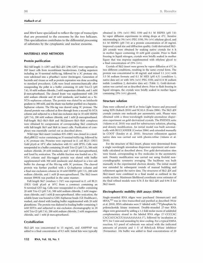
36 3. Results
and Mtr4 have specialized to reflect the type of transcriptsthat are presented to the exosome by the two helicases.This specialization contributes to the differential processingof substrates by the cytoplasmic and nuclear exosome.
MATERIALS AND METHODS
Protein purification
Ski2 full length (1–1287) and Ski2-DN (296–1287) were expressed inHi5 insect cells from recombinant baculoviruses. Coding sequencesincluding an N-terminal 6xHis-tag, followed by a 3C protease site,were subcloned into a pFastBac1 vector (Invitrogen). Generation ofbacmids and viruses as well as protein expression was done accordingto standard procedures. Cells were lysed osmomechanically afterresuspending the pellet in a buffer containing 10 mM Tris-Cl (pH7.4), 10 mM sodium chloride, 2 mMmagnesium chloride, and 1 mMb-mercaptoethanol. The cleared lysate was supplemented with 250mM sodium chloride and 20 mM imidazole, and loaded on a Ni-NTA-sepharose column. The protein was eluted by an imidazolegradient to 300 mM, and the eluate was further purified on a heparin–Sepharose column. The His-tag was cleaved using 3C protease. Thecleaved protein was collected as flow-through from a second Ni-NTAcolumn and subjected to a final size-exclusion step in 10 mM HEPES(pH 7.4), 200 mM sodium chloride, and 1 mM b-mercaptoethanol.Full-length Ski2–Ski3–Ski8 and Ski2Dinsert–Ski3–Ski8 complexeswere obtained by coexpression of corresponding Ski2 constructswith full-length Ski3 and Ski8 in insect cells. Purification of com-plexes was essentially carried out as described above.
Wild-type Ski2-insert (residues 835–1085) was cloned in a mod-ified pBR322 vector containing an N-teminal 6xHis tag, followed bya 3C protease site. Proteins were expressed in E. coli BL21 (DE3)Gold pLysS at 18°C after induction with 0.1 mM IPTG. Cells wereresuspended in a buffer containing 20 mM Tris-Cl (pH 7.5), 500 mMsodium chloride, 20 mM imidazole, and 1 mM b-mercaptoethanol,and lysed by sonication. The soluble fraction was loaded on a Ni-NTA column and His-tagged protein was eluted with buffersupplemented with 300 mM imidazole and dialyzed in a low-saltbuffer for cleavage of the His-tag with 3C protease. The cleavedprotein was further purified with a Q–Sepharose column anda final size-exclusion column in 10 mM HEPES (pH 7.5), 200 mMsodium chloride, and 1 mM b-mercaptoethanol. The Ski2-insertmutant R903E was purified in the same manner.
Full-length Ski7 (residues 1–747) was expressed in E. coli BL21(DE3) Gold pLysS at 18°C from a construct containing anN-terminal GST-tag. Cells were resuspended in a buffer containing20 mM Tris-Cl (pH 7.4), 500 mM sodium chloride, 2 mM magne-sium chloride, and 1 mM b-mercaptoethanol. After sonication, thesoluble lysate was bound to GSH–Sepharose beads (GE Healthcare),washed, and eluted with loading buffer supplemented with 20 mMglutathione. The protein was dialyzed in loading buffer containing 5mM EDTA and subjected to size-exclusion chromatography in 20mM Tris-Cl (pH 7.4), 200 mM sodium chloride, 2 mM magnesiumchloride, and 1 mM b-mercaptoethanol.
Crystallization
Ski2-DN was concentrated to 15 mg/mL, and AMPPNP wasadded to a final concentration of 0.5 mM. Initial hits were typically
obtained in 15% (w/v) PEG 3350 and 0.1 M HEPES (pH 7.0)by vapor diffusion experiments in sitting drops at 4°C. Iterativemicroseeding in 3% (w/v) PEG 3350, 5% (v/v) ethylene glycol, and0.1 M HEPES (pH 7.0) at a protein concentration of 20 mg/mLimproved crystal size and diffraction quality. Gold-derivatized Ski2-DN crystals were obtained by soaking native crystals for 6 hin mother liquor containing 10 mM gold cyanide. Prior to flashfreezing in liquid nitrogen, crystals were briefly soaked in motherliquor that was stepwise supplemented with ethylene glycol toa final concentration of 25% (v/v).
Crystals of Ski2-insert were grown by vapor-diffusion at 4°C intwo different conditions, resulting in the same crystal form. Theprotein was concentrated to 40 mg/mL and mixed 1:1 (v/v) with3.5 M sodium formate and 0.1 M MES (pH 6.5) (condition 1,native data set) or with 16% (w/v) PEG 3350, and 0.14 M sodiumiodide (condition 2, derivative data set) (Table 1). Gold-derivati-zation was carried out as described above. Prior to flash freezing inliquid nitrogen, the crystals were briefly soaked in mother liquorcontaining 25% (v/v) glycerol.
Structure solution
Data were collected at 100 K at Swiss Light Source and processedusing XDS (Kabsch 2010) and SCALA (Evans 2006). The Ski2-DNcrystals contain one molecule per asymmetric unit. Phases wereobtained with a three-wavelength multiple-anomalous disper-sion experiment on gold-derivatized crystals. The PHENIX suite(Adams et al. 2010) was used for substructure solution, phasing,and density modification. An initial model was built automat-ically with BUCCANEER (Cowtan 2006) and extended manuallyin COOT (Emsley et al. 2010). Structure refinement againstnative data was carried out with phenix.refine (Adams et al.2010).
For the structure of Ski2-insert, phases were determined froma single wavelength anomalous dispersion experiment and essen-tially calculated as described above. Five gold-derivatization siteswere found, corresponding to five molecules in the asymmetricunit. Density modification was carried out using fivefold non-crystallographic symmetry averaging. The backbone was builtmanually in the experimental electron density. The initial modelwas extended by subsequent rounds of manual building andrefinement against the native data. The structures of Ski2-DN andSki2-insert were combined to a final model as outlined in theresults section. Maximum likelihood coordinate error estimates ofthe final refined models were 0.76 A for Ski2-DN and 0.94 A forSki2-insert.
Electrophoretic mobility shift assays (EMSA)
Single-stranded RNA oligos were purchased (biomers.net) andtRNAi
Met was in vitro transcribed and purified as described (Weiret al. 2010). RNA substrates were 59-labeled with [32P]phosphate bypolynucleotide kinase treatment. Double-stranded 27-mer RNAoligos were generated by adding a 1.2-fold molar excess of unlabeledcomplementary strand to the labeled RNA oligo (59-CCCCACCACCAUCACUUAAAAAAAAAA-39), followed by incubation at95°C for 5 min and annealing by slow cooling. For a typical EMSAreaction, 0.5 pmol of substrate was mixed with the indicatedamounts of protein and 1 U of RiboLock RNase inhibitor(Fermentas). 10x buffer was added to final concentrations of 20
Halbach et al.
132 RNA, Vol. 18, No. 1
Cold Spring Harbor Laboratory Press on December 13, 2011 - Published by rnajournal.cshlp.orgDownloaded from

3.1 Crystal Structure of the S. cerevisiae Ski2 helicase 37
mMHEPES (pH 7.5), 50 mM potassium acetate, 5 mMmagnesiumacetate, 0.1% (v/v) NP-40, and 2 mM dithiothreitol. Reactions werecarried out in a volume of 10 mL with a final salt concentration of75 mM sodium chloride and 50 mM potassium chloride in allsamples. Samples were incubated at 4°C for 30 min and separatedon a native 6% (w/v) polyacrylamide gel. Gels were analyzed byphosphorimaging.
Pull-down assays
GST-tagged prey protein (4 mg) was mixed with equal molaramounts of bait protein. Buffer was added to a volume of 200mL and final concentrations of 10 mM HEPES (pH 7.5), 75 mMsodium chloride, 2 mM magnesium acetate, 0.1% (v/v) NP-40, 1mM dithiothreitol, and 12.5% (v/v) glycerol (buffer A). A totalof 40 mL of a 50% (v/v) suspension of GSH–Sepharose beads(GE Healthcare) were added, and the reaction was incubatedfor 30 min at 4°C. Beads were washed three times with bufferA before eluting the precipitated protein. Samples were ana-lyzed on a 4%–12% (w/v) Bis-Tris polyacrylamide gradient gel(Invitrogen).
DATA DEPOSITION
The coordinates and the structure factors have been deposited inthe Protein Data Bank with accession codes 4a4z (Ski2-DN) and4a4k for (Ski2-insert).
SUPPLEMENTAL MATERIAL
Supplemental material is available for this article.
ACKNOWLEDGMENTS
We thank Jerome Basquin, Karina Valer-Saldana, and SabinePleyer at the MPI-Martinsried crystallization facility, and thestaff of the PX beamlines at the Swiss Light Source (Villigen) forassistance during data collection. We also thank Peter Brick andmembers of our laboratory for suggestions and critical readingof the manuscript. This study was supported by the BoehringerIngelheim Fonds to F.H.; by the Max Planck Gesellschaft, theSonderforschungsbereich SFB646, the Gottfried Wilhelm Leib-niz Program of the Deutsche Forschungsgemeinschaft (DFG),and the Center for Integrated Protein Science Munich (CIPSM)to E.C.
Received July 27, 2011; accepted October 25, 2011.
REFERENCES
Adams PD, Afonine PV, Bunkoczi G, Chen VB, Davis IW, Echols N,Headd JJ, Hung LW, Kapral GJ, Grosse-Kunstleve RW, et al. 2010.PHENIX: a comprehensive Python-based system for macromolec-ular structure solution. Acta Crystallogr D Biol Crystallogr 66: 213–221.
Allmang C, Kufel J, Chanfreau G, Mitchell P, Petfalski E, Tollervey D.1999. Functions of the exosome in rRNA, snoRNA and snRNAsynthesis. EMBO J 18: 5399–5410.
Anderson JS, Parker RP. 1998. The 39 to 59 degradation of yeastmRNAs is a general mechanism for mRNA turnover that requires
the SKI2 DEVH box protein and 39 to 59 exonucleases of theexosome complex. EMBO J 17: 1497–1506.
Araki Y, Takahashi S, Kobayashi T, Kajiho H, Hoshino S, Katada T.2001. Ski7p G protein interacts with the exosome and the Skicomplex for 39-to-59 mRNA decay in yeast. EMBO J 20: 4684–4693.
Bonneau F, Basquin J, Ebert J, Lorentzen E, Conti E. 2009. The yeastexosome functions as a macromolecular cage to channel RNAsubstrates for degradation. Cell 139: 547–559.
Bousquet-Antonelli C, Presutti C, Tollervey D. 2000. Identification ofa regulated pathway for nuclear pre-mRNA turnover. Cell 102:765–775.
Brown JT, Bai X, Johnson AW. 2000. The yeast antiviral proteinsSki2p, Ski3p, and Ski8p exist as a complex in vivo. RNA 6: 449–457.
Buttner K, Wenig K, Hopfner KP. 2005. Structural framework for themechanism of archaeal exosomes in RNA processing. Mol Cell 20:461–471.
Buttner K, Nehring S, Hopfner KP. 2007. Structural basis for DNAduplex separation by a superfamily-2 helicase. Nat Struct Mol Biol14: 647–652.
Chen VB, Arendall WB III, Headd JJ, Keedy DA, Immormino RM,Kapral GJ, Murray LW, Richardson JS, Richardson DC. 2010.MolProbity: all-atom structure validation for macromolecularcrystallography. Acta Crystallogr D Biol Crystallogr 66: 12–21.
Cowtan K. 2006. The Buccaneer software for automated modelbuilding. 1. Tracing protein chains. Acta Crystallogr D Biol Crys-tallogr 62: 1002–1011.
Cristodero M, Bottcher B, Diepholz M, Scheffzek K, Clayton C.2008. The Leishmania tarentolae exosome: purification andstructural analysis by electron microscopy. Mol Biochem Parasitol159: 24–29.
de la Cruz J, Kressler D, Tollervey D, Linder P. 1998. Dob1p (Mtr4p)is a putative ATP-dependent RNA helicase required for the 39 endformation of 5.8S rRNA in Saccharomyces cerevisiae. EMBO J 17:1128–1140.
Dziembowski A, Lorentzen E, Conti E, Seraphin B. 2007. A singlesubunit, Dis3, is essentially responsible for yeast exosome coreactivity. Nat Struct Mol Biol 14: 15–22.
Emsley P, Lohkamp B, Scott WG, Cowtan K. 2010. Features anddevelopment of Coot. Acta Crystallogr D Biol Crystallogr 66: 486–501.
Evans P. 2006. Scaling and assessment of data quality. Acta CrystallogrD Biol Crystallogr 62: 72–82.
Gatfield D, Izaurralde E. 2004. Nonsense-mediated messenger RNAdecay is initiated by endonucleolytic cleavage in Drosophila.Nature 429: 575–578.
He Y, Andersen GR, Nielsen KH. 2010. Structural basis for thefunction of DEAH helicases. EMBO Rep 11: 180–186.
Hilleren P, McCarthy T, Rosbash M, Parker R, Jensen TH. 2001.Quality control of mRNA 39-end processing is linked to thenuclear exosome. Nature 413: 538–542.
Houalla R, Devaux F, Fatica A, Kufel J, Barrass D, Torchet C,Tollervey D. 2006. Microarray detection of novel nuclear RNAsubstrates for the exosome. Yeast 23: 439–454.
Houseley J, Tollervey D. 2009. The many pathways of RNA degrada-tion. Cell 136: 763–776.
Jackson RN, Klauer AA, Hintze BJ, Robinson H, van Hoof A, JohnsonSJ. 2010. The crystal structure of Mtr4 reveals a novel arch domainrequired for rRNA processing. EMBO J 29: 2205–2216.
Johnson AW, Kolodner RD. 1995. Synthetic lethality of sep1 (xrn1)ski2 and sep1 (xrn1) ski3 mutants of Saccharomyces cerevisiae isindependent of killer virus and suggests a general role for thesegenes in translation control. Mol Cell Biol 15: 2719–2727.
Kabsch W. 2010. XDS. Acta Crystallogr D Biol Crystallogr 66: 125–132.Kadaba S, Krueger A, Trice T, Krecic AM, Hinnebusch AG, Anderson
J. 2004. Nuclear surveillance and degradation of hypomodifiedinitiator tRNAMet in S. cerevisiae. Genes Dev 18: 1227–1240.
Ski2 crystal structure
www.rnajournal.org 133
Cold Spring Harbor Laboratory Press on December 13, 2011 - Published by rnajournal.cshlp.orgDownloaded from

38 3. Results
Kyrpides NC, Woese CR, Ouzounis CA. 1996. KOW: a novel motiflinking a bacterial transcription factor with ribosomal proteins.Trends Biochem Sci 21: 425–426.
LaCava J, Houseley J, Saveanu C, Petfalski E, Thompson E, Jacquier A,Tollervey D. 2005. RNA degradation by the exosome is promotedby a nuclear polyadenylation complex. Cell 121: 713–724.
Lebreton A, Seraphin B. 2008. Exosome-mediated quality control:substrate recruitment and molecular activity. Biochim Biophys Acta1779: 558–565.
Lebreton A, Tomecki R, Dziembowski A, Seraphin B. 2008. Endonu-cleolytic RNA cleavage by a eukaryotic exosome. Nature 456: 993–996.
Lejeune F, Li X, Maquat LE. 2003. Nonsense-mediated mRNA decayin mammalian cells involves decapping, deadenylating, andexonucleolytic activities. Mol Cell 12: 675–687.
Liu Q, Greimann JC, Lima CD. 2006. Reconstitution, activities, andstructure of the eukaryotic RNA exosome. Cell 127: 1223–1237.
Lorentzen E, Walter P, Fribourg S, Evguenieva-Hackenberg E, Klug G,Conti E. 2005. The archaeal exosome core is a hexameric ringstructure with three catalytic subunits. Nat Struct Mol Biol 12:575–581.
Lorentzen E, Dziembowski A, Lindner D, Seraphin B, Conti E. 2007.RNA channelling by the archaeal exosome. EMBO Rep 8: 470–476.
Lorentzen E, Basquin J, Conti E. 2008a. Structural organization of theRNA-degrading exosome. Curr Opin Struct Biol 18: 709–713.
Lorentzen E, Basquin J, Tomecki R, Dziembowski A, Conti E. 2008b.Structure of the active subunit of the yeast exosome core, Rrp44:diverse modes of substrate recruitment in the RNase II nucleasefamily. Mol Cell 29: 717–728.
Lykke-Andersen S, Brodersen DE, Jensen TH. 2009. Origins andactivities of the eukaryotic exosome. J Cell Sci 122: 1487–1494.
Mitchell P, Tollervey D. 2003. An NMD pathway in yeast involvingaccelerated deadenylation and exosome-mediated 39/59 degra-dation. Mol Cell 11: 1405–1413.
Mitchell P, Petfalski E, Shevchenko A, Mann M, Tollervey D. 1997.The exosome: a conserved eukaryotic RNA processing complexcontaining multiple 39/59 exoribonucleases. Cell 91: 457–466.
Orban TI, Izaurralde E. 2005. Decay of mRNAs targeted by RISC requiresXRN1, the Ski complex, and the exosome. RNA 11: 459–469.
Pyle AM. 2008. Translocation and unwinding mechanisms of RNAand DNA helicases. Annu Rev Biophys 37: 317–336.
Ridley SP, Sommer SS, Wickner RB. 1984. Superkiller mutations inSaccharomyces cerevisiae suppress exclusion of M2 double-strandedRNA by L-A-HN and confer cold sensitivity in the presence of Mand L-A-HN. Mol Cell Biol 4: 761–770.
Schaeffer D, Tsanova B, Barbas A, Reis FP, Dastidar EG, Sanchez-Rotunno M, Arraiano CM, van Hoof A. 2009. The exosome
contains domains with specific endoribonuclease, exoribonucleaseand cytoplasmic mRNA decay activities. Nat Struct Mol Biol 16:56–62.
Schneider C, Leung E, Brown J, Tollervey D. 2009. The N-terminalPIN domain of the exosome subunit Rrp44 harbors endonucleaseactivity and tethers Rrp44 to the yeast core exosome. Nucleic AcidsRes 37: 1127–1140.
Selmer M, Dunham CM, Murphy FV IV, Weixlbaumer A, Petry S,Kelley AC, Weir JR, Ramakrishnan V. 2006. Structure of the 70Sribosome complexed with mRNA and tRNA. Science 313: 1935–1942.
Takahashi S, Araki Y, Sakuno T, Katada T. 2003. Interaction betweenSki7p and Upf1p is required for nonsense-mediated 39-to-59mRNA decay in yeast. EMBO J 22: 3951–3959.
Toh-E A, Wickner RB. 1979. A mutant killer plasmid whosereplication depends on a chromosomal ‘‘superkiller’’ mutation.Genetics 91: 673–682.
van Hoof A, Lennertz P, Parker R. 2000a. Yeast exosome mutantsaccumulate 39-extended polyadenylated forms of U4 small nuclearRNA and small nucleolar RNAs. Mol Cell Biol 20: 441–452.
van Hoof A, Staples RR, Baker RE, Parker R. 2000b. Function of theSki4p (Csl4p) and Ski7p proteins in 39-to-59 degradation ofmRNA. Mol Cell Biol 20: 8230–8243.
van Hoof A, Frischmeyer PA, Dietz HC, Parker R. 2002. Exosome-mediated recognition and degradation of mRNAs lacking a termi-nation codon. Science 295: 2262–2264.
Vanacova S, Wolf J, Martin G, Blank D, Dettwiler S, Friedlein A,Langen H, Keith G, Keller W. 2005. A new yeast poly(A)polymerase complex involved in RNA quality control. PLoS Biol3: e189. doi: 10.1371/journal.pbio.0030189.
Walbott H, Mouffok S, Capeyrou R, Lebaron S, Humbert O, vanTilbeurgh H, Henry Y, Leulliot N. 2010. Prp43p contains a proces-sive helicase structural architecture with a specific regulatorydomain. EMBO J 29: 2194–2204.
Wang L, Lewis MS, Johnson AW. 2005. Domain interactions withinthe Ski2/3/8 complex and between the Ski complex and Ski7p.RNA 11: 1291–1302.
Weir JR, Bonneau F, Hentschel J, Conti E. 2010. Structural analysisreveals the characteristic features of Mtr4, a DExH helicaseinvolved in nuclear RNA processing and surveillance. Proc NatlAcad Sci 107: 12139–12144.
Wyers F, Rougemaille M, Badis G, Rousselle JC, Dufour ME, Boulay J,Regnault B, Devaux F, Namane A, Seraphin B, et al. 2005. CrypticPol II transcripts are degraded by a nuclear quality controlpathway involving a new poly(A) polymerase. Cell 121: 725–737.
Zhang W, Dunkle JA, Cate JH. 2009. Structures of the ribosome inintermediate states of ratcheting. Science 325: 1014–1017.
Halbach et al.
134 RNA, Vol. 18, No. 1
Cold Spring Harbor Laboratory Press on December 13, 2011 - Published by rnajournal.cshlp.orgDownloaded from

3.1 Crystal Structure of the S. cerevisiae Ski2 helicase 39
!"##$%&%'()$*+,-".%*/
!!
!
!"
#
!"
$
#$%!&
5/)/
'$%()
*$%+$
#$%(!
5&.)
6/).'&.$
#$%!&
5/)/
'$%()
*$%+$
#$%(!
5&.)
6/).'&.$
#),)
-!./0!.)
1(((
*((.
#),!
'!+/
$.2
$
(
."
!"
."
.%34
+%34
+
!
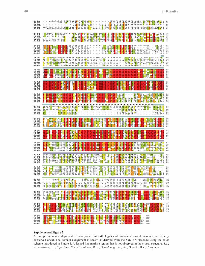
40 3. Results
!"##$%&%'()$*+,-".%*/!"#$%&'(%)" *)+$),-)" .%'/,#),&" 01" )$2.340&'-"52'6" 03&70%0/*" 897'&)" ',:'-.&)*" ;.3'.<%)" 3)*':$)*=" 3):" *&3'-&%4"
*-7)#)"',&30:$-):"',">'/$3)"?@"!":.*7):"%',)"#.32*"."3)/'0,"&7.&"'*",0&"0<*)3;):"',"&7)"-34*&.%"*&3$-&$3)@"5@-@="!"#$%&%'()(*%="A@(@="+"#,*)-.&()="B@.@="/"#*01($*2)="C@#@="3"#4%0*2.5*)-%&="C@3@="3"#&%&(.="D@*@="6"#)*,(%2)"*
!"#"$!%&'(")"$!%&'*"+"$!%&',"-"$!%&'."/"$!%&',"0"$!%&'
!"#"$!%&'(")"$!%&'*"+"$!%&',"-"$!%&'."/"$!%&',"0"$!%&'
!"#"$!%&'(")"$!%&'*"+"$!%&',"-"$!%&'."/"$!%&',"0"$!%&'
!"#"$!%&'(")"$!%&'*"+"$!%&',"-"$!%&'."/"$!%&',"0"$!%&'
!"#"$!%&'(")"$!%&'*"+"$!%&',"-"$!%&'."/"$!%&',"0"$!%&'
!"#"$!%&'(")"$!%&'*"+"$!%&',"-"$!%&'."/"$!%&',"0"$!%&'
!"#"$!%&'(")"$!%&'*"+"$!%&',"-"$!%&'."/"$!%&',"0"$!%&'
!"#"$!%&'(")"$!%&'*"+"$!%&',"-"$!%&'."/"$!%&',"0"$!%&'
!"#"$!%&'(")"$!%&'*"+"$!%&',"-"$!%&'."/"$!%&',"0"$!%&'
!"#"$!%&'(")"$!%&'*"+"$!%&',"-"$!%&'."/"$!%&',"0"$!%&'
!"#"$!%&'(")"$!%&'*"+"$!%&',"-"$!%&'."/"$!%&',"0"$!%&'
!"#"$!%&'(")"$!%&'*"+"$!%&',"-"$!%&'."/"$!%&',"0"$!%&'
!"#"$!%&'(")"$!%&'*"+"$!%&',"-"$!%&'."/"$!%&',"0"$!%&'
!"#"$!%&'(")"$!%&'*"+"$!%&',"-"$!%&'."/"$!%&',"0"$!%&'
!"#"$!%&'(")"$!%&'*"+"$!%&',"-"$!%&'."/"$!%&',"0"$!%&'
!"#"$!%&'(")"$!%&'*"+"$!%&',"-"$!%&'."/"$!%&',"0"$!%&'
!"#"$!%&'(")"$!%&'*"+"$!%&',"-"$!%&'."/"$!%&',"0"$!%&'
! ! ! ! ! ! ! ! ! ! ! ! ! ! ! ! ! ! ! ! ! ! ! ! ! ! ! ! ! ! ! ! ! ! ! ! ! ! ! ! ! ! ! ! ! ! ! ! ! ! ! ! ! ! ! ! ! ! ! ! ! ! ! ! ! ! ! ! ! ! ! ! ! ! ! ! ! ! ! !
! ! ! ! ! ! ! ! ! ! ! ! ! ! ! ! ! ! ! ! ! ! ! ! ! ! ! ! ! ! ! ! ! ! ! ! ! ! ! ! ! ! ! ! ! ! ! ! ! ! ! ! ! ! ! ! ! ! ! ! ! ! ! ! ! ! ! ! ! ! ! ! ! ! ! ! ! ! ! !
! ! ! ! ! ! ! ! ! ! ! ! ! ! ! ! ! ! ! ! ! ! ! ! ! ! ! ! ! ! ! ! ! ! ! ! ! ! ! ! ! ! ! ! ! ! ! ! ! ! ! ! ! ! ! ! ! ! ! ! ! ! ! ! ! ! ! ! ! ! ! ! ! ! ! ! ! ! ! !
! ! ! ! ! ! ! ! ! ! ! ! ! ! ! ! ! ! ! ! ! ! ! ! ! ! ! ! ! ! ! ! ! ! ! ! ! ! ! ! ! ! ! ! ! ! ! ! ! ! ! ! ! ! ! ! ! ! ! ! ! ! ! ! ! ! ! ! ! ! ! ! ! ! ! ! ! ! ! !
! ! ! ! ! ! ! ! ! ! ! ! ! ! ! ! ! ! ! ! ! ! ! ! ! ! ! ! ! ! ! ! ! ! ! ! ! ! ! ! ! ! ! ! ! ! ! ! ! ! ! ! ! ! ! ! ! ! ! ! ! ! ! ! ! ! ! ! ! ! ! ! ! ! ! ! ! ! ! !
! ! ! ! ! ! ! ! ! ! ! ! ! ! ! ! ! ! ! ! ! ! ! ! ! ! ! ! ! ! ! ! ! ! ! ! ! ! ! ! ! ! ! ! ! ! ! ! ! ! ! ! ! ! ! ! ! ! ! ! ! ! ! ! ! ! ! ! ! ! ! ! ! ! ! ! ! ! ! !
! ! ! ! ! ! ! ! ! ! ! ! ! ! ! ! ! ! ! ! ! ! ! ! ! ! ! ! ! ! ! ! ! ! ! ! ! ! ! ! ! ! ! ! ! ! ! ! ! ! ! ! ! ! ! ! ! ! ! ! ! ! ! ! ! ! ! ! ! ! ! ! ! ! ! ! ! ! ! !
! ! ! ! ! ! ! ! ! ! ! ! ! ! ! ! ! ! ! ! ! ! ! ! ! ! ! ! ! ! ! ! ! ! ! ! ! ! ! ! ! ! ! ! ! ! ! ! ! ! ! ! ! ! ! ! ! ! ! ! ! ! ! ! ! ! ! ! ! ! ! ! ! ! ! ! ! ! ! !
! ! ! ! ! ! ! ! ! ! ! ! ! ! ! ! ! ! ! ! ! ! ! ! ! ! ! ! ! ! ! ! ! ! ! ! ! ! ! ! ! ! ! ! ! ! ! ! ! ! ! ! ! ! ! ! ! ! ! ! ! ! ! ! ! ! ! ! ! ! ! ! ! ! ! ! ! ! ! !
! ! ! ! ! ! ! ! ! ! ! ! ! ! ! ! ! ! ! ! ! ! ! ! ! ! ! ! ! ! ! ! ! ! ! ! ! ! ! ! ! ! ! ! ! ! ! ! ! ! ! ! ! ! ! ! ! ! ! ! ! ! ! ! ! ! ! ! ! ! ! ! ! ! ! ! ! ! ! !
! ! ! ! ! ! ! ! ! ! ! ! ! ! ! ! ! ! ! ! ! ! ! ! ! ! ! ! ! ! ! ! ! ! ! ! ! ! ! ! ! ! ! ! ! ! ! ! ! ! ! ! ! ! ! ! ! ! ! ! ! ! ! ! ! ! ! ! ! ! ! ! ! ! ! ! ! ! ! !
! ! ! ! ! ! ! ! ! ! ! ! ! ! ! ! ! ! ! ! ! ! ! ! ! ! ! ! ! ! ! ! ! ! ! ! ! ! ! ! ! ! ! ! ! ! ! ! ! ! ! ! ! ! ! ! ! ! ! ! ! ! ! ! ! ! ! ! ! ! ! ! ! ! ! ! ! ! ! !
! ! ! ! ! ! ! ! ! ! ! ! ! ! ! ! ! ! ! ! ! ! ! ! ! ! ! ! ! ! ! ! ! ! ! ! ! ! ! ! ! ! ! ! ! ! ! ! ! ! ! ! ! ! ! ! ! ! ! ! ! ! ! ! ! ! ! ! ! ! ! ! ! ! ! ! ! ! ! !
! ! ! ! ! ! ! ! ! ! ! ! ! ! ! ! ! ! ! ! ! ! ! ! ! ! ! ! ! ! ! ! ! ! ! ! ! ! ! ! ! ! ! ! ! ! ! ! ! ! ! ! ! ! ! ! ! ! ! ! ! ! ! ! ! ! ! ! ! ! ! ! ! ! ! ! ! ! ! !
! ! ! ! ! ! ! ! ! ! ! ! ! ! ! ! ! ! ! ! ! ! ! ! ! ! ! ! ! ! ! ! ! ! ! ! ! ! ! ! ! ! ! ! ! ! ! ! ! ! ! ! ! ! ! ! ! ! ! ! ! ! ! ! ! ! ! ! ! ! ! ! ! ! ! ! ! ! ! !
! ! ! ! ! ! ! ! ! ! ! ! ! ! ! ! ! ! ! ! ! ! ! ! ! ! ! ! ! ! ! ! ! ! ! ! ! ! ! ! ! ! ! ! ! ! ! ! ! ! ! ! ! ! ! ! ! ! ! ! ! ! ! ! ! ! ! ! ! ! ! ! ! ! ! ! ! ! ! !
! ! ! ! ! ! ! ! ! ! ! ! ! ! ! ! ! ! ! ! ! ! ! ! ! ! ! ! ! ! ! ! ! ! ! ! ! ! ! ! ! ! ! ! ! ! ! ! ! ! ! ! ! ! ! ! ! ! ! ! ! ! ! ! ! ! !
! ! ! ! ! ! ! ! ! ! ! ! ! ! ! ! ! ! ! ! ! ! ! ! ! ! ! ! ! ! ! ! ! ! ! ! ! ! ! ! ! ! ! ! ! ! ! ! ! ! ! ! ! ! ! ! ! ! ! ! ! ! ! ! ! ! ! ! ! ! ! ! ! ! ! ! ! ! ! !
! ! ! ! ! ! ! ! ! ! ! ! ! ! ! ! ! ! ! ! ! ! ! ! ! ! ! ! ! ! ! ! ! ! ! ! ! ! ! ! ! ! ! ! ! ! ! ! ! ! ! ! ! ! ! ! ! ! ! ! ! ! ! ! ! ! ! ! ! ! ! ! ! ! ! ! ! ! ! !
! ! ! ! ! ! ! ! ! ! ! ! ! ! ! ! ! ! ! ! ! ! ! ! ! ! ! ! ! ! ! ! ! ! ! ! ! ! ! ! ! ! ! ! ! ! ! ! ! ! ! ! ! ! ! ! ! ! ! ! ! ! ! ! ! ! ! ! ! ! ! ! ! ! ! ! ! ! ! !
! ! ! ! ! ! ! ! ! ! ! ! ! ! ! ! ! ! ! ! ! ! ! ! ! ! ! ! ! ! ! ! ! ! ! ! ! ! ! ! ! ! ! ! ! ! ! ! ! ! ! ! ! ! ! ! ! ! ! ! ! ! ! ! ! ! ! ! ! ! ! ! ! ! ! ! ! ! ! !
! ! ! ! ! ! ! ! ! ! ! ! ! ! ! ! ! ! ! ! ! ! ! ! ! ! ! ! ! ! ! ! ! ! ! ! ! ! ! ! ! ! ! ! ! ! ! ! ! ! ! ! ! ! ! ! ! ! ! ! ! ! ! ! ! ! ! ! ! ! ! ! ! ! ! ! ! ! ! !
! ! ! ! ! ! ! ! ! ! ! ! ! ! ! ! ! ! ! ! ! ! ! ! ! ! ! ! ! ! ! ! ! ! ! ! ! ! ! ! ! ! ! ! ! ! ! ! ! ! ! ! ! ! ! ! ! ! ! ! ! ! ! ! ! ! ! ! ! ! ! ! ! ! ! ! ! ! ! !
! ! ! ! ! ! ! ! ! ! ! ! ! ! ! ! ! ! ! ! ! ! ! ! ! ! ! ! ! ! ! ! ! ! ! ! ! ! ! ! ! ! ! ! ! ! ! ! ! ! ! ! ! ! ! ! ! ! ! ! ! ! ! ! ! ! ! ! ! ! ! ! ! ! ! ! ! ! ! !
! ! ! ! ! ! ! ! ! ! ! ! ! ! ! ! ! ! ! ! ! ! ! ! ! ! ! ! ! ! ! ! ! ! ! ! ! ! ! ! ! ! ! ! ! ! ! ! ! ! ! ! ! ! ! ! ! ! ! ! ! ! ! ! ! ! ! ! ! ! ! ! ! ! ! ! ! ! ! !
! ! ! ! ! ! ! ! ! ! ! ! ! ! ! ! ! ! ! ! ! ! ! ! ! ! ! ! ! ! ! ! ! ! ! ! ! ! ! ! ! ! ! ! ! ! ! ! ! ! ! ! ! ! ! ! ! ! ! ! ! ! ! ! ! ! ! ! ! ! ! ! ! ! ! ! ! ! ! !
! ! ! ! ! ! ! ! ! ! ! ! ! ! ! ! ! ! ! ! ! ! ! ! ! ! ! ! ! ! ! ! ! ! ! ! ! ! ! ! ! ! ! ! ! ! ! ! ! ! ! ! ! ! ! ! ! ! ! ! ! ! ! ! ! ! ! ! ! ! ! ! ! ! ! ! ! ! ! !
! ! ! ! ! ! ! ! ! ! ! ! ! ! ! ! ! ! ! ! ! ! ! ! ! ! ! ! ! ! ! ! ! ! ! ! ! ! ! ! ! ! ! ! ! ! ! ! ! ! ! ! ! ! ! ! ! ! ! ! ! ! ! ! ! ! ! ! ! ! ! ! ! ! ! ! ! ! ! !
! ! ! ! ! ! ! ! ! ! ! ! ! ! ! ! ! ! ! ! ! ! ! ! ! ! ! ! ! ! ! ! ! ! ! ! ! ! ! ! ! ! ! ! ! ! ! ! ! ! ! ! ! ! ! ! ! ! ! ! ! ! ! ! ! ! ! ! ! ! ! ! ! ! ! ! ! ! ! !
! ! ! ! ! ! ! ! ! ! ! ! ! ! ! ! ! ! ! ! ! ! ! ! ! ! ! ! ! ! ! ! ! ! ! ! ! ! ! ! ! ! ! ! ! ! ! ! ! ! ! ! ! ! ! ! ! ! ! ! ! ! ! ! ! ! ! ! ! ! ! ! ! ! ! ! ! ! ! !
! ! ! ! ! ! ! ! ! ! ! ! ! ! ! ! ! ! ! ! ! ! ! ! ! ! ! ! ! ! ! ! ! ! ! ! ! ! ! ! ! ! ! ! ! ! ! ! ! ! ! ! ! ! ! ! ! ! ! ! ! ! ! ! ! ! ! ! ! ! ! ! ! ! ! ! ! ! ! !
! ! ! ! ! ! ! ! ! ! ! ! ! ! ! ! ! ! ! ! ! ! ! ! ! ! ! ! ! ! ! ! ! ! ! ! ! ! ! ! ! ! ! ! ! ! ! ! ! ! ! ! ! ! ! ! ! ! ! ! ! ! ! ! ! ! ! ! ! ! ! ! ! ! ! ! ! ! ! !
! ! ! ! ! ! ! ! ! ! ! ! ! ! ! ! ! ! ! ! ! ! ! ! ! ! ! ! ! ! ! ! ! ! ! ! ! ! ! ! ! ! ! ! ! ! ! ! ! ! ! ! ! ! ! ! ! ! ! ! ! ! ! ! ! ! ! ! ! ! ! ! ! ! ! ! ! ! ! !
! ! ! ! ! ! ! ! ! ! ! ! ! ! ! ! ! ! ! ! ! ! ! ! ! ! ! ! ! ! ! ! ! ! ! ! ! ! ! ! ! ! ! ! ! ! ! ! ! ! ! ! ! ! ! ! ! ! ! ! ! ! ! ! ! ! !
!"
#$
%&$
"""
!''
!('
&#'
$!&
#%!
#)!
**!
($!
)"*
))(
%'#"
%%&"
%"""
%"(*
$*
%!"
%)"
"$(
!!(
&%(
&)"
$$(
#!(
*%(
*)(
(*!
)&!
%''(
%'((
%%#(
%"!!
$'
%""
%(!
"#'
!&'
&"'
&)&
$##
#&#
*"#
('#
((!
)$$
%'"'
%%''
%%('
%"&$
)"
%#"
"!!
!%!
!)%
&*%
$'!
$(!
##!
*&!
(%(
()(
)*(
%'$*
%%!'
%%)*
*"
%&"
"%'
"(%
!#%
&!)
$%)
$$!
#!!
*%!
*)!
(#)
)&)
%'"$
%%'&
%%('
%"&#
*$
%&)
"%)
"()
!#)
&&*
$"*
$#"
#&"
*""
(''
(*#
)$"
%'"(
%%'*
%%(!
%"&)
" " " " # $ % & ' $ $ $ $ ( ) % * ) $ + , ( - . / 0 1 ' " " " " " " " " " " " % 0 2 ( - , + 0 ' % $ - 0 % 3 , 4 / . 0 ( ( 0 2 2 . / 5$
+ 0# 1 ) 0 1 4 - $ $ 3 % 0 6 $ & + * , % , 1 0 3 ( . $ * ) - + % , $ , % , % 1 / / $ ( - $ ( . 4 . )
. 1 / ) - ( 3 # , $ 1 , - 0 $ % ) . & $ $ - # / . / - , + , 0 &) % #. ( , 3 # 0 $ 3 / % . % 0 ) " " " . ) ' , %
, ) . ( 0 . % + 0 ( 2 ( % / . % / , + , % % , $ / , $ ( $ % % ( # % % / - % % - - / 0 . / 0 0 / " " % + ( & ( 0 ' & 2 - , 1 $ , $ 1 3 1 ,
4#*1$0..1%
)11+0(.10%
2/%$).(13(,""""""5(.(..*')
, 4 , % ( . & % $ / , & / 3 $ , 0 . & 2 & $ - / 2 & $ . - 2 0 & . $ " - 2 & & / . 2 & / / ( & $ . , , ' - ) $ ,
3 % ( , % # 1 , % 5 % & ( . 6 . . , ) % $ ( - 0 - $ %
$-%+.($(-('+1+
%%)1,))+-%,/*%.21#-1%,-/($$.*/
) - ( % * , $ % ( 0 " . ( % , + % + # + / , % / 1 . #) % # 1 , $ ( " 4 ( , % / ' 2 3 . 0 6 + " , 1 ' , + " " " , /
1 6 ( # - - , 3 * + " " 3 . & % 3 . 4 ( ' , / & * 2 2 2 . ' 3 ' ) " , - 0 ' * # % % 1 1 - / % 1 , 2 , ' / / 3 + & , 1 , , " " " " " "
" " " " " " " 0 1 / + . % ' . / - . . ( " " " " + 0 , - + , / ( " ( % , ) & + , ( 4 ) ( + + 0 2 - . ( 2 0 % ' , " " " " + , $ , . + $ ( 1
$&(1*+4.)'%-0,/,$#4*%(,,,(14/,',3
1 1 , 2 % 3 ( 1 / ) 2 % % * , , # 6 ' . - 4 ( 3 - 0 / % + 0 2 / ##. +
$ , % # 3 1 1 -5 ( - $ ( . - # $ 2 + / " "
" " " " " " " " " " # 2 % ( 0 ( + ) % ( 4 0 + $ + " , 1 0 3 " " " " " " " " " " " 0 - ) - , ' ) ' $ 1 - " % % % % ( . 0 + , ) * - 0 ) ( 3 0
+ , % ' + ( ( 0 % $ 0 ) , " " " " + ' * ) / 1 , ( 3 ' # 0 , ( , , ( % , 1 % / + $ & " 0 - $ $ ' 4 , / % . &
$ ( ) + + 1 $ , / 1 , 0 - % $ ( . . " " " " " " " " " + 2 0 0 ) + % + ) + " " " " " " " " % , " " " / 0 - % +
1 0 % $ , + % $ . % - + % . ) ( 3 $ & 0 1 % + + 0 % - + % % 0 / ( ) % & " " " " " " " " " " " " " / + $ 0 ( ' $ , " 2 , - % 2 3 ) / / 2
/(.#1,*)-#14.+0+)
(11)0(%04
1 # # $ 1 0 1 ( ( 15 " " " " " " $ 0 , 1 $ 2 . )
4 $ 0 1 " " " " , 3 1 & $ ) " " " . 2 " . / 2 & / $ $ $ $ - 1 $ $ 2 / 1 - " " " " " " - , 2 . ,
- . ) $ 4 0 $ ( 1 / % )& . 6 & $ 2 2 0 / 1 $ % # # $
-$-+%$-(-/1#+
(,)(%.,&$)--%.11-())3,$%%.*$
/ % ( $ ) / & ) % 0 3 / , , $ - / ( 6 0 - , + & % ( 2 + $ + ( + 4 2 " 2 ' 2 2 # ' ( * - / 0 & ' 3 " / 3 / + * 0 ( , # " " " $ $
+ / " " " " 0 ( % 2 6 % % . . - & 3 ( % $ 3 $ ' ( , . - ( 3 5, ' , . ' 2 ' - - ( ' . , 1 ) * 1 2 ' 1 ) + + , $ 2# " " " " " "
" " " " " " " % 3 % $ ( % , % % ) ( ) " " " " + 4 ' * , , ' % * 5$ / $ + - ( 3 ) . 0 + * , , ( ) 0 * 2 , ( / . " " " " / 3 1 ) $ + 2 % (
0/+/5(1+401'%%.#01$,-(+2$2$0+0,()
1 / 2 2 0 0 % / + % , 2 0 1 $ 4 + % ( ' ) 0 , 1 $ $ / % $ + % / . 1 . , -
$ $ 0 , $ 1 - 1 $ + / 0 $ , 6 " "
" " " " " " " " " " " " " " " " " " # ( ( % 1 3 - 0 3 + 1 1 0 " " " " " " " " " " " - - 1 - - * % 6 0 + . " / % % 2 2 ) . + - 0 + . 3 % ( .5%
( " 0 + 1 . , + % ( " . " " " " % ( ( 0 , 1 ( . % 2 + $ ) ' 2 $ ( 4 4 - ) % . . " " % + % $ . - ( . & * $ $ ,
% . $ ' ) + ( ) ) / / - % 2 % 3 , 0 $ & + " " " " " " " . 2 . 4 1 + % 1 " " " " " " " " & & " " " & $ & $ "
" & $ 0 ( % / $ $ % % " 0 $ + % & 1 4 ( $ / / $ % / / + . , $ - ( + 0 & 1 $ , 3 , 3 - 1 3 ' 0 . , % & 1 ' 0 * . ' , " * - 0 , , . / + 0 2
64#'1-#1)01-
% , ( 0 (11+
2%.)221,).""""""$1(01.'*0
, 4 , 0 + % &&& , , . % + 3 $ - #& $ " " " $ 3 & - / 2 && . & 2 " " " & & ) . / 2 . ' $ & 3 , " & " 2 , .
( 0 # , $ . . / 1 , % 2 . . / 2 0 2 / 1 & % ( # &
,$$2#$(-$/61+
),(0*2).+1)-$4%,$$-11%,0-3$00*/
) $ $ - ( - 3 $ , $ % & - % - 6 . + + - % . + * & % 6 1 1 0 ( 4 , 1 + , $ ) + 2 1 6 ' 2 1 . . & / 2# 1 1 , $ 0 " " " / . .
/ ( + - " 0 4 & 0 * % % / ( % , * , 3 ( ( 2 " * ( - , . ' 3 ( , ' $ 2 + 2 1 + 1 * % . 6 ' & 2 * $ - $ 1 . $ . " " " " " "
" " " " " " " % , $ ) % $ % ) ( ) ( " " " " - , * ) . $ ' % + / ' , ' - 2 ) + $ + 4 0 + - 1 % , 0 ) + % , + , $ " " " " $ , / * 1 . 2 1
% * + 0 * + $ ) 1 , % 2 ( % % ) % #& ) 4 1 1 5 ( ( 1 . &
/ 1 6 2 % 3 + % , + % % , + , * 1 % , 1 $ - $ % 0 % $ / / . . +
$ . ) ( 1 1 - ( . + - + 1 , 6 " "
" " " " " " " " " " " " " " " " " " " " # . ( 2 & % 0 . + 2 + " " " " " 2 * ( + 2 % + $ 4 . 3 0 1 + " " " " " " " " " " " " " " " " " " " " " " "
" " " " " " " " " " " " " 3 " " " " ' 3 # 2 4 / 2 6 & $ , / 3 2 0 ) " $ 4 % ' 1 " + / " 1 & / . # #) % + + %
0 / - 4 $ ' 5 ' 0 ) 2 " ) ) ( / / + 0 1 " " " " " " " " " $ . ' ) & 0 , + - 1 $ $ * ' ) $ ) $ 1 - + 3 3 1 1 / 0 3 $ . 1 0
+ % . + ) 0 1 ) 5# + - 2 $ % % - $ $ - & - , $ 3 , ) $ . 1 ) + 3 $ % % ' 2 " " " " " " " " " 0 1 0 4 ( # / 0 + , 3 1 + . ( $ - - - , . ' $
2+0,$*)2,)1'0#6),..3)$(#%0
4%3.5-$6$-%+"",,2*
+ . # + + 0 1 $ , 2 , 1 + - * - / && $ 2 0 0 ( + ) , * )& * %
, " " " " " " " " 2 #)& / , " " " " " " " " " " " " " " " " " " " " " " " " " " " " " & 3 2 4 + . " " " " " " " " " " " / , ) % ) *
( & ( 0 ' 2 $ .## / + 2 . $ . + / / ) + . % , & / 1 ) , ' + ) 6 / , 3 3 - ( 1 1 + , 0 / % &
%4(*,#%+',6#1+.)#1($
/ ( & $ 1 3 3 $ # % + 2 3 ( + % ( 2 / 1 - 6 $ ( , ( # ' , ' . + , + ) - )) ) + 2 + / % 0 , '
/ # 3 - + & % " " " 4 ) 3 1 . 0 , / 1 5, % , 4 2 ( # , ' 1 1 - ) , ( ) " % , / 1 ( 1 ( - ) & , * " , / ( + . - , 1 3 & , -
( * , 1 + 0 4 ) ' / , 0 $ . $ + ) )& % + * , ( + $ + - 3 2 .# - ) 1 ( & + 0 ( , / ( ( $ , $ - 0 / 0 / 2 5% ) ) +
% 2 , 0 $ $ & $ 1 , 1 - . ) . % $ * ( 0 . 3 0 . ( , * + $ , % ( ( 1 . / 0 $ % 1 / # + . * 1 0 4 + + 2 , 1 & % ' + 3 4 - . ( / & % % ' /
, 1 * 2# % ( 4 ( % 2 , . / 2 * 6 . , 2 , + 0 % - , #&) " . ( ( 6 # ' . +
/ & + ' ) / , # 0 , " " 1 3 % / , % 6 1 / ' % ) . 0 - / % % ) 2 ' / / ( % - 0 " " " " " . 2 + . + . ,
/ , - - 1 ) ( ) . - 1 0 - ( 2 . 3 & ) $ $ / / - - % +
## % - % 2 + 1 + 3 3 3 0 3 + 3 + 2 / 1 + & 6 - & 45 + " " " " " " + 3 & / % $ $ 3 4 & 3 6 / " 3 0 + ) ) % " / % ) + $ $ 3 /5 + 4
& 1 " " " " % 4 $ / 2 ,5) " " " " - 35$ / 1 + & / 1 3 $ 0 ) / ) 4 3 - - ) , " + . " - $ - - 2 3 3 /
) 5 . 3 2 * 5 #0 3 - ( - 0 + . - 2 % % / % " " " " " " " % % ( 0 , 0 + - , , # " " / 3 , 0 6 3 - 3 " " " / 3 & + + $
$ 6 + " + & & " " " " & 0 % 0 % . / 1 &) && 3 2 & 0 - 1 $ / $ 3 6 $ / 3 + / 2 / $ " " " " $ + % 0 1 + % / - / 1 $ - % / 3 % 3 3 $ ) %
24,)42)13/)')2*&13$-/3()
%15$$$4++""&.2
**&-4/2$++&)-$$.-'*-1)22+0+/$0+
" " " " " " " " / 2 # $ 4 ) " " " " " " " " " " " " " " " " " " " " " " " " " " - ' & / , ) " 3 - 4 ) & " " " " " " " " " " " 3 / ) 0 2 & 1
* + $ + / $ - / ) 2 & ) $ & - + + - $ $ + + ) 2 6 / 2 & $ ) 1 4 $ . + &
31*0-$##12#1($
+ + & 2 1 3 % # / 0 + 4 2 ## , $ ) - 1 0 # ' 3 $ 2 , 0 $ , / ) / + / - 2
& / % % 3 0# - & " ) 1 3 * $5& % + - % - ) 4# ( ) 2 2 ( # 1 . & " $ $ / 1 1 1 , . ) % 4 " / 1 ( ) " . $ - $ 2
1 - - + 6 0 , 3 + $ ) 0 3 ) 0 2 & 3 / - / % 1 * 3 0 + 1 & ' , + + % 3 6 0 1 , + ) 3 & # / / - , 1 2 . & % , + % 0 ' $ , ))
3 , , , 0 + / - - 1 ) 2 / ) / 4 3 / 3 3 - + 0 3 0 + ) + , 0# $ 1 1 % && + 2 / 2 , % % + ) & " " " " / ) 6 1 4 $ 2 3 / ) +
, + 2 #) ( ) , # 2 2 + $ + ' % 4 1 % 2 - & 1 / & - , / / # $ " 4 # ' / $ - + 2
& + 6 ) $ 3 & 0 / & " " 0 ) + 3 . - , ) ( % 2 1 2 / 1 , 2 & % " " ) 1 / 6 & . - 1 % & % + . 1 #
$ + / & & - 3 + 1 / # $ + 2 & 2 + 1 % 3 1 & / - / - + + 2 / - ) "
" " " # 0 , ( 0 + 3 3 1 & 3 0 3 + $ + + #& 6 $ & 2 ' ( - 4 - + 3 , , 3 + 3 4 $ - 3 4 & 3 - 6 " + 0 + , - % " 1 % , 2 2 0 3 /5 1 4
0 1 0 0 / ' . , ' + , ( $ , " " " " % 1 0 $ 4 6 $ + $ + 4 $ & $ 1 1 0 3 - - # + 0 ' - " + 0 " - & $ , ) ) 3 3
% . * + / # % + - + % ) ( , . , $ % + % " " " " " " " % 0 ( 0 , 0 # - 1 + , / # $ 0 , % / 2 . - , $ " " " % 1 . + + $
+ $ - ' 0 0 1 / 3 3 % % % & 1 $ ) , % ( 1 , + 2 - . $ + % 0 + & ( , $ & $ $ ) 3 )& . - & " " $ + - % ) , , . + % " " " " " " " " % % , & 0 . ,
24,)$4/2)1*,'02,*/63$$(3(,
%/5%$.$&++""&.
**&-.-+#,)--$.-**(42,(%$1/$+
" " " " " " " " / 2 $ 4 ) " " " " " " " " " " " " " " " " " " " " " " " " " " $ ' & - , . 1 ) 4 . - - " " " " " " " " " " " / $ ) 0 2 / 1
) - + $ ) )) - / 2 - . 2 - + + - $ ( $ + ) $ - 2 && ) + 0 , &
/(*-(#12#1$
( + / & 1 4 0#& % + 4 $ # , 1 4 - 1 - # " " 4 2 0 - ) / 2 $ 2 . -
$ $ 3 3 + 0 - % & " ) $ + $ * 4 - ( - + 4 ( - ) $ )) / 1 + 1 . & " / $ 1 1 ( 1 . . $ ) 4 " $ 1 ( ) " 0 / 1 . 2
- - / ( ( 6 % , & . % % / / $ 0 0 ) $ . , 1 / + " " " " ( * $ , - + ( % 3 6 $ ) , + , + ) $ / - , / ( 3 % 2 0 * . , + )
3 2 2 + 0 &) ( $ - - ) 2 / % / .# $ # - 1 + 0 3 0 + 4 + , & 1 0 1 1 % & 1 #2 ) 2 1 ) 0 $ + , 0 " " " " ' 4 6 4 $ - $ % ) ' -
2 1 ) # $ 1 ) % + 0 , + 1 $ % 4 ( % ) 1 $ $ & / ) ) $ " 4 + ' % - $ 3 + /
$ 6 + ' - ) . ( " " 4 . - ) % ( . ) 1 + / 1 ) 2 & 0 + " " ) 2 0 6 & ( / - / 0 / ) ' , - 6 #
"4--2/+$2$&112)2,+1)10-)//
+ % . % + , ' + 3 $ + 3
2$+$./..+01%*&(&&2,'-23++0,3+
&&2&3(0'+&4+&&3'3+-$&2$
,33+0(%%%++
5/ 4 1 0 + 4 , ( % ' % + ( 3 . 3 / 2 53 ' % + 0 - ' ) , % / 4 + % )& 0 $ 1 ' 1 / / 4 - $ / & , - 1 1 / % * / ( / / 2 .# - , - ( * - $ 3
( , / + $ . ) , ' 2 0 ' , - ' 0 & + ( - & 0 1 ) ( . 3 / . 6 + ( # - - % ( + 2 $ # + * 2 & / 0 ( 2 0 % ' 1 ( ' 0 % 1 4 * . 0 0 2 & 1 15% %
1 ( ( # + 3 4 1 , ( + + $ / - 1 3 . - % ' / 5 ( & 2 - , ) , ( * 1 ( $ - 3 , 2 3 1 3 + % / , % + . ' + % . '
-,3&0'2&$&&2&2&&2&&&2&-+
5 1 . * + 2 2 + + 3 1 1 ' ' $ , 2 6 % * / 0 + ' % , $ ( 4 # ' ( , 2 + , , % 0 2 + 3 ) ( + , 2 + + 2 & ( /
1 4 4 && + 3 ( , % ( % ( + ' $ , & , 1 + ' / - % - ' / #& + . + 3 2 - 1 ' $ 2 , 4 0 & & 2 + 3 & % ' - )# / & 2 / & 2 2 & + 0 - &
- 1 ( 1 # 3 + ' & 3 - + ) $ ) ' 2 + - * # ( + . + + 2 ( % / + 2 1 % # ( , $ ' $ % . 3 % 4 % , ( + +
6 6 0 ' * - + $ 3 + + & 2 + 1 0 + & ' 1 $ 0
(-((3,03*+,'1
4)36((.%&%/
% + + $ 0 ) . + + + 3 0 * % 2 + 1 + ( 0 . 1 + , & 2 1 / 6 % ( . $ & % + + - % + + 0 . ' + ' %
3 % % ( / + + $ ' 1 * % & - % % 3 - 3 2 + , & , ( ( + 1 ) + ) % ' , 2 ' 1 1 * %5/ 2 &
' % ( # $ % / % & - 1 2 ( + 0 % 6 2 % 1 , / ( ( & $ + 4 , # / ) % ( , 2 0 ( 1 ' / $ + * +
3$3+,33+.0
32+$$.+.+0%*&(&&2,'-23+,3+
&&2'&3(0'+&4+&&3'3+.--&2$
3$3+0(%03%3+
5/ 4 1 0 + 4 , % ' % + 3 . / 2 53 ' % + 0 - ' ) % / ( 4 + % & 0 $ 1 ' 1 / / 4 - $ / & , - 1 1 / % * ( / / .# - , ( * - $ 3
( , / + $ . ) , ' 2 0 ' , ' 0 1 & + ( - & 0 1 ) ( . % / . 6 + ( # - - % ( + 2 $ # + * 2 & / 0 1 ( 2 0 % ' 1 ( ' 0 % 1 4 * . 0 0 2 & 1 15% %
1 ( ( # + 3 % 4 1 , ( + $ / - 1 3 . - % ' / 5 & 2 - , ) , * 1 ( $ - 3 , 2 3 3 + % ' / , + ' 1 0 . ' + % , . '
/ + 1 , , % - / & 2 && & 2 && 2 & & 2 && $ . & 2 ' 0 & 3 , -
5 + 1 * + 2 + + 3 1 ' ' $ , 2 6 % * / 0 $ + 1 ' - % $ ( 4 # ' ( , 2 + , , % 0 2 + 3 ) ( , # 2 % + 2 & ( /
1 4 4 && + 3 ( , % ( % ( + ' , + , 1 + ' / - % - ' / #& + . + 3 2 - 1 1 ' 2 , 4 0 & & ' 2 . + + 3 & % ' - )# & 2 / & 2 2 & + 0 - &
- 1 ( 1 # 3 + ' + & 3 - , + $ ) ' 2 + - * # ( + . + + 2 ( % / + 2 1 % # ( , 2 $ ' $ % . + % % , ( % + ,
0'0+&,3%+-*'%+66
((1+-3&,3*+'1+1'
*436((.%++/
+ + + $ 0 . - + + 3 0 * % ) 2 + , 1 + + 0 . 1 + , & 2 / 6 % ( . $ & % + + - % + + 0 . ' + 0 '
3 % % ( / + + $ ' 1 * % & - % 3 - 3 2 + , & , / ( ( + ) + % % ' , 2 ' + 1 1 * %5/ &
' ( # + $ % / % & - ( 1 2 ( 2 + 0 % 6 2 % 1 , / / ( ( & 0 $ + 4 , # % / ) % ( , 2 0 ( 1 ' / $ + * +
$3+,33.%
+ 3 2 , + 3 2 - ' , 2 & & ( & * % 1 + . $ + $ 2 $
&&2'&33(0'+&%4+&&3'3+-&2+$
,$3+(%0,%3+
5/ 4 1 0 + 4 , ( % 0 ' % + 3 . / 2 53 ' % + 0 - ' ) , % / 4 + % )& 0 $ 1 ' 1 / / 4 - $ / & , - 1 1 / % * / ( / / 2 .# - , ( * - $ 3
( , / + $ . ) , ' 2 0 ' , - ' 0 1 & + ( - & 0 1 ) ( . 3 % / . 6 + ( # - - % ( + 2 $ # + * 2 & / 0 ( 2 0 % ' 1 ( ' 0 % 1 4 * . 0 0 2 & 1 15% %
1 ( ( # + 3 4 1 , ( + + $ / - 1 3 . - % ' / 5 & 2 - , ) , ( * 1 ( $ - 3 , 2 3 1 3 + % ' / , + ' 1 0 / . ' % '
-,3&0'2&.&&2&&2&&2&&&2&-+
5 + 1 . * + + 3 1 1 ' ' $ , 2 6 % * / 0 $ + $ 1 0 ' % , $ % ( 4 # ' ( 2 + , , % 0 2 + 3 ) ( + , 2 % + 2 & ( /
1 4 4 && + 3 ( , % ( % ( + ' , + 1 , 1 + ' / - % - ' / #& + . + 3 2 - 1 ' $ 2 , 4 0 & ' 2 . + + 3 & % ' - )# & 2 / & 2 2 & + 0 - &
- 1 ( 1 # 3 + ' + & 3 - , + $ ) ' 2 + - * # ( + . + + 2 ( % / + 1 % # ( , $ ' $ % . + 3 % , % , +
$'&401+2&+3$*%%+66
.((,+((31&,03*+,'+1
*4)'36+%+/1/
2 + % % + + $ 0 . + + + 3 0 * % ) 2 + 1 + / * ( 0 % . 1 + , & 2 1 / 6 % . $ & % + - % + + . ' + 0 ' %
3 % ( / + + $ ' 1 * % & - ) % % 3 ( - 3 2 + , & , / ( ( / + 1 ) + % % ' 1 , 2 ' + 1 1 * %5/ &
' % ( # $ % / % & - ( 1 2 ( 2 + 0 % 6 2 % 1 , / / ( ( & 0 $ + 4 , # % / ) % ( , 2 0 ( 1 ' / $ + * +
% % . 3 + + 3
2 , 0 + + 3 3 - + 2 & ( + % 0 + % . / . $ $ 2 3 &
'0&'&33+'%&&3'3.$&2
,,03,%0+%+
5/ 1 0 ( ' % ( 3 3 / 3 ' % + 0 ' ) , / ( + + % ) 1 ' 1 / / 4 - $ / & , - 1 1 / % * / ( / + 2 - - ( * - $ 3
( , / + $ . ) , 2 0 ' - ' 0 1 & + ( - & 0 ) ( 3 / 6 + ( # - - % ( + 2 $ # + * & 0 1 2 0 + % 1 ( ' 0 % 1 4 * ( . 2 & 15% %
1 ( ( + 3 % 4 1 ( ( + $ / - 1 3 . - % / 5 & - , * 1 ( $ - , 2 3 1 3 + 4 ' + & , ' + 1 0 / & + .
-.&&&&,%,,%1/
5 + + 2 3 1 1 ' - $ 2 6 0 + $ 1 0 - % , ' + + , 0 2 3 ) + + 2 & (
1 4 4 & ( + 3 ( + , % % + ' & + 1 , + ' / - % - ' / #& . 3 / 2 - 1 1 ' 0 $ , 0 & 2 . + 3 & % )# / & 2 / & 2 2 & 0 - &
,3+%'$,0%1%(2++(*+')$)+,3&+#,6#(-
0$++.42&+,3*%*'+++
2.((1,+-((01-4&&3'*,1+
.1++%/133' ' )
% 2 + % + ' $ . + - + 3 0 * + , 1 + / + * ( 0 % 1 + , & 1 / 6 % % + + - % + + . 0 %
2/5%*11%+&')+((/+(3)1$++//(%3
3 ' % ( # + % & ( 1 2 6 ( ) + % 2 1 , / / ( & + , # % % / ( , 2 0 ( 1 ' / $ + *
3++'3++3.+%%+0
2 , 0 + + 3 + 2 & ( + & * % 1 + % . + / $ + $ + 2 3 & $
'0&'&33(+%'%&&3'3-&+$
3$,+3%0+3%++
5/ 1 0 0 ' + ( 3 3 / 5 ' % 0 ' ) , / ( + 4 + % 0 $ 1 ' 1 / / 4 - $ / & , - 1 1 / % * / ( / + / # - - ( * - $ 3
( , / + $ . ) , ' 2 0 ' - ' 0 1 & + - & 0 1 ) 3 % / 6 + ( # - - % ( + 2 $ # + * & 0 1 ( 2 0 + % 1 ( ' 0 % 1 4 * ( . 0 2 & 1 15% %
1 ( # + 3 4 1 ( ( + + $ / - 1 3 . % ' / 5 ( & 2 , ( * 1 ( $ - 2 3 1 3 + % 4 + & , % + ' + 0 & ' ,
/ / 1 % , , % , / &
+ + 2 2 + 3 1 1 1 ' - ' $ 2 6 0 % / + $ 0 - % , $ % ( 4 ' + 2 + 0 2 + 3 ) + # % + + 2 &
1 4 4 & ( + 3 ( + , % % + ' $ & + 1 , 1 + ' / - % - ' / #& . 3 / 2 - 1 1 ' 0 $ 2 , 4 0 & ' 2 + + 3 & % )# / & 2 / & 2 2 & + 0 - &
+,+%%4%$'$2,#0%12+/2++.+(#*-+2')$)+3&#,6(1-
$$1+&+.412&+,+$%%*%+0++
2(1+-(01-4&3'031+'
*'3(+.1&+++%/1/33'
% 2 % % + ' + $ 0 ) + + 3 * ) 2 1 + + * 0 % 1 + & 2 1 / 6 $ % + + + - % + 0 . +
&2/5%*11%+&'1'%)+1(/&+1$++//(%%3
3 ' % + $ % & 1 2 6 ( ) 2 + % 6 2 / / & + , # % / 2 0 ( 1 ' / $ + *
3++'3++3.+%%+0
2 , , + + 3 + 2 & + % 1 + % + / . $ + $ + 2 3 & $
'0&&33+%'%&3'3.-$&2+$
,03%,0++
5/ 1 0 0 ' ( 3 3 / 3 ' % + 0 ' ) , / ( + + % 0 $ 1 ' 1 / / 4 - $ / & , - 1 1 / % * / ( / + # - - ( * - $ 3
( , / + $ . ) , ' 2 0 ' , - ' 0 1 & + - & 0 1 ) . 3 % 6 + ( # - - % ( + 2 $ # + * & 1 ( 2 0 + % 1 ( ' 0 % 1 4 * ( . 0 2 & 1 15% %
1 ( # + 3 % 4 1 ( ( + + $ / - 1 3 . % ' 5 ( & 2 , ( * 1 ( $ - , 2 3 1 3 + % 4 + & , % + ' + 0 / & ' + ,
/ / 1 % , , % - , / $
5 + . * + 2 3 1 1 ' - ' $ 2 6 0 % / $ + $ 0 - % , $ % ( 4 ' , + 2 + 0 2 + 3 ) ( + # 2 + + 2 & (
1 4 4 & ( + 3 ( + , % ( % + ' $ & + 1 , 1 + ' / - % - ' / #& . 3 / 2 - 1 1 ' 0 $ 2 , 4 0 & & ' 2 . + + 3 & % )# / & 2 / & 2 2 & + 0 - &
++%(,%4.%$'$2,#012+/%2++.+(#*-+2')$+-3&+#,6(1-
+ + 0 + * % - + % $ + , + & 2 1 4 . + & + 1 $ $
2.((1,+-((01-4&3'3+'
'3(+.1&+++%//33'
% 2 % + ' + $ 0 ) + - + + 3 * ) 2 , 1 + / + * 0 1 + , & 2 1 / 6 ( $ % + + + - % + . +
&2/5*11%+&'1'%)(/&+-(3%)-1$++//%%3
3 ' % ( + % & - ( 1 2 6 ( ) 2 + 0 % % 1 / / ( & 0 $ + , # % / ( 2 0 ( 1 ' / $ + *
65
7572325057
145
14914292122132
222
219210162183192
300
289281233260258
380
369361313340338
460
447439391420418
534
527519471494492
613
562553503566558
693
642633583646638
773
722713663726718
853
800793743806798
927
876869818883873
998
952949898955943
1062
1028102597810201008
1142
11071104105711001088
1222
11831180113011801168
124912461197124512331287

42 3. Results
3.2 The Ski complex: Crystal structure and substrate
channeling to the exosome
This article was published in 2013 in Cell (Issue 154, pages 814-826). The supplemen-
tal material is attached at the end of the article (pages 56-64). Figures and tables of
the manuscript are referred to by “3.1.X”, where X follows the numbering within the
manuscript.
3.2 The Ski complex: Crystal structure and substrate channeling to the exosome 43
The Yeast Ski Complex:Crystal Structure and RNA Channelingto the Exosome ComplexFelix Halbach,1 Peter Reichelt,1 Michaela Rode,1 and Elena Conti1,*1Department of Structural Cell Biology, Max Planck Institute of Biochemistry, Am Klopferspitz 18, 82152 Martinsried/Munich, Germany*Correspondence: [email protected]://dx.doi.org/10.1016/j.cell.2013.07.017
SUMMARY
The Ski complex is a conserved multiprotein assem-bly required for the cytoplasmic functions of theexosome, including RNA turnover, surveillance, andinterference. Ski2, Ski3, and Ski8 assemble in atetramer with 1:1:2 stoichiometry. The crystal struc-ture of an S. cerevisiae 370 kDa core complex showsthat Ski3 forms an array of 33 TPRmotifs organized inN-terminal and C-terminal arms. The C-terminal armof Ski3 and the two Ski8 subunits position the heli-case core of Ski2 centrally within the complex,enhancing RNA binding. The Ski3 N-terminal armand the Ski2 insertion domain allosterically modulatethe ATPase and helicase activities of the complex.Biochemical data suggest that the Ski complex canthread RNAs directly to the exosome, coupling thehelicase and the exoribonuclease through a contin-uous RNA channel. Finally, we identify a Ski8-bindingmotif common to Ski3 and Spo11, rationalizing themoonlighting properties of Ski8 in mRNA decay andmeiosis.
INTRODUCTION
RNA degradation is involved in the processing, turnover, andsurveillance of virtually all RNAs in eukaryotic cells and is thusa central process for gene expression (reviewed in Houseleyand Tollervey, 2009). A conserved multiprotein complex, theexosome, is the main nuclease that degrades RNAs in the30-to-50 direction (reviewed in Lebreton and Seraphin, 2008;Lykke-Andersen et al., 2009). The 400 kDa core complex of theeukaryotic exosome is formed by ten subunits (Exo-10) (Mitchellet al., 1997). Nine subunits are catalytically inert (Dziembowskiet al., 2007; Liu et al., 2006) but form a conserved channel thatthreads single-stranded RNA substrates to the processiveexoribonuclease in the complex, Rrp44 (Bonneau et al., 2009;Makino et al., 2013; Wasmuth and Lima, 2012). All Exo-10subunits are essential in yeast and form a stable assemblypresent in both the nucleus and the cytoplasm (Allmang et al.,1999; Mitchell et al., 1997).
Auxiliary factors have been identified that bind and regulatethe exosome in a compartment-specific manner (reviewed inHouseley et al., 2006; Lebreton and Seraphin, 2008; Lykke-Andersen et al., 2009; Vanacova and Stefl, 2007). Among these,important exosome regulators are RNA helicases that arespecifically localized either to the cytoplasm (Ski2) (Andersonand Parker, 1998; Brown et al., 2000) or to the nucleus (Mtr4)(de la Cruz et al., 1998). Ski2 and Mtr4 share a similar overallfold (Halbach et al., 2012; Jackson et al., 2010; Weir et al.,2010) and are also similar in that they participate in proteincomplexes. Mtr4 functions both in isolation and in the contextof the TRAMP complex (LaCava et al., 2005; van Hoof et al.,2000a; Vanacova et al., 2005; Wyers et al., 2005). Ski2 asso-ciates with Ski3 and Ski8 to form the so-called Ski complex(Anderson and Parker, 1998; Brown et al., 2000).The Ski complex is evolutionarily conserved and has been
shown to participate in many cytoplasmic pathways of the exo-some in both yeast and metazoan cells, including 30-to-50 mRNAturnover (Anderson and Parker, 1998; Araki et al., 2001; van Hoofet al., 2000b), nonstop decay (van Hoof et al., 2002), nonsense-mediated decay (Mitchell and Tollervey, 2003), and RNAi (Orbanand Izaurralde, 2005). In S. cerevisiae, SKI2, SKI3, and SKI8mutants are synthetically lethal with a deletion of SKI1/XRN1(Anderson and Parker, 1998; Johnson and Kolodner, 1995), the50-to-30 exoribonuclease that operates redundantly with theexosome (Garneau et al., 2007). An additional protein, Ski7,physically links the Ski and exosome complexes (Araki et al.,2001). Interestingly, genetic data indicate that Ski8 is uniqueamong all Ski proteins in that it also moonlights in a separateprocess: meiotic DNA recombination (Arora et al., 2004).The Ski complex is predicted to harbor a single enzymatic
activity embedded in the helicase core of Ski2. Ski3 and Ski8contain tetratricopeptide repeats (TPRs) and WD40 repeats,respectively. These structural motifs typically mediate protein-protein interactions (D’Andrea and Regan, 2003; Stirnimannet al., 2010). Native mass spectrometry analysis has revealedthat the endogenous complex from yeast contains one copy ofSki2 and Ski3 and two copies of Ski8 (Synowsky and Heck,2008). How and why the Ski complex is organized in a 1:1:2 stoi-chiometry and why Ski2 interacts with Ski3 and Ski8 to performits functions are unknown. In addition, it remains unclear how theSki complex cooperates with the exosome. In this manuscript,we report a combination of biochemical and structural studiesthat address these questions.
814 Cell 154, 814–826, August 15, 2013 ª2013 Elsevier Inc.
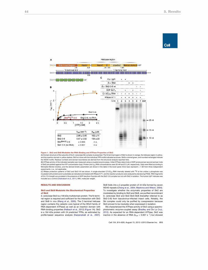
44 3. Results
RESULTS AND DISCUSSION
Ski3 and Ski8 Modulate the Biochemical Propertiesof Ski2S. cerevisiae Ski2 is a 146 kDamultidomain protein. The N-termi-nal region is required and sufficient for the interaction with Ski3and Ski8 in vivo (Wang et al., 2005). The C-terminal helicaseregion contains the catalytic core typical of the DExH family ofRNA-dependent ATPases as well as an insertion domain withRNA-binding properties (Halbach et al., 2012) (Figure 1A). Ski3is a 164 kDa protein with 24 predicted TPRs, as estimated byprofile-based sequence analysis (Karpenahalli et al., 2007).
Ski8 folds into a b propeller protein of 44 kDa formed by sevenWD40 repeats (Cheng et al., 2004; Madrona and Wilson, 2004).To investigate whether the enzymatic properties of Ski2 aremodulated by binding to Ski3 and Ski8, we purified recombinantS. cerevisiae Ski2 and Ski2-Ski3-Ski8 (hereafter referred to asSki2-3-8) from baculovirus-infected insect cells. Notably, theSki complex could only be purified by coexpression becauseSki3 proved to be insoluble when expressed in isolation.We characterized the ATPase activity of Ski2 using a spectro-
photometric enzyme-coupled assay (Bradley and De La Cruz,2012). As expected for an RNA-dependent ATPase, Ski2 wasinactive in the absence of RNA (kcat < 0.007 s!1) but showed
Figure 1. Ski3 and Ski8 Modulate the RNA-Binding and ATPase Properties of Ski2(A) Domain structure of the subunits of the S. cerevisiae Ski complex is presented. The N-terminal region of Ski2 is shown in orange, the helicase region in yellow,
and the insertion domain in yellow dashes. Ski3 is in bluewith the individual TPRmotifs indicated as boxes. Ski8 is colored green, and rounded rectangles indicate
the WD40 motifs. Residue numbers and domain boundaries are derived from the structural analysis reported here.
(B) ATPase activity of the indicated samples was measured using a coupled enzyme assay. Initial ATPase rates (mole of ADP produced per second and per mole
of Ski2) are plotted against the ATP concentration (top). Protein and U25 RNA concentrations were 30 nM and 0.5 mM, respectively. Data were fitted according to
Michaelis-Menten kinetics, and the derived kinetic parameters are shown in the table in the lower panel. Error bars represent ± 1 SD from three independent
experiments. n.a., not applicable.
(C) RNase protection patterns of Ski2 and Ski2-3-8 are shown. A single-stranded C(*UC)28 RNA internally labeled with 32P at the uridine a-phosphate was
incubatedwith proteins and nucleotides as indicated and treatedwith RNase A/T1, and the reaction products were analyzed by denaturing PAGE. RNA fragments
of 9 to 10 nt length accumulated in the presence of ADP-beryllium fluoride with the Ski2-3-8 complex but not with Ski2 in isolation. The human Upf1 helicase was
included as a control (Chakrabarti et al., 2011). MW, molecular weight.
Cell 154, 814–826, August 15, 2013 ª2013 Elsevier Inc. 815

3.2 The Ski complex: Crystal structure and substrate channeling to the exosome 45
ATPase activity in the presence of a U25 RNA oligo (kcat !3 s"1)(Figure 1B). These values are comparable to those previouslyreported for the Mtr4 helicase using a similar assay (Bernsteinet al., 2010). When in complex with Ski3 and Ski8, the ATPaserate of Ski2 decreased 5-fold, whereas the KM for ATP remainedessentially unchanged (Figure 1B). The ATPase activity of theSki2-3-8 complex was abolished by a single-point mutation inthe Ski2 DExH core (Glu445 to Gln), confirming that the catalyticsite resides exclusively in Ski2 (Figure 1B). We conclude thatSki2 is a less efficient ATPase when bound to Ski3 and Ski8.
We next employed RNase protection assays to investigate theextent of RNA-protein interactions. Protein samples incubatedwith an internally labeled single-stranded C(*UC)28 RNA oligowere treated with RNase A/T1, and the protected RNA fragmentswere analyzed by denaturing PAGE. No RNase protection wasobserved with Ski2 in isolation, neither in the presence nor inthe absence of ATP analogs (Figure 1C, lanes 4 and 5). In thecase of Ski2-3-8, fragments of 9 to 10 nt accumulated in thepresence of nucleotide analogs that mimic the ground stateof the ATPase reaction (ADP-beryllium fluoride, Figure 1C,compare lanes 6 and 7). We conclude that the association withSki3 and Ski8 stabilizes RNA binding to Ski2. The finding thatSki3 and Ski8 increase RNA binding and decrease the ATPaseactivity of Ski2 suggests that, in addition to theN-terminal region,
the helicase core of Ski2 is also involved in and/or regulatedby the association with Ski3 and Ski8. To understand the under-lying mechanisms, we determined the crystal structure of a coreSki complex.
Overall Structure of the Yeast Ski2Dinsert-3-8 ComplexAttempts to crystallize the full-length yeast Ski complex werenot successful. We had previously reported that the flexibleinsertion domain of Ski2 is dispensable for formation of Ski2-3-8 (Halbach et al., 2012). Removal of this domain (residues835–1,085 replaced by a Gly-Ser-Arg-Gly linker, designatedSki2Dinsert) enabled us to obtain crystals of the complex suitablefor structure determination. The structure of Ski2Dinsert-3-8was determined at 3.7 A resolution using selenomethionine-based single-wavelength anomalous diffraction (SAD) com-bined with molecular replacement (using the coordinates ofSki2DN-Dinsert [Halbach et al., 2012] and of Ski8 [Cheng et al.,2004]). Despite the moderate resolution, we could build andrefine the structure to an Rfree of 26.5% and an Rwork of 23.1%,with good stereochemistry (Table 1). The accurate tracing ofthe polypeptide chains was aided by a combination of factors:the quality of the electron density map after averaging and B fac-tor sharpening (DeLaBarre and Brunger, 2006), the position ofthe labeled methionines as sequence markers, and the regulartopology of TPRs (Figure S1A available online). The atomicmodels of the two Ski2Dinsert-3-8 complexes present in theasymmetric unit of the crystals include essentially the completepolypeptides. Notable exceptions are two long (and poorlyconserved) loops in Ski3 and Ski2 (residues 340–398 andresidues 542–606, respectively) and the linker between theN-terminal and helicase regions of Ski2 (residues 207–300),which were either disordered or partially built as a polyalaninemodel (Figure S1B).The crystal structure of Ski2Dinsert-3-8 reveals a tetrameric
assembly, with one molecule of Ski2 and two molecules ofSki8 arranged around Ski3 (Figure 2A). Ski3 forms a TPRsolenoid consisting of two arms of similar length (!140 A) androughly perpendicular to each other, resulting in an L-shapedmolecule. The N-terminal region of Ski2 wraps around theC-terminal arm of Ski3. The helicase region of Ski2 docks to acentral surface of Ski3 and contacts both molecules of Ski8.One Ski8 subunit has an outer, peripheral position in the complex(hereby referred to as Ski8OUT), whereas the other Ski8 subunitis located more centrally within the complex (referred to asSki8IN) (Figure 2A).
Ski3 Is a Solenoid of 33 TPR Motifs and Formsthe Scaffold of the Ski ComplexCanonical TPR motifs consist of 34 residues with a character-istic pattern of small and large hydrophobic side chains(Hirano et al., 1990; Sikorski et al., 1990). TPRs fold into twoantiparallel a helices (termed A and B) and usually occur inarrays of up to 16 repeats (Das et al., 1998). Consecutiverepeats typically pack side-by-side with an !50# rotation,giving rise to superhelical solenoids with a concave and aconvex surface. The Ski2Dinsert-3-8 crystal structure revealsthat the entire polypeptide chain of Ski3 forms an array of33 contiguous TPR motifs. Nevertheless, the N-terminal and
Table 1. Data Collection and Refinement Statistics for Native andSelenomethionine-Substituted Ski2Dinsert-3-8 Crystals
Data Set Native SeMet (Peak)
Space group P212121 P212121
Cell dimension a (A) 183.4 184.0
Cell dimension b (A) 200.4 200.0
Cell dimension c (A) 340.2 341.2
Molecules/asymmetric unit 2 2
Data Collection
Wavelength (A) 0.9788 0.9788
Resolution (A) 3.7 (3.9–3.7) 4.6 (4.85–4.6)
Rmerge 0.14 (0.91) 0.21 (1.27)
I/sI 5.8 (1.4) 19.6 (4.5)
Completeness (%) 99.1 (99.2) 100 (100)
Multiplicity 3.3 26.0
CC1/2 0.99 (0.46) 1.0 (0.92)
Refinement
Number of unique reflections 132,408
Rwork/Rfree (%) 23.1/26.5
Average B factors (A2) 116.8
Number of atoms (nonhydrogen) 43,020
Stereochemistry
Rmsd bond lengths (A) 0.005
Rmsd bond angles (#) 0.81
Ramachandran outliers (%) 0.3
Ramachandran favored (%) 95.5
Values for the highest-resolution shell are given in parentheses. Structure
validation was carried out using MolProbity (Chen et al., 2010). See alsoFigure S1.
816 Cell 154, 814–826, August 15, 2013 ª2013 Elsevier Inc.
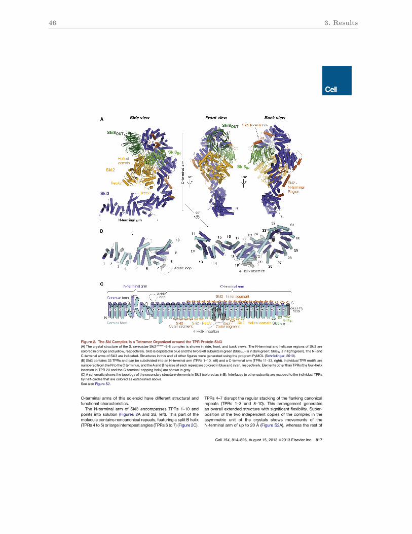
46 3. Results
C-terminal arms of this solenoid have different structural andfunctional characteristics.The N-terminal arm of Ski3 encompasses TPRs 1–10 and
points into solution (Figures 2A and 2B, left). This part of themolecule contains noncanonical repeats, featuring a split B helix(TPRs 4 to 5) or large interrepeat angles (TPRs 6 to 7) (Figure 2C).
TPRs 4–7 disrupt the regular stacking of the flanking canonicalrepeats (TPRs 1–3 and 8–10). This arrangement generatesan overall extended structure with significant flexibility. Super-position of the two independent copies of the complex in theasymmetric unit of the crystals shows movements of theN-terminal arm of up to 20 A (Figure S2A), whereas the rest of
Figure 2. The Ski Complex Is a Tetramer Organized around the TPR Protein Ski3(A) The crystal structure of the S. cerevisiae Ski2Dinsert-3-8 complex is shown in side, front, and back views. The N-terminal and helicase regions of Ski2 are
colored in orange and yellow, respectively. Ski3 is depicted in blue and the two Ski8 subunits in green (Ski8OUT is in dark green; Ski8IN is in light green). The N- and
C-terminal arms of Ski3 are indicated. Structures in this and all other figures were generated using the program PyMOL (Schrodinger, 2010).
(B) Ski3 contains 33 TPRs and can be subdivided into an N-terminal arm (TPRs 1–10, left) and a C-terminal arm (TPRs 11–33, right). Individual TPR motifs are
numbered from the N to theC terminus, and the A andB helices of each repeat are colored in blue and cyan, respectively. Elements other than TPRs (the four-helix
insertion in TPR 20 and the C-terminal-capping helix) are shown in gray.
(C) A schematic shows the topology of the secondary structure elements in Ski3 (colored as in B). Interfaces to other subunits are mapped to the individual TPRs
by half-circles that are colored as established above.
See also Figure S2.
Cell 154, 814–826, August 15, 2013 ª2013 Elsevier Inc. 817
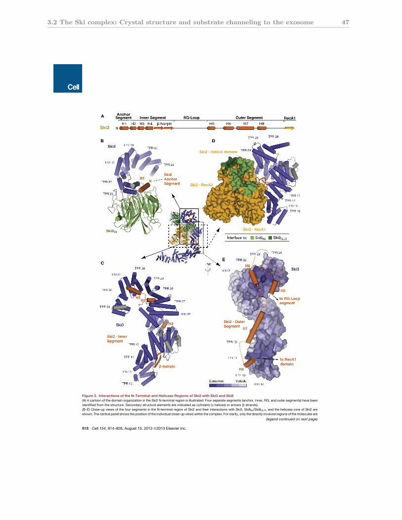
3.2 The Ski complex: Crystal structure and substrate channeling to the exosome 47
Figure 3. Interactions of the N-Terminal and Helicase Regions of Ski2 with Ski3 and Ski8(A) A cartoon of the domain organization in the Ski2 N-terminal region is illustrated. Four separate segments (anchor, inner, RG, and outer segments) have been
identified from the structure. Secondary structure elements are indicated as cylinders (a helices) or arrows (b strands).
(B–E) Close-up views of the four segments in the N-terminal region of Ski2 and their interactions with Ski3, Ski8IN/Ski8OUT, and the helicase core of Ski2 are
shown. The central panel shows the position of the individual close-up viewswithin the complex. For clarity, only the directly involved regions of themolecules are
(legend continued on next page)
818 Cell 154, 814–826, August 15, 2013 ª2013 Elsevier Inc.
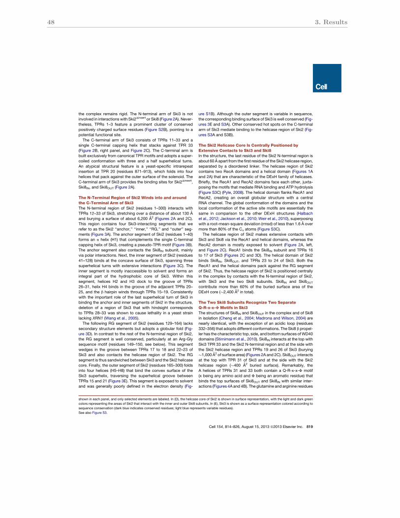
48 3. Results
the complex remains rigid. The N-terminal arm of Ski3 is notinvolved in interactions with Ski2Dinsert or Ski8 (Figure 2A). Never-theless, TPRs 1–3 feature a prominent cluster of conservedpositively charged surface residues (Figure S2B), pointing to apotential functional site.The C-terminal arm of Ski3 consists of TPRs 11–33 and a
single C-terminal capping helix that stacks against TPR 33(Figure 2B, right panel, and Figure 2C). The C-terminal arm isbuilt exclusively from canonical TPR motifs and adopts a super-coiled conformation with three and a half superhelical turns.An atypical structural feature is a yeast-specific intrarepeatinsertion at TPR 20 (residues 871–913), which folds into fourhelices that pack against the outer surface of the solenoid. TheC-terminal arm of Ski3 provides the binding sites for Ski2Dinsert,Ski8IN, and Ski8OUT (Figure 2A).
The N-Terminal Region of Ski2 Winds into and aroundthe C-Terminal Arm of Ski3The N-terminal region of Ski2 (residues 1–300) interacts withTPRs 12–33 of Ski3, stretching over a distance of about 130 Aand burying a surface of about 6,200 A2 (Figures 2A and 2C).This region contains four Ski3-interacting segments that werefer to as the Ski2 ‘‘anchor,’’ ‘‘inner,’’ ‘‘RG,’’ and ‘‘outer’’ seg-ments (Figure 3A). The anchor segment of Ski2 (residues 1–40)forms an a helix (H1) that complements the single C-terminalcapping helix of Ski3, creating a pseudo-TPR motif (Figure 3B).The anchor segment also contacts the Ski8IN subunit, mainlyvia polar interactions. Next, the inner segment of Ski2 (residues41–128) binds at the concave surface of Ski3, spanning threesuperhelical turns with extensive interactions (Figure 3C). Theinner segment is mostly inaccessible to solvent and forms anintegral part of the hydrophobic core of Ski3. Within thissegment, helices H2 and H3 dock to the groove of TPRs26–31, helix H4 binds in the groove of the adjacent TPRs 20–25, and the b hairpin winds through TPRs 15–19. Consistentlywith the important role of the last superhelical turn of Ski3 inbinding the anchor and inner segments of Ski2 in the structure,deletion of a region of Ski3 that with hindsight correspondsto TPRs 28–33 was shown to cause lethality in a yeast strainlacking XRN1 (Wang et al., 2005).The following RG segment of Ski2 (residues 129–164) lacks
secondary structure elements but adopts a globular fold (Fig-ure 3D). In contrast to the rest of the N-terminal region of Ski2,the RG segment is well conserved, particularly at an Arg-Glysequence motif (residues 149–150, see below). This segmentwedges in the groove between TPRs 17 to 18 and 22–23 ofSki3 and also contacts the helicase region of Ski2. The RGsegment is thus sandwiched between Ski3 and the Ski2 helicasecore. Finally, the outer segment of Ski2 (residues 165–300) foldsinto four helices (H5–H8) that bind the convex surface of theSki3 superhelix, traversing the superhelical groove betweenTPRs 15 and 21 (Figure 3E). This segment is exposed to solventand was generally poorly defined in the electron density (Fig-
ure S1B). Although the outer segment is variable in sequence,the corresponding binding surface of Ski3 is well conserved (Fig-ures 3E and S3A). Other conserved hot spots on the C-terminalarm of Ski3 mediate binding to the helicase region of Ski2 (Fig-ures S3A and S3B).
The Ski2 Helicase Core Is Centrally Positioned byExtensive Contacts to Ski3 and Ski8In the structure, the last residue of the Ski2 N-terminal region isabout 60 A apart from the first residue of the Ski2 helicase region,separated by a disordered linker. The helicase region of Ski2contains two RecA domains and a helical domain (Figures 1Aand 2A) that are characteristic of the DExH family of helicases.Briefly, the RecA1 and RecA2 domains face each other, juxta-posing the motifs that mediate RNA binding and ATP hydrolysis(Figure S3C) (Pyle, 2008). The helical domain flanks RecA1 andRecA2, creating an overall globular structure with a centralRNA channel. The global conformation of the domains and thelocal conformation of the active site motifs are essentially thesame in comparison to the other DExH structures (Halbachet al., 2012; Jackson et al., 2010; Weir et al., 2010), superposingwith a root-mean-square deviation (rmsd) of less than 1.6 A overmore than 80% of the Ca atoms (Figure S3C).The helicase region of Ski2 makes extensive contacts with
Ski3 and Ski8 via the RecA1 and helical domains, whereas theRecA2 domain is mostly exposed to solvent (Figure 2A, left,and Figure 2C). RecA1 binds the Ski8IN subunit and TPRs 16to 17 of Ski3 (Figures 2C and 3D). The helical domain of Ski2binds Ski8IN, Ski8OUT, and TPRs 23 to 24 of Ski3. Both theRecA1 and the helical domains pack against the RG segmentof Ski2. Thus, the helicase region of Ski2 is positioned centrallyin the complex by contacts with the N-terminal region of Ski2,with Ski3 and the two Ski8 subunits. Ski8IN and Ski8OUT
contribute more than 60% of the buried surface area of theDExH core (!2,400 A2 in total).
The Two Ski8 Subunits Recognize Two SeparateQ-R-x-x-F Motifs in Ski3The structures of Ski8IN and Ski8OUT in the complex and of Ski8in isolation (Cheng et al., 2004; Madrona and Wilson, 2004) arenearly identical, with the exception of an acidic loop (residues332–356) that adopts different conformations. The Ski8 b propel-ler has the characteristic top, side, and bottom surfaces ofWD40domains (Stirnimann et al., 2010). Ski8IN interacts at the top withSki3 TPR 33 and the Ski2 N-terminal region and at the side withthe Ski2 helicase region and TPRs 19 and 26 of Ski3 (burying!1,000 A2 of surface area) (Figures 2A and 2C). Ski8OUT interactsat the top with TPR 31 of Ski3 and at the side with the Ski2helicase region (!400 A2 buried surface). Remarkably, theA helices of TPRs 31 and 33 both contain a Q-R-x-x-F motif(x being any amino acid and F being an aromatic residue) thatbinds the top surfaces of Ski8OUT and Ski8IN with similar inter-actions (Figures 4A and 4B). The glutamine and arginine residues
shown in each panel, and only selected elements are labeled. In (D), the helicase core of Ski2 is shown in surface representation, with the light and dark green
colors representing the areas of Ski2 that interact with the inner and outer Ski8 subunits. In (E), Ski3 is shown as a surface representation colored according to
sequence conservation (dark blue indicates conserved residues; light blue represents variable residues).
See also Figure S3.
Cell 154, 814–826, August 15, 2013 ª2013 Elsevier Inc. 819
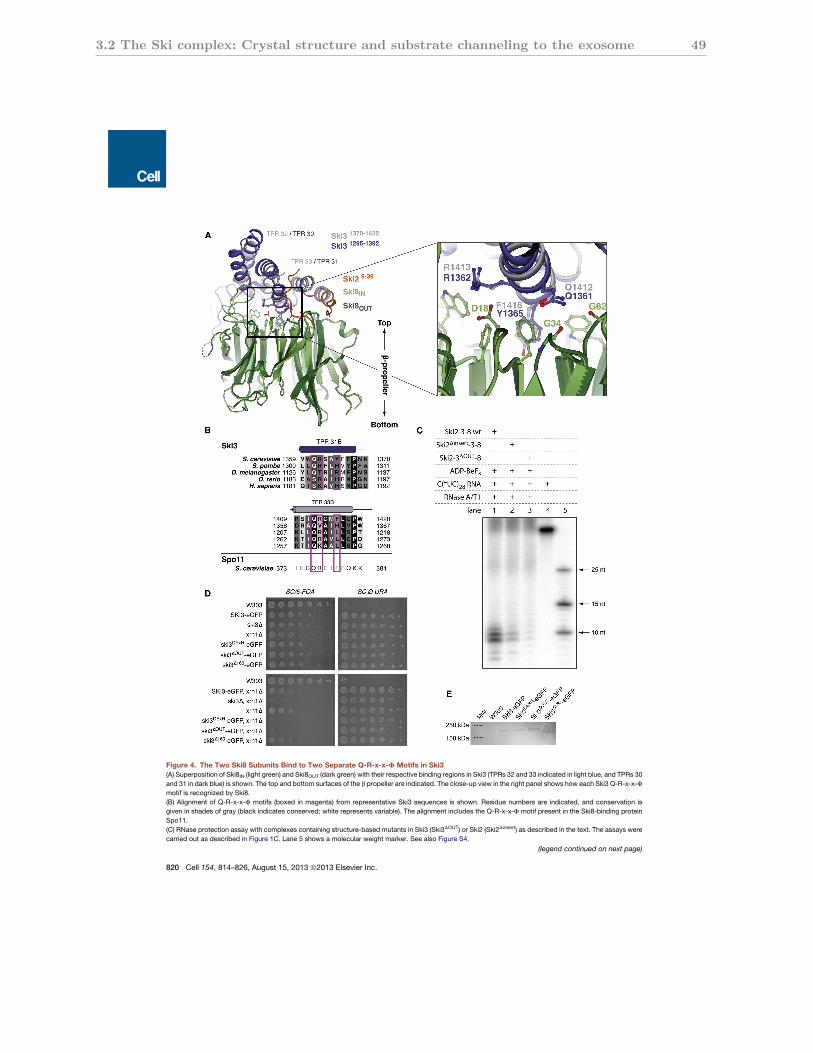
3.2 The Ski complex: Crystal structure and substrate channeling to the exosome 49
Figure 4. The Two Ski8 Subunits Bind to Two Separate Q-R-x-x-F Motifs in Ski3(A) Superposition of Ski8IN (light green) and Ski8OUT (dark green) with their respective binding regions in Ski3 (TPRs 32 and 33 indicated in light blue, and TPRs 30
and 31 in dark blue) is shown. The top and bottom surfaces of the b propeller are indicated. The close-up view in the right panel shows how each Ski3 Q-R-x-x-F
motif is recognized by Ski8.
(B) Alignment of Q-R-x-x-F motifs (boxed in magenta) from representative Ski3 sequences is shown. Residue numbers are indicated, and conservation is
given in shades of gray (black indicates conserved; white represents variable). The alignment includes the Q-R-x-x-F motif present in the Ski8-binding protein
Spo11.
(C) RNase protection assay with complexes containing structure-based mutants in Ski3 (Ski3DOUT) or Ski2 (Ski2Dinsert) as described in the text. The assays were
carried out as described in Figure 1C. Lane 5 shows a molecular weight marker. See also Figure S4.
(legend continued on next page)
820 Cell 154, 814–826, August 15, 2013 ª2013 Elsevier Inc.
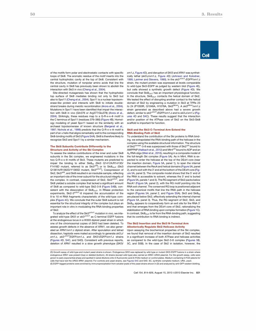
50 3. Results
of the motifs form polar and electrostatic contacts with specificloops of Ski8. The aromatic residue of the motif inserts into thecentral hydrophobic cavity at the top of Ski8. Consistent withthe structure, mutation of nonpolar amino acids that line thecentral cavity in Ski8 has previously been shown to abolish theinteraction with Ski3 in vivo (Cheng et al., 2004).Site-directed mutagenesis has shown that the hydrophobic
top surface of Ski8 mediates binding not only to Ski3 butalso to Spo11 (Cheng et al., 2004). Spo11 is a nuclear topoisom-erase-like protein and interacts with Ski8 to initiate double-strand breaks during meiotic recombination (Arora et al., 2004).Mutations in Spo11 have been identified that impair the interac-tion with Ski8 in vivo (Gln376 or Arg377/Glu378) (Arora et al.,2004). Strikingly, these residues map to a Q-R-x-x-F motif inthe C terminus of Spo11 (residues 376–380) (Figure 4B). Homol-ogy modeling of yeast Spo11 based on the similarity with anarchaeal topoisomerase of known structure (Bergerat et al.,1997; Nichols et al., 1999) predicts that the Q-R-x-x-F motif ispart of an a helix that aligns remarkably well to the correspondingSki8-bindingmotifs of Ski3 (Figure S4A). Ski8 is therefore likely torecognize Ski3 and Spo11 by a similar mechanism.
The Ski8 Subunits Contribute Differently to theStructure and Activity of the Ski ComplexTo assess the relative contributions of the inner and outer Ski8subunits in the Ski complex, we engineered mutations in thetwo Q-R-x-x-F motifs of Ski3. These mutants are predicted toimpair the binding to either Ski8IN (Ski3 Q1412A/R1413D/F1416D mutant, referred to as Ski3DIN) or to Ski8OUT (Ski3Q1361A/R1362D/Y1365D mutant, Ski3DOUT). Coexpression ofSki2, Ski3DIN, and Ski8 resulted in an insoluble sample, reflectingan important role of the inner subunit for the structural integrity ofthe complex. In contrast, coexpression of Ski2, Ski3DOUT, andSki8 yielded a soluble complex that lacked a significant amountof Ski8 as compared to wild-type Ski2-3-8 (Figure S4B), con-sistent with the dissociation of Ski8OUT. In RNase protectionexperiments, Ski2-3DOUT-8 impaired the accumulation of the9 to 10 nt RNA fragments characteristic of the wild-type com-plex (Figure 4C). We conclude that the outer Ski8 subunit is notessential for the structural integrity of the complex but plays animportant role in vitro in modulating the RNA-binding propertiesof Ski2.To analyze the effect of the Ski3DOUT mutation in vivo, we inte-
grated wild-type SKI3 or ski3DOUT as C-terminal EGFP fusionsat the endogenous locus in a W303 diploid yeast strain in whichone of the chromosomal copies of SKI3 had been deleted. Toassess growth defects in the absence of XRN1, we also gener-ated an XRN1/xrn1D diploid strain. After sporulation and tetraddissection, haploids were mated accordingly to generate ski3D/xrn1D, ski3DOUTEGFP/xrn1D, and SKI3-EGFP/xrn1D strains(Figures 4D, S4C, and S4D). Consistent with previous reports,deletion of XRN1 resulted in a slow growth phenotype (SKI3/
xrn1D, Figure 4D), and disruption ofSKI3 and XRN1was synthet-ically lethal (ski3D/xrn1D, Figure 4D) (Johnson and Kolodner,1995; Larimer and Stevens, 1990). In the ski3DOUT-EGFP/xrn1Dstrain, the mutant protein was expressed at levels comparableto wild-type Ski3-EGFP as judged by western blot (Figure 4E),but cells showed a synthetic growth defect (Figure 4D). Weconclude that Ski8OUT has an important physiological function.In the structure, Ski8OUT contacts the helical domain of Ski2.We tested the effect of disrupting another contact to the helicaldomain of Ski2 by engineering a mutation in Ski3 at TPRs 23to 24 (P1050R, Q1046A, H1078A, Ski3DExH). A ski3DExH/xrn1Dstrain generated as described above had a severe growthdefect, similar to ski3DOUT-EGFP/xrn1D and to ski3D/xrn1D (Fig-ures 4D and S4C). These results suggest that the interactionand/or position of the ATPase core of Ski2 on the Ski3-Ski8scaffold is important for function.
Ski8 and the Ski3 C-Terminal Arm Extend theRNA-Binding Path of Ski2To understand the contribution of the Ski proteins to RNA bind-ing, we extrapolated the RNA-binding path of the helicase in thecomplex using the available structural information. The structureof Ski2Dinsert-3-8 was superposed with those of Ski2DN bound toAMPPNP (Halbach et al., 2012) andMtr4DN bound to ADP and anA5 RNA oligo (Weir et al., 2010), resulting in a compositemodel ofthe full-length Ski complex (Figure 5A). As in Mtr4, RNA is ex-pected to enter the helicase at the top of the DExH core (nearthe insertion domain, Figure 5A, panel 1), to span the internalchannel between the RecA and helical domains (Figure 5A, panel2), and to exit with the 30 end at the bottom of the DExH core (Fig-ure 5A, panel 3). The composite model shows that the 50 end ofthe RNA is accessible to solvent, whereas the 30 end is buried(Figure 5A, panels 1 and 3). The RG segment of Ski2 packs belowRecA1 (Figure 5A, panel 2), with the RG motif pointing into theRNA exit channel. The conserved RG loop is positioned adjacentto the canonical motifs that line the RNA path in the helicaseregion (Figures 5A, panel 2, and Figure S5A). Ski3 and Ski8INare placed below Ski2, effectively extending the internal channel(Figure 5A, panel 3). Thus, the RG segment of Ski2, Ski3, andSki8IN appears to cooperatively form an exit site for the RNA 30
end that emerges from the DExH core of Ski2, rationalizing thestabilization of RNA binding upon complex formation (Figure 1C).In contrast, Ski8OUT is far from the RNA-binding path, suggestingthat its contribution to RNA binding is indirect.
The Ski2 Insertion and the Ski3 N-Terminal ArmAllosterically Regulate Ski2 Helicase ActivityUpon assaying the biochemical properties of the Ski complex,we found that removal of the insertion domain of Ski2 resultedin a significant increase of both ATPase and helicase activitiesas compared to the wild-type Ski2-3-8 complex (Figures 5B,5C, and S5B). In the case of Ski2 in isolation, however, the
(D) Growth assay of wild-type and mutant yeast strains is shown. Endogenous SKI3 was replaced by wild-type or mutant SKI3-EGFP fusions in a strain where
endogenous XRN1 was present (top) or deleted (bottom). All strains (except wild-type) also carried an XRN1-URA3 plasmid. For the growth assay, cells were
grown to early exponential phase and spotted in serial dilutions onto 5-fluoroorotic acid (5-FOA) medium or control plates. Medium containing 5-FOA selects for
cells that have lost the URA3-XRN1 plasmid. For controls and yeast strains, see Figures S4C and S4D. SC, synthetic complete medium; URA, uracil.
(E) EGFP-tagged proteins were enriched by immunoprecipitation from soluble lysate of the yeast strains shown in (D) and analyzed by anti-GFP western blotting.
Cell 154, 814–826, August 15, 2013 ª2013 Elsevier Inc. 821

3.2 The Ski complex: Crystal structure and substrate channeling to the exosome 51
insertion domain did not significantly alter the rate of ATP hydro-lysis (Figures 5B and S5B). These data suggest that binding ofSki3 and/or Ski8 modulates the effect of the insertion domainon Ski2. The model of the full-length Ski complex suggeststhat the insertion domain of Ski2 is juxtaposed to the N-terminalarm of Ski3 (Figure 5A, panel 1). These two domains arepredicted to reside above the RNA entry site of the DExH coreand would thus be in a favorable position to modulate thehelicase. To test a possible effect of the N-terminal arm ofSki3 on helicase activity, we purified a Ski complex containingthe Ski2 insertion domain but lacking the ten N-terminal TPRs(residues 1–487) (Figure S5C). Although the Ski3 N terminusis not required to bind to Ski7 nor for complex formation,its removal resulted in a significant increase in the helicase andATPase activities of the Ski complex, comparably to the effectobserved with the Ski2Dinsert-3-8 complex (Figures 5B, 5C,and S5B).
The insertion domain of Ski2 is essential for exosome-medi-ated functions in yeast (Klauer and vanHoof, 2013).We thereforeassessed the importance of the N-terminal arm of Ski3 in vivo.Reasoning that the first three TPR motifs of Ski3 contain aconserved surface patch (Figure S2B) and are predicted fromthe composite model to approach the globular domain of theSki2 insertion (Figure 5A, panel 1), we deleted a small region ofthe N-terminal arm encompassing residues 1–160 (Ski3D160). Aski3D160/xrn1D strain generated as described above impaired
growth (Figures 4D and S4C). The effect was not as severe asin the case of mutations in the C-terminal arm but appears tobe specific because the mutant protein was present at a levelcomparable to the Ski3-EGFP knockin (Figure 4E). We concludethat the N-terminal arm of Ski3 cooperates with the insertiondomain of Ski2 to allosterically regulate the helicase activity ofthe complex. Notably, the insertion domain of the related nuclearhelicaseMtr4 has similar properties as compared to the insertionof Ski2: it has a comparable architecture and position, it is notrequired for the assembly of the TRAMP complex (Weir et al.,2010), and it is important in vivo (Jackson et al., 2010). Whetherits insertion domain also regulates Mtr4 allosterically in thecontext of the TRAMP or higher-order complexes is currentlyunknown.
RNA Channeling from the Ski Complex to the ExosomeNext, we evaluated the RNA-binding path of the Ski complexbound to the exosome. Coimmunoprecipitation studies haveshown that in S. cerevisiae, the Ski2-3-8 complex interactswith the exosome via the protein Ski7 (Araki et al., 2001; Wanget al., 2005). In GST pull-down assays with purified recombinantproteins, Ski7 interacted directly with Ski2-3-8 (or Ski2Dinsert-3-8), with the exosome and concomitantly with both (Figure S6A).Notably, Ski7 did not interact with Ski2 or Ski8 in isolation (Fig-ure S6A). Having established the physical association of bothcomplexes, we next asked how this interaction impacts on
Figure 5. RNA-Binding Path and Regulation in the Ski Complex(A) Model of the full-length Ski complex based on the superposition of the Ski2Dinsert-3-8 structure with Ski2DN-AMPPNP (4A4Z) and of Mtr4DN-ADP-A5 (2XGJ) is
shown. The three close-up views (panels 1–3) show important features in the RNA-binding path: the ‘‘lid’’ formed by the Ski insertion and the Ski3 N-terminal arm
above the RNA 50 end (panel 1), the RG segment of Ski2 extending the RNA-binding motifs in the DExH core (numbered, panel 2), and Ski3 and Ski8IN forming an
exit channel for the RNA 30 end (panel 3).
(B) ATPase activities of wild-type andmutant Ski2 and Ski2-3-8 samples are given in terms of kcat/KM (normalized to full-length Ski2-3-8). Removal of the insertion
domain of Ski2 or of the N-terminal arm of Ski3 derepresses the ATPase activity of Ski2 when bound in the Ski complex. Removal of the insertion domain has no
effect on Ski2 in isolation (see Figures 1C and S5B for raw data). Error bars represent ± 1 SD from three independent experiments.
(C) Unwinding activity of wild-type and mutant Ski complexes is shown. RNA duplexes with a 30 overhang were incubated with the indicated amounts of protein
and separated by native PAGE. Removal of the Ski2 insertion domain or the Ski3 N terminus stimulates the helicase activity of the complex. See also Figure S5.
822 Cell 154, 814–826, August 15, 2013 ª2013 Elsevier Inc.
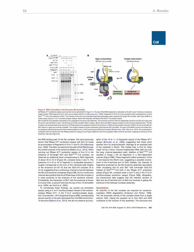
52 3. Results
the RNA-binding path of the Ski complex. We have previouslyshown that RNase A/T1 protection assays with Exo-10 revealan accumulation of fragments of 10 to 11 and 31–33 nt (Bonneauet al., 2009). The latter correspond to the path of the RNA throughthe central channel of the exosome (Makino et al., 2013). Whencarrying out RNase A/T1 protection assays of Exo-10 in thepresence of full-length Ski7 and Ski2Dinsert-3-8 complex, weobserved an additional band corresponding to RNA fragmentsof about 43 to 44 nt (Figure 6A, compare lanes 2 and 7). Thepresence of a 43 to 44 nt fragment is remarkable because itroughly corresponds to the sum of the individual path lengthsof the exosome (31–33 nt) and the Ski2-3-8 complex (9 to10 nt), suggesting that a continuous channel is formed betweenthe Ski and exosome complexes (Figure 6B). Such a continuouschannel also predicts that the ATPase base of the Ski complex isin close proximity to the entrance of the exosome channel.Consistently, the interaction of Ski7 with the exosome is knownto involve Csl4, a subunit that lines the top of Exo-10 (Schaefferet al., 2009; van Hoof et al., 2002).To corroborate these findings, we carried out protection
assays using a 50-to-30 exoribonuclease instead of the endonu-cleases RNase A/T1. In the 50-to-30 exoribonuclease assay,Exo-10 protected!28 nt fragments (Figure S6B), correspondingalmost exactly to the path estimated from the RNA-bound Exo-10 structure (Makino et al., 2013). We did not observe accumu-
lation of the 10 to 11 nt fragments typical of the RNase A/T1assays (Bonneau et al., 2009), suggesting that these shortspecies form by endonucleolytic cleavage at an exposed partof the substrate in Rrp44. This implies that, in Exo-10, theremight not be a short, channel-independent RNA path but onlythe long, channel-dependent path. Addition of Ski2Dinsert-3-8resulted in longer (!46 nt) fragments in a Ski7-dependentmanner (Figure S6B). These fragments reflect protection of the50 end beyond the DExH core, suggesting a possible involve-ment of the N-terminal arm of Ski3. Notably, the intensity offragments protected by the Ski-exosome assembly decreasedwhen incubating Exo-10 and Ski7 with full-length Ski2-3-8instead of Ski2Dinsert-3-8 both in the RNase A/T1 protectionassays (Figure 6A, compare lanes 5 and 7) and in the 50-to-30
exoribonuclease protection assays (Figure S6B). Altogether,the biochemical data suggest that the insertion domain ofSki2 and the N-terminal arm of Ski3 are involved in gating theentrance of the helicase-nuclease assembly.
ConclusionsAll subunits of the Ski complex are required for exosome-mediated mRNA degradation (Anderson and Parker, 1998).Yet, the enzymatic activity of the complex arises from a singlesubunit, Ski2, raising the question as to how Ski3 and Ski8contribute to the function of this assembly. The structural and
Figure 6. RNA Channeling in the Exosome-Ski Assembly(A) RNase A/T1 protection assays were carried out as described for Figure 1C. The size of the RNA fragments is indicated on the left. Lane 10 shows a molecular
weight marker. RNA fragments of 31–33 nt accumulated with Exo-10 (Bonneau et al., 2009). Fragments of 43 to 44 nt accumulated when incubating Exo-10 and
Ski2Dinsert-3-8 in the presence of Ski7. The intensity of the 43 to 44 nucleotide fragments decreased when using the full-length Ski complex. See Figure S6B for a
similar assay using a 50-to-30 exoribonuclease instead. Rrp44-DM indicates the Rrp44 D551N/D171N double mutant.
(B) A model for the substrate channeling in the cytoplasmic exosome-Ski assembly. The exosome is shown with the catalytically inactive core (Exo-9) in gray and
the active subunit Rrp44 in pink. The drawing of the Ski complex (Ski2 in yellow, Ski3 in blue and Ski8 in green) is based on the structure reported here. The Ski
complex can be described as formed by an ATPase base and a regulatory lid (the N-terminal arm of Ski3 and the insertion domain of Ski2). No structural data for
Ski7 (shown in gray) are currently available. The model is based on known interaction data (Figures S5C and S6A). The path of the RNA is shown as a black line,
according to individual structures of the exosome (Makino et al., 2013) and of known DExH box proteins (Buttner et al., 2007; Weir et al., 2010). The overall path is
consistent with the results from the RNase protection assays (A and Figure S6B) and with the synergistic effect of the lid domains in gating the entrance of the
assembly. See also Figure S6.
Cell 154, 814–826, August 15, 2013 ª2013 Elsevier Inc. 823
3.2 The Ski complex: Crystal structure and substrate channeling to the exosome 53
biochemical data reported here reveal intricate interactionnetworks that engage the catalytic and noncatalytic subunitsand sophisticated mechanisms that control the ATPase activityof the complex.
Overall, the Ski complex can be thought of as formed by anATPase base and a regulatory lid (Figure 6B). In the ATPasebase, the DExH core of Ski2 is located in a central position,surrounded by the C-terminal arm of Ski3, two Ski8 subunits,and the N-terminal region of Ski2. With the exception of the outerSki8 subunit, all other nonenzymatic constituents of the baseappear to directly extend the RNA channel of Ski2, forming anexit tunnel where the unwound RNA 30 end is expected toemerge. Despite adopting the most peripheral position in thecomplex and being dispensable for Ski2-Ski3 interaction, theouter Ski8 subunit is important for function in vivo and forRNA binding in vitro, possibly by restraining the ATPase core ina productive conformation. Most of the DExH core of Ski2 isembedded in protein-protein interactions within the base. How-ever, the RecA2 domain of the helicase is exposed to solvent,rationalizing how the required conformational plasticity of theDExH core in the course of the ATPase cycle can be maintainedwithout disrupting the complex. The regulatory lid that sur-mounts the ATPase base is the most dynamic part of the com-plex. The lid is formed by the insertion domain of Ski2 and theN-terminal arm of Ski3. Neither domain is required to assemblethe ATPase base, but together they regulate ATPase activity,possibly by gating substrate access to the ATPase base. Bindingto Ski3 and Ski8 also allows Ski2 to connect to Ski7 and henceto the exosome complex. Our biochemical results suggest thatthe Ski complex directly channels single-stranded RNA into theexosome (Figure 6B). This mechanismwould couple the helicaseand nuclease activities of the complexes, resulting in a directpipeline for mRNP remodeling and degradation.
The RNA-degrading exosome complex has conceptual simi-larities to the proteasome, a cellular machinery that breaksdown polypeptides (Lorentzen and Conti, 2006; van Hoof andParker, 1999). The exosome core and the 20S proteasomeboth possess cylindrical chambers where unfolded substratesare sequestered and degraded. The 20S proteasome associateswith a major regulatory complex: the 19S complex (Kish-Trierand Hill, 2013). The 19S complex contains an ATPase basethat unfolds polypeptides and injects them into the 20S complexfor degradation. It also contains a dynamic lid that recognizes thesubstrates and transfers them to the ATPase base. In the contextof our structural and biochemical data on the Ski complex, theemerging picture is that the conceptual similarities in the degra-dation mechanisms of exosome and proteasome are likely toextend to their regulatory complexes.
EXPERIMENTAL PROCEDURES
See also the Extended Experimental Procedures.
Protein PurificationRecombinant S. cerevisiae Ski2 and Ski2-3-8 complexes (wild-type and
mutants) were purified from insect cells as previously described (Halbach
et al., 2012). Ski7 was expressed in E. coli and purified as reported earlier
(Halbach et al., 2012). The exosome (nine or ten subunit complexes) was
reconstituted as published by Makino et al. (2013). Details on constructs,
expression systems, and purification procedures are available in the Extended
Experimental Procedures.
Crystallization and Structure SolutionCrystallization and structure solution are described in detail in the Extended
Experimental Procedures. Briefly, a combination of molecular replacement
(using the Ski2 helicase core [4A4Z] and Ski8 [1S4U] as search models) and
selenomethionine SAD phases yielded an experimental electron density map
that was improved by solvent flattening and phase extension. Manual model
building and refinement allowed completion of the model.
Biochemical AssaysSteady-state ATPase activity was analyzed using an assay that couples oxida-
tion of NADH to regeneration of ATP (Bradley and De La Cruz, 2012). KM and
kcat were derived by fitting the data according to Michaelis-Menten kinetics.
Single turnover unwinding assays were carried out essentially as described
in Lucius et al. (2003). As substrate, a 17-mer single-strandedRNAwas labeled
at the 50 end with 32P and annealed with a 1.5 molar excess of a 27-mer RNA
(consisting of a complementary sequence and a 30 extension of 10 nt). RNase
protection assays have been described in Bonneau et al. (2009). See the
Extended Experimental Procedures for details.
Yeast StrainsSee the Extended Experimental Procedures for generation of yeast strains.
ACCESSION NUMBERS
The coordinates and structure factors have been deposited in the Protein Data
Bank with ID code 4BUJ.
SUPPLEMENTAL INFORMATION
Supplemental Information includes Extended Experimental Procedures and
six figures and can be found with this article online at http://dx.doi.org/
10.1016/j.cell.2013.07.017.
ACKNOWLEDGMENTS
We thank the MPI-Biochemistry Core Facility and Crystallization Facility,
F. Bonneau for help with biochemical assays, E. Kowalinski, J. Ebert, and
D. Makino for exosome samples or plasmids, and S. Chakrabarti for Upf1
samples. We also thank the staff of the Swiss Light Source synchrotron for
assistance during data collection and Z. Storchova, S. Jentsch, and the
CSH course on yeast genetics for guidance and training in the in vivo
experiments. We also thank M. Krause for the drawing in Figure 6B and the
members of our lab for discussions and for critical reading of the manuscript.
This study was supported by the Boehringer Ingelheim Fonds (to F.H.), the
Max Planck Gesellschaft, the European Research Council (ERC Advanced
Investigator Grant 294371 and ITN RNPnet), and the Deutsche Forschungsge-
meinschaft (DFG SFB646, SFB1035, GRK1721, FOR1680, and CIPSM; to
E.C.). F.H. purified proteins and conducted crystallization and structure
determination as well as all in vitro assays. P.R. generated and analyzed yeast
strains. M.R. carried out the insect-cell expression. E.C. supervised the proj-
ect. F.H. and E.C. wrote the manuscript.
Received: October 17, 2012
Revised: May 13, 2013
Accepted: July 12, 2013
Published: August 15, 2013
REFERENCES
Allmang, C., Kufel, J., Chanfreau, G., Mitchell, P., Petfalski, E., and Tollervey,
D. (1999). Functions of the exosome in rRNA, snoRNA and snRNA synthesis.
EMBO J. 18, 5399–5410.
824 Cell 154, 814–826, August 15, 2013 ª2013 Elsevier Inc.
54 3. Results
Anderson, J.S., and Parker, R.P. (1998). The 30 to 50 degradation of yeast
mRNAs is a general mechanism for mRNA turnover that requires the SKI2
DEVH box protein and 30 to 50 exonucleases of the exosome complex.
EMBO J. 17, 1497–1506.
Araki, Y., Takahashi, S., Kobayashi, T., Kajiho, H., Hoshino, S., and Katada, T.
(2001). Ski7p G protein interacts with the exosome and the Ski complex for
30-to-50 mRNA decay in yeast. EMBO J. 20, 4684–4693.
Arora, C., Kee, K., Maleki, S., and Keeney, S. (2004). Antiviral protein Ski8 is
a direct partner of Spo11 in meiotic DNA break formation, independent of its
cytoplasmic role in RNA metabolism. Mol. Cell 13, 549–559.
Bergerat, A., de Massy, B., Gadelle, D., Varoutas, P.C., Nicolas, A., and For-
terre, P. (1997). An atypical topoisomerase II from Archaea with implications
for meiotic recombination. Nature 386, 414–417.
Bernstein, J., Ballin, J.D., Patterson, D.N., Wilson, G.M., and Toth, E.A. (2010).
Unique properties of the Mtr4p-poly(A) complex suggest a role in substrate
targeting. Biochemistry 49, 10357–10370.
Bonneau, F., Basquin, J., Ebert, J., Lorentzen, E., and Conti, E. (2009). The
yeast exosome functions as a macromolecular cage to channel RNA
substrates for degradation. Cell 139, 547–559.
Bradley, M.J., and De La Cruz, E.M. (2012). Analyzing ATP utilization by
DEAD-Box RNA helicases using kinetic and equilibrium methods. Methods
Enzymol. 511, 29–63.
Brown, J.T., Bai, X., and Johnson, A.W. (2000). The yeast antiviral proteins
Ski2p, Ski3p, and Ski8p exist as a complex in vivo. RNA 6, 449–457.
Buttner, K., Nehring, S., and Hopfner, K.P. (2007). Structural basis for DNA
duplex separation by a superfamily-2 helicase. Nat. Struct. Mol. Biol. 14,
647–652.
Chakrabarti, S., Jayachandran, U., Bonneau, F., Fiorini, F., Basquin, C.,
Domcke, S., Le Hir, H., and Conti, E. (2011). Molecular mechanisms for the
RNA-dependent ATPase activity of Upf1 and its regulation by Upf2. Mol.
Cell 41, 693–703.
Chen, V.B., Arendall, W.B., 3rd, Headd, J.J., Keedy, D.A., Immormino, R.M.,
Kapral, G.J., Murray, L.W., Richardson, J.S., and Richardson, D.C. (2010).
MolProbity: all-atom structure validation for macromolecular crystallography.
Acta Crystallogr. D Biol. Crystallogr. 66, 12–21.
Cheng, Z., Liu, Y., Wang, C., Parker, R., and Song, H. (2004). Crystal structure
of Ski8p, aWD-repeat protein with dual roles inmRNAmetabolism andmeiotic
recombination. Protein Sci. 13, 2673–2684.
D’Andrea, L.D., and Regan, L. (2003). TPR proteins: the versatile helix. Trends
Biochem. Sci. 28, 655–662.
Das, A.K., Cohen, P.W., and Barford, D. (1998). The structure of the tetratrico-
peptide repeats of protein phosphatase 5: implications for TPR-mediated
protein-protein interactions. EMBO J. 17, 1192–1199.
DeLaBarre, B., and Brunger, A.T. (2006). Considerations for the refinement of
low-resolution crystal structures. Acta Crystallogr. D Biol. Crystallogr. 62,
923–932.
de la Cruz, J., Kressler, D., Tollervey, D., and Linder, P. (1998). Dob1p (Mtr4p)
is a putative ATP-dependent RNA helicase required for the 30 end formation
of 5.8S rRNA in Saccharomyces cerevisiae. EMBO J. 17, 1128–1140.
Dziembowski, A., Lorentzen, E., Conti, E., and Seraphin, B. (2007). A single
subunit, Dis3, is essentially responsible for yeast exosome core activity. Nat.
Struct. Mol. Biol. 14, 15–22.
Garneau, N.L.,Wilusz, J., andWilusz, C.J. (2007). The highways and byways of
mRNA decay. Nat. Rev. Mol. Cell Biol. 8, 113–126.
Halbach, F., Rode, M., and Conti, E. (2012). The crystal structure of
S. cerevisiae Ski2, a DExH helicase associated with the cytoplasmic functions
of the exosome. RNA 18, 124–134.
Hirano, T., Kinoshita, N., Morikawa, K., and Yanagida, M. (1990). Snap helix
with knob and hole: essential repeats in S. pombe nuclear protein nuc2+.
Cell 60, 319–328.
Houseley, J., and Tollervey, D. (2009). The many pathways of RNA degrada-
tion. Cell 136, 763–776.
Houseley, J., LaCava, J., and Tollervey, D. (2006). RNA-quality control by the
exosome. Nat. Rev. Mol. Cell Biol. 7, 529–539.
Jackson, R.N., Klauer, A.A., Hintze, B.J., Robinson, H., van Hoof, A., and
Johnson, S.J. (2010). The crystal structure of Mtr4 reveals a novel arch
domain required for rRNA processing. EMBO J. 29, 2205–2216.
Johnson, A.W., and Kolodner, R.D. (1995). Synthetic lethality of sep1 (xrn1)
ski2 and sep1 (xrn1) ski3 mutants of Saccharomyces cerevisiae is independent
of killer virus and suggests a general role for these genes in translation control.
Mol. Cell. Biol. 15, 2719–2727.
Karpenahalli, M.R., Lupas, A.N., and Soding, J. (2007). TPRpred: a tool for
prediction of TPR-, PPR- and SEL1-like repeats from protein sequences.
BMC Bioinformatics 8, 2.
Kish-Trier, E., and Hill, C.P. (2013). Structural biology of the proteasome.
Annu. Rev. Biophys. 42, 29–49.
Klauer, A.A., and van Hoof, A. (2013). Genetic interactions suggest multiple
distinct roles of the arch and core helicase domains of Mtr4 in Rrp6 and
exosome function. Nucleic Acids Res. 41, 533–541.
LaCava, J., Houseley, J., Saveanu, C., Petfalski, E., Thompson, E., Jacquier,
A., and Tollervey, D. (2005). RNA degradation by the exosome is promoted
by a nuclear polyadenylation complex. Cell 121, 713–724.
Larimer, F.W., and Stevens, A. (1990). Disruption of the gene XRN1, coding for
a 50——30 exoribonuclease, restricts yeast cell growth. Gene 95, 85–90.
Lebreton, A., and Seraphin, B. (2008). Exosome-mediated quality control:
substrate recruitment and molecular activity. Biochim. Biophys. Acta 1779,
558–565.
Liu, Q., Greimann, J.C., and Lima, C.D. (2006). Reconstitution, activities, and
structure of the eukaryotic RNA exosome. Cell 127, 1223–1237.
Lorentzen, E., and Conti, E. (2006). The exosome and the proteasome: nano-
compartments for degradation. Cell 125, 651–654.
Lucius, A.L., Maluf, N.K., Fischer, C.J., and Lohman, T.M. (2003). General
methods for analysis of sequential ‘‘n-step’’ kinetic mechanisms: application
to single turnover kinetics of helicase-catalyzed DNA unwinding. Biophys. J.
85, 2224–2239.
Lykke-Andersen, S., Brodersen, D.E., and Jensen, T.H. (2009). Origins and ac-
tivities of the eukaryotic exosome. J. Cell Sci. 122, 1487–1494.
Madrona, A.Y., andWilson, D.K. (2004). The structure of Ski8p, a protein regu-
lating mRNA degradation: implications for WD protein structure. Protein Sci.
13, 1557–1565.
Makino, D.L., Baumgartner, M., and Conti, E. (2013). Crystal structure of an
RNA-bound 11-subunit eukaryotic exosome complex. Nature 495, 70–75.
Mitchell, P., and Tollervey, D. (2003). An NMD pathway in yeast involving
accelerated deadenylation and exosome-mediated 30—>50 degradation.
Mol. Cell 11, 1405–1413.
Mitchell, P., Petfalski, E., Shevchenko, A., Mann, M., and Tollervey, D. (1997).
The exosome: a conserved eukaryotic RNA processing complex containing
multiple 30—>50 exoribonucleases. Cell 91, 457–466.
Nichols, M.D., DeAngelis, K., Keck, J.L., and Berger, J.M. (1999). Structure
and function of an archaeal topoisomerase VI subunit with homology to the
meiotic recombination factor Spo11. EMBO J. 18, 6177–6188.
Orban, T.I., and Izaurralde, E. (2005). Decay of mRNAs targeted by RISC
requires XRN1, the Ski complex, and the exosome. RNA 11, 459–469.
Pyle, A.M. (2008). Translocation and unwinding mechanisms of RNA and DNA
helicases. Annu. Rev. Biophys. 37, 317–336.
Schaeffer, D., Tsanova, B., Barbas, A., Reis, F.P., Dastidar, E.G., Sanchez-
Rotunno, M., Arraiano, C.M., and van Hoof, A. (2009). The exosome contains
domains with specific endoribonuclease, exoribonuclease and cytoplasmic
mRNA decay activities. Nat. Struct. Mol. Biol. 16, 56–62.
Schrodinger. (2010). The PyMOL Molecular Graphics System, Version 1.5.0.4
(Schrodinger, LLC).
Sikorski, R.S., Boguski, M.S., Goebl, M., and Hieter, P. (1990). A repeating
amino acid motif in CDC23 defines a family of proteins and a new relationship
among genes required for mitosis and RNA synthesis. Cell 60, 307–317.
Cell 154, 814–826, August 15, 2013 ª2013 Elsevier Inc. 825
3.2 The Ski complex: Crystal structure and substrate channeling to the exosome 55
Stirnimann, C.U., Petsalaki, E., Russell, R.B., and Muller, C.W. (2010). WD40
proteins propel cellular networks. Trends Biochem. Sci. 35, 565–574.
Synowsky, S.A., and Heck, A.J. (2008). The yeast Ski complex is a hetero-
tetramer. Protein Sci. 17, 119–125.
Vanacova, S., and Stefl, R. (2007). The exosome and RNA quality control in the
nucleus. EMBO Rep. 8, 651–657.
Vanacova, S., Wolf, J., Martin, G., Blank, D., Dettwiler, S., Friedlein, A.,
Langen, H., Keith, G., and Keller, W. (2005). A new yeast poly(A) polymerase
complex involved in RNA quality control. PLoS Biol. 3, e189.
van Hoof, A., and Parker, R. (1999). The exosome: a proteasome for RNA? Cell
99, 347–350.
van Hoof, A., Lennertz, P., and Parker, R. (2000a). Yeast exosome mutants
accumulate 30-extended polyadenylated forms of U4 small nuclear RNA and
small nucleolar RNAs. Mol. Cell. Biol. 20, 441–452.
van Hoof, A., Staples, R.R., Baker, R.E., and Parker, R. (2000b). Function of the
ski4p (Csl4p) and Ski7p proteins in 30-to-50 degradation of mRNA. Mol. Cell.
Biol. 20, 8230–8243.
van Hoof, A., Frischmeyer, P.A., Dietz, H.C., and Parker, R. (2002). Exosome-
mediated recognition and degradation of mRNAs lacking a termination codon.
Science 295, 2262–2264.
Wang, L., Lewis, M.S., and Johnson, A.W. (2005). Domain interactions within
the Ski2/3/8 complex and between the Ski complex and Ski7p. RNA 11,
1291–1302.
Wasmuth, E.V., and Lima, C.D. (2012). Exo- and endoribonucleolytic activities
of yeast cytoplasmic and nuclear RNA exosomes are dependent on the non-
catalytic core and central channel. Mol. Cell 48, 133–144.
Weir, J.R., Bonneau, F., Hentschel, J., and Conti, E. (2010). Structural analysis
reveals the characteristic features of Mtr4, a DExH helicase involved in nuclear
RNA processing and surveillance. Proc. Natl. Acad. Sci. USA 107, 12139–
12144.
Wyers, F., Rougemaille, M., Badis, G., Rousselle, J.C., Dufour, M.E., Boulay,
J., Regnault, B., Devaux, F., Namane, A., Seraphin, B., et al. (2005). Cryptic
pol II transcripts are degraded by a nuclear quality control pathway involving
a new poly(A) polymerase. Cell 121, 725–737.
826 Cell 154, 814–826, August 15, 2013 ª2013 Elsevier Inc.
56 3. Results
Supplemental Information
EXTENDED EXPERIMENTAL PROCEDURES
Protein PurificationFull-length Ski2 and Ski2Dinsert (residues 835 - 1085 replaced by a Gly-Ser-Arg-Gly linker) were cloned with an N-terminal hexahis-tidine tag followed by a 3C protease site and subcloned into pFastBac1 vectors (Invitrogen). Proteins were expressed in baculovirus-infected Hi-Five insect cells (Invitrogen) at 26!C for 70 hr. Protein purification was carried out at 4!C or on ice and all buffers weresupplemented with 1 mM b-mercaptoethanol. Cells were homogenized in low salt buffer (10 mM Tris-Cl pH 7.4, 10 mM NaCl,2 mMMgCl2), supplemented with NaCl and imidazole to 250 mM and 20 mM, respectively, and lysate was cleared by centrifugation.His-tagged protein was enriched on Ni2+-NTA resin (Clontech), eluted with lysis buffer containing 300 mM imidazole and directlyloaded on heparin sepharose resin (GE Healthcare). For elution, a gradient from 100 to 1,000 mM NaCl in 20 mM Tris-Cl pH 7.4was used. The eluate wasmixed with 3C protease and dialyzed in 20mMTris-Cl pH 7.4, 200mMNaCl and 3mM b-mercaptoethanoluntil complete removal of His-tags. Cleaved proteins were further purified by size-exclusion chromatography (SEC) in 10 mMHEPESpH 7.4 and 200 mM NaCl.For expression of Ski2Dinsert-3-8, a pFL vector (Fitzgerald et al., 2006) was generated containing full-length Ski8 and full-length Ski3
(with an N-terminal hexahistidine tag followed by a 3C protease site) in themultiple cloning site (MCS) 1 and 2, respectively. The com-plex was then expressed fromHi-Five insect cells infected with two viruses encoding Ski2Dinsert (see above) and the dual Ski3His-Ski8cassette. Purification was carried out as described for Ski2Dinsert (see above). Selenomethionine-labeled Ski2Dinsert-3-8 wasexpressed in insect cells grown in methionine-free medium (Expression Systems, LLC). L-Selenomethionine (Acros Chemicals)was added 8 hr postinfection to the medium at a final concentration of 100 mg/l. Cells were harvested 60 hr postinfection andthe complex was purified as described. Incorporation rates of selenomethionine were estimated by mass-spectrometry to rangefrom 66 to 75%.Ski2-Ski3DN-Ski8 complex was expressed and purified as described above. Briefly, a pFL vector was cloned containing f.l. Ski8
and Ski3DN (residues 488-1432) fused to a 3C-cleavable N-terminal hexahistidine tag. The complex was then expressed in insectcells coinfected with one virus expressing the Ski3DN-His-Ski8 cassette and one virus expressing f.l. Ski2. For expression of full-lengthSki2-3-8, a f.l. Ski2 expression cassette was subclonedwithout N-terminal hexahistidine tag into a pFL vector. The cassette was theninserted into the Ski3His-Ski8 containing pFL vector via restriction / ligation using the BstZ17I and SpeI restriction sites (Fitzgeraldet al., 2006). Full-length Ski2-3-8 complex was then purified from Hi-Five insect cells infected with a single virus containing thetriple-expression cassette. Mutants used in this study were generated according to the Quikchange protocol (Stratagene) andexpressed as the wild-type Ski2-3-8 complex.Full-length Ski8 was subcloned with a hexahistidine tag followed by a TEV protease site into a pET-MCN vector (Romier et al.,
2006). Protein was expressed in E. coli BL21 (DE3) Gold pLysS cells at 18!C for 16 hr after induction with 0.3 mM IPTG. Cellswere resuspended in buffer A (20 mM Tris-Cl pH 7.5, 100 mM NaCl, 10 mM imidazole and 1 mM b-mercaptoethanol) broken bysonication and lysate was cleared by centrifugation. His-tagged protein was captured by Ni2+-NTA affinity chromatography andeluted with buffer A containing 500 mM imidazole. Eluate was mixed with TEV protease and dialyzed in buffer A until completeremoval of the His-tag. The protein was the further purified on a monoQ anion-exchange column (GE Healthcare) and subjectedto SEC in 10 mM HEPES pH 7.5 and 200 mM NaCl. Ski7, GST-Ski7 (Halbach et al., 2012) and Exo-10 (Bonneau et al., 2009) werepurified as described earlier.
Crystallization, Structure Determination, and RefinementSki2Dinsert-3-8 was concentrated to 13.5 mg/ml, supplemented with Tris(2-carboxyethyl)phosphine hydrochloride to 1 mM andmixed 1:1 (v/v) with reservoir solution (1.88 M NH4SO4, 100 mM Bis-Tris pH 6.0). 1 ml drops were set up in the sitting drop formatand equilibrated against 500 ml reservoir solution at 4!C. For mounting, crystals were transferred to mother liquor stepwise supple-mented with increasing amounts of glycerol. At the final glycerol concentration of 25% (v/v) the crystals were flash frozen in liquidnitrogen. Selenomethionine-substituted crystals were produced and manipulated in the same way, except that the reservoir solutioncontained 1.7 M NH4SO4 and 100 mM Bis-Tris pH 5.5. Data were collected at 100 K on the PX-II beamline at Swiss Light Source,Paul-Scherrer Institute, Switzerland.The data were processed using XDS (Kabsch, 2010) and SCALA (Evans, 2006). A high resolution cut-off for the data was chosen by
I/s(I) > 1, near 100% completeness and particularly by correlation between half-data sets (CC1/2) > 50% (Karplus and Diederichs,2012). A partial solution could be obtained using molecular replacement (MR) with the Ski2 helicase core (residues 299 – 835 and1,085 – 1,287, PDB code 4A4Z) and Ski8 (residues 1 – 397, 1S4U) and the program PHASER (McCoy et al., 2007). The partial MRsolution was combined with experimental phase information from a selenomethionine single anomalous diffraction (SAD) experimentusing PHASER. This gave an initial experimental electron density map and allowed location of 112 out of 126 methionine positions inthe asymmetric unit. Two copies of the Ski2Dinsert-3-8 complex were present in the asymmetric unit and related by a 2-fold noncrys-tallographic symmetry (NCS) axis. Solvent flattening with 2-fold NCS-averaging was carried out using DM (Cowtan, 1994), and thenative data to 3.7 A were included by phase extension using phenix.autobuild (Adams et al., 2010). Parts of the structure that werepreviously unknown (Ski2 N-terminal segment, full-length Ski3) were then built manually in the resulting electron density map. Refine-ment was carried out with phenix.refine (version 1.82, [Adams et al., 2010]). Initial rounds of rigid body refinement were followedby positional refinement, TLS refinement (TLS groups determined using the TLSMD server [Painter and Merritt, 2006]) and group
Cell 154, 814–826, August 15, 2013 ª2013 Elsevier Inc. S1
3.2 The Ski complex: Crystal structure and substrate channeling to the exosome 57
B-factor refinement (the moderate resolution and the size of the asymmetric unit suggested using B-factor instead of individualB-factor refinement). The refinement additionally used secondary structure and NCS restraints (released during final stages of refine-ment) while B-factors remained unrestrained. Model building was carried out in COOT (Emsley et al., 2010). For fitting of side-chains,B-factor sharpened electron density maps were used (COOT) that allowed establishing a sequence register consistent with theselenomethionine positions. Iterative cycles of building and refinement improvedmodel completeness and quality of electron densitymap. The electron density for the Ski8OUT subunit in the second NCS copy was partially broken, probably due to lacking crystalcontacts. Nonetheless, four peaks in the anomalous difference density map allowed unambiguous placement of Ski8 as a rigidbody. In this manuscript we refer to the model as taken from the first NCS copy, which appeared to be generally better ordered.The model-sequence coverage of the final model is indicated in Figure S1B. Data collection and refinement parameters as givenin Table 1 are defined as follows: Rmerge = !
Phkl
Pj
!!Ihkl;j " hIhkli!!#=!
Phkl
Pj!Ihkl;j##, where hIhkli is the average of symmetry-related
observations of a unique reflection; Rwork;free = !P
hkl
!!Fobshkl " Fcalc
hkl
!!#=!P
hkl!Fobshkl ##, where Rwork and Rfree are calculated from the work-
ing or the test set of reflections, respectively; I=s!I# is defined as the average intensity of a group of reflections belonging to a givenresolution shell divided by the average standard deviation (sigma) of the same group of reflections.
RNase Protection AssaysAn internally labeled 57-mer single stranded RNA (50 - C(*UC)28 - 3
0) was generated by in vitro-transcription in presence of a-32P UTPusing the MEGAshortscript kit (Ambion) and purified by poly-acrylamide gel electrophoresis. In a typical RNase protection reaction,10 pmol protein was mixed with 5 pmol internally labeled RNA to a final reaction volume of 20 ml (final buffer: 50 mM HEPES pH 7.5,50mMNaCl, 5 mMmagnesium diacetate, 10% (w/v) glycerol, 0.1% (w/v) NP40, and 1mMdithiothreitol). Samples were incubated at4$C for 1 hr and treatedwith 1 mgRNase A and 2.5 URNase T1 (Fermentas) or with 1 U Terminator 50-to-30 exoribonuclease (EpicenterBiotechnologies) for 20 min at 20$C. Protected RNA fragments were then extracted twice with phenol:chloroform:isoamyl alcohol(25:24:1, v/v, Invitrogen), precipitated with ethanol, separated on a 20% (w/v) denaturing poly-acrylamide gel and visualized byphosphorimaging.
Pull-Down AssaysGST-tagged prey protein (4 mg) wasmixed with equal molar amounts of bait protein and input samples were taken. Buffer was addedto a volume of 200 ml and final concentrations of 10mMHEPES pH 7.5, 75mM sodium chloride, 2mMmagnesium acetate, 0.1% (v/v)NP-40, 1 mM dithiothreitol and 12.5% (v/v) glycerol (buffer A). 40 ml of a 50% (v/v) suspension of GSH-sepharose beads (GE Health-care) were added and the reaction was incubated for 30 min at 4$C. Beads were washed three times with buffer A before eluting theprecipitated protein. Samples were analyzed on a 8.5% (w/v) SDS-poly(acrylamide) gel.
Helicase AssayRNA oligo Css17 (AAGUGAUGGUGGUGGGG) was labeled at the 50 position with 32P by standard polynucleotide kinase treatmentand mixed with a 1.5 molar excess of RNA oligo ss27 (CCCCACCACCAUCACUUAAAAAAAAAA). Annealing was induced by heatingto 95$C and slow cooling. For a typical unwinding reaction, 2 nM duplex were mixed with indicated amounts of proteins in a buffercontaining 100 mM KCl, 0.1 mM EDTA, 1 mM DTT and 20 mM MES pH 6.0. The reaction was started by adding a mix of ATP (finalconcentration of 2 mM), MgCl2 (2 mM) and RNA trap-oligo (Css17, 500 nM). Samples were incubated at 30$C for 30 min and thenmixed with quenching buffer (150 mM NaAc, 10 mM EDTA, 0.5% (w/v) SDS, 25% (v/v) glycerol, 0.05% (w/v) xylene cyanol,0.05% (w/v) bromophenol blue). Samples were separated by native PAGE at 4$C.
Yeast StrainsInitially, three working strains were created using the base strain W303 (MATa/MATa {leu2-3,112 trp1-1 can1-100 ura3-1 ade2-1his3-11,15, RAD5): a SKI3 deletion strain, a strain carrying a c-terminally EGFP-tagged SKI3 and an XRN1 deletion strain. TheSKI3 deletion strain (ski3D::klURA3) was obtained by transforming the diploid wild-type W303 with a klURA3 cassette (Gueldeneret al., 2002) targeted to the region upstream and downstream of SKI3. The knockout was confirmed by PCR. Diploid cells weresporulated and tetrads were dissected to test for the nonessential nature of the SKI3 deletion. The EGFP c-terminally tagged strain(SKI3-EGFP::kanMX4) was created by transforming a diploid wild-type with a PCR fragment generated from plasmid pym27 (PCRToolbox [Janke et al., 2004]). The obtained diploid strain was sporulated and tetrads were analyzed for any grave growth defectsdue to the inserted tag. The XRN1 deletion strain (xrn1D::natNT2) was essentially constructed as described above, except that ina second transformation the klURA3 cassette was exchangedwith a natNT2 selectionmarker generated from pFA6a-natNT2 bearinghomologies to the region upstream and downstream of xrn1D::klURA3. The marker switch was confirmed by PCR and testing for anatR and ura" phenotype. Subsequently the diploid strain was sporulated and tetrad dissected.
For generation of SKI3 mutants, SKI3-EGFP including 500bp upstream and downstream of the ORF was subcloned from theSKI3-EGFP strain into a Ycplac33 vector by SmaI linearization cotransformation followed by gap repair. The plasmid was thenmutagenized to obtain the SKI3 mutations as indicated in Figure S4D.
Mutant strains were created from the ski3D::klURA3 strain by gene replacement (Amberg et al., 2006; Gietz and Schiestl, 2007).PCR products bearing the desired mutations were generated from the mutagenized Ycplac33-SKI3-EGFP::kanMX4 plasmids.Cells were transformed and plated on YPD/G418 plates and subsequently replicated on SC-FOA plates. Cells having a G418R
S2 Cell 154, 814–826, August 15, 2013 ª2013 Elsevier Inc.
58 3. Results
and ura! phenotype were sporulated and tetrads dissected. Candidate strains were further analyzed via sequencing to test for thepresence of the desired mutation. Generation of the double mutation strains was carried out by crossing the ski3mutant strains withthe xrn1D::natNT2 / Ycplac33-Xrn1 strain, followed by sporulation and tetrad dissection. Yeasts were grown in standard media sup-plemented with the appropriate antibiotic or in standard synthetic media to select against auxotrophies.
Western Blot AnalysisYeast strains were grown to midlog phase. Cells were lysed using the Yeast Buster kit (Novagen) and soluble lysate was preparedby centrifugation. After normalizing levels of total protein input, EGFP-tagged protein was enriched by immunoprecipitation withimmobilized anti-GFP nanobodies. Samples were analyzed by western blot using a mouse anti-GFP primary antibody (SantaCruz Biotechnology) and an HRP-conjugated goat anti-mouse secondary antibody (Bio-Rad). Blots were visualized using ECLAdvance developer solution (GE Healthcare) and a LAS 4000 imaging system (GE Healthcare).
SUPPLEMENTAL REFERENCES
Adams, P.D., Afonine, P.V., Bunkoczi, G., Chen, V.B., Davis, I.W., Echols, N., Headd, J.J., Hung, L.W., Kapral, G.J., Grosse-Kunstleve, R.W., et al. (2010).
PHENIX: a comprehensive Python-based system for macromolecular structure solution. Acta Crystallogr. D Biol. Crystallogr. 66, 213–221.
Amberg, D.C., Burke, D.J., and Strathern, J.N. (2006). ‘‘Quick and dirty’’ plasmid transformation of yeast colonies. CSH Protoc. 2006, pdb.prot4146.
Cowtan, K. (1994). ‘dm’: an automated procedure for phase improvement by density modification. Joint CCP4 and ESF-EACBM Newsletter on Protein Crystal-
lography 31, 34–38.
Emsley, P., Lohkamp, B., Scott, W.G., and Cowtan, K. (2010). Features and development of Coot. Acta Crystallogr. D Biol. Crystallogr. 66, 486–501.
Evans, P. (2006). Scaling and assessment of data quality. Acta Crystallogr. D Biol. Crystallogr. 62, 72–82.
Fitzgerald, D.J., Berger, P., Schaffitzel, C., Yamada, K., Richmond, T.J., and Berger, I. (2006). Protein complex expression by usingmultigene baculoviral vectors.
Nat. Methods 3, 1021–1032.
Gietz, R.D., and Schiestl, R.H. (2007). High-efficiency yeast transformation using the LiAc/SS carrier DNA/PEG method. Nat. Protoc. 2, 31–34.
Gueldener, U., Heinisch, J., Koehler, G.J., Voss, D., and Hegemann, J.H. (2002). A second set of loxPmarker cassettes for Cre-mediatedmultiple gene knockouts
in budding yeast. Nucleic Acids Res. 30, e23.
Janke, C., Magiera, M.M., Rathfelder, N., Taxis, C., Reber, S., Maekawa, H., Moreno-Borchart, A., Doenges, G., Schwob, E., Schiebel, E., and Knop, M. (2004).
A versatile toolbox for PCR-based tagging of yeast genes: new fluorescent proteins, more markers and promoter substitution cassettes. Yeast 21, 947–962.
Kabsch, W. (2010). Xds. Acta Crystallogr. D Biol. Crystallogr. 66, 125–132.
Kabsch,W., and Sander, C. (1983). Dictionary of protein secondary structure: pattern recognition of hydrogen-bonded and geometrical features. Biopolymers 22,
2577–2637.
Karplus, P.A., and Diederichs, K. (2012). Linking crystallographic model and data quality. Science 336, 1030–1033.
Kelley, L.A., and Sternberg, M.J. (2009). Protein structure prediction on the Web: a case study using the Phyre server. Nat. Protoc. 4, 363–371.
McCoy, A.J., Grosse-Kunstleve, R.W., Adams, P.D., Winn, M.D., Storoni, L.C., and Read, R.J. (2007). Phaser crystallographic software. J. Appl. Cryst. 40,
658–674.
Painter, J., and Merritt, E.A. (2006). Optimal description of a protein structure in terms of multiple groups undergoing TLS motion. Acta Crystallogr. D Biol. Crys-
tallogr. 62, 439–450.
Romier, C., Ben Jelloul, M., Albeck, S., Buchwald, G., Busso, D., Celie, P.H., Christodoulou, E., De Marco, V., van Gerwen, S., Knipscheer, P., et al. (2006). Co-
expression of protein complexes in prokaryotic and eukaryotic hosts: experimental procedures, database tracking and case studies. Acta Crystallogr. D Biol.
Crystallogr. 62, 1232–1242.
Schneider, C.A., Rasband, W.S., and Eliceiri, K.W. (2012). NIH Image to ImageJ: 25 years of image analysis. Nat. Methods 9, 671–675.
Cell 154, 814–826, August 15, 2013 ª2013 Elsevier Inc. S3
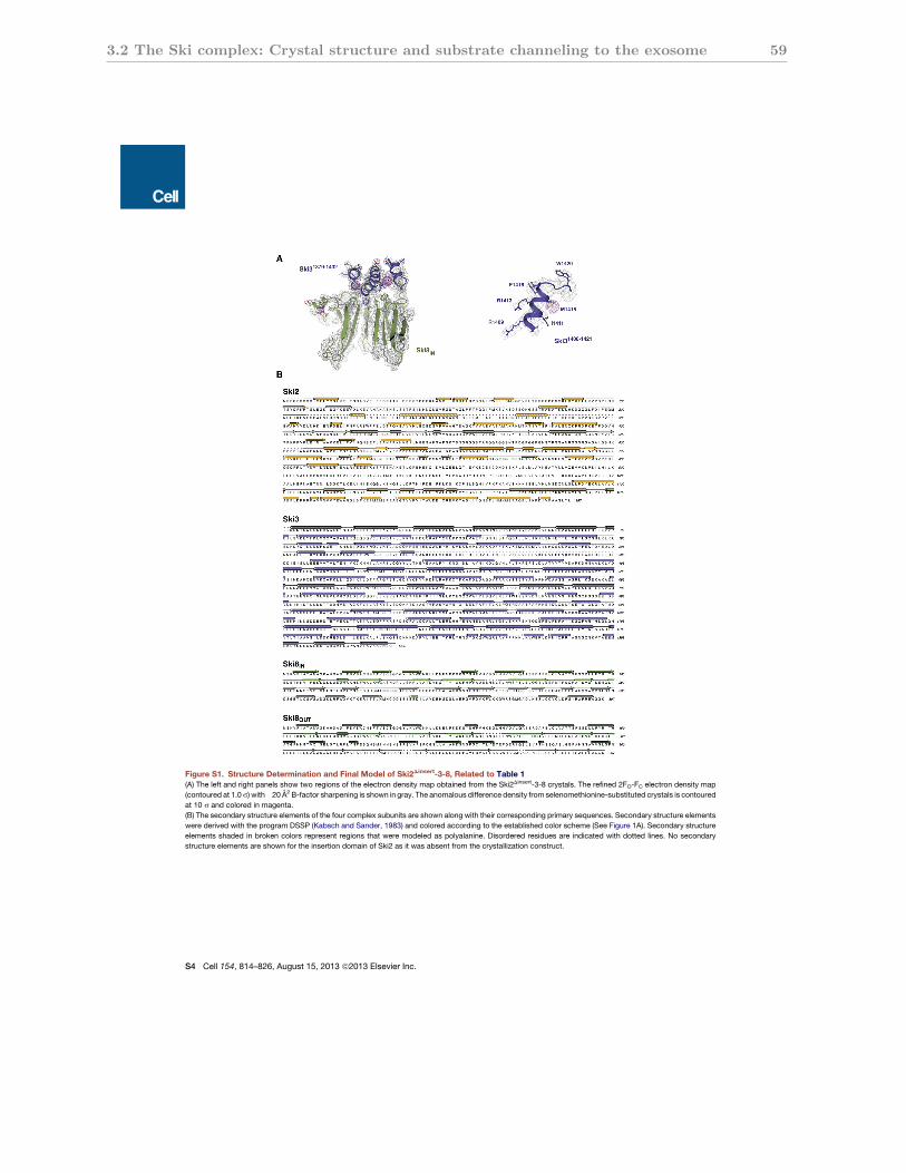
3.2 The Ski complex: Crystal structure and substrate channeling to the exosome 59
Figure S1. Structure Determination and Final Model of Ski2Dinsert-3-8, Related to Table 1(A) The left and right panels show two regions of the electron density map obtained from the Ski2Dinsert-3-8 crystals. The refined 2FO-FC electron density map
(contoured at 1.0s) with!20 A2 B-factor sharpening is shown in gray. The anomalous difference density from selenomethionine-substituted crystals is contoured
at 10 s and colored in magenta.
(B) The secondary structure elements of the four complex subunits are shown along with their corresponding primary sequences. Secondary structure elements
were derived with the program DSSP (Kabsch and Sander, 1983) and colored according to the established color scheme (See Figure 1A). Secondary structure
elements shaded in broken colors represent regions that were modeled as polyalanine. Disordered residues are indicated with dotted lines. No secondary
structure elements are shown for the insertion domain of Ski2 as it was absent from the crystallization construct.
S4 Cell 154, 814–826, August 15, 2013 ª2013 Elsevier Inc.
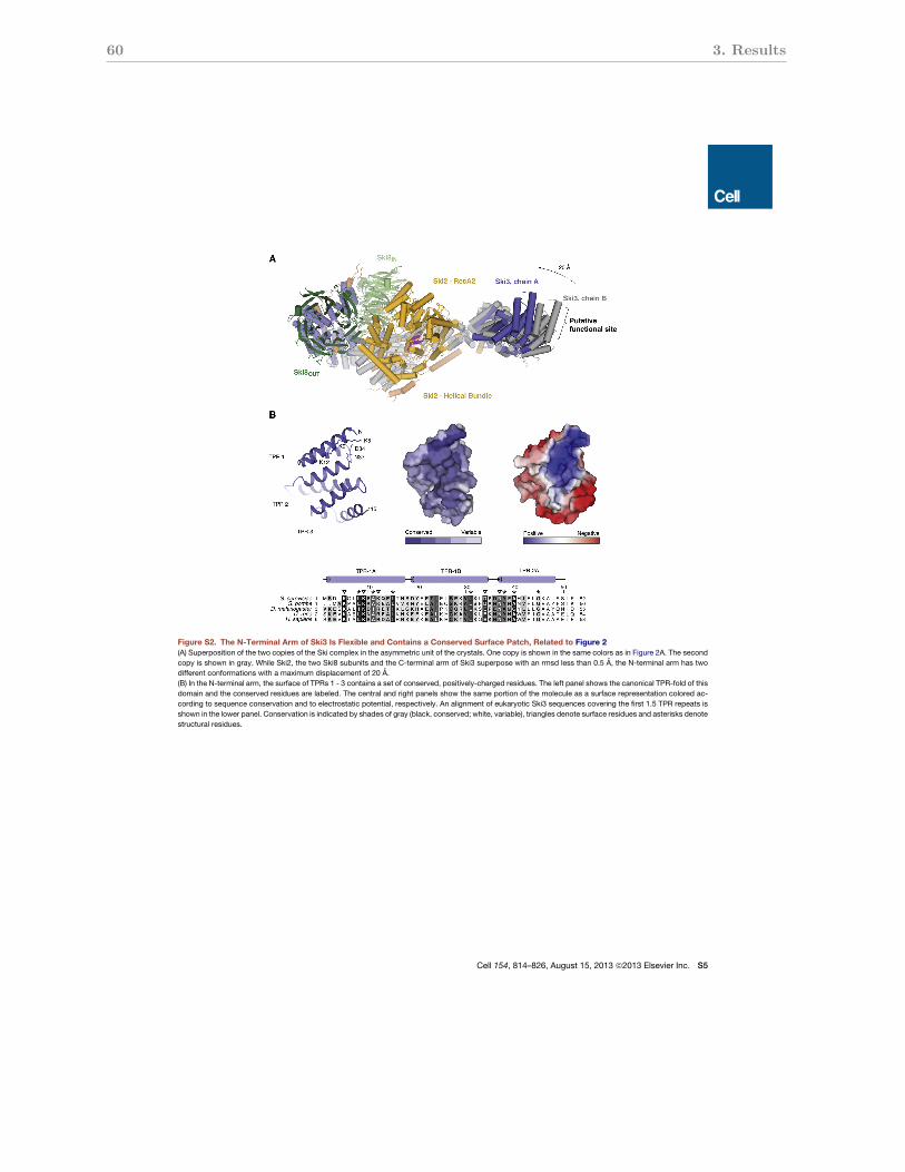
60 3. Results
Figure S2. The N-Terminal Arm of Ski3 Is Flexible and Contains a Conserved Surface Patch, Related to Figure 2(A) Superposition of the two copies of the Ski complex in the asymmetric unit of the crystals. One copy is shown in the same colors as in Figure 2A. The second
copy is shown in gray. While Ski2, the two Ski8 subunits and the C-terminal arm of Ski3 superpose with an rmsd less than 0.5 A, the N-terminal arm has two
different conformations with a maximum displacement of 20 A.
(B) In the N-terminal arm, the surface of TPRs 1 - 3 contains a set of conserved, positively-charged residues. The left panel shows the canonical TPR-fold of this
domain and the conserved residues are labeled. The central and right panels show the same portion of the molecule as a surface representation colored ac-
cording to sequence conservation and to electrostatic potential, respectively. An alignment of eukaryotic Ski3 sequences covering the first 1.5 TPR repeats is
shown in the lower panel. Conservation is indicated by shades of gray (black, conserved; white, variable), triangles denote surface residues and asterisks denote
structural residues.
Cell 154, 814–826, August 15, 2013 ª2013 Elsevier Inc. S5
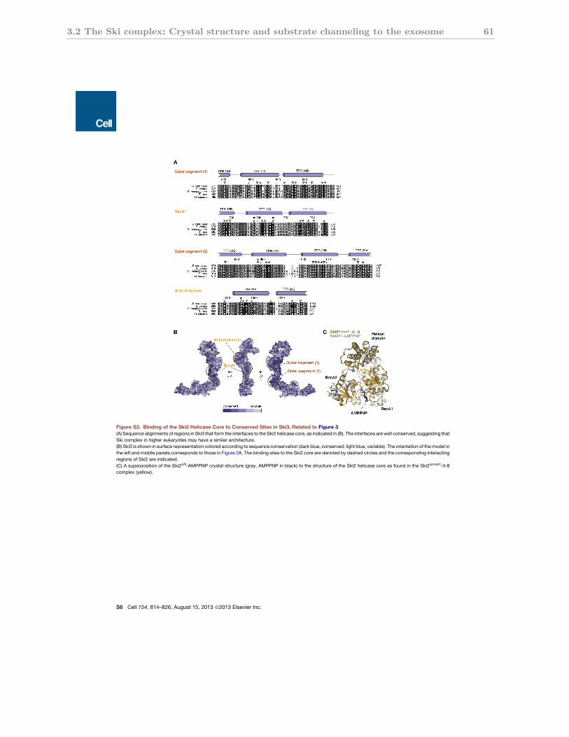
3.2 The Ski complex: Crystal structure and substrate channeling to the exosome 61
Figure S3. Binding of the Ski2 Helicase Core to Conserved Sites in Ski3, Related to Figure 3(A) Sequence alignments of regions in Ski3 that form the interfaces to the Ski2 helicase core, as indicated in (B). The interfaces are well conserved, suggesting that
Ski complex in higher eukaryotes may have a similar architecture.
(B) Ski3 is shown in surface representation colored according to sequence conservation (dark blue, conserved; light blue, variable). The orientation of themodel in
the left and middle panels corresponds to those in Figure 2A. The binding sites to the Ski2 core are denoted by dashed circles and the corresponding interacting
regions of Ski2 are indicated.
(C) A superposition of the Ski2DN-AMPPNP crystal structure (gray, AMPPNP in black) to the structure of the Ski2 helicase core as found in the Ski2Dinsert-3-8
complex (yellow).
S6 Cell 154, 814–826, August 15, 2013 ª2013 Elsevier Inc.
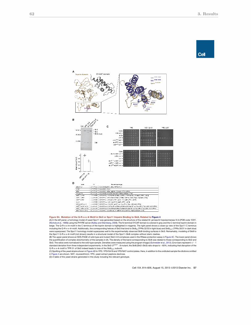
62 3. Results
Figure S4. Mutation of the Q-R-x-x-F Motif in Ski3 or Spo11 Impairs Binding to Ski8, Related to Figure 4(A) In the left panel, a homology model of yeast Spo11 was generated based on the structure of the related M. jannaschii topoisomerase VI-A (PDB code 1D3Y,
(Nichols et al., 1999)) using the PHYRE server (Kelley and Sternberg, 2009). The N-terminal 5YCAP domain is colored in gray and the C-terminal toprim domain in
beige. The Q-R-x-x-F motif in the C terminus of the toprim domain is highlighted in magenta. The right panel shows a close-up view of the Spo11 C terminus
including the Q-R-x-x-Fmotif. Additionally, the corresponding helices of Ski3 that bind to Ski8IN (TPRs 32/33 in light blue) and Ski8OUT (TPRs 30/31 in dark blue)
were superposed. The Spo11 homology model superposes well to the experimentally observed Ski8-binding surfaces in Ski3. Remarkably, modeling of Ski8 to
the Spo11 Q-R-x-x-F motif (not shown) results in a structural model of the Spo11-Ski8 complex without steric clashes.
(B) The upper panel shows an SDS-PAGE of wild-type and mutant Ski2-3-8 complexes used in the RNase protection assay in Figure 4C. The lower panel shows
the quantification of complex stoichiometry of the samples in (A). The density of the band corresponding to Ski8 was related to those corresponding to Ski2 and
Ski3. The ratios were normalized to the wild-type sample. Densities were measured using the program ImageJ (Schneider et al., 2012). Error bars represent +/! 1
standard deviation from three independent experiments. In the Ski2-3DOUT!8 mutant, the Ski8:(Ski2+Ski3) ratio drops to"60%, indicating that disruption of the
Q-R-x-x-F motif in TPR 31 of Ski3 indeed leads to loss of the Ski8OUT subunit.
(C) Spotting of the yeast strains shown in Figure 4D to YPD, YPD/G418 and YPD/NAT control plates. Here, in addition to the undiluted sample the dilutions omitted
in Figure 4 are shown. NAT, nourseothricin; YPD, yeast extract peptone dextrose.
(D) A table of the yeast strains generated in this study including the relevant genotype.
Cell 154, 814–826, August 15, 2013 ª2013 Elsevier Inc. S7
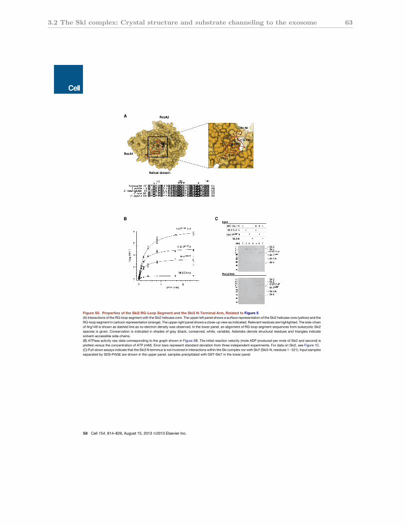
3.2 The Ski complex: Crystal structure and substrate channeling to the exosome 63
Figure S5. Properties of the Ski2 RG-Loop Segment and the Ski3 N-Terminal Arm, Related to Figure 5(A) Interactions of the RG-loop segment with the Ski2 helicase core. The upper left panel shows a surface representation of the Ski2 helicase core (yellow) and the
RG-loop segment in cartoon representation (orange). The upper right panel shows a close-up view as indicated. Relevant residues are highlighted. The side-chain
of Arg149 is shown as dashed line as no electron density was observed. In the lower panel, an alignment of RG-loop segment sequences from eukaryotic Ski2
species is given. Conservation is indicated in shades of gray (black, conserved; white, variable). Asterisks denote structural residues and triangles indicate
solvent-accessible side-chains.
(B) ATPase activity raw data corresponding to the graph shown in Figure 5B. The initial reaction velocity (mole ADP produced per mole of Ski2 and second) is
plotted versus the concentration of ATP (mM). Error bars represent standard deviation from three independent experiments. For data on Ski2, see Figure 1C.
(C) Pull-down assays indicate that the Ski3 N terminus is not involved in interactions within the Ski complex nor with Ski7 (Ski3-N, residues 1 - 521). Input samples
separated by SDS-PAGE are shown in the upper panel, samples precipitated with GST-Ski7 in the lower panel.
S8 Cell 154, 814–826, August 15, 2013 ª2013 Elsevier Inc.
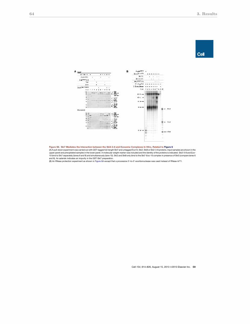
64 3. Results
Figure S6. Ski7 Mediates the Interaction between the Ski2-3-8 and Exosome Complexes In Vitro, Related to Figure 6(A) A pull-down experiment was carried out with GST-tagged full-length Ski7 and untagged Exo10, Ski2, Ski8 or Ski2-3-8 proteins. Input samples are shown in the
upper panel and precipitated samples in the lower panel. A molecular weight marker was included and the identity of the proteins is indicated. Ski2-3-8 and Exo-
10 bind to Ski7 separately (lanes 6 and 9) and simultaneously (lane 10). Ski2 and Ski8 only bind to the Ski7-Exo-10 complex in presence of Ski3 (compare lanes 6
and 8). An asterisk indicates an impurity in the GST-Ski7 preparation.
(B) An RNase protection experiment as shown in Figure 6A except that a processive 50-to-30 exoribonuclease was used instead of RNase A/T1.
Cell 154, 814–826, August 15, 2013 ª2013 Elsevier Inc. S9
4 Discussion
The major achievement of the PhD work at hand was to determine the crystal structures
of the Ski2 helicase region (Ski2∆N), the Ski2 insertion domain as well as the Ski2∆insert-
Ski3-Ski8 complex. The assembly of the individual structures yields the first comprehensive
structural description of the full-length yeast Ski2-Ski3-Ski8 complex (Fig. 4.1). Additional
functional characterization helped to understand the role of the individual subunits and
to identify two regulatory mechanisms that may contribute to the regulation of exosome
activity.
The following sections discuss the structure and function of the Ski2 helicase region
alone (section 4.1) as well as in the context of the Ski2-Ski3-Ski8 complex (section 4.2), the
role of the Ski3 TPR scaffold (section 4.3) and the structure and function of the two Ski8
subunits and the implications for the role of Ski8 in meiotic recombination (section 4.4).
A final section discusses the two regulatory mechanisms of the SKI complex in context of
the exosome (section 4.5).
4.1 Ski2 contains a conserved DExH box core and a
variable insertion domain
The helicase region of Ski2 was defined by limited proteolysis to encompass residues 296 -
1287 (Ski2∆N) and was crystallized in complex with adenosine 5′ -(β,γ-imido)triphosphate
(AMPPNP). The helicase core consists of a ring-like arrangement of the two RecA domains,
the ratchet and winged helix (WH) domains (Fig. 4.1). It is well conserved in sequence
and highly similar in structure to other DExH box helicases like Mtr4 and Hel308 which
have been structurally characterized in complex with RNA substrates (Buttner et al., 2007;
Weir et al., 2010). This suggests that in Ski2 RNA is bound along a similar path through
the helicase core. Consistently, Ski2 also contains a conserved β-hairpin at the presumable
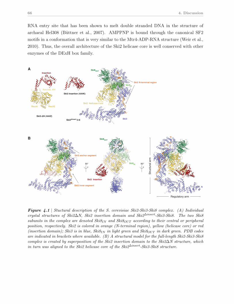
66 4. Discussion
RNA entry site that has been shown to melt double stranded DNA in the structure of
archaeal Hel308 (Buttner et al., 2007). AMPPNP is bound through the canonical SF2
motifs in a conformation that is very similar to the Mtr4-ADP-RNA structure (Weir et al.,
2010). Thus, the overall architecture of the Ski2 helicase core is well conserved with other
enzymes of the DExH box family.
A
Ski2 Insertion (4A4K)
Ski2 -3-8
90° 90°
Ski8OUT
Ski8OUT
RecA1
Insertion
RecA2
Ski3
Ski2 Insertion
Ski8
Ski8
Stru
ctur
al a
rm
Regulatory arm
B
Figure 4.1 | Stuctural description of the S. cerevisiae Ski2-Ski3-Ski8 complex. (A) Individualcrystal structures of Ski2∆N, Ski2 insertion domain and Ski2∆insert-Ski3-Ski8. The two Ski8subunits in the complex are denoted Ski8IN and Ski8OUT according to their central or peripheralposition, respectively. Ski2 is colored in orange (N-terminal region), yellow (helicase core) or red(insertion domain); Ski3 is in blue, Ski8IN in light green and Ski8OUT in dark green. PDB codesare indicated in brackets where available. (B) A structural model for the full-length Ski2-Ski3-Ski8complex is created by superposition of the Ski2 insertion domain to the Ski2∆N structure, whichin turn was aligned to the Ski2 helicase core of the Ski2∆insert-Ski3-Ski8 structure.
4.2 The structure of the SKI complex: a framework for Ski2 to function in concert with the
exosome 67
From the globular helicase core of Ski2 the so-termed insertion domain emerges (residues
835 - 1085). It consists of a globular β-barrel that is connected to the helicase core through
a flexible α-helical stalk. A comparison with the Mtr4 insertion domain (also termed KOW
domain) shows that both domains share a similar architecture (consisting of a helical stalk
and a β-barrel). While the presence of the Ski2 insertion domain and the position where
it emerges from the enzyme core (within the WH domain above the RNA entry site) is
conserved with Mtr4, the domain itself is not conserved in sequence even among Ski2
proteins from other yeast species.
On the functional level, both domains differ, too. The Mtr4 KOW domain has been
shown to bind structured RNAs in vitro (Weir et al., 2010) and has been linked to rRNA
maturation in vivo (Jackson et al., 2010; Klauer and van Hoof, 2013). In contrast, elec-
trophoretic mobility shift assays (EMSAs) indicate that the Ski2 insertion domain binds
RNA unspecifically, consistent with the presence of a positively charged surface patch.
The promiscuous RNA-binding properties and the location above the RNA entry site in
the helicase core suggest that the Ski2 insertion domain plays a general role in substrate
loading.
4.2 The structure of the SKI complex: a framework
for Ski2 to function in concert with the exosome
The fact that the Ski2 insertion domain protrudes from the globular helicase core without
being required for formation of the Ski2-Ski3-Ski8 complex (Fig. 3.1.4) suggested its re-
moval for purposes of structure determination. The resulting Ski2∆insert-Ski3-Ski8 complex
could be readily crystallized and the structure was solved by a single wavelength anomalous
diffraction (SAD) experiment.
The crystal structure of the Ski2∆insert-Ski3-Ski8 complex revealed the architecture of
the yeast SKI complex (Fig. 4.1). Ski3 consists of 33 TPR motifs that form a long
superhelical solenoid with an N-terminal and a C-terminal arm. The C-terminal arm
binds both Ski8 subunits as well as the N-terminal and the helicase region of Ski2. The
two Ski8 subunits (Ski8IN , central position. Ski8OUT , peripheral position. See also Fig.
4.1) bind back to the helicase core. The helicase is thus centrally located within complex,
reflecting the presumably pivotal role of its activity for function of the SKI complex. In the
Ski2∆insert-Ski3-Ski8 complex structure (representing the apo state), the Ski2 DExH core
has a conformation that is nearly identical to Ski2∆N bound to AMPPNP (Fig. 3.2.4). It
68 4. Discussion
is possible that the other subunits induce this conformation in the helicase core, but more
experimental data (e.g. in the form of RNA- or nucleotide-bound structures) is needed
to clarify whether Ski3 and Ski8 actively trigger any conformational changes in the Ski2
helicase core.
Nevertheless, formation of the Ski2-Ski3-Ski8 complex leads to a series of structural
rearrangements in Ski2. First, the Ski2 insertion domain is positioned at the entry site
of the RNA to the helicase core. The insertion domain is not present in the Ski2∆insert-
Ski3-Ski8 structure, but modeling it in this position (Fig. 4.1) does not produce any
steric clashes, which makes it reasonable to assume a similar orientation as in the Ski2∆N
structure. Second, the RG-loop segment of the Ski2 N-terminal region is placed by Ski3
such that its conserved Arg-Gly dipeptide flanks the canonical RNA-interacting SF2 motifs
towards the 3′ end of the RNA (see Fig. 3.2.4). This extends the bona fide RNA-binding
path through the helicase core. Third, portions of Ski8IN and Ski3 are arranged around
the presumable exit site of the RNA 3′ end from the helicase core. Even though the
electrostatic potential and the conservation of involved surface patches do not show any
striking features, a cleft is created through which the RNA can leave the complex.
All three structural features cluster along the RNA-binding path of Ski2 and thus
highlight the importance of RNA-routing through the SKI complex. They point towards
a functional interplay with the exosome where a helicase would need to accept substrates
or control their access (via the insertion domain), unwind secondary structure elements
and transfer the substrate to the exosome (via the extension of the RNA-binding path by
the RG-loop and the formation of an RNA exit cleft). In agreement with such a model,
the Ski2-Ski3-Ski8 complex displays RNA-dependent ATPase activity and unwinds RNA
duplexes with 3′ overhang. The resulting single stranded RNA substrates could then be
readily degraded by the exosome (Bonneau et al., 2009).
It thus appears that formation of the Ski2-Ski3-Ski8 complex sets a structural frame-
work that primes the helicase core of Ski2 for assisting the exosome in substrate degra-
dation. Consistent with this model, direct substrate channeling between both complex is
observed and will be discussed in section 4.5.
4.3 The Ski3 TPR scaffold organizes the SKI complex
Mapping of domain interactions within the Ski2-Ski3-Ski8 complex in yeast in vivo sug-
gested that Ski3 is involved in all subunit interactions (Wang et al., 2005). The crystal
4.3 The Ski3 TPR scaffold organizes the SKI complex 69
structure of the Ski2∆insert-Ski3-Ski8 complex rationalizes this observation by revealing how
the TPR solenoid of Ski3 acts as scaffold for the entire complex.
Containing 33 TPRs, Ski3 can be divided in an N-terminal arm and a C-terminal arm.
The C-terminal arm binds the remaining subunits and thus organizes the architecture of
the complex (Fig. 4.1). It extensively interacts with a series of motifs in the Ski2 N-
terminus that but also binds to the helicase core of Ski2 (the RecA1 and ratchet domains).
Interestingly, the interfaces with the helicase core are conserved in higher eukaryotes, sug-
gesting a similar complex topology. The Ski3 C-terminal arm also binds both Ski8 subunits
(see section 4.4).
Size, versatility and flexibility predestine TPR proteins as molecular scaffolds
TPR proteins and particularly their complexes with peptides have been well characterized
in terms of structure and biochemistry (D’Andrea and Regan, 2003; Zeytuni and Zarivach,
2012). Their ability to engage in protein-protein interactions has earned them a repu-
tation as scaffold subunits in protein complexes, but structural characterization of such
entire assemblies remains scarce. The unusally large TPR array of Ski3 illustrates several
properties of TPR proteins that allow them to efficiently organize large complexes.
First, if a protein acts as central hub within a complex, i.e. it interacts with all or
most of the other subunits, a large surface must be dedicated to forming interfaces with
those proteins. Structures of TPR proteins with up to 14 consecutive repeats have been
reported, for instance those of O-linked glycosyl transferase (Jinek et al., 2004) or the
anaphase promoting complex subunit Cdc16 (Zhang et al., 2010). The finding that the
Ski3 C-terminus contains 23 consecutive canonical repeats (Fig. 3.2.2) illustrates that TPR
scaffolds can be considerably longer than assumed. It can be anticipated that uncharacter-
ized TPR-containing proteins may form even longer solenoids. The size creates sufficient
surface area that can be dedicated to interactions with partner proteins. Probably more
important than the mere molecular size is the surface-to-residue ratio. In TPR proteins,
the superhelical stacking leads to an extended solenoid shape of the molecule. Compared
to a globular protein of similar size, such a fold results in a considerably higher ratio of
surface area to residues. Thus, the combination of molecular size and its efficient trans-
formation into binding surfaces enables TPR proteins to form highly optimized protein
interaction platforms.
Second, TPR proteins are versatile binders. Typically, a ligand binds as an extended
polypeptide to the concave surface of the TPR superhelix, even though the convex surface
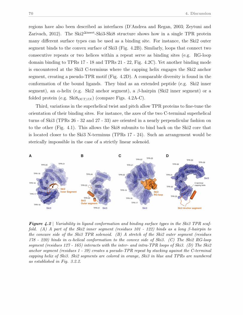
70 4. Discussion
regions have also been described as interfaces (D’Andrea and Regan, 2003; Zeytuni and
Zarivach, 2012). The Ski2∆insert-Ski3-Ski8 structure shows how in a single TPR protein
many different surface types can be used as a binding site. For instance, the Ski2 outer
segment binds to the convex surface of Ski3 (Fig. 4.2B). Similarly, loops that connect two
consecutive repeats or two helices within a repeat serve as binding sites (e.g. RG-loop
domain binding to TPRs 17 - 18 and TPRs 21 - 22, Fig. 4.2C). Yet another binding mode
is encountered at the Ski3 C-terminus where the capping helix engages the Ski2 anchor
segment, creating a pseudo-TPR motif (Fig. 4.2D). A comparable diversity is found in the
conformation of the bound ligands. They bind as an extended peptide (e.g. Ski2 inner
segment), an α-helix (e.g. Ski2 anchor segment), a β-hairpin (Ski2 inner segment) or a
folded protein (e.g. Ski8OUT/IN) (compare Figs. 4.2A-C).
Third, variations in the superhelical twist and pitch allow TPR proteins to fine-tune the
orientation of their binding sites. For instance, the axes of the two C-terminal superhelical
turns of Ski3 (TPRs 26 - 32 and 27 - 33) are oriented in a nearly perpendicular fashion on
to the other (Fig. 4.1). This allows the Ski8 subunits to bind back on the Ski2 core that
is located closer to the Ski3 N-terminus (TPRs 17 - 24). Such an arrangement would be
sterically impossible in the case of a strictly linear solenoid.
TPR-21
Ski2
Outer segment
TPR-22
TPR-23
TPR-24
Ski2
RG-loop
Ski2 Anchor segmentSki3Ski3Ski3
TPR-20
TPR-19
TPR-18
TPR-17
TPR-21
TPR-22 TPR-23
TPR-20
TPR-19
TPR-18
TPR-17
TPR-16
Ski2
Inner Segment
TPR-21
A B C D
Ski3
TPR-33
C-terminalcapping helix
TPR-32
TPR-31
TPR-30
TPR-29TPR-28
Figure 4.2 | Variability in ligand conformation and binding surface types in the Ski3 TPR scaf-fold. (A) A part of the Ski2 inner segment (residues 101 - 122) binds as a long β-hairpin tothe concave side of the Ski3 TPR solenoid. (B) A stretch of the Ski2 outer segment (residues178 - 220) binds in α-helical conformation to the convex side of Ski3. (C) The Ski2 RG-loopsegment (residues 127 - 165) interacts with the inter- and intra-TPR loops of Ski3. (D) The Ski2anchor segment (residues 1 - 39) creates a pseudo-TPR repeat by stacking against the C-terminalcapping helix of Ski3. Ski2 segments are colored in orange, Ski3 in blue and TPRs are numberedas established in Fig. 3.2.2.
4.4 Ski8 is a versatile adapter protein with multiple roles 71
These properties appear to be intrinsic to TPR proteins, but there may be a mutual
dependency between scaffold and binding target. For instance, the Ski2 N-terminal segment
threads through the C-terminal arm of Ski3, thereby stabilizing it (Fig. 3.2.3). This may
help to properly orient the remaining binding sites (e.g. of the Ski8 subunits).
In Ski3, conservation of the TPR fold correlates with binding site functionality
TPRs are ancient motifs that most likely have been present in the last common ancestor
since they are found in bacteria, archaea and eukaryotes. However, conservation of the
motif is poor in terms of sequence identity. Rather, classes of amino acids (small vs. large)
are conserved on a given position, in-line with the “hole-and-knobs” model for interaction
between the helices (Hirano et al., 1990). In contrast, the entire fold is well conserved,
and TPR blocks in unrelated proteins from distinct species are structurally very similar,
often with a root mean square deviation (r.m.s.d.) below 1.5 A. Variations are usually only
found in the loops that connect the helices of a given motif or two entire motifs.
The structural information on Ski3 now offers the opportunity to analyze how the TPR
fold and presence of binding sites correlate within a single polypeptide. In Ski3, the C-
terminal arm forms the binding platform for Ski2 and both Ski8 subunits (Fig. 4.1) and
thus has been under evolutionary pressure to maintain its binding interfaces (see also Fig.
3.2.S3). The C-terminal arm is built from canonical TPRs that are structurally very similar
to other bona fide TPR proteins, e.g. O-linked glycosyl-transferase (Jinek et al., 2004)
(Fig. 3.2.S2). The N-terminal arm also contains a block of three canonical TPR motifs
that according to surface conservation presumably form a functional site (Fig. 3.2.S2).
Only the linker region between the two arms contains non-canonical TPRs that deviate
considerably from the typical fold in terms of geometry and length of the individual helices.
It thus appears that functionality as a binding interface correlates with conservation of the
TPR fold. Only in the linker region, which was not subjected to evolutionary pressure to
maintain interactions with partner proteins, atypical TPR motifs could evolve.
4.4 Ski8 is a versatile adapter protein with multiple
roles
The Ski2-Ski3-Ski8 complex contains two Ski8 subunits (Ski8IN and Ski8OUT , see Fig.
4.1), in line with previous mass-spectrometric data (Synowsky and Heck, 2008). Both Ski8
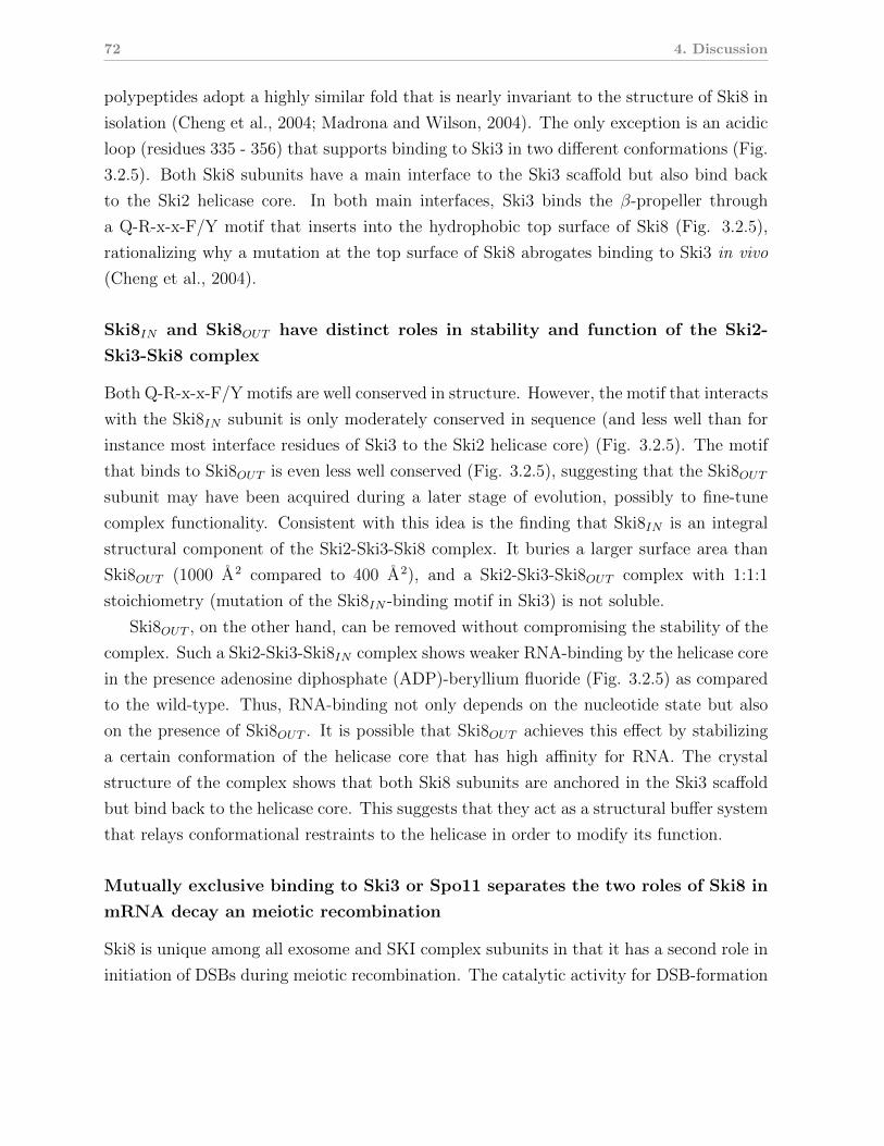
72 4. Discussion
polypeptides adopt a highly similar fold that is nearly invariant to the structure of Ski8 in
isolation (Cheng et al., 2004; Madrona and Wilson, 2004). The only exception is an acidic
loop (residues 335 - 356) that supports binding to Ski3 in two different conformations (Fig.
3.2.5). Both Ski8 subunits have a main interface to the Ski3 scaffold but also bind back
to the Ski2 helicase core. In both main interfaces, Ski3 binds the β-propeller through
a Q-R-x-x-F/Y motif that inserts into the hydrophobic top surface of Ski8 (Fig. 3.2.5),
rationalizing why a mutation at the top surface of Ski8 abrogates binding to Ski3 in vivo
(Cheng et al., 2004).
Ski8IN and Ski8OUT have distinct roles in stability and function of the Ski2-
Ski3-Ski8 complex
Both Q-R-x-x-F/Y motifs are well conserved in structure. However, the motif that interacts
with the Ski8IN subunit is only moderately conserved in sequence (and less well than for
instance most interface residues of Ski3 to the Ski2 helicase core) (Fig. 3.2.5). The motif
that binds to Ski8OUT is even less well conserved (Fig. 3.2.5), suggesting that the Ski8OUT
subunit may have been acquired during a later stage of evolution, possibly to fine-tune
complex functionality. Consistent with this idea is the finding that Ski8IN is an integral
structural component of the Ski2-Ski3-Ski8 complex. It buries a larger surface area than
Ski8OUT (1000 A2 compared to 400 A2), and a Ski2-Ski3-Ski8OUT complex with 1:1:1
stoichiometry (mutation of the Ski8IN -binding motif in Ski3) is not soluble.
Ski8OUT , on the other hand, can be removed without compromising the stability of the
complex. Such a Ski2-Ski3-Ski8IN complex shows weaker RNA-binding by the helicase core
in the presence adenosine diphosphate (ADP)-beryllium fluoride (Fig. 3.2.5) as compared
to the wild-type. Thus, RNA-binding not only depends on the nucleotide state but also
on the presence of Ski8OUT . It is possible that Ski8OUT achieves this effect by stabilizing
a certain conformation of the helicase core that has high affinity for RNA. The crystal
structure of the complex shows that both Ski8 subunits are anchored in the Ski3 scaffold
but bind back to the helicase core. This suggests that they act as a structural buffer system
that relays conformational restraints to the helicase in order to modify its function.
Mutually exclusive binding to Ski3 or Spo11 separates the two roles of Ski8 in
mRNA decay an meiotic recombination
Ski8 is unique among all exosome and SKI complex subunits in that it has a second role in
initiation of DSBs during meiotic recombination. The catalytic activity for DSB-formation
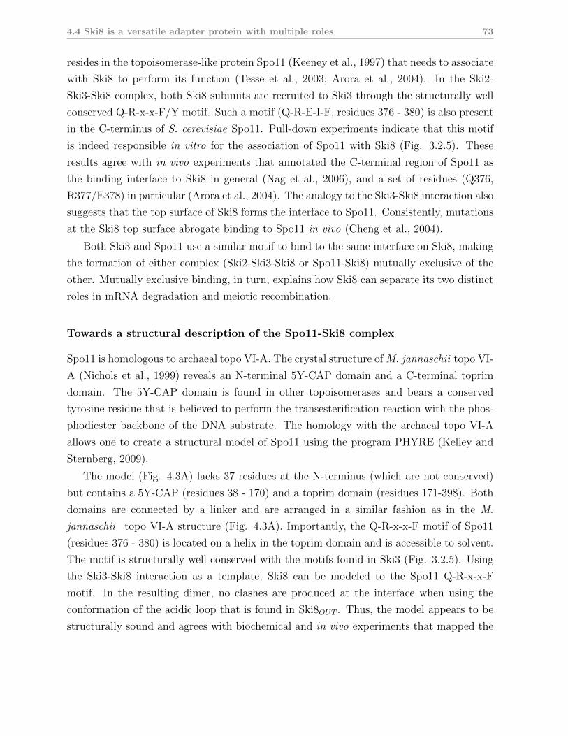
4.4 Ski8 is a versatile adapter protein with multiple roles 73
resides in the topoisomerase-like protein Spo11 (Keeney et al., 1997) that needs to associate
with Ski8 to perform its function (Tesse et al., 2003; Arora et al., 2004). In the Ski2-
Ski3-Ski8 complex, both Ski8 subunits are recruited to Ski3 through the structurally well
conserved Q-R-x-x-F/Y motif. Such a motif (Q-R-E-I-F, residues 376 - 380) is also present
in the C-terminus of S. cerevisiae Spo11. Pull-down experiments indicate that this motif
is indeed responsible in vitro for the association of Spo11 with Ski8 (Fig. 3.2.5). These
results agree with in vivo experiments that annotated the C-terminal region of Spo11 as
the binding interface to Ski8 in general (Nag et al., 2006), and a set of residues (Q376,
R377/E378) in particular (Arora et al., 2004). The analogy to the Ski3-Ski8 interaction also
suggests that the top surface of Ski8 forms the interface to Spo11. Consistently, mutations
at the Ski8 top surface abrogate binding to Spo11 in vivo (Cheng et al., 2004).
Both Ski3 and Spo11 use a similar motif to bind to the same interface on Ski8, making
the formation of either complex (Ski2-Ski3-Ski8 or Spo11-Ski8) mutually exclusive of the
other. Mutually exclusive binding, in turn, explains how Ski8 can separate its two distinct
roles in mRNA degradation and meiotic recombination.
Towards a structural description of the Spo11-Ski8 complex
Spo11 is homologous to archaeal topo VI-A. The crystal structure of M. jannaschii topo VI-
A (Nichols et al., 1999) reveals an N-terminal 5Y-CAP domain and a C-terminal toprim
domain. The 5Y-CAP domain is found in other topoisomerases and bears a conserved
tyrosine residue that is believed to perform the transesterification reaction with the phos-
phodiester backbone of the DNA substrate. The homology with the archaeal topo VI-A
allows one to create a structural model of Spo11 using the program PHYRE (Kelley and
Sternberg, 2009).
The model (Fig. 4.3A) lacks 37 residues at the N-terminus (which are not conserved)
but contains a 5Y-CAP (residues 38 - 170) and a toprim domain (residues 171-398). Both
domains are connected by a linker and are arranged in a similar fashion as in the M.
jannaschii topo VI-A structure (Fig. 4.3A). Importantly, the Q-R-x-x-F motif of Spo11
(residues 376 - 380) is located on a helix in the toprim domain and is accessible to solvent.
The motif is structurally well conserved with the motifs found in Ski3 (Fig. 3.2.5). Using
the Ski3-Ski8 interaction as a template, Ski8 can be modeled to the Spo11 Q-R-x-x-F
motif. In the resulting dimer, no clashes are produced at the interface when using the
conformation of the acidic loop that is found in Ski8OUT . Thus, the model appears to be
structurally sound and agrees with biochemical and in vivo experiments that mapped the
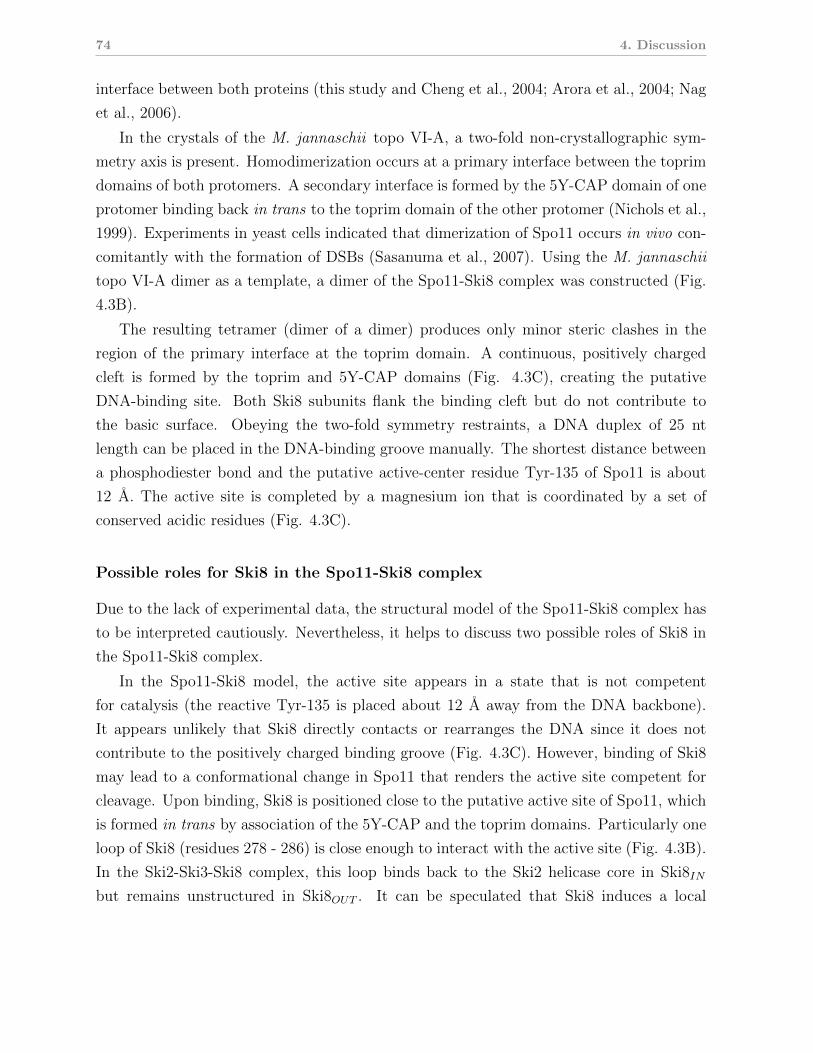
74 4. Discussion
interface between both proteins (this study and Cheng et al., 2004; Arora et al., 2004; Nag
et al., 2006).
In the crystals of the M. jannaschii topo VI-A, a two-fold non-crystallographic sym-
metry axis is present. Homodimerization occurs at a primary interface between the toprim
domains of both protomers. A secondary interface is formed by the 5Y-CAP domain of one
protomer binding back in trans to the toprim domain of the other protomer (Nichols et al.,
1999). Experiments in yeast cells indicated that dimerization of Spo11 occurs in vivo con-
comitantly with the formation of DSBs (Sasanuma et al., 2007). Using the M. jannaschii
topo VI-A dimer as a template, a dimer of the Spo11-Ski8 complex was constructed (Fig.
4.3B).
The resulting tetramer (dimer of a dimer) produces only minor steric clashes in the
region of the primary interface at the toprim domain. A continuous, positively charged
cleft is formed by the toprim and 5Y-CAP domains (Fig. 4.3C), creating the putative
DNA-binding site. Both Ski8 subunits flank the binding cleft but do not contribute to
the basic surface. Obeying the two-fold symmetry restraints, a DNA duplex of 25 nt
length can be placed in the DNA-binding groove manually. The shortest distance between
a phosphodiester bond and the putative active-center residue Tyr-135 of Spo11 is about
12 A. The active site is completed by a magnesium ion that is coordinated by a set of
conserved acidic residues (Fig. 4.3C).
Possible roles for Ski8 in the Spo11-Ski8 complex
Due to the lack of experimental data, the structural model of the Spo11-Ski8 complex has
to be interpreted cautiously. Nevertheless, it helps to discuss two possible roles of Ski8 in
the Spo11-Ski8 complex.
In the Spo11-Ski8 model, the active site appears in a state that is not competent
for catalysis (the reactive Tyr-135 is placed about 12 A away from the DNA backbone).
It appears unlikely that Ski8 directly contacts or rearranges the DNA since it does not
contribute to the positively charged binding groove (Fig. 4.3C). However, binding of Ski8
may lead to a conformational change in Spo11 that renders the active site competent for
cleavage. Upon binding, Ski8 is positioned close to the putative active site of Spo11, which
is formed in trans by association of the 5Y-CAP and the toprim domains. Particularly one
loop of Ski8 (residues 278 - 286) is close enough to interact with the active site (Fig. 4.3B).
In the Ski2-Ski3-Ski8 complex, this loop binds back to the Ski2 helicase core in Ski8IN
but remains unstructured in Ski8OUT . It can be speculated that Ski8 induces a local
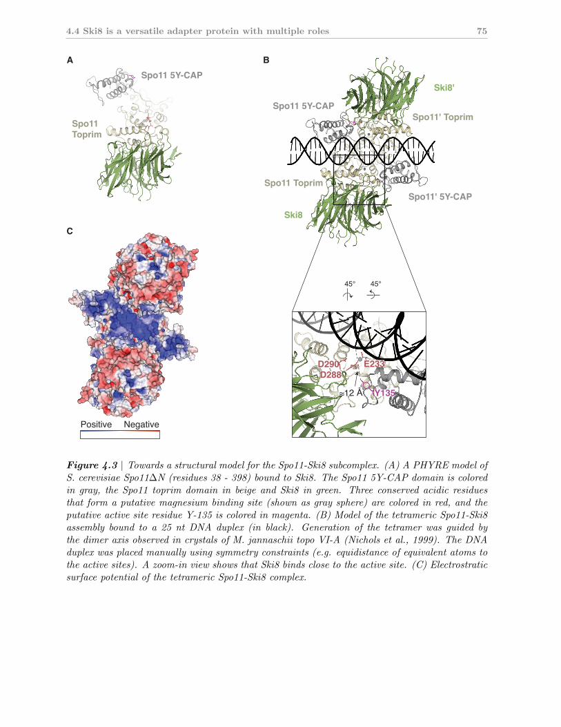
4.4 Ski8 is a versatile adapter protein with multiple roles 75
Positive Negative
C
Spo11' 5Y-CAP
Ski8
Ski8'
Spo11 Toprim
Spo11' ToprimSpo11 5Y-CAP
BA
45°45°
Spo11 5Y-CAP
Spo11 Toprim
E233D290D288
Y135
Figure 4.3 | Towards a structural model for the Spo11-Ski8 subcomplex. (A) A PHYRE model ofS. cerevisiae Spo11∆N (residues 38 - 398) bound to Ski8. The Spo11 5Y-CAP domain is coloredin gray, the Spo11 toprim domain in beige and Ski8 in green. Three conserved acidic residuesthat form a putative magnesium binding site (shown as gray sphere) are colored in red, and theputative active site residue Y-135 is colored in magenta. (B) Model of the tetrameric Spo11-Ski8assembly bound to a 25 nt DNA duplex (in black). Generation of the tetramer was guided bythe dimer axis observed in crystals of M. jannaschii topo VI-A (Nichols et al., 1999). The DNAduplex was placed manually using symmetry constraints (e.g. equidistance of equivalent atoms tothe active sites). A zoom-in view shows that Ski8 binds close to the active site. (C) Electrostraticsurface potential of the tetrameric Spo11-Ski8 complex.
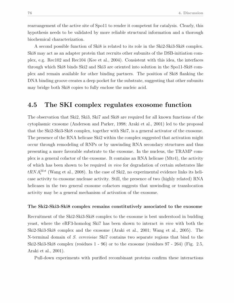
76 4. Discussion
rearrangement of the active site of Spo11 to render it competent for catalysis. Clearly, this
hypothesis needs to be validated by more reliable structural information and a thorough
biochemical characterization.
A second possible function of Ski8 is related to its role in the Ski2-Ski3-Ski8 complex.
Ski8 may act as an adapter protein that recruits other subunits of the DSB-initiation com-
plex, e.g. Rec102 and Rec104 (Kee et al., 2004). Consistent with this idea, the interfaces
through which Ski8 binds Ski2 and Ski3 are oriented into solution in the Spo11-Ski8 com-
plex and remain available for other binding partners. The position of Ski8 flanking the
DNA binding groove creates a deep pocket for the substrate, suggesting that other subunits
may bridge both Ski8 copies to fully enclose the nucleic acid.
4.5 The SKI complex regulates exosome function
The observation that Ski2, Ski3, Ski7 and Ski8 are required for all known functions of the
cytoplasmic exosome (Anderson and Parker, 1998; Araki et al., 2001) led to the proposal
that the Ski2-Ski3-Ski8 complex, together with Ski7, is a general activator of the exosome.
The presence of the RNA helicase Ski2 within the complex suggested that activation might
occur through remodeling of RNPs or by unwinding RNA secondary structures and thus
presenting a more favorable substrate to the exosome. In the nucleus, the TRAMP com-
plex is a general cofactor of the exosome. It contains an RNA helicase (Mtr4), the activity
of which has been shown to be required in vivo for degradation of certain substrates like
tRNAMeti (Wang et al., 2008). In the case of Ski2, no experimental evidence links its heli-
case activity to exosome nuclease activity. Still, the presence of two (highly related) RNA
helicases in the two general exosome cofactors suggests that unwinding or translocation
activity may be a general mechanism of activation of the exosome.
The Ski2-Ski3-Ski8 complex remains constitutively associated to the exosome
Recruitment of the Ski2-Ski3-Ski8 complex to the exosome is best understood in budding
yeast, where the eRF3-homolog Ski7 has been shown to interact in vivo with both the
Ski2-Ski3-Ski8 complex and the exosome (Araki et al., 2001; Wang et al., 2005). The
N-terminal domain of S. cerevisiae Ski7 contains two separate regions that bind to the
Ski2-Ski3-Ski8 complex (residues 1 - 96) or to the exosome (residues 97 - 264) (Fig. 2.5,
Araki et al., 2001).
Pull-down experiments with purified recombinant proteins confirm these interactions
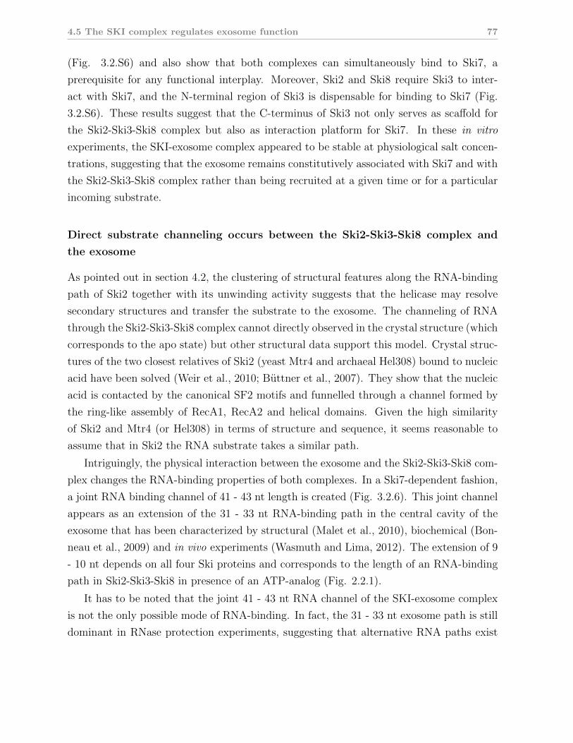
4.5 The SKI complex regulates exosome function 77
(Fig. 3.2.S6) and also show that both complexes can simultaneously bind to Ski7, a
prerequisite for any functional interplay. Moreover, Ski2 and Ski8 require Ski3 to inter-
act with Ski7, and the N-terminal region of Ski3 is dispensable for binding to Ski7 (Fig.
3.2.S6). These results suggest that the C-terminus of Ski3 not only serves as scaffold for
the Ski2-Ski3-Ski8 complex but also as interaction platform for Ski7. In these in vitro
experiments, the SKI-exosome complex appeared to be stable at physiological salt concen-
trations, suggesting that the exosome remains constitutively associated with Ski7 and with
the Ski2-Ski3-Ski8 complex rather than being recruited at a given time or for a particular
incoming substrate.
Direct substrate channeling occurs between the Ski2-Ski3-Ski8 complex and
the exosome
As pointed out in section 4.2, the clustering of structural features along the RNA-binding
path of Ski2 together with its unwinding activity suggests that the helicase may resolve
secondary structures and transfer the substrate to the exosome. The channeling of RNA
through the Ski2-Ski3-Ski8 complex cannot directly observed in the crystal structure (which
corresponds to the apo state) but other structural data support this model. Crystal struc-
tures of the two closest relatives of Ski2 (yeast Mtr4 and archaeal Hel308) bound to nucleic
acid have been solved (Weir et al., 2010; Buttner et al., 2007). They show that the nucleic
acid is contacted by the canonical SF2 motifs and funnelled through a channel formed by
the ring-like assembly of RecA1, RecA2 and helical domains. Given the high similarity
of Ski2 and Mtr4 (or Hel308) in terms of structure and sequence, it seems reasonable to
assume that in Ski2 the RNA substrate takes a similar path.
Intriguingly, the physical interaction between the exosome and the Ski2-Ski3-Ski8 com-
plex changes the RNA-binding properties of both complexes. In a Ski7-dependent fashion,
a joint RNA binding channel of 41 - 43 nt length is created (Fig. 3.2.6). This joint channel
appears as an extension of the 31 - 33 nt RNA-binding path in the central cavity of the
exosome that has been characterized by structural (Malet et al., 2010), biochemical (Bon-
neau et al., 2009) and in vivo experiments (Wasmuth and Lima, 2012). The extension of 9
- 10 nt depends on all four Ski proteins and corresponds to the length of an RNA-binding
path in Ski2-Ski3-Ski8 in presence of an ATP-analog (Fig. 2.2.1).
It has to be noted that the joint 41 - 43 nt RNA channel of the SKI-exosome complex
is not the only possible mode of RNA-binding. In fact, the 31 - 33 nt exosome path is still
dominant in RNase protection experiments, suggesting that alternative RNA paths exist

78 4. Discussion
that may bypass the Ski2-Ski3-Ski8 complex when bound to the exosome. Furthermore,
the role of these pathways in vivo remains to be clarified.
The extension of the exosome’s RNA channel also points towards the S1/KH ring as
the interaction site with Ski7. Since the Rrp44D171N, D551N double mutant used in the
RNase protection experiments buries the 3′ end of the RNA (Lorentzen et al., 2008), any
additional factors must bind the nucleic acid at the 5′ end that emerges from the S1/KH
ring of the central channel, consistent with electron microscopy data using 5′ -labeled RNA
(Malet et al., 2010). In line with these observations, in vivo experiments identified the C-
terminus of the S1/K1 ring member Csl4 as a crucial component for the functions of the
cytoplasmic exosome. (Schaeffer et al., 2009; van Hoof et al., 2000b).
Together, these results suggest a model according to which Ski7 binds to the S1/KH ring
of the exosome (Fig. 3.4A). It then recruits the Ski2-Ski3-Ski8 complex most likely through
Ski3. In this way, the RNA 3′ exit site from the helicase complex is positioned very close to
the RNA entry site on the S1/KH ring. A joint substrate channel is formed that connects
the helicase activity of Ski2 to the nuclease activities of Rrp44. Thus, secondary structures
in RNA substrates could be effectively unwound by Ski2-Ski3-Ski8 and the single stranded
substrate could be funnelled through the joint substrate channel to reach the Rrp44 active
sites for degradation. Even though direct evidence (e.g. in form of structural data) is
still missing, these results represent the first indications of how the Ski2-Ski3-Ski8 complex
activates the exosome.
The Ski2 insertion domain and the Ski3 N-terminal arm cooperate to regulate
access of RNA to Ski2
Inspection of the structural model for the full-length Ski2-Ski3-Ski8 complex reveals two
domains that protrude from the core complex that is arranged around the C-terminal arm
of Ski3 (Fig. 4.1): the Ski2 insertion domain and the N-terminal arm of Ski3. Both
domains are flexible (Fig. 3.1.S1 and Fig. 3.2.S2) and dispensable for complex formation
or binding to Ski7 (Fig. 3.1.4 and Fig. 3.2.S6).
The Ski2 insertion domain is structurally and topologically related to the Mtr4 KOW
domain (Weir et al., 2010; Jackson et al., 2010) and binds RNA apparently unspecifically
(Fig. 3.1.3). Its location above the entry site of the RNA into the helicase core suggests
a role for substrate loading. Indeed, removal of the Ski2 insertion domain promotes the
formation of the 41 - 43 nt RNA channel as judged by RNase protection assays (Fig.
3.2.6). Similarly, its removal enhances unwinding activity of Ski2-Ski3-Ski8 and stimulates

4.5 The SKI complex regulates exosome function 79
ATPase activity of the complex approximately 10-fold (Fig. 3.2.S6). In contrast, deletion
of the insertion domain does not affect ATPase activity of Ski2 in isolation, suggesting that
complex formation contributes an element that is required for the inhibitory effect of this
domain.
Consistent with this hypothesis, removal of the N-terminal arm of Ski3 (residues 1 - 487)
prompts a similar activation of helicase and ATPase activities of the Ski2-Ski3-Ski8 complex
(Fig. 3.2.S6). The flexibility of both domains as observed in the crystal structures and
in normal mode analyses suggests that both domains can potentially come into contact.
However, the Ski3 N-terminal arm (containing the conserved surface patch on TPRs 1
and 2) does not interact with the Ski2 insertion domain in size-exclusion chromatography
experiments (not shown), but transient interactions cannot be excluded.
Both inhibitory domains do not act redundantly since removal of any one is sufficient to
prompt the inhibitory effect. Additionally, the effects of removal of each domain are similar
in magnitude. Together, this suggests that both domains act cooperatively to exert their
function. Considering the flexibility, the location at the RNA entry site and particularly
the effect on formation of a joint substrate channel, the Ski2 insertion domain can be
Figure 4.4 | A model for activation of the cytoplasmic exosome by the Ski2-Ski3-Ski8 complex.(A) A model of how Ski7 bridges the Ski2-Ski3-Ski8 and Exo10 complexes, leading to a jointRNA channel between both assemblies. This connects the helicase and the nuclease activities ofSki2-Ski3-Ski8 and of the exosome, respectively. It remains unclear where Ski7 binds on bothcomplexes (indicated by the question marks). (B) The Ski2 insertion domain and the Ski3 N-terminus cooperate to inhibit the ATPase and helicase activity of the Ski2-Ski3-Ski8 complex,most likely by regulating substrate access into the helicase core of Ski2.

80 4. Discussion
speculated to block substrate access to the helicase core when the Ski3 N-terminal arm
is present, effectively shutting down the helicase. Given the fact that helicase activity is
linked to nuclease activity by the joint substrate channel, this would also provide a means
of down-regulating exosome activity. Conversely, activation of the Ski2-Ski3-Ski8 complex
might then require the Ski3 N-terminal arm to be recruited away from the insertion domain,
possibly by binding events that target the conserved surface patch on TPRs 1 and 2 (Fig.
3.2.S2).
4.6 Major conclusions
All three subunits of the SKI complex are required for exosome-mediated mRNA degra-
dation in the cytoplasm. The results of this PhD thesis offer a first view to understand
the role of the individual subunits in the complex, thus rationalizing why each protein is
essential for the entire biological process.
Ski2 contains in addition to its canonical DExH box core a variable insertion domain
that appears to regulate substrate access to the helicase. Ski3 forms a large TPR array
that is formed by two arms. The N-terminal regulatory arm cooperates with the Ski2
insertion domain to control substrate loading. The C-terminal arm acts as scaffold for the
other subunits and may provide the interface to Ski7.
The finding that the Ski2-Ski3-Ski8 complex contains an internal regulatory mechanism
is intriguing because it lends a broader perspective to the notion of the SKI complex
as a general cofactor of the cytoplasmic exosome. First, its ATP-dependent remodeling
capabilities along with the described direct substrate channeling mechanism may activate
the exosome to degrade structured substrates. Second, the identified internal inhibitory
mechanism in the Ski2-Ski3-Ski8 complex may also inhibit exosome activity indirectly by
refusing substrate transfer. Both kinds of regulation (positive and negative) would provide
tools that - together with a yet to be identified sensory system - could control the exosome’s
substrate specificity.
5 Outlook
The structure of the Ski2-Ski3-Ski8 complex and its functional characterization are a first
step to understand the mechanisms by which the cytoplasmic exosome is regulated. The
next step will include a structural characterization of the exosome bound to Ski7 and the
Ski2-Ski3-Ski8 complex. This will help to understand the details of how Ski7 recruits the
helicase complex to the nuclease complex. In particular it will shed more light on the
function of the Ski7 N-terminus as an adapter between both complexes. More structural
data, possibly including cryo-electron microscopy studies, will also help to analyze the
RNA-channelling between both complexes on the molecular level.
On the functional level, the joint substrate channel needs to be characterized in more
detail by different techniques, e.g. electron microscopy or a combination of chemical cross-
linking an mass-spectrometry. The role of alternative entry paths for RNA into the exosome
remains to be examined. While the current data support the presence of an internal
inhibitory mechanism of Ski2-Ski3-Ski8 complex that controls substrate loading, the details
of the process have to be elucidated. Particularly interesting is also the question of how
this inhibition is released once the helicase complex binds to the exosome. Related to this
problem may be the question of how upstream factors of quality control pathways interact
with the Ski2-Ski3-Ski8 complex. Finally, it needs to be confirmed that degradation of
certain substrates by the exosome is directly promoted by means of the helicase activity
of Ski2.
The long-term goal on the functional level is to understand how the substrate speci-
ficity of the exosome is regulated, and the above-mentioned questions can contribute sub-
stantially to clarify this question. However, a comprehensive understanding of all other
exosome cofactors (e.g. the TRAMP complex), specifically in terms of their activities and
interactions with the exosome, will be indispensable to tackle this problem.
Another somewhat unrelated aspect is the function of Ski7 and the Ski2-Ski3-Ski8 com-
plex in the context of the ribosome, e.g. during no-go decay. Crystallographic and electron
82 5. Outlook
microscopy analysis of ribosome particles bound to translational factors are technically well
established. They offer the possibility to investigate how Ski7 positions the Ski2-Ski3-Ski8
complex on the ribosome and to understand how stalling mRNAs are relayed to the degra-
dation system. Of specific interest is the role of the Ski7 GTPase domain: does it act like
a true eRF3-homolog and are other translational factors (like eRF1-homologs) required for
its interaction with the ribosome? Since Ski7 is not present in other eukaryotic geni than
Saccharomyces, it will be intriguing to find out how the Ski2-Ski3-Ski8 complex interacts
with the exosome and the ribosome in higher eukaryotes.
A Appendix
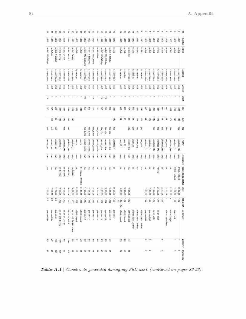
84 A. Appendix
Ski com
plex constructs
Author:
Felix Halbach
Date:
1/11/13
IDnum
bernam
especies
proteinstart
endsize
tagvector
resistancesequencing_status
datelab_book
comment
primer_f
primer_rev
0c001
scSki2
s. cerevisiaeski2
11287
146his
pfastbac_htaam
pS
132L, N884S
09.02.081.12
12
1c002
scSki2
s. cerevisiaeski2
11287
146pfastbac_1
amp
S132L, N
884S11.02.08
1.22insert lost
12
2c003
scSki3
s. cerevisiaeski3
11432
164his
pfastbac_htaam
pok
21.02.08-
cloned by jet
3c004
scSki8
s. cerevisiaeski8
1397
44his
pet_mcn
amp
ok21.02.08
-cloned by fabiana
4c005
scSki8
s. cerevisiaeski8
1397
44his
pfastbac_htaam
pok
22.02.081.36
56
5c006
scSki2
s. cerevisiaeski2
11287
146his
pfastbac_htaam
pN
884S27.02.08
1.41qc on c001
6c007
scSki2
s. cerevisiaeski2
11287
146his
pfastbac_htaam
pok
17.03.081.42
qc on c006
7c008
scSki3
s. cerevisiaeski3
11432
164pfastbac_1
amp
ok17.03.08
1.44pcr on c003
34
8c009
scSki8
s. cerevisiaeski8
1397
44pfastbac_1
amp
ok17.03.08
1.44pcr on c004
56
9c010
hsSki2
h. sapiensski2
11246
138gst
pet_mcn
amp
n.a.cloned by D
. Lindner
10c011
hsSki3
h. sapiensski3
11564
175tap
pcmv_tap
amp
n.a.cloned by D
. Lindner
11c012
hsSki8
h. sapiensski8
1305
34gst
pgex_csam
pok
cloned by D. Lindner
12c013
scSki7cbp
s. cerevisiaeski7
1747
85cbp
peckht_cbpkan
n.a.10.05.08
1.72gD
NA cloned
4748
13c014
spSki8
s. pombe
ski81
30233
puc_18am
pn.a.
10.05.081.82
cDN
A cloned55
56
14c015
hsSki8
h. sapiensski8
1305
34puc_18
amp
ok10.05.08
1.72, 1.84,1.101
cDN
A cloned53
54
15c016
scSki2
s. cerevisiaeski2
11287
146his
pfastbac_1am
pok
05.05.081.80
pcr on c7
16c017
scSki7 1-265cbp
s. cerevisiaeski7
1265
his_cbppeckht_cbp
kann.a.
13.05.081.102
pcr on c1347
87
17c018
scSki7 116-265cbp
s. cerevisiaeski7
116265
his_cbppeckht_cbp
kann.a.
13.05.081.102
pcr on c1385
87
18c019
scSki7 116-747cbp
s. cerevisiaeski7
116747
his_cbppeckht_cbp
kann.a.
13.05.081.102
pcr on c1385
48
19c020
scSki7sum
os. cerevisiae
ski71
747his_sum
opeckhi_sum
okan
ok14.05.08
1.116pcr on c13
8848
20c021
scSki7 116-C
sumo
s. cerevisiaeski7
116747
his_sumo
peckhi_sumo
kann.a.
14.05.081.116
pcr on c1389
48
21c022
scSki7 1-265sum
os. cerevisiae
ski71
265his_sum
opeckhi_sum
okan
n.a.14.05.08
1.116pcr on c13
8887
22c023
scSki7 116-265sum
os. cerevisiae
ski7116
265his_sum
opeckhi_sum
okan
n.a.14.05.08
1.116pcr on c13
8987
23c024
hsSki2
h. sapiensski2
11246
psc_bam
pQ
1133stop, R1133X
01.06.081.106
cDN
A cloned49
50
24c025
hsSki3
h. sapiensski3
11564
psc_bam
pok
01.06.081.106
cDN
A cloned51
52
25c026
scSki2 D
444Ns. cerevisiae
ski21
1287146
pfastbac_1am
pok (D
444N)
30.07.081.133
qc on c16, D444N
mutant
9899
26c027
scSki2 E
445Qs. cerevisiae
ski21
1287146
hispfastbac_hta
amp
ok (E445Q
)30.07.08
1.133qc on c7, E
445Q100
101
27c028
scSki2 D
444Ns. cerevisiae
ski21
1287146
hispfastbac_hta
amp
ok (D444N
)10.09.08
1.132qc on c7, D
444N98
99
28c029
scSki2 E
445Qs. cerevisiae
ski21
1287146
pfastbac_1am
pok (E
445Q)
10.09.081.132
qc on c16, E445Q
100101
29c030
scSki3 1-522gst
s. cerevisieaeski3
1522
88gst
peckht_gstkan
n.a.17.10.08
2.8pcr on c3
103104
30c031
scSki7gst
s. cerevisiaeski7
1747
113gst
peckht_gstkan
n.a.17.10.08
2.8pcr on c20a
4748
31c032
scSki7 116-747gst
s. cerevisiaeski7
116747
gstpeckht_gst
kann.a.
17.10.082.8
pcr on c20a85
48
Page 1/6
Table A.1 | Constructs generated during my PhD work (continued on pages 89-93).
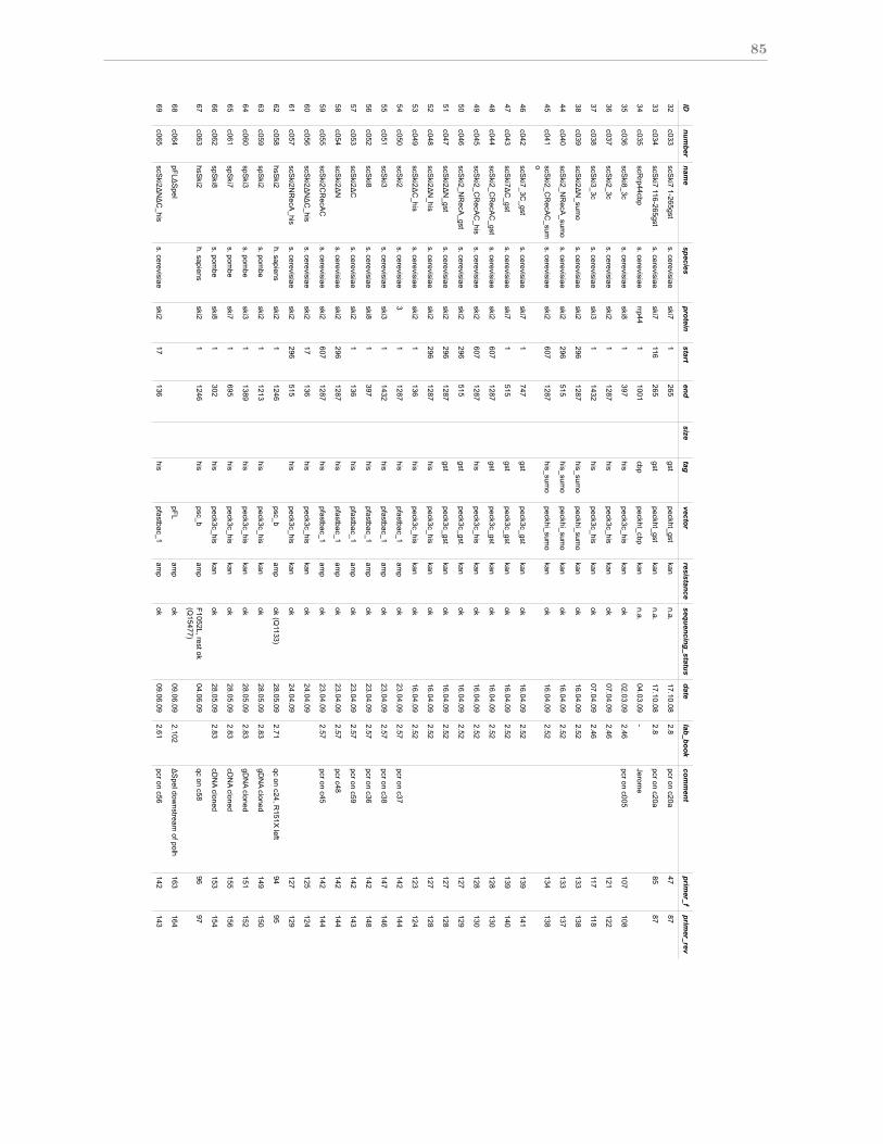
85
Title:
Author:
Felix Halbach
Date:
1/11/13
IDnum
bernam
especies
proteinstart
endsize
tagvector
resistancesequencing_status
datelab_book
comment
primer_f
primer_rev
32c033
scSki7 1-265gst
s. cerevisiaeski7
1265
gstpeckht_gst
kann.a.
17.10.082.8
pcr on c20a47
87
33c034
scSki7 116-265gst
s. cerevisiaeski7
116265
gstpeckht_gst
kann.a.
17.10.082.8
pcr on c20a85
87
34c035
scRrp44cbp
s. cerevisiaerrp44
11001
cbppeckht_cbp
kann.a.
04.03.09-
Jerome
35c036
scSki8_3c
s. cerevisiaeski8
1397
hispeck3c_his
kanok
02.03.092.46
pcr on c005107
108
36c037
scSki2_3c
s. cerevisiaeski2
11287
hispeck3c_his
kanok
07.04.092.46
121122
37c038
scSki3_3c
s. cerevisiaeski3
11432
hispeck3c_his
kanok
07.04.092.46
117118
38c039
scSki2!
N_sum
os. cerevisiae
ski2296
1287his_sum
opeckhi_sum
okan
ok16.04.09
2.52133
138
44c040
scSki2_N
RecA
_sumo
s. cerevisiaeski2
296515
his_sumo
peckhi_sumo
kanok
16.04.092.52
133137
45c041
scSki2_C
RecA
C_sum
os. cerevisiae
ski2607
1287his_sum
opeckhi_sum
okan
ok16.04.09
2.52134
138
46c042
scSki7_3C
_gsts. cerevisiae
ski71
747gst
peck3c_gstkan
ok16.04.09
2.52139
141
47c043
scSki7!
C_gst
s. cerevisiaeski7
1515
gstpeck3c_gst
kan ok
16.04.092.52
139140
48c044
scSki2_C
RecA
C_gst
s. cerevisiaeski2
6071287
gstpeck3c_gst
kanok
16.04.092.52
128130
49c045
scSki2_C
RecA
C_his
s. cerevisiaeski2
6071287
hispeck3c_his
kanok
16.04.092.52
128130
50c046
scSki2_N
RecA
_gsts. cerevisiae
ski2296
515gst
peck3c_gstkan
ok16.04.09
2.52127
129
51c047
scSki2!
N_gst
s. cerevisiaeski2
2961287
gstpeck3c_gst
kanok
16.04.092.52
127128
52c048
scSki2!
N_his
s. cerevisiaeski2
2961287
hispeck3c_his
kanok
16.04.092.52
127128
53c049
scSki2!
C_his
s. cerevisiaeski2
1136
his peck3c_his
kanok
16.04.092.52
123124
54c050
scSki2
s. cerevisiae3
11287
hispfastbac_1
amp
ok23.04.09
2.57pcr on c37
142144
55c051
scSki3
s. cerevisiaeski3
11432
hispfastbac_1
amp
ok23.04.09
2.57pcr on c38
147146
56c052
scSki8
s. cerevisiaeski8
1397
hispfastbac_1
amp
ok23.04.09
2.57pcr on c36
142148
57c053
scSki2!
Cs. cerevisiae
ski21
136his
pfastbac_1am
pok
23.04.092.57
pcr on c59142
143
58c054
scSki2!
Ns. cerevisiae
ski2296
1287his
pfastbac_1am
pok
23.04.092.57
pcr c48142
144
59c055
scSki2C
RecA
Cs. cerevisiae
ski2607
1287his
pfastbac_1am
pok
23.04.092.57
pcr on c45142
144
60c056
scSki2!
N!
C_his
s. cerevisiaeski2
17136
hispeck3c_his
kanok
24.04.09125
124
61c057
scSki2N
RecA
_hiss. cerevisiae
ski2296
515his
peck3c_hiskan
ok24.04.09
127129
62c058
hsSki2
h. sapiensski2
11246
psc_bam
pok (Q
1133)28.05.09
2.71qc on c24, R
151X left
9495
63c059
spSki2
s. pombe
ski21
1213his
peck3c_hiskan
ok28.05.09
2.83gD
NA cloned
149150
64c060
spSki3
s. pombe
ski31
1389his
peck3c_hiskan
ok28.05.09
2.83gD
NA cloned
151152
65c061
spSki7
s. pombe
ski71
695his
peck3c_hiskan
ok28.05.09
2.83cD
NA cloned
155156
66c062
spSki8
s. pombe
ski81
302his
peck3c_hiskan
ok28.05.09
2.83cD
NA cloned
153154
67c063
hsSki2
h. sapiensski2
11246
hispsc_b
amp
F1052L, rest ok(Q
15477) 04.06.09
qc on c58 96
97
68c064
pFL!S
peIpFL
amp
ok09.06.09
2.102!
SpeI dow
nstream of polh
163164
69c065
scSki2!
N!
C_his
s. cerevisiaeski2
17136
hispfastbac_1
amp
ok09.06.09
2.61pcr on c56
142143
Page 2/6

86 A. Appendix
Title:
Author:
Felix Halbach
Date:
1/11/13
IDnum
bernam
especies
proteinstart
endsize
tagvector
resistancesequencing_status
datelab_book
comment
primer_f
primer_rev
70c066
scSki2nR
ecA_his
s. cerevisiaeski2
296515
hispfastbac_1
amp
ok09.06.09
2.61pcr on c57
142145
71c067
spSki2_his
s. pombe
ski21
1213his
pfastbac_1am
pok
17.06.092.92
pcr on c59188
172
72c068
spSki2
s. pombe
ski21
1213pfastbac_1
amp
ok17.06.09
2.92pcr on c59
171172
73c069
spSki7_his
s. pombe
ski71
695his
pfastbac_1am
pok
17.06.092.92
pcr on c61188
174
74c070
spSki7
s. pombe
ski71
695pfastbac_1
amp
ok17.06.09
2.92pcr on c61
173174
75c071
spSki8_his
s. pombe
ski81
302his
pfastbac_1am
pok
17.06.092.92
pcr on c062188
176
76c072
spSki8
s. pombe
ski81
302pfastbac_1
amp
ok17.06.09
2.92pcr on c062
175176
77c073
LLC_sum
olam
bdallc
1221
his_sumo
peckhi_sumo
kan19.06.09
2.141clones from
cba189
190
78c074
spSki3
s. pombe
ski31
1389pfastbac_1
amp
ok01.07.09
2.94pcr on c60
169170
79c075
scSki8
s. cerevisiaeski8
1397
pFLam
p03.07.09
2.112pcr on 36;; S
ki8 in MC
S1
196195
80c076
spSki3_his
s. pombe
ski31
1389his
pfastbac_1am
pok
29.07.092.96
pcr on c60202
169
81c077
scSki3_m
b_hiss. cerevisiae
ski31
1432his
pFLdspeam
p14.08.09
2.116ski3 in M
CS
2; MC
S1 em
pty;pcr on c038
205206
82c078
scSki3his:scS
ki8:pFLs. cerevisiae
ski3, ski8his
pFLdspeam
pok
27.08.092.118
ski3 in MC
S2; ski8 in M
CC
S1
196195
83c079
scSki2:pFL
s. cerevisiaeski2
11287
pFLdspeam
pok
27.08.092.123
ski2 in MC
S2; M
CS
1 empty
197198
84c080
scSki238:pFL
s. cerevisiaeski2, ski3,ski8
hispFLdspe
amp
13.10.092.132
c78 ligated into c79
85c081
scSki2!
C:pFL
s. cerevisiaeski2
1136
pFLdspeam
p20.10.09
2.145ski2 1-136 in M
CS
1214
214
86c082
scSki2!
N!
stalk_hiss. cerevisiae
ski2296
128784
hispeck3c_his
kanA
1255D, rest O
K04.03.10
2.164!
N!
stalk: 296-834-GS
RG
-1086-1287
227228
88c083
scSki2!
N!
stalk_gsts. cerevisiae
ski2296
128784
gstpeck3c_gst
kan04.03.10
2.164!
N!
stalk: 296-834:G
SR
G:1086-1287
227228
89c084
scSki2!
stalk_hiss. cerevisiae
ski21
1287117
hispeck3c_his
kanV
1147L, 5'fragment
not seq04.03.10
2.164!
stalk: 1-834:GS
RG
:1086-1287
227228
90c085
scSki2!
stalk_gsts. cerevisiae
ski21
1287117
gstpeck3c_gst
kan04.03.10
2.164!
stalk: 1-834:GS
RG
:1086-1287
227228
91c086
scSki2_S
4H"_his
s. cerevisiaeski2
8351085
29his
peck3c_hiskan
ok05.03.10
cloned by p&T
231236
92c087
scSki2_S
4H"_gst
s. cerevisiaeski2
8351085
gstpeck3c_gst
kanok
05.03.10cloned by P
&T
231236
93c088
scSki2_S
2H"_his
s. cerevisiaeski2
8671048
hispeck3c_his
kanok
05.03.10cloned by P
&T
232235
94c089
scSki2_S
2H"_gst
s. cerevisiaeski2
8671048
gstpeck3c_gst
kanok
05.03.10cloned by P
&T
232235
95c090
scSki2_S
"_hiss. cerevisiae
ski2893
1018his
peck3c_hiskan
ok05.03.10
cloned by P&
R233
234
96c091
scSki2_S
"_gsts. cerevisiae
ski2893
1018gst
peck3c_gstkan
ok05.03.10
cloned by P&
T233
234
97c092
spSki2!
s_fls. pom
beski2
11213
108his
peck3c_hiskan
ok09.04.10
2.170!
stalk, V758G
SR
GL1014
241242
98c093
scSki2_!
N!
ss. cerevisiae
ski2296
128784
hispeck3c_his
kanA
1255 backmut.,
rest?07.04.10
2.172qc on c82, !
stalk243
244
99c094
scSki2_!
s_fls. cerevisiae
ski21
1287117
hispeck3c_his
kanV
1147 backmut,
rest?07.04.10
2.172qc on c84, !
stalk245
246
Page 3/6
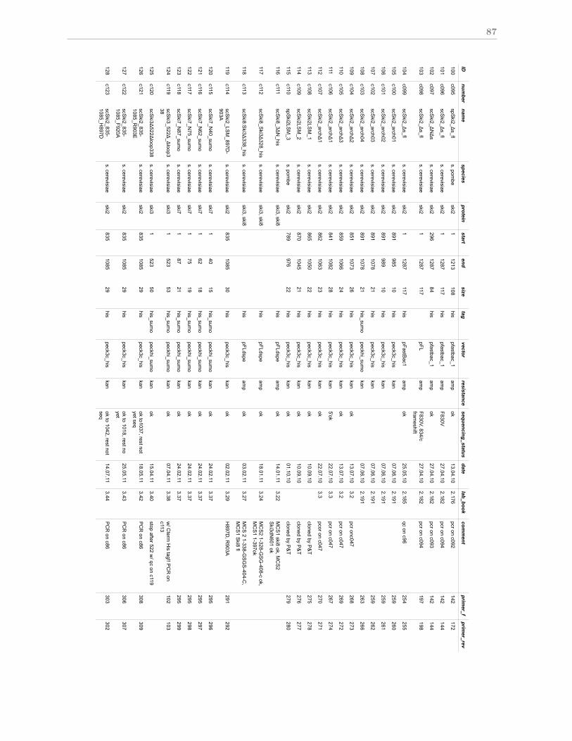
87
Title:
Author:
Felix Halbach
Date:
1/11/13
IDnum
bernam
especies
proteinstart
endsize
tagvector
resistancesequencing_status
datelab_book
comment
primer_f
primer_rev
100c095
spSki2_!
s_fls. pom
beski2
11213
108his
pfastbac_1am
pok
13.04.102.176
pcr on c092142
172
101c096
scSki2_!
s_fls. cerevisiae
ski21
1287117
hispfastbac_1
amp
F830V27.04.10
2.182pcr on c094
142144
102c097
scSki2_!
N!
ss. cerevisiae
ski2296
128784
hispfastbac_1
amp
ok27.04.10
2.182pcr on c093
142144
103c098
scSki2_!
s_fls. cerevisiae
ski21
1287117
pFLam
pF830V, 834/cfram
eshift27.04.10
2.182pcr on c094
197198
104c099
scSki2_!
s_fls. cerevisiae
ski21
1287117
hispFastB
ac1am
pok
25.05.102.185
qc on c96254
255
105c100
scSki2_arch01
s. cerevisiaeski2
891985
10his
peck3c_hiskan
07.06.102.191
259260
106c101
scSki2_arch02
s. cerevisiaeski2
891989
10his
peck3c_hiskan
07.06.102.191
259261
107c102
scSki2_arch03
s. cerevisiaeski2
8911078
21his
peck3c_hiskan
07.06.102.191
259262
108c103
scSki2_arch04
s. cerevisiaeski2
8911078
21his_sum
opeckhi_sum
okan
07.06.102.191
263266
109c104
scSki2_arch!
2s. cerevisiae
ski2851
107326
hispeck3c_his
kanok
13.07.103.2
pcr onc047268
273
110c105
scSki2_arch!
3s. cerevisiae
ski2859
106624
hispeck3c_his
kanok
13.07.103.2
pcr on c047269
272
111c106
scSki2_arch!
1s. cerevisiae
ski2841
108228
hispeck3c_his
kan5'ok
22.07.103.3
pcr on c047267
274
112c107
scSki2_arch!
1s. cerevisiae
ski2862
106323
hispeck3c_his
kanok
22.07.103.3
pcor on c047270
271
113c108
scSki2LS
M_1
s. cerevisiaeski2
8651050
22his
peck3c_hiskan
ok10.09.10
cloned by P&
T275
278
114c109
scSki2LS
M_2
s. cerevisiaeski2
8701045
21his
peck3c_hiskan
ok10.09.10
cloned by P&
T276
277
115c110
spSki2LS
M_3
s. pombe
ski2789
97622
hispeck3c_his
kanok
01.10.10cloned by P
&T
279280
116c111
scSki8_3!
N_his
s. cerevisiaeski3, ski8
hispFLdspe
amp
ok14.01.11
3.22M
CS
1 ski8 ok, MC
S2
Ski3dN
601 ok
117c112
scSki8_S
ki3!328_his
s. cerevisiaeski3, ski8
hispFLdspe
amp
ok18.01.11
3.24M
CS
2 1-328-GS
G-408-c ok,
MC
S1 1-397ok
118c113
scSki8:S
ki3!338_his
s. cerevisiaeski3, ski8
hispFLdspe
amp
ok03.02.11
3.27M
CS
2 1-338-GS
GS
-404-C,
MC
S1 S
ki8 fl
119c114
scSki2_LS
M_897D
-903A
s. cerevisiaeski2
8351085
30his
peck3c_hiskan
ok02.02.11
3.29H
897D, R
903A291
292
120c115
scSki7_N
40_sumo
s. cerevisiaeski7
140
15his_sum
opeckhi_sum
okan
ok24.02.11
3.37295
296
121c116
scSki7_N
62_sumo
s. cerevisiaeski7
162
18his_sum
opeckhi_sum
okan
ok24.02.11
3.37295
297
122c117
scSki7_N
75_sumo
s. cerevisiaeski7
175
19his_sum
opeckhi_sum
okan
ok24.02.11
3.37295
298
123c118
scSki7_N
87_sumo
s. cerevisiaeski7
187
21his_sum
opeckhi_sum
okan
ok24.02.11
3.37295
299
124c119
scSki3_522!
_!loop3
38s. cerevisiae
ski31
52353
his_sumo
peckhi_sumo
kanok
07.04.113.38
w/ C
term H
is tag!! PC
R on
c113102
103
125c120
scSki3!
522!loop338
s. cerevisiaeski3
1523
50his_sum
opeckhi_sum
okan
ok15.04.11
3.40stop after 522 w
/ qc on c119
126c121
scSki2_835-
1085_R903E
s. cerevisiaeski2
8351085
29his
peck3c_hiskan
ok to1037, rest notyet seq
18.05.113.42
PC
R on c86
308309
127c122
scSki2_835-
1085_F920As. cerevisiae
ski2835
108529
hispeck3c_his
kanok to 1018, rest noyet
25.05.113.43
PC
R on c86
306307
128c123
scSki2_835-
1085_H897D
s. cerevisiaeski2
8351085
29his
peck3c_hiskan
ok to 1042, rest notseq
14.07.113.44
PC
R on c86
303302
Page 4/6
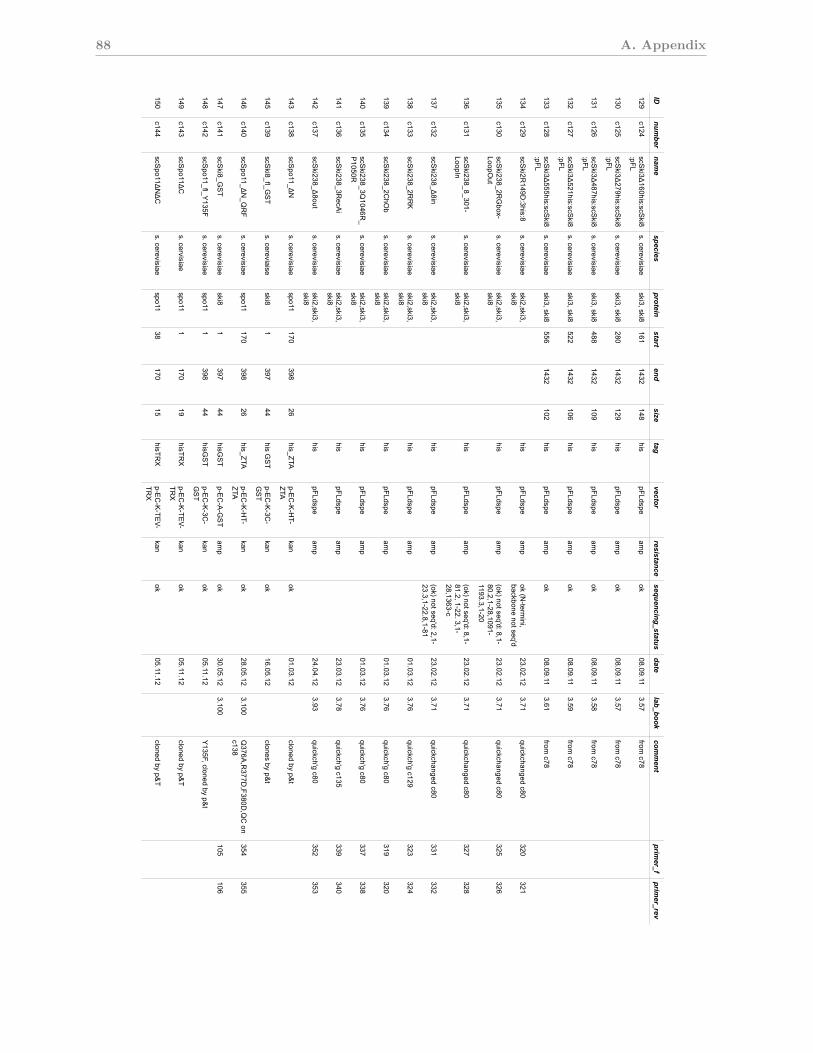
88 A. Appendix
Title:
Author:
Felix Halbach
Date:
1/11/13
IDnum
bernam
especies
proteinstart
endsize
tagvector
resistancesequencing_status
datelab_book
comment
primer_f
primer_rev
129c124
scSki3!
160his:scSki8
:pFLs. cerevisiae
ski3, ski8161
1432148
hispFLdspe
amp
ok08.09.11
3.57from
c78
130c125
scSki3!
279his:scSki8
:pFLs. cerevisiae
ski3, ski8280
1432129
hispFLdspe
amp
ok08.09.11
3.57from
c78
131c126
scSki3!
487his:scSki8
:pFLs. cerevisiae
ski3, ski8488
1432109
hispFLdspe
amp
ok08.09.11
3.58from
c78
132c127
scSki3!
521his:scSki8
:pFLs. cerevisiae
ski3, ski8522
1432106
hispFLdspe
amp
ok08.09.11
3.59from
c78
133c128
scSki3!
555his:scSki8
:pFLs. cerevisiae
ski3, ski8556
1432102
hispFLdspe
amp
ok08.09.11
3.61from
c78
134c129
scSki2R
149D:3his:8
s. cerevisiaeski2,ski3,ski8
hispFLdspe
amp
ok (N-term
ini,backbone not seq'd
23.02.123.71
quickchanged c80320
321
135c130
scSki238_2R
Gbox-
LoopOut
s. cerevisiaeski2,ski3,ski8
hispFLdspe
amp
(ok) not seq'd: 8,1-80.2,1-28,1091-1193.3,1-20
23.02.123.71
quickchanged c80325
326
136c131
scSki238_8_301-
LoopIns. cerevisiae
ski2,ski3,ski8
his pFLdspe
amp
(ok) not seq'd: 8,1-81.2, 1-22. 3,1-28,1363-c
23.02.123.71
quickchanged c80327
328
137c132
scSki238_!
8ins. cerevisiae
ski2,ski3,ski8
hispFLdspe
amp
(ok) not seq'd: 2,1-23.3,1-22.8,1-81
23.02.123.71
quickchanged c80331
332
138c133
scSki238_2R
RK
s. cerevisiaeski2,ski3,ski8
hispFLdspe
amp
01.03.123.76
quickch'g c129323
324
139c134
scSki238_2C
hOb
s. cerevisiaeski2,ski3,ski8
hispFLdspe
amp
01.03.123.76
quickch'g c80319
320
140c135
scSki238_3Q
1046R_
P1050R
s. cerevisiaeski2,ski3,ski8
hispFLdspe
amp
01.03.123.76
quickch'g c80337
338
141c136
scSki238_3R
ecAi
s. cerevisiaeski2,ski3,ski8
hispFLdspe
amp
23.03.123.78
quickch'g c135339
340
142c137
scSki238_!
8outs. cerevisiae
ski2,ski3,ski8
hispFLdspe
amp
24.04.123.93
quickch'g c80352
353
143c138
scSpo11_!
Ns. cerevisiae
spo11170
39826
his_ZTAp-E
C-K
-HT-
ZTAkan
ok01.03.12
cloned by p&t
145c139
scSki8_fl_G
ST
s. cereviaiseski8
1397
44his G
ST
p-EC
-K-3C
-G
ST
kanok
16.05.12clones by p&
t
146c140
scSpo11_!
N_Q
RF
s. cerevisiaespo11
170398
26his_ZTA
p-EC
-K-H
T-ZTA
kanok
28.05.123.100
Q376A
,R377D
,F380D,Q
C on
c138354
355
147c141
scSki8_G
ST
s. cerevisiaeski8
1397
44hisG
ST
p-EC
-A-G
ST
amp
ok 30.05.12
3.100105
106
148c142
scSpo11_fl_Y
135Fs. cerevisiae
spo111
39844
hisGS
Tp-E
C-K
-3C-
GS
Tkan
ok05.11.12
Y135F, cloned by p&
t
149c143
scSpo11!
Cs. cervisiae
spo11 1
17019
hisTRX
p-EC
-K-TE
V-TR
Xkan
ok05.11.12
cloned by p&T
150c144
scSpo11!
N!
Cs. cerevisiae
spo1138
17015
hisTRX
p-EC
-K-TE
V-TR
Xkan
ok05.11.12
cloned by p&T
Page 5/6
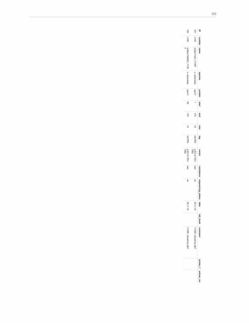
89
Title:
Author:
Felix Halbach
Date:
1/11/13
IDnum
bernam
especies
proteinstart
endsize
tagvector
resistancesequencing_status
datelab_book
comment
primer_f
primer_rev
151c145
scSpo11!
C_Y
135Fs. cerevisiae
spo111
17019
hisTRX
p-EC
-K-TE
V-TR
Xkan
ok05.11.12
Y135F, cloned by p&
T
152c146
scSpo11!
N!
C_Y
135F
s. cerevisiaespo11
38170
15hisTR
Xp-E
C-K
-TEV-
TRX
kanok
05.11.12Y
135F, cloned by p&T
Page 6/6
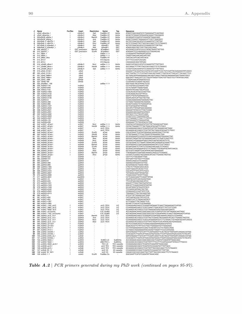
90 A. AppendixSki complex oligos cloning sequencings.cerevisiae, s. pombe, h. sapiens
# Name For/Rev Insert Restriction Vector Tag Sequence storage comment1 ySki2_pfbachta_f f ySki2p fl SalI pFastBAc HTA 6xHis ATAGTCGACAGATGTCTGAGGGATTCAGTAGC Box I, 1 also f pfastBac12 ySki2_pfbachta_r r ySki2p fl XbaI pFastBAc HTA 6xHis GCGCTCTAGACTATAAATACAAACTTGCGGCG Box I, 2 also f pfastBac13 003ySki3fl_pfbhta_f f ySki3p fl BamHI pFastBAc HTA 6xHis GCAGGATCCGATGTCGGATATTAAACAGC Box I, 3 also f pfastBac14 004ySki3fl_pfbhta_r r ySki3p fl xhoI pFastBAc HTA 6xHis GAGCTCGAGTTAGAAACATTCGTTTAGCGCC Box I also f pfastBac15 005ySki8_fl_pfbHTA_f f ySki8p fl SalI pFastBAc HTA 6xHis GCTGTCGACGCATGTCCAAAGTGTTTATTGC Box I also f pfastBac16 006ySki8_fl_pfbHTA_r r ySki8p fl XhoI pFastBAc HTA 6xHis GCTCTCGAGTTATTTACCGCCAGCTTCTCTAAACC Box I also f pfastBac17 007ySki8_fl_pGex6p1_f f ySki8p fl SalI pGex6P1 GST ACTGTCGACGCATGTCCAAAGTGTTTATTGC Box I8 008ySki8_fl_pGex6p1_r r ySki8p fl NotI pGex6P1 GST ATAGCGGCCGCTTATTTACCGCCAGC Box I9 009_pfbGST_f f GST, precission BamHI pFastBAc1 GST ATCGGATCCATGTCCCCTATACTAGGTTATTGG Box I creates GST pfast Bac10 010_pfbGST_r r GST, precission EcoRI pFastBAc1 GST ATAGAATTCCCAGGGGCCCCTGGAACAG Box I creates GST pfast Bac11 011_pfBac_f f - - pFastBac 1/ HTA - CCGGAATATTAATAGATCATGG sequencing pfBac1/HTA12 012_pfBac_r r - - pFastBac 1/ HTA - CAAGTTAACAACAACAATTGC sequencing pfBac1/HTA13 013_pfBac_6his f - - pFastBAc HTA - CATCACCATCACCATCACG pfbacHTA only14 014_M13-f f - - DH10 Bacmids - GTTTTCCCAGTCACGAC m13 for D10(multi)Bac derived bacmids15 015_M13-r r - - DH10 Bacmids - CAGGAAACAGCTATGAC m13 for D10(multi)Bac derived bacmids16 016_ySki8fl_pfbac-f f ySki8p fl NcoI pFastBac 1/ HTA 6xHis GAGCCATGGCCATGTCCAAAGTGTTTATTGCC17 017_ySki8fl_pfbac-r r ySki8p fl HindIII pFastBac 1/ HTA 6xHis GCCAAGCTTTTATTTACCGCCAGCTTCTCTAAACC18 018_ySki8fl_pfbac-f f ySki8p fl SalI pFastBac 1/ HTA 6xHis CCGGTCGACGAATGTCCAAAGTGTTTATTGCC19 019_ySki2_S132-f f ySki2 - - - GGAAGTTGCAAATGCCAATGCATCAAATTCACTGTCGATTACGAGAAGTATCAACC mutagenesis for wildtype S132. For c001,00220 020_ySki2_S132-r r ySki2 - - - GGTTGATACTTCTCGTAATCGACAGTGAATTTGATGCATTGGCATTTGCAACTTCC mutagenesis for wildtype S132. For c001,00221 021_ySki2_N884-f f ySki2 - - GCTGGCATATAAGGAGGCAACAGTCAACCTAATGCAAGAAATGGTTAAATCGCC22 022_ySki2_N884-r r ySki2 - - - GGCGATTTAACCATTTCTTGCATTAGGTTGACTGTTGCCTCCTTATATGCCAGC23 023_ySki3_+889 f ySki3p - - - CTGGACAACATGGATGCCCC sequencing24 024_ySki8+180 f ySki3p - - - CCATAAGTCCGGATTGCACC sequencing25 025_pfastBac_-190 f - - pFastBac 1/ HTA - GGTTGGCTACGTATACTCCG sequencing, 190bp 5' to 6his26 026_hsSKI2+145 f hsSKI2 - - - CCTTGTGCCCCAGATCTGC27 027_hsSKI2+649 f hsSKI2 - - - CCTCTGGATTTGGGTGGG28 028_hSKI2+1155 f hsSKI2 - - - GGATGTACAGCTGCATCCG29 029_hsSKI2+1683 f hsSKI2 - - - CCGCACACGTGCCCAGTTGC30 030_hsSKI2+2132 f hsSKI2 - - - CCACAGGCACCGTTATCC31 031_hsSKI2+2622 f hsSKI2 - - - GGTCTTGTGTGATAAGCCC32 032_hsSKI2+3135 f hsSKI2 - - - GGATCAGTCATTGCTGCTGC33 033_hsSKI3+5 f hsSKI3 - - - CCAGCAAGGAAGTGAAGACTGC34 034_hsSKI3+498 f hsSKI3 - - - CCTGGCTGAAAGTACAGAGG35 035_hsSKI3+986 f hsSKI3 - - - GCAGTCAAGCTCTGAAGATCG36 036_hsSKI3+1537 f hsSKI3 - - - GCTCGTGGATGTTATAGG37 037_hsSKI3+2024 f hsSKI3 - - - GCCATCTTATGATGGCAAAAGC38 038_hsSKI3+2521 f hsSKI3 - - - GGAAATTATGCCCTTGCTCAGC39 039_hsSKI3+2991 f hsSKI3 - - - GGAAGCAGCAAATGCATACC40 040_hsSKI3+3482 f hsSKI3 - - - GGTGCCTTCTTACATCAGC41 041_hsSKI3+3963 f hsSKI3 - - - CCAGTCCCTTGAAAAGTGG42 042_hsSKI3+4421 f hsSKI3 - - - CCTTGAAGCTTTGCTTTTGTCC43 043_hsSKI8+129 f hsSKI8 - - - GGTGAAGGTCTGGAAATGG44 044_hsSKI8+538 f hsSKI8 - - - GGAAAACTTCTGCATACCCTGG45 045_scSKI7_pFast-f f scSKI7 NcoI pFastBac 1/ HTA 6xHis CGATACCATGGGGATGTCGTTATTAGAGCAATTAGC46 046_scSKI7_pFast-r r scSKI7 HindIII pFastBac 1/ HTA 6xHis CGTACTGAAGCTTTTACTGGCATGCAATTCTGCC47 047_scSKI7_pLIC-f f scSKI7 - pLIC (TEV) CCAGGGAGCAGCCTCGATGTCGTTATTAGAGCAATTAGC48 048_scSKI7_pLIC-r r scSKI7 - pLIC (TEV) GCAAAGCACCGGCCTCGTTATTACTGGCATGCAATTCTGCC49 049_hsSKI2_pFast-f f hsSKI2 EcoRI pFast 6xHis CGCATGAATTCATGATGGAGACAGAGCGACTTGTGC50 050_hsSKI2_pFast-r r hsSKI2 HindIII pFast 6xHis CCTATGAAGCTTTCACTGGGTGTAGAGGCTGG51 051_hsSKI3_pFast-f f hsSKI3 BamHI pFast 6xHis GCATAGGGATCCAATGTCCAGCAAGGAAGTGAAGACTGC52 052_hsSKI3_pFast-r r hsSKI3 XbaI pFast 6xHis GGTATCTAGATTATTGTGAGGACAATCTCTGATTCAGTTCC53 053_hsSKI8_pFast-f f hsSKI8 NcoI pFast 6xHis GCATACCATGGGGATGACCAACCAGTACGGTATTCTC54 054_hsSKI8_pFast-r r hsSKI8 EcoRI pFast 6xHis CGTGCGAATTCTTAAATTGGACAATCATAGATGTGAATTTCC55 055_spSKI8_pFast-f f spSKI8 BamHI pFast 6xHis GCATAGGATCCGATGAGGAAAGAGTATCTCGTTAGCC56 056_spSKI8_pFast-r r spSKI8 EcoRI pFast 6xHis GCGATGAATTCTTATTCTGTAGCAGCAGCTCTATACC57 057_spSKI2_pFast-f f spSKI2 NcoI pFast 6xHis GCATACCATGGGGATGTCTTCTAAACTTGTAGATGCAATCAACG58 058_spSKI2_pFast-r r spSKI2 XbaI pFast 6xHis GCTCGTCTAGATTCACATATACAGTGAAGGGC59 059_spSKI3_pFast-f f spSKI3 BamHI pFast 6xHis TTATAGGATCCGATGGCAAAACCAGCCCTCAAAGC60 060_spSKI3_pFast-r r spSKI3 XhoI pFast 6xHis CGTATCTCGAGCTACGAAGCATCACTTGAAACTAGTGC61 061_spSKI8+222 spSKI8 - - - CGTTTCTTGCGGATTTGG62 062_spSKI8+721 spSKI8 - - - GGTGATTTGTTGCTTTCGGC63 063_spSKI3+6 spSKI3 - - - AAAACCAGCCCTCAAAGC64 064_spSKI3+496 spSKI3 - - - GAAAGCATGGATCGTTGC65 065_spSKI3+980 spSKI3 - - - ACAATAGCGCTGAACTAGC66 066_spSKI3+1465 spSKI3 - - - CTACTCTTAGACGACCACG67 067_spSKI3+1947 spSKI3 - - - CAATTGGCATCACACTTCC68 068_spSKI3+2424 spSKI3 - - - CGCATGTTTATCATTCTGCC69 069_spSKI3+2897 spSKI3 - - - TGTGGGCAAATTATGGTGC70 070_spSKI3+3389 spSKI3 - - - TCGGCAGGTTATATTTGGC71 071_spSKI3+3831 pspSKI3 - - - CCTCAAACAAGTTCAGATTATGG72 072_spSKI2+18 spSKI2 - - - AGATGCAATCAACGAAGTAGC73 073_spSKI2+525 spSKI2 - - - TTCCATGACAGCGACATCC74 074_spSKI2+1051 spSKI2 - - - GGAATTTTAACTGGTGATGTCC75 075_spSKI2+1543 spSKI2 - - - GGCGTTCAAACGAATATGATGC76 076_spSKI2+1948 spSKI2 - - - GCAATGGGTGTAAATATGCC77 077_spSKI2+2432 spSKI2 - - - TCCGAGCAATAACTACTGC78 078_spSKI2+2924 spSKI2 - - - CCGGAAATCCAATTATCTCG79 079_spSKI2+3413 spSKI2 - - - TGGAGGTTTGCTACGAGTGG80 080_scSKI7+3 scSKI7 - - - GTCGTTATTAGAGCAATTAGC81 081_scSKI7+494 scSKI7 - - - CATCAATACCGCTATCGTCG82 082_scSKI7+952 scSKI7 - - - AAGGTCATTCTAGACAATACC83 083_scSKI7+1397 scSKI7 - - - GTTCGGGTTTATTAGGTTCG84 084_scSKI7+1860 scSKI7 GCAATTTCACATTCGTAAGGG85 085_scSKI7_116C_pLIC f scSKI7 - pLIC (TEV) LIC CCAGGGAGCAGCCTCGGATGATAAACTCAACTTAGAAGAGTCATGG86 086_scSKI7_266C_pLIC f scSKI7 - pLIC (TEV) LIC CCAGGGAGCAGCCTCGCTGAATTTGACATGTTTGTTCCTCGG87 087_scSKI7_N265_pLIC r scSKI7 - pLIC (TEV) LIC GCAAAGCACCGGCCTCGTTAAGGATGGGTGGCAATGAATG88 088_scSKI7_pLICsumo f scSKI7 - pLIC (SuMO) LIC ACCAGGAACAAACCGGCGGCCGCTCGATGTCGTTATTAGAGCAATTAGC89 089_scSKI7_116C_pLICsumo f scSKI7 - pLIC (SuMO) LIC ACCAGGAACAAACCGGCGGCCGCTCGGATAAACTCAACTTAGAAGAGTCATGG90 090_spSKI3_pLIC_cry-f f spSKI3 BamHI pLIC (TEV) - CCAGGGAGCAGCCTCGGGATCCGATGGCAAAACCAGCCCTCAAAGC91 091_spSKI3_pLIC_cry-r r spSKI3 xhoI pLIC (TEV) - GCAAAGCACCGGCCTCGTTACTCGAGCTACGAAGCATCACTTGAAACTAGTGC92 092_spSKI2_pLIC_cry-f f spSKI2 NcoI pLIC (TEV) - CCAGGGAGCAGCCTCGCCATGGGGATGTCTTCTAAACTTGTAGATGCAATCAACG93 093_spSKI2_pLIC_cry-r r spSKI2 XbaI pLIC (TEV) - GCAAAGCACCGGCCTCGTTATCTAGATTCACATATACAGTGAAGGGC94 094_hsSKI2_Q1133-f f hsSKI2 - - - GCTCCCAAACACCCTCAAGCAGGGAATAGAACGTGTCC95 095_hsSKI2_Q1133-r r hsSKI2 - - - GGACACGTTCTATTCCCTGCTTGAGGGTGTTTGGGAGC96 096_hsSKI2_R151-f f hsSKI2 - - - CCTTATGGGGAAATCCAACTCGGTATCCCTTCTGGCCAGG97 097_hsSKI2_R151-r r hsSKI2 - - - CCTGGCCAGAAGGGATACCGAGTTGGATTTCCCCATAAGG98 098_scSKI2_D444N_f f scSKi2 - - - GATGTAGAGTTTGTCATTTTCAATGAAGTTCACTACGTTAATGATCAAGACCGTGG99 099_scSKI2_D444N_r r scSKI2 - - - CCACGGTCTTGATCATTAACGTAGTGAACTTCATTGAAAATGACAAACTCTACATC100 100_scSKI2_E445_Q_f f scSkI2 - - - GATGTAGAGTTTGTCATTTTCGATCAAGTTCACTACGTTAATGATCAAGACCGTGG101 101_scSKI2_E445Q_r r scSKi2 - - - CCACGGTCTTGATCATTAACGTAGTGAACTTGATCGAAAATGACAAACTCTACATC102 102_scSKI3_N522_f f scSKI3 - SUMO LIC SuMOHis ACCAGGAACAAACCGGCGGCCGCTCGATGTCGGATATTAAACAGC103 103_scSKI3_N522_r r scSKI3 - SUMO/TEV LIC SuMOHis GCAAAGCACCGGCCTCGTTATTTTTCCTTTCAATAAAAATTATACCTTTGCCC104 104_scSKI3_N522_pLIC-f f scSKI3 - TEV LIC TEV cassette CCAGGGAGCAGCCTCGATGTCGGATATTAAACAGC105 105_scSKI8_pLIC-f f scSki8 - pLIC TEV TEV cassette CCAGGGAGCAGCCTCGATGTCCAAAGTGTTTATTGCC106 106_scSKI8_pLIC-r r scSki8 - pLIC TEV TEV cassette GCAAAGCACCGGCCTCGTTATTTACCGCCAGCTTCTCTAAACC107 107_scSki8_3C_1f f scSki8 - pLIC 3C 3C cassette CCAGGGGCCCGACTCGATGTCCAAAGTGTTTATTGC108 108_scski8_3C_397r r scSki8 - pLIC 3C 3C cassette CAGACCGCCACCGACTGCTTATTTACCGCCAGCTTCTCTAAACC109 109_scSki3_1_f f scski3 EcoRI pFastBac Dual - GGCGAATTCATGTCGGATATTAAACAGC
Table A.2 | PCR primers generated during my PhD work (continued on pages 95-97).

91Ski complex oligos cloning sequencing
s.cerevisiae, s. pombe, h. sapiens
110 110_scSki3_1432_r r scSKI3 NotI pFastBac Dual - GATGCGGCCGCTTAGAAACATTCGTTTAGC111 111_scSki8_1f f scSki8 - LIC 3C 3C cassette CCAGGGGCCCGACTCGATGTCCAAAGTGTTTATTGCC112 112_scSki8_397r r scSki8 - LIC 3C 3C cassette CAGACCGCCACCGACTGCTTATTTACCGCCAGCTTCTCTAAACC113 113_scSki8_NHis3Cf f scSki8 Xho pFastBac Dual - CAGCTCGAGATGAAACATCACCATCACC114 114_scSki8_397r r scSki8 KpnI pFastBac Dual - CGAGGTACCTTATTTACCGCCAGC115 115_PH_seq_f f polyhedrin prom . . AAAATGATAACCATCTCGC116 116_p10_seq_f f p10 prom . . . CGGACCTTTAATTCAACCC117 117_scSKi3_3CLIC_1f f scSKI3 - 3c lic 3C cassette CCAGGGGCCCGACTCGATGTCGGATATTAAACAGCTATTGAAGG118 118_scSki3_3CLIC_1432_r r scSKI3 - 3c lic 3C cassette CAGACCGCCACCGACTGCTTAGAAACATTCGTTTAGCGCCTTCAC119 119_3CsiteFastBac_f f - - - - GATCAATGAAACATCACCATCACCATCACCATCACCATCACTCCGCGGGTCTG
GAAGTTCTGTTCCAGGGGCCCGACGTCG120 120_3CsiteFastBac_r r - - - - GATCCGACGTCGGGCCCCTGGAACAGAACTTCCAGACCCGCGGAGTGATGG
TGATGGTGATGGTGATGGTGATGTTTCATT121 121_scSki2_3CLIC_1f f scSKi2 - 3C LIC 3C cassette CCAGGGGCCCGACTCGATGTCTGAGGGATTCAGTAGC122 122_scSki2_3CLIC_1286_r r scSKi2 - 3C LIC 3C cassette CAGACCGCCACCGACTGCTTATAAATACAAACTTGCGGCG123 123_Ski2_3C_2FW f scSKi2 - 3C LIC 3C cassette CCAGGGGCCCGACTCGATGTCTGAGGGATTCAGTAGCAGTTC124 124_Ski2_3C_136RV r scSKi2 - 3C LIC 3C cassette CAGACCGCCACCGACTGCTTACGACAGTGAATTTGATGCATTGGC125 125_Ski2_3C_17FW f scSKi2 - 3c lic 3C cassette CCAGGGGCCCGACTCGATGTTAAAAGAAATCACTAACAACGCAGACG126 126_Ski2_3C_122RV r scSKi2 - 3C LIC 3C cassette CAGACCGCCACCGACTGCTTAATCAACCTCTTCTTTGTAGCCTG127 127_Ski2_3C_296FW f scSKi2 - 3c lic 3C cassette CCAGGGGCCCGACTCGATGGTTCCTGTTAAAAAGGAATGGGCC128 128_Ski2_3C_1286RV r scSKi2 - 3c lic 3C cassette CAGACCGCCACCGACTGCTTATAAATACAAACTTGCGGCGAAAACAATATC129 129_Ski2_3C_515RV r scSKi2 - 3c lic 3C cassette CAGACCGCCACCGACTGCTTATTTAGCCCATATATTTATTTCCAATGGAAC130 130_Ski2_3C_607FW f scSKi2 - 3c lic 3C cassette CCAGGGGCCCGACTCGATGGATGGTCCTTCGAAAAAAACATGG131 131_Ski2_sumo_2FW f scSKi2 - Sumo SuMOHis ACCAGGAACAAACCGGCGGCCGCTCGATGTCTGAGGGATTCAGTAGCAGTTC132 132_Ski2_sumo_17FW f scSKi2 - Sumo SuMOHis ACCAGGAACAAACCGGCGGCCGCTCGATGTTAAAAGAAATCACTAACAACGCAGACG133 133_Ski2_sumo_296FW f scSKi2 - Sumo SuMOHis ACCAGGAACAAACCGGCGGCCGCTCGATGGTTCCTGTTAAAAAGGAATGGGCC134 134_Ski2_sumo_607FW f scSKi2 - Sumo SuMOHis ACCAGGAACAAACCGGCGGCCGCTCGATGGATGGTCCTTCGAAAAAAACATGG135 135_Ski2_sumo_122RV r scSKi2 - Sumo SuMOHis GCAAAGCACCGGCCTCGTTAATCAACCTCTTCTTTGTAGCCTG136 136_Ski2_sumo_136RV r scSKi2 - Sumo SuMOHis GCAAAGCACCGGCCTCGTTACGACAGTGAATTTGATGCATTGGC137 137_Ski2_sumo_515RV r scSKi2 - Sumo SuMOHis GCAAAGCACCGGCCTCGTTATTTAGCCCATATATTTATTTCCAATGGAAC138 138_Ski2_sumo_1286RV r scSKi2 - Sumo SuMOHis GCAAAGCACCGGCCTCGTTATAAATACAAACTTGCGGCGAAAACAATATC139 139_Ski7_3C_2FW f scSKI7 - 3c lic 3C cassette CCAGGGGCCCGACTCGATGTCGTTATTAGAGCAATTAGCAAGAAAAAG140 140_Ski7_3C_515RV r scSKI7 - 3c lic 3C cassette CAGACCGCCACCGACTGCTTAATTATGCTCTACCAATAGATATAGTTGCG141 141_Ski7_3C_747RV r scSKI7 - 3c lic 3C cassette CAGACCGCCACCGACTGCTTACTGGCATGCAATTCTGCCAAC142 142_Ski2_3CtP_His_FW f scSki2:3CHis SpeI pFast - GGACTAGTATGAAACATCACCATCACC143 143_Ski2_3CtP_136RV r SCSki2 Acc65I pFast - GCGGTACCCTACGACAGTGAATTTGATGC144 144_Ski2_3CtP_1286RV r scSki2 Acc65I pFast - CCGGTACCCTATAAATACAAACTTGCGGCG145 145_Ski2_3CtP_515RV r scSki2 Acc65I pFast - CCGGTACCCTATTTAGCCCATATATTTATTTCC146 146_Ski3_3CtP_1432RV r scSki3 Acc65I pFast - CCGGTACCCTAGAAACATTCGTTTAGCGCC147 147_Ski3_3CtP_HisFW f scSki3 EcoRI pFast - CCGAATTCATGAAACATCACCATCACC148 148_Ski8_3CtP_397RV r scSki8 Acc65I pFast - CCGGTACCCTATTTACCGCCAGCTTCTCTAAACC149 149_spSki2_3C_1FW f spSki2 - 3C LIC 3C cassette CCAGGGGCCCGACTCGATGTCTTCTAAACTTGTAGATGCAATCAAC150 150_spSki2_3C_1213RV r spSKI2 - 3C LIC 3C cassette CAGACCGCCACCGACTGCTTACATATACAGTGAAGGGCAGAAGAC151 151_spSki3_3C_1FW f spSKI3 - 3C LIC 3C cassette CCAGGGGCCCGACTCGATGGCAAAACCAGCCCTCAAAG152 152_spSki3_3C_1389RV r spSKI3 - 3C LIC 3C cassette CAGACCGCCACCGACTGCTTACGAAGCATCACTTGAAACTAGTGC153 153_spSki8_3C_1FW f spSKI8 - 3C LIC 3C cassette CCAGGGGCCCGACTCGATGAGGAAAGAGTATCTCGTTAGC154 154_spSki8_3C_302RV r spSKI8 - 3C LIC 3C cassette CAGACCGCCACCGACTGCTTATTCTGTAGCAGCAGCTCTATACC155 155_spSki7_3C_1FW f spSKI7 - 3C LIC 3C cassette CCAGGGGCCCGACTCGATGTCTAGGTTGTCTCAGTTGTTAAAC156 156_spSki7_3C_695RV r spSKI7 - 3C LIC 3C cassette CAGACCGCCACCGACTGCTTAGTCATGCAGAGATAGTACTGTTCC157 157_hsSki2_3C_1FW f hsSki2 - 3C LIC 3C cassette CCAGGGGCCCGACTCGATGATGGAGACAGAGCGACTTG158 158_hsSki2_3C_1246RV r hsSki2 - 3C LIC 3C cassette CAGACCGCCACCGACTGCTTACTGGGTGTAGAGGCTGGC159 159_hsSki3_3C_1FW f hsSki3 - 3C LIC 3C cassette CCAGGGGCCCGACTCGATGTCCAGCAAGGAAGTGAAGAC160 160_hsSki3_3C_1564RV r hsSki3 - 3C LIC 3C cassette CAGACCGCCACCGACTGCTTATTGTGAGGACAATCTCTGATTCAG161 161_hsSki8_3C_1FW f hsSki8 - 3C LIC 3C cassette CCAGGGGCCCGACTCGATGACCAACCAGTACGGTATTCTC162 162_hsSki8_3C_305RV r hsSki8 - 3C LIC 3C cassette CAGACCGCCACCGACTGCTTAAATTGGACAATCATAGATGTGAATTTCC163 163_pFBDM_SpeI_FW f - - - - CCTACGTCGACGAGCTCTCTAGTCGCGGCCGCTTTCG quikchange: second SpeI site 3' of ph promoter neutralized.164 164_pFBDM_SpeI_RV r - - - - CGAAAGCGGCCGCGACTAGAGAGCTCGTCGACGTAGG quikchange: second SpeI site 3' of ph promoter neutralized.165 165_pFBDM_1 f - - - - GAAGTGGTTCGCATCCTCGG166 166_pFBDM_2 f - - - - CCATGGCTCGAGATCCCGGG167 167_pFBDM_3 f - - - - GGATCATAATCAGCCATACC168 168_spSki3_3CtP_FW f spSki3 XhoI pFastBac 1 His 3C GGCGCTCGAGATGAAACATCACCATCACC169 169_spSki3_3Ctp_RV r spSKI3 Acc65I pFastBac 1 His 3C/- CCGGTACCCTACGAAGCATCACTTGAAACTAGTGC170 170_spSki3_notag_FW f spSKI3 XhoI pFastBac 1 - CCATCTCGAGATGGCAAAACCAGCCCTCAAAGC171 171_spSki2_notag_FW f spSKI2 SpeI pFastBac 1 - GGACTAGTATGTCTTCTAAACTTGTAGATGC172 172_spSki2_3Ctp_RV r spSKI2 Acc65I pFastBac 1 His 3C/- CCGGTACCCTACATATACAGTGAAGGG173 173_spSki7_notag_FW f spSKI7 SpeI pFastBac 1 - GGACTAGTATGTCTAGGTTGTCTCAGTTG174 174_spSki7_3Ctp_RV r spSKI7 Acc65I pFastBac 1 His 3C/- CCGGTACCCTAGTCATGCAGAGATAGTACTG175 175_spSki8_notag_FW f spSki8 SpeI pFastBac 1 - GGACTAGTATGAGGAAAGAGTATCTCGTTAGC176 176_spSki8_3Ctp_RV r spSki8 Acc65I pFastBac 1 His 3C/- CCGGTACCCTATTCTGTAGCAGCAGCTCTATACC177 177_scSki8_mb_FW f scski8 - pFL etc - GACTTGATCACCCGGATGTCCAAAGTGTTTATTGCCACAGC178 178_scSki8_mb_His_Fw f scSki8 - pFL etc His 3C GACTTGATCACCCGGATGAAACATCACCATCACCATCACTCC179 179_scski8_mb_RV r scSki8 - pFL etc His 3C / - GGCTCGAGATCCCGGCTATTTACCGCCAGCTTCTCTAAACC180 180_scSki3_mb_His_FW f scSki3 - pFL etc His 3C GAAGCGCGCGGAATTATGAAACATCACCATCACCATCACTCC181 181_scSki3_mb_RV r scSki3 - pFL etc His 3C GTAGGCCTTTGAATTCTAGAAACATTCGTTTAGCGCCTTCACTGC182 182_scSki2_mb_FW f scSki2 - pFL etc - GAAGCGCGCGGAATTATGTCTGAGGGATTCAGTAGC183 183_scSki2_mb_RV r scSki2 - pFL etc - GTAGGCCTTTGAATTCTATAAATACAAACTTGCGGCG184 184_spSki7_30 f spSki7 - - - GTTGTCTCAGTTGTTAAACTCC185 185_spSki7_499 f spSKI7 - - - GCGACCAACTGAAGAAGAACC186 186_spSki7_1002 f spSKI7 - - - GCTAAAGTCCACCAAAACG187 187_spSki7_1485 f spSKI7 - - - GATACAAATCATGGCACATGG188 188_spSki278_3C_FW f spSKI278 spei pfastbac his/- GGACTAGTATGAAACATCACCATCACC189 189_LLC_sumo_FW f lambda phosphatase pEC-K_HI_sumo his ACCAGGAACAAACCGGCGGCCGCTCGATGCGCTATTACGAAAAAATTGATGGC190 190_LLC_sumo_RV r labda ph. pEC-K_HI_sumo his GCAAAGCACCGGCCTCGTTATGCGCCTTCTCCCTGTACC191 191_LLC_pfb_FW f lambda ph. pfb SpeI pfastbac - GGCACTAGTATGCGCTATTACGAAAAAATTGATGGC192 192_LLC_pfb_RV r lambda ph. pfb Acc65I pfastbac - CCGGTACCCTATGCGCCTTCTCCCTGTACC193 193_scSki3_MBhis_f f scski3 EcoRI multibac 3c his CCAGAATTCATGAAACATCACCATCACC194 194_scSki3_MB_r r scSki3 StuI multibac 3c his /- GCAAGGCCTTTAGAAACATTCGTTTAGCG195 195_scSki8_MB_f f scSki8 XmaI multibac - CATACCCGGGATGTCCAAAGTGTTTATTGCC196 196_scSki8_MB_r r scSki8 NcoI multibac - GGATTCCATGGTTATTTACCGCCAGCTTCTCTAAACC197 197_scSki2_MB_f f scSki2 SacI multibac - CCTGAGCTCATGTCTGAGGGATTCAGTAGC198 198_scSki2_MB_r r scSki2 XbaI multibac - GCTCTAGACTATAAATACAAACTTGCGGCG199 199_scSki8_MBhis_f f scSki8 XmaI multibac 3c his CATACCCGGGATGAAACATCACCATCACC200 200_ss17_compl ss17 trap CCCCACCACCATCACTT201 201_A25cccca7 polyA binding ssRNATTTTTTTTTTTTTTTTTTTTTTTGGGGTGGGGTGGGGTGGGGTGGGGTG
GGGTGGGGTCCTATAGTGAGTCGTATTACATATGCGTG202 202_spSki3_notag_f f spSki3 BamHI pFastBac 3C his AGCGGATCCATGAAACATCACCATCACC201 201_scSki3_mbHis_f f scSki3 EcoRI pFL 3c His CCGAATTCATGAAACATCACCATCACC202 202_scSki3_mb_r r scSki3 StuI pFL CAGAGGCCTCTAGAAACATTCGTTTAGCGC203 203_scski8_mbF_f f scSki8 XmaI pFL - CCATACCCGGGATGTCCAAAGTGTTTATTGCC204 204_scSki8_mb_r r scski8 NcoI pFL GATCCATGGCTATTTACCGCCAGCTTCTCTAAACC205 205_scSki3_3cHis_f f scSki3 BamHI pFL 3c his CACGGATCCATGAAACATCACCATCACC206 206_scSki3_r r scSki3 EcoRI pFL 3c his CGCGAATTCTTAGAAACATTCGTTTAGCGCC207 207_hsHBS1like_1FW f hsHBS1-like - 3C LIC CCAGGGGCCCGACTCGATGGCCCGGCATCGGAATG208 208_hsHBS1like_684RV r hsHBS1-like - 3C LIC CAGACCGCCACCGACTGCTTATTCTTTTATCTCAGTGACAACACCAG209 209_pFL_MCS1_f f - - - - ACGGACCTTTAATTCAACCC sequencing primer for pFL series vectors210 210_pFL_MCS2_f f - - - - CGATTCGCGACCTACTCCGG " "211 211_scSki2_3806_r r - - - - CTTAATCAACTCTTGAGCCC reverse sequencing primer for polycis constructs212 212_scSki3_4270_r r - - - CGTTTAGCGCCTTCACTGC " "213 213_scSki8_1169_r r - - - - CCGCCAGCTTCTCTAAACC " "214 214_scSki2_dC_f f scSki2 XmaI pFL etc - CCATACCCGGGATGTCTGAGGGATTCAGTAGC 1-136, no tag215 215_scSki2_dC_r r scSki2 NcoI pFL etc - GATCCATGGTTACGACAGTGAATTTGATGCATTGGC "
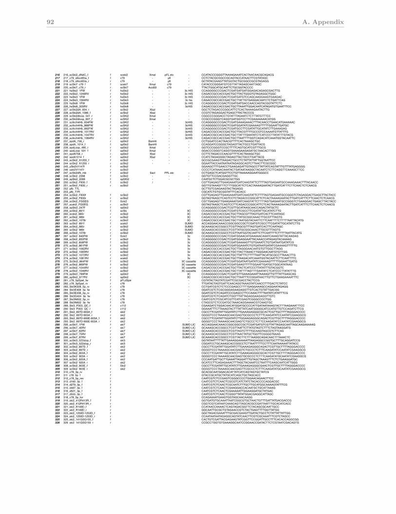
92 A. Appendix
Ski complex oligos cloning sequencings.cerevisiae, s. pombe, h. sapiens
216 216_scSki2_dNdC_f f scski2 XmaI pFL etc - CCATACCCGGGTTAAAAGAAATCACTAACAACGCAGACG 17-136, no tag217 217_c79_dAcc65Ia_f f c79 - pfl - CCTCTACGCGGCCGCAGTACCATAACTTCGTATAGC218 218_c79_dAcc65Ia_r r c79 - pfl - GCTATACGAAGTTATGGTACTGCGGCCGCGTAGAGG219 219_scSki7_c79_f f scski7 XmaI c79 - CATACCCGGGATGTCGTTATTAGAGCAATTAGC220 220_scSki7_c79_r r scSki7 Acc65I c79 - TTACTGGCATGCAATTCTGCGGTACCCC221 221_hsSki2_1FW f hsSki2 - - 3c HIS CCAGGGGCCCGACTCGATGATGATGGAGACAGAGCGACTTG222 222_hsSki2_1246RV r hsSki2 - - 3c HIS CAGACCGCCACCGACTGCTTACTGGGTGTAGAGGCTGGC223 223_hsSki3_1FW f hsSki3 - - 3c HIS CCAGGGGCCCGACTCGATGATGTCCAGCAAGGAAGTGAAGAC224 224_hsSki3_1564RV r hsSki3 - - 3c his CAGACCGCCACCGACTGCTTATTGTGAGGACAATCTCTGATTCAG225 225_hsSki8_1FW f hsSki8 - - 3c HIS CCAGGGGCCCGACTCGATGATGACCAACCAGTACGGTATTCTC226 226_hsSki8_305RV r hsSki8 - - 3cHIS CAGACCGCCACCGACTGCTTAAATTGGACAATCATAGATGTGAATTTCC227 227_scSki2dA_834_r r scSki2 XbaI - - GGCTCTAGACCCGGCATTCTCACTAAAAGAATACTTG loop out arch 835 incl 1085 228 228_scSki2dA_1086_f f scSki2 XbaI - - CCGTCTAGAGGACTGAGCTTACTACCCG loop out arch 835 incl 1085 229 229_scSki2dloop_541_r r scSKi2 XmaI - - CGGCCCGGGACCTGTATTTAGAATCTCTTTATGTTTCC loop out RecA linker 542 incl 606230 230_scSki2dloop_607_f f scSKi2 XmaI - - CCGCCCGGGTCAGGTGATGGTCCTTCGAAAAAAACATGG loop out RecA linker 542 incl 606231 231_scArch4Hb_834FW f scSKi2 - - 3cHIS CCAGGGGCCCGACTCGATGAAAGAAACTTTACAACCTGAACATGAAAAAC arch constructs scski2232 232_scArch4Hb_866FW f scSKi2 - - 3cHIS CCAGGGGCCCGACTCGATGGATATCGAAAAGTTTTTGGAATTGATGC arch constructs scski2233 233_scArch4Hb_892FW f scSKi2 - - 3cHIS CCAGGGGCCCGACTCGATGCCTTCGATATTGCATATCTTGAAAGAG arch constructs scski2234 234_scArch4Hb_1017RV r scSKi2 - - 3cHIS CAGACCGCCACCGACTGCTTACGTTTTGCCGTCCAAAATGTTATTTG arch constructs scski2235 235_scArch4Hb_1047RV r scSKi2 - - 3cHIS CAGACCGCCACCGACTGCTTATTTGAATATCTCATCCCTTATATTTGTACG arch constructs scski2236 236_scArch4Hb_1084RV r scSKi2 - - 3cHIS CAGACCGCCACCGACTGCTTAATTTTGGTCAGACATCAAATGGTACAATTC arch constructs scski2237 237_spdA_758_r r spSki2 BamHi - - CCTGGATCCACTAACGTTTTCACTAAAGCTGC loop out arch 759 incl 1013238 238_spdA_1014_f f spSki2 BamHI - - CCAGGATCCGGGCTAGAGTTACTGCCTGATTACG loop out arch 759 incl 1013239 239_spdLoop_490_r r spSki2 XmaI - - GGTCCCGGGTCCGCTTTTCAGTGCATCGTTTGCG loop out RecA linker 491 incl 530240 240_spdLoop_531_f f spSki2 XmaI - - GGACCCGGGTCAGGTGAAAGAAGAGATGCTAACACTTGG loop out RecA linker 491 incl 530241 241_spdA758_r r spSki2 XbaI - CCTTCTAGACCCAACGTTTTCACTAAAGCTGC loop out arch 759 incl 1013242 242_spdA1014_f f spSki2 XbaI - - CCATCTAGAGGGCTAGAGTTACTGCCTGATTACG loop out arch 759 incl 1013243 243_scSki2_A1255_f f scSki2 - - - GCCGCGAAGTTAAGACTGCCTCTATTATTATTGGTAATTCC244 244_scSki2_A1255_r r scSki2 - - GGAATTACCAATAATAATAGAGGCAGTCTTAACTTCGCGGC245 245_ySki2V1147f f scSki2 GGAAGCTTTGAACCTGAAGAGATTGTAGCCTTATTATCAGTATTTGTTTATGAGGGG246 246_yski2V1147r r scSki2 CCCCTCATAAACAAATACTGATAATAAGGCTACAATCTCTTCAGGTTCAAAGCTTCC247 247_scSki2dN_mb f scSki2 SacI PFL etc - CCTGAGCTCATGGTTCCTGTTAAAAAGGAATGGGCC248 248_scSki2_2398 f scSki2 - - - GGTGTTCCGACGAGGTTGC249 249_spSki2_2066 f spSKI2 - - - CAATGCTCTGGACGCGCTGG250 250_scSki2_F830_f f scSki2 / CGTTGAGAGTTGAAGAAATGATCAAGTATTCTTTTAGTGAGAATGCCAAAGAAACTTTACAACC251 251_scSki2_F830_r r scSki2 GGTTGTAAAGTTTCTTTGGCATTCTCACTAAAAGAATACTTGATCATTTCTTCAACTCTCAACG252 252_pfb_T7L GCTTGTCGAGAAGTACTAGAGG253 253_pfb_T7R CGCATCTGTGCGGTATTTCACACC254 254_scSki2_F830f f scSki2 CGTTGAGAGTTGAAGAAATGATCAAGTATTCTTTTAGTGAGAATGCCGGGTCTAGAGGACTGAGCTTACTACC255 255_scSki2_F830r r scSki2 GGTAGTAAGCTCAGTCCTCTAGACCCGGCATTCTCACTAAAAGAATACTTGATCATTTCTTCAACTCTCAACG256 256_scSki2_FGSEG f Scki2 CGTTGAGAGTTGAAGAAATGATCAAGTATTCTTTTAGTGAGAATGCCGGGTCTGAAGGACTGAGCTTACTACC257 257_scski2_FGSEG r scSki2 GGTAGTAAGCTCAGTCCTTCAGACCCGGCATTCTCACTAAAAGAATACTTGATCATTTCTTCAACTCTCAACG258 258_spSki2_247F f spSki2 CCAGGGGCCCGACTCGTTGCATAAGCAACCAGACTATGCTC259 259_scSki2_891f f scSki 3C CCAGGGGCCCGACTCGATGTCGCCTTCGATATTGCATATCTTG260 260_scski2_985r r scSki2 3C CAGACCGCCACCGACTGCTTAGCGTTTAGTGATCACTTCAATAGC261 261_scSki2_989r r scSki2 3C CAGACCGCCACCGACTGCTTATGCGGCAAACTTGCGTTTAGTG262 262_scSki2_1078r r scSki2 3C CAGACCGCCACCGACTGCTTAATGGTACAATTCTTCAATTTTCTTTTTAATTACATG263 263_scSki2_891f f scski2 SUMO ACCAGGAACAAACCGGCGGCCGCTCGATGTCGCCTTCGATATTGCATATCTTG264 264_scSki2_985r r scSki2 SUMO GCAAAGCACCGGCCTCGTTAGCGTTTAGTGATCACTTCAATAGC265 265_scSki2_989r r scSki2 SUMO GCAAAGCACCGGCCTCGTTATGCGGCAAACTTGCGTTTAGTG266 266_scSki2_1078r r scSki2 SUMO GCAAAGCACCGGCCTCGTTAATGGTACAATTCTTCAATTTTCTTTTTAATTACATG267 267_scSki2_840FW f Scki2 3c CCAGGGGCCCGACTCGATGGAACATGAAAAACAAATCAAAGTATTACAAGAG268 268_scSki2_850FW f scSki2 3c CCAGGGGCCCGACTCGATGGAGGAATTACAAACCATAGAGTACAAAAG269 269_scSki2_858FW f scSki2 3c CCAGGGGCCCGACTCGATGAAAAGTTGTGAAATCTGTGATAATGATATCG270 270_scSki2_861FW f scSki2 3c CCAGGGGCCCGACTCGATGGAAATCTGTGATAATGATATCGAAAAGTTTTTG271 271_scSki2_1062RV r scSki2 3c CAGACCGCCACCGACTGCTTAGGGAACAATGTGTTGGCTTAGG272 272_scSki2_1065RV r scSki2 3c CAGACCGCCACCGACTGCTTACTTAAACTTGGGAACAATGTGTTGG273 273_scSki2_1072RV r scSki2 3c CAGACCGCCACCGACTGCTTATTTCTTTTTAATTACATGCGCCTTAAACTTG274 274_scSki2_1081RV r scski2 3c CAGACCGCCACCGACTGCTTAAGACATCAAATGGTACAATTCTTCAATTTTC275 275_ScSki2_864FW f scSki2 3C cassette CCAGGGGCCCGACTCGATGGATAATGATATCGAAAAGTTTTTGGAATTGATG276 276_scSki2_869FW f scSki2 3C cassette CCAGGGGCCCGACTCGATGAAGTTTTTGGAATTGATGCTGGCATATAAG277 277_scSki2_1044RV r scSki2 3C cassette CAGACCGCCACCGACTGCTTACTCATCCCTTATATTTGTACGGTC278 278_scSki2_1049RV r scSki2 3C cassette CAGACCGCCACCGACTGCTTATTTTAGTTTGAATATCTCATCCCTTATATTTG279 279_spSki2_789FW f spSki2 3C CCAGGGGCCCGACTCGATGTTGAAAGAAATTAAAAGTTGTTTATTGAGCAG280 280_spSki2_977RV r spSki2 3C CAGACCGCCACCGACTGCTTAATTTCCGGATAACTTGTTCTGAAGAAAATTTC281 281_c78_5pSpeI_fw f pFLdSpe - CGTATACTAGTATCGATTCGCGACCTACTCCG282 282_c78_3pSpeI_rv r c78 - TTGATACTAGTGATTCAACAGGTAAAGTATCAACCTTTGACTCTATCC283 283_Ski3K328_5p_rv r c78 - CCTGATCGTCTCTCCCGAGCCTTTTGAGGAAAGCCAGGAATAGAGG284 284_Ski3E408_3p_fw f c78 - GGATCGTCTCGCGGGAGAAGAGGTTGTCACTGTATTGACGG285 285_Ski3E338_5p_rv r c78 - GGATCGTCTCAGATCCCGAGCCTTCCAAACTTTTAATATCATATTTCG286 286_Ski3G404_3p_fw f c78 - GGATCGTCTCAGATCTGGTTTATTAGAGGAAGAGGTTGTC287 287_Ski3N602_5p_rv r c78 - CAATCGTCTCGCATCGTTCATCGAGTCGGGCCCCTGG288 288_Ski3N602_3p_fw f c78 - CTAGCGTCTCCGATGCTAAACAGGAAAACGTCAAGTGC289 289_Ski3_P303_QC_f f c112 - CGAAGATCTGGACAACATGGATGCCCCATTGATAATAAAGTACTTTAAGAAATTTCC290 290_Ski3_P303_QC_r r c112 GGAAATTTCTTAAAGTACTTTATTATCAATGGGGCATCCATGTTGTCCAGATCTTCG291 291_Ski2_897D-903A_f f ski2 - CGCCTTCGATATTGGATATCTTGAAAGAGGGCGCACTCGTTGCTTTTAGGGACCCC292 292_Ski2_897D-903A_r r ski2 - GGGGTCCCTAAAAGCAACGAGTGCGCCCTCTTTCAAGATATCCAATATCGAAGGCG293 293_Ski2_897D-900E-903A_f f ski2 - CGCCTTCGATATTGGATATCTTGGAAGAGGGCAGACTCGTTGCTTTTAGGGACCCC294 293_Ski2_897D-900E-903A_r r ski2 - GGGGTCCCTAAAAGCAACGAGTCTGCCCTCTTCCAAGATATCCAATATCGAAGGCG295 295_scSki7_1FW f ski7 SUMO LIC ACCAGGAACAAACCGGCGGCCGCTCGATGATGTCGTTATTAGAGCAATTAGCAAGAAAAAG296 296_scSki7_40RV r ski7 SUMO LIC GCAAAGCACCGGCCTCGTTAATTCTTATGTAGTCTTTCTAGTAAGGATG297 297_scSki7_62RV r ski7 SUMO LIC GCAAAGCACCGGCCTCGTTAGTCTTTTGCAAGTAGCGTCTTCAG298 298_scSki7_75RV r ski7 SUMO LIC GCAAAGCACCGGCCTCGTTAACTATGCTGGTTCGGGGTAAAG299 299_scSki7_87RV r ski7 SUMO LIC GCAAAGCACCGGCCTCGTTACTTCTTTAAGGCAGACAACTTTAAACTC300 300_scSki3_522stop_f f ski3 GGTATAATTTTTATTGAAAGGAAAAATTAAGAGGCCGGTGCTTTGCAGGATCCG301 301_scSki3_522stop_r r ski3 CGGATCCTGCAAAGCACCGGCCTCTTAATTTTTCCTTTCAATAAAAATTATACC302 302_scSki2_897D_f f ski2 CGCCTTCGATATTGGATATCTTGAAAGAGGGCAGACTCGTTGCTTTTAGGGACCCC303 303_scSki2_897D_r r ski2 GGGGTCCCTAAAAGCAACGAGTCTGCCCTCTTTCAAGATATCCAATATCGAAGGCG304 304_scSki2_903A_f f ski2 CGCCTTCGATATTGCATATCTTGAAAGAGGGCGCACTCGTTGCTTTTAGGGACCCC305 305_scSki2_903A_r r ski2 GGGGTCCCTAAAAGCAACGAGTGCGCCCTCTTTCAAGATATGCAATATCGAAGGCG306 306_scSki2_920A_f f ski2 CCCAATGATTGCTTGAAATTAGGATTTGTAGCTAAAGTTTCTCTGAAGGATGC307 307_scSki2_920A_r r ski2 GCATCCTTCAGAGAAACTTTAGCTACAAATCCTAATTTCAAGCAATCATTGGG308 308_scSki2_903E_f f Ski2 CGCCTTCGATATTGCATATCTTGAAAGAGGGCGAACTCGTTGCTTTTAGGGACCCC309 309_scSki2_903E_r r ski2 GGGGTCCCTAAAAGCAACGAGTTCGCCCTCTTTCAAGATATGCAATATCGAAGGCG310 310_c78_3p_rv r GCACGCAATGGACACATTATCATCAGTAGTGCTATCG311 311_c78_5p_f f GTACCGCATGCTATGCATCAGCTGCTAGCACC 312 312_c78_5p_rev r CAATCGTCTCCGAGTCGGGCCCCTGGAACAGAACTTCC313 313_d160_3p_f f CAATCGTCTCAACTCGCGTCATCTATCTACACCCCAGGACGC 314 314_d279_3p_f f CAATCGTCTCAACTCGCAATCTTTGCTTGCATGGCAAAAGTATTTCG 315 315_d487_3p_f f CAATCGTCTCAACTCGAAGGACCACAATGCTGCATTAAAG 316 316_d521_3p_f f CAATCGTCTCAACTCGAAAAATTGGAAGGATGCTATGAC 317 317_d555_3p_f f CAATCGTCTCAACTCGGGTTATATGGACGAGGCATTAGC 318 318_c78_5p_for f CCAGAAAATGAAGTGGTAGCAACAAGG319 319_ski2_412R413R_f f GGTGATGTGCAAATTAATCGGCGTGCTAACTGTTTGATTATGACGACCG320 320_ski2_412R413R_r r CGGTCGTCATAATCAAACAGTTAGCACGCCGATTAATTTGCACATCACC321 321_ski2_R149D_f f CCATAACCAAAACTCAGTAGACGGTTCTACAGCGCAATTGCC322 322_ski2_R149D_r r GGCAATTGCGCTGTAGAACCGTCTACTGAGTTTTGGTTATGG323 323_ski2_1250D-1253D_f f GGCTAGACGAAATTTGCGACGAAGTTGATACTGCCTCTATTATTATTGG324 324_ski2_1250D-1253D_r r CCAATAATAATAGAGGCAGTATCAACTTCGTCGCAAATTTCGTCTAGCC325 325_ski2_141GSG155_f f CACTGTCGATTACGAGAAGTATCGGTTCCGGATTGCCTTTCACACCAGGCGG326 326_ski2_141GSG155_r r CCGCCTGGTGTGAAAGGCAATCCGGAACCGATACTTCTCGTAATCGACAGTG
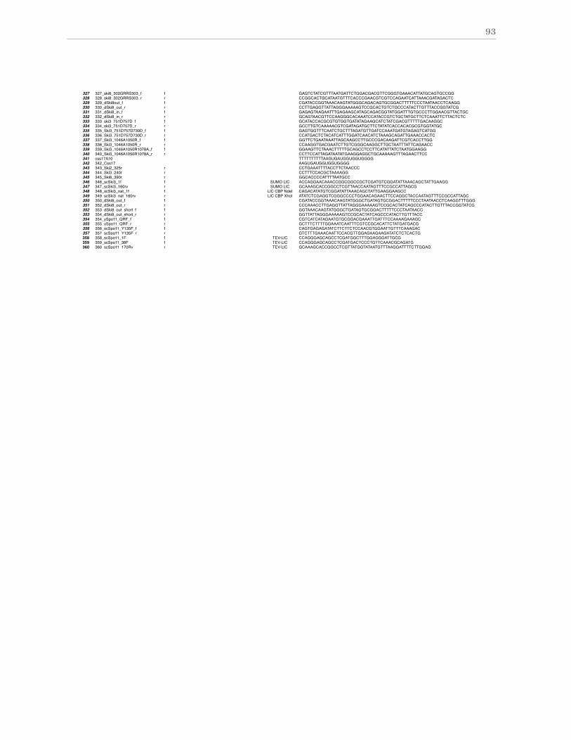
93
Ski complex oligos cloning sequencings.cerevisiae, s. pombe, h. sapiens
327 327_ski8_302GRRS303_f f GAGTCTATCGTTTAATGATTCTGGACGACGTTCGGGTGAAACATTATGCAGTGCCGG328 328_ski8_302GRRS303_r r CCGGCACTGCATAATGTTTCACCCGAACGTCGTCCAGAATCATTAAACGATAGACTC329 329_dSki8out_f f CGATACCGGTAAACAAGTATGGGCAGACAGTGCGGACTTTTTCCCTAATAACCTCAAGG330 330_dSki8_out_r r CCTTGAGGTTATTAGGGAAAAAGTCCGCACTGTCTGCCCATACTTGTTTACCGGTATCG331 331_dSki8_in_f f GAGAGTAAGAATTTGAGAAGCATAGCAGACGGTATGGATTTGTGCCCTTGGAACGTTACTGC332 332_dSki8_in_r r GCAGTAACGTTCCAAGGGCACAAATCCATACCGTCTGCTATGCTTCTCAAATTCTTACTCTC333 333_ski3_751D757D_f f GCATACCACGCGTGTGGTGATATAGAAGCATCTATCGACGTTTTTGACAAGGC334 334_ski3_751D757D_r r GCCTTGTCAAAAACGTCGATAGATGCTTCTATATCACCACACGCGTGGTATGC335 335_Ski3_751D757D730D_f f GAGTGGTTTCAATCTGCTTTAGATGTTGATCCAAATGATGTAGAGTCATGG336 336_Ski3_751D757D730D_r r CCATGACTCTACATCATTTGGATCAACATCTAAAGCAGATTGAAACCACTC337 337_Ski3_1046A1050R_f f GGTTCTGAATAAATTAGCAAGCCTTGCCCGACAAGATTCGTCACCTTGG338 338_Ski3_1046A1050R_r r CCAAGGTGACGAATCTTGTCGGGCAAGGCTTGCTAATTTATTCAGAACC339 339_Ski3_1046A1050R1078A_f f GGAAGTTCTAAACTTTTTGCAGCCTCCTTCATATTATCTAATGGAAGG340 340_Ski3_1046A1050R1078A_r r CCTTCCATTAGATAATATGAAGGAGGCTGCAAAAAGTTTAGAACTTCC341 css17A10 TTTTTTTTTTAAGUGAUGGUGGUGGGG342 342_Css17 AAGUGAUGGUGGUGGGG343 343_Ski2_325r r CCTGAAATTTTACCTTCTAACCC344 344_Ski3_240r r CCTTTCCACGCTAAAAGG345 345_Ski8_390r r GGCACCCCATTTTAATGCC346 346_scSki3_1f f SUMO LIC ACCAGGAACAAACCGGCGGCCGCTCGATGTCGGATATTAAACAGCTATTGAAGG347 347_scSki3_160rv r SUMO LIC GCAAAGCACCGGCCTCGTTAACCAATAGTTTCCGCCATTAGCG348 348_scSki3_nat_1f f LIC CBP NdeI CAGACATATGTCGGATATTAAACAGCTATTGAAGGAAGCC349 349_scSki3_nat_160rv r LIC CBP XhoI ATATCTCGAGGTCGGGCCCCTGGAACAGAACTTCCAGGCTACCAATAGTTTCCGCCATTAGC350 350_dSki8_out_f f CGATACCGGTAAACAAGTATGGGCTGATAGTGCGGACTTTTTCCCTAATAACCTCAAGGTTTGGG351 352_dSki8_out_r r CCCAAACCTTGAGGTTATTAGGGAAAAAGTCCGCACTATCAGCCCATACTTGTTTACCGGTATCG352 353_dSki8_out_short_f f GGTAAACAAGTATGGGCTGATAGTGCGGACTTTTTCCCTAATAACC353 354_dSki8_out_short_r r GGTTATTAGGGAAAAAGTCCGCACTATCAGCCCATACTTGTTTACC354 354_ySpo11_QRF_f f CGTCATCATAGAATGTGCGGACGAAATTGATTTCCAAAAGAAAGC355 355_ySpo11_QRF_r r GCTTTCTTTTGGAAATCAATTTCGTCCGCACATTCTATGATGACG356 356_scSpo11_Y135F_f f CAGTGAGAGATATCTTCTTCTCCAACGTGGAATTGTTTCAAAGAC357 357_scSpo11_Y135F_r r GTCTTTGAAACAATTCCACGTTGGAGAAGAAGATATCTCTCACTG358 358_scSpo11_1F f TEV-LIC CCAGGGAGCAGCCTCGATGGCTTTGGAGGGATTGCG359 359_scSpo11_38F f TEV-LIC CCAGGGAGCAGCCTCGATGACTCCCTGTTCAAACGCAGATG360 360_scSpo11_170Rv r TEV-LIC GCAAAGCACCGGCCTCGTTATGGTATAATGTTTAAGGATTTTCTTGGAG

94 A. Appendix
Bibliography
Allmang, C., Kufel, J., Chanfreau, G., Mitchell, P., Petfalski, E., and Tollervey, D.
(1999). Functions of the exosome in rRNA, snoRNA and snRNA synthesis. EMBO
J, 18(19):5399–410.
Anderson, J. S. and Parker, R. P. (1998). The 3’ to 5’ degradation of yeast mRNAs is a
general mechanism for mRNA turnover that requires the SKI2 DEVH box protein and
3’ to 5’ exonucleases of the exosome complex. EMBO J, 17(5):1497–506.
Araki, Y., Takahashi, S., Kobayashi, T., Kajiho, H., Hoshino, S., and Katada, T. (2001).
Ski7p G protein interacts with the exosome and the Ski complex for 3’-to-5’ mRNA
decay in yeast. EMBO J, 20(17):4684–93.
Arigo, J. T., Eyler, D. E., Carroll, K. L., and Corden, J. L. (2006). Termination of cryptic
unstable transcripts is directed by yeast RNA-binding proteins Nrd1 and Nab3. Mol
Cell, 23(6):841–851.
Arora, C., Kee, K., Maleki, S., and Keeney, S. (2004). Antiviral protein Ski8 is a direct
partner of Spo11 in meiotic DNA break formation, independent of its cytoplasmic role
in RNA metabolism. Mol Cell, 13(4):549–59.
Atkinson, G. C., Baldauf, S. L., and Hauryliuk, V. (2008). Evolution of nonstop, no-go
and nonsense-mediated mRNA decay and their termination factor-derived components.
BMC Evol Biol, 8:290.
Badis, G., Saveanu, C., Fromont-Racine, M., and Jacquier, A. (2004). Targeted mRNA
degradation by deadenylation-independent decapping. Mol Cell, 15(1):5–15.
Becker, T., Armache, J.-P., Jarasch, A., Anger, A. M., Villa, E., Sieber, H., Motaal, B. A.,
Mielke, T., Berninghausen, O., and Beckmann, R. (2011). Structure of the no-go mRNA
96 BIBLIOGRAPHY
decay complex Dom34-Hbs1 bound to a stalled 80S ribosome. Nat Struct Mol Biol,
18(6):715–720.
Beelman, C. A., Stevens, A., Caponigro, G., LaGrandeur, T. E., Hatfield, L., Fortner,
D. M., and Parker, R. (1996). An essential component of the decapping enzyme required
for normal rates of mRNA turnover. Nature, 382(6592):642–646.
Benard, L., Carroll, K., Valle, R. C., Masison, D. C., and Wickner, R. B. (1999). The Ski7
antiviral protein is an EF1-alpha homolog that blocks expression of non-poly(A) mRNA
in Saccharomyces cerevisiae. J Virol, 73(4):2893–2900.
Benard, L., Carroll, K., Valle, R. C., and Wickner, R. B. (1998). Ski6p is a homolog
of RNA-processing enzymes that affects translation of non-poly(A) mRNAs and 60S
ribosomal subunit biogenesis. Mol Cell Biol, 18(5):2688–2696.
Bergerat, A., de Massy, B., Gadelle, D., Varoutas, P. C., Nicolas, A., and Forterre, P.
(1997). An atypical topoisomerase II from Archaea with implications for meiotic recom-
bination. Nature, 386(6623):414–7.
Bernstein, J., Patterson, D. N., Wilson, G. M., and Toth, E. A. (2008). Characterization
of the essential activities of saccharomyces cerevisiae mtr4p, a 3’-¿5’ helicase partner of
the nuclear exosome. J Biol Chem, 283(8):4930–4942.
Bernstein, J. A., Khodursky, A. B., Lin, P.-H., Lin-Chao, S., and Cohen, S. N. (2002).
Global analysis of mRNA decay and abundance in Escherichia coli at single-gene res-
olution using two-color fluorescent DNA microarrays. Proc Natl Acad Sci U S A,
99(15):9697–9702.
Bernstein, J. A., Lin, P.-H., Cohen, S. N., and Lin-Chao, S. (2004). Global analysis of
Escherichia coli RNA degradosome function using DNA microarrays. Proc Natl Acad
Sci U S A, 101(9):2758–2763.
Bonneau, F., Basquin, J., Ebert, J., Lorentzen, E., and Conti, E. (2009). The yeast exosome
functions as a macromolecular cage to channel RNA substrates for degradation. Cell,
139(3):547–59.
Bousquet-Antonelli, C., Presutti, C., and Tollervey, D. (2000). Identification of a regulated
pathway for nuclear pre-mRNA turnover. Cell, 102(6):765–75.
BIBLIOGRAPHY 97
Brown, C. E. and Sachs, A. B. (1998). Poly(A) tail length control in Saccharomyces
cerevisiae occurs by message-specific deadenylation. Mol Cell Biol, 18(11):6548–6559.
Brown, J. T., Bai, X., and Johnson, A. W. (2000). The yeast antiviral proteins Ski2p,
Ski3p, and Ski8p exist as a complex in vivo. RNA, 6(3):449–57.
Buttner, K., Nehring, S., and Hopfner, K. P. (2007). Structural basis for DNA duplex
separation by a superfamily-2 helicase. Nat Struct Mol Biol, 14(7):647–52.
Buttner, K., Wenig, K., and Hopfner, K. P. (2005). Structural framework for the mechanism
of archaeal exosomes in RNA processing. Mol Cell, 20(3):461–71.
Callahan, K. P. and Butler, J. S. (2008). Evidence for core exosome independent function
of the nuclear exoribonuclease Rrp6p. Nucleic Acids Res, 36(21):6645–6655.
Carpousis, A. J. (2007). The RNA degradosome of Escherichia coli: an mRNA-degrading
machine assembled on RNase E. Annu Rev Microbiol, 61:71–87.
Carroll, K. L., Ghirlando, R., Ames, J. M., and Corden, J. L. (2007). Interaction of yeast
RNA-binding proteins Nrd1 and Nab3 with RNA polymerase II terminator elements.
RNA, 13(3):361–373.
Chang, Y.-F., Imam, J. S., and Wilkinson, M. F. (2007). The nonsense-mediated decay
RNA surveillance pathway. Annu Rev Biochem, 76:51–74.
Chen, C. K.-M., Chan, N.-L., and Wang, A. H.-J. (2011). The many blades of the β-
propeller proteins: conserved but versatile. Trends Biochem Sci, 36(10):553–561.
Chen, C. Y., Gherzi, R., Ong, S. E., Chan, E. L., Raijmakers, R., Pruijn, G. J., Stoecklin,
G., Moroni, C., Mann, M., and Karin, M. (2001). AU binding proteins recruit the
exosome to degrade ARE-containing mRNAs. Cell, 107(4):451–464.
Chen, L., Muhlrad, D., Hauryliuk, V., Cheng, Z., Lim, M. K., Shyp, V., Parker, R., and
Song, H. (2010). Structure of the Dom34-Hbs1 complex and implications for no-go decay.
Nat Struct Mol Biol, 17(10):1233–1240.
Cheng, Z., Liu, Y., Wang, C., Parker, R., and Song, H. (2004). Crystal structure of Ski8p,
a WD-repeat protein with dual roles in mRNA metabolism and meiotic recombination.
Protein Sci, 13(10):2673–84.
98 BIBLIOGRAPHY
Cole, F., Keeney, S., and Jasin, M. (2010). Evolutionary conservation of meiotic DSB
proteins: more than just Spo11. Genes Dev, 24(12):1201–1207.
Conti, E. and Izaurralde, E. (2005). Nonsense-mediated mRNA decay: molecular insights
and mechanistic variations across species. Curr Opin Cell Biol, 17(3):316–325.
Costello, J. L., Stead, J. A., Feigenbutz, M., Jones, R. M., and Mitchell, P. (2011). The
C-terminal region of the exosome-associated protein Rrp47 is specifically required for
box C/D small nucleolar RNA 3’-maturation. J Biol Chem, 286(6):4535–4543.
D’Andrea, L. D. and Regan, L. (2003). TPR proteins: the versatile helix. Trends in
biochemical sciences, 28(12):655–62.
Dangel, A. W., Shen, L., Mendoza, A. R., Wu, L. C., and Yu, C. Y. (1995). Human
helicase gene SKI2W in the HLA class III region exhibits striking structural similarities
to the yeast antiviral gene SKI2 and to the human gene KIAA0052: emergence of a new
gene family. Nucleic Acids Res, 23(12):2120–2126.
Das, A. K., Cohen, P. W., and Barford, D. (1998). The structure of the tetratricopep-
tide repeats of protein phosphatase 5: implications for TPR-mediated protein-protein
interactions. The EMBO journal, 17(5):1192–9.
Daugeron, M. C., Mauxion, F., and Sraphin, B. (2001). The yeast POP2 gene encodes a
nuclease involved in mRNA deadenylation. Nucleic Acids Res, 29(12):2448–2455.
Davis, C. A. and Ares, M. (2006). Accumulation of unstable promoter-associated tran-
scripts upon loss of the nuclear exosome subunit Rrp6p in Saccharomyces cerevisiae.
Proc Natl Acad Sci U S A, 103(9):3262–3267.
Dez, C., Houseley, J., and Tollervey, D. (2006). Surveillance of nuclear-restricted
pre-ribosomes within a subnucleolar region of Saccharomyces cerevisiae. EMBO J,
25(7):1534–1546.
Doma, M. K. and Parker, R. (2006). Endonucleolytic cleavage of eukaryotic mRNAs with
stalls in translation elongation. Nature, 440(7083):561–564.
Dunckley, T. and Parker, R. (1999). The DCP2 protein is required for mRNA decapping in
Saccharomyces cerevisiae and contains a functional MutT motif. EMBO J, 18(19):5411–
5422.
BIBLIOGRAPHY 99
Dziembowski, A., Lorentzen, E., Conti, E., and Seraphin, B. (2007). A single subunit,
Dis3, is essentially responsible for yeast exosome core activity. Nat Struct Mol Biol,
14(1):15–22.
Egecioglu, D. E., Henras, A. K., and Chanfreau, G. F. (2006). Contributions of Trf4p-
and Trf5p-dependent polyadenylation to the processing and degradative functions of the
yeast nuclear exosome. RNA, 12(1):26–32.
Evguenieva-Hackenberg, E., Walter, P., Hochleitner, E., Lottspeich, F., and Klug, G.
(2003). An exosome-like complex in Sulfolobus solfataricus. EMBO Rep, 4(9):889–893.
Fairman-Williams, M. E., Guenther, U.-P., and Jankowsky, E. (2010). SF1 and SF2 heli-
cases: family matters. Curr Opin Struct Biol, 20(3):313–324.
Franks, T. M. and Lykke-Andersen, J. (2008). The control of mRNA decapping and P-body
formation. Mol Cell, 32(5):605–615.
Frischmeyer, P. A., van Hoof, A., O’Donnell, K., Guerrerio, A. L., Parker, R., and Dietz,
H. C. (2002). An mRNA surveillance mechanism that eliminates transcripts lacking
termination codons. Science, 295(5563):2258–2261.
Garneau, N. L., Wilusz, J., and Wilusz, C. J. (2007). The highways and byways of mRNA
decay. Nature reviews. Molecular cell biology, 8(2):113–26.
Giaever, G., Chu, A. M., Ni, L., Connelly, C., Riles, L., Vronneau, S., Dow, S., Lucau-
Danila, A., Anderson, K., Andr, B., Arkin, A. P., Astromoff, A., El-Bakkoury, M., Bang-
ham, R., Benito, R., Brachat, S., Campanaro, S., Curtiss, M., Davis, K., Deutschbauer,
A., Entian, K.-D., Flaherty, P., Foury, F., Garfinkel, D. J., Gerstein, M., Gotte, D.,
Gldener, U., Hegemann, J. H., Hempel, S., Herman, Z., Jaramillo, D. F., Kelly, D. E.,
Kelly, S. L., Ktter, P., LaBonte, D., Lamb, D. C., Lan, N., Liang, H., Liao, H., Liu, L.,
Luo, C., Lussier, M., Mao, R., Menard, P., Ooi, S. L., Revuelta, J. L., Roberts, C. J.,
Rose, M., Ross-Macdonald, P., Scherens, B., Schimmack, G., Shafer, B., Shoemaker,
D. D., Sookhai-Mahadeo, S., Storms, R. K., Strathern, J. N., Valle, G., Voet, M., Vol-
ckaert, G., yun Wang, C., Ward, T. R., Wilhelmy, J., Winzeler, E. A., Yang, Y., Yen,
G., Youngman, E., Yu, K., Bussey, H., Boeke, J. D., Snyder, M., Philippsen, P., Davis,
R. W., and Johnston, M. (2002). Functional profiling of the Saccharomyces cerevisiae
genome. Nature, 418(6896):387–391.
100 BIBLIOGRAPHY
Hamill, S., Wolin, S. L., and Reinisch, K. M. (2010). Structure and function of the poly-
merase core of TRAMP, a RNA surveillance complex. Proc Natl Acad Sci U S A,
107(34):15045–15050.
Hartung, S., Niederberger, T., Hartung, M., Tresch, A., and Hopfner, K.-P. (2010). Quan-
titative analysis of processive RNA degradation by the archaeal RNA exosome. Nucleic
Acids Res, 38(15):5166–5176.
He, F., Li, X., Spatrick, P., Casillo, R., Dong, S., and Jacobson, A. (2003). Genome-
wide analysis of mRNAs regulated by the nonsense-mediated and 5’ to 3’ mRNA decay
pathways in yeast. Mol Cell, 12(6):1439–1452.
He, Y., Andersen, G. R., and Nielsen, K. H. (2010). Structural basis for the function of
DEAH helicases. EMBO Rep, 11(3):180–6.
Hilleren, P., McCarthy, T., Rosbash, M., Parker, R., and Jensen, T. H. (2001). Quality con-
trol of mRNA 3’-end processing is linked to the nuclear exosome. Nature, 413(6855):538–
42.
Hirano, T., Kinoshita, N., Morikawa, K., and Yanagida, M. (1990). Snap helix with knob
and hole: essential repeats in S. pombe nuclear protein nuc2+. Cell, 60(2):319–28.
Houalla, R., Devaux, F., Fatica, A., Kufel, J., Barrass, D., Torchet, C., and Tollervey, D.
(2006). Microarray detection of novel nuclear RNA substrates for the exosome. Yeast,
23(6):439–54.
Houseley, J. and Tollervey, D. (2009). The many pathways of RNA degradation. Cell,
136(4):763–76.
Hsu, C. L. and Stevens, A. (1993). Yeast cells lacking 5’-3’ exoribonuclease 1 contain
mRNA species that are poly(A) deficient and partially lack the 5’ cap structure. Mol
Cell Biol, 13(8):4826–4835.
Izaurralde, E., Lewis, J., McGuigan, C., Jankowska, M., Darzynkiewicz, E., and Mattaj,
I. W. (1994). A nuclear cap binding protein complex involved in pre-mRNA splicing.
Cell, 78(4):657–668.
Jackson, R. N., Klauer, A. A., Hintze, B. J., Robinson, H., van Hoof, A., and Johnson,
S. J. (2010). The crystal structure of Mtr4 reveals a novel arch domain required for
rRNA processing. EMBO J, 29(13):2205–16.
BIBLIOGRAPHY 101
Jankowsky, E. and Fairman, M. E. (2007). RNA helicases - one fold for many functions.
Curr Opin Struct Biol, 17(3):316–324.
Jinek, M., Rehwinkel, J., Lazarus, B. D., Izaurralde, E., Hanover, J. A., and Conti, E.
(2004). The superhelical TPR-repeat domain of O-linked GlcNAc transferase exhibits
structural similarities to importin alpha. Nat Struct Mol Biol, 11(10):1001–1007.
Johnson, A. W. and Kolodner, R. D. (1995). Synthetic lethality of SEP1 (XRN1), SKI2
and SEP1 (XRN1) SKI3 mutants of Saccharomyces cerevisiae is independent of killer
virus and suggests a general role for these genes in translation control. Mol Cell Biol,
15(5):2719–27.
Kadaba, S., Wang, X., and Anderson, J. T. (2006). Nuclear RNA surveillance in Saccha-
romyces cerevisiae: Trf4p-dependent polyadenylation of nascent hypomethylated tRNA
and an aberrant form of 5S rRNA. RNA, 12(3):508–521.
Kee, K., Protacio, R. U., Arora, C., and Keeney, S. (2004). Spatial organization and
dynamics of the association of Rec102 and Rec104 with meiotic chromosomes. EMBO
J, 23(8):1815–1824.
Keeney, S., Giroux, C. N., and Kleckner, N. (1997). Meiosis-specific DNA double-strand
breaks are catalyzed by Spo11, a member of a widely conserved protein family. Cell,
88(3):375–84.
Keller, W. and Minvielle-Sebastia, L. (1997). A comparison of mammalian and yeast
pre-mRNA 3’-end processing. Curr Opin Cell Biol, 9(3):329–336.
Kelley, L. A. and Sternberg, M. J. (2009). Protein structure prediction on the Web: a case
study using the Phyre server. Nature protocols, 4(3):363–71.
Kessler, S. H. and Sachs, A. B. (1998). RNA recognition motif 2 of yeast Pab1p is required
for its functional interaction with eukaryotic translation initiation factor 4G. Mol Cell
Biol, 18(1):51–57.
Khemici, V. and Carpousis, A. J. (2004). The RNA degradosome and poly(A) polymerase
of Escherichia coli are required in vivo for the degradation of small mRNA decay inter-
mediates containing REP-stabilizers. Mol Microbiol, 51(3):777–790.
102 BIBLIOGRAPHY
Khemici, V., Poljak, L., Toesca, I., and Carpousis, A. J. (2005). Evidence in vivo that the
DEAD-box RNA helicase RhlB facilitates the degradation of ribosome-free mRNA by
RNase E. Proc Natl Acad Sci U S A, 102(19):6913–6918.
Kim, M., Vasiljeva, L., Rando, O. J., Zhelkovsky, A., Moore, C., and Buratowski, S. (2006).
Distinct pathways for snoRNA and mRNA termination. Mol Cell, 24(5):723–734.
Klauer, A. A. and van Hoof, A. (2013). Genetic interactions suggest multiple distinct roles
of the arch and core helicase domains of Mtr4 in Rrp6 and exosome function. Nucleic
Acids Res, 41(1):533–541.
Krogan, N. J., Cagney, G., Yu, H., Zhong, G., Guo, X., Ignatchenko, A., Li, J., Pu,
S., Datta, N., Tikuisis, A. P., Punna, T., Peregrn-Alvarez, J. M., Shales, M., Zhang,
X., Davey, M., Robinson, M. D., Paccanaro, A., Bray, J. E., Sheung, A., Beattie, B.,
Richards, D. P., Canadien, V., Lalev, A., Mena, F., Wong, P., Starostine, A., Canete,
M. M., Vlasblom, J., Wu, S., Orsi, C., Collins, S. R., Chandran, S., Haw, R., Rilstone,
J. J., Gandi, K., Thompson, N. J., Musso, G., Onge, P. S., Ghanny, S., Lam, M.
H. Y., Butland, G., Altaf-Ul, A. M., Kanaya, S., Shilatifard, A., O’Shea, E., Weissman,
J. S., Ingles, C. J., Hughes, T. R., Parkinson, J., Gerstein, M., Wodak, S. J., Emili,
A., and Greenblatt, J. F. (2006). Global landscape of protein complexes in the yeast
Saccharomyces cerevisiae. Nature, 440(7084):637–643.
Kshirsagar, M. and Parker, R. (2004). Identification of Edc3p as an enhancer of mRNA
decapping in Saccharomyces cerevisiae. Genetics, 166(2):729–739.
Kuhn, U. and Wahle, E. (2004). Structure and function of poly(A) binding proteins.
Biochim Biophys Acta, 1678(2-3):67–84.
LaCava, J., Houseley, J., Saveanu, C., Petfalski, E., Thompson, E., Jacquier, A., and Toller-
vey, D. (2005). RNA degradation by the exosome is promoted by a nuclear polyadeny-
lation complex. Cell, 121(5):713–24.
Lapouge, K., Smith, S. J., Walker, P. A., Gamblin, S. J., Smerdon, S. J., and Rittinger,
K. (2000). Structure of the TPR domain of p67phox in complex with Rac-GTP. Mol
Cell, 6(4):899–907.
Larimer, F. W. and Stevens, A. (1990). Disruption of the gene XRN1, coding for a 5’-3’
exoribonuclease, restricts yeast cell growth. Gene, 95(1):85–90.
BIBLIOGRAPHY 103
Lebreton, A., Tomecki, R., Dziembowski, A., and Seraphin, B. (2008). Endonucleolytic
RNA cleavage by a eukaryotic exosome. Nature, 456(7224):993–6.
Lee, H. H., Kim, Y.-S., Kim, K. H., Heo, I., Kim, S. K., Kim, O., Kim, H. K., Yoon, J. Y.,
Kim, H. S., Kim, D. J., Lee, S. J., Yoon, H. J., Kim, S. J., Lee, B. G., Song, H. K.,
Kim, V. N., Park, C.-M., and Suh, S. W. (2007). Structural and functional insights into
Dom34, a key component of no-go mRNA decay. Mol Cell, 27(6):938–950.
Lee, S. G., Lee, I., Park, S. H., Kang, C., and Song, K. (1995). Identification and charac-
terization of a human cDNA homologous to yeast SKI2. Genomics, 25(3):660–666.
Liu, H., Rodgers, N. D., Jiao, X., and Kiledjian, M. (2002). The scavenger mRNA de-
capping enzyme DcpS is a member of the HIT family of pyrophosphatases. EMBO J,
21(17):4699–4708.
Liu, Q., Greimann, J. C., and Lima, C. D. (2006). Reconstitution, activities, and structure
of the eukaryotic RNA exosome. Cell, 127(6):1223–37.
Lohman, T. M., Tomko, E. J., and Wu, C. G. (2008). Non-hexameric DNA helicases and
translocases: mechanisms and regulation. Nat Rev Mol Cell Biol, 9(5):391–401.
Lorentzen, E., Basquin, J., Tomecki, R., Dziembowski, A., and Conti, E. (2008). Structure
of the active subunit of the yeast exosome core, Rrp44: diverse modes of substrate
recruitment in the RNase II nuclease family. Mol Cell, 29(6):717–28.
Lorentzen, E. and Conti, E. (2005). Structural basis of 3’ end RNA recognition and
exoribonucleolytic cleavage by an exosome RNase PH core. Mol Cell, 20(3):473–81.
Lorentzen, E., Dziembowski, A., Lindner, D., Seraphin, B., and Conti, E. (2007). RNA
channelling by the archaeal exosome. EMBO Rep, 8(5):470–6.
Lorentzen, E., Walter, P., Fribourg, S., Evguenieva-Hackenberg, E., Klug, G., and Conti,
E. (2005). The archaeal exosome core is a hexameric ring structure with three catalytic
subunits. Nat Struct Mol Biol, 12(7):575–81.
Lykke-Andersen, S., Brodersen, D. E., and Jensen, T. H. (2009). Origins and activities of
the eukaryotic exosome. J Cell Sci, 122(Pt 10):1487–94.
Madrona, A. Y. and Wilson, D. K. (2004). The structure of Ski8p, a protein regulating
mRNA degradation: Implications for WD protein structure. Protein Sci, 13(6):1557–65.
104 BIBLIOGRAPHY
Malet, H., Topf, M., Clare, D. K., Ebert, J., Bonneau, F., Basquin, J., Drazkowska, K.,
Tomecki, R., Dziembowski, A., Conti, E., Saibil, H. R., and Lorentzen, E. (2010). RNA
channelling by the eukaryotic exosome. EMBO Rep, 11(12):936–942.
Manley, J. L. and Takagaki, Y. (1996). The end of the message - another link between
yeast and mammals. Science, 274(5292):1481–1482.
Matsumoto, Y., Sarkar, G., Sommer, S. S., and Wickner, R. B. (1993). A yeast antiviral
protein, Ski8, shares a repeated amino acid sequence pattern with beta-subunits of G
proteins and several other proteins. Yeast, 9(1):43–51.
Midtgaard, S. F., Assenholt, J., Jonstrup, A. T., Van, L. B., Jensen, T. H., and Brodersen,
D. E. (2006). Structure of the nuclear exosome component Rrp6p reveals an interplay
between the active site and the HRDC domain. Proc Natl Acad Sci U S A, 103(32):11898–
11903.
Milligan, L., Decourty, L., Saveanu, C., Rappsilber, J., Ceulemans, H., Jacquier, A., and
Tollervey, D. (2008). A yeast exosome cofactor, Mpp6, functions in RNA surveillance
and in the degradation of noncoding RNA transcripts. Mol Cell Biol, 28(17):5446–5457.
Mitchell, P., Petfalski, E., Houalla, R., Podtelejnikov, A., Mann, M., and Tollervey, D.
(2003). Rrp47p is an exosome-associated protein required for the 3’ processing of stable
RNAs. Mol Cell Biol, 23(19):6982–6992.
Mitchell, P., Petfalski, E., Shevchenko, A., Mann, M., and Tollervey, D. (1997). The
exosome: a conserved eukaryotic RNA processing complex containing multiple 3’-5’ ex-
oribonucleases. Cell, 91(4):457–66.
Mitchell, P. and Tollervey, D. (2003). An NMD pathway in yeast involving accelerated
deadenylation and exosome-mediated 3’-5’ degradation. Mol Cell, 11(5):1405–13.
Muhlrad, D., Decker, C. J., and Parker, R. (1994). Deadenylation of the unstable mRNA
encoded by the yeast MFA2 gene leads to decapping followed by 5’-3’ digestion of the
transcript. Genes Dev, 8(7):855–866.
Muhlrad, D., Decker, C. J., and Parker, R. (1995). Turnover mechanisms of the stable
yeast PGK1 mRNA. Mol Cell Biol, 15(4):2145–2156.
BIBLIOGRAPHY 105
Nag, D. K., Pata, J. D., Sironi, M., Flood, D. R., and Hart, A. M. (2006). Both conserved
and non-conserved regions of Spo11 are essential for meiotic recombination initiation in
yeast. Molecular genetics and genomics : MGG, 276(4):313–21.
Navarro, M. V. A. S., Oliveira, C. C., Zanchin, N. I. T., and Guimares, B. G. (2008).
Insights into the mechanism of progressive RNA degradation by the archaeal exosome.
J Biol Chem, 283(20):14120–14131.
Nichols, M. D., DeAngelis, K., Keck, J. L., and Berger, J. M. (1999). Structure and function
of an archaeal topoisomerase VI subunit with homology to the meiotic recombination
factor Spo11. EMBO J, 18(21):6177–88.
Orban, T. I. and Izaurralde, E. (2005). Decay of mRNAs targeted by RISC requires XRN1,
the Ski complex, and the exosome. RNA, 11(4):459–69.
Paczkowski, J. E., Richardson, B. C., Strassner, A. M., and Fromme, J. C. (2012). The
exomer cargo adaptor structure reveals a novel GTPase-binding domain. EMBO J,
31(21):4191–4203.
Parker, R. (2012). RNA degradation in Saccharomyces cerevisae. Genetics, 191(3):671–702.
Phillips, S. and Butler, J. S. (2003). Contribution of domain structure to the RNA 3’
end processing and degradation functions of the nuclear exosome subunit Rrp6p. RNA,
9(9):1098–1107.
Py, B., Higgins, C. F., Krisch, H. M., and Carpousis, A. J. (1996). A DEAD-box RNA
helicase in the Escherichia coli RNA degradosome. Nature, 381(6578):169–172.
Pyle, A. M. (2008). Translocation and unwinding mechanisms of RNA and DNA helicases.
Annu Rev Biophys, 37:317–36.
Rhee, S. K., Icho, T., and Wickner, R. B. (1989). Structure and nuclear localization signal
of the Ski3 antiviral protein of Saccharomyces cerevisiae. Yeast, 5(3):149–158.
Richter, J. D. and Sonenberg, N. (2005). Regulation of cap-dependent translation by eIF4E
inhibitory proteins. Nature, 433(7025):477–480.
Ridley, S. P., Sommer, S. S., and Wickner, R. B. (1984). Superkiller mutations in Sac-
charomyces cerevisiae suppress exclusion of M2 double-stranded RNA by L-A-HN and
confer cold sensitivity in the presence of M and L-A-HN. Mol Cell Biol, 4(4):761–70.

106 BIBLIOGRAPHY
Roppelt, V., Klug, G., and Evguenieva-Hackenberg, E. (2010). The evolutionarily con-
served subunits Rrp4 and Csl4 confer different substrate specificities to the archaeal
exosome. FEBS Lett, 584(13):2931–2936.
Sasanuma, H., Murakami, H., Fukuda, T., Shibata, T., Nicolas, A., and Ohta, K. (2007).
Meiotic association between Spo11 regulated by Rec102, Rec104 and Rec114. Nucleic
acids research, 35(4):1119–33.
Schaeffer, D., Tsanova, B., Barbas, A., Reis, F. P., Dastidar, E. G., Sanchez-Rotunno, M.,
Arraiano, C. M., and van Hoof, A. (2009). The exosome contains domains with specific
endoribonuclease, exoribonuclease and cytoplasmic mRNA decay activities. Nat Struct
Mol Biol, 16(1):56–62.
Schneider, C., Leung, E., Brown, J., and Tollervey, D. (2009). The N-terminal PIN domain
of the exosome subunit Rrp44 harbors endonuclease activity and tethers Rrp44 to the
yeast core exosome. Nucleic Acids Res, 37(4):1127–40.
Seago, J. E., Chernukhin, I. V., and Newbury, S. F. (2001). The Drosophila gene twister,
an orthologue of the yeast helicase SKI2, is differentially expressed during development.
Mech Dev, 106(1-2):137–141.
Sikorski, R. S., Boguski, M. S., Goebl, M., and Hieter, P. (1990). A repeating amino acid
motif in CDC23 defines a family of proteins and a new relationship among genes required
for mitosis and RNA synthesis. Cell, 60(2):307–17.
Simon, E., Camier, S., and Sraphin, B. (2006). New insights into the control of mRNA
decapping. Trends Biochem Sci, 31(5):241–243.
Singleton, M. R., Dillingham, M. S., and Wigley, D. B. (2007). Structure and mechanism
of helicases and nucleic acid translocases. Annu Rev Biochem, 76:23–50.
Stead, J. A., Costello, J. L., Livingstone, M. J., and Mitchell, P. (2007). The PMC2NT
domain of the catalytic exosome subunit Rrp6p provides the interface for binding with
its cofactor Rrp47p, a nucleic acid-binding protein. Nucleic Acids Res, 35(16):5556–5567.
Symmons, M. F., Jones, G. H., and Luisi, B. F. (2000). A duplicated fold is the structural
basis for polynucleotide phosphorylase catalytic activity, processivity, and regulation.
Structure, 8(11):1215–1226.

BIBLIOGRAPHY 107
Synowsky, S. A. and Heck, A. J. (2008). The yeast Ski complex is a hetero-tetramer.
Protein science : a publication of the Protein Society, 17(1):119–25.
Takahashi, S., Araki, Y., Sakuno, T., and Katada, T. (2003). Interaction between Ski7p
and Upf1p is required for nonsense-mediated 3’-to-5’ mRNA decay in yeast. EMBO J,
22(15):3951–9.
Tarun, S. Z. and Sachs, A. B. (1995). A common function for mRNA 5’ and 3’ ends in
translation initiation in yeast. Genes Dev, 9(23):2997–3007.
Tarun, S. Z. and Sachs, A. B. (1996). Association of the yeast poly(A) tail binding protein
with translation initiation factor eIF-4G. EMBO J, 15(24):7168–7177.
Tarun, S. Z., Wells, S. E., Deardorff, J. A., and Sachs, A. B. (1997). Translation initiation
factor eIF4G mediates in vitro poly(A) tail-dependent translation. Proc Natl Acad Sci
U S A, 94(17):9046–9051.
Tesse, S., Storlazzi, A., Kleckner, N., Gargano, S., and Zickler, D. (2003). Localization
and roles of Ski8p protein in Sordaria meiosis and delineation of three mechanistically
distinct steps of meiotic homolog juxtaposition. Proceedings of the National Academy of
Sciences of the United States of America, 100(22):12865–70.
Thiebaut, M., Kisseleva-Romanova, E., Rougemaille, M., Boulay, J., and Libri, D. (2006).
Transcription termination and nuclear degradation of cryptic unstable transcripts: a role
for the Nrd1-Nab3 pathway in genome surveillance. Mol Cell, 23(6):853–864.
Toh, E. A., Guerry, P., and Wickner, R. B. (1978). Chromosomal superkiller mutants of
Saccharomyces cerevisiae. Journal of bacteriology, 136(3):1002–7.
Tomson, B. N. and Arndt, K. M. (2012). The many roles of the conserved eukaryotic Paf1
complex in regulating transcription, histone modifications, and disease states. Biochim
Biophys Acta.
Torchet, C., Bousquet-Antonelli, C., Milligan, L., Thompson, E., Kufel, J., and Toller-
vey, D. (2002). Processing of 3’-extended read-through transcripts by the exosome can
generate functional mRNAs. Mol Cell, 9(6):1285–1296.
Tucker, M., Valencia-Sanchez, M. A., Staples, R. R., Chen, J., Denis, C. L., and Parker, R.
(2001). The transcription factor associated Ccr4 and Caf1 proteins are components of the
108 BIBLIOGRAPHY
major cytoplasmic mRNA deadenylase in Saccharomyces cerevisiae. Cell, 104(3):377–
386.
van Hoof, A., Frischmeyer, P. A., Dietz, H. C., and Parker, R. (2002). Exosome-
mediated recognition and degradation of mRNAs lacking a termination codon. Science,
295(5563):2262–4.
van Hoof, A., Lennertz, P., and Parker, R. (2000a). Yeast exosome mutants accumulate
3’-extended polyadenylated forms of U4 small nuclear RNA and small nucleolar RNAs.
Mol Cell Biol, 20(2):441–52.
van Hoof, A., Staples, R. R., Baker, R. E., and Parker, R. (2000b). Function of the Ski4p
(Csl4p) and Ski7p proteins in 3’-to-5’ degradation of mRNA. Mol Cell Biol, 20(21):8230–
43.
Vanacova, S., Wolf, J., Martin, G., Blank, D., Dettwiler, S., Friedlein, A., Langen, H.,
Keith, G., and Keller, W. (2005). A new yeast poly(A) polymerase complex involved in
RNA quality control. PLoS Biol, 3(6):e189.
Vasiljeva, L., Kim, M., Mutschler, H., Buratowski, S., and Meinhart, A. (2008). The
Nrd1-Nab3-Sen1 termination complex interacts with the Ser5-phosphorylated RNA poly-
merase II C-terminal domain. Nat Struct Mol Biol, 15(8):795–804.
Walbott, H., Mouffok, S., Capeyrou, R., Lebaron, S., Humbert, O., van Tilbeurgh, H.,
Henry, Y., and Leulliot, N. (2010). Prp43p contains a processive helicase structural
architecture with a specific regulatory domain. EMBO J, 29(13):2194–204.
Wang, L., Lewis, M. S., and Johnson, A. W. (2005). Domain interactions within the
Ski2/3/8 complex and between the Ski complex and Ski7p. RNA, 11(8):1291–302.
Wang, X., Jia, H., Jankowsky, E., and Anderson, J. T. (2008). Degradation of hypomodified
tRNA(iMet) in vivo involves RNA-dependent ATPase activity of the DExH helicase
Mtr4p. RNA, 14(1):107–116.
Wasmuth, E. V. and Lima, C. D. (2012). Exo- and endoribonucleolytic activities of yeast
cytoplasmic and nuclear RNA exosomes are dependent on the noncatalytic core and
central channel. Molecular cell.
BIBLIOGRAPHY 109
Weir, J. R., Bonneau, F., Hentschel, J., and Conti, E. (2010). Structural analysis reveals
the characteristic features of Mtr4, a DExH helicase involved in nuclear RNA processing
and surveillance. Proc Natl Acad Sci U S A, 107(27):12139–44.
Wells, S. E., Hillner, P. E., Vale, R. D., and Sachs, A. B. (1998). Circularization of mRNA
by eukaryotic translation initiation factors. Mol Cell, 2(1):135–140.
Widner, W. R. and Wickner, R. B. (1993). Evidence that the SKI antiviral system of
Saccharomyces cerevisiae acts by blocking expression of viral mRNA. Mol Cell Biol,
13(7):4331–4341.
Wiederhold, K. and Passmore, L. A. (2010). Cytoplasmic deadenylation: regulation of
mRNA fate. Biochem Soc Trans, 38(6):1531–1536.
Wyers, F., Rougemaille, M., Badis, G., Rousselle, J. C., Dufour, M. E., Boulay, J., Reg-
nault, B., Devaux, F., Namane, A., Seraphin, B., Libri, D., and Jacquier, A. (2005).
Cryptic Pol II transcripts are degraded by a nuclear quality control pathway involving
a new poly(A) polymerase. Cell, 121(5):725–37.
Zeytuni, N. and Zarivach, R. (2012). Structural and functional discussion of the tetratri-
copeptide repeat, a protein interaction module. Structure, 20(3):397–405.
Zhang, Z., Kulkarni, K., Hanrahan, S. J., Thompson, A. J., and Barford, D. (2010). The
APC/C subunit Cdc16/Cut9 is a contiguous tetratricopeptide repeat superhelix with a
homo-dimer interface similar to Cdc27. EMBO J, 29(21):3733–3744.
Zhu, B., Mandal, S. S., Pham, A.-D., Zheng, Y., Erdjument-Bromage, H., Batra, S. K.,
Tempst, P., and Reinberg, D. (2005). The human PAF complex coordinates transcription
with events downstream of RNA synthesis. Genes Dev, 19(14):1668–1673.
Zuo, Y. and Deutscher, M. P. (2001). Exoribonuclease superfamilies: structural analysis
and phylogenetic distribution. Nucleic Acids Res, 29(5):1017–1026.

110
Acknowledgements
I would like to thank my family in the first place: thank you Mom & Dad, and thank
you, Tobi, for all the different kinds of support you gave me during the years that have
passed!
Thanks to my friends! For not giving up on me when I was working weekends and
late hours in the lab.
Elena, I won’t go back to the proteolysis, but: thanks for the trust you put in me and
for supporting me during my thesis.
My dear colleagues: I am grateful for you making this lab a place that I (nearly)
always looked forward to coming to. I am much obliged to Michaela for all the energy you
invested in the project, particularly for getting all those expressions going. Rajan, you
soon filled up the empty spot in our bay and I couldn’t imagine a better bay mate. While
others joined, you left, Holger, but not before having introduced me to the lab. It was
good fun with you on the slopes (and in the lab, too). We didn’t catch that train, Ben,
but I am glad that you did jump on the MPI express along with your thoughtfulness and
empathy. Dear Sutapa, you joined the same time I did, and I hope you’ll take the next
step not much later than me. And don’t you forget that Yosemite trip that is still pending!
Uma & Prakash, without you the lab isn’t the same anymore, so see you in Edinburgh.
I am thankful for all the help and discussion on the project, Fabien. Still, what I enjoyed
most were our rock trips to the mountains. Thanks for you patience, Christian, and for
not freaking out on all the questions I threw at you. I enjoyed the conversations about all
things different from science. Ingmar, thanks for the music and the movies, but most of
all thanks for your honesty and open-minded critical thinking. Eva, keep on rocking in
the Ski world! (You know which one I’m talking about). Ajla, thanks for sharing all those
nicely quiet lunch breaks in the sun and for communicating your culinary coordinates of the
city. Sebastian, thanks for being modest and well-balanced (and for equilibrating in the
cold-room in the few exceptions you were not). Please, survive the canteen - it would be
112
boring without you in the lab. Jerome & Claire, thanks for helping me with diffraction
& fluorescence, and for your extra help especially at the beginning. Atlanta, thanks for
teaching me to not get greedy for the tenths of Angstroms. In the end, it all turned out
to be fine. Dear Gretel, I very much enjoyed your quiet presence in the lab. I’ll miss the
Monday morning chats about the latest news and matches. Peter, Saulenheiliger, thanks
for keeping those chaperones off my preps. And for staying for that extra beer. Thanks to
Petra for keeping the bureaucracy monster chained. I much liked our chats in the kitchen
and your distinct view of things. Masami-san, your steady supply with Japanese sweets,
specialities and moist air made our bay a special place to work in. But most of all, your
courage and curiosity was inspiring. Katharina, thanks for your quietness and music.
Many thanks to Judith for teaching me how to purify proteins and for all the help in the
beginning, as well as for keeping the lab running. Varun & Humayun, I am grateful
for all the Thursday’s lunches and for your special connections to the subcontinent’s fine
foods. Esben, Christian & Naoko, you brought fresh air to this department - thanks
for discussions and frank opinions. When we hit the slopes next time, Sagar, I expect
so see some turns! In the meantime thanks for filling the second floor and our lab with
life. And that holds true for you, too, Ira & Justine. We named it, you fixed it - thanks
Walter! Also many thanks to Mark for the Spo11 preps, to Petra & Tatjana for cloning
and thanks to Ariane for expression tests. Karina, Sabine & Laura: I guess the pile of
plates you set up for me is larger than any of you, so thanks a lot! Jorg, yet another one
working on the special Ski project. I’d also like to thank Marion, Christel & Sylvie for
keeping the kitchen clean.
I would like to acknowledge the Boehringer Ingelheim Fonds for supporting me with
a Ph.D. fellowship. Particularly, I am very grateful to the people involved with this: Dr.
Claudia Walther, Sandra Schedler, Dr. Anja Hoffmann and Kirsten Achenbach.
Copyright © 2022 FDOKUMEN