Structural determination and functional annotation of ChuS ...
180
Structural determination and functional annotation of ChuS and ChuX, two members of the heme utilization operon in pathogenic Escherichia coli O157:H7 By MICHAEL DOUGLAS LEO SUITS A thesis submitted to the Department of Biochemistry in conformity with the requirements for the degree of Doctor of Philosophy Queen's University Kingston, Ontario, Canada April, 2007 Copyright © Michael Douglas Leo Suits, 2007
-
Upload
khangminh22 -
Category
Documents
-
view
0 -
download
0
Transcript of Structural determination and functional annotation of ChuS ...

Structural determination and functional annotation of ChuS and ChuX,
two members of the heme utilization operon in pathogenic
Escherichia coli O157:H7
By
MICHAEL DOUGLAS LEO SUITS
A thesis submitted to the Department of Biochemistry in conformity with the
requirements for the degree of Doctor of Philosophy
Queen's University
Kingston, Ontario, Canada
April, 2007
Copyright © Michael Douglas Leo Suits, 2007

ii
Abstract
For pathogenic microorganisms, heme uptake and degradation is a critical
mechanism for iron acquisition that enables multiplication and survival within hosts they
invade. While the bacterial proteins involved in heme transport had been identified at the
initiation of our investigation, the fate of heme once it reached the cytoplasm was largely
uncharacterized. Here we report the first crystal structures of two members of the heme
utilization operon from the human pathogen Escherichia coli O157:H7. These are the
heme oxygenase ChuS in its apo and heme-complexed forms, and the apo form of heme
binding protein ChuX. Surprisingly, despite minimal sequence similarity between the N-
and C-terminal halves, the structure of ChuS is a structural repeat. Furthermore, the ChuS
monomer forms a topology that is similar to the homodimeric structure of ChuX. Based
on spectral analysis and carbon monoxide measurement by gas chromatography, we
demonstrated that ChuS is a heme oxygenase, the first to be identified in any E. coli
strain. We also show that ChuS coordinates heme in a unique fashion relative to other
heme oxygenases, potentially contributing to its enhanced activity. As ChuS and ChuX
share structural homology, we extended the structural insight gained in our analysis of
ChuS to purport a hypothesis of heme binding for ChuX. Furthermore, we demonstrated
that ChuX may serve to modulate cytoplasmic stores of heme by binding heme and
transferring it to other hemoproteins such as ChuS. Based on sequence and structural
comparisons, we designed a number of site-directed mutations in ChuS and ChuX to
probe heme binding sites and mechanisms in each. ChuS and ChuX mutants were
analyzed through reconstitution experiments with heme and functional analyses,
including enzyme catalysis by ChuS and mutants, and in culture development during
heme challenge experiments by ChuX and mutants. Taken together, our results suggested
that ChuX acts upstream of ChuS, and regulates heme uptake through ChuX-mediated
heme binding and release. ChuS can degrade heme as a potential iron source or
antioxidant, thereby contributing directly to E. coli O157:H7 pathogenesis. Functional
implications that may be revealed from sequence and structure based information will be
addressed as they pertained to our evaluation of ChuS and ChuX.

iii
Acknowledgements
First and foremost, I would like to express my sincerest gratitude to my supervisor,
Dr. Zongchao Jia, for his guidance, support, and belief in my abilities. His clever insight
and unwavering encouragement have enabled me to attain numerous goals and have
instilled in me the drive to continue to seek greater scientific achievement. I would also
like to acknowledge the support of past and present members of the Jia research group,
including Dr. Melanie Adams for her crystallographic expertise and contagious drive, and
Drs. Gour Pal, Vinay Singh and Qilu Ye, who have allowed me to draw on their vast and
various scientific backgrounds. I am also grateful to Jarrett Adams, Daniel Lee, Peter
Iannuzzi, Dr. Allan Matte, Dr. Chris Udell, Andrew Wong, and Jimin Zheng for their
support, cooperation and technical expertise. Thank you to Drs. Kanji Nakatsu, Michael
Nesheim, Steve Smith, and Glenville Jones for professional support and mentoring. I
extend sincere appreciation to the Synchrotron facilities, notably the beamline scientists
at APS, NSLS, and CHESS for technical support and assistance in interpreting diffraction
data. I am especially grateful to Leonid Flaks and Howard Robinson, at NSLS beamlines
X8C and X29, respectively.
I would like to acknowledge my siblings, parents and grandparents, to whom I am
so appreciative for their unbounded love and encouragement. Their collective passion for
learning, quest for knowledge, and enduring support has inspired my own academic
career. Thank you, thank you, and thank you.
I would like to thank my wife Jocelyne for speaking the same language, and for
encouraging me through technical insights and exceptional writing abilities to better
convey ideas and concepts on paper. Thank you for sharing love and laughter in all
aspects of our life.
This research has been funded by the Canadian Institutes of Health Research and
various Queen’s University Fellowships including: Queen’s Graduate Award, R.S.
McLaughlin, E.G. Bauman, and Franklin Bracken fellowships and Graduate Dean’s
Travel Grant for Ph.D. Field Research. Further support was provided by an Ontario
Graduate Scholarship.

iv
TABLE OF CONTENTS
Abstract ii Acknowledgements iii List of Tables vii List of Figures viii List of Abbreviations x List of Protein Mutants xi Chapter 1: General Introduction 1
1.1. General introduction and relevance 2 1.2. Bacterial iron acquisition from heme sources 4 1.3. Role of heme and structural diversity of heme-proteins 12 1.4. Bacterial heme oxygenases 15 1.5. Heme utilization protein ChuS 17 1.6. ChuW, ChuX, ChuY and bacterial heme proteins 18
Chapter 2: Identification of an Escherichia coli O157:H7 heme oxygenase with
tandem functional repeats 22
2.1. Abstract 23 2.2. Introduction 24 2.3. Materials and methods 27
2.3.1 Sequence analysis, protein expression, and purification 27 2.3.2 Crystallization of full-length ChuS 28
2.3.3 Data collection and structure determination 28 2.3.4 Reconstitution with heme and absorbance measurements 29 2.3.5 CO activity assay and inhibition 30 2.3.6 Dynamic light scattering of C-ChuS and N-ChuS 30 2.3.7 EPR experiments 31
2.4. Results 31 2.4.1 Expression and purification 31 2.4.2 Structure determination and analysis 32
2.4.3 ChuS contains a structural duplication 36 2.4.4 Spectral properties of the heme-ChuS complex 38 2.4.5 Heme oxygenation by ChuS 43 2.4.6 Detection of CO release 45 2.4.7 Organization of the heme uptake operon 45 2.4.8 Dynamic light scattering 46 2.4.9 Inhibition by Sn(IV) mesoporphyrin 47
2.5. Discussion 51 2.6. Acknowledgements 54

v
Chapter 3: Structure of the Escherichia coli heme oxygenase ChuS in complex with heme and enzymatic inactivation by mutation of the heme coordinating residue His-193. 55
3.1. Abstract 56 3.2. Introduction 57 3.3. Experimental Procedures 59
3.3.1 Protein expression and purification, site-directed mutagenesis 59 3.3.2 ChuS-heme reconstitution 61 3.3.3 Crystallization of ChuS-heme 62 3.3.4 Data collection and structure determination 62 3.3.5 Spectral analysis and CO measurement 63
3.4. Results 64 3.4.1 Structure determination and analysis of ChuS-heme 64 3.4.2 Heme binding 68 3.4.3 Spectroscopic properties of ChuS, H73A, and H193N in complex with heme 71 3.4.4 Heme degradation by ChuS 71 3.5. Discussion 75 3.6. Footnotes 78
Chapter 4: Structure of the Escherichia coli O157:H7 heme binding protein ChuX
and its role in regulating heme uptake 80
4.1. Abstract 81
4.2. Introduction 82
4.3. Experimental procedures 85 4.3.1 Sequence analysis, protein expression and purification,
site directed mutagenesis Crystallization and Data Collection 85
4.3.2 Crystallization of ChuX 88
4.3.3 Data collection and structure determination 88
4.3.4 Heme reconstitution 89
4.3.5 Growth of ChuX and DM in heme 90
4.4. Results 90
4.4.1 Sequence analysis 90
4.4.2 ChuX protein fold and structural analysis 91
4.4.3 The ChuX structure may represent an open and closed
conformation 99

vi
4.4.4 ChuX-heme association 100
4.4.5 ChuX heme binding affinity 104
4.4.6 ChuX protection from heme toxicity 104
4.5. Discussion 107
4.6. Footnotes 111
Chapter 5: General discussion and summary 112
5.1. Gaining functional information from protein structures 113
5.2. ChuS and ChuX: structural and functional implications
for heme utilization 118
5.3. Inhibition of heme utilization as a potential mode for
treatment 123
5.4. Conclusion 127
Reference list 130 Appendix I: Crystal structure of the conserved Gram-negative
lipopolysaccharide transport protein LptA 144
A1.1. Abstract 145
A1.2. Introduction 146
A1.3. Materials and methods 149
A1.3.1 Sequence analysis, protein expression and purification 149
A1.3.2 Crystallization of LptA 150
A1.3.3 Data collection and structure determination 151
A1.4. Results and Discussion 153
A1.4.1 Sequence analysis 153
A1.4.2 Overcoming severe anisotropy in diffraction data 156
A1.4.3 LptA protein fold and structure analysis 159
A1.4.4 Conclusions 163
A1.4.5 Footnotes 163
A1.5. Appendix References 164

vii
LIST OF TABLES
Table 1.1 Summary of proteins encoded within the heme utilization
operon from pathogenic Escherichia coli O157:H7. 11
Table 2.1 Diffraction data and refinement statistics for ChuS. 34
Table 3.1 Diffraction data and refinement statistics for ChuS-Heme. 66
Table 4.1 Diffraction data and refinement statistics for ChuX. 94
Table A.1 Diffraction data and refinement statistics for LptA. 157

viii
LIST OF FIGURES Figure 1.1 Structure of Heme (protoporphyrin IX) and mechanism
of heme degradation catalyzed by heme oxygenases. 6 Figure 1.2 Schematic of the heme assimilation system from Gram-
negative bacteria at the initiation of our investigation and organization of the heme utilization operon from Escherichia coli O157:H7. 20
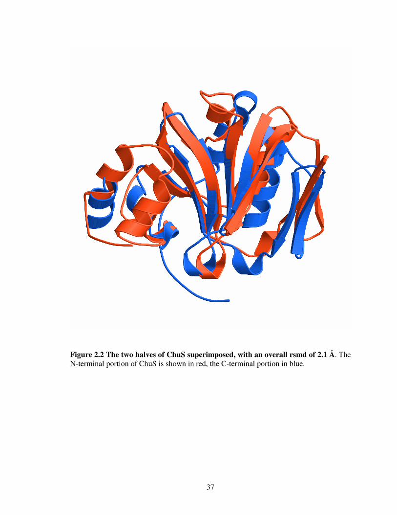
Figure 2.1 Ribbon diagram of ChuS. 35 Figure 2.2 The two halves of ChuS superimposed, with an overall rsmd 37
of 2.1 Å. Figure 2.3 Spectral analysis of ChuS reconstituted with heme. 40
Figure 2.4 EPR analysis of ChuS reconstituted with heme. 41
Figure 2.5 ChuS reconstituted with heme. 42 Figure 2.6 Spectral change of ChuS reconstituted with heme,
monitored over time following ascorbic acid addition. 44 Figure 2.7 Compiled average CO detected for ChuS, N-ChuS, and
C-ChuS. 46 Figure 2.8 Sequence alignment of ChuS with homologues from
various Gram-negative bacteria. 49 Figure 2.9 Inhibition of ChuS heme degradation by Sn(IV)
mesoporphyrin IX. 50 Figure 3.1 Ribbon diagram of ChuS in complex with heme. 67 Figure 3.2 Difference omit map for the heme moiety and two water molecules (blue) within the active site pocket of ChuS contoured to 2σ σ σ σ in the R3 space group. 70 Figure 3.3 Spectral change of ChuS reconstituted with heme,
monitored over time following ascorbic acid addition. 73 Figure 3.4 CO measurement via gas chromatography of ChuS, H73A,
H193 and heme following ascorbic acid addition and 25 min incubation (n=3). 74

ix
Figure 4.1 Sequence alignment of Chux with homologues from various Gram-negative bacteria. 93
Figure 4.2 Ribbon diagram of the four ChuX molecules in the crystallographic asymmetric unit. 95 Figure 4.3 Structural similarity of ChuX with ChuS and putative heme
binding cleft. 98 Figure 4.4 Spectral analysis of ChuX reconstituted with heme. 102 Figure 4.5 Spectra of ChuX and mutants reconstituted with heme 103
Figure 4.6 ChuX protection against heme toxicity and heme transfer 106 by ChuX. Figure 5.1 Heme degradation by ChuS in the presence of the hHO-1 specific inhibitor (2[2-(4-chlorophenyl)ethyl]-2- [(1H-imidazol-1-yl)methyl]-1,3-dioxolane). 126 Figure 5.2 Modified skematic of the heme assimilation system from
Gram-negative bacteria and organization of the heme utilization operon from Escherichia coli O157:H7. 129
Figure A.1 Sequence alignment of LptA with various Gram-negative bacterial homologues, many of which are human pathogens. 154 Figure A.2 Q-TOF Mass Spectrometry result, highlighting the LptA has been processed prior to crystallization. 155 Figure A.3 Anisotropic diffraction pattern from LptA MAD (left) and two-molecule native (right) crystals. 158
Figure A.4 Ribbon diagram of LptA with two molecules in the asymmetric unit at 2.6 Å resolution. 160 Figure A.5 Ribbon diagram of LptA highlighting residues conserved in sequence alignment with other Gram-negative homologues. 161 Figure A.6 Ribbon diagram of LptA with eight molecules in the asymmetric unit at 3.25 Å resolution. 162

x
ABBREVIATIONS
The Abbreviations used throughout this manuscript are:
ANL Argonne National Laboratory APS Advanced Photon Source ATP adenosine triphosphate BGG bovine globulin G BV biliverdin BR bilirubin BNL Brookhaven National Laboratory BSA bovine serum albumin CCD charge-coupled device CHESS Cornell High Energy Synchrotron Source CN cyanide CO carbon monoxide CPR cytochrome P450 reductase DLS dynamic light scattering DNA deoxyribonucleic acid EHEC enterohemmorhagic Escherichia coli
EPR eletroparamagnetic resonance FUR ferric uptake regulator GST glutathione S transferase HO heme oxygenase HO-1 heme oxygenase-1 HO-2 heme oxygenase-2 HPLC high-performance liquid chromatography HUS haemolytic uraemic syndrome IPTG isopropyl-1-thio-b-D-galactopyranoside IM inner membrane KDO 3-deoxy-D-manno-oct-2-ulosonic acid LB Luria Burtani Broth lipid A a phosphorylated glucosamine disaccharide acylated with fatty acids LPS lipopolysaccharide MacCHESS Macromolecular diffraction facilities at CHESS MAD multiple anomalous dispersion MS/MS tandem mass spectrometry MW molecular weight NADH reduced β-nicotinamide adenine dinucleotide NADPH reduced β-nicotinamide adenine dinucleotide phosphate Ni-NTA nickel nitrilotriacetate agarose NO nitrogen monoxide NMR nuclear magnetic resonance spectroscopy NSLS National Synchrotron Light Source OM outer membrane ORF open reading frame

xi
PAGE polyacrylamide gel electrophoresis PCR polymerase chain reaction PEG polyethylene glycol PDB protein data bank rmsd root mean squared deviation SAD single anomalous dispersion SCOP structural classification of proteins SDS sodium dodecyl sulphate SSM secondary structure matching STEC shiga-toxin producing Escherichia coli TB terrific broth TCA trichloroacetic acid

xii
LIST OF MUTANT PROTEINS Mutants generated as part of this investigation include: ChuS Mutants:
H73A, H193N, N-ChuS (N-terminal residues M1-P171 fused to a C-terminal His6-tag),
C-ChuS (C-terminal residues V172-A342 fused to an N-terminal GST-tag).
ChuX Mutants:
H65L, H98D, BetaX (ChuX triple mutant V69E/L71H/F73S), and DM (ChuX double
mutant H65L/H98D).

Chapter 1
General Introduction
2
1.1. General introduction and relevance
Escherichia coli is a constituent of the normal flora of the large intestine in
healthy vertebrates, and is widely distributed in soil and water. However, certain
pathogenic strains of E. coli such as O157 and CFT073 are known to cause a host of
clinical diseases in humans and animals including: mild, self-limiting diarrhea; severe
invasive gastro-intestinal infections such as dysentery and hemorrhagic colitis; urinary
tract infections; septicemia; and meningitis (131). While many of the patients infected
with these enterohaemorrhagic strains of E. coli (EHEC) (estimated 73,000 cases
annually in the United States of America (108)) respond well to treatment and recover
within a short period of time (70), the World Health Organization estimates that up to
10% of EHEC cases develop into hemolytic uremic syndrome (HUS), with an associated
fatality rate of 3-5 % (3). As a result, O157 is classified as an opportunistic pathogen and
a global public health concern. Other E. coli strains such as O26:H11 and O111:NM
share a similar pathogenic potential, but it is the O157:H7 serotype that has recently been
responsible for a few large EHEC outbreaks as well as a series of smaller cases
worldwide (56, 99). The sporadic cases of O157 outbreaks are often due to exposure to
undercooked ground beef (1), unpasteurized dairy (71), contaminated water and fruits or
vegetables (4, 13, 150), person to person transfer (23), and is especially harmful to
patients under five years of age (108). In Ontario, the highly publicized Walkerton
tragedy (May, 2000) was due to E. coli O157:H7 and Campylobacter jejuni contaminated
drinking water, which resulted in an estimated 2,300 people being infected; seven cases
of which were fatal (62).
3
Despite that E. coli is the model organism for scientific investigation there still
remain a large number of proteins and open reading frames (ORFs) from various strains
with either unknown function or with debatable annotation based on weak sequence
homology. Even in K-12, which is the most common and non-pathogenic strain of E. coli,
an estimated 30% of identified genes have unknown function and many other genes are
poorly characterized (68). A comparison of the O157 and K-12 E. coli genomes using
Sakai oligoDNA microarray and whole genome PCR scanning revealed that the two
chromosomes share 4.1 Mb of genetic information, with 1.4 Mb of O157 sequence-
specific genetic information (56, 99). Much of this O157-specific DNA is thought to have
arisen for horizontally transferred foreign DNA, and contains many bacteriophage
elements that mediate genetic reorganization and exchange, contributing to its
pathogenesis through frequent genetic change (56). Sequence analysis suggested the
O157 specific DNA encodes for 1632 unique ORFs, 20 unique tRNAs, and that at least
131 of the encoded proteins are recognized virulence factors (56). These unique elements
are therefore highly important, as understanding their function and interplay with other
protein partners will contribute to our overall understanding of bacterial pathogenesis as
well as principles and mechanism of infection. One level of protein characterization
involves structural determination of O157 specific proteins via cryo-electron microscopy,
NMR, and X-ray crystallography. As structural information can provide a plethora of
detail into the precise 3-D folding, interaction of proteins with substrates and other
molecules, mechanisms of action, and interaction with other proteins; as a whole,
structural characterization has the potential to improve our ability to prevent disease and
improve treatments in infected patients (74). The focus of this body of work is the

4
structural determination and functional annotation of ChuS and ChuX, two members of
the heme utilization operon in pathogenic E. coli O157:H7.
1.2. Bacterial iron acquisition from heme sources
Iron is the fourth most abundant element in the Earth's crust (6). However, due to
the low solubility of iron(III) salts at physiological pH in the presence of oxygen, iron
uptake, storage, and transport is a serious obstacle for bacteria to overcome for survival
and pathogenesis. A variety of bacterial iron-acquisition and transport systems have
evolved to assist survival and pathogenesis, such as the secretion of low-molecular-
weight (MW) siderophores (FyuA and IreA) capable of binding tightly to free iron and
mediating iron internalization via iron-specific membrane receptors (33, 100).
Reinforcing the importance for bacterial iron acquisition is that the iron assimilation
strategies they employ are highly redundant, for example, at least seven different
mechanisms have been identified in E. coli (130). As part of a coordinated immune
response against invading microorganisms, mammals specifically limit iron availability
by producing the iron-binding proteins transferrin and lactoferrin, which depress free
extracellular iron to a concentration of ~1x10-18 M in serum, insufficient to sustain
bacterial growth (6, 20). Furthermore, invading microorganisms are confronted with the
obstacle that 99.9% of iron within the human body is sequestered as protein bound heme
in hemoglobin, myoglobin, and various other heme enzymes (130).

5
Through the sequential participation of eight different enzymes, partly in the
mitochondria and partly in the cytoplasm, nucleated human cells and many bacteria can
synthesize heme from glycine and succinyl CoA (154). Heme is a propionic substituted
tetrapyrrole porphyrin group complexed to a central iron atom (Figure 1.1A). As a result
of the delocalized π-electron system of the porphyrin ring, the redox properties of the
central iron atom, and the variety of unique interactions possible for heme coordination
(104), heme is a widely used prosthetic group that enables proteins from various
organisms to fulfill many vital biological roles in photosynthesis, cell differentiation and
proliferation, as well as oxygen binding, transport, and utilization (89, 154). Beyond
heme simply acting as a prosthetic group, in HeLa cells, succinyl acetone-induced heme
deficiency was shown to increase the protein levels of the tumor suppressor gene product
p53 and CDK inhibitor p21. The effect was an overall decrease in the protein levels of
Cdk4, Cdc2, and cyclin D2, and diminish the activation/phosphorylation of Raf, MEK1/2,
and ERK1/2-components of the MAP kinase signaling pathway (168). This result
suggests a role for heme as an effector molecule beyond the regulation of proteins
associated with heme and iron metabolism. Furthermore, the regulatory potential effected
by hemoproteins contribute to all levels of gene expression including; DNA splicing,
transcription, mRNA stability, translation, and post-translational modifications (154). The
cumulative effect of heme uptake is therefore a cascade of gene up-regulation, with heme
effectively acting as a signaling molecule for invading microorganisms that they have
colonized a host (83). As a result, understanding the proteins and mechanisms involved
heme metabolism, transfer, and utilization has the potential to contribute greatly to our
understanding of many integral cellular processes as well as pathogen-host interactions.

6
αααα
ββββ
γγγγ
δδδδ
B
A
αααα
ββββ
γγγγ
δδδδ
B
A
Figure 1.1 Structure of Heme (protoporphyrin IX) and mechanism of heme degradation catalyzed by heme oxygenases. A) Carbon (yellow), oxygen (red), nitrogen (blue) form a conjugated planar molecule that coordinates a central iron (orange) atom. Meso-carbon positions are denoted with Greek symbols. The heme group is rotated to show that the molecule porphyrin is planar, and that the propionic side chains and vinyl groups extend out of the plane of the porphyrin ring. B) Heme oxygenase catalyzed degradation of heme to biliverdin requires a total of seven electrons and two equivalents of molecular oxygen. Most heme oxygenases are regioselective for the α-meso carbon position of the heme group.

7
Some microorganisms such as Haemophilus and Bacterioides are unable to
synthesize heme and hence must acquire heme in large amounts from host organisms.
Other bacteria, such as Borrelia and streptococci, are thought to neither synthesize nor
require heme for growth (130). Many other pathogens have been identified which use
host heme sources and have had heme utilization genes identified including: Bordetella
pertussis (18), Bradyrhizobium japonicum (106), C. jejuni (111), Haemophilus influenzae
(29), Neisseria meningitides (129), Porphyromonas gingivalis (124), Serratia marcescens
(50), Vibrio cholera (58), Yersinia enterocolitica (128), Yersinia pestis (63), E. coli
O157:H7 (91), and Shigella dysenteriae (91). Certain strains have evolved the capacity to
actively seek out and utilize host heme sources though the production of host-specific
hemoprotein proteases such as EspC (38), Pic (59), or Hbp (147), which separate heme
from host proteins such as hemoglobin and hepatoglobin. Heme may then be taken up by
bacterially produced hemophores such as HxuA (28) and HasA (50, 85), or transferred
directly to outer membrane receptors that have been identified in at least 18
microorganisms, many of which are human pathogens (48). Another class of receptors
interact with hemoproteins directly, mediating heme delivery without the need for
hemophores (156). However, sequence comparison of human heme oxygenase-1 (HO-1)
and bacterial HOs have failed to reveal homologues in some heme-utilizing bacteria such
as Escherichia, Plesiomonas, Shigella, Vibrio, and Yersinia (130).
Heme has a higher solubility relative to free iron and is therefore a more readily
transportable source of the metal (130). Once internalized, heme can readily be degraded
by HOs, releasing iron (Fe2+) along with the products biliverdin (BV) and carbon
monoxide (CO). In mammals, the downstream products of heme catabolism have

8
recently been found to mediate the antioxidant, anti-apoptotic, anti-proliferative,
vasodilatory, and anti-inflammatory properties of HO-1 in mammalian cells (45). For
bacteria, the liberated iron atom can then be incorporated into various proteins where it
acts as a biocatalyst or electron carrier, enabling organisms to fulfill many vital biological
processes such as N2 fixation, methanogenesis, H2 production and consumption,
respiration, the citric acid cycle, oxygen transport, gene regulation, and DNA
biosynthesis (6, 89). Alternatively, heme cleaved at the α-meso carbon position by HOs
produces BV IXa, which can subsequently be converted to phytobilins, precursors of the
chromophores for the light harvesting phycobiliproteins and photoreceptor phytochromes
(44). The structure and function of HOs will be addressed in Section 1.4.
While heme is one of the most wide spread enzyme cofactors in nature (130), the
properties which contribute to its usefulness in a controlled protein associated form also
make heme potentially cytotoxic in the free form. This is because heme is both a
hydrophobic molecule capable of freely diffusing into membranes where it can alter the
bilayer structure, thereby disrupting cell integrity (119), and can act as a potent
prooxidative agent, promoting the light-dependent formation of reactive oxygen species
that can cause non-enzymatic redox reactions (54). Heme synthesis and metabolism with
its utilization must therefore be tightly regulated; for this reason heme is normally found
tightly associated in various hemoproteins (54). Furthermore, invading microorganisms
must also tightly regulate heme acquisition and metabolism by either degrading heme or
storing it for later use in various processes (155). The regulation of heme uptake and
utilization must also be coordinated with iron requirements in order to avoid iron induced
toxicity. Using a human-based example, the disease associated with chronic iron overload

9
known as hemochromatosis causes rapidly progressing, multisystem disorders (32). Iron
metabolism is therefore tightly regulated in host organisms through the use of iron
scavenging elements such as transferrin and lactoferrin (6, 20), as well as through effects
mediated by transcriptional regulatory proteins such as Fur (48), DtxR (117), or FecI (40),
which bind to iron related elements on DNA known as Fur binding sites.
Enterohemorrhagic pathogens such as E. coli O157:H7, for which isolates from
human patients have been shown to have stimulated growth in the presence of heme and
hemoglobin; may induce hemolytic lesions in order to gain access to serum sources of
nutrients such as heme and iron (83). It may therefore be in part as a result of attempting
to acquire heme which elicits the clinical effects of pathogenic E. coli in HUS. Using a
laboratory strain of E. coli (1017 (ent::Tn5)) that was unable to import heme into the
cytoplasm, it was established that the outer membrane receptor ChuA (E. coli heme-
utilization gene A) is required for heme uptake along with TonB, an energy-transducing
protein associated with ExbB and ExbD that uses the proton motive force of the
cytoplasmic membrane for the passage of ligands into the periplasm (146). Four other
members of the heme uptake operon from Y. enterocolitica were shown to be required to
complete heme uptake, transport, and use as an iron source by E. coli K-12 (127)
including: the periplasmic heme-binding protein HemT, the heme permease protein
HemU, the ATP-binding hydrophilic protein HemV, and the protein HemS, whose role
may be to regulate the amount of non-protein associated heme in the cell (127, 128).
Restriction mapping and sequence analysis of selected regions of E. coli O157:H7
genome have revealed that the organization of the heme transport locus of this strain is
strikingly similar to that of other enteric bacteria such as Y. enterocolitica and
10
S. dysenteriae (127, 128). This genetic organization is especially true with respect to the
region encoding heme outer membrane receptors such as ChuA, ShuA, and HemR,
followed by a short gap, and then the gene encoding ChuS or one of its homologues. In S.
dysenteriae the intergenic region between ShuA and ShuS are separated by a 48-
nucleotide region with sequence homology to the consensus Fur box, but no obvious –10
and –35 promoter elements (128). However, in response to iron limiting conditions, ShuS
was upregulated, permitting heme as an iron source and preventing heme toxicity (165).
A summary of the proteins encoded within the heme utilization operon from E. coli
O157:H7 is summarized in Table 1.1.

11
Table 1.1 Summary of proteins encoded within the heme utilization operon from pathogenic Escherichai coli O157:H7.
Protein Localization Length (amino acids) Function
ChuA Outer
Membrane 660 Heme Receptor
ChuT Periplasm 304 Heme Transport
ChuU Inner
Membrane 330 Permease ABC Transport Protein
ChuV Inner
Membrane 256 Permease ABC Transport Protein
ChuS Cytoplasm 342 Heme/hemoglobin transport
protein ChuW Cytoplasm 445 Coproporphyrinogen III oxidase ChuX Cytoplasm 164 Hypothetical ChuY Cytoplasm 207 Hypothetical

12
1.3. Role of heme and structural diversity of heme-proteins
While traditional heme studies have focused on heme as a prosthetic group within
hemoglobin, myoglobin, and iron processing elements, novel heme-Fcontaining proteins
have recently been discovered. These include the cytochrome c chaperone CcmE (123),
the iron-dependent regulator of heme biosynthesis Irr (107), and the heme-based
aerotactic transducer Hem-AT (64). Other hemoproteins function through conformational
changes induced as a result of modifications to the ferrous heme environment upon the
coordination of small effector ligands such as NO, O2, or CO, as in the case of guanylate
cyclase (172), FixL (52), and CooA (81), respectively. Other newly identified heme-
associated signaling proteins include AxPDEA1, NPAS2, and EcDos whose interaction
between heme and various gases in the sensor domains mediate structural changes in the
effector domains of these proteins (115). While the cellular consequences mediated by
these signaling proteins are diverse, the common feature between them is that small
molecules can trigger major changes to the overall heme-protein environment, which can
distort the heme group itself or the interactions stabilizing the heme group, resulting in a
robust structural and functional transition.
While heme helps to mediate diverse responses, it is the interaction of heme with
various coordinating proteins that ultimately confer molecular function. Therefore,
characterizing the interactions that contribute to heme stabilization will also help to
characterize how heme mediates cell signaling. Furthermore, while heme helps to
mediate diverse responses, it is the interaction of heme with various coordinating proteins
that ultimately confer molecular function. Structural insights provided from vibrational

13
spectroscopy (EPR and Raman), nuclear magnetic resonance spectroscopy (NMR), and
X-ray crystallography have indicated that heme proteins often coordinate the iron center
of the heme group between a histidine side chain and a variety of other possible residues
including tyrosine (8), methionine (22), cysteine (39), proline (81), histidine (103),
arginine (137); while other proteins have been shown to coordinate the heme iron center
without additional assistance from other side chains (52, 121). Other proximal side chains
and regions of the polypeptide backbones have been shown to interact with other parts of
the heme group, contributing to the stabilization of the heme moiety within the protein
environment, and participating in the interactions which ultimately confers hemoprotein
function. These heme-protein interactions are both hydrophobic and hydrophilic,
stabilizing the porphyrin ring or propionic elements of the heme group, respectively, and
are mediated through various interactions.
Given the diversity of functions attributed to heme proteins together with the
various heme polypeptide interactions that have evolved, it is not surprising that heme
proteins feature a broad range of structural motifs and overall architectures. The structure
of sperm whale myoglobin, the first high resolution macro molecular structure solved by
X-ray crystallography, consists of eight α-helices that wrap around the heme group,
seating it within a hydrophobic pocket (72). The overall architecture of myoglobin is
similar to that of HO-1 whose overall structure is also mainly α-helical, and sandwiches
the heme group between two α-helices that are termed the proximal and distal helices
(121). In the smaller hemoprotein cytochrome c, the heme group is associated within a
simple, mainly α-helical fold (86) but resides closer to the surface than either myoglobin
or HO-1. Heme proteins may also assume a variety of other folds. In the heme

14
sequestering protein hemopexin found in serum, heme is coordinated between two β-
propeller domains (103), and in HasA the heme binding site is formed by loop regions
which extend from an αβ-fold (8). The structure of FixL resembles an open pita or clam
which coordinates heme in the center of the open mouth between a series of antiparallel
β-sheets at the protein core and an α-helix (52). A unique example of heme coordination
was recently reported for the iron regulated lipoprotein from C. jejuni, ChaN, whose
function is poorly understood, but seems to associate with the outer membrane receptor
ChuR (25). ChaN forms a homodimer which sandwiches a pair of heme molecules
between a pair of conserved tyrosine residues donated from opposing monomers, that
originate at the base of two α-helices. ChaN does not have a homologue in O157 or K-12,
but homologues have been identified in other E. coli strains.

15
1.4. Bacterial heme oxygenases
Although many reports have focused on the proteins involved in heme transport
across the outer membrane and periplasm, the fate of heme once it has reached the
bacterial cytoplasm has only recently received attention. Investigations of eukaryotes
revealed that release of iron stores requires the heme porphyrin ring to be degraded by
monooxygenases known HOs. HOs are enzymatically unique, as they use heme as both a
substrate and as a cofactor in the binding of oxygen for intramolecular degradation of the
porphyrin macromolecule to BV, CO, and Fe2+ (Figure 1.1B) (174). Structural insight
contributed by EPR and X-ray crystallography studies suggests that heme cleavage
catalyzed by HOs proceeds via similar mechanisms (73, 92). Initially, HO bound heme
accepts an electron from either NADPH-cytochrome P450 reductase (CPR), yielding a
ferrous (Fe2+) heme-HO complex. Molecular oxygen binds with the complex between the
iron atom and the axial histidine residue, forming a metastable oxy-form, that receives
another electron from CPR and a proton from the distal water pocket, and is rapidly
converted to a hydroperoxide intermediate (Fe3+–OOH). The oxygen of the
hydroperoxide intermediate attacks the α-meso carbon (or other regioselective position),
yielding a ferric α-meso-hydroxyheme. This intermediate then receives another oxygen
molecule and an electron and is converted to ferrous-verdoheme. Another oxygen and
reducing equivalent are received, transforming ferrous-verdoheme to ferric-BV and is the
least understood part of the mechanism (44). In the last step ferric-BV is further reduced
to the ferrous state, yielding BV and Fe2+. Overall the reaction requires an input of seven
electrons and two molecules of oxygen (Figure 1.1) (73, 92).

16
Several bacterial and mammalian HOs have been recently identified, including
HemO (173), HmuO (116), HmuQ/D (106), cyanobacterial HO-1 and HO-2 (30), IsdG/I
(125), mammalian HO-1 and HO-2 (121), nm-HO (122), PCC_6803 (132), and
PigA/BphO (46); and crystal structures have been determined for HemO (122), HmuO
(61), nm-HO (122), pa-HO (46), PigA (46), and Syn HO-2 (132), as well as a partial
structural characterization of BphO by NMR (157). Furthermore, extensive structural
work has been conducted on various HOs in complex with effector molecules such as
azide (135), biliverdin (134), CO and cyanide (136), ferric and ferrous forms of iron (79),
imidazole, and nitrogen monoxide (79) in order to effectively capture “snapshots” along
the reaction coordinate of heme degradation.
The overall topology of known bacterial HOs share strong structural similarity to
mammalian HOs in that they are mainly α-helical and sandwich heme between two
helices at the base of the structures, with the propionic side chains oriented away from the
protein (44). In this mode of heme coordination, HOs are regioselective for O2 addition
across the α-meso position of heme, which is oriented toward a series of polar residues
that do not participate directly in the reaction, but likely contribute to regioselectivity
(121). The exception to this mode of coordination is pa-HO from Pseudomonas
aeruginosa which is regioselective for the β- and δ-meso positions of heme in a 30:70
ratio, which has been attributed to heme being rotated by 100º in the active site (46). The
purpose of the generation of these two BV isomers is currently unknown, but is likely
related to the fact that P. aeruginosa has a second HO, BphO, with α-meso carbon
specificity (44). Despite the structural conservation across bacterial HOs identified to
date, it should be noted that many of these were targeted because they shared sequence

17
homology with mammalian HOs. In pa-HO the propionic side chains of the heme moiety
are stabilized in a unique fashion compared to other HOs identified. This highlights how
a variety of protein-heme interactions can mediate heme catalysis beyond those well
characterized in the hHO-1 related system. In many bacteria where sequence analysis
fails to suggest the enzyme responsible for heme processing, other approaches must be
utilized such as systematic gene knockouts and phenotypic characterization to identify
the genes responsible for heme utilization (165). At the initiation of our investigation
presented in this thesis, bioinformatics analysis had failed to identify a HO in any E. coli
strain, although both E. coli O157:H7 and CFTO73 were known to use heme as an iron
source, suggesting that a heme degrading enzyme was necessary.
1.5. Heme utilization protein ChuS
Because of its poorly understood role in modulating heme utilization and its
genetic organization downstream of the outer membrane receptor chuA, we directed our
attention to the gene product of chuS, the E. coli O157:H7 and CFT073 homologue of
shuS, as a candidate for intracellular heme breakdown (128). Sharing 98% identity to the
S. dysenteriae homologue ShuS, the structure and function of these two homologues is
likely highly similar. In the initial report characterizing ShuS in isolation of other
members of the heme utilization operon, ShuS was shown to form a high molecular
weight oligomer (650 kDa) from 37 kDa subunits, to nonspecifically bind double
stranded DNA, and was not observed to exhibit any HO activity in standard NADPH or
ascorbate based assays (158). In contrast, in an S. dysenteriae knockout, the shuS mutant

18
was defective in utilizing heme as an iron source and ShuS was required under
microaerobic and anaerobic conditions for the optimal utilization of heme (165). ShuS
expression also protected cells from heme toxicity at high concentrations, and was
therefore suggested to bind to and potentially transfer heme from the transport proteins in
the membrane to either heme containing or heme degrading proteins, or involve itself as a
heme degrading enzyme (165). Furthermore, expression of the Y. enterocolitica
homologue to ChuS, HemS, protected E. coli cells against heme toxicity (127).
1.6. ChuW, ChuX, ChuY and bacterial heme proteins
In addition to chuS, three other genes: chuW, chuX, and chuY are located within
the heme utilization locus of O157 were poorly characterized at the initiation of our study.
ChuW does not share any homology to any protein identified in heme transport, but does
have weak homology to HemN, the oxygen-independent form of the heme biosynthetic
enzyme co-proporphyrinogen oxygenase III present in various bacteria including Bacillus
ssp. Salmonella typhimurium and E. coli K-12 (164, 167). This homology suggests that
ChuW may play a role in some aspect of heme transport or metabolism. In S. dysenteriae
shuW begins 19 nucleotides downstream of shuT, and the lack of any apparent
transcription termination or promoter sequences suggests that shuT and shuW are located
on the same transcript (127, 128, 164, 165). In S. dysenteriae type 1, a premature stop
codon exists within shuW suggesting that ShuW may be defective, although in E. coli
O157:H7 this stop codon is absent in chuW suggesting that ChuW may be completely

19
functional (164). Furthermore, DNA microarray analysis of CFTO73 strains suggested
that chuW was upregulated by more than 5-fold during urinary tract infection (126).
Twelve nucleotides downstream of shuW is the start codon for shuX, and the
initiation condon of shuY overlaps the stop codon of shuX. This indicate that these two
smaller genes may also be co-transcribed with shuT (164). In a similar sequence analysis
of the heme uptake locus from Y. pestis, homologous to chuX and chuY (orfX and orfY,
respectively) were determined to be located immediately downstream of the heme
receptor gene hmuR and were therefore likely co-expressed (145). In Vibrio anguillarum,
which can use heme and hemoglobin as a source of iron, using a lacZ reporter system it
was shown that a homologue of chuX, huvX, was co-transcribed with huvZ under iron
limiting conditions (95). An E. coli homologue to huvZ was not identified via NCBI
Blastp sequence analysis at (www.ncbi.nlm.nih.gov/BLAST/). Furthermore, an
examination of the -10 and -35 elements downstream of huvX revealed sequence with
similarity to sigma70-promoters, as well as the presence of a Fur element (95).
Characterization of either chuX or chuY gene products or their homologues from various
heme utilizing organisms had not received any attention at the beginning of our
investigation. However, based on Blastp sequence analysis ChuX was suggested to be a
protein associated with heme-iron utilization, and that ChuY is a hypothetical protein or
nucleoside-diphosphate-sugar epimerase (143). A schematic-diagram of the heme
assimilation system of Gram-negative bacteria, as well as the structures and functions of
members of the heme utilization operon at the initiation of our investigation is
summarized in Figure 1.2.

20
OM
PP
IM
HasA
Hbp
ChuA?
TonB
ExbB? ExbD?ExbB?ExbB? ExbD?
ChuT?
ChuU? ChuV?
ChuW?ChuW?
Function
V U Y X W T A S
? ? ? ?
Figure 1.2 Schematic-diagram of the heme assimilation system from Gram-negative bacteria at the initiation of our investigation and organization of the heme utilization operon from Escherichia coli O157:H7. Proteins involved in heme transport from pathogenic E. coli O157:H7 are presented in their locations in the outer membrane (OM), periplasm (PP), and inner membrane (IM). Where the structures of proteins are unknown, structures of homologues have been included and names followed by a question mark. Putative promoter elements within the heme utilization operon are depicted with arrows. The function of ChuS, ChuW, ChuX, and ChuY were unknown at that time. The proteins focused on as part of this body of work are ChuX and ChuS.

21
In light of the general importance of heme as a signaling molecule and cofactor,
this thesis examines the structure and function of two members of the E. coli O157:H7
heme utilization operon: the utilization protein ChuS in its apo and heme bound forms,
and the heme binding protein ChuX. The method of X-ray crystallography, specifically,
the anomalous dispersion method using seleno-methionine labeled proteins has been used
for structural determination. A combination of spectral characterization, site-directed
mutagenesis of conserved residues, gas chromatography, and sensitivity of cultures
challenged with heme provide biochemical evidence for the function of ChuS and ChuX.
The application of structure based functional characterization as well as the implications
of our findings for heme uptake and utilization in E. coli O157:H7 and Gram-negative
bacteria in general is addressed.

22
Chapter 2
Identification of a novel Escherichia coli O157:H7 heme oxygenase with tandem functional repeats
Preface:
This chapter was published in The Proceedings of the National Academy of Sciences:
Michael D. L. Suits, Gour P. Pal, Kanji Nakatsu, Allan Matte, Miroslaw Cygler, and
Zongchao Jia (2005) Identification of a novel Escherichia coli O157:H7 heme oxygenase
with tandem functional repeats. PNAS, 102(47):16955-60.
Michael Suits was responsible for protein expression, purification, crystallization, data
collection, structure solution and refinement, and all biochemical experiments in the
investigation of function. Dr. B. McLaughlin helped with gas chromatography and
interpretation, Dr. B. Hill conducted EPR experimentation and interpretation, and expert
help from Dr. Mike Nesheim in heme reconstitution setup and interpretation of results to
calculate the Kd for ChuS-heme. The chuS gene was cloned by Pietro Iannuzzi and the N-
and C-terminal halves of ChuS by Jim Blonde. The manuscript was written by Michael
Suits with editorial input from Dr. Zongchao Jia and with additional help from Dr. John S.
Elce.
Data deposition footnote: Coordinates and structure factors for ChuS are deposited in the RSCB Protein Data Bank under accession code 1U9T.

23
2.1. Abstract
Heme oxygenases (HOs) catalyze the oxidation of heme to biliverdin, carbon
monoxide (CO), and free iron. Iron acquisition is critical for invading microorganisms to
enable survival and growth. Here we report the crystal structure of ChuS, which displays
a novel fold and is unique compared to other HOs characterized to date. Despite only
19% sequence identity between the N- and C-terminal halves, these segments of ChuS
represent a structural duplication, with a root mean square deviation of 2.1 Å between the
two repeats. ChuS is capable of using either ascorbic acid or cytochrome P450 reductase-
NADPH as electron sources for heme oxygenation. CO detection confirmed that ChuS is
a HO, the first to be identified in any strain of Escherichia coli. Based on sequence
analysis, this novel HO is present in many bacteria, though not in the E. coli K-12 strain.
The N- and C-terminal halves of ChuS are each a functional HO.

24
2.2. Introduction
The incorporation of iron into proteins as a biocatalyst or electron carrier enables
organisms to fulfill many vital biological processes including photosynthesis, N2 fixation,
methanogenesis, H2 production and consumption, respiration, the tricarboxylic acid cycle,
oxygen transport, gene regulation, and DNA biosynthesis (89). The importance of iron
for bacterial survival and pathogenesis is evident by the variety of iron-acquisition and
transport systems that have evolved, such as the secretion of low-molecular-weight (MW)
siderophores capable of binding free iron and transporting it into the cell via specific
membrane receptors (33). As part of a coordinated immune response against invading
microorganisms, mammals specifically limit iron availability by producing the iron-
binding proteins transferrin and lactoferrin, which depress free extracellular iron to levels
insufficient to sustain bacterial growth (3). Alternatively, invading pathogens may
acquire iron directly from host iron sources such as heme, hemoglobin, or hemopexin
either through the production of specific outer membrane receptors that bind directly to
host heme-sequestering proteins or through the secretion of heme-binding proteins
(hemophores) that then deliver the heme-protein complex to bacterial cell surface
receptors (48). Enterohemorrhagic pathogens such as Escherichia coli O157:H7, for
which isolates from human patients have been shown to have stimulated growth in the
presence of heme and hemoglobin, may induce hemolytic lesions in order to gain access
to these serum sources of iron (83).
Using a laboratory strain of E. coli (1017 (ent::Tn5)) that was unable to import
heme into the cytoplasm, it was established that the outer membrane receptor ChuA

25
(E. coli heme-utilization gene A) is required for heme uptake along with TonB, an
energy-transducing protein associated with ExbB and ExbD that uses the proton motive
force of the cytoplasmic membrane for the passage of ligands into the periplasm (146).
Four other members of the heme uptake operon from Yersinia enterocolitica are required
to complete heme uptake, transport, and use as an iron source by E. coli K-12 (127)
including: the periplasmic heme-binding protein HemT, the heme permease protein
HemU, the ATP-binding hydrophilic protein HemV, and the protein HemS, whose role
may be to regulate the amount of non-protein-associated heme in the cell (128).
Restriction mapping and sequence analysis of selected regions of E. coli O157:H7
DNA, the serotype responsible for outbreaks of hemorrhagic colitis and hemolytic uremic
syndrome, has revealed that the organization of the heme transport locus of this strain is
strickingly similar to that of other enteric bacteria such as Y. enterocolitica (164). This
organization is especially true with respect to the region encoding heme outer membrane
receptors such as ChuA, which are followed by a short gap and then by ChuS or one of
its homologues. In Shigella dysenteriae the intergenic region between ShuA and ShuS are
separated by a 48-nucleotide region with sequence homology to the consensus Fur box
but no obvious –10 and –35 promoter elements (127). Furthermore, a promoter was likely
to be present within the intergenic region because a mini=Tn10 insertion in ShuA was not
polar with respect to ShuS expression in minicells (91).
Although many reports have focused on the proteins involved in heme transport
across the outer membrane and periplasm, the fate of heme once it has reached the
bacterial cytoplasm has only recently received attention. Investigations in eukaryotes
revealed that release of iron stores requires the heme porphyrin ring to be degraded by

26
monooxygenases known as heme oxygenases (HOs). HOs are enzymatically unique, as
they use heme as both a substrate and a cofactor in the binding of oxygen for
intramolecular degradation of the porphyrin macromolecule to biliverdin, CO, and free
iron (121). Several bacterial HOs have been identified, including HemO (173), HmuO
(26), IsdG/I (125), cyanobacterial HO-1 and HO-2 (132), and PigA/BphO (110). All of
these were reviewed (44), and the structures of HemO (122), HmuO (61), and PigA (46)
have been determined, as well as a partial structural characterization for BphO (157). The
crystal structures of HemO, HmuO, and PigA all share a strong structural similarity to
mammalian HOs: they are mainly α-helical and sandwich heme between two helices. At
present, no sequence comparison or structural analysis has identified a HO in an E. coli
strain.
Here we report the crystal structure of ChuS, which has a novel fold with two
tandem repeats and does not show structural similarity to known mammalian or bacterial
HOs. ChuS is capable of binding heme and can break down heme using ascorbic acid or
cytochrome P450 reductase-NADPH (CPR) as electron sources for oxygenation. CO
detection by gas chromatography confirmed that ChuS is a HO, the first to be identified
in E. coli. In addition, HO activity occurs independently in the N- and C-terminal halves
of ChuS.

27
2.3. Materials and methods
2.3.1 Sequence analysis, protein expression, and purification
Sequence analysis was carried out using Blast (5). Full-length ChuS and the N-
and C-terminal halves of ChuS were subcloned into pET expression vectors, yielding
fusion proteins to either a N- or C-terminal His tag or an N-terminal GST tag. Based on
the results of small-scale expression trials, we selected the following constructs: ChuS
fused to an N-terminal His6 tag (ChuS), the N-terminal half fused to a C-terminal His6 tag
(N-ChuS), and the C-terminal half fused to an N-terminal GST tag (C-ChuS). In each
case, 1-L cultures of BL21(DE3) cells carrying plasmid for the respective recombinant
protein were grown at 37°C in Terrific Broth (Bioshop, Burlington, Canada)
supplemented with 100 µg/ml ampicillin. Protein expression was induced using 0.4 mM
IPTG for 5 h. ChuS protein substituted with seleno-methionine was expressed in the
metA- E. coli strain DL41 in LeMaster medium (60).
All proteins were purified using standard methods. Briefly, His6-tagged proteins
were purified via Ni-NTA batch purification in phosphate buffer (pH 8.0), followed by
purification by Resource Q and Hi-Trap metal-chelating columns on an ATKA Explorer
FPLC. Collected fractions were monitored by SDS-PAGE, for which those that contained
pure protein were pooled together and dialyzed against 50 mM Tris-HCl buffer (pH 8.0),
150 mM NaCl for subsequent crystallization, or 100 mM sodium phosphate buffer (pH
7.0) for HO spectral and activity measurements. C-ChuS was purified on a GSTrap FF
column.

28
2.3.2 Crystallization of full-length ChuS
Crystals were obtained by the hanging-drop vapor diffusion method by mixing
2.5 µl of 20 mg/ml ChuS with 2.5 µl of reservoir solution consisting of 20% (w/v) PEG
3350 and 100 mM Bis-Tris (pH 6.5). Crystals appeared after 1-3 d. ChuS crystals were
further improved by microseeding using 12–20% (w/v) PEG 3350. SeMet derivative
crystals were obtained in the same way. For cryoprotection, crystals were soaked in the
reservoir solution containing ethylene glycol (concentration increased stepwise to 30%
[v/v]), picked up using a nylon loop, and flash-cooled in the N2 cold stream at 100K. Two
nonisomorphous crystal forms belonging to space group P21 were obtained. Crystals
having the larger unit cell contain three molecules in the asymmetric unit, whereas those
with the smaller unit cell contain only one molecule.
2.2.3 Data collection and structure determination
Native and multiple-wavelength anomalous dispersion (MAD) data sets were
collected, respectively, for the two different crystal forms at beamlines 17-ID and 19-BM
of the Advanced Photon Source, Argonne National Laboratory. The native data were
collected for 180° in total, with 1.0° oscillation. SeMet MAD data were collected at Se
peak and inflection wavelengths for 360° and remote wavelength for 180°, all with 1.0°
oscillation. Data were processed with HKL2000 (102). Initial phases for the three-
molecule form was determined using the MAD data with the program SOLVE (144).
Density modification, including three-fold NCS averaging, was carried out using

29
RESOLVE (144). Automatic chain tracing was performed using RESOLVE (144), and
the remainder of the model was built by manual fitting using XtalView/Xfit (90). When
the model of one of the three molecules was about 80% built, it was used as a search
model in molecular replacement using data obtained from the single-molecule form. This
resulted in an unambiguous solution that was subsequently refined using CNS (19).
2.3.4 Reconstitution with heme and absorbance measurements
Heme complexes of ChuS, N-ChuS, and C-ChuS were prepared by dissolving
heme into 0.5% (v/v) ethanolamine and adding small volumes of this mixture to protein
until a final ratio of 4:1 (mol:mol) was obtained for ChuS and 2:1 for N-ChuS and
C-ChuS. ChuS-heme and N-ChuS-heme were then applied to Resource Q or Superdex
200 Prep Grade columns, respectively, to remove noncoordinated heme. Reconstituted
protein-heme complex concentrations were determined by adapting the reported
extinction coefficient for ShuS (ε410) of 79.5 and 159 mM-1cm-1 for ChuS and N-ChuS,
respectively (11). Pure fractions of C-ChuS were concentrated and used for
spectrophotometric analysis and CO detection in which twice the amount of heme was
added to protein prior to measurements. Absorbance data were then collected using a
microplate spectrophotometer (Bio-Tek Instruments, Inc.). As a control in separate
experiments, 10 µM catalase and 10 µM superoxide dismutase were added to the reaction
wells, followed by ascorbic acid or cytochrome P450 reductase (CPR)-NADPH, and the
difference spectra were recorded. A quadruplicate series of ChuS-heme reconstitution
titrations were also conducted to examine the ChuS-heme interaction.

30
2.3.5 CO activity assay and inhibition
Briefly, in 2-ml vials 250 µl reaction volumes of 20 µM heme and 10 µM ChuS,
N-ChuS, or C-ChuS in 100 mM phosphate buffer (pH 7.0) were incubated with constant
shaking at room temperature for 6 min at which time enzymatic heme degradation was
initiated by adding either CPR and NADPH or ascorbic acid at final concentrations of
17.7 nM and 135 µM, or 50 µM, respectively. Vials were then sealed with screw caps
and blue silicone rubber septa and the headspace above each was immediately purged
with CO-free air, and incubated for 45 min at 24°C with constant shaking. The reaction
was stopped by placing the vials on powdered dry ice (-78°C), where they remained for
approximately 30 min. The amount of liberated CO in the headspace of each vial was
measured using a gas chromatograph equipped with an HgO reduction detector (Trace
Analytical). The amount of CO resulting from ChuS-catalyzed breakdown of heme was
determined by comparing peak area measurements for CO against linear CO standard
curves (n = 20 determinations; average correlation coefficient 0.993). Technical details
are described elsewhere (27, 153). In a parallel experiment, Sn(IV) mesoporphyrin IX
dissolved in 0.5% (v/v) ethanolamine was also added during initial mixing which acted as
a competitive inhibitor.
2.3.6 Dynamic light scattering of C-ChuS and N-ChuS

31
Dynamic light scattering (DLS) was conducted for ChuS, ChuS-heme, and N-
ChuS each at 1 mg/ml concentration in 100 mM phosphate buffer (pH 7.0) using a
DynaPro99 instrument (Protein Solutions). DLS was also conduced on samples of
1 mg/ml ChuS and ChuS-heme in 50 mM Tris and 150 mM NaCl (pH 8.0).
2.3.7 EPR experiments
EPR spectra were obtained on a Bruker EMX spectrometer at X-band fitted with
an HSQ cavity equipped with a liquid helium flow cryostat (Oxford Instruments). The
standard instrument conditions were T = 10K, modulation amplitude = 20 G, power =
2 mW at a gain of 1 × 10⁴.
2.4 Results
2.4.1 Expression and purification
ChuS overexpression resulted in cells exhibiting a blue-green pigment
characteristic of biliverdin accumulation (87, 157, 174). Following Ni-NTA purification
and dialysis in low-ionic-strength Tris buffer, fractions containing ChuS appeared purple
and turned to a straw color over time. EPR of ChuS fractions following Ni-NTA
purification did not reveal the presence of any coordinated metal ion, and spectral data
did not reveal the source of the pigmentation. This pigmentation was not observed in
ChuS grown in LeMaster medium in DL41(DE3) cells or for the N-ChuS or C-ChuS

32
constructs. The purple coloration of ChuS was lost following the subsequent FPLC anion
exchange step. Expression of full-length ChuS and N-ChuS yielded more than 60 mg of
pure protein from 1 L culture. Recombinant C-ChuS was insoluble in pilot expression
trials for N- or C-terminal His-tagged versions, and could only be expressed in soluble
form as a GST-C-ChuS construct. Average yields of 2.6 mg pure GST-C-ChuS/ L culture
were obtained.
2.4.2 Structure determination and analysis
Two different crystal forms were obtained from the same crystallization
conditions, that shared the same overall morphology and space group (P21), but differed
in unit cell dimensions and number of molecules in the asymmetric unit. We collected
MAD data for the three-molecule crystal form and native data for the single-molecule
crystal form, and the crystal structure of ChuS was solved using a combination of MAD
and molecular replacement with the two crystal forms. The higher resolution (2.15 Å)
structure from the single-molecule form is presented here, although the final structures
for ChuS from the two crystal forms are essentially identical, with an average root mean
square deviation (rsmd) of 0.92 Å. The final structure of ChuS contains residues 1–337 of
a total 342 amino acids with no ligand. Due to an absence of electron density, a gap exists
in the structure between residues 12 and 23 (12-QNPGKYARDI-23), and between 165
and 173 (165-KAVDAPV-173). In total, 99.7% of residues are within the allowed region
of the Ramachandran plot. Only one residue, Asp 114, was in the disallowed region and

33
resides within a surface exposed β-turn. Final refinement statistics are summarized in
Table 1.
The overall structure of ChuS comprises a central core of two large pleated
β-sheets, each consisting of nine anti-parallel β-strands sandwiched together and bowing
outward in a saddle motif (Figure 2.1). Each β-sheet is flanked at its N-terminus by one
pair of parallel α-helices and at its C-terminus by a set of three α-helices in an α-loop-α-
loop-α configuration. Two large clefts are found on opposite sides of the central core of
β-pleated sheets, with the third side of the clefts delineated by the flanking sets of three
α-helices. A long stretch of coil connecting the N- and C-terminal halves runs from I156
to V183, in which the gap in density occurs (Figure 2.1). Structural comparison of ChuS
against structures within the SCOP database (97) was performed with the program SSM
(77). This search did not reveal any significant match; hence ChuS represents a novel
fold.

34
Table 2.1 Diffraction data and refinement statistics for ChuS
Native Peak Inflection Remote Space group P21 P21 Cell dimensions a (Å) 41.683 46.072 b (Å) 57.878 194.172 c (Å) 59.575 60.461 β (°) 96.020 100.383 No. of molecules in ASU 1 3 Wavelength (Å) 0.9795 0.9803 0.9804 0.9642 Resolution range (Å) 50–2.15 50–2.60 50–2.60 50–2.40
Total reflections 53,596a 170,485b 176,691b 149,352b
Unique reflections 14,606a 32,694b 32,966b 40,453b
Completeness (%) 96.9 (82.8)# 99.8 (99.6) 99.7 (98.4) 96.3 (76.5)
Rsym(I) (%) 7 9.7 9.1 7.8 I/σI 15.0 (2.4) 17.4 (2.4) 16.6 (2.0) 11.4 (1.0) Refinement (single molecule form) Resolution range (Å) 50 – 2.15 Rwork, (%) 20.1 Rfree, (%) 26.3 No. reflections total/ Rfree 12315/ 744 No. atoms, protein / solvent (H2O)/ total 2428/ 331/ 2759
B factors (Å2) protein/ solvent (H2O)/ total 29.7/ 48.6/ 32.0 RMS, bond length (Å)/ bond angle(o) 0.009/ 1.44
#Values in parentheses are for the outermost shell (2.23–2.15 Å) aNumber of reflections following merging bNumber of reflections without merging of Bijovet pairs

35
Figure 2.1 Ribbon diagram of ChuS. The core of nine anti-parallel β-strands is visible in the front face. The gaps in the structure are shown with a dashed lines.

36
2.4.3 ChuS contains a structural duplication
On close inspection, the two halves of ChuS were found to be structurally similar.
Including side chains the two halves superimpose to an rmsd of 2.1 Å (Figure 2.2).
Sequence alignment of the two halves reveals only 19% identity. Nevertheless, at the
structure based level, many residues are well conserved structurally, including R29 and
R209, F127 and F304, and Y138 and Y315. The two halves of ChuS associate across the
central portion of the β-pleated sheets. At the interface, residues from the C-terminal half
are hydrophobic (V244, V250, F274, L276), point toward the interior of the C-terminal
subunit, and expose regions of the backbone across the interface. In contrast, most
interface residues from the N-terminal half are hydrophilic (N66, Y70, H73, N75, R95)
and point across the interface between the two halves. The side chains of the N-terminal
interacting residues are in contact with the backbone region of the C-terminal half. This
structural similarity suggests that the two halves of ChuS may be functionally
independent or that they have dual functions. Furthermore, the second gap in the structure
is exactly at the midpoint of the ChuS sequence in a flexible/disordered linker connecting
the two halves.
37
Figure 2.2 The two halves of ChuS superimposed, with an overall rsmd of 2.1 Å. The N-terminal portion of ChuS is shown in red, the C-terminal portion in blue.

38
2.4.4 Spectral properties of the heme-ChuS complex
Based on difference spectra comparing free heme to full-length ChuS, N-ChuS,
and C-ChuS, we determined that all three formed complexes when reconstituted with
heme (Figures 2.3A and 2.3B). The spectra of ChuS resemble those of other HOs in that
a Soret maximum is evident at 408 nm, with a smaller set of peaks for the β-band at
545 nm and for the α-band at 580 nm, suggesting that the ChuS-heme complex formed is
ferric hexacoordinate in the high-spin state at neutral pH (26). The spectrum for N-ChuS
has a peak at 402 nm (which is smaller than for ChuS), a smaller peak at 530 nm, and a
shoulder at 625 nm. Although the spectrum for N-ChuS with heme did not show
definitive heme coordination, it remained unchanged following gel filtration, indicating a
specific interaction between the two. C-ChuS with heme showed a broad peak centered at
384 nm, a smaller peak at 540 nm, and two small shoulders at 585 and 625 nm.
Furthermore, EPR spectra analysis suggested the presence of two populations of heme
molecules (Figure 2.4).
Despite structural similarity between the two halves, the spectra for C-ChuS and
N-ChuS differed, perhaps indicating different environments for heme coordination.
Based on absorbance at 408 nm during ChuS-heme reconstitution and assuming two
binding sites, the estimated Kd is 1.0 ± 0.3 µM (n = 3) based on the following expression:
])24}2({2[5.0)( 2/100
20000 HsitesPKHPsitesHKPsites ddfb ⋅−++⋅−++⋅−=∆Α εε ,
39
where P0 is the amount of protein, H0 is the concentration of heme, and (εb – εf) is the
difference in the Soret absorption spectra between the bound and free states (Figure 2.5).
This estimated Kd suggests a slightly weaker association than that characterized for the
mammalian hHO-1 (0.84 ± 0.2 µM) (159, 160) but tighter than that for other bacterial
heme oxygenases: HmuO (2.5 ± 1.0 µM) (162), IsdG (5.0 ± 1.5 µM) and IsdI (3.5 ±
1.4 µM) (125).

40
Figure 2.3 Spectral analysis of ChuS reconstituted with heme. Full-length ChuS and the N- and C-terminal halves of ChuS all form complexes when reconstituted with heme, with peaks at 408, 402, and 384 nm, respectively (back lines). A) Following addition of ascorbic acid as an electron donor, ChuS and its two halves were all capable of heme oxygenation, as evident from the spectral shifts (gray lines). Absorbance values corresponding to wavelengths greater than 500 nm have been multiplied by 10 to amplify absorbance signals corresponding to the β- and α-bands. B) Similar to A), but NADPH and the P450 reductase system were used as the electron source.

41
Figure 2.4 EPR analysis of ChuS reconstituted with heme. Based on spectra comparison with myoglobin reconstituted with heme, ChuS-heme exists in two spin states.

42
µµµµµµµµ
Figure 2.5 ChuS reconstituted with heme. Based on absorbance at 408 nm during ChuS-heme reconstitution and assuming two binding sites, the estimated Kd is 1.0 ± 0.3 µM (n = 3).

43
2.4.5 Heme oxygenation by ChuS
Bacterial and mammalian HOs use the CPR-NADPH system, ferredoxin,
flavodoxin, or ascorbic acid as reducing partners in vitro (160). To effectively monitor
and quantify the HO activity of ChuS, 50 µM ascorbic acid was used. This concentration
is between 100- (21) and 350-fold (26) less than that used for characterizing other HOs.
The use of this lower concentration of ascorbic acid demonstrates highly efficient
catalysis of ChuS with this electron donor. Furthermore, by using small amounts of
ascorbic acid, heme degradation through coupled oxidation was minimized (34). To
examine the heme degrading activity of ChuS, we monitored the reaction by UV-visible
spectroscopy after the addition of ascorbic acid (Figure 2.6). As the reaction progressed,
the Soret peak at 408 nm decreased, the small broad peak at 560 nm increased, and the
absorption at 680 nm initially increased then decreased. Because the addition of catalase
and superoxide dismutase did not affect the trends of the spectral change, this effect can
be attributed to specific heme degradation by ChuS and its variants. These spectral shifts
are similar to those observed for other HOs, which suggests the formation of biliverdin
and free iron rather than the Fe3+-biliverdin complex formed by HO-1. Furthermore,
similar spectral shifts were also observed for heme reconstituted both to N-ChuS and C-
ChuS, indicating that both halves of ChuS are capable of degrading heme independently
(Figure 2.3A). Similar endpoint spectra were also observed for ChuS, N-ChuS, and C-
ChuS when CPR was used as the electron donor (Figure 2.3B).

44
0
0.1
0.2
0.3
0.4
0.5
0.6
0.7
0.8
320
340
360
380
400
420
440
460
480
500
520
540
560
580
600
620
640
660
680
700
Wavelength (nm)
Ab
so
rban
cy Zero
45 sec
1.5 min
3 min
4.5 min
6 min
7.5min
8.5 min
10 min
16 min
20 min
1 hour
5x
Figure 2.6 Spectral change of ChuS reconstituted with heme, monitored over time following ascorbic acid addition. Absorbance measurements between 500 and 700 nm have been multiplied by five to amplify the change in the β- and α-bands. Arrows follow changes in absorption at 408, 545, and 680 nm during reaction progression. The spectral change corresponds with the formation of the product, biliverdin.

45
2.4.6 Detection of CO release
All three ChuS constructs were capable of releasing CO (Figure 2.7). When
ascorbic acid was used as the electron source, C-ChuS was 1.5 times more effective at
CO production than ChuS and 10 times more effective then N-ChuS, whose release was
only slightly above the background level of heme breakdown. When both N-ChuS and
C-ChuS were added to the same reaction vessel, the amount of CO evolved was greater
than the levels observed for ChuS alone, but not as great as those observed for C-ChuS.
When CPR was used as the electron source, the amount of CO produced by ChuS and
C-ChuS was five times less than when ascorbic acid was used. In contrast, N-ChuS
produced more than four times as much CO as when ascorbic acid was used and was
slightly more effective at producing CO than either the C-ChuS or full-length ChuS.

46
0.0
2.0
4.0
6.0
8.0
10.0
12.0
14.0
16.0
18.0
Hem
e
ChuS
N-C
huS
C-C
huS
N +
C C
huS
Av
era
ge
CO
Re
lea
se
d (
pm
ol/
min
) Ascorbic Acid
Cytochrome P450Reductase-NADPH
Figure 2.7 Compiled average CO detected for ChuS, N-ChuS, and C-ChuS. Electron donors were either ascorbic acid (red) or cytochrome P450 reductase-NADPH (blue) as an electron source for heme oxygenation. Error bars represent standard errors.

47
2.4.7 Organization of the heme uptake operon
Given the similarity between the heme uptake operon of Y. enterocolitica and
E. coli O157:H7, it is likely that homologues of HemR, HemT, HemU, HemV, and HemS
would be required for heme assimilation and use in E. coli. Sequence comparison
between the heme uptake loci of these organisms revealed the homologous genes ChuA,
ChuT, ChuU, various putative ATP-binding components of the heme transport system,
and ChuS. Sequence identities ranged between 38% and 68%, and similarities were
between 58% and 80%. The highest identity was conserved between HemS and ChuS,
suggesting conservation of function in iron acquisition and prevention of heme toxicity.
The lower identity with the other proteins may reflect differences between the
periplasmic and membrane environments of the two organisms. Furthermore, sequence
comparison revealed the following ChuS orthologs with strong sequence similarity: ChuS
(CFT073), ShuS (Shigella), EhuS (Enterobacter, Erwinia), HemS (Yersinia), plu2633
(Photorhabdus), HmuS (Rhizobium, Sinorhizobium), and BhuS (Bordetella), suggesting
that this HO is present in many other bacteria (Figure 2.8). Furthermore, conserved
histidine residues at positions 73, 87, 193 and 277 may be important for heme
coordination.
2.4.8 Dynamic light scattering
The derived molecular radii of N-ChuS, ChuS, and ChuS-heme in phosphate
buffer and ChuS and ChuS-heme in Tris buffers were all approximately 2.90 nm,

48
corresponding to a MW of 41 kDa for an elongated protein. This is in agreement with
that expected for an N-ChuS dimer or ChuS monomer. The longest dimension of ChuS
from the crystal structure corresponds to a radius of 2.88 nm, indicating that the N-ChuS
homodimer is similar in shape to full-length ChuS. The monomeric organization of ChuS
was confirmed using a gel filtration column calibrated with a set of MW standards. This
finding is contradictory to a previous report showing that a homologue of ChuS, ShuS
(98% identity), forms an oligomer with an estimated MW of 650 kDa (11). When ChuS
or ChuS reconstituted with heme were examined in the Tris buffer system following 4 d
of incubation at 4°C, DLS results suggested the formation of various high-MW
aggregates without any detection of a ChuS monomer. In contrast, the protein remained
monomeric after 1 week in phosphate buffer.
2.4.9 Inhibition by Sn(IV) mesoporphyrin
In the presence of Sn(IV) mesoporphyrin IX, a competitive inhibitor for heme
binding (152), a dose-dependent decrease in HO activity was observed from 2- to 7-fold.
This level of inhibition is similar to that characterized for other HOs in the presence of
competitive inhibitors (152) (Figure 2.9).

49
10 20 30 40 50 60
ChuS:O157:H7 MNHYTR WLELKEQNPG KYARDIAGLM NIREAELAFA RVT-HDAWRM HGDIREILAA LESVGETKCI
ShuS:Shigella MNHYTR WLELKEQNPG KYVRDIAGLM NIREAELAFA RVT-HDAWRM RGDIREILAA LESVGETKCI
ChuS:CFT07 MNHYTR WLELKEQNPG KYARDIAGLM NISEAELAFA RVT-HDAWRM RGDIREILAA LESVGETKCI
EhuS:Enterobac MVIPFGATIQ KDKIMGHYTR WLELKEEHPG KYARDIAGLM HISEAELAFA RVG-HDAWRL RGEIREILAA LESVGETKCI
HemS:Yersinia MSKSIYEQ YLQAKADNPG KYARDLATLM GISEAELTHS RVS-HDAKRL KGEARALLAA LEAVGEVKAI
EhuS:Erwinia MQTSIYQQ YLQAKVEHPR KYARDLATLL NISEAELTHA RVG-HDAQRL QGEAKVWLSA LEQVGNTKSI
plu2633:Photor MNSSLYER YLQAKSDNKA KYARDLAAYL NVSEGELLHS RVG-IDAKRL NVEAPTLLEE LAVVGEIKAI
HmuS:Rhizobium M TEQTRPAPAE IRTFRAENPK MRERDIAAQL KISEAALVAA ETG-ISVTRI DGSALKLLER VAGLGEVMAL
HmuS:Sinorhizo MTM TESIRPTPSE IRAYRAENPK LRERDIAAKL GISEAALVAA ECG-LTAVRI ESSAGRFLER VEELGEVLAL
BhuS:Bordetell MTDINF ETRAADLRAR HDAYAASQPG QRARNAAQAL EVSEAEWVAA GCGGARVTAL RGEPQTIFRE LGTLGEVMAL
Consensus ...... .......y.r .l..ka.np. kyaR#.A..$ .!sEael..a rvg..da.rl .g.a...l.. le.vG#vkai
70 80 90 100 110 120 130 140
ChuS:O157:H7 CRNEYAVHEQ VGTFTNQHLN GHAGLILNPR ALDLRLFLNQ WASVFHIKEN TARGE-RQSI QFFDHQGDAL LKVYATDNTD
ShuS:Shigella CRNEYAVHEQ VGAFTNQHLN GHAGLILNPR ALDLRLFLNQ WASVFHIKEN TARGE-RQSI QFFDHQGDAL LKVYATDNTD
ChuS:CFT073 CRNEYAVHEQ VGAFTNQHLN GHAGLILNPR ALDLRLFLNQ WASVFHIKEN TARGE-RQSI QFFDHQGDAL LKVYATDNTD
EhuS:Enterobac CRNEYAVHEQ VGAFTNQHLG GHAGLVLNPR ALDLRLFLNQ WASAFHISET TSRGE-RQSI QFFDHQGDAL LKVYTTDHTD
HemS:Yersinia TRNTYAVHE. MGRYENQHLN GHAGLILNPR NLDLRLFLNQ WASAFTLTEE TRHGV-RQSI QFFDHQGDAL HKVYVTEQTD
EhuS:Erwinia TRNEYAVHEH TGYYQNQTLD GHAGLILNPR ELDLRLFLSQ WASAFALREN TARGE-RQSI QFFDHQGDAI LKVYTTDTTD
plu2633:Photor TRNDFAVHEH IGRYVNTHFS SHAGLVLNPR ELDLRIFFGH WSSAFYLVEP AKNGM-RHSI QFFDHQGDAL HKVYTTGNTD
HmuS:Rhizobium SRNESAVHEK IGVFENIKSG AQAAIVLGE- NIDLRIFPSR WEHGFAVSKK DGDQE-RLSL QYFDKTGNAV HKVHLRPSSN
HmuS:Sinorhizo TRNESAVHEK VGVYENIKQG HAAALVLGS- EIDLRIFPGA WAHGFAVTKT D.KGEVRRSL QFFDKWGHAV HKVHLRPQSN
BhuS:Bordetell SRNEWCVHER HGRYEDIQAQ GHVGLVLGP- DIDLRVFFGH WKSAWAV--- --EQDGRHSL QFFDGAGVAV HKVYRTEATD
Consensus .RNe.aVHE. .G.%e#.... ghagl!Lnpr .lDLR.F... Was.fa..e. ...ge.R.Si Q%FDhqGdA. hKVy.t..t#
150 160 170 180 190 200 210 220
ChuS:O157:H7 MAAWSELLAR FITDE--NTP LELKAVDA-P -VVQTRADAT VVEQEWRAMT DVHQFFTLLK RHNLTRQQAF NLVADDLACK
ShuS:Shigella MAAWSELLAR FITDE--NMP LELKAVDA-P -VVQTRADAT VVEQEWRAMT DVHQFFTLLK RHNLTRQQAF NLVADDLACK
ChuS:CFT073 MAAWSELLAR FITDE--NTP LELKAVDA-P -VVQTRADAS VVEQEWRAMT DVHQFFTLLK RHNLTRQQAF NLVADDLACK
EhuS:Enterobac VAAWGDVLTR FIIAD--NPT LALKAVDA-P -AHSDGADAG SVEKEWRAMT DVHQFFSLLK RHNLSRQQAF RLVSDDLACK
HemS:Yersinia MPAWEALLAQ FITTE--IPE LQLEPLSA-P EVTEPTATDE AVDAEWRAMT DVHQFFQLLK RNNLTRQQAF RAVGNDLAYQ
EhuS:Erwinia MAAWQLFVEK FTSSE--NPA LTRRPASN-T AVEQPV.HNP QFEADWRAMT DVHDFFILLR RYNLTRQQAF NSVADDLAYR
plu2633:Photor MTAWEALIEK YQTED--NPA LVIKPIEEIT YSTLTNELKA QLDEEWRAMT NVHQFFTLLK NHNLSRQQVF NAVHDDLAYK
HmuS:Rhizobium VEAYHAIVAE LKLEDQSQEF VEAETSNAAD DTADVSRD-- ELRDNWSKLT DTHQFFGMLK RLKIGRQAAV RTVGDDYAWR
HmuS:Sinorhizo LAAYDKMVED LRL.DQSQDF IADAGAPATD DAVDAAVDTA ELRDRWSKLT DTHQFPGMLR KLNIGRRRAL HAIGDDFAWR
BhuS:Bordetell EAAWNALIEK .A----GPPV WPLPTAIAAA EESTEVTDPQ ALREAWLAMQ DTHDFFPLLR KFKVSRLAAL AAVGSDLAQA
Consensus .aAw.al.e. f.......p. l......a.. .......d.. .l...Wra$t #vH#Ff.$Lk r.nl.Rqqaf .a!gdDlACK
230 240 250 260 270 280 290
ChuS:O157:H7 VSNSALAQIL ESAQQDGNEI MVFVGNRGCV QIFTGVVEKV V---PM---K GWLNIFNPTF TLHLLEESIA EAWVTRKPTS
ShuS:Shigella VSNSALAQIL ESAQQDGNEI MVFVGNRGCV QIFTGVVEKV V---PM---K GWLNIFNPTF TLHLLEESIA EAWVTRKPTS
ChuS:CFT073 VSNSALAQIL ESAQQDGNEI MVFVGNRGCV QIFTGVVEKV V---PM---K GWLNIFNPTF TLHLLEESIA EAWVTRKPTS
EhuS:Enterobac VDNSALAQLL ETARQDGNEI MIFVGNRGCV QIFTGVVEKL T---PM---K GWLNIFNPTF TLHLLEETIA ETWVTRKPTA
HemS:Yersinia VDNSSLTQLL NIARQEQNEI MIFVGNRGCV QIFTGMIEKV T---PH---Q DWINVFNQRF TLHLIETTIA ESWITRKPTK
EhuS:Erwinia VENDALSQIL FAAREDQNEI MVFVGNRGCV QIFTGTLERV ERMEQH---A EWINIFNKDF TLHLIEGAIA ESWITRKPTA
plu2633:Photor VENSALNELI NTAYNDQNEI MIFVSNHGCV QIFTGKLERL MPHQEENSDQ KWINIFNRNF TLHLIESAIA ESWVTRKPTE
HmuS:Rhizobium LDNSATAEMM HASVKSGLPI MCFVANDGIV QIHSGPIFNV QSMGP----- -WINIMDPTF HLHLRQDHIA ETWAVRKPTT
HmuS:Sinorhizo LDTACIEMMM RSAAETALPI MCFVGNGGVI QIHSGPVVKI GTMGP----- -WLNVMDETF HLHLRTDHIA ELWAVRKPTA
BhuS:Bordetell VPLDSAERML TQAAACGLPI MCFVGNRGMI QIHTGPVESL RRTGP----- -WFNVLDARF NLHLNT.AIA QSWVVNKPTA
Consensus v.nsal...l ..a...gneI M.FVgNrGc! QIftG.ve.. ....p..... .W.N!f#..F tLHL.e..IA #sWvtrKPT.
300 310 320 330 340 Identity Similarity
ChuS:O157:H7 DGYVTSLELF AHDGTQIAQL YGQRTEGEQE QAQWRKQIAS LI-PEGVAA - -
ShuS:Shigella DGYVTSLELF AHDGTQIAQL YGQRTEGEQE QAQWRKQIAS LI-PEGVAA 98% 98%
ChuS:CFT073 DGHVTSLELF AHDGTQIAQL YGQRTEGEQE QAQWRKQIAS LI-PEGVTA 98% 98%
EhuS:Enterobac DGHVTSLELF AADGTQIAQL YGQRTEGEPE QSQWRRQIDA LT-PEGLAA 82% 90%
HemS:Yersinia DGFVTSLELF AADGTQIAQL YGQRTEGQPE QTQWRDEIAR LN-NKDIAA 66% 77%
EhuS:Erwinia DGIVTSLELY AAEGSQIAQL FGQRTEGTPE QQQWRNQIDA LKLRKDLAA 64% 76%
plu2633:Photor DGFVTSLELF DANGDQIAQL FGQRTEGTPE QTQWREQVAA LPKLQSIQEE RVA 55% 70%
HmuS:Rhizobium DGHVTSLEAY NAEGEMIIQF FGKRQEGSDE RAAWREIMEN LPRAASVAA 36% 55%
HmuS:Sinorhizo DGHVTSLEGL DAKGEMIIQF FGKRKEGSSE RAEWRSLVEE LPRLEAVVAA 37% 54%
BhuS:Bordetell DGWVTSLEIY AANGDLIVQF FGERKPGKPE LPVWRELMLS LCEQPLAA 37% 54%
Consensus DG.VTSLEl. aa.G.qIaQl %GqRteG.pE q.qWR..... L......aa. ...
Figure 2.8 Sequence alignment of ChuS with homologues from various Gram-negative bacteria. Highly conserved residues are highlighted in red, residues with sequence similarity are presented in blue. Numbering corresponds to ChuS. Conserved histidines across the organisms examined at positions 73, 87, 193 and 277 may be important for heme coordination by ChuS.

50
0
1
2
3
4
5
6
7
8
9
Ratio Sn to Heme
pm
ol
CO
/min
100 µµµµM
Heme
1:1 2:1 4:1 20:1 100 µµµµM
SnHeme
No ChuS
0
1
2
3
4
5
6
7
8
9
Ratio Sn to Heme
pm
ol
CO
/min
100 µµµµM
Heme
1:1 2:1 4:1 20:1 100 µµµµM
SnHeme
No ChuS
Figure 2.9 Inhibition of ChuS heme degradation by Sn(IV) mesoporphyrin IX. ChuS activity was inhibited in a dose dependent manner and was similar to that observed for other heme oxygenases. Error bars represent standard error.

51
2.5. Discussion
ChuS is the first HO identified in an E. coli strain. For pathogenic organisms that
cause hemolytic lesions, such as the E. coli O157:H7 and CFT073 strains, heme
degradation by oxygenation may be a significant mechanism for iron acquisition and/or
prevention of heme toxicity. Monitoring the spectral changes of ChuS reconstituted with
heme when ascorbic acid or NADPH-CPR were used as electron donors provided direct
evidence that ChuS catalyzes the breakdown of heme to biliverdin, CO, and free iron. In
vivo, E. coli may use reductants such as ferredoxins and flavodoxin as electron sources
for heme oxygenation (157). The liberation of CO and inhibition of CO release by
addition of the prototypical HO inhibitor Sn(IV) mesoporphyrin IX support ChuS as a
HO. While the spectra of N-ChuS and C-ChuS do not definitively suggest HO activity,
unequivocal detection of CO release indicates that both halves of ChuS possess the
ability to break down heme. Based on the significant sequence similarity of proteins
between Enterobacter, Erwinia, Shigella, Yersinia, to E. coli ChuS, we can extend our
identification of ChuS as an HO to these other bacterial genera. Further work will
characterize heme breakdown products resulting from the action of N-ChuS and C-ChuS
and the underlying mechanisms.
Although the overall structure of all bacterial and mammalian HOs characterized
to date share the same mainly α-helical fold, the structure of ChuS is unique in that it is
comprised of two central sets of anti-parallel β-sheets, each flanked by two pairs of
α-helices. The absorption spectra of the two halves of ChuS following reconstitution with
heme are notably different from each other and the full-length protein. This is especially
52
true for the C-terminal half, whose broad peak centered at 384 nm is unique compared to
the Soret range of 402 to 412 nm observed for other HOs identified to date (44).
The structure of ChuS consists of a structural duplication, despite the fact that the
two halves share only 19% sequence identity. This unique structural feature may be
explained in part by the expression profiles of recombinant full-length ChuS, N-ChuS,
and C-ChuS. Recombinant forms of full-length ChuS and N-ChuS were highly expressed,
whereas the expression of C-ChuS was at least 30 times lower and required a large GST
tag to increase protein solubility. Considering its structure, this decreased solubility of C-
ChuS is not surprising. In contrast to the N-terminal half, the interface of the C-terminal
half consists of hydrophobic residues (V244, V250, F274, L276), exposure of which
would certainly lead to poor solubility. CO release experiments provide further insights
as to why an enzyme having a structural repeat should exist when the N-terminal half
alone would suffice. In the presence of ascorbic acid as an electron donor, C-ChuS
produced over 18 times more CO than the weakly acting N-ChuS. When CPR was used
as the electron source, N-ChuS was almost twice as reactive as C-ChuS. Together, the
two halves of ChuS would enable pathogenic E. coli to use different electron donors
while stabilizing the less-soluble C-terminal half of the molecule. This, in turn, would
increase the chance of survival of E. coli strains in the host, thereby maximizing their
virulence.
In addition to having an essential role in the acquisition of iron from host sources,
ChuS may also protect E. coli against heme toxicity. There is growing evidence that
heme accumulation can result in cell damage and tissue injury, as heme catalyzes the
formation of reactive oxygen species, resulting in oxidative stress (9, 12). Furthermore,

53
because heme has a low MW and is lipophilic, it can easily intercalate into the membrane
and impair lipid bilayers and organelles, such as mitochondria and nuclei, and destabilize
the cytoskeleton (113, 149). Therefore, the HO activity of ChuS may be required to
convert heme to biliverdin, a precursor to the less-reactive species bilirubin. This role for
ChuS was also suggested through the appearance of a blue-green pigment during ChuS
expression, characteristic of biliverdin accumulation. The ORF YhhX has been identified
in E. coli K12, O157:H7, and CFT073. YhhX is downstream of two Fur binding sites
(149), shares 19% sequence identity to human BVR chain A, and may be the oxido-
reductase that converts biliverdin to bilirubin. The structure of a homologue to YhhX
(41% identity) has recently been deposited in the PDB, and publication of its structural
and functional analysis could confirm the identity of the second enzyme in the heme
degradation pathway. Finally, our results from DLS demonstrated that ChuS and ChuS-
heme both form high-molecular-weight aggregates after 4 d of incubation in Tris buffer
at 4°C. This could be the cause of the absence of HO activity previously reported for
ShuS, a homologue of ChuS (11).
In summary, we determined the structure of ChuS, which represents a novel fold
and is different from that of known mammalian and bacterial HOs. The structure contains
tandem structural repeats, which are both functional in terms of heme oxygenation
activity, albeit with somewhat different properties. Through heme spectral analysis and
CO quantification, we identified ChuS as an HO, the first to be identified in E. coli.

54
2.6. Acknowledgments
We thank Dr. Gerald Marks for his support, Dr. B. McLaughlin for help with gas
chromatography, Dr. B. Hill for EPR experimentation and interpretation, other members
of the Jia lab (especially M. Adams) for help with synchrotron data collection, and Jim
Blonde for help with cloning. The expert help of Dr. Mike Nesheim is greatly appreciated.
We are also grateful to the Advanced Photon Source beamlines 17-ID and 19-BM, where
the diffraction data were collected. This work was supported by the CIHR. MS was
supported by an E.G. Bauman Fellowship, and ZJ is a Canada Research Chair in
Structural Biology and an NSERC Steacie Fellow.

55
Chapter 3
Structure of the Escherichia coli O157:H7 heme oxygenase ChuS in complex with heme and enzymatic inactivation by mutation of the heme coordinating residue
His-193
Preface:
This chapter was published in The Journal of Biological Chemistry: Michael D. L. Suits,
Neilin Jaffer, and Zongchao Jia (2006). Structure of the Escherichia coli O157:H7 heme
oxygenase ChuS in complex with heme and enzymatic inactivation by mutation of the
heme coordinating residue His-193. J Biol Chem. 281(48): 36776-82.
Michael Suits was responsible for protein expression, purification, crystallization, data
collection, structure solution and refinement, and all biochemical experiments in the
investigation of function. The chuS gene was cloned by Pietro Iannuzzi and Neilin Jaffer
was responsible for generation of the two ChuS mutants: H73A and H193N. The
manuscript was written by Michael Suits with editorial input from Dr. Zongchao Jia and
with additional help from Dr. John S. Elce and Jocelyne Suits.
Coordinates for ChuS-heme are deposited in the RSCB Protein Data Bank under accession code 2HQ2.

56
3.1 Abstract
Heme oxygenases catalyze the oxidation of heme to biliverdin, CO, and free iron.
For pathogenic microorganisms, heme uptake and degradation is a critical mechanism for
iron acquisition that enables multiplication and survival within hosts they invade. Here
we report the first crystal structure of the pathogenic Escherichia coli O157:H7 heme
oxygenase ChuS in complex with heme at 1.45 Å resolution. Compared to other heme
oxygenases, ChuS has a unique fold, including structural repeats and a beta sheet core.
Not surprisingly, the mode of heme coordination by ChuS is also distinct, whereby heme
is largely stabilized by residues from the C-terminal domain, assisted by a distant
arginine from the N-terminal domain. Upon heme binding, there is no large
conformational change beyond fine tuning of a key histidine (H193) residue. Most
intriguingly, in contrast to other heme oxygenases, the propionic side chains of heme are
orientated towards the protein core, exposing the α-meso carbon position where O2 is
added during heme degradation. This unique orientation may facilitate presentation to an
electron donor, explaining the significantly reduced concentration of ascorbic acid
needed for the reaction. Based on the ChuS-heme structure, we converted the histidine
residue responsible for axial coordination of the heme group to an asparagine residue
(H193N), as well as a second histidine to an alanine residue (H73A) for comparison
purpose. We employed spectral analysis and CO measurement by gas chromatography to
analyze catalysis by ChuS, H193N and H73A, demonstrating that H193 is the key residue
for the heme degrading activity of ChuS.

57
3.2 Introduction
Iron is an essential cofactor whose role as a biocatalyst or electron carrier enables
organisms to fulfill many vital biological processes including respiration, the citric acid
cycle, oxygen transport and utilization, gene regulation, and DNA biosynthesis (89). For
bacteria invading mammalian hosts, iron acquisition, transport, and utilization is essential
for survival and pathogenesis. However, due to the low solubility of iron(III) salts at
physiological pH in the presence of oxygen, iron uptake, storage, and transport are
considerable obstacles for invading bacteria to overcome. Enterohemorrhagic pathogens
such as Escherichia coli O157:H7 may induce hemolytic lesions in order to gain access
to host nutrient stores, such as heme; the most abundant source of invertebrate host iron
(83). Furthermore, growth of E. coli O157 isolated from human patients is stimulated in
the presence of heme and hemoglobin, suggesting that heme may be an important
signaling molecule for the up-regulation of factors contributing to host colonization and
virulence (83).
The proteins involved in heme internalization by enterohemorrhagic pathogens
have been shown to involve homologues to members of the heme utilization locus (127).
Using a laboratory strain of E. coli (1017 (ent::Tn5)) that was unable to import heme into
the cytoplasm, it was established that the outer membrane receptor ChuA was required
for heme uptake along with TonB, an energy-transducing protein associated with ExbB
and ExbD, forming a complex that uses the proton motive force of the cytoplasmic
membrane for the transport of ligands into the periplasm (146). Four other members of
the heme uptake operon from Yersinia enterocolitica were shown to be required to

58
complete heme uptake, transport, and use as an iron source by E. coli K-12 (127)
including: the periplasmic heme-binding protein HemT, the heme permease protein
HemU, the ATP-binding hydrophilic protein HemV, and the protein HemS, whose role
was supposed to have been involved in regulating the amount of non-protein-associated
heme in the cell (128). These Y. enterocolitica proteins are homologous to members of
the E. coli O157:H7 heme utilization locus chuT, chuU, chuV, and chuS, respectively.
We recently reported the crystal structure of apo-ChuS and confirmed via spectral
analysis and CO measurement that ChuS was a HO, the first to be identified in any strain
of E. coli (41). A Shigella dysenteriae chromosomal knockout of shuS, a homologue to
chuS, was shown to be defective in utilizing heme as an iron source and that expression
of ShuS was upregulated when challenged with iron-limiting conditions (22).
Furthermore, shuS was implicated to have a cytoprotective role against heme toxicity in
that growth of the shuS knockout mutant was impaired at intermediate concentrations of
heme (15 µM) and that shuS was absolutely required for colony growth at high heme
concentrations (40 µM) (22). Together, these studies suggest that ChuS and its
homologues are up-regulated under iron-limiting conditions, and that ChuS activity
enables pathogenic E. coli O157:H7 to use heme as an iron source, while conferring a
cytoprotective role in the prevention of heme toxicity. It appears very probable that ChuS
is an essential protein in heme utilization by various pathogenic, enterohemorrhagic
bacteria.
The crystal structures of HOs from other Gram-negative bacteria such as HemO
(174), HmuO (61), and PigA (46) all share a high structural similarity with human HO-1
(122); they are mainly α-helical and the heme is sandwiched between a histidine residue
59
and a glycine residue. However, the structure of ChuS revealed a unique architecture
compared to other HOs in that it contains a central set of antiparallel β-sheets and that the
N- and C-terminal halves are structural repeats. In light of the identification of ChuS as a
novel HO and its unique overall structure, it was of interest to elucidate the mechanism of
its heme binding and degradation. To this end, we have determined the X-ray crystal
structure of ChuS in complex with heme at 1.45 Å resolution. We have further performed
site-directed mutagenesis of the histidine residue (H193) (shown in the crystal structure
to be responsible for axial coordination of the heme group within ChuS) to an asparagine
(H193N), and of another conserved histidine residue (H73) to an alanine (H73A) as a
control. Using spectral analysis and CO measurement by gas chromatography to analyze
heme degradation catalyzed by ChuS, H193N, and H73A, we have demonstrated that
H193 is a key residue for the heme degrading activity of ChuS.
3.3 Experimental Procedures
3.3.1 Protein expression and purification, site-directed mutagenesis
ChuS fused to an N-terminal His6-tag was subcloned into a pET expression
vector. Based on the structure of the heme complex and sequence conservation analysis,
the mutations of H73A and H193N of ChuS were selected (41). Using the Quickchange®
site-directed mutagenesis kit (Stratagene, CA), wild-type ChuS was mutated at these two
histidine positions using the following primers. Base pair changes have been underlined.
H73A: sense 5'-CGTAATGAATATGCAGTCGCGGAGCAAGTTGGTACG-3',

60
antisense 5'-CGTACCAACTTGCTCCGCGACTGCATATTCATTACG-3', and H93N:
sense 5'-GGGCGATGACCGACGTTAATCAGTTTTTTACGTTGCTCAAG-3',
antisense 5'-GAGCAACGTAAAAAACTGATTAACGTCGGTCATCGCCC-3'.
Briefly, polymerase chain reaction conditions and cycling parameters recommended by
Stratagene (123) were followed using PfuTurbo polymerase for thirty cycles between 30
sec 95°C melt, 60 sec 55°C anneal, and 9 min 68°C extend. Methylated (non-PCR
amplified) DNA was then digested with Dpn1 restriction enzyme for 1 h and the digested
DNA was heat shock transformed into MC1061 cells. Transformed cells were
subsequently plated onto LB-agar plates supplemented with 100 µg/ml ampicillin. Single
colonies were grown overnight at 37°C in 5 ml Terrific Broth (Bioshop, Burlington, CAN)
supplemented with 100 µg/ml ampicillin (TB-amp). Plasmid DNA was purified by means
of a QIAprep Spin Miniprep Kit (Qiagen, Mississauga, CAN) was transformed into
BL21(DE3), and the desired mutations confirmed by sequence analysis (Robarts
Research Inst., London, CAN).
Cultures (1 L) of BL21(DE3) cells carrying plasmids for the respective
recombinant protein were grown at 37°C in TB-amp. Protein expression was induced at a
culture OD600 between 0.6-1.0 using 0.4 mM IPTG and grown for 5 h. ChuS, H73A, and
H193N were then purified via nickel nitrilotriacetate agarose (Ni-NTA) batch purification
in phosphate buffer (pH 8.0) and dialyzed overnight in 30 mM Tris, 5 mM NaCl, pH 8.0.
Over-expression of ChuS and H73A resulted in cells exhibiting a blue-green pigment
which is characteristic of biliverdin accumulation (87, 156, 174). ChuS, H73A and
H193N all expressed at high levels (>20 mg/L culture depending on batch) and were
soluble in Tris (pH 8.0) or phosphate (pH 7.0) buffers following Ni-NTA purification.

61
Purification of each protein was monitored by SDS PAGE, and each was estimated to be
>95% pure prior to spectral analysis, CO measurement, or heme reconstitution. H193N
and H73A were subsequently dialyzed twice more to eliminate any residual imidazole.
Protein concentrations were determined by the BioRad protein assay (Biorad,
Mississauga, CAN) in 96 well format, using Bovine Globulin G as a protein standard.
3.3.2 ChuS-heme reconstitution
The ChuS-heme complex was prepared by dissolving heme (ferriprotoporphyrin
IX chloride) (Sigma-Aldrich, Oakville, CAN) into 0.5% (v/v) ethanolamine for 30 min,
then into 30 mM Tris, and the pH was adjusted to 8.0. Small volumes of this mixture
were then added to dialyzed ChuS until a final ratio of 4:1 heme to protein was reached.
The ChuS-heme mixture was then incubated for 15 min, centrifuged at 16,000 rpm for 15
min, in a JA-20 Beckman Coulter rotor (Beckman Coulter, Mississauga, CAN), and
purified using a Resource Q column on an ATKA Explorer FPLC with 30 mM Tris, 1 M
NaCl (pH 8.0) as the elution buffer. Collected fractions were examined by SDS-PAGE,
and those that were evaluated to contain pure protein were pooled. Pure ChuS was
concentrated using an Amicon Ultra 15 kDa cutoff centrifugal filtration device (Fisher,
Mississauga, CAN) and used immediately for crystallization. Reconstituted ChuS-heme
complex concentrations were determined by using the extinction coefficient reported for
ShuS (εεεε410) of 159 mM-1cm-1 for ChuS (11).

62
3.3.3 Crystallization of ChuS-heme
ChuS-heme crystals were obtained only by the sitting drop vapor diffusion
method, in 96-well plates (Greiner Bio-One, NC, USA), using freshly prepared protein
and reagents. Crystallization was achieved by mixing 1.75 µl of 20 mg/ml ChuS-heme
with 1.75 µl of chilled reservoir solution consisting of 12-15% (w/v) PEG 3350, 0.15-
0.20 M magnesium formate, and 10-20 mM NAD. Crystallization plates were incubated
in the dark at 4°C, with both hexagonal and triangular prism crystals appearing after 1 to
3 d. For cryoprotection, crystals were soaked in the reservoir solution containing ethylene
glycol (concentration increased stepwise to 30% [v/v]), picked up using a nylon loop, and
flash-cooled in the N2 cold stream at 100K. Two crystal forms belonging to space groups
P65 and R3 were obtained whose morphology were hexagonal and triangular prism,
respectively.
3.3.4 Data collection and structure determination
Diffraction data of the P65 and R3 crystals were collected at beam line X29
equipped with an ADSC Quantum-315, nine quadrant, CCD detector at the National
Synchrotron Light Source, Brookhaven National Laboratory. For both the P65 and R3
crystals, data were collected for 180° (0.5° and 1.0° oscillations, respectively) at the
wavelength 1.100 Å. Data were processed with HKL2000 (102) and the structure of the
R3 crystal was solved via molecular replacement using the program Phaser (88) with
apo-ChuS (PDB ID code 1U9T) as the initial probe structure. Although the P65 crystals

63
diffracted beyond 2.0 Å resolution, due to the long unit cell dimensions and resulting spot
proximity, diffraction was only collected to 2.7 Å. The heme moiety was manually placed
in the ChuS structure according to the difference density in both space groups. The
ChuS-heme complex structure in R3 space group was completed by iterative cycles of
manual fitting using XtalView/Xfit (84) and refinement using Refmac5 (96, 148) at a
final resolution of 1.45 Å. Coordinates and structure factors for ChuS-heme are deposited
in the RSCB Protein Data Bank under accession code 2HQ2. Due to lower resolution and
poor data quality in regions along the long axes, the P65 complex structure was not further
refined beyond confirming heme the position.
3.3.5 Spectral analysis and CO measurements
Reconstitution of ChuS, H73A, and H193N with heme was achieved by
dissolving heme into 0.5% (v/v) ethanolamine and diluted into phosphate buffer just prior
to mixing with 10 µM protein samples to a final ratio of 1:1 protein to heme. Absorbance
data were collected after 15 min incubation at room temperature in 100 mM phosphate
buffer (pH 7.0) using a microplate spectrophotometer (Bio-Tek Instruments, Inc. NY,
USA.). Heme degradation was initiated in parallel by adding ascorbic acid to a final
concentration of 50 µM, and spectra were recorded between 300 and 700 nm at 5 nm
intervals.
For CO measurement, in 2-ml amber vials, 250 µl reaction volumes of 10 µM
heme and 10 µM ChuS, H73A, or H193N in 100 mM phosphate buffer (pH 7.0) were
incubated with constant shaking at room temperature for 15 min at which time enzymatic

64
heme degradation was initiated by adding ascorbic acid to a final concentration of 50 µM.
The amber vials were then sealed with screw caps and blue silicone rubber septa, the
headspace above each was immediately purged with CO-free air, and the vials were
incubated for 25 min at 37°C with constant shaking. The reaction was stopped by placing
the vials on powdered dry ice (-78°C), where they remained for approximately 30 min.
The amount of liberated CO in the headspace of each vial was measured using a gas
chromatograph equipped with an HgO reduction detector (Trace Analytical). The amount
of CO resulting from ChuS-catalyzed breakdown of heme was determined by comparing
n=3 peak area measurements for CO against linear CO standard curves (n=2
determinations; average correlation coefficient 0.9859). Technical details of CO detection
are described elsewhere (27, 153).
3.4 Results
3.4.1 Structure determination and analysis of ChuS-heme
Crystallization of ChuS reconstituted with heme using the hanging drop vapor
diffusion method turned from red to blue-green following room temperature incubation
periods of over one week. In the sitting drop vapor diffusion setup, two different crystal
forms of ChuS-heme were obtained from the same set of crystallization conditions over
the same time period, but differed dramatically in their morphology. In the case of the
P65 crystals, which appeared more frequently at room temperature, the crystals appeared
as broad (>1mm), flat, hexagonal plates. Crystals belonging to the R3 space group
appeared as clusters of long (>1 mm) triangular prisms and diffracted at 1.45 Å resolution.

65
Crystals from the P65 space group contained two molecules in the asymmetric unit
whereas those belonging to the R3 space group contained only one.
The final structure of ChuS-heme complex contains residues 0-337 of a total 354
amino acids including the His6-affinity tag for the R3 space group (Figure 3.1). In total,
100% of residues are within the allowed region of the Ramachandran plot. Only one
residue, A96, was in the generously allowed region and resides just prior to a surface-
exposed β-turn. Final refinement statistics are summarized in Table 3.1.
The ChuS-heme complex structure is virtually identical to that of apo-ChuS. The
overall structure comprises a central core of two large pleated β-sheets, each consisting of
nine anti-parallel β-strands sandwiched together and bowing outward in a saddle motif
(Figure 3.1). Each β-sheet is flanked at its N-terminus by one pair of parallel α-helices
and at the C-terminus by three α-helices in an α-loop-α-loop-α motif, forming a
structural repeat. When ChuS-heme is superimposed with apo-ChuS, the only notable
structural change is that the two α-helices N-terminal of H193 have shifted toward the
core of the protein (H193 by 3.58 Å), resulting in the appropriate orientation of this key
histidine residue for heme coordination (see Section 3.4).

66
Table 3.1: Diffraction data and refinement statistics for ChuS-heme
Space Group P65 R3 Cell Dimensions a = b (Å) 58.4 106.5 c (Å) 390.0 90.2 α = β (°) 90 90
γ (°) 120 120 No. molecules in ASU 2 1
λ (Å) 1.100 1.100 Resolution Range 50 - 2.7 50 - 1.45 Total Reflectionsa 92612 361180 Unique Reflectionsa 20724 67663 Completeness (%)b 85.2 (97.0) 98.7 (98.4) Rsym (I) (%) 10.0 6.7 I/σIb 23.9 (14.8) 38 (1.8)
Refinement Statistics Resolution range (Å) 50 – 1.45
Rwork, (%) 17.05
Rfree, (%) 19.30 No. reflections unique/ Rfree 63392 / 3371 No. atoms, protein / Heme/ solvent (H2O) / total 2606 / 45 / 354 / 3005
B factors (Å2) protein / heme/ solvent / total 21.4 / 24.3 / 32.2 / 22.7 RMS, bond length (Å) / bond angle(o) 0.0199 / 1.894 PDB Accession Code 2HQ2
aNumber of reflections following merging bValues in parentheses are for the outermost shell (P65: 2.81-2.70 Å, and R3: 1.50-1.45 Å)

67
N-terminal Domain
C-terminal Domain
H193
R100
H73
N-terminal Domain
C-terminal Domain
H193
R100
H73
Figure 3.1 Ribbon diagram of ChuS in complex with heme. ChuS binds to heme in a cleft region delineated by the C-terminal half (green) and N-terminal half (slate), between H193 at the base of an α-helix and R100 via two water molecules (blue) from central set of β-sheets at the core of ChuS. The residues important for heme coordination as well as the mutant control position H73 are depicted in red. Fig. 3.1 and 3.2 were generated using PyMOL (DeLano, 2006, The PyMOL Molecular Graphics System, www.pymol.org).

68
3.4.2 Heme binding
The heme group is coordinated in a cleft region delineated between H193 at the
base of a C-terminal α-helix and R100 from the central set of β-sheets at the core of
ChuS (Figure 3.1). A network of hydrogen bonds is formed between R100 to the Fe atom
of the heme group via two water molecules with distances of 2.93, 2.17, and 1.93 Å
respectively. In this cleft the heme group is stabilized by the non-polar series L90, L92,
F102, V192, and F243, and the polar series R100, H193, R206, M241, K291, Q313,
Y315 and R318 (Figure 3.2). In contrast to other HOs, the propionic side chains of the
heme moiety point toward the interior of one of the structural lobes formed between the
two halves of ChuS and are stabilized by the positively charged side chains R206, K291,
and R318 (Figure 3.2). These interactions are probably important in maintaining the
correct orientation of heme within ChuS. The burying of the propionic side chains
exposes the α-meso carbon of the heme group which is where O2 is selectively added
during heme degradation in HO reactions. Although the P65 data were of lower quality,
the identical heme position in this structure was unmistakable. The heme orientation in
ChuS is unique compared to other HOs, such as HO-1, in which the propionic side chains
point outward (117), although the heme degrading enzymes IsdG/IsdI may bind heme
with the propionic side chains pointing inward (125).
Essential to the heme coordination is His193, which makes axial interaction with
the central iron atom of the heme group. In ChuS, the interaction distance is 2.03 Å
between the NE2 atom of H193 and the center of the iron atom, allowing H193 to clamp
the heme firmly into place. R100, from the N-terminal domain, interacts with heme via

69
two water molecules and originates from the protein core beneath the heme group on one
of the central β-sheets (Figure 3.2). These water molecules were clearly visible even at
early refinement using multiple data sets, and are well defined in the final high-resolution
structure.

70
H193
F102
L90
L92
R100
M241
R318
Y315
Q313
V192
R206
K291
F243
αααα-mesocarbon
H193
F102
L90
L92
R100
M241
R318
Y315
Q313
V192
R206
K291
F243
αααα-mesocarbon
Figure 3.2 Difference omit map for the heme moiety and two water molecules (blue) within the active site pocket of ChuS contoured to 2σ σ σ σ in the R3 space group. Heme and waters were omitted from refinement prior to map calculations. Residues contributing to heme stabilization include the non-polar series L90, L92, F102, V192, and F243, and the polar series R100, H193, R206, M241, K291, Q313, Y315 and R318. Heme is therefore mainly coordinated by the C-terminal half but also by a distant residue, R100 from the N-terminal half. A network of hydrogen bonds is formed between R100 to the Fe atom of the heme group via two water molecules with distances of 2.93, 2.17, 1.93 Å respectively. In this orientation the propionic side chains of the heme group point toward the protein interior, exposing the α-meso carbon edge (black arrow). This presentation of the α-meso edge may facilitate electron attack during heme degradation.

71
3.4.3 Spectroscopic properties of ChuS, H73A, and H193N in complex with heme
H193 resides at the end of an α-helix, pointing into a cleft delineated by the
α-helix and the core set of β-sheets, interacting directly with iron in the heme group.
Thus, the mutation of H193 was expected to inactivate the protein. On the other hand,
H73 is within the central β-sheets, pointing between the N- and C-terminal halves and
does not make contact with the heme group. Since H73 is fully conserved, but does not
interact with heme, it provides a good control for H193. As previously reported, the
absorbance spectrum of native ChuS exhibits a clear Soret peak at 410 nm, together with
the common characteristic α/β-bands of HOs around 550 and 580 nm (11, 41, 118). The
H73A mutation was not observed to instigate any spectral changes compared to native
ChuS. However, the spectrum of the H193N mutant broadened, showed a decrease in the
magnitude of absorbance, and shifted the peak absorbance to 390 nm compared to the
Soret absorption observed for ChuS and H73A. A similar trend of spectral flattening and
peak shift to 625 nm was also observed for H193N in the α/β-band region.
3.4.4 Heme degradation by ChuS
Bacterial and mammalian HOs use the Cytochrome P450 Reductase-NADPH
system, ferredoxin, flavodoxin, and/or ascorbic acid as reducing partners in vitro (26, 160,
174). We have previously demonstrated the ability of ChuS to use either CPR-NADPH or
ascorbic acid as an electron donor for heme degradation (16, 41). It is important to note
that in our analysis we employed only 50 µM ascorbic acid, a concentration between 100-

72
(77) and 350-fold (26) less than that used for characterizing other HOs, thereby
minimizing the non-enzymatic degradation of heme through coupled oxidation.
Furthermore, calatase and superoxide dismutase were used for additional controls. To
examine the heme-degrading activity of ChuS, H73A, and H193N, we monitored the
reaction by UV-visible spectroscopy for 1 h after the addition of ascorbic acid. As the
reaction proceeded, the Soret peak for ChuS and H73A at 410 nm decreased, the small
broad peak at 560 nm increased, and the absorption at 680 nm initially increased, then
decreased (Figure 3.3). When ascorbic acid was added to H193N reconstituted with heme,
the profile of the spectrum did not change, but was observed to increase slightly in
absorbance. In the case of native ChuS, addition of catalase and superoxide dismutase did
not affect the observed spectral change, demonstrating specific enzyme activity.
When HOs degrade heme to biliverdin, CO is released as a byproduct. Using gas
chromatography, we measured CO release for wild-type ChuS, as well as the two mutants.
As shown in Figure 3.4, whereas wild-type ChuS and H73A were able to generate CO as
expected, the H193N mutation inactivated the enzyme. However, based on the
appearance of a broad peak for H193N near the Soret region of native ChuS, this mutant
seems to retain some interaction with heme.

73
Figure 3.3 Spectral change of ChuS reconstituted with heme, monitored over time following ascorbic acid addition. Absorbance measurements between 500 and 700 nm have been amplified five fold to emphasize the change in the β- and α-bands. Coloured arrows corresponding to the spectra of each protein follow changes in absorption at 408, 545, and 680 nm during reaction progression.

74
0
20
40
60
80
100
120
Heme H193N H73A ChuS
% A
ctiv
ity
- C
O M
easu
rem
ent
Figure 3.4 CO measurement via gas chromatography of ChuS, H73A, H193 and heme following ascorbic acid addition and 25 min incubation (n=3). Mutation at conserved H193 abolished heme degradation activity by ChuS.

75
3.5 Discussion
The X-ray crystal structure of the E. coli O157:H7 HO ChuS-heme complex has
revealed that the ChuS employs a unique mode of heme coordination. Whereas the
structures of other HOs characterized to date coordinate heme between a set of α-helices,
ChuS coordinates the central iron atom of the heme group between His193 at the end of
an α-helix and R100 (via two water molecules) originating from one of the central set of
β-sheets. In this fashion, the heme binding site of ChuS occurs between two distinct
interfaces, delineated by the repeating structural elements contributed mainly from the
C-terminal repeat and, to a lesser extent, the N-terminal half. While this heme
coordination is unique compared to other HOs, it is analogous to the mode of heme
coordination assumed for the heme degrading enzymes IsdG and IsdI from
Staphylococcus aureus (125) and is also similar to the heme binding PAS domain of
BjFixL (21) and EcDos (16).
Due to an absence of electron density in the data derived from the ChuS-heme
complex, a gap exists in the structure between residues 168 and 176 (V168-DAPVVQT-
R176), dividing the structural repeats of ChuS almost at the mid point. A similar gap was
also observed for apo-ChuS in the region of K166 to V173. Interestingly, the key heme
coordination residue H193 is located at the end of an α-helix towards the C-terminus of
the gap. In fact, H193 is in the only region that exhibits considerable conformational
change in the entire ChuS structure. It is thus conceivable that the flexible linker between
the N- and C-halves provides the flexibility for the critical iron-coordinating H193
residue of the C-terminal half. In the N-terminal half, R100 is also important for

76
coordination of the iron atom from the β-sheet core via a hydrogen bonding network
created through two water molecules. Therefore, the flexibility observed in the linker
between the two halves of ChuS may be required for heme binding. Communication
between the two lobes may also result in increased control over heme processing, similar
to that of the hemopexin dimer (40).
In spite of the evidence that genes homologous to chuS have been shown to be up-
regulated under iron limiting conditions, that these homologues prevented heme toxicity
in S. dysenteriae (22), and that no other heme degrading enzyme has been identified in
any E. coli strain, the precise role for ChuS and its homologues has been downplayed in
recent reports to be that of a cytoplasmic trafficker of heme, or a non-specific oligomeric
complex that binds to DNA (11, 118). By carefully examining the spectral progression
and CO measurement of ChuS catalyzed degradation of heme in the presence of minimal
amounts of ascorbic acid, we conclude that ChuS specifically degrades heme and that
His193 is the essential amino acid responsible. The confirmation of H193 as an integral
catalytic residue is affirmed through its sequence conservation, its role in coordination of
the heme moiety in the crystal structure, as well as the comparison of the H73A control
mutant. The spectral shifts observed for ChuS and H73A are similar to those observed for
other HOs, which suggest the formation of biliverdin and free iron rather than the
Fe3+-biliverdin complex formed by HO-1. Because the addition of catalase and
superoxide dismutase did not affect the trends of the spectral change, the observed
activity of ChuS can be attributed only to specific heme degradation (41).
It is difficult to extrapolate the patho-physiological significance of the heme
degrading ability of ChuS to its homologues in other bacteria due to the fact that other

77
redundant systems have been identified for iron acquisition and utilization from heme
sources including proteins from Pseudomonas aeruginosa (PigA and BphP) and
Staphylococcus aureus (IsdG and IsdI). Furthermore, redox enzymes are notoriously non-
specific with respect to their reaction with oxygen species (52). This has led to a lack of
straightforward characterization of other bacterial cytoplasmic heme binding proteins
outside those with sequence homology to HO-1. Therefore, understanding the interplay
between sequence and structural motifs in heme binding and utilization are of particular
interest in order to clarify our understanding of heme utilization by pathogenic bacteria.
As the propionic side chains of the heme group point toward the β-sheet core of ChuS,
the orientation of the heme group within ChuS may allow for specific targeting of heme
for degradation. This degradation may occur through the initial activation of the two
water molecules by R100, which would activate O2 in a similar fashion to the mechanism
hypothesized for HO-1 and its homologues (46). The distinct orientation of heme may
further explain the minimal amount of ascorbic acid needed for the degradation reaction,
because the α-meso carbon atom is exposed to facilitate interaction with an electron
donor.
During our investigation, a variety of practical obstacles were overcome which
could help in future studies of ChuS and its homologues. As previously reported, ChuS
and its homologues form high MW aggregates following incubation over a few days at
4oC (11, 16, 118). In order to overcome aggregation, fresh, unfrozen preparations of pure
ChuS were absolutely necessary for crystallization and biophysical studies. Furthermore,
even residual amounts of imidazole were observed to inhibit crystallization with heme

78
and the heme degradation reaction. Purification of ChuS must therefore ensure complete
removal of imidazole from reaction buffers.
In conclusion, we have provided a first view of the heme-bound structure of ChuS;
a novel HO from a pathogenic bacterium with a unique structure. The heme binding site
in ChuS is very different from those of other known HO-heme complexes. H193 plays a
key role in anchoring the heme group by tightly interacting with the central iron ion. The
majority of the heme stabilization is contributed by the C-terminal half, along with R100
from the N-terminal half that also interacts with iron via two fully conserved water
molecules. The exposed α-meso region, which is unique among known HO-heme
complexes, may facilitate presentation to an electron donor and thus provide an
explanation for the much lower concentration of ascorbic acid needed for the reaction.
The H193N mutation, examined by spectral and CO measurements, clearly demonstrates
the critical role of H193.
3.6 Footnotes
We thank Dr. B. McLaughlin and Dr. K. Nakatsu for help with gas
chromatography. We would like to express our gratitude to Howard Robinson and
Brookhaven National Laboratory beamline X-29, where the diffraction data were
collected for both the P65 and R3 crystals of ChuS-heme. Excellent equipment
management was also contributed by Hans Metz. This work was supported by the CIHR.
MS was supported by an E.G. Bauman Fellowship and an Ontario Graduate Scholarship.
ZJ is a Canada Research Chair in Structural Biology and an NSERC Steacie Fellow.

79
Coordinates and structure factors for ChuS-heme are deposited in the RSCB Protein Data
Bank under accession code 2HQ2.

80
Chapter 4
Structure of the Escherichia coli O157:H7 heme binding protein ChuX and its role in regulating heme uptake
Preface: Michael Suits was responsible for protein expression, purification, mutagenesis
crystallization, data collection, structure solution and refinement, and all biochemical
experiments in the investigation of function. Dr. Gour P. Pal conducted the initial
expression and crystallization which was refined and expanded by Michael Suits. Dr.
Mike Nesheim helped in heme reconstitution setup and interpretation of results to
calculate the Kd for ChuX-heme. The chuX gene was cloned by Pietro Iannuzzi. The
manuscript was written by Michael Suits with editorial input from Dr. Zongchao Jia and
with additional help from Brent Wathen and Jocelyne Suits.

81
Coordinates and structure factors for ChuX are deposited in the RCSB Protein Data Bank (www.rcsb.org) under accession code 2OVI. 4.1 Abstract
The genes encoded within the heme utilization operon enable various Gram-
negative bacteria to effectively take up and utilize heme as an iron source. For many
pathogenic microorganisms, iron acquisition from heme sources stimulates growth and
enables successful multiplication and survival within hosts they invade. While the
proteins involved in heme internalization are well characterized functionally, the fate of
heme once it has reached the cytoplasm has only recently begun to be resolved. Here we
report the first crystal structure of the conserved heme utilization protein ChuX from
pathogenic Escherichia coli O157:H7 determined at 2.05 Å resolution. ChuX forms a
dimer which, surprisingly, displays a very similar fold to the monomer structure of two
other heme utilization operon proteins ChuS and HemS from E. coli and Yersinia
enterocolitica, despite sharing less than 19% sequence identity. Absorption spectral
analysis of heme reconstituted ChuX demonstrates that ChuX binds to heme in a 1:1
manner implying that each homodimer coordinates two heme molecules, in contrast to
ChuS where only one heme molecule is bound. Based on sequence and structural
comparisons, we designed a number of site-directed mutations in ChuX to probe the
heme binding sites and dimer interface. Mutations of both H65L and H98D were required
to abolish heme binding capacity of ChuX. Further, mutations within the dimer interface
resulted in compromised heme binding, supporting the notion that varying ChuX
juxtapositions observed in the structure may serve to modulate heme binding. Taken

82
together, it appears that ChuX acts upstream of ChuS and regulates heme uptake through
ChuX-mediated heme binding and release.
4.2 Introduction
Iron is the fourth most abundant element in the Earth's crust (6). However, due to
the low solubility of iron(III) salts at physiological pH in the presence of oxygen, iron
uptake, storage, and transport is a serious obstacle for bacteria to overcome for survival
and pathogenesis. Furthermore, invading microorganisms are confronted with the
obstacle that 99.9% of remaining body iron is sequestered as protein bound heme in
hemoglobin, myoglobin, and various other host heme coordinating proteins (127).
Through mechanisms of heme detection and liberation from host hemoproteins, bacteria
have evolved the ability to effectively uptake heme through translocation machinery and
then to utilize heme as a prosthetic group or as a valuable pool of iron. Additionally, the
products of heme degradation, biliverdin and bilirubin, may serve a cytoprotective role in
bacteria where they can act as antioxidants (73).
Enterohemorrhagic pathogens such as Escherichia coli O157:H7, for which
isolates from human patients have been shown to have stimulated growth in the presence
of heme and hemoglobin, may induce hemolytic lesions in order to gain access to this
potential sources of heme and iron (83). Using a laboratory strain of E. coli (1017
(ent::Tn5)) that is unable to import heme into the cytoplasm, it was established that the
outer membrane receptor ChuA (E. coli heme-utilization protein A) was required for
heme uptake along with TonB, an energy-transducing protein associated with ExbB and
ExbD that uses the proton motive force of the cytoplasmic membrane for the passage of

83
ligands into the periplasm (146). Three other members of the heme uptake operon from
Y. enterocolitica were shown to be required to complete heme uptake and transport by
E. coli K-12 (127) including: the periplasmic heme-binding protein HemT, the heme
permease protein HemU, and the ATP-binding hydrophilic protein HemV (128).
Once internalized, heme would be rapidly degraded by bacterial proteins such as
ChuS (E. coli O157:H7) (139), Cj1613c (Campylobacter jejuni) (111), HmuQ
(Bradyrhizobium japonicum) (106), IsdG (Bacillus anthracis) (125), and ShuS (Shigella
dysenteriae) (165), all of which could use the abundance of potential electron donors to
liberate iron that can then be incorporated into various proteins where it acts as a
biocatalyst or electron carrier. Alternatively, internalized heme could be stored or used in
various vital biological processes including photosynthesis, gene regulation, oxygen
utilization, N2 fixation, methanogenesis, H2 production and consumption, the
tricarboxylic acid (TCA) cycle, or DNA biosynthesis (89, 91). A tightly regulated
balance between heme uptake and degradation is of critical importance.
Restriction mapping and sequence analysis of selected regions of E. coli O157:H7
DNA, the serotype responsible for outbreaks of hemorrhagic colitis and hemolytic uremic
syndrome, has revealed that the organization of the heme transport locus is strikingly
similar to that of other enteric bacteria such as Y. enterocolitica and S. dysenteriae (127,
128). These include the three other genes clustered within the heme utilization locus,
chuW, chuX, and chuY which are poorly characterized, but whose homologues appear to
be co-transcribed with the periplasmic binding protein shuT in S. dysenteriae (127, 128).
Twelve nucleotides downstream of shuW is the start codon for shuX, and the initiation
codon of shuY overlaps the stop codon of shuX which may indicate that these two smaller

84
genes are co-transcribed (164). In a similar sequence analysis of the heme uptake locus
from Y. pestis, genes homologues to chuX and chuY (orfX and orfY, respectively) were
evaluated to be located immediately downstream of the heme receptor gene hmuR and
were therefore likely co-expressed (145). In Vibrio anguillarum, which can use heme and
hemoglobin as a source of iron, using a lacZ reporter system it was shown that a
homologue of chuX, huvX, was co-transcribed with huvZ under iron limiting conditions
(95). An E. coli homologue to huvZ was not identified via NCBI Blastp sequence analysis
at (www.ncbi.nlm.nih.gov/BLAST/). Furthermore, an examination of the -10 and -35
elements downstream of huvX revealed sequence with similarity to sigma70-promoters, as
well as the presence of a Fur element (95). Characterization of either chuX or chuY gene
product or their homologues from various heme utilizing organisms had not received any
attention at the beginning of our investigation. However, based on Blastp sequence
analysis ChuX was suggested to be either a hypothetical protein or protein associated
with heme-iron utilization, and that ChuY is a hypothetical protein or nucleoside-
diphosphate-sugar epimerase (143).
While traditional heme studies have focused on heme as a prosthetic group within
hemoglobin and myoglobin, or general heme involvement in iron processing, many novel
heme associating proteins have been discovered recently. These include the cytochrome c
chaperone CcmE (123), the iron-dependent regulator of heme biosynthesis Irr (107), and
the heme-based aerotactic transducer Hem-AT (64). Other heme proteins function
through conformational changes induced by a modification to the ferrous heme
environment upon the coordination of an effector ligand such as NO, O2, or CO, as in the
case of guanylate cyclase (172), FixL (52), and CooA (81), respectively. Furthermore,

85
heme has recently been shown to have functionality beyond simply acting as a prosthetic
group or a source of iron. In HeLa cells, succinyl acetone-induced heme deficiency was
shown to increase the protein levels of the tumor suppressor gene product p53 and CDK
inhibitor p21, decrease the protein levels of Cdk4, Cdc2, and cyclin D2, and diminish the
activation/phosphorylation of Raf, MEK1/2, and ERK1/2-components of the MAP kinase
signaling pathway (168). This set of results highlights a role for heme as an effector
molecule beyond the regulation of proteins associated with heme and iron metabolism.
Despite the emerging knowledge concerning hemoproteins in many Gram-
negative bacteria, very little has been published concerning the three lesser characterized
members of the heme utilization operon, chuW, chuX, or chuY. We have determined the
crystal structure of ChuX at 2.05 Å resolution. Through site-directed mutagenesis of
conserved histidine residues, spectral analysis, and structural comparison of ChuX with
ChuS and HemS we have identified heme coordination sites within ChuX and the
structural basis of heme binding and release. Furthermore, we have shown that ChuX
plays a protective role against heme toxicity.
4.3 Experimental Procedures
4.3.1 Sequence analysis, protein expression and purification, site-directed
mutagenesis
Sequence analysis was carried out using Blastp (www.ncbi.nlm.nih.gov) (5) and
Multialign (http://bioinfo.genopole-toulouse.prd.fr/multalin/) (31). ChuX fused to an

86
N-terminal His6-tag was subcloned into a pET 15b expression vector. Based on the
apo-ChuX structure and sequence conservation analysis, the two single mutants H65L
and H98D, a double mutant (H65L/H98D) (termed DM) and a triple mutant
(V69E/L71H/F73S) (termed BetaX) of ChuX were created. The BetaX triple mutant
alters three nonpolar residues shown in the crystal structure to interact across the central
β-sheets in ChuX homo-dimers, changing them to polar ones in an attempt to disrupt
homo-dimerization. Using the QuickChange® site-directed mutagenesis kit (Stratagene,
CA), wild-type ChuX was mutated at these three positions using the following sense
primers. Base pair changes have been underlined. H65L: sense
5'-CGTCACCACGTTAGTACTTACTGCCGATGTAATCC-3', H98D: sense
5'-GCACGGCATGTCCGGGGATATCAAAGCAGAAAACTG-3', and BetaX:
sense 5'-GTACATACTGCCGATGAAATCCACGAATCTAGCGGCGAACTGCCTTC
GGG-3', Antisense primers were reverse complements to sense primers in each case.
Briefly, polymerase chain reaction conditions and cycling parameters recommended by
Stratagene (123) were followed using PfuTurbo polymerase for thirty cycles between
30 sec 95°C melt, 60 sec 55°C anneal, and 9 min 68°C extend. Methylated (non-PCR
amplified) DNA was then digested with the restriction enzyme Dpn1 for 1 h and the
digested DNA was heat shock transformed into MC1061 cells. Transformed cells were
subsequently plated onto Lauria Broth (LB)-agar plates supplemented with 100 µg/ml
ampicillin. Single colonies were grown overnight at 37°C in 5 ml Terrific Broth (Bioshop,
Burlington, CAN) supplemented with ampicillin. Plasmid DNA purified by means of a
QIAprep Spin Miniprep Kit (Qiagen, Mississauga, CAN) was transformed into
BL21(DE3), and the desired mutations confirmed by sequence analysis. 1 L cultures of

87
BL21(DE3) cells carrying plasmids for the respective recombinant protein were grown at
37°C in Terrific Broth-ampicillin. Protein expression was induced at a culture OD600
between 0.6-1.0 using 0.4 mM isopropyl-1-thio-β-D-galactopyranoside and grown for 5 h.
ChuX protein substituted with seleno-methionine was expressed in the metA- E. coli
strain DL41 in LeMaster medium supplemented with ampicillin (51). Over-expression of
ChuX resulted in cells exhibiting a slight yellow colour compared to H65L, H98D, DM,
or BetaX cultures. ChuX and mutants were all purified via nickel nitrilotriacetate agarose
batch purification in phosphate buffer (pH 8.0) and dialyzed overnight in 30 mM Tris, pH
8.0. Proteins were then purified further via anion exchange, using a 6 ml Resource Q
column on an ATKA Explorer FPLC in 30 mM Tris, pH 8.0 with 30 mM Tris, 500 mM
NaCl, pH 8.0 as the elution buffer. Purification was monitored by SDS PAGE, and each
protein was estimated to be >95% pure prior to concentrated using an Amicon Ultra
15 kDa cutoff centrifugal filtration device (Fisher, Mississauga, CAN). Protein
concentrations were determined by BioRad protein assay (Biorad, Mississauga, CAN)
using Bovine Globulin G as a protein standard and used immediately for crystallization.
ChuX, H65L, H98D, and DM all expressed at high levels (>20 mg/L culture depending
on batch) and were soluble in Tris (pH 8.0) or phosphate (pH 7.0) buffers following
affinity purification. BetaX expression levels (2.4 mg/L culture) were much lower than
those for native ChuX or the other mutants. ChuX used in reconstitution experiments was
stored in liquid nitrogen, the concentrations of which were equalized prior to spectral
analysis to ensure that spectral comparisons were the result of differences in heme
coordination and not due to small changes in relative protein concentrations.

88
4.3.2 Crystallization of ChuX
ChuX crystals were obtained using the hanging drop vapor diffusion method,
using freshly prepared protein and reagents. Crystallization was achieved by mixing
equal parts 40 mg/ml ChuX with reservoir solution consisting of 1.50 M ammonium
sulfate, 50 mM Hepes, pH 7.5, 2% (v/v) PEG 400. Large rectangular crystals (3.0 x 1.5 x
0.75 mm) appeared after 2-4 d. For cryoprotection, crystals were looped through
Paratone-N oil, and flash-cooled in the N2 cold stream at 100K.
4.3.3 Data collection and structure determination
ChuX crystals were determined to belong to the space group P41 with four
molecules in the asymmetric unit. Native and single-wavelength anomalous dispersion
(SAD) data sets were collected, respectively, at beamlines X8C and X25, Brookhaven
National Laboratory. SeMet SAD data were collected at a peak wavelength determined
by fluorescence scan to be 0.97950 Å, for 360° with 1.0° oscillations. The native data
were collected for 180° in total, using 1.0° oscillations. Both datasets were processed
with HKL2000 (102). Heavy atom positions were initially determined using HySS (64,
168) refined, and phases determined with SOLVE (144). Density modification, solvent
flattening, and automatic chain tracing were performed using RESOLVE (144). The
remainder of the model was built by manual fitting using XtalView/Xfit (84). This

89
structure was refined using native data with the program CNS (19). A structural
homology search using the SCOP database (97) was performed with the program SSM
(77) and protein structures were superimposed using the CCP4 program LSQKAB
(Superpose) (2, 67, 77, 78). Modeling of heme binding to ChuX was done using
Autodock 3.0 (93) and crystal contacts were evaluated with CryCo (42).
4.3.4 Heme reconstitution
ChuX-heme was prepared by dissolving heme (ferriprotoporphyrin IX chloride)
(Sigma, Oakville, CAN) in 0.5% (v/v) ethanolamine for 30 min, and transferring the
solution into 50 mM NaH2PO4. The pH was adjusted to 7.0 just prior to incubation with
ChuX in all reconstitution experiments. Small volumes of the heme mixture were then
added until a final ratio of 2:1 heme to ChuX was reached. The ChuX-heme mixture was
then incubated for 15 min, centrifuged at 16,000 rpm for 15 min at 4oC in a JA-20
Beckman Coulter rotor (Beckman Coulter, Mississauga, CAN), and purified using a
Sepharose-G200 column on an ATKA Explorer FPLC in 50 mM NaH2PO4. The
spectrum of concentrated, ChuX-heme purified by size exclusion was then recorded.
ChuX binding kinetics were determined by the addition of 15 µM ChuX to
various amounts of heme (0-30 µM), followed by a 15 min incubation at room
temperature, and absorbance measured at 404 nm. Spectral characterization of ChuX
mutants was achieved by adding 5 µM heme to a molar equivalent of protein. In heme
transfer experiments, protein heme complexes of ChuX or ChuS were setup as in the
mutant spectral characterization experiments. To a set of ChuX-heme mixtures an
90
equivalent amount of ChuS was added, and 50 µM of ascorbic acid was added to each.
Changes in absorption at 408 nm, the peak wavelength for ChuS-heme, were then
monitored over time. All absorbance data were collected in duplicate using a microplate
spectrophotometer (Bio-Tek Instruments, Inc. NY, USA.) for spectra between 300 and
700 nm at 1 nm intervals, 250 µl volumes, and the mean values plotted.
4.3.5 Growth of ChuX and DM in heme
Quadruplicates of BL21(DE3) cells were freshly transformed with pET15b
plasmids that contained ChuX, DM, or C1528 (a 12.2 kDa hypothetical protein from
E. coli CFTO73 unrelated to heme processing). Individual colonies were selected and
grown overnight in 100 ml LB supplemented with 100 µg/ml ampicillin. Cultures were
then diluted in 50 ml pre-warmed LB ampicillin to an OD600 reading of 0.05 and grown
for 20 min at 37oC with aeration. Protein expression was induced using 0.2 mM
isopropyl-1-thio-β-D-galactopyranoside, and cultures were grown for another 20 min.
Sterile filtered heme was then added to bring the solution to a concentration of 2 µM and
the OD650 readings were then measured over time. This wavelength was selected to
minimize the amount of additional absorbance detected due to heme coordination.
4.4 Results
4.4.1 Sequence Analysis

91
NCBI Blastp sequence analysis suggested that ChuX belongs to a family of
conserved putative heme-iron utilization proteins with the conserved domain DUF1008
(143). ChuX has high sequence similarity with proteins from human pathogens such as
S. dysenteriae (ShuX) and various Yersinia strains (COG3721), as well as
Photobacterium damselae (HutX), and Listonella anguillarum (HuvX). ChuX
homologues also appear in photoactive bacteria such as Halorhodospira halophila and
Rhodopseudomonas palustris which use either iron or the products of heme degradation
in the production of light harvesting photoreceptors (47). Sequence alignment of ChuX
and its homologues revealed many conserved residues (Figure 4.1), especially in the C-
terminal half. Notably residues H65, H98, and the segment of residues K134 to L145 are
highly conserved across ChuX homologues.
4.4.2 ChuX protein fold and structural analysis
The final structure of ChuX contains four molecules in the asymmetric unit, with
two sets of monomers interacting to form two homodimers (Figure 4.2). Due to the
absence of clear electron density for a beta turn, a gap in the structure exists in molecule
B (residues 90-93). The center of each dimer is composed of two sets of nine antiparallel
β-sheets, which bow outward to form an overall saddle motif. Each set of β-sheets are
made up of six strands contributed by one of the ChuX monomers and three others
contributed via a domain swap from the partnering ChuX molecule. Two α-helices flank
the small gap that is formed as a result of the bowing β-core, and the N-terminal 36
residues of each ChuX monomer flank each end of the central β-core in an α-loop-α-

92
loop-α motif. Each monomer couple interacts across equivalent polar residues from the
core sets of β-sheets, forming two pairs of closely associated dimers. Molecular weight
evaluation by gel filtration and dynamic light scattering suggests that ChuX forms a
homodimer in solution (data not shown). Ramachandran analysis of the final ChuX
structure shows 488 residues (88.1%) within the most favored region, 55 (9.9%) in
additional allowed, 10 (1.8%) in generously allowed, and only 1 residue (0.2%) in the
disallowed region (K11 from molecule C). Crystallographic statistics for diffraction data
and final structural refinement are presented in Table 4.1.

93
1 10 20 30 40 50 60 65
70 80 90 98 110 120 130 140 150 160
1 10 20 30 40 50 60 65
70 80 90 98 110 120 130 140 150 160
Figure 4.1 Sequence alignment of ChuX with homologues from various Gram-negative bacteria. Highly conserved residues are highlighted in red, residues with sequence similarity are presented in blue. Numbering corresponds to ChuX. Conserved histidines across the organisms examined at positions 65 and 98 are underlined.

94
Table 4.1. Diffraction data and refinement statistics for ChuX
Data Collection Native Se-Met
Space group P41 P41
Cell Dimensions a and b (Å) 76.56 76.83 c (Å) 140.86 141.64 α, β, and γ (°) 90 90 No. of molecules in ASU 4 4 Wavelength (Å) 1.1000 0.9795 Resolution range (Å) 50 – 2.05 50 – 2.20
Total reflections 359,110a 529,767a
Unique reflections 50,833a 82,255b
Completeness (%) 100.0 (100.0)# 98.6 (94.7)#
Rsym(I) (%) 4.3 4.0
I/σI 28.3 (1.7) # 45.3 (6.0) #
Refinement Statistics Resolution Range (Å) 50 – 2.05
Rwork, (%) 20.5
Rfree, (%) 27.4
No. reflections total/ Rfree 47915/ 3137
No. atoms, protein / solvent (H2O)/ total 4833/ 360/ 5193
B-factors (Å2) protein/ solvent (H2O)/ total 49.3/ 53.8/ 49.6
RMS, bond length (Å)/ bond angle(o) 0.020/1.87 #Values in parentheses are for the outermost shells (Native: 2.12–2.05 Å, SeMet: 2.27-2.20 Å) aNumber of reflections following merging bNumber of reflections without merging of Bijovet pairs

95
C
A
B
C
D
C
C
NN
N
C
A
B
C
D
C
C
NN
N
Figure 4.2 Ribbon diagram of the four ChuX molecules in the crystallographic asymmetric unit. Each of the four ChuX monomers that make up two pairs of ChuX homo-dimers are depicted with different colours. ChuX homo-dimerization occurs via interactions between equivalent residues of red (A) and blue (B), yellow (C) and green (D) monomers, respectively. Interactions between ChuX monomers occur across the central sets of β-sheets, involving a domain swap between associated ChuX couples. The gap in the structure is shown with a dotted line.

96
Structural comparison revealed that the ChuX monomer shares structural
similarity with the unpublished hypothetical protein AGR_C_4470 (2HQV, 1.3 Å) from
Agrobacterium tumefaciens and overall topology with the E. coli metal chelating enzyme
glyoxalase I (57) (1FA8, 3.8 Å). Furthermore, an excellent structural alignment exists
between the ChuX dimer and the monomeric structures of two other members of the
heme utilization operon, ChuS and HemS. ChuX aligns with these protein structures with
rmsd values between 2.4 and 2.8 Å, depending on which ChuX dimer is used and
whether the apo- or holoenzyme structure of ChuS or HemS is used (Figure 4.3A). This
structural similarity was surprising given that ChuX shares less than 18.3% sequence
identity with either the N- or C-terminal sequences of ChuS or HemS. Within each ChuX
dimer, a pair of α-helices together with several segments of the β-core, delineate two sets
of broad clefts. These clefts are equivalent to the heme binding positions demonstrated
for ChuS (137) and HemS (120) (Figure 4.3B).
In both ChuS and HemS the histidine side chain responsible for heme
coordination originates from one of the flanking α-helices. An equivalent residue is,
however, absent in the structure of ChuX. Instead, the two conserved histidine residues,
H65 and H98, originating from elements of the central β-core, are proximal to these
structurally conserved heme binding clefts (Figure 4.3B). These histidines are contributed
from opposite ChuX monomers, suggesting that domain swapping is involved in heme
coordination similar to that characterized for HasA (35). The first three residues of the
conserved sequence K134 to L145 reside on the medial of the three β-sheets contributed
in the domain swap to the β-sheet core and may therefore be important for dimerization.
This region of ChuX is also at the base of the conserved heme binding cleft, with K134

97
extending to only 3.76 Å from H65, potentially contributing to the stabilization of the
imidazole group for iron coordination. The other residues of this conserved segment
reside on a loop that delineates one edge of the heme binding cleft and may therefore be
involved in guiding heme insertion, or stabilizing the heme moiety once it is coordinated
by ChuX.

98
Figure 4.3 Structural similarity of ChuX with ChuS and putative heme binding cleft. A) Superimposition of ChuX (blue) and ChuS-heme (red) giving an rmsd of 2.8 Å. Despite low sequence similarity, the topology formed by the ChuX dimer is akin to the structural repeat of the ChuS monomer. Notably, the α-helix responsible in ChuS for heme coordination via H198 has shifted away from the heme binding cleft (*). B) Ribbon diagram of the ChuX homo dimer oriented to highlight the differences between the open cleft (top) and closed cleft (bottom) conformations. The putative heme binding clefts are emphasized with arrows. The conserved histidines shown to be responsible for heme coordination through mutagenesis and subsequent spectral analysis are highlighted (H65 in green, H98 in purple). The α-carbon positions of the central set of β-sheets used to generate the beta mutant form of ChuX in the inhibited dimer form are highlighted in green.

99
4.4.3 The ChuX structure may represent an open and closed conformation
Structural alignment of the two homodimers in the ChuX asymmetric unit
revealed an interesting difference in that one of the four putative heme binding clefts is
significantly more open compared to the other three (Figure 4.3B). In the three closed
cleft conformations, the gap between α-carbons of H84 and M115 are 13.3, 15.3 and
15.5 Å across, whereas the equivalent gap distance of the open cleft is 23.3 Å. This
difference is largely due to a 3.2 Å shift of the α-helix delineated by residues 16 to 26
that define the lateral edge of the cleft. In ChuX, this helix is structurally equivalent to the
helix responsible for claming heme into place in ChuS and HemS, and which shifts upon
heme binding by as much as 3.8 and 4.0 Å, respectively (120, 137). Crystal contact
analysis (42) revealed relatively few contacts among symmetry-related molecules. Those
that were observed interacted in a similar fashion with either ChuX homodimer,
suggesting that the structural difference was not due to crystal packing.
The overall effect of the gap differential is that H65 is effectively buried at the base
of the putative heme binding cleft in the closed cleft conformations, while H98 remains
oriented into the cleft space. When heme was modeled into each of the four clefts, the
docking positions were equivalent to those characterized for ChuS and HemS, with the
central iron atom proximal to an axial H98 in each of the closed conformations, but was
axial to H65 in the open cleft example. In either model, the docking energy predicted for
heme binding by ChuX were favorable (93), with corresponding mean values for ∆Gpred
of -9.9 and -11.4 kcal/mol for the open and closed clefts respectively. Furthermore, this
docking potential could reflect how the difference in dimer juxtapositions between the

100
open and closed cleft conformations of ChuX could help mediate heme uptake in that
H98 is important for heme coordination in the open cleft conformations while H65 is
similarly important in closed conformations. Other residues located within the heme
binding cleft which could contribute to heme stabilization include T17, E19, T61, L63,
I70, E72, F114, K134, and F136. All of which, except T17, are conserved across
sequence homologues of ChuX.
4.4.4 ChuX-heme association
Heme proteins have characteristic visible spectra that convey information about
the structure and coordination state of their heme binding sites. The spectra of ChuX
resemble these of other heme proteins in that a Soret maximum is evident at 404 nm
(Figure 4.4A) depending on buffer pH, and accompanied by a smaller peak for the
α-band at 620 nm. The spectra of ChuX-heme is consistent with the formation of a ferric
hexacoordinate histidine-imidazole complex (169). Monitoring the appearance of this
peak during heme reconstitution to ChuX suggests that the association occurs rapidly,
reaching its maximum within five minutes. Furthermore, differential spectroscopy of
heme versus ChuX-heme suggests that the association is 1:1, implying that the ChuX
homodimer binds two molecules of heme (Figure 4.4B).
Spectral analysis of the ChuX mutants indicate that H65L and H98D both affect
heme binding as the Soret peak for each has shifted to 414 nm, with the absorbance
intensity at 360 nm decreased, while the shoulders in the β-band region intensified
compared to those of ChuX (Figure 4.5). The spectrum of the BetaX mutant is similar to

101
ChuX with the exception of a decrease in the magnitude of the Soret peak, suggesting a
reduction in heme coordination. Notably, while the independent mutations of the
conserved histidines did not significantly affect heme coordination by ChuX, the
spectrum of DM ChuX reflects a reduction in heme coordination due to the elimination of
a clear Soret peak, as well as a reduction in absorption of the α- and β-bands.

102
y = 0.6977x
y = 0.3823x
y = 0.3808x
0
0.2
0.4
0.6
0.8
1
1.2
0 0.2 0.4 0.6 0.8 1 1.2 1.4 1.6 1.8 2 2.2
Ratio of Heme to ChuX
Ab
so
rban
ce 4
04n
m
ChuX plus Heme <1:1
Heme Standards
ChuX plus Heme >1:1
0
0.1
0.2
0.3
0.4
0.5
0.6
0.7
0.8
320 330 340 350 360 370 380 390 400 410 420 430 440
Wavelength (nm)
Ab
so
rban
ce
ChuX - Heme
Heme
A B
y = 0.6977x
y = 0.3823x
y = 0.3808x
0
0.2
0.4
0.6
0.8
1
1.2
0 0.2 0.4 0.6 0.8 1 1.2 1.4 1.6 1.8 2 2.2
Ratio of Heme to ChuX
Ab
so
rban
ce 4
04n
m
ChuX plus Heme <1:1
Heme Standards
ChuX plus Heme >1:1
0
0.1
0.2
0.3
0.4
0.5
0.6
0.7
0.8
320 330 340 350 360 370 380 390 400 410 420 430 440
Wavelength (nm)
Ab
so
rban
ce
ChuX - Heme
Heme
A B
Figure 4.4 Spectral analysis of ChuX reconstituted with heme. A) A clear Soret peak is visible at 404 nm for reconstituted ChuX-heme purified by size exclusion chromatography, suggesting that the ChuS-heme complex formed is ferric hexacoordinate histidine-imidazole in the high-spin state at neutral pH (52). B) Differential spectroscopy of heme versus ChuX-heme at 404 nm. A transition in the slope of the line of fit for ChuX-heme inflection point is evident for ChuX to heme at a ratio of 1:1. At higher heme concentrations the slope of the line changes to one similar to that observed for free heme, suggesting that ChuX has become saturated.

103
4x4x
Figure 4.5 Spectra of ChuX and mutants reconstituted with heme. The region between 500 and 700 nm has been amplified four-fold to highlight peak differences. The spectrum of ChuX-heme resembles that of other heme proteins in that a Soret maximum is evident at 404 and an α-band at 620 nm, consistent with the formation of a ferric hexacoordinate histidine-imidazole complex (169). Mutations H65L and H98D both affect heme binding, evident in the shift in the wavelength of the Soret peak to 414 nm. The spectrum of the BetaX mutant has a decrease in the magnitude of the Soret peak, suggesting reduced heme coordination. Notably, while the H65L and H98D independent mutations did not significantly affect heme coordination by ChuX, the spectrum of DM ChuX reflects a reduction in heme coordination due to the elimination of a clear Soret peak, as well as a reduction in absorption of the α- and β-bands.
104
4.4.5 ChuX heme binding affinity
Based on absorbance at 404 nm during ChuX-heme reconstitution, the estimated
Kd is 1.99 ± 0.02 µM (n = 2) as calculated from the following expression:
∆A = (εb – εf)0.5[Po + Kd + Ho – ({Po + Ho+Kd}2 -4·Po·Ho)
1/2]
where P0 is the amount of protein, H0 is the concentration of heme, and (εb – εf) is the
difference in the Soret absorption spectra between the bound and free states. The
estimated Kd for ChuX-heme complex formation suggests a weaker heme association for
ChuX than that characterized for other heme binding proteins such as the mammalian
hHO-1 (0.84 ± 0.2 µM) (159, 161), HasA of 5.0 ± 3.1 nM (85), and ChuS (1.0 ± 0.3 µM)
(139), but tighter than that reported for HmuO (2.5 ± 1.0 µM) (15), IsdG (5.0 ± 1.5 µM)
and IsdI (3.5 ± 1.4 µM) (125).
4.4.6 ChuX protection from heme toxicity
Heme is a potentially toxic molecule due to its ability to disrupt normal redox
processes and catalyze free radical formation (54, 101). As a result, uncommitted heme
concentrations are normally maintained below 10-9 M (141). When challenged with low
concentrations of heme (2 µM), LB-ampicillin cultures expressing ChuX were protected
against heme toxicity and grew at an enhanced rate relative to those expressing the DM
ChuX mutant or the C1528 control (Figure 4.6A).

105
When an equal amount of ChuS was added to ChuX reconstituted with heme, a
small spectral shift in the Soret peak was evident from 404 to 408 nm (Figure 4.6B). This
Soret peak shift indicates that heme was being released from ChuX and taken up by ChuS.
Furthermore, when ascorbic acid, an electron donor for heme degradation was added, a
decrease in the spectra for heme coordination was observed compared to either ChuX-
heme or free heme, indicating that heme was degraded as previously characterized
(Figure 4.6B) (139). However, because the ChuX homodimer is similar in shape, size,
and molecular weight to the ChuS monomer, as well as having similar Soret peaks, it was
difficult to evaluate the specific amount of heme transfer (14, 80).

106
0
0.1
0.2
0.3
0.4
0.5
0.6
0.7
0.8
0.9
1
0 30 60 90 120 150 180 210 240
Time (minutes)
Rela
tive A
bso
rban
ce O
D 6
50n
m
ChuX-Uninduced
ChuX-No Heme
ChuX
DM
C1528
0
0.05
0.1
0.15
0.2
0.25
0.3
0.35
0 10 20 30 40 50 60 70 80 90 100 110 120 130
Time (minutes)
Ab
so
rban
ce
408
nm
Buffer
Heme
ChuX only
ChuS only
ChuX and ChuS
BA
0
0.1
0.2
0.3
0.4
0.5
0.6
0.7
0.8
0.9
1
0 30 60 90 120 150 180 210 240
Time (minutes)
Rela
tive A
bso
rban
ce O
D 6
50n
m
ChuX-Uninduced
ChuX-No Heme
ChuX
DM
C1528
0
0.05
0.1
0.15
0.2
0.25
0.3
0.35
0 10 20 30 40 50 60 70 80 90 100 110 120 130
Time (minutes)
Ab
so
rban
ce
408
nm
Buffer
Heme
ChuX only
ChuS only
ChuX and ChuS
BA
Figure 4.6 ChuX protection against heme toxicity and heme transfer by ChuX. A) Progression of BL21(DE3) cells expressing ChuX, DM ChuX, and C1528. ChuX, DM, C1528 were each challenged with 2 µM heme. Cells expressing ChuX were protected relative to those expressing the ChuX double mutant or C1528. B) Transfer of heme from ChuX to the heme degrading enzyme ChuS. An initial increase in the peak for ChuX and ChuS shows that ChuS is becoming reconstituted with heme. Following ascorbic acid addition ChuS catalyzed degradation of heme was then tracked by monitoring the decrease in the Soret peak at 408 nm.

107
4.5 Discussion
Here we present the first crystal structure of ChuX, a member of the heme
utilization operon from pathogenic E. coli O157:H7, and propose that ChuX may help to
regulate heme utilization through structure-mediated heme binding and release. Heme is a
hydrophobic molecule capable of freely diffusing into membranes where it can alter the
bilayer structure and disrupt cell integrity (119). Furthermore, heme is a potent
prooxidative, promoting the light-dependent formation of reactive oxygen species that
can cause non-enzymatic redox reactions (54). Organisms must therefore tightly regulate
heme acquisition and metabolism by either degrading heme or storing it for later use in
various processes (155). Largely through gene knockouts and phenotypic characterization
the transport machinery responsible for heme uptake in E. coli O157:H7 has been
elucidated (127, 128). However, the regulation of internalized heme metabolism has only
recently received attention. Furthermore, in order to avoid iron induced toxicity, heme
degradation and iron storage must also be balanced. This has been shown to occur
through the regulation of iron scavenging elements by transcriptional regulator proteins
such as Fur (168), DtxR (116), and FecI (40). Alternatively, iron toxicity can also be
avoided by reducing the amount of heme degraded within the cytoplasm through heme
storage or short term sequestering until the organism is experiencing iron limiting
conditions (55). A heme storage role has recently been suggested for the gene products
ght from Neisseria meningitides (109) and hmsT from Yersinia pestis (66), as well as the
protein HutZ from Vibrio cholerae (166). Our results suggest a cytoprotective role for
ChuX based on bacterial cultures challenged with low levels of heme relative to ChuX

108
mutants or non-heme processing controls. Furthermore, we demonstrated that ChuX is
capable of transferring heme to other heme utilizing proteins such as ChuS, although we
cannot discount at this time that heme transfer may have occurred through passive
diffusion.
Structural insights provided from vibrational spectroscopy (EPR and Raman),
NMR, and X-ray crystallography have indicated that heme proteins often coordinate the
iron center of the heme group between a histidine side chain and a variety of other
possible residues including tyrosine (8), methionine (22), cysteine (39), proline (81),
histidine (103), arginine (137); some heme proteins can even coordinate their heme iron
centers via a sole histidine sidechain without additional sidechain assistance (52, 121).
Other proximal side chains and polypeptide backbones have been shown to interact with
the propionic and vinyl parts of the heme group, contributing to the stabilization of the
heme moiety within the protein environment which ultimately confers heme protein
function. The structure of the dimeric oxygen sensing kinase FixL resembles an open pita
or clam which coordinates heme in the center of the open mouth between a series of
antiparallel β-strands arranged in a β-barrel, and an α-helix (121). This mode of heme
coordination is also similar to the topology utilized by ChuS and HemS in which the
central iron atom of heme is sandwiched between a histidine side chain and an arginine
side chain via two water molecules (120, 137). The structural similarity shared between
ChuX and ChuS/HemS suggests a similar mechanism of heme coordination. However,
the axial histidine side chain responsible for heme coordination in ChuS/HemS was
notably absent when the structure of ChuX was superimposed with either ChuS or HemS.

109
Spectral analysis of ChuX reconstituted with heme suggested that a histidine
residue was involved in ChuX heme coordination and that heme coordination occurred in
a 1:1 fashion. When the ChuX sequence was aligned with homologues from various
Gram-negative bacteria, the conserved residues H65 and H98 were identified as potential
heme coordinating residues. In the crystal structure of ChuX, both of these residues are
within the putative ChuX heme binding cleft, but single residue mutants that changed
these histidine residues did not abolish heme binding. However, spectral analysis of the
ChuX double H65/H98 mutants demonstrated that (i) either both residues may be
involved in heme coordination; (ii) that alternate histidine residues may be involved; or
(iii) that heme binding sites within ChuX may be overlapping. This variability in protein-
heme coordination has been detailed for the extracellular hemophore HasA from Serratia
marcescens, which is also a domain swapping dimer (85). Lastly, the spectra of heme
reconstituted with the BetaX mutant, suggested that under conditions where ChuX
dimerization was impaired, heme coordination was compromised. Collectively, based on
the fact that the putative heme binding sites of ChuX are contributed by two ChuX
molecules, the identification of conserved residues and sequence segments through
sequence homology comparison, and the evaluation of mutant ChuX variants, we
conclude that ChuX dimerization is absolutely obligatory for coordinating heme
molecules and that the two conserved residues (H65 and H98) located within the
conserved heme binding cleft are both essential for heme coordination.
Two different ChuX homodimer conformations were observed within the ChuX
asymmetric unit. One ChuX dimer assumed a structure with two similarly spaced gaps
across the conserved heme binding cleft, while the second dimer had one gap similar in

110
size to those found in the first dimer conformation as well as a noticeably broader one.
We propose that this structural plasticity may reflect an open, ligand accepting form and
a closed, ligand bound form of ChuX. Similar protein flexibility has been shown to be
important for hemoglobin to reversibly regulate heme coordination (53). We found
ChuX-heme coordination to be conformation-dependent: when heme was docked to the
closed conformation of ChuX, heme coordination was observed to cluster around H65
with favorable energy predictions, whereas when heme was docked to the open
conformation of ChuX, heme coordination was found to cluster around H98. Furthermore,
many conserved residues in the ChuX sequence are clustered within and proximal to the
heme binding cleft. Together, the variation of ChuX juxtapositions in homodimers as
well as the docking of heme to ChuX at the two conserved histidine residues H65 and
H98 located within the conserved heme binding pocket, suggest a potential structural
mechanism for heme binding and sequestering.
In conclusion, based on the crystal structure of ChuX we have proposed that
dimer juxtaposition may serve as a structural mechanism in regulating heme binding and
that ChuX may function as a cytoplasmic heme shuttling protein, thereby regulating
heme processing through sequestering or storage.

111
4.6 Footnotes
Thank you to Dr. Michael Nesheim for his expertise in designing and interpreting
heme binding studies and John Wagner for the generation of the H98D mutant. We thank
Dr. Mirek Cygler and Dr. Allan Matte for their support. We are also grateful to
Brookhaven National Laboratory beamline staff Leonid Flaks and Martin McMillan
(X8C) as well as Anand Saxena (X12C), where the SAD and native data for ChuX were
collected respectively. This work has been supported by Bracken E.G. Bauman and R.
Samuel McLaughlin Queen’s Graduate Fellowships as well as CIHR and OGS. ZJ is a
Canada Research Chair in Structural Biology. Coordinates and structure factors for ChuX
are deposited in the RCSB Protein Data Bank (www.rcsb.org) under accession code
2OVI.

112
Chapter 5
General discussion and summary

113
5.1 Gaining functional information from protein structures
With the completion of sequencing of many microbial and eukaryotic genomes,
the essential next steps toward understanding the molecular mechanisms and interactions
that ultimately facilitate cellular function. To do this, one must first identify the genomic
complement of ORFs, and then to determine the structures and functions of the proteins
encoded. Historically, the first stage of ORF characterization has involved primary
sequence homology searches to infer functionality from previously annotated proteins or
segments of proteins. However, it has become obvious that current sequence homology-
based functional annotation alone is not sufficient for complete genome functional
coverage. During translation, protein sequences fold into conformations that serve as
scaffolds for residues whose interactions establish the microenvironments for directing
localization as well as protein-protein and protein-ligand interfaces that ultimately
determine a protein’s function. Consequently, the global interactions present in the final
fold of the protein can be better conserved than its sequence alone. Protein structure
comparisons may therefore have the potential to reveal evolutionarily distant
relationships that may be used for functional annotation through the identification of
active sites and integral functional residues, suggesting potential ligands, and may also
contribute information as to possible mechanisms of action.
Whereas traditional structural studies have often involved targeting proteins of
known function in order to completely understand their mechanism of action, our efforts
simultaneously combined structure determination and functional characterization of two
proteins, ChuS and ChuX, which had little functional information attributed to them at
the initiation of our investigation. To successfully establish this concurrent structural

114
determination and functional annotation methodology, a set of criteria must be
established to maximize the allocation of resources. The most important of these is target
selection; to highlight proteins from organisms of medical or functional interest that may
be functionally significant in cell operation. Initially, the evaluation of putative genes
relies on sequence comparisons and homology searches as well as investigations of the
partial or complete functions attributed to genes organized into operons. This process was
facilitated for ChuS and ChuX through the use of sequence analysis tools such as the
Clusters of Orthologous Groups (142). Protein selection may also enrich for proteins
present in pathogenic organisms which do not have homologues in more benign strains.
ChuS and ChuX are genes encoded within the heme utilization operon from E. coli
O157:H7 and CFT073, but not in the benign K-12 strain. Depending on project
methodology, protein selection could also encompass well characterized proteins in order
to better understand their mechanism of action or the nature of their interactions with
ligands, inhibitors, and/or binding partners.
Lastly, protein selection must also apply bioinformatics-based analysis to address
the inherent challenges of X-ray crystallography. As a large number of potential targets
may be initially selected, a systematic ranking of targeted protein priority must also be
assigned (17). This may include an evaluation of protein solubility and cellular
localization. Furthermore, protein analysis may consider whether full length proteins,
truncations, or co-expression with other proteins should be implemented. Other important
considerations such as whether inhibitors or supplements to culture media may be
necessary, as well as special considerations into growth conditions such as whether
special expression systems may be better suited for certain proteins of interest. This latter

115
analysis may also be applied retroactively once difficulties are encountered during
various phases of the investigation.
Once the structures of ChuS and ChuX had been determined, in silico functional
prediction proceeded via three structure-based methods simultaneously due to the
inherent strengths and weaknesses attributed to each (114). These levels of structural
analysis were: (i) structure homology and overall topology searches, (ii) putative cleft
identification, and (iii) cleft and surface charge distributions. Together these
computational methods for probing structure have the potential to provide functional
information at phenotypic, cellular and molecular levels (114). Conveniently, many of the
tools used for fold and sequence alignment, structure modeling, model evaluation, and
META servers are implemented as web servers, and are readily available to the public.
In structure homology searches new structures may be compared against a
database of structures with or without functional annotation, or a sample of structures
from the protein data bank. As the number of experimentally determined structures
increases, the application of homology based or comparative modeling will likely become
more frequent in structural evaluation, as it provides speed, accuracy, and detail that other
modeling programs lack (94). Evidence suggests that there are a limited number of
different classifications of protein folds (49). It may therefore be possible to assign
specific fold and function classifications, patterns of phylogenetic occurrence, expression
data, and suggest protein-protein interactions from architecture homology searches (49).
Several current search engines such as CATH, CE, DALI, DEJAVU, MATRAS, PALI,
SCOP, SUPFAM, and VAST provide the opportunity for this type of analysis, and have
the added benefit of increasing their value as more structures have their function

116
annotated (49, 98, 140). Other comparisons of closely related structure families can be
performed using the servers CASP, CRABP, and DDMP, which can suggest substrate
specificity or protein-protein interactions. A recent evaluation of these class of databases
suggested that CE, DALI, MATRAS, and VAST showed the best performance, but
understandably none of the servers achieved a 100% success rate (98). For both ChuX
and ChuS, a DALI search was the most informative, but only when the homologous
structures were subsequently evaluated in greater detail; including sequence and
structural investigation.
The second method for predicting function would be to use software such as
pvSOAR, DOCK, GRASP, LigPROF, PROFUNC, PROSITE, SLIDE, SUMO and
SURFNET that compare highlighted regions against catalogues of chemical structures
and functional mechanisms (49). Akin to randomly fitting molecular pieces into a protein
jigsaw puzzle, this method involves docking libraries of molecules to regions on the
“virtual screening” structure. When the program DOCK is applied, factors such as
rigidity, energy, contact, and solvation scorings are considered to determine how well a
molecule fits sites on the structure. For both ChuS and ChuX, pvSOAR correctly
predicted that both were heme binding proteins, and even suggested putative residues
responsible for heme coordination. However, in both cases the correct pvSOAR
predictions were not the top results, or even in the top 10. This experience highlights that
where some functional information has already been acquired, it can facilitate the
processing of the vast amount of information output during in silico characterization. An
advantage of PROFUNC is that it combines multiple analysis of a protein structure in
parallel including: a prediction of cellular localization, sequence and structure alignments,

117
and cleft identification. PROFUNC therefore provides the advantage of a complete
summary of sequence and structural based analysis in a single output, which facilitates
comparisons and may highlight more distant relationships that would otherwise be
overlooked.
In the final technique, a system such as principal component analysis would be
used to compare subtle surface and cleft characteristic differences across orthologous
groups of structures (24), such as our analysis of apo ChuS with heme-ChuS, and ChuX
with ChuS and HemS. Key regions of conformational changes were highlighted, in
addition to consensus pockets and surface regions for interactions. These analyses were
particularly informative in suggesting the position of heme coordination in ChuX and
potentially suggested a mechanism of heme uptake. More broadly applied, this method
could provide information concerning substrate specificity and inhibitors that could be
tested in binding studies, applied in co-crystallization experiments, or in catalytic
investigations with the potential to postulate molecular mechanisms for action (24).
However, this method is often more computationally expensive than the previous two;
requiring time and computing power, but has the potential to yield a more complete
characterization of a class of structures with careful analysis.
With the initiation of structural genomics projects, many examples of structure
based functional annotation have emerged. The crystal structure of a previously
uncharacterized H. influenzae protein YbaK was reported and through the
implementation of combination of sequence and structure homology searches to
demonstrate that YbaK was a tRNA synthetase involved in nucleotide/oligonucleotide
binding (171). Alternatively, structures of previously uncharacterized proteins may

118
provide insight into their function through the shape and location of the active site
pockets or through the presence of unexpected cofactors in the solved structures. The
structure of MJ0577 was solved with an ATP molecule bound and shared the
corresponding binding motif with a family of ATP binding structures (170). These
attributes strongly suggested that the protein was an ATPase or ATP-mediated molecular
switch, which was later confirmed through biochemical experimentation. Furthermore,
obtaining protein structures that share sequence homology to families of proteins with
annotated function may complete our understanding of their molecular mechanisms,
highlight key regions important for function and substrate specificity, or reveal novel
mechanisms of action. An example of this last class of structures was demonstrated for
the G-domain of Rnd3/RhoE, a protein member within a subfamily of Rho GTPases (43).
The structure of Rnd3/RhoE shared a similar fold to other Rho GTPases but revealed
striking differences with respect to charge distribution, GTPase center, ligand binding
sites, and interfaces for known regulators and effectors. Together these examples, as well
as the plethora of positive cases across a variety of prokaryotes, eukaryotes, and
archaebacteria, demonstrate the feasibility, pitfalls, and potential impact for functional
assignment following structural solution of ORFs and poorly annotated proteins (10).
5.2 ChuS and ChuX: structural and functional implications for heme utilization
Due to their localization within the heme binding operon, and phenotypic
evaluation of gene knockouts, a putative function had been ascribed for ChuS and
suggested for ChuX prior to the initiation of our studies. It was therefore important for

119
our investigation to develop testable hypotheses as to particular mechanistic and in vitro
functions for these two proteins. Primarily, information was gathered based on literature
evaluation and through the incorporation of as much bioinformatics-based information as
possible into the study. In order to process the large amount of computational based data
generated, a combined approach of bioinformatic analysis, experimental observations,
and structural characterization improved our overall analysis. In the case of ChuS,
biochemical clues were revealed prior to a successful structure determination. During
ChuS over-expression, a blue-green pigment developed and persisted in fractions
containing ChuS following affinity purification (139). As ChuS is organized into an
operon responsible for heme uptake and utilization, the formation of this pigment was
suggested to be the result of the production of biliverdin (BV) or bilirubin (Figure 1.1B).
Furthermore, the colour causing pigment was eliminated following subsequent
purification indicating that the pigment producing agent was not tightly coordinated or
covalently bound by ChuS. These observations suggested that while bilirubin or
biliverdin may be responsible for the observed pigment produced during ChuS
over-expression, neither was the preferred substrate or ligand for ChuS.
Concurrent with efforts to obtain the structure of ChuS, we initiated experiments
to reconstitute ChuS with heme and conducted spectral analysis of the protein. When
hemoproteins coordinate heme, a characteristic spectrum results that is dependent on the
nature of the protein-heme interactions. The spectra of ChuS reconstituted with heme has
a Soret maximum at 408 nm, with a smaller set of peaks at 545 and 580 nm, suggesting
that the ChuS-heme complex formed was ferric hexacoordinate and that a histidine was
responsible for this coordination (26). A comparison of the ChuS-heme spectra with other

120
hemoproteins revealed similarities with HOs (26, 158). HOs use molecular oxygen
together with an electron donor such as cytochrome P450 reductase-NADPH or ascorbic
acid to degrade heme. When either electron donor was added, the spectra of ChuS-heme
changed over time in a similar fashion to that characterized for other HOs, producing BV,
CO, and free iron. Furthermore, we used sensitive gas chromatography to measure CO
production during heme degradation, confirming that ChuS was a HO. In order to
confirm that this change was not due to non-enzymatic coupled oxidation, catalase and
superoxide dismutase were also added prior to the addition of either electron donor and
similar trends in spectral analysis and CO measurement were observed, demonstrating
specific HO activity.
Concurrent with ChuS-heme interaction study, the structure of the apo-ChuS had
initially been solved by our group prior to experimental characterization of ChuS to be a
HO. However, as ChuS represented a novel fold at that time, structural information alone
failed to suggest any immediate functional information. Based on the appearance of a
Soret peak in heme coordination studies, we postulated that an axial histidine side chain
may be responsible for heme coordination. An examination of the structure of ChuS in
light of highly conserved residues from this protein family, revealed four candidate
histidine residues (H73, H87, H198, H277), all in proximity to two broad clefts. These
clefts in ChuS are delineated on opposite sides of the central core of β-pleated sheets,
with the third side of the clefts delineated by the flanking sets of three α-helices (Figure
2.1). The exception was H73 which was proximal to the putative cleft, but was oriented
toward the interior of the structure. A careful examination of the ChuS structure revealed
that ChuS is a structural repeat (Figure 2.2). This would not have been predicted from

121
sequence analysis, as the N- and C-terminal halves of ChuS share low sequence identity
(139). Notably, the structure of ChuS does not share any structural homology to
characterized heme degrading enzymes, which are mainly α-helical and coordinate heme
by sandwiching it between a histidine residue and a glycine, such as observed in hHOs
(structures reviewed in (105)), but has since been determined to share homology with
ChuX, HemS, AGR_C_4470, and glyoxalase I.
In an attempt to identify the key heme coordinating residues, we initiated
mutagenesis of these conserved histidine residues. Simultaneously, we obtained two
isoforms of ChuS-heme co-crystals, which led to structure determination of ChuS in
complex with heme and provided a detailed view of the heme environment. Spectral
analysis and CO detection by gas chromatography of native ChuS together with H73A
and H193N mutants was conducted. Together, the mutagenesis study and the structural
solution of ChuS-heme indicated that H193 was important for axial ligation of heme, and
that R100 further stabilized the heme group from the medial side of the protein via two
water molecules. Our initial hypothesis was that ChuS bound to two equivalents of heme.
Despite that the conserved histidine 277 was in proximity to the second putative heme
binding cleft it originates from the central set of β-sheets, and not the α-helix shown to be
responsible for heme coordination by ChuS. This difference between the two clefts may
explain why ChuS and HemS have been shown through crystal structure analysis to only
bind one equivalent of heme (120, 137). Furthermore, we must amend our evaluation of
the Kd for ChuS-heme to 0.4 ± 0.1 µM from our previously estimated Kd of 1.0 ± 0.3 µM.
In summary, ChuS represented a novel fold; it was the first HO to be identified in
any strain of E. coli, and is therefore an important factor in heme utilization as an iron

122
source. Furthermore, the mode of heme coordination was unique compared to other heme
degrading enzymes which potentially could be related to the increased activity observed
for ChuS relative to other bacterial enzymes.
The axiom of sequence based structure and function prediction is that similar
sequences have similar folds. The corollary of this statement, that dissimilar sequences
will fold into slightly different structures, has been shown to be untrue in the cases of
AGR_C_4470, ChuS (137, 139), ChuX (138), and HemS (120). Despite sharing low
sequence similarity, all four structures have very similar topology. Furthermore, as the
monomeric structures of ChuS and HemS resemble the homodimer formed by ChuX and
AGR_C_4470, this could imply an evolutionary relationship where ChuS has evolved
from a chuX gene duplication event. Absorption spectral analysis of ChuX reconstituted
heme demonstrates that ChuX binds to heme in a 1:1 manner, implying that each
homodimer coordinates two heme molecules, in contrast to ChuS where only one heme
molecule is bound. Due to the structural similarity observed between the two proteins, we
extended the mechanism of ChuS heme binding to ChuX, and suggested the putative
location in the protein responsible for heme coordination. Based on sequence
comparisons of ChuX and its homologues, as well as structural comparisons with ChuS
and HemS, we designed a number of site-directed mutations to probe heme binding sites
as well as the dimer interface. In ChuS a single His mutation abolished the capacity to
coordinate heme, but in ChuX, two mutations (H65L and H98D) were required to
achieve the same result. Further, dimer interface mutations resulted in compromised
heme binding, supporting the notion that varying ChuX juxtapositions observed in the
structure may serve to modulate heme binding and release. We also demonstrated that

123
ChuX can transfer heme to ChuS for degradation although currently we cannot exclude
the possibility that this transfer is occurring through diffusion. Furthermore, we
demonstrated that ChuX expression had a cytoprotective role against heme toxicity. In
conclusion, ChuX appears to act upstream of ChuS and may regulate proper heme uptake
and utilization through ChuX-mediated heme binding and release.
5.3 Inhibition of heme utilization as a potential mode for treatment
HOs catalyze the degradation of heme to BV, CO, and free iron. In bacteria, this
liberated iron can be incorporated into various proteins where it can act as a cofactor in
many reactions that contribute to pathogenesis. Beyond a role in iron metabolism, it has
also been revealed that the two other products of oxidative heme cleavage, BV and CO,
are important signaling molecules in mammalian systems. BV is subsequently reduced by
BV reductase, forming BR, a potent cellular antioxidant (12, 113). While excessive levels
of BR cause toxicity, adequate levels play an important role in cytoprotection form
oxidative stress (69). Furthermore, approximately 85% of the physiologically derived CO
is produced through HO-1 mediated heme degradation (112). CO has been shown to act
as an important signaling molecule that plays a role in modulating vasodilation,
neurotransmission in the central or peripheral nervous systems, immune responses, and
anti-inflammatory, anti-apoptotic, and/or anti-proliferative potentials (7, 36, 75). In
addition, HO-1 activity has been shown to be important for tumor growth, implicating a
role for HO-1 inhibitors as anticancer agents (37).

124
Because of the broad range of effects mediated by the products of heme
degradation, HO-1 specific inhibitors have the potential to modulate the function of many
physiological systems. Traditional HO-1 inhibitors have involved the use of various
metalloporphyrins, where the central iron atom of heme is replaced with various other
metals including chromium, tin, and zinc (7). However, these heme derivatives will also
interact with other important hemoproteins such as nitric oxide synthase and soluble
guanylyl cyclase (76). New classes of HO-1 inhibitors have emerged that are based on
azalanstat and azole (76, 151), as well as targeted delivery of HO-1 inhibitors (65).
Investigation into these class of compounds include the structure and biochemical
properties of a ternary complex of rat HO-1, heme, and an imidazole-dioxolane
compound (2[2-(4-chlorophenyl)ethyl]-2-[(1H-imidazol-1-yl)methyl]-1,3-dioxolane) was
recently described (133). The usefulness of these classes of chemicals in HO inhibition
stems from their similarity to the natural imidazole group of the preferred coordinating
histidine residue. Thereby contributing additional stabilization of the heme moiety by
interacting with the iron atom and shielding the heme group from the oxygen and electron
addition required for heme degradation.
We have shown that in the presence of Sn(IV) mesoporphyrin IX, a competitive
inhibitor of heme binding and degradation (152), the activity of ChuS decreased between
2- to 7-fold in a dose-dependent manner (139) (Figure 2.7). This level of inhibition was
similar to that characterized for HO-1 (152). However, when we added the HO-1 specific
inhibitor (2[2-(4-chlorophenyl)ethyl]-2-[(1H-imidazol-1-yl)methyl]-1,3-dioxolane) to
ChuS reconstituted with heme, we did not detect any inhibition during spectral analysis
(Figure 5.1). The inability of this compound to inhibit the activity of ChuS, is likely due

125
to the structural differences between ChuS and HO-1, and their different modes of heme
coordination and overall stabilization. From a therapeutic perspective these differences
may potentially be exploited to specifically target the mechanism of iron acquisition
mediated by ChuS during E. coli O157:H7 infections, while not disrupting normal HO-1
function and signaling. As the structure of ChuS has been solved in complex with heme,
it may be used for inhibitor design and in silico screening. This could involve parallel
screening of the existing catalogue of azalanstat and azole inhibitors of HO-1, or could
involve the synthesis of unique inhibitors that potentially would interact with ChuS-heme.

126
Figure 5.1 Heme degradation by ChuS in the presence of the hHO-1 specific inhibitor (2[2-(4-chlorophenyl)ethyl]-2-[(1H-imidazol-1-yl)methyl]-1,3-dioxolane). Reaction progression by monitoring the decrease in the Soret peak (408 nm) for ChuS were similar in the presence or absence of the hHO-1 inhibitor.
127
5.4. Conclusion
At the initiation of our experiments, the proteins involved in heme uptake through
the OM, periplasm, and IM had been identified largely through genetic knockouts (91,
127, 128). However, the fate of heme once it reached the cytoplasm was lacking in detail.
It was recognized that heme was a signaling molecule that would simultaneously
stimulate increases in DNA splicing, transcription, mRNA stability, translation, and post
translational modifications (154) and, therefore, the cumulative effect of heme uptake
was effectively a signal for successful colonization of a host (83). While many bacterial
heme degrading enzymes had been identified (130), and it had been hypothesized that a
heme binding or storage protein also existed in E. coli (158, 165), the genes responsible
had not been identified. We have therefore contributed both structural and functional
insight for two proteins involved in cytoplasmic heme processing in pathogenic E. coli
O157:H7, and can extend our findings for ChuS catalyzed degradation of heme for the
production of antioxidants and as a source of iron. Based on conclusions drawn from this
structural and function insight, we can therefore augment the structures and mechanism
of heme uptake in pathogenic E. coli O157:H7 (Figure 5.2). Having demonstrated that
homologues to ChuS and ChuX are present in many Gram-negative bacteria, including
Enterobacter, Erwinia, Shigella, and Yersinia, we can extend our findings for these two
proteins across various organisms with respect to heme utilization. Due to the
implications of heme degradation for iron acquisition, and heme binding as it pertains to
prevention against heme toxicity and proper utilization of heme, our characterization of
these two proteins has contributed to our overall understanding of the pathogenesis of
E. coli O157:H7.

128
Future work will involve characterization of the interactions of the cytoplasmic
proteins ChuS, ChuW, ChuX, and ChuY and their roles in heme processing and
utilization in E. coli O157:H7 or its homologues. Through improved characterization of
the heme utilization proteins and the interplay in mediating heme utilization, we will
likely be able to extend our findings in bacterial systems to fungal, protozoan, and
mammalian heme transports systems, which at this point are poorly understood.
Follow-up investigations of E. coli O157:H7 system may involve tracking heme
internalization (82, 163) as well as a characterization of protein expression profiles
during heme limiting and heme surplus conditions. In order to characterize the protein-
protein interactions involved a synthetic heme derivative may be used where two heme
molecules are covalently attached by a linker. This may be used in a type of tethered pull
down experiment to explore possible protein-protein interactions. Conversely,
understanding the structural interplay and modes of heme coordination in infectious
bacteria may also be exploited to mediate medical treatments through screening of
chemical compounds that may interact with these proteins. As the structures of ChuS and
ChuX revealed unique modes of coordination compared to mammalian hHO-1, structure
based drug design may be viable to specifically inhibit Gram-negative bacteria. At this
time these azole based inhibitors seem to be the most suitable candidates for inhibiting
heme uptake.

129
C
NN
C
23.3Å
15.5Å
H65
H65
H98
H98
C
NN
C
23.3Å
15.5Å
H65
H65
H98
H98
OM
PP
IM
HasA
Hbp
ChuA?
TonB
ExbB? ExbD?ExbB?ExbB? ExbD?
ChuT?
ChuU? ChuV?
ChuW?
ChuX
ChuS
V U Y X W T A S
? ? Function
V U Y X W T A S
? ? Function
Figure 5.2 Modified schematic of the heme assimilation system from Gram-negative bacteria and organization of the heme utilization operon from Escherichia coli O157:H7. Proteins involved in heme transport from pathogenic E. coli O157:H7 are presented in their locations in the outer membrane (OM), periplasm (PP), and inner membrane (IM). Where the structures of proteins are unknown, structures of homologues have been included and names followed by a question mark. Putative promoter elements within the heme utilization operon are depicted with arrows. The structure and function of ChuS, ChuS-heme, and ChuX have been included, while the function of ChuW and ChuY is still unknown at this time.

130
Reference List
1. 1993. Update: multistate outbreak of Escherichia coli O157:H7 infections from hamburgers--western United States, 1992-1993. MMWR Morb. Mortal. Wkly. Rep. 42:258-263.
2. 1994. The CCP4 suite: programs for protein crystallography. Acta Crystallogr. D. Biol. Crystallogr. 50:760-763.
3. 1999. New frontiers in the development of vaccines against enterotoxinogenic (ETEC) and enterohaemorrhagic (EHEC) E. coli infections. Part I. Wkly. Epidemiol. Rec. 74:98-101.
4. Ackers, M. L., B. E. Mahon, E. Leahy, B. Goode, T. Damrow, P. S. Hayes, W. F. Bibb, D. H. Rice, T. J. Barrett, L. Hutwagner, P. M. Griffin, and L. Slutsker. 1998. An outbreak of Escherichia coli O157:H7 infections associated with leaf lettuce consumption. J Infect. Dis. 177:1588-1593.
5. Altschul, S. F., T. L. Madden, A. A. Schaffer, J. Zhang, Z. Zhang, W. Miller, and D. J. Lipman. 1997. Gapped BLAST and PSI-BLAST: a new generation of protein database search programs. Nucleic Acids Res 25:3389-3402.
6. Andrews, S. C., A. K. Robinson, and F. Rodriguez-Quinones. 2003. Bacterial iron homeostasis. FEMS Microbiol. Rev. 27:215-237.
7. Appleton, S. D., M. L. Chretien, B. E. McLaughlin, H. J. Vreman, D. K. Stevenson, J. F. Brien, K. Nakatsu, D. H. Maurice, and G. S. Marks. 1999. Selective inhibition of heme oxygenase, without inhibition of nitric oxide synthase or soluble guanylyl cyclase, by metalloporphyrins at low concentrations. Drug Metab Dispos. 27:1214-1219.
8. Arnoux, P., R. Haser, N. Izadi, A. Lecroisey, M. Delepierre, C. Wandersman, and M. Czjzek. 1999. The crystal structure of HasA, a hemophore secreted by Serratia marcescens. Nat. Struct. Biol 6:516-520.
9. Balla, G., H. S. Jacob, J. W. Eaton, J. D. Belcher, and G. M. Vercellotti. 1991. Hemin: a possible physiological mediator of low density lipoprotein oxidation and endothelial injury. Arterioscler. Thromb. 11:1700-1711.
10. Banfield, M. J., J. J. Barker, A. C. Perry, and R. L. Brady. 1998. Function from structure? The crystal structure of human phosphatidylethanolamine-binding protein suggests a role in membrane signal transduction. Structure. 6:1245-1254.
11. Barker, P. D., E. P. Nerou, S. M. Freund, and I. M. Fearnley. 1995. Conversion of cytochrome b562 to c-type cytochromes. Biochemistry 34:15191-15203.

131
12. Beri, R. and R. Chandra. 1993. Chemistry and biology of heme. Effect of metal salts, organometals, and metalloporphyrins on heme synthesis and catabolism, with special reference to clinical implications and interactions with cytochrome P-450. Drug Metab Rev. 25:49-152.
13. Besser, R. E., S. M. Lett, J. T. Weber, M. P. Doyle, T. J. Barrett, J. G. Wells, and P. M. Griffin. 1993. An outbreak of diarrhea and hemolytic uremic syndrome from Escherichia coli O157:H7 in fresh-pressed apple cider. JAMA. 269:2217-2220.
14. Bhakta, M. N. and A. Wilks. 2006. The mechanism of heme transfer from the cytoplasmic heme binding protein PhuS to the delta-regioselective heme oxygenase of Pseudomonas aeruginosa. Biochemistry. 45:11642-11649.
15. Bibb, L. A., N. D. King, C. A. Kunkle, and M. P. Schmitt. 2005. Analysis of a heme-dependent signal transduction system in Corynebacterium diphtheriae: deletion of the chrAS genes results in heme sensitivity and diminished heme-dependent activation of the hmuO promoter. Infect. Immun. 73:7406-7412.
16. Bonnett, R. and A. F. McDonagh. 1973. The meso-reactivity of porphyrins and related compounds. VI. Oxidative cleavage of the haem system. The four isomeric biliverdins of the IX series. J Chem Soc [Perkin 1] 9:881-888.
17. Bray, J. E., R. L. Marsden, S. C. Rison, A. Savchenko, A. M. Edwards, J. M. Thornton, and C. A. Orengo. 2004. A practical and robust sequence search strategy for structural genomics target selection. Bioinformatics. 20:2288-2295.
18. Brickman, T. J., C. K. Vanderpool, and S. K. Armstrong. 2006. Heme transport contributes to in vivo fitness of Bordetella pertussis during primary infection in mice. Infect. Immun. 74:1741-1744.
19. Brunger, A. T., P. D. Adams, G. M. Clore, W. L. DeLano, P. Gros, R. W. Grosse-Kunstleve, J. S. Jiang, J. Kuszewski, M. Nilges, N. S. Pannu, R. J. Read, L. M. Rice, T. Simonson, and G. L. Warren. 1998. Crystallography & NMR system: A new software suite for macromolecular structure determination. Acta Crystallogr. D. Biol. Crystallogr. 54:905-921.
20. Bullen, J. J., H. J. Rogers, and E. Griffiths. 1978. Role of iron in bacterial infection. Curr. Top. Microbiol. Immunol. 80:1-35.
21. Caignan, G. A., R. Deshmukh, A. Wilks, Y. Zeng, H. W. Huang, P. Moenne-Loccoz, R. A. Bunce, M. A. Eastman, and M. Rivera. 2002. Oxidation of heme to beta- and delta-biliverdin by Pseudomonas aeruginosa heme oxygenase as a consequence of an unusual seating of the heme. J Am Chem Soc 124:14879-14892.
22. Campos, A. P., A. P. Aguiar, M. Hervas, M. Regalla, J. A. Navarro, J. M. Ortega, A. V. Xavier, M. A. De La Rosa, and M. Teixeira. 1993. Cytochrome

132
c6 from Monoraphidium braunii. A cytochrome with an unusual heme axial coordination. Eur. J Biochem 216:329-341.
23. Carter, A. O., A. A. Borczyk, J. A. Carlson, B. Harvey, J. C. Hockin, M. A. Karmali, C. Krishnan, D. A. Korn, and H. Lior. 1987. A severe outbreak of Escherichia coli O157:H7--associated hemorrhagic colitis in a nursing home. N. Engl. J Med. 317:1496-1500.
24. Casari, G., C. Sander, and A. Valencia. 1995. A method to predict functional residues in proteins. Nat. Struct. Biol. 2:171-178.
25. Chan, A. C., B. Lelj-Garolla, I. Rosell, K. A. Pedersen, A. G. Mauk, and M. E. Murphy. 2006. Cofacial heme binding is linked to dimerization by a bacterial heme transport protein. J Mol. Biol. 362:1108-1119.
26. Chu, G. C., K. Katakura, X. Zhang, T. Yoshida, and M. Ikeda-Saito. 1999. Heme degradation as catalyzed by a recombinant bacterial heme oxygenase (HmuO) from Corynebacterium diphtheriae. J. Biol. Chem. 274:21319-21325.
27. Cook, M. N., K. Nakatsu, G. S. Marks, B. E. McLaughlin, H. J. Vreman, D. K. Stevenson, and J. F. Brien. 1995. Heme oxygenase activity in the adult rat aorta and liver as measured by carbon monoxide formation. Can. J. Physiol Pharmacol. 73:515-518.
28. Cope, L. D., S. E. Thomas, J. L. Latimer, C. A. Slaughter, U. Muller-Eberhard, and E. J. Hansen. 1994. The 100 kDa haem:haemopexin-binding protein of Haemophilus influenzae: structure and localization. Mol. Microbiol. 13:863-873.
29. Cope, L. D., R. Yogev, U. Muller-Eberhard, and E. J. Hansen. 1995. A gene cluster involved in the utilization of both free heme and heme:hemopexin by Haemophilus influenzae type b. J Bacteriol. 177:2644-2653.
30. Cornejo, J., R. D. Willows, and S. I. Beale. 1998. Phytobilin biosynthesis: cloning and expression of a gene encoding soluble ferredoxin-dependent heme oxygenase from Synechocystis sp. PCC 6803. Plant J 15:99-107.
31. Corpet, F. 1988. Multiple sequence alignment with hierarchical clustering. Nucleic Acids Res 16:10881-10890.
32. Cox, T. M. and D. J. Halsall. 2002. Hemochromatosis-neonatal and young subjects. Blood Cells Mol. Dis. 29:411-417.
33. Crosa, J. H. 1989. Genetics and molecular biology of siderophore-mediated iron transport in bacteria. Microbiol. Rev. 53:517-530.
34. Crusats, J., A. Suzuki, T. Mizutani, and H. Ogoshi. 1998. Regioselective Porphyrin Bridge Cleavage Controlled by Electronic Effects. Coupled Oxidation

133
of 3-Demethyl-3-(trifluoromethyl)mesohemin IX and Identification of Its Four Biliverdin Derivatives. J Org. Chem 63:602-607.
35. Czjzek, M., S. Letoffe, C. Wandersman, M. Delepierre, A. Lecroisey, and N. Izadi-Pruneyre. 2007. The Crystal Structure of the Secreted Dimeric Form of the Hemophore HasA Reveals a Domain Swapping with an Exchanged Heme Ligand. J Mol. Biol. 365:1176-1186.
36. Devadas, K. and S. Dhawan. 2006. Hemin activation ameliorates HIV-1 infection via heme oxygenase-1 induction. J Immunol. 176:4252-4257.
37. Doi, K., T. Akaike, S. Fujii, S. Tanaka, N. Ikebe, T. Beppu, S. Shibahara, M. Ogawa, and H. Maeda. 1999. Induction of haem oxygenase-1 nitric oxide and ischaemia in experimental solid tumours and implications for tumour growth. Br. J Cancer. 80:1945-1954.
38. Drago-Serrano, M. E., S. G. Parra, and H. A. Manjarrez-Hernandez. 2006. EspC, an autotransporter protein secreted by enteropathogenic Escherichia coli (EPEC), displays protease activity on human hemoglobin. FEMS Microbiol. Lett. 265:35-40.
39. Dugad, L. B., X. Wang, C. C. Wang, G. S. Lukat, and H. M. Goff. 1992. Proton nuclear Overhauser effect study of the heme active site structure of chloroperoxidase. Biochemistry 31:1651-1655.
40. Enz, S., V. Braun, and J. H. Crosa. 1995. Transcription of the region encoding the ferric dicitrate-transport system in Escherichia coli: similarity between promoters for fecA and for extracytoplasmic function sigma factors. Gene 163:13-18.
41. Escolar, L., L. de, V, and J. Perez-Martin. 1997. Metalloregulation in vitro of the aerobactin promoter of Escherichia coli by the Fur (ferric uptake regulation) protein. Mol. Microbiol. 26:799-808.
42. Eyal, E., S. Gerzon, V. Potapov, M. Edelman, and V. Sobolev. 2005. The limit of accuracy of protein modeling: influence of crystal packing on protein structure. J Mol. Biol. 351:431-442.
43. Fiegen, D., L. Blumenstein, P. Stege, I. R. Vetter, and M. R. Ahmadian. 2002. Crystal structure of Rnd3/RhoE: functional implications. FEBS Lett. 525:100-104.
44. Frankenberg-Dinkel, N. 2004. Bacterial heme oxygenases. Antioxid. Redox. Signal. 6:825-834.
45. Fredenburgh, L. E., M. A. Perrella, and S. A. Mitsialis. 2007. The role of heme oxygenase-1 in pulmonary disease. Am. J Respir. Cell Mol. Biol. 36:158-165.

134
46. Friedman, J., L. Lad, H. Li, A. Wilks, and T. L. Poulos. 2004. Structural basis for novel delta-regioselective heme oxygenation in the opportunistic pathogen
Pseudomonas aeruginosa. Biochemistry 43:5239-5245.
47. Gambetta, G. A. and J. C. Lagarias. 2001. Genetic engineering of phytochrome biosynthesis in bacteria. Proc. Natl. Acad. Sci. U. S. A. 98:10566-10571.
48. Genco, C. A. and D. W. Dixon. 2001. Emerging strategies in microbial haem capture. Mol. Microbiol. 39:1-11.
49. Gerstein, M. 2000. Integrative database analysis in structural genomics. Nat. Struct. Biol. 7 Suppl:960-3.:960-963.
50. Ghigo, J. M., S. Letoffe, and C. Wandersman. 1997. A new type of hemophore-dependent heme acquisition system of Serratia marcescens reconstituted in Escherichia coli. J Bacteriol. 179:3572-3579.
51. Gilles-Gonzalez, M. A. and G. Gonzalez. 2005. Heme-based sensors: defining characteristics, recent developments, and regulatory hypotheses. J Inorg. Biochem 99:1-22.
52. Gong, W., B. Hao, S. S. Mansy, G. Gonzalez, M. A. Gilles-Gonzalez, and M. K. Chan. 1998. Structure of a biological oxygen sensor: a new mechanism for heme-driven signal transduction. Proc Natl Acad Sci U S A 95:15177-15182.
53. Halder, P., J. T. Trent, III, and M. S. Hargrove. 2007. Influence of the protein matrix on intramolecular histidine ligation in ferric and ferrous hexacoordinate hemoglobins. Proteins. 66:172-182.
54. Hamza, I., S. Chauhan, R. Hassett, and M. R. O'Brian. 1998. The bacterial irr protein is required for coordination of heme biosynthesis with iron availability. J Biol. Chem. 273:21669-21674.
55. Hamza, I., Z. Qi, N. D. King, and M. R. O'Brian. 2000. Fur-independent regulation of iron metabolism by Irr in Bradyrhizobium japonicum. Microbiology. 146:669-676.
56. Hayashi, T., K. Makino, M. Ohnishi, K. Kurokawa, K. Ishii, K. Yokoyama, C. G. Han, E. Ohtsubo, K. Nakayama, T. Murata, M. Tanaka, T. Tobe, T. Iida, H. Takami, T. Honda, C. Sasakawa, N. Ogasawara, T. Yasunaga, S. Kuhara, T. Shiba, M. Hattori, and H. Shinagawa. 2001. Complete genome sequence of enterohemorrhagic Escherichia coli O157:H7 and genomic comparison with a laboratory strain K-12. DNA Res. 8:11-22.
57. He, M. M., S. L. Clugston, J. F. Honek, and B. W. Matthews. 2000. Determination of the structure of Escherichia coli glyoxalase I suggests a structural basis for differential metal activation. Biochemistry. 39:8719-8727.

135
58. Henderson, D. P. and S. M. Payne. 1993. Cloning and characterization of the Vibrio cholerae genes encoding the utilization of iron from haemin and haemoglobin. Mol. Microbiol. 7:461-469.
59. Henderson, I. R., J. Czeczulin, C. Eslava, F. Noriega, and J. P. Nataro. 1999. Characterization of pic, a secreted protease of Shigella flexneri and enteroaggregative Escherichia coli. Infect. Immun. 67:5587-5596.
60. Hendrickson, W. A., J. R. Horton, and D. M. LeMaster. 1990. Selenomethionyl proteins produced for analysis by multiwavelength anomalous diffraction (MAD): a vehicle for direct determination of three-dimensional structure. EMBO J 9:1665-1672.
61. Hirotsu, S., G. C. Chu, M. Unno, D. S. Lee, T. Yoshida, S. Y. Park, Y. Shiro, and M. Ikeda-Saito. 2004. The crystal structures of the ferric and ferrous forms of the heme complex of HmuO, a heme oxygenase of Corynebacterium
diphtheriae. J. Biol. Chem. 279:11937-11947.
62. Holme, R. 2003. Drinking water contamination in Walkerton, Ontario: positive resolutions from a tragic event. Water Sci. Technol. 47:1-6.
63. Hornung, J. M., H. A. Jones, and R. D. Perry. 1996. The hmu locus of Yersinia pestis is essential for utilization of free haemin and haem--protein complexes as iron sources. Mol. Microbiol. 20:725-739.
64. Hou, S., R. W. Larsen, D. Boudko, C. W. Riley, E. Karatan, M. Zimmer, G. W. Ordal, and M. Alam. 2000. Myoglobin-like aerotaxis transducers in Archaea and Bacteria. Nature 403:540-544.
65. Iyer, A. K., K. Greish, J. Fang, R. Murakami, and H. Maeda. 2007. High-loading nanosized micelles of copoly(styrene-maleic acid)-zinc protoporphyrin for targeted delivery of a potent heme oxygenase inhibitor. Biomaterials. 28:1871-1881.
66. Jones, H. A., J. W. Lillard, Jr., and R. D. Perry. 1999. HmsT, a protein essential for expression of the haemin storage (Hms+) phenotype of Yersinia
pestis. Microbiology. 145:2117-2128.
67. Kabsch, W. A solution for the best rotation to relate two sets of vectors.
68. Kang, Y., T. Durfee, J. D. Glasner, Y. Qiu, D. Frisch, K. M. Winterberg, and F. R. Blattner. 2004. Systematic mutagenesis of the Escherichia coli genome. J Bacteriol. 186:4921-4930.
69. Kapitulnik, J. 2004. Bilirubin: an endogenous product of heme degradation with both cytotoxic and cytoprotective properties. Mol. Pharmacol. 66:773-779.

136
70. Karch, H., P. I. Tarr, and M. Bielaszewska. 2005. Enterohaemorrhagic Escherichia coli in human medicine. Int. J Med. Microbiol. 295:405-418.
71. Keene, W. E., K. Hedberg, D. E. Herriott, D. D. Hancock, R. W. McKay, T. J. Barrett, and D. W. Fleming. 1997. A prolonged outbreak of Escherichia coli O157:H7 infections caused by commercially distributed raw milk. J Infect. Dis. 176:815-818.
72. Kendrew, J. C. 1959. Structure and function in myoglobin and other proteins. Fed. Proc. 18:740-751.
73. Kikuchi, G., T. Yoshida, and M. Noguchi. 2005. Heme oxygenase and heme degradation. Biochem. Biophys. Res. Commun. 338:558-567.
74. Kim, B. Y., J. Kang, and K. S. Kim. 2005. Invasion processes of pathogenic Escherichia coli. Int. J Med. Microbiol. 295:463-470.
75. Kim, H. P., S. W. Ryter, and A. M. Choi. 2006. CO as a cellular signaling molecule. Annu. Rev. Pharmacol. Toxicol. 46:411-49.:411-449.
76. Kinobe, R. T., R. A. Dercho, J. Z. Vlahakis, J. F. Brien, W. A. Szarek, and K. Nakatsu. 2006. Inhibition of the enzymatic activity of heme oxygenases by azole-based antifungal drugs. J Pharmacol. Exp. Ther. 319:277-284.
77. Krissinel, E. and K. Henrick. 2004. Secondary-structure matching (SSM), a new tool for fast protein structure alignment in three dimensions. Acta Crystallogr D Biol Crystallogr 60:2256-2268.
78. Krissinel, E. and K. Henrick. 2004. Protein structure comparison in 3D based on secondary structure matching (SSM) followed by Cα alignment, scored by a new structural similarity function., p. 88. In Andreas J.Kungl & Penelope J.Kungl (Eds.) (ed.).
79. Lad, L., J. Wang, H. Li, J. Friedman, B. Bhaskar, P. R. Ortiz de Montellano, and T. L. Poulos. 2003. Crystal structures of the ferric, ferrous, and ferrous-NO forms of the Asp140Ala mutant of human heme oxygenase-1: catalytic implications. J Mol. Biol. 330:527-538.
80. Lansky, I. B., G. S. Lukat-Rodgers, D. Block, K. R. Rodgers, M. Ratliff, and A. Wilks. 2006. The Cytoplasmic Heme-binding Protein (PhuS) from the Heme Uptake System of Pseudomonas aeruginosa Is an Intracellular Heme-trafficking Protein to the delta-Regioselective Heme Oxygenase. J Biol. Chem. 281:13652-13662.
81. Lanzilotta, W. N., D. J. Schuller, M. V. Thorsteinsson, R. L. Kerby, G. P. Roberts, and T. L. Poulos. 2000. Structure of the CO sensing transcription activator CooA. Nat. Struct. Biol 7:876-880.
137
82. Lara, F. A., U. Lins, G. H. Bechara, and P. L. Oliveira. 2005. Tracing heme in a living cell: hemoglobin degradation and heme traffic in digest cells of the cattle tick Boophilus microplus. J Exp. Biol. 208:3093-3101.
83. Law, D. and J. Kelly. 1995. Use of heme and hemoglobin by Escherichia coli O157 and other Shiga-like-toxin-producing E. coli serogroups. Infect. Immun. 63:700-702.
84. Layer, G., J. Moser, D. W. Heinz, D. Jahn, and W. D. Schubert. 2003. Crystal structure of coproporphyrinogen III oxidase reveals cofactor geometry of Radical SAM enzymes. EMBO J 22:6214-6224.
85. Letoffe, S., C. Deniau, N. Wolff, E. Dassa, P. Delepelaire, A. Lecroisey, and C. Wandersman. 2001. Haemophore-mediated bacterial haem transport: evidence for a common or overlapping site for haem-free and haem-loaded haemophore on its specific outer membrane receptor. Mol. Microbiol. 41:439-450.
86. Louie, G. V. and G. D. Brayer. 1990. High-resolution refinement of yeast iso-1-cytochrome c and comparisons with other eukaryotic cytochromes c. J Mol. Biol. 20;214:527-555.
87. Maki, H., K. Matsuura, K. Shimada, and K. V. Nagashima. 2003. Chimeric photosynthetic reaction center complex of purple bacteria composed of the core subunits of Rubrivivax gelatinosus and the cytochrome subunit of Blastochloris
viridis. J Biol. Chem 278:3921-3928.
88. McCoy, A. J. 2007. Solving structures of protein complexes by molecular replacement with Phaser. Acta Crystallogr. D. Biol. Crystallogr. 63:32-41.
89. McHugh, J. P., F. Rodriguez-Quinones, H. Abdul-Tehrani, D. A. Svistunenko, R. K. Poole, C. E. Cooper, and S. C. Andrews. 2003. Global iron-dependent gene regulation in Escherichia coli. A new mechanism for iron homeostasis. J. Biol. Chem. 278:29478-29486.
90. McRee, D. E. 1999. XtalView/Xfit--A versatile program for manipulating atomic coordinates and electron density. J Struct. Biol 125:156-165.
91. Mills, M. and S. M. Payne. 1995. Genetics and regulation of heme iron transport in Shigella dysenteriae and detection of an analogous system in Escherichia coli
O157:H7. J Bacteriol. 177:3004-3009.
92. Montellano, P. R. 2000. The mechanism of heme oxygenase. Curr. Opin. Chem Biol. 4:221-227.
93. Morris, G. M., D. S. Goodsell, R. S. Halliday, R. Huey, W. E. Hart, R. K. Belew, and A. J. Olson. 1998. Automated docking using a Lamarckian genetic algorithm and an empirical binding free energy function. J Comput Chem 19:1639-1662.

138
94. Moult, J. and E. Melamud. 2000. From fold to function. Curr. Opin. Struct. Biol. 10:384-389.
95. Mourino, S., C. R. Osorio, M. L. Lemos, and J. H. Crosa. 2006. Transcriptional organization and regulation of the Vibrio anguillarum heme uptake gene cluster. Gene. 374:68-76.:68-76.
96. Murshudov, G. N., A. A. Vagin, and E. J. Dodson. 1997. Refinement of macromolecular structures by the maximum-likelihood method. Acta Crystallogr. D. Biol. Crystallogr. 53:240-255.
97. Murzin, A. G., S. E. Brenner, T. Hubbard, and C. Chothia. 1995. SCOP: a structural classification of proteins database for the investigation of sequences and structures. J Mol. Biol. 247:536-540.
98. Novotny, M., D. Madsen, and G. J. Kleywegt. 2004. Evaluation of protein fold comparison servers. Proteins. 54:260-270.
99. Ogura, Y., K. Kurokawa, T. Ooka, K. Tashiro, T. Tobe, M. Ohnishi, K. Nakayama, T. Morimoto, J. Terajima, H. Watanabe, S. Kuhara, and T. Hayashi. 2006. Complexity of the genomic diversity in enterohemorrhagic Escherichia coli O157 revealed by the combinational use of the O157 Sakai OligoDNA microarray and the Whole Genome PCR scanning. DNA Res. 13:3-14.
100. Okeke, I. N., I. C. Scaletsky, E. H. Soars, L. R. Macfarlane, and A. G. Torres. 2004. Molecular epidemiology of the iron utilization genes of enteroaggregative
Escherichia coli. J Clin. Microbiol. 42:36-44.
101. Olczak, T., K. Siudeja, and M. Olczak. 2006. Purification and initial characterization of a novel Porphyromonas gingivalis HmuY protein expressed in Escherichia coli and insect cells. Protein Expr. Purif. 49:299-306.
102. Otwinowski, Z. and W. Minor. 1997. [20] Processing of X-ray diffraction data collected in oscillation mode, p. 307-326. In Methods in Enzymology. Academic Press.
103. Paoli, M., B. F. Anderson, H. M. Baker, W. T. Morgan, A. Smith, and E. N. Baker. 1999. Crystal structure of hemopexin reveals a novel high-affinity heme site formed between two beta-propeller domains. Nat. Struct. Biol 6:926-931.
104. Paoli, M., J. Marles-Wright, and A. Smith. 2002. Structure-function relationships in heme-proteins. DNA Cell Biol. 21:271-280.
105. Poulos, T. L. 2005. Structural and functional diversity in heme monooxygenases. Drug Metab Dispos. 33:10-18.

139
106. Puri, S. and M. R. O'Brian. 2006. The hmuQ and hmuD genes from
Bradyrhizobium japonicum encode heme-degrading enzymes. J Bacteriol. 188:6476-6482.
107. Qi, Z., I. Hamza, and M. R. O'Brian. 1999. Heme is an effector molecule for iron-dependent degradation of the bacterial iron response regulator (Irr) protein. Proc Natl Acad Sci U S A 96:13056-13061.
108. Rangel, J. M., P. H. Sparling, C. Crowe, P. M. Griffin, and D. L. Swerdlow. 2005. Epidemiology of Escherichia coli O157:H7 outbreaks, United States, 1982-2002. Emerg. Infect. Dis. 11:603-609.
109. Rasmussen, A. W., H. L. Alexander, D. Perkins-Balding, W. M. Shafer, and I. Stojiljkovic. 2005. Resistance of Neisseria meningitidis to the toxic effects of heme iron and other hydrophobic agents requires expression of ght. J Bacteriol. 187:5214-5223.
110. Ratliff, M., W. Zhu, R. Deshmukh, A. Wilks, and I. Stojiljkovic. 2001. Homologues of neisserial heme oxygenase in gram-negative bacteria: degradation of heme by the product of the pigA gene of Pseudomonas aeruginosa. J Bacteriol. 183:6394-6403.
111. Ridley, K. A., J. D. Rock, Y. Li, and J. M. Ketley. 2006. Heme utilization in Campylobacter jejuni. J Bacteriol. 188:7862-7875.
112. Rodgers, P. A., H. J. Vreman, P. A. Dennery, and D. K. Stevenson. 1994. Sources of carbon monoxide (CO) in biological systems and applications of CO detection technologies. Semin. Perinatol. 18:2-10.
113. Ryter, S. W. and R. M. Tyrrell. 2000. The heme synthesis and degradation pathways: role in oxidant sensitivity. Heme oxygenase has both pro- and antioxidant properties. Free Radic. Biol. Med. 28:289-309.
114. Sanchez, R., U. Pieper, F. Melo, N. Eswar, M. A. Marti-Renom, M. S. Madhusudhan, N. Mirkovic, and A. Sali. 2000. Protein structure modeling for structural genomics. Nat. Struct. Biol. 7 Suppl:986-90.:986-990.
115. Sasakura, Y., T. Yoshimura-Suzuki, H. Kurokawa, and T. Shimizu. 2006. Structure-function relationships of EcDOS, a heme-regulated phosphodiesterase from Escherichia coli. Acc. Chem Res. 39:37-43.
116. Schmitt, M. P. 1997. Utilization of host iron sources by Corynebacterium
diphtheriae: identification of a gene whose product is homologous to eukaryotic heme oxygenases and is required for acquisition of iron from heme and hemoglobin. J Bacteriol. 179:838-845.

140
117. Schmitt, M. P. and R. K. Holmes. 1991. Iron-dependent regulation of diphtheria toxin and siderophore expression by the cloned Corynebacterium diphtheriae
repressor gene dtxR in C. diphtheriae C7 strains. Infect. Immun. 59:1899-1904.
118. Schmitt, M. P., E. M. Twiddy, and R. K. Holmes. 1992. Purification and characterization of the diphtheria toxin repressor. Proc Natl Acad Sci U S A 89:7576-7580.
119. Schmitt, T. H., W. A. Frezzatti, Jr., and S. Schreier. 1993. Hemin-induced lipid membrane disorder and increased permeability: a molecular model for the mechanism of cell lysis. Arch. Biochem. Biophys. 307:96-103.
120. Schneider, S., K. H. Sharp, P. D. Barker, and M. Paoli. 2006. An induced fit conformational change underlies the binding mechanism of the heme transport proteobacteria-protein HemS. J Biol. Chem. 281:32606-32610.
121. Schuller, D. J., A. Wilks, P. R. Ortiz de Montellano, and T. L. Poulos. 1999. Crystal structure of human heme oxygenase-1. Nat. Struct. Biol 6:860-867.
122. Schuller, D. J., W. Zhu, I. Stojiljkovic, A. Wilks, and T. L. Poulos. 2001. Crystal structure of heme oxygenase from the gram-negative pathogen Neisseria
meningitidis and a comparison with mammalian heme oxygenase-1. Biochemistry 40:11552-11558.
123. Schulz, H., H. Hennecke, and L. Thony-Meyer. 1998. Prototype of a heme chaperone essential for cytochrome c maturation. Science 281:1197-1200.
124. Simpson, W., T. Olczak, and C. A. Genco. 2000. Characterization and expression of HmuR, a TonB-dependent hemoglobin receptor of Porphyromonas
gingivalis. J Bacteriol. 182:5737-5748.
125. Skaar, E. P., A. H. Gaspar, and O. Schneewind. 2004. IsdG and IsdI, heme-degrading enzymes in the cytoplasm of Staphylococcus aureus. J. Biol. Chem. 279:436-443.
126. Snyder, J. A., B. J. Haugen, E. L. Buckles, C. V. Lockatell, D. E. Johnson, M. S. Donnenberg, R. A. Welch, and H. L. Mobley. 2004. Transcriptome of uropathogenic Escherichia coli during urinary tract infection. Infect. Immun. 72:6373-6381.
127. Stojiljkovic, I. and K. Hantke. 1994. Transport of haemin across the cytoplasmic membrane through a haemin-specific periplasmic binding-protein-dependent transport system in Yersinia enterocolitica. Mol. Microbiol. 13:719-732.
128. Stojiljkovic, I. and K. Hantke. 1992. Hemin uptake system of Yersinia
enterocolitica: similarities with other TonB-dependent systems in Gram-negative bacteria. EMBO J. 11:4359-4367.
141
129. Stojiljkovic, I., V. Hwa, M. L. de Saint, P. O'Gaora, X. Nassif, F. Heffron, and M. So. 1995. The Neisseria meningitidis haemoglobin receptor: its role in iron utilization and virulence. Mol. Microbiol. 15:531-541.
130. Stojiljkovic, I. and D. Perkins-Balding. 2002. Processing of heme and heme-containing proteins by bacteria. DNA Cell Biol 21:281-295.
131. Su, C. and L. J. Brandt. 1995. Escherichia coli O157:H7 infection in humans. Ann. Intern. Med. 123:698-714.
132. Sugishima, M., Y. Hagiwara, X. Zhang, T. Yoshida, C. T. Migita, and K. Fukuyama. 2005. Crystal structure of dimeric heme oxygenase-2 from Synechocystis sp. PCC 6803 in complex with heme. Biochemistry. 44:4257-4266.
133. Sugishima, M., Y. Higashimoto, T. Oishi, H. Takahashi, H. Sakamoto, M. Noguchi, and K. Fukuyama. 2007. X-ray Crystallographic and Biochemical Characterization of the Inhibitory Action of an Imidazole-Dioxolane Compound on Heme Oxygenase. Biochemistry. 46:1860-1867.
134. Sugishima, M., H. Sakamoto, Y. Higashimoto, M. Noguchi, and K. Fukuyama. 2003. Crystal structure of rat heme oxygenase-1 in complex with biliverdin-iron chelate. Conformational change of the distal helix during the heme cleavage reaction. J Biol. Chem. 278:32352-32358.
135. Sugishima, M., H. Sakamoto, Y. Higashimoto, Y. Omata, S. Hayashi, M. Noguchi, and K. Fukuyama. 2002. Crystal structure of rat heme oxygenase-1 in complex with heme bound to azide. Implication for regiospecific hydroxylation of heme at the alpha-meso carbon. J Biol. Chem. 277:45086-45090.
136. Sugishima, M., H. Sakamoto, M. Noguchi, and K. Fukuyama. 2003. Crystal structures of ferrous and CO-, CN(-)-, and NO-bound forms of rat heme oxygenase-1 (HO-1) in complex with heme: structural implications for discrimination between CO and O2 in HO-1. Biochemistry. 42:9898-9905.
137. Suits, M. D., N. Jaffer, and Z. Jia. 2006. Structure of the Escherichia coli O157:H7 heme oxygenase ChuS in complex with heme and enzymatic inactivation by mutation of the heme coordinating residue His-193. J Biol. Chem. 281:36776-36782.
138. Suits, M. D., G. P. Pal, and Z. Jia. 2007. Structure of the Escherichia coli O157:H7 heme binding protein ChuX and its mechanism of heme uptake. Submitted to J. Biol. Chem.
139. Suits, M. D., G. P. Pal, K. Nakatsu, A. Matte, M. Cygler, and Z. Jia. 2005. Identification of an Escherichia coli O157:H7 heme oxygenase with tandem functional repeats. Proc. Natl. Acad. Sci. U. S. A. 102:16955-16960.

142
140. Szustakowski, J. D. and Z. Weng. 2000. Protein structure alignment using a genetic algorithm. Proteins. 38:428-440.
141. Taketani, S. 2005. Aquisition, mobilization and utilization of cellular iron and heme: endless findings and growing evidence of tight regulation. Tohoku J Exp. Med. 205:297-318.
142. Tatusov, R. L., E. V. Koonin, and D. J. Lipman. 1997. A genomic perspective on protein families. Science. 278:631-637.
143. Tatusova, T. A. and T. L. Madden. 1999. BLAST 2 Sequences, a new tool for comparing protein and nucleotide sequences. FEMS Microbiol. Lett. 174:247-250.
144. Terwilliger, T. C. and J. Berendzen. 1999. Automated MAD and MIR structure solution. Acta Crystallogr. D. Biol. Crystallogr. 55:849-861.
145. Thompson, J. M., H. A. Jones, and R. D. Perry. 1999. Molecular characterization of the hemin uptake locus (hmu) from Yersinia pestis and analysis of hmu mutants for hemin and hemoprotein utilization. Infect. Immun. 67:3879-3892.
146. Torres, A. G. and S. M. Payne. 1997. Haem iron-transport system in enterohaemorrhagic Escherichia coli O157:H7. Mol. Microbiol. 23:825-833.
147. Tsutsui, K. and G. C. Mueller. 1982. A protein with multiple heme-binding sites from rabbit serum. J Biol. Chem. 257:3925-3931.
148. Vagin, A. A., R. A. Steiner, A. A. Lebedev, L. Potterton, S. McNicholas, F. Long, and G. N. Murshudov. 2004. REFMAC5 dictionary: organization of prior chemical knowledge and guidelines for its use. Acta Crystallogr. D. Biol. Crystallogr. 60:2184-2195.
149. Vassinova, N. and D. Kozyrev. 2000. A method for direct cloning of fur-regulated genes: identification of seven new fur-regulated loci in Escherichia coli. Microbiology 146 Pt 12:3171-3182.
150. Verma, A., F. J. Bolton, D. Fiefield, P. Lamb, E. Woloschin, N. Smith, and R. McCann. 2007. An outbreak of E. coli O157 associated with a swimming pool: an unusual vehicle of transmission. Epidemiol. Infect. :1-4.:1-4.
151. Vlahakis, J. Z., R. T. Kinobe, R. J. Bowers, J. F. Brien, K. Nakatsu, and W. A. Szarek. 2005. Synthesis and evaluation of azalanstat analogues as heme oxygenase inhibitors. Bioorg. Med. Chem Lett. 15:1457-1461.
152. Vreman, H. J., D. A. Cipkala, and D. K. Stevenson. 1996. Characterization of porphyrin heme oxygenase inhibitors. Can. J Physiol Pharmacol. 74:278-285.
143
153. Vreman, H. J., L. K. Kwong, and D. K. Stevenson. 1984. Carbon monoxide in blood: an improved microliter blood-sample collection system, with rapid analysis by gas chromatography. Clin. Chem. 30:1382-1386.
154. Wagener, F. A., H. D. Volk, D. Willis, N. G. Abraham, M. P. Soares, G. J. Adema, and C. G. Figdor. 2003. Different faces of the heme-heme oxygenase system in inflammation. Pharmacol. Rev. 55:551-571.
155. Wandersman, C. and P. Delepelaire. 2004. Bacterial iron sources: from siderophores to hemophores. Annu. Rev. Microbiol. 58:611-47.:611-647.
156. Wandersman, C. and I. Stojiljkovic. 2000. Bacterial heme sources: the role of heme, hemoprotein receptors and hemophores. Curr. Opin. Microbiol. 3:215-220.
157. Wegele, R., R. Tasler, Y. Zeng, M. Rivera, and N. Frankenberg-Dinkel. 2004. The Heme Oxygenase(s)-Phytochrome System of Pseudomonas aeruginosa. J. Biol. Chem. 279:45791-45802.
158. Wilks, A. 2001. The ShuS protein of Shigella dysenteriae is a heme-sequestering protein that also binds DNA. Arch Biochem Biophys 387:137-142.
159. Wilks, A., S. M. Black, W. L. Miller, and P. R. Ortiz de Montellano. 1995. Expression and characterization of truncated human heme oxygenase (hHO-1) and a fusion protein of hHO-1 with human cytochrome P450 reductase. Biochemistry 34:4421-4427.
160. Wilks, A. and P. R. Ortiz de Montellano. 1993. Rat liver heme oxygenase. High level expression of a truncated soluble form and nature of the meso-hydroxylating species. J Biol. Chem 268:22357-22362.
161. Wilks, A., P. R. Ortiz de Montellano, J. Sun, and T. M. Loehr. 1996. Heme oxygenase (HO-1): His-132 stabilizes a distal water ligand and assists catalysis. Biochemistry 35:930-936.
162. Wilks, A. and M. P. Schmitt. 1998. Expression and characterization of a heme oxygenase (Hmu O) from Corynebacterium diphtheriae. Iron acquisition requires oxidative cleavage of the heme macrocycle. J Biol Chem 273:837-841.
163. Worthington, M. T., S. M. Cohn, S. K. Miller, R. Q. Luo, and C. L. Berg. 2001. Characterization of a human plasma membrane heme transporter in intestinal and hepatocyte cell lines. Am. J Physiol Gastrointest. Liver Physiol. 280:G1172-G1177.
164. Wyckoff, E. E., D. Duncan, A. G. Torres, M. Mills, K. Maase, and S. M. Payne. 1998. Structure of the Shigella dysenteriae haem transport locus and its phylogenetic distribution in enteric bacteria. Mol. Microbiol. 28:1139-1152.

144
165. Wyckoff, E. E., G. F. Lopreato, K. A. Tipton, and S. M. Payne. 2005. Shigella
dysenteriae ShuS promotes utilization of heme as an iron source and protects against heme toxicity. J Bacteriol. 187:5658-5664.
166. Wyckoff, E. E., M. Schmitt, A. Wilks, and S. M. Payne. 2004. HutZ is required for efficient heme utilization in Vibrio cholerae. J Bacteriol. 186:4142-4151.
167. Xu, K., J. Delling, and T. Elliott. 1992. The genes required for heme synthesis in Salmonella typhimurium include those encoding alternative functions for aerobic and anaerobic coproporphyrinogen oxidation. J Bacteriol. 174:3953-3963.
168. Ye, W. and L. Zhang. 2004. Heme controls the expression of cell cycle regulators and cell growth in HeLa cells. Biochem Biophys Res Commun. 315:546-554.
169. Yi, X., M. Mroczko, K. M. Manoj, X. Wang, and L. P. Hager. 1999. Replacement of the proximal heme thiolate ligand in chloroperoxidase with a histidine residue. Proc. Natl. Acad. Sci. U. S. A. 96:12412-12417.
170. Zarembinski, T. I., L. W. Hung, H. J. Mueller-Dieckmann, K. K. Kim, H. Yokota, R. Kim, and S. H. Kim. 1998. Structure-based assignment of the biochemical function of a hypothetical protein: a test case of structural genomics. Proc. Natl. Acad. Sci. U. S. A. 95:15189-15193.
171. Zhang, H., K. Huang, Z. Li, L. Banerjei, K. E. Fisher, N. V. Grishin, E. Eisenstein, and O. Herzberg. 2000. Crystal structure of YbaK protein from Haemophilus influenzae (HI1434) at 1.8 A resolution: functional implications. Proteins. 40:86-97.
172. Zhao, Y., C. Hoganson, G. T. Babcock, and M. A. Marletta. 1998. Structural changes in the heme proximal pocket induced by nitric oxide binding to soluble guanylate cyclase. Biochemistry 37:12458-12464.
173. Zhu, W., D. J. Hunt, A. R. Richardson, and I. Stojiljkovic. 2000. Use of heme compounds as iron sources by pathogenic neisseriae requires the product of the hemO gene. J Bacteriol. 182:439-447.
174. Zhu, W., A. Wilks, and I. Stojiljkovic. 2000. Degradation of heme in Gram-negative bacteria: the product of the hemO gene of Neisseriae is a heme oxygenase. J. Bacteriol. 182:6783-6790.

145
Appendix I
Crystal structure of the conserved Gram-negative lipopolysaccharide transport
protein LptA
Michael D. L. Suits and Zongchao Jia
Preface: structure and functional information are currently in development for this
chapter: Michael D. L. Suits and Zongchao Jia (2007) Crystal structure of the conserved
Gram-negative lipopolysaccharide transport protein LptA.
Michael Suits was responsible for protein expression, purification, crystallization, data
collection, structure solution and refinement. Dr. Christian M. Udell cloned LptA into the
expression vector. Dr. Howard Robinson collected the LptA MAD diffraction data. Dr.
Clemens Vonrhein helped with the application of autoSHARP that ultimately led to a
structural solution. Dr. Michael Sawaya provided useful discussions on how to overcome
anisotropy and developed the anisotropic correction server employed herein. This chapter
was written by Michael Suits with editorial input from Dr. Zongchao Jia and with
additional help from Jocelyne Suits.

146
A1.1 Abstract
LptA is an essential lipopolysaccharide (LPS) transport protein that is conserved
across various Gram-negative bacteria, many of which are human and plant pathogens.
LptA has been implicated in LPS transport from the inner membrane to the outer
membrane, thereby contributing to maintaining cell envelope integrity and outer
membrane assembly. Here we present the first crystal structure of the essential LPS
periplasmic transport protein LptA in two crystal forms. This is the processed form of
LptA which has been exported to the periplasm and has concurrently had the N-terminal
27 residues removed during the export process. The structure of LptA represents a novel
fold which comprises a set of eight antiparallel β-sheets, bent in the middle to resemble a
ribcage or β-jellyroll. The structure of LptA is not completely symmetrical along the
length but opens slightly at both the N and C-terminal ends, creating two putative clefts.
The N-terminal cleft is slightly larger than the C-terminal cleft, and is surrounded by a
conserved segment of residues from P35-K83. Other conserved residues align along the
length of the β-sheets, implicating a structural contribution. Severe anisotropy in
diffraction data was corrected in order to obtain the final refined structure.

147
A1.2 Introduction
Gram-negative bacteria such as Escherichia coli are compartmentalized via an
inner membrane (IM) and outer membrane (OM) into the cytoplasm, periplasm, and extra
cellular environment. This compartmentalization facilitates efficient cellular operation by
concentrating functional components, while avoiding potentially disruptive cross talk,
similar to the system of organelles in eukaryotic cells. Focusing on the outermost
components, the IM, periplasm, and OM make up the cell envelope, a barrier which
separates bacteria from their environments, while affording efficient chemical transport,
detoxification, and cell protection. Between the two membranes is the highly viscous,
oxidizing environment of the periplasm. This compartment houses the peptidoglycan or
cell wall, consisting of a polymer mesh of glycan chains cross-linked by oligopeptides
that helps to maintain structural integrity (9, 25). Proteins are targeted to the periplasm
through type II secretion, a process whereby N-terminal signal sequences direct translated
peptides to IM transport proteins such as the Sec class of proteins (8). In the process of
translocation, the targeting signal sequences are removed from periplasmic proteins,
resulting in a mature form of the protein that can then participate in various functions
such as preventing cellular toxicity through small molecule transport, chemical
modifications, as well as participating in polymer synthesis and degradation (31).
Furthermore, periplasmic proteins interact with the IM and OM, maintaining structural
integrity and proper membrane activity.
While both the IM and OM are highly dynamic lipid bilayers with embedded
proteins and other factors, they have significant differences in their compositions which

148
contribute to their specialized roles as cytoplasmic barrier and semipermeable cell surface
layer, respectively. In the IM, both leaflets are composed of phospholipids such as
phosphatidylethanolamine, with embedded proteins that are typically α-helical bundles. It
has been estimated that as many as 20% of the open reading frames identified in E. coli
encode for IM proteins; the roles of these proteins are diverse, but relate to cellular
metabolic process such energy generation and conversion in the respiratory chain, cell
division, signal transduction, and transport processes (18). The OM however is an
asymmetric bilayer, with an inner leaflet composed of phospholipids and an outer leaflet
composed primarily of lipopolysaccharide (LPS) (33). Proteins embedded in the OM are
primarily β-barrels, and although they are encoded by only 2-3% of the genome in Gram-
negative bacteria (40), they makeup about 90% of all cellular lipoproteins (38).
Furthermore, lipoproteins are attached to the inner leaflet of the OM through post
transcriptional modifications such as acylation (19). This asymmetry of the OM
contributes to its role as a protective, semi-permeable barrier, which effectively interacts
with its environment. However, the asymmetry of the OM also poses a unique challenge
in the actual assembly of the lipid bilayer, in protein transport and insertion, as well as
effective extracellular communication and pathogenesis.
The unique feature of the OM is the essential component LPS, a distal O-antigen
polysaccharide attached to an endotoxin lipid A core (a phosphorylated glucosamine
disaccharide acylated with fatty acids). The O-antigen can be divided further into an outer
core oligosaccharide of various lengths, and an inner core sugar KDO (3-deoxy-D-
manno-oct-2-ulosonic acid) (30). While various E. coli mutants have been identified
which lack O-antigens, Lipid A and KDO have been found to be essential for proper

149
growth (31). Largely through characterization of gene knockouts, more than 50 genes
have been identified to be required for synthesis and assembly of LPS at the cell surface
(42), many of which are validated drug targets including the LpxC inhibitor L-161,240
(11, 22). Furthermore, two genes, kdsD and kdsC (formerly yrbH and yrbI, respectively)
have recently been implicated in KDO assembly (23, 41), and that their action is
genetically redundant (35).
While the genes that contribute to proper LPS assembly and transport have been
highlighted, the mechanisms of how these factors interact to actually facilitate the process
are poorly understood. The exceptions are the proteins MsbA which is responsible for
flipping LPS across the IM (27, 29), and the Imp/RlpB complex involved in LPS
targeting to the OM (42). However, the mechanism and factors involved in LPS transport
across the periplasm, through the peptidoglycan remain largely uncharacterized. Two of
the four other genes in the same operon as kdsD and kdsC, are the lipopolysaccharide
transport genes A and B (lptA and lptB, formerly yhbN and yhbG) have been implicated
in maintaining cell envelope integrity (35) and LPS transport to the OM (34). lptB is
immediately downstream of the sequence for rpoN encoding for sigma factor σ54. The
other two genes in this operon are yrbG and yrbK, which have been suggested to be
homologous to cation/calcium exchanger and IM anchored, respectively.
The lptA gene encodes for a 185 amino-acid protein that has an N-terminal 27
residue periplasmic targeting sequence and is co-transcribed with lptB (35). LptA has
been shown to be essential for growth in E. coli (32, 34), and is upregulated during
swarming in Salmonella, contributing to pathogenesis in Gram-negative bacteria (13).
Conditional gene knockouts of lptA were shown to be sensitive to hydrophobic chemicals,

150
suggesting that these mutants had defective outer membranes (35). Furthermore, mutants
depleted in LptA produce an anomalous form of LPS, resulted in GlcNAc accumulation
in a novel membrane fraction, and were defective in LPS transport to the OM (34). In
order to characterize the essential LPS assembly protein LptA, we have determined its
crystal structures in two crystal forms: one with two molecules in the asymmetric unit at
2.15 Å resolution and another at 3.25 Å resolution with eight molecules in the
asymmetric unit.
A1.3 Materials and methods
A1.3.1 Sequence analysis, protein expression and purification
Sequence analysis was carried out using Blastp (www.ncbi.nlm.nih.gov) (2) and
Multialign (http://bioinfo.genopole-toulouse.prd.fr/multalin/) (7). GeneContext was used
to highlight other proteins in the same operon as LptA (6). LptA fused to a C-terminal
His6-tag (LptA-SGRVEHHHHHH) was subcloned into a pET 21b expression vector and
transformed into BL21(DE3) cells. Cultures (1L) carrying plasmids for the recombinant
protein were grown at 37°C in Terrific Broth-ampicillin (100 µg/ml), with five times the
suggested amount of glycerol. Protein expression was induced at a culture OD600 between
1.0-1.2 using 0.1 mM isopropyl-1-thio-β-D-galactopyranoside and grown for 16 h at
16°C. LptA protein substituted with seleno-methionine was expressed in a similar fashion,
in the metA- E. coli strain DL41, in LeMaster medium supplemented with ampicillin (10).
LptA was purified via nickel nitrilotriacetate agarose batch purification in phosphate

151
buffer (pH 7.0) supplemented with 2% glycerol and dialyzed overnight in 20 mM sodium
acetate (pH 5.5), 2% (v/v) glycerol. LptA was then purified further via anion exchange,
using a 6ml Resource Q column on an ATKA Explorer FPLC in dialysis buffer, with 20
mM sodium acetate (pH 5.5), 2% (v/v) glycerol, 500 mM NaCl as the elution buffer.
During each stage, purification was monitored by SDS PAGE, and LptA was estimated to
be >95% prior to dialysis in 20 mM sodium acetate (pH 5.5), 50 mM NaCl, 2% glycerol
pure prior to concentrated using an Amicon Ultra 5 kDa cutoff centrifugal filtration
device (Fisher, Mississauga, CAN). Protein concentration was determined by BioRad
protein assay (Biorad, Mississauga, CAN) using BGG as a protein standard and used
immediately for crystallization. LptA expressed at low levels (~2.5 mg/L culture
depending on batch). A Micromass Q-TOF mass spectrometer (Mississauga, CAN) was
used to evaluate the processed form of purified LptA.
A1.3.2 Crystallization of LptA
Two different LptA crystal forms were obtained; one with two molecules in the
asymmetric unit and another with eight molecules in the asymmetric unit (see Section
A1.3.3). Crystallization of both crystal forms were obtained by hanging drop vapour
diffusion method using freshly prepared protein and reagents. Crystallization of the two-
molecule form of LptA was achieved by mixing equal parts 15 mg/ml LptA with
reservoir solution consisting of 15% (v/v) PEG 3500, 17% (v/v) glycerol, 25 mM MgSO4,
60 mM BisTris (pH 6.5), 2% Methanol. Rectangular prism crystals (2.2 x 1.0 x 1.0 mm)
appeared after 14-21 d. The eight-molecule form of LptA crystallized in similar
conditions to the two molecule form, except that reservoir solution was supplemented

152
with 5 mg/ml Ra-LPS or lauric acid. Crystals corresponding to the eight-molecule form
had a morphology that resembled an American football (1.6 x 0.9 mm) and developed
after 14-21 d. Where these crystals nucleated from, or grew into the cover-slip, the cross
section of the football shaped crystals were hexagonal in their appearance. For both two-
and eight-molecule forms, crystals were briefly soaked in reservoir solution
supplemented with 30% glycerol as a cryoprotectant, and flash-cooled in the N2 cold
stream at 100K prior to diffraction data collection.
A1.3.3 Data collection and structure determination
LptA crystals were determined to belong to the P212121 and P3221 space groups
with two and eight molecules in the asymmetric unit, respectively. The multiple-
wavelength anomalous dispersion (MAD) data sets were collected on the P212121 form at
beamline X29, Brookhaven National Laboratory. Data collection strategy involved data
collection at the peak λ first, followed by the remote λ, and then at the inflection λ , each
with 1.0° oscillations for 360°. Native two- and eight-molecule data was collected at
beamlines F1 and A1, respectively, Cornell High Energy Synchrotron source. In both
cases diffraction data was collected for 180°, using 1.0° oscillations. For both native and
MAD datasets diffraction data were processed with HKL2000 and scaled with Scalepack
(28). Heavy atom positions were determined and refined with autoSHARP, including
density modification with SOLOMON, and solvent flattening (39). Electron density and
structural solutions were attempted with many other software packages, but failed due to
a lack of suitable anisotropic correction to correctly identify and refine heavy atom

153
positions. Automatic chain tracing was performed using RESOLVE (37). The remainder
of the model was built by manual fitting using XtalView/Xfit (16). Due to the absence of
any tryptophan residues in LptA which would facilitate assigning protein sequence, the
heavy atom positions of selenomethionine were relied upon, and were later evaluated to
be quite accurately estimated by autoSHARP (39). Once a suitable model of LptA was
built using the peak wavelength dataset in the P212121 form, it was used as a molecular
replacement model to place the second molecule in the asymmetric unit prior refinement
of the final structure (21). A single LptA molecule from the high resolution refined LptA
structure was used for locate 8 molecules by molecular replace in the P3221 form.
For both native datasets, an anisotropic correction was applied via Michael
Sawaya’s Anisotropy Correction Server (www.doe-mbi.ucla.edu/~sawaya/anisoscale/)
(36), that facilitated both molecular replacement and improved the quality of the electron
density obtained. In the eight-molecule form, Phaser successfully found six LptA
molecules, and the other two were revealed by RESOLVE (37) through automatically
chain tracing. The extra two molecules revealed by RESOLVE were then used together
with the program LSQKAB (Superpose)(15) to correctly place two more LptA molecules
in the asymmetric unit. The structure of LptA was refined based on both two- and eight-
molecule datasets using CNS (5). Structural homology search using the SCOP database
(24) was performed with the program SSM (14) and protein structures were
superimposed using the CCP4 program LSQKAB (Superpose) (1, 12, 14, 15).

154
A1.4 Results and Discussion
A1.4.1 Sequence analysis
NCBI Blastp sequence analysis suggested that LptA contains sequence similarity
with the Imp and OstA domains that have been implicated in tolerance to organic
solvents (3, 42). Furthermore, LptA is highly conserved across Gram-negative bacteria,
with homologues detected in various strains of E. coli, Erwinia, Haemophilus,
Salmonella, Shigella, and Yersinia, many of which are human pathogens (Figure A1).
While LptA mutants have been shown to have defective OMs (34), LptA overexpression
was also an important obstacle to overcome. Sequence alignment suggested residues with
high homology across the species examined are present throughout the sequence of LptA
with a clustered stretch of residues extending from L45 to P77. Including residues which
have less similarity throughout the sequences examined, we can extend sequence
conservation throughout LptA, with the exception of the N-terminal 27 residues and the
C-terminal 15 residues. Furthermore, LptA was evaluated to contain an N-terminal signal
sequence (M1-A27) (20, 26), that is removed when LptA is exported into the periplasm
(17). This result was corroborated by our own mass spectrometry analysis in which
purified LptA was evaluated to have a molecular mass of 18603 Da, corresponding to a
processed recombinant LptA-His6 protein which has had the N-terminal 27 residues (M1-
A27) removed (Predicted MW= 18645 (4)) prior to crystallization (Figure A2). LptA
processing was also corroborated through size exclusion chromatography which further
suggested that LptA is a monomer in solution at pH 5.5.

155
%ID %SIM91.9 96.8
96.4 96.469.5 81.1
67.6 81.956.1 73
48.9 67.251.1 69.449.5 67.247 64.935.3 54.7
36.4 47.7
%ID %SIM91.9 96.8
96.4 96.469.5 81.1
67.6 81.956.1 73
48.9 67.251.1 69.449.5 67.247 64.935.3 54.7
36.4 47.7
Figure A.1 Sequence alignment of LptA with various Gram-negative bacterial homologues, many of which are human pathogens. Residues highlighted with red represent high homology, and those highlighted with blue represent sequence similarity. While regions with high homology are sporadic throughout protein homologous to LptA, a notable cluster of conserved residues corresponding to residues P35-K83 are evident. Numbering corresponds to LptA, with gaps. Alignment was performed with Multalign.

156
-1.00E+04
0.00E+00
1.00E+04
2.00E+04
3.00E+04
4.00E+04
5.00E+04
6.00E+04
7.00E+04
8.00E+04
18500 18550 18600 18650 18700 18750 18800 18850 18900 18950 19000
Mass (Da)
Rel
ativ
e In
tens
ity
18603
18625
18642
1866418682
18703
Figure A.2 Q-TOF Mass Spectrometry result, highlighting the LptA has been processed prior to crystallization. The predominant protein present in the sample is 18603 Da, corresponding to a processed recombinant LptA that has had its N-terminal 27 residues (M1-A27) cleaved. Four other peaks are ~20 Da apart.

157
A1.4.2 Overcoming severe anisotropy in diffraction data
In all three LptA crystals (Table 1), diffraction data was severely anisotropic,
which has the effect of varying the maximum resolution of diffraction data along
different directions (a*, b*, and c*) (Figure A3). For the MAD dataset the diffraction
limits were 4.2, 3.9, 3.2 Å along a*, b*, and c*, respectively, and in the native two-
molecule form the diffraction limits were 2.7, 2.9, and 2.1 Å along a*, b*, and c*,
respectively. In the eight-molecule form, the data was still severely anisotropy, with
diffraction limits of 3.3, 3.3, 3.9 Å along a*, b*, and c*, respectively. The effect of this
inherent anisotropy was discontinuous electron density especially for loop regions, poor
side chain density for longer residues such as lysine and arginine, and the absence of
clear density for solvent atoms. In order to overcome the problem of severe anisotropy
and correct the structure factors associated with the LptA diffraction data, an anisotropy
correction was applied (36). Effectively, this correction truncated any data outside of a
defined ellipse instead of a sphere which is normally used, the data was then scaled into
the defined ellipse, and a negative b-factor correction was applied in order to restore the
magnitude of the high resolution reflections to their original values. The structural
improvement following these corrections improved Rwork and Rfree for the two-molecule
native dataset from 36% and 40% for the uncorrected data, to 30% and 37%, respectively,
prior to solvent addition. Ramachandran analysis of LptA was also improved. A similar
improvement in structural refinement was observed for the eight-molecule form. Data
processing and refinement statistics are summarized for MAD and native crystals, as well
as structural refinement in Table 1.

158
Table A.1 Diffraction data and refinement statistics for LptA
aNumber of reflections following merging #Values in parentheses are for the outermost shell
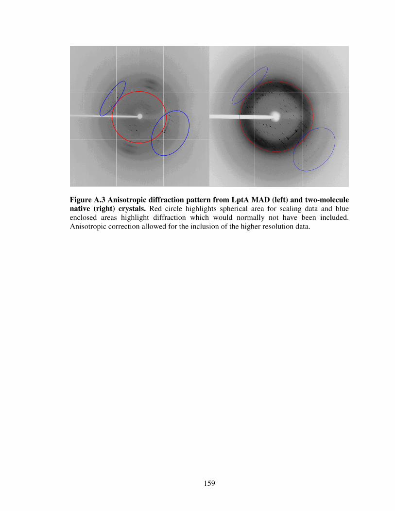
159
Figure A.3 Anisotropic diffraction pattern from LptA MAD (left) and two-molecule native (right) crystals. Red circle highlights spherical area for scaling data and blue enclosed areas highlight diffraction which would normally not have been included. Anisotropic correction allowed for the inclusion of the higher resolution data.

160
A1.4.3 LptA protein fold and structural analysis
The overall refined structure of LptA resembles a series of eight antiparallel
β-sheets that wind back along the path of the previous peptide stretch, throughout the
length of the protein. These antiparallel sheets are folded approximately in the middle
along their length, creating an overall topology where LptA resembles a ribcage or
β-jellyroll (Figure A4), where each rib is approximately seven residues in length.
Furthermore, the structure of LptA is not completely symmetrical along the length but
opens slightly at both the N- and C-terminal ends, creating two clefts. The N-terminal
cleft is slightly larger than the C-terminal cleft, and is surrounded by the conserved
segment of residues from P35-K83 (Figure A5). Other conserved residues align along the
length of the β-sheets, implicating a structural contribution. An evaluation of the surface
electrostatics of LptA suggested a mainly negatively charged molecule, with a
pronounced cleft delineated by the N-terminal cleft. In the eight-molecule form LptA is
organized in a head-to-tail fashion, effectively forming fibers of LptA that intertwine
(Figure A6). In this case, there was an absence of density for LPS or lauric acid in the
partially refined structure of LptA. At this time it is therefore unclear whether the change
observed for LptA has been influenced due to an interaction with LPS or lauric acid.
Structural comparison of LptA against structures within the SCOP database (24) did not
reveal any significant structural matches; hence, LptA represents a previously
uncharacterized fold, although the structure of LptA does resemble a left-handed β-helix
or β-jellyroll in general.

161
N
C
C
N
N
C
C
N
C
N
N
C
C
N
N
C
90o
N
C
C
N
N
C
C
N
C
N
N
C
C
N
N
C
90o90o
Figure A.4 Ribbon diagram of LptA with two molecules in the asymmetric unit at 2.15 Å resolution. Each of the LptA molecules is presented with different colours and gaps with dotted lines. LptA has also been rotated by 90o to highlight the gap along the length of LptA.

162
90o
N
N
C
90o
N
N
C
Figure A.5 Ribbon diagram of LptA highlighting residues conserved in sequence alignment with other Gram-negative homologues. Two clefts are present at the ends of LptA due to opening of the folded β-sheets that run along its length. A notable cluster of conserved residues flank the N-terminal cleft delineated at the end of the LptA structure, corresponding to residues P35-K83.

163
Figure A.6 Ribbon diagram of LptA with eight molecules in the asymmetric unit at 3.25 Å resolution. LptA molecules are organized head to tail fashion, forming a set of intertwined fibres. There was no clear density that resembled LPS or lauric acid in the partially refined model of LptA.
164
A1.4.4 Conclusions
Here we present the crystal structure of the processed form of the periplasmic
localized, LPS transport protein LptA at 2.6 Å resolution with two molecules in the
asymmetric unit and at 3.25 Å resolution with eight molecules in the asymmetric unit in
the presence of LPS or lauric acid. The structure of LptA forms a unique set of eight
antiparallel β-sheets which open at their ends. Conserved residues are not clustered to a
particular structural location in LptA, with the exception of the N-terminal residues P35-
K83 which are clustered proximal to one of the terminal clefts in LptA. This could
potentially be a position that contributes to ligand binding or to mediate protein-protein
contact. Based on sequence homology we can extend the structural information for LptA
to various Gram-negative bacteria. Furthermore, due to the implication of LptA function
in maintaining membrane integrity and LPS transport we can extend our findings to
multiple organisms, with implications for an LptA contribution for pathogenesis.
A1.4.5 Footnotes
Thank you to Dr. Christian M. Udell for cloning LptA into the expression vector.
Thank you to Dr. Robert Sweet for organizing the Brookhaven National Laboratory
(BNL), Rapid Data collection course 2006 that ultimately led to a structural solution for
LptA. LptA has not been deposited yet in the Protein Data Bank, pending further
refinement. We are also grateful to Howard Robinson X-29 where the MAD dataset was
collected, and the staff at MacCHESS where native data for both isoforms was collected.

165
Thank you to Dr. Clemens Vonrhein for expert assistance in implementing autoSHARP,
and Dr. Michael Sawaya for useful discussions and anisotropy correction server to
improve the map quality obtained from LptA data. This work has been supported CIHR.
MS is supported by Bracken E.G. Bauman and R. Samuel McLaughlin Queen’s Graduate
Fellowships as well as OGS. ZJ is a Canada Research Chair in Structural Biology.
A1.4.6 Appendix References 1. 1994. The CCP4 suite: programs for protein crystallography. Acta Crystallogr. D.
Biol. Crystallogr. 50:760-763.
2. Altschul, S. F., T. L. Madden, A. A. Schaffer, J. Zhang, Z. Zhang, W. Miller, and D. J. Lipman. 1997. Gapped BLAST and PSI-BLAST: a new generation of protein database search programs. Nucleic Acids Res 25:3389-3402.
3. Aono, R., T. Negishi, K. Aibe, A. Inoue, and K. Horikoshi. 1994. Mapping of organic solvent tolerance gene ostA in Escherichia coli K-12. Biosci. Biotechnol. Biochem. 58:1231-1235.
4. Appel, R. D., A. Bairoch, and D. F. Hochstrasser. 1994. A new generation of information retrieval tools for biologists: the example of the ExPASy WWW server. Trends Biochem. Sci. 19:258-260.
5. Brunger, A. T., P. D. Adams, G. M. Clore, W. L. DeLano, P. Gros, R. W. Grosse-Kunstleve, J. S. Jiang, J. Kuszewski, M. Nilges, N. S. Pannu, R. J. Read, L. M. Rice, T. Simonson, and G. L. Warren. 1998. Crystallography & NMR system: A new software suite for macromolecular structure determination. Acta Crystallogr. D. Biol. Crystallogr. 54:905-921.
6. Ciria, R., C. breu-Goodger, E. Morett, and E. Merino. 2004. GeConT: gene context analysis. Bioinformatics. 20:2307-2308.
7. Corpet, F. 1988. Multiple sequence alignment with hierarchical clustering. Nucleic Acids Res 16:10881-10890.
8. Danese, P. N. and T. J. Silhavy. 2003. Targeting and assembly of periplasmic and outer-membrane proteins in Escherichia coli. Annual. Review of Genetics 32:59.

166
9. Fujiki, K., A. Fukuda, and Y. Okada. 1976. Amino acid composition of peptidoglycan in Caulobacter crescentus. J Biochem. (Tokyo). 80:1453-1455.
10. Gilles-Gonzalez, M. A. and G. Gonzalez. 2005. Heme-based sensors: defining characteristics, recent developments, and regulatory hypotheses. J Inorg. Biochem 99:1-22.
11. Jackman, J. E., C. A. Fierke, L. N. Tumey, M. Pirrung, T. Uchiyama, S. H. Tahir, O. Hindsgaul, and C. R. Raetz. 2000. Antibacterial agents that target lipid A biosynthesis in Gram-negative bacteria. Inhibition of diverse UDP-3-O-(r-3-hydroxymyristoyl)-n-acetylglucosamine deacetylases by substrate analogs containing zinc binding motifs. J Biol. Chem. 275:11002-11009.
12. Kabsch, W. A solution for the best rotation to relate two sets of vectors.
13. Kim, W. and M. G. Surette. 2004. Metabolic differentiation in actively swarming Salmonella. Mol. Microbiol. 54:702-714.
14. Krissinel, E. and K. Henrick. 2004. Secondary-structure matching (SSM), a new tool for fast protein structure alignment in three dimensions. Acta Crystallogr D Biol Crystallogr 60:2256-2268.
15. Krissinel, E. and K. Henrick. 2004. Protein structure comparison in 3D based on secondary structure matching (SSM) followed by Cα alignment, scored by a new structural similarity function., p. 88. In Andreas J.Kungl & Penelope J.Kungl (Eds.) (ed.).
16. Layer, G., J. Moser, D. W. Heinz, D. Jahn, and W. D. Schubert. 2003. Crystal structure of coproporphyrinogen III oxidase reveals cofactor geometry of Radical SAM enzymes. EMBO J 22:6214-6224.
17. Lopez-Campistrous, A., P. Semchuk, L. Burke, T. Palmer-Stone, S. J. Brokx, G. Broderick, D. Bottorff, S. Bolch, J. H. Weiner, and M. J. Ellison. 2005. Localization, annotation, and comparison of the Escherichia coli K-12 proteome under two states of growth. Mol. Cell Proteomics. 4:1205-1209.
18. Luirink, J., H. G. von, E. Houben, and J. W. de Gier. 2005. Biogenesis of inner membrane proteins in Escherichia coli. Annu. Rev. Microbiol. 59:329-55.:329-355.
19. Magee, T. and M. C. Seabra. 2005. Fatty acylation and prenylation of proteins: what's hot in fat. Curr. Opin. Cell Biol. 17:190-196.
20. Marino-Ramirez, L., J. L. Minor, N. Reading, and J. C. Hu. 2004. Identification and mapping of self-assembling protein domains encoded by the Escherichia coli K-12 genome by use of lambda repressor fusions. J Bacteriol. 186:1311-1319.

167
21. McCoy, A. J. 2007. Solving structures of protein complexes by molecular replacement with Phaser. Acta Crystallogr. D. Biol. Crystallogr. 63:32-41.
22. Mdluli, K. E., P. R. Witte, T. Kline, A. W. Barb, A. L. Erwin, B. E. Mansfield, A. L. McClerren, M. C. Pirrung, L. N. Tumey, P. Warrener, C. R. Raetz, and C. K. Stover. 2006. Molecular validation of LpxC as an antibacterial drug target in Pseudomonas aeruginosa. Antimicrob. Agents Chemother. 50:2178-2184.
23. Meredith, T. C. and R. W. Woodard. 2003. Escherichia coli YrbH is a D-arabinose 5-phosphate isomerase. J Biol. Chem. 278:32771-32777.
24. Murzin, A. G., S. E. Brenner, T. Hubbard, and C. Chothia. 1995. SCOP: a structural classification of proteins database for the investigation of sequences and structures. J Mol. Biol. 247:536-540.
25. Nanninga, N. 1991. Cell division and peptidoglycan assembly in Escherichia coli. Mol. Microbiol. 5:791-795.
26. Nielsen, H., J. Engelbrecht, S. Brunak, and H. G. von. 1997. Identification of prokaryotic and eukaryotic signal peptides and prediction of their cleavage sites. Protein Eng. 10:1-6.
27. Oliver, D. B. and J. Beckwith. 1982. Regulation of a membrane component required for protein secretion in Escherichia coli. Cell. 30:311-319.
28. Otwinowski, Z. and W. Minor. 1997. [20] Processing of X-ray diffraction data collected in oscillation mode, p. 307-326. In Methods in Enzymology. Academic Press.
29. Polissi, A. and C. Georgopoulos. 1996. Mutational analysis and properties of the msbA gene of Escherichia coli, coding for an essential ABC family transporter. Mol. Microbiol. 20:1221-1233.
30. Raetz, C. R. and C. Whitfield. 2002. Lipopolysaccharide endotoxins. Annu. Rev. Biochem. 71:635-700. Epub;2001 Nov 9.:635-700.
31. Ruiz, N., D. Kahne, and T. J. Silhavy. 2006. Advances in understanding bacterial outer-membrane biogenesis. Nat. Rev. Microbiol. 4:57-66.
32. Serina, S., F. Nozza, G. Nicastro, F. Faggioni, H. Mottl, G. Deho, and A. Polissi. 2004. Scanning the Escherichia coli chromosome by random transposon mutagenesis and multiple phenotypic screening. Res. Microbiol. 155:692-701.
33. Smit, J., Y. Kamio, and H. Nikaido. 1975. Outer membrane of Salmonella
typhimurium: chemical analysis and freeze-fracture studies with lipopolysaccharide mutants. J Bacteriol. 124:942-958.

168
34. Sperandeo, P., R. Cescutti, R. Villa, B. C. Di, D. Candia, G. Deho, and A. Polissi. 2007. Characterization of lptA and lptB, two essential genes implicated in lipopolysaccharide transport to the outer membrane of Escherichia coli. J Bacteriol. 189:244-253.
35. Sperandeo, P., C. Pozzi, G. Deho, and A. Polissi. 2006. Non-essential KDO biosynthesis and new essential cell envelope biogenesis genes in the Escherichia
coli yrbG-yhbG locus. Res. Microbiol. 157:547-558.
36. Strong, M., M. R. Sawaya, S. Wang, M. Phillips, D. Cascio, and D. Eisenberg. 2006. Toward the structural genomics of complexes: crystal structure of a PE/PPE protein complex from Mycobacterium tuberculosis. Proc. Natl. Acad. Sci. U. S. A. 103:8060-8065.
37. Terwilliger, T. C. and J. Berendzen. 1999. Automated MAD and MIR structure solution. Acta Crystallogr. D. Biol. Crystallogr. 55:849-861.
38. Tokuda, H. and S. Matsuyama. 2004. Sorting of lipoproteins to the outer membrane in E. coli. Biochim. Biophys. Acta. 1694:IN1-IN9.
39. Vonrhein, C., E. Blanc, P. Roversi, and G. Bricogne. 2006. Automated Structure Solution With autoSHARP. Methods Mol. Biol. 364:215-30.:215-230.
40. Wimley, W. C. 2003. The versatile beta-barrel membrane protein. Curr. Opin. Struct. Biol. 13:404-411.
41. Wu, J. and R. W. Woodard. 2003. Escherichia coli YrbI is 3-deoxy-D-manno-octulosonate 8-phosphate phosphatase. J Biol. Chem. 278:18117-18123.
42. Wu, T., A. C. McCandlish, L. S. Gronenberg, S. S. Chng, T. J. Silhavy, and D. Kahne. 2006. Identification of a protein complex that assembles lipopolysaccharide in the outer membrane of Escherichia coli. Proc. Natl. Acad. Sci. U. S. A. 103:11754-11759.