Structural and Dynamic Basis of Phospholamban and Sarcolipin Inhibition of
11
Current Topics Structural and Dynamic Basis of Phospholamban and Sarcolipin Inhibition of Ca 2+ -ATPase ² Nathaniel J. Traaseth, ‡ Kim N. Ha, ‡ Raffaello Verardi, § Lei Shi, ‡ Jarrod J. Buffy, § Larry R. Masterson, ‡ and Gianluigi Veglia* ,‡,§ Department of Chemistry and Department of Biochemistry, Molecular Biology, and Biophysics, UniVersity of Minnesota, Minneapolis, Minnesota 55455 ReceiVed August 17, 2007; ReVised Manuscript ReceiVed October 16, 2007 ABSTRACT: Phospholamban (PLN) and sarcolipin (SLN) are two single-pass membrane proteins that regulate Ca 2+ -ATPase (SERCA), an ATP-driven pump that translocates calcium ions into the lumen of the sarcoplasmic reticulum, initiating muscle relaxation. Both proteins bind SERCA through intramembrane interactions, impeding calcium translocation. While phosphorylation of PLN at Ser-16 and/or Thr-17 reestablishes calcium flux, the regulatory mechanism of SLN remains elusive. SERCA has been crystallized in several different states along the enzymatic reaction coordinates, providing remarkable mechanistic information; however, the lack of high-resolution crystals in the presence of PLN and SLN limits the current understanding of the regulatory mechanism. This brief review offers a survey of our hybrid structural approach using solution and solid-state NMR methodologies to understand SERCA regulation from the point of view of PLN and SLN. These results have improved our understanding of the calcium translocation process and are the basis for designing new therapeutic approaches to ameliorate muscle malfunctions. Two membrane proteins, sarco(endo)plasmic reticulum calcium ATPase (SERCA) 1 and the ryanodine receptors (Ryr), play major roles in calcium regulation within muscle cells. Ryr1 and Ryr2 are responsible for releasing Ca 2+ from the sarcoplasmic reticulum (SR) of skeletal and cardiac muscle, respectively, resulting in muscle contraction. SERCA1a and SERCA2a pump calcium from the cytosol into the SR in skeletal and cardiac muscle, respectively, initiating muscle relaxation. Phospholamban (PLN), a 52- residue protein spanning the SR membrane (1), is an endogenous inhibitor of SERCA, lowering the apparent calcium affinity of the ATPase. The relief of SERCA inhibition is achieved by phosphorylation of PLN at Ser-16 by protein kinase A and/or Thr-17 by Ca 2+ /calmodulin- dependent protein kinase (2). In vivo studies demonstrate that PLN phosphorylation at Ser-16 and Thr-17 has different physiological effects, suggesting that these mechanisms act independently (3, 4). Sarcolipin (SLN) has a primary sequence homologous to that of the transmembrane domain of PLN (5, 6). Initially, SLN was thought to be the counterpart of PLN within skeletal muscle, playing only an ancillary role in cardiac muscle. Recently, however, significant expression levels of SLN have been detected in cardiac atrial muscle, with lower levels in ventricular muscle (7-9), suggesting that it may play an important role in regulation of the heart. When SLN was initially copurified with fast-twitch skeletal SERCA1a (10), no post-translational modifications were identified, which led to the conclusion that the regulation of SLN depended on its variable expression levels (11, 12). Recent evidence shows that SLN inhibition can be fully reversed by isoproterenol, a -adrenergic receptor agonist in PLN knockout mice (13). These results led to the hypothesis that the inhibitory effect of SLN can be reversed via phospho- rylation in a manner similar to that of PLN. In vitro experiments have shown that SLN can be phosphorylated at Thr-5 when cotransfected with serine/threonine kinase 16 (STK16) (13). From these biological data, it is clear that phosphorylation of PLN and SLN constitute driving forces for calcium re-uptake into cardiac SR. Several crystal structures of SERCA in different confor- mations within the enzymatic cycle have revealed important atomic details regarding SERCA’s mechanism (Figure 1) (14-19). PLN is thought to bind and inhibit the low-affinity calcium form of SERCA (E2) and detach from the enzyme (either partially or totally) upon phosphorylation at Ser-16, reversing its inhibitory effect and restoring the affinity of SERCA for Ca 2+ ions. The only experimental structure of ² This work was supported by grants to G.V. (NIH Grant GM64742, NIH Grant HL80081, and AHA Grant 0160465Z). * To whom correspondence should be addressed: Department of Chemistry, University of Minnesota, 207 Pleasant St. S.E., Minneapolis, MN 55455. Telephone: (612) 625-0758. Fax: (612) 626-7541. E-mail: [email protected]. ‡ Department of Chemistry. § Department of Biochemistry, Molecular Biology, and Biophysics. 1 Abbreviations: PLN, phospholamban; SLN, sarcolipin; SERCA, sarco(endo)plasmic reticulum calcium ATPase; DOPC, dioleoyl-sn- glycero-3-phosphocholine; DOPE, dioleoyl-sn-glycero-3-phosphoetha- nolamine; CPMG, Carr-Purcell-Meiboom-Gill; wt-PLN, wild-type PLN; DC, dipolar coupling; CSA, chemical shift anisotropy. 3 Biochemistry 2008, 47, 3-13 10.1021/bi701668v CCC: $40.75 © 2008 American Chemical Society Published on Web 12/15/2007
Transcript of Structural and Dynamic Basis of Phospholamban and Sarcolipin Inhibition of
Current Topics
Structural and Dynamic Basis of Phospholamban and Sarcolipin Inhibition ofCa2+-ATPase†
Nathaniel J. Traaseth,‡ Kim N. Ha,‡ Raffaello Verardi,§ Lei Shi,‡ Jarrod J. Buffy,§ Larry R. Masterson,‡ andGianluigi Veglia*,‡,§
Department of Chemistry and Department of Biochemistry, Molecular Biology, and Biophysics, UniVersity of Minnesota,Minneapolis, Minnesota 55455
ReceiVed August 17, 2007; ReVised Manuscript ReceiVed October 16, 2007
ABSTRACT: Phospholamban (PLN) and sarcolipin (SLN) are two single-pass membrane proteins that regulateCa2+-ATPase (SERCA), an ATP-driven pump that translocates calcium ions into the lumen of thesarcoplasmic reticulum, initiating muscle relaxation. Both proteins bind SERCA through intramembraneinteractions, impeding calcium translocation. While phosphorylation of PLN at Ser-16 and/or Thr-17reestablishes calcium flux, the regulatory mechanism of SLN remains elusive. SERCA has been crystallizedin several different states along the enzymatic reaction coordinates, providing remarkable mechanisticinformation; however, the lack of high-resolution crystals in the presence of PLN and SLN limits thecurrent understanding of the regulatory mechanism. This brief review offers a survey of our hybrid structuralapproach using solution and solid-state NMR methodologies to understand SERCA regulation from thepoint of view of PLN and SLN. These results have improved our understanding of the calcium translocationprocess and are the basis for designing new therapeutic approaches to ameliorate muscle malfunctions.
Two membrane proteins, sarco(endo)plasmic reticulumcalcium ATPase (SERCA)1 and the ryanodine receptors(Ryr), play major roles in calcium regulation within musclecells. Ryr1 and Ryr2 are responsible for releasing Ca2+ fromthe sarcoplasmic reticulum (SR) of skeletal and cardiacmuscle, respectively, resulting in muscle contraction.SERCA1a and SERCA2a pump calcium from the cytosolinto the SR in skeletal and cardiac muscle, respectively,initiating muscle relaxation. Phospholamban (PLN), a 52-residue protein spanning the SR membrane (1), is anendogenous inhibitor of SERCA, lowering the apparentcalcium affinity of the ATPase. The relief of SERCAinhibition is achieved by phosphorylation of PLN at Ser-16by protein kinase A and/or Thr-17 by Ca2+/calmodulin-dependent protein kinase (2). In vivo studies demonstratethat PLN phosphorylation at Ser-16 and Thr-17 has differentphysiological effects, suggesting that these mechanisms actindependently (3, 4).
Sarcolipin (SLN) has a primary sequence homologous tothat of the transmembrane domain of PLN (5, 6). Initially,SLN was thought to be the counterpart of PLN withinskeletal muscle, playing only an ancillary role in cardiacmuscle. Recently, however, significant expression levels ofSLN have been detected in cardiac atrial muscle, with lowerlevels in ventricular muscle (7-9), suggesting that it mayplay an important role in regulation of the heart. When SLNwas initially copurified with fast-twitch skeletal SERCA1a(10), no post-translational modifications were identified,which led to the conclusion that the regulation of SLNdepended on its variable expression levels (11, 12). Recentevidence shows that SLN inhibition can be fully reversedby isoproterenol, aâ-adrenergic receptor agonist in PLNknockout mice (13). These results led to the hypothesis thatthe inhibitory effect of SLN can be reversed via phospho-rylation in a manner similar to that of PLN. In vitroexperiments have shown that SLN can be phosphorylated atThr-5 when cotransfected with serine/threonine kinase 16(STK16) (13). From these biological data, it is clear thatphosphorylation of PLN and SLN constitute driving forcesfor calcium re-uptake into cardiac SR.
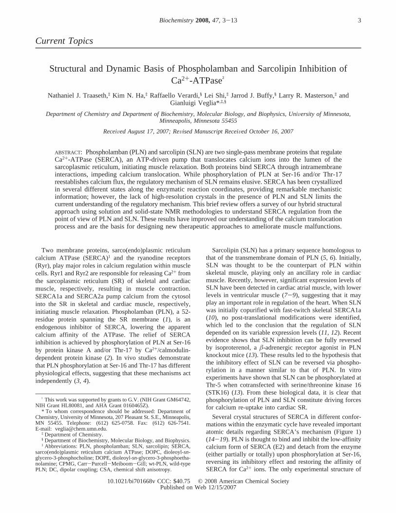
Several crystal structures of SERCA in different confor-mations within the enzymatic cycle have revealed importantatomic details regarding SERCA’s mechanism (Figure 1)(14-19). PLN is thought to bind and inhibit the low-affinitycalcium form of SERCA (E2) and detach from the enzyme(either partially or totally) upon phosphorylation at Ser-16,reversing its inhibitory effect and restoring the affinity ofSERCA for Ca2+ ions. The only experimental structure of
† This work was supported by grants to G.V. (NIH Grant GM64742,NIH Grant HL80081, and AHA Grant 0160465Z).
* To whom correspondence should be addressed: Department ofChemistry, University of Minnesota, 207 Pleasant St. S.E., Minneapolis,MN 55455. Telephone: (612) 625-0758. Fax: (612) 626-7541.E-mail: [email protected].
‡ Department of Chemistry.§ Department of Biochemistry, Molecular Biology, and Biophysics.1 Abbreviations: PLN, phospholamban; SLN, sarcolipin; SERCA,
sarco(endo)plasmic reticulum calcium ATPase; DOPC, dioleoyl-sn-glycero-3-phosphocholine; DOPE, dioleoyl-sn-glycero-3-phosphoetha-nolamine; CPMG, Carr-Purcell-Meiboom-Gill; wt-PLN, wild-typePLN; DC, dipolar coupling; CSA, chemical shift anisotropy.
3Biochemistry2008,47, 3-13
10.1021/bi701668v CCC: $40.75 © 2008 American Chemical SocietyPublished on Web 12/15/2007
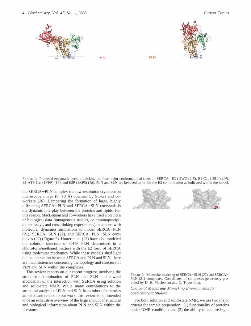
the SERCA-PLN complex is a low-resolution cryoelectronmicroscopy image (8-10 Å) obtained by Stokes and co-workers (20). Hampering the formation of large, highlydiffracting SERCA-PLN and SERCA-SLN cocrystals isthe dynamic interplay between the proteins and lipids. Forthis reason, MacLennan and co-workers have used a plethoraof biological data (mutagenesis studies, coimmunoprecipi-tation assays, and cross-linking experiments) in concert withmolecular dynamics simulations to model SERCA-PLN(21), SERCA-SLN (22), and SERCA-PLN-SLN com-plexes (22) (Figure 2). Hutter et al. (23) have also modeledthe solution structure of C41F PLN determined in achloroform/methanol mixture with the E2 form of SERCAusing molecular mechanics. While these models shed lighton the interaction between SERCA and PLN and SLN, thereare inconsistencies concerning the topology and structure ofPLN and SLN within the complexes.
This review reports on our recent progress involving thestructure determination of PLN and SLN and towardelucidation of the interaction with SERCA using solutionand solid-state NMR. While many contributions to thestructural analysis of PLN and SLN from other laboratoriesare cited and related to our work, this review is not intendedto be an exhaustive overview of the large amount of structuraland biological information about PLN and SLN within theliterature.
Choice of Membrane Mimicking EnVironments forSpectroscopic Studies
For both solution and solid-state NMR, we use two majorcriteria for sample preparation: (1) functionality of proteinsunder NMR conditions and (2) the ability to acquire high-
FIGURE 1: Proposed enzymatic cycle mimicking the four major conformational states of SERCA: E2 (1IWO) (15), E1-Ca2 (1SU4) (14),E1-ATP-Ca2 (1VFP) (16), and E2P (1XP5) (18). PLN and SLN are believed to inhibit the E2 conformation as indicated within the model.
FIGURE 2: Molecular modeling of SERCA-SLN (22) and SERCA-PLN (21) complexes. Coordinates of complexes generously pro-vided by D. H. Maclennan and C. Toyoshima.
4 Biochemistry, Vol. 47, No. 1, 2008 Current Topics
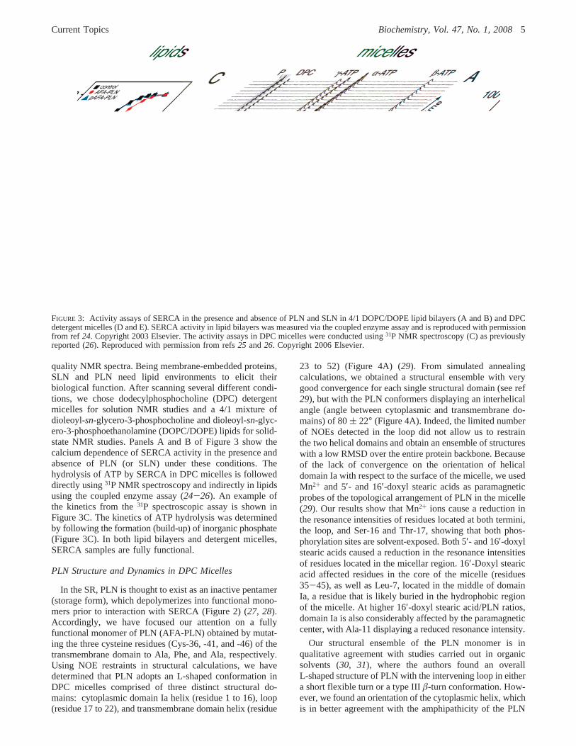
quality NMR spectra. Being membrane-embedded proteins,SLN and PLN need lipid environments to elicit theirbiological function. After scanning several different condi-tions, we chose dodecylphosphocholine (DPC) detergentmicelles for solution NMR studies and a 4/1 mixture ofdioleoyl-sn-glycero-3-phosphocholine and dioleoyl-sn-glyc-ero-3-phosphoethanolamine (DOPC/DOPE) lipids for solid-state NMR studies. Panels A and B of Figure 3 show thecalcium dependence of SERCA activity in the presence andabsence of PLN (or SLN) under these conditions. Thehydrolysis of ATP by SERCA in DPC micelles is followeddirectly using31P NMR spectroscopy and indirectly in lipidsusing the coupled enzyme assay (24-26). An example ofthe kinetics from the31P spectroscopic assay is shown inFigure 3C. The kinetics of ATP hydrolysis was determinedby following the formation (build-up) of inorganic phosphate(Figure 3C). In both lipid bilayers and detergent micelles,SERCA samples are fully functional.
PLN Structure and Dynamics in DPC Micelles
In the SR, PLN is thought to exist as an inactive pentamer(storage form), which depolymerizes into functional mono-mers prior to interaction with SERCA (Figure 2) (27, 28).Accordingly, we have focused our attention on a fullyfunctional monomer of PLN (AFA-PLN) obtained by mutat-ing the three cysteine residues (Cys-36, -41, and -46) of thetransmembrane domain to Ala, Phe, and Ala, respectively.Using NOE restraints in structural calculations, we havedetermined that PLN adopts an L-shaped conformation inDPC micelles comprised of three distinct structural do-mains: cytoplasmic domain Ia helix (residue 1 to 16), loop(residue 17 to 22), and transmembrane domain helix (residue
23 to 52) (Figure 4A) (29). From simulated annealingcalculations, we obtained a structural ensemble with verygood convergence for each single structural domain (see ref29), but with the PLN conformers displaying an interhelicalangle (angle between cytoplasmic and transmembrane do-mains) of 80( 22° (Figure 4A). Indeed, the limited numberof NOEs detected in the loop did not allow us to restrainthe two helical domains and obtain an ensemble of structureswith a low RMSD over the entire protein backbone. Becauseof the lack of convergence on the orientation of helicaldomain Ia with respect to the surface of the micelle, we usedMn2+ and 5′- and 16′-doxyl stearic acids as paramagneticprobes of the topological arrangement of PLN in the micelle(29). Our results show that Mn2+ ions cause a reduction inthe resonance intensities of residues located at both termini,the loop, and Ser-16 and Thr-17, showing that both phos-phorylation sites are solvent-exposed. Both 5′- and 16′-doxylstearic acids caused a reduction in the resonance intensitiesof residues located in the micellar region. 16′-Doxyl stearicacid affected residues in the core of the micelle (residues35-45), as well as Leu-7, located in the middle of domainIa, a residue that is likely buried in the hydrophobic regionof the micelle. At higher 16′-doxyl stearic acid/PLN ratios,domain Ia is also considerably affected by the paramagneticcenter, with Ala-11 displaying a reduced resonance intensity.
Our structural ensemble of the PLN monomer is inqualitative agreement with studies carried out in organicsolvents (30, 31), where the authors found an overallL-shaped structure of PLN with the intervening loop in eithera short flexible turn or a type IIIâ-turn conformation. How-ever, we found an orientation of the cytoplasmic helix, whichis in better agreement with the amphipathicity of the PLN
FIGURE 3: Activity assays of SERCA in the presence and absence of PLN and SLN in 4/1 DOPC/DOPE lipid bilayers (A and B) and DPCdetergent micelles (D and E). SERCA activity in lipid bilayers was measured via the coupled enzyme assay and is reproduced with permissionfrom ref 24. Copyright 2003 Elsevier. The activity assays in DPC micelles were conducted using31P NMR spectroscopy (C) as previouslyreported (26). Reproduced with permission from refs25 and26. Copyright 2006 Elsevier.
Current Topics Biochemistry, Vol. 47, No. 1, 20085
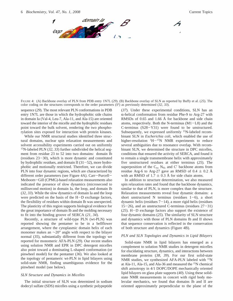
sequence (29). The most relevant PLN conformations in PDBentry 1N7L are those in which the hydrophobic side chainsin domain Ia (Val-4, Leu-7, Ala-11, and Ala-15) are orientedtoward the interior of the micelle and the hydrophilic residuespoint toward the bulk solvent, rendering the two phospho-rylation sites exposed for interaction with protein kinases.
While our NMR structural studies identified three struc-tural domains, nuclear spin relaxation measurements andsolvent accessibility experiments carried out on uniformly15N-labeled PLN (32, 33) further subdivided the helical seg-ment from residue 23 to 52 into two domains: domain Ib(residues 23-30), which is more dynamic and constitutedby hydrophilic residues, and domain II (31-52), more hydro-phobic and motionally restricted. Therefore, we can dividePLN into four dynamic regions, which are characterized bydifferent order parameters (see Figure 4A). Carr-Purcell-Meiboom-Gill (CPMG)-based relaxation measurements alsoindicated the presence of slow dynamics (microsecond tomillisecond motion) in domain Ia, the loop, and domain Ib(32, 33). While the slow dynamics of domain Ia and the loopwere predicted on the basis of the H-D exchange factors,the flexibility of residues within domain Ib was unexpected.The plasticity of this region supports biological evidence forthe great importance of domain Ib and the molding necessaryto fit into the binding groove of SERCA (21, 34).
Recently, a structure of wild-type PLN (wt-PLN) wasreported showing the pentamer to be in a bellflowerarrangement, where the cytoplasmic domain helix of eachmonomer makes an∼20° angle with respect to the bilayernormal (35), substantially different from the topology wereported for monomeric AFA-PLN (29). Our recent studiesusing solution NMR and EPR in DPC detergent micellesalso point toward a dominating L-shaped conformation (orpinwheel model) for the pentamer (36). We also looked atthe topology of pentameric wt-PLN in lipid bilayers usingsolid-state NMR, finding unambiguous evidence for thepinwheel model (see below).
SLN Structure and Dynamics in Micelles
The initial structure of SLN was determined in sodiumdodecyl sulfate (SDS) micelles using a synthetic polypeptide
(37). Under these experimental conditions, SLN has anR-helical conformation from residue Phe-9 to Arg-27 withRMSDs of 0.65 and 1.66 Å for backbone and side chainatoms, respectively. Both the N-terminus (M1-L8) and theC-terminus (S28-Y31) were found to be unstructured.Subsequently, we expressed uniformly15N-labeled recom-binant SLN inEscherichia coli, which enabled the use ofhigher-resolution1H-15N NMR experiments to reduceseveral ambiguities due to resonance overlap. With recom-binant SLN, we determined the structure in DPC micelles,conditions that ensured the activity of SERCA, and found itto remain a single transmembrane helix with approximatelyfive unstructured residues at either terminus (25). Thesuperposition of the CR, NH, and C′ backbone atoms fromresidue Arg-6 to Arg-27 gave an RMSD of 0.4( 0.2 Åwith an RMSD of 1.7( 0.3 Å for side chain atoms.
In addition to structure determination, we also measuredspin relaxation rates and found that the backbone dynamics,similar to that of PLN, is more complex than the structure.Relaxation measurements reveal four dynamic domains: ashort unstructured N terminus (residues 1-6), a shortdynamic helix (residues 7-14), a more rigid helix (residues15-26), and an unstructured C-terminus (residues 27-31)(25). H-D exchange factors also support the existence offour dynamic domains (25). The similarity of SLN structureand dynamics with those of PLN domains Ib and II showsthat sequence conservation is reflected in the conservationof both structure and dynamics (Figure 4B).
PLN and SLN Topologies and Dynamics in Lipid Bilayers
Solid-state NMR in lipid bilayers has emerged as acomplement to solution NMR studies in detergent micellesfor elucidating structure, dynamics, and interactions betweenmembrane proteins (38, 39). For our first solid-stateNMR studies, we synthesized AFA-PLN labeled with15Nat Ala-11, Ala-15, and Ala-36 and measured the15N chemicalshift anisotropy in 4/1 DOPC/DOPE mechanically orientedlipid bilayers on glass plate supports (40). Using these solid-state NMR measurements in concert with rigid body mo-lecular mechanics, we found that domains Ib and II areoriented approximately perpendicular to the plane of the
FIGURE 4: (A) Backbone overlay of PLN from PDB entry 1N7L (29). (B) Backbone overlay of SLN as reported by Buffy et al. (25). Thecolor coding on the structures corresponds to the order parameters (S2) as previously determined (32, 33).
6 Biochemistry, Vol. 47, No. 1, 2008 Current Topics
bilayer with the interhelical (i.e., interdomain) angle rangingbetween 60 and 100°, ruling out the possibility of acontinuousR-helix and also suggesting that the cytoplasmicdomain of PLN interacts with the membrane surface.
Similar solid-state NMR measurements on SLN orientedin mechanically aligned DOPC/DOPE bilayers revealed theapproximate parallel orientation of the SLN helix with respectto the membrane bilayer normal (37). Since the limitednumber of labeled sites did not allow us to give quantitativetopological angles for SLN and PLN within the bilayer,we then proceeded to use two-dimensional (2D)1H-15NPISEMA (polarization inversion spin exchange at the magicangle) experiments (41). This separated-local-field experi-ment correlates the15N chemical shift anisotropy (CSA) withthe1H-15N dipolar coupling (DC). Since the values of bothCSA and DC depend on the orientation of the peptide planewith respect to the direction of the magnetic field, theassignment of the amide resonances allows for the determi-nation of the structure and topology in aligned lipid bilayers.
We determined that in mixed 4/1 DOPC/DOPE lipidbilayers, AFA-PLN has an overall L-shaped conformationwhere the helix comprising domains Ib and II makes a tiltangle of∼21° with respect to the bilayer normal (42). Asexpected from the one-dimensional (1D) solid-state NMRstudies on synthetically15N-labeled AFA-PLN (37), PISEMANMR spectroscopy clearly shows that domain Ia interactswith the membrane surface, making an angle of∼90° withrespect to the bilayer normal (42). A current model of thePLN monomer is reported in Figure 5E.
In addition to the structural and topological informationobtained from oriented alignments, tilting the aligned samplesto different angles with respect to the direction of the staticfield makes it possible to investigate the rotational dynamicsof the protein within the bilayer (43-45). Tilting the AFA-PLN sample by 90° revealed that domains Ib and II undergofast long-axial rotational diffusion about the bilayer normalwith the cytoplasmic domain undergoing this motion and
other complex dynamics, scaling both the values of CSAand DC (42). The dynamics detected in both our solutionand solid-state NMR experiments may explain variabilitywithin the literature regarding the topology of the cytoplasmicdomain of PLN. For example, a magic-angle-spinning (MAS)solid-state NMR study carried out by Baldus and co-workersfound that while cross-polarization (CP)-based pulse se-quences were adequate for detection of the transmembranedomain of AFA-PLN, showing the existence of a well-defined helix, the cytoplasmic domain residues were toodynamic to be detected (46). Instead,J coupling coherencetransfers, similar to those in solution NMR experiments, wereused to detect the dynamic cytoplasmic domain, leading tothe conclusion that the cytoplasmic domain was completelyunstructured. While this study represents an advancementin MAS methodology, the structure most likely represents aminor conformational state and is inconsistent with a wealthof data, including those from our laboratory, which consis-tently show a predominant helical cytoplasmic domain withan overall L-shaped monomeric structure in lipid bilayersand detergent micelles.
A close inspection of PISEMA spectra from selectivelylabeled samples reveals the presence of two peak populationsthat exemplify two slightly different topologies for AFA-PLN domains Ib and II (42). The two topologies have thesame tilt angle (θ) for domains Ib and II with respect to themembrane normal, but slightly different rotational anglesaround the helix axis (F). Multiple populations of PLN havealso been observed by the Lorigan and Middleton groupsusing MAS NMR experiments in lipid vesicles (47, 48). Thedetection of multiple conformers underscores the plasticityof PLN and might be an important recognition mechanismfor SERCA, protein kinase A, Ca2+/calmodulin-dependentprotein kinase, and protein phosphatase 1, previously shownto be necessary for physiological processes (34).
As with those of AFA-PLN, the PISEMA spectra of SLNobtained on uniformly15N-labeled and selectively labeled
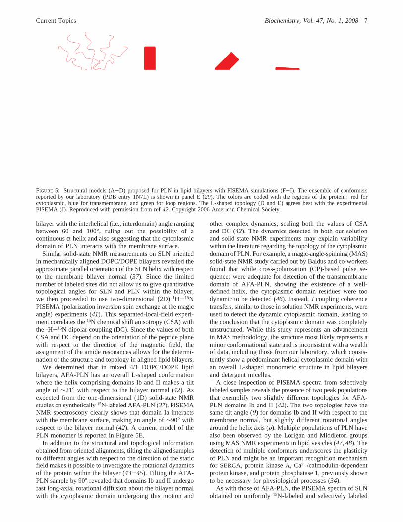
FIGURE 5: Structural models (A-D) proposed for PLN in lipid bilayers with PISEMA simulations (F-I). The ensemble of conformersreported by our laboratory (PDB entry 1N7L) is shown in panel E (29). The colors are coded with the regions of the protein: red forcytoplasmic, blue for transmembrane, and green for loop regions. The L-shaped topology (D and E) agrees best with the experimentalPISEMA (J). Reproduced with permission from ref42. Copyright 2006 American Chemical Society.
Current Topics Biochemistry, Vol. 47, No. 1, 20087
[15N-Leu], [15N-Ile], and [15N-Val] samples also revealed theexistence of two distinct topologies (49). Both the major andthe minor populations of the resonances corresponding todomains Ib and II are oriented∼23° with respect to the lipidbilayer normal but vary in the rotation angle about the helicalaxis by∼5° (in remarkable agreement with AFA-PLN). Theprimary sequence homology between SLN and PLN resultsin nearly identical structural and dynamic properties of thesetwo regulatory proteins.
Pentameric wt-PLN Topology in Lipid Bilayers
More recently, our group has embarked on the validationof the pentameric structure of wt-PLN in lipid bilayers anddetergent micelles (36). While monomeric PLN has previ-ously been shown to bind and inhibit SERCA, recently itwas hypothesized that the pentamer could also bind andinhibit SERCA (50). While there is broad consensus regard-ing the secondary structure of pentameric wt-PLN, there isdisagreement in the literature about the orientation of thecytoplasmic helix. In particular, there are four proposedmodels for pentameric wt-PLN (Figure 6). The first model(extended helix/sheet) shows wt-PLN to be comprised of twoR-helices connected by an antiparallelâ-sheet (residues22-32), where the cytoplasmic domain is oriented 50-60°relative to the bilayer normal (51). The second model depictswt-PLN as a continuousR-helix with a tilt angle of 28( 6°with respect to the bilayer normal (52, 53). The third model(pinwheel) shows that the most stable pentamer has apinwheel geometry in which the cytoplasmic domain helicesare oriented∼90° with respect to the membrane bilayernormal (54). The fourth and most recent model (bellflower)shows the structure of the pentamer to be in a bellflowerassembly with the cytoplasmic domain helices oriented∼20°with respect to the bilayer normal (35).
To study the topology of pentameric wt-PLN, we recon-stituted the protein in mechanically aligned 4/1 DOPC/DOPElipid bilayers and analyzed the protein’s architecture using1H-15N PISEMA spectroscopy. As with the AFA-PLNmonomer (42), we found that the wt-PLN PISEMA spectrumis composed of three different populations of resonances (seethe 1D spectrum in Figure 7A) corresponding to domainsIb and II (with the resonances located between 170 and 220
ppm), an in-plane cytoplasmic domain (with resonanceslocated between 50 and 100 ppm), and a more flexible region(loop and termini) with resonances clustered around∼110ppm (isotropic portion of the spectrum). Since our 1Dspectrum shows three distinct regions, indicating three uniquewt-PLN domain alignments with respect to the membranebilayer normal, this eliminates the possibility of the continu-ous helix model.
To distinguish among the other models depicted in Figure6, we performed PISEMA experiments using selectivelylabeled [15N-Ala], [15N-Thr], [15N-Leu], [15N-Ile], [15N-Cys],and [15N-Asn] wt-PLN samples. Our experimental PISEMAspectra, reported as an overlay in Figure 7, show a remark-able similarity with the AFA-PLN monomer. In fact,simulations for domain Ia resonances (Leu-7, Thr-8, Ala-11, Ile-12, Ala-15, Thr-17, and Ile-18) correspond to a helixwith a tilt angle of∼90° with respect to the bilayer normal(Figure 7E) (36).
Figure 8 shows PISEMA spectra simulated from thepinwheel and bellflower PDB coordinates for those selec-tively labeled sites shown in Figure 7. If the pentamertopology corresponded to the pinwheel model, the cytoplas-mic domain residues would resonate in the upfield regionof the spectrum (50-100 ppm) (Figure 8A). On the otherhand, if the architecture of wt-PLN were consistent with thebellflower model, domain Ia resonances in the PISEMApattern would occupy the downfield portion of the spectrum(170-220 ppm), as represented in Figure 8B. If theexperimental domain Ia spectra are compared, it is clear thatin lipid bilayers the cytoplasmic domain Ia is orientedperpendicular with the bilayer normal forming an overallpinwheel geometry.
Structural fitting with an ideal helix in Figure 7D revealedthat the helix corresponding to domains Ib and II ofpentameric wt-PLN has a tilt angle (θ) of ∼15° with respectto the bilayer normal (36). Monomeric AFA-PLN has a tiltangle of∼21° (42), which requires pentamer formation totilt by ∼6° to accommodate the leucine/isoleucine zipperholding the pentamer together (55, 56).
Allosteric ActiVation Model
The functionality of SERCA under NMR conditions andthe quality of AFA-PLN spectra upon addition of SERCA
FIGURE 6: Structural models of wt-PLN. The pinwheel (1XNU) and bellflower (1ZLL) pentamer models were taken directly fromPDB coordinates. Reproduced with permission from ref36. Copyright 2007 National Academy of Sciences of the United States of America.
8 Biochemistry, Vol. 47, No. 1, 2008 Current Topics
enabled the unprecedented atomic mapping of the interactionsbetween these two integral membrane proteins in detergentmicelles (57). In its free form, AFA-PLN exists in a dynamicequilibrium between two conformations, T and R states,where the T state or L-shaped conformation is thermody-namically stable and the R state or extended form is identifiedwith a more dynamic cytoplasmic domain (Figure 9) (57,58). These two states are readily detected using EPRspectroscopy in both micelles and lipid bilayers (57, 58),but due to the time scale of the exchange, NMR can onlyimply the existence of these forms from relaxation dispersionmeasurements (i.e., conformational interconversion). How-ever, upon addition of SERCA to AFA-PLN, chemical shiftperturbation analyses reveal the appearance of a secondpopulation of peaks within domain Ia, the loop, and domainIb, indicating a conformational switch of AFA-PLN fromthe T state to the R state, exemplifying an allosteric activationmechanism (57).
Resonances from the hydrophobic portion of the trans-membrane region (domain II) also show chemical ex-change to the R state (26). A difference plot of the1HN
chemical shift before and after addition of SERCA forresidues within domain II (residues 31-52) shows a sym-metric bimodal behavior where the C-terminal part of domainII shifted upfield and the residues near the N-terminal partdownfield (26). Since upfield and downfield shifts havebeen correlated to the strength of hydrogen bonds (59),
one possible explanation for the data is that the C-ter-minal end of the transmembrane domain (residues 46-52)unwinds upon binding SERCA. This hypothesis was firstproposed by MacLennan and co-workers (21), who indicatedan overall change in the secondary structure of the domainII, with residues 49-52 unwinding upon interaction withSERCA, a process that might facilitate binding.
These results are echoed in the binding of SLN to SERCA(25). Overall, SLN behaves like the domains Ib and II ofAFA-PLN, with each dynamic domain mimicking thebehavior of the corresponding domain in AFA-PLN. Uponaddition of SERCA, the transmembrane domain is in fastexchange between two free forms (T and R states). As pre-viously indicated, spin relaxation measurements divided thetransmembrane domain of SLN into two regions we nameddomain Ib and domain II in analogy with AFA-PLN. Thechemical shift changes of these two regions follow thebimodal behavior of the transmembrane domain of AFA-PLN, indicative of a similar mechanism involving an un-winding of the C-terminal residues and a stabilization of theresidues in the N-terminal portion of the protein as a resultof the interactions with SERCA (25). This supports thehypothesis that both SLN and PLN transmembrane do-mains bind SERCA in the same site and with an identicalmechanism.
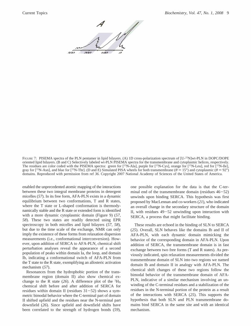
FIGURE 7: PISEMA spectra of the PLN pentamer in lipid bilayers. (A) 1D cross-polarization spectrum of [U-15N]wt-PLN in DOPC/DOPEoriented lipid bilayers. (B and C) Selectively labeled wt-PLN PISEMA spectra for the transmembrane and cytoplasmic helices, respectively.The residues are color coded with the PISEMA spectra: green for [15N-Ala], purple for [15N-Cys], orange for [15N-Leu], red for [15N-Ile],gray for [15N-Asn], and blue for [15N-Thr]. (D and E) Simulated PISA wheels for both transmembrane (θ ) 15°) and cytoplasmic (θ ) 92°)domains. Reproduced with permission from ref36. Copyright 2007 National Academy of Sciences of the United States of America.
Current Topics Biochemistry, Vol. 47, No. 1, 20089
Effects of PLN Phosphorylation on the AllostericMechanism
The inhibition of SERCA by PLN can be reversed byphosphorylation at Ser-16 by cAMP-dependent protein kinaseA (2). We determined the structure of Ser-16-phosphory-lated AFA-PLN (pS16-AFA-PLN) and found that residues14-16, previously helical, became unwound upon phospho-
rylation, revealing an order-to-disorder transition (33). Inaddition, we found that there are pronounced changes inpS16-AFA-PLN backbone dynamics on both the picosecondto nanosecond and microsecond to millisecond time scales(33). Although small, some of the changes are propagatedthroughout the entire protein backbone, demonstrating thatwhile the structural transitions following phosphorylation are
FIGURE 8: Simulated and experimental PISEMA spectra for the pinwheel and bellflower models. Unlike the pinwheel model, the bellflowermodel shows no high-field resonances. Experimental PISEMA spectra (C and F) show the remarkable agreement with the pinwheel model(A and D). Reproduced with permission from ref36. Copyright 2007 National Academy of Sciences of the United States of America.
FIGURE 9: (A) Allosteric model of interaction of PLN with SERCA. The PLN monomer interconverts between the L-shaped form (T state)and the less stable (more dynamically disordered) extended form (R state). (B) PLN-SERCA model developed by Toyoshima et al. (21)highlighting the long-range allosteric control phosphorylation at Ser-16 has on domain Ib. Panel A reproduced with permission from ref57.Copyright 2005 National Academy of Sciences of the United States of America. Panel B reproduced with permisson from ref26. Copyright2006 Elsevier.
10 Biochemistry, Vol. 47, No. 1, 2008 Current Topics
localized, the changes in backbone dynamics are radiatedthroughout the protein.
How can this order-to-disorder transition help in under-standing the interaction with SERCA? To answer thisquestion, we proceeded with the analysis of the chemicalshift perturbation of pS16-AFA-PLN induced by SERCA(26). We found that the conformational equilibrium betweenthe T and R states upon addition of SERCA is influencedby single phosphorylation at Ser-16; specifically, phospho-rylation shifts the equilibrium toward the R state in acooperative manner (26). Another considerable differenceupon phosphorylation includes a change in both the surfaceand the dynamics of domain Ib. In particular, a remarkablechange is observed for the side chain binding behavior. Inconstrast with unphosphorylated PLN, the Gln-26 resonancein the phosphorylated protein is unperturbed by SERCA withother smaller changes seen for side chain residues Asn-27,-30, and -34. A possible mechanism for explaining theseresults is the rotation or rearrangement of domain Ib uponphosphorylation that disrupts crucial intermolecular hydrogenbonds, resulting in relief of inhibition. On the basis of themolecular model of MacLennan and co-workers (21), weproposed that a crucial hydrogen bond between Arg-324 andGln-26 may be broken between SERCA and PLN afterphosphorylation at Ser-16 (26). These findings are inagreement with mutagenesis studies, showing that Q26A isa loss-of-function mutant (60). In addition, cross-linkingstudies show that an N27C mutation in pS16-AFA-PLN isno longer able to cross-link with SERCA (21). While thereare changes in the binding interface for domain Ib, the overallbinding of domain II to SERCA is not affected by phos-phorylation. The dissociation constants (Kd) for domain IIin both pS16-AFA-PLN and AFA-PLN are∼60 µM (26).This demonstrates that the major changes are in domain Ia,loop, and domain Ib, with domain II only marginally affected,supporting the hypothesis that phosphorylation at Ser-16 doesnot dissociate PLN from SERCA completely. We proposeddomain Ib as a bridgehead region, which transmits thedynamics induced by phosphorylation at Ser-16 from domainIa to domain Ib, thereby regulating the intramembraneprotein-protein interaction (26). A schematic of the allostericmodel for phosphorylation is reported in Figure 9.
PerspectiVe
What can we learn from the analysis of the structure anddynamics of PLN? More importantly, how are structure anddynamics of PLN correlated to SERCA’s function, and canwe control the extent of inhibition of SERCA by manipulat-ing PLN structural dynamics? These are questions we havebegun to address in our recent publication, showing thatindeed it is possible to control SERCA’s activity by tuningPLN structural dynamics (62). We will continue to tacklethese questions in the upcoming years.
Given the plethora of biochemical and molecular biologydata currently available, it is a very exciting moment forstructural biologists involved in research on PLN and SLNand their interactions with SERCA. While our studies to datehave focused on monitoring the effects of PLN induced bySERCA, our future challenge involves detecting SERCAchanges from PLN within the entire enzymatic cycle.Another important challenge is to study the SERCA-PLN
complex in the complex network of interactions involvingprotein kinase A and protein phosphatase 1. While solutionNMR will help identify some important pieces of thiscomplex puzzle, solid-state NMR will be the method ofchoice for the elucidation of the structural dynamics andinteractions in these large complexes.
ACKNOWLEDGMENT
We are grateful to the former members of the VegliaLaboratory (Alessandro Mascioni, Bethany Buck-Koehntop,Jamillah Zamoon, and Emily Metcalfe) and for the produc-tive collaboration with the laboratory of David Thomas.
REFERENCES
1. Fujii, J., Ueno, A., Kitano, K., Tanaka, S., Kadoma, M., and Tada,M. (1987) Complete Complementary DNA-Derived Amino AcidSequence of Canine Cardiac Phospholamban,J. Clin. InVest. 79,301-304.
2. Wegener, A. D., Simmerman, H. K., Lindemann, J. P., and Jones,L. R. (1989) Phospholamban Phosphorylation in Intact Ventricles.Phosphorylation of Serine 16 and Threonine 17 in Response toâ-Adrenergic Stimulation,J. Biol. Chem. 264, 11468-11474.
3. Mundina-Weilenmann, C., Vittone, L., Ortale, M., de Cingolani,G. C., and Mattiazzi, A. (1996) Immunodetection of Phosphory-lation Sites Gives New Insights into the Mechanisms UnderlyingPhospholamban Phosphorylation in the Intact Heart,J. Biol. Chem.271, 33561-33567.
4. Chu, G., Lester, J. W., Young, K. B., Luo, W., Zhai, J., andKranias, E. G. (2000) A Single Site (Ser16) Phosphorylation inPhospholamban is Sufficient in Mediating its Maximal CardiacResponses toâ-Agonists,J. Biol. Chem. 275, 38938-38943.
5. Wawrzynow, A., Theibert, J. L., Murphy, C., Jona, I., Martonosi,A., and Collins, J. H. (1992) Sarcolipin, the “Proteolipid” ofSkeletal Muscle Sarcoplasmic Reticulum, is a Unique, Amphi-pathic, 31-Residue Peptide,Arch. Biochem. Biophys. 298, 620-623.
6. Odermatt, A., Taschner, P. E., Scherer, S. W., Beatty, B., Khanna,V. K., Cornblath, D. R., Chaudhry, V., Yee, W. C., Schrank, B.,Karpati, G., Breuning, M. H., Knoers, N., and MacLennan, D. H.(1997) Characterization of the Gene Encoding Human Sarcolipin(SLN), a Proteolipid Associated with SERCA1: Absence ofStructural Mutations in Five Patients with Brody Disease,Ge-nomics 45, 541-553.
7. Gayan-Ramirez, G., Vanzeir, L., Wuytack, F., and Decramer, M.(2000) Corticosteroids Decrease mRNA Levels of SERCA Pumps,Whereas they Increase Sarcolipin mRNA in the Rat Diaphragm,J. Physiol. 524(Part 2), 387-397.
8. Minamisawa, S., Wang, Y., Chen, J., Ishikawa, Y., Chien, K. R.,and Matsuoka, R. (2003) Atrial Chamber-Specific Expression ofSarcolipin is Regulated during Development and HypertrophicRemodeling,J. Biol. Chem. 278, 9570-9575.
9. Babu, G. J., Bhupathy, P., Carnes, C. A., Billman, G. E., andPeriasamy, M. (2007) Differential Expression of Sarcolipin Proteinduring Muscle Development and Cardiac Pathophysiology,J. Mol.Cell. Cardiol. 43, 215-222.
10. Maclennan, D. H., Yip, C. C., Iles, and Seeman, P. (1972) Isolationof Sarcoplasmic Reticulum Proteins,Cold Spring Harbor Symp.Quant. Biol. 37, 469-478.
11. Odermatt, A., Becker, S., Khanna, V. K., Kurzydlowski, K.,Leisner, E., Pette, D., and MacLennan, D. H. (1998) SarcolipinRegulates the Activity of SERCA1, the Fast-Twitch SkeletalMuscle Sarcoplasmic Reticulum Ca2+-ATPase,J. Biol. Chem. 273,12360-12369.
12. Uemura, N., Ohkusa, T., Hamano, K., Nakagome, M., Hori, H.,Shimizu, M., Matsuzaki, M., Mochizuki, S., Minamisawa, S., andIshikawa, Y. (2004) Down-Regulation of Sarcolipin mRNAExpression in Chronic Atrial Fibrillation,Eur. J. Clin. InVest. 34,723-730.
13. Gramolini, A. O., Trivieri, M. G., Oudit, G. Y., Kislinger, T., Li,W., Patel, M. M., Emili, A., Kranias, E. G., Backx, P. H., andMaclennan, D. H. (2006) Cardiac-Specific Overexpression ofSarcolipin in Phospholamban Null Mice Impairs Myocyte Func-tion that is Restored by Phosphorylation,Proc. Natl. Acad. Sci.U.S.A. 103, 2446-2451.
Current Topics Biochemistry, Vol. 47, No. 1, 200811
14. Toyoshima, C., Nakasako, M., Nomura, H., and Ogawa, H. (2000)Crystal Structure of the Calcium Pump of Sarcoplasmic Reticulumat 2.6 Å Resolution,Nature 405, 647-655.
15. Toyoshima, C., and Nomura, H. (2002) Structural Changes in theCalcium Pump Accompanying the Dissociation of Calcium,Nature 418, 605-611.
16. Toyoshima, C., and Mizutani, T. (2004) Crystal Structure of theCalcium Pump with a Bound ATP Analogue,Nature 430, 529-535.
17. Toyoshima, C., Nomura, H., and Tsuda, T. (2004) Lumenal GatingMechanism Revealed in Calcium Pump Crystal Structures withPhosphate Analogues,Nature 432, 361-368.
18. Olesen, C., Sorensen, T. L., Nielsen, R. C., Moller, J. V., andNissen, P. (2004) Dephosphorylation of the Calcium PumpCoupled to Counterion Occlusion,Science 306, 2251-2255.
19. Sorensen, T. L., Moller, J. V., and Nissen, P. (2004) PhosphorylTransfer and Calcium Ion Occlusion in the Calcium Pump,Science304, 1672-1675.
20. Young, H. S., Jones, L. R., and Stokes, D. L. (2001) LocatingPhospholamban in Co-Crystals with Ca2+-ATPase by CryoelectronMicroscopy,Biophys. J. 81, 884-894.
21. Toyoshima, C., Asahi, M., Sugita, Y., Khanna, R., Tsuda, T., andMacLennan, D. H. (2003) Modeling of the Inhibitory Interactionof Phospholamban with the Ca2+ ATPase,Proc. Natl. Acad. Sci.U.S.A. 100, 467-472.
22. Asahi, M., Sugita, Y., Kurzydlowski, K., De Leon, S., Tada, M.,Toyoshima, C., and MacLennan, D. H. (2003) Sarcolipin Regu-lates Sarco(Endo)Plasmic Reticulum Ca2+-ATPase (SERCA) byBinding to Transmembrane Helices Alone or in Association withPhospholamban,Proc. Natl. Acad. Sci. U.S.A. 100, 5040-5045.
23. Hutter, M. C., Krebs, J., Meiler, J., Griesinger, C., Carafoli, E.,and Helms, V. (2002) A Structural Model of the Complex Formedby Phospholamban and the Calcium Pump of SarcoplasmicReticulum obtained by Molecular Mechanics,ChemBioChem 3,1200-1208.
24. Buck, B., Zamoon, J., Kirby, T. L., DeSilva, T. M., Karim, C.,Thomas, D., and Veglia, G. (2003) Overexpression, Purification,and Characterization of Recombinant Ca-ATPase Regulators forHigh-Resolution Solution and Solid-State NMR Studies,ProteinExpression Purif. 30, 253-261.
25. Buffy, J. J., Buck-Koehntop, B. A., Porcelli, F., Traaseth, N. J.,Thomas, D. D., and Veglia, G. (2006) Defining the IntramembraneBinding Mechanism of Sarcolipin to Calcium ATPase usingSolution NMR Spectroscopy,J. Mol. Biol. 358, 420-429.
26. Traaseth, N. J., Thomas, D. D., and Veglia, G. (2006) Effects ofSer16 Phosphorylation on the Allosteric Transitions of phospho-lamban/Ca2+-ATPase Complex,J. Mol. Biol. 358, 1041-1050.
27. Kimura, Y., Kurzydlowski, K., Tada, M., and MacLennan, D. H.(1997) Phospholamban Inhibitory Function is Activated byDepolymerization,J. Biol. Chem. 272, 15061-15064.
28. Reddy, L. G., Jones, L. R., and Thomas, D. D. (1999) Depolym-erization of Phospholamban in the Presence of Calcium Pump:A Fluorescence Energy Transfer Study,Biochemistry 38, 3954-3962.
29. Zamoon, J., Mascioni, A., Thomas, D. D., and Veglia, G. (2003)NMR Solution Structure and Topological Orientation of Mono-meric Phospholamban in Dodecylphosphocholine Micelles,Bio-phys. J. 85, 2589-2598.
30. Pollesello, P., Annila, A., and Ovaska, M. (1999) Structure of the1-36 Amino-Terminal Fragment of Human Phospholamban byNuclear Magnetic Resonance and Modeling of the PhospholambanPentamer,Biophys. J. 76, 1784-1795.
31. Lambeth, S., Schmid, H., Muenchbach, M., Vorherr, T., Krebs,J., Carafoli, E., and Griesinger, C. (2000) NMR Solution Structureof Phospholamban,HelV. Chim. Acta 83, 2141-2152.
32. Metcalfe, E. E., Zamoon, J., Thomas, D. D., and Veglia, G. (2004)1H/15N Heteronuclear NMR Spectroscopy shows Four DynamicDomains for Phospholamban Reconstituted in Dodecylphospho-choline Micelles,Biophys. J. 87, 1205-1214.
33. Metcalfe, E. E., Traaseth, N. J., and Veglia, G. (2005) Serine 16Phosphorylation Induces an Order-to-Disorder Transition inMonomeric Phospholamban,Biochemistry 44, 4386-4396.
34. Schmidt, A. G., Zhai, J., Carr, A. N., Gerst, M. J., Lorenz, J. N.,Pollesello, P., Annila, A., Hoit, B. D., and Kranias, E. G. (2002)Structural and Functional Implications of the PhospholambanHinge Domain: Impaired SR Ca2+ Uptake as a Primary Causeof Heart Failure,CardioVasc. Res. 56, 248-259.
35. Oxenoid, K., and Chou, J. J. (2005) The Structure of Phospho-lamban Pentamer Reveals a Channel-Like Architecture in Mem-branes,Proc. Natl. Acad. Sci. U.S.A. 102, 10870-10875.
36. Traaseth, N. T., Verardi, R., Torgersen, K. D., Karim, C. B.,Thomas, D. D., and Veglia, G. (2007) Spectroscopic Validationof the Pentameric Structure of Phospholamban,Proc. Natl. Acad.Sci. U.S.A. 104, 14676-14681.
37. Mascioni, A., Karim, C., Barany, G., Thomas, D. D., and Veglia,G. (2002) Structure and Orientation of Sarcolipin in LipidEnvironments,Biochemistry 41, 475-482.
38. Opella, S. J. (1997) NMR and Membrane Proteins,Nat. Struct.Biol. 4 (Suppl.), 845-848.
39. Baldus, M. (2006) Molecular Interactions Investigated by Multi-Dimensional Solid-State NMR,Curr. Opin. Struct. Biol. 16, 618-623.
40. Mascioni, A., Karim, C., Zamoon, J., Thomas, D. D., and Veglia,G. (2002) Solid-State NMR and Rigid Body Molecular Dynamicsto Determine Domain Orientations of Monomeric Phospholamban,J. Am. Chem. Soc. 124, 9392-9393.
41. Wu, C. H., Ramamoorthy, A., and Opella, S. J. (1994) High-Resolution Heteronuclear Dipolar Solid-State NMR Spectroscopy,J. Magn. Reson. 109, 270-272.
42. Traaseth, N. J., Buffy, J. J., Zamoon, J., and Veglia, G. (2006)Structural Dynamics and Topology of Phospholamban in OrientedLipid Bilayers using Multidimensional Solid-State NMR,Bio-chemistry 45, 13827-13834.
43. Grage, S. L., Wang, J., Cross, T. A., and Ulrich, A. S. (2002)Solid-State19F-NMR Analysis of 19F-Labeled Tryptophan inGramicidin A in Oriented Membranes,Biophys. J. 83, 3336-3350.
44. Aisenbrey, C., and Bechinger, B. (2004) Investigations of Polypep-tide Rotational Diffusion in Aligned Membranes by2H and15NSolid-State NMR Spectroscopy,J. Am. Chem. Soc. 126, 16676-16683.
45. Park, S. H., Mrse, A. A., Nevzorov, A. A., De Angelis, A. A.,and Opella, S. J. (2006) Rotational Diffusion of MembraneProteins in Aligned Phospholipid Bilayers by Solid-State NMRSpectroscopy,J. Magn. Reson. 178, 162-165.
46. Andronesi, O. C., Becker, S., Seidel, K., Heise, H., Young, H. S.,and Baldus, M. (2005) Determination of Membrane ProteinStructure and Dynamics by Magic-Angle-Spinning Solid-StateNMR Spectroscopy,J. Am. Chem. Soc. 127, 12965-12974.
47. Hughes, E., Clayton, J. C., and Middleton, D. A. (2005) Probingthe Oligomeric State of Phospholamban Variants in PhospholipidBilayers from Solid-State NMR Measurements of RotationalDiffusion Rates,Biochemistry 44, 4055-4066.
48. Karp, E. S., Tiburu, E. K., Abu-Baker, S., and Lorigan, G. A.(2006) The Structural Properties of the Transmembrane Segmentof the Integral Membrane Protein Phospholamban Utilizing13CCPMAS, 2H, and REDOR Solid-State NMR Spectroscopy,Biochim. Biophys. Acta 1758, 772-780.
49. Buffy, J. J., Traaseth, N. J., Mascioni, A., Gor’kov, P. L.,Chekmenev, E. Y., Brey, W. W., and Veglia, G. (2006) Two-Dimensional Solid-State NMR Reveals Two Topologies ofSarcolipin in Oriented Lipid Bilayers,Biochemistry 45, 10939-10946.
50. Stokes, D. L., Pomfret, A. J., Rice, W. J., Glaves, J. P., and Young,H. S. (2006) Interactions between Ca2+-ATPase and the Pentam-eric Form of Phospholamban in Two-Dimensional Co-Crystals,Biophys. J. 90, 4213-4223.
51. Tatulian, S. A., Jones, L. R., Reddy, L. G., Stokes, D. L., andTamm, L. K. (1995) Secondary Structure and Orientation ofPhospholamban Reconstituted in Supported Bilayers from Polar-ized Attenuated Total Reflection FTIR Spectroscopy,Biochemistry34, 4448-4456.
52. Arkin, I. T., Rothman, M., Ludlam, C. F., Aimoto, S., Engelman,D. M., Rothschild, K. J., and Smith, S. O. (1995) Structural Modelof the Phospholamban Ion Channel Complex in PhospholipidMembranes,J. Mol. Biol. 248, 824-834.
53. Smith, S. O., Kawakami, T., Liu, W., Ziliox, M., and Aimoto, S.(2001) Helical Structure of Phospholamban in Membrane Bilayers,J. Mol. Biol. 313, 1139-1148.
54. Robia, S. L., Flohr, N. C., and Thomas, D. D. (2005) Phospho-lamban Pentamer Quaternary Conformation Determined by in-Gel Fluorescence Anisotropy,Biochemistry 44, 4302-4311.
55. Simmerman, H. K., Kobayashi, Y. M., Autry, J. M., and Jones,L. R. (1996) A Leucine Zipper Stabilizes the PentamericMembrane Domain of Phospholamban and Forms a Coiled-CoilPore Structure,J. Biol. Chem. 271, 5941-5946.
12 Biochemistry, Vol. 47, No. 1, 2008 Current Topics
56. Karim, C. B., Stamm, J. D., Karim, J., Jones, L. R., and Thomas,D. D. (1998) Cysteine Reactivity and Oligomeric Structures ofPhospholamban and its Mutants,Biochemistry 37, 12074-12081.
57. Zamoon, J., Nitu, F., Karim, C., Thomas, D. D., and Veglia, G.(2005) Mapping the Interaction Surface of a Membrane Protein:Unveiling the Conformational Switch of Phospholamban inCalcium Pump Regulation,Proc. Natl. Acad. Sci. U.S.A. 102,4747-4752.
58. Karim, C. B., Kirby, T. L., Zhang, Z., Nesmelov, Y., and Thomas,D. D. (2004) Phospholamban Structural Dynamics in LipidBilayers Probed by a Spin Label Rigidly Coupled to the PeptideBackbone,Proc. Natl. Acad. Sci. U.S.A. 101, 14437-14442.
59. Wagner, G., Pardi, A., and Wuthrich, K. (1983) Hydrogen BondLength and Proton NMR Chemical Shifts in Proteins,J. Am.Chem. Soc. 105, 5948-5949.
60. Kimura, Y., Asahi, M., Kurzydlowski, K., Tada, M., andMacLennan, D. H. (1998) Phospholamban Domain Ib Mutations
Influence Functional Interactions with the Ca2+-ATPase Isoformof Cardiac Sarcoplasmic Reticulum,J. Biol. Chem. 273, 14238-14241.
61. Inesi, G., Lewis, D., Ma, H., Prasad, A., and Toyoshima, C. (2006)Concerted Conformational Effects of Ca2+ and ATP are Requiredfor Activation of Sequential Reactions in the Ca2+ ATPase(SERCA) Catalytic Cycle,Biochemistry 45, 13769-13778.
62. Ha, K. N., Traaseth, N. J., Verardi, R., Zamoon, J., Cembran, A.,Karim, C. B., Thomas, D. D., Veglia, G. (2007) Controlling theInhibition of the Sarcoplasmic Ca2+-ATPase by Tuning Phos-pholamban Structural Dynamics,J. Biol. Chem.E-pub Sep. 30,2007.
BI701668V
Current Topics Biochemistry, Vol. 47, No. 1, 200813