A nuclear-encoded protein, mTERF6, mediates transcription ...
Cell Host & Microbe
Article
Strong Immunogenicity and Cross-Reactivity ofMycobacterium tuberculosis ESX-5 Type VII Secretion-Encoded PE-PPE Proteins Predicts Vaccine PotentialFadel Sayes,1,4 Lin Sun,1,4,7 Mariagrazia Di Luca,5 Roxane Simeone,2 Nathalie Degaiffier,1,4 Laurence Fiette,3,6
Semih Esin,5 Roland Brosch,2 Daria Bottai,5 Claude Leclerc,1,4 and Laleh Majlessi1,4,*1Unite de Regulation Immunitaire et Vaccinologie2Unite de Pathogenomique Mycobacterienne Integree3Unite d’Histopathologie Humaine et Modeles Animaux
Institut Pasteur, Paris F-75015, France4INSERM U1041, Paris F-75015, France5Dipartimento di Patologia Sperimentale, Biotecnologie Mediche, Infettivologia ed Epidemiologia, Universita di Pisa, 56100 Pisa, Italy6Faculte de Medecine, DER Histologie, Universite Versailles-Saint Quentin en Yvelines, 78000 Versailles, France7Present address: Jiangsu Key Laboratory of Zoonosis, Yangzhou University, 225009 Yangzhou, Jiangsu, China
*Correspondence: [email protected]
DOI 10.1016/j.chom.2012.03.003
SUMMARY
The genome of Mycobacterium tuberculosis (Mtb)encodes five type VII secretion systems, ESX-1 toESX-5, most of which are associated with genesencoding PE/PPE proteins, named after theirN-terminal Pro-Glu (PE) or Pro-Pro-Glu (PPE) motifs.Here, we describe the strong T cell immunogenicityof the ESX-5-encoded PE/PPE proteins, which sharea large panel of cross-reactive CD4+ epitopes withsubstantial numbers of their ESX-5-nonassociatedPE/PPE homologs. The immunogenicity of thesenumerous PE/PPE proteins is dependent on theirexport by a functional EccD5, the predicted trans-membrane channel of the ESX-5 secretion appa-ratus. The Mtb Dppe25-pe19 mutant deleted for allESX-5-associated pe and ppe genes, although highlyattenuated in immunocompetent mice, remains ableto induce immunity against the ESX-5-associatedPE/PPE virulence factors, via cross-reactivity withtheir numerous homologs, and against the ESX-1virulence factors ESAT-6/CFP-10. The Dppe25-pe19strain is strongly protective against Mtb infectionin mice and represents a potential antituberculosisvaccine candidate.
INTRODUCTION
Mycobacterium tuberculosis (Mtb) remains one of the world’s
most successful pathogens, infecting one-third of humans,
with �8 million new infections/year leading to �2 million
deaths/year. The currently available Mycobacterium bovis
Bacillus Calmette-Guerin (BCG) vaccine is of limited efficacy
against adult pulmonary tuberculosis (http://www.who.int/tb/
publications/global_reports/2009/). Identification of mycobacte-
rial virulence determinants and components that interact with the
352 Cell Host & Microbe 11, 352–363, April 19, 2012 ª2012 Elsevier
host immune system is critical to understand the mechanisms of
pathogenesis and immunogenicity of mycobacteria to develop
new drugs and vaccines. Compared to the virulent strain Mtb
H37Rv, BCG has numerous genetic deletions (Brosch et al.,
2000), including the 9.8 kb region of difference 1 (RD1). RD1
encodes members of a secretion system dedicated to the
export/secretion of the highly immunogenic major virulence
factors 6 kDa early secreted antigenic target (ESAT-6) and
10 kDa culture filtrate protein (CFP-10) (Brodin et al., 2004;
Mahairas et al., 1996; Simeone et al., 2009), as well as the Esp
proteins, which are putative virulence factors (Abdallah et al.,
2007; Bitter et al., 2009b; DiGiuseppe Champion et al., 2009).
ESAT-6 is the canonical member of the ESX protein family and
is secreted by the ESX-1 type VII secretion system (Abdallah
et al., 2007; Bitter et al., 2009b; DiGiuseppe Champion et al.,
2009). The products exported by the ESX-1 system modulate
the interaction of Mtb with the host immune system (Majlessi
et al., 2005; Stanley et al., 2007; van der Wel et al., 2007).
In addition to the esx-1 (RD1) region, the genome of Mtb
contains four other esx clusters, esx-2 to esx-5, which encode
paralogous ESX type VII secretion systems (Brodin et al., 2004;
Gey Van Pittius et al., 2001; Tekaia et al., 1999). These conserved
clusters contain tandem repeats of esx genes flanked by
genes encoding putative members of type VII secretion systems
(Bitter et al., 2009a; Brodin et al., 2004; DiGiuseppe Champion
et al., 2009). All esx clusters except for esx-4 include one
or more pe and ppe genes. The PE/PPE proteins, named for
their characteristic N-terminal Pro-Glu (PE) or Pro-Pro-Glu
(PPE) motifs (Bottai and Brosch, 2009; Brennan and Delogu,
2002; Cole et al., 1998), are two large protein families that are
unique to mycobacteria. The Mtb genome contains �100 pe
and �69 ppe genes (Cole et al., 1998), which may have
evolved from esx-encoded ancestral pe/ppe copies and are
characterized by homologous repetitive sequences (Gey van
Pittius et al., 2006). This close evolutionary link between esx
clusters and pe/ppe genes suggests that PE/PPE proteins
contribute to infection and pathogenesis. PE/PPE proteins
have been associated with virulence, persistence, and patho-
genesis in the host and may play a role in antigenic variation
Inc.
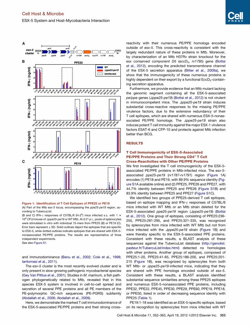
Figure 1. Identification of T Cell Epitopes of PPE25 or PE19
(A) Part of the Mtb esx-5 locus, encompassing the ppe25-pe19 region, ac-
cording to TubercuList.
(B and C) IFN-g responses of C57BL/6 (H-2b) mice infected s.c. with 1 3
106 CFU/mouse of Dppe25-pe19 or WTMtb. At d 21 p.i., pools of splenocytes
were stimulated in vitro with individual 15-mers from PPE25 (B) or PE19 (C).
Error bars represent ± SD. Solid outlines depict the epitopes that are specific
to ESX-5, while dotted outlines indicate epitopes that are shared with ESX-5-
nonassociated PE/PPE proteins. The results are representative of three
independent experiments.
See also Figure S1.
Cell Host & Microbe
ESX-5 System and Host-Mycobacteria Interaction
and immunodominance (Banu et al., 2002; Cole et al., 1998;
Iantomasi et al., 2011).
The esx-5 cluster is the most recently evolved cluster and is
only present in slow-growing pathogenic mycobacterial species
(Gey Van Pittius et al., 2001). Studies inM. marinum, a fish path-
ogen phylogenetically related to Mtb, revealed that in this
species ESX-5 system is involved in cell-to-cell spread and
secretion of several PPE proteins and all PE members of the
PE-polymorphic GC-rich sequences (PE-PGRS) subfamily
(Abdallah et al., 2006; Abdallah et al., 2009).
Here, we demonstrate themarked T cell immunodominance of
the ESX-5-associated PE/PPE proteins and their strong cross-
Cell H
reactivity with their numerous PE/PPE homologs encoded
outside of esx-5. This cross-reactivity is consistent with the
largely redundant nature of these proteins in Mtb. Moreover,
by characterization of an Mtb H37Rv strain knockout for the
esx conserved component D5 (eccD5, rv1795) gene (Bottai
et al., 2012), encoding the predicted transmembrane channel
of the ESX-5 secretion apparatus (Bitter et al., 2009a), we
show that the immunogenicity of these numerous proteins is
highly dependent on their export by a functional EccD5-contain-
ing secretion apparatus.
Furthermore, we provide evidence that an Mtb mutant lacking
the genomic segment containing all the ESX-5-associated
pe/ppe genes (Dppe25-pe19) (Bottai et al., 2012) is not virulent
in immunocompetent mice. The Dppe25-pe19 strain induces
substantial cross-reactive responses to the missing PE/PPE
virulence factors, due to the extensive redundancy of their
T cell epitopes, which are shared with numerous ESX-5-nonas-
sociated PE/PPE homologs. The Dppe25-pe19 strain also
induces potent T cell immunity against themajor ESX-1 virulence
factors ESAT-6 and CFP-10 and protects against Mtb infection
better than BCG.
RESULTS
T Cell Immunogenicity of ESX-5-AssociatedPE/PPE Proteins and Their Strong CD4+ T CellCross-Reactivities with Other PE/PPE ProteinsWe first investigated the T cell immunogenicity of the ESX-5-
associated PE/PPE proteins in Mtb-infected mice. The esx-5-
associated ppe25-pe19 (rv1787-rv1791) region (Figure 1A)
encodes (1) PE18 and PE19, with 89.9% sequence identity (Fig-
ure S1A available online) and (2) PPE25, PPE26 and PPE27, with
44.7% identity between PPE25 and PPE26 (Figure S1B) and
83.9% identity between PPE25 and PPE27 (Figure S1C).
We identified two groups of PPE25-derived T cell epitopes,
based on epitope mapping and IFN-g responses of C57BL/6
mice infected with WT Mtb or an Mtb strain deleted for the
ESX-5-associated ppe25-pe19 region (Dppe25-pe19) (Bottai
et al., 2012). One group of epitopes, consisting of PPE25:236-
255, PPE25:281-295, and PPE25:321-335, was recognized
by splenocytes from mice infected with WT Mtb but not from
mice infected with the Dppe25-pe19 strain (Figure 1B) and
were thereby specific to the ESX-5-associated PPE proteins.
Consistent with these results, a BLAST analysis of these
sequences against the TubercuList database (http://genolist.
pasteur.fr/TubercuList/index.html) detected no homologies
with other proteins. Another group of epitopes, composed of
PPE25:1-20, PPE25:41-65, PPE25:186-200, and PPE25:201-
215 (Figure 1B), was recognized by splenocytes from both
WT Mtb- or Dppe25-pe19-infected mice, indicating that they
are shared with PPE homologs encoded outside of esx-5.
Consistent with these results, a BLAST analysis identified
substantial sequence similarities among these PPE25 epitopes
and numerous ESX-5-nonassociated PPE proteins, including
PPE32, PPE22, PPE45, PPE30, PPE29, PPE60, PPE19, PPE18,
or PPE50, listed in order of decreasing sequence identity with
PPE25 (Table 1).
PE19:1-18 was identified as an ESX-5-specific epitope, based
on its recognition by splenocytes from mice infected with WT
ost & Microbe 11, 352–363, April 19, 2012 ª2012 Elsevier Inc. 353
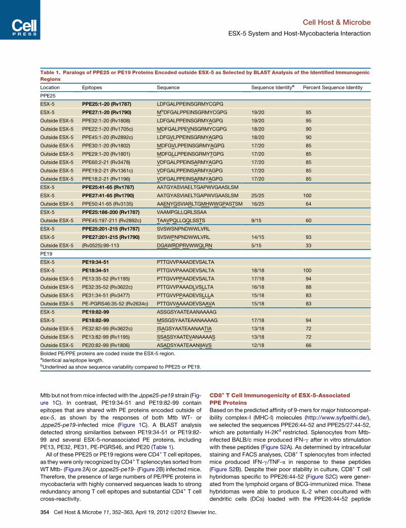
Table 1. Paralogs of PPE25 or PE19 Proteins Encoded outside ESX-5 as Selected by BLAST Analysis of the Identified Immunogenic
Regions
Location Epitopes Sequence Sequence Identitya Percent Sequence Identity
PPE25
ESX-5 PPE25:1-20 (Rv1787) LDFGALPPEINSGRMYCGPG
ESX-5 PPE27:1-20 (Rv1790) MbDFGALPPEINSGRMYCGPG 19/20 95
Outside ESX-5 PPE32:1-20 (Rv1808) LDFGALPPEINSGRMYAGPG 19/20 95
Outside ESX-5 PPE22:1-20 (Rv1705c) MDFGALPPEVNSGRMYCGPG 18/20 90
Outside ESX-5 PPE45:1-20 (Rv2892c) LDFGVLPPEINSGRMYAGPG 18/20 90
Outside ESX-5 PPE30:1-20 (Rv1802) MDFGVLPPEINSGRMYAGPG 17/20 85
Outside ESX-5 PPE29:1-20 (Rv1801) MDFGLLPPEINSGRMYTGPG 17/20 85
Outside ESX-5 PPE60:2-21 (Rv3478) VDFGALPPEINSARMYAGPG 17/20 85
Outside ESX-5 PPE19:2-21 (Rv1361c) VDFGALPPEINSARMYAGPG 17/20 85
Outside ESX-5 PPE18:2-21 (Rv1196) VDFGALPPEINSARMYAGPG 17/20 85
ESX-5 PPE25:41-65 (Rv1787) AATGYASVIAELTGAPWVGAASLSM
ESX-5 PPE27:41-65 (Rv1790) AATGYASVIAELTGAPWVGAASLSM 25/25 100
Outside ESX-5 PPE50:41-65 (Rv3135) AAENYGSVIARLTGMHWWGPASTSM 16/25 64
ESX-5 PPE25:186-200 (Rv1787) VAAMPGLLQRLSSAA
Outside ESX-5 PPE45:197-211 (Rv2892c) TAAVPQLLQQLSSTS 9/15 60
ESX-5 PPE25:201-215 (Rv1787) SVSWSNPNDWWLVRL
ESX-5 PPE27:201-215 (Rv1790) SVSWPNPNDWWLVRL 14/15 93
Outside ESX-5 (Rv0525):99-113 DGAWRDPRVWWQLRN 5/15 33
PE19
ESX-5 PE19:34-51 PTTGVVPAAADEVSALTA
ESX-5 PE18:34-51 PTTGVVPAAADEVSALTA 18/18 100
Outside ESX-5 PE13:35-52 (Rv1195) PTTGVVPPAADEVSALTA 17/18 94
Outside ESX-5 PE32:35-52 (Rv3622c) PTTGVVPAAADLVSLLTA 16/18 88
Outside ESX-5 PE31:34-51 (Rv3477) PTTGVVPPAADEVSLLLA 15/18 83
Outside ESX-5 PE-PGRS46:35-52 (Rv2634c) PTTGVVAAAADEVSAAVA 15/18 83
ESX-5 PE19:82-99 ASSGSYAATEAANAAAAG
ESX-5 PE18:82-99 MSSGSYAATEAANAAAAG 17/18 94
Outside ESX-5 PE32:82-99 (Rv3622c) ISAGSYAATEAANAATIA 13/18 72
Outside ESX-5 PE13:82-99 (Rv1195) SSASSYAATEVANAAAAS 13/18 72
Outside ESX-5 PE20:82-99 (Rv1806) ASADSYAATEAANIIAVS 12/18 66
Bolded PE/PPE proteins are coded inside the ESX-5 region.aIdentical aa/epitope length.bUnderlined aa show sequence variability compared to PPE25 or PE19.
Cell Host & Microbe
ESX-5 System and Host-Mycobacteria Interaction
Mtb but not frommice infectedwith theDppe25-pe19 strain (Fig-
ure 1C). In contrast, PE19:34-51 and PE19:82-99 contain
epitopes that are shared with PE proteins encoded outside of
esx-5, as shown by the responses of both Mtb WT- or
Dppe25-pe19-infected mice (Figure 1C). A BLAST analysis
detected strong similarities between PE19:34-51 or PE19:82-
99 and several ESX-5-nonassociated PE proteins, including
PE13, PE32, PE31, PE-PGRS46, and PE20 (Table 1).
All of these PPE25 or PE19 regions were CD4+ T cell epitopes,
as they were only recognized by CD4+ T splenocytes sorted from
WTMtb- (Figure 2A) or Dppe25-pe19- (Figure 2B) infected mice.
Therefore, the presence of large numbers of PE/PPE proteins in
mycobacteria with highly conserved sequences leads to strong
redundancy among T cell epitopes and substantial CD4+ T cell
cross-reactivity.
354 Cell Host & Microbe 11, 352–363, April 19, 2012 ª2012 Elsevier
CD8+ T Cell Immunogenicity of ESX-5-AssociatedPPE ProteinsBased on the predicted affinity of 9-mers for major histocompat-
ibility complex-I (MHC-I) molecules (http://www.syfpeithi.de/),
we selected the sequences PPE26:44-52 and PPE25/27:44-52,
which are potentially H-2Kd restricted. Splenocytes from Mtb-
infected BALB/c mice produced IFN-g after in vitro stimulation
with these peptides (Figure S2A). As determined by intracellular
staining and FACS analyses, CD8+ T splenocytes from infected
mice produced IFN-g/TNF-a in response to these peptides
(Figure S2B). Despite their poor stability in culture, CD8+ T cell
hybridomas specific to PPE26:44-52 (Figure S2C) were gener-
ated from the lymphoid organs of BCG-immunized mice. These
hybridomas were able to produce IL-2 when cocultured with
dendritic cells (DCs) loaded with the PPE26:44-52 peptide
Inc.
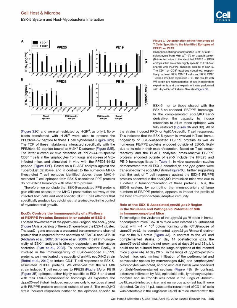
Figure 2. Determination of the Phenotype of
T Cells Specific to the Identified Epitopes of
PPE25 or PE19
Responses of magnetically sorted CD4+ or CD8+ T
splenocytes from Mtb WT- (A) or Dppe25-pe19-
(B) infected mice to the identified PPE25 or PE19
epitopes that are either highly specific to ESX-5 or
shared with PE/PPE encoded outside of ESX-5.
The CD4+ or CD8+ fractions contained, respec-
tively, at least 99% CD4+ T cells and 97% CD8+
T cells. Error bars represent ± SD. The results with
WT strain are representative of two independent
experiments and one experiment was performed
with Dppe25-pe19 strain. See also Figure S2.
Cell Host & Microbe
ESX-5 System and Host-Mycobacteria Interaction
(Figure S2C) and were all restricted by H-2Kd, as only L fibro-
blasts transfected with H-2Kd were able to present the
PPE26:44-52 peptide to these T cell hybridomas (Figure S2D).
The TCR of these hybridomas interacted specifically with the
PPE26:44-52 peptide bound to H-2Kd Dextramer (Figure S2E).
The latter allowed ex vivo detection of PPE26:44-52-specific
CD8+ T cells in the lymphocytes from lungs and spleen of Mtb-
infected mice, and stimulated in vitro with the PPE26:44-52
peptide (Figure S2F). Based on a BLAST analysis against the
TubercuList database, and in contrast to the numerous MHC-
II-restricted T cell epitopes identified above, these MHC-I-
restricted T cell epitopes from ESX-5-associated PPE proteins
do not exhibit homology with other Mtb proteins.
Therefore, we conclude that ESX-5-associated PPE proteins
gain efficient access to the MHC-I presentation pathway of the
infected host cells and elicit specific CD8+ T cell effectors that
specifically produce key cytokines that are involved in the control
of mycobacterial growth.
EccD5 Controls the Immunogenicity of a Plethoraof PE/PPE Proteins Encoded in or outside of ESX-5Located downstream of the ppe25-pe19 region, the eccD5 gene
(Figure 1A) is a paralog of the eccD1 gene from the ESX-1 cluster.
The eccD1 gene encodes a presumed transmembrane channel
protein that is required for the secretion of ESAT-6 and CFP-10
(Abdallah et al., 2007; Simeone et al., 2009). T cell immunoge-
nicity of ESX-1 antigens is directly dependent on their active
secretion (Pym et al., 2003). To address whether EccD5 is
involved in the immunogenicity of ESX-5-encoded PE/PPE
proteins, we investigated the capacity of an Mtb eccD5KO strain
(Bottai et al., 2012) to induce CD4+ T cell responses to ESX-5-
associated PE/PPE proteins. We observed that the WT Mtb
strain induced T cell responses to PPE25 (Figure 3A) or PE19
(Figure 3B) epitopes, either highly specific to ESX-5 or shared
with their ESX-5-nonassociated homologs. As expected, the
Dppe25-pe19 strain induced responses only to epitopes shared
with PE/PPE proteins encoded outside of esx-5. The eccD5KO
strain induced responses neither to the epitopes specific to
Cell Host & Microbe 11, 352–3
ESX-5, nor to those shared with the
ESX-5-no-encoded PE/PPE homologs.
In the complemented eccD5KO::esx-5
derivative, the capacity to induce
responses to all of these epitopes was
fully restored (Figures 3A and 3B). All of
the strains induced PPD- or Ag85A-specific T cell responses.
This indicates that the ESX-5 system is involved in T cell immu-
nogenicity of ESX-5-associated PE/PPE proteins as well as
numerous PE/PPE proteins encoded outside of ESX-5, likely
due to its role in their export/secretion. Based on T cell cross-
reactivity and the BLAST analyses, cross-reactive PE/PPE
proteins encoded outside of esx-5 include the PPE25 and
PE19 homologs listed in Table 1. In vitro expression studies
demonstrated that all ESX-5-encoded pe and ppe genes were
transcribed in the eccD5KO strain (Figure 3C), further suggesting
that the lack of T cell response against the ESX-5 PE/PPE
proteins observed in the eccD5KO-immunized mice was due to
a defect in transport/secretion of these proteins. Thus, the
ESX-5 system, by controlling the immunogenicity of large
numbers of PE/PPE proteins, appears to impact the profile of
the host anti-mycobacterial adaptive immunity.
Role of the ESX-5-Associated ppe25-pe19 Regionin the Virulence and Pathogenic Potential of Mtbin Immunocompetent MiceTo investigate the virulence of the Dppe25-pe19 strain in immu-
nocompetent mice, C57BL/6 mice were infected i.n. (intranasal
route) with �1 3 104 colony forming units (CFU)/mouse of
Dppe25-pe19, its complemented Dppe25-pe19::esx-5 deriva-
tive or the WT strain (Figure 4A). In contrast to the WT and
complemented strains, on day 14 postinfection (p.i.), the
Dppe25-pe19 strain did not grow, and at days 24 and 28 p.i. it
could not be cultured from the lungs or spleens of the infected
mice (Figure 4A). At day 28 p.i. in the lungs of Dppe25-pe19-in-
fected mice, only minimal infiltration of the peribronchial and
perivascular spaces by macrophages (MF) and lymphocytes/
plasmocytes was noted, and no acid-fast bacilli were observed
on Ziehl-Neelsen-stained sections (Figure 4B). By contrast,
extensive infiltration by MF, epitheloid cells, lymphocytes/plas-
mocytes and neutrophils was noted in the lungs of Dppe25-
pe19::esx-5-infected mice, and numerous acid-fast bacilli were
detected. On day 14 p.i., substantial recruitment of CD11b+ cells
was detectable in the lungs from C57BL/6 mice infected with the
63, April 19, 2012 ª2012 Elsevier Inc. 355
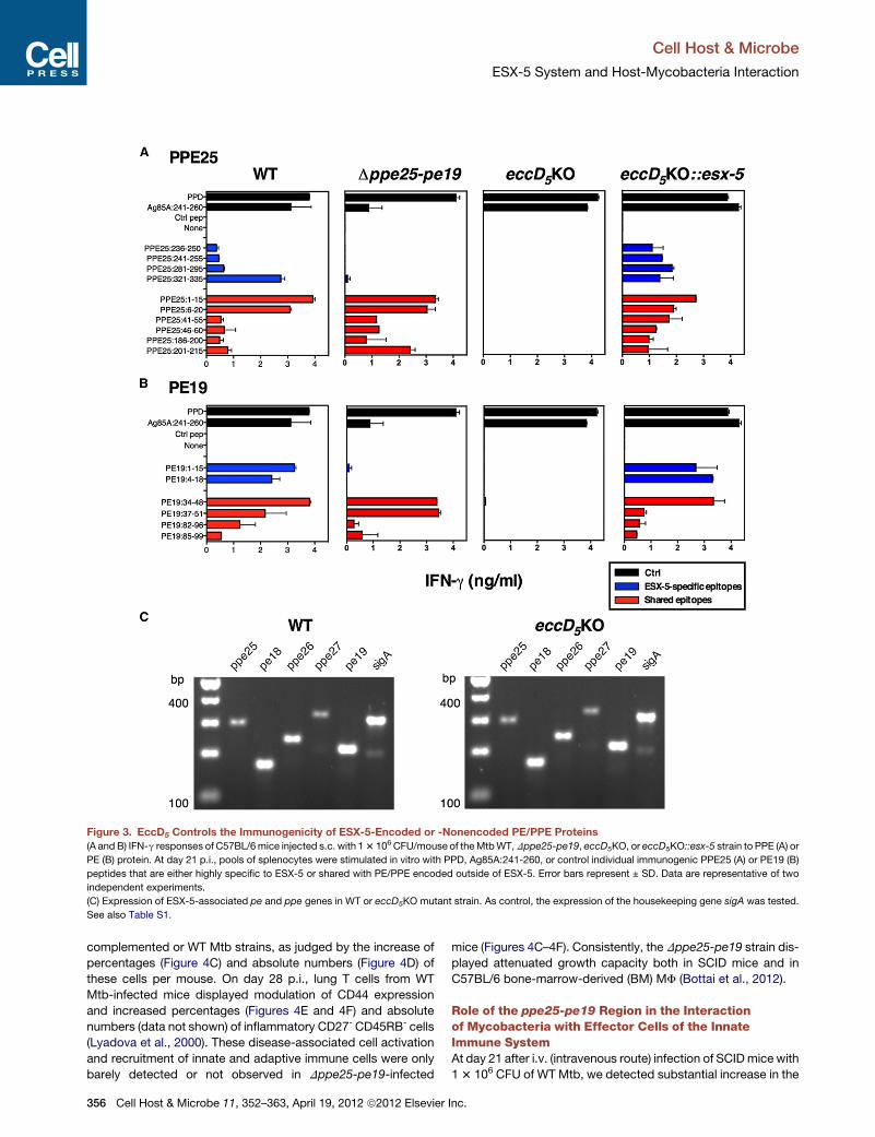
Figure 3. EccD5 Controls the Immunogenicity of ESX-5-Encoded or -Nonencoded PE/PPE Proteins
(A and B) IFN-g responses of C57BL/6mice injected s.c. with 13 106 CFU/mouse of theMtbWT,Dppe25-pe19, eccD5KO, or eccD5KO::esx-5 strain to PPE (A) or
PE (B) protein. At day 21 p.i., pools of splenocytes were stimulated in vitro with PPD, Ag85A:241-260, or control individual immunogenic PPE25 (A) or PE19 (B)
peptides that are either highly specific to ESX-5 or shared with PE/PPE encoded outside of ESX-5. Error bars represent ± SD. Data are representative of two
independent experiments.
(C) Expression of ESX-5-associated pe and ppe genes in WT or eccD5KO mutant strain. As control, the expression of the housekeeping gene sigA was tested.
See also Table S1.
Cell Host & Microbe
ESX-5 System and Host-Mycobacteria Interaction
complemented or WT Mtb strains, as judged by the increase of
percentages (Figure 4C) and absolute numbers (Figure 4D) of
these cells per mouse. On day 28 p.i., lung T cells from WT
Mtb-infected mice displayed modulation of CD44 expression
and increased percentages (Figures 4E and 4F) and absolute
numbers (data not shown) of inflammatory CD27- CD45RB- cells
(Lyadova et al., 2000). These disease-associated cell activation
and recruitment of innate and adaptive immune cells were only
barely detected or not observed in Dppe25-pe19-infected
356 Cell Host & Microbe 11, 352–363, April 19, 2012 ª2012 Elsevier
mice (Figures 4C–4F). Consistently, the Dppe25-pe19 strain dis-
played attenuated growth capacity both in SCID mice and in
C57BL/6 bone-marrow-derived (BM) MF (Bottai et al., 2012).
Role of the ppe25-pe19 Region in the Interactionof Mycobacteria with Effector Cells of the InnateImmune SystemAt day 21 after i.v. (intravenous route) infection of SCIDmice with
13 106 CFU of WT Mtb, we detected substantial increase in the
Inc.
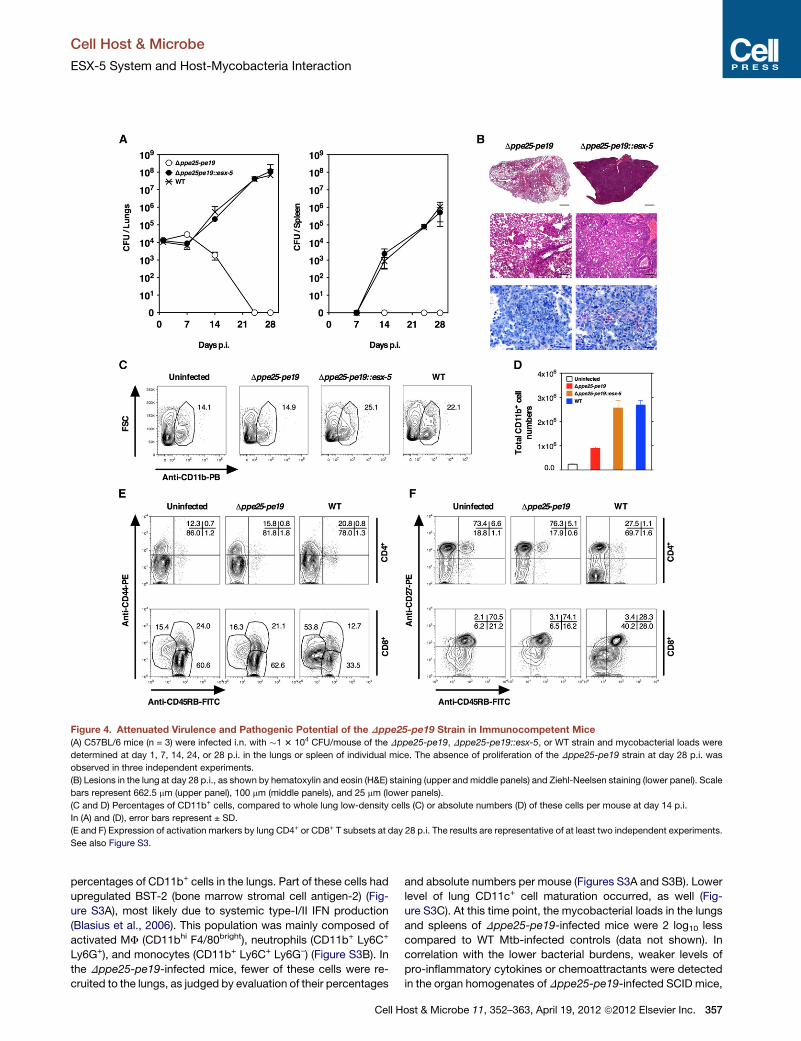
Figure 4. Attenuated Virulence and Pathogenic Potential of the Dppe25-pe19 Strain in Immunocompetent Mice
(A) C57BL/6 mice (n = 3) were infected i.n. with �1 3 104 CFU/mouse of the Dppe25-pe19, Dppe25-pe19::esx-5, or WT strain and mycobacterial loads were
determined at day 1, 7, 14, 24, or 28 p.i. in the lungs or spleen of individual mice. The absence of proliferation of the Dppe25-pe19 strain at day 28 p.i. was
observed in three independent experiments.
(B) Lesions in the lung at day 28 p.i., as shown by hematoxylin and eosin (H&E) staining (upper and middle panels) and Ziehl-Neelsen staining (lower panel). Scale
bars represent 662.5 mm (upper panel), 100 mm (middle panels), and 25 mm (lower panels).
(C and D) Percentages of CD11b+ cells, compared to whole lung low-density cells (C) or absolute numbers (D) of these cells per mouse at day 14 p.i.
In (A) and (D), error bars represent ± SD.
(E and F) Expression of activation markers by lung CD4+ or CD8+ T subsets at day 28 p.i. The results are representative of at least two independent experiments.
See also Figure S3.
Cell Host & Microbe
ESX-5 System and Host-Mycobacteria Interaction
percentages of CD11b+ cells in the lungs. Part of these cells had
upregulated BST-2 (bone marrow stromal cell antigen-2) (Fig-
ure S3A), most likely due to systemic type-I/II IFN production
(Blasius et al., 2006). This population was mainly composed of
activated MF (CD11bhi F4/80bright), neutrophils (CD11b+ Ly6C+
Ly6G+), and monocytes (CD11b+ Ly6C+ Ly6G–) (Figure S3B). In
the Dppe25-pe19-infected mice, fewer of these cells were re-
cruited to the lungs, as judged by evaluation of their percentages
Cell H
and absolute numbers per mouse (Figures S3A and S3B). Lower
level of lung CD11c+ cell maturation occurred, as well (Fig-
ure S3C). At this time point, the mycobacterial loads in the lungs
and spleens of Dppe25-pe19-infected mice were 2 log10 less
compared to WT Mtb-infected controls (data not shown). In
correlation with the lower bacterial burdens, weaker levels of
pro-inflammatory cytokines or chemoattractants were detected
in the organ homogenates of Dppe25-pe19-infected SCID mice,
ost & Microbe 11, 352–363, April 19, 2012 ª2012 Elsevier Inc. 357
Cell Host & Microbe
ESX-5 System and Host-Mycobacteria Interaction
as compared to their WT Mtb-infected counterparts (Figures
S3D and S3E). We wondered whether this weak inflammation
reflects the reduced virulence/persistence of the Dppe25-pe19
strain in a SCID host or its altered capacity to simulate cells of
the innate immune system. In vitro infection with the Dppe25-
pe19, Dppe25-pe19::esx-5 or WT Mtb strains induced compa-
rable phenotypic maturation of DCs (Figure S4A). DCs (Fig-
ure S4B) or MF (Figure S4C) infected with the Dppe25-pe19,
Dppe25-pe19::esx-5 or WT strain also expressed similar levels
of several inflammatory cytokines and chemokines which play
key roles in the innate immunity against mycobacteria. There-
fore, the attenuated Dppe25-pe19 strain remains a potent acti-
vator of the innate immune system.
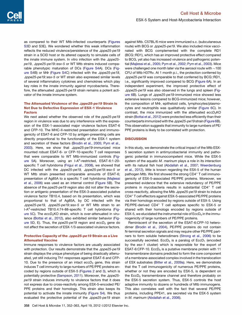
The Attenuated Virulence of the Dppe25-pe19 Strain IsNot Due to Defective Expression of ESX-1 VirulenceFactorsWe next asked whether the observed role of the ppe25-pe19
region in virulence was due to any interference with the expres-
sion of the ESX-1-associated major virulence factors ESAT-6
and CFP-10. The MHC-II-restricted presentation and immuno-
genicity of ESAT-6 and CFP-10 by antigen-presenting cells are
directly proportional to the functionality of the ESX-1 system
and secretion of these factors (Brodin et al., 2005; Pym et al.,
2003). Here, we show that Dppe25-pe19-immunized mice
mounted robust ESAT-6- or CFP-10-specific T cell responses
that were comparable to WT Mtb-immunized controls (Fig-
ure 5A). Moreover, using an I-Ab-restricted, ESAT-6:1-20-
specific T cell hybridoma (Frigui et al., 2008), we showed that
DC infected with the Dppe25-pe19, Dppe25-pe19::esx-5 or
WT Mtb strain presented comparable amounts of ESAT-6;
presentation of Ag85A to a specific T cell hybridoma (Majlessi
et al., 2006) was used as an internal control (Figure 5B). The
absence of the ppe25-pe19 region also did not alter the secre-
tion or antigenic presentation of the ESX-3-associated putative
virulence factor TB10.4, based on its presentation, which was
proportional to that of Ag85A, by DC infected with the
Dppe25-pe19, Dppe25-pe19::esx-5 or WT Mtb strain to an
I-Ad-restricted TB10.4:74-88-specific T cell hybridoma (Fig-
ure 5C). The eccD5KO strain, which is over-attenuated in viru-
lence (Bottai et al., 2012), also exhibited similar behavior (Fig-
ure 5D, E). Thus, the ppe25-pe19 region and eccD5 gene do
not affect the secretion of ESX-1/3-associated virulence factors.
Protective Capacity of theDppe25-pe19 Strain as a LiveAttenuated VaccineImmune responses to virulence factors are usually associated
with protection. Our results demonstrate that the Dppe25-pe19
strain displays the unique phenotype of being drastically attenu-
ated, yet still inducing Th1 responses against ESAT-6 and CFP-
10. Due to the presence of an intact eccD5 gene, this strain
induces T cell immunity to large numbers of PE/PPE proteins en-
coded by regions outside of ESX-5 (Figures 2 and 3), which is
potentially protective (Sampson, 2011). Moreover, the Dppe25-
pe19 strain induces immunity to virulence factors that it does
not express due to cross-reactivity among ESX-5-encoded PE/
PPE proteins and their homologs. This strain also keeps its
potential to activate the innate immunity (Figure S4). We thus
evaluated the protective potential of the Dppe25-pe19 strain
358 Cell Host & Microbe 11, 352–363, April 19, 2012 ª2012 Elsevier
against Mtb. C57BL/6 mice were immunized s.c. (subcutaneous
route) with BCG or Dppe25-pe19. We also included mice vacci-
nated with BCG complemented with the complete RD1
(BCG::RD1), which has an improved protective effect compared
to BCG, yet also has increased virulence and pathogenic poten-
tial (Majlessi et al., 2005; Pym et al., 2002; Pym et al., 2003). Mice
were challenged one month later via the aerosol route with�100
CFU of Mtb H37Rv. At 1 month p.i., the protection conferred by
Dppe25-pe19 was comparable to that conferred by BCG::RD1,
i.e., significantly improved compared to BCG (Figure 6A). In an
independent experiment, the improved protective effect of
Dppe25-pe19 was also observed in the lungs and spleen (Fig-
ure 6B). Lungs of Dppe25-pe19-immunized mice showed less
extensive lesions compared to BCG-immunized mice; however,
the composition of MF, epitheloid cells, lymphocytes/plasmo-
cytes and neutrophils was qualitatively similar (Figure 6C). In
contrast, the mice immunized with the attenuated eccD5KO
strain (Bottai et al., 2012) were protected less efficiently than their
counterparts immunizedwith theDppe25-pe19strain (Figure6B).
This observation suggests that immunity to large numbers of PE/
PPE proteins is likely to be correlated with protection.
DISCUSSION
In this study, we demonstrate the critical impact of the Mtb ESX-
5 secretion system in antimycobacterial immunity and patho-
genic potential in immunocompetent mice. While the ESX-5
system of the aquatic M. marinum plays a role in its interaction
with its natural fish host (Abdallah et al., 2007; Weerdenburg
et al., 2012), little is known regarding the ESX-5 of the human
pathogen Mtb. We first showed the strong CD4+ T cell immuno-
genicity of ESX-5-associated PE/PPE proteins. Moreover, we
presented evidence that the extensive redundancy of PE/PPE
proteins in mycobacteria results in substantial CD4+ T cell
cross-reactivity, allowing the Mtb Dppe25-pe19 strain to induce
CD4+ T cell effectors against ESX-5-associated PE/PPE proteins
via their homologs encoded by regions outside of ESX-5. Using
PE/PPE-derived CD4+ T cell epitopes specific to ESX-5 or
shared with their homologs encoded by regions outside of
ESX-5, we elucidated the instrumental role of EccD5 in the immu-
nogenicity of large numbers of PE/PPE proteins.
Reminiscent of the secretion of the ESAT-6:CFP-10 hetero-
dimer (Brodin et al., 2004), PE/PPE proteins do not contain
N-terminal secretion signals andmay require other PE/PPE part-
ners to form soluble heterodimers (Strong et al., 2006) to be
successfully secreted. EccD5 is a paralog of EccD1 (encoded
by the esx-1 cluster) which is responsible for the export of
ESAT-6:CFP-10. EccD5 is a putative membrane protein with 11
transmembrane domains predicted to form the core component
of amembrane-associated complex involved in the translocation
of ESX substrates (Bitter et al., 2009a). Here, we demonstrate
that the T cell immunogenicity of numerous PE/PPE proteins,
whether or not they are encoded by ESX-5, is dependent on
the EccD5 transmembrane channel and therefore probably on
the ESX-5 secretion system. Thus, ESX-5 controls the host
adaptive immunity to dozens or hundreds of Mtb immunogens.
This also correlates well with the fact that several PE/PPE
proteins, including PPE41, are secreted via the ESX-5 system
in M. marinum (Abdallah et al., 2006).
Inc.
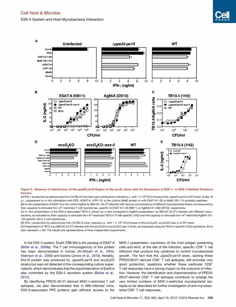
Figure 5. Absence of Interference of the ppe25-pe19 Region or the eccD5 Gene with the Expression of ESX-1- or ESX-3-Related Virulence
Factors
(A) IFN-g production by splenocytes fromC57BL/6mice that were uninfected or infected s.c. with 13 106CFU/mouse of theDppe25-pe19 orWT strain, at day 18
p.i., subsequent to in vitro stimulation with PPD, rESAT-6, rCFP-10, or the control rMalE protein or with ESAT-6:1-20 or MalE:100-114 synthetic peptides.
(B) In vitro presentation of ESAT-6 or the control Ag85A by BM-DC- (H-2b) infected with various concentrations of different mycobacterial strains, asmeasured by
their capacity to stimulate the I-Ab-restricted T cell hybridomas, specific to ESAT-6:1-20 (NB11) or Ag85A:241-260 (DE10), respectively.
(C) In vitro presentation of the ESX-3-associated TB10.4, shown as a ratio compared to Ag85A presentation, by BM-DC (H-2d) infected with different myco-
bacteria, as indicated by their capacity to stimulate the I-Ad-restricted TB10.4:74-88-specific (1H2) over the capacity to stimulate the I-Ad-restricted Ag85A:101-
120-specific (2A1) T cell hybridomas.
(D) IFN-g production by splenocytes from C57BL/6 mice, injected s.c. with 1 3 106 CFU/mouse of the eccD5KO, eccD5KO::esx-5 or WT strain.
(E) Presentation of TB10.4 by BM-DC (H-2d) infected with the eccD5KO or eccD5KO::esx-5 strain, as measured using the TB10.4-specific (1H2) hybridoma. Error
bars represent ± SD. The results are representative of three independent experiments.
Cell Host & Microbe
ESX-5 System and Host-Mycobacteria Interaction
In the ESX-5 system, EsxN (TB9.9A) is the paralog of ESAT-6
(Bitter et al., 2009a). The T cell immunogenicity of this protein
has been demonstrated in human (Al-Attiyah et al., 2004;
Alderson et al., 2000) and bovine (Jones et al., 2010). Notably,
Esx-N protein was produced by Dppe25-pe19 and eccD5KO
strains but was not detected in the corresponding culture super-
natants, which demonstrates that the export/secretion of EsxN is
also controlled by the ESX-5 secretion system (Bottai et al.,
2012).
By identifying PPE25-26-27-derived MHC-I-restricted T cell
epitopes, we also demonstrated that in Mtb-infected mice,
ESX-5-associated PPE proteins gain efficient access to the
Cell H
MHC-I presentation machinery of the host antigen presenting
cells and elicit, at the site of the infection, specific CD8+ T cell
effectors that produce key cytokines to control mycobacterial
growth. The fact that the Dppe25-pe19 strain, lacking these
PPE25/26/27-derived CD8+ T cell epitopes, still provides very
good protection, questions whether these particular CD8+
T cell responses have a strong impact on the outcome of infec-
tion. However, the identification and characterization of PPE25/
26/27-derived CD8+ T cell epitopes contribute to enlarge the
very limited numbers of MHC-I-restricted mycobacterial epi-
topes so far described for further investigation of anti-mycobac-
terial CD8+ T cell responses.
ost & Microbe 11, 352–363, April 19, 2012 ª2012 Elsevier Inc. 359
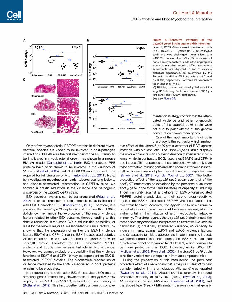
Figure 6. Protective Potential of the
Dppe25-pe19 Strain against Mtb Infection
(A and B) C57BL/6 mice were immunized s.c. with
BCG, BCG::RD1, Dppe25-pe19, or eccD5KO
strain and were challenged 1 month later with
�100 CFU/mouse of WT Mtb H37Rv via aerosol
route. Themycobacterial loads in the lungs/spleen
were determined at 1 month p.i. Two independent
experiments are depicted. * and ** indicate
statistical significance, as determined by the
Student’s t and Mann-Whitney tests, p < 0.01 and
p < 0.008, respectively. Horizontal bars represent
the means of six mice.
(C) Histological sections showing lesions of the
lung, H&E staining. Scale bars represent 662.5 mm
(left panel) and 100 mm (right panel).
See also Figure S4.
Cell Host & Microbe
ESX-5 System and Host-Mycobacteria Interaction
Only a few mycobacterial PE/PPE proteins in different myco-
bacterial species are known to be involved in host-pathogen
interactions. PPE46 was the first member of the PPE family to
be implicated in mycobacterial growth, as shown in a mouse
BM-MF model (Camacho et al., 1999). ESX-5-encoded PPE
proteins have been shown to be involved in the virulence of
M. avium (Li et al., 2005), and PE-PGRS30 was proposed to be
required for full virulence of Mtb (Iantomasi et al., 2011). Here,
by investigating mycobacterial loads, tuberculous lung lesions,
and disease-associated inflammation in C57BL/6 mice, we
showed a drastic reduction in the virulence and pathogenic
properties of the Dppe25-pe19 strain.
ESX secretion systems can be transregulated (Frigui et al.,
2008) or exhibit crosstalk among themselves, as is the case
with ESX-1-encoded PE35 (Brodin et al., 2006). Therefore, it is
possible that ppe25-pe19 depletion and the resulting ESX-5
deficiency may impair the expression of the major virulence
factors related to other ESX systems, thereby leading to this
drastic reduction in virulence. We ruled out this possibility, at
least for the known major ESX-associated virulence factors, by
showing that the expression of neither the ESX-1 virulence
factors ESAT-6 and CFP-10, nor the ESX-3-associated putative
virulence factor TB10.4, was affected in Dppe25-pe19 or
eccD5KO strains. Therefore, the ESX-5-associated PE/PPE
proteins and EccD5 play an essential role in Mtb virulence.
However, we cannot exclude the possibility that the virulence
functions of ESAT-6 and CFP-10 may be dependent on ESX-5-
associated PE/PPE proteins. The biochemical mechanism of
virulence mediated by the ESX-5-associated PE/PPE proteins
remains to be elucidated.
It is important to note that other ESX-5-associated KOmutants
affecting genes immediately downstream of the ppe25-pe19
region, i.e., esxNKO or rv1794KO, retained a virulent phenotype
(Bottai et al., 2012). This fact together with our genetic comple-
360 Cell Host & Microbe 11, 352–363, April 19, 2012 ª2012 Elsevier Inc.
mentation strategy confirm that the atten-
uated virulence and other phenotypic
traits of the Dppe25-pe19 strain were
not due to polar effects of the genetic
construct on downstream genes.
One of the most important findings in
this study is the particularly high protec-
tive effect of the Dppe25-pe19 strain over that of BCG against
infection with virulent Mtb. The Dppe25-pe19 strain displays
the unique characteristics of being drastically attenuated in viru-
lence, while, in contrast to BCG, it secretes ESAT-6 and CFP-10
and induces Th1 responses to these antigens, which are known
to be protective immunogens and also seem to intervene in intra-
cellular localization and phagosomal escape of mycobacteria
(Simeone et al., 2012; van der Wel et al., 2007). The better
protective effect of the Dppe25-pe19 strain over that of the
eccD5KO mutant can be explained by the presence of an intact
eccD5 gene in the former and therefore its capacity at inducing
T cell immunity against a plethora of ESX-5-nonassociated
PE/PPE proteins and, due to their strong cross-reactivity,
against the ESX-5-associated PE/PPE virulence factors that
this strain has lost. Moreover, the Dppe25-pe19 strain remains
potent at inducing the activation of the innate system, which is
instrumental in the initiation of anti-mycobacterial adaptive
immunity. Therefore, overall, the Dppe25-pe19 strain meets the
three necessary conditions to represent an attractive live vaccine
candidate: (1) drastically attenuated virulence, (2) capacity to
induce immunity against ESX-1 and ESX-5 virulence factors,
and (3) capacity to initiate appropriate innate immunity. Indeed,
we demonstrated that this attenuated ESX-5 mutant had
a protective effect comparable to BCG::RD1, which is known to
be more protective than BCG. However, unlike BCG::RD1
(Majlessi et al., 2005; Pym et al., 2002), the Dppe25-pe19 strain
is neither virulent nor pathogenic in immunocompetent mice.
During the preparation of this manuscript, the prominent
protective effect of a recombinantM. smegmatis Desx-3mutant
complemented with the orthologous Mtb esx-3 was reported
(Sweeney et al., 2011). Altogether, the strongly improved
protective capacity of BCG::RD1 (esx-1) (Pym et al., 2003),
M. smegmatis Desx-3::Mtb esx-3 (Sweeney et al., 2011), and
the Dppe25-pe19 esx-5 Mtb mutant demonstrate that genetic
Cell Host & Microbe
ESX-5 System and Host-Mycobacteria Interaction
manipulation of the type VII secretion systems constitutes a
powerful strategy to generate live attenuated anti-Mtb vaccines.
One of the most prominent characteristics of the Dppe25-pe19
strain is its capacity to induce T cell responses against ESX-1
virulence factors, and—by cross reactivity—against the ESX-5-
associated PE/PPE virulence factors that it has lost. Therefore,
the Dppe25-pe19 mutant may pave the way to set up a new
generation of safe live attenuated vaccine against infection
with pathogenic Mtb.
EXPERIMENTAL PROCEDURES
Mice and Infections
C57BL/6 (Janvier, Le Genest-Saint-Isle, France) or BALB/c (Charles River,
Arbresle, France) mice were anesthetized by i.p. injection of 100 mg/kg
Ketamine (Imalgene, Lyon, France) and 10 mg/kg Xylazine (Rompun, KVP
Kiel, Germany) and were infected i.n. with 60 ml/mouse of mycobacterial
suspensions at 2.5 3 105 CFU/ml for a total inoculation of �1 3 104 CFU/
mouse. Subcutaneous (s.c.) immunization was performed at the base of the
tail. Six-week-old female SCID mice (Charles River) were infected i.v. via
the tail vein with 1 3 106 CFU/mouse. All animal studies were approved by
the Institut Pasteur Safety Committee, in accordance with French and Euro-
pean guidelines and regulations.
Cytofluorometry
Low-density cells or adaptive immune cells from infected mice were prepared
as previously described (Majlessi et al., 2005). They were first incubated with
an anti-CD16/CD32 monoclonal antibody (mAb), followed by the indicated
mAbs, all of which were purchased from BD PharMingen (Le Pont-de-Claix,
France). Intracellular cytokine staining was performed as described previously
(Mouries et al., 2008). The PPE26:44-52 peptide, bound to polymerized H-2Kd,
immobilized on a Dextramer backbone and conjugated to PE was from
Immundex (Copenhagen, Denmark). The stained cells were fixed with 4%
paraformaldehyde overnight at 4�C and were acquired with a CyAn system
using Summit software (Beckman Coulter, Villepinte, France). Data were
analyzed with FlowJo software (Treestar, OR).
RNA Extraction and RT-PCR Reactions
RNA was extracted from mycobacteria in exponential growth phase, as previ-
ously described (Bottai et al., 2012). RNA (300 ng) was denatured by incuba-
tion at 70�C for 5 min and subjected to RT-PCR reactions with the SS-One
Step RT-PCR kit (Life Technologies). Retrotranscription and amplification
reactions were carried out at 50�C (retrotranscription) and 94�C, 58�C, and72�C (denaturation, annealing and extension in amplification cycles), respec-
tively. Sequences of primers used are listed in Table S1.
Generation of Anti-TB10.4 or PPE-Specific T Cell Hybridomas
T cell hybridomas specific to mycobacterial antigens were generated from
BALB/c (H-2d) mice immunized s.c. with 1 3 107 CFU/mouse of the BCG
Pasteur 1173P2 strain, as previously described (Majlessi et al., 2003). The
BW5147 cell line or BW5147::CD8 cells were used as fusion partners for
MHC-II-restricted hybrids and MHC-I-restricted hybrids, respectively. The
resulting T cell hybridomas were screened for their capacity to recognize
BM-DC loaded with various peptides.
Antigen Presentation Assays
BM-DC were infected at 13 105 cells/well with different mycobacterial strains
at various multiplicities of infection in antibiotic-free complete RPMI, as previ-
ously described (Majlessi et al., 2003). After overnight incubation at 37�C, cellswere washed and incubated overnight with 1 3 105 cells/well of the appro-
priate T cell hybridomas. The amount of IL-2 produced by the specific T cell
hybridomas was measured by ELISA analysis of the coculture supernatants.
T Cell Assays
Pooled splenocytes from groups of infected mice were cultured at 1 3
106 cells/well in HL-1 medium (Biowhittaker, Lonza, France) complemented
Cell H
with 2 mM GlutaMax (Invitrogen, Life Technologies, France), 5 3 10�5 M
b-mercaptoethanol, 100 U/ml penicillin, and 100 mg/ml streptomycin (Sigma-
Aldrich, France) in the presence of PPD, rESAT-6 or rCFP-10 proteins or
appropriate peptides. After 72 hr of incubation, IFN-g in the culture superna-
tants was quantified by ELISA.
Pepscan Assay
Mice were immunized s.c. with 1–53 106 CFU/mouse of different mycobacte-
rial strains. Two to 3 weeks p.i., total splenocytes or sorted CD4+ or CD8+ T
splenocytes were stimulated with 10 mg/ml of individual synthetic 15-mers
from PPE25 (71 15-mers, overlapping by 5 aa) or PE19 (29 15-mers, overlap-
ping by 3 aa) (Mimotopes, Melbourne, Australia). After 72 hr of incubation,
IFN-g was quantified by ELISA. CD4+ or CD8+ T splenocytes from H37Rv
WT Mtb-immunized mice were positively sorted with anti-CD4 or anti-CD8
mAbs coupled to magnetic beads (Miltenyi Biotec, Mercoeur, France).
ELISA
ELISA assays of the culture supernatants or organ homogenates were per-
formed as previously described (Majlessi et al., 2005). mAbs specific to IL-2,
IL-6, IL-10, IL-12p40, or IFN-g were from BD PharMingen. mAbs specific to
IL-1b, CCL-2, and CCL-3 were from R&D Systems (Lille, France). Anti-TNF-a
mAbs were from eBioscience (Paris, France).
Protection Assay
Female C57BL/6 mice were immunized s.c. with 1 3 106 CFU/mouse of
different mycobacterial strains. One month later, mice were challenged as
detailed elsewhere (Jaron et al., 2008). The infected mice were placed in
isolator in A3 protection-level facilities. One month p.i., the lungs and
spleen were individually homogenized with a MillMixer organ homogenizer
(QIAGEN, Courtaboeuf, France). Serial 5-fold dilutions were plated on
7H11 Agarmedium supplementedwith ADC (Difco, Becton Dickinson, Sparks,
MD). The CFU were counted after 18 days of incubation at 37�C. For histolog-ical analyses, the left lobe of the lungs was fixed in formalin and embedded
in paraffin. Five micrometer sections were stained with H&E and Ziehl-
Neelsen.
Statistical Analyses
Statistical analyses were performedwith GraphPad Prism software (GraphPad
Software, La Jolla, CA). The Student’s t and Mann-Whitney tests were
employed based on the distribution of the data.
SUPPLEMENTAL INFORMATION
Supplemental Information includes four figures and one table and can be
found with this article online at doi:10.1016/j.chom.2012.03.003.
ACKNOWLEDGMENTS
This work was supported by the European Community’s FP7 program under
grant agreement number 201762. F.S. is a recipient of a PhD fellowship from
the University of Damascus, Syria. The authors gratefully acknowledge
Dr. Francoise Guinet (Institut Pasteur) and Dr. Richard Lo-Man (Institut
Pasteur) for critical reading of the manuscript, Dr. Peter Sebo (Academy of
Sciences, Czech Republic) for providing recombinant ESAT-6 and CFP-10,
and Eddie Maranghi and Karim Sebastien for expert assistance in animal
care in the A3 facilities. F.S., D.B. and L.M. performed the experiments and
analyzed data. L.S. performed several pepscan assays and generated
MHC-I restricted T-cell hybridomas. N.D. generated and characterized the
anti-TB10.4 T-cell hybridoma. M.D.L., D.B., R.S., R.B., and S.E. designed
and constructed mycobacterial mutants. L.F. performed histopathological
analyses. D.B., R.B., C.L., and L.M. designed and interpreted experiments.
D.B., C.L., and L.M. wrote the manuscript.
Received: November 28, 2011
Revised: February 23, 2012
Accepted: March 22, 2012
Published: April 18, 2012
ost & Microbe 11, 352–363, April 19, 2012 ª2012 Elsevier Inc. 361
Cell Host & Microbe
ESX-5 System and Host-Mycobacteria Interaction
REFERENCES
Abdallah, A.M., Verboom, T., Hannes, F., Safi, M., Strong, M., Eisenberg, D.,
Musters, R.J., Vandenbroucke-Grauls, C.M., Appelmelk, B.J., Luirink, J.,
and Bitter, W. (2006). A specific secretion system mediates PPE41 transport
in pathogenic mycobacteria. Mol. Microbiol. 62, 667–679.
Abdallah, A.M., Gey van Pittius, N.C., Champion, P.A., Cox, J., Luirink, J.,
Vandenbroucke-Grauls, C.M., Appelmelk, B.J., and Bitter, W. (2007). Type
VII secretion—mycobacteria show the way. Nat. Rev. Microbiol. 5, 883–891.
Abdallah, A.M., Verboom, T., Weerdenburg, E.M., Gey van Pittius, N.C.,
Mahasha, P.W., Jimenez, C., Parra, M., Cadieux, N., Brennan, M.J.,
Appelmelk, B.J., and Bitter, W. (2009). PPE and PE_PGRS proteins of
Mycobacterium marinum are transported via the type VII secretion system
ESX-5. Mol. Microbiol. 73, 329–340.
Al-Attiyah, R., Mustafa, A.S., Abal, A.T., El-Shamy, A.S., Dalemans, W., and
Skeiky, Y.A. (2004). In vitro cellular immune responses to complex and newly
defined recombinant antigens of Mycobacterium tuberculosis. Clin. Exp.
Immunol. 138, 139–144.
Alderson, M.R., Bement, T., Day, C.H., Zhu, L., Molesh, D., Skeiky, Y.A., Coler,
R., Lewinsohn, D.M., Reed, S.G., and Dillon, D.C. (2000). Expression cloning of
an immunodominant family of Mycobacterium tuberculosis antigens using
human CD4(+) T cells. J. Exp. Med. 191, 551–560.
Banu, S., Honore, N., Saint-Joanis, B., Philpott, D., Prevost, M.C., and Cole,
S.T. (2002). Are the PE-PGRS proteins ofMycobacterium tuberculosis variable
surface antigens? Mol. Microbiol. 44, 9–19.
Bitter, W., Houben, E.N., Bottai, D., Brodin, P., Brown, E.J., Cox, J.S.,
Derbyshire, K., Fortune, S.M., Gao, L.Y., Liu, J., et al. (2009a). Systematic
genetic nomenclature for type VII secretion systems. PLoS Pathog. 5,
e1000507.
Bitter, W., Houben, E.N., Luirink, J., and Appelmelk, B.J. (2009b). Type VII
secretion in mycobacteria: classification in line with cell envelope structure.
Trends Microbiol. 17, 337–338.
Blasius, A.L., Giurisato, E., Cella, M., Schreiber, R.D., Shaw, A.S., and
Colonna, M. (2006). Bone marrow stromal cell antigen 2 is a specific marker
of type I IFN-producing cells in the naive mouse, but a promiscuous cell
surface antigen following IFN stimulation. J. Immunol. 177, 3260–3265.
Bottai, D., and Brosch, R. (2009). Mycobacterial PE, PPE and ESX clusters:
novel insights into the secretion of these most unusual protein families. Mol.
Microbiol. 73, 325–328.
Bottai, D., Di Luca, M., Majlessi, L., Frigui, W., Simeone, R., Sayes, F., Bitter,
W., Brennan, M.J., Leclerc, C., Batoni, G., et al. (2012). Disruption of the
ESX-5 system of Mycobacterium tuberculosis causes loss of PPE protein
secretion, reduction of cell wall integrity and strong attenuation. Mol.
Microbiol. 83, 1195–1209.
Brennan, M.J., and Delogu, G. (2002). The PE multigene family: a ‘molecular
mantra’ for mycobacteria. Trends Microbiol. 10, 246–249.
Brodin, P., Rosenkrands, I., Andersen, P., Cole, S.T., and Brosch, R. (2004).
ESAT-6 proteins: protective antigens and virulence factors? Trends
Microbiol. 12, 500–508.
Brodin, P., de Jonge, M.I., Majlessi, L., Leclerc, C., Nilges, M., Cole, S.T., and
Brosch, R. (2005). Functional analysis of early secreted antigenic target-6, the
dominant T-cell antigen of Mycobacterium tuberculosis, reveals key residues
involved in secretion, complex formation, virulence, and immunogenicity.
J. Biol. Chem. 280, 33953–33959.
Brodin, P., Majlessi, L., Marsollier, L., de Jonge, M.I., Bottai, D., Demangel, C.,
Hinds, J., Neyrolles, O., Butcher, P.D., Leclerc, C., et al. (2006). Dissection of
ESAT-6 system 1 of Mycobacterium tuberculosis and impact on immunoge-
nicity and virulence. Infect. Immun. 74, 88–98.
Brosch, R., Gordon, S.V., Pym, A., Eiglmeier, K., Garnier, T., and Cole, S.T.
(2000). Comparative genomics of the mycobacteria. Int. J. Med. Microbiol.
290, 143–152.
Camacho, L.R., Ensergueix, D., Perez, E., Gicquel, B., and Guilhot, C. (1999).
Identification of a virulence gene cluster of Mycobacterium tuberculosis by
signature-tagged transposon mutagenesis. Mol. Microbiol. 34, 257–267.
362 Cell Host & Microbe 11, 352–363, April 19, 2012 ª2012 Elsevier
Cole, S.T., Brosch, R., Parkhill, J., Garnier, T., Churcher, C., Harris, D., Gordon,
S.V., Eiglmeier, K., Gas, S., Barry, C.E., 3rd., et al. (1998). Deciphering the
biology ofMycobacterium tuberculosis from the complete genome sequence.
Nature 393, 537–544.
DiGiuseppe Champion, P.A., Champion, M.M., Manzanillo, P., and Cox, J.S.
(2009). ESX-1 secreted virulence factors are recognized by multiple cytosolic
AAA ATPases in pathogenic mycobacteria. Mol. Microbiol. 73, 950–962.
Frigui, W., Bottai, D., Majlessi, L., Monot, M., Josselin, E., Brodin, P.,
Garnier, T., Gicquel, B., Martin, C., Leclerc, C., et al. (2008). Control of
M. tuberculosis ESAT-6 secretion and specific T cell recognition by PhoP.
PLoS Pathog. 4, e33.
Gey Van Pittius, N.C., Gamieldien, J., Hide, W., Brown, G.D., Siezen, R.J.,
Beyers, A.D., and Beyers, A.D. (2001). The ESAT-6 gene cluster of
Mycobacterium tuberculosis and other high G+C Gram-positive bacteria.
Genome Biol. 2, H0044.
Gey van Pittius, N.C., Sampson, S.L., Lee, H., Kim, Y., van Helden, P.D., and
Warren, R.M. (2006). Evolution and expansion of the Mycobacterium
tuberculosis PE and PPE multigene families and their association with the
duplication of the ESAT-6 (esx) gene cluster regions. BMC Evol. Biol. 6, 95.
Iantomasi, R., Sali, M., Cascioferro, A., Palucci, I., Zumbo, A., Soldini, S.,
Rocca, S., Greco, E., Maulucci, G., De Spirito, M., et al. (2011). PE_PGRS30
is required for the full virulence of Mycobacterium tuberculosis. Cell.
Microbiol. 14, 356–367.
Jaron, B., Maranghi, E., Leclerc, C., and Majlessi, L. (2008). Effect of attenua-
tion of Treg during BCG immunization on anti-mycobacterial Th1 responses
and protection against Mycobacterium tuberculosis. PLoS ONE 3, e2833.
Jones, G.J., Gordon, S.V., Hewinson, R.G., and Vordermeier, H.M. (2010).
Screening of predicted secreted antigens from Mycobacterium bovis reveals
the immunodominance of the ESAT-6 protein family. Infect. Immun. 78,
1326–1332.
Li, Y., Miltner, E., Wu, M., Petrofsky, M., and Bermudez, L.E. (2005). A
Mycobacterium avium PPE gene is associated with the ability of the bacterium
to grow in macrophages and virulence in mice. Cell. Microbiol. 7, 539–548.
Lyadova, I.V., Eruslanov, E.B., Khaidukov, S.V., Yeremeev, V.V., Majorov,
K.B., Pichugin, A.V., Nikonenko, B.V., Kondratieva, T.K., and Apt, A.S.
(2000). Comparative analysis of T lymphocytes recovered from the lungs of
mice genetically susceptible, resistant, and hyperresistant to Mycobacterium
tuberculosis-triggered disease. J. Immunol. 165, 5921–5931.
Mahairas, G.G., Sabo, P.J., Hickey, M.J., Singh, D.C., and Stover, C.K. (1996).
Molecular analysis of genetic differences betweenMycobacterium bovis BCG
and virulent M. bovis. J. Bacteriol. 178, 1274–1282.
Majlessi, L., Rojas, M.J., Brodin, P., and Leclerc, C. (2003). CD8+-T-cell
responses of Mycobacterium-infected mice to a newly identified major
histocompatibility complex class I-restricted epitope shared by proteins of
the ESAT-6 family. Infect. Immun. 71, 7173–7177.
Majlessi, L., Brodin, P., Brosch, R., Rojas, M.J., Khun, H., Huerre, M., Cole,
S.T., and Leclerc, C. (2005). Influence of ESAT-6 secretion system 1 (RD1) of
Mycobacterium tuberculosis on the interaction between mycobacteria and
the host immune system. J. Immunol. 174, 3570–3579.
Majlessi, L., Simsova, M., Jarvis, Z., Brodin, P., Rojas, M.J., Bauche, C.,
Nouze, C., Ladant, D., Cole, S.T., Sebo, P., and Leclerc, C. (2006). An increase
in antimycobacterial Th1-cell responses by prime-boost protocols of immuni-
zation does not enhance protection against tuberculosis. Infect. Immun. 74,
2128–2137.
Mouries, J., Moron, G., Schlecht, G., Escriou, N., Dadaglio, G., and Leclerc, C.
(2008). Plasmacytoid dendritic cells efficiently cross-prime naive T cells in vivo
after TLR activation. Blood 112, 3713–3722.
Pym, A.S., Brodin, P., Brosch, R., Huerre, M., and Cole, S.T. (2002). Loss of
RD1 contributed to the attenuation of the live tuberculosis vaccines
Mycobacterium bovis BCG and Mycobacterium microti. Mol. Microbiol. 46,
709–717.
Pym, A.S., Brodin, P., Majlessi, L., Brosch, R., Demangel, C., Williams, A.,
Griffiths, K.E., Marchal, G., Leclerc, C., and Cole, S.T. (2003). Recombinant
Inc.
Cell Host & Microbe
ESX-5 System and Host-Mycobacteria Interaction
BCG exporting ESAT-6 confers enhanced protection against tuberculosis.
Nat. Med. 9, 533–539.
Sampson, S.L. (2011). Mycobacterial PE/PPE proteins at the host-pathogen
interface. Clin. Dev. Immunol. 2011, 497203.
Simeone, R., Bottai, D., and Brosch, R. (2009). ESX/type VII secretion systems
and their role in host-pathogen interaction. Curr. Opin. Microbiol. 12, 4–10.
Simeone, R., Bobard, A., Lippmann, J., Bitter, W., Majlessi, L., Brosch, R., and
Enninga, J. (2012). Phagosomal rupture by Mycobacterium tuberculosis
results in toxicity and host cell death. PLoS Pathog. 8, e1002507.
Stanley, S.A., Johndrow, J.E., Manzanillo, P., and Cox, J.S. (2007). The Type I
IFN response to infection with Mycobacterium tuberculosis requires ESX-1-
mediated secretion and contributes to pathogenesis. J. Immunol. 178,
3143–3152.
Strong, M., Sawaya,M.R., Wang, S., Phillips, M., Cascio, D., and Eisenberg, D.
(2006). Toward the structural genomics of complexes: crystal structure of
a PE/PPE protein complex from Mycobacterium tuberculosis. Proc. Natl.
Acad. Sci. USA 103, 8060–8065.
Cell H
Sweeney, K.A., Dao, D.N., Goldberg, M.F., Hsu, T., Venkataswamy, M.M.,
Henao-Tamayo, M., Ordway, D., Sellers, R.S., Jain, P., Chen, B., et al.
(2011). A recombinantMycobacterium smegmatis induces potent bactericidal
immunity against Mycobacterium tuberculosis. Nat. Med. 17, 1261–1268.
Tekaia, F., Gordon, S.V., Garnier, T., Brosch, R., Barrell, B.G., and Cole, S.T.
(1999). Analysis of the proteome of Mycobacterium tuberculosis in silico.
Tuber. Lung Dis. 79, 329–342.
van der Wel, N., Hava, D., Houben, D., Fluitsma, D., van Zon, M., Pierson, J.,
Brenner, M., and Peters, P.J. (2007). M. tuberculosis and M. leprae trans-
locate from the phagolysosome to the cytosol in myeloid cells. Cell 129,
1287–1298.
Weerdenburg, E.M., Abdallah, A.M., Mitra, S., de Punder, K., van der Wel,
N.N., Bird, S., Appelmelk, B.J., Bitter, W., and van der Sar, A.M. (2012).
ESX-5-deficient Mycobacterium marinum is hypervirulent in adult zebrafish.
Cell. Microbiol., in press. Published online January 18, 2012. 10.1111/j.
1462-5822.2012.01755.x.
ost & Microbe 11, 352–363, April 19, 2012 ª2012 Elsevier Inc. 363
Copyright © 2022 FDOKUMEN