Steps to standardization and validation of hippocampal volumetry as a biomarker in clinical trials...
20
Steps to standardization and validation of hippocampal volumetry as a biomarker in clinical trials and diagnostic criteria for Alzheimer’s disease Clifford R Jack Jr. 1,* , Frederik Barkhof 2 , Matt A Bernstein 1 , Marc Cantillon 3 , Patricia E Cole 4 , Charles DeCarli 5 , Bruno Dubois 6 , Simon Duchesne 7 , Nick C Fox 8 , Giovanni B Frisoni 9 , Harald Hampel 10 , Derek LG Hill 11 , Keith Johnson 12 , Jean-François Mangin 13 , Philip Scheltens 14 , Adam J Schwarz 15 , Reisa Sperling 16 , Joyce Suhy 17 , Paul M Thompson 18 , Michael Weiner 19 , and Norman L Foster 20 1 Dept of Radiology, Mayo Clinic, 200 First St, Rochester MN * Corresponding Author: Clifford R Jack, Jr., M.D., Department of Radiology, Mayo Clinic and Foundation, 200 First Street SW, Rochester, MN, 55905, USA, Telephone: 507.284.8548, Fax: 507-284-9778, [email protected]. Disclosures Clifford R. Jack Jr. serves as a consultant for Janssen, Lilly, GE, Johnson and Johnson, Eisai, and Élan. He is an investigator in clinical trials sponsored by Pfizer, Allon and Baxter, Inc. He receives research funding from the NIA [R01-AG11378 (PI), P50- AG16574 (Co-I), R21-AG38736 (Co-I), and U01 AG024904-01 (Co-I)], and the Alexander Family Alzheimer’s Disease Research Professorship of the Mayo Foundation. He owns stock in Johnson and Johnson. Frederik Barkhof has received compensation for consulting from Roche, Lundbeck, Janssen Alz Immunotherapy, and GE Medical Systems. Matthew A. Berstein receives research contract funding from Pfizer Inc., and Baxter Allon. Marc Cantillon is an employee of Coalition Against Major Diseases. Patricia E. Cole reports no conflicts. Charles DeCarli reports no conflicts. Bruno Dubois has served on the advisory boards of GE healthcare and Eisai/Pfizer. Simon Duchesne reports no conflicts. Nick C. Fox served on the scientific advisory boards of Alzheimer’s Research Forum, Alzheimer’s Society and Alzheimer’s Research Trust and editorial boards of Alzheimer’s Disease and Associated Disorders; Neurodegenerative Diseases and BioMed Central - Alzheimer’s Research and Therapy. He holds a patent for QA Box that may accrue revenue. In the last five years his research group has received payment for consultancy or for conducting studies from Abbott Laboratories, Elan Pharmaceuticals, Eisai, Eli Lilly, GE Healthcare, IXICO, Lundbeck, Pfizer Inc, Sanofi-Aventis and Wyeth Pharmaceuticals. He receives research support from MRC [G0801306 (PI), G0601846 (PI)] NIH [U01 AG024904 (Co- investigator(sub contract)], Alzheimer Research Trust [ART/RF/2007/1 (PI)], NIHR (Senior Investigator) and EPSRC [GR/S48844/01 (PI)]. Giovanni B. Frisoni serves/has served on the advisory boards for Lilly, BMS, Bayer, Lundbeck, Elan, Astra Zeneca, Pfizer, Taurx, and Wyeth. He is a member of the editorial boards of Lancet Neurology, Aging Clinical & Experimental Research, Alzheimer’s Disease & Associated Disorders, and Neurodegenerative Diseases. He serves as the Imaging Selection Editor of Neurobiology of Aging. He has received grants from Wyeth Int’l, Lilly Int’l, Lundbeck Italia, and the Alzheimer’s Association. Harald Hampel disclosed no conflicts. Derek L.G. Hill is an employee and stock holder in IXICO Ltd. Keith Johnson reports no conflicts. Jean-Francois Mangin reports no conflicts. Philip Scheltens serves/has served on the advisory boards of Genentech, Novartis, Roche, Danone, Nutricia, Baxter and Lundbeck. He has been a speaker at symposia organized by Lundbeck, Merz, Danone, Novartis, Roche and Genentech. For all his activities he receives no personal compensation. He serves on the editorial board of Alzheimer’s Research & Therapy and Alzheimers Disease and Associated Disorders, is a member of the scientific advisory board of the EU Joint Programming Initiative and the French National Plan Alzheimer. The Alzheimer Center receives unrestricted funding from various sources through the VUmc Fonds. Adam J. Schwartz is an employee and shareholder of Eli Lilly and Company. Reisa Sperling has been a consultant for Pfizer, Janssen, Elan, Bristol-Myers-Squibb, Bayer and Avid (unpaid). She is a site investigator for clinical trials for Bristol-Myers-Squibb, Pfizer, Janssen, and Avid. Joyce Suhy is an employee of Synarc. Paul M. Thompson reports no conflicts. Michael Weiner serves/has served on the advisory boards of Elan/ Wyeth, Novartis, Banner, VACO, Lilly, Araclon and Insitut Catala de Neurociencies Aplicades, Biogen Idec, and Pfizer. He serves/ has served as a consultant for Elan/Wyeth, Novartis, Forest, Ipsen, Daiichi Sankyo, Inc., Astra Zeneca, Araclon, Medivation/Pfizer, TauRx Therapeutics LTD, Bayer Healthcare, Biogen Idec, Exonhit Therapeutics, SA, Servier, Synarc, and Pfizer. Funding for his travel has been provided by Elan/Wyeth, Alzheimer’s Association, Forest, University of California, Davis, Tel-Aviv University Medical School, Colloquium Paris, Ipsen, Wenner-Gren Foundations, Social Security Administration, Korean Neurological Association, National Institutes of Health, Washington University at St. Louis, Banner Alzheimer’s Institute, CTAD, Veterans Affairs Central Office, Beijing Institute of Geriatrics, Innogenetics, New York University, NeuroVigil, Inc., CHRU-Hospital Roger Salengro, Siemens, AstraZeneca, Geneva University Hospitals, Lilly, University of California, San Diego – ADNI, Paris University, Institut Catala de Neurociencies Aplicades, University of New Mexico School of Medicine, Pfizer, Paul Sabatier University, and Novartis. He serves on the editorial advisory boards of Alzheimer’s and Dementia and MRI. He has received honoraria from American Academy of Neurology, Ipsen, NeuroVigil, Inc., and Insitut Catala de Neurociencies Aplicades. He receives research support from Merck and Avid. He owns stock in Synarc and Elan. Norman L. Foster disclosed no conflicts. NIH Public Access Author Manuscript Alzheimers Dement. Author manuscript; available in PMC 2012 July 13. Published in final edited form as: Alzheimers Dement. 2011 July ; 7(4): 474–485.e4. doi:10.1016/j.jalz.2011.04.007. NIH-PA Author Manuscript NIH-PA Author Manuscript NIH-PA Author Manuscript
Transcript of Steps to standardization and validation of hippocampal volumetry as a biomarker in clinical trials...
Steps to standardization and validation of hippocampalvolumetry as a biomarker in clinical trials and diagnostic criteriafor Alzheimer’s disease
Clifford R Jack Jr.1,*, Frederik Barkhof2, Matt A Bernstein1, Marc Cantillon3, Patricia ECole4, Charles DeCarli5, Bruno Dubois6, Simon Duchesne7, Nick C Fox8, Giovanni BFrisoni9, Harald Hampel10, Derek LG Hill11, Keith Johnson12, Jean-François Mangin13,Philip Scheltens14, Adam J Schwarz15, Reisa Sperling16, Joyce Suhy17, Paul MThompson18, Michael Weiner19, and Norman L Foster20
1Dept of Radiology, Mayo Clinic, 200 First St, Rochester MN
*Corresponding Author: Clifford R Jack, Jr., M.D., Department of Radiology, Mayo Clinic and Foundation, 200 First Street SW,Rochester, MN, 55905, USA, Telephone: 507.284.8548, Fax: 507-284-9778, [email protected].
DisclosuresClifford R. Jack Jr. serves as a consultant for Janssen, Lilly, GE, Johnson and Johnson, Eisai, and Élan. He is an investigator inclinical trials sponsored by Pfizer, Allon and Baxter, Inc. He receives research funding from the NIA [R01-AG11378 (PI), P50-AG16574 (Co-I), R21-AG38736 (Co-I), and U01 AG024904-01 (Co-I)], and the Alexander Family Alzheimer’s Disease ResearchProfessorship of the Mayo Foundation. He owns stock in Johnson and Johnson. Frederik Barkhof has received compensation forconsulting from Roche, Lundbeck, Janssen Alz Immunotherapy, and GE Medical Systems. Matthew A. Berstein receives researchcontract funding from Pfizer Inc., and Baxter Allon. Marc Cantillon is an employee of Coalition Against Major Diseases.Patricia E. Cole reports no conflicts. Charles DeCarli reports no conflicts. Bruno Dubois has served on the advisory boards of GEhealthcare and Eisai/Pfizer. Simon Duchesne reports no conflicts. Nick C. Fox served on the scientific advisory boards of Alzheimer’sResearch Forum, Alzheimer’s Society and Alzheimer’s Research Trust and editorial boards of Alzheimer’s Disease and AssociatedDisorders; Neurodegenerative Diseases and BioMed Central - Alzheimer’s Research and Therapy. He holds a patent for QA Box thatmay accrue revenue. In the last five years his research group has received payment for consultancy or for conducting studies fromAbbott Laboratories, Elan Pharmaceuticals, Eisai, Eli Lilly, GE Healthcare, IXICO, Lundbeck, Pfizer Inc, Sanofi-Aventis and WyethPharmaceuticals. He receives research support from MRC [G0801306 (PI), G0601846 (PI)] NIH [U01 AG024904 (Co-investigator(sub contract)], Alzheimer Research Trust [ART/RF/2007/1 (PI)], NIHR (Senior Investigator) and EPSRC [GR/S48844/01(PI)]. Giovanni B. Frisoni serves/has served on the advisory boards for Lilly, BMS, Bayer, Lundbeck, Elan, Astra Zeneca, Pfizer,Taurx, and Wyeth. He is a member of the editorial boards of Lancet Neurology, Aging Clinical & Experimental Research,Alzheimer’s Disease & Associated Disorders, and Neurodegenerative Diseases. He serves as the Imaging Selection Editor ofNeurobiology of Aging. He has received grants from Wyeth Int’l, Lilly Int’l, Lundbeck Italia, and the Alzheimer’s Association.Harald Hampel disclosed no conflicts. Derek L.G. Hill is an employee and stock holder in IXICO Ltd. Keith Johnson reports noconflicts. Jean-Francois Mangin reports no conflicts. Philip Scheltens serves/has served on the advisory boards of Genentech,Novartis, Roche, Danone, Nutricia, Baxter and Lundbeck. He has been a speaker at symposia organized by Lundbeck, Merz, Danone,Novartis, Roche and Genentech. For all his activities he receives no personal compensation.He serves on the editorial board of Alzheimer’s Research & Therapy and Alzheimers Disease and Associated Disorders, is a memberof the scientific advisory board of the EU Joint Programming Initiative and the French National Plan Alzheimer. The AlzheimerCenter receives unrestricted funding from various sources through the VUmc Fonds. Adam J. Schwartz is an employee andshareholder of Eli Lilly and Company. Reisa Sperling has been a consultant for Pfizer, Janssen, Elan, Bristol-Myers-Squibb, Bayerand Avid (unpaid). She is a site investigator for clinical trials for Bristol-Myers-Squibb, Pfizer, Janssen, and Avid. Joyce Suhy is anemployee of Synarc. Paul M. Thompson reports no conflicts. Michael Weiner serves/has served on the advisory boards of Elan/Wyeth, Novartis, Banner, VACO, Lilly, Araclon and Insitut Catala de Neurociencies Aplicades, Biogen Idec, and Pfizer. He serves/has served as a consultant for Elan/Wyeth, Novartis, Forest, Ipsen, Daiichi Sankyo, Inc., Astra Zeneca, Araclon, Medivation/Pfizer,TauRx Therapeutics LTD, Bayer Healthcare, Biogen Idec, Exonhit Therapeutics, SA, Servier, Synarc, and Pfizer. Funding for histravel has been provided by Elan/Wyeth, Alzheimer’s Association, Forest, University of California, Davis, Tel-Aviv UniversityMedical School, Colloquium Paris, Ipsen, Wenner-Gren Foundations, Social Security Administration, Korean NeurologicalAssociation, National Institutes of Health, Washington University at St. Louis, Banner Alzheimer’s Institute, CTAD, Veterans AffairsCentral Office, Beijing Institute of Geriatrics, Innogenetics, New York University, NeuroVigil, Inc., CHRU-Hospital Roger Salengro,Siemens, AstraZeneca, Geneva University Hospitals, Lilly, University of California, San Diego – ADNI, Paris University, InstitutCatala de Neurociencies Aplicades, University of New Mexico School of Medicine, Pfizer, Paul Sabatier University, and Novartis. Heserves on the editorial advisory boards of Alzheimer’s and Dementia and MRI. He has received honoraria from American Academy ofNeurology, Ipsen, NeuroVigil, Inc., and Insitut Catala de Neurociencies Aplicades. He receives research support from Merck andAvid. He owns stock in Synarc and Elan. Norman L. Foster disclosed no conflicts.
NIH Public AccessAuthor ManuscriptAlzheimers Dement. Author manuscript; available in PMC 2012 July 13.
Published in final edited form as:Alzheimers Dement. 2011 July ; 7(4): 474–485.e4. doi:10.1016/j.jalz.2011.04.007.
NIH
-PA Author Manuscript
NIH
-PA Author Manuscript
NIH
-PA Author Manuscript

2Dept of Radiology and Image Analysis Center, VU University Medical Center, Amsterdam, NL3Coalition Against Major Diseases, Critical Path Institute, 14955 Shady Grove Rd, Rockville, MD4Imagepace, 4620 Wesley Ave, Cincinnati, OH5Dept of Neurology and Center for Neuroscience, University of California – Davis, 4860 Y St,Sacramento, CA6Dept of Neurology, Salpetriere Hospital, Pierre and Marie Curie University, Paris, France7Centre de recherche Université Laval Robert-Giffard, Quebec, QC; Dept of Radiology, UniversitéLaval, Québec, QC8Dementia Research Centre, UCL Institute of Neurology, Queen Square, London, UK9Laboratory of Epidemiology and Neuroimaging, IRCCS San Giovanni di Dio-FBF, Via Pilastroni4, Brescia, Italy10Dept of Psychiatry, Psychosomatic Medicine & Psychotherapy, Goethe University, Heinrich-Hoffman-Str. 10, Frankfurt, Germany11Centre for Medical Image Computing, University College – London, and IXICO Ltd, The LondonBioscience Innovation Centre, London, UK12Center for Alzheimer Research and Treatment, Brigham and Women’s Hospital, MassachusettsGeneral Hospital, Harvard Medical School, Boston, MA13Neurospin, I2BM, CEA, Saclay, France14Dept of Neurology and Alzheimer Center, VU University Medical Center, Amsterdam, NL15Translational Medicine, Eli Lilly and Company, Indianapolis, IN16Dept of Neurology, Brigham and Women’s Hospital, Harvard Medical School, Boston, MA17Synarc Inc., 575 Market St, San Francisco, CA18Laboratory of Neuro Imaging, Dept of Neurology, UCLA School of Medicine, Los Angeles, CA19Center for Imaging of Neurodegenerative Disease, VA Medical Center, University of California –San Francisco, San Francisco, CA20Center for Alzheimer’s Care, Imaging and Research, Dept of Neurology, University of Utah, 650Komas Dr, Salt Lake City, UT
AbstractBackground—The promise of Alzheimer’s disease (AD) biomarkers has led to theirincorporation in new diagnostic criteria and in therapeutic trials; however, significant barriers existto widespread use. Chief among these is the lack of internationally accepted standards forquantitative metrics. Hippocampal volumetry is the most widely studied quantitative magneticresonance imaging (MRI) measure in AD and thus represents the most rational target for an initialeffort at standardization.
Methods and Results—The authors of this position paper propose a path toward this goal. Thesteps include: 1) Establish and empower an oversight board to manage and assess the effort, 2)Adopt the standardized definition of anatomic hippocampal boundaries on MRI arising from theEADC-ADNI hippocampal harmonization effort as a Reference Standard, 3) Establish ascientifically appropriate, publicly available Reference Standard Dataset based on manualdelineation of the hippocampus in an appropriate sample of subjects (ADNI), and 4) Define
Jack et al. Page 2
Alzheimers Dement. Author manuscript; available in PMC 2012 July 13.
NIH
-PA Author Manuscript
NIH
-PA Author Manuscript
NIH
-PA Author Manuscript
minimum technical and prognostic performance metrics for validation of new measurementtechniques using the Reference Standard Dataset as a benchmark.
Conclusions—Although manual delineation of the hippocampus is the best available referencestandard, practical application of hippocampal volumetry will require automated methods. Ourintent is to establish a mechanism for credentialing automated software applications to achieveinternationally recognized accuracy and prognostic performance standards that lead to thesystematic evaluation and then widespread acceptance and use of hippocampal volumetry. Thestandardization and assay validation process outlined for hippocampal volumetry is envisioned asa template that could be applied to other imaging biomarkers.
KeywordsAlzheimer’s disease; biomarkers; Magnetic resonance imaging; hippocampus; biomarkerstandards
1. IntroductionA biomarker is a physiological, biochemical, or anatomic parameter that can be objectivelymeasured as an indicator of normal biologic processes, pathological processes, or responsesto a therapeutic intervention (1). Biomarkers used in the Alzheimer’s disease (AD) fieldinclude both imaging measures and biofluid analytes. Biofluid analytes in this context canrefer to proteins in any biofluid, however cerebrospinal fluid (CSF) biomarkers are presentlythe most well developed (2). The five most widely studied biomarkers in AD can be dividedinto two major categories: 1) Biomarkers of cerebral Aβ amyloid accumulation - these areincreased radiotracer retention on amyloid-tracer based positron emission tomography (PET)imaging and low CSF Aβ 1-42, and 2) Biomarkers of neuronal degeneration or injury - theseare elevated CSF tau (both total and phosphorylated tau); decreased fluorodeoxyglucose(FDG) uptake on PET in the temporo-parietal cortex; and brain atrophy in the medial, basaland lateral temporal lobes and the medial and lateral parietal cortices determined fromstructural magnetic resonance imaging (MRI) or computed tomography (CT) (3). Three ofthese five major AD biomarkers are imaging measures and imaging is the primary focus ofthis position paper. Biomarkers are increasingly important in AD in two contexts: clinicaldiagnosis/prognosis and therapeutic trials.
Criteria for the clinical diagnosis of AD were established in 1984 (4). These criteria havebeen widely adopted, validated against neuropathological examination in many studies, andare still used today. A consensus now exists, however, that diagnostic criteria for AD shouldbe updated to reflect the scientific advances of the past quarter of a century. One of mostimportant of these advances is the development of biomarkers for AD. This recognition hasinspired recent efforts on several fronts to revise diagnostic criteria for AD. The two mostwell-known such efforts are those of Dubois et al (5, 6) and the National Institute on Aging(NIA)-Alzheimer’s Association (AA) (7-10). The NIA-AA commissioned three workgroups to revise diagnostic criteria. Each was assigned the task of defining or revisingcriteria for one of three recognized phases of the disease: pre-clinical or asymptomatic AD,symptomatic pre-dementia or mild cognitive impairment (MCI), and the AD dementia phase(7-10). Biomarkers providing evidence of in situ AD pathophysiology are employed in therevised definitions of AD in all three phases of the disease by the NIA-AA and are alsoincluded in the criteria of Dubois et al (5, 6).
The second major use for biomarkers of AD is in clinical trials, where biomarkers can beemployed for several distinct purposes. As an indicator of AD pathophysiological processes,AD biomarkers may be used for subject inclusion/exclusion – to ensure study subjects are
Jack et al. Page 3
Alzheimers Dement. Author manuscript; available in PMC 2012 July 13.
NIH
-PA Author Manuscript
NIH
-PA Author Manuscript
NIH
-PA Author Manuscript

appropriate for targeting of the therapeutic mechanism of action or as an enrichment strategyto improve efficiency of therapeutic trials (2, 11). Biomarkers also provide a biologically-based measure of disease severity. They can be used as a covariate in outcome analyses andas safety measures. Finally, an important application of AD biomarkers in clinical trials is asoutcome measures, in which an effect on the biomarker is sought as evidence ofmodification of the underlying pathological AD process (12-21). However, since ADpathophysiology is increasingly being recognized to be very complex and multifaceted,effects of candidate drugs on some individual pathophysiological aspects of AD may notnecessarily be of functional or cognitive relevance. Therefore, increasing efforts are beingspent on developing biomarkers which could serve as surrogate endpoints in clinical trials,accurately predicting and reflecting clinically significant outcomes (2, 22) Biomarkers aremore objective and reliable quantitative measures of AD pathophysiological processes thantraditional cognitive and functional outcomes that are affected by subject motivation andextrinsic factors such as alertness, environmental stresses, and informant mood and distress.
The evaluation of the value of biomarkers is different for therapeutic trials than for clinicaldiagnosis, but the rationale and methods to standardize and validate the reliability of themeasures are very similar. Moreover, if an imaging biomarker is used as an inclusioncriterion for subjects participating in a clinical trial of a compound that subsequentlyachieves regulatory approval, then it is possible, some would say likely, that regulators willrequire the same biomarker must be approved as a diagnostic to identify patients that aresuitable for treatment. This would then require that the biomarker, in our case imaging, beeasily implementable in clinical imaging facilities world-wide. Therefore, althoughrequirements in terms of precision and sensitivity to pathology may vary, issues pertainingto standardization of an imaging biomarker for use in clinical trials and for clinicaldiagnostics are inextricably interwoven.
The potential value of quantitative imaging biomarkers for both clinical diagnosis andclinical trials is clear, but major barriers exist to widespread acceptance and implementation.The most substantive barriers have been the lack of standardized methods for 1) imageacquisition, 2) extraction of quantitative information from images, and 3) linkingquantitative metrics to internationally recognized performance criteria. These in turn haveimpeded the establishment of cut points in the continuous range of quantitative values thatcan be used in diagnosis and evaluating change in clinical trials. Standardization of imageacquisition for structural MRI and PET scans has been a major focus of the Alzheimer’sDisease Neuroimaging Initiative (ADNI) project (23, 24) and ADNI acquisition protocolshave become the de facto standard for clinical trials and could be applied clinically. On theother hand, little progress has been made in the standardization of techniques forquantitative image analysis, either in ADNI or in the field in general. This is particularly truefor MRI where the lack of standardization has led to publication of values that are highlydisparate across the literature. For example, greater than two-fold differences inhippocampal volume of cognitively normal elderly subjects have been reported fromdifferent centers (25). This is unlikely to have a basis in biology and is almost certainly dueto inter-center differences in the measurement tools and the anatomical protocols fordelineating the hippocampus. Likewise, a strong methodological dependence is evident inpublished rates of hippocampal atrophy. Three-fold differences in rates of hippocampalatrophy have been reported in elderly controls as well as wide variations in apparentlysimilar cohorts of AD patients (26). For example, Du et al (27) reported annualized rates ofhippocampal atrophy in healthy elderly controls mean age 77 of 0.8%/yr; Jack et al (28) incontrols age 78 of 1.4%/yr and Wang et al (29) mean age 73 of 2.3%/yr. This strongdependence upon the method used and its specific implementation undermines thecredibility of the results. Both newly proposed diagnostic criteria explicitly point out that
Jack et al. Page 4
Alzheimers Dement. Author manuscript; available in PMC 2012 July 13.
NIH
-PA Author Manuscript
NIH
-PA Author Manuscript
NIH
-PA Author Manuscript
extensive work on imaging biomarker standardization is needed prior to widespreadadoption for diagnostic purposes.
2. Why hippocampal volume?Qualification or general acceptance of the validity of a biomarker in clinical trials must reston a well-established body of evidence beginning with widespread agreement that there isclinical significance to the result of the biomarker and that it can be measured withappropriate accuracy and reproducibility. Quantitative measurement of hippocampal volumefulfills these basic criteria. The advantages of hippocampal volume as a target for an initialstandardization and assay validation exercise are: 1) The hippocampus is an anatomicallydefined structure with boundaries that are visually definable in a properly acquired MRIscan. 2) The hippocampus is involved early and progressively with neuronal loss andneurofibrillary tangles, which is one of the primary hallmarks of AD pathology (30). 3) Alarge imaging and pathology literature provides evidence that loss of hippocampal volume issignificant in AD. Numerous studies have shown the association of hippocampal atrophywith neurodegenerative pathology at autopsy (31-36), with clinical diagnoses of AD or MCI(37-43), and with the severity of cognitive disorders and episodic memory deficits due toAD pathophysiology (44, 45). In addition, longitudinal measures of change in hippocampalvolume both predict the future cognitive decline and correlate with contemporary indices ofclinical decline (46, 47), and quantitative measures of the hippocampus predict progressionfrom MCI to AD (48-63).. 4) Fully automated software tools are now available that canmeasure hippocampal volume efficiently and reproducibly (21, 37, 58, 64-71). Visual rating(72-74), while convenient and currently used in some diagnostic settings, does not lend itselfto detecting subtle size differences, lacks precision relative to quantitative methods, anddoes not take advantage of the power of current technology. Formal computer-aided manualtracing of the entire hippocampus was introduced over two decades ago to aid in seizurelateralization (75). Although manual hippocampal tracing has been effective for researchstudies in different diseases, and still serves as the best available Reference Standardmeasure of the hippocampus on MRI (76), it is time consuming and requires highly trainedoperators. Thus it is not feasible in routine clinical practice and due to its expense it isimpractical in clinical trials. Fully-automated hippocampal volumetry using standardizedmethods would be a practical alternative to manual methods. Automated hippocampalvolumetry has successfully enabled the discovery of novel genes associated withhippocampal volume in over 7000 subjects scanned at multiple internationally distributedsites. This result supports the assertion that such methods can be efficiently and reproduciblyapplied on a worldwide scale (77). Furthermore, software methods that employ within-subject registration permit sensitive measures of volume change over time (51, 78). 5) Whilemore complex MRI measures of disease-related atrophy consisting of combinations ofmultiple regions of interest (ROI) might have superior diagnostic properties compared tohippocampal volume (79-84), the analysis of hippocampal volume is less complex thanmulti-ROI approaches so a reference standard is easier to generate. Specifically, thehippocampus can be delineated by hand, but the disease signatures of more complex analyticmethods are a result of training and machine learning methods that would present a furtherchallenge to validate, and are likely to evolve over time.
Further supporting hippocampal volumetry as a target for initial AD imaging biomarkerstandardization and assay validation is the fact that clinical guidelines in many countries (85,86) dictate that all patients investigated for cognitive impairment should undergo structuralbrain imaging to exclude treatable causes such as tumors and hematoma. An MRIacquisition sequence that would permit quantitative analysis of hippocampal volume is easyto include in a routine clinical MRI examination, only lengthens the exam by a few minutes,and is currently considered to be an essential part of a clinically diagnostic imaging protocol
Jack et al. Page 5
Alzheimers Dement. Author manuscript; available in PMC 2012 July 13.
NIH
-PA Author Manuscript
NIH
-PA Author Manuscript
NIH
-PA Author Manuscript
at some centers. Moreover, a significant effort has already been expended to standardizeacquisition parameters for the high resolution 3D anatomical MR imaging sequence neededfor quantitative volume measures across MRI vendors in the ADNI study (23). The ADNI3D T1 anatomical sequence used for volumetric measurements can be performed in astandardized manner in an overwhelming majority of imaging centers worldwide. Finally,there is an ongoing international initiative led by one of the co-authors (GBF) to establish aReference Standard in hand-drawn hippocampal volumes, which is the EuropeanAlzheimer’s Disease Centers (EADC) – ADNI Hippocampal Harmonization Effort (87, 88).
The issue of validating imaging biomarkers for AD has recently drawn the attention of non-profit organizations, including the Radiological Society of North America (RSNA) and theCoalition Against Major Disease (CAMD). CAMD is part of Critical Path Institute anonprofit public private partnership dedicated to more efficient drug development.Qualification of hippocampal atrophy for use in clinical trial enrichment is being pursued byCAMD with the US Food and Drug Administration (FDA) and European Medicines Agency(EMA). At a meeting of The Radiological Society of North America Quantitative ImagingBiomarkers consortium in September, 2010 a work group was convened to address the issueof standardizing quantitative imaging of AD. Among the candidate imaging modalitiesdiscussed, measures of hippocampal volume on structural MRI were identified as the mostwidely used in the context of multicenter clinical trials, and therefore were the most obviouscandidates for an initial (exemplar) effort to standardize quantitative imaging biomarkers.This position paper follows from the recommendations of this RSNA work group.
3. Biomarker developmentIn general terms, three separate steps are required for biomarker development: 1) Assayvalidation (also called technical or analytical performance validity) to show that, whenfollowing defined standardized procedures, the biomarker can be measured precisely andaccurately compared to a reference standard (89), 2) Clinical Validation to establish that thebiomarker has value for a specific intended task and context of use, and 3) Qualification ofthe biomarker with the appropriate regulatory agencies based upon wide-spread consensusthat the biomarker is “fit for purpose” for a particular use. Each proposed task (e.g.,diagnostic, prognostic, outcome) needs to be considered separately. Qualification of abiomarker for clinical trials may be a stepping stone to a qualification for its use as a clinicaldiagnostic. However, the use of a biomarker in clinical diagnosis is distinct from its use intherapeutic trials, and development may focus on one or the other first. The use of abiomarker in clinical trials is at the discretion of the trial sponsor, but mechanisms have beenintroduced by which regulatory bodies (e.g., the US Food and Drug Administration Centerfor Drug Evaluation and Research, FDA CDER; or the European Medicines Agency EMA)qualify biomarkers for use in clinical trials. The use of a biomarker for clinical diagnosisrequires regulatory approval in the relevant jurisdiction (e.g., approval by FDA Center forDevices and Radiological Health, CDRH, in the USA; or CE marking in Europe), and mayseparately also require approval from healthcare funders for reimbursement.
4. Steps to standardization and validation of hippocampal volumetry as abiomarker of AD
Below we outline the steps of a proposed work plan that would lead to standardization ofquantitative (automated or manual) hippocampal volumetry as a biomarker for AD inevaluative studies in the context of clinical trials and for diagnosis.
1. Establish an Oversight Board to manage the effort and empower this body withauthority to make decisions necessary to assess the results as outlined below. The
Jack et al. Page 6
Alzheimers Dement. Author manuscript; available in PMC 2012 July 13.
NIH
-PA Author Manuscript
NIH
-PA Author Manuscript
NIH
-PA Author Manuscript
Oversight Board should have the following attributes: a) include all necessary areasof expertise, b) be unbiased, c) represent both academia as well as industry, and d)be international. All potential conflicts of interest must be fully disclosed. Ourrecommendation is that this oversight board be linked to the Alzheimer’sAssociation.
2. Identify a standardized definition of anatomic hippocampal boundaries on MRIwith the assistance of expert neuroanatomists for use as a Reference Standard.Anatomic boundary criteria should be acceptable to the international scientificcommunity and consistent with use in all neuroscience disciplines. We recognizethat for hippocampal volume measures to be widely used diagnostically in clinicalpractice and in clinical trials, automated techniques are essential. However, manualtracing of the hippocampus using a consensus-from-experts approach in accordancewith a standardized definition provides the most effective Reference Standard toevaluate automated methods. Expert opinion is an accepted method to create areference standard. This is preferable to the alternative, arbitrarily picking oneautomated method and anointing it as the Reference Standard, which would beproblematic. Because most, if not all, automated techniques rely on some a priorianatomical notion of hippocampal boundaries, such an arbitrary approach wouldnot reflect a consensus from the scientific community as a whole and would notresult in a Reference Standard with broad-based support from all stakeholders.Since an international effort is currently in place with precisely this aim, leveragingthe work of the EADC-ADNI Hippocampal Harmonization effort (87, 88) is themost logical and practical approach. The Reference Standard recommended by theauthors of this position paper is therefore the manual hippocampal tracing of ADNIsubjects who will be developed by the EADC-ADNI effort.
3. Establish a Reference Standard Dataset based on manual delineation of thehippocampus in accordance with the standardized definition. The ReferenceStandard Dataset should have the following attributes:
a. All subjects in the reference database must have given informed consentfor public access under an ethics board-approved protocol. Compliancewith relevant privacy legislation to the jurisdiction where the data werecollected, and permission of a research ethics committee for use of thedata should be obtained. In the US, the relevant guidelines are those of theHealth Insurance Portability and Accountability Act (HIPAA); however,other jurisdictions will have different regulations.
b. Access to the database must be straightforward, open, and readilyavailable.
c. Appropriate subjects, in clinical characteristics and number, must beincluded in the reference database – in this case, elderly cognitivelynormal control, MCI and AD subjects diagnosed according tointernationally recognized diagnostic criteria.
d. MRI scans must have been acquired with a standardized protocol that isamenable to widespread use.
e. Appropriate clinical meta-data must be linked to the MRI scans andreadily available to users – i.e., demographics, clinical diagnosis, basicneuropsychology, and longitudinal clinical course. The subjects, 3Dvolume T1-weighted images, and clinical data of ADNI represent a dataset that meets these criteria. The authors recommend that the EADC-
Jack et al. Page 7
Alzheimers Dement. Author manuscript; available in PMC 2012 July 13.
NIH
-PA Author Manuscript
NIH
-PA Author Manuscript
NIH
-PA Author Manuscript

ADNI harmonization traces or masks of the 1.5T ADNI MPRAGE dataserve as the hippocampal volume Reference Standard Dataset.
4. Extend the Reference Standard Dataset to enable a thorough evaluation of technicalaspects of MR acquisition on measurement performance. This includes the effectsof MR vendor, receiver coil type, accelerated acquisition methods, and fieldstrength. Although the EADC-ADNI harmonization plan focuses are on 1.5T data,a significant portion of neuroimaging in the future will be performed at 3T, withacquisition acceleration, and with increasingly complex coil arrays. The potentialeffects of these technical advances on measurement standardization should beinvestigated (90).
5. Split the complete sample of traced hippocampi into balanced training and test datasets for assessing the technical performance characteristics of new analysismethods. This would enable automated methods to be trained on a portion of thereference data and then test performance against an independent subset of thereference data. Careful attention to the composition of these subsets is important sothat age, gender or clinical variables are not inadvertently unbalanced.
6. Develop standards for reporting measurement units including a standardizedapproach for normalization of raw hippocampal volume measures. This willinclude defining correct measures of head size through standardization ofintracranial volume measures. In addition to disease severity, hippocampal volumeis affected by other variables that are easily ascertained such as age, sex, and headsize (taller people tend to have larger brains and thus larger intracranial volume)(91). Experience indicates that normalization of raw hippocampal volumes forthese descriptive or confounds variables improves the performance of hippocampalvolumetry in evaluation studies, and thus recommendations for standardizednormalization procedures for adjusting raw hippocampal volumes (e.g., by headsize, age, sex) in the reference data set will be necessary.
7. Define minimum technical performance metrics as benchmarks to judge newanalysis methods (89). At a minimum these metrics should include:
a. Accuracy with respect to the manually traced Reference Standard Dataset.We note that automated techniques will likely not precisely match amanually traced Reference Standard. However, a straightforwardmathematical transformation of the output an accurate automatedalgorithm to match the reference standard should be possible. Criteriawould need to be set as to how close the automated method would have tomatch the manual tracing in order for it to be credentialed by the oversightboard.
b. Test/re-test precision. This would include not just numeric precision at thevolume level, but also more exacting indices of area/pixel overlap such asDice coefficients.
c. Compliance with regulatory requirements (Good Clinical Practice (GCP),FDA 21 CFR part 11, EU GMP Annex 11 on Computerized Systems) forany computer systems running these algorithms.
8. Define minimum prognostic performance metrics for new analysis methods basedupon benchmarks established from Reference Standard Dataset: We recommendmetrics that predict conversion from MCI to AD within 24 months, progression ofdementia severity at 24 months in patients with AD, and maintenance of normalcognition at 24 months in cognitively normal subjects (sensitivity, specificity,
Jack et al. Page 8
Alzheimers Dement. Author manuscript; available in PMC 2012 July 13.
NIH
-PA Author Manuscript
NIH
-PA Author Manuscript
NIH
-PA Author Manuscript
positive and negative predictive value, ROC analysis). This will serve as furtherassay validation for new analysis methods.
9. Empower the oversight board to oversee credentialing of applications for analysismethods. While the Reference Standard Dataset can be used to credential newmanual tracers, its primary use is envisioned as a means of validating andcredentialing automated hippocampal quantification methods for use in therapeutictrials and for new clinical diagnostic criteria. The board could also make context ofuse recommendations based on limitations identified during the evaluation of aparticular method. In order for a potential hippocampal volume measurementapplication to be credentialed by the oversight board it would have to meetestablished technical and prognostic performance benchmarks using the referencedata set described above.
Ideally, the work plan would follow the timeline above where initial steps would focus onestablishing the reference standard of manual hippocampus traces, generating a standardizedapproach to volume normalization and benchmark performance metrics. Once the referencestandard is established, then the focus likely would be on evaluation studies and qualifyingthe reference standard with the FDA and EMA for diagnostic, prognostic and outcome usein clinical trials. Standardized acquisition of MRI scans suitable for hippocampal volumetryare already widely performed and support from the pharmaceutical industry is likely.Subsequently, we expect evaluation studies will be conducted to show the diagnostic valueof hippocampal volumetry use outside the context of clinical trials. We wish to emphasizethat the intent of this position paper is not to stifle existing alternative methods or innovativedevelopment of new methods, but rather to facilitate the development of widely availableimplementations of automated hippocampal volumetry methods, and to serve as a templatefor an initial effort which can then be used for other imaging biomarkers.
5. IllustrationAs an example illustrating the approach discussed above we identified 373 ADNI subjectsdiagnosed as MCI at baseline who qualified for an analysis of time to progression to AD. Ofthe 397 ADNI subjects diagnosed as MCI at baseline, 16 had no follow-up visits, and 8failed quality control, leaving 373 for this analysis (Table 1). A list of the ADNI subject IDnumbers used in the example MCI analyses is included as a Supplement. All subjects hadhippocampal volume measured in three ways, labeled Methods A, B and C here. In thisexercise, we considered Method A to represent the Reference Standard Dataset, andassessed Methods B and C in two ways: technical performance accuracy relative to theReference Standard Dataset and prognostic performance in predicting conversion from MCIto AD at 2 years post baseline. While the data presented below are real, and nothypothetical, the specific methods are left undefined because we do not wish to have thisposition paper misconstrued as evidence that the authors endorse a particular method forcredentialing.
Of the 373 patients, 166 progressed from MCI to AD during follow-up and 8 progressed tonon-AD dementia based upon clinical criteria. We also examined a subset of 313 subjectsthat either progressed to AD at or prior to the 24 month visit (n=135) or had availablefollow-up through the 24 month visit without progressing to AD (n=178) to evaluatedifferences in hippocampal volume for those that progressed at 24 months vs. those thatremain stable. Subjects who progressed to non-AD dementia at or before 24 months wereexcluded from this analysis.
Method B potentially meets two major criteria for credentialing – it is highly accurate in thegroup-wise and individual measurement of hippocampal volume relative to Method A as
Jack et al. Page 9
Alzheimers Dement. Author manuscript; available in PMC 2012 July 13.
NIH
-PA Author Manuscript
NIH
-PA Author Manuscript
NIH
-PA Author Manuscript
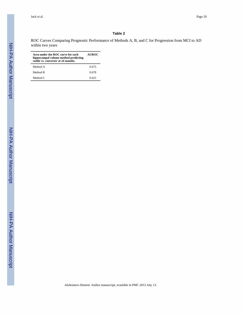
shown in the table and scatter plots, and it also has essentially identical performance inpredicting conversion from MCI to AD (Fig. 1, Table 2). Method C has a similar prognosticperformance in predicting conversion to AD as Method A as shown in the ROC analysis, butin its current form might not meet technical accuracy criteria relative to the referencestandard dataset. This is how we would envision the credentialing process would proceed formost automated applications, with the EADC-ADNI harmonization data set of manuallytraced hippocampi serving as the Reference Standard Dataset and the oversight committeesetting predetermined minimal benchmark criteria to judge the performance of individualmethods.
One important feature of the process for critically evaluating automated hippocampalsegmentation algorithms is the failure rate. For a variety of reasons, usually related to poorscan quality, automated algorithms will fail to produce a plausible result in some proportionof cases in a study. Taken to the extreme, imagine, for example, a method that producedperfect predictive results in cases that underwent successful hippocampal segmentation, butthe method failed in 99% of the time. The method would score quite well on prognosticmetrics, but would not be practical. A fair and objective approach therefore is needed topenalize automated segmentation algorithms that fail in an unacceptably high proportion ofcases.
6. Future efforts1. Although a position paper is a first step, the objective of standardizing hippocampal
volumetry as an AD biomarker will require active participation by stakeholders inacademia and industry. The authors’ objective is to see hippocampal volumetryevolve from its current state, a measure that is valid only in specific studies or asingle institution, to a universally accepted biomarker with standardized units ofmeasure. In some cases, this could simply involve having developers of automatedmeasurement tools directly import the EADC-ADNI anatomic definition of thehippocampal boundaries into the atlas of the automated application.
2. Standardizing single time point hippocampal volume as an AD biomarker is themost logical and readily achievable initial goal; however, the authors recognize thatother more complex topographic structural MRI measures might be more specific,or ultimately more powerful. The major difficulty here is identifying an appropriatereference standard if an anatomically based classifier does not conform to theboundaries of a classically defined anatomic structure as the hippocampus does.
3. Longitudinal change measures on structural MRI should be standardized using theapproach outlined above as a template. This could include an extension of theEADC-ADNI effort to include expert manual tracing of serial hippocampi to createa longitudinal reference standard dataset using the same model as the single timepoint dataset proposed in this position paper.
4. FDG PET, amyloid PET imaging, and possibly other MRI modalities (e.g., restingstate functional connectivity, diffusion tensor imaging, and arterial spin labeledperfusion imaging) are also important imaging biomarkers for AD. Pursuingstandardized quantitative metrics for these imaging modalities is a high priority.The efforts to standardize, validate and evaluate quantitative measures in thesemodalities could roughly follow the same approach outlined above for hippocampalvolume.
5. For all imaging biomarkers, future efforts will need to focus on developing aquantitative score to allow the assessment of individual imaging biomarker
Jack et al. Page 10
Alzheimers Dement. Author manuscript; available in PMC 2012 July 13.
NIH
-PA Author Manuscript
NIH
-PA Author Manuscript
NIH
-PA Author Manuscript

measures against well-developed norms that incorporate other appropriatecovariates, such as age, sex and head size are for the hippocampus (91, 92).
6. Ideally, diagnostic biomarkers should be evaluated against post-mortemhistopathological findings. It is well established that hippocampal atrophy while afeature of AD, is not specific for AD as it occurs in other conditions (32, 93).
7. To optimize the use of biomarkers in new AD diagnostic criteria - future effortswill need to focus on establishing diagnostic cut points in the continuous range ofquantitative values to identify normal, abnormal, and indeterminate levels inindividual subjects. For use in clinical practice, quantitative metrics will need to bedeveloped and then tested in clinically typical and representative populations.Diagnostic biomarkers in AD should function analogously to those in otherdiseases where, for example, cut points in the continuous range of blood pressureand fasting serum glucose are universally recognized as useful in aiding thediagnosis of hypertension and diabetes and standardized treatment protocols arebased on these biomarker cut points. For the purposes of diagnosis in typicalclinical settings, cut points should be derived from carefully characterized groupsof subjects chosen in such a way that the results can be generalized to the overallpopulation. For example, ADNI subjects were selected to represent a typical ADclinical trial, with specific inclusion/exclusion criteria. Thus the results from ADNIare not generalizable to the overall population and are not optimal to generatenormative data for general diagnostic purposes. Selecting meaningful diagnosticcut points is complicated by the fact the many cognitively normal elderly subjectsharbor significant AD pathology. Thus the definition of normal is notstraightforward. Consensus guidelines have been established for evaluating andreporting the clinical utility of diagnostic biomarkers and should be followed instudies using the results of the assay validation steps described here. In clinicalsettings, the sensitivity of detecting AD should exceed 80% and specificity fordistinguishing AD from other similar dementias also should exceed 80% (94).Standardized reporting of results should follow STARD criteria (95) and forclinical settings additional reporting criteria to demonstrate pragmatic utility areneeded (96).
Supplementary MaterialRefer to Web version on PubMed Central for supplementary material.
AcknowledgmentsHeather J Wiste, Mayo Clinic, data analysis
References1. Biomarkers Definitions Working Group. Biomarkers and surrogate endpoints: preferred definitions
and conceptual framework. Clin Pharmacol Ther. 2001; 69(3):89–95. [PubMed: 11240971]
2. Hampel H, Frank R, Broich K, Teipel SJ, Katz RG, Hardy J, et al. Biomarkers for Alzheimer’sdisease: academic, industry and regulatory perspectives. Nat Rev Drug Discov. 2010; 9(7):560–74.[PubMed: 20592748]
3. Jack CR Jr. Knopman DS, Jagust WJ, Shaw LM, Aisen PS, Weiner MW, et al. Hypothetical modelof dynamic biomarkers of the Alzheimer’s pathological cascade. Lancet Neurol. 2010; 9(1):119–28.[PubMed: 20083042]
4. McKhann G, Drachman D, Folstein M, Katzman R, Price D, Stadlan EM. Clinical diagnosis ofAlzheimer’s disease: report of the NINCDS-ADRDA Work Group under the auspices of
Jack et al. Page 11
Alzheimers Dement. Author manuscript; available in PMC 2012 July 13.
NIH
-PA Author Manuscript
NIH
-PA Author Manuscript
NIH
-PA Author Manuscript
Department of Health and Human Services Task Force on Alzheimer’s Disease. Neurology. 1984;34(7):939–44. [PubMed: 6610841]
5. Dubois B, Feldman HH, Jacova C, Dekosky ST, Barberger-Gateau P, Cummings J, et al. Researchcriteria for the diagnosis of Alzheimer’s disease: revising the NINCDS-ADRDA criteria. LancetNeurol. 2007; 6(8):734–46. [PubMed: 17616482]
6. Dubois B, Feldman HH, Jacova C, Cummings JL, Dekosky ST, Barberger-Gateau P, et al. Revisingthe definition of Alzheimer’s disease: a new lexicon. Lancet Neurol. 2010
7. Albert MS, DeKosky ST, Dickson D, Dubois B, Feldman HH, Fox NC, et al. The diagnosis of mildcognitive impairment due to Alzheimer’s disease: Recommendations from the National Institute onAging and Alzheimer’s Association Workgroup. Alzheimers Dement. 2011 Epub ahead of print.
8. Jack CR Jr. Albert MS, Knopman DS, McKhann GM, Sperling RA, Carillo M, et al. Introduction tothe recommendations from the National Institute on Aging and the Alzheimer Associationworkgroup on diagnostic guidelines for Alzheimer’s disease. Alzheimers Dement. 2011 Epub aheadof print.
9. McKhann GM, Knopman DS, Chertkow H, Hyman BT, Jack CR Jr. Kawas CH, et al. The diagnosisof dementia due to Alzheimer’s disease: Recommendations from the National Institute on Agingand the Alzheimer’s Assocation Workgroup. Alzheimers Dement. 2011 Epub ahead of print.
10. Sperling RA, Aisen PS, Beckett LA, Bennett DA, Craft S, Fagan AM, et al. Toward defining thepreclinical stages of Alzheimer’s disease: Recommendations from the National Institute on Agingand the Alzheimer Assocation Workgroup. Alzheimers Dement. 2011 Epub ahead of print.
11. McEvoy L, Hagler D, Holland D, Roddey J, Fennema-Notestine C, Salmon D, et al. NeuroimagingEnrichment Strategy for Secondary Prevention Trials in Alzheimer’s Disease. Alzheimer’sDisease and Related Disorders. 2010; 24(3):269–77.
12. Fox NC, Black RS, Gilman S, Rossor MN, Griffith SG, Jenkins L, et al. Effects of Abetaimmunization (AN1792) on MRI measures of cerebral volume in Alzheimer disease. Neurology.2005; 64(9):1563–72. [PubMed: 15883317]
13. Holland D, Brewer JB, Hagler DJ, Fenema-Notestine C, Dale AM, Weiner M, et al. Subregionalneuroanatomical change as a biomarker for Alzheimer’s disease. Proc Natl Acad Sci U S A. 2009;106(49):20954–20959. [PubMed: 19996185]
14. Jack CR Jr. Slomkowski M, Gracon S, Hoover TM, Felmlee JP, Stewart K, et al. MRI as abiomarker of disease progression in a therapeutic trial of milameline for AD. Neurology. 2003;60(2):253–60. [PubMed: 12552040]
15. Schott JM, Frost C, Whitwell JL, Macmanus DG, Boyes RG, Rossor MN, et al. Combining shortinterval MRI in Alzheimer’s disease: Implications for therapeutic trials. J Neurol. 2006; 253(9):1147–53. [PubMed: 16998650]
16. Fox NC, Cousens S, Scahill R, Harvey RJ, Rossor MN. Using serial registered brain magneticresonance imaging to measure disease progression in Alzheimer disease: power calculations andestimates of sample size to detect treatment effects. Arch Neurol. 2000; 57(3):339–44. [PubMed:10714659]
17. Leung KK, Clarkson MJ, Bartlett JW, Clegg S, Jack CR Jr. Weiner MW, et al. Robust atrophy ratemeasurement in Alzheimer’s disease using multi-site serial MRI: Tissue-specific intensitynormalization and parameter selection. Neuroimage. 2010; 50(2):516–523. [PubMed: 20034579]
18. Hua X, Leow AD, Lee S, Klunder AD, Toga AW, Lepore N, et al. 3D characterization of brainatrophy in Alzheimer’s disease and mild cognitive impairment using tensor-based morphometry.Neuroimage. 2008; 41(1):19–34. [PubMed: 18378167]
19. Hua X, Gutman B, Boyle C, Rajagopalan P, Leow AD, Yanovsky I, et al. Accurate measurementof brain changes in longitudinal MRI scans using tensor-based morphometry. Neuroimage. 2011Epub ahead of print.
20. Vemuri P, Wiste HJ, Weigand SD, Knopman DS, Trojanowski JQ, Shaw LM, et al. Serial MRIand CSF Biomarkers in Normal Aging, MCI and AD. Neurology. 2010; 75(2):143–151. [PubMed:20625167]
21. Wolz R, Heckemann RA, Aljabar P, Hajnal JV, Hammers A, Lotjonen J, et al. Measurement ofhippocampal atrophy using 4D graph-cut segmentation: application to ADNI. Neuroimage. 2010;52(1):109–18. [PubMed: 20382238]
Jack et al. Page 12
Alzheimers Dement. Author manuscript; available in PMC 2012 July 13.
NIH
-PA Author Manuscript
NIH
-PA Author Manuscript
NIH
-PA Author Manuscript
22. Hampel H, Wilcock G, Andrieu S, Aisen P, Blennow K, Broich K, et al. Biomarkers forAlzheimer’s disease therapeutic trials. Prog Neurobiol. 2010 Epub ahead of print.
23. Jack CR Jr. Bernstein MA, Fox NC, Thompson P, Alexander G, Harvey D, et al. The Alzheimer’sDisease Neuroimaging Initiative (ADNI): MRI methods. J Magn Reson Imaging. 2008; 27(4):685–91. [PubMed: 18302232]
24. Jagust WJ, Bandy D, Chen K, Foster NL, Landau SM, Mathis CA, et al. The Alzheimer’s DiseaseNeuroimaging Initiative positron emission tomography core. Alzheimers Dement. 2010; 6(3):221–9. [PubMed: 20451870]
25. Geuze E, Vermetten E, Bremner JD. MR-based in vivo hippocampal volumetrics: 1. Review ofmethodologies currently employed. Mol Psychiatry. 2005; 10(2):147–59. [PubMed: 15340353]
26. Barnes J, Bartlett JW, van de Pol LA, Loy CT, Scahill RI, Frost C, et al. A meta-analysis ofhippocampal atrophy rates in Alzheimer’s disease. Neurobiol Aging. 2009; 30(11):1711–23.[PubMed: 18346820]
27. Du AT, Schuff N, Kramer JH, Ganzer S, Zhu XP, Jagust WJ, et al. Higher atrophy rate ofentorhinal cortex than hippocampus in AD. Neurology. 2004; 62(3):422–7. [PubMed: 14872024]
28. Jack CR Jr. Shiung MM, Gunter JL, O’Brien PC, Weigand SD, Knopman DS, et al. Comparison ofdifferent MRI brain atrophy rate measures with clinical disease progression in AD. Neurology.2004; 62(4):591–600. [PubMed: 14981176]
29. Wang L, Swank JS, Glick IE, Gado MH, Miller MI, Morris JC, et al. Changes in hippocampalvolume and shape across time distinguish dementia of the Alzheimer type from healthy aging.Neuroimage. 2003; 20(2):667–82. [PubMed: 14568443]
30. Braak H, Braak E. Neuropathological stageing of Alzheimer-related changes. Acta Neuropathol.1991; 82(4):239–59. [PubMed: 1759558]
31. Bobinski M, de Leon MJ, Wegiel J, Desanti S, Convit A, Saint Louis LA, et al. The histologicalvalidation of post mortem magnetic resonance imaging-determined hippocampal volume inAlzheimer’s disease. Neuroscience. 2000; 95(3):721–5. [PubMed: 10670438]
32. Jack CR Jr. Dickson DW, Parisi JE, Xu YC, Cha RH, O’Brien PC, et al. Antemortem MRIfindings correlate with hippocampal neuropathology in typical aging and dementia. Neurology.2002; 58(5):750–7. [PubMed: 11889239]
33. Gosche KM, Mortimer JA, Smith CD, Markesbery WR, Snowdon DA. Hippocampal volume as anindex of Alzheimer neuropathology: findings from the Nun Study. Neurology. 2002; 58(10):1476–82. [PubMed: 12034782]
34. Csernansky JG, Hamstra J, Wang L, McKeel D, Price JL, Gado M, et al. Correlations betweenantemortem hippocampal volume and postmortem neuropathology in AD subjects. Alzheimer DisAssoc Disord. 2004; 18(4):190–5. [PubMed: 15592129]
35. Silbert LC, Quinn JF, Moore MM, Corbridge E, Ball MJ, Murdoch G, et al. Changes in premorbidbrain volume predict Alzheimer’s disease pathology. Neurology. 2003; 61(4):487–92. [PubMed:12939422]
36. Zarow C, Vinters HV, Ellis WG, Weiner MW, Mungas D, White L, et al. Correlates ofhippocampal neuron number in Alzheimer’s disease and ischemic vascular dementia. Ann Neurol.2005; 57(6):896–903. [PubMed: 15929035]
37. Colliot O, Chetelat G, Chupin M, Desgranges B, Magnin B, Benali H, et al. Discriminationbetween Alzheimer disease, mild cognitive impairment, and normal aging by using automatedsegmentation of the hippocampus. Radiology. 2008; 248(1):194–201. [PubMed: 18458242]
38. Jack CR Jr. Petersen RC, O’Brien PC, Tangalos EG. MR-based hippocampal volumetry in thediagnosis of Alzheimer’s disease. Neurology. 1992; 42(1):183–8. [PubMed: 1734300]
39. Killiany RJ, Gomez-Isla T, Moss M, Kikinis R, Sandor T, Jolesz F, et al. Use of structuralmagnetic resonance imaging to predict who will get Alzheimer’s disease. Ann Neurol. 2000;47(4):430–9. [PubMed: 10762153]
40. McEvoy LK, Fennema-Notestine C, Roddey JC, Hagler DJ Jr. Holland D, Karow DS, et al.Alzheimer disease: quantitative structural neuroimaging for detection and prediction of clinicaland structural changes in mild cognitive impairment. Radiology. 2009; 251(1):195–205. [PubMed:19201945]
Jack et al. Page 13
Alzheimers Dement. Author manuscript; available in PMC 2012 July 13.
NIH
-PA Author Manuscript
NIH
-PA Author Manuscript
NIH
-PA Author Manuscript
41. Desikan RS, Cabral HJ, Hess CP, Dillon WP, Glastonbury CM, Weiner MW, et al. AutomatedMRI measures identify individuals with mild cognitive impairment and Alzheimer’s disease.Brain. 2009; 132(Pt 8):2048–57. [PubMed: 19460794]
42. Gerardin E, Chetelat G, Chupin M, Cuingnet R, Desgranges B, Kim HS, et al. Multidimensionalclassification of hippocampal shape features discriminates Alzheimer’s disease and mild cognitiveimpairment from normal aging. Neuroimage. 2009; 47(4):1476–86. [PubMed: 19463957]
43. Dickerson BC, Goncharova I, Sullivan MP, Forchetti C, Wilson RS, Bennett DA, et al. MRI-derived entorhinal and hippocampal atrophy in incipient and very mild Alzheimer’s disease.Neurobiol Aging. 2001; 22(5):747–54. [PubMed: 11705634]
44. McDonald CR, Gharapetian L, McEvoy LK, Fennema-Notestine C, Hagler DJ Jr. Holland D, et al.Relationship between regional atrophy rates and cognitive decline in mild cognitive impairment.Neurobiol Aging. 2010 Epub ahead of print.
45. Sarazin M, Chauvire V, Gerardin E, Colliot O, Kinkingnehun S, de Souza LC, et al. The amnesticsyndrome of hippocampal type in Alzheimer’s disease: an MRI study. J Alzheimers Dis. 2010;22(1):285–94. [PubMed: 20847406]
46. Fox NC, Warrington EK, Freeborough PA, Hartikainen P, Kennedy AM, Stevens JM, et al.Presymptomatic hippocampal atrophy in Alzheimer’s disease. A longitudinal MRI study. Brain.1996; 119(Pt 6):2001–7. [PubMed: 9010004]
47. Jack CR Jr. Petersen RC, Xu Y, O’Brien PC, Smith GE, Ivnik RJ, et al. Rates of hippocampalatrophy correlate with change in clinical status in aging and AD. Neurology. 2000; 55(4):484–89.[PubMed: 10953178]
48. Jack CR Jr. Petersen RC, Xu YC, O’Brien PC, Smith GE, Ivnik RJ, et al. Prediction of AD withMRI-based hippocampal volume in mild cognitive impairment. Neurology. 1999; 52(7):1397–403.[PubMed: 10227624]
49. Visser PJ, Scheltens P, Verhey FR, Schmand B, Launer LJ, Jolles J, et al. Medial temporal lobeatrophy and memory dysfunction as predictors for dementia in subjects with mild cognitiveimpairment. J Neurol. 1999; 246(6):477–85. [PubMed: 10431775]
50. Risacher SL, Saykin AJ, West JD, Shen L, Firpi HA, McDonald BC. Baseline MRI predictors ofconversion from MCI to probable AD in the ADNI cohort. Curr Alzheimer Res. 2009; 6(4):347–61. [PubMed: 19689234]
51. Henneman WJ, Sluimer JD, Barnes J, van der Flier WM, Sluimer IC, Fox NC, et al. Hippocampalatrophy rates in Alzheimer disease: added value over whole brain volume measures. Neurology.2009; 72(11):999–1007. [PubMed: 19289740]
52. Brys M, Glodzik L, Mosconi L, Switalski R, De Santi S, Pirraglia E, et al. Magnetic resonanceimaging improves cerebrospinal fluid biomarkers in the early detection of Alzheimer’s disease. JAlzheimers Dis. 2009; 16(2):351–62. [PubMed: 19221425]
53. Landau SM, Harvey D, Madison CM, Reiman EM, Foster NL, Aisen PS, et al. Comparingpredictors of conversion and decline in mild cognitive impairment. Neurology. 2010; 75(3):230–8.[PubMed: 20592257]
54. Kantarci K, Weigand SD, Przybelski SA, Shiung MM, Whitwell JL, Negash S, et al. Risk ofdementia in MCI: combined effect of cerebrovascular disease, volumetric MRI, and 1H MRS.Neurology. 2009; 72(17):1519–25. [PubMed: 19398707]
55. Karas G, Sluimer J, Goekoop R, van der Flier W, Rombouts SA, Vrenken H, et al. Amnestic mildcognitive impairment: structural MR imaging findings predictive of conversion to Alzheimerdisease. AJNR Am J Neuroradiol. 2008; 29(5):944–9. [PubMed: 18296551]
56. Jack CR Jr. Wiste HJ, Vemuri P, Weigand SD, Senjem ML, Zeng G, et al. Brain beta-amyloidmeasure and magnetic resonance imaging atophy both predict time-to-progression from mildcognitive impairment to Alzheimer’s disease. Brain. 2010; 133(11):3336–3348. [PubMed:20935035]
57. Fleisher AS, Sun S, Taylor C, Ward CP, Gamst AC, Petersen RC, et al. Volumetric MRI vs clinicalpredictors of Alzheimer disease in mild cognitive impairment. Neurology. 2008; 70(3):191–9.[PubMed: 18195264]
Jack et al. Page 14
Alzheimers Dement. Author manuscript; available in PMC 2012 July 13.
NIH
-PA Author Manuscript
NIH
-PA Author Manuscript
NIH
-PA Author Manuscript

58. Leung KK, Barnes J, Ridgway GR, Bartlett JW, Clarkson MJ, Macdonald K, et al. Automatedcross-sectional and longitudinal hippocampal volume measurement in mild cognitive impairmentand Alzheimer’s disease. Neuroimage. 2010; 51(4):1345–59. [PubMed: 20230901]
59. Devanand DP, Pradhaban G, Liu X, Khandji A, De Santi S, Segal S, et al. Hippocampal andentorhinal atrophy in mild cognitive impairment: prediction of Alzheimer disease. Neurology.2007; 68(11):828–36. [PubMed: 17353470]
60. Apostolova LG, Dutton RA, Dinov ID, Hayashi KM, Toga AW, Cummings JL, et al. Conversionof mild cognitive impairment to Alzheimer disease predicted by hippocampal atrophy maps. ArchNeurol. 2006; 63(5):693–9. [PubMed: 16682538]
61. Apostolova LG, Mosconi L, Thompson PM, Green AE, Hwang KS, Ramirez A, et al. Subregionalhippocampal atrophy predicts Alzheimer’s dementia in the cognitively normal. Neurobiol Aging.2010; 31(7):1077–88. [PubMed: 18814937]
62. Chetelat G, Fouquet M, Kalpouzos G, Denghien I, De la Sayette V, Viader F, et al. Three-dimensional surface mapping of hippocampal atrophy progression from MCI to AD and overnormal aging as assessed using voxel-based morphometry. Neuropsychologia. 2008; 46(6):1721–31. [PubMed: 18289618]
63. Beckett LA, Harvey DJ, Gamst A, Donohue M, Kornak J, Zhang H, et al. The Alzheimer’s DiseaseNeuroimaging Initiative: Annual change in biomarkers and clinical outcomes. AlzheimersDement. 2010; 6(3):257–64. [PubMed: 20451874]
64. Fischl B, Salat DH, Busa E, Albert M, Dieterich M, Haselgrove C, et al. Whole brainsegmentation: automated labeling of neuroanatomical structures in the human brain. Neuron.2002; 33(3):341–55. [PubMed: 11832223]
65. Dale AM, Fischl B, Sereno MI. Cortical surface-based analysis. I. Segmentation and surfacereconstruction. Neuroimage. 1999; 9(2):179–94. [PubMed: 9931268]
66. Barnes J, Foster J, Boyes RG, Pepple T, Moore EK, Schott JM, et al. A comparison of methods forthe automated calculation of volumes and atrophy rates in the hippocampus. Neuroimage. 2008;40(4):1655–71. [PubMed: 18353687]
67. Morra JH, Tu Z, Apostolova LG, Green AE, Avedissian C, Madsen SK, et al. Automated 3Dmapping of hippocampal atrophy and its clinical correlates in 400 subjects with Alzheimer’sdisease, mild cognitive impairment, and elderly controls. Hum Brain Mapp. 2009; 30(9):2766–88.[PubMed: 19172649]
68. Kim H, Besson P, Colliot O, Bernasconi A, Bernasconi N. Surface-based vector analysis usingheat equation interpolation: a new approach to quantify local hippocampal volume changes. MedImage Comput Comput Assist Interv. 2008; 11(Pt 1):1008–15. [PubMed: 18979844]
69. Mouiha A, Duchesne S. HIPPOCAMPAL ATROPHY RATES IN ALZHEIMER’S DISEASE:AUTOMATED SEGMENTATION VARIABILITY ANALYSIS. Neurosci Lett. 2011 Epubahead of print.
70. Chupin M, Hammers A, Liu RS, Colliot O, Burdett J, Bardinet E, et al. Automatic segmentation ofthe hippocampus and the amygdala driven by hybrid constraints: method and validation.Neuroimage. 2009; 46(3):749–61. [PubMed: 19236922]
71. Brewer JB, Magda S, Airriess C, Smith ME. Fully-automated quantification of regional brainvolumes for improved detection of focal atrophy in Alzheimer disease. AJNR Am J Neuroradiol.2009; 30(3):578–80. [PubMed: 19112065]
72. Scheltens P, Leys D, Barkhof F, Huglo D, Weinstein HC, Vermersch P, et al. Atrophy of medialtemporal lobes on MRI in “probable” Alzheimer’s disease and normal ageing: diagnostic valueand neuropsychological correlates. J Neurol Neurosurg Psychiatry. 1992; 55(10):967–72.[PubMed: 1431963]
73. de Leon MJ, Golomb J, George AE, Convit A, Tarshish CY, McRae T, et al. The radiologicprediction of Alzheimer disease: the atrophic hippocampal formation. AJNR Am J Neuroradiol.1993; 14(4):897–906. [PubMed: 8352162]
74. DeCarli C, Frisoni GB, Clark CM, Harvey D, Grundman M, Petersen RC, et al. Qualitativeestimates of medial temporal atrophy as a predictor of progression from mild cognitive impairmentto dementia. Arch Neurol. 2007; 64(1):108–15. [PubMed: 17210817]
Jack et al. Page 15
Alzheimers Dement. Author manuscript; available in PMC 2012 July 13.
NIH
-PA Author Manuscript
NIH
-PA Author Manuscript
NIH
-PA Author Manuscript
75. Jack CR Jr. Sharbrough FW, Twomey CK, Cascino GD, Hirschorn KA, Marsh WR, et al.Temporal lobe seizures: lateralization with MR volume measurements of the hippocampalformation. Radiology. 1990; 175(2):423–9. [PubMed: 2183282]
76. Teipel SJ, Ewers M, Wolf S, Jessen F, Kolsch H, Arlt S, et al. Multicentre variability of MRI-based medial temporal lobe volumetry in Alzheimer’s disease. Psychiatry Res. 2010; 182(3):244–50. [PubMed: 20493672]
77. The ENIGMA Consortium. Genome-Wide Association Meta-Analysis of Hippocampal Volumevia the ENIGMA Consortium; Organization for Human Brain Mapping Meeting; Quebec City,Canada. 2011;
78. van de Pol LA, Barnes J, Scahill RI, Frost C, Lewis EB, Boyes RG, et al. Improved reliability ofhippocampal atrophy rate measurement in mild cognitive impairment using fluid registration.Neuroimage. 2007; 34(3):1036–41. [PubMed: 17174572]
79. Vemuri P, Gunter JL, Senjem ML, Whitwell JL, Kantarci K, Knopman DS, et al. Alzheimer’sdisease diagnosis in individual subjects using structural MR images: validation studies.Neuroimage. 2008; 39(3):1186–97. [PubMed: 18054253]
80. Davatzikos C, Fan Y, Wu X, Shen D, Resnick SM. Detection of prodromal Alzheimer’s diseasevia pattern classification of magnetic resonance imaging. Neurobiol Aging. 2008; 29(4):514–23.[PubMed: 17174012]
81. Fan Y, Shen D, Davatzikos C. Classification of structural images via high-dimensional imagewarping, robust feature extraction, and SVM. Med Image Comput Comput Assist Interv Int ConfMed Image Comput Comput Assist Interv. 2005; 8(Pt 1):1–8.
82. Stonnington CM, Tan G, Kloppel S, Chu C, Draganski B, Jack CR Jr. et al. Interpreting scan dataacquired from multiple scanners: a study with Alzheimer’s disease. Neuroimage. 2008; 39(3):1180–5. [PubMed: 18032068]
83. Kloppel S, Stonnington CM, Chu C, Draganski B, Scahill RI, Rohrer JD, et al. Automaticclassification of MR scans in Alzheimer’s disease. Brain. 2008; 131(Pt 3):681–9. [PubMed:18202106]
84. Duchesne S, Caroli A, Geroldi C, Barillot C, Frisoni GB, Collins DL. MRI-based automatedcomputer classification of probable AD versus normal controls. IEEE Trans Med Imaging. 2008;27(4):509–20. [PubMed: 18390347]
85. Knopman DS, DeKosky ST, Cummings JL, Chui H, Corey-Bloom J, Relkin N, et al. Practiceparameter: diagnosis of dementia (an evidence-based review). Report of the Quality StandardsSubcommittee of the American Academy of Neurology. Neurology. 2001; 56(9):1143–53.[PubMed: 11342678]
86. Hort J, O’Brien JT, Gainotti G, Pirttila T, Popescu BO, Rektorova I, et al. EFNS guidelines for thediagnosis and management of Alzheimer’s disease. Eur J Neurol. 2010; 17(10):1236–48.[PubMed: 20831773]
87. Boccardi, M.; Ganzola, R.; Duchesne, S.; Redolfi, A.; Bartzokis, G.; Csernansky, J.; et al. ICAD -Alzheimer’s Imaging Consortium. Survey of segmentation protocols for manual hippocampalvolumetry: Prepatory phase for an EADC-ADNI harmonization protocol. Honolulu, HI: 2010.
88. Frisoni GB, Jack CR. Harmonization of magnetic resonance-based manual hippocampalsegmentation: A mandatory step for wide clinical use. Alzheimers Dement. 2011; 7(2):171–4.[PubMed: 21414554]
89. Fox NC, Ridgway GR, Schott JM. Algorithms, atrophy and Alzheimer’s disease: Cautionary talesfor clinical trials. Neuroimage. 2011 Epub ahead of print.
90. Cover KS, van Schijndel RA, van Dijk BW, Redolfi A, Knol DL, Frisoni GB, et al. Assessing thereproducibility of the SienaX and Siena brain atrophy measures using the ADNI back-to-back MP-RAGE MRI scans. Psychiatry Research. In Press.
91. Barnes J, Ridgway GR, Bartlett J, Henley SM, Lehmann M, Hobbs N, et al. Head size, age andgender adjustment in MRI studies: a necessary nuisance? Neuroimage. 2010; 53(4):1244–55.[PubMed: 20600995]
92. Jack CR Jr. Petersen RC, Xu YC, Waring SC, O’Brien PC, Tangalos EG, et al. Medial temporalatrophy on MRI in normal aging and very mild Alzheimer’s disease. Neurology. 1997; 49(3):786–94. [PubMed: 9305341]
Jack et al. Page 16
Alzheimers Dement. Author manuscript; available in PMC 2012 July 13.
NIH
-PA Author Manuscript
NIH
-PA Author Manuscript
NIH
-PA Author Manuscript
93. Jagust WJ, Zheng L, Harvey DJ, Mack WJ, Vinters HV, Weiner MW, et al. Neuropathologicalbasis of magnetic resonance images in aging and dementia. Ann Neurol. 2008; 63(1):72–80.[PubMed: 18157909]
94. Reagan Working Group; The Ronald and Nancy Reagan Research Institute of the Alzheimer’sAssociation and the National Institute on Aging Working Group. Consensus report of the WorkingGroup on: “Molecular and Biochemical Markers of Alzheimer’s Disease. Neurobiol Aging. 1998;19(2):109–16. [PubMed: 9558143]
95. Bossuyt PM, Reitsma JB, Bruns DE, Gatsonis CA, Glasziou PP, Irwig LM, et al. Towardscomplete and accurate reporting of studies of diagnostic accuracy: the STARD initiative. FamPract. 2004; 21(1):4–10. [PubMed: 14760036]
96. Glasgow RE, Magid DJ, Beck A, Ritzwoller D, Estabrooks PA. Practical clinical trials fortranslating research to practice: design and measurement recommendations. Med Care. 2005;43(6):551–7. [PubMed: 15908849]
Jack et al. Page 17
Alzheimers Dement. Author manuscript; available in PMC 2012 July 13.
NIH
-PA Author Manuscript
NIH
-PA Author Manuscript
NIH
-PA Author Manuscript
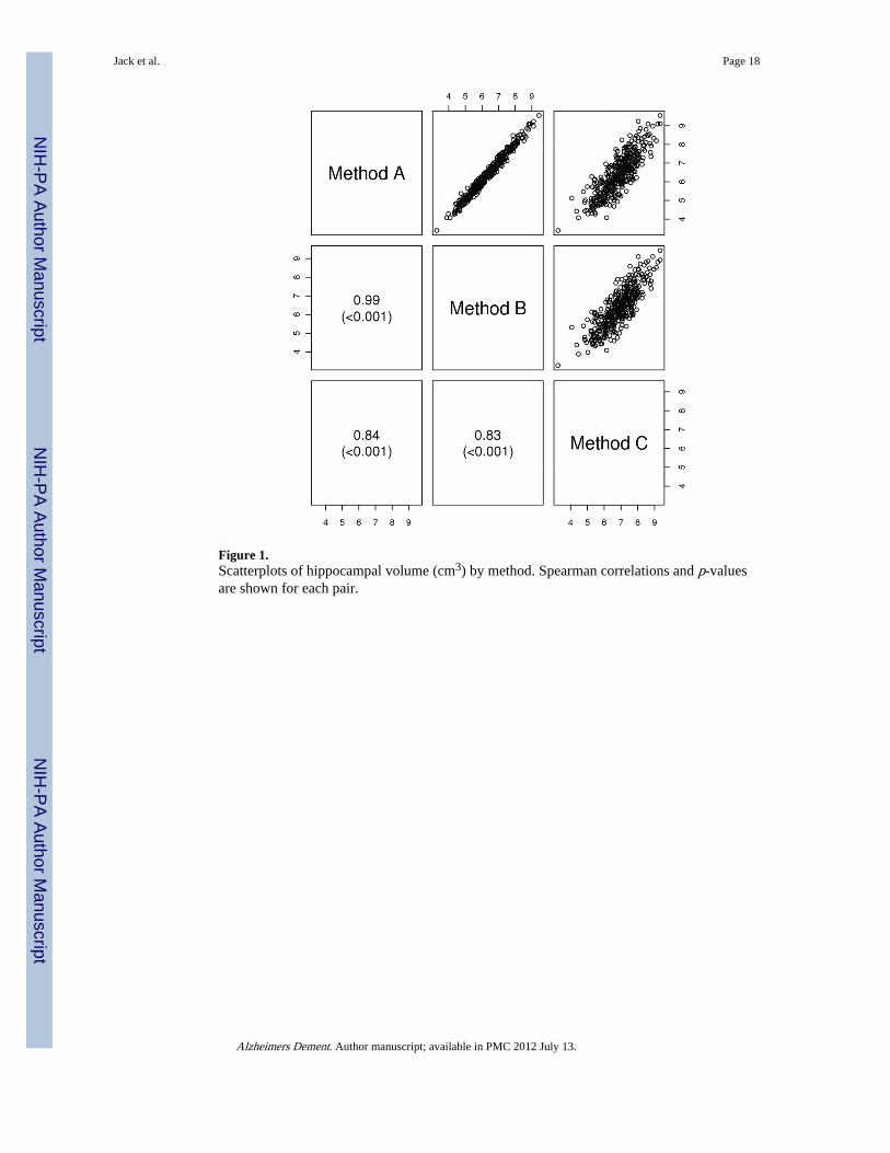
Figure 1.Scatterplots of hippocampal volume (cm3) by method. Spearman correlations and p-valuesare shown for each pair.
Jack et al. Page 18
Alzheimers Dement. Author manuscript; available in PMC 2012 July 13.
NIH
-PA Author Manuscript
NIH
-PA Author Manuscript
NIH
-PA Author Manuscript

NIH
-PA Author Manuscript
NIH
-PA Author Manuscript
NIH
-PA Author Manuscript
Jack et al. Page 19
Table 1
Descriptive Characteristics
Characteristic All Stable MCI AD Converter
N 373 178 135
Age, years 75 (70, 80) 75 (71, 81) 75 (70, 80)
Female gender, no. (%) 136 (36) 63 (35) 51 (38)
Education, years 16 (14, 18) 16 (14, 18) 16 (14, 18)
MMSE 27 (26, 28) 28 (26, 29) 27 (25, 28)
Hippocampal Volume, cm3
Method A 6.3 (5.6, 7.1) 6.7 (6.0, 7.4) 6.0 (5.2, 6.6)
Method B 6.3 (5.6, 7.1) 6.6 (5.9, 7.2) 5.9 (5.1, 6.6)
Method C 6.9 (6.2, 7.5) 7.1 (6.5, 7.6) 6.6 (6.0, 7.2)
All values are reported as median (inter quartile range - IQR) unless otherwise noted Stable/Converter is defined as progression to AD by 24months
MMSE, Mini Mental State Exam
Alzheimers Dement. Author manuscript; available in PMC 2012 July 13.
NIH
-PA Author Manuscript
NIH
-PA Author Manuscript
NIH
-PA Author Manuscript
Jack et al. Page 20
Table 2
ROC Curves Comparing Prognostic Performance of Methods A, B, and C for Progression from MCI to ADwithin two years
Area under the ROC curve for eachhippocampal volume method predictingstable vs. converter at 24 months.
AUROC
Method A 0.675
Method B 0.678
Method C 0.625
Alzheimers Dement. Author manuscript; available in PMC 2012 July 13.