Astrocytes Give Rise to New Neurons in the Adult Mammalian Hippocampus
Review
Stem cells of the adult mammalian brain and their nicheO. Basak and V. Taylor*
Department of Molecular Embryology, Max Planck Institute of Immunobiology, Stubeweg 51, 79108 Freiburg(Germany), Fax: +49 761 5108 474, e-mail: [email protected]
Received 03 September 2008; received after revision 09 October 2008; accepted 14 October 2008Online First 18 November 2008
Abstract. The mammalian brain is a paradox ofevolution. Although the advance in complexity ofthe human brain has exceeded the development ofother organs, it has practically lost the ability toregenerate, and damage is repaired mainly by func-tional plasticity. This disparity is, however, not due tothe lack of progenitor cells in the adult mammalianbrain, but to their diminished or repressed capacity to
replace neurons in most brain regions. Here, wediscuss the current literature describing the processesof neurogenesis in the adult mammalian brain, and therecent advances in adult neural stem cells (aNSCs)with a focus on their identity, cell cycle and nichesignals. Understanding these processes may hopefullylead to therapies in the future to reinstate self-repairof the brain from endogenous progenitors.
Keywords. Neural stem cells, neurogenesis, stem cell niche, adult brain, subventricular zone.
The discovery of neurogenesis in the adult brain
NSCs are the foundations of the brain during develop-ment, and despite poor regenerative capacity, theypersist in the mammalian brain throughout postnataldevelopment and into adulthood and continue togenerate neurons. aNSCs self-renew and retain multi-potency in almost all mammalian species analyzed,including humans. NSCs reside in specialized germinallayers where multiple cell-types interact with themand contribute to generate niches that control theirfate choices and permit neurogenesis. Genetic andfunctional analyses have revealed roles for severalgenes and signaling pathways in controlling adultneurogenesis. However, the identities of the stem cellsin the brain remain elusive, which presents anelementary problem for the interpretation of theseresults. In attempts to identify aNSCs, several groupshave defined populations that contain cells with stem
cell features, although unambiguous markers are stillmissing. Given their potential as putative tools forcell-based therapies, elucidating the molecular path-ways that identify aNSCs and govern their cell fate isessential.Neurons in most regions of the adult mammalian brainare generated from NSCs and restricted progenitorsduring embryonic development before a switch togliogenesis in peri- and postnatal development [1]. Asearly as the 1960s and 70s, newly generated neuronswere identified by nucleotide analogue incorporationassays in adult rodents [2, 3]. These results, however,met with skepticism due to the poor regenerativecapacity of the mammalian brain. Detailed studieswent on to show extensive neurogenesis in the vocalcontrol center in the brains of adult canaries, whichcorrelates with seasonal song learning [4, 5]. After abrief re-visitation to the topic in the mid-1980s, it wasthe use of retroviral lineage tracing assays thatidentified newborn neurons in the adult mammalianbrain. These findings were quickly followed by theidentification of zones with neurogenic capacity in* Corresponding author.
Cell. Mol. Life Sci. 66 (2009) 1057 – 10721420-682X/09/061057-16DOI 10.1007/s00018-008-8544-x� Birkh�user Verlag, Basel, 2008
Cellular and Molecular Life Sciences
multiple mammalian species including humans [2, 3,6 – 9]. Evidence of continued adult neurogenesisintrigued many who long believed that the adultmammalian brain had lost its neurogenic capacitycompared to the avian and fish brain, and furtherprovoked questions about the neural progenitorswhich retain proliferative capacity and are capableof giving-rise to more differentiated progeny includ-ing neurons. The phenomenon of NSC as a life-longsource of neural progenitors and newly generatedneurons in adults was proposed after the isolation ofcells that can be propagated for an unlimited amountof time and generate all three lineages of the adultbrain in vitro [6, 7, 10].
Cellular architecture of the neurogenic zones
Although multiple proliferative zones have now beenidentified in the adult central nervous system, neuro-genesis is most prominent and best studied in twodefined germinal centers, the subependymal layer ofthe lateral ventricle wall covering the striatum (SEZ)and the dentate gyrus of the hippocampal formation.The SEZ is the major source of adult neurogenesisgenerating at least 5 subtypes of interneuron of theolfactory bulb and, to a limited extent, oligodendro-cytes of the corpus callosal white matter [2, 11, 12].Estimations suggest that 30 – 60,000 new neurons maybe generated per day in the adult rodent olfactory bulb[13, 14].Morphologically, four major cell-types have beenidentified in the rodent SEZ. SEZ astrocytes aremitotically inactive and can be subdivided dependingon chromatin structure [15, 16]. One undisputed roleof the astrocytes is to form “glial tubes” that ensheathmigrating neuroblasts, confining them to the rostralmigratory stream (RMS) and separating them fromthe surrounding parenchyma. Neuroblasts are theprincipal product of the SEZ and migrate in chains tothe olfactory bulb where they distribute radially to thegranule and glomerular layers before undergoingterminal differentiation. Neuroblasts are generatedfrom resident stem cells in the SEZ through a transientamplifying population (TAPs) [11, 13, 15]. The neuro-genic zone is covered by a ventricular lining ofmitotically-inactive ependymal cells with motor ciliaextending into the ventricle that circulate cerebrospi-nal fluid generated by the choroids plexus. Evidenceindicates that ependymal cell-mediated directedmovement of factors, including Slit proteins in thecerebrospinal fluid, determines migration of neuro-blasts to the olfactory bulb [17].Similarly, the human SEZ is divided into four mor-phologically distinguishable layers with cell-types
reminiscent of those found in rodents. Lining theventricles, layer 1 is composed of ependymal cellsextending basal processes into a hypocellular gap(layer 2). Layer 2 contains some neurons and neuronalfibers and occasional astrocytes [18]. Interestingly,neither rodents nor non-human primates have thishypocellular gap which, in addition to humans, has sofar only been detected in bovine brains [18 – 20]. Layer3 contains a ribbon of astrocytes that extend processesto layer 2 and might make contact with the ependymalcells [18]. Layer 3 contains cells morphologicallysimilar to the TAPs in rodents and has been suggestedto contain aNSCs [18, 21]. The SEZ is delineated fromthe brain parenchyma by the myelin rich layer 4 [20].A migratory track reminiscent of the RMS in the othermammals is also present in humans and recent reportsclaimed to have identified b-tubulinIII+ (Tuj1) neu-roblasts; however, evidence for chain migration islacking [18, 20, 22].Neurogenesis in the adult mammalian hippocampusinvolves similar steps to those of the SEZ, althoughunlike in the forebrain, the source (subgranule zone –SGZ) and target (granule cell layer) of the newbornneurons are juxtaposed [23, 24]. Astrocytes of theSGZ have two basic morphologies. Those withpyramidal cell-bodies and radial processes extendingthrough the granule cell layer towards the surface ofthe dentate, similar to embryonic radial glia, include apopulation of stem cells [23, 24]. Recently, a secondpopulation of SGZ astrocytes with tangential ratherthan radial processes has been shown to expressprogenitor markers and behave like stem cells in vivo[25]. Interestingly, this finding questions the dogma ofradial astrocytes as the sole stem cells of the dentateand indicates that tangential cells may make-up thepredominant active stem cells of the hippocampus. Inaddition, retroviral labeling indicates that these non-radial cells undergo asymmetric cell division togenerate neurons but can also produce daughtersthat adopt a radial morphology. As in the SEZ,dentate neurogenesis progresses through transientintermediates and mitotically active neuroblasts be-fore newborn neurons migrate radially into thegranule cell layer.
Neural progenitors in the adult brain
Endogenous NSCs are, according to the definition,capable of self-renewal throughout the life of theorganism and multi-lineage differentiation (multipo-tency) into neurons, astrocytes and oligodendrocytes[26]. However, the exact identity of the aNSCs has yetto be determined. Due to the lack of unambiguousmarkers, single aNSCs in the adult neurogenic niches
1058 O. Basak and V. Taylor Adult neural stem cells
cannot be identified [7]. As a consequence, markergene expression, viral and genetic approaches canonly be used to label populations of cells that include avariety of cell-types with different antigenic, cell cycle,self-renewal, and differentiation potentials. Thus,these experiments are inevitably a retrospectiveestimation of cell populations that may contain multi-potent or multiple lineage restricted stem cells. Forinstance, although both neurons and oligodendrocytesare generated in the adult mouse SEZ, there is noevidence that a single cell gives-rise to both lineages invivo. Furthermore, although strictly speaking the stemcell should maintain self-replicating potentialthroughout the life of the organism, a more flexibleand potentially more tangible scenario is that the stemcells have a limited life span. Thus, active stem cellsmay become exhausted with time, either resulting in areduction in progenitors with age, which is observed inboth adult neurogenic brain regions, or that the activeNSCs are replaced by precursors that, under normalcircumstances, do not contribute significantly to activeneurogenesis. In reality, neurogenesis may use acombination of both strategies. It will be a centralobjective in the future to combine genetics and lineagetracing not only to identify the NSCs in the adult brainbut also to elucidate the mechanisms that control theirmaintenance and fate in vivo.Although it has not been possible to date to identifyaNSCs in situ, cells with NSC-like character can beisolated from the adult brain and expanded in vitrousing the neurosphere assay. Spherogenic cells self-renew in vitro in response to epidermal growth factor(EGF) or fibroblast growth factor 2 (FGF2) andclonally generate neurons, astrocytes and oligoden-drocytes [6, 7, 27 – 29]. Spherogenic cells representprospective NSCs, although the retrospective natureof the method evoked questions about their sourceand the relationship to the NSCs in vivo. In addition,expression analysis and differentiation experimentshave cast doubt on the potential and validity ofneurosphere-derived cells for cell therapy [29 – 32].Furthermore, evidence suggests that not only NSCsbut more differentiated progeny can also generateneurospheres in vitro. Ablation of rapidly dividingcells from the SEZ by treatment of mice with anti-mitotic drugs such as cytosine arabinoside (AraC)results in a reduction in neurosphere-forming cellsisolated in EGF-containing medium [31]. This wasinterpreted to indicate that a significant proportionof the EGF-responsive spherogenic cells are TAPs.However, although there may be some foundation tothis interpretation of the anti-mitotic paradigm, theexperiments may also be biased. It has been suggest-ed that a feedback mechanism to the NSC from itsprogeny may also regulate stem cell status (see later).
Thus, in the situation where the SEZ degenerates andthe progeny of the NSC, TAPs and neuroblasts, arereduced due to anti-mitotic drug treatment, NSCsmay be promoted to enter the cell cycle precociouslyto compensate, thus rendering them more suscepti-ble to AraC than would have been predicted. Hence,NSCs could also be killed by the anti-mitotic treat-ment which would also result in a reduction inspherogenic cells. The lack of definitive markersagain prevents the conclusive dissemination of theresults. More detailed analysis is required to establishif neural progenitors that have a short cell cycle(TAPs) are able to form neurospheres from a non-lesioned forebrain. Clearly, the estimated number ofnon-NSC derived neurospheres indicates that veryfew TAPs (the major mitotic population in the SEZ)are spherogenic. In the future, it will be interesting toestablish if these putative non-NSC spherogenic cellsmay be an intermediate between committed TAPsand NSC. Alternatively, spherogenic TAPs maybelong to the relatively minor oligodendroglia pro-genitor population in the SEZ. Although the source
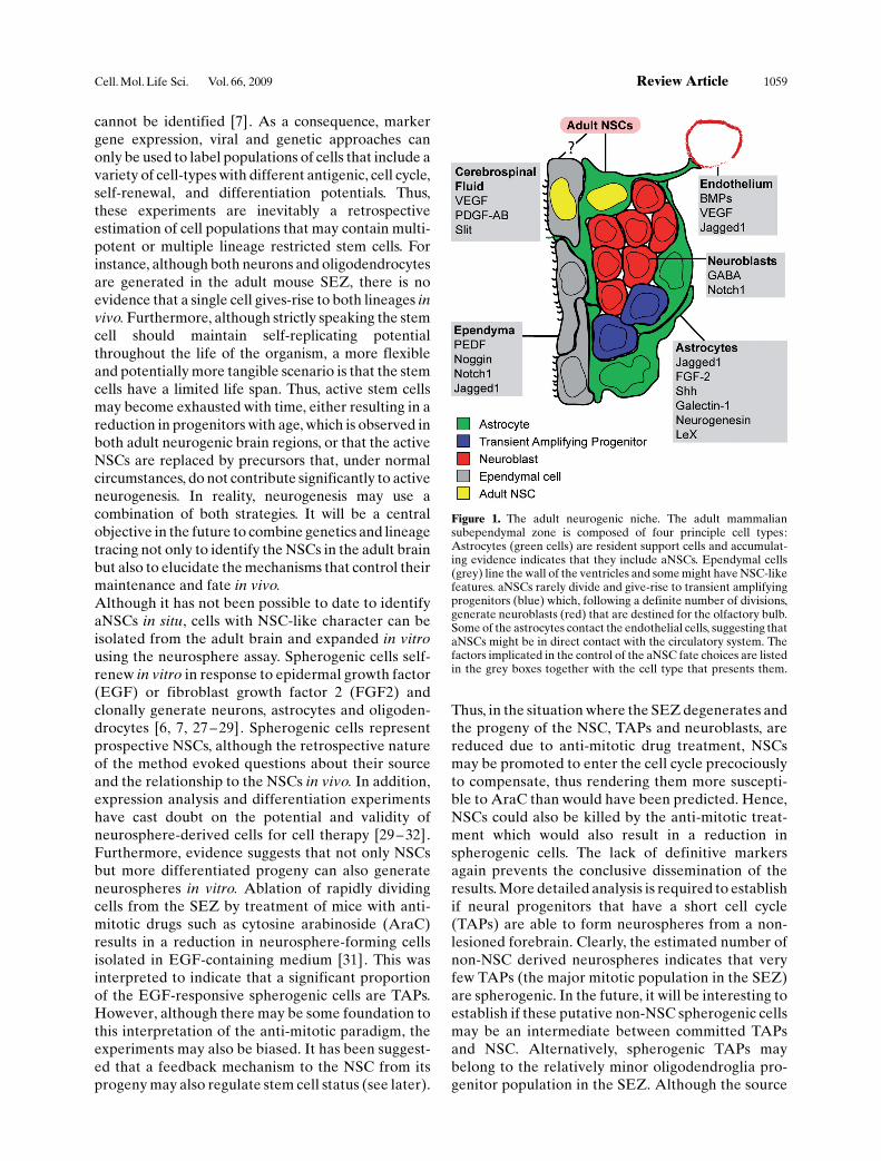
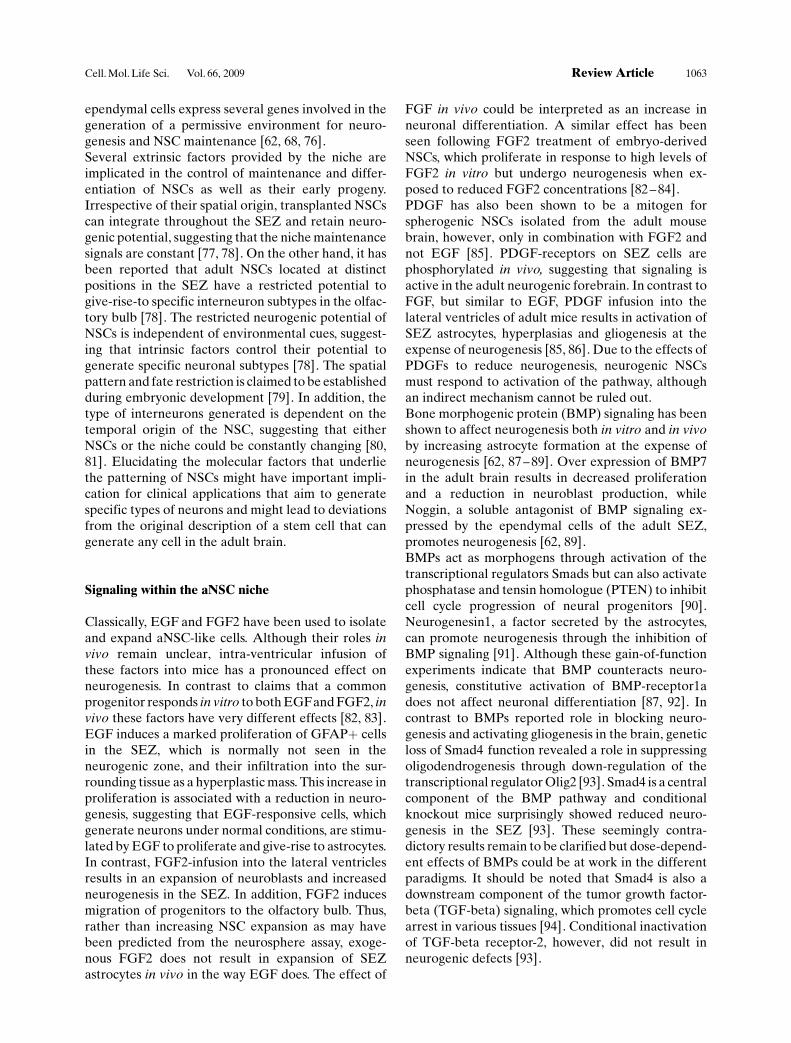
Figure 1. The adult neurogenic niche. The adult mammaliansubependymal zone is composed of four principle cell types:Astrocytes (green cells) are resident support cells and accumulat-ing evidence indicates that they include aNSCs. Ependymal cells(grey) line the wall of the ventricles and some might have NSC-likefeatures. aNSCs rarely divide and give-rise to transient amplifyingprogenitors (blue) which, following a definite number of divisions,generate neuroblasts (red) that are destined for the olfactory bulb.Some of the astrocytes contact the endothelial cells, suggesting thataNSCs might be in direct contact with the circulatory system. Thefactors implicated in the control of the aNSC fate choices are listedin the grey boxes together with the cell type that presents them.
Cell. Mol. Life Sci. Vol. 66, 2009 Review Article 1059
of spherogenic cells isolated from the adult brain andtheir relationship to NSCs in vivo remains unclear,they may still be of clinical use. However, there is alsoan obvious lack of in vivo models to address the fulldifferentiation potential of in vitro expanded sphero-genic cells in a stem cell assay comparable to thereconstitution and serial transplantation assay thathas been used so successfully in the hematopoieticsystem [29].
Adult neural stem cells
The identification and isolation of adult human NSCsthat generate neurons in vitro were milestones inregenerative medicine [9, 33 – 37]. Explant culturesestablished from epileptic patients provided the firstdata that the adult human brain, like that of rodents,contains neural progenitors that generate neurons in
vitro [33, 34]. Analysis of post-mortem brains ofcarcinoma patients treated with bromodeoxyuridine(BrdU), which is incorporated into the genome of theproliferating cells, provided the first evidence ofnewborn neurons in the human brain [9] . Self-renewing and multipotent human NSCs were cul-tured soon after [36, 37] . Due to the inability togenetically manipulate human NSCs in vivo, themajority of the data remains descriptive but provo-cative, and whether there is indeed continuousneurogenesis in humans is still under debate [38,39]. Thus, the majority of the information relating tohuman aNSCs stems from rodents. Hence, althoughamenable for genetic and experimental manipula-tion to address function and potential, the relation-ship of rodent cells to putative human aNSCs thatmay be useful for therapy remains vague and must beviewed critically.
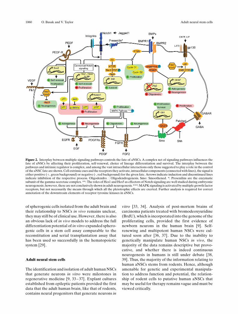
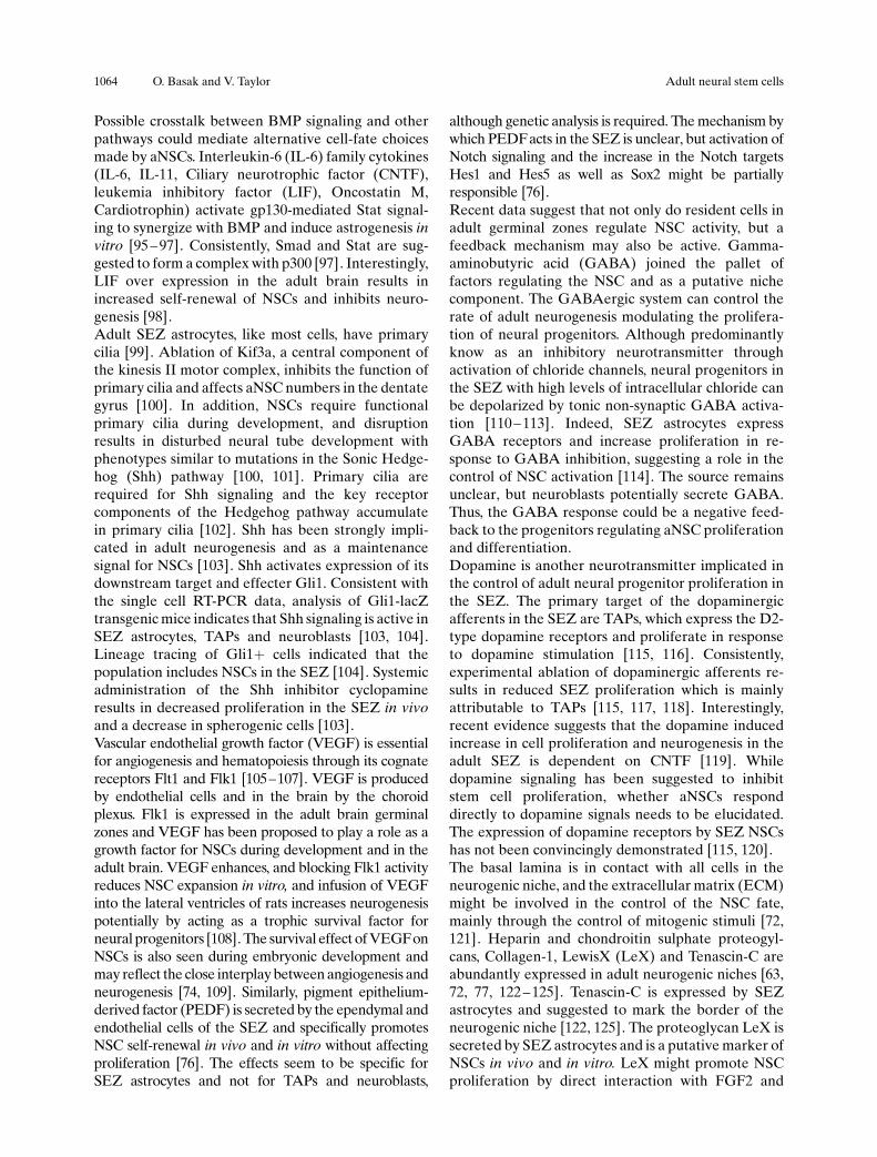
Figure 2. Interplay between multiple signaling pathways controls the fate of aNSCs. A complex net of signaling pathways influences thefate of aNSCs by affecting their proliferation, self-renewal, choice of lineage differentiation and survival. The interplay between thepathways and intrinsic regulator is complex, and among the vast intracellular interactions only those suggested to play a role in the controlof the aNSC fate are shown. Cell extrinsic cues and the receptors they activate, intracellular components (connected with lines), the signal iseither positive (+, green background) or negative (-, red background) for the given fate. Arrows indicate induction and discontinued linesindicate inhibition of the respective process. Oligodendro. : Oligodendrogenesis. Smo: Smoothened. *: Presenilins are the enzymaticsubunit of the gamma-secretase complex. **: The roles of Hes1 and Hes5 as effectors of Notch signaling are well studied during embryonicneurogenesis; however, these are not conclusively shown in adult neurogenesis. ***: MAPK signaling is activated by multiple growth factorreceptors, but not necessarily the means through which all the pleiotrophic effects are exerted. Further analysis is required for correctannotation of the downstream elements of receptor tyrosine kinases in aNSCs.
1060 O. Basak and V. Taylor Adult neural stem cells
Astrocytes as stem cells
The prevailing evidence accumulated since the mid-1990s indicates that aNSCs in rodents belong toastrocytic lineages. SEZ astrocytes and some neuro-genic NSCs are labeled in transgenic mice expressing
reporter proteins from regulatory elements of thehuman intermediate filament Glial Fibrillary AcidicProtein (gfap) gene [16, 40]. In addition, SEZ GFAP+cells are quiescent and many survive anti-mitotic drugtreatment. They rapidly enter the cell cycle uponwithdrawal of the anti-mitotic drug supporting their
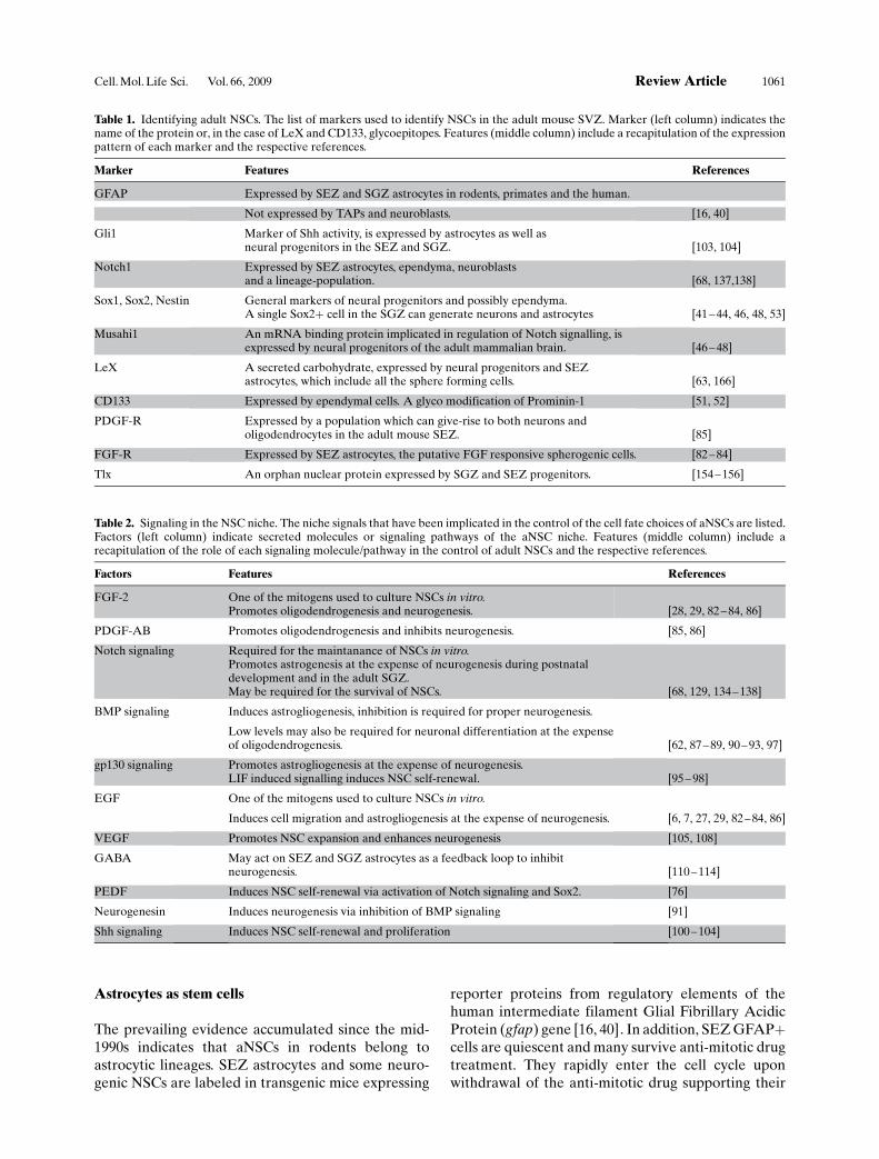
Table 1. Identifying adult NSCs. The list of markers used to identify NSCs in the adult mouse SVZ. Marker (left column) indicates thename of the protein or, in the case of LeX and CD133, glycoepitopes. Features (middle column) include a recapitulation of the expressionpattern of each marker and the respective references.
Marker Features References
GFAP Expressed by SEZ and SGZ astrocytes in rodents, primates and the human.
Not expressed by TAPs and neuroblasts. [16, 40]
Gli1 Marker of Shh activity, is expressed by astrocytes as well asneural progenitors in the SEZ and SGZ. [103, 104]
Notch1 Expressed by SEZ astrocytes, ependyma, neuroblastsand a lineage-population. [68, 137,138]
Sox1, Sox2, Nestin General markers of neural progenitors and possibly ependyma.A single Sox2+ cell in the SGZ can generate neurons and astrocytes [41–44, 46, 48, 53]
Musahi1 An mRNA binding protein implicated in regulation of Notch signalling, isexpressed by neural progenitors of the adult mammalian brain. [46–48]
LeX A secreted carbohydrate, expressed by neural progenitors and SEZastrocytes, which include all the sphere forming cells. [63, 166]
CD133 Expressed by ependymal cells. A glyco modification of Prominin-1 [51, 52]
PDGF-R Expressed by a population which can give-rise to both neurons andoligodendrocytes in the adult mouse SEZ. [85]
FGF-R Expressed by SEZ astrocytes, the putative FGF responsive spherogenic cells. [82–84]
Tlx An orphan nuclear protein expressed by SGZ and SEZ progenitors. [154–156]
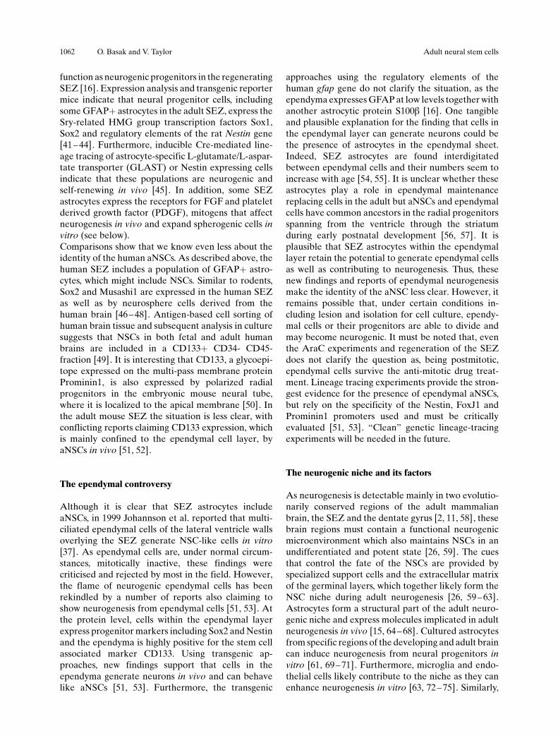
Table 2. Signaling in the NSC niche. The niche signals that have been implicated in the control of the cell fate choices of aNSCs are listed.Factors (left column) indicate secreted molecules or signaling pathways of the aNSC niche. Features (middle column) include arecapitulation of the role of each signaling molecule/pathway in the control of adult NSCs and the respective references.
Factors Features References
FGF-2 One of the mitogens used to culture NSCs in vitro.Promotes oligodendrogenesis and neurogenesis. [28, 29, 82 –84, 86]
PDGF-AB Promotes oligodendrogenesis and inhibits neurogenesis. [85, 86]
Notch signaling Required for the maintanance of NSCs in vitro.Promotes astrogenesis at the expense of neurogenesis during postnataldevelopment and in the adult SGZ.May be required for the survival of NSCs. [68, 129, 134–138]
BMP signaling Induces astrogliogenesis, inhibition is required for proper neurogenesis.
Low levels may also be required for neuronal differentiation at the expenseof oligodendrogenesis. [62, 87–89, 90–93, 97]
gp130 signaling Promotes astrogliogenesis at the expense of neurogenesis.LIF induced signalling induces NSC self-renewal. [95 –98]
EGF One of the mitogens used to culture NSCs in vitro.
Induces cell migration and astrogliogenesis at the expense of neurogenesis. [6, 7, 27, 29, 82–84, 86]
VEGF Promotes NSC expansion and enhances neurogenesis [105, 108]
GABA May act on SEZ and SGZ astrocytes as a feedback loop to inhibitneurogenesis. [110–114]
PEDF Induces NSC self-renewal via activation of Notch signaling and Sox2. [76]
Neurogenesin Induces neurogenesis via inhibition of BMP signaling [91]
Shh signaling Induces NSC self-renewal and proliferation [100–104]
Cell. Mol. Life Sci. Vol. 66, 2009 Review Article 1061
function as neurogenic progenitors in the regeneratingSEZ [16]. Expression analysis and transgenic reportermice indicate that neural progenitor cells, includingsome GFAP+ astrocytes in the adult SEZ, express theSry-related HMG group transcription factors Sox1,Sox2 and regulatory elements of the rat Nestin gene[41 – 44]. Furthermore, inducible Cre-mediated line-age tracing of astrocyte-specific L-glutamate/L-aspar-tate transporter (GLAST) or Nestin expressing cellsindicate that these populations are neurogenic andself-renewing in vivo [45]. In addition, some SEZastrocytes express the receptors for FGF and plateletderived growth factor (PDGF), mitogens that affectneurogenesis in vivo and expand spherogenic cells invitro (see below).Comparisons show that we know even less about theidentity of the human aNSCs. As described above, thehuman SEZ includes a population of GFAP+ astro-cytes, which might include NSCs. Similar to rodents,Sox2 and Musashi1 are expressed in the human SEZas well as by neurosphere cells derived from thehuman brain [46 –48]. Antigen-based cell sorting ofhuman brain tissue and subsequent analysis in culturesuggests that NSCs in both fetal and adult humanbrains are included in a CD133+ CD34- CD45-fraction [49]. It is interesting that CD133, a glycoepi-tope expressed on the multi-pass membrane proteinProminin1, is also expressed by polarized radialprogenitors in the embryonic mouse neural tube,where it is localized to the apical membrane [50]. Inthe adult mouse SEZ the situation is less clear, withconflicting reports claiming CD133 expression, whichis mainly confined to the ependymal cell layer, byaNSCs in vivo [51, 52].
The ependymal controversy
Although it is clear that SEZ astrocytes includeaNSCs, in 1999 Johannson et al. reported that multi-ciliated ependymal cells of the lateral ventricle wallsoverlying the SEZ generate NSC-like cells in vitro[37]. As ependymal cells are, under normal circum-stances, mitotically inactive, these findings werecriticised and rejected by most in the field. However,the flame of neurogenic ependymal cells has beenrekindled by a number of reports also claiming toshow neurogenesis from ependymal cells [51, 53]. Atthe protein level, cells within the ependymal layerexpress progenitor markers including Sox2 and Nestinand the ependyma is highly positive for the stem cellassociated marker CD133. Using transgenic ap-proaches, new findings support that cells in theependyma generate neurons in vivo and can behavelike aNSCs [51, 53]. Furthermore, the transgenic
approaches using the regulatory elements of thehuman gfap gene do not clarify the situation, as theependyma expresses GFAP at low levels together withanother astrocytic protein S100b [16]. One tangibleand plausible explanation for the finding that cells inthe ependymal layer can generate neurons could bethe presence of astrocytes in the ependymal sheet.Indeed, SEZ astrocytes are found interdigitatedbetween ependymal cells and their numbers seem toincrease with age [54, 55]. It is unclear whether theseastrocytes play a role in ependymal maintenancereplacing cells in the adult but aNSCs and ependymalcells have common ancestors in the radial progenitorsspanning from the ventricle through the striatumduring early postnatal development [56, 57]. It isplausible that SEZ astrocytes within the ependymallayer retain the potential to generate ependymal cellsas well as contributing to neurogenesis. Thus, thesenew findings and reports of ependymal neurogenesismake the identity of the aNSC less clear. However, itremains possible that, under certain conditions in-cluding lesion and isolation for cell culture, ependy-mal cells or their progenitors are able to divide andmay become neurogenic. It must be noted that, eventhe AraC experiments and regeneration of the SEZdoes not clarify the question as, being postmitotic,ependymal cells survive the anti-mitotic drug treat-ment. Lineage tracing experiments provide the stron-gest evidence for the presence of ependymal aNSCs,but rely on the specificity of the Nestin, FoxJ1 andProminin1 promoters used and must be criticallyevaluated [51, 53]. “Clean” genetic lineage-tracingexperiments will be needed in the future.
The neurogenic niche and its factors
As neurogenesis is detectable mainly in two evolutio-narily conserved regions of the adult mammalianbrain, the SEZ and the dentate gyrus [2, 11, 58], thesebrain regions must contain a functional neurogenicmicroenvironment which also maintains NSCs in anundifferentiated and potent state [26, 59]. The cuesthat control the fate of the NSCs are provided byspecialized support cells and the extracellular matrixof the germinal layers, which together likely form theNSC niche during adult neurogenesis [26, 59 – 63].Astrocytes form a structural part of the adult neuro-genic niche and express molecules implicated in adultneurogenesis in vivo [15, 64 – 68]. Cultured astrocytesfrom specific regions of the developing and adult braincan induce neurogenesis from neural progenitors invitro [61, 69 – 71]. Furthermore, microglia and endo-thelial cells likely contribute to the niche as they canenhance neurogenesis in vitro [63, 72 –75]. Similarly,
1062 O. Basak and V. Taylor Adult neural stem cells
ependymal cells express several genes involved in thegeneration of a permissive environment for neuro-genesis and NSC maintenance [62, 68, 76].Several extrinsic factors provided by the niche areimplicated in the control of maintenance and differ-entiation of NSCs as well as their early progeny.Irrespective of their spatial origin, transplanted NSCscan integrate throughout the SEZ and retain neuro-genic potential, suggesting that the niche maintenancesignals are constant [77, 78]. On the other hand, it hasbeen reported that adult NSCs located at distinctpositions in the SEZ have a restricted potential togive-rise-to specific interneuron subtypes in the olfac-tory bulb [78]. The restricted neurogenic potential ofNSCs is independent of environmental cues, suggest-ing that intrinsic factors control their potential togenerate specific neuronal subtypes [78]. The spatialpattern and fate restriction is claimed to be establishedduring embryonic development [79]. In addition, thetype of interneurons generated is dependent on thetemporal origin of the NSC, suggesting that eitherNSCs or the niche could be constantly changing [80,81]. Elucidating the molecular factors that underliethe patterning of NSCs might have important impli-cation for clinical applications that aim to generatespecific types of neurons and might lead to deviationsfrom the original description of a stem cell that cangenerate any cell in the adult brain.
Signaling within the aNSC niche
Classically, EGF and FGF2 have been used to isolateand expand aNSC-like cells. Although their roles invivo remain unclear, intra-ventricular infusion ofthese factors into mice has a pronounced effect onneurogenesis. In contrast to claims that a commonprogenitor responds in vitro to both EGFand FGF2, invivo these factors have very different effects [82, 83].EGF induces a marked proliferation of GFAP+ cellsin the SEZ, which is normally not seen in theneurogenic zone, and their infiltration into the sur-rounding tissue as a hyperplastic mass. This increase inproliferation is associated with a reduction in neuro-genesis, suggesting that EGF-responsive cells, whichgenerate neurons under normal conditions, are stimu-lated by EGF to proliferate and give-rise to astrocytes.In contrast, FGF2-infusion into the lateral ventriclesresults in an expansion of neuroblasts and increasedneurogenesis in the SEZ. In addition, FGF2 inducesmigration of progenitors to the olfactory bulb. Thus,rather than increasing NSC expansion as may havebeen predicted from the neurosphere assay, exoge-nous FGF2 does not result in expansion of SEZastrocytes in vivo in the way EGF does. The effect of
FGF in vivo could be interpreted as an increase inneuronal differentiation. A similar effect has beenseen following FGF2 treatment of embryo-derivedNSCs, which proliferate in response to high levels ofFGF2 in vitro but undergo neurogenesis when ex-posed to reduced FGF2 concentrations [82 – 84].PDGF has also been shown to be a mitogen forspherogenic NSCs isolated from the adult mousebrain, however, only in combination with FGF2 andnot EGF [85]. PDGF-receptors on SEZ cells arephosphorylated in vivo, suggesting that signaling isactive in the adult neurogenic forebrain. In contrast toFGF, but similar to EGF, PDGF infusion into thelateral ventricles of adult mice results in activation ofSEZ astrocytes, hyperplasias and gliogenesis at theexpense of neurogenesis [85, 86]. Due to the effects ofPDGFs to reduce neurogenesis, neurogenic NSCsmust respond to activation of the pathway, althoughan indirect mechanism cannot be ruled out.Bone morphogenic protein (BMP) signaling has beenshown to affect neurogenesis both in vitro and in vivoby increasing astrocyte formation at the expense ofneurogenesis [62, 87 – 89]. Over expression of BMP7in the adult brain results in decreased proliferationand a reduction in neuroblast production, whileNoggin, a soluble antagonist of BMP signaling ex-pressed by the ependymal cells of the adult SEZ,promotes neurogenesis [62, 89].BMPs act as morphogens through activation of thetranscriptional regulators Smads but can also activatephosphatase and tensin homologue (PTEN) to inhibitcell cycle progression of neural progenitors [90].Neurogenesin1, a factor secreted by the astrocytes,can promote neurogenesis through the inhibition ofBMP signaling [91]. Although these gain-of-functionexperiments indicate that BMP counteracts neuro-genesis, constitutive activation of BMP-receptor1adoes not affect neuronal differentiation [87, 92]. Incontrast to BMPs reported role in blocking neuro-genesis and activating gliogenesis in the brain, geneticloss of Smad4 function revealed a role in suppressingoligodendrogenesis through down-regulation of thetranscriptional regulator Olig2 [93]. Smad4 is a centralcomponent of the BMP pathway and conditionalknockout mice surprisingly showed reduced neuro-genesis in the SEZ [93]. These seemingly contra-dictory results remain to be clarified but dose-depend-ent effects of BMPs could be at work in the differentparadigms. It should be noted that Smad4 is also adownstream component of the tumor growth factor-beta (TGF-beta) signaling, which promotes cell cyclearrest in various tissues [94]. Conditional inactivationof TGF-beta receptor-2, however, did not result inneurogenic defects [93].
Cell. Mol. Life Sci. Vol. 66, 2009 Review Article 1063
Possible crosstalk between BMP signaling and otherpathways could mediate alternative cell-fate choicesmade by aNSCs. Interleukin-6 (IL-6) family cytokines(IL-6, IL-11, Ciliary neurotrophic factor (CNTF),leukemia inhibitory factor (LIF), Oncostatin M,Cardiotrophin) activate gp130-mediated Stat signal-ing to synergize with BMP and induce astrogenesis invitro [95 – 97]. Consistently, Smad and Stat are sug-gested to form a complex with p300 [97]. Interestingly,LIF over expression in the adult brain results inincreased self-renewal of NSCs and inhibits neuro-genesis [98].Adult SEZ astrocytes, like most cells, have primarycilia [99]. Ablation of Kif3a, a central component ofthe kinesis II motor complex, inhibits the function ofprimary cilia and affects aNSC numbers in the dentategyrus [100]. In addition, NSCs require functionalprimary cilia during development, and disruptionresults in disturbed neural tube development withphenotypes similar to mutations in the Sonic Hedge-hog (Shh) pathway [100, 101]. Primary cilia arerequired for Shh signaling and the key receptorcomponents of the Hedgehog pathway accumulatein primary cilia [102]. Shh has been strongly impli-cated in adult neurogenesis and as a maintenancesignal for NSCs [103]. Shh activates expression of itsdownstream target and effecter Gli1. Consistent withthe single cell RT-PCR data, analysis of Gli1-lacZtransgenic mice indicates that Shh signaling is active inSEZ astrocytes, TAPs and neuroblasts [103, 104].Lineage tracing of Gli1+ cells indicated that thepopulation includes NSCs in the SEZ [104]. Systemicadministration of the Shh inhibitor cyclopamineresults in decreased proliferation in the SEZ in vivoand a decrease in spherogenic cells [103].Vascular endothelial growth factor (VEGF) is essentialfor angiogenesis and hematopoiesis through its cognatereceptors Flt1 and Flk1 [105–107]. VEGF is producedby endothelial cells and in the brain by the choroidplexus. Flk1 is expressed in the adult brain germinalzones and VEGF has been proposed to play a role as agrowth factor for NSCs during development and in theadult brain. VEGF enhances, and blocking Flk1 activityreduces NSC expansion in vitro, and infusion of VEGFinto the lateral ventricles of rats increases neurogenesispotentially by acting as a trophic survival factor forneural progenitors [108]. The survival effect of VEGFonNSCs is also seen during embryonic development andmay reflect the close interplay between angiogenesis andneurogenesis [74, 109]. Similarly, pigment epithelium-derived factor (PEDF) is secreted by the ependymal andendothelial cells of the SEZ and specifically promotesNSC self-renewal in vivo and in vitro without affectingproliferation [76]. The effects seem to be specific forSEZ astrocytes and not for TAPs and neuroblasts,
although genetic analysis is required. The mechanism bywhich PEDFacts in the SEZ is unclear, but activation ofNotch signaling and the increase in the Notch targetsHes1 and Hes5 as well as Sox2 might be partiallyresponsible [76].Recent data suggest that not only do resident cells inadult germinal zones regulate NSC activity, but afeedback mechanism may also be active. Gamma-aminobutyric acid (GABA) joined the pallet offactors regulating the NSC and as a putative nichecomponent. The GABAergic system can control therate of adult neurogenesis modulating the prolifera-tion of neural progenitors. Although predominantlyknow as an inhibitory neurotransmitter throughactivation of chloride channels, neural progenitors inthe SEZ with high levels of intracellular chloride canbe depolarized by tonic non-synaptic GABA activa-tion [110 – 113]. Indeed, SEZ astrocytes expressGABA receptors and increase proliferation in re-sponse to GABA inhibition, suggesting a role in thecontrol of NSC activation [114]. The source remainsunclear, but neuroblasts potentially secrete GABA.Thus, the GABA response could be a negative feed-back to the progenitors regulating aNSC proliferationand differentiation.Dopamine is another neurotransmitter implicated inthe control of adult neural progenitor proliferation inthe SEZ. The primary target of the dopaminergicafferents in the SEZ are TAPs, which express the D2-type dopamine receptors and proliferate in responseto dopamine stimulation [115, 116]. Consistently,experimental ablation of dopaminergic afferents re-sults in reduced SEZ proliferation which is mainlyattributable to TAPs [115, 117, 118]. Interestingly,recent evidence suggests that the dopamine inducedincrease in cell proliferation and neurogenesis in theadult SEZ is dependent on CNTF [119]. Whiledopamine signaling has been suggested to inhibitstem cell proliferation, whether aNSCs responddirectly to dopamine signals needs to be elucidated.The expression of dopamine receptors by SEZ NSCshas not been convincingly demonstrated [115, 120].The basal lamina is in contact with all cells in theneurogenic niche, and the extracellular matrix (ECM)might be involved in the control of the NSC fate,mainly through the control of mitogenic stimuli [72,121]. Heparin and chondroitin sulphate proteogyl-cans, Collagen-1, LewisX (LeX) and Tenascin-C areabundantly expressed in adult neurogenic niches [63,72, 77, 122 – 125]. Tenascin-C is expressed by SEZastrocytes and suggested to mark the border of theneurogenic niche [122, 125]. The proteoglycan LeX issecreted by SEZ astrocytes and is a putative marker ofNSCs in vivo and in vitro. LeX might promote NSCproliferation by direct interaction with FGF2 and
1064 O. Basak and V. Taylor Adult neural stem cells
Wnt1 to present the mitogens to neural progenitors[63,126]. Indeed, several components of the Wntsignaling pathway are expressed in the SEZ and SGZof the adult mice [127, 128]. Although the role of thecanonical Wnt signaling in the adult SEZ is unknown,Wnt1-mediated b-catenin signaling is implicated inthe induction of neurogenesis in the SGZ. WhetherNSCs directly respond to Wnt signaling is not clear[128]. Furthermore, integrin activity regulates neuro-genesis at multiple levels, from synergizing with Notchin NSCs and regulating proliferation and differentia-tion [129], to chain migration of neuroblasts in theRMS [123]. The interplay between the basementmembrane, ECM and growth factors in the niche mayplay an important role in fine-tuning neurogenesis.
Notch and lateral signaling during adult neurogenesis
Although the role of Notch signaling to block neuro-genesis and maintain undifferentiated progenitorsduring development has been addressed, the role ofNotch signaling in adult neurogenesis remains un-known. Notch signaling requires a complex proteo-lytic activation process, the final and critical step ofwhich involves a Presenilin (PS) containing gamma-secretase [130]. PS1-mutant mice die in utero with aphenotype similar to that observed for Notch signalingmutants [131 –133]. However, PS1+/- mice survive toadulthood but show a decreased neurogenesis in theSEZ and a reduction in spherogenic cells [134, 135].Similarly, pharmacological inhibition of PS-activityresults in reduced proliferation and increased celldeath in SEZ-derived progenitors [136]. However, thecell autonomous role of PS in NSCs has not beendemonstrated convincingly, as a reduction in gamma-secretase activity could lead to developmental defectsor altered niche activity. During development, PS arerequired to maintain proper neurogenesis, similar toNotch [134]. However, PS and gamma-secretaseshave dozens of targets in addition to Notchs, thus itcannot be excluded that the effect of reducing PS onneurogenesis is indeed restricted to its action on Notchsignaling.Components of the Notch signaling pathway includingNotch1, Jagged1 and the downstream target Hes5 areexpressed in the adult SEZ, suggesting that, as in theembryo, Notch may play a role in adult neurogenesis[137, 138]. Conditional inactivation of Notch1 fromadult-derived NSCs results in a complete loss of self-renewal and stem cell character without any apparentcell death or change in lineage potential [68]. Theputative ligand for Notch1 in the adult neurogenicniche is Jagged1, which is also required for NSCmaintenance and can replace mitogens to promote
NSCs self-renewal and maintenance in vitro and invivo [68]. Jagged1 is expressed by ependymal cells andSEZ astrocytes and might provide the maintenancesignal to Notch expressing NSCs. In agreement,Notch1 and Jagged1 double-hemizygous mice showdefects in postnatal neurogenesis [68], and activationof Notch signaling via soluble ligand infusion into theventricles results in enhanced proliferation [136].Despite effects on the spherogenic cell population andproliferation, it is still unresolved whether Notchsignaling acts directly at the level of NSCs. Geneticanalysis of mutant mice is required to address thefunction of Notch signaling and, indeed, which Notchreceptors are involved in adult SEZ neurogenesis.Notch signaling via a non-canonical pathway throughactivation of a Stat3-mTOR pathway in the SEZactivates Shh expression to increase progenitor cellssurvival in a non cell-autonomous way [136]. Consis-tent with this, the pro-survival effects of Notchactivation can be mimicked by expression of activatedStat3 or Hes3 [136].
Intrinsic control of NSCs and neurogenesis
NSCs are a rare population in the adult neurogenicSEZ making up a minor fraction of the proliferatingcells, but they display unique cell cycle features [139].Retroviral birth-dating in adult mice revealed thatputative NSCs divide slowly, with an average cell cycletime of over 15 days, which has been proposed to allowthem to retain their potential throughout life [7].Their progeny, TAPs, divide multiple times with anestimated cell cycle time of 12 hours and are themajority of the dividing cells in the SEZ. The majorprogeny of the TAPs, neuroblasts, retain a similarproliferative capacity but gradually become post-mitotic during migration en route to the olfactorybulb. The differences in cell cycle kinetics have beenexploited to preferentially label specific cell popula-tions using nucleoside analogue administration andmarkers of cell cycle progression.BrdU is a thymidine analogue incorporated into newlyreplicated DNA and is frequently used to labelproliferating cells and their progeny. BrdU is rela-tively short-lived in vivo, being excreted within a fewhours after i.p. administration. Thus, a brief exposureto BrdU, single injection or < 24 hours per os, labelsfast dividing cells in the SEZ, whereas NSCs are rarelylabeled. On the other hand, prolonged exposure(>>24 hours) of animals to BrdU results in labelingof NSCs which, due to their long cell cycle, shouldretain the label over an extended period of time [140,141]. Although label-retaining cells include putativeNSCs, the evidence to date is indirect and the func-
Cell. Mol. Life Sci. Vol. 66, 2009 Review Article 1065
tional data are still lacking [142]. In addition, markersof proliferation such as proliferating cell nuclearantigen (PCNA) and Ki67 are frequently used tolabel cells within the cell cycle; however, theirassociation with NSCs has not been demonstrated,partially due to the lack of markers. Mcm2, anessential component of the origin of replicationcomplex, is expressed by adult neural progenitors aswell as label retaining cells and might play a role inactively cycling NSCs [143]. However, the currenttheories on the NSC are based on a single populationof cells that are continuously in the cell cycle and havehomogenous kinetics. Although this simplifies theinterpretation of labeling experiments, in reality NSCsmay be a dynamic cell population that can leave andenter the cell cycle and respond to local environmentalcues. For instance, a mitotically active aNSC couldundergo a few rounds of division before falling into amitotically inactive state, while an inactive aNSCcould contribute to brain regeneration by transitinginto an active state. In addition, it is slowly becomingclear that the SEZ contains multiple populations ofNSCs and it needs to be clarified whether thesedifferent populations may have distinct cell cyclecharacteristics [78]. It remains a major questionwhether all NSCs in the adult brain are active andcontribute to neurogenesis throughout life or whethersome become exhausted and are replaced or replen-ished by a potentially mitotically inactive NSCpopulation.Genetic analysis has revealed roles for specific genesinvolved in the intrinsic regulation of the cell cycle ofadult neural progenitor populations, including NSCs.However, caution is needed in evaluating the dataconcerning the cell cycle of aNSCs due to the absenceof definitive markers and in vivo assays. Cyclin genespromote progression through the cell cycle in coop-eration with the cyclin-dependent kinases (CDKs), anactivity regulated by CDK inhibitors (CDKIs). In theabsence of the G1-specific cyclin D2, but not cyclin D1,proliferation ceases and neurogenesis in the SEZ fails,indicating that neural progenitors are either notestablished or fail to be replenished in cyclin D2
mutants [144].On the other hand, the phosphatase PTEN decreaseslevels of cyclin D1 through inhibition of AKT andmight contribute to the long cell cycle time of adultSEZ NSCs by restricting G1-S transition [145 – 147].In parallel, retinoblastoma gene family members (Rb,p107, p130) are well-known for their role in inhibitingcell cycle progression by binding to E2F familymembers [148]. In the absence of p107, mice displayan increase in label-retaining cells in the SEZ, whichcorrelates with an elevation in the number of sphero-genic cells [149]. This strongly suggests an increase in
aNSC in p107 mutant mice. Roles of Rb and p130 inadult neurogenesis remain elusive, while duringembryogenesis Rb is up-regulated upon initiation ofdifferentiation and p130 is expressed in most-mitoticneurons [150,151]. In addition, Rb mutant micedisplay a general deficit in the embryonic progenitorcell pool with no apparent NSC phenotype [149].Thus, Rb family members seem to have distinct, non-redundant functions in the regulation of adult neuro-genesis.In support of a connection between the cell cycleprogression and regulation of progenitor cell fatechoices, mutants of several cyclin dependent kinaseinhibitors (CDKI) display striking defects in adultneurogenesis. Evidence suggests that p21cip1/waf1 pro-motes a more quiescent state and, in its absence, NSCsinitially expand beyond wild type levels but areeventually exhausted [141]. An up-stream factor ofp21cip1/waf1, the tumor suppressor protein p53, is alsoimplicated in promoting quiescence of adult NSCs[152]. In addition, genetic data support a role ofp27Kip1 in the inhibition of neural progenitor prolifer-ation, although the phenotype can be attributed atleast partially to an effect on TAPs [153]. Proliferationof SGZ and SEZ progenitors requires the orphannuclear receptor Tlx, which is implicated in theinhibition of p21cip1/waf1 but not p27 Kip1, furtherconfirming differential functions of specific cell cycleregulators [154 – 156]. Tlx functions, at least partially,by recruiting histone deacetylases, stressing the im-portance of epigenetic regulation in the control ofNSC maintenance [157]. In agreement, the polycombfamily repressor Bmi1 is required for postnatal main-tenance of NSCs [158]. Once again connecting cellcycle regulation to other cell fate regulators, increasedlevels of the tumor suppressor p16Ink4a in Bmi1-deficient mice lead to reduced self-renewal anddepletion of NSCs early in postnatal life, mimickingits function during ageing [159, 160]. Multiple path-ways converge on the regulators of the cell cycleprogression which might represent key switchesgoverning NSC fate choices.
Crosstalk between cell cycle components andextracellular signals
It is tempting to speculate that specific molecularcomponents that control cell cycle progression inneural progenitors might be directly associated withthe factors that govern NSC self-renewal, longevityand differentiation potential. This is elaborated inneural progenitors, where p107 binds to the promoterand elevates the expression of Notch1 resulting inincreased Notch activity in adult derived neuro-
1066 O. Basak and V. Taylor Adult neural stem cells
spheres in vitro [149]. Activation of Notch signaling inthe adult spinal cord in response to injury inducesproliferation linking environmental signals to cellcycle progression [161]. In muscle stem cells (MSCs),Notch signaling is also implicated in the control ofCDKI expression. It inhibits the expression of p15Ink4b,p16Ink4a, p21cip1/waf1 and p27 Kip1 induced by the inhib-ition of TGFb-dependent Smad3 activity [162]. Incontrast to embryonic neurogenesis, decreased levelsof Notch signaling in MSCs of aged mice leads to a lossof proliferative and regenerative capacity rather thanprecocious differentiation [163]. In a reciprocal path-way, p16Ink4a has been implicated in the inhibition ofHes1, a target of Notch signaling [164]. It will beinteresting to further investigate the role of Notchsignaling and other pathways in the regulation of theNSC cell cycle.
Conclusions and perspectives
NSCs persist in the adult mammalian brain throughoutlife and generate functional neurons. The proliferationand differentiation of the neural progenitors is gov-erned by multiple signaling mechanisms provided bythe specialized microenvironment of the niche. Thestriking regenerative capacity of NSCs raises hopes fortherapy for multiple degenerative disorders includingstroke, Parkinson�s, Huntington�s and Alzheimer�sdisease. Correct generation and manipulation of spe-cific neuronal subtypes require extensive understand-ing of the identity of different neural progenitorpopulations and the signals that govern their fate.Although the presence of NSCs is accepted, whether asingle population of aNSCs exist in vivo, or multiplefunctionally distinct populations constitute the endog-enous pool remains to be substantiated. It is, however,of critical importance to elucidate the lineage potentialof NSCs using in vivo experimental paradigms.
1 Guillemot, F. (2007) Cell fate specification in the mammaliantelencephalon. Prog. Neurobiol. 83, 37–52.
2 Altman, J. (1962) Are new neurons formed in the brains ofadult mammals? Science 135, 1127–1128.
3 Altman, J. and Das, G. D. (1965) Autoradiographic andhistological evidence of postnatal hippocampal neurogenesisin rats. J. Comp. Neurol. 124, 319–335.
4 Goldman, S. A. and Nottebohm, F. (1983) Neuronal produc-tion, migration, and differentiation in a vocal control nucleusof the adult female canary brain. Proc. Natl. Acad. Sci. USA80, 2390–2394.
5 Nottebohm, F., Alvarez-Buylla, A., Cynx, J., Kirn, J., Ling, C.Y., Nottebohm, M., Suter, R., Tolles, A. and Williams, H.(1990) Song learning in birds: the relation between perceptionand production. Philos. Trans. R. Soc. Lond. B. Biol. Sci. 329,115–124.
6 Reynolds, B. A. and Weiss, S. (1992) Generation of neuronsand astrocytes from isolated cells of the adult mammaliancentral nervous system. Science 255, 1707–1710.
7 Morshead, C. M., Reynolds, B. A., Craig, C. G., McBurney, M.W., Staines, W. A., Morassutti, D., Weiss, S. and van der Kooy,D. (1994) Neural stem cells in the adult mammalian forebrain:a relatively quiescent subpopulation of subependymal cells.Neuron 13, 1071–1082.
8 Bernier, P. J., Bedard, A., Vinet, J., Levesque, M. and Parent,A. (2002) Newly generated neurons in the amygdala andadjoining cortex of adult primates. Proc. Natl. Acad. Sci. USA99, 11464–11469.
9 Eriksson, P. S., Perfilieva, E., Bjork-Eriksson, T., Alborn, A.M., Nordborg, C., Peterson, D. A. and Gage, F. H. (1998)Neurogenesis in the adult human hippocampus. Nat. Med. 4,1313–1317.
10 Kilpatrick, T. J. and Bartlett, P. F. (1993) Cloning and growthof multipotential neural precursors: requirements for prolif-eration and differentiation. Neuron 10, 255–265.
11 Luskin, M. B. (1993) Restricted proliferation and migration ofpostnatally generated neurons derived from the forebrainsubventricular zone. Neuron 11, 173–189.
12 Menn, B., Garcia-Verdugo, J. M., Yaschine, C., Gonzalez-Perez, O., Rowitch, D. and Alvarez-Buylla, A. (2006) Originof oligodendrocytes in the subventricular zone of the adultbrain. J. Neurosci. 26, 7907–7918.
13 Lois, C. and Alvarez-Buylla, A. (1994) Long-distance neuro-nal migration in the adult mammalian brain. Science 264,1145–1148.
14 Biebl, M., Cooper, C. M., Winkler, J. and Kuhn, H. G. (2000)Analysis of neurogenesis and programmed cell death revealsa self-renewing capacity in the adult rat brain. Neurosci.Lett. 291, 17–20.
15 Doetsch, F., Garcia-Verdugo, J. M. and Alvarez-Buylla, A.(1997) Cellular composition and three-dimensional organi-zation of the subventricular germinal zone in the adultmammalian brain. J. Neurosci. 17, 5046–5061.
16 Doetsch, F., Caille, I., Lim, D. A., Garcia-Verdugo, J. M. andAlvarez-Buylla, A. (1999) Subventricular zone astrocytes areneural stem cells in the adult mammalian brain. Cell 97, 703–716.
17 Sawamoto, K., Wichterle, H., Gonzalez-Perez, O., Cholfin, J.A., Yamada, M., Spassky, N., Murcia, N. S., Garcia-Verdugo,J. M., Marin, O., Rubenstein, J. L., Tessier-Lavigne, M.,Okano, H. and Alvarez-Buylla, A. (2006) New neurons followthe flow of cerebrospinal fluid in the adult brain. Science 311,629–632.
18 Sanai, N., Tramontin, A. D., Quinones-Hinojosa, A., Bar-baro, N. M., Gupta, N., Kunwar, S., Lawton, M. T.,McDermott, M. W., Parsa, A. T., Manuel-Garcia Verdugo,J., Berger, M. S. and Alvarez-Buylla, A. (2004) Uniqueastrocyte ribbon in adult human brain contains neural stemcells but lacks chain migration. Nature 427, 740–744.
19 Rodriguez-Perez, L. M., Perez-Martin, M., Jimenez, A. J. andFernandez-Llebrez, P. (2003) Immunocytochemical charac-terisation of the wall of the bovine lateral ventricle. CellTissue Res. 314, 325–335.
20 Quinones-Hinojosa, A., Sanai, N., Soriano-Navarro, M.,Gonzalez-Perez, O., Mirzadeh, Z., Gil-Perotin, S., Romero-Rodriguez, R., Berger, M. S., Garcia-Verdugo, J. M. andAlvarez-Buylla, A. (2006) Cellular composition and cytoarch-itecture of the adult human subventricular zone: a niche ofneural stem cells. J. Comp. Neurol. 494, 415–434.
21 Nunes, M. C., Roy, N. S., Keyoung, H. M., Goodman, R. R.,McKhann, G., 2nd, Jiang, L., Kang, J., Nedergaard, M. andGoldman, S. A. (2003) Identification and isolation of multi-potential neural progenitor cells from the subcortical whitematter of the adult human brain. Nat. Med. 9, 439–447.
22 Curtis, M. A., Kam, M., Nannmark, U., Anderson, M. F.,Axell, M. Z., Wikkelso, C., Holtas, S., van Roon-Mom, W. M.,Bjork-Eriksson, T., Nordborg, C., Frisen, J., Dragunow, M.,Faull, R. L. and Eriksson, P. S. (2007) Human neuroblastsmigrate to the olfactory bulb via a lateral ventricularextension. Science 315, 1243–1249.
Cell. Mol. Life Sci. Vol. 66, 2009 Review Article 1067
23 Seri, B., Garcia-Verdugo, J. M., McEwen, B. S. and Alvarez-Buylla, A. (2001) Astrocytes give rise to new neurons in theadult mammalian hippocampus. J. Neurosci. 21, 7153–7160.
24 Kempermann, G., Jessberger, S., Steiner, B. and Kronenberg,G. (2004) Milestones of neuronal development in the adulthippocampus. Trends Neurosci. 27, 447–452.
25 Suh, H., Consiglio, A., Ray, J., Sawai, T., D�Amour, K. A. andGage, F. H. (2007) In Vivo Fate Analysis Reveals theMultipotent and Self-Renewal Capacities of Sox2(+) NeuralStem Cells in the Adult Hippocampus. Cell Stem Cell 1, 515–528.
26 Gage, F. H. (2000) Mammalian neural stem cells. Science 287,1433–1438.
27 Reynolds, B. A., Tetzlaff, W. and Weiss, S. (1992) A multi-potent EGF-responsive striatal embryonic progenitor cellproduces neurons and astrocytes. J. Neurosci. 12, 4565–4574.
28 Gritti, A., Parati, E. A., Cova, L., Frolichsthal, P., Galli, R.,Wanke, E., Faravelli, L., Morassutti, D. J., Roisen, F., Nickel,D. D. and Vescovi, A. L. (1996) Multipotential stem cells fromthe adult mouse brain proliferate and self-renew in responseto basic fibroblast growth factor. J. Neurosci. 16, 1091–1100.
29 Reynolds, B. A. and Rietze, R. L. (2005) Neural stem cells andneurospheres–re-evaluating the relationship. Nat. Methods 2,333–336.
30 Seaberg, R. M. and van der Kooy, D. (2002) Adult rodentneurogenic regions: the ventricular subependyma containsneural stem cells, but the dentate gyrus contains restrictedprogenitors. J. Neurosci. 22, 1784–1793.
31 Doetsch, F., Petreanu, L., Caille, I., Garcia-Verdugo, J. M.and Alvarez-Buylla, A. (2002) EGF converts transit-amplify-ing neurogenic precursors in the adult brain into multipotentstem cells. Neuron 36, 1021–1034.
32 Galli, R., Gritti, A., Bonfanti, L. and Vescovi, A. L. (2003)Neural stem cells: an overview. Circ. Res. 92, 598–608.
33 Kirschenbaum, B., Nedergaard, M., Preuss, A., Barami, K.,Fraser, R. A. and Goldman, S. A. (1994) In vitro neuronalproduction and differentiation by precursor cells derivedfrom the adult human forebrain. Cereb. Cortex 4, 576–589.
34 Pincus, D. W., Harrison-Restelli, C., Barry, J., Goodman, R.R., Fraser, R. A., Nedergaard, M. and Goldman, S. A. (1997)In vitro neurogenesis by adult human epileptic temporalneocortex. Clin. Neurosurg. 44, 17 –25.
35 Kornack, D. R. and Rakic, P. (1999) Continuation of neuro-genesis in the hippocampus of the adult macaque monkey.Proc. Natl. Acad. Sci. USA 96, 5768–5773.
36 Kukekov, V. G., Laywell, E. D., Suslov, O., Davies, K.,Scheffler, B., Thomas, L. B., O�Brien, T. F., Kusakabe, M. andSteindler, D. A. (1999) Multipotent stem/progenitor cells withsimilar properties arise from two neurogenic regions of adulthuman brain. Exp. Neurol. 156, 333–344.
37 Johansson, C. B., Momma, S., Clarke, D.L., Risling, M.,Lendahl, U. and Frisen, J. (1999) Identification of a neuralstem cell in the adult mammalian central nervous system. Cell96, 25–34.
38 Kornack, D. R. and Rakic, P. (2001) Cell proliferation withoutneurogenesis in adult primate neocortex. Science 294, 2127–2130.
39 Seress, L., Abraham, H., Tornoczky, T. and Kosztolanyi, G.(2001) Cell formation in the human hippocampal formationfrom mid-gestation to the late postnatal period. Neuroscience105, 831–843.
40 Morshead, C. M., Garcia, A. D., Sofroniew, M. V. and van DerKooy, D. (2003) The ablation of glial fibrillary acidic protein-positive cells from the adult central nervous system results inthe loss of forebrain neural stem cells but not retinal stemcells. Eur. J. Neurosci. 18, 76–84.
41 Brazel, C. Y., Limke, T. L., Osborne, J. K., Miura, T., Cai, J.,Pevny, L. and Rao, M. S. (2005) Sox2 expression defines aheterogeneous population of neurosphere-forming cells in theadult murine brain. Aging Cell 4, 197–207.
42 Ellis, P., Fagan, B. M., Magness, S. T., Hutton, S., Taranova,O., Hayashi, S., McMahon, A., Rao, M. and Pevny, L. (2004)
SOX2, a persistent marker for multipotential neural stem cellsderived from embryonic stem cells, the embryo or the adult.Dev. Neurosci. 26, 148–165.
43 Barraud, P., Thompson, L., Kirik, D., Bjorklund, A. andParmar, M. (2005) Isolation and characterization of neuralprecursor cells from the Sox1-GFP reporter mouse. Eur. J.Neurosci. 22, 1555–1569.
44 Kawaguchi, A., Miyata, T., Sawamoto, K., Takashita, N.,Murayama, A., Akamatsu, W., Ogawa, M., Okabe, M., Tano,Y., Goldman, S. A. and Okano, H. (2001) Nestin-EGFPtransgenic mice: visualization of the self-renewal and multi-potency of CNS stem cells. Mol. Cell. Neurosci. 17, 259–273.
45 Mori, T., Tanaka, K., Buffo, A., Wurst, W., Kuhn, R. andGotz, M. (2006) Inducible gene deletion in astroglia and radialglia–a valuable tool for functional and lineage analysis. Glia54, 21–34.
46 Schwartz, P. H., Bryant, P. J., Fuja, T. J., Su, H., O�Dowd, D. K.and Klassen, H. (2003) Isolation and characterization ofneural progenitor cells from post-mortem human cortex. J.Neurosci. Res. 74, 838–851.
47 Kaneko, Y., Sakakibara, S., Imai, T., Suzuki, A., Nakamura,Y., Sawamoto, K., Ogawa, Y., Toyama, Y., Miyata, T. andOkano, H. (2000) Musashi1: an evolutionally conservedmarker for CNS progenitor cells including neural stem cells.Dev. Neurosci. 22, 139–153.
48 Pincus, D. W., Keyoung, H. M., Harrison-Restelli, C., Good-man, R. R., Fraser, R. A., Edgar, M., Sakakibara, S., Okano,H., Nedergaard, M. and Goldman, S. A. (1998) Fibroblastgrowth factor-2/brain-derived neurotrophic factor-associatedmaturation of new neurons generated from adult humansubependymal cells. Ann. Neurol. 43, 576–585.
49 Uchida, N., Buck, D. W., He, D., Reitsma, M. J., Masek, M.,Phan, T. V., Tsukamoto, A. S., Gage, F. H. and Weissman, I. L.(2000) Direct isolation of human central nervous system stemcells. Proc. Natl. Acad. Sci. USA 97, 14720–14725.
50 Dubreuil, V., Marzesco, A. M., Corbeil, D., Huttner, W. B.and Wilsch-Brauninger, M. (2007) Midbody and primarycilium of neural progenitors release extracellular membraneparticles enriched in the stem cell marker prominin-1. J. CellBiol. 176, 483–495.
51 Coskun, V., Wu, H., Blanchi, B., Tsao, S., Kim, K., Zhao, J.,Biancotti, J. C., Hutnick, L., Krueger, R. C., Jr., Fan, G., deVellis, J. and Sun, Y. E. (2008) CD133+ neural stem cells in theependyma of mammalian postnatal forebrain. Proc. Natl.Acad. Sci. USA 105, 1026–1031.
52 Pfenninger, C. V., Roschupkina, T., Hertwig, F., Kottwitz, D.,Englund, E., Bengzon, J., Jacobsen, S. E. and Nuber, U. A.(2007) CD133 is not present on neurogenic astrocytes in theadult subventricular zone, but on embryonic neural stem cells,ependymal cells, and glioblastoma cells. Cancer Res. 67,5727–5736.
53 Meletis, K., Barnabe-Heider, F., Carlen, M., Evergren, E.,Tomilin, N., Shupliakov, O. and Frisen, J. (2008) Spinal cordinjury reveals multilineage differentiation of ependymal cells.PLoS Biol. 6, e182.
54 Luo, J., Daniels, S. B., Lennington, J. B., Notti, R. Q. andConover, J. C. (2006) The aging neurogenic subventricularzone. Aging Cell 5, 139–152.
55 Luo, J., Shook, B. A., Daniels, S. B. and Conover, J. C. (2008)Subventricular zone-mediated ependyma repair in the adultmammalian brain. J. Neurosci. 28, 3804–3813.
56 Merkle, F. T., Tramontin, A. D., Garcia-Verdugo, J. M. andAlvarez-Buylla, A. (2004) Radial glia give rise to adult neuralstem cells in the subventricular zone. Proc. Natl. Acad. Sci.USA 101, 17528–17532.
57 Spassky, N., Merkle, F. T., Flames, N., Tramontin, A. D.,Garcia-Verdugo, J. M. and Alvarez-Buylla, A. (2005) Adultependymal cells are postmitotic and are derived from radialglial cells during embryogenesis. J. Neurosci. 25, 10 –18.
58 Kuhn, H. G., Dickinson-Anson, H. and Gage, F. H. (1996)Neurogenesis in the dentate gyrus of the adult rat: age-related
1068 O. Basak and V. Taylor Adult neural stem cells
decrease of neuronal progenitor proliferation. J. Neurosci. 16,2027–2033.
59 Alvarez-Buylla, A. and Lim, D. A. (2004) For the long run:maintaining germinal niches in the adult brain. Neuron 41,683–686.
60 Palmer, T. D., Willhoite, A. R. and Gage, F. H. (2000) Vascularniche for adult hippocampal neurogenesis. J. Comp. Neu-rol. 425, 479–494.
61 Song, H., Stevens, C. F. and Gage, F. H. (2002) Astrogliainduce neurogenesis from adult neural stem cells. Nature 417,39–44.
62 Lim, D. A., Tramontin, A. D., Trevejo, J. M., Herrera, D. G.,Garcia-Verdugo, J. M. and Alvarez-Buylla, A. (2000) Nogginantagonizes BMP signaling to create a niche for adultneurogenesis. Neuron 28, 713–726.
63 Capela, A. and Temple, S. (2002) LeX/ssea-1 is expressed byadult mouse CNS stem cells, identifying them as nonependy-mal. Neuron 35, 865–875.
64 Lafon-Cazal, M., Adjali, O., Galeotti, N., Poncet, J., Jouin, P.,Homburger, V., Bockaert, J. and Marin, P. (2003) Proteomicanalysis of astrocytic secretion in the mouse. Comparison withthe cerebrospinal fluid proteome. J. Biol. Chem. 278, 24438–24448.
65 Seri, B., Garcia-Verdugo, J. M., Collado-Morente, L., McE-wen, B. S. and Alvarez-Buylla, A. (2004) Cell types, lineage,and architecture of the germinal zone in the adult dentategyrus. J. Comp. Neurol. 478, 359–378.
66 Taupin, P., Ray, J., Fischer, W. H., Suhr, S. T., Hakansson, K.,Grubb, A. and Gage, F. H. (2000) FGF-2-responsive neuralstem cell proliferation requires CCg, a novel autocrine/paracrine cofactor. Neuron 28, 385–397.
67 Shetty, A. K., Hattiangady, B. and Shetty, G. A. (2005) Stem/progenitor cell proliferation factors FGF-2, IGF-1, andVEGF exhibit early decline during the course of aging inthe hippocampus: role of astrocytes. Glia 51, 173–186.
68 Nyfeler, Y., Kirch, R. D., Mantei, N., Leone, D. P., Radtke, F.,Suter, U. and Taylor, V. (2005) Jagged1 signals in the postnatalsubventricular zone are required for neural stem cell self-renewal. Embo J. 24, 3504–3515.
69 Lim, D. A. and Alvarez-Buylla, A. (1999) Interaction betweenastrocytes and adult subventricular zone precursors stimu-lates neurogenesis. Proc. Natl. Acad. Sci. USA 96, 7526–7531.
70 Toda, H., Takahashi, J., Mizoguchi, A., Koyano, K. andHashimoto, N. (2000) Neurons generated from adult rathippocampal stem cells form functional glutamatergic andGABAergic synapses in vitro. Exp. Neurol. 165, 66 –76.
71 Song, H. J., Stevens, C. F. and Gage, F. H. (2002) Neural stemcells from adult hippocampus develop essential properties offunctional CNS neurons. Nat. Neurosci. 5, 438–445.
72 Mercier, F., Kitasako, J. T. and Hatton, G. I. (2002) Anatomyof the brain neurogenic zones revisited: fractones and thefibroblast/macrophage network. J. Comp. Neurol. 451, 170–188.
73 Palmer, T. D. (2002) Adult neurogenesis and the vascularNietzsche. Neuron 34, 856–858.
74 Shen, Q., Goderie, S. K., Jin, L., Karanth, N., Sun, Y.,Abramova, N., Vincent, P., Pumiglia, K. and Temple, S. (2004)Endothelial cells stimulate self-renewal and expand neuro-genesis of neural stem cells. Science 304, 1338–1340.
75 Walton, N. M., Sutter, B. M., Laywell, E. D., Levkoff, L. H.,Kearns, S. M., Marshall, G. P., 2nd, Scheffler, B. and Steindler,D.A. (2006) Microglia instruct subventricular zone neuro-genesis. Glia 54, 815–825.
76 Ramirez-Castillejo, C., Sanchez-Sanchez, F., Andreu-Agullo,C., Ferron, S.R., Aroca-Aguilar, J. D., Sanchez, P., Mira, H.,Escribano, J. and Farinas, I. (2006) Pigment epithelium-derived factor is a niche signal for neural stem cell renewal.Nat. Neurosci. 9, 331–339.
77 Jankovski, A. and Sotelo, C. (1996) Subventricular zone-olfactory bulb migratory pathway in the adult mouse: cellularcomposition and specificity as determined by heterochronic
and heterotopic transplantation. J. Comp. Neurol. 371, 376–396.
78 Merkle, F. T., Mirzadeh, Z. and Alvarez-Buylla, A. (2007)Mosaic organization of neural stem cells in the adult brain.Science 317, 381–384.
79 Young, K. M., Fogarty, M., Kessaris, N. and Richardson, W. D.(2007) Subventricular zone stem cells are heterogeneous withrespect to their embryonic origins and neurogenic fates in theadult olfactory bulb. J. Neurosci. 27, 8286–8296.
80 Batista-Brito, R., Close, J., Machold, R. and Fishell, G. (2008)The distinct temporal origins of olfactory bulb interneuronsubtypes. J. Neurosci. 28, 3966–3975.
81 De Marchis, S., Bovetti, S., Carletti, B., Hsieh, Y. C.,Garzotto, D., Peretto, P., Fasolo, A., Puche, A. C. and Rossi,F. (2007) Generation of distinct types of periglomerularolfactory bulb interneurons during development and in adultmice: implication for intrinsic properties of the subventricularzone progenitor population. J. Neurosci. 27, 657–664.
82 Kuhn, H. G., Winkler, J., Kempermann, G., Thal, L. J. andGage, F. H. (1997) Epidermal growth factor and fibroblastgrowth factor-2 have different effects on neural progenitors inthe adult rat brain. J. Neurosci. 17, 5820–5829.
83 Craig, C. G., Tropepe, V., Morshead, C. M., Reynolds, B. A.,Weiss, S. and van der Kooy, D. (1996) In vivo growth factorexpansion of endogenous subependymal neural precursor cellpopulations in the adult mouse brain. J. Neurosci. 16, 2649–2658.
84 Gritti, A., Frolichsthal-Schoeller, P., Galli, R., Parati, E. A.,Cova, L., Pagano, S. F., Bjornson, C. R. and Vescovi, A. L.(1999) Epidermal and fibroblast growth factors behave asmitogenic regulators for a single multipotent stem cell-likepopulation from the subventricular region of the adult mouseforebrain. J. Neurosci. 19, 3287–3297.
85 Jackson, E. L., Garcia-Verdugo, J. M., Gil-Perotin, S., Roy,M., Quinones-Hinojosa, A., VandenBerg, S. and Alvarez-Buylla, A. (2006) PDGFR alpha-positive B cells are neuralstem cells in the adult SVZ that form glioma-like growths inresponse to increased PDGF signaling. Neuron 51, 187–199.
86 Lachapelle, F., Avellana-Adalid, V., Nait-Oumesmar, B. andBaron-Van Evercooren, A. (2002) Fibroblast growth factor-2(FGF-2) and platelet-derived growth factor AB (PDGF AB)promote adult SVZ-derived oligodendrogenesis in vivo. Mol.Cell. Neurosci. 20, 390–403.
87 Gross, R. E., Mehler, M. F., Mabie, P. C., Zang, Z., Santschi,L. and Kessler, J. A. (1996) Bone morphogenetic proteinspromote astroglial lineage commitment by mammalian sub-ventricular zone progenitor cells. Neuron 17, 595–606.
88 Chen, H. L. and Panchision, D. M. (2007) Concise review:bone morphogenetic protein pleiotropism in neural stem cellsand their derivatives–alternative pathways, convergent sig-nals. Stem Cells 25, 63–68.
89 Cho, S. R., Benraiss, A., Chmielnicki, E., Samdani, A.,Economides, A. and Goldman, S. A. (2007) Induction ofneostriatal neurogenesis slows disease progression in a trans-genic murine model of Huntington disease. J. Clin. In-vest. 117, 2889–2902.
90 Mathieu, C., Sii-Felice, K., Fouchet, P., Etienne, O., Haton,C., Mabondzo, A., Boussin, F. D. and Mouthon, M. A. (2008)Endothelial cell-derived bone mo rphogenetic proteins con-trol proliferation of neural stem/progenitor cells. Mol. Cell.Neurosci. 38, 569–577.
91 Ueki, T., Tanaka, M., Yamashita, K., Mikawa, S., Qiu, Z.,Maragakis, N. J., Hevner, R. F., Miura, N., Sugimura, H. andSato, K. (2003) A novel secretory factor, Neurogenesin-1,provides neurogenic environmental cues for neural stem cellsin the adult hippocampus. J. Neurosci. 23, 11732–11740.
92 Coskun, V., Venkatraman, G., Yang, H., Rao, M. S. andLuskin, M. B. (2001) Retroviral manipulation of the expres-sion of bone morphogenetic protein receptor Ia by SVZaprogenitor cells leads to changes in their p19(INK4d)expression but not in their neuronal commitment. Int. J.Dev. Neurosci. 19, 219–227.
Cell. Mol. Life Sci. Vol. 66, 2009 Review Article 1069
93 Colak, D., Mori, T., Brill, M. S., Pfeifer, A., Falk, S., Deng, C.,Monteiro, R., Mummery, C., Sommer, L. and Gotz, M. (2008)Adult neurogenesis requires Smad4-mediated bone morpho-genic protein signaling in stem cells. J. Neurosci. 28, 434–446.
94 Attisano, L. and Wrana, J. L. (2000) Smads as transcriptionalco-modulators. Curr. Opin. Cell Biol. 12, 235–243.
95 Bonni, A., Sun, Y., Nadal-Vicens, M., Bhatt, A., Frank, D. A.,Rozovsky, I., Stahl, N., Yancopoulos, G. D. and Greenberg, M.E. (1997) Regulation of gliogenesis in the central nervoussystem by the JAK-STAT signaling pathway. Science 278,477–483.
96 Johe, K. K., Hazel, T. G., Muller, T., Dugich-Djordjevic, M.M. and McKay, R. D. (1996) Single factors direct thedifferentiation of stem cells from the fetal and adult centralnervous system. Genes Dev. 10, 3129–3140.
97 Nakashima, K., Yanagisawa, M., Arakawa, H., Kimura, N.,Hisatsune, T., Kawabata, M., Miyazono, K. and Taga, T.(1999) Synergistic signaling in fetal brain by STAT3-Smad1complex bridged by p300. Science 284, 479–482.
98 Bauer, S. and Patterson, P. H. (2006) Leukemia inhibitoryfactor promotes neural stem cell self-renewal in the adultbrain. J. Neurosci. 26, 12089–12099.
99 Tramontin, A. D., Garcia-Verdugo, J. M., Lim, D. A. andAlvarez-Buylla, A. (2003) Postnatal development of radialglia and the ventricular zone (VZ): a continuum of the neuralstem cell compartment. Cereb. Cortex 13, 580–587.
100 Han, Y. G., Spassky, N., Romaguera-Ros, M., Garcia-Verdugo, J. M., Aguilar, A., Schneider-Maunoury, S. andAlvarez-Buylla, A. (2008) Hedgehog signaling and primarycilia are required for the formation of adult neural stem cells.Nat. Neurosci. 11, 277–284.
101 Alvarez-Buylla, A., Garcia-Verdugo, J. M. and Tramontin, A.D. (2001) A unified hypothesis on the lineage of neural stemcells. Nat. Rev. Neurosci. 2, 287–293.
102 Kovacs, J. J., Whalen, E. J., Liu, R., Xiao, K., Kim, J., Chen,M., Wang, J., Chen, W. and Lefkowitz, R. J. (2008) Beta-arrestin-mediated localization of smoothened to the primarycilium. Science 320, 1777–1781.
103 Palma, V., Lim, D. A., Dahmane, N., Sanchez, P., Brionne, T.C., Herzberg, C. D., Gitton, Y., Carleton, A., Alvarez-Buylla,A. and Ruiz i Altaba, A. (2005) Sonic hedgehog controls stemcell behavior in the postnatal and adult brain. Development132, 335–344.
104 Ahn, S. and Joyner, A. L. (2005) In vivo analysis of quiescentadult neural stem cells responding to Sonic hedgehog. Nature437, 894–897.
105 Shalaby, F., Rossant, J., Yamaguchi, T. P., Gertsenstein, M.,Wu, X. F., Breitman, M. L. and Schuh, A. C. (1995) Failure ofblood-island formation and vasculogenesis in Flk-1-deficientmice. Nature 376, 62 –66.
106 Shalaby, F., Ho, J., Stanford, W. L., Fischer, K. D., Schuh, A.C., Schwartz, L., Bernstein, A. and Rossant, J. (1997) Arequirement for Flk1 in primitive and definitive hematopoi-esis and vasculogenesis. Cell 89, 981–990.
107 Ema, M., Faloon, P., Zhang, W. J., Hirashima, M., Reid, T.,Stanford, W. L., Orkin, S., Choi, K. and Rossant, J. (2003)Combinatorial effects of Flk1 and Tal1 on vascular andhematopoietic development in the mouse. Genes Dev. 17,380–393.
108 Schanzer, A., Wachs, F. P., Wilhelm, D., Acker, T., Cooper-Kuhn, C., Beck, H., Winkler, J., Aigner, L., Plate, K. H. andKuhn, H. G. (2004) Direct stimulation of adult neural stemcells in vitro and neurogenesis in vivo by vascular endothelialgrowth factor. Brain. Pathol. 14, 237–248.
109 Wada, T., Haigh, J. J., Ema, M., Hitoshi, S., Chaddah, R.,Rossant, J., Nagy, A. and van der Kooy, D. (2006) Vascularendothelial growth factor directly inhibits primitive neuralstem cell survival but promotes definitive neural stem cellsurvival. J. Neurosci. 26, 6803–6812.
110 Owens, D. F. and Kriegstein, A. R. (2002) Is there more toGABA than synaptic inhibition? Nat. Rev. Neurosci. 3, 715–727.
111 Tozuka, Y., Fukuda, S., Namba, T., Seki, T. and Hisatsune, T.(2005) GABAergic excitation promotes neuronal differentia-tion in adult hippocampal progenitor cells. Neuron 47, 803–815.
112 Wang, J. M., Johnston, P. B., Ball, B. G. and Brinton, R. D.(2005) The neurosteroid allopregnanolone promotes prolif-eration of rodent and human neural progenitor cells andregulates cell-cycle gene and protein expression. J. Neuro-sci. 25, 4706–4718.
113 Ge, S., Goh, E. L., Sailor, K. A., Kitabatake, Y., Ming, G. L.and Song, H. (2006) GABA regulates synaptic integration ofnewly generated neurons in the adult brain. Nature 439, 589–593.
114 Liu, X., Wang, Q., Haydar, T. F. and Bordey, A. (2005)Nonsynaptic GABA signaling in postnatal subventricularzone controls proliferation of GFAP-expressing progenitors.Nat. Neurosci. 8, 1179–1187.
115 Hoglinger, G. U., Rizk, P., Muriel, M. P., Duyckaerts, C.,Oertel, W. H., Caille, I. and Hirsch, E. C. (2004) Dopaminedepletion impairs precursor cell proliferation in Parkinsondisease. Nat. Neurosci. 7, 726–735.
116 Freundlieb, N., Francois, C., Tande, D., Oertel, W. H., Hirsch,E. C. and Hoglinger, G. U. (2006) Dopaminergic substantianigra neurons project topographically organized to thesubventricular zone and stimulate precursor cell proliferationin aged primates. J. Neurosci. 26, 2321–2325.
117 Baker, S. A., Baker, K. A. and Hagg, T. (2004) Dopaminergicnigrostriatal projections regulate neural precursor prolifer-ation in the adult mouse subventricular zone. Eur. J. Neuro-sci. 20, 575–579.
118 Winner, B., Geyer, M., Couillard-Despres, S., Aigner, R.,Bogdahn, U., Aigner, L., Kuhn, G. and Winkler, J. (2006)Striatal deafferentation increases dopaminergic neurogenesisin the adult olfactory bulb. Exp. Neurol. 197, 113–121.
119 Yang, P., Arnold, S. A., Habas, A., Hetman, M. and Hagg, T.(2008) Ciliary neurotrophic factor mediates dopamine D2receptor-induced CNS neurogenesis in adult mice. J. Neuro-sci. 28, 2231–2241.
120 Kippin, T. E., Kapur, S. and van der Kooy, D. (2005)Dopamine specifically inhibits forebrain neural stem cellproliferation, suggesting a novel effect of antipsychotic drugs.J. Neurosci. 25, 5815–5823.
121 Doetsch, F. (2003) A niche for adult neural stem cells. Curr.Opin. Genet. Dev. 13, 543–550.
122 Gates, M. A., Thomas, L. B., Howard, E. M., Laywell, E. D.,Sajin, B., Faissner, A., Gotz, B., Silver, J. and Steindler, D.A.(1995) Cell and molecular analysis of the developing and adultmouse subventricular zone of the cerebral hemispheres. J.Comp. Neurol. 361, 249–266.
123 Jacques, T. S., Relvas, J. B., Nishimura, S., Pytela, R.,Edwards, G. M., Streuli, C. H. and ffrench-Constant, C.(1998) Neural precursor cell chain migration and division areregulated through different beta1 integrins. Development125, 3167–3177.
124 Murase, S. and Horwitz, A. F. (2002) Deleted in colorectalcarcinoma and differentially expressed integrins mediate thedirectional migration of neural precursors in the rostralmigratory stream. J. Neurosci. 22, 3568–3579.
125 Garcion, E., Halilagic, A., Faissner, A. and ffrench-Constant,C. (2004) Generation of an environmental niche for neuralstem cell development by the extracellular matrix moleculetenascin C. Development 131, 3423–3432.
126 Capela, A. and Temple, S. (2006) LeX is expressed byprinciple progenitor cells in the embryonic nervous system, issecreted into their environment and binds Wnt-1. Dev.Biol. 291, 300–313.
127 Morris, D. C., Zhang, Z. G., Wang, Y., Zhang, R. L., Gregg, S.,Liu, X. S. and Chopp, M. (2007) Wnt expression in the adultrat subventricular zone after stroke. Neurosci. Lett. 418, 170–174.
128 Lie, D. C., Colamarino, S. A., Song, H. J., Desire, L., Mira, H.,Consiglio, A., Lein, E. S., Jessberger, S., Lansford, H., Dearie,
1070 O. Basak and V. Taylor Adult neural stem cells
A. R. and Gage, F. H. (2005) Wnt signalling regulates adulthippocampal neurogenesis. Nature 437, 1370–1375.
129 Campos, L. S., Decker, L., Taylor, V. and Skarnes, W. (2006)Notch, epidermal growth factor receptor, and beta1-integrinpathways are coordinated in neural stem cells. J. Biol.Chem. 281, 5300–5309.
130 Mumm, J. S. and Kopan, R. (2000) Notch signaling: from theoutside in. Dev. Biol. 228, 151–165.
131 Shen, J., Bronson, R. T., Chen, D. F., Xia, W., Selkoe, D. J. andTonegawa, S. (1997) Skeletal and CNS defects in Presenilin-1-deficient mice. Cell 89, 629–639.
132 Wong, P. C., Zheng, H., Chen, H., Becher, M. W., Sirinath-singhji, D. J., Trumbauer, M. E., Chen, H. Y., Price, D. L., Vander Ploeg, L. H. and Sisodia, S. S. (1997) Presenilin 1 isrequired for Notch1 and DII1 expression in the paraxialmesoderm. Nature 387, 288–292.
133 de la Pompa, J. L., Wakeham, A., Correia, K. M., Samper, E.,Brown, S., Aguilera, R. J., Nakano, T., Honjo, T., Mak, T. W.,Rossant, J. and Conlon, R. A. (1997) Conservation of theNotch signalling pathway in mammalian neurogenesis. De-velopment 124, 1139–1148.
134 Hitoshi, S., Tropepe, V., Ekker, M. and van der Kooy, D.(2002) Neural stem cell lineages are regionally specified, butnot committed, within distinct compartments of the develop-ing brain. Development 129, 233–244.
135 Alexson, T. O., Hitoshi, S., Coles, B. L., Bernstein, A. and vander Kooy, D. (2006) Notch signaling is required to maintain allneural stem cell populations–irrespective of spatial or tem-poral niche. Dev. Neurosci. 28, 34 –48.
136 Androutsellis-Theotokis, A., Leker, R. R., Soldner, F.,Hoeppner, D. J., Ravin, R., Poser, S. W., Rueger, M. A.,Bae, S. K., Kittappa, R. and McKay, R. D. (2006) Notchsignalling regulates stem cell numbers in vitro and in vivo.Nature 442, 823–826.
137 Stump, G., Durrer, A., Klein, A. L., Lutolf, S., Suter, U. andTaylor, V. (2002) Notch1 and its ligands Delta-like and Jaggedare expressed and active in distinct cell populations in thepostnatal mouse brain. Mech. Dev. 114, 153–159.
138 Irvin, D. K., Nakano, I., Paucar, A. and Kornblum, H. I.(2004) Patterns of Jagged1, Jagged2, Delta-like 1 and Delta-like 3 expression during late embryonic and postnatal braindevelopment suggest multiple functional roles in progenitorsand differentiated cells. J. Neurosci. Res. 75, 330–343.
139 Morshead, C. M. (2004) Adult neural stem cells: attempting tosolve the identity crisis. Dev. Neurosci. 26, 93–100.
140 Karpowicz, P., Morshead, C., Kam, A., Jervis, E., Ramunas,J., Cheng, V. and van der Kooy, D. (2005) Support for theimmortal strand hypothesis: neural stem cells partition DNAasymmetrically in vitro. J. Cell Biol. 170, 721–732.
141 Kippin, T. E., Martens, D. J. and van der Kooy, D. (2005) p21loss compromises the relative quiescence of forebrain stemcell proliferation leading to exhaustion of their proliferationcapacity. Genes Dev. 19, 756–767.
142 Golmohammadi, M. G., Blackmore, D. G., Large, B., Azari,H., Esfandiary, E., Paxinos, G., Franklin, K. B., Reynolds, B.A. and Rietze, R. L. (2008) Comparative analysis of thefrequency and distribution of stem and progenitor cells in theadult mouse brain. Stem Cells 26, 979–987.
143 Maslov, A. Y., Barone, T. A., Plunkett, R. J. and Pruitt, S. C.(2004) Neural stem cell detection, characterization, and age-related changes in the subventricular zone of mice. J. Neuro-sci. 24, 1726–1733.
144 Kowalczyk, A., Filipkowski, R. K., Rylski, M., Wilczynski, G.M., Konopacki, F. A., Jaworski, J., Ciemerych, M. A.,Sicinski, P. and Kaczmarek, L. (2004) The critical role ofcyclin D2 in adult neurogenesis. J. Cell Biol. 167, 209–213.
145 Groszer, M., Erickson, R., Scripture-Adams, D. D., Lesche,R., Trumpp, A., Zack, J. A., Kornblum, H. I., Liu, X. and Wu,H. (2001) Negative regulation of neural stem/progenitor cellproliferation by the Pten tumor suppressor gene in vivo.Science 294, 2186–2189.
146 Radu, A., Neubauer, V., Akagi, T., Hanafusa, H. andGeorgescu, M. M. (2003) PTEN induces cell cycle arrest bydecreasing the level and nuclear localization of cyclin D1.Mol. Cell. Biol. 23, 6139–6149.
147 Goberdhan, D. C. and Wilson, C. (2003) PTEN: tumoursuppressor, multifunctional growth regulator and more. Hum.Mol. Genet. 12 Spec No 2, R239–248.
148 Ang Sun, L.B.S.T.G.R.A.G. (2007) From G0 to S phase: Aview of the roles played by the retinoblastoma (Rb) familymembers in the Rb-E2F pathway. J. Cell. Biochem. 102, 1400–1404.
149 Vanderluit, J. L., Ferguson, K. L., Nikoletopoulou, V., Parker,M., Ruzhynsky, V., Alexson, T., McNamara, S. M., Park, D. S.,Rudnicki, M. and Slack, R. S. (2004) p107 regulates neuralprecursor cells in the mammalian brain. J. Cell Biol. 166, 853–863.
150 Jiang, Z., Zacksenhaus, E., Gallie, B. L. and Phillips, R. A.(1997) The retinoblastoma gene family is differentially ex-pressed during embryogenesis. Oncogene 14, 1789–1797.
151 Slack, R. S., El-Bizri, H., Wong, J., Belliveau, D. J. and Miller,F. D. (1998) A Critical Temporal Requirement for theRetinoblastoma Protein Family During Neuronal Determi-nation. J. Cell Biol. 140, 1497–1509.
152 Meletis, K., Wirta, V., Hede, S. M., Nister, M., Lundeberg, J.and Frisen, J. (2006) p53 suppresses the self-renewal of adultneural stem cells. Development 133, 363–369.
153 Doetsch, F., Verdugo, J. M., Caille, I., Alvarez-Buylla, A.,Chao, M. V. and Casaccia-Bonnefil, P. (2002) Lack of the cell-cycle inhibitor p27Kip1 results in selective increase of transit-amplifying cells for adult neurogenesis. J. Neurosci. 22, 2255–2264.
154 Shi, Y., Chichung Lie, D., Taupin, P., Nakashima, K., Ray, J.,Yu, R.T., Gage, F. H. and Evans, R.M. (2004) Expression andfunction of orphan nuclear receptor TLX in adult neural stemcells. Nature 427, 78–83.
155 Zhang, C. L., Zou, Y., He, W., Gage, F. H. and Evans, R. M.(2008) A role for adult TLX-positive neural stem cells inlearning and behaviour. Nature 451, 1004–1007.
156 Liu, H. K., Belz, T., Bock, D., Takacs, A., Wu, H., Lichter, P.,Chai, M. and Schutz, G. (2008) The nuclear receptor tailless isrequired for neurogenesis in the adult subventricular zone.Genes Dev. 22, 2473–2478.
157 Sun, G., Yu, R. T., Evans, R. M. and Shi, Y. (2007) Orphannuclear receptor TLX recruits histone deacetylases to represstranscription and regulate neural stem cell proliferation. Proc.Natl. Acad. Sci. USA 104, 15282–15287.
158 Molofsky, A. V., Pardal, R., Iwashita, T., Park, I. K., Clarke,M. F. and Morrison, S. J. (2003) Bmi-1 dependence distin-guishes neural stem cell self-renewal from progenitor pro-liferation. Nature 425, 962–967.
159 Molofsky, A. V., He, S., Bydon, M., Morrison, S. J. and Pardal,R. (2005) Bmi-1 promotes neural stem cell self-renewal andneural development but not mouse growth and survival byrepressing the p16Ink4a and p19Arf senescence pathways.Genes Dev. 19, 1432–1437.
160 Molofsky, A. V., Slutsky, S. G., Joseph, N. M., He, S., Pardal,R., Krishnamurthy, J., Sharpless, N.E. and Morrison, S.J.(2006) Increasing p16INK4a expression decreases forebrainprogenitors and neurogenesis during ageing. Nature 443, 448–452.
161 Yamamoto, S., Nagao, M., Sugimori, M., Kosako, H.,Nakatomi, H., Yamamoto, N., Takebayashi, H., Nabeshima,Y., Kitamura, T., Weinmaster, G., Nakamura, K. andNakafuku, M. (2001) Transcription factor expression andNotch-dependent regulation of neural progenitors in theadult rat spinal cord. J. Neurosci. 21, 9814–9823.
162 Carlson, M. E., Hsu, M. and Conboy, I. M. (2008) Imbalancebetween pSmad3 and Notch induces CDK inhibitors in oldmuscle stem cells. Nature 454, 528–532.
163 Conboy, I. M., Conboy, M. J., Smythe, G. M. and Rando, T. A.(2003) Notch-mediated restoration of regenerative potentialto aged muscle. Science 302, 1575–1577.
Cell. Mol. Life Sci. Vol. 66, 2009 Review Article 1071
164 Janzen, V., Forkert, R., Fleming, H. E., Saito, Y., Waring, M.T., Dombkowski, D. M., Cheng, T., DePinho, R. A., Sharp-less, N. E. and Scadden, D. T. (2006) Stem-cell ageing modified
by the cyclin-dependent kinase inhibitor p16INK4a. Nature443, 421–426.
To access this journal online:http://www.birkhauser.ch/CMLS
1072 O. Basak and V. Taylor Adult neural stem cells
Copyright © 2022 FDOKUMEN