
Neurosurgery and Consciousness: Historical Sketch and Future Possibilities
Statistical Validation of Brain Tumor Shape Approximation viaSpherical Harmonics for Image-Guided Neurosurgery1
Daniel Goldberg-Zimring, PhD, Ion-Florin Talos, MD, Jui G. Bhagwat, MD, MPH, Steven J.Haker, PhD, Peter M. Black, MD, PhD, and Kelly H. Zou, PhD
AbstractRationale and Objectives—Surgical planning now routinely uses both two-dimensional (2D)and three-dimensional (3D) models that integrate data from multiple imaging modalities, eachhighlighting one or more aspects of morphology or function. We performed a preliminary evaluationof the use of spherical harmonics (SH) in approximating the 3D shape and estimating the volume ofbrain tumors of varying characteristics.
Materials and Methods—Magnetic resonance (MR) images from five patients with brain tumorswere selected randomly from our MR-guided neurosurgical practice. Standardized mean squarereconstruction errors (SMSRE) by tumor volume were measured. Validation metrics for comparingperformances of the SH method against segmented contours (SC) were the dice similarity coefficient(DSC) and standardized Euclidean distance (SED) measure.
Results—Tumor volume range was 22413–85189 mm3, and range of number of vertices intriangulated models was 3674–6544. At SH approximations with degree of at least 30, SMSRE werewithin 1.66 × 10⁻⁵ mm−1. Summary measures yielded a DSC range of 0.89–0.99 (pooled median,0.97 and significantly >0.7; P < .001) and an SED range of 0.0002–0.0028 (pooled median, 0.0005).
Conclusion—3D shapes of tumors may be approximated by using SH for neurosurgicalapplications.
KeywordsShape approximation; spherical harmonics (SH) ; dice similarity coefficient (DSC) ; standardizedEuclidean distance (SED) ; oligodendroglioma; anaplastic astrocytoma
Magnetic resonance (MR) imaging (MRI), in conjunction with computerized stereotacticsystems, has become an indispensable tool for guiding brain tumor resections. During the lastdecade, image guidance has led to improvement in both lesion localization and definition oftumor margins, thus increasing the precision and reducing the invasiveness of brain tumorsurgery. Data obtained by means of newly developed MRI techniques, such as diffusion tensorMRI and functional MRI, allow for visualization of white-matter fiber tracts and corticalactivity, respectively.
Along with the exponential spread of intraoperative navigational tools, the role of computer-assisted surgical planning has expanded. Surgical planning takes advantage of both two-
1From the Departments of Radiology (D.G.-Z., I.-F.T., J.G.B., S.J.H., K.H.Z.) and Neurosurgery (P.M.P.), Brigham and Women’sHospital and Harvard Medical School (P.M.B.), 75 Francis St, Boston, MA 02115; and Department of Health Care Policy (K.H.Z.),Harvard Medical School, Boston, MA.Address correspondence to: D.G.-Z. e-mail:[email protected] study was supported by grants no. R01LM007861-01A1 and R21MH67054 from The National Institutes of Health and grant no.RG 3478A2/2 from the National Multiple Sclerosis Society.
NIH Public AccessAuthor ManuscriptAcad Radiol. Author manuscript; available in PMC 2006 March 28.
Published in final edited form as:Acad Radiol. 2005 April ; 12(4): 459–466.
NIH
-PA Author Manuscript
NIH
-PA Author Manuscript
NIH
-PA Author Manuscript
dimensional (2D) and three-dimensional (3D) models, which integrate data from multipleimaging modalities, each highlighting one or more aspects of morphology or function. Thus,multimodality fusion-based models are used increasingly for optimizing lesion targeting andsimulating different surgical approaches (1).
It is well known that intrinsic brain tumors tend to spread along the path of adjacent white-matter tracts. Recent studies have shown that these tumors may alter white-matter structure incomplex ways (infiltration, displacement, and disruption). Studies attempting to correlatetumor shape with the underlying anatomic substrate (eg, white-matter fiber tract configurationdefined by means of diffusion tensor MRI) may provide new insights into the patterns of braintumor growth and invasion. However, shape analysis of brain tumors is a relatively unexploredfield. Different segments of the same tumor may behave differently, with some infiltrating andothers displacing fiber tracts. In the absence of anatomic constraints, a tumor likely would growequally in all directions; hence, it would develop into a sphere-like structure. However, mostintra-axial neoplasms show a more irregular shape, reflecting the anisotropic structure of theunderlying white-matter. It can be theorized that shape analysis, derived from anatomic MRI,may provide additional clues in respect to the tumor’s biological behavior and help predict thelikelihood of therapeutic success.
Shape analysis using spherical harmonics (SH) may help define different patterns of tumorinvasion and solve the question of preferential tumor spread along particular fiber tracts. SHare an orthonormal basis of functions defined on the unit sphere. SH expansion defines a 3Dsurface in a spherical coordinate system (θ, ϕ) in terms of the coefficients of the correspondingSH basis functions. The resulting analytically defined surface closely approximates the desiredtarget surface as the number of coefficients is increased. SH have been used to quantitativelydefine and approximate 3D geometric features in various fields, including multiple sclerosislesions (2–4), and complicated anatomic shapes, such as cerebral ventricles (5–7).
MATERIALS AND METHODSMRI Acquisition
Five sets of MR images were randomly selected from retrospective neurosurgical patients whounderwent surgery under intraoperative MRI guidance at our institution through 2002 afterobtaining institutional review board approval (#2003-P-001606). Patient and tumorcharacteristics of this series of cases are listed in Table 1. All MR images were acquired on a0.5 T open interventional MRI system (Signa SP; GE Medical Systems, Milwaukee, WI). Forthe present analysis, we used axial fast spin echo T2-weighted acquisitions, repetition time,5000 milliseconds; echo time, 99 milliseconds; field of view, 220 mm; slice thickness, 5 mm;gap, 1 mm. MR images were transferred onto a UNIX network through Ethernet. Brain tumorswere manually segmented in each case by using the 3D Slicer (www.slicer.org) softwarepackage. 2D tumor contours defined by segmentation are referred to here as segmentedcontours (SC).
SHSH, defined in equation (1), are an orthonormal basis of functions defined on the unit spherethat can be used to describe complicated surfaces in 3D.
Y1m(θ, ϕ) = 2l + 1(l − m)!
4π (l + m)! P1m(cosθ)exp(imϕ) (1)
where Y1m (θ, ϕ) is the corresponding SH function, P1
m (cos θ) is the associated Legendrepolynomial, the function’s degree l is a non-negative integer, and the function’s order m canhave only the values −l, − (l − 1)…, 0, …, (l − 1), l.
Goldberg-Zimring et al. Page 2
Acad Radiol. Author manuscript; available in PMC 2006 March 28.
NIH
-PA Author Manuscript
NIH
-PA Author Manuscript
NIH
-PA Author Manuscript
Characterization of 3D Tumor Shape by SHAfter a trained expert (I.-F.T.) manually segmented the tumor region on each image of the MRIscan, these segmentations were saved as binary images. The segmented tumor was used as the3D target to be approximated by SH. The marching cubes algorithm (8) was applied to thebinary images to create a 3D triangulated mesh of the tumor’s surface.
To apply SH approximation to the tumor surface, a spherical parameterization of the surfacewas defined by mapping the tumor surface bijectively to the surface of the unit sphere by usingthe conformal mapping method described in (9,10). An anatomic surface was modeled as athin elastic sheet. Regardless of the convoluted nature of such a surface or variations in itsconvexity and concavity, this method yielded a one-to-one mapping of the tumor surface tothe sphere. The solution to two sparse linear systems of equations was obtained. We chose tomap the two points farthest away from each other on the tumor surface to the two poles of theunit sphere. Intertumor registration was not required for our purposes.
After spherical mapping was completed, we associated each point on the segmented tumorsurface with the spherical coordinates (θ, ϕ) of the point on the sphere to which it was mapped.These spherical coordinates were used in the final approximation by the SH method. Degreeof approximation, L > 0, was selected, then corresponding SH functions were estimated. Thelatter were used to express the approximated surface S (θ, ϕ) as:
S(θ, ϕ) = ∑l=0
L∑
m=−l
lCl
mYlm(θ, ϕ) (2)
where the coefficients C1m are 3D vectors because S(θ, ϕ) = (x(θ, ϕ), y(θ, ϕ), z(θ, ϕ))T and were
solved through least-squares optimization.
Shape Representation of SH Against SCFirst, the reconstructed surface lying close to the SC in the same coordinate system was recutalong the axial plane into discrete slices. A shape comparison between the SC and itscorresponding contours from the recut SH surface was performed.
Subsequently, for each tumor, the shape representation of the SH method was assessed againstSC contour-wise. The Euclidian distance between the corresponding points of the contourswas computed within each 2D image, where the distance was defined as:
d(SSH , SSC) = (XSH − XSC)2 + (YSH − YSC)2 (3)
To achieve point correspondences between contours, two steps were taken: First, all tracedcontours were interpolated and resampled using a smooth-curve fitting method (11) to obtainan equal number of points per contour. Second, these points were ordered along the contour.We further computed the standardized Euclidian distance (SED) by dividing by thecorresponding area of SSC.
Finally, because traditional distance-based metrics might be influenced by tumor volumes, thedice similarity coefficient (DSC) also was used as a validation metric of spatial shaperepresentation. Denoting the area of SSH by ASH and the area of SSC by ASC, DSC is definedas:
D(SSH , SSC) = 2(ASH∩SC)ASH + ASC
(4)
Goldberg-Zimring et al. Page 3
Acad Radiol. Author manuscript; available in PMC 2006 March 28.
NIH
-PA Author Manuscript
NIH
-PA Author Manuscript
NIH
-PA Author Manuscript
where ASH∩SC is the area of the intersection of the SH and SC areas. DSC ranges from 0 to 1,indicating no overlap or complete overlap, respectively. A DSC of 70% was interpreted assatisfactory spatial overlap (12,13).
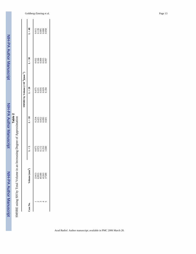
Statistical MethodsFor each case, we derived the satisfactory degree L of the SH model by computing the meansquare reconstruction error (MSRE; square millimeters), standardized by the corresponding3D volume (Vol in cubic millimeters), yielding the standardized MSRE (SMSRE = MSRE/Vol in mm−1) for each L ∈ {5, 10, 20, 30, 40}. We further showed the result of the SHapproximation by increasing the degree in the SH model.
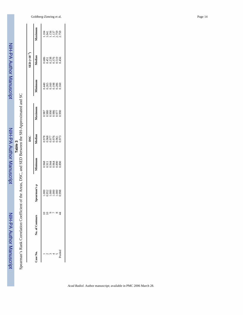
To validate the overall agreement of the SH method against SC, Spearman’s rank correlationcoefficient ρ was calculated between the area of the derived SH contours and the area of theSC for each case. In addition, tumor-specific minimum, median, and maximum of the DSCvalues were summarized, along with those of the SEDs by their corresponding areas by SC.Box plots of DSC and distance measures were created.
Surface extraction and SH reconstruction codes were written in Matlab 6.1(www.mathworks.com) and C language.
RESULTSTumor size range was 22413–85189 mm3. For cases in our study, range of number of verticeson the tumor surfaces was 3674–6544. SMSRE (mm−1) are listed in Table 2 as functions ofthe SH approximation degree (L). We observed that SMSRE was less than 1.66 × 10⁻⁵ mmfor L ≥ 30.
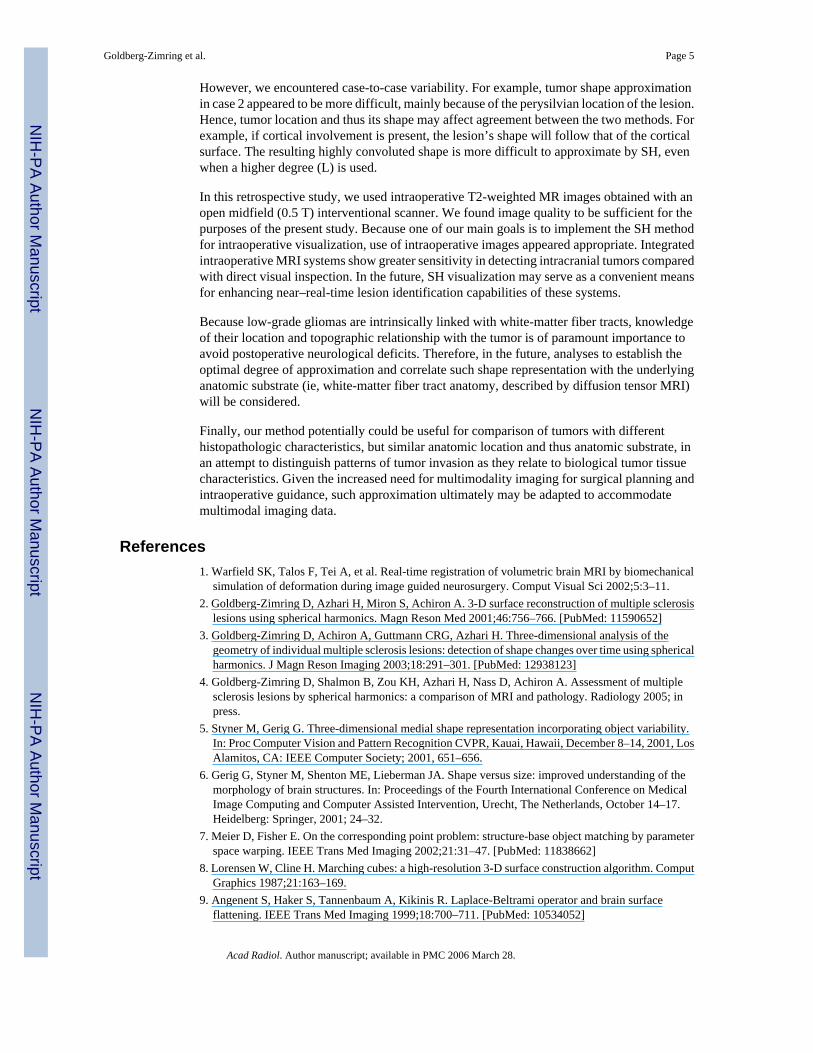
One representative slice for case 4 (left frontal oligodendroglioma) and its correspondingsegmentation are shown in Figure 1a–b, respectively. Figure 2a shows the original SC used asthe 3D target, whereas Figure 2b–d presents the reconstructed shapes for case 4 in increasingvalue for L (L = 5, L = 10, and L = 40, respectively). Based on these reconstructions and resultslisted in Table 2, subsequent analyses adopted L = 40, shown in Figure 2d, when applying theSH method for shape approximation.
Nonparametric Spearman’s rank correlation coefficient ρ indicated a high correlationcoefficient between the SH and SC, with perfect correlation other than that for case 2 (ρ ≥0.952). DSC values range was 0.890–0.990, with a pooled median DSC of 0.973, alsosuggesting high spatial similarity. All SEDs were ≤ 2.759 × 10⁻³ (Table 3).
Figure 3 shows a scatter plot of the areas derived by SH (on the x-axis) and SC (on the y-axis)for all 44 contours. Box plots of the DSC, which are close to 1, and the SED, which arenegligible, also are shown for all cases.
As an illustration, in Figure 4, we show the difference between SH and SC for the 7 contoursbelonging to case 4 at L = 40. The close approximation achieved can be visually verified.
DISCUSSIONIn our present study, after a manual segmentation procedure for tumor identification, weinvestigated the potential use of SH for improving brain tumor shape approximation and 3Dvisualization for surgical planning and assessing tumor invasion. Satisfactory agreementbetween the SH and SC methods was observed, suggested by a high DSC, low SMSRE, andlow SED.
Goldberg-Zimring et al. Page 4
Acad Radiol. Author manuscript; available in PMC 2006 March 28.
NIH
-PA Author Manuscript
NIH
-PA Author Manuscript
NIH
-PA Author Manuscript
However, we encountered case-to-case variability. For example, tumor shape approximationin case 2 appeared to be more difficult, mainly because of the perysilvian location of the lesion.Hence, tumor location and thus its shape may affect agreement between the two methods. Forexample, if cortical involvement is present, the lesion’s shape will follow that of the corticalsurface. The resulting highly convoluted shape is more difficult to approximate by SH, evenwhen a higher degree (L) is used.
In this retrospective study, we used intraoperative T2-weighted MR images obtained with anopen midfield (0.5 T) interventional scanner. We found image quality to be sufficient for thepurposes of the present study. Because one of our main goals is to implement the SH methodfor intraoperative visualization, use of intraoperative images appeared appropriate. Integratedintraoperative MRI systems show greater sensitivity in detecting intracranial tumors comparedwith direct visual inspection. In the future, SH visualization may serve as a convenient meansfor enhancing near–real-time lesion identification capabilities of these systems.
Because low-grade gliomas are intrinsically linked with white-matter fiber tracts, knowledgeof their location and topographic relationship with the tumor is of paramount importance toavoid postoperative neurological deficits. Therefore, in the future, analyses to establish theoptimal degree of approximation and correlate such shape representation with the underlyinganatomic substrate (ie, white-matter fiber tract anatomy, described by diffusion tensor MRI)will be considered.
Finally, our method potentially could be useful for comparison of tumors with differenthistopathologic characteristics, but similar anatomic location and thus anatomic substrate, inan attempt to distinguish patterns of tumor invasion as they relate to biological tumor tissuecharacteristics. Given the increased need for multimodality imaging for surgical planning andintraoperative guidance, such approximation ultimately may be adapted to accommodatemultimodal imaging data.
References1. Warfield SK, Talos F, Tei A, et al. Real-time registration of volumetric brain MRI by biomechanical
simulation of deformation during image guided neurosurgery. Comput Visual Sci 2002;5:3–11.2. Goldberg-Zimring D, Azhari H, Miron S, Achiron A. 3-D surface reconstruction of multiple sclerosis
lesions using spherical harmonics. Magn Reson Med 2001;46:756–766. [PubMed: 11590652]3. Goldberg-Zimring D, Achiron A, Guttmann CRG, Azhari H. Three-dimensional analysis of the
geometry of individual multiple sclerosis lesions: detection of shape changes over time using sphericalharmonics. J Magn Reson Imaging 2003;18:291–301. [PubMed: 12938123]
4. Goldberg-Zimring D, Shalmon B, Zou KH, Azhari H, Nass D, Achiron A. Assessment of multiplesclerosis lesions by spherical harmonics: a comparison of MRI and pathology. Radiology 2005; inpress.
5. Styner M, Gerig G. Three-dimensional medial shape representation incorporating object variability.In: Proc Computer Vision and Pattern Recognition CVPR, Kauai, Hawaii, December 8–14, 2001, LosAlamitos, CA: IEEE Computer Society; 2001, 651–656.
6. Gerig G, Styner M, Shenton ME, Lieberman JA. Shape versus size: improved understanding of themorphology of brain structures. In: Proceedings of the Fourth International Conference on MedicalImage Computing and Computer Assisted Intervention, Urecht, The Netherlands, October 14–17.Heidelberg: Springer, 2001; 24–32.
7. Meier D, Fisher E. On the corresponding point problem: structure-base object matching by parameterspace warping. IEEE Trans Med Imaging 2002;21:31–47. [PubMed: 11838662]
8. Lorensen W, Cline H. Marching cubes: a high-resolution 3-D surface construction algorithm. ComputGraphics 1987;21:163–169.
9. Angenent S, Haker S, Tannenbaum A, Kikinis R. Laplace-Beltrami operator and brain surfaceflattening. IEEE Trans Med Imaging 1999;18:700–711. [PubMed: 10534052]
Goldberg-Zimring et al. Page 5
Acad Radiol. Author manuscript; available in PMC 2006 March 28.
NIH
-PA Author Manuscript
NIH
-PA Author Manuscript
NIH
-PA Author Manuscript

10. Brechbuhler C, Gerig G, Kubler O. Parametrization of closed surfaces for 3-D shape description.Comput Vision Image Understanding 1995:154–170.
11. Akima A. A new method of interpolating and smooth curve fitting based on local procedures. AssocComput Mach 1970;17:589–602.
12. Zou KH, Warfield SK, Bharatha A, et al. Statistical validation of image segmentation quality basedon a spatial overlap index. Acad Radiol 2004;11:178–189. [PubMed: 14974593]
13. Zou KH, Wells WM III, Kikinis R, Warfield SK. Three validation metrics for automated probabilisticimage segmentation of brain tumors. Stat Med 2004;23:1259–1282. [PubMed: 15083482]
Goldberg-Zimring et al. Page 6
Acad Radiol. Author manuscript; available in PMC 2006 March 28.
NIH
-PA Author Manuscript
NIH
-PA Author Manuscript
NIH
-PA Author Manuscript

Figure 1.(a) One representative slice for case 4 tumor (left frontal oligodendroglioma) and (b) itscorresponding segmentation.
Goldberg-Zimring et al. Page 7
Acad Radiol. Author manuscript; available in PMC 2006 March 28.
NIH
-PA Author Manuscript
NIH
-PA Author Manuscript
NIH
-PA Author Manuscript
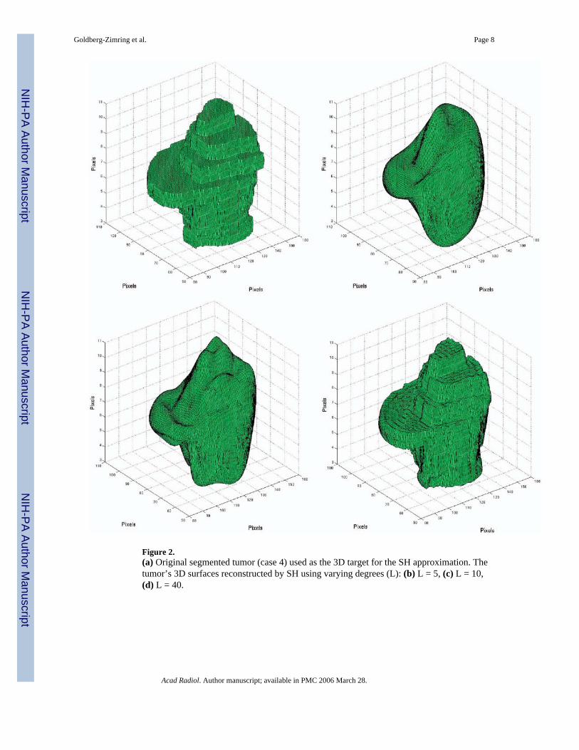
Figure 2.(a) Original segmented tumor (case 4) used as the 3D target for the SH approximation. Thetumor’s 3D surfaces reconstructed by SH using varying degrees (L): (b) L = 5, (c) L = 10,(d) L = 40.
Goldberg-Zimring et al. Page 8
Acad Radiol. Author manuscript; available in PMC 2006 March 28.
NIH
-PA Author Manuscript
NIH
-PA Author Manuscript
NIH
-PA Author Manuscript
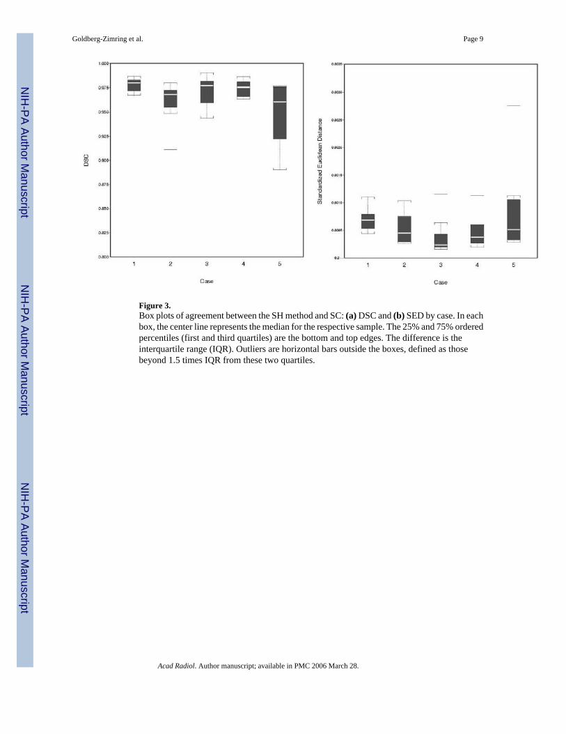
Figure 3.Box plots of agreement between the SH method and SC: (a) DSC and (b) SED by case. In eachbox, the center line represents the median for the respective sample. The 25% and 75% orderedpercentiles (first and third quartiles) are the bottom and top edges. The difference is theinterquartile range (IQR). Outliers are horizontal bars outside the boxes, defined as thosebeyond 1.5 times IQR from these two quartiles.
Goldberg-Zimring et al. Page 9
Acad Radiol. Author manuscript; available in PMC 2006 March 28.
NIH
-PA Author Manuscript
NIH
-PA Author Manuscript
NIH
-PA Author Manuscript
Goldberg-Zimring et al. Page 10
Acad Radiol. Author manuscript; available in PMC 2006 March 28.
NIH
-PA Author Manuscript
NIH
-PA Author Manuscript
NIH
-PA Author Manuscript
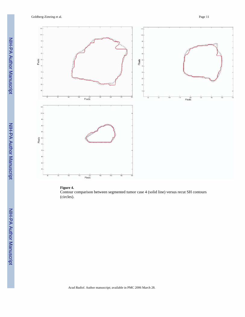
Figure 4.Contour comparison between segmented tumor case 4 (solid line) versus recut SH contours(circles).
Goldberg-Zimring et al. Page 11
Acad Radiol. Author manuscript; available in PMC 2006 March 28.
NIH
-PA Author Manuscript
NIH
-PA Author Manuscript
NIH
-PA Author Manuscript

NIH
-PA Author Manuscript
NIH
-PA Author Manuscript
NIH
-PA Author Manuscript
Goldberg-Zimring et al. Page 12Ta
ble
1Tu
mor
Cha
ract
eris
tics o
f Cas
es in
this
Stu
dy
Cas
e N
o.A
ge (y
)L
ocat
ion
Typ
eW
orld
Hea
lth O
rgan
izat
ion
Gra
de
149
Rig
ht fr
onto
tem
pora
lO
ligod
endr
oglio
ma
II/IV
249
Rig
ht fr
onta
lO
ligod
endr
oglio
ma
II/IV
340
Left
fron
topa
rieta
lO
ligod
endr
oglio
ma
II/IV
448
Left
fron
tal
Olig
oden
drog
liom
aII
/IV5
23Le
ft fr
onta
lA
napl
astic
ast
rocy
tom
aII
I/IV
Acad Radiol. Author manuscript; available in PMC 2006 March 28.
NIH
-PA Author Manuscript
NIH
-PA Author Manuscript
NIH
-PA Author Manuscript
Goldberg-Zimring et al. Page 13Ta
ble
2SM
SRE
usin
g SH
by
Tota
l Vol
ume
in a
n In
crea
sing
Deg
ree
of A
ppro
xim
atio
n
SMSR
E b
y V
olum
e (×
10−4
)(mm−1
)
Cas
e N
o.V
olum
e (m
m3 )
L =
5L
= 1
0L
= 2
0L
= 3
0L
= 4
0
122
413
0.87
50.
254
0.07
50.
032
0.01
52
6395
50.
972
0.41
50.
221
0.16
60.
139
385
189
0.17
20.
052
0.01
60.
009
0.00
54
4126
30.
253
0.09
50.
029
0.01
50.
008
537
589
1.84
00.
601
0.18
40.
097
0.05
8
Acad Radiol. Author manuscript; available in PMC 2006 March 28.
NIH
-PA Author Manuscript
NIH
-PA Author Manuscript
NIH
-PA Author Manuscript
Goldberg-Zimring et al. Page 14Ta
ble
3Sp
earm
an’s
Ran
k C
orre
latio
n C
oeff
icie
nt o
f the
Are
as, D
SC, a
nd S
ED B
etw
een
the
SH-A
ppro
xim
ated
and
SC
DSC
SED
(×10
−3)
Cas
e N
o.N
o. o
f Con
tour
sSp
earm
an’s
ρM
inim
umM
edia
nM
axim
umM
inim
umM
edia
nM
axim
um
110
1.00
00.
968
0.97
80.
987
0.44
00.
686
1.10
42
100.
952
0.91
10.
968
0.98
00.
265
0.45
11.
041
39
1.00
00.
944
0.97
70.
990
0.16
00.
238
1.15
64
71.
000
0.96
40.
976
0.98
60.
198
0.37
61.
137
58
1.00
00.
890
0.96
10.
977
0.28
60.
510
2.75
9Po
oled
440.
998
0.89
00.
973
0.99
00.
160
0.45
62.
759
Acad Radiol. Author manuscript; available in PMC 2006 March 28.
Copyright © 2022 FDOKUMEN