Staphylococcus aureus Panton-Valentine Leukocidin Is a Very Potent Cytotoxic Factor for Human...
8
/ www.sciencexpress.org / 18 January 2007 / Page 1 / 10.1126/science.1137165 The Staphylococcus aureus Panton Valentine leukocidin (PVL) is a pore-forming toxin secreted by strains epidemiologically associated with the current outbreak of community-associated methicillin-resistant Staphylococcus aureus (CA-MRSA) and with the often lethal necrotizing pneumonia. To investigate the role of PVL in pulmonary disease, we tested the pathogenicity of clinical isolates, isogenic PVL-negative and PVL-positive S. aureus strains, as well as purified PVL, in a mouse acute pneumonia model. Here we show that PVL is sufficient to cause pneumonia and that the expression of this leukotoxin induces global changes in transcriptional levels of genes encoding secreted and cell-wall-anchored staphylococcal proteins, including the lung inflammatory factor staphylococcal protein A (Spa). PVL is a bi-component, pore-forming exotoxin (5) that targets cells of the immune system such as polymorphonuclear neutrophils (PMNs). The active form of PVL requires the assembly of two polypeptides, LukS-PV and LukF-PV, into a hetero-oligomeric pore. Although PVL has potent cytolytic and inflammatory activities in vitro (6, 7), its role in necrotizing pneumonia has not been demonstrated. To analyze the molecular pathogenesis of PVL-expressing S. aureus strains, we have established a murine model of acute primary pneumonia. We infected mice with strains isolated from necrotizing (PVL-positive) or non-necrotizing (PVL-negative) staphylococcal pneumonia patients (table S1). PVL-positive strains caused murine necrotizing pneumonia with manifestations resembling human disease (fig. S1). In the PVL-positive strains, the lukS-PV, lukF-PV genes are organized as an operon within a phage (φSLT, or other similar phages) that could potentially contribute to the virulence of these strains. To define the role of PVL, we developed several isogenic strains (8). A PVL-negative, transformable S. aureus strain was lysogenized with φSLT or with a mutated φSLT where the PVL operon (luk-PV) was deleted. We complemented the PVL-negative strains with a plasmid containing the luk-PV operon under the control of its own promoter (table S1). Mice infected with PVL-positive strains showed symptoms of severe illness: lethargy, hunched posture, ruffled fur and significant weight loss. Lungs from infected mice were examined 48 hours post-inoculation. Tissue sections from lungs infected with PVL-positive strains revealed a strong recruitment of neutrophils and significant inflammation in the lung parenchyma, bronchial epithelial damage, tissue necrosis, and hemorrhage (Fig. 1 and table S2). The lungs infected with PVL-negative strains showed normal lung structures, despite some leukocyte infiltration. By contrast, when the PVL-negative strains were complemented with a plasmid encoding PVL, we observed massive tissue damage and 35-80% mortality within 24 hours after inoculation (Fig. 1 and fig. S2). We stained lung sections using anti-LukS-PV antibodies (fig. S3), and showed that the toxin was detected in the PVL-positive infected tissues. Administration of increasing equimolar amounts of native LukS-PV and LukF-PV resulted in concentration-dependent localized lesions, weight loss and, at concentrations higher than 3 micrograms, high rates of mortality (Fig. 1). The protein-inoculated mice recovered the lost weight after 24 hrs, whereas those infected with PVL-positive bacteria were still ill at 48 hrs, (Fig. 1c compared to Fig. 1k) demonstrating that an active bacterial infection is required to cause severe morbidity. Previous studies have demonstrated that PVL-positive strains had increased adherence to injured airway epithelium (3, 9). To examine the expression of surface proteins in these strains, cell wall extracts and supernatants from both exponential and stationary phase cultures were examined by SDS-PAGE (Fig. 2). Samples from a PVL-positive strain showed an enhanced expression of at least two cell-wall Staphylococcus aureus Panton Valentine Leukocidin Causes Necrotizing Pneumonia Maria Labandeira-Rey, 1 Florence Couzon, 2–5 Sandrine Boisset, 2–5 Eric L. Brown, 1* Michele Bes, 2–5 Yvonne Benito, 2–5 Elena M. Barbu, 1 Vanessa Vazquez, 1 Magnus Höök, 1 Jerome Etienne, 2–5 François Vandenesch, 2–5† M. Gabriela Bowden 1† 1 Center for Extracellular Matrix Biology, Institute of Biosciences and Technology, The Texas A&M University System Health Science Center, Houston, TX 77030, USA. 2 Université de Lyon, Lyon, F-69008, France. 3 Université de Lyon 1, Faculté Laennec, Lyon, F-69008, France. 4 INSERM E0230, Centre National de référence des staphylocoques, Lyon, F-69008, France. 5 Hospices Civils de Lyon, Hôpital Edouard Herriot, Lyon, F-69003, France. *Present address: University of Texas School of Public Health, Houston, TX 77030, USA. †To whom correspondence should be addressed. E-mail: [email protected] and [email protected]
-
Upload
independent -
Category
Documents
-
view
2 -
download
0
Transcript of Staphylococcus aureus Panton-Valentine Leukocidin Is a Very Potent Cytotoxic Factor for Human...
/ www.sciencexpress.org / 18 January 2007 / Page 1 / 10.1126/science.1137165
The Staphylococcus aureus Panton Valentine leukocidin (PVL) is a pore-forming toxin secreted by strains epidemiologically associated with the current outbreak of community-associated methicillin-resistant Staphylococcus aureus (CA-MRSA) and with the often lethal necrotizing pneumonia. To investigate the role of PVL in pulmonary disease, we tested the pathogenicity of clinical isolates, isogenic PVL-negative and PVL-positive S. aureus strains, as well as purified PVL, in a mouse acute pneumonia model. Here we show that PVL is sufficient to cause pneumonia and that the expression of this leukotoxin induces global changes in transcriptional levels of genes encoding secreted and cell-wall-anchored staphylococcal proteins, including the lung inflammatory factor staphylococcal protein A (Spa).
PVL is a bi-component, pore-forming exotoxin (5) that targets cells of the immune system such as polymorphonuclear neutrophils (PMNs). The active form of PVL requires the assembly of two polypeptides, LukS-PV and LukF-PV, into a hetero-oligomeric pore. Although PVL has potent cytolytic and inflammatory activities in vitro (6, 7), its role in necrotizing pneumonia has not been demonstrated. To analyze the molecular pathogenesis of PVL-expressing S. aureus strains, we have established a murine model of acute primary pneumonia. We infected mice with strains isolated from necrotizing (PVL-positive) or non-necrotizing (PVL-negative) staphylococcal pneumonia patients (table S1). PVL-positive strains caused murine necrotizing pneumonia with manifestations resembling human disease (fig. S1). In the PVL-positive strains, the lukS-PV, lukF-PV genes are organized as an operon within a phage (φSLT, or other similar phages) that could potentially contribute to the virulence of these strains. To define the role of PVL, we developed several isogenic strains (8). A PVL-negative, transformable S. aureus strain was lysogenized with φSLT or
with a mutated φSLT where the PVL operon (luk-PV) was deleted. We complemented the PVL-negative strains with a plasmid containing the luk-PV operon under the control of its own promoter (table S1). Mice infected with PVL-positive strains showed symptoms of severe illness: lethargy, hunched posture, ruffled fur and significant weight loss. Lungs from infected mice were examined 48 hours post-inoculation. Tissue sections from lungs infected with PVL-positive strains revealed a strong recruitment of neutrophils and significant inflammation in the lung parenchyma, bronchial epithelial damage, tissue necrosis, and hemorrhage (Fig. 1 and table S2). The lungs infected with PVL-negative strains showed normal lung structures, despite some leukocyte infiltration. By contrast, when the PVL-negative strains were complemented with a plasmid encoding PVL, we observed massive tissue damage and 35-80% mortality within 24 hours after inoculation (Fig. 1 and fig. S2). We stained lung sections using anti-LukS-PV antibodies (fig. S3), and showed that the toxin was detected in the PVL-positive infected tissues. Administration of increasing equimolar amounts of native LukS-PV and LukF-PV resulted in concentration-dependent localized lesions, weight loss and, at concentrations higher than 3 micrograms, high rates of mortality (Fig. 1). The protein-inoculated mice recovered the lost weight after 24 hrs, whereas those infected with PVL-positive bacteria were still ill at 48 hrs, (Fig. 1c compared to Fig. 1k) demonstrating that an active bacterial infection is required to cause severe morbidity. Previous studies have demonstrated that PVL-positive strains had increased adherence to injured airway epithelium (3, 9). To examine the expression of surface proteins in these strains, cell wall extracts and supernatants from both exponential and stationary phase cultures were examined by SDS-PAGE (Fig. 2). Samples from a PVL-positive strain showed an enhanced expression of at least two cell-wall
Staphylococcus aureus Panton Valentine Leukocidin Causes Necrotizing Pneumonia Maria Labandeira-Rey,1 Florence Couzon,2–5 Sandrine Boisset,2–5 Eric L. Brown,1* Michele Bes,2–5 Yvonne Benito,2–5 Elena M. Barbu,1 Vanessa Vazquez,1 Magnus Höök,1 Jerome Etienne,2–5 François Vandenesch,2–5† M. Gabriela Bowden1† 1Center for Extracellular Matrix Biology, Institute of Biosciences and Technology, The Texas A&M University System Health Science Center, Houston, TX 77030, USA. 2Université de Lyon, Lyon, F-69008, France. 3Université de Lyon 1, Faculté Laennec, Lyon, F-69008, France. 4INSERM E0230, Centre National de référence des staphylocoques, Lyon, F-69008, France. 5Hospices Civils de Lyon, Hôpital Edouard Herriot, Lyon, F-69003, France.
*Present address: University of Texas School of Public Health, Houston, TX 77030, USA.
†To whom correspondence should be addressed. E-mail: [email protected] and [email protected]
/ www.sciencexpress.org / 18 January 2007 / Page 2 / 10.1126/science.1137165
anchored polypeptides identified as SdrD and protein A (Spa) by N-terminal sequencing and Western analysis (Fig. 2, c to e, and fig. S4). The SdrD and Spa over-expression was not observed in a strain carrying the phage with the deleted luk-PV operon, indicating that this effect was not mediated by products encoded by other phage genes or by its insertion in the chromosome of S. aureus. PVL-negative strains complemented with the luk-PV operon in a multicopy plasmid (table S1) also showed an increased expression of Spa during both logarithmic and stationary phase (Fig. 2f). A group of polypeptides with apparent molecular weights between 32 and 47.5 kD (Fig. 2b, dots) were present in the supernatants from the PVL-negative but not the PVL-positive strains. Some of the secreted polypeptides were identified as proteases using zymograms (Fig. 2g). Thus, expression of the luk-PV operon resulted in an altered regulation of cell-wall-anchored and secreted protein production. Spa is a known virulence factor in mouse models of S. aureus infections including pneumonia (10, 11); we therefore examined the role of Spa in necrotizing pneumonia. Animals infected with the spa-deleted isogenic strains had less severe symptoms of disease (Fig. 3a). However, the lungs from animals infected with spa-deleted, PVL-positive strains showed localized lesions with massive leukocyte infiltration (fig. S5), demonstrating that PVL alone was sufficient to cause pneumonia. Complementation of Spa-positive strains with PVL rendered them lethal, whereas the Spa-negative, PVL-plasmid strains did not cause mortality (Fig. 3b). These data suggested that PVL and Spa may act together to cause the overwhelming inflammation and tissue damage that are seen in necrotizing pneumonia. PVL-positive strains expressed Spa during both exponential and stationary growth phases (Fig. 2). To analyze the effect of PVL on spa transcription, PVL was introduced into mutants deficient in spa regulators. The Spa production was abolished in a sarS-deletion mutant (fig. S6), whether PVL was present or not, indicating that PVL acts upstream of SarS. To evaluate the transcriptional profile of a PVL-positive strain compared to the PVL-negative strain, we used microarray analysis: 28 genes showed a distinct expression pattern during exponential growth (table S3); whereas during the stationary phase, 133 genes showed differential expression (table S4). The agr transcripts and several exoproteins were repressed, whereas genes encoding for cell-wall-anchored proteins, and the spa activator sarS (12) were up-regulated (Fig. 4a). Elevated expression of the spa and sdrD transcripts correlated with the enhanced production of Spa and SdrD observed in Western blot analysis (Fig. 2 and fig. S4). The repression of exoprotein transcripts paralleled the absence of the 32-47.5 kD exoproteins in the supernatants of PVL-positive strains (Fig. 2). This pattern of transcription implicated PVL in an interaction with a factor(s) that controls
gene expression during the transition from logarithmic to stationary phase. The regulatory model proposed here was inferred using strains derived from RN6390, a strain harboring a deletion in rsbU, which encodes a regulator necessary for the activity of the stress sigma factor σB. Strains with a reduced σB (13) activity display, among other traits, a decreased production of cell-wall anchored proteins and an increased production of exoproteins. We subsequently generated isogenic PVL-positive and PVL-negative strains in the SH1000 (14) rsbU+ background and observed overexpression of Spa during the stationary phase of growth (fig. S7a). The SH1000-derived PVL-positive strain was more virulent compared with its PVL-negative isogenic pair (fig. S7, c and d). When compared to RN6390, the SH1000 lineage showed an increased expression of Spa, but produced up to five-fold less PVL, although some variability was seen (fig. S7a) (15), suggesting that RsbU/σB partially regulates PVL. We show here that PVL is a significant S. aureus virulence factor, and that PVL-positive strains can cause murine necrotizing pneumonia with manifestations that resemble those observed in human patients. Our results demonstrate that the expression of the genes that encode PVL (lukS-PV and lukF-PV) or direct inoculation with native toxin is sufficient to induce pneumonia in mice. The expression of the luk-PV operon also resulted in an altered expression of multiple proteins, including the tightly regulated (16, 17) pro-inflammatory factor Spa. In PVL-positive strains, many secreted proteins are down-regulated (Fig. 4b), similarly to data reported by Vojtov et. al. (18) who demonstrated that two staphylococcal superantigens, toxic shock syndrome toxin-1 (TSST-1) and enterotoxin B (SEB), strongly repressed production of secreted proteins. It is possible that these toxins act similarly to PVL, interacting with unknown factors that interfere with regulatory networks. Several genes encoding putative and known microbial surface components recognizing adhesive matrix molecules (MSCRAMMs) (including SdrD) are up-regulated in the PVL-positive strains (Fig. 4a). The up-regulation of MSCRAMMs may lead to enhanced tissue adherence and colonization of PVL-expressing strains, thereby contributing to the virulence potential of these strains (19, 20). Spa is highly expressed in PVL-positive strains. Our in vivo data underscores the documented role of Spa as a pro-inflammatory factor in pneumonia (11). Increased production of Spa, coupled with the ability of PVL to lyse PMNs and macrophages (6), could lead to a vicious cycle of cell recruitment, lysis and release of inflammatory mediators (7), resulting in overwhelming tissue inflammation and necrosis. Here, we show that PVL is not only a key virulence factor in pulmonary infections, but also that expression of the luk-PV

/ www.sciencexpress.org / 18 January 2007 / Page 3 / 10.1126/science.1137165
genes interfere with global regulatory networks, which may also enhance virulence. A detailed analysis of such dysregulation will be useful to identify targets for the potential development of novel therapies to treat S. aureus infections.
References and Notes 1. T. J. Foster, M. Höök, Trends Microbiol 6, 484 (1998). 2. T. J. Foster, Nat Rev Microbiol 3, 948 (2005). 3. Y. Gillet et al., Lancet 359, 753 (2002). 4. H. F. Chambers, N Engl J Med 352, 1485 (2005). 5. P. N. Panton, M. C. Camb, F. C. O. Valentine, M. R. C. P.
Lond, Lancet 1, 506 (1932). 6. A. L. Genestier et al., J Clin Invest 115, 3117 (2005). 7. B. Konig, G. Prevost, Y. Piemont, W. Konig, J Infect Dis
171, 607 (1995). 8. Materials and Methods are available as supporting
materials on Science Online. 9. S. de Bentzmann et al., J Infect Dis 190, 1506 (2004). 10. N. Palmqvist, T. Foster, A. Tarkowski, E. Josefsson,
Microb Pathog 33, 239 (2002). 11. M. I. Gomez et al., Nat Med 10, 842 (2004). 12. J. Oscarsson, C. Harlos, S. Arvidson, Int J Med Microbiol
295, 253 (2005). 13. Kullic, I. Giachino, P. and T. Fuchs, J Bacteriol 180,
4814 (1998). 14. M. J. Horsburgh et al., J Bacteriol 184, 5457 (2002). 15. B. Said-Salim et al., J Clin Microbiol 43, 3373 (2005) 16. A. L. Cheung, K. Eberhardt, J. H. Heinrichs, Infect
Immun 65, 2243 (1997). 17. E. Huntzinger et al., EMBO J 24, 824 (2005). 18. N. Vojtov, H. F. Ross, R. P. Novick, Proc Natl Acad Sci
U S A 99, 10102 (2002). 19. P. Thomas, M. Riffelmann, B. Schweiger, S. Dominik, C.
H. von Konig, Pediatr Infect Dis J 22, 201 (2003). 20. L. O'Brien et al., Mol Microbiol 44, 1033 (2002). 21. The authors equally contributed to this work. We thank S.
L. Mueller-Ortiz and S. M. Drouin for their invaluable help establishing the animal model; Z. Chroneos for his input in the interpretation of histological samples; C. Badiou for technical assistance; G. Lina for scientific advice; and S. Foster, T. Foster, B. Fournier, R. Novick, and P. Mc Namara for providing strains. This work was supported by grants from the TAMUS HSC to M.G.B., from the French Ministry of Research to F.V. and NIH AI020624 to M.H. Support from the Neva and Wesley West and the Hamill Foundations were awarded to M.H. The authors declare that they have no competing financial interests. Microarray data is deposited in the GEO database (http://www.ncbi.nlm.nih.gov/projects/geo/) under the accession number GPL4653.
Supporting Online Material www.sciencemag.org/cgi/content/full/1137165/DC1
Materials and Methods Figures S1 to S8 Tables S1 to S5 References
13 November 2006; accepted 12 December 2006 Published online 18 January 2007; 10.1126/science.1137165 Include this information when citing this paper.
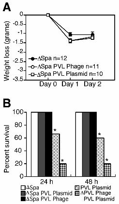
Fig. 1. Expression of PVL enhances the virulence of isogenic S. aureus strains. (a, b, e, f, j) Lung histology of mice infected with PVL-positive and PVL-negative strains or inoculated with PVL toxin. The sections are representative of at least three separate experiments. Bar represents 100 µm. (a) PBS; (b) PVL Phage; (e) Parental; (f) ∆PVL Phage; (i) 5µg purified native LukS subunit (j) 3 µg purified native PVL. (c, g, k) Line graphs indicate weight loss in grams. (c) Parental vs. PVL Phage. *P < 0.001; (g) Parental vs. ∆PVL Phage. No statistical difference observed; (k) Animals inoculated with 3 µg of LukS+F-PV vs. 5 µg LukS-PV. *P < 0.01 on day 1. (d, h, l) Mouse survival. (d) Parental and PVL Plasmid; (h) PVL Phage, ∆PVL Phage, and ∆PVL Phage PVL Plasmid; (l) 20 µg or 10 µg of LukS+LukF-PV vs. 3 µg LukS+LukF-PV or 5 µg LukS-PV, *P < 0.0001.
Fig. 2. PVL alters the expression pattern of cell-wall anchored and secreted proteins. (a to e) Lanes 1, 2 and 3 represent extracts from Parental, ∆PVL Phage and PVL Phage strains respectively; samples were isolated from bacterial cultures grown at exponential (EXP) and stationary (STAT) phase of growth. (a) Lysostaphin extracts analyzed by SDS-PAGE. The arrows point to over-expressed proteins. (b) Exoproteins from culture supernatants harvested from bacterial cultures analyzed by SDS-PAGE. Arrows point to overexpressed proteins, dots indicate exoproteins reduced or absent in PVL-positive strains. (c and d) Western blot analysis of lysostaphin extracts and supernatants using an anti-Spa monoclonal antibody. Complete gels shown in fig. S4 (8). *Samples were diluted 1:100. (e) Western blot analysis of lysostaphin extracts using anti-SdrD polyclonal antibodies. SdrD is detected as two polypeptides due to the proteolytic cleavage of an N-terminus subdomain. (f) Western blot analysis of supernatants from Parental PVL Plasmid stationary phase (7 hrs) cultures (lane 1) and ∆PVL Phage PVL Plasmid (lane 2) using anti-Spa monoclonal antibodies. (g) Zymogram analysis of exoproteins from cultures grown at stationary phase. Lanes 1, 2 and 3 represent proteins extracted from Parental, ∆PVL Phage and PVL Phage respectively.
Fig. 3. Spa enhances the virulence of PVL-positive strains. (a) Line graph indicates weight loss in grams, *P < 0.01 ∆Spa vs. ∆Spa PVL Phage or ∆Spa PVL Plasmid. (b) Percent survival of animals infected with ∆Spa, ∆Spa PVL Phage,

/ www.sciencexpress.org / 18 January 2007 / Page 4 / 10.1126/science.1137165
∆Spa PVL Plasmid, PVL Plasmid, and ∆PVL Phage PVL Plasmid, *P < 0.001 ∆Spa, ∆Spa PVL Phage and ∆Spa PVL Plasmid vs. PVL Plasmid or ∆PVL Phage PVL Plasmid.
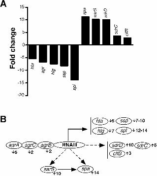
Fig. 4. S. aureus PVL-positive strains show an altered transcriptional profile. (a) Fold increase/decrease levels of transcript from selected genes. Total RNA extracted from cultures grown to stationary phase. Genes were considered to be induced or repressed in the PVL Phage if they were transcribed at least two-fold higher/lower than ∆PVL Phage. The shown transcripts encode: agrA-C, accessory gene regulator system; sarS, staphylococcal accessory regulator S; spa, staphylococcal protein A, sdrD, serine-aspartate repeat protein D, sdrC, serine-aspartate repeat protein C, clfB, clumping factor B; hla, alpha toxin, ssp, a representative of serine proteases sspB-C, spl, a representative of splA-F proteases. (b) A schematic overview of the interactions between regulators involved in cell-wall anchored and secreted protein genes (full and broken lines indicate positive and negative regulation, respectively) based on previously published data. Numbers next to the gene name indicate fold-change based on microarray analysis (upward arrow indicates up-regulation, downward arrow indicates down-regulation). The down-regulation of RNAIII (the effector of the agr system) results in the down-regulation of secreted protein genes (hla, hlgC, hlgB, and proteases) and the up-regulation of sarS and cell-wall-anchored proteins (spa, sdrD,C, clfB). In addition, the up-regulation of sarS results in the up-regulation of spa.