Stanford Synchrotron Radiation Laboratory - UNT Digital Library
222
1 - Stanford Synchrotron Radiation Laboratory SSRL Activity Report 1989
-
Upload
khangminh22 -
Category
Documents
-
view
2 -
download
0
Transcript of Stanford Synchrotron Radiation Laboratory - UNT Digital Library

1 - Stanford Synchrotron Radiation Laboratory
SSRL Activity Report 1989
SSRL Report 90/01
Editor - Katherine Cantwell Technical Production - Carol Mitchell

Portioxs of this document may be illegible in dectronic image products hag= are produced from the best available original dOCUlIlent,

STANFORD SYNCHR RADIATION LABORATORY
--
DISCLAIMER
This report was prepared as an account of work sponsored by an agency of the United States Government. Neither the United States Government nor any agency thereof, nor any of their employees, makes any warranty, express or implied, or assumes any legal liability or responsi- bility for the accuracy, completeness, or usefulness of any information, apparatus, product, or process disclosed, or represents that its use would not infringe privately owned rights. Refer- ence herein to any specific commercial product, process, or service by trade name, trademark, manufacturer, or otherwise does not necessarily constitute or imply its endorsement, recom- mendation, or favoring by the United States Government or any agency thereof. The views and opinions of authors expressed herein do not necessarily state or reflect those of the United States Government or any agency thereof.
____
ACTIVITY REPORT FOR 1989
ABOUT THE STANFORD SYNCHROTRON RADIATION LABORATORY
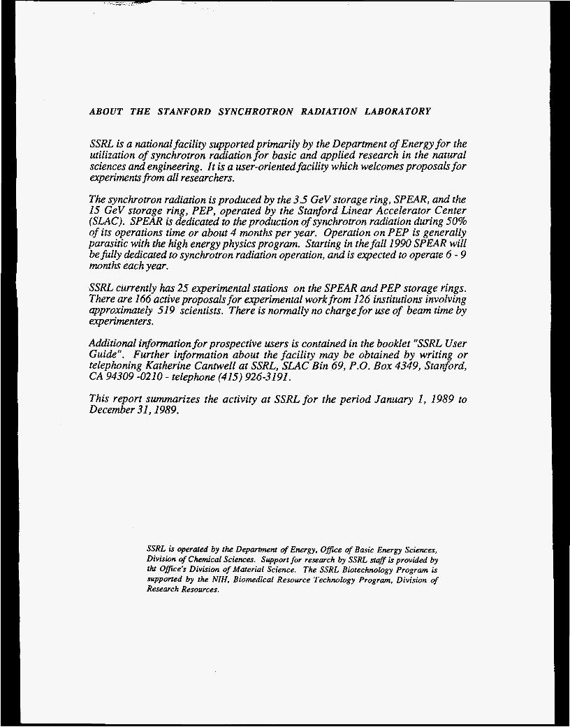
SSRL is a national facility supported primarily by the Department of Energy for the utilization of synchrotron radiation for basic and applied research in the natural sciences and engineering. It is a user-oriented facility which welcomes proposals for experimentsfrom all researchers.
The synchrotron radiation is produced by the 3.5 GeV storage ring, SPEAR, and the 15 GeV storage ring, PEP, operated by the Stanford Linear Accelerator Center (SLAC). SPEAR is dedicated to the production of synchrotron radiation during 50% of its operations time or about 4 months per year. Operation on PEP is generally parasitic with the high energy physics program. Starting in the fall 1990 SPEAR will be filly dedicated to synchrotron radiation operation, and is expected to operate 6 - 9 months each year.
SSRL currently has 25 experimental stations on the SPEAR ana' PEP storage rings. There are 166 active proposals for experimental work from 126 institutions involving approximately 519 scientists. There is normally no charge for use of beam time by experimenters.
Additional information for prospective users is contained in the booklet "SSRL User Guide". Further information about the facility may be obtained by writing or telephoning Katherine Cantwell at SSUL, SLAC Bin 69, P.O. Box 4349, Stanford, CA 94309 -0210 - telephone (415) 926-3191.
This report summarizes the activity at SSRL for the period January 1, 1989 to December 31,1989.
SSRL is operated by the Department of Energy, Office of Basic Energy Sciences, Division of Chemical Sciences. Support for research by SSRL staff is provided by tht Ofice's Division of Material Science. The SSRL Biotechnology Program is supported by the NIH, Biomedical Resource Technology Program, Division of Research Resources.

I
II
111
IV.
V.
VI.
VII.
VIII.
IX.
Stanford Synchrotron Radiation Laboratory 1989 ACTIVITY REPORT
TABLE OF CONTENTS
Laboratory Operations SPEAR Operations PEP Operations Beam and SPEAR Usage Tables
The 3 GeV Injector & Accelerator Research The 3 GeV Injector Accelerator Research Projects
Experimental Facilities X-ray Facilities VUV Facilities Biotech Facilities PRT Experimental Facilities Support Facilities
SSRL Organization SSRL Advisory Panels
Experimental Progress Reports Index to Experimental Progress Reports Materials Proposals Biology Proposals VUV Proposals
Theses Based on Research at SSRL
Active Proposals
SSRL Experimenters and Proposals by Institution
SSRL Publications
Page
11 11 15
17 17 18 19 20 27
29 35
37 37 41 83 98
126
136
1 58
168

Overview of SSRL -Photo by Joe Faust
The SPEAR ring is shown on the left side of the picture. SSRL has 23 stations housed in the two building (131,120) adjacent to the north and south arcs of the ring. The large white buitding in the center is the shelter for the new 3 GeV injector. The building (LOS) on the far right houses the scientific, administrative and engineering staffs.

ACTIVITY REPORT 1989
INTRODUCTION
The April, 1990 SPEAR synchrotron radiation run was one of the two or three best in SSRL’s history.
High currents (1 00 milliamps) were accumulated, ramping went easily, lifetimes were long (averaging
23 hours), beam dumps were infrequent and the average current was 42.9 milliamps. In the one month of
operation, 63 different experiments involving 208 scientists from 50 institutions received beam. The
end-of-run summary forms completed by the experimenters indicated high levels of user satisfaction with
the beam quality and with the outstanding support received from the SSRL technical and scientific staffs.
These fine experimental conditions result largely from the SPEAR repairs and improvements performed
during the past year and described in Section I. Also quite signficant was Max Cornacchia’s leadership
of the SLAC staff.
SPEAR’S performance this past April stands in marked contrast to that of the January-March, 1989 run
which is also described in Section 1. It is, we hope, a harbinger of the operation which will be provided in FY ’91, when the SPEAR injector project is completed (Section II) and SPEAR is fully dedicated to
synchrotron radiation research.
Over the coming years, SSRL intends to give highest priority to increasing the effectiveness of SPEAR
and its various beam lines. The beam line and facility improvements performed during 1989 are described
in Section Ill.
In order to concentrate effort on SSRL‘s three highest priorities prior to the March-April run: (1) to have a
successful run, (2) to complete and commission the injector, and (3) to prepare to operate, maintain and
improve the SPEAWinjector system, SSRL was reorganized. In the new organization, all the technical
staff is contained in three groups: Accelerator Research and Operations Division, Injector Project and
Photon Research and Operations Division, as described in Section IV. Responsibility for safety and
quality assurance, as well as scheduling and proposal review, resides in the Director’s office.
In spite of the limited effectiveness of the January-March, 1989 run, SSRL’s users made significant
scientific progress, as described in Section V of this report.
Arthur Bieninstock Director

Section I

I. LABORATORY OPERATIONS Two factors had very strong influences on SSRL’s operations in 1989. These were: the concurrent operation of the SLAC Linear Collider for Z, produc- tion, which placed Severe limits on SPEAR injection and maintenance and the expectation that the SPEAR storage ring, with a new independent injec- tor, will become completely dedicated to synchrotron radiation research in 1990. Thus, if it receives sufficient funding, SSRL will be able to operate SPEAR for a very large portion of 1991. SSRL should be in a position, at last, to provide its users with a steady stream of photons for research. Hence, emphasis was placed on converting SPEAR into a ring which can run reliably and effectively through maintenance, improvements and modifications. Only one experimental run was provided.
§PEAR OPERATION§
January 23 - March 17 Run - This run, which was plagued by operational and weather-related prob- lems, was scheduled for 145 shifts. Only 63 shifts, or 43%, were delivered to users. Only slightly over half of these shifts were at initial currents above 25 mA. The electron energy was 3.3 GeV in order to en- hance studies of human patients being conducted under the angiography program. In the final hours of the run, with the cooperation of SLAC and all of the SSRL users on site, these studies were successfully completed and significant progress was made. (See Experimental Progress Report No. 10468).
During the run problems were experienced with several ring components including the beam position monitoring system, RF system, power supplies and beam line control systems. The failure of a ceramic window in one of the RF cavities caused the ring to be vented and resulted in 13 days of down time.
These difficulties were exacerbated by limited injection. Due to the demands of the SLAC Linear Collider.(SLC) program, as discussed in the 1987 Activity Report, injection to SPEAR was available only at 12 hour intervals. This made it very difficult to diagnose and cure the various component problems.
Immediately after the run, B. Richter, Director of SLAC, chaired a series of meetings to identify and correct the most serious problems in anticipation of a scheduled summer run. In particular, it was clear that an RF transformer must be repaired and that align-
ment of the beam lines and storage ring should occur. This work was started.
The Cornacchia Report - Max Cornacchia, the newly appointed Head of Storage Rings at SLAC, then led a more detailed study of SPEAR which produced recommendations, costs, and time esti- mates for the work involved to make SPEAR a reliably operating synchrotron radiation facility. This study involved technical, engineering and scientific staff from SLAC and SSRL.
In summary, the report stated: “The priorities given to major SPEAR improvement projects described in this repdrt came from a consideration of the operational problems caused by the subsystems, their projected lifetime, safety implications and the estimated impact on SSRL running resulting from the work necessary to replace the subsystems. These items are, in order of priority: replacement of the ion pump cable plant, addition of RF isolators, fabrication of a spare klystron, miscellaneous RF improvements, upgrading of the low conductivity water (LCW) system, upgrad- ing of the general cable plant, replacementlupgrade of the magnet power supplies and improvement of the instrumentation and control (lac) system.”
The report also cited a high priority need for accel- erator physics studies once the machine was opera- tional and the need for a self-consistent alignment of the storage ring and beam lines to reduce orbit deviations.
Cancellation of the Summer Run - After receipt of the Cornacchia Report the decision was made to cancel the summer run for three reasons. The run was not expected to be significantly more successful than the January-March run with the limited injection to be provided by SLAC and SPEAR’S performance status. The nrn would interfere significantly with a critical SLAC Linear Collider (SLC) run. SSRL had to assure that it had sufficient funding to initiate the major SPEAR up-grade program required for effec- tive and full-time operation.
§PEAR Improvement Projects - Starting in the late spring several improvement projects were under- taken by the SSRL and SLAC staffs. These included work on the RF system, a power supply upgrade, a rebalancing of the low conductivity water (LCW) system, orbit control improvements, new vacuum
SSRL ACTIVITY REPORT 1989 Page 1

control systems for Beam Lines 1 and 2, a new high voltage ion pump distribution system, and alignment of the beam lines relative to the storage ring, as well as a number of smaller maintenance and improve- ment tasks. Highlights of the major projects are described below.
RF Svstem : There were three significant RF prob- lems during the winter run: a cavity window failure, frequent reflected power trips, and intermittent RF transformer difficulties. The window failure was attributed to accidentally overpowering a klystron, a condition which is now prevented by software con- trols. Renewing the nitride coating of both cavities and rebuilding the cavity tuners addressed the second problem. The problematic RF transformer was completely rebuilt. A transformer shelter is being erected by SSRL to help prevent future weather-related problems.
Power S-: There were two areas of power supply problems. The power supplies which drive the beam steering system were giving spurious outputs causing beam dumps. When this system was thoroughly tested, a manufacturing error was discov- ered and repaired. The power supplies which drive the bend magnets and quadrupoles were found to have problems tracking with each other during the ramp. Software, which will be tested with beam in March 1990, has been refined to correct this prob- lem.
Low Conduct ivitv Water (LCW) ; A substantial number of trips caused by inadequate flow in parts of the vacuum and the magnet LCW systems occurred during the run. The entire system has been exten- sively reexamined by SLAC and SSRL personnel. It was determined that the total flow was actually sufficient, but a flow balancing problem existed. The vacuum LCW has now been completely rebalanced and stabilized.
For some time SLAC has been concerned that SPEAR LCW transients caused problems in the SLC system. These problems were traced to four switches in the SLC arcs. Delays have been put on these switches which should eliminate this problem. These improvements should reduce the perturbations of the SLAC site LCW system caused by SPEAR, and provide stable and adequate LCW flow for SPEAR and SSRL components during dedicated running .
Delays have also been put on all the flow circuits as to make them less sensitive to power dips or minor perturbations in the LCW system.
In addition to rebalancing the system, the booster pump control system was improved. This system, which controls the return water pressure, was discovered to be insufficiently sensitive to regulate the flow. The resulting variations in supply pressure were causing trips.
The magnet LCW system is a larger problem, and will be tackled after the spring 1990 run. The problem is basically one of plumbing. Hose connections to the magnet busses fail frequently and will be re- placed with new connecting hardware.
ment; A large effort has been invested in evaluating and correcting mechanical misalignments of both the beam lines and the storage ring. The process began during the winter run with the discov- ery that the upstream apertures of Beam Line 2 were 6 mm below the plane of the storage ring. This motivated a program to align all of the beam lines, which was accomplished during the summer of 1989. The beam lines in the South Arc had maintained their position quite well, but those in the North Arc had dropped with respect to SPEAR. The amount of the displacement appears to increase with time, leading to problems with new beam lines located near older ones. Before October 17 all beam lines had been completely aligned.
The Richter magnitude 7.1 earthquake of October 17 was not kind to SSRL. At first glance it appeared that, on a local scale, the relative motion of beam line components was less than one millimeter. When, however, the storage ring realignment process began in late November, it was apparent that there had been substantial motion on a more global scale, requiring another full realignment of the beam lines as well as the storage ring. The earthquake caused the storage ring to tilt as a plane, with an elevation difference from one side to the other of 5 mm. When the insertion devices were surveyed, some were found to have undergone larger motions than would have been expected when compared with adjacent magnets. The plane of the storage ring was redefined to the mean quadrupole elevation and aligned with respect to this new plane [Figure 11. This had the advantage of minimizing the amount of motion necessary to correct the vertical position of the beam line components.
Page 2 SSRL ACTIVITY REPORT 1989

~ FIGURE 1 0 Dec 89 Survey
0.3
0.225 - 0.15 -
0.075 = C .- o Y
- 0 . 1 5 - -0-075i -0 .225 -
-0.3
I I I I I
SPEAR Quadrupole Elevations - - -
OO 0 O 0
d h n n n n n n o a n n ~ n ~ u n ~ ~ ~ o D n n n u ij-
O oo - 2 West Pit
A - - -.050 5@ ,qono - 00 ooooooooo
0
- 0 +East Pit
1 I I I I I
Unfortunately, the nature of the horizontal alignment of the magnets precludes a quantitative assessment of potential problems caused by the earthquake. It is difficult to separate the movement of the individual magnets from the movement of the references. All that can be determined is that there has not been a significant motion of one magnet girder with respect to another. The recalibration of the horizontal references would best be done during a long shut- down after the High Energy Physics Mark 111 Detector has been removed. In the meantime, the beam lines will be realigned to the local magnet geometry of their sources .
's): The lack Flectron Beam Position Monitors lRPM of a complete set of beam position monitors severely hampered the machine operators in establishing orbits. Newly designed electron beam position monitors have been installed to replace defective units on each end of the insertion devices on Beam Lines 6 and 10. As a result, SSRL now has the means of locating the SPEAR electron beam position as it enters and leaves the insertion devices on these beam lines. This more complete distribution of BPM's around the machine should greatly improve closed orbit distortions and facilitate reproducing orbits and photon beam steering.
..
I
: SSRL's original Beam Line 1 and 2 control systems, now 15 years old, have been replaced to eliminate faults which have shut down the SPEAR beam. Starting in the summer of 1989 a new vacuum control system was designed, fabricated, installed and tested. The new system utilizes modular construction and plug-in P.C. cards to allow ease of maintenance and trouble- shooting, expedite repairs and provide flexibility for modifications as required. This new system should serve as a prototype for future SSRL beam lines.
Jon PumD Hiah V o l W D istribution Syste m: The SPEAR ion pump high voltage distribution system was recabled. The motivations for this were safety, (the distributed ion pumps in the ring dipoles had to be started by vacuum technicians present in the ring housing while the magnets were powered) and improved operation (individual ion pumps could not be monitored while beam was stored or while the ring magnets were powered). Now each individual pump has its own cable running from the ring through a high voltage terminal cabinet to the SPEAR power supply building. From the power supply building vacuum technicians have electrical access to all vacuum pumps in the storage ring.
SSRL ACTIVITY REPORT 1989 Page 3

A major goal of the March 1990 run is to test and commission the improvements described above.
Scheduling of SSRL Users at Other Facilities: In response to the shortage of beam time at SSRL, three other U.S. synchrotron radiation laboratories (NSLS, CHESS and the SRC) offered to accommo- date SSRL users on the basis of their existing SSRL proposals. SSRL and it users are extremely grateful for this cooperation. The NSLS accommodated 41 SSRL experiments this fall. Four groups ran at the SRC and another 8 are being accommodated at CHESS.
PEP OPERATIONS
SSRL has two synchrotron radiation beam lines on the 15 GeV storage ring PEP. These lines operate parasitically when PEP is run for high energy physics (see 1988 Activity Repoff). After the successful Decem- ber 1988 run, when four synchrotron radiation user groups took data, a planned down time until the fall was scheduled, to allow work on the SLAC Linear Collider. A high energy physics run on PEP was expected to start in October 1989 and continue until the end of the year.
Cancellation of Fall PEP Run: This planned fall PEP run was another casualty of the October 17 earthquake, which caused an 8 mm peak-to-peak variation in the machine elevation. The present schedule calls for a PEP start-up in May 1990. The storage ring will be realigned by that time.
BEAM AND SPEAR USAGE TABLES
The following tables and graphs contain statistics on SPEAR running, experimental use and characteris- tics of SSRL stations. For earlier information, previ- ous Activity Reports should be consulted.
Table 1 and the accompanying graph show the number of SPEAR shifts scheduled and delivered since dedicated running commenced in 1979.
Table 2 and the accompanying graph illustrate the use of beam at SSRL for experimental purposes.
Table 3 lists the characteristics of the 25 SSRL SPEAR and PEP experimental stations and Table 4 shows the number of shifts requested versus the number of shifts actually scheduled for dedicated time.
~~~
Page 4 SSRL ACTIVITY REPORT 1989

~
TABLE 1
BEAM TIME STATISTICWDEDICATED TIME
DEDICATED RUN
1 0/20-11 /05/79 12/03-12/21/79 02/08-03/05/80 0411 6-0511 giao
09129-1 011 4/80 12/02-12/22~ao
06/30-07/30/80
01 /26-03/03/81 0511 6-06/30181 1 i ~ i a - i 2 / 2 1 ~ a i 01 10a1-oa2aa2 03/09-04/26 J82 1011 5-1 1 /05/82 12/27-02/22183 05109-06/30183 1 1107-1 2/23/03 03/21 -04/30/84 01 J1 0-02/21 I85 03/15-07/22/85 1011 4-1 1 /1 1185 04/11 -061'30J86 1 1 I1 7-1 2124186 01 /02-02/07/87 0311 a-0510aa7
01 /23-~3117~a9 1 0/26-12/24107
SCHEDULED DELIVERED HOURS
152 352 472 764 726 336 440 792
546
995 473
1050 1195
988
748
a57 a35 905
1502 41 6
1550 752 696
1112 1360 1160
HOURS
95.3 299.4 366.3 588.2 320.4 194.9 309 600.9 727 363.6 612.5
31 6 825.6 960.3
830.9
662.8 674.3 606.6
1056.5 203.7
1 106.5 527 522 769
504 a01
%DELIVERED
62
77 76 44 58 70 76 73 66
a5
a i a3
7a a0
a0
66
77
67 70 4a 71 70 75 69 59 43
The history of dedicated time at SSRUSPEAR since its inception in 1979 is shown. Until 1986 SSRL received one-half of the SPEAR operating time in a mode dedicated to synchrotron radiation while the other half was used for colliding beam physics. There has been no colliding beam physics on SPEAR in the last few years. Dedicated shifts have been limited by SSRL budgetary considerations, construction and the SLC schedule.
S S R L ACTIVITY REPORT 1989 Page 5

Scheduled and Delivered Shifts 4 0 0
300
2 0 0
1 0 0
0 1979 1 9 8 0 1 9 8 1 1 9 8 2 1 883 1 9 8 4 1 9 8 6 I 9 8 6
CALENDAR YEAR 1 9 8 7 1 9 8 8 1 9 8 9
I SCHEDUW DELIVERED
Delivered User Shifts 5 0 0 0
1 4000
3000
2 0 0 0
1 0 0 0
0 1 9 7 9 1980 1981 1982 1983 1984 1985 1988 1987 1 9 8 8 1989
Page 6 SSRL ACTIVITY REPORT 1989

USER SHIFTS BY EXPERIMENTAL STATION Calendar 1989
TABLE 2
Experimental Station
1/23/89 - 311 7/89
Beam Line 1 1-1 1 -2 1-4 1 -5
57 42 (15)" 40 4 (33)
Beam Line 2 2-1 2-2 2-3
57
47 48 (7)
Beam Line 3 3-1 3-2 3-3 3-4
57 46 48 54
Beam Line 4 4-1 4-2 4-3
Beam Line 5 5-2
Beam Line 6 6- 1 6-2
Beam Line 7 7- 1 7-2 7-3
Beam Line 8 8-1 8-2
Beam Line 10 10-2
TOTAL Shifts Used Proposals Run
*( ) = facility characterization time
49 43 44
0 (30) 45
55 45 48
52 52
964 (123) 88
In 1989, 964 8-hour shifts were used by experimenters for data acquisition on SSRL's 23 operating SPEAR stations. Additional shifts were used for beam line check out between experimenters. A total of 123 shifts were used by SSRL staff, PRT members and collaborators for characterization, upgrading and commissioning time. In 1989 both the new soft x-ray line on 6-1 and the multi-undulator on Beam Line 5 were being commissioned.
SSRL ACTIVITY REPORT 1989 Page 7
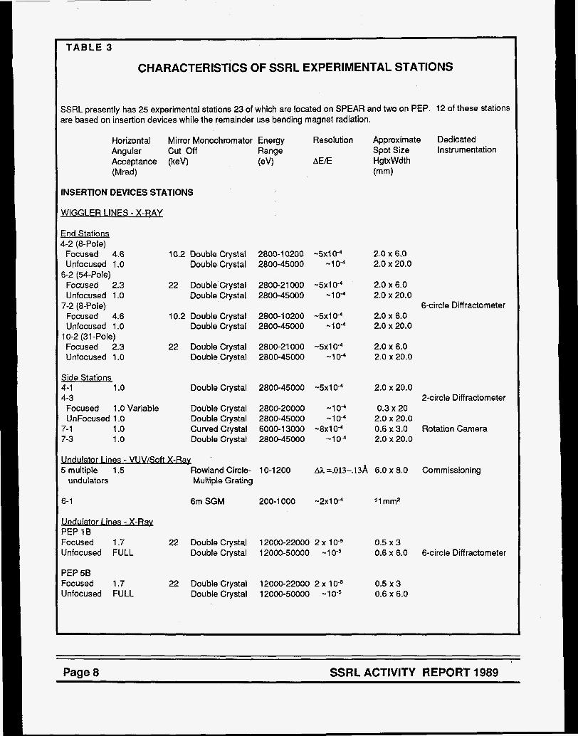
CHARACTERISTICS OF SSRL EXPERIMENTAL STATIONS
SSRL presently has 25 experimental stations 23 of which are located on SPEAR and two on PEP. 12 of these stations are based on insertion devices while the remainder use bending magnet radiation.
Horizontal Mirror Monochromator Energy Angular cut Off Range
(Mrad) Acceptance (keV) (eV)
Resolution
BEE
INSERTION DEVICES STATIONS
WIGGLER LINES - X-RAY
End Stations 4-2 (8-Pole) Focused 4.6 Unfocused 1 .O
Focused 2.3 Unfocused 1 .O
Focused 4.6 Unfocused 1 .O
Focused 2.3 Unfocused 1 .O
6-2 (54-Pole)
7-2 (8-Pole)
10-2 (31 -Pole)
Side Stations 4-1 1 .o 4-3 Focused 1 .O Variable UnFocused 1 .O
7- 1 1 .o 7-3 1 .o
10.2 Double Crystal 2800-1 0200 -5x1 0-4 Double Crystal 2800-45000 -1 0-4
22 Double Crystal 2800-21 000 -5x1 O 4 Double Crystal 2800-45000 - l o 4
10.2 Double Crystal 2800-1 0200 4 x 1 O 4 Double Crystal 2800-45000 -1 0-4
22 Double Crystal 2800-21 000 -5x1 O 4 Double Crystal 2800-45000 -1 0-4
Double Crystal 2800-45000 -5x1 0-4
Double Crystal 2800-20000 -1 O 4 Double Crystal 2800-45000 -1 0-4 Curved Crystal 6000-1 3000 -8x1 0-4 Double Crystal 2800-45000 -1 0-4
Approximate Spot Size HgtxWdt h (mm)
Dedicated Instrumentation
2.0 x 6.0 2.0 x 20.0
2.0 x 6.0 2.0 x 20.0
2.0 x 6.0 2.0 x 20.0
6-circle Diff ractometer
2.0 x 6.0 2.0 x 20.0
2.0 x 20.0 2-circle Diff ractometer
0.3 x 20 2.0 x 20.0 0.6 x 3.0 Rotation Camera 2.0 x 20.0
Undulator Lines - VUV/Soft X-Ray 5 multiple 1.5 Rowland Circle- 1 0-1 200
undulators Multiple Grating A I =.013-.13A 6.0 x 8.0 Commissioning
6-1
Undulator Lines - X-Rax PEP 18 Focused 1.7 Unfocused FULL
PEP 58 Focused 1.7 Unfocused FULL
6m SGM 200-1 000 -2~10-4
22 Double Crystal 12000-22000 2 x 1 O 5 Double Crystal 12000-50000 -1 0-5
22 Double Crystal 12000-22000 2 x 1 O 5 Double Crystal 12000-50000 -1 O 5
sl mm2
0.5 x 3 0.6 x 6.0 6-circle Diffractometer
0.5 x 3 0.6 x 6.0
Page 8 SSRL ACTIVITY REPORT 1989

Horizontal Angular Acceptance (M rad)
Mirror Monochromator Energy Resolution cut Off Range (keV) (eV) AWE
BENDING MAGNET STATIONS
X-RaL 1 -4 2.0 1-5 1 .o 2-1 (Foc.) 4.8 2-2 1 .O-6.1 2-3 1 .o
VUV/Soft X-Rav 1-1 2.0 1-2 4.0 3-1 2.0 3-2 4.0 3-3 8-1 0
3-4 0.6
8-1 12
B-2 5.0
Curved Crystal 6700-1 0800 0.3 x 1 D3
8.9 Double Crystal 2800-8900 -5 x 1 0-4
Double Crystal 2800-30000 -5 x 1 O 4
Double Crystal 2800-30000 -1 0 - 4
None 3200-30000
Grasshopper 64-1 000
Grasshopper 25-1 000 Seya-Namioka 5-40
4.5 UHV Double 800-4500
6m TGM 8-90
Ctystal (Jumbo) Multilayer 0-3000
6m TGM
6m SGM
8-1 80
150-1 000
Approximate Dedicated Spot Size Instrumentation HgtxWdth (mm)
0.25 x 0.5 SAS Detector 3 x 20 Area Detector/CAD-4 1 x 4 4 x 22 - 4 x 134 3 x 20
a h = .l-.2A 1.0 x 1.0 -2 x 10³ 1.0 x 2.0 Ah= .05-2A 1.OX1.0 AX = .2-6 A 2 ~7 - 5 ~ 1 O 4 1.5 x 2.5
White or 2 x 8
-9 x 10⁻³ 11 mm2 A h n = .6%
-3 x 10⁻⁴ 11 mm2
Vaccum DiffractometeV
Angle Resolved e-
Angle Resolved e-
Litho. Expo.Station
Spectrometer
Spectrometer
SSRL ACTIVITY REPORT 1989 Page 9

TABLE 4
DEDICATED DEMAND vs. ACCOMMODATION ON SSRL EXPERIMENTAL STATIONS
January 1989-March 1989
Proposals %Assigned Assigned
Shifts Shifts Over- Proposals Requested Assigned Subscription Requesting
Station
Wiaalers Lines End Stations 4-2 6-2* 7-2 Diffractometer 10-2*
503 191 242 66
126 42
126 42
466% 455% 200% 151%
13 4
14 4
4 3 7 2
Side Stations 4-3 Diffractometer 125 7-3,4-1 570 7-1 Rotation Camera 11 0
108 242 126
1 16% 226% 115%
7 45 10
5 14 10
Bendina Maanet X-rav Linea 1-4 SAS 91 1-5 Area Detector 119 2-1 Focused 114 2-2 White 240 2-3 EXAFS 12
126 126 72
108 108
72% 94%
158% 222%
1 1 O/O
4 11 14 11 2
4 4 6 6 8**
100% 36% 43% 55%
400%
VUWSoft X-rav Lines Grasshoppers (1-1, 3-1) 246 198 124% 8 5 63%
Double Crystal (3-3) 119
124
107
126
132
42
94%
94%
255%
75%
75%
33%
TGM's (1 -2, 8-1 *)
SGM (8-2')
Lithography (3-4) 85 126 67% 4 4 looo/o
Seya (3-2) 70 126 56% 3 3 100%
PRTline - general user time on& shown ** 2-3 was run in white-radiation mode part of the time to
accommodate overdemand from 2-2.
Demand for experimental time on the SSRL branch lines varies considerably. The percentage of proposals receiving beam time is often higher than the percentage of shifts accommodated since most experimenters do not receive the ful allotment of shifts requested. Demand is also limited by perceived availability of beam time. Experimenters have indicated that they could use much more time if it were available. In addition, many new groups have been discourage1 from starting programs based on synchrotron radiation at SSRL since the time available is so limited. This situation should improve once SPEAR becomes dedicated to synchrotron use.
~~~~~~ ~
Page 10 SSRL ACTIVITY REPORT 1989

Section I1 I I
The 3 GeV Injector & Accelerator Research

II THE 3 GEV INJECTOR & ACCELERATOR RESEARCH THE 3 GEV INJECTOR
SSRL is in the final stages of constructing a 3 GeV booster synchrotron as an injector for SPEAR This injector will make the SPEAR storage ring independ- ent from the SLAC Linear Accelerator. After authori- zation of the project by the DOE in February of 1988, most of the first year's effort was spent on detailed technical designs, fabrication of prototype compo- nents and construction of conventional facilities. During 1989 activities concentrated on the procure- ment, fabrication, and assembly of injector sub- systems. Many components were designed or specified by injector personnel, while fabrication occurred in industry, or SLAC shops, but primarily in- house within specially formed project groups. Assem- bly and test of sub-systems was also performed by the injector group. By year's end most of the main components were fabricated and installed. In particular, the main magnets with their supports, the vacuum chamber, the first linac modulator and all linac DLWG sections had been installed. Completion of the installation phase is projected for the first quarter of 1990 followed by a six month commission- ing phase.
LINAC System
Microwa ve Gun - In collaboration with Varian Associ- ates, a microwave gun has been developed to produce a high intensity electron beam as the first step of the accelerator chain comprising the injector project. The main feature of this type of electron source is its small beam emittance and high intensity. First tests show a beam current which exceeds, by at least a factor of two, the required intensity for the in- jector project. The electron source for the gun consists of a cathode immersed in the field of an RF cavity. Electrons emerging from the cathode through thermionic emission become accelerated immedi- ately by a high RF field of the order of 50 MV/m and the repelling electrostatic forces within an intense electron beam are compensated very quickly. As a consequence the electron beam frov a microwave gun has a much higher brightness than from ordinary sources. Three linac modulators have been con- structed at SSRL, each driving one klystron con- nected to a ten foot long S-band accelerating section. The accelerator sections were manufactured at the High Energy Physics Laboratory in Beijing. One of the sections has now been powered up to 30 MW or an accelerating field of 18 MeV/m.
Microwave Gun
Booster System
Booster R ina Maanet Svstem - The ring magnets for the booster were designed and assembled at SSRL. In order to allow cycling the booster at 10 Hz all ring magnets are constructed from laminations of mag- netic steel. The final choice of material was based on prototype tests performed with a variety of materials under realistic operating conditions. All magnet cores have been stacked, assembled with coils delivered by industry and tested by the injector magnet group. To allow efficient installation and alignment all magnets are preassembled and prealigned resting on precision locators on rigid girders. This preassembly
SSRL ACTIVITY REPORT 1989 Page 11

was done in the magnet assembly area and included the installation of the vacuum chamber and bussing.
Design work is underway to allow SPEAR to inject at full energy. This requires modification of the SPEAR
The completed girders were then transported to the ring tunnel for final installation.
Main Maanet Power Supplv - To minimize the interference of the cycling operation of the booster with other sensitive facilities at the SLAC site a White circuit was chosen to power the main magnets. In such a circuit the magnets are connected in parallel with capacitor banks to form a resonant circuit tuned to the operating frequency of the booster. The losses of this circuit are then compensated by energy pulses via magnetic chokes. All the magnets are designed for cycling up to 3.5 GeV at 10 or 15 Hz and the White circuit can also be configured to oscillate at 10 or 15 Hz.
m u u m Svstem - Due to the cyclical magnetic fields in synchrotrons, special vacuum chamber designs are needed to avoid eddy current losses. In some machines cermanic chambers have been used. For this injector the technique pioneered by DESY, con- structing the vacuum chambers of 0.3 mm thin stainless steel tubes strengthened by external metallic ribs, was used. This construction method proved very successful and has been incorporated into the injector design. All the magnet chambers have been fabricated, installed and pumped down.
Booste r Acceleratina Svstem - In order to minimize the maintenance complexity it was decided to duplicate the SPEAR RF system for the booster. One of the original four accelerating cavities of SPEAR has been recommissioned for the booster ring.
Beam Transport System
J&!ction/Fmct ion S v m - The injection and ex- traction transport systems are designed, and fabrica- tion complete. The magnets which transport elec- trons from the booster to SPEAR have been meas- ured and are installed. Installation of the transport components from the injector LINAC to the booster synchrotron has begun. The kicker magnets for injecting electrons into the booster and for extracting them out of the booster are ferrite picture frame magnets mounted inside the booster vacuum. They are built up from standard modules: 2 units for injection, 4 units for extraction. Construction of the kicker pulsers is underway. The injection septum is a DC horizontal deflecting magnet. The extraction septum will be a pulsed Lambertson type magnet.
injection components, including the septum magnet.
Project Schedule
It is expected that all injector components will be installed by the spring of 1990, and after a six month commissioning period, a dedicated, full energy injector for SPEAR should be available in October 1990.
I
Bending Magnet Engineering Model with Coil (Top Half Removed)
Page 12 SSRL ACTIVITY REPORT 1989

385 MHz RF Cavity in Ring Tunnel
Beam Transport Line from Booster to SPEAR
SSRL ACTIVITY REPORT 1989 Parte 13

Ring Bending Magnet, Quadrupole, Vacuum Chamber
Page 14 SSRL ACTIVITY REPORT 1989

ACCELERATORRESEARCH PROJECTS
SPEAR Studies
Accelerator research projects have been pursued in connection with the training of graduate students. Specific emphasis is given in combining theoretical studies with practical hands-on experience with accelerator systems and the design, fabrication and testing of new accelerator components.
Major accelerator research efforts concentrated on the design of the ring lattice and stability studies for the new 3 GeV injector, and the beam transport lines between the pre-injector linac and the booster as well as between the booster and SPEAR.
A low emittance configuration for SPEAR has been developed to reach a factor of 3 smaller beam emittance. This requires a third injection kicker in SPEAR which is part of the injector project. Injection studies and stability simulations have been per- formed and experimental verification is expected to occur during the injector commissioning.
With the transition of SPEAR to a dedicated synchro- tron radiation source, consideration is now being given to hardware modifications to the long interac- tion region straight sections to enable them to accommodate long undulators. Preliminary studies indicate that it should be possible to revise the present straight sections so that the free straight section length is increased from the present 2.5 meters to 10 meters or more. Emphasis is placed on developing an optics for a 5 meter long undulator which will be the source for a proposed 1-4 keV beam line.
PEP Studies
Accelerator physics research on PEP has diminshed due to the higher priority of SPEAR and the injector, the limited operation time of PEP and the uncertainty of PEP'S future as SLAC develops a proposal for modifying PEP to function as a B-meson factory.
3 GeV Booster Ring
SSRL ACTIVITY REPORT 1989 Page 15

Some work continues on the development of a longitudinal feedback system for PEP.
Feedback systems have been found necessary to reach high currents with stable beams in many light sources. SSRL is presently developing a system to combat longitudinal instabilities in PEP. Such systems are likely to be needed also on SPEAR, particularly when the low emittance mode is imple- mented.
The work that has been done includes detailed characterization of the frequency response of the cavity, comparison of phase detection with energy detection, the design of a stripline monitor and the choice of a suitable location for it in the PEP ring, the exploration of different placements of the cavity in tht ring, the possible impact of the cavity on PEP opera- tions and the development of the feedback electron- ICs.
The detailed pian for the system was developed in consultation with many laboratories (SSRL, SLAC, LBL, NSLS, Argonne, DESY and Hiroshima Univer- isty). The cavity is scheduled for installation into the PEP ring in March 1990. The first tests for the longitudinal feedback systems are anticipated for summer 190.
Also, reports have been completed on the use of the PEP emittance control wigglers as sources of circular polarized hard x-rays and on the modifica- tionsneeded to PEP if it were to become a dedicated light source. These reports are available from SSRL. The work on PEP as a source of circular polarized x- rays will be published in the proceedings of SRI-89.
General Accelerator Physics Studies
Detailed theoretical studies of optimum microwave gun designs have been undertaken leading to the construction of such electron sources for the injector project. Systematic measurements of beam parame- ters and comparison with theory will follow in the coming year.
Fundamental limits of beam optical aberrations have been studied. As a consequence the feasibility of a storage ring with a beam emittance of 0.04 nanome- ters has been established. This emittance is a factor of one hundred smaller than that for the APS or ALS and would produce fully spatially coherent radiation down to wavelengths of 3A.
The simulation programs, PATRICIA and PATPET, have been further developed. These programs, which allow the simulation of particle motion in the presence of a variety of perturbations, are now being used in a number of laboratories throughout the world.
Page 16 SSRL ACTIVITY REPORT 1989

Section I11 I I
Emerimental Facilities I


111 EXPERIMENTAL FACILITIES Experimental facilities at SSRL are of two types: general facility stations and participating research team (PRT) stations. General facility stations have been funded by various government agencies, principally the DOE, NIH and NSF and are open to the user community on a competitive basis for 100% of their operating time.
SSRL has three operational PRT's with a fourth in the process of developing a branch line. All present PRTs are three party collaborations with SSRL as one of the parties. The two outside institutions receive 2/3rds of the available beam time while the last third is resewed for the SSRL general users. The PRT arrangements are for a 3 year period. Renewal is based on review by an ad hoc committee, appointed by the Stanford University Dean of Re- search, which considers scientific merit, contributions to graduate student education and to the SSRL user community.
X-RAY FACILITIES
There are 13 x-ray stations located on six beam lines at SSRL. Two of these are PRT lines (wiggler lines 6 and 10). The other 11 are SSRL facility stations. Of the facility lines, two (Beam Lines 4 and 7) have wigglers as sources for six stations and the other two (Beam Lines 1 and 2), sewing five stations, have bending magnet sources. There are also two x-ray beam lines on the PEP storage ring, each serving a single experimental station.
Improvements to Existing Experimental Stations
Beam Line 1 ; The two x-ray branch lines on Beam Line 1 are a Small Angle Scattering line (1 -4) and an EXAFS line (1-5), which can also be run in a mode dedicated to a Protein Crystallography Area Detector. A major upgrade to Station 1-4 was started in 1988 and has continued this year. During 1989 a commer- cial version of the original photodiode array detector was purchased. This detector has been customized for low temperature operation and beam stops were added. IBM donated a PC which is used to operate the detector. A valve to allow changing the sample- to-detector distance without exposing the cold array to ambient air has been added. Finally, the mono- chromator has been fitted with absolute encoders to better monitor its configuration.
On the EXAFS line the older racks were replaced with taller versions in order to consolidate the con- trols and free more area for setup space.
Beam I ine 2; Several upgrades were made to the three branch lines on Beam Line 2. During the last run it was determined that the 2-1 mirror had finally reached the end of its useful life: a new mirror has been ordered. An additional driver chassis has been constructed for remote operation of the diffractome- ters on the white light line (2-2), thus alleviating the need for connector changes when switching from one diffractometer to the other. A spectrum analyzer to monitor the Fourier components of the photon beam noise has been installed as part of the dedicated diagnostics port on this beam line.
Branch Line 4-3; The hutch table was modified to increase the tilt range of the diffractometer to match the larger range of angles allowed by the new 4-3 mirror.
B-: In 1989 this branch line moved into the microVax era. During the winter run the new version of SUPER had its baptism, and after some initial frantic debugging, proved to be a very success- ful control package. It is expected that SUPER will become the standard program to control all diffrac- tometers at SSRL and is also being used with ithe x- ray generator. The on-line HELP file is available as an ASCII file and can be transmitted to interested users.
General X-ray Facility Improvements
Although much of the efforts of the x-ray staff were directed to the SPEAR improvement projects de- scribed in Section1 , several other tasks were com- pleted during the long 1989 shutdown. An extensive survey of all of the electrical outlets in both experi- mental halls was undertaken, and all wiring brought up to current safety standards. A bar-code inventory control system for x-ray equipment was implemented.
The first crystals to be completely cut and polished in-house were finished. The t) orientation of these three sets of Si(220) crystals was chosen to comple- ment the already existing crystals.
-
SSRL ACTIVITY REPORT 1989 Page 17

Two new sample positioners and their support chassis were constructed.
In addition, a number of x-ray staff members went to the NSLS to provide support for SSRL users running on NSLS Beam Line X19A. Standard SSRL sample positioners were also sent to Brookhaven for use by SSRL users.
Crvsta I Coolina Pro synchrotron radiation laboratories has shown that the performance of a typical double-crystal silicon monochromator suffers when the power density on the first crystal reaches a few watts per square millimeter. Thermal gradients in the crystal cause its surface to pillow slightly, and the consequent warping of the crystal planes reduces the transmitted flux. Since the power density at SSRL's wiggler lines 6-2 and 10-2 can reach this level, and since significantly higher values will be achieved on future beam lines, SSRL has been studying methods for more efficient cooling of monochromator crystals.
Recent research at several
In 1989 we began studying a technique developed at LLNL for the cooling of integrated circuits. This technique uses numerous tiny water cooling chan- nels cut directly into the crystal. During the past year, in collaboration with scientists from Livermore, methods for cutting the cooling channels and attach- ing the associated plumbing without introducing strain have been developed. Several prototype crystal assemblies are now ready for testing at a synchrotron beam line, which should occur during the spring of 1990.
VUV FACILITIES
There are currently ten VUV or soft x-ray branch lines at SSRL. Four of these facilities were built by PRT's and the general user community has access to 33% of the time on these lines. The facilities being scheduled for users are: two TGM's (1 -2 and 8-1), two grasshopper monochromators (1-1 and 3-1), a Seya-Namioka monochromator (3-2), an in-vacuum double crystal monochromator (3-3), a SGM (8-2) and the lithography/optics line (3-4). Two PRT branch lines, 6-1 and 5, are in the final stages of commissioning.
mprovements to Existing Experimental Stations
jranckLbe 1-1 ; The old grasshopper was operated uith the new elliptical M1 mirror installed in late 1988. The new mirror reduced the spot size from 90 p to IO p. , with a resulting factor of three increase in flux hrough the entrance slit. With the small spot size, md the cantilever design of the new mirror holder, ribration of the mirror caused intensity fluctuations hat did not damp out until one second after the nonochromator stopped scanning. A modification to he mirror holder was made to hold the end of the M1 nirror and to make fine in situ adjustments of the ocus at the entrance slit. Also, the scanning motor was changed to a smoother operating 5 phase Stepper. It is now expected that the dead time due to nirror vibrations should be negligible, and the spot ;ize at the entrance slit should be reduced to an ?stimated 25 p, with a further increase in flux.
jranch I ine 1-2: The new TGM on Branch Line 1-2 was opened to users in the 1989 run. The first VUV >earn line with a microVAX workstation for mono- :hromator control and data acquisition, it operated Nith the EXP and AUTO data acquisition and analy- ;is packages. During the run, it was confirmed that he 2400 Vmrn grating is of poor quality, and will have o be replaced. Thus, the range of the monochroma- or is limited from 7 to 90 eV. The roll motion on the UO was reassembled to ease the alignment of the )earn into the monochromator. This adjustment, iowever, cannot completely remove the cant built nto the 15 year old copper MO mirror. To correct this 3 Sic mirror has been ordered (with the assistance i f LANL) which will greatly increase the flux through he monochromator. A permanent I, section and meumatic exit valve have been added after the *efocusing mirror. The section has a metal pneumatic Zxit valve.
Branch I ine 3-1: A microVAX workstation is being installed on the new grasshopper. It will be operating with the EXP and AUTO data acquisition and analy- sis packages.
Branch Line 3-2: The main bearing for the grating rotation galled during the last run. It was rebuilt and modified to prevent reoccurence of the problem.
A new fused silica mirror was been received to replace the one that has been in use on Jumbo since 1979.
Page 18 SSRL ACTIVITY REPORT 1989

BIOTECHNOLOGY FACILITIES
The research and user support in the Biotechnology area at SSRL are funded jointty by the NIH Division of Research Resources and the core SSRL DOE operations budget. Two specialized branch lines for protein crystallography (1-5 Area Detector and 7-1 Rotation Camera) exist as well as a variety of sup- porting equipment and instrumentation.
The current NIH grant is in its last budget year. A 5- year renewal proposal was submitted in May 1989. As part of the proposal review process, the SSRL Biotechnology Resource had a site visit in November. The Special Study Section gave a very favorable review and rating of the proposal, and it is now clear from NIH that the SSRL Biotechnology Resource will receive continued funding at the present level or higher. Major projects/instnrmentation included in the proposal are an imaging plate detector system for use in crystallography and small angle scattering, an additional area detector for the system on Branch Line 1-5, acquisition of a Ge detector array system for XAS studies, implementation of a harmonic rejectiorVfocusing mirror for Branch Line 7-3 (US), and a low-energy enhancement to Branch Line 6-2 including an in-vacuum monochromator and a Ni- coated mirror with an energy cutoff at about 6 keV.
Facilities for X-rav AbsorD . tion SDect roscoD . v : Contin- ued progress in the development of Hgl, detector arrays was achieved through careful characterization of a 5eIement array under realistic operation condi- tions at SSRL. With an improved front end design the detectors maintained nearly ideal spectrometric behavior to input count rates in excess of 100 kHz.
The “rapid turnaround station for XAS measure- ments was put in use for the first time. Due to the limited amount of beam time, only two such users were accommodated. After the successful use during the winter run of the new motorized alignment rails for XAS measurements, an additional rail was manufactured and assembled. Two rails are now dedicated to Branch Line 7-3 and to the rapid turn- around EXAFS station, respectively, while a third rail is available on request for the remaining XAS beam lines. A design effort has been successfully com- pleted in the construction and assembly of a single crystal XAS alignment system to be used with the motorized rail system. It consists of a K arc (rotation 0-100” and a 4 rotation stage (full rotation). The system includes a movable laser-based beam/crystal
alignment module attached to the rail through a standard slide.
ies for Protein CrvstwaDhv: During the past year a permanent radiation enclosure for the area detector data acquisition system on Branch tine 1-5 has been built. This new enclosure, which is the size of a small room, replaces the portable hutch that has been in use since the area detector system was first built. The enclosure is large enough to permit the future addition of a liquid nitrogen cryostat for low temperature crystallographic data collection. It will also provide enough space to accommodate an additional area detector at a future data which would greatly enhance the efficiency of multi-wavelength anomalous scattering data collection. Furthermore, the diff ractometer has now been mounted directly onto the concrete floor slab for additional mechanical stability, and there is now adequate space for users to walk around the instrument for alignment pur- poses. This will make the system far more conven- ient to use.
The liquid nitrogen cryostat, that was built last year for the rotation camera system on Beam Line 7-1, has been further refined and tested on protein samples. This cryostat is now available to visiting user groups as part of the rotation camera system. A translation stage has been added to the rotation camera crystal monochromator so that a different region of the monochromator crystal can be used without opening the monochromator tank should the crystal become damaged by the x-ray beam.
A computer-controlled single-crystal goniometer for white beam Laue diffraction, incorporating a very fast x-ray shutter system, is under construction. When complete, this instrument will be used to perform time-resolved studies in protein crystallography using an Imaging Plate Detector system.
m l i t i e s for Small Ana le Scattenna. ’ * Amajorim- provement to the SAXS camera has been completed with the integration of a new detector into the system. This detector, a quadrant detector (obtained from Dr. A. Gabriel, EMBL, Grenoble), is of gas chamber type with spatial resolution in one dimension, but with a sensitive area covering a 60 degree quadrant. Integration of signals along 60 degree arcs provides a very significant improvement in signal-to-noise ratio at higher Q values when compared with single-wire
SSRL ACTIVITY REPORT 1989 Page 19

linear detectors. The new detector is most applicable to isotropic scattering experiments, but can also be used with oriented samples if some degree of mosaic spread is present. A fully motorized X-2 translation stage has been constructed to allow remote center- ing of the detector in the x-ray beam. As a sepa- rately moving component of the camera, the detector allows the existing alignment rail, motorized stages, and defining slits to be used for positioning samples and controlling the beam size. A sample-to-detector helium path has been manufactured to eliminate air scattering. I, (transmitted intensity) measurement is achieved using a design in which I, photons hit a flourescent screen mounted on the beam stop and the emitted light collected by a fiber optic cable and transmitted to a photomultiplier tube for measure- ment.
PRT EXPERIMENTAL FACILITIES
Improvements to Existing Experimental Stations
Beam Line 5 (Xerox/Sfmford University/SSRL) - During the early shifts of SPEAR storage ring opera- tion in 1989, the new monochromator on the multi- undulator Beam Line 5 took its first spectra. Perform- ance during initial commissioning confirmed that the design and fabrication goals have been met. This is illustrated in Figure 2 which shows the observed transmitted flux compared to the calculated values.
Photon Energy (eV) 0. 200 400 600 800 l o o 0
L . . . . , . . I - - . t . . . I . t . . . . t
Photon Energy (eV)
FIGURE 2
Beam Line 5 Monochromator Transmission Undulator Fundamental at 400 eV
- 2 2 43
2 Degree Grating
Observed Transmission vs. Calculated Values, Beam Line 5. The functional form of the observed transmitted flux of the beam line at a fixed undulator setting is compared to calculated values. The transmission of both the 2" and 4" gratings, as well as both the end station and side station are shown. The total electron yield from gold was used as a measure of the transmitted flux. The calculated transmitted flux includes the calculated spectral distribution of the source and the reflectivity of the mirrors, and assumes a grating efficiency function based on measurements of previous gratings pur- chased from Astron The peak in transmitted flux at about 120 eV is due to a peak in reflectivity of the optics, while the 400 and 800 eV peaks are due to the undulator fundamental and 2nd harmonic. The qualitative agreement and overall function form of the transmitted flux confirms that scattered light is not a serious problem. In addition, the side deflecting mirrors demonstrate predicted reflectivity performance, which results in usable side station flux up to 800 eV.
Page 20 S S R L ACTIVITY REPORT 1989

FIGURE 3 I - - . ' . - - - I ' . - r . - - - I . . . . ' - . - - 1 - - - , - . . - I . -. - I - .- - i - . . - I . i . . . - r - - - -
BL5 Endstation 600 hv= 385eV - -
c 1s fi-om Diamond CMA detection - -
AES slits =d' 0 - 25 e V Pass Energy d 0 Beam Current 22 23.4mA a
-
Undulator fbndamental - - set to 400 eV 3
- 0
;h ,u_ - - -+ A E = 1.34 e V -
.c; - -
.d e 200- -
-4
E W - - - -
- --
I I I I 1 1 1 1 0 0 102 104 106 0.
Kinetic Energy <eV>
The Carbon 1 s Photoemission Spectra at 385 eV on Beam Line 5
Operation of the beam line was limited to fixed undulator gaps for this initial run. Early in the run it was discovered that the exit slit had jammed open. Ironically, the monochromator resolution was limited by this stuck slit to that of the grasshoppers at SSRL. Shown in Figure 3 is the carbon 1 s photoemission spectrum from diamond at an excitation photon energy of about 385 eV. The peak width is almost entirely due to the monochromator, as the natural linewidth of the C 1s is much less than 1 eV. The apparent monochromator resolution confirms that the slit opening is stuck at an opening of roughly 50 p. In comparison to carbon 1 s photoemission measure- ments made on the new grasshopper, Branch Line 3- 1, (March 1982) at similar photon energies (and operating at the same photon resolution) it is esti- mated that the Beam Line 5 photon flux at 400 eV is about a factor of 20 more intense (normalized to beam current). Final optimization of the beam line during April, 1990 may alter (Le., improve) this figure.
After the run the 1 and 2 meter gratings of the monochromator were replaced with new specifically figured Astron gratings. These gratings were opti- cally aligned for optimum instrument resolution using a mercury lamp technique. Extensive characteriza-
tion of the instrument's optical performance was made, particularly, flux measurements as a function of slit width, determination of zero order positions for the various optical elements, determination of the slit position as a function of encoder position, and extensive exercise of the mechanical motions and repeatability of position. Various improvements were also made to the electronics systems and monochro- mator software. Undulator control capability from the beam line has also been implemented, utilizing direct communciation with the SPEAR control computer.
Beam Line 8 (University of California/Nationa/ Laboratories/SSRL) - Very good spectral resolutions have been measured for TGM Beam Line 8-1 using an electron time-of-flight spectrometer. For the G2 grating (822 gr/mm), FWHM linewidths of 39 meV and 85 meV were obtained for the Kr 4s-l line at 27.52 eV and the Xe (4dV2-6p) line at 65.1 1 eV, respectively. A FWHM linewidth of 41 meV was obtained at 65.1 1 eV using the G1 grating (2400 gr/mm). These results were derived by subtracting in quadrature an instrumental spreading of 20 meV. The electron spectrometer data yielded negligible higher-order light for G1 above 11 0 eV. This is
SSRL ACTIVITY REPORT 1989 Page 21

consistent with an estimated factor of five suppres- sion of higher orders in this energy range by prefo- cusing mirror roll off. Below 100 eV, higher-order contributions to the G1 output did not exceed 20%. Very little higher-order contributions were observed for G2 above 35 eV. In contrast, very significant higher-order contributions were observed for G2 below 35 eV and for G3 (288 gr/mm) through its entire range. A suitable mirror roll-off device, such as Sic at 20' with a sharp roll off above 90 eV, is recommended for TGM operation below 35 eV. Prefilters now installed on the branch line include In, Sn, AI, Si, Be/C, B, and C.
A new multipurpose sample chamber was installed on SGM Branch Line 8-2 and tested during the spring 1989 run. It includes a 150-nm AI vacuum barrier for operation at 1 0-6 Torr, a well-shielded flux monitor
mounted near a pair of Huber adjustable entrance slits, a translatable filter holder, a calibrated NBS- style diode featuring a Au cathode that can be inserted into the beam near the chamber exit, and computer-coupled rotation stages for multilayer reflectivity measurements. A large (1 x 1 cm) GaAsP detector was mounted on the detector arm which, despite its size, had adequately low dark current. A nearly ideal focus was obtained at the chamber center (less than 1 x 3 mm), thus verifying excellent exit mirror performance. Two new I, monitors both performed very well. A new channeltron-amplified I, monitor located upstream of the window section gave similar results to a gold mesh I, monitor located in the scattering chamber near the entrance slits. The unamplified gold mesh is very well shielded yielding dark currents that were a factor of 5 or 10 lower than previous designs.
FIGURE 4 2 I I I 1 I I I I I I
BL-Vlll-2 SGM -200 gr/mm - -(I15 scale) 100 pm slits
3 GeV 100 mA -
1.5 - A 500 gr/mm -
o calculated i 9 n measured
photons
0 I I I I 1 I I I
0 0.2 0.4 0.6 0.8 1 1.2 1.4 x-ray energy, keV
Calculated SGM Throughput Compared to Au Diode Results, Branch Line 8-2. The calculated results are based on a 3 GeV, 100-mA SPEAR bending magnet spectrum, ideal grating efficiency, and ideal Pt reflectivity at 2.5" and 2" (first and exit mirrors); measured results were corrected for transmission through the 150-nm AI window. Increases in measured flux for the 500-gr/mm case below 300 eV and above 550 eV are due to second-order and zero-order contributions, respectively. The second order component is preferentiaily transmitted through the AI window and has not been sub- tracted. These results are in satisfactory agreement considering the corrections required and the preliminary status of the grating efficiency calculations.
Page 22 SSRL ACTIVITY REPORT 1989

FIGURE 5
0.4 -
Transmission -
0.2 -
0
1 109. g/mm +I500 A" AI
cu l a /
0.2 0.4 0.6 0.8 1 1.2 1 x-ray energy, keV
Measured Transmission Results for Typical Station 8-2 Gratings
FIGURE 6
BL-Vlll-2 S G M
x-ray energy, keV Second Order Results from the 1100 gr/mm Grating. The second-order results obtained for the SGM 1 IOO-gr/mm grating based on Fe, Ni, Cu, and Mg edges are shown. Each value has been corrected for the moderate detector sensitivity difference at the two edges. Corrections for third and higher orders are negligible. Note the evidence for rather weak third-order at 434 eV (31 1 eV) for Mg (Cu). These results suggest that above 600 eV this grating range is relatively free from second- or higher-order contributions and that the second-order fraction is somewhat reduced if the vacuum barrier is not required.
SSRL ACTIVITY REPORT 1989 Paae 23

Good data on prefilter transmission and monochro- mator plus window output was obtained using an
To obtain photons in the 1-3 keV region, modifica- tions were made to allow or>eration without the Be
NBS style gold x-ray diode. Absolute calibration of the detectors was done at LLNL using proton- induced K and L lines in the range of interest, Figure 4 shows calculated SGM throughput compared to results obtained using the Au diode. These results are in satisfactory agreement considering the correc- tions required and the preliminary status of the grating efficiency calculations.
Prefilters installed in SGM Branch Line 8-2 include C, Ag, V, Fe, Ni, Cu, and Mg. Figure 5 shows meas- ured transmission results for typical filters. Note that higher orders are exaggerated in Figure 5 because the first-order amplitude has been reduced by the vacuum barrier. Filter transmission data has been used to determine absolute second-order contribu- tions. Figure 6 shows second-order results obtained for the SGM 11 00-gr/mm grating based on Fe, Ni, Cu, and Mg edges. These results suggest that above 600 eV this grating range is relatively free from second- or higher-order contributions and that the second-order fraction is somewhat reduced if the vacuum barrier is not required.
Line 1 Q (University of California/Nafional Laboratories/SSRL) - During the spring 1989 run the flux of Branch Line 10-2 in the focused mode was determined. With SPEAR operating at 3.3 GeV and 59 mA ring current, the measured flux at 10 keV photon energy was 3.4 x lo1* photonskec for a wiggler field of 1 .O Tesla.
In 1989 a new tandem diffraction hutch was installed on Branch Line 10-2. This hutch is shielded to handle white light and can accommodate a 1.5 m radius swing. A large (48" x 72") optical table top is being installed in the front hutch for EXAFS, imaging, and other experiments.
The beam movement and energy drift problems encountered during the 1989 run have been diag- nosed and solved - the energy drift by eliminating false pulses sent to the monochromator motor drive during scanning and the horizontal beam movement by correcting the off-parallel alignment of the plates housing the monochromator crystals and crystal holders. As a result of the earthquake, Branch Line 10-2 dropped -.1" with respect to the SPEAR plane. Realignment has been completed.
window module. This included installation of a differential pump section to serve as a transition region from UHV to HV and modifications of the monochromator to enable operation at high vacuum ( l o 7 Torr). Rh-C multilayers (2d = 80 A and 2d = 40 A) and a beryl crystal will be used as dispersive elements. A HV experimental chamber is also available. A schematic drawing of the modified beam line is shown in Figure 7. A protocol for the change- over from hard to soft x-rays has been developed. Tests and characterization of the modifications will be conducted during the spring 1990 run. Because the present monochromator cooling is probably insuffi- cient for operation at the full wiggler field, the initial measurements will be made with operation in undula- tor mode.
We are also implementing a quick scan EXAFS capability. R. Frahm (HASYLAB) is collaborating with LLNL to install this new capability and to perform time-resolved EXAFS measurements of thin film growth processes and interface formation in the time domain of seconds.
PRT Facilities Under Development
Rranch Line 6-1 (LBUSSRUEXXON) - During the last dedicated SPEAR run a new Spherical Grating Monochromator (SGM) saw its first synchrotron light at the LBUEXXON/SSRL 54-pole wiggler Station 6-1 . This monochromator and two premirrors, designed and fabricated by LBL for high resolution and high flux requirements, had been installed at SSRL in 1988.
The monochromator is based on a Rowland Circle geometry with a large radius (R=55 m) and movable slits. The first optical component, a movable and water-cooled plane mirror, horizontally reflects a part of the wiggler beam onto a second mirror, a toroid. This focuses vertically on the entrance and horizon- tally on the exit slit of the monochromator. The grating chamber may accommodate three large (65 mm xl80 mm) water-cooled metal gratings. These gratings were not available for the winter run. They are now nickel-coated and polished, but the ruling process will still take several months. Instead, a single fused silica grating (600 Vmm, no cooling) from Ferranti Astron of outstandingly high quality (very small surface roughness and slope errors) was installed.
Page 24 SSRL ACTIVITY REPORT 1989

FIGURE 7
- r N
Y Schematic Drawing of Line 10-2
The first output data of the monochromator taken with an aluminum photodiode show an unusually low stray light contribution, which confirms the quality of the grating. The energy range of this grating covers 180 eV to 820 eV. The absolute photon flux is calculated to be 1 xl0" Phot/sec/lOO mA at 440 ev with 0.5 eV resolution (Le., 100 p slits).
Two strong peaks which are visible in the output spectrum at 220 eV and 440 eV were identified as the 1 st and 2nd harmonic of the 54-pole wiggler (0.5 T). This means that, for future applications, the wiggler could be used to increase the photon flux in small, selected energy ranges.
For a fine alignment of the monochromator, and hence improvement of its resolution, the photodiode was replaced by an ion chamber (supplied with 0.01 Torr nitrogen). The ion yield from nitrogen displays five clearly resolved vibrational final states structures at the ls-2p' resonance at about 401 eV. This dem- onstrates that the achieved instrumental resolution at the end of the run was already less than the lifetime
broadening, which is about 130 meV for N 1 s vacan- cies.
A deconvolution using Lorentzian functions for the lifetime width and with Gaussian functions for the monochromator resolution shows a width of 80 meV for the monochromator resolution, i.e., at 400 eV a resolving power of 5000. This is the second best resolution ever achieved with a monochromator covering the K-edges of the elements C, N, 0, and F. (C.T.Chen and F. Sette, Bell Labs claim 10,000 for their new SGM with an 800 Vmm grating at NSLS).
It is worth mentioning that the measurements at SSRL were performed under "low intensity" condi- tions, which means the wiggler was mainly operated at 0.5 T and the mirror MO was only modestly in- serted into the wiggler fan, (an intensity of 4-9% was subtracted from the x-ray Branch Line 6-2).
It is expected that the photon flux and the resolving power can be improved further during the next SPEAR run.
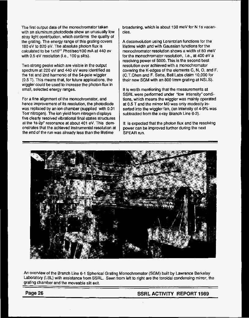
An overview of the Branch Line 6-1 Spherical Grating Monochromator (SGM) built by Lawrence Berkeley Laboratory (LBL) with assistance from SSRL. Seen from left to right are the toroidal condensing mirror, the grating chamber and the moveable slit exit.
Page 26 SSRL ACTIVITY REPORT 1989

I
Branch Line 10 - 1 (lBM/Sanford University/SSRL) - During the year the front end components of Beam Line 10 were modified to accept the components for the new 10-1 station. The mirrors and monochroma- tor will be delivered in the first half of 1990. For specific details of the branch line see the 1988 Activity Report. The expected date for first light in this line is October 1990.
SUPPORT FAClLlTlES
- Preparations for the Comwtational Faci lim conversion of all PDP-11 beam line data acquisition systems to VAXstations have continued. Program- ming for the base-level driver system (called VAX motors) is fully complete and programming for higher-level user-interface software for specific
.. .
applications is ongoing. The first major application of the new hardwarekoftware control system will be for EXAFS applications on Branch Line 7-3 during the coming Spring 1990 run. Following full debugging and documentation, all remaining systems are expected to be converted to VAXstation control by the end of Summer, 1990.
Biochemical I ahzikyy. One major addition was made to the lab in 1989, a refrigerated Beckman GPKR centrifuge with rotors.
Yser Clean roo^: The clean room vacated by the vacuum group, on their move to Building 137, has been set up for use by staff and users. It has benches, tools, spot welder, fume hood for cleaning small parts and a limited supply of hardware that can be signed out.
The grating chamber with facility for three interchangeable, water-cooled gratings. Underneath black covers are a HP laser interferometer and stepping motor from the Orasis Corporation, which precisely control the angle of the grating. Standing next to the chamber is assembly technician Bill Gath.
SSRL ACTlVlTY REPORT 1989 Page 27
Page 28 SSRL ACTIVITY REPORT 1989

Section IV
SSRL Organization

IV SSRL ORGANIZATION
On December 22, 1989, SSRL Director A. Bienenstock announced a reorganization of the SSRL technical staff which became effective January 2, 1990. The goals of the reorganization were to focus effort on SSRL‘s three highest pricrities: to complete and commission the injector, to prepare to operate and maintain SPEAR and the injector and to have a successful spring 1990 experimental run. In the new organization, all the technical staff is contained in three groups:
1 . Accelerator Research and Operations Division 2. Injector Project 3. Photon Research and Operations Division.
Responsibility for safety and quality assurance resides in the Director’s Office.
The functions of the Accelerator Research & Operations Division are: accelerator operations, accelerator physics research, engineering, technical resources and user support. The Photon Research & Operations Division is responsible for: beam line management, beam line development, research using synchrotron radiation and user support. At the completion of the injector project personnel from this project will be incorporated into the other divisions as appropriate. In addition, there is a Computational and Administrative Resources Division.
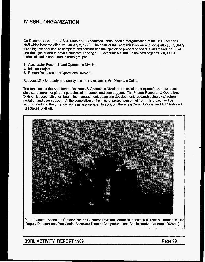
3ero Pianetta (Associate Director Photon Research Division), Arthur Bienenstock (Director), Herman WinicE :Deputy Director) and Ron Gould (Associate Director Computional and Administrative Resource Division).
SSRL ACTIVITY REPORT 1989 Page 29
BRANCH LINE
1-1 1-2 1-4 1 -5 1 -5AD
2-1 2-2 2-3
3-1 3-2 3-3 3-4
4-1 4-2 4-3
5
6-1 6-2
7- 1 7-2 7-3
8-1 8-2
10-2
PEPlB , PEP 58
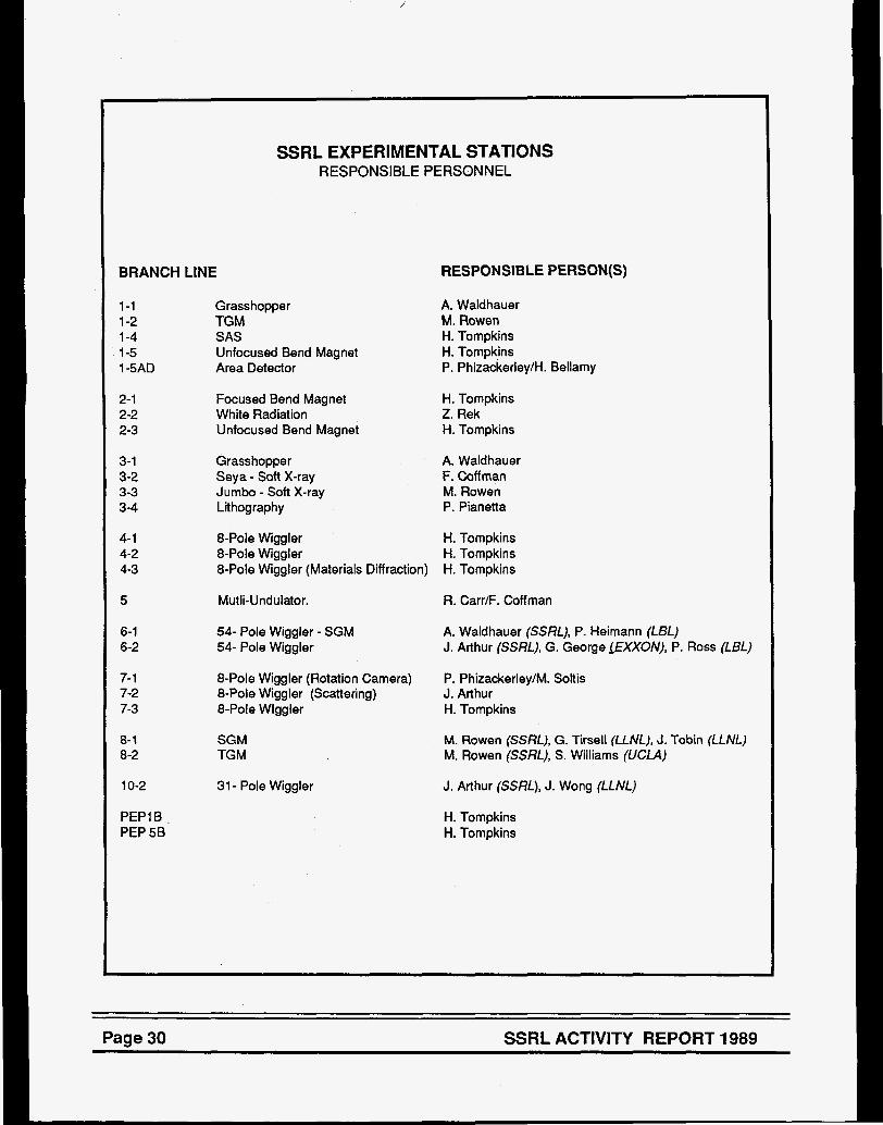
SSRL EXPERIMENTAL STATIONS RESPONSIBLE PERSONNEL
Grass hopper TGM SAS Unfocused Bend Magnet Area Detector
Focused Bend Magnet White Radiation Unfocused Bend Magnet
Grasshopper Seya - Soft X-ray Jumbo - Soft X-ray Lithography
RESPONSIBLE PERSON(S)
A. Waldhauer M. Rowen H. Tornpkins H. Tornpk,ins P. Phizackerley/H. Bellamy
H. Tompkins Z. Rek H. Tompkins
A. Waldhauer F. Coff man M. Rowen P. Pianetta
8-Pole Wiggler H. Tornpkins 8-Pole Wiggler H. Tompkins 8-Pole Wiggler (Materials Diffraction) H. Tornpkins
Mutli-Undulator.
54- Pole Wiggler - SGM 54- Pole Wiggler
R. Carr/F. Coff man
A. Waldhauer (SSRL), P. Heimann (LBL) J. Arthur (SSRL), G. George fEXXON), P. Ross (LBL)
8-Pole Wiggler (Rotation Camera) 8-Pole Wiggler (Scattering) J. Arthur 8-Pole Wlggler H. Tompkins
P. Phizackerley/M. Soltis
SGM TGM
31 - Pole Wiggler
M. Rowen (SSRL), G. Tirsell (LLNL), J. Tobin (LLNL) M. Rowen (SSRL), S. Williams (UCLA)
J. Arthur (SSRL), J. Wong (LLNL)
H. Tompkins H. Tornpkins
Page 30 SSRL ACTIVITY REPORT 1989

INSTRUMENTATION/FACILITY RESPONSIBILITIES MATERIALS DIFFRACTOMETER: S. Brennan, H. Tompkins PERKIN-ELMER CHAMBER: M. Rowen VG SAMPLE CHAMBER: M. Rowen AREA DETECTOR: P. Phizackerley, H. Bellamy ROTATION CAMERA: P. Phizackerley, M. Soltis CAD-4 DIFFRACTOMETER: M. Soltis COMPUTER SYSTEMS: T. Cox 7-2 SPECTROMETER: S. Brennan, H. Tompkins BIOCHEMISTRY LABORATORY: B. Hedman, R. Mayer DARKROOMS: M. Soltis, Z. Rek BEAM LINE STEERING: R. Hettel EXAFS EQUIPMENT AND SOFTWARE: 8. Hedman, R. Mayer SAS CAMERA (BIOTECH): S. Wakatsuki EXAFS CONSULTANT: B. Hedman SCATTERING CONSULTANT: S. Brennan RT-1 1 SOFTWARE CONSULTANT: S. Brennan TOPOGRAPHY EQUIPMENT: Z. Rek RAPID TURNAROUND EXAFS FACILITIES: B. Hedman, R. Mayer MIRROR COATlNGlMETROLOGY LABORATORY: D. Ernst
SSRL ACTIVITY REPORT 1989 Page 31

L
Page 32 SSRL ACTIVITY REPORT 1989

I
SSRL ADVISORY BOARDS
A main task of the Proposal Review Panel is the review and rating of scientific proposals to SSRL based largely on reports obtained from outside (non- panel) referees. The panel met on January 14,1989 and on July 20 and 21,1989 at SSRL and rated the new proposals which had been received in Septem- ber 1988 and March, 1989 respectively.
As of December 31,1989 SSRL has received a total of 21 07 proposals of which 166 are presently active.
The Proposal Review Panel meets twice yearly, generally in June and January. Deadlines for receipt of proposals for consideration at the next meeting are the first of September and the first of March of each year.
The panel members in 1989 were:
Pioloav S u b - P m Don Engelman, Yale University Wayne Hendrickson, Columbia University William Orme-Johnson, MIT
Materials Sllb-Danel G. Slade Cargill, IBM Research Center Howard Birnbaum, University of Illinois Russell Chianelli, EXXON Denis McWhan, AT&T Bell Laboratories (term ended 1 /89) Peter Pershan, Haward University (term started 7/89)
VUV Sub-- Charles Fadley, University of Hawaii Warren Grobman, IBM Watson Research Center [Chairpsrson) Torgny Gustafsson, Rutgers University
SCIENCE POLICY BOARD The Science Policy Board reviews all aspects of SSRL operation, development and plans for the future. It reports to Stanford University President Donald Kennedy. The Board met once during this reporting period, on May 4-5, 1989. A joint meeting with the SLAC Science Policy committee was held during the meeting. Members of the 1989 Board were:
B. McDaniel, Cornell University (Chairperson) C. Barrett, Intel P. Chaudhari, IBM E. Ginzton, Varian E. Knapp, University Research Associates W. Kohn, University of California-Santa Barbara V. Narayanamurti, Sandia Laboratories W. Orme-Johnson, MlT Y. Petroff, LURE P. Wolff, MIT M. Wrighton, MIT
SSRL USE RS ORGANIZATION
Members of the Executive Committee of the SSRLUO were appointed at the 16th Annual SSRL Users Group Meeting as follows:
Stephen Laderman, Hewlett-Packard (Chairperson) Brad Pate, Washington State University (Vice- Chairperson) Troy Barbee, LLNL Gordon Brown, Stanford University Phil Heimann, LBL Janet Kahn, Stanford University Paul King, Stanford University Marjorie Olmstead, University of California David Templeton, LBL AI Thompson, LBL Katherine Cantwell (Secretary-SSRL Liaison)
The Users Organization is responsible for organizing the annual users conference. The 16th Users Conference was held October 17th - 18th, chaired by Steve Laderman and Fred Senf.
SSRL ACTIVITY REPORT 1989 Page 35

Page 36 SSRL ACTIVITY REPORT 1989

Section v


k ESS RT
1 OOM
956Mp
956Mp
994M
999Mp
1021 Mp
1066Mp
1078M
2021 Mp
2033Mp
2049Mp
2052M
2055M
2061 M
2062Mp
2088M
8003M
“X-Ray Absorptlon Studies of Disordered Systems” E.D. Crozier, D.T. Jiang, R. Ingalis, J. Freund, B. Houser
“Sodium #-Edge XAFS Investigation of Coal Ash” N. Shah, F.E. Huggins, G.P. Huffman, S. Mitra, A. Shah
“An XAFS Investigation of Chlorine in U.S. Coals” F.E. Huggins, G.P. Huffman, F.W. Lytle, R.B. Greegor
‘~Char~cteri~at~on of the Coordination Environment of Divalent Iron In Sillcate Crystals, Glasses and Liquids as 8 Function of Temperature and Composition Using X-Ray Absorption Spectroscopy” W.E. Jackson, G.A. Waychunas, G.E. Brown, J-M. Combes
“X-Ray Absorption Spectroscopy of ion Sorption Complexes at Mineralmater Interfaces” P.A. O’Day, C.J. Chisholm-Brause, N. Xu, J-M. Combes, G.E. Brown, G.A. Parks
“Synchrotron X-Ray Po lyc~s ta l~ in~ D i f f ~ ~ c t ~ m @ ~ r y - VI” W. Parrish, C. Erickson, N. Masciocchi, H. Toraya, 6. Gilles
“EXAFS Spectroscopy of ~ l e c t r ~ c ~ ~ m i c a ~ l y Gen~ra~@d Species: Copper (BCP-S), and Prussian Blue” R.C. Eider, W.R. Heineman
“EXAFS Studies of (400) GaAs Treated with ~ ~ ~ ’ ~ ~ ~ ~ ~ ~ ~ ~ , ~ ~ , * ” S.R. Lunt, G.M. Miskelly, M.J. Sailor, P.G. Santangelo, B.J. Tufts, N.S. Lewis, K.O. Hodgson
“Analysis of M ~ n l c ~ p ~ ~ ~ ~ s t e ~ a t e r - ~ l u d g ~ Incinerator Emission Sempies using X-Ray Absorption Methods for the Determination of Chromium and Nickel Species” N.F. Mangelson, L.B. Rees, J.E. Silk, M.W. Hill, R.B. Greegor, F.W. Lyle
“S K Edge XAS Studies of Photographic ate ria is" J.G. DeWitt, T.A. Smith, 8. Hedman, K.O. Hodgson
“Comparison of Fluorescent and e-Yield X-Ray Absorption Spectra” F.W. Lytle, R.B. Graegor
“The Energy Dependence of the Production of the E’ Center in Am~rphQ~s SiS; D.B. Kerwin, F.L. Galeener
“Sn-Related DX Centers In Ga,~,AI,,As” T.M. Hayes, D.L. Williamson, A. Outzourhit, P. Small, P. Gibart, A. Rudra
“‘Electrochemically Deposited Metal Monolayers: Structure, Compressibility and Growth” M.F. Toney, J.G. Gordon, M.G. Samant, G.L. Borges, D. Yee, L.B. Sorensen
“Structural improvements in Multilayers of Instrumentation Applications” W.K. Warburton, T.W. Barbae
“Anomalous Small Angle X-Ray Scattering Study of Amorphous Metal-Germanium Alloys” M. Rice, S. Wakatsuki, A. Bienenstock
“Studying Hyperfine Fields with Resonant Nuclear Diffraction of X-Rays” J. Arthur, D.E. Brown, S.L. Ruby, G.S. Brown, G.K. Shenoy
41
43
45
46
49
52
54
56
58
60
62
63
64
66
67
69
71

73 9035M “Grazing Incidence X-Ray Diffraction Studies of Polymer Films” B.J. Factor, T.P. Russell, M.F. Toney
9042M
9043M
9044M
9902M
9902M
75 “Static Structures of Associating Polymers and DNA Fragments via Small Angle X-Ray Scattering” A.P. Gast, F. Leermakers, K. Cogan, J. Raeder, R. Pecora, J. Tracy, M. Tracy
77
78
79
81
“White Beam and Section Topographic Survey of Mo Thin Films on Silicon Substrates” L.H. Lee, H.Y. Wang, H.J. Wu, Z.U. Rek, J.C. Bilello
“/n Situ Topographic Observations During Dynamic Loading of a Mo Grain Boundary” L.H. Lee, J.C. Bilello, Z.U. Rek, H.Y. Wang
“Modeling and Experimental Measurements of Residual Stress Using Synchrotron Radiation” J.F. Shackeiford, B.D. Brown, J.S. Park, R.W. Ryon, E. Flower
“X-Ray Tomographic Microscopy of Metal Matrix Composites” J.H. Kinney, U. Bonse, M.C. Nichols, Q.C. Johnson, R.A. Saroyan, R. Nusshardt, F. Busch
9902M “Thin Film and Interface Structure” J. Wong, T.W. Barbee, E.M. Larson a2
BIOLOGY PROPOSALS
“EXAFS and WAXSlDAS Analyses of Tc and Re Drugs and Tc-Metallothionein” R.C. Elder, E. Deutsch
991 Bp
922Bp
83
“EXAFS, WAX§ and DAS of Gold Based Drugs used to Treat Rheumatoid Arthritis” R.C. Elder, K. Tepperman
85
“Synchrotron Radiation Coronary Angiography in a Human Subject with a Dual Beam, Dual Detector Imaging System” E. Rubenstein, J.C. Giacomini, H.J. Gordon, A.C. Thompson, G. Brown, R. Hofstadter, W. Thomlinson, H.D. Zeman
10468 87
2031 Bp
20358
“Reactivity of the Laccase Trinuclear Copper Active Site with Dioxygen: An X-Ray Absorption Edge Study” J.L. Cole, G.O. Tan, E.K. Yang, K.O. Hodgson, €.I. Solomon
“Structural Comparison of Purple and Blue Forms of Bacteriorhodopsin” S. Wakatsuki, Y. Kimura, N. Gillis, D. Eliezer, W. Stoeckenius, K.O. Hodgson, S. Doniach
a9
90
1 A68B “Virus Structure” M. Rossman, J. Smith, M. Luo, S. Kim, J. Badger,. P. Dumas, S. Krishnaswamy, K. Piantek
92
1 A93B “Crystallographic Studies of the Nitrogenase Iron Protein from Azotobacter vine/and/t‘ M.M. Georgiadis, P. Chakrabatti, D.C. Rees
“X-Ray Crystallography on Crystals on N. gonorrhoeae Pilin” H.E. Parge, D. Christensen, T. Hong, K. Andrews, D.E. McRee, E.D. Getzoff, J.A. Tainer
94
2A01 B 95
81 16B 96 “On the Need to Account for Electron Spin when Interpreting Linearly Polarized XANES with Application to the Polarized Molybdenum L, and L, Absorption Edges of Single Crystal MoS,Q,2-” T.A. Tyson, D.A. Case, B. Hedman, K.O. Hodgson
Page 38 SSRL ACTIVITY REPORT 1989

VUV PROPOSALS
935vp
935vp
935vp
935vp
941 Vp
943vp
943vp
943vp
943vp
943vp
1022vp
1028V
1 oaovp
21 03Vp
9901 v
9901v
9901v
9901 v 9901v
9901v
“Photoemission Spectroscopy of Ordered Overlayers on GaP(110)” K.E. Miyano, R. Cao, T. Kendelewicz, A.K. Wahi, 1. Lindau, W.E. Spicer
“Vacuum Ultraviolet Photoelectron Spectroscopy of (NHJ2S Treated GaAs(lO0) Surfaces” C.J. Spindt, D. Liu, K. Miyano, P.L. Meissner, T.T. Chiang, T. Kendelewicz, 1. Lindau, W.E. Spicer
“Use of Epitaxial Overlayers to Study Surface Core Level Shifts at the Cleaved Si, InP and Gap Surfaces” T. Kendelewicz, J.C. Woicik, K. Miyano, R. Cao, P. Pianesa, 1. Lindau, W.E. Spicer
“Interfacial Chemistry and Band Bending at In, AI, Ag and Pt Interfaces with CdTe and ZnTe(l10)” A.K. Wahi, K. Miyano, G.P. Carey, T.T. Chiang, 1. Lindau, W.E. Spicer
“Near Edge Absorption from Yb Sllicides” L. Braicovich, E. Puppin, M. Sancrotti, F. Marchetti, G.L. Olcese, 1. Lindau
“Threshold PES of Kr 4s’ Satellites” L.J. Medhurst, M. Siggel, D.A. Shirley
“Temperature Dependent ARPEFS Study of c(2~2)Cl/Cu(OOl)” L-Q. Wang, Z.G. Ji, L.S. Wang, A.E. Schach von Wittenau, Z.Q. Huang, T. Shulman, D.A. Shirley
“LOW Temperature ARPEFS Study of p(2x2)S/Ni(111) and 4343 R30° SINi(111)” A.E. Schach von Wittenau, L.Q. Wang, Z.Q. Huang, Z. Hussain, D.A. Shirley
“Low Temperautre ARPEFS Study of p(2x2)S/Cu(OOl)” A.E. Schach von Wittenau, L.Q. Wang, Z.Q. Huang, Z. Hussain, Z.G. Ji, T. Shulman, D.A. Shirley
“ARPEFS Study of P(2x2)K/Ni(l11)” Z.Q. Huang, L.Q. Wang, A.E. Schach von Wittenau, 2. Hussain, D.A. Shirley
“Photoelectron Spectroscopic Studies of CQ Adsorption on the CuCl(111) Single Crystal Surface” J.-Y. Lin, P.M. Jones, J.A. Guckert, E.I. Solomon
“Photoemission Studies of Narrow Band Materials” J.W. Allen, J.-S. Kang, 0. GUnRarSSOR, 0. Jepsen, T. Fujiwara, O.K. Andersen, Z.-X. Shen, W.P. Ellis, J.T. Market, Y. Dalichaouch, M.B. Maple, P.A.P. Lindberg, B.O. Wells, D.S. Dessau, A. Borg, K.N. Yang, M.S. Torikachvili, Y. Lassailly, B.B. Pate, 1. Lindau, W.E. Spicer
“Orientation of Submonolayer Coverages of Furan and 2,5-Dihydrofuran on Ag(l1O) Determined by NEXAFS” J.L. Solomon, R.J. Madix, J. Stohr
“Photoemission Study of High Temperature Superconductors and Related Materials” B.O. Wells, Z-X. Shen, D.S. Dessau, P.A.P. Lindberg, 1. Lindau, W.E. Spicer
“A Microchannel-Plate-Intensified, Subnansecond, X-Ray imaging Camera” M.R. Carter, B.J. McKinely, K.G. Tirsell
“Water Window Multilayer Structures” T.W. Barbee
“Calibration of Synchrotron Radiation Grating Monochromators” T.W. Barbee, G. Tirseli, M. Moran
“Fluorescent EXAFS and NEXAFS io the S ~ f t X-Ray and Extreme Ultra-Violet” T.W. Barbee
“Rhodium/Carbon Multilayer Softs X-Ray Reflectivity” T.W. Barbee
“L Shell XANES for Solid Metals: Ti, V, Cry Fey Ni, Cu” N.K. DelGrande
98
99
100
101
102
103
104
105
106
107
108
110
112
114
116
117
118
120
122
124
SSRL ACTIVITY REPORT 1989 Page 39

k a 1989
Materials Progress Reports


X-RAY ABSORPTION STUDIES OF DISORDERED SYSTEMS
E.D. Crozier and D.T. Jiang Physics Department, Simon Fraser University, Burnaby, B.C., Canada V5A IS6
R.Ingalls, J. Freund and B. Houser Physics Department, University of Washington, Seattle, WA, 98 195
This activity report summarizes experiments done at SSRL on beamline IV-1 during our February 1989 run and work sponsored by SSRL at NSLS on beamline X-23A. September 1989. At SSRL we continued our glancing- incidence EXAFS investigations of the structure of buried films, obtaining high quality data on films grown by molecular beam epitaxy which were only a few monolayers thick. We also continued our high pressure study of the 12 gPa phase transition in Fe. Our experiments at NSLS were done in conjunction with C.E. Bouldin. They involved high, pressure measurements to study: asymmetry effects in RbBr; the mixed valent transition in TlRe04; the pressure- induced displacement of niobium atoms in KTa03; and multiple scattering effects in the high Tc superconductor YBa2Cu307-6 . We also initiated an XAS study of the intercalation of Li into MnO2
MBE Films
Ni epitaxially grown on the (001) face of iron single crystals shows interesting magnetic and structural properties. Although the first 6ML (monolayers) of Ni have been shown to be ferromagnetic, additional layers exhibit large cubic in-plane magnetic anisotropy. In earlier work we applied the glancing incidence XAS technique to examine the changes in the short range structure with the number of monolayers of Ni grown. We have elrtended this work to a study of the epitaxially grown multilayer system: 10 ML of Aul 9ML of Ni/9 ML of Fe which was itself grown on the (001) surface of a Ag single crystal. The 9 ML of Fe was chosen to reduce the magnetic contribution of the bulk iron of the earlier study. The Au was present as a passive overlayer to protect the Ni/Fe from oxidation during ferromagnetic resonance measurements. The Au permitted us to examine the extent to which the evanescent wave in the glancing incidence mode will penetrate surface films of high atomic number permitting study of underlying films of smaller atomic number ,
Both Ni and Fe K-edge XAS spectra were obtained for angles above and below the critical angle €or total reflection. Neither the Ni nor the Fe XANESBXAFS are characteristic of their normal bulk structures. As shown in Fig. 1, the Ni K-edge XANES shows a dramatic progressive chnnge with
E L
10
%
~
I
2
8330 8350 8370 8390
E (W
increasing angle of incidence. The Ni EXAFS reveals that for angles less than the critical angle there is a contraction of 0.05 8, in the nearest neigbour distance relative to angles greater than the critical angle. These data suggest that the interaction of Ni atoms at the Au-Ni interface is different from that at the Ni-Fe interface. There are also angular dependent changes in the Fe spectra. These changes are somewhat unexpected. RHEED patterns and RHEED oscillations taken during film growth indicate that Au grows expitaxially on the Ni without interdiffusion and similarly for Ni on Fe. This needs to be reconciled with the XANESEXAFS spectra. Our present analysis has been simple. A more detailed analysis involving Ni, Au and Fe atoms will have to be done to give a better specification of the structures of the rnultilayers.
The data are also of interest from an experimental viewpoint. The angular dependent changes observed in the K-edge XANES of Ni and Fe demonstrate that the glancing incidence XAS technique may be able to provide both electronic and structural information for interfaces that are buried under a thin overlayer of atomic number higher than that of the underlayer. In addition, the data at glancing incidence were collected using simultaneously two or three modes of detection: total electron yield with a He gas flow detector, fluorescence and/or reflectivity. This provided an efficient means of determining the reproducibility of the data.
High Pressure Studies
A series of XAFS experiments were performed on samples subjected to static pressures in the range 0 to 20 gPa. The work focussed on pressure induced atomic disorder and structural phase changes.
A study of rubidium bromide was undertaken. Like most of the other alkali halides, RbBr undergoes a B1-B2 transition under pressure which is clearly visible in the XAF& Data analysis shows that the second and third cumulants, i.e. quantities related to the second and third moments of the bond lengths, depend on pressure. We are working on a lattice model to explain these dependences and expect to relate them to the first and second perturbations of the pair potentials between the atoms,
Meawremente were performed on niobium impurities in KTaO3. The niobium ntomn substitute for tantalum and are known to be dlsplnced in the [111] directtone at a prensure of 1 bar. It hall been inferred through susceptibllify measurements that pressure inducer the niobium to move on-center. When fully analyzed, our abrorption spectra will tell whether movement of the niobium is indeed oecuring, of whether there is nome other rtructural trmsition rerponsibie €or the changer in nurceptibility.
We also examined the compound TIReO4. According to Jayaraman this compound has the scheelite structure at 1 bar but undergoes subtle structural transitions at approximately 0.5, 1.9 and 9.7 gPa. The latter has been speculated to have a simultaneous valence change of the Re ion from 7+ to 5+. The present experiment reached 16 gPa, and focussed on the Re-LIII XANES, with full EXAFS spectra only taken at selected pressures. The 9.7 gPa
4

transition primarily manifested itself as a pronounced narrowing and slight shift of the "white line" of the Re- LIII edge. Analysis of the XAFS is in progress. Further work must involve careful comparison with other Re compounds in its various valence states, as well as the T1 L- edges, which should also indicate a valence change from 1+ to 3+.
10
011 - ul
G ?
0 2
2 2 06 @ 0 4 $ 2 II: v
02
00
-20 - 10 0 10 20
Relative Photon Energy (ev)
We continued our high pressure study of high T, superconductors. X-ray diffraction suggests that pressure causes a monotonic compression of the unit cells of both La1.85Sr.15Cu04 and YBa2Cu307-8. However, the limited number of Bragg reflections observed prevented definitive comment on changes within the unit cell. Our earlier XAS results for Lal.gSSr.15CuO4 showed that at room temperature compression causes the Cu-0 octahedra to rotate. At the same time our analysis of YBa2Cu307-8 indicated that the Cu-0-Cu bonds begin to bend at about 10 kbar. In this case, the analysis is complicated due to focussed multiple scattering and we were unable to distinguish whether the bending occurs in the linear chains, the puckered sheets, or both. In an attempt to clarify the multiple scattering analysis, pressure- dependent measurements were made at room temperature on two samples of YBa2CugOx : one with x= 6.95 containing the linear chains and the other with x=6.10 which does not contain the linear chains. Analysis is in progress.
The x-ray absorption spectrum of metallic iron was studied as a function of pressure to 9.0 gPa, together with metallic copper, which served as a pressure calibrant. This portion of our work was covered by proposal 1047M and was done at SSRL during our February run. The purpose was for comparison with an earlier uncalibrated run in which the iron by itself was taken to approximately 20 gPa. This experiment, in which the iron transforms from the bcc structure to the hcp structure at 12 gPa, is important for studying phase transition mechanisms from a local standpoint. It must still be carried to higher pressures.
Li Intercalated MnO2
We also initiated an XAS study of the intercalation of Li into Mn02 . Li intercalated Mn02 is used in the manufacture of light-weight, high energy density batteries. MnO2 exists in a number of crystal structures, all characterized as "tunnel" compounds. In the intercalation process, which occurs when the electrochemical cell discharges, Li atoms are inserted reversibly into the tunnels, producing L ixMn02 . It is presumed that insertion of the Li causes a decrease in the valence of Mn from Mn4+ to Mn3+. Conduction then occurs via electrons hopping from one site to another in the ionic LiXMnO2 which would explain the small dc electrical conductivity. From a practical viewpoint it is important to understand the conduction mechanism in detail, since this may lead to methods of increasing the conductivity, the low value of which is one of the limiting factors in battery performance.
42
XANES spectroscopy was used to study the valence state. The 1 eV decrease that we observe in the onset of absorption at the Mn K-edge of Li317Mn02 relative to pyrolusite ( pure p-MnO2) is consistent with a decrease in the Mn valence (Fig.3). A similar decrease is observed in
the edge features of unheated electrolytic manganese oxide (EMD or y-MnO ) relative to heated EMD. Unheated EMD contains the Mn3+ ions resulting from the presence of H+ ions through the incorporation of water in the "tunnels", whereas heated EMD presumbaly contains predominantly Mn4+ ions. The interpretation of the XANES features must also include the structural changes indicated by the Mn K- edge EXAFS. The magnitude of the Fourier transform of kX(k) (Fig. 4) shows that replacement of the protons in unheated EMD by Li+ produces a substantial decrease in the strength of the Mn-Mn "corner" peak associated with
~-
Mn Ox ides
Pyrolusite
2 4 6 8
R ( A )
linkage of Mn oxygen octahedra via the common comer oxygen. Detailed analysis is in progress. Further experiments will involve EXAFS/XANES measurements of the in-situ intercalation of Li into an electrochemical cell to study changes as a function of the charge state of the battery.
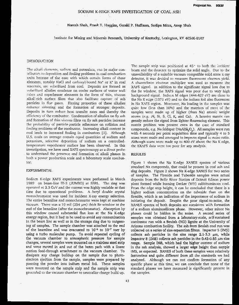
SODIUM K-EDGE X a F S INVESTIGATION OF COAL ASH
Naresh Shaln, Frank E. Gerald P. Huffman, Su&pa M h a , Anup Shah
The alkali elements, sodiun: and potassium, can be major con- tributo-s to deposition and fouling problem in coal combustion units because of the ease with which certain forms of these elements, notably NaCl and carboxyl-bound Na' or K+ in coal macerals, are volatilized from coal. Deposits are formed as volatilized alkalies condense on cooler surfaces of water wall tubes and superheater elements in the form of thin, viscous alkali-rich surface films that also facilitate capture of ash particles in flue gases. Fluxing properties of these alkalies enhance sintering and the formation of stronger deposits. Deposits in turn reduce heat transfer rates and thereby the efficiency of the combustor. Condensation of alkalies on fly ash and formation of thin viscous films on fly ash particles increase the probability of particle-particle adherence on collision and fouling problems of the combustor. Increasing alkali content in coal leads to increased fouling in combustors [l]. Although U.S. coals on average contain equal quantities of sodium and potassium, selective deposition of sodium on a controlled- temperature superheater surface has Seen observed. In this investigation, we have used XAFS spectroscopy as a direct probe to understand the presence and formation of alkali phases in both a power production scale and a laboratory scale combus- tor.
EXPERIMENTAL
Sodium K-edge XAFS experiments were performed in March 1989 on beam-line 111-3 (JUMBO) at SSRL. The ring was operated at 3.3 GeV and the current was highly variable at that time due to operational problems. A beryl double crystal monochromator was used for incident X-ray energy selection. The entire beamline and monochromator were kept at machine vacuum. There was a 10 mil (254 pm) thick Be window at the end of the beamline (after the monochromator). Absorption by this window caused substantial flux loss at the Na K-edge energy region, but it had to be used to avoid any contamination in the beam line as well as in the storage ring due to outgass- ing of samples. The sample chamber was attached to the end of the beamline and was evacuated to 10-6 to 10-7 torr by using a turbo molecular pump, To avoid repeated cycling of the vacuum chamber to atmospheric pressure for sample changes, several samples were mounred on a stainless steel strip and were moved in and out of the beam path with a linear motion feed-through attachment to the vacuum chamber. To dissipare any charge buildup on the sample due to photo- electron e j e d o n from the sample, samples were prepared by pressing the powder into indium metal foil. These samples were mounted on the sample strip and the sample strip was grounded to the vacuum chamber to neutralize charge build up.
The sample strip was pos%oned at 450 to both the incidenl beam and the detector to optimize the solid angle. D;e to the unavailability of a suitabk vacuum compatible solid-state x-ray detector, it was decided to measure fluorescent electron yield. A channeltron electron multiplier was used to measure the XAFS signal. in addition to the significant signal loss due to the Be window, the XAFS signal was poor due to very high background signal. Indiua M edges (444-827 eV) are close to the Na K edge (1071 eV) and so the indium foil also fluoresces in Na XqFS region. Moreover, Na loading in the samples was quite low (iess than 10%) and the matrices of most of the samples were made up of higher (than Na) atomic weight atoms (e.g. Al, Si, S, C1, K, and Ca). A heavier matrix can greatly reduce the signal from lighter fluorescing element. This matrix problem was present even in the case of standard compounds, e.g. Na feldspar (NaAlSi 0 ). All samples were run with 4 seconds per point acquisition time and typically 4 to 5 scans were made and averaged to improve signal to noise ratio. Although scans were made up to 400 eV above the Na K edge, the EXAFS data were too poor for any analysis.
3 8
RESULTS
Figure 1 shows the Na K-edge XANES spectra of various standard Na compounds, that could be present in coal ash and slag deposits. Figure 2 shows Na K-edge XANES for two series of samples. The Fireside and Tubeside samples were actual deposits from the Belle River Station of Detroit Edison power plant formed while burning Decker (WY) subbituminous coal. From the edge step height, it can be concluded that there is a higher sodium concentration on the tubeside than on the fireside, which is an indication of the importance of sodium in initiating the deposit. Despite the poor signal-to-noise, the XANES spectra of both deposits are consistent with formation of a sodium aluminosilicate phase. However, other minor Na phases could be hidden in the noise. A second series of samples was obtained from a laboratory-scale, self-sustained combustor run with a Beulah (ND) lignite at the University of Arizona combustion facility. The ash from Beulah coal run was collected on a series of size-separation filters. Impactor 5 (IM.5) collects ash particles in the size range 2.1-3.3 pm, while impactor 8 (IM8) collects ash particles in rhe 0.43-0.65 pm size range. Sample IM8, which had the higher content of sodium in the ash analysis, showed a larger edge height than sample IM5, as expected. XANES of both these samples were relatively featureless and quite different from all the standards we had analyzed. Although we can not confirm formation of any specific phase at this time, we can conclude that none of the standard phases we have measured is significantly present in the samples.
43
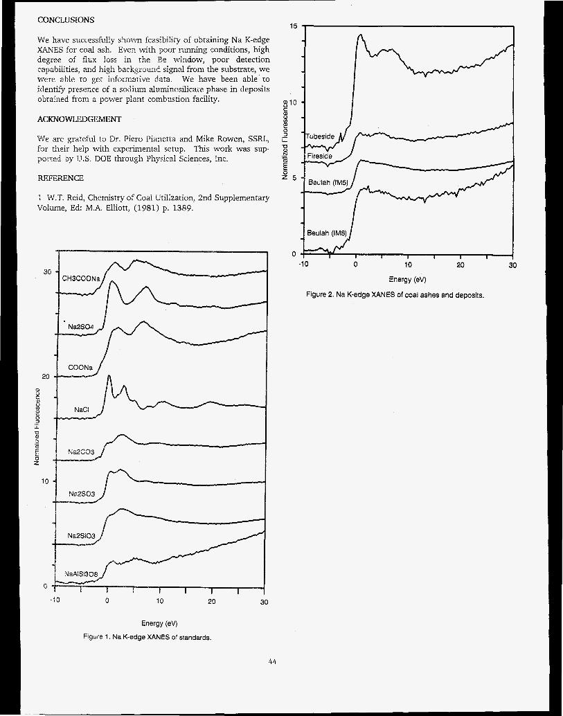
CONCLUSIONS 15 I
We have successfully shown feasibility of obtaining Na K-edge XANES for coal ash. Even with poor running conditions, high degree of flux loss in the Be window, poor detection capabilities, and high backgrmnr? signal from the substrate, we were able to get informative data. We have been able to identify presence of a sodium aluminosilicace phase in deposits obtained from a power plant combustioil facility.
ACKNOWLEDGEMENT
We are grateful to Dr. Piero Pianetta and Mike Rowen, SSRL, for their help with experimental setup. This work was sup- porred by U.S. DOE through Physical Sciences, Inc.
REFERENCE
i W.T. Reid, Chemistry of Coal Utilization, 2nd Supplementary Volume, Ed: M.A. Elliott, (1981) p. 1389.
ao
20
0
.I 0 0 10 20 30
I 3 10
8 c
v)
0 E
ii U .- w E - 8 2 5
0 -1 0 0 10 20 30
Energy (ev)
Figure 2. Na K-edge XANES of coal ashes and deposits.
Energy (ev) Figure 1. Na K-edge XANES of standards.
44

PmpOSalNO. 956M AN XAFS INVESTIGATION OF CHLORTNE IN U.S. COALS
F. E. Huggins', G. P. Huffman', F. W. Lytlez and R B. Greego8
'Institute for Mining and Minerals Research, University of Kentucky, Lexington, KY 40506 and ZThe Boeing Company, Seattle, WA 98124
INTRODUCTION The chlorine contents of most U S . coals are typically less than 0.3 wt%, except for certain coals from the Illinois basin in which the chlorine contents may be as high as 1.0 wt% [l]. Such high chlorine coals can give rise to severe corrosion problems in coal utilization. However, relatively little is known about how chlorine occurs in coals from the Illinois Basin or from other seams in the U.S. Based largely on indirect observations made mostly on British and Indian high chlorine coals, chlorine has been postulated to occur in coal as alkali or alkaline-earth chlorides (NaC1, KC1, CaCl,, etc.), as organic chlorides, as hydrochlorides, and as chloride ions in the water associated with coal [2,3]. In this investigation, we have used XAFS spectroscopy as a direct probe of the chlorine environ- ment in coal to establish how this element occurs in US. coals.
EXPERIMENTAL Chlorine K-edge XAFS experiments were performed in January 1987 on beam-line VII-3 at SSRL. XAFS spectra were recorded in fluorescent geometry. The chlorine K edge in sodium chloride (NaC1) at 2826 eV and the argon K edge (from air contamination in the beam path) at 3205 eV were used as primary standards. The latter edge was present in every spectrum and served as an effective internal calibration point, but its presence limited the chlorine EXAFS region to only about 380 eV. Further details are given elsewhere [41.
RESULTS XAFS data were collected for coals from eight different U.S. seams (chlorine contents varied from 0.04 to 0.84 wt%) and, additionally, from a number of Illinois #6 coals that had been subjected to an aqueous leaching process to remove some of the chlorine [SI. These data (Fig. 1) showed that the chlorine XANES fingerprint from the coals was relatively universal and unaltered by aqueous leaching, despite removal of over 70% of the chlorine from the coal [4]. Furthermore, this characteristic XANES spectrum was quite unlike any of the standard inorganic or organic chlorides commonly thought to be present in coal, although two of the coals did clearly contain a minor fraction (<25%) of the chlorine as NaCl [4]. The XANES of chlorine in the coal resembled those of chlorine in saturated NaCl solution, CaC1,.6H,O, and organic hydrochlorides. These three substances do have some structural and bonding features in common that are, no doubt, reflected in similar XANES spectra: (i) chlorine is present in these substances as chloride anions, (ii) the bonding is relatively weak and largely ionic in char- acter, and (iii) the chlorine-oxygen or chlorine-nitrogen distances are long: between 3.0 and 3.3 A. Hence, the chlorine in coals will likely be similar in structure and bonding because of the similarity of its XANES to these materials. Analysis of the EXAFS region was possible for some of the coals with higher chlorine contents and the best agreement for the phase-
shift for chlorine in coal was found for a Cl-H,O distance of 3.03 A when modelled by the experimentally derived phase- shift for CaC12.6H,0, in which the chlorine anion is surrounded by six water molecules at an average distance of 3.15 A 161.
CONCLUSIONS XAFS spectroscopy has proved extremely informative in establishing how chlorine occurs in U.S. coals. The data are consistent with chloride anions in the moisture associated with coal being the major form-of-occurrence of chlorine. This finding is in agreement with the latest work on British coals obtained by much less direct methods [21.
WFERENCES 1 H.J. Gluskoter and R.R. Ruch, Fuel SO, 65-76, (1971). 2 N.J. Hodges, W.R. Ladner and T.G. Martin, J. Inst. Energy,
3 XN. Chakrabarti, in: Analytical Methods for Coal and Coal Products, Vol. 1, Academic Press, NY, 1978, 323-345.
4 F.E. Huggins, G.P. Huffman, F.W. Lytle and R.B. Greegor, ACS Div. Fuel Chem., Preprints, 340, 551-558 (1989).
5 H.L. Chen and M. Pagano, Fuel Proc. Technol., 13, 261- 269, (1986).
6 R.W.G. Wyckoff, Crystal Structures, Vol. 3, J. Wiley & Sons, New York, 1963.
56, 158-169 (1983).
ACKNOWLEDGEMENTS We are grateful to Professor Han Lin Chen of Southern [liinois University for providing us with samples of raw and leached Illinois #6 coals. This work was supported by the U.S. DOE under Contract NO. DE-FG22-86 PC90520 and the Electric Power Research Institute under Contract No. RP-8803-20.
I \ A 2.8
2.6 2.4
5 2.2 E 2
a 'o 1.4 3 1.2
- 1.8
8 1.6
- - a 1 E 0.8 z" 0.6 x:: 1 J , , , , , , I
0 -20 0 20 40 60
Energy in ev (NaCI standard)
Mineral, KS (0.27% CI)
I J Illinois #6 (0.42% Cl) I
Fig. 1: Chlorine XANES spectra of three U.S. coals
45


PrOpOSal NO. 994M
Characterization of the Coordination Environment of Divalent Iron in Silicate Crystals, Glasses and Liquids as a Function of
Temperature and Composition Using X-ray Absorption Spectroscopy
William E. Jackson, Glenn A. Waychunas, Gordon E. Brown, Jr., and Jean-Marie Combes Center for Materials Research and Department of Geology
Stanford University, Stanford, CA 94305
Introduction The main purpose of this research program is to define the structural role
of Fez* in crystalline and amorphous silicates as a function of composition and temperature up to and above the temperature of fusion (TJ in the com- pounds studied. Iron in its 2+ oxidation state is the most abundant transition metal in the Earth and is an important component in slags and natural silicate liquids. The local environment of Fez* in silicate crystals and liquids is not always predictable because the ion can adopt octahedrally coordinated, tetrahedrally coordinated or, in certain cases, other types of coordination geometry (e.g. square-planar, 7- or 8-coordinated) depending on a number of variables. When octahedrally coordinated, Fez+ is thought to behave as a clas- sical network modifier in silicate glasses and liquids, whereas when tetrahedrally coordinated, it may behave as a network former or a molecular complex. In general, the greater the ratio of network formers to network modifiers in a silicate liquid, the greater iv; bulk polymerization. The degree of temhedral polymerization in such a liquid affects certain of its bulk physi- cal pmpedes, such as viscosity and density, and can profoundly affect cry- stallization. For example, the configurational entropy contribution to the en- tropy of fusion tends to decrease with increasing polymerization of the liquid for common silicates. It is expected that the high configurational entropy as- sociated with Fe,SiO, liquid, which is primarily responsible for the difficulty in forming glass of this composition, is a consequcncc of structural changes in long range order (> - 4 A) and pcrhaps even short range order (4 A <) at the melting point (see, for example, Jcanloz, 1985).
There exists, however, littlc direct structural information on the coordina- tion environment of Fe2+ in silicate liquids. This information has previously been difficult (0 obrain because of the redox potential of Fez+ at high tempera- tures and the typically high T, associated with iron silicates (e.g. Fe,SiO,, Tf = 1478 K). Indeed, most past studies of Fcz+ in silicate ‘liquids’ have actually been performed on the qucnchcd glasses which do not necessarily have the same average structure as the corresponding liquid. The nccd to examine the structure of liquids in situ is nccessary due to thc possibility of structural rearrangements within the liquid during quench. Thc low glass mnsition tcm- peratures associated with extrcmcly dcpolymerixd liquids suggest a major degree of structural rcarrangemcnt in Ihc liquid during glass formation and, hence, the greatest difference bctwccn the glass and liquid structure.
Our past research (e.g. Waychunas et al., 1987, 1988) has focussed on defining the coordination cnvironmcnt of Fez+ in polymcrized ‘feldspar’ -com- position glasses and corresponding liquids using Mossbauer, EXAFS and XANES spectroscopy. Fez+ was found to occupy nctwork-forming, tetrahedral si- in glasses and liquids of SFcSi,O, compositions (M= Li, Na, K, Rb). Few spectroscopic melhods are capable of yielding direct information on the structural environment of Fez+ in silicate liquids at high temperature. X-ray absorption spectroscopy using a high intensity synchrotron source is one of the few methods that can help overcome problems such as the need to exam- ine liquids in ceramic containers inside high temperaturc furnaces, and that can also provide estimates of bond lcngths, coordination numbers, and infor- mation on oxidation state. This application of EXAFS spectro.oscopy is not without signilicant problems, however, including the effcct of thermal motion on EXAFS amplitudes and resolution, particularly at k > 10 A-1, and the resultant IOU of information on next-ncarest ncighbon around Fez+.
We are. currently studying the local coordination environment of Feh as a function of temperature and composition (polymerization) in a range of Fe*-bearing syslems, including glasscs and liquids using x-ray absorption spectroscopy and a s p i a l furnace assembly. Thc high-temperature samples
being studied include the compositions Fe,SiO,, CaFeSiO,, CaFeSi,O,, C%pe13S406, and compositions along the NaPeS40, - CaFeSi,O, join. Room temperature Fe K-EXAFS and K-XANES studies have been made on a unique glass of Fe,SiO, composition, on glasses from the N%FeSi,O, - CaFeSi,O, join, and on selected crystalline silicates. We have also carried out XAS studies of well-characterized model compounds (FeCO,, C~Fe~Si,Olz, and CaFeSipd in order to extract Fe-0 phase and amplitude information.
Experimental No data in this program have been obtained at SSRL since December
1987. In September 1989, room temperaturc and elevated temperature XAS studies were performed at the National Synchrotron Light Source on BL X19A (unfocussed, 2 X 20 mm photon spot size) and X23B (focussed, 1 X 1 nun spot size) using Si (111) monochromator crystals. Ring conditions were 2.5 GeV and 80 - 180mA. The high temperature data were collected in the fluorescence mode with a Lytle-type detector and a water-cooled, controlled- atmosphere furnace capable of sustaining temperatures up to 1750 K. The fur- nace assemblage consisted of a water-cooled A1 jacket, Mo sample holder, Mo heating coils, alumina insulation, inert atmosphere WJ, and an S-type thermocouple. Kapton film covered the incident and fluorescence photon poas. The oxygen partial pressure was controlled by the Mo and a Nz atmo- sphere. Mossbauer spectroscopic analyses on thc qucnchcd samples indicate no more Few @Ius an equal amount of Fco) than that produced by h e slight incongruent melting of the samples (about 5% Fe?+). The samples were con- tained in Be0 crucibles aligned to cxposc the mclt directly to the incident beam and fluorescence detector in order to climinate any signal from the crucible-melt interface. The Be0 crucibles proved to be rcfractory and inert even when in contact with the iron-bearing silicatc liquids for over 3 hours at T > 1650 K. An average of 3-5 scans wcre collccted for each high- temperature crystalline compound and melt.
Results and Discussion Using F e O phase and amplitudc information takcn from the room tem-
perature model compounds listed above, the structural cnvironmcnt of Fez+ in the glasses was successfully modeled. The Fc K-EXAFS and K-XANES data are. consistent with tetrahedral Fez+ in all feldspar-composition glasses. These results confirm our earlier conclusions that Fe2+, like Fe3 (Brown et al., 1978). can be chargebalanced in tetrahedral coordination in fully polymer-
Analysis of the FezSiO, glass yielded the unexpected result of tetrahedral Fe% (Figure 1). which when taken in contcxt of the high confiyational en- tropy associated with Fe2Si0, at T, is not too surprising. Tetrahedral Fe (11) and Si (IV) require an average oxygen coordination of three, as compared to an oxygen coordination of four in crystalline Fe,SiO,. This type of polymeri- zation in the Fe2Si0, liquid, may be related to Shiraishi’s (1978) rcport of an anomalous maximum in viscosity at the Fe2Si0, composition in melts along the FeO-SiO, join.
Analysis of the EXAFS spectra from high-temperature crystalline sili- cates and liquids is currently in progress and requires an understanding of the empenture effect On the Fe-0 bond. High-temperature XAS studies and/or studies of disordered systems require knowledge of the asymmetric distribu- tion function for the Fe-0 pair as well as any anharmonic vibration effects (e.& Einberger and Brown, 1979; DeCrescenzi, et al. 1981). This asym- mesic disaibution function and anharmonic vibration behavior are generally
ized silicate glasses.
46

accommodated in the modeling of the x (k) function which must include the -&.ions ta both phase and amplitude. Such modeling involves non- Gaussian Debye-Waller formulations and cxplicit G(r) functions for the Fe-0 pair &,hibution, which correclly interprets the apparent bond length conlrac- tion and amplitude reduction seen in the EXAFS signal as a function of in- creasing temperature. To accurately model the asymmeVic distribution func- tion and anharmonic effects expccted for the iron-bearing silicate liquids, we collected EXAFS data from Fez+ and Fe3+ in the crystalline silicate model compounds as a function of temperature. To begin to correct for the anhar- monic Fe-0 bond vibrations, we arc currently modcling the high-temperature Fe K-EXAFS collected from Fe3+ which resides in a centrosymmetric oc- tahedral site in Ca,Fe2Si,OIz (Figure 2). Once the anharmonic contribution to phe Fe-0 bond vibration as a function of tempcrature has been approximated, corrections for a non-Gaussian distribution of distances can be extracted from the Fe K-EXAFS collected from crystalline FeSiO, Crystalline FezSi04 con- tains Fe- in two dhtortcd octahedral sites. The EXAFS patial RDFs for Fe2Si0, are seen in Figure 3. As expected, the EXAFS results give an ap- parent bond length contraction of approximately 0.07 A over the temperature change of 780 K (90 K to 870 K). To correct for this contraction, these EX- AFS results will be modelcd in light of the high-temperature x-ray structure refinements of crystalline Fe,Si04 which suggest an Fe-0 expansion of 0.02 - 0.03 A over a comparable temperature rangc (Smyth, 1975).
Despite the high-temperature corrections, the study becomes a bit prob- lematic if we suppose Fez+ changes its coordination environment from oc- tahedral to tetrahedral in the liquid as the study of glassy FezSi04 suggests. Such a coordination change will serve to change the distribution of oxygen distances, perhaps to a more Gaussian arrangcmcnt, as well as influence the the amplitude of the Fe-0 bond vibration and, hence, the anharmonic contri- bution. Consequently, it is necessary to consider the phase and amplitude in- formation for Fez+ in tetrahcdral coordination in high temperature solids ils
well. The phases studicd by EXAFS as a function of temperature include FeAJO. (hercynite spinel), and lhe wFeSi30, glasses.
Some of the Fe K-EXAFS data currently being modeled, as described, are illustrated in Figure 4 which plots thc partial RDFs for Fe,Si04 as a func- tion of temperature and physical state. The x (k) functions are shown for crystalline Fe,SiO, (298 K), liquid Fe,SiO, (1520 K), and glassy FezSi04 (298 K). It is apparent from the Fourier uansrorm data alone that the EXAFS sig- nal from the glass is markedly diffcrent from that of the liquid. In addition to the modeling of Fe,SiO, liquid, we are modeling the effect on the Fez+ coor- dination environment of a liquid with a pyroxene composition whose silica content is intermediate between that of olivine and feldspar. We are also exa- mining the effect of substituting Ca** for Fea in silicate liquids with the idea that the s m g network modifying effect of Cazc (Le. almost always occupies un octahedral or larger coordination environment) will cause FeB to revert to octahedral coordination 10 satisfy the charge on ncarby oxygens.
Conclusions Our Fe IC-EXAFS studies of Fc (II) in feldspar and olivine-composition
glasses indicate Fe (ID is tctrahcdrally coordinated in both systems and prob- ably m e s es a network former in linking the SiO, tetruhcdru, These results agree with our earlier studies, nnd mny uid in the intergrctution of the thermo- dynamic and visodty data for silicate liquids,
By i n c a v t i n g p h m and nrnplitude informution tukm from motlel comgounL nu a function of t%rnporature, we ure beginning to model the effectn of hlgh.tempatw on tho Fe cowdinaiion envlronmant. This infor. mation will help utl eonsmirr the rtrueturul diffmnees between glas~a and heir eaneqxmding molts RS well ns the shetursl remangowonts at Tf for impmnt mk md magma fomirtg dlisuios,
p a r t i a l RDF f o r f a y a l i t e glass
(uncorrected f o r phase s h i f t )
RADIUS A Figure 1. Partial radial distribution function for Fez+ in Fe,Si04 glass (un- corrected for phase shift). Inset: Using Fe-0 phase and amplitude information extracted from Fe K-EXAFS data of CaFeSi,O, (y'Fc-0 = 2.13 A) and FeCO, we-0 = 2.14 A), the best least-squares fit to Fc K-EXAFS data collected from Fe,SiO, glass yield: 3.36 (i- 0.5) oxygcns coordinating Fez+ at an aver- age distance of 2.01 (50.02) A with a Debye-Waller parameter equal to 0.0127 A2.
C??FezSi3On Fit Shell (Fe-0. - 1.6 A) top ta
* bottom: 90K. W K . 410K. 560K, 670K, 870K. 1070K
References . Brown, G.E., Jr., Kocfcr, K.D., Fcnn, P.M. (1978) EXAFS Study of Iron-
Bearing Silicato Glassscs: Iron coordination Environrncnt and Oxidation State, Geol. Soc. Am. Abstr. With Programs 1 0 173.
47
r

First Shell (Fe-0, - 1.6 A) top to bottom: 90K, 300K. 670K. 870K
0 1 2 3 4 5
R (A) Figure 3. partial RDFs €Dr Fez+ in the two distorted sites (Le. highly non- Gaussian distribution of oxygen distances) of crystalline Fe,SiO, as a function of temperature (uncorrected for phase shift). The apparent bond length con- traction of 0.07 A over the temperature change is being modeled in in light of an actual 0.02 A expansion for this temperature change as determined from XRD (Smyth. 1975).
Fe,SiO, Fis t Shell (Fe-0. - 1.6 A) lop to bottom: XU UXIK, glass 300K. liquid 1520K 'I- -
0 i a a i i i
De CrescenZi, M., Balzarotti, A., Comin, F., Incocciu, L., Mobilio, S., Molta. N. (1981) EXAFS Measurements on Fe-B Metallic Glasses: Asymmetry of the Radial Distribution Function, Solid Slate Communications, Vol. 37,921-923.
Eisenberger, P., and Brown, G.S. (1979) The Study of Disordercd Systems by EXAFS: Limitations, Solid State Communicalions, Vol. 29, 481484.
Jeanloz, R. (1985) Thermodynamics of Phase Transitions, In Reviews in Mineralogy Vol. 14: Microscopic fo Macroscopic, Mineralogical Society of America 1985,428~.
Rakai, RJ. (1975) Crystal Structure of Spessartine and Andradite at Elevated Temperatures, MSc thesis, Univ. British Columbia.
Shiraishi, Y., Ikeda, K., Tamura, A., and Saito, T. (1978) On the Viscosity and Density of the Molten FeO-SiO, System, Trans JIM, Vol. 19, 264- 274.
Smyth, J.R. (1975) High Temperature Crystal Chemistry of Fayalite, Amen-' can Minerahgist, Vol. 60, 1092-1097.
Waychunas, G.A., Brown, G.E., Jr., Jackson, W.E., and Ponader, C.W. (1988) molten Evidence from X-ray Absorption for Network-Forming FS*
alkali silicates, Nature 332,251-253.
Waychunas, G.A.. Brown, G.E., Jr., Ponader, C.W., and Jackson, W.E. (1987) High-Temperature X-ray Absorption Study of Iron Sites in Crystalline, Glassy, and Molten Silicates and Oxides, SSRL. Report 87/01,139-141.
48


X-RAY ABSORPTION SPECTROSCOPY OF ION SORPTION COMPLEXES AT MINERALWATER INTERFACES
Peggy A. O'Day, Catherine I. Chisholm-Brause, Ning Xu, Jean-Marie Combes, Gordon E. Brown, Jr., George A. Parks
School of Earth Sciences, Stanford University, Stanford, CA 94305
INTRODUCTION The sorption of ions from aqueous solutions onto mineral
surfaces controls the transport and partitioning of aqueous species in a wide variety of systems such as contaminent tpan- sport, nuclear waste disposal, ore deposition, electrochemical processes, corrosion control, solar energy conversion, and enhanced oil recovery. Quantitative descriptions of molecuIar level reactions are required to accurately predict the interaction of ions with mineral surfaces in macroscopic models. The quanti- tative structural information provided by synchrotron-based x-ray absorption spectroscopy WAS) permits characterization of in sin4 sorption complexes at mineral/wate.r interfaces at very low slir- face concentrations representative of natural conditions.
Our previous investigations focused on two aqueous iow'oxide systems: (1) Se oxyanion sorption on goethite (a- FeOOH)(') (2) Pb(1Ij sorption on y-Al2OP3) and goethite(4). In both of these systems, we were able to distinguish between inner sphere and outer sphere sorption complexes with XAS. In addi- tian, .XANES and EXAFS analyses of Pb(II) complexes on goethite and y A 1 2 4 indicate the presence of Pb atoms in the second coordination shell, suggesting that aqueous Pb complexes may form clusters on the mineral surface.
In order to investigate how mineral surfaces control the size and structure of sorption complexes, we have begun a detdled study of the sorption of Co(II) complexes onto a variety of natural and synthetic minerals. We previously reported results for cobalt sorption on y-A120$5*a). In the past year, we have col- lected XAS data on cobalt sorbed to kaolinite, rutile, silica, and calcite. The samples studied by XAS cover a wide range of surface sorption densities in order to detect changes in the atomic structure of Co(i1) complexes as a function of surface coverage. We have collected data on sorption samples estimated to have from about 10 to greater than 95 percent of a monolayer of oxygen-ligated surface cobalt atoms. Interpretation of the EXAFS data allows us to distinguish between surface adsorption, Surface precipitation, and diffusion of metal ions into the solid. OW ini- tial results show that Co(II) uptake by minerals occurs by surface adsorption and formation of inner spheE ~ c o m p l e x e ~ ( ~ ~ ~ ~ ~ . coni- parison of complex formation on different minerals suggests that surface clusters may be a common phenomenon of metal ion sorption, but mineral surfaces themselves strongly control the size of the cluster which form^(*^^).
Proposal NO. 999Mp
EXPERIMENTAL XAS data were collected at SSRL during the dedicated run
in February and March, 1989, on beam lines VII-3 (wiggler field = 18kG) and 11-1 (bending magnet line). Through SSlU, we obtained additional beamtime at the National Synchrotron Light Source (NSLS) in July, 1989, on beam line X-19A. On beam line VII-3, all cobalt spectra collected were not usable due to interference from unshielded high energy x-rays and spectra noise from shutter opening and closing on an adjacent beamline. Data collection on this line was limited to Kr K-, Se K-, and Np Lm - absorption edges. Absorption spectra were collected on cobalt sorption samples and iron model compounds on beam line U-1. However, the significantly lower intensity of the bending magnet beam in comparison with a wiggler beam resulted in a very low signal-to-noise ratio and poorly resolved EXAFS spec- ea. Usable data from this line were limited to a few high con- centration sorption samples and model compounds. Spectra for cobalt sorbed to kaolinite, silica, and calcite were collected on NSLS X19-A. Although data quaility for lower concentration samples was better on this line than on 11-1, it was still far infe- tior to high resolution data obtainable on SSRL wiggler lines (Figure 1). On all beamlines, data for sorption samples and
0 Y X n
x 25
4 6 8 10 12
k(k') Figure 1. EXAFS specm for &(XI) Sorbed to kaolinite at a sorption density of 1 about 1.2 pmOleS/m2 collected on three different beamlines. (a) SSRL wiggler line IV-1, (b) N S u x- 1 9 ~ , (c) SSRL bending magnet line 11-1. Sample (a) has a higher water content than (b) or (c) which accounts for the damping of the "beat" on the second oscillation at about k=5,
49

glasses were collected in fluorescence mode using a Lytle-type detector with the sample oriented at 45' to the incoming x-ray beam. Spectra for solid model compounds were collected in transmission mode with the sample oriented perpendicular to the
Cobalt sorption samples were prepared by equilibration of the aqueous metal ion in nitrate solution with a solid mineral in amounts from 50-12Og mineraVlOOOg aqueous solution, depend- ing on the surface area of the mineral. The pH of the suspension was adjusted such that approximately 95% of Co(I1) was removed from solution by uptake on the solid. Initial Co(I1) solu- tion concentrations ranged from 0.6 to 18 mM in order to obtain variable amounts of surface sorption density at 95% ion uptake from solution. The resulting sorption densities varied from 1.2 to 39 pmoles/m2. The samples were centrifuged and the super- natent solution removed, leaving the mineral solid as a wet paste. The pastes were loaded and sealed in teflon sample cells with mylar windows. Samples were checked carefully before and after data collection to insure no drying of the pastes.
x-ray beam.
The Np sorption sample was prepared in a similar fashion using lg goethite/lOOOg aqueous solution and an initial Np solu- tion concentration of 5 mM. Adjustment of pH to 7.1 resulted in about 95% uptake from solution corresponding to a sorption den- sity of moles Nplm' goethite.
COBALT SORPTION ON MINERAL SURFACES
Four sorption samples representing surface adsorption densities of 16.0. 9.9, 5.0, and 1.2 pnoleslm' of C o o sorbed to kaolinite were studied by XAS. The kaolinite used was a chemically pure, moderately well-crystallized sample from Dry Branch, Geor- gia("). Analyses of the EXAFS spectra indicate that Co(JI) is in octahedral coordination with oxygen, consistent with results from wet chemical analyses("). Fourier transforms of the EXAFS spectra give radial suucture functions (rsf) (Figure 2) which show a progressive decrease in the amplitude of the second shell peak as a function of surface coverage. Best fits of the back- transforms of these peaks using empirical parameters from Co(OH)* indicate cobalt atoms in the second coordination shell at 3.11 to 3.13 A. The number of cobalt atoms progessiveiy decreases from about six to two as the surface coverage decreases. In addition, the fit to the experimental data is gen- erally improved with inclusion of either A1 or Si, representing an atom from the kaolinite surface, in the EXAFS model. Use of theoretical parameters for A1 or Si results in about 0.5 to 1 atom at 3.19 to 3.22 A. A peak in the rsf at r = 5 to 6 (Figure 2) arises from a colinear arrangement of the central, second, and fourth coordination shell cobalt atoms as in Co(OH)J8.9). Ana- lyses of this peak (with empirical parameters from c O ( o ~ d indicate one to three fourth shell cobalt atoms at a distance of 6.21 10 6.23 A from a central absorber, in contrat to six cobalt atoms at 6.35 A in Co(OH)>
r= 16. 9.9 5.0
1 1.2 0 2 4 6
Figure 2. Radial structure functions for &(E) sorbed to kaolin- ite at four different surface sorption densities (0 in pmoles/m2. Note the reduction in the amplitude of the second peak as a function of decreasing surface coverage indicating fewer cobalt nearest neighbor atoms in the second coordination shell.
The results from XAS analyses suggest that sorption behavior of Co(II) on kaolinite is similar to that of cobalt on pA12017*g). Bond distances for 0-0 and Co-Co are similar in both systems; however, coordination numbers for second and fourth shell neighbor cobalt atoms are slightly lower (= 1 to 2 fewer atoms) for co/kaolinite at the same surface sorption den- sity. The presence of A1 or Si atoms in the second coordination shell points to the formation of inner sphere complexes as in the Co/YA1203 system. Comparison of second and fourth shell dis- tances and coordination numbers with those of Co(0H)z show that atomic distances are shorter and coordination numbers are lower for multinuclear sorption complexes on kaolinite, suggest- ing the lack of a well-ordered surface precipitate at coverages approaching a monolayer of cobalt atoms.
ColRutile EXAFS analysis of Co(II) sorbed to rutile (Ti02) at a sur-
face sorption density of 1.3 pmoles/m2 suggests formation of cobalt inner sphere complexes as in the y-A1203 and kaolinite systems. However, best fits of the fourier back-transform of the second coordination shell (using parameters from Co(0H)i) results in fewer second shell cobalt atoms (CN = 1) and longer CO-Co interatomic distances (3.36 A) than for cobalt sorbed to y-A1203 and kaolinite. These results suggest that monomeric Species or small clusters of cobalt atoms form on the rutile sur- face and they are structurally different from larger multinuclear complexes which form on y-A1203 and ka~ l in i t e (~*~*~) .
50

CoISilica CooI) sorbed to d i ca (SiO,) was examined by XAS at a
surface sorption density of 9.9 pmoles/m2. The silica used was ground quartz (MIN-U-SIL) with a surface area of approximately 0.2 d / g . Second shell cobalt coordination numbers from prelim- inary EXAFS analysis suggests that cobalt forms slightly smaller surface complexes on silica than on kaolinite or y-Al2O3 at the same surface coverage, although second shell Co-Co distances are similar (3.11 A). The absence of a fourth shell cobalt peak at high r indicates the lack of large multinuclear complexes contain- ing colinear, equidistant cobalt atoms.
Co/Calcite XAS data was collected on Co(I1) sorbed to calcite at a
sorption density of 39 poles/m2. The EXAFS spectrum of the sorption sample shows a distinctive "beat" pattern which differs from that of hydrated COCOS (solid). Preliminary analysis sug- gest that cobalt sorbed to the surface is not diffusing into solid calcite in significant amounts as proposed by previous workers for cd2' sorption on calcite surfaces(12).
NEPTUNIUM SORPTION ON GOETHITE XAS data for Np sorbed to goethite was compared to
absorption spectra for solid Np02 and an aqueous Np solution. U N E S and EXAFS analyses suggest that Np is present on the goethite surface as a monomeric species in a bipyramidd oxygen coordination environment similar to that of aqueous NpO;. Although it could not be determined whether the sorption com- plex is inner sphere or outer sphere, Np is clearly not forming a surface precipitate on goethite.
ACKNOWLEDGEMENTS This research was supported in part by NSF grants EAR-
8513488 and EAR-8805540. Karen Gruebel, George Redden, and Jim Leckie prepared Se sorption samples and assisted in data collection. We gratefully acknowledge the experimental assis- tance of Britt Hedman. We would especially like to thank the SSlU staff, in particular Glenn Ken, C. Troxel, Jr., and Hal Tompkins, €or special assistance during difficult times.
REFERENCES
(1) Hayes K.F., A.L. Roe, G.E. Brown, Jr., K.O. Hodgson, J.O. Leckie and G.A. Parks (1987) In situ x-ray absorption study of surface complexes at oxide/water interfaces: Selenium oxyanions on a-FeOOH, Science 238 783-786.
(2) Chisholm-Brause, C.J., K.F. Hayes, A.L. Roe, G.E. Brown, Jr., G.A. Parks and J.O. Leckie (1990) Spectroscopic inves- tigation of Pb(1I) complexes at the y-Al20dwater interface, Geochimica et Cosmochimica Acta, in press.
(3) Chisholm-Brause, C.J., A.L. Roe, K.F. Hayes, G.E. Brown, Jr., G.A. Parks and J.O. Leckie (1989) XANES and EXAFS study of aqueous Pb(1l) adsorbed on oxide surfaces, Physica B 158 674-675.
(4) Roe, A.L., K.F. Hayes, C.J. Chisholm-Brause, G.E. Brown, Jr., G.A. Parks, K.O. Hodgson and J.O. Leckie (1990) In situ x-ray absorption study of lead surface complexes at the goethitelwater interface, Langmuir, in press.
(5) Chisholm-Brause, C.J., G.E. Brown, Jr. and G.A. Parks (1988) EXAFS study of Co(I1) complexes at oxide/water interfaces, (Abstract), EOS 69 1482.
(6) Chisholm-Brause, C.J., G.E. Brown, Jr. and G.A. Parks (1989) EXAFS investigation of aqueous Co(1I) adsorbed on oxide surfaces in-situ, Physica B 158 646-648.
(7) O'Day, P.A., C.J. Chisholm-Brause, G.E. Brown, Jr. and G.A. Parks (1988) Characterization of Co(n> complexes at the kaolinitelwater interface by x-ray absorption spectros- copy, (Abstract), EOS 69 1482.
(8) O'Day, P.A., C.J. Chisholm-Brause, G.E. Brown, Jr. and G.A. Parks (1989) Synchrotron-based XAS study of Cool) sorption complexes at minerallwater interfaces: Effect of mineral surfaces, 28th International Geological Congress Abstracts 2 534.
(9) Brown, G.E., Jr., G.A. Parks and C.J. Chisholm-Brause (1989) In-situ x-ray absorption spectroscopic studies of ions at oxide-water interfaces, Chimia 43 248-256.
(10) May, H.M., D.G. Kinniburgh, P.A. Helmke and M.L. Jack- son (1986) Aqueous dissolution, solubilities and thermo- dynamic stabilities of common aluminosilicate clay minerals: Kaolinite and smectites, Geochimica Cosmochim- ica Acta 50 1667-1677.
(11) Baes, C.F., Jr. and R.E. Mesmer (1976) The Hydrolysis of Cations, New York, Wiley, 489 p.
(12) Davis, J.A., C.C. Fuller and A.D. Cook (1987) A model for trace metal sorption processes at the caicite surface: Adsorption of cd2' and subsequent solid solution formation, Geochimica Cosmochimica Acta 51 1477-1490.
5 1


SYNCIIROTKON X-RAY POLYCRYS'lAI,I,lN1~ I l l I ~ I ~ R A ( " I ' O ~ ~ ~ ~ I ~ I ~ ~ ' -- VI
W. Parrish,] C. Erickson,l N. Masciocchi,* 11. 'l'orayn.3 arid 13. <;illcc4
Proposal No. 1021Mp
IIBM Research Division, Almaden Research Center, Sari .losc, ('alilhrnia 95 120-6000 21stituto Chimica Strutturistica Inorganica, University Milano, 1-201 33 Milano. Italy 3Ceramic Eng. Res. I,&., Nagoya Inst. 'Technology, Asahigaoka, l'njimi 507. .lapan
4C.II.N.G., XSX, 38041 Grcnoble Ccdcx. I:rancc
Introduction The reconstructed Beam Line 11 and the new white beam hutch were dedicated and began operation in February 1989. The hutch is greatly improved with much more room allowing installation of larger instrumentation, more eficient use and better alignment procedures with the adjustable table height. The adjacent beam pipe was moved farther away permitting the use of specimen chambers that were not oossible previously.
Powder Diffraction Powder data were collwted for a number of crystal structure refinements. 'The first three listed below werc done with Pcarson VII profile fitting, a modified Rictveld method6 and /z = 1.53.5A.
Na2AI2Ti6016. Space group C2/m, monoclinic, a = 12.1239(3), b = 3.7749( I ) , c= 6.4180(2)& f i = 107.59", R(Bragg)= 6.4%, Rp = 3.45'0, R,, = 4.3% Partial ordering of AI3 + and Ti4 + in the two crystallographically non-identical octahedral sitcs.7
CUO. Space group C2/1:, monoclinic, a - 4.6837(1), b = 3.4238(1), c=5.1297(2)& /?= 99.4", y(0)=0.4191(6), R(Bragg)= 30/, R, = 7.8%0, R,, = 9.8%). The Cu valence was + 1.9 rather than + 2 probably due to the (small) contribution of the special oxygcn in the C u - 0 elongated octahedron to the valence charge.
Ndt.xsCe0.1 Space group 14/mmm, tetragonal, a = 3.94698(4), c= 12.0795(1)A, R, = 2.556, RWp = 3:3':/0. The valence state of the Cu ion was + 1.80 confirming its mixed valence character. A full time-of-flight neutron diffi.action analysis was published during the preparation of our paper.2
AI203 TWO routine rur s were made for the international Center for Powder Diffraction round-robin to compare results from various laboratories using the same specimen.] One run was done in 1.7 hours and the other in 0.4 hours for the range 20"-80" Using
1.53SA radiation. Both had vcr!; high li~ur.~.s-ollriicr.it ivi t l i l ' , above 600 and MN 1000, shrnving thc higli t4ii:ilitv 01 tlic p:tr:illcl beam method with long Sollcr slit optics.
MnCrlnS4. Space group Fd3m (spincl). cubic, a 10.4207(5)/2. 1=0.748A, R(Bragg)= I.S%i, R, = 1.21'0. 'l'lic tlatn w r c ohtairictl in a previous run and analyzed by the two-stcp method.* '!.he refined cation distribution was
(Mn.,s[n,,,)(Mn.,,In,,,(:r,,)7S, '
Comparisons of the parallel hcam and convcntional x-ray tuhc focusing methods were p b l i ~ h c d . ~ ~ ~
Figure 1 is a synchrotron radiation powdcr pattcrn of the polyrncr poly-(p-hydrobenzoic acid) PI IRA at room tcmpcraturc.Q 'I'his is an example of the high resolution, pcnk-to-hnckgroiiritl and symmetrical peak shapes that can he obtainctl with the incthort. They are used to supplement conventional tiatit for intlcxing, Iittticc parameter determination and structure refincnicnts. Although it is not possible to do a complete research program using the rnctliotl because of the limited SSRI, time availalilc a t present. sclcctcd patterns provide an important aid in intcrpreting convcntionnl laboratory patterns.
Grazing lncidence D i k e New instrumentation for grazing incidcncc dilrraction studies of thin films, multi-layer films and substratcs w x s clcvclopctl using it rotating anode X-ray It consists of two channel monochromator (Ge ;I20 for X-ray tuhc and Si I I I for synchrotron), one placed before and the othcr altcr the spccirncn. I t was installed in the new hutch to test thc atlvantagcs of thc synchrotron parallel beam. (infortunately cxccssivc orhital shifts and unstable beam made it very diflicult to align and kccp i t aligned long enough to record a pattern. I t will hc tricd again when better beam conditions are available.
FWHM - A. Cowentional 0.175"
- 6. Synchrotron 0.056'
c. Synchrotron 0.015'
RS 0.04'
PS 0.06'
Si 111 CM
Si 11 1 NBS 640b - z - 0 5 - I
27.9
Figure 1 Figure 2
52

IIigh Resolution Figure 2 i s an example of the very high rcsolution that can be obtained with the two channel nionochroniator arrangement with a powder sample. I t shnws the 11 1 reflection of silicon ohtaincd with three methods: curve A with conventional X-ray tuhe focusing showing the beginning of separation of C u K q from Kq; curve I3 with our standard long Soller slit collimator; curve (' with the new two channel monochromator method. The curvei were normalized and clearly show thc large reduction in width (FWIIM). It should be noted, however, that the intensity i i greatly reduced and recording timc increascd hccause 0.001" steps had to be used.
References I . IIuang, T. C., Parrish, W., Masaocchi, N. and Wang, 1'. M.
(199O), M u . in X-hlny Anal. 33, in press.
6.
7.
8.
9.
Izumi, F., Matsui, Y., 'I'akagi, 11., IJchida, S., 'I'okura, Y. and Asano, 11. (1989), Physica C 158,433-439. Parrish, W. (1990), Int. Tables for Cryqtallography, Vol. C, Section 2.3.1, in press. Parrish, W. and IIart, M. (1989), Adv. in X-Ray Anal. 32, 481-488. Parrish, W., Gilles, B. , IIart, M. and Crickson, C. (1990), Ani . Cryst. Assoc., April 8, 1990. Toraya, €1. and Marumo, F. (1980), Rgporr Res. 1,ah. I h K . Mat., Tokyo Inst. Tech. 5, 55-64. Toraya, I I . , Masciocchi, N. and Parrish, W. (1990), submitted for publication. Will, G . , Masciocchi, N., Parrish, W. arid h t 7 , 11. I). (1990), Zeit .f . Krist., in ptess. Yoon, D., Masciocchi, N., Depero, I, . , Viney, C . and Parrish, W. (1990), Macronrolecules, in press.
53

Proposal No. 1066Mp
EXAFS Spectroscopy of Klectrochemically Generated Species: Copper (BCP-S)g and hvssian Blue
Richard C. Elder and William R. Heineman Department of Chemistry, University of Cincinnati
Cincinnati, OH 45221-0172
coPPer(bcps)2: The combination of X-ray absorption spectroscopy
with an electrochemicnl potential step technique allows oxidation state control while determiing the coordination chemistry of various transition metals (1). In some of our experiments, we probe the coordination environment of metal complexes contained within polymeric films, to understand the role of the polymee in the electrochemical process. Recently, the use of electrodes modified with semi-rigid polymer films for %olid-statel' voltammetry has attracted considerable attention (2). Such electmchemical cells enable gas phase detection of electroactive species (3). The water soluble polymer poly(dimethyldial1ylammonium chloride) or p(DMDAAC) may be used for this type of polymer m odi f ication.
In our EXAFS spectroelectrochemistry experiment, positively charged p(DMDAAC) not only physically contains the negatively charged metal complex in the X-ray beam, but also acts as the electrolyte during electrolysis, thus enabling a study of the complex's behavior only in the presence of the polymer. The negatively charged complex used in this investigation is [Cu1(bcp-s)d-3 where bcp-s = 2,9-dimethyl-4,7-diphenyl-l,lO-phenanthroline disulfonic acid. The electrochemical cell utilized consists of a gold minigrid optically transparent thin-layer working electrode, two platinum foil auxiliary electrodes, and a n oxidized silver wire reference electrode. These electrochemical cell components were then attachd t o a Mylar window. The p(DMDAAC) containing [C~I(bcp-s)d-~ was then cast onto the electrochemical cell and allowed to dry. In order t o provide the required ionicconductivity, the completed electrochemical cell was encased in a constant relative humidity chamber (Figure 1). While the polymer was maintained at a constant relative humidity, a cyclic voltammogram of the system was performed to locate the potential of the copper(I)/(II) redox couple and to confirm that this couple was electrochemically reversible.
Sat'd KNO, Pt Aux.
Ag Ref. Figure 1 I
I Au Work.
P P 7 Mylar
:I~ Mylar
Confirmation of the stability of each oxidation state allows analysis of the structural characteristics of both the CUI and CUI' states while in p(DMDAAC). The structures were determined from analysis of our EXAFS spectroelec- trochemistry experiment, and compared to the results from solution. The model employed in this experiment was
[Cul(dmp)$ BF4, where dmp = 2,9-dimethyl-l, 1 O-phenan- throline. Since dmp and bcp-s have identical phenanthroline backbones, [Cu1(dmp)dBF4 should be an ideal model compound for Cu-N interaction in Cu(bcp-s)p. When optimizing the filtered reverse Fourier transforms of [CuI , I I (b~p-sy-~ , -~ (measured in solution) with phase and amplitude unctions of the model compound, the coordination number was observed to increase from 4 to 5. This is consistent with results obtained with [Cu(dmp)d+,+2 in solution (4). Then optimizing the filtered reverse Fourier transform functions of Cu(bcp-s)2 in p(DMDAAC) we observe change in coordination number upon oxidation to the CuII species.
For the first time, we have evidence that the structural rearrangement, which follows electron transfer is different when in a polymer film than i t is in solution. This is believed to be due to the electrastatic immobilization of the complex within the polymer film. Rearrangement of the complex to a 5-coordinate structure is believed t o be energetically unfavorable when the complex is electro- statically paired through the sulfonated phenyl group to quaternary ammonium functionalities of the p(DMDAAC) matrix. This is due to the physical restrictions imposed upon the complex by the semi-rigid p(DMDAAC) matrix.
Prussian Blue: Electrodes modified by hexacyanometalate films
formed from various metals have been shown t o have interesting electrochemical and spectral properties (5). Prussian blue (PB), one of these films, has been utilized as an important component of pigments in paints, lacquers, inks, and other color applications (6). An electrochemically generated and maintained PB film is potentially useful for implementation into electrochromic display devices. This is due to i ts thin-layer, electrochemical behavior and the extent of color change i t exhibits, from blue t o colorless, upon application of the appropriate potentials. Due t o the complex environments about the iron centers, determination of the structures of these electrochemically generated materials is very difficult.
Mossbauer, infrared, and optical absorption spectro- scopic investigations confirm the fact that the insoluble film is an iron(1II) hexacyanoferrate, I FeIn[Fe14CNjd 1-l. X-ray crystallography has been used to characterize the structure of the insoluble crystalline form of the complex (7). Evaluation of the structural report shows three different crystalline forms, each possessing four unique iron coordination environments. Upon first inspection, the iron coordination structure of PB appears to be too hetero- geneous to practically analyze via XAFS, yet there is distinct F e c t F e separation of 5 . 0 8 1 This distance is associated with a characteristic Fe-CEN-Fe linkage within the caged structure. The colinearity of the Fe-CzN-Fe allows direct observation of the separation of the iron centers within the cage of PB at a distance not normally achievable with conventional room temperature EXAFS analysis (8). Although the structure of the PB form of the film is known, little or no information has been obtained concerning the in situ behavior of the electrochemically deposited material and the structure of i ts oxidized and reduced forms. EXAFS analysis allows evaluation of the distance between the iron atoms of the cage. Electrochem- ically generated forms of PB include the totally reduced Berlin green (BG) and its totally oxidized state Everitt's salt
54

(EVS). Most important of t he electrochemical attr ibutes of PB is that the various oxidation states are easily accessed and those products are stable over a long period of time, provided potential control is maintained.
Our investigations have involved t h e electrochemical deposition of PB onto a gold film electrode, 300 thick, vapor-deposited onto Mylar and then incorporated into a thin-layer EXAFS spectroelectrochemical cell. The gold film not only provides a suitable X-ray transparent electrode but a physical support for PB.
High quality X-ray fluorescence spectra were collected on each of the three generated oxidation states. The Fourier transforms of t h e EXAFS, (Figure 2A), indicate
Figure 2A
3 4 5 6
r + SI- ( A )
iron-iron interactions. The peak appears at a shorter distance, (*4.5 8), than the expected 5.08 2 due to t h e EXAFS phase shift, as expected. A marked decrease in amplitude of the Fec+Fe peak at ~ 4 . 5 8 is seen when comparing BG to PB to EVS. The decrease of t he iron backscattering interaction is attributed to distortions of t he cage structure of PB upon i t s reduction. Such distortion likely occurs when the cage has to incorporate hydrated cations to counterbalance the charge that accumulates upon decreasing the overall charge on iron. This is further supported by comparing the amplitudes of the back- transforms of t h e peaks associated with the Fe-Fe backscattering interaction for t he different oxidation states (Figure 2B). Upon decreasing t h e overall charge of the iron within the film, from EVS t o PB and to BG, the peak magnitude descreases.
ACKNOWLEDGEMENTS
This work was supported by Air Force Grant AFOSR- 88-0089. The X-ray spectroscopy and calculations reported here were performed by David H. Igo and Dr. Lee R. Sharpe.
REFERENCES
1.
2.
3.
4.
5.
6.
7
8.
D.A. Smith, R.C. Elder and W.R. Heineman, J. Am. Chem. Soc., 106, 3053-3054 (1986).
L. Geng, A. Reed, M.H. Kim. T.T. Woster, B.N. Oliver, J. Egekeze, R.T. Kennedv, J.W. Jorgenson, J.F. Parcher and R.W. Murray, -J. Am. Chem. So&, 110, 1614-1619 (1989).
J.F. Parcher, C.J. Barbour and R.W. Murray, Chem., 61, 584-589 (1989).
R.C. Elder, C.E. Lunte, A.F.M.M. Rahman, J.R. Rirchhoff, H.D. Dewald, W.R. Heineman, Electroanal. -9 Chem 240, 361-364 (1988).
D. Ellis, M. Eckoff, 1225-1231 (1981).
V.D. Neff, J. Phgs. Chem., 85,
H. Holtzman, Ind. Env. Chem., 37, 855 (1945).
H.J. Buser, D. Schwarzenbach, W. Petter, A. Ludi, Inorg. Chem., 16, 2704-2710 (1977).
B.-K. Teo, J. Am. Chem. Soc., 103, 3990-4001 (1981).
5.00 r
55
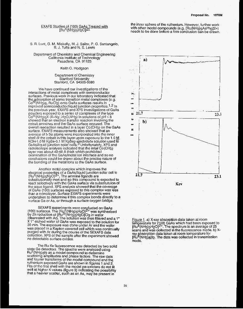
EXAFS Studies.of (1 00) GaAs Treated with ~ " ~ 3 ) 5 ! d 2 ~ ~
S. R. Lunt, G. M. Miskelly, M. J. Sailor, P. G. Santangelo, B. J. Tufts and N. S. Lewis
Department of Chemistry and Chemical Engineering California institute of Technology
Pasadena, CA 91 125
Keith 0. Hodgson
Department of Chemistry Stanford University
Stanford, CA 94305-5080
We have continued our investigations of the interactions of metal complexes with semiconductor surfaces. Previous work in our laboratory indicated that the dsorption of some transition metal complexes (e.9.
improved semiconductor/liquid junction properties.1 72 In the previous year, EXAFS and XPS investigations of GaAs poypers exposed to a series of complexes of the type Co '(NH3)5X (X=N3-,W20,NH3) in solutions of pH > 8 showed that an electron transfer reaction involving the cobalt ammines and the GaAs surface occured. The overali reacaction resulted in a layer Co(OHf2 on the GaAs surface. EXAFS measurements also showed that an average of 5 Se atoms were incorporated into the inner shell of the cobalt in this layer upon exposure to the 1 .O M KOH-1 .0 &j K2Se-0.1 &j K2Se2 e ectrolyte solution used fo
radioisotope analysis iqdicated that the inital Co(OH)2 layer was about 40-60 A thick which prohibited examination of the GaAsimetal ion interface and so no conclusions could be drawn about the precise nature of the bonding of the metal ions lo the GaAs surface.
Co 18 (NH3)6, RuCl3) onto GaAs surfaces results in
GaAsAiquid junction solar cells. 4 Unfortunately, XPS and
Another metal complex which improves the ele rical properit s of a GaAs/liquid junction solar cell is
substitutionally inert and so this compound is expected to react selectively with the GaAs surface via substitutuion of the aquo ligand. XPS analysis showed that the coverage of GaAs (1 00) surfaces exposed to this complex was less than a monolayer. Surface EXAFS experiments were undertaken to determine if this complex bonds directly to a surface Ga or As. or through a surface oxygen bridge.
SEXAFS experim nts were co ducted on GaAs
by Zn reduction of [RuH1(NH3)5H20]C13 in water (deareated with Ar). The solution was then filtered and a 1" X 1" etched wafer of GaAs was exposed to the solution for 30 min. The exposure was done under Ar and the wafer was placed in a Kapton covered cell which was continually purged with Ar during the course of the SEXAFS data collection. XPS of the sample after the experiment showed no detectable surface oxides.
state Ge detectors. The spectra were analyzed using RuIt(NH3)612 as a model compound to determine scatteirng amplitudes and phase factors. The raw data and fourier transforms of the model compound and the ruthenium exposed GaAs are shown in figures 1 and 2. Fits of the first shell with the model parameters do not fit well at higher K values (figure 3) indicating the possibility that a heavier scatter, such as an As, may be present in
[Ru 71 (NH3)5H2Ol5'. The ammine ligands are
(100) surfaces. The [R I7 (NH3)5H2O] ?? + was synthesized
The Ru Ka fluorescence was detected by two solid
the inner sphere of the ruthenium. However, further work with other model compounds (e.g. [Ru(NH3)5AsPh3]2+) needs to be done before a firm conclusion can be drawn.
Fiaure 1. a) X-ray absorption data taken at room tem erature for (1 00) GaAs which had been exposed to
scans and was collected in the fluorescence mode. b) X- ray bsorption data taken at room temperature for
mode.
[Ru R (NH&H20]2+. The spectrum is an average of 25
[Ru a (NH3)6]12. The data was collected in transmission
56

3.0 11.5 K
Fiaure 3. EXAFS dat and best fit of first shell using model parameters from [ J I ( N H ~ ) ~ ~ I ~
57
Acknowledaements
We wish to thank Dr. M. D. E. Forbes of Caltech for help in this research and Dr. 6. Hedman of SSRL for useful discussions. We also thank Dr. J. Gordon of IBM Almaden for the use of equipment and helpful discussions.
References
1. B. J. Tufts, I . A. Abrahams, P. G. Santangelo, G. R. Ryba, L. G. Casagrande, N. S. Lewis, Nature, 326, 861 (1987).
2. I. A. Abrahams, B. J. Tufts, N. S. Lewis, J. Am. Chem. Sot., 109,3472 (1987).
3. 6. J. Tuffs, I. L. Abrahams, C. E. Caley, S. R. Lunt, G. M. Miskelly, M. J. Sailor, P. G. Santangelo, N. S. Lewis, 6. M. Hedman, A. L. Roe, K. 0. Hodgson, submitted.
R + a
Fiaure 2. a) Fourier transform of EXAFS from data in figure 1 a. b) Fourier transform of EXAFS from data in figure 1 b.

proposal No. 2021Mp
ANALYSIS OF MUNICIPAL WASTEWATER-SLUEE INCINERATOR FKCSSION SAMPLES USING X-RAY ABSORPTION BEXTIODS FOR THE DETEWINATION
OF CHROMIUM AND NICKEL SPECIES
N.F. Mangelson", L.B. Rees, J.E. Silk and M.W. Hill Departments of Chemistry and Physics
Brigham Young University Provo, Utah 84602
R.B. Greegor and F.W. Lytle The Boeing Company
Seattle, Washington 98124
I
INTRODUCTION Samples in this study were analyzed to determine chromium and nickel species present in ash samples. These data will be used with results from chemical analysis to establish emission standards for wastewater-sludge incinerators. Berry [l] has discussed the need to regulate toxic substances in the air. Fly- ash samples have been shown to have significant biological activity and cytotoxicity [2,3]. The toxic nature of the particulate matter is believed to be due in large part to inorganic compounds present in the fly ash. Since the toxicity of several elements is dependent on the chemical form in which the element is present [4,5], it is necessary to know the chemical species of elements in fly ash before meaningful risk- assessment analyses can be made.
Chromium in the Cr(V1) oxidation state and nickel subsulfide (Ni3S?) are suspected carcinogens. Sensitive chemical methods have been. developed for determination of metal species, but some interferences may exist. Therefore, it is important to have a corroborative method of analysis. XANES and EXAFS are physical methods of metal speciation which avoid the necessity of chemical treatment before analysis.
Municipal wastewater sludge from four sludge incinerator facilities was ashed in a laboratory furnace. Ash from each sample was placed on a quartz filter. Bulk ash was also prepared for analysis.
Nickel and chromium K-edge XRA spectra were taken at SSRL on beam line IV-1. Current of the 3.3 GeV electron beam varied from 1 to 55 microamps. A parallel crystal monochromator with silicon 220 crystals was used.
In a related study, bulk flyash from a municipal wastewater-sludge incinerator was collected at the inlet to the electrostatic precipitator and effluent particulate material was collected on quartz filters at the outlet of the precipitator.
Nickel and chromium K-edge XRA spectra were measured at the National Synchrotron Light Source (NSLS), Brookhaven National Laboratory. The electron beam was 2.5 GeV with 90 to 200 microamps of current. Data were taken on beam line X23-A2 which contains a Golovchenko-Cowan, two-crystal monochromator using silicon 220 crystals. At both facilities, data for the samples were usually taken in the fluorescence mode and data for the reference compounds and mixtures were taken in the transmission mode.
PESULTS A few representative spectra are shown in
EXPERIMENTAL
-20 -10 0 10 20 30 40 50 Energy (eV)
Figure 1. Solid lines are the chromium K-edge XANES spectrum for the laboratory ashed sample 1N2S. Dotted lines are spectra constructed as linear combinations of a chromium(V1) compound and a chromium(lI1) compound. Each curve is labeled with the Cr(VI)%.
Figures 1 to 4. Figure 1 shows a chromium, K- edge XANES spectrum for the laboratory-ashed sample, 1N2S. The pre-edge peak at about 4 eV clearly indicates the presence of cr(V1). The dotted lines are linear combinations of a Cr(V1) spectrum and a Cr(II1) spectrum, and the labels show the percent Cr(V1). These data suggest that chromium in this laboratory ashed sample is about 55% Cr(V1).
The solid lines in Figure 2 are the experimental, chromium K-edge XANES spectrum for a mixture of K2Cr207 (a Cr(V1) reference compound) and FeCr204 (a Cr (111) reference compound). The dotted lines are a linear combination of spectra for the two pure reference compounds. The mixture of compounds was 49.7% Cr(V1) and the linear combination spectra give the best fit to the pre-edge peak at 50% Cr(V1). These data support the analysis method assumed in the discussion of Figure 1.
Figure 3 shows the nickel K-edge XANES and first derivative spectra for the unspiked, laboratory-ashed sample, 3N2. The heavier lines are spectra from the reference compound nickel spinel, NiFe204. A excellent fit is seen indicating that laboratory ashing of this sample produces the nickel spinel compound.
58

The solid lines in Figure 4 show the nickel K-edge XANES spectrum for the incinerator outlet sample 5C-OUT which was collected on a quartz filter. The nickel concentration was a few hundred ppm by weight which accounts for the poor statistics on the edge spectrum. Dotted lines are XANES spectra for several reference compounds. It i s clearly seen that the1 major compounds in the incinerator flyash are not the nickel subsulfide species. Additional study of thesq data suggest that nickel subsulfide is less than 20% of the total nickel in the sample.
Cr MIX 1
-20 -10 0 10 20 30 40 50
Energy (eV)
Figure 2. Solid lines are the chromium T-edge XANES spectrum of a mixture of K2Cr207 and chromite (FeCr20b) with 49.7% cr(V1). Dotted lines are spectra constructed as linear combinations of K2Cr207 and chromite spectr?. Each curve is labeled with the Cr(VI)%.
3 N 2 vs NiFe204
I I
Absorption 0 z
-20 0 20 40 60 80 100 120 Energy (eV)
Figure 3. Light lines are the nickel K-edge XANES spectrum and its derivative for the laboratory ashed sample 3N2. Heavy lines are the nickel, X-edge XANES spectrum and its derivative for a nickel spinel (NiFe204) reference compound-
Ni SC-OUT XANES F I T
-20 -10 0 10 20 30 40 50 Energy (eV)
Figure 4. Solid lines are the nickel K-edge XANES spectrum for the incinerator effluent sample 5C-OUT-Ni. Dotted lines are the spectra for several reference compounds as indicated in the figure.
ACKNOWLEDGMENTS We gratefully acknowledge the help given us at NSLS by C. E. Bouldin and Joe Woicik. We also appreciate the help from staff and crew at both SSRL and NSSLS. This work was supported in part by the U.S.E.P.A., Cincinnati, OH order No. 9C6119NNEE and by Entropy Environmentalists, Inc. order NO. 89-4181-01. The experiments were performed at SSRL, which. is supported by DOE through the Office of Basic Energy sciences and NIH through the Biotechnology Resources Program, and at NSLS, which is supported by the DOE through the Division of Materials Sciences.
BEFERENCES 1. D.K. Berry. Environ. Sci. Technol., 2 0
2. G.L. Fisher, K.L. McNeill, B.A. Prentics and A.R. HcFarland. Environ. Health persDect. , 51 (1983) 181-186.
3. C. Wei, 0.0. Raabe and B.J. Kimble.
(1986) 647-651.
L, 32 (1984) 179-186.
4. L. I). Hansen, G. L. Fisher, C. E. Chrisp and D.J. Eatough. Proceedings of the Fifth International Svmiposium. Polynuclear Aromatic Hvdrocarbons, M. Cooke and A . 3 . Dennis , eds . , Battelle Press, Columbus , OH, (1981) 507-517.
5. C. wei, M.A. A1 Bayati, M.R. Culberston, L.S. ROSenblatt and L.D. Hansen. J-
687. y-, 10 (1982) 673-
59
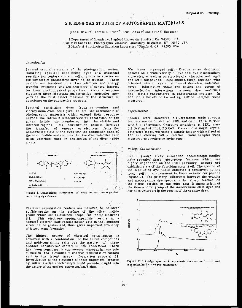
Proposal No. 2033Mp
S E; EDGE XAS STUDIES OF PHOTOGRAPHIC MATERIALS
Jane G. DeWitt', Teresa A. Smith2, Britt H
1 Department of Chemistry, Stanford University Stanford, CA 94305 USA.
3 Stanford Synchrotron Radiation Laboratory, Stanford, CA 94305 USA.
man3 and Keith 0. Hodgsonl
2 Eastman Kodak Co., Photographic Research Laboratory Rochester, NY 14650 USA.
Introduction
Several crucial elements of the photographic system including spec t ra l sensitizing dyes and chemical sensitization centers contain sulfur atoms in species on the surfaces of photoactive silver halide crystals. These centers a re involved in surface electron and energy transfer processes and are, therefore, of general interest for their photophysical properties. X-ray absorption studies of these important surface-active molecules may provide the first direct measure of the structure of adsorbates on the photoactive substrate.
Spectral sensitizing dyes (such as cyanine and merocyanine dyes, see figure 1) are the components of photographic materials which extend their response beyond the intrinsic blue/ultraviolet absorption of the silver halide photoconductor into the visible and infrared regions. This sensitization process involves the transfer of energy or electrons from the photoexcited state of the dyes into the conduction band of the silver halide and requires that the dye molecules exist in an .adsorbed state on the surface of the silver halide gr ains.
n -0.1.2
X - S. 0. N-Elhyl
R.W - alkyl. aryl
X -0.N-Elhyl
Figure 1. Generalized structures of cyanine and merocyaninr sensitizing dye classes.
Chemical sensitization centers are believed to be silver sulfide specks on the surface of the silver halide grains which act as electron traps for photo-electrons [ 11. This electron-trapping capability results in a reduced electron-hole recombination rate in the exposed silver halide grains and, thus, gives improved efficiency of latent image formation.
The highest degree of chemical sensitization is achieved with a combination of the sulfur compounds and gold-containing salts but the nature of these chemical sensitization centers is little understood. There has been considerable controversy surrounding the role of gold in the structure of chemical sensitization centers and in t h e la ten t image formation process [ I ] . Investigation of the structure of these important centers by sulfur K-edge spectroscopy could provide insight into the nature of the surface-active Ag/Au/S sites.
We have measured sulfur K-edge x-ray absorption spectra on a wide variety of dye and dye-intermediary molecules, as well as on structurally characterized Ag-S and Au-S compounds. These studies, taken together with oriented single crystal studies of dye-class molecules, reveal information about the nature and extent of intermolecular interactions between dye molecules and metals encountered in photographic systems. In addition, a variety of A u and Ag sulfide samples were measured.
Experimental
Spectra were measured in fluorescence mode at room temperature on BL 4-1 at SSRL and on BL X19A at NSLS with Si( 1 1 1) crystals. Operating conditions at SSRL were 3.3 GeV and a t NSLS. 2.5 GeV. Pre-oriented single crystal data were 'measured using a sample holder with x fixed at 180 and allowing full @ rotation. Solid samples were measured as powders on mylar tape.
Results and Discussion
Sulfur K-edge x-ray absorption spectroscopic studies have revealed sharp absorption features which are highly dependent on the local geometry around and oxidation state of the absorbing atom [2-41. The spectra of the sensitizing dye nuclei indicated a sensitivity to the local sulfur environment in these organic compounds (figure 2). The primary difference between the cyanine and merocyanine dye spectra is the sharp feature on the rising portion of the edge that is characteristic of the thiocarbonyl group of the merocyanine class dyes and has no counterpart in the spectra of the cyanine dyes.
J
S 0
.__.....I
I 2470 0400 9490 2500
EV
2. S K-edge spectra of representative cyanine (-) and anine C - * 3 dye molecules.
60

The sharp features on the rising portion of the S K- Sulfur-containing materials exhibit sharp K-absorption edge reflect transitions to localized, unfilled atomic or edge features which are polarized along specific molecular s ta tes and exhibit angular-dependent molecular bonds and can be used to determine the intensities in oriented samples of anisotropic centers. orientation of dye molecules in well-characterized Oriented single crystal spectra of 2-thiohydantoin are systems. The resul ts f rom these preliminary compared to the powder spectra in figure 3. The single investigations will form the basis for interpreting crystal polarized spectra suggests that the bound state feature characteristic of the thiocarbonyl group is almost entirely polarized along the Z molecular axis (normal to the plane of the molecule) and is attributed to a Is--px* transition in the terminal sulfur. The white line feature is polarized along the C=S bond in the plane of the molecule and is attributed to a Is+po* transition.
J
r 0
2-lhlahydanlein
EV
Figure 3. The powder spectra of 2-thiohydantoin (-1 and the spectra corresponding to polarization along the average S i. *'. * -1, Y (-.---) and 'L (- -1 molecular axis.
Direct interaction of sulfur with silver and gold atoms ( in metal sulfides and other metal/sulfur complexes) results in increased intensity and broadening of predominant S K-edge features. The dramatic reduction of the thiocarbonyl feature of 3-carboxymethyl-4- methyl-4-thiazoline-2-thione upon complexation with Ag (figure 4) indicates that the metal interacts with the ligand via the thiocarbonyl group. This feature, which we have found to be polarized normal to the C=S bond, suggests that the metal/ligand interaction involves the sulfur px: atomic orbitals and the Ag d orbitals.
..... 2480 2490 2500
. . ... . ,
2470 0 . 0
2460
EV
Figure 4. The powder spectra of ARC (-) and C 4gC is a polymeric structure with each thione S bound to 2 Ag's.
polarized surface XAS spectra of dyes in ordered systems, such as Langmuir-Blodgett films and dyes adsorbed to silver halide single crystals. Analysis of the EXAFS region of the K-edge spectra will provide a direct determination of the substrate-adsorber atomic distance and the first quantitative measure of the sulfur-surface distance for the adsorbed dye molecules.
Chemical sensitization centers are inherently surface active species and we intend to use a glancing-angle configuration to measure the K-edge spectra of sulfur and cry s t a1 s . Comparison of the chemical sensitization site K-edges and EXAFS with Ag/Au/S-containing models may provide considerable insight into the structure of the elusive sensitization centers on the silver halide surface.
s u If u r / go1 d - t r e a te d silver ha lid e
A cknowfe dgm ent
This work is supported by research funds from Eastman Kodak Company. The data were measured at SSRL and NSLS which are supported by the Department of Energy, Office of Basic Energy Sciences.
1. H.E. Spencer, R.E. Atwell. M. Levy, J. Phof. Sci.. 3 1. 158 ( 1 983). 2. Hedman, B., Frank, P.. Penner-Hahn. J.E.. Roe, A.L.. Hodgson,
K.O., Carbon, R.M.K., Brown, G., Cerino, J.. Hetiel, R.. Troxel, T. Winick, H., Yang, J. Nucl. Instr. and Mefh.. A246. 79 (1986).
3. Lytle, F.W.. Greegor, R.B.. Sandstrom. D.R.. Marques, EX.. Wong. J., Spiro, C.L., Huffman, G.P.. Huggins. F.E. N u d . Inslr. and Meth.. 226 , 542 (1984).
4. Sugiura. C. J. Chem. Phys.. 79, 48 1 1 ( I 983).
61

Proposal No. 2049Mp
COMPARISON OF FLUORESCENT AND e-Y IELD X-RAY ABSORPTION SPECTRA
F. W. L y t l e and R . B. G r e e g o r
The B o e i n g Company, S e a t t l e , WA 98124
The problems i n h e r e n t i n p r e p a r i n g t h i n , u n i f o r m a b s o r b e r s o r t h e s e l f a b s o r p t i o n which o c c u r s i n f l u o r e s c e n t EXAFS d a t a f r o m c o n c e n t r a t e d s a m p l e s c a n be a v o i d e d by u s i n g t h e e l e c t r o n y i e l d t e c h n i q u e . K o r d e s c h a n d Hoffman ( 1 ) f i r s t d e m o n s t r a t e d t h a t c o l l e c t i o n o f t h e e l e c t r o n s f rom t h e f a c e o f a sample i n a g a s f i l l e d chamber was a v i a b l e t e c h n i q u e f o r m e a s u r i n g XAS. L a t e r E r b i l e t a l . ( 2 ) and Elam e t a l . ( 3 ) showed e - y i e l d c o l l e c t i o n i n t h e p r e s e n c e o f a n a t m o s p h e r e p r o v i d e d a s i g n i f i c a n t g a i n ( u p t o 100) compared t o c o l l e c t i o n i n a vacuum. T h i s o c c u r s b e c a u s e o f s e c o n d a r y i o n i z a t i o n o f t h e c o u n t e r g a s by Auger e l e c t r o n s .
The t e c h n i q u e i s most u s e f u l f o r l o w Z a b s o r b e r s where t h e f l u o r e s c e n t y i e l d i s low, s e l f - a b s o r p t i o n o f t h e f l u o r e s c e n t s i g n a l i s h i g h and t h i n , u n i f o r m a b s o r b e r s a r e e s s e n t i a l l y i m p o s s i b l e t o p r e p a r e . Note t h a t d i l u t i o n o f t h e sample w i t h a l o w e r Z m a t e r i a l w i l l do no good f o r e i t h e r a n a b s o r b e r o r f l u o r e s c e n t sample b e c a u s e t h e i n d i v i d u a l p a r t i c l e s i z e s a r e t o o l a r g e by a t l e a s t a f a c t o r o f 10. The e f f e c t i s shown i n F i g . 1 f o r a sample composed o f a p r e s s e d d i s k of s u l f u r . A l l o f t h e f l u o r e s c e n t d a t a a r e so d i s t o r t e d by s e l f a b s o r p t i o n a s t o be u n r e c o g n i z a b l e a s t h e same m a t e r i a l . F l u o r e s c e n t d a t a f rom a s u i t e of c o n c e n t r a t e d r e f e r e n c e compounds would be u s e f u l o n l y f o r compar ison t o s i m i l a r l y c o n c e n t r a t e d s a m p l e s b e c a u s e f l u o r e s c e n t d a t a f rom, e . g. a b i o l o g i c a l sample c o n t a i n i n g a s m a l l amount , wel l d i s p e r s e d , would r e s u l t i n s p e c t r a more n e a r l y l i k e t h e e - y i e l d s p e c t r a . The d a t a were o b t a i n e d s i m u l t a n e o u s l y on BL IV-1, 3 G e V , 30 ma, S i ( 1 1 1 ) c r y s t a l s d e t u n e d 9 0 % i n a He f i l l e d d e t e c t o r r o t a t e d a t 45' t o t h e beam s o t h a t b o t h s i g n a l s c o u l d be c o l l e c t e d i n t h e same s c a n . The f l u o r e s c e n t g a i n was l o l o , e - y i e l d g a i n was l o 8 . F o r s u l f u r t h e f l u o r e s c e n t y i e l d i s 7% and t h e e - y i e l d s i g n a l a r i s e s p r i m a r i l y f rom t h e KLL Auger e l e c t r o n s ( 2 KeV). S i n c e t h e f i r s t i o n i z a t i o n p o t e n t i a l f o r He i s 25 eV, e a c h e l e c t r o n w i l l c r e a t e 8 0 i o n p a i r s . T h i s g a s g a i n f a c t o r compared t o t h e f l u o r e s c e n t y i e l d a n d t h e 2 p i s o l i d a n g l e compared t o 0 . 2 p i f o r t h e f l u o r e s c e n t de tec tor a c c o u n t f o r t h e h i g h s i g n a l l e v e l f rom t h e e - y i e l d d e t e c t o r .
REFERENCES 1. M. E . Kordesch and R . W . Hoffman, Phys. R e v . B29, 491 ( 1 9 8 4 ) . 2. A . E r b i l , c. s. C a r g i l l , R . Frahm a n d R . F. Boehme, Phys. Rev. B37, 2450 ( 1988) . 3 . W . T . Elam, J . P. K i r k l a n d , R . A . Neiser and P. D. Wolf, Phys. R e v . B38, 26 ( 1988) .
ACKNOWLEDGEMENT S u p p o r t e d i n p a r t by DOE G r a n t DE-FC06-84ER45121.
62
4 I 0
X 4
15. t
-15. t
Fig. 1 The reduced amplitude i n the fluorescent spectra of the edge resonance, EXAFS and FT as well as the apparent broadening a t the edge are caused by the expmential diminution o f the edge jump due t o se l f absorption.


Proposal No. 2052M
THE ENERGY DEPENDENCE OF THE PRODUCTION OF THE E' CENTER IN AMORPHOUS SiO,
D.B. Kerwin and F.L. Galeener Department of Physics, Colorado State University, Fort Collins, CO 80523
l n t r o d u c t i m The E point defect center in amorphous Si02 has been studied for over 30 years. It has been determined with a high degree of accuaracy that the E' center corresponds to an electron in an sp3 orbital of a silicon atom that is bonded to three (rather than four) oxygens. However, the mechanism of E' defect formation is not as well understood. Experiments on the desorption of oxygen from Si02 surfaces by Knotek[l] suggested that either a cooperative Auger transition or a Coulomb explosien[2] could produce an oxygen vacancy in the bulk when a silicon atom was photoionized. The paramagnetic version of the oxygen vacancy is just the E' center. The goal of this experiment was to determine whether the cross section for E' production differed significantly for x- irradiation below and above the k-edge of silicon. 0
F x I) e r i men t a l The samples used were high hydroxyl containing silica (1 200 ppm OH) and low hydroxyl containing silica from Heraeus- Amersil (Suprasil-1 and Suprasil-W1 , respectively). The samples were irradiated on beamline 111-4 in March 1989. Although it had been intended to use the C-Rh multilayer synthetic crystals to monochromate the beam, the flux was too low to obtain the necessary exposures in a reasonable amount of time. Therefore, white radiation was used with Si and Be filters to produce spectral distributions that were either rich or deficient in radiation just above the silicon k-edge. The post-radiation spin sig'nals were measured on the Eruckar ER300 series ESR at Colorado State University.
Results Several exposures were made with the white beam with only a 25 pm Be filter, and a few exposures were made with both a 5 prn Be filter and a 2 prn Si filter. The latter combination produced a spectral distribution which was deficient in photons of energies just above the k-edge of silicon. Measurement of the number of E spins indicated no significant differences between exposures containing photons above the k-edge and those deficient in photons with energies above the k-edge. Figure 1 shows the results for Suprasil-1 for exposures up to 500 mJ cm-2. Based on the difference in the absorption coefficient of silicon 200 eV below and 200 eV above the k-edge, one would expect an almost four fold increase in the production of E' centers for the radiation containing a significant distribution of photons with energies above the k-edge. Preliminary results indicate that the above mentioned mechanisms do not determine the number of E defects observed in SO, irradiated in this energy regime.
Figure 1
L * I- 300 L L _j 400
Exposure ( mJ cm-2 1
Number of E spins vs exposure for Suprasil-1 samples. Circles - 25 pm Be filter, Squares - 5 pm Be + 2 pm Si filters.
REFERENCES 1. M.L. Knotek and P.L. Feibelman, Surf. Sci. a, 78 (1979). 2. J.H.O. Varley, Nature J.U. 886 (1954).
ACKNOWLEDGEMENTS The assistance of P. Pianetta and L. Pan of SSRL was greatly appreciated. The assistance of A. Miller of Colorado State University in performing the experiments was also appreciated. This research was performed in part at SSRL, which is supported by the DOE'S Office of Basic Energy Science.
63

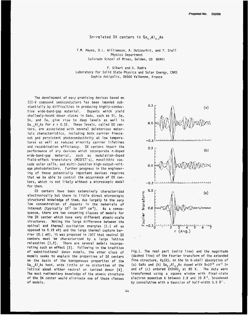
Proposal NO. 2055M
Sn-re1 ated DX cen te r s in Gao,,A1 0 , 3 A ~
T.M. Hayes, D.L. Williamson, A. Outzourhit, and P. Small Phys.i cs Department
Colorado School of Mines, Golden, CO 80401
P. Gibart and A . Rudra Laboratory for Solid State Physics and Solar Energy, CNRS
Sophia Antipolis, 06560 Valbonne, France
The development of many promising devices based on 111-V compound semiconductors has been impeded sub- stantially by difficulties in producing highly-conduc- tive wide-band-gap material. Dopants which yield shallowly-bound donor states in GaAs, such as Si, Se, Sn, and Te, give rise to deep levels as well in Ga,-xAlxAs for x > 0.22. These levels, called DX cen- ters, are associated with several deleterious mater- ials characteristics, including both carrier freeze- out and persistent photoconductivity at low tempera- tures as well as reduced minority carrier lifetimes and recombination efficiency. DX centers impair the performance of any devices which incorporate n-doped wide-band-gap materia?, such as modulation-doped field-effect transistors (MGDFET's), monolithic cas- cade solar cells, and multi-junction high-output-volt- age photodetectors. Further progress in the engineer- ing of these potentially important devices requires that we be able to control the occurrence of DX cen- ters, which is not likely without a microscopic model for them.
DX centers have been extensively characteriled electronically but there is little direct microscopic structural knowledge o f them, due largely to the very low concentration of dopants in the materials of interest (typica'lly IOz7 to IO" ~ m - ~ ) . As a conse- quence, there are two competing classes o f models for the DX center which have very different atomic-scale structures. Noting the large difference between the optical and thermal excitation energies (1.1 eV as opposed to 0.19 eV) and the large thermal capture bar- rier (0.1 eV), it was proposed in 1977 that neutral DX centers must be characterized by a large lattice re1 axation [ 1,2]. There are several model s i ncorpo- rating such an effect [ 3 ] . Following in the tradition of substitutional donor models, the other class of models seeks to explain the properties of DX centers on the basis of the homogeneous properties of the Ga,-xAlxAs host, with little or no distortion of the lattice about either neutral or ionized donor [ 4 ] . The most rudimentary knowledge of the atomic structure of the DX center would eliminate one of these classes of models.
64
Fig.1. The real part (solid line) and the magnitude (dashed line) of the Fourier transform of the extended fine structure, k ( k ) , on the Sn K-shell absorption of (a) GaAs and (b) Ga,,,Al,,,As doped with 5 ~ 1 0 ' ~ cm-3 Sn and of (c) ordered ZnSnAs, at 80 K. The data were transformed using a square window with final-state electron momentum k between 2.8 and 10 A - l , broadened by convolution with a Gaussian of half-width 0.5 A-' .

A v a r i e t y o f samples w i t h x between 0 and 1 and doping l e v e l s o f 4 t o 9x10l8 cm-3 l19Sn have been s tud- i e d u s i n g Mossbauer e f f e c t spectroscopy (MES) [5]. Clear evidence f o r t h e DX cen te r was found i n a s h i f t e d and broadened resonance which i s most prom- i n e n t f o r x=O.3-0.4. The broadening suggests a quadrupole i n t e r a c t i o n a t t h e Sn DX s i t e and t h e r e f o r e a l o c a l d i s t o r t i o n f rom cub ic symmetry.
The x - r a y abso rp t i on spec t ra assoc ia ted w i t h t h e Sn K s h e l l i n GaAs and Ga0,,A1,,,As doped w i t h 5~10'~ cm-3 Sn have been measured a t 80 K us ing f luorescence d e t e c t i o n methods. The samples were coo led i n t h e da rk t o popu la te t h e DX cen te r . The extended f i n e s t r u c t u r e i n these spec t ra have been compared i n d e t a i l with each o t h e r and with t h a t i n t h e spectrum f rom ordered ZnSnAs,, i n which each Sn atom i s su r - rounded by f o u r As atoms a t 2.60 A. The F o u r i e r t rans forms o f those spec t ra a re shown i n F ig . 1. We conclude t h a t each Sn atom i n t h e doped samples has four As ne ighbors a t -2.58 A, bu t no te t h a t t h e shape o f t h e neares t -ne ighbor (NN) peak i n t h e r a d i a l d i s - t r i b u t i o n f u n c t i o n does n o t have a s imp le Gaussian shape. Whi le t h e r e are s l i g h t d i f f e r e n c e s between NN peaks i n t h e two doped samples, t h e r e i s no evidence o f a l a r g e s h i f t i n NN d i s tance assoc ia ted wi th t h e n e u t r a l DX cen te r i n Ga, ,Al, 3 A ~ , thereby e l i m i n a t i n g t h e p o s s i b i l i t y o f a l a r g e d i l a t o r y l a t t i c e r e l a x a t i o n i n t h i s sample. An angu lar d i s t o r t i o n c o n s i s t e n t w i t h t h e MES measurements would o f course be poss ib le . Measurements o f t h e Sn donor s i t e under d i f f e r e n t dop- i n g c o n d i t i o n s a re i n progress.
1. R.J. Nelson, Appl . Phys. L e t t . 31, 351 (1977) 2 . D.V. Lang and R.A. Logan, Phys. Rev. L e t t . 39, 635
(1977) 3. See, f o r example, D . V . Lang, R.A. Logan, and M.
Jaros, Phys. Rev. 819, 1015 (1979); K. Kobayashi, Y . Uchida, and H. Nakashima, Jpn. J. Appl. Phys. 24, L928 (1985); A. Oshiyama and S. Ohnishi , Phys. Rev. 633, 4320 (1986); T.N. Morgan, Phys. Rev. 834, 2664 (1986); H. Hasegawa and H. Ohno, Jpn. J . Appl. Phys. 25, L643 (1986)
4. See, f o r example, A.K. Saxena, S o l i d S t a t e E lec- t r o n . 25, 127 (1982); H.P. Hjalmarson and T.J. Drummond, Appl . Phys. L e t t . 48, 656 (1986); J.C.M. Henning and J.P.M. Ansems, Semicond. Sc i . Technol. 2, 1 (1987)
5. P. G iba r t , D.L. Wi l l iamson, 8. E l Jan i , and P. Basmaji, Phys. Rev. B 38, 1885 (1988)
65
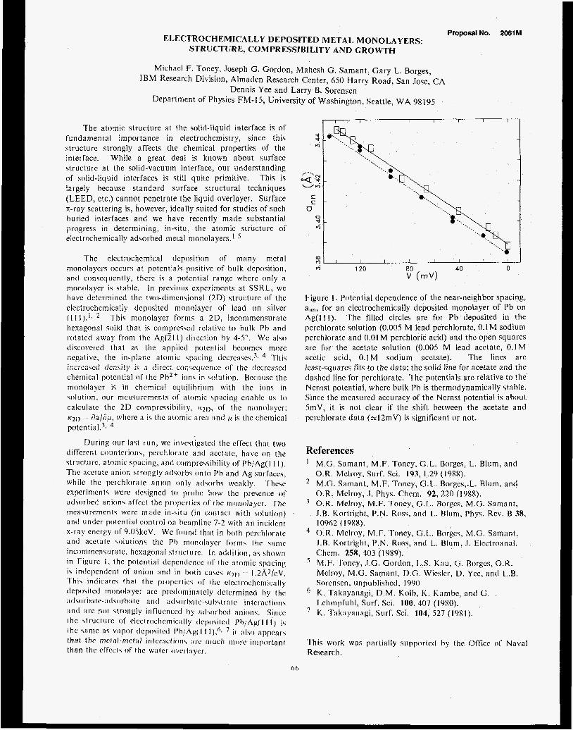
ELECTROCHEMICALLY DEPOSITED METAL MONOLAYERS: STRUCTURE, COMPRESSIBlLITY AND GROWTH
Michael F. Toney, Joseph G. Gordon, Mahesh G. Samant, Gary L. Borges, IBM Research Division, Almaden Research Center, 6.50 Harry Road, San Jose, CA
Dennis Yee and Larry B. Sorensen Department of Physics FM-I 5, University of Washington, Seattle, WA 98195
Proposal No. 2061 M
The atomic structure a t the solid-liquid interface is of fundamental importance in electrochemistry, since this structurc strongly affects the chemical properties of the interface. While a great deal is known about surfacc structure a t the solid-vacuum interface, our understanding of solid-liquid interfaces is still quite primitive. This is largely because standard surface structural techniques (LEED, etc.) cannot penetrate the Jiyuid overlayer. Surface x-ray scattering is, however, ideally suited for studies of such buried interfaces and we have reccntly made substantial progress jn detcrmining, in-situ, the atomic structurc of electrochemically adsorbed metal monolayers.1-5
The elcctrochemical dcpocition of many metal monolayers occurs a t potentials positive of bulk deposition, and consequently, thcrc is a potcntial rangc whcrc only a monolaycr is stable. In previous cxpcrimcnts a t SSRL, we have dctcrmined thc two-dimensional ( 2 D ) structurc of the clcctrochcmically dcpositcd monolaycr of lead o n silver ( I 1 I ) . ' , * This monolaycr forms a 2D, incommcnsurntc hexagonal solid that is comprcssctl rclativc to bulk Pb a n d rotatcd away from the AgOl 1) tlircction hy 4-S". Wc also ciiscovcred that as thc applicd potcntial hccomes morc ncgalive, thc in-planc atomic \pacing ticcrcascs.3, This
chemical potential of thc Pb2+ ions in colution. Bccausc the monolaycr is in chemical cquilihrium with thc ions in solution, our mcasurcmcnts of atomic \pacing enable uc io calculate thc 2D compressibility, 1<21), of thc monolaycr: ~ 2 1 ) := aa/np, whcrc a is thc atomic arca ant1 11 is thc chcmical potcntiaI.3,
i r ,L .*no ,,w! c!cnsity i: 3 (!ircct cocrequcncc i ) T the tlcc;casctl
During our last run, wc invcstigatcti thc cffcct that two difTcrcnt countcrions, pcrchloratc a n t l acctatc, havc o n t.hc struct.urc, atomic spacing, antl comprc\sihilii.y o f Pb/Ag( I I 1 ). Thc acctatc anion strongly atisorhs o n t o Ph antl Ag surraccs, whilc thc perchlorale anion only atl\orhs wcakly. 'T'hcsc cxpcrirncnts wcrc tlesigned to prohc how 1 hc prcscncc of atlsorbcd anions affcct the propcrlics of thc monolaycr. 'T'hc mcasurcmcnts wcrc rnaclc in -s i tu f i n contact with wlution) an t l undcr potenlial control o n hcamline 7-2 with a n incitlcnt x-ray cncrgy of9.OSkcV. Wc found t h a t in both pcrchlor:lic anti acctale solutions thc Ph monol:iycr fot-m\ ihc same incommcnwratc, hexagonal slriict~~rc. In addition, a \ shown in Figure I . thc potential dcpcntlcncc of Ihc :it.omic spacing is indcpcntlcnt of a n i o n a n d i n both c:iscs ~ 2 1 ) -- I . ~ K ~ / c v . 'This intlicatcs tha t f he propcrt ics o f thc clcclrochcriiic;11ly deposit cd m o tio1:i ycr a re prct lc )ni i 11 :i 1 cly dct crm i nctl by I Iic at1 cor h : ~ t e-at I \ ( i r h le :I n (1 ; J d \ O l h I e-\ 11 h\I I':I I c in 1cr:icl i o n \ an t1 arc n o t strongly influcnccd hy :itl\orbctl :inions. Siticc ihc \lruclurc of clcc1rocIicmic:~lly tlcpositcd Ph/Ag(l I I ) is 1 he same a \ vapor tlcpositctl Ph/Ag(l I i t ; r l ~ :tppc;rt.; that thc riicl:il-mct;ll intcr:icfion\ ;Ire much t i i ( m important than thc cffccts o f thc water ovc~~I:~ycI.
I I I I I I
Figure I . Potential dcpendence of the near-neighbor spacing, a,,", for a n clcctrochemically deposited monolayer of Pb on Ag(1 1 I ) . The fillcd circles are for Pb deposited in the pcrchloratc solution (0.00s M lead perchlorate, 0.1 M sodium pcrchlorate and 0.01 M perchloric acid) and thc open squares arc for the acetatc solution (0.00S M lead acetate, 0.1M acetic acid, 0.1M sodium acctate). The lines are Icast-squarcs fits to thc data; the solid iine for acetate and the dashed line for perchlorate. The potentials arc relative to the Ncrnst potcntial, wherc bulk Pb ic thermodynamically stable. Since thc mcacurctl accuracy of the Nernct potential is about StnV, it is n o t clear if thc shift between the acetate and pcrchlorate data ( ~ 1 2 m V ) is significant or not.
References I M.G. Samant, M.F. Toncy, G.L. Borges, L. Blum, and
O.R. Mclroy, Surf. Sci. 193, L29 (1988). M.C. Samant, M.F. Toncy, G.L. Borgcy,.L. Blum, and O.R. Mclroy, .I . Phys. Chcm. 92, 220 (1988). ' O.R. McJroy, M.F. Toncy, G.1,. Borgc\, M.G. Snmant, J.B. Kortright, P.N. Ross, and L.. Blum, Phys. Rev. B 38,
O.R. Melroy, M.F. Toncy, (3.1,. Borgcs, M.G. Samant, I . R . Koitright, P.N. Ross, antl L. Blum, I . Elcctroanal. Chcm. 258, 403 (1989). M.F. Toncy, J.G. Gotdon, 1 3 . Kau, G. Rorgcs, O.R. Meltoy, M.G. Samant, D.G. Wic4cr, D. Ycc, and L . R . Sor cnsc n , u n pu blis hccl , 1990
I h n p f u h l , Surf. Sci. 100, 407 (1980). K . rak:iyanagi, Surf. Sci 104, 527 (19x1).
I0962 ( I 9XX) .
' K. Takayanagi, D.M. Kolh, K . Kambc, antl (3 .
This work was partially supportctl by the Officc ol' Naval Research,
66


Proposal No. 2062Mp
Structural Improvements in Multilayers for Instrumentation Applications
William K. Warburton X-ray Instrumentation Associates, 1300 Mills St., Menlo Park, CA 94025
Troy W. Barbee, Jr. Lawrence Livermore National Laboratory, P.O. Box 808, Livermore, CA 94550
1 : Introduction
When a diffraction grating is overcoated with a multi- layer, a new x-ray optical element results which possesses nove1,characteristics found in neither the grating nor the multilayer taken separately. [l , 21 First, the devices can be much more efficient than single metal overcoated gratings because the multilayer can be designed to be an efficient x- ray reflector at the energy of interest. Further, because of the finite multilayer bandwidth, the grating orders become dis- continuous in energy. Finally, the devices act like x-ray prisms, maintaining a constant deflection for a given energy as the incident angle is changed. Barbee and coliaborators have extensively observed and verified these effects in the soft x-ray regime below 1500 eV. [2, 31 Barbee has further developed an equation for the angle between the different grating orders based on the grating equation and Bragg's Law: [3]
Here m's are reflection orclers, d (D) is the multilayer (grating) spacing, and 6 is the average anomalous scattering factor. Warburton has addressed the same problem using kinematic scattering theory [4] to derive an equivalent expression for the offset angles between the grating orders as:
where R is the bracketed term to the 1/2 power in Eqn 1. The difference between the expressions becomes most Important as 8 approaches zero.
These devices are expected to become extremely Important for Instrumentation since they are not only efflclent and dispersive but may also resolve many of the overlapping ordsr problems assoclated with simple gratings, in the pres- ent work we are extendlng earlier measurements Into the hard x-ray reglon.
2: Experimental
sures of the dispersed beam were taken w l h interposed elemental filters ranging from Mn through Ge. The elemental absorption edges could be clearly seen and moved across the dispersed beam with absorption edge energy in accord with the previously observed prism effect. We then proceed- ed to quantitative data by installing an 0.001" slit on the exit arm in front of an ion chamber so the dispersed beam could be scanned.
3: Results
Due to poor ring performance, only a few scans could be obtained, but these are extremely interesting. Figure 1 shows the white beam as dispersed by 89037. The center of the peak (0 grating order) corresponds to an energy of 5.9 keV from the Bragg relationship and the multilayer d spacing. The multilayer coated grating displays very complex behav- ior, particularly at low angles, which we are working to un- derstand and will discuss below. When an Fe foil is inserted, the result is as shown in Figure 2, with the absorption edge at 7.1 keV clearly visible at about 2.5" 28. This is the same value of 28 at which the multilayer would Bragg reflect 7.1 keV light, but the reflection has unequal angles of incidence and reflection in the present case. Finally, in Figure 3, we present a view of the diffraction structure in the vicinity the
t
0.001.. . . . .-!. . . , . . . , . . . . .k-. . . . . . 1:o do 3: 0 40
Anglo (deg)
Figure 1: White llght reflected from a 8 prn pltch gratlng coated with a 40A d spaclng multilayer.
The data were taken in BL 2.3 in white Ilght mode during the March I989 run. The sample, 89037, was a 40A d spacing Rh/C multilayer deposited on an 8 mm D spaclng grating etched into a SI wafer, The sample was inltlally placed on the 0) gonlometer of a small angle diffractometer described elsewhere 151 at an angle of 1 . 5 O and film expo-
67

grating 0 order which has been expanded to show the de- tailed structure of the peaks.
4: Discussion
The spacing of the peaks near 0 grating order is fairly well understood. From either equation 1 or 2, ignoring the correction terms in brackets, the interpeak spacing for 1st multilayer Bragg order would be A$ = 2d/D = 0.057O for 89037's values of d and D. The average interpeak angle computed simply form the peak maxima is O.O56O, in good agreement. In addition, the subpeaks and shoulders seen in Fig 3 fall at intervals given approximately by 0.056'/2 and 0.056'/3, corresponding to the 2nd and 3rd Bragg orders from the multilayer.
The peak amplitudes, on the other hand, have yet to be explained. According to the kinematic theory they should fall off as l/mD2, which they clearly do not. Since we do not yet know to what extent this shape depends upon the align- ment of the grating in the beam, we have postponed its inter- pretation until we are sure that it is not an experimental arti- fact. The other area of interest is the small angle shoulder region. In this region there is a transition between Bragg re- flection from the multilayer and total external reflection from its surface. The cutoff at 1.5', in fact, corresponds to an exit beam travelling parallel to the grating surface since the angle of incidence is also 1.5O. The description of the multilayer coated grating in this region will therefore be more complex than in the region near 0 grating order and its description may require combining a dynamical description of the multi- layer with the grating equation.
5: References
111 T.W. Barbee Jr., in: Multilayers: Synthesis, Properties and Non-Electronic Applications, Mat. Res. SOC. Symp. Proc. 103 (1 988) 307
21 T.W. Barbee Jr., Rev. Sci. Instrum. 60 (1989) 1588.
[3] T.W. Barbee Jr., in: X-ray and Vacuum Ultraviolet Interaction Databases, Calculations and Measurements, Proc. SPIE 91 1 (1 988)
[4] W.K. Warburton, 6th National Conference on Synchrotron Radiation Instrumentation, Nucl. Instr. and Meth. Accepted for publication. (1 990)
. . . . 1:o 210- - 3:O 4:O
Angle (deg)
Figure 2: Identical conditions to Fig. 1 but with a 10 pm Fe foil inserted after the grating.
0.40 I I I I I
0.30
F 0.20
0 - . - 0.10
0.00
2.75 2.85 2.95 3.05 3.15 Angle (deg)
Figure 3: Enlarged view of the peak region of Fig. 1.
[5] W.K. Warburlon, T.W. Barbee Jr. and Z.U. Rek, 6th National Conference on Synchrotron Radiation Instrumentation, Nucl. Instr. and Meth. Accepted for publication. (1 990)
68

ANMALOUS S J S L L ANGLE X-RAY SCATTERING STODY OF AMORPHOUS =TAL-GERMANIUM AI;LoYS
Marybeth Rice Department of Electrical Engineering
Stanford University
Soichi Wakatsuki Department of Chemistry
Stanford University
Arthur Bienenstock Stanford Synchrotron Radiation Laboratory
INTRODUCTION
Anomalous small angle x-ray s c a t t e r i n g (ASAXSJ i s emerging a s a t o o l f o r detect ing and character iz ing phase .separation i n amorphous mater ia ls . In
c r y s t a l l i n e mater ia l s phase separation i s shown readi ly by t h e superposition of t h e broadened x-ray d i f f r a c t i o n powder pa t te rns from each phase. Because there a r e no sharp d i f f r a c t i o n peaks i n an amorphous mater ia l , it i s impossible t o t e l l whether t h e pa t te rn or ig ina tes from a s ingle , or from multiple, phases.
Classical ly , information a b o u t ' such phase separat ion i s obtained from small angle x-ray sca t te r ing . SAXS i s sens i t ive t o e lec t ron densi ty f luc tua t ions on a scale of tens t o thousands of Angstroms. Therefore, SAXS indicates t h e presence of inhomogeneities a r i s i n g from such diverse phenomena a s phase separat ion or cracks and voids. Unfortunately, SAXS alone cannot d i s t inguish between these various p o s s i b i l i t i e s .
Recently, inves t iga tors have begun t o combine SAXS with anomalous dispersion t o achieve such a d i s t i n c t i o n . We a r e using ASAXS a s a t o o l f o r understanding t h e s t ruc ture of amorphous metal-germanium t h i n f i lms. In par t icu lar , we a r e seeking t o determine whether these f i l m s a r e phase separated on a very f i n e (2-4 nanometers) scale , t h e size of t h e s ing le phase domains being l imited by t h e k ine t ics of f i lm formation.
1
The metal-germanium amorphous films, which can be prepared t y p i c a l l y from pure Ge t o approximately 70 atomic percent metal, a r e semiconducting f o r low metal concentrations and then meta l l ic when t h e metal concentration reaches 10-15 atomic percent. When t h e metal concentration r i s e s even more, physically i n t e r e s t i n g meta l l ic charac te r i s t ics a r i s e . Superconductivity appears i n the Mo-Ge system a t 13 atomic percent Mo whereas ferromagnetism appears a t approximately 40 atomic percent Fe i n t h e Fe-Ge system. We a r e using ASAXS t o determine whether these t r a n s i t i o n s occur v i a phase separation or through homogeneous a l l o y formation.
ExeERIMENTaE ProposalNo. 20881
In February of 1989 we performed ASAXS experiments on a-W8Geg2, a-Mo8Gegg and a-Fe Ge 1 2 88 On SSRL beamline IV-2. We used the SSRL Biotechnology Group's SAXS camera with its posi t ion sens i t ive l i n e a r detector (described by Hubbard ) . We intended t o c o l l e c t data a t 100 eV, 20 eV, and 7 eV below t h e absorption edge of each element i n a l l the three samples, but t h e bearc conditions prohibi ted t h e co l lec t ion of data a t the Ge edge f o r t h e FeGe sample and a t t h e Mo edge f o r the MoGe sample. For the WGe
sample we col lected a complete data s e t . The region of reciprocal space probed i n these experiments runs from s=2sinB /h=O . 0 1 t o s=O. 2 inverse Angstroms. The data shown here a r e scaled by t h e i n t e n s i t y of t h e t ransmit ted beam and are background subtracted. To remove t h e contr ibut ion of t h e resonant Raman sca t te r ing , t h e three data s e t s a t each edge a r e forced t o coincide a t s=0.2 inverse Angstroms where t h e i n t e n s i t y of t h e small angle sca t te r ing approaches its asymptotic value.
RESULTS AND DISCUSSION
2
Corrected ASAXS data f o r the a-W8Geg2 sample a r e reproduced i n Figures 1 and 2. A s t h e W edge i s approached from below t h e i n t e n s i t y of sca t te red rad ia t ion decreases. A t t h e Ge edge the i n t e n s i t y of sca t te red radiat ion remains the same a s t h e photon energy increases beneath t h e edge. This means t h a t t h e Ge i s not contr ibut ing t o the small angle scatte'ring. The s c a t t e r i n g a r i s e s from f luc tua t ions i n the electron densi ty of the W and the Ge number densi ty i s uniform across t h e sample. A s a rough check on whether it i s possible f o r Ge t o be present i n two phases i n a-W8Geg2 and have the same densi ty within each phase, we invest igated the density of Ge atoms i n c-Ge and i n c-MoGe (assuming t h a t the metastable phases of WGe strongly resemble those of MoGe, since there a r e no compound i n t h e WGe equilibrium phase diagram). A s shown i n Table 1, t h e number densi ty of Ge i n pure Gel MoGe2 and Mo13Ge23 i s nearly the same. Therefore, phase separation of a-W8Geg2 int.0 a-Ge and an a-MoGe2 or a-Moo13Ge23-iike substance i s consis tent with the da ta .
Crysiiils Number or a (A) c (A) N I V Ge Rtoms per
unit cell Cry s t iil I in e 8 5 . 6 5 8 0 . 0 4 4 2
Ce MoCez 4 3 .3 I 3 x. I 9s 0 . 0 4 4 5
M o i n G w 9 2 5.990 63.54 0 . 0 4 0 4
69

Although our j a t a s e t s on the a-Mo8Geg2 and a-Fs12Ge88 samples c?re inconplete, they both exhibi t s m a l l angle sca t te r ing near s=O.O5 inverse Angstroms s imilar to t h a t seen i n t h e a-W8Geg2 sample. A s shown i n Figure 3, the i n t e n s i t y o f the small angle sca t te r ing remains constact a s the photon energy i s increased beneath the Ge edge i n t h e a-Mo8Geg2 sample. The a-Fe12Ge8b r e s u l t s f o r TCC Fz edge are shown i n Figure 4 . The sca t te red i n t e n s i t y decreases with increasing photon energ? beneath che Fe edge. Both of these r e s u l t s a re consis tent with the a-W8Geg2 r e s u l t s , although they are iriconpiete. These r e s u l t s ssggest t h a t there -may be s t rong s i r r . i lar i t ies i n the s t ruc ture of these m.etal-gemanim f i ims i n the low metal concentration regime.
We performed more ASAXS experinents on t h e a-FeGe, a-WGe and a-MoGe systems a t NSLS beamline X14A i n December 1989. We col lec ted data f o r samples of d i f fe ren t concentrations across t h e Srcad amorphous region of each systen. These data w i l l be presented
xt0.4
f ' 1
1~
f Y 2 - - a - u7
0 -
L
0.00 0.05 0.10 0.1s 0.m Sc~it~rrlng vector S (>/A)
FIGURE 2. Tie i n t e n s i t y of t h e SAXS peak, f o r t h e a-W ~e
8 92 sinplc, remained consmni a s the Ge edge was apprzzched from below.
,-I I !
.io-4
when they are analyzed.
REFERENCES
% - See, f o r example: P . Godeau, A . Naudon, A. -
Chamberod, B. Rodmacq, and C. E . Williams, Europhys. F 0 L e t t . 3 ( 3 ) , 269 (1987) B
- I
* ( 1 9 b 7 ) .
S. R. Hubbard, Ph.D. t h e s i s , Stanford University
ACKNOWLEDGMENTS
This research i s supported by the Department of Energy through t h e Office of Basic Energy Science.
x,0.4 I '
FIGURI?. 1 . rrr .__ * - _ a-lqGaq? s i q l e , we otsorved a decrease i n t h e - _ _ x:er.sF:y of t h e S:xs peak as t h e X edge was tc-rcacnea .. f r m below. . .
70
1
J 0.00 0.05 0.10 0.15 0.20
Saitsilng Vector s (IIA)
FIGURI.: 3. PCS t% a-Mo8Geg2 smp:?, t h e i n t e n s i t y of t h e SAXS peak did not change as t h e Ge edqe was approached from beloij .
.--___
L i
0.00 0.02 0.04 0.06 0.08 0.10 0.12 0.14 Scattering vector S (i lb)
FIGURE 4. Fcr the a-FeI2Geg8 sample, t h e i n t e n s i t y of t h e SAXS peak decreased as t h e Fe edge was approach from below.

Proposal No. 8003M STUDYING HYPERFINE FIELDS WITH RESONANT NUCLEAR DiFFRACTlON OF X RAYS
J. Arthur, D. E. Brown, S. L. Ruby, and G. S. Brown Stanford Synchrotron Radiation Laboratory, Stanford, CA 94309
G. K. Shenoy Advanced Photon Source, Argonne National Laboratory, Argonne, IL 60439
In the SSRL Activity Report for 1988 we presented some of the results of an experiment carried out in December, 1988 at the PEP 1B beam line. During 1989 we analyzed more of the data from that experiment. This report is condensed from a paper prepared for the 34th Annual Conference on' Magnetism and Magnetic Materials, held in Boston in November, 1989.'
The use of resonant nuclear diffraction to study hyperfine fields in a crystal depends simply on the spectral and temporal characteristics of the synchrotron radiation pulse. A synchrotron source provides very short (<1 ns) pulses of radiation, with a bandwidth that is much wider than the energy range of the nuclear resonance in the sample. If the sample resonance is split by hyperfine fields, the synchrotron pulse will simultaneously and coherently excite all the levels of the split resonance (subject to the polarization selection rules discussed below). The coherence between the excited levels is preserved during the excited state lifetime, which can be much longer than the synchrotron pulse, and so the radiative decay of the excited state exhibits interference effects. The time distribution of the scattered radiation contains a beat structure determined mainly by the energy differences between the coherently excited levels.*
a synchrotron source provides radiation with a well-defined polarization. If the sample is magnetic, the experiment can be designed to give specific relationships between the directions of polarization of the incident and scattered light and the magnetic field in the sample. The nuclear excitation matrix elements give polarization selection rules which can be used to simplify the experimental analysis.
X-ray diffraction selection rules can also be used to simplify the experimental analysis, in the case where a sample contains more than one crystallographically distinct group of resonant nuclei. The hyperfine field environments for the nuclei in different groups may be different, but by choosing a reflection for which only one crystallographic group has a nonvanishing structure factor, the hyperfine field for that group can be examined separately.
experiment using resonant diffraction from the 14.4 keV Mossbauer nuclear resonance in 57Fe, in a nearly perfect crystal of yttrium iron garnet (YIG). The
In addition to its well-defined time structure,
We observed all of these effects in our
(002) and (0 0 10) reflections that we observed are crystallograpnically forbidden except for nuclear scattering from a small subgroup of the Fe nuclei. Furthermore, we could control the orientation of the nuclear moments in the magnetized crystal with a small external guide field.
diffracted distribution from the same set of nuclei with no change in the hyperfine fields, when the polarization selection rules are changed. Figure l(a) shows the beat pattern obtained with the moments oriented nearly parallel to the direction of. propagation of the incident X-ray beam. In this case some of the hyperfine levels are excited by left- hand-circular (LHC) light while others are excited by right-hand-circular (RHC) light. This gives a beat pattern due to interference between the levels excited by the LHC component of the incident light, overlapped by a beat pattern due to interference between the levels excited by the RHC component. Each beat pattern contains a short period of about 12 ns and a long period of about 130 ns (note that t=O occurs at a null in the long period beat). The decay envelope arising from the lifetime of the excited state is also clearly visible in Fig. l(a). Fitting parameters for the calculation shown were the strengths of the magnetic field and electric quadrupole splitting at the nuclear sites, and the incident beam deviation angle.
the guide magnetic field (and therefore the internal moments as well) rotated so as to be perpendicular to the scattering plane, and parallel to the polarization direction of the incident radiation. The nuclear scattering matrix is still diagonal to good approximation, but the basis states are linearly polarized. between levels which had differing polarizations and therefore did not interfere in the case shown in Fig. l(a). This causes the rapid beat pattern.
Figure 2 shows two distributions taken with identical diffraction conditions, but with different hyperfine fields in the sample. The data shown in Fig. 2(a) were taken with the sample at 295+2 K, while the data shown in Fig. 2(b) were taken at 150+7 K. magnetization of the sample is nearly saturated, whereas at room temperature it is not (the Nee1 temperature for our sample was 550 K3).
The use of synchrotron radiation to excite Mossbauer resonances is still very much in the exploratory stage. The brightest beams now
Figure 1 demonstrates changes in the
Figure l (b ) shows the time distribution with
In this case interference can occur
At the lower temperature, the
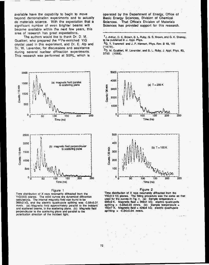
available have the capability to begin to move beyond demonstration experiments and to actually do materials science. With the expectation that a significant number of even brighter beams will become available within the next few years, this area of research has great expectations.
The authors would like to thank Dr. D. M. Gualtieri, who prepared the 57Fe-enriched YIG crystal used in this experiment, and Dr. E. Alp and Dr. W. Lavender, for discussions and assistance during several nuclear diffraction experiments. This research was performed at SSRL. which is
(a) magnetic field parallel to scattering plane
0 Time (ns)
I 2000
5 0 1500
2
0
(b) magnetic field perpendicular to scattering plane C
Y . 2 1000
500
0 0 Time (ns)
Figure 1 Time distribution of X rays resonantly diffracted from the YIG(002) planes. The solid curves are dynamical diffraction calculations. The internal magnetic field was found to be 36852 kG, and the electric quadrupole splitting was -0.89jB.01 mmls. (a): Magnetic field approximately parallel to the incident and scattered beams, in the scattering plane. (b): Magnetic field perpendicular to the scattering plane and parallel to the polarization direction of the incident light.
operated by the Department of Energy, Office of Basic Energy Sciences, Division of Chemical Sciences. That Office's Division of Materials Sciences has provided support for this research.
J. Arthur, D. E. Brown. S. L Ruby, G. S. Brown, and G. K. Shenoy, to be published in J. Appl. Phys. *G. T. Trammel and J. P. Hannon, Phys. Rev. B 18, 165 (1978). 3D. M. Gualtieri, W. Lavender, and S. L Ruby, J. Appl. Phys. 63, 3795 (1988).
(a) T = 295 K
loo0L 0,
Time (ns)
!joo > 400
E .- E 0 300
3 0 . 5 200 6
100
0 0 50 100 150 200 250 Time (ns)
Figure 2 Time distribution of X rays resonantly diffracted from the YIG(0 0 10) planes. The fitting procedure was the same as that used for the curves in Fig. f . (a): Sample temperature = 295% K. Magnetic field - 36833 kG; electric quadrupole splitting = -0.85f0.03 mmls. (b): Sample temperature = 150i7 K. Magnetic field I 42924 kG; electric quadrupole splitting = -0.54k0.04 mrnls.
72

GRAZING INCIDENCE X-RAY DIFFRACTION STUDIES OF POLYMER FILMS
B. .I. Factor Department of Applied Physics
Stanford University
T. P. Russell M. F. Toney
IBM Almaden Research Center
INTRODUCTION
The behavior o f polymer molecules a t the airjpolymer interface is of both scientific and technological importance. The presence of an interface places restrictions on the conformations of the molecules near the interface, and, consequently, the structure of the polymers near the inter- face can be substantially altered '. To enable the investi- gatipn of polymer structure as a function of distance from the air/polymer intcrface, the technique of grazing inci- dence x-ray diffraction (GIXD) has been employed. The goal of this study is to examine the near surfacc structure of several types of polymers and compare the surfacc structure with that found in the bulk.
In GIXD, the x-ray beam impinges on the sample a t angles close to the angle for total external reflection, typi- cally 3mrad for polymers using 1 = 1 .S68,. By varying the incidence angle of the x-ray$, the x-ray penetration depth can be varied from S0A to microns '. For the penetration depth to be well defined, the angle of incidenc well defined as well, and, thcreforc, the divergence of the the incident x-ray beam must be small, typically lmrad or less. This requirement along with the small scattering vo- lume and the low scattering power of hydrocarbon polym- ers necessitates the use of synchrotron radiation.
The polymer poly(hexy1-penty1)silane (PHPS), con- taining a silicon backbo with two pendant alkyl groups, n-pentane and n-hexan attached to each silicon atom, was investigated. Due to the silicon n the back- bone, PHPS scatters x-rays more rtro n the corre- sponding hydrocarbon analog. Steric hinderance among the sidegroups stiffens the polymer chain, changing what would be a flexible polymcr without sidegroups into a stiff, rodlike polymer when the sidegroups are largc. Conse- quently, the PHPS chains pack in a well defined manner. In contrast to other polyalkylsilane polymers, PHPS, due to the assymmetry of the sidegroups, is not crystalline a t room temperature where the measurements reported her- ein were performed.
Proposal No. 9035M EXP E R I MENTAL
PHPS with a molecular weight of 3X,700 and a poly- dispersity of 1.6 was dissolved in isooctane a t 2.4"/0. This solution was cast onto a polished 7.5cm diameter by Smm thick Si(100) substrate, spun a t 2 x IO3 rpm and annealed a t 90°C for 2hrs. to remove excess $olvent. A film with a thickness of 23508, and an rms roughness of 5-68, at the air/polymer and polymerjsubstrate interfaces, as deter- mined by X-ray reflectivity, was obtained.
G I X D measurements were performed in August 1989 on beam line X20C a t the National Synchrotron Light Source a t the Brookhaven National Laboratory. A pair of WjSi multilayers with a d-spacing of 238t was used to monochromate the incident x-rays at a wavelength of h = 1.56A with a 1% bandwidth. Scattering was per- formed with a four circle goniometer (with standard 6,20, x and q'~ motions) in two different scattering geom- etries in order to examine electron density correlations both parallel and perpendicular to the plane of the film. In the first case, which will be referred to as "in plane" GIXD, the sample lay nearly vertical, and the angle of in- cidence, a, was selected by appropriate motion of x and 6 angles according to the relation sin a = sin x sin 6. In the second case, which will be referred to as "out of plane" GIXD, the sample lay nearly horizontal, and the angle of incidence was chosen by movement of the 6 angle I n this geometry, the scattering vector tilts a t an angle 6 with re- spect to the surface normal. For both geometries, the di- vergence of the incidence angle was lmrad. The data are
47? 1 1 2
plottcd against q = -sin - (28).
During the measurementr, the sample lay in a helium environment to prevent oxidation. Nonetheless, degrada- tion of the polysilane samples was noted during the mea$- urements. An increased scattering background was observed, with little change in position and intcnsity of the diffraction peaks. The samples were translated period- ically in order to ensure that undamaged portions of the sample were being observed.
RESULTS A N D DISCUSSION
The PHPS powder diffraction pattern (fig. I) from a 200p thick film has sharp peaks at 0.492,0.855,0.987, and 1.31A-I. The peak positions in the pattern have a ratio of 1:,,1%:2:fl which is indicative of a hexagonal packing of the polymer chains with an interchain spacing of 14.78,. The 1.32A-1 peak ir superimposed on a broad peak which arises from the packing of the hexyl and pentyl sidegroups. The sharpness of the peaks indicates the hexagonal chain packing is coherent over a large distance of a t least 2n/dq-300A.
"In plane'' and "out of plane" G I X D were performed on the 23S0A PHPS film for several different a. For the "in plane" measurement, the diffraction in the region from 0.4-0SA-1 changes dramatically with a (fig. 2). At the
73

critical angle, a = 0.15", the peak a t 0.492A-' has a large shoulder. With greater surface sensitivity, the shoulder develops into a separate peak located at 0.44A-I when a = 0.12", and the two peaks are almost equal in intensity when a = 0.10". The peak a t 0.44A-I originates from a change in the electron density correlation parallel to the surface which could be associated with a novel structure a t the surface. Modelling of the data are consistent with the notion that the novel surface structure penetrates 200-300A into the bulk of the film.
For the "out of plane" GIXD,'the 0.492A I peak does not change in position or structure as a is varied, except for a small broadening due to the fact that the penetration jepth approaches the same length scale as the coherence ength. At the 0.492A-I peak, 8 = 3.5". Therefore, the lensity correlations tilted 3.5" from the surface normal re- main unchanged with changes in surface sensitivity. This indicates that the novel surface phase has the same density correlations nearly perpendicular to the surface as in the bulk of the film.
In order to precisely determine the structure of the novel surface phase, future GIXD measurements will be performed with the scattering vector positioned a t arbi- trary angles away from the surface normal.
REFERENCES
'Kumar, S.K.; Vacatello, M.; Yoon, D.Y. J . Chem. Phys., 89,5206( 1988).
2Fisher-Colbrie, A. Ph.D. Thesis, Stanford University, 1986.
ACKNOWLEDGEMENTS
This research is supported by the Department of En- ergy through the Office of Basic Energy Science, and IBM. The x-ray powder diffraction measurement of PHPS was performed by P. lannelli a t IBM.
Figure I : Plot of x-ray powder diffraction for 200p thick film of poly(hexy1-penty1)silane.
I I I I I I I
Figure 2: Plot of grazing incidence x-ray diffraction for 2350A film of poly(hexy1-pcnty1)silane for ,several anglcs of incidencc. The scattering vcctor is parallel to the film surface. The angle for total cxternal rcflcction is a = 0.15"
74

Static Structures of Associating Polymers a n d DNA Fragments via
Small Angle X-Ray Scattering
Alice P. Gast, Frans Leermakers, Kathleen Cogan, and Jennifer Raeder
Department of Chemical Engineering Robert Pecora, John Tracy and Mark Tracy
Department of Chemistry Stanford University, Stanford, C A 94305 USA
bt roduct ion
Amphiyhilic polymers have a tendency to associate in solution. When linear diblock copolymers are dissolved in a solvent selective for one block, they can form spherical micelles, semicrystalline aggregates or extended micellar or vesicular Structures. We have been studying two such systems, poly(ethy1ene oxide)-poly styrene (PEO-PS), and poly(methacry1ic acid)-poly(buty1 methacrylate) (PMAA-PBMA), in cyclopentane, a selective solvent for polystyrene and PBMA. From light scattering studies(1) we have found that micelles from PEO-PS can be extremely sensitive to the presence of trace amounts of water in the cyclopentane solutions. Similarly, the PMAA-PBMA structures are influenced by water as well as ionic impurities.
It is our intention to study these structures in more detail with small-angle x-ray scattering, and in particular, we wish to compare the spherical structures formed from block-copolymers with those predicted for a multi-arm star polymer(2). Each arm is represented by a succession of blobs with blob size increasing from the center to the outside of the star. Single chain behavior is found within each blob with a local concentration varying inversely with blob size. Logarithmic plots of scattering intensity versus scattering vector provide information regarding concentration profiles within the palymer structures. Differences from single chain scaling behavior become more pronounced with increasing number of arms in a micelle or star polymer. Beyond the flat Guinier region, scattering from single chains decay with a slope related to the Flory swelling exponent. For star-like structures, the slope approaches -4 as the density profile approaches that of a solid sphere. The slope should eventually reflect single chain scaling behavior when q is large enough to probe within the blobs comprising the star arms.
The polymers, described in Table 1, will be denoted by listing the number of repeat units in each block. We prepared solutions of PEO-PS block copolymers in cyclopentane with varying amounts of water as described before (1). We made similar solutions of PMAA-PBMA in cyclopentane with varying amounts of water and 1M NaCl added. The small angle scattering experiments were carried out at NSLS beamline X-12B under the guidance of Malcolm Capel, beamline supervisor. With the current configuration at this station, scattering vectors from q4.008 to 0.1 A 1 were accessible.
3uLL M.W. N e
BMAA/PBMA 6,488 6/42 6,0 4.0 (BuPont) 13,762 4192 4.3 4.8
mops 11,140 6SB0 11.6 (dry) 11,4 (Polymer €ab9 19.7 (wet) 18.5
187,500 170/1730 47.0 (wet)
We found from our pnliminary experiments that the PMBA- PBMA copolymer exhibited a strong scattering signal, easily measurable within five minutes of beam exposure. Logarithmic
75
Proposal NO. 9042M
plots of scattering intensity against scattering vector, shown in Figure 1, clearly distinguish between micellar solutions as in the 6/42 sample, and single chains found in 4/92 Scattering from single chains reveals near theta conditions from slope of -1.8 on log(1) vs log(q), close to minus the inverse of the Flory exponent. Micelles, having a higher density and more compact spherical structure, show a steeper slope of -4.3, typical of the Porod envelope for scattering from spheres(2). At high q we expect to see another transition to a lower slope of approximately -2 which would reflect scattering fror,: the semidilute chains within the blobs comprising the star arms. Additional experiments probing wider q ranges are pIanned.
We observe a large Guinier scattering region for both polymers. Guinier plots provided radii of gyration to be compared with hydrodynamic radii found from dynamic light scattering presented in Table 1 (3). We find that the ratio of R, !o Rh changes from 0.67 for the micelles to 1.12 for the single chains; the theoretical ratio for solid spheres is 0.77 while for Gaussian chains we expect 1.4-1.5 (4).
The PEO-PS block copolymers associate into larger micelles than those described above. Our largest sample, 170/1730, having 170 units of EO and 1730 units of styrene, forms micelles with an expected Rg on the order of 30 nm; this radius will be measurable after the scheduled improvements a t X12B. Log-log slopes of approximately -2 throughout the q-range investigated reveal near theta conditions within the blobs comprising the star arms. Smaller q is necessary to measure star-like behavior.
The measured q-range corresponds to the intermediate scaling region for micelles formed by a 65/80 sample. Figure lb) illustrates the scattering curves for 1 wt% solutions saturated and unsaturated with water. Several features indicate that the saturated micelles have a higher aggregation number; i.e., more arms. Scattering from the saturated solution exhibits a change in slope from -2.3 to -1.4 at a q - 2 . 5 ~ 1 0 ~ ~ A-1, similar to changes seen in molecular dynamics simulations@) of star polymers with 10 or more arms. The leveling off at small q for the unsaturated solution 2.gnifies the approach to the Guinier region and hence, a smaller radids of gyration than that for the saturated micelles. The blobs comprising the outermost layer of the star decrease in size with increasing number of arms. Therefore, a larger q is necessary to probe the single chain behavior within the blobs of the saturated micelles than for the unsaturated micelles. A value of -2 is seen for the limiting slope in the unsaturated solution, as expected, since polystyrene chains are at near theta conditions in cyclopentane at mom temperature.
The ex erimental q range included enough of the Guinier region for the !./80 spherical micelles to estimate radii o€ gyration, listed in Table 1. Ratios of radius of gyration to hydrodynamic radius on the order of unity for both wet and dry micelles suggest concentration profiles intermediate to that for Gaussian chains and solid spheres.
The majority of our samples wwrmt scattering measurements nt smaller mgles. In addition to better resolving the C h i n k region, we plan to extrapolate absolute intensities to q’”0 to d e t e d n e micelle appgntion numbers. Since polystyrene and poly(ethy1ene oxide) have similar electron density c o n m t in eyelopentans, we minimize the complications due to compogition polydispersity revdent in static light scattering measurements of this system. de a h have light scattering and microscop evidence that rod-like or lamellar structures re lace the sphericymicelles ns we change copolymer, water and d concenmtions. We need smaller scattering vectors to messm thicknesses or diameters of these structures.
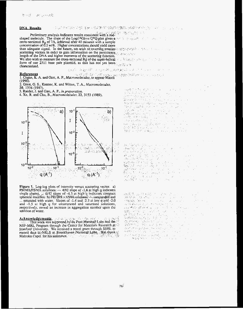
Jsl&Lmw 1. Cogan, K. A. and Gast, A. P., Macr (1990). 2. Grest, G. S., Kremer, K. and Witten, T. A., Macromolecules, 20, 1376 (1987). 3. Raeder, J. and Gast, A. P., in preparation. 4. Xu, R. and Chu, B., Macromolecules, 22, 3153 (1989).
Figure 1. Log-log plots of intenstiy versus scattering vector. a)
and -1.5 hL high q for unsaturated and saturated solutions, respectively, reveal an increase in aggregation number upon the additkm of water.
76

i
WHITE BEAM AND SECTION TO
1 Department of Materials S&I& &&
2Depanment of Materials, ShanghtdUriiMtydf
3 Shanghai Research Insti
Irttroduction Mo thin films are usefuI in a wide variety of high temperature applications. These films can be produced by sputter deposition onto a substrate followed by a heat treatment. Strains are often develqed due to the deposition process, phase transformations Of the deposited material, or the difference in the thermal expansion cocffkicnt between the Mo layer and the substrate. The magnitudes of the residual strains are related to the morphology of the film and the degree of coherence, both are vital factors to the rfonnance and
(100) substrates, as well as some blank Si wafex's were surveyed by white beam Laue topography and section topography. The p was to measure the quality of the substrates to ensure good base material for future deposition work, and to compare with the coated wafers.
reliability. In this work the quality of as-deposit e r Mo films OB Si
ExDerimental Mo &in layers were deposited onto Si (100) substrates, cleaved idto quarters from 3 inch wafers, at room temperature by argon plasm sputtering. The operating parameters were set at 2200 volts for target-anode acceleration voltage with 150 mTorr argon pssut.e, A low deposition rate of 0.6 &sec was maintained. Film thicknesses range from 18 to 290 A. Projection topographs and sectiott topographs were taken with a Mo filter on tho incident slit and recorded on Kodak SR-5 single sided x-ray films. Specimdn-filth distance was set at 100 mm and typical exposures took 30 tb 60 seconds for projection topographs, 4 to 6 minutes for single slit section topographs. These experiments were carried out at SSRL on beamline 2-2.
Results A total of 16 bare quarter-wafers were surveyed by projwtior topography, usually at more than one beam-size area (typically 10 mm x 4 mm). Most of the topographs show even contrast thtough- out the reflection spots, whereas some show thin sni -like f-te on selected areas of a quarter wafer. These saipes co up" d k gfow-in defects in the Si boule, such as twinning or stacking faults, Slip bands were also observed. Figure 1 illustrates a typical defective Si (100) as-received sample. Observable defects have to be taken into consideration during sub uent film growth, Substrates coated WitH 18,35,65, 130, and 2 9 8 Mo films were insp topography in the symmetric setting, and section symmetric and asymmetric setting. Many section topogtaphs show Pendellosung fringes (Fig. 2), which are currently being analyzed with the aid of digital image processing in the authors' laboratory.
e
f
4
77

IN SITU TOPOGRAPHIC OBSERVATIONS DURING DYNAMIC LOADING OF A MO GRAIN BOUNDARY
L. H. b e l , J. C. Bilello', Z. U. Rek2, H. Y. Wan$
Proposal No. 9044M
Department of Materials Science and Engineering, University of Michigan, Ann Arbor, MI 48105
Stanford Synchrotron Radiation Laboratory, Stanford, CA 94309 Department of Materials, Shanghai University of Science and Technology,
Shanghai, China
Introduction The early stages of plastic deformation of Mo single crystals were studied by white beam topography. In. situ observations of the crystals under step-wise loading conditions were performed with a miniature tensile stagel, while simultaneously recording transmission topographs. The objective of this work was to obtain information on slip nucleation behavior at the presence of a low angle grain boundary, as compared to the effect of other sites of stress concentration such as a crack or precipitate. In the vicinity of these regions in a sample, an enhanced intensity is observed on a white beam topograph2. This is directly related to the lattice distortions in the local region of the crystal which creates a severe three dimensional bending in the lattice planes. The lattice curvature in this local region can be estimated by the measured width of the enhanced contrast zone. Studies of these effects for a number of independent refections allows one to sort out the local dislocation density3, and the symmetry of the dislocation network in the region4.
Exuerimental The tensile specimens were cut from a molybdenum single crystal sheet with surface normal near [l 1 41 and the tensile direction near [ 1 1 01. The specimens were mechanically and electrolytically polished to a finished dimension 25mm x 7mm with a gauge length of 11 mm and thicknesses within 200 and 300 pm. A crater-like notch, serving as a stress concentration site, was introduced by spark-discharge at the edge of one specimen which contains no subgrain boundaries. Each exposure, which typically took 3 to 4 minutes, were taken after load relaxation had stabilized for either the loading or unloading step. The experiments were performed at SSRL on beamline 2-2.
Results Each high resolution picture taken on Kodak SR-5 single sided x-ray film captured up to 60 ( h k 1 ) reflection spots. Analysis of the large number of topographic images was facilitated by the use of digital image processing, which allowed detailed study of the general development of microplasticity and slip nucleation in many slip systems. Lattice distortion delineated by enhanced diffraction contrast in streak shape initiating from localized region either near the notch or the subgrain boundary were observed on many reflections at a load corresponding to a fraction of the bulk yield stress ( Fig. 1 and 2) . The image contrast at stress concentration sites of most reflections remain unchanged upon unloading to no-load state, while some reflections move out of contrast partially or completely indicating lattice tilting and/or twisting. The lattice distortion and plastic zone curvature at each loading/unloading step are being analyzed from the large data set. The preliminary results show that the lattice near the subgrain boundary distorted to a significant extent at 24 MPa, whereas no slip bands were initiated at 60 MPa while the lattice were severely distorted without the presence of a subgrain boundary. For the single crystals, slip started at the stress concentration sites at the sample edge; whereas the presence of the low angle boundary caused strain localization and, at higher stresses, boundary breakdown.
Figure 1. ( 3 1 0 ) reflection spot at 24 MPa load showing a low angle grain boundary and local enhanced-contrast streak running vertically on the left side.
Figure 2. ( 3 1 0 ) spot at 60 MPa load showing streak from the notch (center of top edge), and the severe lattice distortion of the entire speciment without the presence of a subgrain boundary.
References: 1.
2.
3.
V. Wakharkat and J. C. Bilello, presented at the 1986 APS Spring Meeting, Las Vegas, NV., 1986 (Unpublished). D. K. Bowen, A. B. Hmelo, S. T. Davies, and J. C. BiIelIo, Daresbury Laboratory Journal, 1982,47. A . B. Hmelo, J. C. Bilello, S . T. Davies and D. K. Bowen, in Applications of X-rav TouomD hic Methods tc Materials Science, eds. S . Weissmann, F. Balibar, and J-F. Petroff, Plenum Press, NY, 1984.
Solids: Plastic Instabilities, eds. V. Balakrishnan and C. E. Bottani, World Scientific Publishing Co., Singapore, 1986.
4. J. C. Bilello, in
pp. 1-46.
bcknowledeementg Department of Energy, under contract No. DE-FG03-88ER45350.
This work was supported by the U.S.
78

Proposal No. 9902M
MBDELIWG AND EXPERIMENTAL MEASUKEMENTS OF RESIDUAL STRESS
USING SYNCHROTRON RADIATION
J. F. Shackelford, B. D. Brown, and J. S. Park Department of Mechanical Engineering
University of California at Davis, CA 95616
R. W. Ryon, and Elane Flower Engineering Sciences Division
Lawrence Livermore National Laboratory Livermore, CA 94550
This work was an extension of recent, related efforts by Lawrence Livermore National Laboratory (LLNL) to identify the most effective method for determining residual stress in metal components using nondestructive techniques [l-31. These activities inc!uded neutron diffraction, x-ray diffraction, and ultrasonics. In 1988, we recognized that the newly installed UC/LLNL beam line at the Stanford Synchrotron Radiation Laboratory (SSRL) could be applied to determining lattice strains in a fashion helpful to our overall research goals. Pioneering work using synchrotron radiation for stress measurements had been reported in Japan 141. Benefits of a synchrotron source to our studies include a highly intense and monochromatic beam with variable energies, allowing significant sample penetration, and with very low beam divergence.
ExDerimental Materials and Methods The extensive time required for system development at SSRL limited experimental runs almost exclusively to one specimen. This was a ground, ball bearing steel with a highly characterized [&,?I surface-stress state (from ifs role in an international round robin testing program).
Much of the beam time allotted for this study (six eight-hour shifts) was devoted to experimental set-up , especially mechanical alignment, and to electronic trouble-shooting. A primary
recommendation for future studies of this type is to provide for precision x-y-z translational positioning control (as well as rotational control about z) for the goniometer table. A secondary recommendation is to isolate the inlet beam slits from the goniometer table. Experimental measurements were taken with a limited number of beam energies. Most work was done using energies of either 20 or 12 keV. Useful data were taken using harmonics at 10 and 24 keV, respectively. Time did not permit obtaining useful results from other fundamental energies or from higher harmonics of the 10 and 12 keV beams.
Much of the effort, following alignment efforts, concentrated on attempting to reproduce the conventional stress measuring technique of specimen tilting. Both forward (iso-) tilting and side tilting methods were attempted. Side tilting has certain advantages especially for high beam energies and associated, smaller Bragg angles. Unfortunately, experimental results from simple tilting techniques are highly dependent on precise specimen fixturing. The alignment problems noted above made such measurements impractical. Fortunately, analytical techniques are available for measurements on untilted samples allowing for some useful results which are described in the next section. "Untilted" refers to a specimen orientation such that the specimen surface is parallel to the crystal planes responsible for a given diffraction peak. This orientation is maintained by conventional diffractometers in which the detector has twice the angular rotation rate as the specimen.
Results and Discussion Diffraction peaks for the (211) plane of the ferritic, round robin specimen &all bearing steel 100 Cr6) were obtained at 10,12,20 and 24 keV beam energies. Figure 1 shows the measured, depth- averaged stress gradient together with the true stress gradient found by an analysis method developed by us [51. The results are in good qualitative agreement with the results of the rsund robin tests [6,7l.
79

Figure 1. Measured depth-averaged stress gradient together with the true stress gradient.
Future Work The work presented in this report indicates the preliminary useful results of the immediate, post- experiment analysis. We intend to carry on additional analysis of the results in conjunction with our ongoing studies of residual stress in cooperation with LLNL. The first effort of this type will be to carefully analyze the diffraction peak shapes for the 10,12,20 and 24 keV step scans of the (211) peak. Specific information about microstrain in the material is available from such analyses [lo].
Finally, it is hoped that further beam time at SSRL will be available in the near future for the purpose of continuing these studies. Encouraging initial results were obtained during a relatively brief allotment of time, given that time had to be devoted to system alignment. We feel that we are poised to obtain significantly more results, giwn an additional block of experimental time. A primary goal would be to have sufficiently predx alignment to allow tilting measurements to be made. Equally important would be to use higher '
harmonics of energy to allow substantially deeper penetration of at least 50 micrometers.
ACKNOWLEDGEMENT: Special thanks are due to Lane Wilson for his management of Beam Line X. His patience and helpfulness, under grueling circumstances, was a source of amazement to all of us.
REFERENCES: 1,
2.
3.
4.
5.
6.
7.
E.C. Flower, S.R. MacEwen, and T.M. Holden, FEA Predictions of Residual Stress in Stainless Steel Compared to Neutron and X- Ray Diffraction Measurements, Lawrence Livermore National Laboratory, Livermore, California, UCRL 96643 (August 1987). J.F. Shackelford and B.D. Brown, A Critical Review of Residual Stress Technology, Final Report, LLNL Intramural Order 5710705 (September 1987). J.F. Shackelford and B.D. Brown, Analysis of Triaxial Residual Stress Measurement Using High-Energy X-Ray Diffraction, Final Report LLNL Intramural Order 2660503 (January 1989). Y. Yoshioka, et al., "X-Ray Residual Stress Analysis in Textured Steels by Use of Synchrotron Radiation Source," Residual Sfresses in Science and Technology, Vol 1, E. Macherauch and V. Hauk, eds., Verlag, Germany, (19871, pp. 369-376. J.F. Shackelford, B.D. Brown and J.S. Park, Mod el ing and Experimen ta 1 Measurement of Residual Stress Using Synchrotron Radiu t ion, Lawrence Livermore National Laboratory, LIvermore, California, UCRL 2119 (May 8, 1989). E. Brinksmeier, et al., "Residual Stresses- Measurement and Cause5 in Machining Processes," Annals of the CIRP, 31, (2) 491- 510. (1982). J,F, Shackelford, ffniversity of California Partidpation in the CIRP Round Robin of Residual Strm Measurement, Lawrence Livennore National Laboratory, Livermore, Celifomia, UCID 19838 (July 1983).
80
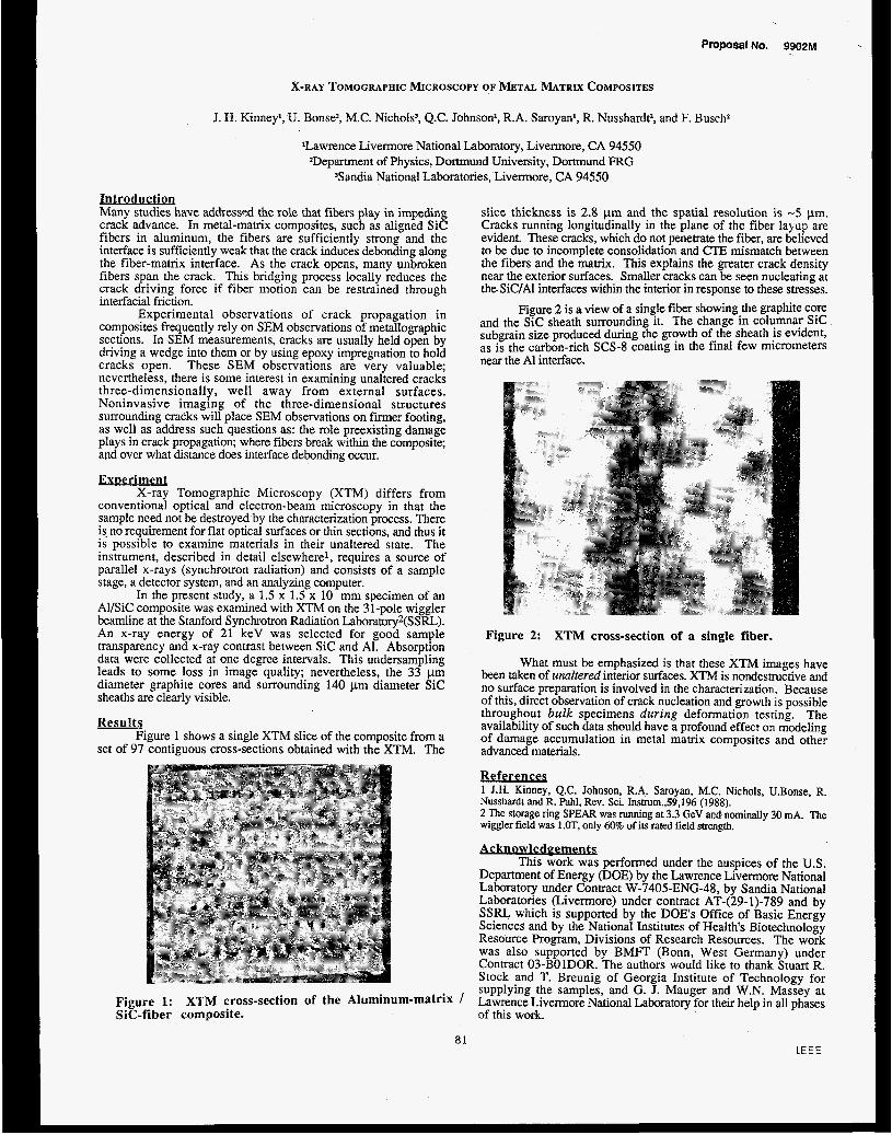
Proposal NO. 9902M
X-RAY TOMOGRAPHIC MICROSCOPY OF METAL MATRIX COMPOSITES
J. H. Kinney', U. Bonse', M.C. Nichols: Q.C. Johnson', R.A. Saroyanl, R. Nusshardt: and F. Busch'
'Lawrence Livermore National Laboratory, Livermore, CA 94550 'Department of Physics, Dortmund University, Dortmund FRG
'Sandia National Laboratories, Livermore, CA 94550
Lntroductioq Many studies have addresspd the role that fibers play in impeding crack advance. In metal-matrix composites, such as aligned S i c fibers in aluminum, the fibers are sufficiently strong and the interface is sufficiently weak that the crack induces debonding along the fiber-matrix interface. As the crack opens, many unbroken fibers span the crack. This bridging process locally reduces the crack driving force if fiber motion can be restrained through interfacial friction.
Experimental observations of crack propagation in composites frequently rely on SEM observations of metallographic sections. In SEM measurements, cracks are usually held open by driving a wedge into them or by using epoxy impregnation to hold cracks open. These SEM observations are very valuable; nevertheless, there is some interest in examining unaltered cracks three-dimensionally, well away from external surfaces. Noninvasive imaging of the three-dimensional structures surrounding cracks will place SEM observations on f i i e r footing, as well as address such questions as: the role preexisting damage plays in crack propagation; where fibers break within the composite; and over what distance does interface debonding occur.
ExDeriment X-ray Tomographic Microscopy (XTM) differs from
conventional optical and electron-beam microscopy in that the sample need not be destroyed by the characterization process. There is. no requirement for flat optical surfaces or thin sections, and thus it is possible to examine materials in their unaltered state. The instrument, described in detail elsewhere', requires a source of parallel x-rays (synchrotron radiation) and consists of a sample stage, a detector system, and an analyzing computer.
In the present study, a 1.5 x 1.5 x 10 mm specimen of an AI/SiC composite was examined with XTM on the 31-pole wiggler beamline at the Stanford Synchrotron Radiation Laboratory2(SSRL). An x-ray energy of 21 keV was selected for good sample transparency and x-ray contrast between S i c and Al. Absorption data were collected at one degree intervals. This undersampling leads to some loss in image quality; nevertheless, the 33 pm diameter graphite cores and surrounding 140 pm diameter S i c sheaths are clearly visible.
Results Figure 1 shows a single XTM slice of the composite from a
set of 97 contiguous cross-sections obtained with the XTM. The
Figure 1: Sic-fiber composite.
XTM cross-section of the Aluminum-matrix /
81
slice thickness is 2.8 pm and the spatial resolution is -5 pm. Cracks running longitudinally in the plane of the fiber layup are evident. These cracks, which do not penetrate the fiber, are believed to be due to incomplete consolidation and CTE mismatch between the fibers and the matrix. This explains the greater crack density near the exterior surfaces. Smaller cracks can be seen nucleating at the SiC/AI interfaces within the interior in response to these stresses.
Figure 2 is a view of a single fiber showing the graphite core and the S i c sheath surrounding it. The change in columnar S i c subgrain size produced during the growth of the sheath is evident, as is the carbon-rich SCS-8 coating in the final few micrometers near the A1 interface.
Figure 2: XTM cross-section of a single fiber.
What must be emphasized is that these XTM images have been taken of unaltered interior surfaces. X T M is nondestructive and no surface preparation is involved in the characterization. Because of this, direct observation of crack nucleation and growth is possible throughout bulk specimens during deformation testing. The availability of such data should have a profound effect on modeling of damage accumulation in metal matrix composites and other advanced materials.
References 1 J.H. Kinney, Q.C. Johnson, R.A. Saroyan, M.C. Nichols, U.Bonse, R. Nusshardt and R. Pahl, Rev. Sci. Instrum.,59,196 (1988). 2 The storage ring SPEAR was running at 3.3 GeV aRd nominally 30 mA. The wiggler field was l.OT, only 60% of its rated field strength.
Acknowledeements This work was performed under the auspices of the U.S.
Department of Energy (DOE) by the Lawrence Livermore National Laboratory under Contract W-7405-ENG-48, by Sandia National Laboratories (Livermore) under contract AT-(29-1)-789 and by SSRL which is supported by the DOES Office of Basic Energy Sciences and by the National Institutes of Health's Biotechnology Resource Program, Divisions of Research Resources. The work was also supported by BMFT (Bonn, West Germany) under Contract 03-BOlDOR. The authors would like to thank Stuart R. Stock and T. Breunig of Georgia Institute of Technology for supplying the samples, and G. J. Mauger and W.N. Massey at Lawrence Livennore National Laboratory for their help in all phases of this work.
l E E E

Proposal No. 9902M
THIN FILM AND INTERFACE STRUCTURE
Joe Wong, l r o y W. Barbee m d E. M. Larson Lawrence Livermore National Laboralory, Livermore, CA 94550
Ex per 'I ments t o bo t t i demons t r a t e s u b-mono 1 a yer seris.i. l . ivity arid 'to study .interfaces i n the copper-tungs.Len b ina ry have becrt performed on BL IO a t SSRL. spu t te r depos'lt tungsten-copper f - i l m on 3:15 cm x 10 cm x 0.25 cm superpolished fused s i ' l i c a x-ray o p t i c f I a t s . depostted f - i r s t and then overcoated " i n s i t u H w i t h copper .layers designed t o g i ve surface concentrat ions o f copper o f 0:l. 0.5, '1.0, 2.0 arid 5.0 monolayers (ML). Mu'l.t i layer s t ructures consis t ing o f 5 ML tungsten/5 ML copper. and 5 ML tungsten/l ML copper were a l so synthesized. A l l s t ructures were overcoated w i t h 0:l nm amorphous carbon t o s tab i Lize the F.i lm/ ambierit surfaces agalnst contamination. l l ie operat ing condi-tioris a t SSRL were abnorma i l y poor [IO kG, t i 0 ms, 3.3 GeV]. Even under such condi t ions the copper EXAFS spectra from a 0:l ML surface concentrat ion f i l m was. e a s i l y acquired. l t iese resul.ts demonstrate .that surface coverages o f 0.01 ML o r l ess are accessible for study us'lng Lhis technique. I n F-ig. 1, t he Four'ler Transform o f EXAFS s-igiials f o r bu'lk copper, the 5. MI., 2 ML and 'I ML copper coverage on tungsten are given. EXAFS sigrial a t coverages o f 2 ML and higher are c l e a r l y re la ted t o t h a t o f the bulk copper. A t lower coverages EXAFS i s s i g n i f i c a n t l y changed, appearing as expected f o r an amorphous so l id. l h e 'Interatomic d-istances derived from these data are consis tent w i t h these in te rp re ta t i ons . Data i nd i ca te t h a t the l o c a l environment o f the copper i n these very t h i n f . i lms i s dominated by carbon from the pass ivat ing overlayer. Addit iona I l y , the near edge s t ruc tu re appears t o change as the copper coverage decreases. . lh is i s cons is tent w i t h evidence f o r an e lec t ron i c i n t e r - fac'lal bonding s t a t e between copper and tungsten t h a t w i l l dominate the low coverage samples.
LXAFS measurements were a l so obtained from the copper 'in t he m u l t i l a y e r samples.
'The copper i n the 5 ML tungsten/SML copper sample was d'lsordered and appeared amorphous. I n the 5 ML tungsten -in t h i s sample, by contrast, was disordered and appeared amorphous. I n the 5 ML $ungsten/l ML copper sample the copper appeared t o be body centered cub'lc, assuming the s t ruc tu re o f .Lungsten.
Mu'lt.i.Iayer syrrlhesis technology was used t o
Tungsten layers 50 rim t h i c k were
.The copper
0 2 4 .6 8 10 Radius (A)
Fig. 1. Phase corrected Four ier transform o f Cu K-edge EXAFS from Cu overlayers sputtered i n a tungsten substrate a t various coverages. A 10 I I proc tec t i ve layer o f amorphous C was sputtered on top o f the Cu layer .
82

Proposal No. 991 Epp EXAFS and WAXSfDAS Analyses of Tc and Re Drugs and TcMetallothionein
Richard C. Elder and Edward Deutsch
Departments of Chemistry and Radiology and the Biomedical Chemistry Research Center University of Cincinnati
Cincinnati, OH 45221-0172
Radipharmaceuticals based on technetium or radio- active isotopes of rhenium have been in use for diagnostic
as imaging and palliative agents (and more thera eutics) for a number of years (la,b). 99mTc, 6Re and 1g8Re are the isotopes involved, each of which provide gamma rays suitable for imaging, while the Re isotopes have short half-lives and are widely available via so-called Tc or Re rrgenerators't. These features make them attractive for use in formulating radiopharmaceuticals in a hospital setting.
Metallic complexes of Tc and Re with a variety of diphosphonate ligands (03PCRR'P034-) are the most useful for bone imaging agents and were the compounds studied. Depending on the method of synthesis and the identity of R and R', these complex mixtures have different biodistribu- tions. They are in general difficult to crystallize. The crystal structure of the Tc-diphosphonate compound with R=R'=H (Technetium and methylene diphosphonate or Tc-MDP) has been determined (2) and an EXAFS structure characterization of an HPLC separated, 8 mM solution of this material was previously reported (3). More rcently, studies of the compound Technetium hydroxyethylidene diphosphonate (Tc-HEDP) using EXAFS and the corresponding Re-HEDP using both EXAFS and WAXSIDAS techniques were performed at room temperature. In addition, samples of Tc bound to the liver protein metallothionein (provided by Maurice Morelock of DuPont), were also investigated with the same techniques but at 10-20K using a He flow cryostat.
EXAFS data were collected a t SSRL beamline N-3 (300K and 10K) and also at NSLS beamline X9A (at Brookhaven National Laboratory). The gross structural features of the M-HEDP compounds (M = Tc or Re) are quite similar. Each of the compounds has pronounced Fourier transform peaks in the 1.5-2.1 a range and further peaks in the 2.3-3.4 The first peak (or range (fig. 1).
peaks) are definitely associated with M - 0 bonds usually requiring two shells of oxygens for the best fits. Model parameters for these M - 0 bonds were taken from the KMQ4 salts, whose EXAFS were also measured. Meanwhile, the peaks at longer distances seem to result from M-M or M-P interactions (or both), but are much more difficult to extract and evaluate. Models for the latter parameters were taken from the [M(DMPE)$+ compounds, where DMPE is the 1,2 -Bis (dim ethy1phosphino)e thane ligand, and from metal foils.
WAXS data were obtained from experiments performed at SSRL beamline VII-2 (low temperature) and a t NSLS beamline X7A. These experiments were performed on Re-(HEDP) samples (provided by David Pipes and Karen Libson a t Mallinckrodt Medical Inc.). Radial distribution functions (RDFs) were obtained showing peaks a t roughly the same distances as suggested by the EXAFS experiment (fig. 2). However, better identification in terms of
Re-HEOP R a d l a I Dlstributlan f u n c t l o n 6.5807 I . I ' I ~ 1 '
3 . 9 3 5 0
H 1 . 2 8 9 2
-4 .0022
- 6 . 6 4 7 9 0,0020 0,2020 0 , 4 0 2 0 0,6020 0.8020 1.0020
R x I O '
F 1
R ~ - H E O P ~ o ~ r l e r Trans. o r E X A F S 3.000
2 . 4 0 0 -
1.800-
H a c xld 1.200-
i
o.6000 0.000 0.000 1.500 3.000 4 . 5 0 0 6.000 7.500
R
Figure 1: Fourier transform of Re-HEDP EXAFS showing the rincipal oxygen backscattering peak near 1.6 1 and the weaker but evident heavy metal backscatterer near 2.7 .8. The transform has not been corrected for phase shifts.
Figure 2: Radial Distribution Function calculated from X-ray scattering data of Re-HEDP. The first peak results from difficult in normalization the peaks near 2.0 and 3.3 H are in reasonable agreement with the conclusions of the EXAFS study.
coordination number and species bound will require cleaner, less noisy data at two energies for the extraction of differential distribution functions (DDFs) from a DAS analysis. This will require measurement of samples without crystalline impurities, at low temperature, with higher photon fluxes andfor greater beam stability, than has been available so far.
Tc metallothionein was prepared by incubating Zn metallothionien (MT) with varying amounts of Tc oxo glucoheptanoate. MT usually has seven binding sites at which a metal such as Zn or Tc may attach. Three samples of the protein were prepared: NENl containing 1-2 Tc atoms per MT, NEN2 with 3-4 Tc atoms per MT, and NEN3 with 5-6 Tc atoms per MT. The objective of this study is to: (1) assess protein structural changes resulting from substitu-
a3

84

Proposal No. 922Bp
EXAFS, WAXS and DAS of Gold Based Drugs Used to Treat Rheumatoid Arthritis
Richard C. Elder* and Katherine Tepperman Biomedical Chemistry Research Center
University of Cincinnati Cincinnati, OH 45221-0172
Gold based drugs have been used for many years to combat rheumatoid arthritis. However, the form of these drugs circulating throughout the body, and the site of uptake is unknown. Gold metabolites of the oligomeric drugs enter the red blood cells (rbs's) of smoking patients but not of nonsmoking patients (1,2). Metabolites of auranofin (triethylphosphine)(2,3,4,6-tetra-O-acetyl-l-thio- -D-gluco- pyranosato-S)gold(I) enter rbc's irrespective of smoking habits (2,3). More subtle metabolic changes due to ligand exchange reactions of auranofin and HCN have not been fully investigated (4).
Cyanide binds t o gold very tightly and the equilibrium competition of HCN and thiols for gold(1) favors cyanide (4). Our aim is to (1) study the competition of HCN with the phosphine and protein thiolate ligands of AlbSAuPEtg, (2) determine the immediate environment around the gold center, and (3) develop a biochemical model for the metabolism of auranofin in smoking chrysotherapy patients.
X-ray absorption spectra were recorded at SSRL on beam line IV-3 about the gold LIII edge using a flowing liquid helium cryostat to cool the sample to 10 K. XANES studies yield the oxidation s ta te information for gold in the samples. A hump in the XANES region at 11.928 e V is characteristic of a gold(1)-phosphorus interaction. In the sample of AlbSAuPEt3 reacted with HCN, the gold was shown to be bound to a phosphorus atom and remained in the +l. oxidation s ta te (Figure 1).
p m y i i I aaw 0.I. 11900-11950 .Y
11.900 11.910 11.920 11.930 11.940 11.910
KEY
Figure 1: XANES for the reaction product of cyanide with albumin-S-AuPEt3. The hump a t 11.928 keV is characteristic of gold-phosphorus interactions.
Analyses of the EXAFS region of the spectra provide structural details of the average gold coordination environment. It is suspected that when HCN is reacted with AlbSAuPEtg that the gold(1) which is typically two- coordinate becomes three coordinate bound to sulfur, phosphorus and cyanide. The Fourier transform of the data shows two distinct peaks a t 1.5 and 2.0 R + (Figure 2). A backtransform of these t w o peaks shows a beat pattern in the reverse filtered Fourier transform. A two shell fit was performed using empirical parameters taken from a two
F T
' I 4 a 9 n
I: : I10
0 .000 1.500 3.000 4 .500 6 .000 7,500
R t alpha
Piguret: The Fourier transform of EXAFS for th material of Fig. 1. The peak near 1.6 fi corresponds t o the Au-C interaction while that near 1.9 a corresponds to the composite of the Au-S and Au-P interactions.
coordinate gold(1) phosphorus model compound and theoretical parameters for carbon. The f i t yielded a thre coordinate struiture containing a carbon atom at 1.95 which compares well with 1.97 8 in the crystal structure of Et3PAuCN. The second shell was fit with empirical parameters from the gold dip osphine structure, yielding two phosphorus atoms a t 2.28 However, i t is impossible to determine whether this represents two phosphorus atoms or one phosphorus and one sulfur atom since EXAFS experiments can not differentiate adjacent elements in the periodic table. The crystal structure of Auranofin shows a gold-phosphorus bond a t 2.26 8 and a gold-sulfur bond distance of 2.29 hl
Our studies of this compound indicate that the gold is indeed three coordinate and that a cyanide is bound to the gold. The most probable structure would be the mixed ligand Et3P and thiol-cysteine complex. However, evidence has shown that the mixed ligand complexes containing sulfur or phosphine and cyanide disproportionate in solution according to:
1
2 Et3PAuCN = [(Et3P)zAd+ + [Au(CN)d-
Therefore, a structure containing two phosphines and a cyanide should be considered as well.
Gold-gold interactions due to clustering in biological systems were studied also. Three gold dimeric species were studied [Au(dppm)gBrl2, [Au(dppm)2PF$2, and [Au(dppp)aBr12 a t low temperature to determine whether the weak gold-gold interactions could be observed. A peak corresponding to this interaction is absent in the Fourier transform at room temperature. This can be attributed to the high thermal disorder caused by vibration of the gold atoms with respect to one another. By reducing the sample temperature to that of liquid helium, thermal disorder should be substantially reduced and a peak might appear in
85

the Fourier transform corresponding to the gold-gold interaction.
Two peaks are seen in the EXAFS of [Au(dppm)2PFd2, one at 1.82 R + a and one at 2.79 R + a (Fig. 3). Fits of the two peaks yielded two phos horus atoms a t 2.33 $ and a gold-gold interaction a t 2.96 k These correspond very well with the crystal structure distances of 2.31 and 3.02 .% Three peaks are seen in the [Au(dppm)2Brl2 corresponding to gold- hosphorus, gold-bromine, and gold-gold interactions a t 2.33 2.87 and 3.02 8 (Fig. 4). The structure for [Au(dppp)2Br]2 has a gold-gold interaction of 5.04 8 which is not seen in the Fourier transform.
[Au(dppm)2PF6]2
c"'"''''i
Figure 3:
Figure Ip:
F T
M l *
G 0. XlOl
A
0.
60
20
80
40
0.0 3.0 6.0 R + 4
Fourier transform of the EXAFS data for a gold dimer showing both binding to phosphorus and also a gold-gold interaction a t 3.0 8.
[Au(dppm)PBr]Z
F 1.60 T
1.20
0.80
0.40
A
XlOl
0.0 3.0 6.0 R + .C
Fourier transform of the EXAFS of material identical to that in Fig. 3 except that noncoordinating PFg- has been replaced by Br- which coordinates and results in a third peak in the transform.
A t low temperature i t is possible to see weak gold- gold interactions a t a distance of 3.0 angstroms. These interactions are absent when data is taken at room temperature. The absence of a peak at 5.04 angstroms in the dppp structure suggests that at some distance between 3-5 angstroms evidence is lost, even in data measured at 10 K, of the metal-metal interaction. The resolution of the data suggests that two atoms with bond distances differing by as little as 0.15 $ can be refined, as in the gold-gold and gold-bromine distances for the dppmbr structure. Hence, by studying samples a t liquid helium temperatures moderately long, weak interactions can be examined.
ACKNOWLEDGEMENTS
Support for this project is from the NIH (GM35404). The work reported here is largely that of W.B. Jones. All data were measured at SSRL.
REFERENCES
1. Graham, G.G., Haavisto, T.M., McNaught, P.J., Browne, C.D., Champion, G.D., J. Rheumatol. 1981, 9, 5 32-5 35.
2. Lewis, D., Capell, H.A., McNiel, C.J., Iqbal, M.S., Brown, D.H., Smith, W.E., Ann. Rheum. Dis. 1983, 42, 566-57 0.
Intoccia, A.P., Flanagan, T.L., Walz, D.T., Gutzait, L., Swagzdis, Bioinorganic Chemistry of Gold Coordination Compounds; Sutton, B.M., Ed., Smith Kline & French Laboratories: Philadelphia, 1983, pp. 21-22.
Isab, A.A., Hormann, A.L., Coffer, M.T., Shaw, C.F. J., Am, Chem. SOC. 1988, 110,3278-3284.
3.
4.
86

Proposal No. 104619 SYNCHROTRON RADIATION CORONARY ANGIOGRAPHY IN A HUMAN SUBJECT WITH A DUAL BEAM, DUAL DETECTOR IMAGING SYSTEM
1 1 1 2 E. Rubenstein , J.C. Giacomini , H.J. Gordon , A. C . Thompson ,
G. Brown , R. Hofstadter , W. Thomlinson and H.D. Zeman 3 4 5 5
1 Stanford University School of Medicine, Stanford, California 94305, USA
Lawrence Berkeley Laboratory, University of California, Berkeley, California 2
94720, USA
3 Stanford Synchrotron Radiation Laboratory, Stanford University, Stanford, California 94305, USA
4 Department of Physics, Stanford University, Stanford, California 94305, USA
5
Island, NY 11973, USA National Synchrotron Light Source, Brookhaven National Laboratory, Long
introduction Two sets of prior studies of transvenous synchrotron radiation coronary angiography in human subjects have demonstrated that the approach can provide images of major portions of the human coronary anatomy [1,2]. The images acquired in these studies lacked clinical quality owing to an inade- quate signal-to-noise ratio. Comparison with prior studies in anesthetized dogs indicated that an eight-fold increase in x-ray fluence is needed 113. Several measures are being undertaken to achieve this increment, and the present report describes the implementation of the first of these, the dual beam, dual detector imaging system [ 3 ] .
Experimental These studies were conducted during March 1989 on beamline IV-2 at the Stanford Synchrotron Radiation Laboratory (SSRL). This beamline is illuminated by an eight-pole, 1.8 T wiggler. In this system the synchrotron radiation is monochromatized by diffraction off a pair of asymmetrically cut crystals which produce two X-ray beams with energy closely bracketing the K-edge of iodine (33.16 keV). The energy separation of the two beams is about 300 eV and the band width of each beam is about 15-25 eV. The beams, 125 mm in horizontal dimension, cross at the position of the subject's heart, diverge, and then impinge on the two dual arrays of 300 pixel elements of the lithium-drifted silicon detector. The efficiency of the detector for 33 kev photons is about 7 0 % ; the efficiency far the transmitted third harmonic (99 keV) is only about 5%. electronic system is 40,OOO:l in a readout time < 2 ms. by 0.5 mm. thermoelectrically cooled cryostat and operated at - 3 O O C . vertically, as in prior experiments, at a rate of 12 cm/s. The exposure time of each
The dynamic range of the
The pixel resolution is 0 . 5 m The detector is housed in a
The subject is scanned
line image is 4 ms. 62-year-old man with angina pectoris due to severe coronary artery disease. Bypass surgery (left internal mammary artery anatomosis and two vein grafts) had been done approximately four years prior to the present study.
The subject was a
Results Images were recorded in two positions, left lateral (Fig. 1) and right anterior oblique (30') (Fig. 2). The images revealed that the left internal mammary artery anastomosis was intact and that there was diminished perfusion of the distal left anterior descending coronary artery. bypass grafts appeared to be patent. image quality was improved over that obtained in prior studies owing to an increase in X-ray fluence, and the ramus intermedius artery (Fig. 1) was visualized for the first time. Fourteen of the 600 channnels functioned improperly owing to 60-cycle noise in some and excessive leakage current in the detector in others. The signals from the channels with the 60-cycle noise were corrected by adjusting the pedestal. The signals in the channels with leakage current were replaced with the average of the two bracketing adjacent channels.
The two vein The
87

Figure 2 This is a right anterior oblique (30°) view of a transvenous synchrotron radiation angiogram done on the same patient ( 3 . 3 GeV, 47 mA). The contrast agent dose was 50 ml, 12 ml/s. The radiation exposure for this frame was 0.47 rad. PV = pulmonary vein; the other abbreviations are the same as in fig. 1.
REFERENCES : 1. Rubenstein, E., Hofstadter, R., Zeman,
H.D., Thompson, A.C., Otis, J.N., Brown, G.S. , Giacomini, J.C., Gordon, H.J., Kernoff, R.S., Harrison, D.C., and Thomlinson, W. (1986), Proc. Nat. Acad.
2 . Thompson, A.C., Hofstadter, R., Otis, J.N. Zeman, H.D. , Kernoff, R.S. , Rubenstein, E., Giacomin, J.C., Gordon, H.J., Brown, G.S., and Thomlinson, W. (1988), Nual. Instr. and Meth. A266, 252-259.
3 . Thompson, A.C., Zeman, H., Thomlinson, W. , Rubenstein, E. , Kernoff , R.S . , Wofstadter, R., Giacomini, J.C., Gordon, H.J., Brown, G.S. (1989), Nucl. Instr.and Meth. B40/41, 407-412.
sei. USA. 83, 9724-9728.
Figure 1 This is a lateral view of a transvenous synchrotron radiation angiogram performed at SSRL in March 1989 ( 3 . 3 GeV, 50 mA). The subject was a 62-year-old man who had previously undergone left internal mammary and vein bypass graft surgery. The contrast agent, Renograffin-76, was injected into the superior vena cava (50 ml, 12 ml/s). The radiation exposure was 0.49 rad for this frame. A0 = aorta, LIMA = left internal mammary artery, VBG = vein bypass graft, RE = ramus intermedius artery, LAD = left anterior descending artery, LV = left ventricle, CA = catheter, CL = surgical clips, WS = wire sutures.
ACKNOWLEDGEMENTS We thank the staff of SSRL for their generous efforts on behalf of-this-project and for their scientific and technical expertise, without which these studies would not have been possible. This work was supported by National Institutes of Health Grant 5R01 HL 39253-02 and Department of Energy Grant DE-FG03-87ER60527. The data were recorded at SSRL which is supported by the WE'S Office of Basic Energy Science.

Proposal No. 2031 Bp
Reactivity of the Lac Active Site
An X-ray Absorption Edge Study James L. Cole, Grace 0. Tan,
Keith 0. Hodgson and Edw
Department of Chemistry, Stanford Univer
The multicopper oxidases (laccase, ascorbate o ceruloplasmin) catalyze the four-electron reduction of dioxygen to water. Laccase contains four Cu atoms: a type 1, a type 2 and a binuclear type 3 center. Low-temperature MCD measurements have demonstrated that the type 2 and type 3 centers comprise a trinuclear Cu cluster site,l and this model has been supported in a recent X-ray crystal structure determination2 of ascorbate oxidase. In the present study, X-ray absorption edge spectroscopy has been used to determine Cu oxidation states following reaction of reduced laccase derivatives with dioxygen, leading to a description of which of the Cu centers are required for reactivity.
A pre-edge ls+4p transition near 8984 eV is present in the edge spectra of 2-coordinate and 3-coordinate Cu(1) compounds, but not in Cu(I1) compounds3. Thus the degree of oxidation of the laccase derivatives may be determined against a scale obtained by subtracting the edge of a fully oxidized sample (reacted with H202) from that of a fully reduced sample.
The feature near 8984 eV is present in the spectra of fully reduced type 2-depleted (T2D) laccase and type 1 Hg2+-substituted (TlHg) laccase. In T2D laccase, this feature persists upon exposure to air (see Figure l ) , indicating that the sample remains partially reduced. Optical spectroscopy on the T2D samples shows that the type 1 Cu is completely oxidized, enabling us to conclude that the binuclear type 3 center remains reduced. In contrast, fully reduced TlHg laccase, which contains a valid type 2-type 3 trinuclear Cu cluster site, reacts readily with dioxygen, resulting in complete reoxidation of the type 2 and type 3 centers (see Figure 2). These results demonstrate that the type 2-type 3 trinuclear site represents the minimal active site required for the multielectron reduction of dioxygen.4
Acknowledgements. This work was supported by NSF Grant CHE 8817702 (KOH) and NIH Grant AM31450 (EIS). Beam time was provided by SSRL, supported by the DOE'S Office of Basic Energy Sciences and the NM's Division of Research Resources.
References (1) (a) Allendorf, M.D.; Spira, D.J.; Solomon, E.I. Proc. Natl. Acad. Sci. USA 1985,82, 3063-3067. (b) Spira-Solomon, D.J.; Allendorf, M.D.; Solomon, E.I. J.Am. Chem. SOC. 1986,108,5318-5328. ( 2 ) Messerschmidt, A.; Rossi, A.; Ladenstein, R.; Huber, R.; Bolognesi, M; Gatti, G; Marchesini, A.; Petruzzelli, R; Finazzi- A@, A. J . Mol. Biol. 1989,206,513-529. (3) Kau, L.-S.,; Spira-Solomon, D.J.; Penner-Hahn, J.E.; Hodgson, K.O.; Solomon, E.I. J . Am. Chem. SOC. 1987,109,6433-6442. (4) Cole, J.L.; Tan, G.O.; Yang, E.K.; Hodgson, K.O.; Solomon, E.I., in press, J. Am. Chem. SOC.
Energy (eV)
% Cu oxidized T2D TI T2 T3
0
14
--_ (a) T2D fully reduced 0 (b) T2D exposed 15 min. 0 _-_ O* (c) T2D exposed 63 hrs. 100 _ _ _
*The slight decrease at 8984 eV is due to oxidation of the 10% of native laccase present in the T2D preparation
89


STRUCTURAL COMPARISON OF PURPLE AND BLUE No* 2035B
FORM§ OF BACTERIORHODOPSIN
Soichi Wakatsukil, Y. Kimura2, N. Gillis3, D. EliezcP, W. Stoeckenius2, Keith 0. Hodgsonl, and S. Doniach3
1 Department of Chemistry, Stanford University Stanford, CA 2 Cardiovascular Research Institute, University of California, San Francisco, CA 94143.
3 D e p a m e n t of Applied Physics, StanFord University Stanford, CA 4 Department of Physics, Stanford University Stanford, CA
94305.
94305. 94305.
I n t r o d u c t i o n A blue form of bacteriorhodopsin lacks some
photocycle intermediates and does not function as a proton pump when it is transformed either by deionization or by acidification from the native, purple form. Therefore, the dctermination of conformational change that accompanies the purple-to-blue transition may give insights into how the transition deactivates the pump.
during the purple-to-blue transition have been controversial in part due to liable nature of the blue form giving rise to loss of 2D lattice structure. Contradictory descriptions of the structure of blue membranes have appeared: one claiming a well-preserved l a t t i ~ e l ? ~ and one a distorted l a t t i ~ e ~ , ~ . blue membranes, it has been suggested that their lattice structure is largely disordered in ultracentrifuged samplcs thus preventing structure determinations using x-ray diffraction from such samples.
W e have reported that blue membrane samples have a distinct diffraction pattern, comparable in quality to that from a purple form, which, however, disappears after a fcw hours of drying5. In this report we describe the change of the diffraction patterns during drying and compare structures of blue and purple membranes.
E x p e r i m e n t a l
JW5 reconstituted with retinal and naturally produced ET1001. Purple membranes were washed carefully by centrifugation at 30000 g. After deionization according to a method described earlier1, pellet samples were made by ultracentrifugation at 300000 g for about 10 hours into a cell with mylar windows on both sidcs. pellets is typically 1 mm x lmm x lmm. After centrifugation, the cells were sealed with Corning vacuum g r e a s e .
For drying experiments, the vacuum grease seal was removed immediately before exposure to x-ray. To verify the integrity of the blue samples, the pellets were collected after x-ray exposure in order to measure their optical spectra. In all cases reported here, the blue forms collectcd after the x-ray exposure had the same absorption maxima as those before:' It is highly unlikely that the blue form becomes purple in the pellet form and returns to blue when rcsuspended into water. Therefore we strongly believe that the observed structural changes are characteristic of the blue form.
Studies of changes in x-ray diffraction patterns
Because of the low surface pH of the
We used two differcnt purple mcmbranes: a mutant
The size of such
Q u a d r a n t Detector
A. Gabriel of EMBL in Grenoble was purchased and fully integrated into the SSRL Small Angle X-ray Scattering/Diffraction camera with additional electronics modules and minimal software change. Its 60" fan-shape active area is suitable for acquisition of diffraction patterns from moderately oriented membranes such as pelleted purple membranes. The entire system with the quadrant detector, VAX station 11, CAMAC modules, and NIM modulcs was sent to the National Light Source Laboratory (NSLS), Brookhaven National Laboratory and rebuilt on beamline X14A for the data acquired in December 1989.
A quadrant position sensitive detector developed by
Time-reso lved x - r a y d i f f r a c t i o n o f b l u e m e m b r a n e s d u r i n g d r y i n g
In-plane diffraction patterns from the blue membranes were collected during drying at room temperature. Figure 1-A shows an example of continuous time-resolved x-ray diffraction patterns of the blue membranes during 4 hours of drying. The in-plane diffraction peaks whose S range is between 0.03 and 0.12 A- I were lost after about 150 minutes and an inter-membrane Bragg peak at 50.0 8, appeared about the same time. latter arises from lamellar stacking of the membranes.
in-plane diffraction peaks grew for the first 100 minutes. This is clearly seen in Figure 2 which plots changes in nrak heights of the (2,l) reflection and the inter- nicrnbrane Bragg diffraction peak.
The
A 3D plot of the same data, Figure 1-B, shows that the
2
I I A o.02 0 0G 0.66 d 08 0 10
S Iinverse Angstroms)
Fig. 1 Time-resolved x-ray diffraction patterns of the blue membrane during drying. (a) Calibrated data before background subtraction. Exposure of each pattern was 10 minutes and entire data set was recorded in 4 hours. showing improvement of the in-plane diffraction peaks during first 100 minutes and their d i sappearance thereaf ter .
(b) 3D plot of the same data
90
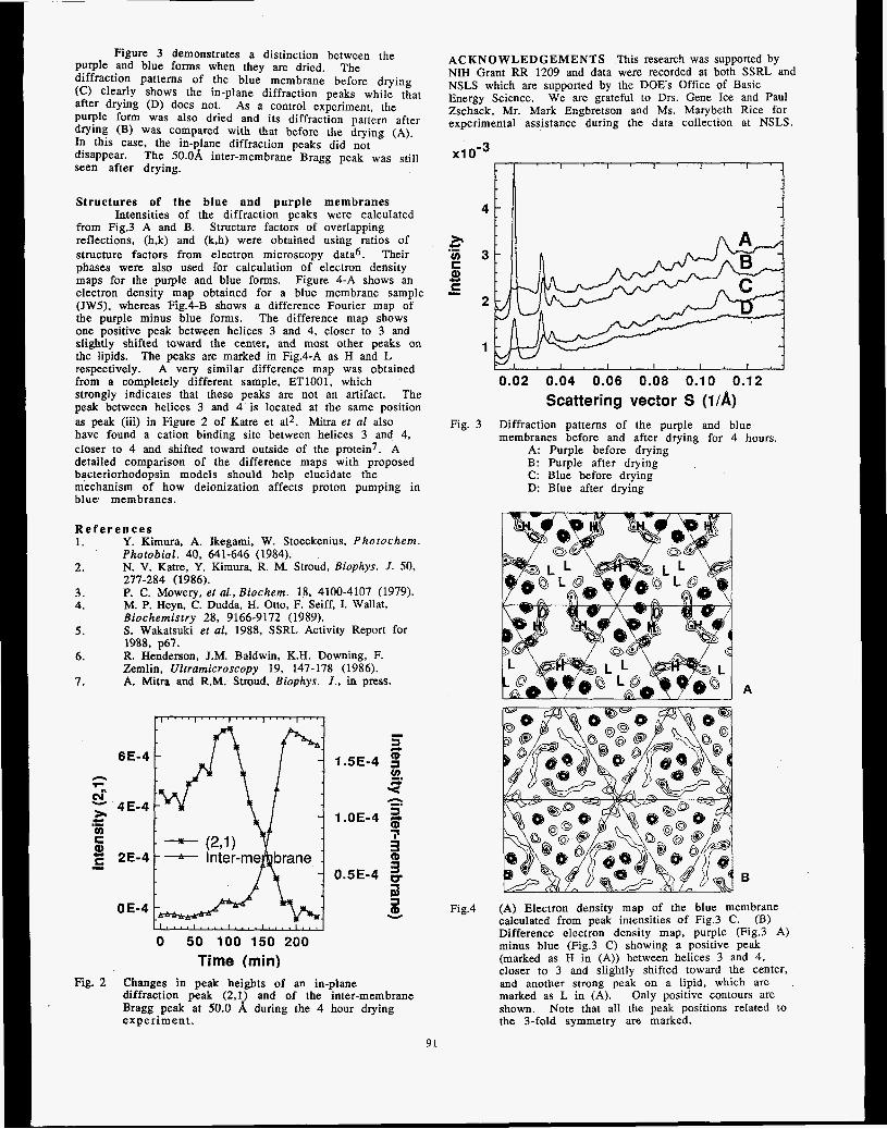
Figure 3 demonstrates a distinction between the purple and blue forms when they are dried. diffraction patterns of the blue membrane before drying (C) clearly shows the in-plane diffraction peaks while that after drying (D) does not. purple form was also dried and its diffraction pattern after drying (B) was compared with that before the drying (A), In this case, the in-plane diffraction peaks did not disappear. The 50.08, inter-membrane Bragg peak was still seen after drying.
The
As a control experiment, the
Structures of the blue and purple membranes
from Fig.3 A and B. reflections, (h,k) and (k,h) were obtained using ratios of structure factors from electron microscopy data6. Their phases were also used for calculation of electron density maps for the purple and blue forms. Figure 4-A shows an electron density map obtained for a blue membrane sample (JW5). whereas Fig.4-B shows a difference Fourier map of the purple minus blue forms. The difference map shows one positive peak between helices 3 and 4, closer to 3 and slightly shifted toward the center, and most other peaks on the lipids. respectively. A very similar difference map was obtained from a completely different sample, ET1001, which strongly indicates that these peaks are not an artifact. The peak between helices 3 and 4 is located at the same position as peak (iii) in Figure 2 of Katre et a12. Mitra et a1 also have found a cation binding site between helices 3 and 4, closer to 4 and shifted toward outside of the protein7. A detailed comparison of the difference maps with proposed bacteriorhodopsin models should help elucidate the mechanism of how deionization affects proton pumping in blue membranes.
Intensities of the diffraction peaks were calculated Structure factors of overlapping
The peaks are marked in Fig.4-A as H and L
R e f e r e n c e s 1. Y. Kimura, A. Ikegami, W. Stoeckenius, Photochem.
Photobiol. 40, 641-646 (1984). 2. N. V. Katre, Y. Kimura, R. M. Stroud, Biophys. J . 50,
3. P. C. Mowery, et al., Biochem. 18, 4100-4107 (1979). 4.
5 . S. Wakatsuki et al, 1988, SSRL Activity Report for
6.
7. A. Mitra and R.M. Stroud, Biophys. J., in press.
277-284 (1986).
M. P. Heyn, C. Dudda, H. Otto, F. Seiff, I. Wallat, Biochemistry 28, 9166-9172 (1989).
1988, p67. R. Henderson, J.M. Baldwin, K.H. Downing, F. Zemlin, Ultramicroscopy 19, 147-178 (1986).
6 E-4 n -. cu - 4E-4 * v) C Q) E 2E-4
CI .-
-
5 1.OE-4 2
OE-4
0 50 100 150 200 Time (min)
Fig. 2 Changes in peak heights of an in-plane diffraction peak (2.1) and of the inter-membrane Bragg peak at 50.0 8, during the 4 hour drying experiment .
ACKNOWLEDGEMENTS NIH Grant RR 1209 and data were recorded at both SSRL and NSLS which are supported by the DOES Office of Basic Energy Science. We are grateful to Drs. Gene Ice and Paul Zschack, Mr. Mark Engbretson and Ms. Marybeth Rice for experimental assistance during the data collection at NSLS.
This research was supported by
x~ o - ~
4;
.- 5 v) C
C 8 I
0.02 0.04 0.06 0.08 0.10 0.12 Scattering vector s (1/A)
Fig. 3 Diffraction patterns of the purple and blue
A: Purple bafore drying B: Purple after drying C: Blue before drying D: Blue after drying
membranes before and after drying for 4 hours.
Fig.4 (A) Electron density map of the blue membrane calculated from peak intensities of Fig.3 C. Difference electron density map, purple (Fig.3 A) minus blue (Fig.3 C) showing a positive peak (marked as H in (A)) between helices 3 and 4, closer to 3 and slightly shifted toward the center, and another strong peak on a lipid, which are . marked as L in (A). shown. the 3-fold symmetry are marked.
(B)
Only positive contours are Note that all the peak positions related to
91

Proposal No. 1A68B
Virus Structure
Michael Rossman, Thomas, J. Smith, Ming Luo, Sangsso Kim, John Badger Philippe Dumas, S. krishnaswamy, Klaus Piontek
Purdue University Dept. of Biological Science
West Lafayette, Indiana
Human Rhinovirus 1A Drua Stud ieS
We have recently been able to obtain a structure of the HRVlA serotype (1). This virus belongs to the minor rhinovirus receptor group. Previously we had worked with HRV14 (2) which belongs to the major rhinovirus receptor group. We had studied the binding of various drugs that inhibit uncoating and attachment of HRV14 (3-5). The studies at SSRL were the first with HFIVlA. The results are currently being written up for publication.
Two compounds were studied (e.g. WIN 56291)
Both bound in a similar manner into the "WIN pocket" of VP1 of HRVlA. However, there are substantial differences to the model of binding that occurs in HRV14. The WIN compound is displaced as much as 4 A in places compared to its HRV14 binding site. These differences are now being analyzed.
. . an Rhinovirus 14 Mut;int
Data was collected on a WIN drug-resistant mutant of HRV14. Unfortunately the crystals were extremely unstable and the data was of insufficient quality.
me Pawovirw
Data collection of native CPV crystals (see 6) was continued. This data has been incorporated Into a now fairly complete data set. We anticipate obtaining a high resolution structure perhaps in 1990, Thls would be the first high resolution structure of an ssDNA virus (but see $Xl74).
We continued the data collection of native $X174 crystals (7). This data has been incorporated into a now fairly complete data set. We anticipate obtaining a high resolution structure in 1990. This would be the first phage structure and one of an ssDNA virus (but see CPV above).
. 92

1 .
2.
Kim, S., T. J. Smith, M. S. Chapman, M. G. Rossmann, D. C. Pevear, F. J. Dutko, P. J. Felock, G. D. Diana, M. A. McKinlay. (1989). Crystal structure of human rhinovirus serotype 1 A (HRVlA). J. Mol. Bid. 210:91-111.
Rossmann, M. G., E. Arnold, J. W. Erickson, E. A. Frankenberger, J. P. Griffith, H. J. Hecht, J. E. Johnson, G. Karner, M. Luo, A. G. Mosser, R. R. Rueckert, B. Sherry, G. Vriend. (1985). Structure of a human common cold virus and functional relationship to other picornaviruses. Nature (London), 31 7:145-153.
3 .
4 .
Smith, T. J., M. J. Kremer, M. Luo, G. Vriend, E. Arnold, 6. Kamer, M. G. Rossmann, M. A. McKinlay, G. D. Diana, M. J. Otto. (1986). The site of attachment in human rhinovirus 14 for antiviral agents that inhibit uncoating. Science, 233:1286-1293.
Badger, J., I . Minor, M. J. Kremer, M. A. Qliveira, T. J. Smith, J. P. Griffith, D. M. A. Guerin, S. Krishnaswarny, M. Luo, M. G. Rossmann, M. A. McKinlay, G. D. Diana, F. J. Dutko, M. Fancher, R. R. Rueckert, B. A. Heinz. (1988). Structural analysis of a series of antiviral agents complexed with human rhinovirus 14. Proc. NatL Acad. Sei. US. 85 : 3 3 0 4 - 3 3 0 8 .
5 .
6 .
7.
Badger, J., I . Minor, M. A. Oliveira, T. J. Smith, M. G. Rossmann. (1989). Structural analysis of antiviral agents that interact with the capsid of human rhinoviruses. Proteins, 611 - 1 9.
Luo, M., J. Tsao, M. G. Rossmann, S. Basak, R. W. Cornpans. (1988). Preliminary X-ray crystallographic analysis of canine parvovirus crystals. J , Mol. Bid. 200:209-211.
Willingrnann, P., S. Krishnaswarny, R. McKenna, T. J. Smith, N. H. Olson, M. G. Rossrnann, P. L. Stow, N. L. Incardona. (1990). Preliminary investigation of the phage @ X i 7 4 crystal structure. J. Mol. Bid. In press.
93

Prorsosai No. 1A93B
CRYSTALLOGRAPHIC STUDIES OF THE NITROGENASE IRON PROTEIN FROM AZOTOBACTER VINELANDII
Millie M. Georgiadis, Pinak Chakrabarti and Douglas C. Rees
Division of Chemistry and Chemical Engineering 147-75CH California Institute of Technology
Pasadena, California 91 125
Introduction The nitrogenase enzyme system provides the only biological
source of fixed nitrogen, which all organisms require as an essential nutrient. The ATP dependent reduction of dinitrogen to ammonia requires two component proteins, iron (Fe-) protein and the molybdenum-iron (MoFe-) protein. We have concentrated our structural studies on Fe-protein, since it is responsible for the coupling of ATP hydrolysis to electron transfer, is highly conserved between the "standard and "alternate" nitrogenases, and is involved in regulation of nitrogenase activity at the protein level (through ADP-ribosylation in photosynthetic bacteria). Fe-protein is a dimer of two identical, 30,000 molecular weight subunits. Each dimer contains one 4Fe:4S cluster, symmetrically liganded to each subunit through Cys-97 and Cys-132. While spectroscopic, extrusion and redox studies of Fe-protein suggest that the 4Fe:4S cluster generally resembles those found in small ferredoxins, it is quite liiely that the cluster environment differs significantly between ferredoxins and Fe-protein. The most striking indication of this is the ability to generate 2Fe:2S clusters in oxidized Fe-protein in the presence of MgATP and chelator; such interconversions have not been observed in ferredoxins. In addition, the Fe-protein cluster appears to be much more exposed to solvent than in ferredoxins, as indicated by rapid D20 exchange in pulsed EPR studies and solvent dependent spin-state interconversions in low temperature EPR studies. Exposure of the cluster almost certainly contributes to the rapid inactivation of Fe-protein by oxygen and by nitrite. We have pursued an approach towards solving the structure based on (1) native anomalous scattering measurements from the iron-sulfur cluster, (2) preparation and location of isomorphous heavy atom derivatives, (3) phase refinement by non-crystallographic symmetry averaging within and between different crystal forms and (4) atomic refinement of an initial structural model to provide the additional phase information needed to complete the polypeptide chain trace.
Experimental Our recent crystallographic efforts have concentrated on
crystals of Fe-protein from Azotobacter vinelandii, space group P21 with a=56.8A, b=92.9A,c=63.6A,b=l00.00, and containing one dimer in the asymmetric unit. Data from 5 crystals each of the native and a gold derivative were collected at the SSRL rotation camera facility to 2.5A resolution. The native data was reduced to 14057 unique reflections out of 29527 reflections measured, with a merging R of 0.074. The gold data was reduced to 11 131 unique reflections out of 27166 measured, with a merging R of 0.102. The gold derivative contains one major site, as identified from difference Patterson studies. Phases calculated from the film data, in combination with additional native anomalous and derivative data measured from a multiwire area detector on a rotating anode x-ray source, have permitted definition of the molecular envelope, with subsequent phase refinement by non-crystallographic symmetry averaging about the dimer two-fold.
94
Results Extended regions of connected chain and secondary
structure are evident in the electron density maps. A general view of the secondary structure arrangement of the Fe-protein dimer is shown in Figure 1. These maps have only been recently calculated, so that a detailed fitting of amino acids to the electron density has not yet been accomplish& Several general observations about the Fe- protein structure may be noted at this stage:
(a) The Fe-protein dimer has a two-fold symmetry axis that relates the two subunits. (b) The cluster is on the surface of the molecule and provides the principal connection between the subunits. Each subunit contains a single domain. A cleft straddling the two-fold axis separates the subunits. (c) The chain fold of Fe-protein monomer is of the a/p type, as is generally observed in nucleotide binding proteins (i.e. the Rossmann fold). (d) The ATP binding site is most probably located in the cleft between the two subunits. (e) An a-helix containing residues 98 to -1 15 is exposed at the top of the molecule, and may represent a major site of interaction for binding to the MoFe-protein.
Figure 1. Chain-trace illustrating general features of the Fe- protein, dimer structure.
Additional Studies of Electron Transfer Proteins Diffraction data were collected at the SSRL rotation camera
facility for the membrane protein complex Rhoabbacter sphaeroides strain 2.4.1 photosynthetic reaction center (2.8A resolution) and the Rhodobacter sphaeroides cytochrome c2 (2.5A resolution). Upon completion of film processing, this data will be used to refine the RC structure, and to solve the cytochrome c2 structure by molecular replacement methods.
Acknowledgements This research was supported by NIH grant 31299 and an
NSF PYI grant. Data were recorded at SSRL which is supported by the DOES Office of Basic Energy Science. The assistance of B.T. Hsu, M. Lewis and J.P. Allen during data collection is greatly appreciated. DCR is an A.P. Sloan research fellow.


X-RAY CRYSTALLOGRAPHY ON CRyS!k'ALS OF N. GONORRHOEAE PILIN rmwJo'''u*
H.E. Parge, D. Christensen, T. Hong, K. Andrew, D.E. McRee, ED. Getzoff,and J.A. Tainer
Department of Molecular Biology, Rwarch Institute of Seripps Clinic, La Jolla, California
92037
Introduction
Pilus fibers are long protein filaments on many pathogenic
bacteria that participate in attachment to host cell8 (I). Al-
though the self-assembling protein pilin is the major structural
component of the .Veeisseria gonorrhoeae pilus several other pro-
teins co-purified with pilin through the repeated solubilisation-
reassociation steps of the biochemical purification. Pilin o h -
bilized in the detergent n-octyl-p-D-glucopyranoside remained
an aggregate of about 100 kD at pH 9.5, but WBB teduced to
a 40 kD dimer at pH 10.5, suggesting that assembly involves
electrostatic interactions of Lys, Tyr, or other side chains with
high pKa's. P i l i dimers and aggregates of htgher nioleculff
mass were partially stable even in the presence of sodium dode-
cy1 sulfate and 8-mercaptoethanol. Removal of pilus-asscrciated
proteins and stabilization of pilin multimers permitted the re-
producible crystallization of pilin.
Experimental
Three-dimensional needle- and plateshaped crystals of pu-
rified N. gonorrhoeae pilin (strain MSll variant C30) grew from
38.40% polyethylene glycol 400, pH 8.0-9.0, in space group
C222, with cell dimensions a = 126.4, b = 121.2, c = 28.7 A and V, = 2.84 A '/dalton for one molecule per asymmetric unit
(2). Crystals were mounted in glass capillaries along with their
mother liquor for experiments conducted at ambient tempera-
tures and on glass fihres after immersion in a hydrocarbon for
low temperature experiments. Data were collected on an Arndt-
Wonacott rotation camera with a crystal to film distance of 10
em, 0.2 mm collimator, and rotation increments of 2.0-3.5"
Results
Plate like crystals obtained from improved isolation proc?
dures were examined in the spring 1989 run on the rotation
camera facility at SSRL. These were shown to belong to the
same space group as our earlier needle shaped crystals on which
all our current data collection and heavy atom screening have
be performed. Further heavy atom screening experiments were
hampered by the significant downtime of the ring but experi-
ments to test the possibility of collecting data on pilin at cryo-
ere performed. The results from these
tests indicated that the crystals withstand the necessary ma-
d give satisfactory diffraction at cryogenic
1. Stromberg, N., Deal, C., Nyberg, G., Normark, S., So,
M. and Karlsson, K. (1988) Proc. NaU. Acad. Sci. U.S.A. 85,
4902-4906.
2. Parge., H.E., Bernstein, S.L., Deal, C.D., McRee, D.E.,
Christensen, D., Capozza, M.A., Kays, B.W., Fieser, T.M.,
Draper, b., So, M., Geteoff, E.D.,and Tainer, J.A. (1990) J .
Biol. Chem. 265, 2278-2285.
95


Proposal No. 81168
On the Need to Account for Electron Spin when Interpreting Linearly Polarized XANES with Application to the Polarized Molybdenum L2 and L3 Absorption
Edges of Single Crystal MoS,OZ2-
T. A. Tysonl, D. A. Case2, B. Hedmanl, and K, 0. Hodgson'
lDepartment of Chemistry, Stanford University, Stanford, California 94305 2Department of Molecular Biology, Research Institute Scripps Clinic, La Jolla, California
92307 3Stanford Synchrotron Radiation Laboratory, SLAC, Bin 69, P. 0. BoA 4349, Stanford,
California 94309
I. Introductlon
We note that in the interpretation of linearly polarized XANES spectra of oriented molecules performed to date the spin or intrinsic angular momentum of the electron has been neglected. Here we discuss two models which account explicitly for electron spin.l In Sec. I1 we describe the formalism for calculation of linearly polarized XANES based on the Dirac model of the electron combined with proper treatment of the final valence levels as functions transforming under double point group irreducible representations (irreps). In Section 111 the same is done for the Pauli model. In Section IV we use these models to interpret the polarization dependece of the Mo L, and L, edges of MoS,0,2-. It is found that the same ratio of 2- to P -polarized intensity (or cross section) is obtained for both the Dirac and the Pauli models. It is then possible to label the hole states by j and m, the quantum numbers of the square and the z-component of the total anglar momentum, respectively.
I I . Dlrac's Theory
The application of Dirac's theory to the case of particles i n central potentials has been treated in the literature2 and a rather thorough bibliography on relativistic rheory of atoms and molecules has been compiled by P ~ y k k b . ~ The solutions of the Dirac equation for (principal quantum number n) for a central potential are four-component spinors of the form
These solutions are eigenstates of i', j,, S', K and II corresponding to $e square of the total angular momentum ( i = c + S ) , its z-component, the square of the intrinsic angular momentum of the electron, an angular quantum number which replaces 1 (K values of -1, 1, -2, 2 , - 3... correspond to the states sl,,, pi,*, the parity operator (n= ~ P s , P, being space inversion), respectively. We use these spinors as core states.
Functions (four component spinors here) of the form used for the core levels are symmetry adapted for the valence states. Specifically, for the half-integer spin states which we are dealing with we construct linear combinations of Dirac spinors4 which transform according to the extra irreps of the double point g r o ~ p . ~ . ~ It is found that for a given n and K the symmetry adapted spinors (that is to say basis functions of the extra irreps) are linear combinations of the form7
fnr Kt rnr( 0 = aKr m %r Kr rnr( r) -t bKr my %, Kr -nu(
and that the irreps are at most two dimensional. The cross section for the Cartesian component 5 is then computed via
d,,,, d5,,...) and
-
og(ha) = 41rkha I<WnK,mI ?E l?nrK,~&1~ 6(Et -&+ha) i f
96
111. Pauli's Theory
Pauli's theory2 of the electron represents the true nonrelativistic reduction of Dirac's theory (not Schrbdinger's theory). In this case we see that the four- component theory of Dirac reduces to a two-component theory ( f n d r ) becomes negligible and gnK(r) goes over into the Schrbdinger radial function) in which the radial functions are approximated by those of the Schrijdinger equation but the angular functions are the two-component spin spherical harmonics X K ~ ? ) . That IS to say, the nonrelativistic (NR) core states are of the form
W s , m , = R , 1, (r) X ~ i x u (i)
and are eigenstates of j' , j,' ,<', z2 and 9. Again linear combinations of the core type wave
functions can be used to represent the valence states. We have therefore, for the nonrelativistic limit, valence states of the form
where the coefficients a ~ , - and bKrW are identical to those of the Dirac approach. The linearly polarized absorption cross section is now given by
og(hw) = 4rr*uhw I<~P~,I rS IGLAZ 8(~, - nt+ fiw) i f
IV. Polarized Spectra of MOS,O,~- and Posslble In te rp re ta t i on
Following the method used by Kutzler et aI* for the K- edge study of the MoS,OZ2- anion, data acquisition at the Mo L, edges and L2was performed at Stanford Synchrotron Radiation Laboratory on beam line 6-2 as discussed in ref. 9. Measurements were made for the electriifield vector 0 along three unique molecular directions ;, 0-0 (caiied 8 below) and z. The similar intensity behavior to the S-S measurement is not presented due to irreproducibility of the data from scan to scan as a result of a low signal-to-noise ratio at the L3 edge.
absorption spectrum for 2- polarized (solid line) and E- polarized (dashed line) while in fig. l (b) we show the LZ spectra where the lines have the same meaning. The data are not normalized since the standard normalization methods are not easily applied here due to the fact that the Mo L-edge spectra are superimposed on the S-K-edge EXAFS (the S K-edge Is located near 2470 eV). The important point to note is that the two edges differ by more than just a scale factor. Specifically, note the absence of the white line in the L3 spectrum for ?-polarized radiation. Clearly any model for polarized edges which treat both edges as equivalent except for a scale factor (statistical weight) is not valid here.
measuremenJ while exhibiting
In fig.t(a) we display the polarized Mo L j x-ray
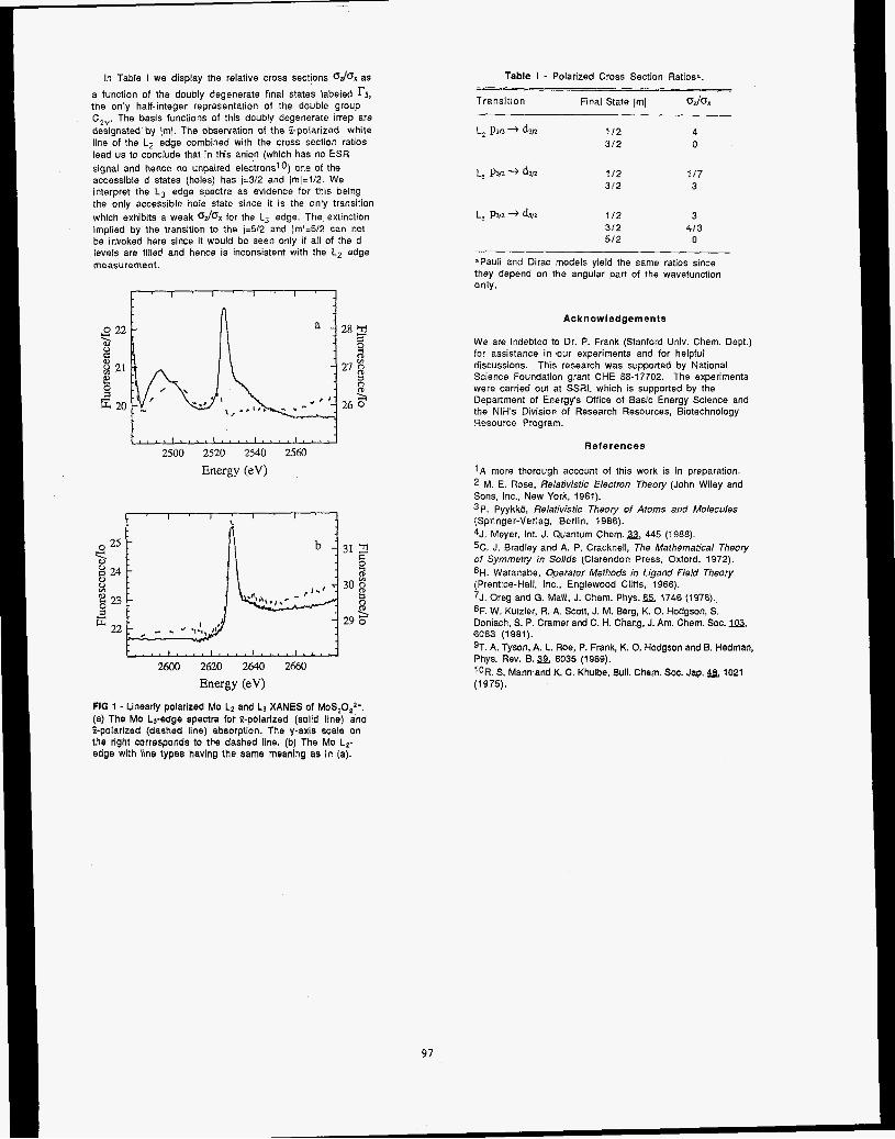
In Table I we display the relative cross sections O& as a function of the doubly degenerate final states labeled r5, the only half-integer representation of the double group C2". The basis functions of this doubly degenerate irrep are designated'by /mJ. The observation of the %polarized white line of the L, edge combined with the cross section ratios lead us to conclude that in this anion (which has no ESR signal and hence no unpaired electronslO) one of the accessible d states (holes) has j=3/2 and /ml=1/2. We interpret the L j edge spectra as evidence for this being the only accessible hole state since it is the only transition which exhibits a weak OJOX for the L, edge. The extinction implied by the transition 10 the j=512 and jmJ=512 can not be invoked here since it would be seen only if all of the d levels are filled and hence is inconsistent with the L, edge measurement.
2500 2520 2540 25M) Energy (eV)
2600 2620 2640 2660 Energy (eV)
FIG 1 - Linearly polarized Mo L2 and Lf XANES of MoS,0,2-. (a) The Mo LJ-edge spectra for 9-polarized (solld line) and %polarized (dashed line) absorption. The y-axis scale on the right corresponds to the dashed line. (b) The Mo Lz- edge wlth llne types having the same meaning as in (a).
Table I - Polarized Cross Section Ratios%.
Transition Final State Iml OJO,
L* P1n + d3n 112 4 312 0
L, PW -+ d3r2 112 1 I 7 312 3
L, P~/L + dm 112 3 3 /2 413 512 0
=Pauli and Dirac models yield the same ratios since they depend on the angular part of the wavefunction only.
Acknowledgements
We are indebted to Dr. P. Frank (Stanford Univ. Chem. Dept.) for assistance in our experiments and for helpful discussions. This research was supported by National Science Foundation grant CHE 88-17702. The experiments were carried out at SSRL which is supported by the Department of Energy's Office of Basic Energy Science and the NIH's Division of Research Resources, Biotechnology Resourca Program.
References
1 A more thorough account of this work is in preparation. M. E. Rose, Relafivisfic Electron Theory (John Wiley and
Sons, Inc.. New York, 1961). 3P. PyykkO, Relativistic Theory of Atoms and Molecules (Springer-Verlag, Berlin, 1986). 4J. Meyer. Int. J. Quantum Chem. a, 445 (1988). 5C. J. Bradley and A. P. Cracknell, The Mathematical Theory of Symmetry in Solids (Clarendon Press, Oxford, 1972). 6H. Watanabe, Operafor Mefhods in Ligand Field Theory (Prentice-Hall, Inc., Englewood Cliffs, 1966). 7J. Oreg and G. Malli, J. Chem. Phys. 65, 1746 (1976). 8F. W. Kutzler, R. A. Scott, J. M. Berg, K. 0. Hodgson, S. Doniach, S. P. Cramer and C. H. Chang, J. Am. Chem. SOC. 6083 (1981). 9T. A. Tyson, A. L. Roe, P. Frank, K. 0. Hodgson and B. Hedman, Phys. Rev. B. a 6035 (1989). l0R. S. Mannand K. C. Khulbe, Bull. Chem. SOC. Jap. 48,1021 (1 975).
97


1
The interfaces between Sn, Sb, and Bi and GaP (1 10) have been examined using soft x-ray photoemission, and the core level attenuation and lineshape changes confirm that these interfaces exhibit S-K growth with the f i t monolayer described by the zigzag chain model originally developed for Sb on GaAs (110). A model of the charge configuration at the one monolayer interface can be invoked to explain the disappearance of the surface components in the context of both charge transfers and Madelung potential differences.1 The disappearance of the surface components is utilized to estimate intrinsic lineshape pinmeters of the Ga 3d and P 2p core levels. These parameters were then employed in the computer fits performed for this paper: the buk-surface core shifts were determined to be roughly 0.32 and 0.39 for Ga 3d and P 2p, respectively. Finally, shifts of the bulk core levels as a function of Sn, Sb, and Bi coverage were examined to determine the band bending behavior and the final surface Fermi level positions at these interfaces. The position of each of the three is in the neighborhood of 1 eV above VBM, the same range as those observed for materials with significantly different work functions and electronegativities such as Au. Thus, Fermi level pinning can be discussed fnr the three interfaces: the pinning seems to have a correlation to the development of metallic or semimetallic character in the overlayer. This work is supported by ONR under contracts No. N00014-89-J- 1083 and N00014-86-K-0736.
MFEMNCES
PHOTOEMISSION SPECTROSCOPY OF ORDERED OVERLAYERS ON GaP (1 10)
K. E. Miyano, R. Cao, T. Kendelewicz, A. K. Wahi, I. Lindau, and W. E. Spicer
Stanford Electronics Laboratories, Stanford University, Stanford, CA 94305
Pt'OPosal No. 935vP
1. K. E. Miyano, R. Cao, T. Kendelewicz, A. K. Wahi, I. Lindau, and W. E. Spicer, in Proceedings of the Ninth International Conference on Vacuum Ultraviolet Physics , Physica Scripta, to be published.
98

Proposal No. 935Vp VACWM ULTRAVIOLJZT PHOTOELECTRON SPECTROSCOPY
OF (NH4)2S TREATED GaAs (100) SURFACES
C. J. Spindt,l D. Liu? K. Miyano,l P. L. Meissner,l T. T. Chiang,’ T. Kendelewicz,l I. Lindau,’ and W. E. Spiced
Istanford Electronics Laboratories, Stanford University, Stanford, CA 94305 2Varian Central Research, Varian Associates, Palo Alto, CA 94303
In 1987, it was discovered by Sandroff, et al. that sulfur containing solutions such as (NH4)2S can reduce the surface recombination velocity of the GaAs surface. The surface chemistry and band bending of the ammonium sulfide treated GaAs (100) surface has been studied using surface sensitive synchrotron radiation photoemission spectroscopy on beamline 111-1, the “new grasshopper”. Although previous publications reported that only arsenic sulfides were found on the surface, we find that the treatment leaves the GaAs surface terminated with roughly a monolayer of sulfur bonded to both As and Ga atoms. In addition, an n-type barrier height of 0.8 eV is measured. The thermal stability of the various chemical components are studied, and it is found that the arsenic sulfide compounds desorb from the surface at ~ 2 5 0 C, while the gallium sulfides are stable at temperatures of at least 400 C. In work stimulated in part by this data, we have proposed a mechanism for the reduced surface recombination velocity obsmed on these surfaces, based on the anti-site defect model. This model is consistent with the increase in band bending observed after treatment, and predicts that the reduced surface recombination velocity is due to the increased band bending (sweeping electrons away), and a change in the charge state of the midgap states (reducing the hole capture cross section).
This work is supponed by the Defense Advanced Research Project Agency and the Office of Naval Research Contract N00014-89-J- 1083.
REFERENCES
1. C. J. Spindt, D. Liu, K. Miyano, P. L. Meissner, T. T. Chiang, T. Kendelewicz, I. Lindau, and W. E, Spicer, Appl. Phys. Lett. 55 (9), 861 (1989).
2. C. J. Spindt and W. E. Spicer, Appl. Phys. Lett. 55 (16), 1653 (1989).
99

Proposal No. 935Vp USE OF EPITAXIAL OVERLAYERS TO STUDY SURFACE CORE LEVEL SHJFlT
AT THE CLEAVED Si, InP, AND GaP SURFACES
T. Kendelewicz, J. C. Woicik, K. Miyano, R. Cao, P. Pianetta, I. Lindau, and W. E. Spicer Stanford Electronics Laboratories, Stanford University, Stanford CA 94305
Monolayer quantities of adsorbates are shown to quench surface components of core level photoemission spectra from cleaved semiconductor surfaces. In this work, we use selected epitaxial and nonreactive overlayers to obtain an accurate estimate of the Si 2p and P 2p bulk core level line shapes. The core hole Lorenzian widths, branching ratios, and spin orbit splittings determined by this method are then used to analyze the core level line shapes from the clean surfaces. Our analysis provides new insight to the surface core level shifts of Si(l11) 2x1 and to the less studied III-V phosphides. The overlayers which we have chosen for this work are Ge and Sb for Si( I1 1) and Sb and Bi for InP( 110) and GaP( 110). Our upper estimate of the Lorenzian width of 0.07 eV for both the Si 2p and P 2p core levels is one third of that used in previous high resolution photoemission studies. As a consequence, we find it impossible to fit our Si(ll1) 2x1 surface sensitive spectra with only two surface shifted components. An additional component finds a straightforward interpretation in the context of the chain model, while earlier photoemission results using only two surface shifted components favor the buckling model of the 2x1 reconstruction. The surface sensitive P 2p spectra show a single surface component shifted 0.29 and 0.42 eV towards lower binding energy relative to the bulk component for InP and Gap, respectively. The surface shifts of the In 4d and the Cia 3d core levels from the same surfaces are in the opposite direction and are nearly equal in magnitude (-0.3 eV).
This work is supported by the Defense Advanced Research Project Agency and the Office of Naval Research Contract No. N00014-89- J-1083.
100

INTERFACIAL CHEMISTRY AND BAND BENDING AT In, Al, Ag, AND Pt INTERFACES WITH CdTe AND ZnTe (1 10)
A. K. Wahi, K. Miyano, G. P. Carey, T. T. Chiang, I. Lindau, and W. E. Spicer
Stanford Electronics Lahoratories, Stanford University, Stanford, CA 94305
Interest in examining Schottky barrier formation on 11-VI semiconductors has increased in recent years. In comparison to III- V semiconductors such as GaAs, relatively little is known concerning Schottky barrier formation and interfacial chemistry at metal/II-VI interface^.'.^ We have investigated interfacial chemistry and morphology in conjunction with band bending behavior for In, Al, Ag, and Pt overlayers on both CdTe (Eg = 1.5 eV) and the wider bandgap ZnTe (Eg = 2.2 eV) using photoemission.3.4 These comparative studies are also important in relation to metal/HgCdTe and metal/HgZnTe interface formation, where HgZnTe has been proposed as structurally more stable than HgCdTe.5
Experiments were performed on Beamline I- 1 and Beamline 1-3 (old and new grasshopper monochromators) at the Stanford Synchrotron Radiation Laboratory (SSRL). Photon energies were chosen to provide a range of surface sensitivities (electron escape depth, h -5 - 7 A and -10 - 20 A). Experiments were also performed using a He discharge lamp for UPS studies (He I and He II,21.2 and 40.8 eV). The kinetic energy of the photoemitted electrons was analyzed using a double-pass cylindrical mirror analyzer (CMA).
The metals chosen provide a range of metal-substrate reactivities: A1 reacts strongly with Te, Ag moderately, and In minimally, with no evidence seen for In reaction on ZnTe. Pt exhibits strong alloying behavior with both Cd and Zn. All four metals are found to yield Schottky barriers on CdTe and ZnTe, with a narrow range of final Fern$ level positions, Efi = Ef- EVBM, observed on CdTe, from 0.9 to 1.05 k 0.1 eV, and on ZnTe from 0.65 to 1.0 rt: 0.1 eV. The prediction of the MIGS model that a difference in barrier height exists for two semiconductors dependent upon their band lineup (valence band offset)6 is examined and found to agree with experiment for Ag, Pt, and AI, but not for In. Far the highly reactive Al, no evidence for the overlayer metallicity required for MIGS to operate is seen on CdTe or ZnTe until after band bending has stabilized. Reaction and intermixing for Al, Ag, and Pt overlayers on CdTe and ZnTe indicate these interfaces are not ideal. In view of the reaction and intermixing seen for Ag, Pt, and Al, and specifically the lack of metallicity seen for AI overlayers, a defect mechanism can provide a more consistent explanation for the observed barrier heights.
These results for the binaries CdTe and ZnTe are also compared to metal/HgCdTe interface formation? We find that Hg loss can significantly influence the extent of reaction and/or intermixing for these overlayers, with resulting disruption either inhibiting or facilitating chemical interaction.
This work is supported by NASALangley under Contract No. NAG1-851 and by DARPA under contract No. N00014-86-K- 0854.
REFERENCES
1.
2.
3.
4.
5.
6.
I. M. Dharmadasa, W. G. Herrenden-Harker, and R. H. Williams, Appl. Phys. Lett. 48, 1802 (1986). D. J. Friedman, I. Lindau, W. E. Spicer, Phys. Rev. B 37, 731 (1988). A. K. Wahi, K. Miyano, G. P. Carey, T. T. Chiang, I. Lindau, and W. E. Spicer, J. Vac. Sci. Tecknol. A, to be published. A. K. Wahi, 6. P. Carey, K. Miyano, T. T. Chiang, I. Lindau, and W. E. Spicer, J. Vac. Sci. Technol. A, to be published in Mar/Apr 1990. A. Sher, A.-B. Chen, W. E. Spicer, and C.-K. Shih, J. Vac. Sci. Technol. A 3, 105 (1985). J. Tersoff, Phys. Rev. Lett. 56, 2755 (1986).
Proposal No. 935Vp
101

m Y b STCICIDES
Proposal NO. 941 vp
L, Braicovich, E. Puppin, M. Sancrotti Istituto di Fisica - Politecnico di Milano
P.za Leonard0 da Vinci 32 - 20133 Milano (Italy)
F. Marchetti Istituto per la Ricerca Scientifica e Tecnologica
Povo 38050 (Italy)
G. L. Olcese Istituto di Chimica - Fisica - Universita' di Genova
C.so Europa 30 - 16132 Genova (Italy)
I. Lindau Stanford Electronics Laboratories - Stanford University
Stanford 94305 CA (U.S.A.)
Near edge absorption spectroscopy can be used to determine the value of valence in compounds which exhibits mixed valence behavior. In the past, this technique has been widely used in order to study the value of valence in rare earths compounds. Whitin this proposal it has been already studied the effect of chemical pressure on Yb mean valence in two families of isostructural Laves phases: Ybl-xCaxA12 and Ybl-xSc,A12, 0 5 x 5 0.8 1. LII and LIII absorption spectra clearly showed that the value of valence monotonically increases with the Yb nearest neighbor distance.
A word of caution, however, must be spent in this context since, in certain cases, the value of valence as extracted from the decomposition of the absorption spectra is considerably different with respect to Yb valences determined with other techniques. TWG examples of this behavior are represented by Ybo.4Sco.6Al2 and Yb0.2Sco.8A12 for which 4f photsmission leads to estimate Yb mean valences remarkably lower than the values inferred by Yb LIII X-ray absorption spectroscopy 1. For this reason it is important to compare the absorption data with other spectroscopic results.
Yb silicides (Yb3Si5, YbSi and YbsSi3) were extensively investigated with photoelectron spectroscopy 2 and the values of valence were extracted from 4f photoemission spectra by obtaining results in good agreement with other techniques. We measured, in the present experiment, the LIII Yb absorption edge in all of these three compounds. We measured the edges of polycrystalline samples scraped with a diamond file in Ultra High Vacuum in order to avoid surface sample contamination. Furthermore, we repeated the experiment by using finely grounded powders (from the same samples) by obtaining the same results.
The Yb valence as a function of the nearest neighbors distance, as extracted from the absoption data, shows the same monotonic trend already observed in 4f photoemission. However, the values extracted from the absorpion spectra are sistematically higher compared to the valence photoemission results. The work is in progress in order to analyze these results.
REFERENCES
1) M. Sancrotti et al., two papers in print on Solid State Communications
2) I. Abbati et al., Phys. Rev B 34, 4150 (1986); I. Abbati et al., Solid State Commun. 62, 35 (1987)
102

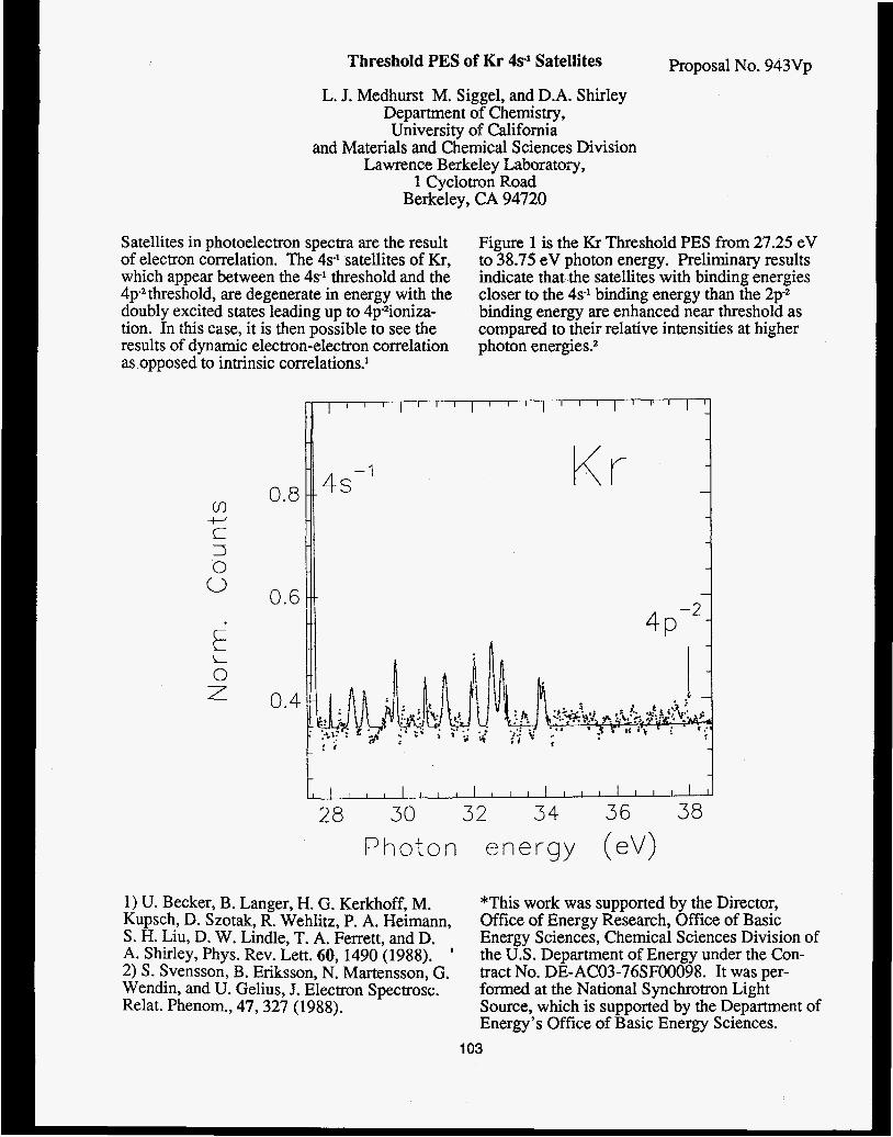
Threshold PES of Kr 4s1 Satellites Proposal No. 943Vp
L. J. Medhurst M. Siggel, and D.A. Shirley Department of Chemistry, University of California
and Materials and Chemical Sciences Division Lawrence Berkeley Laboratory,
1 Cyclotron Road Berkeley, CA 94720
Satellites in photoelectron spectra are the result of electron correlation. The 4s' satellites of Kr, which appear between the 4s' threshold and the 4p2threshold, are degenerate in energy with the doubly excited states leading up to 4pzioniza- tion. In this case, it is then possible to see the results of dynamic electron-electron correlation as opposed to intrinsic correlations.'
Figure 1 is the Kr Threshold PES from 27.25 eV to 38.75 eV photon energy. Preliminary results indicate that the satellites with binding energies closer to the 4s' binding energy than the 2pS binding energy are enhanced near threshold as compared to their relative intensities at higher photon energies?
v,
c 3
i-J
0 0
i L 0 7
O a 8 I r 0.6
Kr
I I l l I l l i l l I l l I l l I
28 30 32 34 36 38 Photon e n e r g y (eV)
1) U. Becker, B. Langer, H. G. Kerkhoff, M. Kupsch, D. Szotak, R. Wehlitz, P. A. Heimann, S. H. Liu, D. W. Lindle, T. A. Ferrett, and D. A. Shirley, Phys. Rev. Lett. 60, 1490 (1988). ' 2) S . Svensson, B. Eriksson, N. Martensson, G. Wendin, and U. Gelius, J. Electron Spectrosc. Relat. Phenom., 47,327 (1988).
*This work was supported by the Director, Office of Energy Research, Office of Basic Energy Sciences, Chemical Sciences Division of the U.S. Department of Energy under the Con- tract No. DE-AC03-76SF00098. It was per- formed at the National Synchrotron Light Source, which is supported by the Department of Energy's Office of Basic Energy Sciences.
103

TEMPERATURE DEPENDENT ARPEFS STUDY OF c(2x2)CIIGu(QOl)*
Li-aione Wang, A.E. Schach von Wittenau, Z.G. Ji, L.S. Wang, Z.Q. Huang, T. Shulman and D.A. Shirley
Department of Chemistry, University of California and Materials and Chemical Sciences Divisior,
Lawrence Berkeley Laboratory, 1 Cyclotron Road Berkeley, CA 94720
Angle-Resolved Photoemission Extended Fine Structure (ARPEFS) is a novel technique to obtain surface structural information using photoelectron diffraction.’
We report the first temperature dependent AR- PEFS measurements of c(2x2)Cl/Cu(OO1) on beam line 111-3 at SSRL. The experiments were performed along two emission directions, [OQl] and [ 1101, and at two temperatures, 110K and 298K. The Multiple-Scattering Spherical Wave analyses determined that the C1 atom adsorbs in the fourfold hollow site, and that the CI-Cu bond length is 2.41M.02& in agreement with LEED results2
*This work was supported by the Director, Office of Energy Research, Office of Basic Energy Sciences, Chemical Sciences Division of the U.S. Department of Energy under the Con- tract No. DE-AC03-76SF00098. It was per- formed at the National Synchrotron Light Source, which is supported by the Department of Energy’s Office of Basic Energy Sciences.
1. J.J. Barton, S.W.Robey, and D.A. Shirley, Phys. Rev. B 34 (1986) 778. 2. F. Jona, D. Westphal, A.Goldmann and P.M. Marcus, J. Phys. C: Solid State Phys.
16 (1983) 3001.
Low temperature ARPEFS has larger oscillation amplitudes and allows us to determine the recon- struction and small corrugation of the second sub- strate copper layer, induced by the adsorption of chlorine. Temperature dependent ARPEFS at two different emission directions also enables us to study the surface atom vibrational anisotropy.
104

Proposal No. 943Vp
LOW TEMPERATURE ARPEFS STU p(2x2)S/Ni(lll) and 43d3 R30° §/Ni(lll)
A.E. Schach von Wittenau, E.Q. Wang, Z.Q. Huang, Z. Hussain," and D.A. Shirley
Department of Chemistry, University of California and Materials and Chemical Sciences Division
Lawrence Berkeley Laboratory, 1 Cyclotron Road Berkeley, CA 94720
Previous LEED1,SEXAFS2, and ion scattering3 studies of p(2x2)S/Ni( 11 1) have shown that S adsorbs into the FCC-like threefold hollow site, i.e. the site with no underlying Ni atom. Varying results for the S-Ni fist layer distance and S-Ni bond length have been obtained. LEED results also indicate adsorbate-induced relaxations in the Ni surface. ARPES4 studies of the 43x43 R30° overlayer suggest a change of bonding site for this coverage.
*This work was supported by the Director, Office of Energy Research, Office of Basic Energy Sciences, Chemical Sciences Division of the U.S. Department of Energy under the Con- tract No. DE-AC03-76SF00098. It was per- formed at the National Synchrotron Light Source, which is supported by the Department of Energy's Office of Basic Energy Sciences. One of us (Z.H.) wish to acknowledge KFUPM f o p the grant of a sabbatical leave.
We have performed low temperature (200" K) ARPEFS studies of p(2x2)S/Ni( 1 1 1) and 43x43 R30° S/Ni( 11 1) using beamline X-24 at NSLS. Measurements were taken in the plane defined by the 11 113 and [O(SUP12(J,2)11] directions for both the ~(2x2) coverage and in the 43x43 R30° overlayer. Preliminary analysis of these low temperature data using multiple-scattering spherical-wave calculations indicates that in the ~(2x2) case the S adsorbs 1.62 A above the Ni surface, with reconstruction of the surface Ni layer. The S-Ni bond length is determined to be 2.16 A. Analysis of the 43x43 R30" S/Ni( 1 11) data continues.
**Permanent address: Department of Physics, King Fahd University of Petroleum and Miner- als, Dhahran 31261, Saudi Arabia.
1) Y.K. Wu and K.A.R. Mitchell Can. J. Chem. 67,1975 (1989). 2) T. Ohta, Y. Kitajima, P.M. Stefan, M.E. Shek, N. Kosugi, and H. Kuroda, .I. Phys. (Paris) 41'3 C8,503 (1986). 3) T. Fauster, H. Duerr, and 119. Hartwig Surace Sci. 178,657 (1986). 4) T.W. Capehart, C.W. Seabury, G.W. Graham and T.N. Rhodin, Surf Sci. 120, E441 (1982).
1 05

Proposal No. 943Vp
LOW TEMPERATURE ARPEFS STUDY OF p(2x2)s/cu(oo1)"
A.E. Schach von Wittenau, L.Q. Wang, Z.Q. Huang, Z. Hussain,** Z.G. JI,
T. Shulman and D.A. Shirley
Department of Chemistry, University of California and Materials and Chemical Sciences Division
Lawrence Berkeley Laboratory, 1 Cyclotron Road Berkeley, CA 94720
Previous ARF'EFS', LEED2, and SEXAFS3 studies of this surface, while agreeing that the S adsorbs into a fourfold hollow site, have given conflicting values for the distance between the S and the first Cu layer. They have also yielded different degrees of reconstruction of the near surface Cu layers..
We have performed low temperature (1 lo' K) AWEFS studies of p(2~2)S/Cu(Ool) using beamlines 111-3 at SSRL and X-24 at NSLS. Measurements were taken in the [Ool] and [Oll] directions. Preliminary analysis of these low temperature data using multiple-scattering spherical-wave calculations indicates that the S adsorbs 1.32 A above the Cu surface, with near surface reconstruction of the Cu layers similar to the LEED2results. The S-Cu bond length is determined to be 2.26 A. We see no net vertical expansion or contraction of the Cu layers from the bulk extrapolated values.
*This work was supported by the Director, Office of Energy Research, Office of Basic Energy Sciences, Chemical Sciences Division of the U.S. Department of Energy under the Con- tract No. DE-AC03-76SF00098. It was per- formed at the Stanford Synchrotron Radiation Laboratory and National Synchrotron Light Source, which are supported by the Department of Energy's Office of Basic Energy Sciences. One of us (Z.H.) wish to acknowledge KFUPM for the grant of a sabbatical leave.
**Permanent address: Department of Physics, King Fahd University of Petroleum and Miner- als, Dhahran 31261, Saudi Arabia.
1) C.C. Bahr, J.J. Barton, Z. Hussain, S.W. Robey, J.G. Tobin, and D.A. Shirley, Phys. Rev. B 35,3773 (1987). 2) H. C. Zeng, R. A. McFarlane, and K. A.R. Mitchell, Phys. Rev. B 39,8000 (1989). 3) J.R. Patel, D.W. Berreman, F. Sette, P.H. Citrin, J.E. Rowe, P.L. Cowan, T. Jach, and B. Karlin, Phys. Rev. B. 40, 1330 (1989).
106
Proposal No. 943Vp
ARPEFS Study of p(2x2)K/Ni(lll)
Z.Q. Huang, L.Q. Wang, A.E. Schach von Wittenau, 2. Hussain,** and D.A. Shirley
Department of Chemistry, University of California and Materials and Chemical Sciences Division
Lawrence Berkeley Laboratory, 1 Cyclotron Road Berkeley, CA 94720
The adsorption of alkali metal on transition metal surfaces has been the subject of many ex- perimental and theoretical studies because alkali additives are known to promote some major heterogeneous catalytic reactions'. Few surface structwes have been determined, however, of these alkali-metal adsorption systems. We report the fxst experimental study of the geometric structure of p(2x2)K/Ni( 11 1) using the technique of Angle-Resolved Photoemission Extended Fine Structure (ARPEFS)2.
The p(2x2)K/Ni( 1 1 1) system was prepared by evaporating K from a commercial SAES getter source onto a clean Ni( 1 1 1) held at room tem- perature until a sharp, clear ~(2x2) LEED pattern with little background was observed. K 1s angle- resolved photoemission was then measured on beamline X24A at NSLS. Results showed an energy dependent oscillation of the K 1s pho- toemission intensity over a kinetic energy range of 65-450 eV, arising from the photoelectron difffraction near the surface. Preliminary Fourier analysis of our data indicates that K atom most likely adsorbs on the three-fold hollow site on Ni( 11 1)surface with a K-Ni distance of 3.2-3.3 A. Comparison of experimental data with Mul- tiple Scattering Spherical-Wave(MSSW) calcu- lation is under way to reveal more structural details.
*This work was supported by the Director, Office of Energy Research, Office of Basic Energy Sciences, Chemical Sciences Division of the U.S. Department of Energy under the Con- tract No. DE-AC03-76SF00098.It was per- formed at the National Synchrotron Light Source, which is supported by the Department of Energy's Office of Basic Energy Sciences. One of us (Z.H.) wish to acknowledge KFUPM for the grant of a sabbatical leave.
**Permanent address: Department of Physics, King Fahd University of Petroleum and Miner- als, Dhahran 31261, Saudi Arabia.
1) M. Grunze, in The Chemical Physics of Solid Surfaces and Heterogeneous Catalysis, eds. D.A. King and D.P. Woodruff (Elsevier, Amsterdam, 1982) vo1.4, p.143 2) J.J. Barton, C.C. Bahr, 2. Hussain, S.W. Robey, J.G. Tobin, L.E. Klebanoff, and D.A. Shirley, Phys.Rev.Lett. 51.272 (1983).
107

I

Photoe lec t ron Spec t roscopic S tud ie s of CO Adsorption
J i a n y i L in , Paul M. Jones , J e f f r e y A. Guckert , E. I. Solomon*
In t roduc t ion
an impor tan t s t e p i n t h e i n d u s t r i a l s y n t h e s i s of methanol. Continued i n t e r e s t l ies i n t h e e l u c i d a t i o n of t h e bonding i n t e r a c t i o n of CO chemisorbed t o such c a t a l y t i c sites. Photoe lec t ron Spec t roscopic (PES) experiments involv ing CO adso rp t ion on ZnO s i n g l e c r y s t a l s u r f a c e s ' i n d i c a t e t h a t 0 donat ion from t h e CO 50 o r b i t a l t o t h e meta l i o n may dominate t h e bonding i n t e r a c t i o n s i n t h i s chemisorption process . These r e s u l t s c o n t r a s t t h e t y p i c a l i n t e r a c t i o n s of CO adso rp t ion on a metal s u r f a c e where metal-to- l i gand 7i back-bonding gene ra l ly dominates. In t h e p re sen t s tudy t h e angular dependence of CO photo-
+ emiss ion has been u t i l i z e d t o f u r t h e r d e f i n e t h e e l e c t r o n i c s t r u c t u r e of CO chemisorbed t o t h e CU s i te i n CuCl(111). This s u r f a c e s e r v e s as a model f o r t h e Cu/ZnO s u r f a c e , whose a c t i v e s i t e w e have shown' t o involve coord ina t ive ly unsa tu ra t ed C3v Cu(1) s i tes on t h e (0001) and (1010) s u r f a c e s of ZnO. In a d d i t i o n , w e have app l i ed Auger y i e l d NEXAFS exper iments , which have been shown t o be a s e n s i t i v e probe of t h e bound C-0 bond l e n g t h 3 , t o t h e CO/CuCl system and c a l c u l a t e d a C-0 bond l eng th f o r bound CO t h a t is s l i g h t l y s h o r t e r than t h a t of f r e e gase- ous CO. These r e s u l t s suppor t a bonding scheme f o r CO adso rp t ion on CuCl(111) i n which 0 donat ion from t h e CO l i gand dominates over t h e a back-bonding involved .
Experimental
r a d i a t i o n from t h e p lane g r a t i n g monochromator on beam l i n e U14A at t h e Na t iona l Synchrotron Light Source (NSLS), Brookhaven Nat iona l Labora tory . A Vacuum Genera tors UHV sys tem, equipped wi th a double- pas s c y l i n d r i c a l mi r ro r ana lyzer (CMA), wgs employed and opera ted a t a base p re s su re of 1 x 10 Fjhile t h e synchro t ron r a d i a t i o n remained f ixed a t an a n g l e of 88" r e l a t i v e t o t h e c e n t r a l a x i s of t h e a n a l y z e r , t h e sample was r o t a t e d r e l a t i v e t o t h e i n c i d e n t beam and t h e CMA t o achieve va r ious angle- dependent s p e c t r a . Accounting f o r a l l instrument- a t i o n , a f i n a l s p e c t r a l r e s o l u t i o n of 200 meV w a s main ta ined .
g r i t and then e tched wi th a H C l / E t O H s o l u t i o n . Fu r the r c l ean ing w a s performed i n UHV by A r i on s p u t t e r i n g followed by annea l ing a t 360K. Surface c l e a n l i n e s s w a s monitored wi th core- leve l XPS.
achieved b in t roduc ing an ambient CO atmosphere ( -1 x lo-' Torr ) i n t o t h e UHV chamber f o r approx- ima te ly t e n minutes whi le main ta in ing t h e sample a t 130K. A l l experiments w e r e c a r r i e d ou t u s ing an e l e c t r o n f lood gun t o compensate f o r charg ing a t t h e co ld semiconducting CuCl su r face .
CO adso rp t ion on Cu/ZnO c a t a l y t i c s u r f a c e s i s
A l l s p e c t r a were c o l l e c t e d wi th synchro t ron
lo Torr .
CuCl s i n g l e c r y s t a l s were po l i shed wi th alumina
Adsorption of CO on t h e sample s u r f a c e was
Resu l t s and Discuss ion A f t e r exposure t o CO, two peaks w e r e observed
i n t h e PES spectrum of CuCl which do not appear i n t h e photoemission spectrum of t h e s a m e s u r f a c e p r i o r t o CO exposure (F igure 1 ) . Peak I , cen te red a t a b inding energy of 12.3 e V o r i g i n a t e s from photo- emiss ion from t h e lone p a i r CO 40 l e v e l . The peak a t lower b ind ing energy , peak 11, can be ass igned as over lapping photoemission from t h e CO 1~i and CO 5 0 l e v e l s through ARPES s t u d i e s . I n o rde r t o r e s o l v e these over lapping states, s p e c t r a were c o l l e c t e d wi th
108
Proposal No. 1022vp
two d i f f e r e n t sample o r i e n t a t i o n s . With t h e sample pos i t i oned so t h a t t h e a x i s of propagat ion of t h e incoming l i g h t was approximately normal t o t h e CuCl s u r f a c e , peak I1 w a s cen tered a t a b inding energy of 9.4 e V ; whereas, w i th a graz ing ang le of inc idence (nea r ly 90" o f f t h e s u r f a c e normal) t t , i s same peak was centered a t 9 .0 e V . Having demonstrated from ARPES t h a t t h e bond a x i s of t h e CO molecule i s o r i en ted perpendicular t o t h e s u r f a c e , w e were a b l e t o r e s o l v e t h e over lapping states of peak 11, as-ign- ing t h e s t a t e a t lower b inding energy as t h e CO 50 and t h e c o n t r i b u t i o n a t s l i g h t l y deeper b ind ing energy as photoemission from t h e CO la l e v e l . Thus a s s igned , an energy sepa ra t ion between t h e 40 and la l e v e l s of 2 . 8 e V is revea led , n e a r l y i d e n t i c a l t o t h e f r e e CO va lue . cU+ a c t i v e s i t e , t h e 50 o r b i t a l s h i f t s 2 . 2 e V c l o s e r i n energy t o t h e l ~ , i n d i c a t i n g t h a t t h e CO 50 o r b i t a l is s t r o n g l y involved i n bonding. Two a d d i t i o n a l f a c t o r s suppor t a CO/CuCl bonding scheme predominantly 0 i n na tu re . S t6hr e t a13, t h e broad shape resonance observed i n t h e NEXAFS spectrum a t 308.5 e V ( see F igure 2 ) y i e l d s a C-0 bond l eng th of 0.110 2 0.002nm f o r CO bound t o CuC1(111), a va lue s l i g h t l y s h o r t e r than t h a t observed i n f r e e CO (0.113nm). Second, from He(1) PES s t u d i e s t h e work func t ion of t h e C O / C u C l s u r f a c e i s found t o be 1 . 2 e V less than t h a t of t h e c l ean s u r f a c e , i n d i c a t i n g a n e t charge t r a n s f e r from t h e adsorbed CO t o t h e s u r f a c e .
However, upon CO adso rp t ion to t h e
F i r s t , based on t h e model der ived by
I hv = 28 eV
U
-
v ) -
C CI
0 " - v .
I - . .
14 I2 10 8
Binding Energy (eV)
Figure 1. Angular dependence of photoemission i n t e n s i t y of t h e va lence o r b i t a l s of CO/CuC1(111); (-) graz ing ang le of i nc idence ; (---) near normal angle of inc idence .
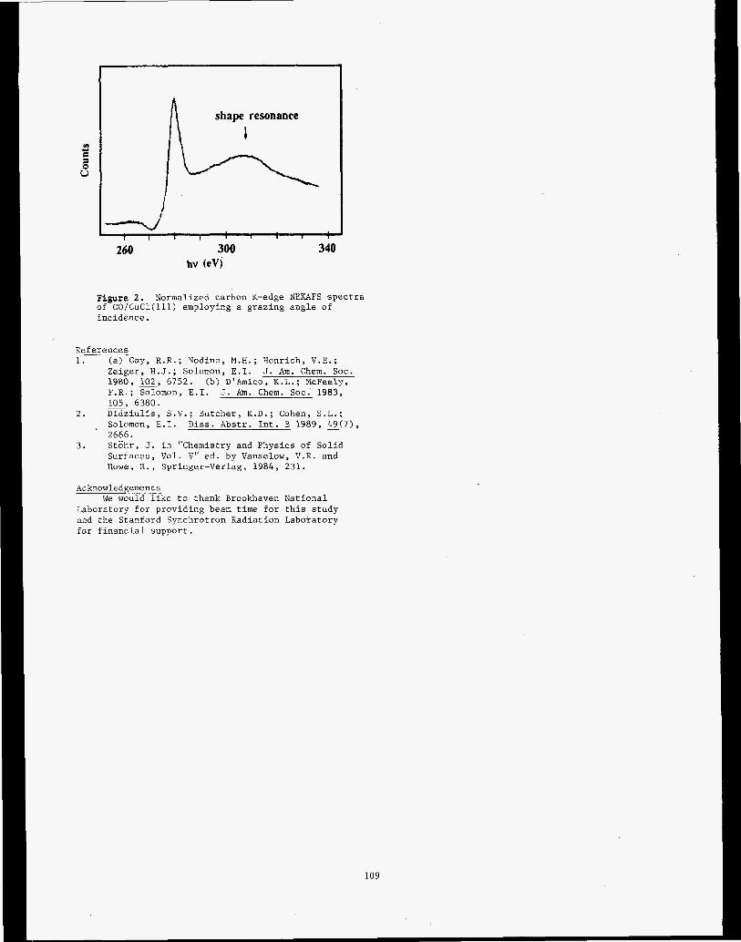
shape resonance
I I I 1 I 1
260 300 340 hv (eV)
Figure 2 . Normalized carbon K-edge NEXAFS spectra of CO/CuCl(l~l) employing a grazing angle of incidence.
References 1. (a) Gay, R.R.; Nodine, M.H.; Henrich, V.E.;
Zeiger, H . J . ; Solomon, E.I. J . Am. Chem. SOC. 1980, 102, 6752. F.R.; Solomon, E . I . J. Am. Chem. SOC. 1983, 105, 6380.
2 . Didziulis, S.V.; Butcher, K.D.; Cohen, S.L.; Solomon, E.I. Diss. Abstr. Int. B 1989, 4 9 ( 7 ) , 2666.
(b) D'Amico, K.L. ; McFeely,
-
3 . Stghr, J . in "Chemistry and Physics of Solid Surfaces, Vol. V" ed. by Vanselow, V.R. and Howe, R., Springer-Verlag, 1984, 231.
Acknowledgements
Laboratory for providing beam time for this study and the Stanford Synchrotron Radiation Laboratory for financial support.
We would like to thank Brookhaven National
109

PfOpOSal NO. 1028V
PHOTOEMISSION STUDIES OF NARROW BAND MATERIALS
J.W. Allen', J.-S. Kang1i2, 0. Gunnarsson3, 0. Jepsen3, T. F'ujiwara3, O.K. Andersen3, 2.-X. Shen4, W.P. Ellis', J.T. Market2, Y . Dalichaouch2, M.B. Maple2, P.A.P. Lindberg4, B.O. Wells4, D.S. Dessau4,
A. Borg4, K.N. Yang2, M.S. Torikachvili', Y . Lassailly7, B.B. Pate4, I. Lindau4, and W.E. Spicer4
Randall Laboratory, University of Michigan, Ann Arbor, M I 48109-1120 Dept. of Physics, University of California a t San Diego, La Jolla, CA 92093
Max-Planck Institut, D-7000 Stuttgart 80, FRG Stanford Electronic Laboratory, Stanford, CA 94305
Los Alamos National Laboratory, Los Alamos, NM 87545 ' Physics Department, San Diego State University, San Diego, CA 92182
' Ecole Polytechnique, 91128 Palaiseau, France
Resonant photoemission studies of NdzCuO4
Using photon energies at the Cu 3p+3d edge, we have per- formed resonant photoemission spectroscopy (RESPES) on the Cu 3d states of ceramic samples of NdzCu04, of importance as a 'parent compound' in high temperature .superconductiv- ity. The sample temperature was about 100K, and the resolu- tion about 0.3 eV. We have also made a theoretical calculation of the RESPES expected for this material if the 3d electrons are modeled by the impurity Anderson Hamiltonian.' All the model parameters for the calculation axe obtained from ab ini- tio calculations or atomic data. Fig. 1 shows the comparison between calculated (solid line) and measured (dots) energy dis-
4- .- VI C a C c
k-l
*L I I I 1 J - I
0 -20 -76 -i2 -8 - 4 E
tribution curves (EDC's). The upper two curves in Fig. 1 are calculated off-resonance spectra for photon polarization paral- lel and perpendicular to the c-axis. Overall, the main features of the calculated spectrum are in rather good agreement with the unpolarized experimental spectrum. The width of the main peak and the location of the satellites at about -10 eV and -12 eV are correct. The model predicts that the lowest lying state, the so-called local singlet, has much more weight than observed experimentally at the corresponding energies. The use of a lat- tice Anderson model might change the predicted weight, e.g., by giving the local singlet a dispersion. The calculation pre- dicts that RESPES occurs only for perpendicular polarization, and that there should be a large off-resonance dependence on photon polarization for the main peak and the local siriglet, offering a possible test of the Anderson model, and for the oc- curance of local singlet. Fig. 2 shows the calculated constant
,
1.2 I
x
cn C al c
r
4-
I 4
1.0 1.4 -2
I I - ' L
-16.5 1 w < l l i l ' l ! ! . ~ ~ ! ~ I
t l W 70 75 80
Fig. 1
110
Fig. 2

initial state (CIS) spectra at various binding energies, showing antiresonance and resonance behaviors in the main band and satellite regions respectively. The experimental CIS spectrum for the largest satellite agrees well with the theory.
Angle resolved photoemission on MnO
Angle resolved photoemission spectra on the (110) surface of an MnO single crystal sample were measured. The data are being analyzed, and will be compared with a band theory calculation.
Electronic structure of Ce(Rul-,Rh,)sBZ
Beginning in 1984, under past program proposal 682VP, and continuing under the present proposal, we have made RESPES studies of the Ce 4f states in the alloy system Ce(Rul-,Rh,)aBa, which, with increasing x, changes from a superconductor to an unusual ferromagnet. We have now constructed a complete scenario for this behavior.' The RESPES data have been com- bined with Bremstrahllung isochromat spectra (BIS) of the unoccupied Ce 4f states, as shown in Fig. 3. The peak near the Fermi energy EF is the Kondo resonance. We have also measured the X-ray photoemission spectra (XPS) of the Ce 3d core levels (not shown). All the data have been analyzed by fit- ting with spectral functions for the impurity Anderson Hamil- tonian, shown as solid lines in Fig. 3. The energy dependence of the hybridization matrix element in the Hamiltonian was taken to be the experimental valence band density of states, obtained in our synchrotron studies, and shown in Fig. 4. For
-10 0 10 ENERGY RELATlVE TO E, (eV)
Fig. 3
111
1 I - - I I I I
hv = 80 eV
-12 -10 -8 -6 -4 -2 0 2 ENERGY RELATIVE TO EF (eV)
Fig. 4
x=l, we fit a recently published3 single crystal Ce 3d XPS spectrum because our polycrystalline samples had some grain boundary oxides, as shown by the difference between the cal- culated and measured x=l BIS spectra of Fig. 3. We find that the Kondo temperature TK, calculated from the spectroscopic parameters, varies from large to small as x changes from 0 to 1, because the density of states at EF decreases. We then argue that the change in the strength of the spin-quenching, mea- sured by TK, promotes the change in ground states, and that this alloy behavior comes about because of the difference in the Ru and Rh 5d states at EF. We propose that the ferromag- netism for x=l be understood in terms of an experimentally unresolved exchange splitting of the EF Kondo resonance.
REFERENCES
1. 0. Gunnarsson, J.W. Allen, 0. Jepsen, T. Fujiwara, O.K. Andersen, C.G. Olson, M.B. Maple, J.-S. Kang, L.Z. Liu, J.-H. Park, R.O. Anderson, W.P. Ellis, R. Liu, J.T. Mar- ket, Y . Dalichaouch, Z.-X. Shen, P.A.P. Lindberg, B.O. Wells, D.S. Dessau, A. Borg, I. Lindau and W.E. Spicer, Phys. RRv. B (Rapid Commu.), accepted for publica- tion.
2. J.W. Allen, M.B. Maple, J.-S. Kang, K.N. Yang, M.S. Torikachvili, Y. Lassailly, W.P. Ellis, B.B. Pate, and I. Lindau, Phys. Rev. B, accepted for publication.
3. M. Kasaya, A. Okabe, T. Takahashi, T. Satoh, T. Kasuya and A. Fujimori, J. Magn. Magn. Mater. 768277, 347 (1988).

Pmposal No. 1080Vp
Orientation of Submonolayer Coverages of Furan and 2,5-Dihydrofuran on Ag(ll0) Determined by NEXAFS
J.E. Solomon and R J. Madix* J. Stohr**
*Department of Chemical Engineering, Stanford University, Stanford, CA 94305 **IBM Almaden Research Center, 650 Harry Road, San Jose, Ca 95 120
We have used near-edge X-ray absorption fine-structure ( W S ) measwements to determine the orientation of furan and 2,5-dihydrofuran (2,5-DHF) on the surface of a silver (1 10) single crystal. The position of the absorption threshold or the edge-jump in the NEXAFS spectra is compared to the X P S binding energy. Experimental
NEXAFS experiments were performed at Stanford Synchrotron Radiation Laboratory on beam line 1-1 with a grasshopper monochromator (1200 lines/mm holographic grating). Spectra were recorded at the carbon K-edge with partial-electron-yield detection using a retarding voltage of -200 volts. All s-pctra were recorded with the surface at 100 K. The final normalized NEXAFS spectra were prepared by dividing spectra recorded for the adsorbate-covered surface by spectra recorded for the clean surface toremove the strong variation in photon intensity present in the transmission function of the monochromator. The final spectra were curvefit to determine peak positions and areas [I]. The A* resonances were fit with symmetric Gaussian peaks, and the o* resonances were fit with symmetric and asymmetric Gaussian peaks [l]. A Gaussian broadened step was used to model the absbrption threshold or edge-jump.
Results The NEXAFS spectra for a submonolayer coverage of furan on
Ag( 110) show that the plane of the furan molecule lies nearly parallel to the plane of the surface (figure 1). At glancing incidence of the X-rays n* resonances dominate the spectra, and the o* resonance are greatly diminished. At normal incidence then* resonances are much smaller than the large+o* resonances. Since the n* resonances are observed when the E is perpendicular to the plane of the furan molecule, and since the o* resonances are observed when the is in the plane of the molecule, the NEXAFS spectra show clearly that the furan molecule is preferentially oriented parallel to the plane of the surface. The intensity variation of an individual resonance from glancing to normal incidence can be used to calculate the orientation of the molecule [2]. For the furan submonolayer the molecule tilts at an angle of 2 3 s as determined from the average of the tilt angles of 21" and 24" calculated from the two 3bl A* resonances. The tilt angles calculated from the o* resonances are consistent with the values determined from then* resonances. TheXPS binding energies for the two C(1s) levels of furan are 284.8 and 286.2 eV [3], while the lowest possible position of the edge-jump is 288.4 eV.
NEXAFS spectra for the 2,5-DHF at submonolayer coverage shows that the p-orbitals comprising the double bond are preferentially oriented perpendicular to the plane of the Ag(ll0) surface (figure 2). The x* resonance is much larger at glancing incidence than at normal incidence, and the a*(C=C) resonance is much largerat normal incidence relative to glancing X-ray incidence. Based on the x* intensity ratio of glancing to normal, the plane containing the n system tilts at an angle of 1 8 H from the surface normal. A slight though distinct azimuthal dependence is observed for the o* resonances associated with the C-0 and C-C single bonds for the submonolayer coverage of 2,5-DHF (figure 2, curves b and c). The resonances observed for the monolayer and submonolayer are at the same energy as the multilayer indicating only weak interaction of 2,5-DHF with the Ag( 110) surface.. The XPS binding energies of 2,§-DHF are 285.1 and 286.6 eV [3]. The lowest possible energy for the edge-jump is 287.8 eV.
112
Discussion The X P S C( 1 s) binding energies of furan and 2,5-DHF are not
at the same energy as the edge-jump in the NEXAFS spectra. The edge-jump in the NEXAFS spectrum corresponds to transitions from the core level (the C(ls) level for data reported here) to the a continuum of states. The continuum of states may be either the Fermi level of silver, or the vacuum level. Since the measured X P S binding energy is the energy difference between the C( 1s) level and the Fermi level, the edge-jump should occur at the XPS binding energy if the edge-jump corresponds to transition to the Fermi level. The lowest possible energy position of the edge-jump for the NEXAFS spectra for furan and 2,5-DHF on Ag( 1 10) is consistently about 2.0 eV higher in energy than the average XPS binding energy. The 0.3 eV uncertainty in the X P S binding energies and the 0.5 eV uncertainty in the energy calibration of the X-ray monochromator used in the NEXAFS experiments will not explain the difference. If, however, the edge-jump corresponds to transitions from the C(1s) level to the vacuum level, the edge-jump position would be the sum of the XPS binding energy and the work function of the silver surface, which is 4.5 eV [4]. Since we are unable to position the edge-jump at the XPS binding energy, the edge-jump has been set at the XPS binding energy plus the work function.
The observation that the edge-jump in the NEXAFS spectra corresponds to transitions from the core level to the vacuum level and not to the Fermi level is consistent With the NEXAFS results which both indicate that furan and 2,5-DHF are only weakly bound to the Ag(ll0) surface. Since the molecules are only weakly bound, there is very little overlap between the molecular orbitals and the metal states, so the probability of transitions to the Fermi level are small. The X P S binding energies also indicate weak adsorption. A difference 1 to 2 eV between the gas phase binding energies, E,, and the XPS binding energies of the adsorbed molecules relative to the vacuum level (the measuredXPS values, E,,, plus the work function, 'p) is indicative on a weakly bound, non-interacting surface species [5]. The XPS measurements are, therefore, also consistent with a weak molecular adsorption since E - EA + cp is between 0.9 and 1.4 eV for the C(1s) and O(ls) levefs of furan and 2,5-DHF. The difference between E, and E, + 'p is due to surface relaxation effects [SI. Acknowledgements
The assistance of J.T. Roberts, A. Muscat, A.C. Liu, W. Jark, and D. Coulman in collecting the NEXAFS data is gratefuHy acknowledged. The NEXAFS experiments were prformed at Stanford Synchrotron Radiation Laboratory which is funded by the Department of Energy undercontract DE-AC03-82ER-13000, Office of Basic Energy Sciences, Division of Chemical Sciences. We also gratefully acknowledge the support of the Center of Material Research at Stanford. References [l]. D.A. Ouaka and J. Stohr, J. Chem Phys. 88 (1988) 3539. 121. J. Stohr and D.A. Outka, Phys. Rev. B 36 (1987) 7891. [31. J.L. Solomon and R.J. Madix, in preparation. [41. A.W. Dweydari and C.H.B. Mee, Phys. Status Solidi A 27
(1975) 223. [51. C.R. Brundle and A.F. Carley, Faraday Disc. Chem. Soc. 60
(1975) 5 I .

Furan Submonolayer / Ag( 110) NEXAFS
2, !az) -
E
.:..:: ........... ................
275 285 295 305 315 325 Photon Energy (eV)
Figure 1. Carbon K-edge NEXAFS spectra of furan submonolayer are shown for glancing and normal X-ray incidence. Normal incidence spectra are shown both for the E vector of the X-rays along ([liO] azimuth) and across ([Ool] azimuth) the rows of the Ag( 110) surface. The clear polar dependance of the n* resonances between the glancing and normal incidence indicate the plane of the furan molecule lies nearly coplanar with the plane of the surface. At normal incidence a small yet clearly observable azimuthal dependance is also observed for the B* resonances.
I I t I i I I I I I 275 285 295 30.5 315 3
Photon Energy (eV)
2,5-DHF Submonolayer / Ag( 110) NEXAFS
Figure 2. Carbon K-edge NEXAFS spectra of 2,5-DHF submonolayer are shown for glancing and normal X-ray incidence for the E. along and across the rows of the Ag(ll0) surface. The double bond of the 2,5-DHF molecule lies nearly parallel to the plane of the surface, as seen by the polar dependance of the x* resonance. A small azimuthal dependance is observed at normal incidence since the B* (C-C,C-O) resonance is larger when the E vector is along the rows ([liol azimuth) than when it is across the rows ([Ool] azimuth).
113

PHOTOEMISSION STUDY OF HIGH TEMPERATURE SUPERCONDUCTORS Proposal No. 2103Vp
AND RELATED MATERIALS B.O. Wells, Z.-X. Shen, D. S . Dessau, P. A. P. Lindberg, I. Lindau, and W. E. Spicer
Stanford Electronics Laboratories, Stanford University, Stanford, CA 94305
INTRODUCI'ION
The recently discovered high temperature superconductors promise to be of great importance but as yet are not well understood. We performed experiments at the Stanford Synchrotron Radiation Laboratory (SSRL) on several families of these new superconductors, Bi2CaSr2Cu208, PbSr2PrCu308, and BaBiO3. These superconductors have very complex structures, with 5 or more types of atoms, and present new, not yet understood physics. To aid in our understanding it is useful to study simpler systems that have similar properties to the high temperature superconductors. For this reason, we have undertaken a study of transition metal oxides such as COO, NiO, and CuO. These compounds are antiferromagnetic insulators, although traditional band theory predicts them to be conductors. The reason for this discrepancy is believed to be the presence of strong electron-electron correlations. The parent compounds of the Cu-0 superconductors are antiferromagnetic insulators, and they become superconducting when doped appropriately. Thus, the attempt to understand the transition metal oxides first is an appropriate approach. Following are some examples of such efforts.
MATERIALS
a. Symmetry of Valence Band States [l]
The symmetry of the electronic states that lie near the Fermi level, and are therefore involved in the superconducting transport, is, of great theoretical importance. There has been work to determine the symmetry of the unoccupied states near the Fermi level in Bi2CaSr2Cu208 [2], but we have made the first determination of the symmetry of the occupied states. We used angle resolved photoemission for this experiment. The highly polarized nature of the synchrotron radiation allows us to examine the changes in the photoemission spectra produced by changing the angle between the surface normal and the electric field of the incident light. When electrons are collected at normal emission, the symmetry selection rules are relatively straightforward. Figure 1 shows the variation of the valence band spectrum with radiation incident angle. We were able to determine that the states we measured near the Fermi level had As crystal symmetry, while the states near the bottom of the valence band had A1 symmetry. This indicates that 0 2px and 0 2p, states lie near the Fermi level, while 0 2p, states lie deeper in the valence band.
b. Band Mapping [3]
Using angle resolved photoemission, we were able to map the dispersion of the bands in Bi2Casr~Cu208. There was very little dispersion along the T-Z k-space direction (normal to the planes). The lack of dispersion indicates very little orbital overlap along the direction parallel to the planes. This confirms the highly two dimensional character of the Bi2CaSr2Cu208 material. Parauel to the planes there was dispersion, more near the bottom of the valence band than near the Fermi level. This is similar to the dispersion seen in transition metal oxides but less pronounced.
We performed the first photoemission electronic structure experiment on Pb2Sr2PrCu308. We noted that, like the other Cu-0 based high Tc superconductors, correlation effects pay an important role in observed spectrum. Of particular interest was a comparison to
114
i i h" '= 18 e"
- 8 u - 7 - 6 - 5 - 4 - 3 - 2 . 1 0
Energy Relative lo the Fermi Energy !eVl
Fig. 1. Polarization-dependent EDC's for (a) 18 eV and (b) 17.25 eV radiation. The 17.25 eV spectrum is included to show feature E, which only appears for low photon energies.
the 123 (RBa2Cu307) material PrBa2Cu307. The two materials are very similar in structure. However, the 123 structure is a superconductor for R as any rare earth atom except Pr, while the Pb2Sr2RCu308 system is still a superconductor for R=Pr. We used resonant photoemission to determine the Pr contribution to the valence band of these compounds. Our results show that Pr 4f states are more strongly hybridized with the Cu-0 states in the PrBa2Cu307 compound than in the PbSr2PrCu308 material (see Fig. 2). We theorize that this extra hybridization interferes with the superconductivity in the Cu-0 states.
I I I
Fig. 2. Valence-band EDC's of Pb2Sr2PrCu308 and PrBa2Cu307.6 [5] at photon energies just above and below the Pr 4d -+ 4f absorption edge. The valence-band maximums have been aligned for easier comparison. The normalization of the EDC's is described in the text. A subtraction of the two curves gives the extracted Pr 4fpartial density of states. The tick marks correspond to the initial energies of the CIS curves in Fig. 3.
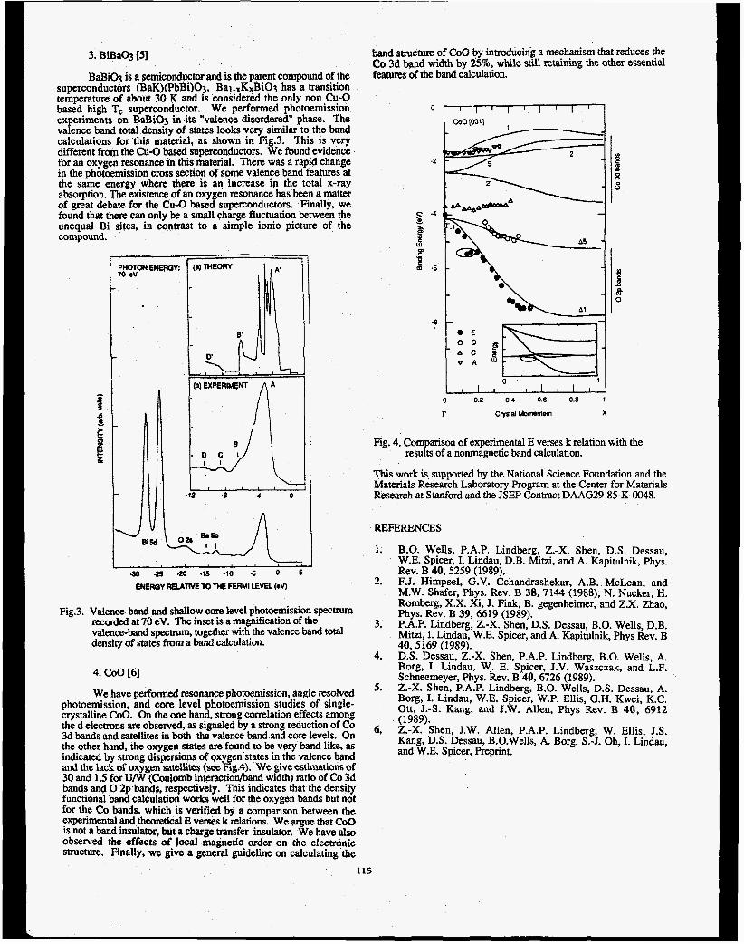
3. BiBaOg [53
BaBiO-j is a setniconductor and is the parent compound of the superconductors (BaK)(PbBi)Oj. Bal.xKxBiOg has a transition temperature of about 30 K and is considered the only non Cu-0 based high Tc superconductor. We performed photoemission experiments on BaBiO3 in its "valence disordered" phase. The valence band total density of states looks very similar to the band calculations for this material, as shown in Fig.3. This is very different from the Cu-0 based soperconductors. We found evidence for an oxygen resonanw in this material. There was a rapid change in the photoemission cross sectian of some valence band features at the same energy where there i s an increase ip the total x-ray absorption. The existence of an oxygen resonance has been a matter of great debate for the Cu-0 based superconductors. Finally, we found that there can only be a small charge fluctuation between the unequal Bi sites, in contrast to a simple ionic picture of the compound.
$ g
, ,\ .I2 -8 -4 0 ~ii ' ' I '
-30 -25 .20 .1s -10 5 0 5 ENERGY RELATNE TO THE FERMI LEVEL (eV)
Fig.3. Valence-band and shallow core level photoemission spectrum recorded at 70 eV. The inset is a magnification of the valence-band spectrum, together with the valence band total density of states from a band calculation.
4. COO [6]
We have performed resonance photoemission, angle resolved photoemission, and core level photoemission studies of single- crystalline COO. On the one hand, strong correlation effects amon% the d electrons are observed, as signaled by a strong reduction of Co 3d bands and satellites in both the valence band and core levels. On the other hand, the oxygen states are found to be very band like, as indicated by strong dispersions of oxygen states in the valence band and the lack of oxygen sateiIite$ (see Fig.4). We give estimations of 30 and 1.5 for U/W (Coulomb interactionhand width) ratio of Co 3d bands and 0 2p b m s , respectively. This indicates that the density functional band calculation works well for !he oxygen bands but not for the Co bands. which i s verified by a comparison between the experimental and theoretical E verses k relations. We argue that Coo is not a band insulator, but a charge m s f e r insulator. We have also observed the effects of local magnetic order on the electronic structure. Finally, we give a general guideline on calculating the
band structure of COO by introducing a mechanism that reduces the Co 3d band width by 25%, while still retainhg the other essential features of the band calculation.
0
0 0.2 0.4 0.6 0.6 1
r clysral Momemem X
Fig. 4. Comparison of experimental E verses k relation with the
This work is supported by the National Science Foundation and the Materials Research Laboratory Program at the Center for Materials Research at Stanford and the JSEP Contract DAAG29-85-K-0048.
results of a nonmagnetic band calculation.
REFERENCES
1.
2.
3.
4.
5.
6,
B.O. Wells, P.A.P. Lindberg, Z.-X. Shen, D.S. Dessau, W.E. Spicer, I. Lindau, D.B. Mitzi, and A. Kapitulnik, Phys. Rev. B 40,5259 (1989). F.J. Himpsel. G.V. Cchandrashekar, A.B. McLean, and M.W. Shafer, Phys. Rev. B 38,7144 (1988); N. Nucker, H. Romberg, X.X. Xi, J. Fink, B. gegenheimer, and Z.X. Zhao, Phys. Rev. B 39, 6619 (1989). P.A.P. Lindberg, Z.-X. Shen, D.S. Dessau, B.O. Wells, D.B. Mitzi, I. Lindau, W.E. Spicer, and A. Kapitulnik, Phys Rev. B 40,5169 (1989). D.S. Dessau, Z.-X. Shen, P.A.P. Lindberg, B.O. Wells, A. Borg, I. Lindau, W. E. Spicer, J.V. Waszczak, and L.F. Schneemeyer, Phys. Rev. B 40,6726 (1989). Z.-X. Shen, P.A.P. Lindberg, B.O. Wells, D.S. Dessau, A. Borg, I. Lindau, W.E. Spicer, W.P. Ellis, G.H. Kwei, K.C. Ott, J.4. Kang, and J.W. Allen, Phys Rev. B 40, 6912 (1989). Z.-X. Shen, J.W. Allen, P.A.P. Lindberg, W. Ellis, J.S. gang, D.S. Dessau, B.O.Wells, A. Borg, S.-J. Oh, I. Lindau, and W.E. Spicer, Preprint.
115

- ._________ _. - -- -____ .. Proposal NO. 9 9 0 1 ~ A microchannel-plate-intensified, subnanosecond, x-ray imaging camera
M. R. Carter, B. J. McKinley, and K. G. Tirsell
University of California, Lawrence Livermore National Laboratory P. 0. Box 808, L-43, Livermore, California 94550
Each uhotocathode was calibrated using three different Introduction We have developed a microchannel-plate-intensified, subnanosecond x-ray detector with 400-psec time resolution and 180 micron spatial resolution for one- Aimensional imaging and spectroscopic measurements of <-ray emission from laser produced plasmas.(l) An x-ray photocathode (CsI, CUI, or gold) is deposited on the MCP to enhance efficiency. Detector output is coupled to a Reticon camera or a streak camera via a fiber optic array. Efficiencies in excess of 1000 (w/cm2 out per w/cm2 in) have been demonstrated. We have recently measured CUI and CsI x-ray detector efficiencies using SSRL BL-VIII- 2. (2)
Detector Description Fig. 1 schematically shows the x-ray imaging detector consisting of a UV filter, an x-ray photocathode, a MCP, focusing electrodes, a fast phosphor, and a fiber optic faceplate for transmitting the tight out of vacuum. Focusing electrodes compress the electron current out of the MCP in one dimension enhancing system gain by a factor of 10. The 1n:CdS phosphor-coated fiberoptic faceplate is biased up to +20kV to attract electrons exiting the MCP. Gains greater than 1000 can be obtained from the MCP depending on the bias voltage. The CsI detector is about six times more sensitive than the CUI detector at 700 eV. This is consistent with uhotocathode resuonse data. (3)
I
F
~
Fiber tacepiato
~
Figure 1. Schematic of an MCP-intensified x-ray
iMeasurements We have measured the relative spectral response of CUI and CsI detectors at SSRL BL-VIII-2 from 450 eV to 1300 eV with the beam incident upon each detector at 35 degrees from normal. The detector was positioned so that the entire 2-mm x 20-mm beam fell on the MCP stripline.
i Incident x-ray flux was measured with a calibrated, gold- ’ mesh monitor, and the detector output recorded with a Reticon camera. Our results are shown in Fig. 2.
I The difference in MCP bias voltages, the measured transmission of the Al W light shield, and the response of the gold mesh monitor have been taken into account.
detector system.
I
-
prefilters (Fe, Cu and Mg) to reduce Gigher-order and specularly-reflected photons exiting the monochromator. Data taken with Cu and Mg prefilters agree well in their region of overlap for both CUI own as one curve. Data t ficantly
data in their region of 1 anderstood. The CsI
data shows a stro sitivity at the Cs M5- edge (726 eV) and at the I M4,gedge (630 eV). The CUI MCP response increases at the Cu L3-edge (932 eV) and at the I M4,s-edge (630 eV). The Cu-edge jump is much smaller in magnitude than that determined from Ep(E) data.(3) Another significant difference between the measured response and a simple Ep(E) model is the gradual rise in efficiency above the Cs edge for CsI and above the I edge in the CUI case. The deviations from
400 600 8 0 0 1000 1 2 0 0 1400
energy (eV) Figure 2. Relative response of CUI and CsI coated MCP
imaging detectors vs x-ray energy. The vertical scale is in units of watts out per x-ray watts incident.
REFERENCES 1. M. R. Carter, B. J and K. G. Tirsell, “A
microchannel-plate-intensified, subnanosecond, x-ray imaging camera,” Physica Scripta;to be published. K. G. Tirsell and N. K. Del Grande, “Considerations for the use of synchrotron radiation sources to measure sub-keV x-ray photoabsorption cross sections in transmission,” SPIE, 911,146-156 (1988).
, 3. B. L. Henke, J. P. Knauer, and K. Premaratne, “The , characterization of x-ray photocathodes in the 0.1-10- 1 keV photon energy region,”J. Appl. Phys. 52 (3), 1509- 1 1520 (1981).
ACKNOWLEDGEMENTS J. Batteux, M. Bowers, C. Bruns, C. Keller, R. Mazuch, D. Price, and R Stewart, LLNL, for their this research.
s of the U. S. I This work was performed under the I Department of Energy by the Lawrence Livermore
National Laboratory under ntract No. W-7405-Eng-48. Data were acquired at SSRL which is supported by the nnp’s Office of Basic Energy Science.
2.
The authors wish to thank
I
116

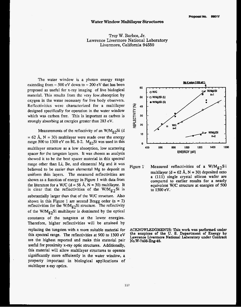
ProposalNo. 9901V Water Window Multilayer Structures
Troy W. Barbee, Jr. Lawrence Livermore National Laboratow
Livermore, California 94550
The water window is a photon energy range extending from - 500 eV down to - 200 eV that has been proposed as useful for x-ray imaging of live biological material. This results from the very 1ow.absorption by oxygen in the water necessary for live body observers. Reflectivities were characterized for a multilayer designed specifically for operation in the water window which was carbon free. This is important as carbon is strongly absorbing at energies greater than 283 eV.
Measurements of the reflectivity of an W/Mg2Si (d = 62 A, N = 30) multilayer were made over the energy range 500 to 1300 eV on BL 8-2. Mg2Si was used in this multilayer structure as a low absorption, low scattering spacer for the tungsten layers. It was chosen as analysis showed it to be the best spacer material in this spectral range other than Li, Be, and elemental Mg and it was believed to be easier than elemental Mg to deposit as uniform thin layers. The measured reflectivities are shown as a function of energy in Figure 1 with data from the literature for a W/C (d = 58 A, N = 30) multilayer. It is clear that the reflectivities of the W/Mg2Si is substantially larger than that of the W/C structure. Also shown in this Figure 1 are second Bragg order (n = 2) reflectivities for the W/Mg2Si structure. The reflectivity of the W/Mg2Si multilayer is dominated by the optical constants of the tungsten at the lower energies. Therefore, higher reflectivities will be attained by replacing the tungsten with a more suitable material for this spectral range. The reflectivities at 900 to 1300 eV are the highest reported and make this material pair useful for proximity x-ray optic structures. Additionally, this material will allow multilayer structures to operate significantly more efficiently in the water window, a property important in biological applications of multilayer x-ray optics.
60
50
- E 40
L c tr 2 y 20
30
LT
10
0 400 600 800 1000 1200 1400 1600
ENERGY (ev)
Figure 1' Measured reflectivities of a W/Mg2S i multilayer (d = 62 8, N = 30) deposited onto a (111) single crystal silicon wafer are compared to earlier results for a nearly equivalent W/C structure at energies of 500 to 1500 eV.
ACKNOWLEDGMENTS: This work was performed under the auspices of the U. S. Department of Energy by Lawrence Livermore National Laberatory under Contract N0.W-7406-Ew-48.
117


CALIBRATION OF SYNCHROTRON RADIATION GRATING MONO CHROMA TO^
Troy W. Barbee, Jr., Glenn Tirsell, and Michael Mor- Lawrence Livermore National Laboratory
Livermore, California 94550
Multilayer reflectivity measurements were performed on BL 8-2. These measurements were made at fixed aagles of incidance onto the multilayer samples in the Bragg-Bretano configuration using a GaAsP - AU Schottky diode detector. The energy of the incident monochromatic light was scanned over the full energy range of the monochromator grating in use. It was observed that peaks in reflectivity occurred at integer multiples of the expected first order Bragg reflection light wavelength of the multilayer. These peaks in reflectivity at the longer wavelengths are due to the higher grating order light transmitted by the grating monochromator at the monochromator wavelengths they are observed. These results therefore allow the quantitative characterization of the higher grating order effects of synchrotron radiation grating monochromators as well as demonstrating a technique for their elimination.
Data presented here are for a tungstedcarbdn multilayer structure deposited on a 4 inch diameter (111) single crystal silicon wafer. The multilayer contained fifty 25.9 periods. The thickness of the tungsten layer was half the multilayer period, minimizing even multilayer Bragg order intensities. Also, the number Qf periods (50) resulted in the intensities of odd Bragg orders n 2 3 being negligible. The reflectivity of this multilayer at
W E :d = 25.9 A :Theta = 11.2 deg
0.16
0.12 iz 2 t 3 0.08 U w IT
0.04
0.00
an angle of incidence of 11.2 deg, the incident grating monochromatized light passing through a magnesium filter ( - 3~ ), is shown in Figure 1 at wavelengths of 8 to 36 A. The Mg edge is indicated by the arrows axid falls to the short wavelength side of all the multilayer Bragg peaks. The Bragg peaks are all multilayer first order ( n=l ) and grating orders as labelled, The measured reflectivities are: n=l, m=l, R=0.166; n t l , m=2, Rs0.1596; n=l, m=3, R=0.106 at monochromator wavelengths of 9.903 A, 19.99 A and 29.697 A respectively, The reflectivities of the n=l, m=l and n=l, m=2 peaks are almost equal indicating that almost all the light at a monochromator reading of 19.99 A is 9.90 A light. The decreased reflectivity of n=l, m=3 probably results from light specularly reflected by the multilayer and the fact that the long wavelength cutoff of this grating is - 40 A. It is important to note that these data show that the higher grating order Contamination is appreciable for this grating monochromator.
W/C : d - 25.9 A :Theta 1; 112 deg
4 w a
a 0
k . -12 0
Figure 2:
10 20 30 40 WAVELENGTH (A)
Diffracted beam detector current as a function of wavelength, The detector used was a GAP-Au Schottky diode.
5 10 15 20 25 30 35 49 WAVELENGTH (A)
Figure 1: Reflectivity as a function of wavelength 118
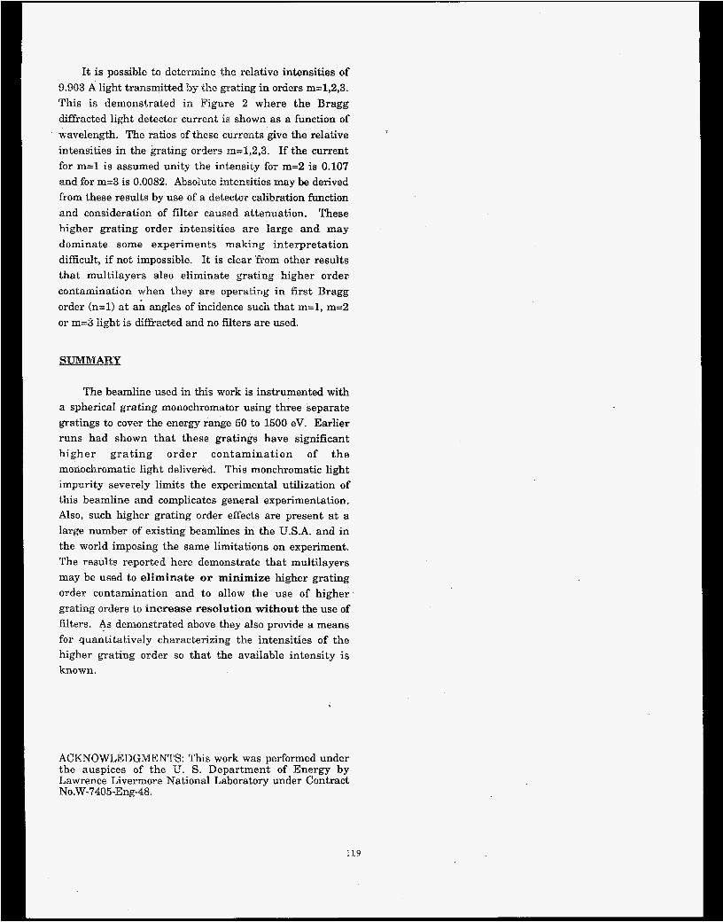
It is possible to determine the relative intensities of 9.903 A light t.ransmitted by the grating in orders m=1,2,3. This is demonstrated in Figure 2 where the Bragg diffracted light detector current is shown as a function of wavelength. The ratios of these currents give the relative intensities in the grating orders m=1,2,3. If the current for m=l is assumed unity the intensity for m=2 is 0.107 and for m=3 is 0.0082. Absolute intensities may be derived from these results by use of a detector calibration function and consideration of filter caused attenuation. These higher grating order intensities are large and may dominate some experiments making interpretation difficult, if not impossible. It is clear 'from other results that multilayers also eliminate grating higher order contamination when they are operating in first Bragg order (n=l) a t ah angles of incidence such that m=l, m=2 or m=3 light is diffracted and no filters are used.
The beamline used in this work is instrumented with a spherical grating monochromator using three separate gratings to cover the energy range 50 t o 1500 eV. Earlier runs had shown that these gratings have significant higher gra t ing order contamination of t he monochromatic light delivered. This monchromatic light impurity severely limits the experimental utilization of this beamline and complicates general experimentation. Also, such higher grating order effects are present a t a large number of existing beamlines in the U.S.A. and in the world imposing the same limitations on experiment. The results reporhed here demonstrate that multilayers may be used to eliminate or minimize higher grating order contamination and t o allow the use of higher grating orders to Increase resolution without the use of filters. As demonstrated above they also provide a means for quantitatively characterizing the intensities of the higher grating order so that the available intensity is known.
ACKNOWLEDGMENTS: This work was performed under the auspices of the U. S. Department of Energy by Lawrence Livermore National Laboratory under Contract N0.W-7405-Eng-48.
119

Proposal No. 9901v
Fluorescent MAFS and NEXAFS in the Soft X-ray and Extreme Ultra-Violet
Troy W. Barbee, Jr. Lawrence Livermore National Laboratory
Livermore, California 94550 The reflectivities of a 304 Stainless Steel-Silicon
multilayer (d = 55& N = 50, y = 0.5 ) were measured over the energy range 300 eV to 1500 eV. The measurements were made in the Bragg-Bretano configuration at fixed angles of incidence , 0 , by scanning the energy of the incident light . A GaAsP-Au Scxhttoky diode was used as a sensitive detector. Though the reflectivities of this multilayer are of interest the important observation in this work was of the fluorescence of the components of the 304ss ( 75% Fe, 18% Cr, &% Ni) during a given reflectivity measurement.
Reflectivities measured are not discussed here as the significant result relates t o observations of L edge fluorescence of the iron and chromium in the 304ss. In these measurements the diffracted beam detector was a GaAsP-Au Scottky diode having a 1 cm2 active area mounted normal t o the diffracted beam 17.8 cm from the illuminated area. This detector sensed the diffracted intensity and any fluorescent radiation generated by the incident beam. A reflectivity scan at 0 = 9.78 deg over the energy range 500 to 800 eV is shown in Figure 1. The first order Bragg peak is at - 605 eV. The structure on the low energy side cf this peak is due excitation of the chrome La1,2 - 572.8 eV and the Lp1 - 582.8 eV lines. These fluorescence peaks are more intense than warranted by the chrome concentration in the 304ss. This results from the antinodes of the standing-wave field in the multilayer characteristic of Bragg reflection at the Bragg peak lying
4.00 .- $2 =) 3.50
- 3.00 I- z 2.50 CT LT 3 2.00 0
1.50
c
2
2 y 1.00
n 0.50
0.00 500 550 600 650 700 750 800
ENERGY (eV)
on the 304ss layers and strongly enhancing the chrome signal as well as multilayer Bragg diffraction of Cr L line radiation into the detector. Fluoresence from the iron is seen at energies of 700 to 800 eV, energies far away from any multilayer Bragg reflection.
The peaks at - 700 eV are shown in more detail in Figure 2 and are from the characteristic L fluorescence lines (La1,2 - 705 eV, Lp1- 718.5 eV) of the iron in the 304ss layers. There is no strong Bragg diffraction from the multilayer structure a t these values of energy and 8: these signals a r e non standing-wave enhanced fluorescence of the iron. The importance of these observations is that the solid angle of the fluorescent source subtended by the detector is very small. This solid angle may be easily increased with this type of solid state detector by increasing the active detector area and moving the detector much closer to the sample. A simple design allows this solid angle to be increased from - 0.003 steradian to > 1.0 steradian. This significantly increases the sensitivity of the experiment to the fluorescence of the sample and makes possible a number of scientifically and technologically significant experiments. The experiments facilitated by such a simple apparatus include: (1) fluorescence EXAFS in the soft x-ray and extreme ultra- violet (30 to 3000 eV) spectral ranges; (2) standing-wave fluorescence studies of multilayer structures in the SXR and EUV; (3) application. of standing-waves in multilayers to the study of adsorbed layers on multilayer structures and other long period Bragg diffractors in the SXR, (4) sensitive x-ray fluorescence analysis (XRF) for the concentration of light elements in bulk samples; (5) detailed study of the physics of x-ray absorptiodemission in the SXR by experimental determination of radiative and non-radiative decay channel strengths.
Figure 1 Reflectivity of a 304ss/Si (d=55 A, N=50, y=0.35) multilayer over the energy range 500 to 800 eV at 0=9.78 deg.
120

Fe Fluoresence : L 1 ines 0.25 . . ~ . l . . . . , . . . . , . . . . , . ~ . . I . . . , , . . . . I . . , . l . . . . , . . . .
Ga,%sP-& Schottky Diode Detector I 2 0.20 Y 2 I- % 0.15 U U 3 0
0.10 e z 0.05
0
0.00
680 700 720 740 760 780 ENERGY (eV)
Figure 2 Characteristic L line fluoresence from iron in the 304ss of the multilayer detected using a Bcm2 active area GalBsP-Au Schottky diode - 18 cm from the fluorescing material and subtending only - 0.001 str.' of the solid angle available.
10 mm x 10 m solid Arge: 0.001 Str
ACKNOWLEDGMENTS: This work was performed under the auspices of the U. S. Department of Energy by Lawrence Livermore National Laboratory under Contract N0.W-7405-Eng-48.
121
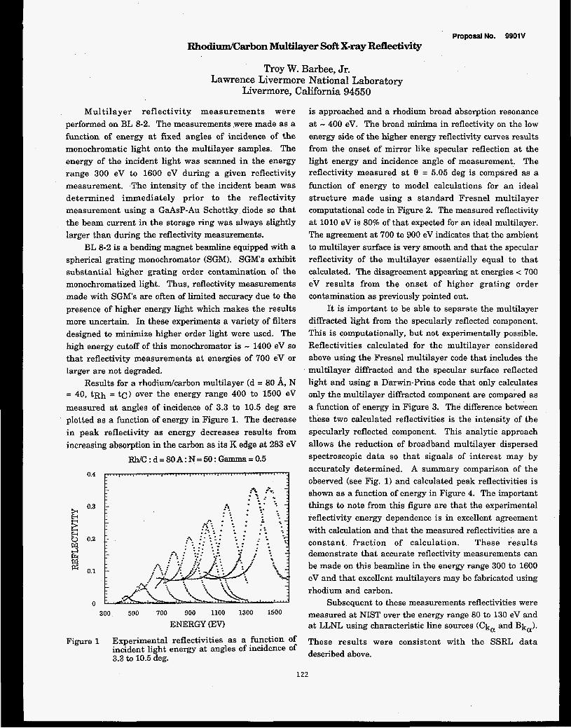
Proposal No. 9901V Rhodium/Carbon Multilayer Soft X-ray Renectivity
Troy W. Barbee, Jr. Lawrence Livermore National Laboratory
Livermore, California 94550
Multilayer reflectivity measurements were performed on BL 8-2. The measurements .were made as a function of energy a t fixed angles of incidence of the monochromatic light onto the multilayer samples. The energy of the incident light was scanned in the energy range 300 eV to 1600 eV during a given reflectivity measurement. The intensity of the incident beam was determined immediately prior to the reflectivity measurement using a GaAsP-Au Schottky diode so that the beam current in the storage ring was always slightly larger than during the reflectivity measurements.
BL 8-2 is a bending magnet beamline equipped with a spherical grating monochromator (SGM). SGMs exhibit substantial higher grating order contamination of the monochromatized light. Thus, reflectivity measurements made with SGM's are often of limited accuracy due to the presence of higher energy light which makes the results more uncertain. In these experiments a variety of filters designed to minimize higher order light were used. The high energy cutoff of this monochromator is - 1400 eV so that reflectivity measurements a t energies of 700 eV or larger are not degraded.
Results for a rhodiudcarbon multilayer (d = 80 A, N = 40, tRh = tc) over the energy range 400 t o 1500 ev measured at angles of incidence of 3.3 to 10.5 deg are plotted as a function of energy in Figure 1. The decrease in peak reflectivity as energy decreases results from increasing absorption in the carbon as its K edge at 283 eV
W C : d = 80 A : N = 50 : Gamma = 0.5 0.4
0.3 E E 3 2 0.1
E-c u 0.2
ki
0 300 500 700 900 1100 1300 1500
ENERGY (EV)
Figure 1 Experimental reflectivities as a function of incident light energy at angles of incidence of 3.3 to 10.5 deg.
is approached and a rhodium broad absorption resonance at - 400 eV. The broad minima in reflectivity on the low energy side of the higher energy reflectivity curves results from the onset of mirror like specular reflection at the light energy and incidence angle of measurement. The reflectivity measured a t 9 = 5.05 deg is compared as a function of energy t o model calculations for an ideal structure made using a standard Fresnel multilayer computational code in Figure 2. The measured reflectivity at 1010 eV is 80% of that expected for an ideal multilayer. The agreement at 700 t o 900 eV indicates that the ambient t o multilayer surface is very smooth and that the specular reflectivity of the multilayer essentially equal t o that calculated. The disagreement appearing at energies < 700 eV results from the onset of higher grating order contamination as previously pointed out.
It is important to be able t o separate the multilayer diffracted light from the specularly reflected component. This is computationally, but not experimentally possible. Reflectivities calculated for the multilayer considered above using the Fresnel multilayer code that includes the multilayer diffracted and the specular surface reflected light and using a Darwin-Prins code that only calculates only the multilayer diffracted component are compared as a function of energy in Figure 3. The difference between these two calculated reflectivities is the intensity of the specularly reflected component. This analytic approach allows the reduction of broadband multilayer dispersed spectroscopic data so that signals of interest may by accurately determined. A summary comparison of the observed (see Rg. 1) and calculated peak reflectivities is shown as a function of energy in Figure 4. The important things to note from this figure are that the experimental reflectivity energy dependence is in excellent agreement with calculation and that the measured reflectivities are a constant fraction of calculation. These results demonstrate that accurate reflectivity measurements can be made on this beamline in the energy range 300 t o 1600 eV and that excellent multilayers may be fabricated using rhodium and carbon.
Subsequent to these measurements reflectivities were measured at NIST over the energy range 80 to 130 eV and at LLNL using characteristic h e sources (ck, and Bk,).
These results were consistent with the SSRL data described above.
122

I
RNC :d = 79.6A: N =40 :Th = 5.05 deg
300 500 700 900 1100 1300
0 MpEFilMarr ENERGY (EV)
Experimental reflectivity at e = 5.05 deq is
range 600 to 1300 eV.
a CALCULATION Figure 2
compared to calculation over the energy 0 0 0
400 800 1200 1600 0
ENERGY (eV)
Figure 4 Experimental and calculated (Fresnel) peak reflectivities are compared as a function of energy. W/C: D = Sorb : N = 40 : GOtTWlO = 0.5 : Th = 5.0 deg
0.30
0.25
;r: 0.20 2 -
0.15 J LL Lu E 0.10
0.05
700 800 900 1000 1100 1200 ENERGY (em
Figure 3 Reflectivities calculated using both the Fresnel and the Darwin-Prins formalisms are compared.
ACKNOWLEDGMENTS: This work was performed under the auspices of the U. S. Department of Energy by Lawrence Livermore National Laboratory under Contract N0.W-7405-Eng-48.
123

Proposal NO. ggoiv L SHELL XANES FOR SOLID METALS: Ti, V. Cr, Fe, Ni. Cu
N.K. Del Grande
Universitv of California, Lawrence Livermore National Laboratory P.O. Box 808 (L-379) Livermore. CA 94550
Introduction The L shell x-ray absorption near edge structure (XANES) for the 3d transition metals Ca-Cu is dominated by atomic- like behavior which is poorly understood.* It has been studied by electron energy loss spectroscopy (EELS) and bremsstrahlung isochromat spectra (BIS).2,3 More and better photoabsorption cross-section measuremer.ts are needed over the sparsely studied 400-1500 eV region to test theoretical methodologies which fuse atomic and condensed matter theories. Progress has been made reporting absolute photoabsorption cross sections taken with the spherical grating monochromator on beamline 8- 2 at SSRL in 1989. These data were used in conjunction with data taken on beamline 3-4 at SSRL and beamline U14A at NSLS.4-7
Exueriment Transmission data for well characterized carbon-only and carbon-metal multilayer targets (100-4000 nm thick) were recorded with a resolution of 1 eV or better for Ti, V, Cr, Fe, Ni and Cu. Absolute cross sections in Figure 1 had overall uncertainties of 10% or less. Comparisons are made with previous result^.^,^ Away from edges, targets had photon penetration depths 1/3 to 3 mean free paths thick. Thinner targets (by a factor of 3 or 4) were used to resolve XANES shown in Figure 2.
- BL - 8 SSRL -+-e- BL - U14 NSL
60
-A-& Plechaty et at, Autoioniztng 1981 9 40
E z
20
J 500 750
Cr (24) - BL - 8 SSRL -+-Om BL - U14 NSL --.b -6- Plechaty et ai.,
Autoionlzing 1981 resonances
' 500 750 1000 Photon energy in eV
Corrections for oxide impurities were possible since synchrotron radiation sources allowed continuous coverage across the oxygen K absorption edge at 543 eV. The determination of the target metallic areal densities (mass per unit area) was known to better than +_ 10%. Tergets were characterized for their total as well as component element purity, weight-per-unit area, thickness and composition, using very accurate balances, ion back-scattering, and particle-induced x-ray emission.
800 1000 1200 15
10 a --
"E, s 5
1500 0
1000 Photon energy In eV
Figure 1. Photoabsorption cross section measurements in cm2/g with 10% overall uncertainties are compared with the 1981 LLNL sub-keV theoretical data base8 and other data9 for five 3d solid metals: Ti, Cr, Fe, Ni, Cu. See reference [7].
1 24

Discussion The Figure 1 comparisons of experiment with single particle Hartree Slater (HS) theory8 shown as a long dashed line agree for most 3d metals below and at twice the energy of the ionization threshold. The 2p-3d autoionizing resonances of Figure 2 are associated with bound-bound transitions which lead to non-radiative Auger decay. What is measured is a combination of the bound-bound photoabsorption and bound-free photoionization cross sections. The HS calculation [in 81 does not include the bound-bound component for the atom or band theory to address the behavior of the solid.
There is good qualitative agreement between L shell XANES and calculations which use a multi-configuration Dirac Fock (MCDF) model with the assumption of a statistical distribution for the initial atomic states.
NI (28) I I
I 1 I I I
Cu (29) 0 I
0 20 40 Energy above L3 edge (eV)
Figure 2. Measured XANES for the transition metals Ti, V, Cr, Fe, Ni and Cu showing the atomic-like 2p-3d autoionizing resonances which dominate the L3 and L2 edge structure.7
Calculations were performed in intermediate coupling with configuration interactions.7 It is not yet clear why the statistical distribution rather than the ground state distribution for the initial states produces the measured XANES for solid metals shown in Figure 3.
Ti (22) Exper.
5
y1
J 852*7
N' (28) calc.
distrib.) (stat.
distrib.)
0 20 0
Energy above edge (ev)
Figure 3. A comparison of measurement with calculation for L shell autoionizing resonances of solid metals dominated by atomic-like behavior. (Calculation by Mau H. Chen.)
REFERENCES 1. Davis, L. C. (1986), J. Appl. Phys. 59- R25-R63. 2. Fink, J., Muller-Heinzerling, M. Th., Scheerer, B.,
Speier, W., Hillebrecht, F.U., Fuggle, J.C., Zaanen, J. and Sawatzky, G.A., (1985), Phys. Rev. B., 32 pp. 4899- 4904.
3. Zaanen, J., Sawatzky, G.A., Fink, J., Speier, W., Fuggle, J.G, (1985), Phys. &V. B., a pp. 4905-4913.
4. Del Grande, N.K., Tirsell, K.G., Schneider, M.B., Garrett, R.F., Kneedler, E.M., and Manson, S.T. (1987), J.
5. Del Grande, N.K., and Tirsell, K.G. (19881, SPIE Vol. 911 X-ray and VUV Interacfion Data Bases, Calculations, and Measurements, pp. 6-10.
6. Tirsell, K.G., and Del Grande, N.K. (19881, SPIE Vol911, X-ray and VUV Interaction Data Bases, Calculations, and Measurements, pp. 146-156.
7. Del Grande, Nancy Kerr (1990), Physica Scripta, in press.
8. Plechaty, E., Cullen, D. and Howerton, R., (1981), Lawrence Livermore Laboratory Report, UCRL-50400, Rev. 3.
9. Del Grande, Nancy Kerr (19861, SPIE Vol. 691, X-ray Imaging I I , pp. 2-10.
Phys. (Paris) 4& C9, pp.951-954.
ACKNOWLEDGEMENTS The author is indebted to K. G. Tirsell for detector improvements, to Mau H. Chen for calculations, to T. Barbee for beamline 3-4 SSRL measurements, and to P. Pianetta and his staff at SSRL for their support. This work was performed under the auspices of the U.S. Department of Energy by LLNL under contract number . W-7405-ENG-48.
125

Theses Based on Research at S


VI. THESES BASED ON RESEARCH AT SSRL
The following is a partial list of Ph.D and Master’s theses based on experimental research at SSRL. It repre- sents 160 theses from 22 universities.
TITLE/AD visoq - 1990
C.J. Chisholm-Brause Stanford University
W.E. Jackson Stanford University
- 1989
M. Ardehali Stanford University
R. Cao Stanford University
T. Teh-I-Chiang Stanford University
M.P. Green Stanford University
Z.-X. Shen Stanford University
J. C. Woicik Stanford University
J. Hwang Stanford University
J.E. Benci Univ. of Pennsylvania
K.D. Butcher Stanford University
M. Kraitchman UC Berkeley
R. Howland Stanford University
Y.H. Chung Georgia Inst. of Tech.
“Sorption Mechanisms of Co(l1) at Oxidewater Interfaces” (Advisor - G.E. Brown)
“X-Ray Absorption Analyses of Iron-Bearing Glasses and Melts: Implications for Glass- Melt Structural Differences and the Effect of Alkali and Alkaline-Earth Metals on Fe(1l) Coordination” (Advisor - G.E. Brown)
“Photoelectron Angular Distribution from the Disordered Condensed Phase: Theory and Experiment” (Advisor - 1. Lindau)
“Schottky Barrier Formation with Metals on Ill-V Semiconductor Surfaces From Thin Coverages to Thick Overlayers” (Advisor - 1. Lindau)
“Annealing Studies of Metals and Arsenic on Gallium Arsenide” (Advisor - W.E. Spicer)
“Scanning Tunneling Microscopy Applied to the Electrochemical Deposition of Lead on the Gold (1 11) Surface” (Advisor - 1. Lindau)
“Photoemission Studies of High Temperature Superconductors and Related Materials” (Advisor - 1. Lindau)
“Synchrotron Radiation Studies of Si and Si-Ge Interfaces” (Advisor - P. Pianetta)
“Effect of Strain on the Electronic Structure of Indium Gallium Arsenide” (Advisor - P. Pianetta)
“A Study of Creep Damage Using Microradiography and Continium Damage Mechanics” (Advisor - D. Pope)
“Variable Energy Photoelectron Spectroscopy on Model Catalytic Active Sites: The Electronic Structure and Bonding in Tetrahederal Iron Chlorides and Sulfides” (Advisor - E.I. Solomon)
“The Unmixing of AI,Li in AI-Li and AI-L1-CU Alloys” (Master’s Thesis - Advisor - D. DeFontaine)
“Determination of Dopant Site Occupancies in Copper-Substituted Yttirium-Barium- Copper-Oxide by a New Application of Differential Anomalous X-ray Scattering” (Advisor - T.H. Geballe)
“Synchrotron White Beam Topographic Study of Damage Accompanying Laser Drilling” (Advisor - S. Stock)
Paae 126 SSRL ACTIVITY REPORT 1989
A. Outrzourhit Colorado school Of Mh8S
P.R.G. Small Colorado School of Mines
L. Emery Stanford University
S.V. Didziulis Stanford University
G.P. Carey Stanford University
P.H. Mahowaid Stanford University
C.E. McCants Stanford University
C.-K. Shih Stanford University
L. Hsu UCLos Angeles
W.M. Lavender Stanford University
J.R. Schoonover Arizona State University
L. Terminello UC-BerkeIey
C.J. Chishoim Skanford University
R. T. Short uniV8Bi&‘ Of T8nn8SSee
S.R. Hubbard Stanford University
L A . Kau Stanford University
“Extended X-ray Absorption Fine Structure Studies of Sn-related DX Centers in Sn- Doped Ga,,,AI,,,As” (Master’s Thesis -Advisor - T.M. Hayes)
’The Atomic-Scaie Structure of Dilute Tin in Aluminum” ( Master’s Thesis - Advisor -T.M. Hayes)
“A Wiggler Based Ultra-Low Emittance Damping Rlng and its Chromatic Correction” (Advisor - H. Wiedemann)
“Photemisslon Spectroscopic Studies of Model Catalyst Surfaces: 1) Bonding Interactions of 3d,, Metal Ion Sites; 2) The Chemistry of Copper Overlayers on Zinc Oxide Single Crystals” (Advisor - E.I. Solomon)
”A Study of the Chemical and Electrical Disruption to Mercury Cadmium Telluride Upon Interface Formation and Surface Processing” (Advisor - W.E. Spicer)
”Experimental Band Alignment at Germanium, Silicon and lndiun Phosphide Heterojunctions” (Advisor - W.E. Spicer)
”Photoemission and Electrical Studies of the Titanium/Gallium Arsenide (110) and PiatinumlGallium Arsenide (1101 Interfaces” (Advisor - W.E. Spicer)
”Heterojunction Band Line-Ups and the Alloying Effect in Compound Semiconductor Alloys - A Photoemission Study” (Advisor - W.E. Spicer)
“Electronic Properties of a PtGa, Single Crystal and Photoemission Studies of the Electron Correlation Effects in the NI-Ga and the Ni-in Intermetallic Compounds” (Advisor - R.S. Williams)
“Observation and Analysis of X-ray Undulator Radiation from PEP (Advisor - G.S. Brown)
‘Time Resolved X-ray Diffraction Using Synchrotron Radiation” (Advisor - S.H. Lin)
“Surface Structures Determined Using Electron Diffraction” (Advisor - D.A. Shirley)
“Structure of Divalent Lead Complexes at the Gamma-AiuminaWater Interface” (Master‘s Thesis - Advisor - G.E. Brown)
”Recoil-Energy Measurements of Slow Highiy-Charged Ions Produced by Synchrotron Radiation and by Swift-Ion Impact” (Advisor - I.A. Sellin)
“Small-Angle X-ray Scattering Studies of Caiclum-Binding Proteins In Solutions” (Advisor - K.O. Hodgson)
”Chemical and Spectroscoplc Studies on Metal Active Sites in Metaiioprotelns and Heterogeneous Catalysts” (Advisor-K.O. Hodgson)
SSRL ACTIVITY REPORT 1989 Page 127

J. t. Cole U C-Berkeley
A.E. McDermoft U C-Berkeley
R.E. Guiles UC-Berkeley
J.J. Yeh Stanford University
D. Friedman Stanford University
A. Viescas Stanford University
P. Jupiter Stanford University
K.A. Bertness Stanford University
J.N. Otis Stanford University
Y.C. Lu Stanford University
C.W. Ponader Stanford University
K.F. Hayes Stanford University
S.W. Robey UC-Berkeley
J. Nogami Stanford University
E.B. Sirota Harvard University
A.L. Johnson UC- Berkeley
K.F. Ludwig, Jr. Stanford University
R.D. Lorentz Stanford University
“Photosynthetic Oxygen Evolution Studied by Electron Paramagnetic Resonances and X-Ray Absorption Spectroscopy” (Advisor - M.P. Klein)
“Structural Studies of Iron and Manganese In Photosynthetic Reaction Centers” (Advisor - M.P. Klein)
“X-ray Absorption with EPR Studies of the Role of Mn in the Photosynthetic Oxygen Evolving System” (Advisor - M.P. Klein)
“Metal/Silicon Interfaces and Their Oxidation Behavior - A Photoemission Spectroscopy Analysis” (Advisor - 1. Lindau)
“Metal Interfaces with Mecury Cadmium Telluride” (Advisor - 1. Lindau)
“Photoemission Studies of Core Level Energy Shifts In Clean and Adsorbate Covered Tungsten and Platinum Single Crystal Surfaces” (Advisor - I. Lindau)
‘Correlatlon Between the Electronic Structure and Surface Chemistry of Tungsten and Tungsten Carbide 4” (Advisor - W.E. Spicer)
“Photoemission Studies of Oxgyen Chemisorption on G8lliUm Arsenide and Indium Phosphide Surfaces” (Advisor - W.E. Spicer)
“Minimally Invasive Coronary Angiography with Monochromatic X-rays: Developmental Studies Utilizing Synchrotron Radiation” (Advisor - R. Hofstadter)
“Vertical Brldgman Growth and Characterization of CdTe Crystals” (Advisor - R. Feigelson)
“Trace Metals in Silicate Glasses and Melts: Coordination Environments, Halogen Complexes, and Soret Diffusion” (Advisor - G.E. Brown)
“Equilibrium, Spectroscopic, and Kinetic Studies of Ion Adsorption at the OxidelAqueous Interface” (Advisor - G.E. Brown)
“Local Surface Structures of c(2x2)S/Ni(011) and (2x2) S/Ge(l11) Determined Using ARPES (Advisor - D.A. Shirley)
“Rare Earth Metai/Semiconductor Interfaces and Compounds” (Advisor - I. Lindau)
“X-ray Scattering Study of the Thickness Dependent Phase Diagram of Liquid Crystal Films” (Advisor - P.S. Pershan)
“Platinum Surface Chemistry Studied by Thermal DesorptlonSpectroscopy and Near Edge X-ray Absorption Fine Structure Absorbate Effects” (Advisor - D.A. Shirley)
“Anomalous X-ray Scattering Studies of Liquids” (Advisor - A.I. Bienenstock)
“Structural Investigations of Amorphous Iron-Germanium Alloys using Synchrotron Radiation” (Advisor - A. 1. Bienenstock)
Page 128 SSRL ACTIVITY REPORT 1989

J.J. Hoyt UC-Berkeley
J.C. Levin University of Oregon
N.W. Lytle Brigham Young Univ.
C.C. Bahr UC- Berkeley
T.A. Ferrett UC- Berkeley
P.A. Heimann UC- Berkeley
M.G. Sarnant Stanford University
D.J. Sajkowski Stanford univ8rsity
S.L. Cohen Stanford University
E.D. Specht MIT
J.A. Silberman Stanford University
K.K. Chin Stanford University
M.D. Williams Stanford University
W.E. Jackson Stanford University
K. Kim Univ. of Washington
R. Pro UC- Berkeley
W.G. Petro Stanford University
E.N. Keller Univ. of Washington
“An Anomalous Small Angle X-ray Scattering Study of Phase Separation in the AlZnAg System” (Advisor - D. DeFontaine)
“Auger Transitions in Noble Gases Following Inner Shell Ionization by Electron Bombardment and by Synchrotron Radiation’’ (Advisor - B. Crasemann)
“Proton-Induced X-ray Emission, Proton-Induced Gamma-ray Emission, and X-ray Absorption” (Advisor - N.F. Mangeison)
“Chemisorption Geometries of Sulfur on Copper and Molybdenum Surfaces” (Advisor - D.A. Shirley)
“Photoionzation of Atoms and Small Molecules using Synchrotron Radiation” (Advisor - D.A. Shirley)
“Near Threshold Studies of Photoelectron Satellites” (Advisor - D.A. Shirley)
“Atomic and Electronic Structure of Supported Monometallic and Bimetallic Clusters” (Advisor - M. Boudart) .
“Catalysis by Metals: Single Crystals, Supported or Unsupported Polycrystals, and Coordination Complexes” (Advisor - M Boudart)
“Variable Energy Photoelectron Spectroscopic Studies of d9 and d10 Metal Chloride and Oxide Surfaces” (Advisor - E. 1. Solomon)
“Synchrotron X-ray DiffracUon Studies of Transitional and Orientational Order in Monolayer Krypton on Graphite” (Advisor R.J. Birgeneau)
“A Photoemlssion Study of the Electronic Structure and Oxidation Properties of Mercury Cadmium Telluride” (Advisors - 1. LindauMI.E. Spicer)
“Initial Stage of the Schottky Barrier Formatlon of Metalfill-V Semiconductor Interfaces” (Advisor - 1. Lindau)
“The Effect of Chemical Reactivity and Charge Transfer on Gallium Arsenide (110) Schottky Barrier Formation” (Advisors - W.E. SpicerA. Lindau)
“EXAFS Study of the Potassium Coordination Environment in Glasses from the NaAISI,O,-KAISi,O, Binary: Structural Implications for the Mixed-Alkali Effect” (Master’s Thesis - Advisor - G.E. Brown)
“A Study of Basic Properties and Applications of EXAFS” (Advisor - E.A. Stern)
“Development of of Partial Structure Function in AI, Ag and Zn Alloys” (Advisor - D. DeFontaine)
“Metal and Oxygen Interactions with Ill-V Compound Semiconductor Surfaces”
“Magnetic X-ray Study of a Gadolinium-Iron Amorphous Alloy” (Advisor - E. A. Stern)
ar Gas-Proportional X-ray Detector System for Determination
(Advisors- W.E. Spicer and 1. Lindau)

G. Zhang Stanford University
T.A. Smith Stanford University
LE. Klebanoff UC-Serkeley
J.G. Nelson UC-Los Angeles
J.J. Barton UC- Berkeley
T.A. Gibson Stanford University
M.A. Berding Stanford University
D.L. Parkhurst Stanford University
D.A. McKeown Stanford University
C.E. Bouldin University of Washington
J.S. Lee Stanford University
M.M. Choudhary Stevens Institute
S.G. Mochrie MIT
J.E. Penner-Hahn Stanford University
M.K. Eidsness University of Cincinnati
J.B. Kortright Stanford University
R.S. Moog Stanford University
J.A. Guest Stanford University
“Molybdenum Carbide Catalysts” (partially based on SSRL) (Advisor - M. Boudart)
“Single Crystal Polarization X-ray Absorption Studies of Copper Complexes” (Advisor - K.O. Hodgson)
“PhOtOeleCtrQn Spectroscopy Studies of Cr(OO1) Surface Ferromagnetism and NearSurface Antiferromagnetism” (Advisor - D,A. Shirley)
“The Electronic Structure of Gold Gaiiium Two and Gold indium Two Intermetallic Compounds” (Advisor - R.S. Williams)
“Angle-Resolved Photoemission Extended Fine Structure” (Advisor - D.A. Shirley)
“Single Crystal Polarized X-ray Absorption Edge Studies of Copper Complexes” (Advisor - K.O. Hodgson)
“Calculation of Cu K-Edge Photoabsorption Spectra Using an Extended Continuum Multiple Scattered Wave Method” (Advisors- K.O. HodgsonS. Doniach)
“The Application of EXAFS and Mean-Activity Coefficient Data to lon-Association Models for the Zinc-Chloride System” (Master’s Thesis - Advisor - G.E. Brown)
“Spectroscopic Study of Silica-Rlch Glasses and Selected Minerals withln the Sodium Alumino-silicate System” (Advisor - G.E. Brown)
“An EXAF§ Study of Amorphous Ge and of the Fe Impurity in Sillcon Nitride” (Advisor - E.A. Stern)
“Catalysis of the Oxidation of Dihydrogen by Supported Gold” (partially based on at SSRL) (Advisor - M. Boudart)
“Surface Sensitive Photoemission EXAFS: A New’Structural Tool” (Advisor - G.M. Rothberg)
“Structures and Transitions in Systems with Competing Interactions” (Advisor - R.J. Birgeneau)
“X-Ray Absorption Studies of Metalloprotein Structure: Cytochrome P-450, Horseradish Peroxidase, Plastocyanln, and Laccase” (Advisor - K.O. Hodgson)
“EXAFS and XANES Studies of Goid in Complexes of Biological Significance” (Advisor - R.C. Eider)
“Structural Studies of Amorphous Mo-Ge Alloys Using Synchrotron Radiation” (Advisor - A. Bienenstock)
“Fluorescence Quenching of Chlorophyllide and Protoporphyrin-Protein Complexes by Electron Transfer” (Advisor - S.G. Boxer)
“Studies of Molecular Photoion and Photofragment Alignment” (Advisor - W.N. Zare)
Page 130 SSRL ACTIVITY REPORT 1989

B.B. Pate Stanford University
R.S. Weber Stanford University
G.D. Meitzner Stanford University
G.B. Bunker University of Washington
H. B. Ponader Stanford University
1983
J. Sanchez-Arrieta Stanford University
P.H. Kobrin UC-Berkeley
J. Collett Harvard University
D.W. Lindle UC- Berkeley
P.M. Stefan Stanford University
A.N. Mansour N. Carolina State
J.G. Tobin UC-Berkeley
C. Marques Washington State Univ.
P.R. Findley UC-Santa Barbara
M.R. Antonio Michigan State
G. B. Stephenson Stanford University
J.M. Tranquada Univ. of Washington
“The Diamond Surface: Atomic and Electronic Structure” (Advisors - W.E. SpicerA. Lindau)
“Structure and Reactivity of Platinum Clusters Supported on Y-Type Zeolites” (Advisor - M. Boudart)
“Characterization of GoldN-Zeolite and Its Catalytic Activity In the H2-02 Reaction” (Advisor - M. Boudart)
“An X-Ray Absorption Study of Transition Metal Oxides” (Advisor - E.A. Stern)
“An EXAFS and X-Ray Diffraction Study of the Local Ca Environment in Anorthite (CaAI,Si,O,) Crystal and Glass” (Master‘s Thesis - Advisor - G.E. Brown)
“Structural Studies of Hydrodesulfurization Catalysts” (Advisor - M. Boudart)
”Atomic Photoelectron Spectroscopy Studies using Synchrotron Radiation” (Advisor - D.A. Shirley)
“X-Ray Scattering Study of Liquld Crystal Thin Films” (Advisor - P.S. Pershan)
“inner-Shell Photoemission from Atoms and Molecules using Synchrotron Radiation” (Advisor - D.A. Shirley)
“A Photoemission Study of the Electronic Structure and Surface Chemical Properties of Tungsten Carbide and of a Carbonized Tungsten Model Surface for Tungsten Carbide” (Advisor - W.E. Spicer)
“X-Ray Absorption Studies of Silica-Supported Platinum Catalysts” (Advisor - D.E. Sayers)
“Dimensionality and Its Effects Upon the Valence Electronic Structure of Ordered Metallic Systems” (Advisor - D.A. Shirley)
“Thermal Motion of Surface Atoms in Silica Supported Platinum and iridlum Catalyst Clusters: An EXAFS Spectroscopy Study” (Advisor - D.R. Sandstrom)
“Optical Properties of PbF, and PbC,, at High Temperatures” (Advisor - W.C. Walker)
“Extended X-Ray Absorption Fine Structure of the Iron-Molybdenum Cofactor of Nitrogenase, the Three-Iron Ferredoxlnli of Desulfovibrio Gigas, and of Synthetic Molybdenum and Tungsten-iron-Sulfur and Iron-Sulfur Model Systems (Part I) and Intercalation of Tetrathiafulvalene into Iron Oxycholoride (Part 11)’’ (Advisor - H. Eick)
“Early-Stage Phase Separation In Amorphous Solids: A Time-Resolved SAXS Study” (Advisor - A.I. Bienenstock)
“X-Ray Absorption Studies of Solids at High Pressure” (Advisor - R. ingalls)
SSRL ACTIVITY REPORT 1989 Paae 131

D. Stearns Stanford University
“Early Photo Luminescence Decay in Amorphous Hydrogenated Silicon” (Advisor - A.I. Bienenstock)
S. Laderman Stanford University
M.S. Co Stanford University
S.D. Conradson Stanford University
R. Miake-Lye Stanford University
C.C. Parks UC-Berkeley
D.B. Goodin U C-Berkeley
- 1982
M.G. Durrett Rice University
M.L. Shek Stanford University
P.R. Skeath Stanford University
M.H. Hecht Stanford University
S.-J. Oh Stanford University
S.M. Brennan Stanford University
R.O. Tatchyn Stanford University
P.A. Heiney MIT
D.H. Rosenblatt UC- Berkeley
J.E. Pollard UC-Berkeley
S.H. Southworth UC- Berkeley
“Structural Studies of Amorphous Semiconductors Containing Transition Metals” (Advisor A.I. Bienenstock)
“X-Ray Absorption Spectroscopy of Hemocyanin and Hemerythrin” (Advisor - K.O. Hodgson)
“X-Ray Absorption Studies of the Molybdenum Site of Nigrogenase” (Advisor - K.O. Hodgson)
“Anomalous X-Ray Scattering as 8 Probe of Biological Structure” (Advisor - K.O. Hodgson)
“The Auger Decay Mechanism in Photon Stimulated Desorption of Ions from Surface” (Advisor - D.A. Shirely)
“The Local Structure of Manganese in the Photosynthetic Apparatus and Superoxide Dismutase: An X-Ray Absorption Study” (Advisor - M.P Klein)
“Study of KrC,/Dr,CI Excited State Kinetics Using Synchrotron Radiation Excitation” (Advisor - G.K. Waiters)
“Electronic Structure of Platinum-Copper Surfaces and Chemisorption of Carbon Monoxide” (Advisors - 1. Lindau/W.E. Spicer)
“Chemical Bonding Phenomena of Column 3 and 5 Adsorbates and Fermi Energy Stabilizatlon Mechanism on GaAs (110)” (Advisors - 1. Lindau/W.E. Spicer)
“Synchrotron Radiation Measurements of Subshell Photoionization Cross Sections: Techniques and Applications” (Advisor - 1. Lindau)
“Resonance and Relaxation Effects In Photoemission Spectroscopy” (Advisor - 1. Lindau)
“Surface EXAFS of Sulphur on Nickel“ (Advisor - A.I. Bienenstock)
“The Introduction and Development of a New Method for Measuring the Optical Constants of Metals in the Soft X-Ray Range” (Advisor - 1. Lindau)
“Phase Transitions of 2D Atomlc and Molecular Crystals” (Advisor - R.J. Birgeneau)
“High Resolution Electron Energy Loss Spectroscopy and Photoelectron Diffraction Studies of the Geometric Structure of Adsorbates on Single Crystal Metal Surfaces” (Advisor - D.A. Shirley)
“Photoelectron Spectroscopy of Supersonic Molecular Beams” (Advisor - D.A. Shirley)
“Atomic and Molecular Photoelectron and Auger Electron Spectroscopy Studies Using Synchrotron Radiation” (Advisor - D.A. Shirley)
Paae 132 SSRL ACTIVITY REPORT 1989

K. Jackson Stanford University
V. Yachandra Princeton University
J.M. Fine Washington State Univ.
c.-Y. s u Stanford University
D.L. Weissrnan Stanford Univesify
R.F. Davis UC- Berkeley
F. Kutzler Stanford University
B. Bunker Univ. of Washington
J.M. Lornston Univ. of Wela ware
P.H. Fuoss Stanford University
D.J. Trevor UG-Serkeley
S.D. Kevan UC-Berkeley
R.V. Taylor UC-Santa Barbara
P. Frank Stanford university
- 1979
M. Breining Univ. of Oregon
F.A. de Parente Univ. of Oregon
“Photoion Fluorescence Polarization: A Study of Molecular Ionization Dynamics” (Advisor - R.N. Zare)
“EXAFS Studies of Carbonic Anhydrase and Resonance Raman Spectroscopy of Iron- Sulfur Proteins” (Advisor - T. Spiro)
“Structure and Stoichiometry in Highly Concentrated Aqueous Solutlons of CuBr,” (Advisor - D.R. Sandstrorn)
“Photoemission Studies of the Interaction of Oxygen with Solid Surfaces” (Advisor - 1. Lindau)
“Oxygen Chemisorption and Water Synthesis on Palladium and Palladium-Gold; A Study by Thermal Desorption and Photoemission” (Advisor - W.E. Spicer)
“Angular Distribution and Atomic Effects In Condensed Phase Photoelectron Spectroscopy” (Advisor - D.A. Shirley)
“Electronic Structure Calculations of Germanium, Molybdenum Chromium and El,. ~ r n Compounds Predicted K and L Edge Absorption Spectra” (Advisor - K.O. Hodgson)
“An X-Ray Absorption Study of Tetrahedral Semiconductors” (Advisor - E. Stern)
“Characterization of Pt/A,,O, Catalysts by Extended X-Ray Absorption Fine Structure” (Advisors - J.R. Kat2erN.M. Schultz)
“Structural Studies of Amorphous Materials Using X-Ray Anomalous Scattering” (Advisor - A.I. Bienenstock)
“Photoelectron Photoion Molecular Beam Spectroscopy” (Advisor - D.A. Shirley)
“Normal Emission Photoelectron Diffraction: A New Technique for Determining Surface Structure” (Advisor - D.A. Shirley)
“Matrix Spectroscopy of Rare-Gas Oxides and Sulfides” (Advisor - W.C. Walker)
“The Biochemistry of Transition Metals: Copper Vanadium, and Iron” (Advisor - K.O. Hodgson)
“Precision Determining of Atomic Level Energies by Absorption Spectrometry with Synchrotron Radiation” (Advisor - B. Crasernann)
“Measurement of the Inelastic Scattering Cross Section of Neon with Synchrotron Radiation” (Advisor - B. Crasemann)
SSRL ACTIVITY REPORT 1989 Page 133

T.D. Bonifield Rice University
“Time Resolved Spectroscopy of Krypton and Xenon Molecules Excited by Electron Impact and by Monochromatic Synchrotron Radiation” (Advisor - G.K. Walters)
J.N. Miller Stanford University
“Photoemission Studies of Adsorption and Oxidation of Selected Metals and Semiconductors” (Advisor - 1. Lindau)
D.T. Ling Stanford University
“Angle-Integrated and Angle-Resolved Photoemission Studies of Chemisorption on Copper and CopperlNickel” (Advisors - 1. LindauNII.E. Spicer)
K.A. Mills UC-Berkeley
“The Determination of Electronic State in Crystalline Semiconductors and Metals by Angle-Resolved Photoemission” (Advisor - D.A. Shirley)
D.R. Denley U C-Berkeley
“Determination of Morphology and Electronic Structure in Solids with 20-1 000 eV Radiation” (Advisor - D.A. Shirley)
E.D. Poliakoff UC-Berkeley
“Time-Resolved Spectroscopy Using Synchrotron Radiation” (Advisor - D.A. Shirley)
M.G. White UC- Berkeley
“Photoelectron Spectroscopy of Heavy Molecules” (Advisor - D.A. Shirley)
“Studies of Electron Correlation in the Photoionization Process” (Advisor - D.A. Shirley) R.A. Rosenberg UC- Berkeley
T.D. Tullius Stanford University
“Structures of Metal Complexes In Biological Systems: EXAFS Studies of Blue Copper Proteins, Xamthlne Oxidase and Vanadocytes” (Advisor - K.O. Hodgson)
- 1978
C.M. Garner Stanford University
“Surface and Interface Studies of Oxygen on Silicon, Ill-V Heterojunctions, Schottky Barriers” (Advisors - 1. Lindau/W.E. Spicer)
J.C. Phillips Stanford University
“Crystal Structure Determination Using Synchrotron Radiation” (Advisor - K.O. Hodgson)
“Photoemission Studies of the Oxidation of and Schottky Barrier Formation on GaSb, InP, and GaAs” (Advisor - W.E. Spicer)
P.W. Chye Stanford University
S.P. Garner Stanford University
“Chemical Application of X-Ray Absorption Spectroscopy-Nitrogenase and Cytochrome P-450” (Advisor - K.O. Hodgson)
T. Eccles Sfanford University
“X-Ray Absorption Studies of Hemocyanin” (Advisor - K.O. Hodgson)
R.S. Williams U C- Berkeley
“Angle-Resolved Photoelectron Spectroscopy Applied to the Determination of the Surface Electronic Structure of Crystalline Metals” (Advisor - D.A. Shirley)
P.S. Wehner UC-Berkeley
“Valence Band Photoemisslon Studies of Clean Metals” (Advisor - D.A. Shirley)
G.E. Ice Univ. of Oregon
“Measurements of X-Ray Scattering Cross Sections of Hydrogen and Helium with Synchrotron Radiation” (Advisor - B. Crasernann)
Paae 134 SSRL ACTIVITY REPORT 1989
P.A. Pianetta Stanford University
S.H. Hunter Stanford University
G.R. Apai Ii UC- Berkeley
K.Y. Yu Stanford University
F.R. McFeely UC-Berkeley
P.E. Gregory Stanford University
B.M. Kincaid Stanford University
C.A. Ashley Stanford University
“Photoemission Studies of Solids Using Synchrotron Radiation” (Advisor - W.E. Spicer)
“A Structural Study of Amorphous Copper-Arsenic Triselenide Alloys Using EXAFS” (Advisor - A.i. Bienenstock)
“Photoemission Studies of Clean and Adsorbate Covered Metal Surfaces Using Synchrotron and UV Radiation Sources” (Advisor - D.A. Shirley)
“Photoemission and Thermal Desorption Studies of the Adsorption of Simple Molecules on Transition Metals and Alloys” (Advisor - W.E. Spicer)
“Relaxation and Cross Section Effects in Valence Band Photoemission Spectroscopy” (Advisor - D.A. Shirley)
“Ultraviolet Photoemission Studies ofthe Surface States and Properties of the (1 10) Gallium Arsenide Surface, and of the Oxidation of Cesium” (Advisor - W.E. Spicer)
“Synchrotron Radiation Studies of K-Edge X-Ray Photoabsorption Spectra: Theory and Experiment” (Advisor - S. Doniach)
“Extented X-Ray Absorption Fine Structure” (Advisor - S. Doniach)
SSRL ACTlVlTY REPORT 1989 Page 135

Section VI1
Active Proposals


VII. ACTIVE PROPOSALS
As of December 31, 1989 there were 166 active SSRL proposals. Proposals remain active for two years after their initial rating by the Proposal Review Panel. Since October 1974 SSRL has received a total of 2107 proposals. The Spokes- person for each proposal is underlined and their institution shown in parentheses. The letter suffix appended to the proposal number indicates the sub-panel of the Proposal Review Panel to which the proposal is assigned: Materials (M), Biology (B) or Vacuum Ultra-Violet (V). The small (p) denotes a program proposal.
922Bp
935vp
943vp
956Mp
957Bp
959vp
Received- m 9/6/84
9/20/84
9/6/84
2/27/85
21ma5
3/1/05
- KATHERINE TEPPERMAN XAS, WAXS, DAS (UNIVERSITY OF CINCINNATI)
lLlNDAU
Gold-Based Antiarthrltls and Anticancer Drugs and Metabolites:
Electronic Structure, Chemistry, and the Fermi Level at Semiconductor Surfaces DAN DESSAU
ALBERT0 HERRERA PAUL MEISSNER KEN MIYANO TOM KENDELEWICZ WILLIAM E SPICER CHRISS SPiNDT ANITA WAHl BARRETT WELLS
(STANFORD UNIVERSITY) - TONY HUFF EDDIE MOLER ZAHID HUSSAIN
ALEXIS SCHACH V WllTENAU PHILIP A HEIMANN
LAURA MEDHURST (LAWRENCE BERKELEY LABORATORY)
Z-X SHEN
Electron Spectroscopy of Gases, Sollds, and Surfaces
LI-WING WANG
ZHENG-QING HUANG
$zuum!w Appllcations a ROBERT B GREEGOR industries R G JENKINS NARESHSHAH FARREL W LYTLE F E HUGGINS (UNIVER§IN OF KENTUCKY)
EXAFS Spectroscopy In tho Steel and Coa.
-LETQM Anomalous Scattering of X-rays L K TEMPLETON (LAWRENCE BERKELEY LABORATORY)
W T 7 B m ROSS D BRINGANS (XEROX)
Core Lovei and Near Edge Structuro Study of Intortaco Formatlon
Page 136 SSRL ACTIVITY REPORT 1989

963Vp
969Bp
973M
981 Bp
991 Bp
994Mp
995Mp
997Mp
999Mp
1 OO8Mp
3/6/85
311 4/85
411 2/85
9/3/85
9/4/85
9/5/85
9/6/85
91 1 0185
911 0185
ioi4ia5
- (IBM RESEARCH LABORATORY) - STEPHEN P CRAMER Metalloenzymes (UNIVERSIN OF GEORGIA)
CHARLES E BOWLDIN E D DONOVAN Semiconductors R A FORMAM G K HUBLER M I BELL (NIS T)
NEXAFS and SEXAFS Studies by Means of X-ray Fluorescence Detection: Local Structure Around Low4 Atoms
X-ray Absorptlon Spectroscopic Studies of Nickel-Containing
EXAFS Stildy of Damage and Annealing of Ion Implanted
High Resolution EXAFS Spectroscopy of Iron and Copper Proteins
- FRANK G FlAMlNGO STEPHEN P CRAMER CRAIG F HEMANN KIMBERLY A POWELL ALLAN A CROTEAU (OUlO STATE UNIVERSITY) - EDWARD A DEUTSCH Radiopharmaceutlcais (UNIVERSITY OF CINCINNATI)
W N A WAYCHUNAS GORDON E BROWN, JR (STANFORD UNIVERSI N )
Technetium and Rhenium Imaging Agents and Therapeutic
High Temperature EXAFS Study of Fe and Ca in Silicate Crystals and Melts
GORDON F BROWN. JR GLENN A WAYCHUNAS JAMES 0 LECKIE GEORGE A PARKS KIM F HAYES KEITH 0 HODGSON (STANFORD UNIVERSITY)
XAS Study of Cation Adsorption at Aqueous/Oxide Interface
I K ROB INSON M ALTMAN P J ESTRUP ROBERT J BiRGENEAU (AT&T BELL LABORATORIES)
The Surface Phase Transition of Clean W(100)
GORDON F BROWN. JR C W PONADER GLENN A WAYCHUNAS W A JACKSON S ROTHFUS (STANFORD UNIVERSITY)
EXAFS Study of Trace Element Environments in Quenched Silicate Melts of Geochemical Interest
W l A M K WARBWRTOb# Binary Liquid Structures Determined by Anomalous KARL F LUDWIG X-ray Scattering ARTHUR BIENENSTOCK (X-RAY INSTRUMENTATION ASSOCIATATION)
SSRL ACTIVITY REPORT 1989 Page 137

1012Bp
101 8Mp
1021Mp
1022vp
1030Bp
1040B
1041 B
1042B
1 1/4/85
3/3/86
3nia6
3/3/86
311 7/86
6/3/86
m i a 6
9/3/86
-1 H F W L O U G Y Membrane X-Ray Diffraction Studies of the Acetylcholine KEITH 0 HODGSON Receptor STEVAN R HUBBARD SEBASTIAN DONIACH D RICHMAN (UNIVERSITY OF CHICAGO) - STEVE SPOONER M KRAITCHMAN JEFF J HOYT (UNIVERSITY OF CALIFORNIA)
Diffusion in Ternary Multilayer Thin Films - Synchrotron X-ray Poiycrystalllne Diffractometry CURT ERICKSON TlNG C HUANG MICHAEL HART MAURlZlO BELLOTTO GEORGE WILL (IBM RESEARCH LABORATORY)
EDWARD I SOLOMpbl KRISTINE BUTCHER Chlorides and Sulfides
(STANFORD UNIVERSITY)
UPS Studies of the Coordination Chemistry of Metal Oxides,
JIAN-YI LIN
STEP- J H ENEMARK Model Compounds M WW ADAMS W CLELAND L S SOLMONSON GRAHAM N GEORGE B E SMITH V MINAK EDWARD I STIEFEL (BROOKHAVEN NATIONAL LABORATORY)
Soft X-ray Spectrocopy of Molybdenum Enzymes, Cofactors and
Buss- STEPHEN P CRAMER (OHIO STATE UNIVERSITY)
X-Ray Absorption Spectroscopy of Xanthine Oxidase
EamLuam MARLY EIDSNESS Anomalous Scattering (UNIVERSITY OF GEORGIA) - LINDA S POWERS MARK CHANCE JAMES RiORDAN BARTON HOLMQU IST B VALLEE DAVID AULD (UNIVERSITY OF PENNSYL VANIA)
Measurement of Metal-Metal Distances in Proteins by X-ray
Studies of Intermediates In the Mechankm of Car boxypept idase-A
Page 138 SSRL ACTIVITY REPORT 1989

1048Mp
1051M
1053B
10558
1059M
911 1 fa6
911 1/86
911 5/86
9/16/86
X-Ray Absorption Spectroscopy of Iron-Iron Interactions: Ribonucleotide Reductase
- GRANT BUNKER GUANGZHANG PETER REICHARD
S M KHALID ALI NAQUI (UNIVERSITY OF PENNSYLVANIA)
DWARD RUBEN STEIN HERBERT D ZEMAN Angiography GEORGE S BROWN ROBERT HOFSTADTER JOHN OTIS DONALD C HARRISON ROBERT KERNOFF ALBERT C THOMPSON W THOMLINSON (STANFORD UNIVERSITY)
BRITT-MARIE SJOBERG
iodine Dichromography with Monochromatic X-ray Beams for
High Pressure Transition Mechanisms via EXAFS - DARYL CROZIER J FREUND (UNIVERSITY OF WASHINGTON)
CAROi INE ST AHLE ARTHUR BIENENSTOCK SEANBRENNAN DOUGLAS OSHEROFF (STANFORD UNIVERSITY)
High Resolution Compton Profiles of Iron and Silicon
U N N A WAYCHUNAS LAWRENCE R BERNSTEIN Geochemically Important Systems JAMES A DAVIS BRIDIG A REA (STANFORD UNIVERSITY)
Crystal and Aqueous Chemistry of Germanium in
ROBERT A SCOn DAVID M DOOLEY Amine Oxidases ROBERT A KERTAYASA (UNIVERSITY OF GEORGIA)
Copper EXAFS Spectroscopy of Non-Blue Copper Proteins. - P FRANK W E NEWTON BRllT HEDMAN FRANK A SCHULTZ C STANFEL B J FELDMAN (SSRL)
XAS of Iron-Molybdenum Cofactor of Nitrogenase under Electrochemical Control
W L I H M A N G W H K MA0 R J HEMLEY Redia tlon J A X U (UNIVERSITY OF HAWAII)
In SItu High P-T Equation-of-State, Phase Transformations and Klnetlcs Studles on Mantle Mlnerai Phases using Synchrotron
SSRL ACTIVITY REPORT 1989 Paae 139

1065Mp 31'4 187
J AKEI I 4 H K MA0 G S SMITH Apparatus. QUINTIN C JOHNSON L C MiNG MURLl H MANGHNANI (LAWRENCE LIVERMORE NATIONAL LABORATORY) - R J DAVIS Spectroscopy (STANFORD UNj VERSI TY)
Static High Pressure and High Temperature X-Ray Diffraction Studies of Some Rare-Earth Elements Using Diamond-Anvil
Characterization of Supported PdlAu Alloys by X-Ray Absorption
1066Mp 3/1/87 RICHARD C ELDER X-Ray Spectroscopy of Electrochemically Generated Species WILLIAM R HEINEMAN (UNIVERSITY OF CINCINNATI)
I 0 6 7 ~ ~ 311 187 ROBERT A S C O n SUNNEY I CHAN P MARK LI (UNIVERSITY OF GEORGIA)
Active Site Structures of Cytochrome c Oxidase
1068Bp 311 187 5 H FFI TON Structure of Transients in the Catalytic Cycle of Dioxygenases E A STERN (GEORGIA INSTITUTE OF TECHNOLOGY)
1069M 3/1/87 NOLAN MANGELSON Characterization of Vanadium, Chromium, Nickel and Arsenic FARREL W LYTLE MAX W HILL (BRIGHAM YOUNG UNIVERSITY,)
in Particulate Matter
1070M 3/1/87
1071M 3/1/87
JHOM AS BEIN KARIN MOLLER (UNIVERSITY OF NEW MEXICO)
Anchoring Chemistry of Transition Metal Complexes and Clusters to lntrazeolite Surfaces
MlCHEL BOUDAR'I: G P VALENCA (STANFORD UNIVERSITY)
Characterization of Clusters of WY-Zeolite and Rhly -AI,O, by X-Ray Absorption Spectroscopy
1072M 3/1/87 STEVE M H EALD Glancing Angle EXAFS and Reflectivity of Aluminum-Metal HEATHERGHEN Interfaces (BROOKHA VEN NATIONAL LA BO RATOR Y)
1073M 3/1/87
1076B 311 I87
X-Ray Absorption Study of La CuO Based Superconductors - JMTRANQUADA M SUENAGA (BROOKHAVEN NATIONAL LABORA TORY)
X-Ray Absorption and Reflectlvlty Studies of Interfaces In Multilayer Optics
- ERIC ZIEGLER DALE E SAYERS JAMES P VICCARO (BROOKHA VEN NA TlONAL LABORA TORY)
W A S T I A N DONIACY ROBERT M STROUD S WAKATSUKI KEITH 0 HODGSON (STANFORD UNIVERSITY)
Study of Conformational Changes In Bacteriorhodopsln using Time-Resolved Small Angle X-Ray Diffraction
Page 140 SSRL ACTIVITY REPORT 1989

1077M 3/1/87
1078M 3/1/87
1079M 3/1/87
1080Vp 311 I87
1081 M
1082B
10838
1084Bp
1085Bp
311 187
3/24/87
311 I87
311 I87
311 I87
KRISHNA M CHOUDHARY Photoemission EXAFS Studies of Surfaces and Interfaces in
(UNIVERSITY OF NOTRE DAME) Compound Semiconductors
X-Ray Absorption Studles of Metal Ion Absorption at Semiconductor Surfaces
l!lxkxE KEITH 0 HODGSON (CALIFORNIA INSTITUTE OF TECHNOLOGY)
JAMES B BO vcg THEODORE H GEBALLE FRANK G BRIDGES TORD CLAESON (XEROX)
Local Atomic Structure of Hig h-T, Superconductors - C M FRIEND J SOLOMON B FACTOR J T ROBERTS A C LiU (STANFORD UNIVERSITY)
Adsorption Structure of Heteroatom-Containing Molecules on Transition Metal Surfaces
DAVID P POPE JOHN E BENCI (UNIVERSITY OF PENNSYLVANIA)
A Study of Creep Damage Using Microradiography
DAVID A I ARSOM DENNIS SHRIEVE with IUdR TERESA TROXEL THEODORE L PHiLLIPS MICHAEL SCHELL CLIFF LING (UNIVERSITY OF CALIFORNIA)
Contribution of Auger Electrons to Radiation Sensitization
M IKEDA -SAITO GRAHAMNGEORGE and Lactoperoxidase ROGER C PRINCE (CASE WESTERN RESERVE UNIVERSITY.)
GRAHAM N GEORGE STEPHEN P CRAMER Water-Oxidizing Complex ROGER C PRINCE (EXXON RESEARCH & ENGINEERING)
X-Ray Absorption Spectroscopy of Myeloperoxidase
Polarized X-Ray Spectroscopy of the Oriented Chloroplast
JAMES E PEN NER-HAHY X-Ray Absorption Spectroscopic Characterization of the M STERN Manganese in Biological Systems J T GROVES W D FRASCH V L PECORARO
C F YOCUM D GHANOTAKIS (UNIVERSITY OF MICHIGAN)
HIM-TAI TSANG
SSRL ACTIVITY REPORT 1989 Page 141

1087Bp
1088M
1089M
lO91Mp
1092M
1093Mp
1094M
3/1/87
31 1 I87
311 187
6/6/87
6/19/87
8/7/87
8120187
WAYNE A HFNDRICKSON Anomalous Scattering Studies of Protein Crystal Structures R PAUL PHIZACKERLEY STEVANRHUBBARD CRAIG OGATA JOHN HORTON WILLIAM E ROYER HM KRiSHNA MURTHY MARIANNE CUFF WE1 YANG ARNO PAHLER (COLUMBIA UNIVERSITY)
JAMES E P W E R - H A W Polarized X-Ray Absorption Near Edge Structure GEOFFREY WALDO SHENGKE WANG GLENN A WAYCHUNAS RICHARD FRONKO (UNIVERSITY OF MICHIGAN)
X-Ray Absorption of Iridium in K-T Boundary Samples and Meteorites
- JULIA PECK GRAHAM N GEORGE STEPHEN P CRAMER WILLIAM MURPHY (SCHLUMBERGER DOLL RESEARCH) - T M HAYES
Characterization of Ion-Implanted Aluminum and Titanium
F M KUSTAS (COLORADO SCHOOL OF MINES) - S WHllTlNGHAM In Phyllosilicates STEPHEN P CRAMER N WADA D R HINES (SCHLUMBERGER DOLL RESEARCH)
Structural Investigation of lnterlayer Potassium and Sodium
STEPHFN I 4DERMAN THEODORE H GEBALLE
J Z SUN K CHAR B OH N MISSERT J MOLL (HEWLETT PACKARD LABORATORIES)
JAKOB BOHR E JOHNSON Aluminium H H ANDERSEN
L F GRAABAEK A JOHANSEN I K ROBINSON (RISO NATIONAL LABORATORY)
Structure of Superconducting Thin Films
ALICE FISCHER-COLBRIE
X-Rays Studies of Rare Gas Inclusions in Single Crystal
L SARHOLT-KRIST
Page 142 SSRL ACTIVITY REPORT 1989

1095Mp
1096Mp
1097Mp
1098M
1099M
2000M
2001 M
2002Mp
2003M
8/7/87 PAUl HFUOSS EXAFS and NEXAFS investigation of OMPVE Growth of llYl D W KISKER Compounds LAURA NORTON (AT&T BELL LABORATORIES)
8120187 PAU L H FUOSS h-Situ X-Ray Scattering Studies of OMVPE Growth of Il-VI LAURA NORTON Compounds D W KISKER (AT& T BELL LABORATORIES)
8/20/87 FARRECW LYTLE Anti-Site Disorder in High T, Superconductors ROBERT B GREEGOR (BOEING COMPANY)
9/1/87 PANlEL A SCHERSON In-Situ EXAFS Transition Metal Macrocycles Absorbed on PHILIP N ROSS Electrode Surfaces (CASE WESTERN RESERVE UNIVERSIP/)
9/15/a7 J v A CRiVOS Study of Organic Metals M CHEN LEI S S P PARKIN J SATCHER D HELMOLDT C BUSTILLO S ARNOLD R ITHNiN (LAWRENCE BERKELEY LABORATORY)
9/15/87 3 V ACRJVOS C BUSTILLO S S P PARKIN S ARNOLD M CHEN LEI R ITHNIN D HELMOLDT J SATCHER MELVIN P KLEIN (LAWRENCE BERKELEY LABORATORY)
Study of Cation, Anion X-Ray Edges In S Intercalated Perovskites [Y, Ba,Cu,(O/S),+ J,
9/15/87 WFRFY R LlNCE Extended X-Ray Absorption Fine Bructure of Transition Metal PAUL D FLEISCHAUER Dischalcogenide Materials (THE AEROSPACE CORPORATION)
9/15/87 3 D BROCK ROBERT J BIRGENEAU J D LiTSTER (CORNELL UNIVERSITY}
Bond Orientational Order In Tilted Hexatic Liquid Crystal Films
911 5/87 W E T O M In-SItu X-Ray Scattering Study of the Structure of Passive Oxides on Single Crystal TI W H SMYRL
OWEN MELROY (IBM RESEARCH LABORATORY}
SSRL ACTIVITY REPORT 1989 Page 143

2005M
2006Mp
20078
20088
2009Vp
201 OBp
201 1v
2012M
201 3M
911 5/87
9/15/87
911 5/87
911 5/87
911 7/87
911 7/87
911 7/87
911 7/87
911 7/87
X-Ray Absorption Studies of Perovskltes as a Function of Pressure
- J M TRANQUADA E A STERN DARYL CROZIER B HOUSER (UNIVERSITY OF WASHINGTON)
LuEmcmM M KRAITCHMAN JEFF J HOYT M WIEDENMEIR S SPOONER (UNIVERSITY OF CALIFORNIA)
Kinetics of Phase Separation in AI-Li
aImmwuE Protein Folding Studled by Synchrotron X-Ray Scattering (ENRAF-NONIUS COMPANY)
WAFS of Active-Slte Metals in Glucose Isomerase and Methane Monooxygenase
- R MARK GUlNN (UNIVERSITY OF CALIFORNIA)
s!uwEmm THOMAS P RUSSELL
Structural Studies of Thin Polymer Films on Surfaces
(IBM RESEARCH LABORA TORY)
X-Ray Spectroscopy of Manganese and Iron in Chloroplasts - KENNETH SAUER VlCKiE DEROSE R GUILES SUSAN DEXHEIMER ANN MCDERMO'IT J L ZIMMERMANN (LAWRENCE BERKELEY LABORATORY)
l 2 Q u A M J TREBES PIER0 PIANETTA (LAWRENCE LIVERMORE NATIONAL LABORATORY)
Synchrotron Based Studies of X-Ray Induced Photoconductivlty
EEALP PEDRO A MONTAN0 L SODERHOLM GOPAL K SHENOY (ARGONNE NATIONAL LABORATORY)
Electronic Structure of Hlgh 1, Oxide Superconductors by X-Ray Absorption Spectroscopy
Photoelectron Spectrometry of Correlation Satellites in Kr LuNsu?m JOHN WILKERSON BERNDCRASEMANN TOM BOWLES GEORGE S BROWN S B WHITFIELD ROGER J BARTLEU S L SORENSEN WALTER J TRELA JOHNHENDERSON DAVE KNAPP RICH MARTIN HAMiSH ROBERTSON (LOS ALAMOS NATIONAL LABORATORY)
Page 144 S S R L ACTIVITY REPORT 1989

201 4M 911 7/87
201 5 ~ p 911 7\87
201 6M 911 7/87
204 7 ~ p 911 7/87
201 8M
201 9v
9/21/87
912 1 187
2020Mp 9/21 187
2021 M
20228
9/21/87
912 1 I87
5Qmwmnm J D SHINDLER
Melting of XelTiS,: Structure and Thermodynamics
R F HAINSEY (CARNEGIE MELLON UNIVERSITY)
In-Situ Synchrotron Microradiography of Delamination in GraphItelEpoxy Composites
S R STOCS G WEBB ZOPHIA U REK S D ANTOLOVICH (GEORGIA INSTITUTE OF TECHNOLOGY)
ciwEEMm ANDREW C LAWSON of Materials ROBERT B VON DREELE ART WILLIAMS JOYCE A GOLDSTONE (LOS ALAMOS NATIONAL LABORATORY) - DAVID M ZEHNER Scattering Experiments S G J MOCHRiE L D GIBBS (BROOKHAVEN NATIONAL LABORATORY)
Anomalous X-Ray Powder Diffraction Studies of the Structure
Structure and Stability of Metal Surfaces: A Proposal for X-Ray
JEFFRFY B KORTRIGH1: MARJORIE OLMSTEAD Standing Wave Fluorescence JONATHAN DENLINGER ROSS D BRINGANS (UWRENCE BERKELEY LABORATORY)
JylARJORIE OLMSTEqP ROSS D BRINGANS Semiconductors JONATHAN DENLiNGER (UNIVERSITY OF CALIFORNIA)
structural Study of Hetero-Epitaxial CaF, on Si(l11) Using
Heteroepitaxial Interface Formation Between Insulators and
w T GROY Transitions J R SCHOONOVER (ARIZONA STATE UNIVERSITY)
X-Ray Diffraction of Solid State Reactions and Phase
NOLAN MANGELSOY ROBERT B GREEGOR Phases FARREL W LYTLE MAX W HILL (BWGHAM YOUNG UNIVERSITY) - S G SLlGAR GRANT BUNKER KE ZHANG HlROYUKl OYANAGI (UNIVERSITY OF PENNSYLVANIA)
=EVE D CONRADSON ZACHARY FISK ANTONIO REDONDO IAN D RAlSTRlCK (LOS ALAMOS NATIONAL LABORATORY)
Structures of Silver(1)-Crown Complexes in Liquid and Solid
Structural Features of Low Temperature Recombination of Hemoproteins, Carbon Monoxide and Oxygen
High T, Superconducting Materials: Their Structural and Electronic Characterization by XAS
SSRL ACTIVITY REPORT 1989 Page 145

2 0 2 4 ~ ~ 9/21/87
2025B 9/21/87
2027M 9/22/07
2028M 9/29/87
2031 Bp 9/29/87
STEVE D CONRADSON ALFRED SATTLEBERGER Valence Platinum Compounds BASiL SWANSON (LOS ALAMOS NATIONAL LABORATORY) - EDGAR DAVIDSON ROBERT POYTON KE ZHANG GRANT BUNKER TSOO E KING (UNIVERSITY OF PENNSYL VANIA)
XAS Studies of Quasi-One-Dimensional Halide-Bridged Mixed
Altered Structures of Cytochrome Oxidase
PIER0 PIANFlTA JEFF NELSON JENNCHANG HWANG RICHARD H STULEN GLENN KUBiAK (SSRL)
SEANBRENNAN ROLAND TlMSlT B FACTOR ARTHUR BIENENSTOCK (SSRL) - ZOPHIA U REK FARREL W LYTLE {EPRI) - BRllT HEDMAN STEVEN A VAUGHN BARBARA K BURGESS (SSRL)
JCEITH 0 HODGSON ROBERT H BEER BRllT HEDMAN JAMES G BENTSEN STEPHEN J LIPPARD {SSRL)
EElTH 0 HORGSON EDWARD I SOLOMON GRACE TAN BRllT HEDMAN {SSRL)
W T H 0 HODGSON BRllT HEDMAN JOHN H DAWSON MASANORI SON0 (SSRL)
The Bonding Behaviour in Strained Semiconductor Heterostructures
Structural Studies of Co-Sputtered Amorphous AlTa Thin Films
Characterization of Cobatt-Bearing Passive Oxides on Stainless Steel by EXAFS
Mo and Se XAS Studies of Small-Mol Nitrogenase
XAS Structural Characterization of the Binuclear Iron-Center In Methane Monooxygenase
X-Ray Absorption Studies of Coupled Binuclear Copper Sites in Proteins
XAS Studies of Heme-Iron Enzymes: Cytochrome P-450 and Peroxidases
Paae 146 SSRL ACTIVITY REPORT 1989

2033Mp 9/29/87 TERESA A SMITH Sulfur K Edge XAS Studies of Photographic Sensitizing BRITT HEDMAN Centers KEITH 0 HODGSON (EASTMAN KODAK RESEARCH LABORATORY)
2034M 9/29/87 KEITH 0 HODGSON XAS Studles of Liquid Crystalline Ruthenium Phthalocyanine BRITT HEDMAN and Porphyrins JAMES P COLLMAN MATTHEW B ZlSK HITOSHI ASAHINA (SSRL)
20358 9/29/87 SEBASTIAN DONIACY X-Ray Diffraction Studies of Helix Linking Regions of ROBERT M STROUD Bacterlorhodopsin S WAKATSUKI U W SPANN KEITH 0 HODGSON (STANFORD UNIVERSITY)
2037Vp 9/29/87 PIER0 PIA N ElTq X-Ray Lithography Process Development DANJEL SELIGSON LAWRENCE PAN PAUL KING (SSRL)
2038M 10/1/87 P G E L M EXAFG Study of Actinide and Fission Product Speciation FARREL W LYTLE ROBERT B GREEGOR E M LARSON J D PURSON (LQS ALAMOS NATIONAL LABORATORY)
in Nuclear Waste Storage Media
2039Vp 10/1/87 Superconductor-Semiconductor Heterostructure Research VICTOR REHN
CAREY SCHWARTZ WAYNE WIEMER AROLD K GREEN SANDY LANGSJOEN PHILLIP LAROE (NAVAL WEAPONS CENTER)
TSU-WE NEE
2 0 4 0 ~ io/4/a7 FR ANK G BR IDGES EXAFS Study of Off-Center Behavior and Aggregation in JAMES B BOYCE (UNIVERSITY OF CALIFORNIA)
Dilute lmmissible Systems
20418 1014187 JOHN P L ANGMORE Synchrotron Studies of Ordered Chromatin Fibers for I BRIAN D AMEY Model Calculatlons
SHAWN WILLIAMS MICHAEL F SMITH (UNIVERSITY OF MICHIGAN)
~ ~-
SSRL ACTIVITY REPORT 1989 Page 147
I I

2042M
2043v
2044Mp
2045M
2046M
20478
2048B
2049Mp
2050B
2051 Bp
io11 2/87
1011 2/87
1 115/87
1 1/5/87
1 1/5/87
1 1 A6187
1 1 I1 6/87
311 /a8
311 ma
31 1 I88
l i m u m x A N A CLARK C R SAFINYA GREG S SMITH (EXXON RESEARCH & ENGINEERING)
Synchrotron Scatterlng Studies of Dilute Membrane Systems
PIETRO CHlARADlq G IORG 10 MARGARITON DO L J BRILLSON (UNIVERSITA DI ROMA)
UAHN PAUL H FUOSS ARTHUR BIENENSTOCK LAURA NORTON SEANBRENNAN (STANFORD UNIVER S l W )
Schottky Barrier Formation in GaP Single Crystals
Direct Scattering Studies of Surface PremeRing
MARYBE TH RICE ARTHUR BIENENSTOCK PAUL H FUOSS LANE C WILSON (STANFORD UNIVERSITY)
p C O L E M Q ZHAO Compounds (PURDUE UNIV&RSIW)
Grazing Incidence X-Ray Scattering Study of Ge Epitaxy on Mo
X-Ray Study of Charge Density Waves (CDW’s) in Layer
JAMES E PENNER-HAHN X-Ray Absorption Spectroscopy of the MER-R Gene Product RICHARD FRONKO
DIANA RALSTON THOMAS V O’HALLORAN JEFF WRIGHT (UNIVERSITY OF MICHIGAN)
HIM-TAI TSANG
JAMES E PENNER-HAHN X-Ray Absorption Spectroscopy of Galactose Oxidase ROBERT KERTAYASA RICHARD FRONKO JAMES W WHITTAKER (UNIVERSITY OF MICHIGAN)
FARREL W LYTLE EDWARD C MARQUES ROBERT B GREEGOR (BOEING COMPANY)
Catalyst Characterization by X-ray Absorption Spectroscopy
J3 PAUL PHIZACKERI EY Laue Diffraction Trials with Protein Crystals on PEP MICHAEL SOLTIS JANOS HAJDU (SSRL)
BRIAN J HALES STEPHEN P CRAMER CATHY COYLE GRAHAMNGEORGE (LOUISIANA STATE UNIVERSITY)
X-Ray Absorption of Vanadium and Tungsten Nitrogenases and Storage Proteins
Page 148 SSRL ACTIVITY REPORT 1989

2053Mp 3110188 G MEITZNER Physical and Electronic Structure of Support Bimetallic JOHN H SiNFELT Catalysts GRAYSON H VIA FARREL W LYTLE (EXXON RESEARCH & ENGINEERING)
2055M 311 5/88 T M HAYES Shallow and Deep Donors in Ga,-,AI,As Semiconducting P GIBART Alloys D L WILLIAMSON (RENSSELAER POLYTECHNIC INSTITUTE)
2056Mp 3/15/88 Ji K MA0 Measurements In the Terapascal Range and Hydrogen J H UNDERWOOD Metallization ALBERT C THOMPSON A P J EPHCOAT RUSSELL J HEMLEY Y wu (CARNEGIE INSTITUTE)
2058M 3/15/88 -N /+Situ EXAFS Transition Metal Macrocycles Adsorbed on PHlLlP N ROSS Electrode Surfaces (CASE WESTERN RESERVE UNIVERSITY)
2059M 311 5/88 MIKE TON EY X-Ray Scattering of Amorphous and Hydrogenated Amorphous PAUL H FUOSS SEANBRENNAN (IBM RESEARCH LABORATORY)
Carbon Thin Films
2060Bp 3/15/88 LINDA S PO WERS Structure-Function Studies of the Active Site of Lignases, M TlEN Peroxidases, and Models J BUMPUS J LI S AUST (UTAH STATE UNIVERSITY)
2061 M 311 7/88 OWEN MELROY Electrochemically Deposited Metal Monolayers: Structure, MIKE TONEY Compressibility and Growth (IBM RESEARCH LABORATORY)
2 0 6 2 ~ ~ 3/17/88 W-N U Structural Improvements in Multilayers for Instrumentation PAUL PLAG Applicat ions EDWARD FRANC0 (X-RAY INSTRUMENTATION ASSOCIATION)
2063Mp 3/17/88 H - J W Characterization of Electrochemical Processes by X-Ray Absorption Spectroscopy
(OHIO UNIVERSITY)
2064M 3/21/88 LAN D RAlSTRlCY X-Ray Absorption Studies of Bimetallic Electrocatalysts STEVE D CONRADSON ANTONIO REDONDO (LOS ALAMOS NATIONAL LABORATORY)
for C, Oxidation
2065M 3/24/88 PL BERT C THOMPSON Elemental X-Ray Imaging with PEP Using an X-Ray Microscope ROBERT D GIAUQUE Y wu J H UNDERWOOD (LAWRENCE BERKELEY LABORATORY)
SSRL ACTIVITY REPORT 1989 Paae 149
2068Bp 5/7/88
20708
2075M 911 3/88
X-Ray Absorption Spectroscopy of Transformation-Toughened LWEI CHEN Zirconia Ceramics T Y TlEN (UNIVERSITY OF MICHIGAN)
JAMES E PENNER-HAHH X-Ray Absorption Spectroscopy of Phthalate Dioxygenase and HIM-TAI TSANG Soybean Llpoxygenase MAX 0 FUNK CHRISTOPHER J BATIE D IMlTRl COUCOUVAN IS DAVID P BALLOU (UNIVERSITY OF MICHIGAN)
JAMES E PENNER-HAHH XAS of Metal-Sulfur Clusters Supported on Metal Oxide Surfaces M DAVID CURTIS (UNIVERSITY OF MICHIGAN)
MARTHA LUDWIG HENRY BELLAMY ETHAN A MERRITT R PAUL PHIZACKERLEY CARL C CORRELL (UNIVERSITY OF MICHIGAN)
Multiple-Wave Length X-Ray Diffraction Analysis of Phthalate Oxygenase Reductase
Measurements on Focussed X-Rays for use with Microsecond Resolved XAFS
E A STERN AARON LEWIS DAN THIEL PETERIS LlVlNS (UNIVERSITY OF WASHINGTON)
Surface Melting of Xenon Adsorbed on Au(l11) s!JwM C L WANG MIKE TONEY R CHIARELLO (NORTHEAS TERN UNIVERSITY)
JEFF J HOYT K C RUSSELL F A GARNER (WASHINGTON STATE UNIVERSITY)
Phase Separation in Fe-Ni
Electronic and Geometric Properties of Impurities ON!IN Carbon BRAD PA= INGOLF LINDAU WILLIAM E SPICER J WU (WASHINGTON STATE UNIVERSIW)
GLENN A WAYCHUNAS JAMES A DAVIS Structure BRiGlD A REA CHRISTOPHER C FULLER (STANFORD UNIVERSITY)
GLENNAWA Y C HUN AS RICHARD J REEDER Solutions CHARLES R ROSS WAYNE A DOLLASE (STANFORD UNIVERSITY.)
Characterization of Ferrihydrite Reactive Surface Sites and
Investigation of Short Range Order in Mineralogical Solid
Paae 150 SSRL ACTIVITY REPORT 1989

SEXAFS and Surface X-Ray Diffraction Study of Self-Assembled Monolayers on Gold Surfaces CHARLES A BROWN
GARY BORGES JOSEPH G GORDON (IBM RESEARCH LABORATORY)
2078V X-Ray Induction of Charge Trapping Defects in Amorphous SiO, FRANK L GALEENER (COLORADO STATE UNIVERSITY)
2079Bp 3/1/89
2084M 3/7/89
2085M 311 4/89
2088M 311 5/89
KEITH 0 HQDGSOY SCOTa ENSIGN PHILIP J STEPHENS Enzyme PAUL W LUDDEN
BRITT HEDMAN (SSRL)
XAS Structural Characterization of the Active Center In the Rhodospiriiium rubrum Carbon Monooxide Dehydrogenase
MARIE-CLAIRE MCKENNA
In-Situ EXAFS Transition Metal Macrocycles Adsorbed on Electrode Surface PHILIP N ROSS
(CASE WESTERN RESERVE UNIVERSITY)
B FACTOR THOMAS P RUSSELL (STANFORD UNIVERSITY}
Glancing Angle Diffraction Studies on Polymer Films
Dynamics of Atomic Inner-Shell Processes GEORGE S BROWN A F KODRE ROGER CARR J RUSCHElNSKl S J SCHAPHORST (UNIVERSITY OF OREGON)
Rare Gas Clusters G FARACI A TERRASI S LA ROSA (UNIVERSITA DI CATANlA}
Small Angle X-Ray Studies of Phase Separation During Bulk JEFF T KORBERSTEIN Copolymerization CHRIS MACBSKO (UNIVERSITY OF MINNESOTA)
THOMAS P RUSSELL, B FACTOR Polymers (IBM RESEARCH LABORATORY)
Glancing Angle Diffraction Studies on Liquid Crystalline
€!Ywmau TOM BOHANON (Langmuir Films) BINHUA LIN MlNGCHlH SHIH (NORTHWESTERN UNIVERSITY)
flAWVBETH RICE ARTHUR BIENENSTOCK Amorphous Metal-Germanium S WAKATSUKI (STANFORD UNIVERSITY)
Diffraction Studies of Organic Monolayers on Water
Anomalous Small Angle X-Ray Scattering Study of
ss CTIVITV REPORT 1989 Pane 151

2089Mp
2090Mp
2091 Mp
311 5/89
311 5/89
311 5/89
20928
2093Vp
2094V
2095V
2096B
20978
20988
2099M
311 5/89
3/15/89
311 5/89
311 5/89
3/30/89
911 0189
911 0189
TOM KENDELEW I= WILLIAM E SPICER JOE WOlClK PiERO PIANElTA INGOLF LINDAU (STANFORD UNIVERSITY)
KIN MAN-YU W WALUKlEWiCZ J JAKLEViC (LAWRENCE BERKELEY LABORATORY)
Surface EXAFS of Epitaxial Semiconductor Interfaces
Fluorescence EXAFS Applied to the Study of Impurity Site Location In Heavily-Doped Ill-V Compounds
Strain Mapping and Crystal Characterization Using Synchrotron Radiation Polychromatic Topography
sMinxx T S GROSS J D PLUMMER A B LEE ZOPHIA U REK Y H CHUNG (GEORGIA INS TlTU TE OF TECHNOLOG Y) - (UTAH STATE UNIVERSITY)
dQcumxm CAREY SCHWARTZ TERl COLE VICTOR REHN (NAVAL WEAPONS CENTER)
Structure e-> Function Studies of RNA Polymerase from E. coli F.Y-H. WU
in-Situ Measurements of Quantum Well Defects and Impurities
J2wQmwB ANNE BORG PAUL KING GORDON KNAPP (SSRL)
PlammEm X YANG (SSRL)
Photoelectron Microscopy
Synchrotron Radiation Studying Ultra-thin Superlattice Band Structures
JANET L SMITH
(PURDUE UNIVERSIN)
!lQmmwQ CONG-YUAN GUO
Measurement of Multiwavelength Diffraction Data from Glutamine PRPP Amidotransferase Crystals
Structural investigation of Terbium Chelates in Aqueous Solution by X-Ray Absorption Spectroscopy
(UNIVERSITY OF MISSOURI-COLUMBIA)
JJmSMAw M D CARDUCCI STEPHEN P CRAMER (UNIVERSITY OF ARIZONA)
Mo K and L Edge Spectroscopy of Cyanide Inhibited Sulfite Oxidase and Model Compounds
GUANG ZHANG GARY S MONDO
(CHEVRON RESEARCH COMPANY)
Study of Framework Demetallation of CoAPO and FAPO Molecular Sieves by X-Ray Absorption Spectroscopy; X-Ray Diffraction and Diffuse Reflectance Spectroscopy
Page 152 SSRL ACTIVITY REPORT 1989

21 OOMp 911 OB9 GUANG 7HANG X-Ray Absorption investigation of Metal Binding Sites in GARY S MONDO (CHEVRON RESEARCH COMPANY)
Crude Oil and Petroleum Products
21 01B 9/10/89 SEBASTIAN D O N U Structural Studies of the Blue Form of the Purple Membranes D ELIEZER N GlLLlS S WAKATSUKI KEITH 0 HODGSON (STANFORD UNIVERSITY)
21 028 911 OB9 KEITH 0 HODGSON Anomalous Dispersion Scattering Studies of Metal Clusters In S WAKATSUKI Biological Systems N GlLLlS SEBASTIAN DONiACH D ELIEZER (SSRL)
21 03Vp 911 0189 X SHm Photoemission Study of High T, Superconductors and Related INGOLF LINDAU Materials WILLIAM E SPICER (STANFORD UNIVERSITY)
21 04Bp 911 OB9 ULRICH KUS THARDT XAS Structural Characterization of Vanadium Bromoperoxidase B R l n HEDMAN KEITH 0 HODGSON HANS ViLTER (TU MUNCHEN)
21 05Mp 911 9/89 S R STOCK Damage and Crack Closure in Composite and Monolithic T M BREUNIG Materiais S D ANTOLOVICH ZOPHIA U REK (GEORGIA INSTITUTE OF TECHNOLOGY)
21068 911 9/89 JFFFRFY T B O W Measurement of Nitrogenase Multiwavelength Anomalous NlNO CAMPOBASSO Diffraction Data WLADEK MINOR R PAUL PHIZACKERLEY (PURDUE UNIVERSITY)
2107M 9/28/89 QA NIEL A SCHERSON In Situ EXAFS Transition Metal Macrocycles Adsorbed on PHILIP N ROSS Electrode Surfaces (CASE WESTERN RESERVE UNIVERSITY)
LETTERS OF INTENT
9034 6/1/88 JOE wolcly In Situ Surface EXAFS Studies of GdSi(l1 l), GelMo(l1 O), and PIER0 PIANETTA FABlO COMIN TOM KENDELEWICZ (NIST)
Model Metal Semiconductor Interfaces
9035 6/1/88 JHOMAS P R U S S U Glancing Angle Diffraction Studies of Polymer Films B FACTOR
I (IBM RESEARCH LABORATORY)
SSRL ACTIVITY REPORT 1989 Page 153 I
I

9036
9037
9038
9041
9042
611 188
6/1/88
611 188
1 1 /I 0188
3/23/88
SBXtQGK ZOPHiA U REK Temperatures QUINTIN C JOHNSON MONTE NICHOLS ULRICH BONSE JOHN H KINNEY (GEORGIA INSTITUTE OF TECHNOLOGY)
Microtomography of Damage in Cu-1% Sb Deformed at Elevated
EtAxwLA Q ZHAO
Overhauser’s Theory of Charge Density Waves
STEPHEN M DURBiN (PURDUE UNIVERSITY)
muLEKz
(UNIVERSITY OF CHICAGO)
Properties of Liquid Surfaces and Ampiphiie Monolayers Supported on Liquid Surfaces
THOMAS P R U S S W (ISM RESEARCH LABORATORY)
Testing of 1-4 Detector as Replacement Polymers
v (STANFORD UNIVERSITY)
Polymer and Colloidal Structure In Solution
PRT PROPOSALS
1001 1/1/86 GRAHAM N GFORGE EXXON PRT Time BL 6 C R SAFiNYA (EXXON RESEARCH & ENGINE€RING)
9900 1 /i /85 PHILIP N ROSS Lawrence Berkeley Laboratory PRT Time BL 6
9901 1 /1/85 M J W E BER National Labs/University of California PRT Time BL 8
(LAWRENCE BERKELEY LABORATORY)
(LAWRENCE LIVERMORE NATIONAL LABORATORY)
9902 9/1/88 J WEBER National Labs/University of California PRT Time BL 10 (LAWRENCE LIVERMORE NATIONAL LABORATORY)
ROTATION CA MERA PROPOSALS
Access to the rotation camera facility for protein crystallography is through the submittal of a brief application which is reviewed by the Biology Sub-panel of the PRP on a short turnaround basis. The 15 rotation camera proposals active in 1989 are listed below.
1A82B 3/1/88 MLCHAEl N Hlgh Resolutlon Crystal Structure of Human Kidney Renln (UNIVERSITY OF ALBERTA)
1A83B 3/1/88 FSC- Met hylamlne Dehydrogenase-Amicyanln Complex LONGYIN CHEN (WASHINGTON UNIVERSITY)
Paae 154 SSRL ACTIVITY REPORT 1989

1 A84B
1 A86B
1 A87B
1 A88B
1 A928
1 A93B
1 A94B
1 A955
1A96B
1 A97B
311 188
311 188
3/21/88
411 188
311 189
311 189
311 4/89
311 189
312 318 9
3/23/89
WILLIAM J RAY MAQSUD RAHMAN MICHAEL G ROSSMANN (PURDUE UNIVERSITY)
w R PAUL PHIZACKERLEY DONGSHENG LIU (IHEP)
JOHN F JOH NSON ZHONGGUO CHEN and Insect Viruses JEAN CAVARELLI PAUL SEHNKE
(PURDUE UNIVERSITY)
Structure of an Enzyme Substrate Analog Complex
Diffraction Data Collection of R-Phycoerythrin
Data Collection from Plant Viruses (with partially ordered RNA)
JEAN-PIERRE WERY
MING LUO S V L NARAYANA GlLLlAN M AIR PAT BOSSART
Structures of Viruses and Virus Protelns
(UNIVERSITY OF ALABAMA-BIRMINGHAM)
Molecular Structure of Calmodulin-Melittin and Caimodulh- Trifluoroperaztne Complexes
!4uGQQK STEVEN EALIC {UNIVERSITY OF ALABAMA-BIRMINGHAM)
POUGL AS C RE€$ (CALIFORNIA INSTITUTE OF TECHNOLOGY)
THOWlASEARNEST PARTHO GHOSH MICHAEL SHUSTER ROBERT M STROUD (UNIVERSITY OF CALIFORNIA) - Structure of Crystalline Proteins CHRIS HILL DUlLlO CASCIO HERMANSCHREUDER (UNIVERSITY OF CALIFORNIA)
E!mDHcs CHARLES EIGENBROT and Complexs MARK ULTSCH MIKE RANDAL (GENENTECH, INCORPORATION)
Structures of Electron Transfer Proteins
Crystal Structure of Transmembrane Ion Channels: Colicin la and the Nicotinic Acetylcholine Receptors
Structure of Human Growth Hormone In (hGH): Its Receptor
SUNG-HOU KIM JAY PANDIT ANDREW BOHM CHULHEE KANG HYUNHOCHUNG (UNIVERSITY OF CALIFORNIA)
LAO and Histidine J Proteins
SSRL ACTIVITY REPORT 1989 Pane 155

1A98B 3/23/89 SU NG-HOU KIM HYUNHOCHUNG WHANCHUL SHIN MIKE MILBURN LIANG TONG (UNIVERSITY OF CALIFORNIA)
GTP Complexes of Normal and T r a ~ ~ f o ~ m ~ ~ ~ ras Proteins
lmmunoregulatory Hormones (I!) 2AOOB 3/15/89 SUNG-HOU I(IIyL ANDREW BOHM JAY PANDIT LIANG TONG (UNIVERSITY OF CALIFORNIA)
Proposal to Collect ~ r y ~ ~ r y s t a ~ l o g r a ~ ~ ~ c Data on Bilin, a Fibre Formlng Protein from
2A01B 3/23/89 HAN S E PARGE T HONG DUNCAN E MCREE S MYLVAGANAM D CHRISTENSEN JOHN A TAiNER ELIZABETH D GETZOFF (RESEARCH INSTITUTE OF SCRIPPS CLINIC)
RAPID TURNAROUND EXAFS
Up to 30% of the time on one of the bending magnet beam lines is reserved for rapid turnaround EXAFS experiments. Both a novice and an experienced user mode is available on the basis of a short application form.
1 EO0
1 E02
1 E03
1 E04
511 /a8 JAMES PFNNER-HAHN PAUL KNOCHEL THF Solutions (UNIVERSITY OF MICHIGAN)
Determlnatlon of the Structure of P-cyanoethyl Zinc Iodide in
5111aa CRAIG E BARNES EXAFS Characterization of Discrete Surface Bound Fa hodurn JAMES E PENNER-HAHN Catalysts (UNiVERSITY OF TENNESSEE)
3/3/89
MARK R ANTONIQ R G TELLER (SP AMERICA)
Cation Distribution and Valence in Iron-Titanium Oxides with the Pseudobrookite Structure
BRUCE M BI WER JOHN K BATES
Hydrated Glass Structure
WILLIAM L EBERT (ARGONNE NATIONAL LABORATORY)
Page 156 SSRL ACTIVITY REPORT 1


Section VI11
SSRL Experimenters and Proposals by Institution

VIII. SSRL EXPERIMENTERS AND PROPOSALS BY INSTITUTIONS
As of December 31, 1989 there were 519 experimenters from 126 institutions officially involved with active proposals at SSRL. In addition, over a 100 others (graduate students, etc.) participated in work at the labora- tory in collaboration with these scientists. The 102 United States Institutions included 67 Universities, 21 private companies and 14 government laboratories.
U.S. CORPORATIONS;
U . S . '
US. UNIVERSITIES;
Amoco Corporation, ARACOR, AT&T Bell Laboratories, Becton Dickinson & Co. Rsch. Cntr., Boeing Company, BP America, Chevron Research Company, Eastrnan Kodak Research Laboratory, Enraf-Nonius Company, EPRI, Exxon Research & Engineering, Genentech, Inc., Hewlett Packard Laboratories, IBM Research Laboratory, Martin Marietta, Schlumberger Doll Research, Sematech, Surface Science Instruments, The Aerospace Corporation, X-Ray Instrumentation Associates, Xerox
Argonne National Laboratory, Brookhaven National Laboratory, Lawrence Berkeiey Laboratory, Lawrence tivermore National Laboratory, Los Alamos National Laboratory, Naval Research Laboratory, Naval Weapons Center, National Institute and Standards of Technology, Oak Ridge National Laboratory. Pacific Northwest Laboratory, Sandia National Laboratory, Stanford Synchrotron Radiation Laboratory, US Geological Survey Water Resources Division, USDNWestern Regional Research Center
Amherst College, Arizona State University, Boston University, Brigham Young Univer- sity, Brown University, California Institute of Technology, California State University, Carnegie Institute, Carnegie Mellon University, Case Western Reserve University, City University of New York. Colorado School of Mines, Colorado State University, Colurn- bia University, Cornell University, Florida-Atlantic University, Georgetown University, Georgia Institute of Technology, Harvard Medical School, Harvard University, Illinois Institute of Technology, Louisiana State University, Massachusetts Institute of Tech- nology, North Carolina State University, Northeastern University, Northwestern University, Ohio State University, Ohio University, Pennsylvania State University, Prin- ceton University, Purdue University, Rensselaer Polytechnic Institute, Research Institute of Scripps Clinic, Stanford University, State University-New York, University City Science Center, University of Alabama-Birmingham, University of Arizona, University of California, University of Chicago, University of Cincinnati, University of Colorado, University of Connecticut, University of Georgia, University of Hawaii, University of Illinois, University of Kentucky, University of Michigan, University of Min- nesota, University of Mississippi, University of Missouri-Columbia, University of New Hampshire, University of New Mexico, University of Notre Dame, University of Oregon, University of Pennsylvania, University of South Carolina, University of South Florida, University of Southern California, University of Tennessee, University of Toledo, Uni- versity of Washington, University of Wisconsin, Utah State University, Washington State University, Washington University, West Virginia University
E Q E U INSTITUTIOM Alcan International Limited, Chalmers Institute of Technology, CNRS, Electrotechnieal Laboratory, European Synchrotron Radiation Facility, HC Orsted Institute, IHEP, Karo- llnska Institute, McGill University, Medical Nobel Institute, Mitsubishi Petrochemical Co., Ltd., Oxfwd University, Riso National Laboratory, Simon Fraser University, The University of Manchester, TU Munchen, Universita di Brescia, Universita di Catania, Universita di Roma, Universitat Bayreuth, Universitat Bonn, university of Alberta, Universitat Dortmund, University of Sussex
Paae 158 SSRL ACTIVITY REPORT 1989

SSRL EXPERIMENTERS AND PROPOSALS BY INSTITUTION UNITED STATES INSTITUTIONS -
DAVID M DOOLEY 1053 - R G TELLER 1 E03
ARACOR EDWARD FRANC0 PAUL PLAG
2062 2062
J GROY 2020 S H LIN 2020 J R SCHOONOVER 2020
ARGONNE NATQNAI 1 ABBRATQRY E E ALP 201 2 JOHN K BATES 1 E04 BRUCE M BlWER 1 E04 WILLIAM L EBERT 1 E04 GOPAL K SHENOY 201 2 L SODERHOLM 2012 JAMES P VICCARO 1074 - PAUL H FUOSS 1095,1096,2044
2045,2059 D W KISKER 1095,1096 LAURA NORTON 1095,1096,2044 I K ROBINSON 997,1094
BFCTON DICKINSON & CO. RSCH. CNTa ALI NAQUl 1043 - ROBERT B GREEGOR
FARREL W LYTLE
EDWARD C MARQUES
BOSTON UNIVERSITY KARL F LUDWIG
BP_AMERICA MARK R ANTONIO
956,1097,2021 2038,2049 956,1069,1097 2021,2028,2038 2049,2053 2049
1008
1 E03
3 MAX W HILL 1069,2021 NOLAN MANGELSON 1069,2021
BORATORY
1040,1084,1089 1092,2051,2098 201 7 10723 073,1074 1 043 201 7 1073 1 046 1073,2005
STEPHEN P CRAMER 969,981,1030
L D GIBBS STEVE M HEALD S M KHALID BENJAMIN OCKO M SUENAGA W THOMLINSON J An TRANQUADA - M ALTMAN P J ESTRUP
997 997
ORNlA INSTITUE OF TECHNQLoGy SUNNEY I CHAN 1067 N S LEWIS 1078 P MARK LI 1067 DOUGLAS C REES 1 A93
3 S ARNOLD 1099,2000 C BUSTILLO 1099,2000 D HELMOLDT 1099,2000 R ITHNIN 1099,2000 M CHEN LEI 1099,2000 J SATCHER 1099,2000 - RUSSELL J HEMLEY H K MA0 A P JEPHCOAT
1059,2056 1059,1064,2056 2056
M R F HAlNSEY 201 4 J D SHINDLER 201 4 ROBERTMSUTER 2014 JAMES W WHITTAKER 2048
\ V N V M IKEDA-SAITO 1083 DANIEL A SCHERSON 1098,2058,2080
21 07
SSRL ACTIVITY REPORT 1989 Page 159

SSRL EXPERIMENTERS AND PROPOSALS BY INSTITUTION - (Continued)
1 GUANG ZHANG 1043,2099,2100 GARY S MONDO 2099,2100
! PEDRO A MONTAN0 201 2
3 N WADA 1092 D L WILLIAMSON 1091,2055
s FRANK L GALEENER 2078 DAVID B KERWIN 2078
1087 MARIANNE CUFF WAYNE A HENDRICKSON 1087 JOHNHORTON 1087 STEVAN R HUBBARD 101 2,1087 STEPHEN J LIPPARD 2030 HM KRISHNA MURTHY 1087 ARNO PAHLER 1087 WILLIAM E ROYER 1087 WE1 YANG 1087
- - J D BROCK AARON LEWIS DAN THIEL
2002 2071 2071
EASTMANKODAKRESFARCHLABORATORY TERESA A SMITH 2033
RAF-NONIUS COMPANY JAMES C PHILLIPS 2007
m HOWARD OCKEN 2028
w M WW ADAMS 1030 CATHY COYLE GRAHAM N GEORGE
KENG S LIANG G MEITZNER V MINAK J M NEWSAM ROGER C PRINCE
2051 1001,1030,1083 1084,1089,2051 1001 2053 1030 1001 1 083,1084
ON RESFARCH - (Cont in~dJ C R SAFINYA 1001,2042 JOHN H SINFELT . 1001,2053 S K SINHA 1001 ERIC SIROTA 2042 GREG S SMITH 2042 EDWARD I STIEFEL 1030 GRAYSON H VIA 1001,2053
FRANK A SCHULTZ 1055
F BART DE VOS CHARLES EIGENBROT MIKE RANDAL MARK ULTSCH
1 A96 1 A96 1 A96 1 A96
P MARK CHANCE 1042
RGlA INSTITUTF OF TFCHNOLOGY 201 5.21 05 S D ANTOLOVICH
T M BREUNIG Y H CHUNG R H FELTON A 8 LEE S R STOCK
G WEBB
2105 2091 1068 2091 2015,2091,2105 9036 2015 -
DAVID AULD 1042 BARTON HOLMQUIST 1042 JAMES RIORDAN 1042 B VALLEE 1042 - C M FRIEND 1080 A C LIU 1080 J T ROBERTS 1080
BORATORIS ALJCE FlSCH ER-COLBRIE 1093 STEPHEN LADERMAN 1093 J MOLL 1093
1 GARY BORGES 2077 CHARLES A BROWN 2077 CURT ERICKSON 1021
Page 160 SSRL ACTIVITY REPORT 1989

SSRL EXPERIMENTERS AND PROPOSALS BY INSTITUTION - (Continued)
- (Continued) JOSEPH G GORDON 2077 TlNG C HUANG 1021 OWEN MELROY 2003,2061 S S P PARKIN 1099,2000 WILLIAM PARRISH 1021 THOMAS P RUSSELL 2009,2081,2085
M SAMANT 2077 JOACHIM STOHR 963,2009 MIKE TONEY 2003,2059,2061
9035,9041
2072
OF TF,CHNOLOGY HEATHER CHEN 1072
J V ACRIVOS 1099,2000 VlCKlE DEROSE 201 0 SUSAN DEXHEIMER 201 0 ROBERT D GIAUQUE 2065 R GUILES 201 6 PHILIP A HEIMANN 943 TONY HUFF 943 ZAHID HUSSAIN 943 J JAKLEVIC 2090 MELVIN P KLEIN 2000,2010 JEFFREY B KORTRIGHT 201 8 KIN MAN-YU 2090 ANN MCBERMOTT 201 0 EDDIE MOLER 943 PHILIP N ROSS 1098,2058,2080
KENNETH SAUER 201 0 DAVID A SHIRLEY 943 DAVID H TEMPLETON 957 L K TEMPLETON 957 ALBERT C THOMPSON 1046,2056,2065 J H UNDERWOOD 2056,2065 WLADYSLAW WALUKIEWICZ 2090 LI-WING WANG 943 ALEXIS SCHACH V WITTENAU
943 YWU 2056,2065 J L Z I M ~ ~ ~ M A N N 201 0
21 07,9900
N A T I W LABoFlATaRV - (Continued) QUINTIN C JOHNSON 1064,9036 DON KANIA 201 1 JOHN H KlNNEY 9036 DAVE KNAPP 2013 E M LARSON 2038 G S SMITH 1064 GLENN TIRSELL 9901 J TREBES 201 1 C L WANG 2072 MARVIN J WEBER 9901,9902
LABORATORY ROGER J BARTLETT 2013 TOM BOWLES 201 3 STEVE D CONRADSON P G ELLER ZACHARY FISK JOYCE A GOLDSTONE GEORGE KWEi ANDREW C LAWSON RICH MARTIN J D PURSON IAN D RAlSTRlCK ANTONIO REDONDO HAMISH ROBERTSON ALFRED SATLEBERGER BASIL SWANSON WALTER J TRELA ROBERT B VON DREELE DAVE WARK JOHN WILKERSON ART WILLIAMS
2023,2024,2064 2038 2023 201 6 2016 2016 201 3 2038 2023,2064 2023,2064 2013 2024 2024 201 3 201 6 2013 201 3 201 6
LOUISIANA STATF UNlVFRSlTY BRIAN J HALES 2051 - F M KUSTAS 1091
SSACWSFTTS INSTITUTF OF TECHNOLOGY ROBERT H BEER 2030 JAMES G BENTSEN 2030 ROBERT J BIRGENEAU 997,2002 J D LITSTER 2002 K C RUSSELL 2073 I
J AKELLA 1064 NANCY DEL GRANDE 9901 JOHNHENDERSON 2013 E D DONOVAN 973
G K HUBLER 973
1 3
~
SSRL ACTIVITY REPORT 1989 Page 161
1

SSRL EXPERIMENTERS AND PROPOSALS BY INSTITUTION - (Continued)
NAVAL WFAPONS CENTER TERl COLE JON ERICKSON AROLD K GREEN SANDY LANGSJOEN PHILLIP LAROE
VICTOR REHN CAREY SCHWARTZ WAYNE WIEMER
TSU-WIE NEE
2093 2039,2093 2039 2039 2039 2039 2039,2093 2039,2093 2039
NATIONAL INSTITUTE OF STANDARDS AND TECHNOLOGY M I BELL 973 CHARLES E BOULDIN 973 R A FORMAN 973 JOE WOlClK 2089,9034
DALEESAYERS 1074
; R CHIARELLO 2072 J KRlM 2072
u TOM BOHANON 2086 PULAK DUTTA 2086 BINHUA LIN 2086 THOMAS V O’HALLORAN 2047 DIANA RALSTON 2047 MING-CHIH SHIH 2086 JEFF WRIGHT 2047
OAK RIDGE NATIONAL I ABORATORY S SPOONER 1018,2006 DAVID M ZEHNER 201 7
HI JAMES 0 ALBEN 98 1 ALLAN A CROTEAU 98 1 FRANK G FlAMlNGO 981 CRAIG F HEMANN 981 RUSSELL HILLE 1040 KIMBERLY A POWELL 981
OHIO UNIVERS In HOWARD D DEWALD 2063
P A C I F I C . N O R T H W E S T Y F A GARNER 2073
ATE UNlVFRSlTY R G JENKINS M TlEN - J T GROVES M STERN
PURDUE_UNIVERSITV JEFFREY T BOLIN NlNO CAMPOBASSO JEAN CAVARELLI ZHONGGUO CHEN R COLELLA STEPHEN M DURBIN JOHN E JOHNSON WLADEK MINOR MAQSUD RAHMAN WILLIAM J RAY MICHAEL G ROSSMANN PAUL SEHNKE JANET L SMITH
Q ZHAO JEAN-PIERRE WERY
956 2060
1085 1085
21 06 2106 1 A87 1 A87 2046,9037 9037 1 A87 2106 1 A84 1 A84 1 A84 1 A87 2096 1 A87 2046,9037
N S § W E R POI YTFCHNIC INSTITUTE T M HAYES 1091,2055
D CHRISTENSEN 2A01 ELIZABETH D GETZOFF 2A01 T HONG 2A0 1 DUNCAN E MCREE 2A0 1 S MYLVAGANAM 2A0 1 HANS E PARGE 2A0 1 JOHN A TAINER 2A01
SANDIA NATIONAL LABORATORY GLENN KUBIAK 2026 JEFF NELSON 2026 MONTE NICHOLS 9036 RICHARD H STULEN 2026
D R HINES 1092 JAMES HOWARD 1089,1092 WILLIAM MURPHY 1089 JULIA PECK 1089
Page 162 SSRL ACTIVITY REPORT 1989

SSRL EXPERIMENTERS AND PROPOSALS BY INSTITLITION - (Continued)
SE MATF Cy DANIEL SELIGSON 2037
ANNE BORG SEANBRENNAN
GEORGE S BROWN ROGER CARR BRITT HEDMAN
KEITH 0 HODGSON
INGOLF LINDAU
R PAUL PHIZACKERLEY
PIER0 PIANETTA
ZOPHIA U REK
MICHAEL SOLTIS TERESA TROXEL - MCHEL BOUDART GORDON E BROWN, JR KRISTINE BUTCHER K CHAR JAMES P COLLMAN R J DAVIS DAN DESSAU SEBASTIAN DONIACH
B ELIEZER B FACTOR
P FRANK ALICE P GAST
2070 1008,1048,2027 2044,2045,2088 2094 1048,2027,2044 2059 1046,201 3,2082 2082 1055,2029,2030 2031,2032,2033 2034,20 79,2 1 04 995,1012,1055 1076,1078,2029 2030,2031,2032 2033,2034,2035 2079,2101,2102 21 04 935,2074,2089 21 03 1087,1A86,2050 2070,2106 201 1,2026,2037 2089,2094,2095 9034 201 5,2028,2091 21 05,9036 2050 1082
1 065 ,1071 994,995,999 1022 1093 2034 1065 935 1012,1076,2035 2101,2102, 2101,2102 1080,2027,2081 2085,9035 1055 9042
STANFORD UNIVERSITY - (Continued) THEODORE H GEBALLE N GlLLlS DONALD C HARRISON KIM F HAYES ALBERT0 HERRERA ROBERT HOFSTADTER JENNCHANG HWANG W A JACKSON J KAHN TOM KENDELEWICZ ROBERT KERNOFF PAUL KING JAMES 0 LECKIE
ROBERT J MADlX PAUL MEISSNER N MISSERT KEN MIYANO B OH DOUGLAS OSHEROFF JOHN OTIS LAWRENCE PAN GEORGE A PARKS J D PLUMMER C W PONADER MARYBETH RICE S ROTHFUS Z X SHEN EDWARD i SOLOMON J SOLOMON WILLIAM E SPICER
JIAN-YI LIN
CHRISS SPINDT CAROLINE STAHLE C STANFEL J Z SUN GRACE TAN G P VALENCA ANlTA WAHl S WAKATSUKI
GLENN A WAYCHUNAS
BARREIT WELLS JWU X YANG HERBERT D ZEMAN MATTHEW B ZISK
1 079,1093 21 01,2102 1046 995 935 1046 2026 999 2044 935,2089,9034 1046 2037,2094 995 1022 1080 935 1093 935 1093 1048 1046 2037 995 2091 999 2045,2088 999 935,2103 1022,2031 1080 935,2074,2089 21 03 935 1048 1055 1093 203 1 1071 935 1076,2035,2088 2101,2102 994,995,999 1051,1088,2075 2076 935 2074 2095 1046 2034
SSRL ACTIVITY REPORT 1989 Paae 163

SSRL EXPERIMENTERS AND PROPOSALS BY INSTITUTION - (Continued)
STATE UNIVERSITY-NEW YORY RICHARD J REEDER 2076 S WHITTINGHAM 1092 F.Y-H. WU 2092
SURFACE SC IENCE INSTRUMENTS GORDON KNAPP 2094
THE AEROSPACE COR PORAT lory PAUL D FLEISCHAUER 2001 JEFFREY R LiNCE 2001
UNIVERSITY C I T Y D C F I;ENTFq GRANT BUNKER 1043,2022,2025 TSOO E KING 2025 KE ZHANG 2022,2025
UNIVERSITY OF ALABAMA-RIRM IN- GlLLlAN M AIR 1 A88 PAT BOSSART 1 A88 W J COOK 1 A92 STEVEN EALICK 1 A92 MING LUO 1 A88 S V L NARAYANA 1 A88
UNIVERSITY OF ARIZONA M D CARDUCCI J H ENEMARK
2098 1030,2098
UNlVERSlTY OF CAI IFORNIA ANDREW BOHM 1 A97,2A00 FRANK G BRIDGES 1079,2040 BARBARA K BURGESS 2029 DUlLlO CASCIO 1 A95 HYUNHOCHUNG 1A97,1A98 DOUGLAS S CLARK 2008 D DE FONTAINE 1018,2006 JONATHAN DENLINGER 2018,2019 WAYNE A DOLLASE 2076 THOMAS EARNEST 1 A94 DAVID EISENBERG 1 A95 B J FELDMAN 1055 PARTHO GHOSH 1 A94 R MARK GUINN 2008 CHRIS HILL 1 A95 ZHENG-QING HUANG 943 CHULHEE KANG 1 A97 SUNG-HOU KIM 1A97,1A98,2A00 M KRAITCHMAN 1018,2?006 DAVID A LARSON 1082
UNIVERSITY OF CALIFORNIA - (Continued) CLIFF LING 1082 LAURA MEDHURST 943 SARATH MENON 1018 MIKE MILBURN 1 A98 CRAIG OGATA 1087 MARJORIE OLMSTEAD 201 8,2019 JAY PANDIT 1A97,2A00 THEODORE L PHILLIPS 1082 MICHAEL SCHELL 1082 HERMAN SCHREUDER 1 A95 WHANCHUL SHIN 1 A98 DENNIS SHRIEVE 1082 MICHAEL SHUSTER 1 A94 ROBERT M STROUD 1076,l A94,2035 LIANG TONG 1 A98,2A00 STEVEN A VAUGHN 2029 M WJEDENMEIR 2006 - ROBERT H FAIRCLOUGH 101 2 STUART RICE 9038 D RICHMAN 1012
P EDWARD A DEUTSCH 99 1 RICHARD C ELDER 922,991,1066 WILLIAM R HEINEMAN 1066 KATHERINE TEPPERMAN 922
P N A CLARK 2042 ROBERT POYTON 2025
YNlVERSlTY OF GEORGIA MARLY EIDSNESS 1041 ROBERT KERTAYASA 2048 ROBERT A SCOTT 969,1041,1053
1067 UNIVERSITY OF HAWAU MURLl H MANGHNANI 10593 064 L C MlNG 1059,1064 J A X U 1059
UNIVERS ITY OF ILLINOIS S G SLIGAR 2022
Page 164 S S R L ACTIVITY REPORT 1989

1
SSRL EXPERlMENTERS AND PROPOSALS BY INSTITUTION - (Continued)
UNIVERSITY OF UNTUCKY G P HUFFMAN 956 F E HUGGINS 956 NARESH SHAH 956
UNIVERSITY QF MICHIGAN 8RlAN D ATHEY DAVID P BALLOU CHRISTOPHER J BATIE
CARL C CORRELL DlMlTRl COUCOUVANIS M DAVID CURTIS W D FRASCH RICHARD FRONKO D GHANOTAKIS JOHN P LANGMORE MARTHA LUDWIG PAUL KNOCHEL V L PECORARO
LWEI CHEN
JAMES E PENNER-HAHN
MICHAEL F SMITH T Y TIEN
GEOFFREY WALDO SHENGKE WANG SHAWN WlLLlAMS C F YOCUM
HIM-TAI TSANG
2041 2068 2868 2067 2070 2068 2069 1085 1088,2047,2048 1085 2041 2070 1 EO0 1085 10851 088,2047 2048,2067,2068 2069,l EOO, 1 E02 2041 2067 1085,2047,2068 1088 1088 2041 1085
UNIVERSITY OF MINNESOTA CHRIS MACOSKO 2084 TONY RYAN 2084 W H SMYRL 2003
UNIVERSITY OF MlSSlSSlPPl W CLELAND 1030
UNIVERSITY OF M ISSOURI-COLUMB 14 CONG-YUAN GUO 2097 JOHN E KUO 2097
Y NlVERSlTY OF NEW HAMPSHIRE T S GROSS 2091
UNIVERSITY OF NEW MEXICO THOMAS BElN 1070 KARIN MOLLER 1070
UNIVERSITY OF NOTRE D A W KRISHNA M CHOUDHARY 1077
2013,2082 - BERND CRASEMANN A F KODRE 2082 J RUSCHEINSKI 2082 S J SCHAPHORST 2082 S L SORENSEN 201 3 S B WHITFIELD 2013
VERSITY OF PENNSYLVANIA JOHN E BENCl 1081 BRITTON CHANCE 10423 043,2022
2025 EDGAR DAVIDSON 2025 DAVID P POPE 1081
UNIVERSITY OF SOUTH C AROLINA JOHN H DAWSON 2032 MASANORI SON0 2032
UNIVERSITY OF SOUTH FLORIDA L S SOLMONSON 1030
UNIVERSITY OF SOUTHERN CAI ,lFORNIA
PHILIP J STEPHENS 2079 MARIE-CLAIRE MCKENNA 2079
UNIVERSITY OF TENNESSEE CRAIG E BARNES 1 E02
YNIVERSITY OF TOLEDQ MAX 0 FUNK 2068
UNIVERS ITY OF WASH INGTON J FREUND 1047 B HOUSER 2005 ROBERT L INGALLS 1047,2005 PETERIS LlVlNS 2071 ETHAN A MERRITT 2070 E A STERN 1068,2005,2071
P SCOTT ENSIGN 2079 PAUL W LUDDEN 2079 GlORGlO MARGARITONDO 2043
SSRL ACTIVITY REPORT 1989 Page 165
SSRL EXPERIMENTERS AND PROPOSALS BY INSTITUTION - (Continued)
RESOURC ES DIV JAMES A DAVIS 1051,2075 CHRISTOPHER C FULLER 2075 BRlGlD A REA 105 1,2075
USDANESTERN REG10 NAI RESCTQ W E NEWTON 1055
S AUST 2060 J BUMPUS 2060 J LI 2060 LINDA S POWERS 1042,2060,2092
gYASHlNGTON STATE UNfVEBSfTY JEFF J HOYT 101 8,2006,2073 BRAD PATE 2074
WASHING’I1ONUNlVERSlTY LONGYIN CHEN 1 A83 F SCOTT MATHEWS 1 A83
P LANE C WILSON 2045,9902
X-RAY INSTRUMENTA TlON ASSOC IATES WILLIAM K WARBURTON 1008,2062
xwu 959 ROBERT Z BACHRACH
JAMES B BOYCE 1079,2040 L J BRILLSON 2043 ROSS D BRINGANS 959,2018,2019
- SSRL EXPERIMENTERS AND PROPOSALS BY INSTITUTIONS
FOREIGN INSTITUTIONS
A . A $ l M I T E D (Canada) ROLAND TIMSIT 2027
CHALMERS INSTITUTE OF TFCHNOLOGY
TORD CLAESON 1 079 (Sweden)
CNRS (France) P GIBART 2055
g (Japan) HIROYUKI OYANAGI 2022
AN SYNCHROTRON RADIATION FACILITY (France) FABIO COMIN 9034 ERIC ZIEGLER 1074
HC 0 RSTED INSTITUTF (Denmark) H H ANDERSEN 1094 L F GRAABAEK 1 094 A JOHANSEN 1094 E JOHNSON 1094 L SARHOLT-KRIST 1094
-(People’s Republic of China) LI LlANG 1 A86 DONGSHENG LIU 1 A86
1<AROL INSKA INST ITUTE (Sweden)
GILL UNlV ERSITY (Canada)
BRITT-MARIE SJOBERG 1043
S G J MOCHRIE
M & j E E (Sweden) PETER REICHARD 1043
MTSUBISHI PFTROCHEMlCAL CO. LTD, (Japan) HITOSHI ASAHINA 2034
OXFORD U NlVERSlTY (England) JANOS HAJDU 2050
PISO NATIONAL LABORATORY (Denmark) JAKOB BOHR 1094
SIMON FRASER UNlVERSlTY (Canada) DARYL CROZIER 1047,2005
201 7
Page 166 SSRL ACTJVITY REPORT 1989

SSRL EXPERIMENTERS AND PROPOSALS BY INSTITUTION - (Continued)
THEUNlVERSlTYOFMANCHESTEREngland) MICHAEL HART 1021
TU MUNCHEN (West Germany) ULRICH KUSTHARDT 21 04
UNlVERSlTA DI B R F S W tltalvl MAURlZlO BELLOnO 1021
UNIVFRS ITA DI CATANlA (WY) G FARACI 2083 S LA ROSA 2083 A TERRASI 2083
UNIVERSITA DI ROMA W Y ) PIETRO CHlARADlA 2043
W I T A T BAYmUTH (West Germany) CHARLES R ROSS 2076
-SITAT BONN (West Germany) HANS VILTER 21 04 GEORGE WILL 1021
W S l T Y . OF ALBERTA (Canada) MICHAEL N G JAMES 1 A82
WIVERS1IAT DO- (West Gemany) ULRICH BONSE 9036
U N l V E R s l T V t E n g l a n d . 6 E SMITH 1030
SSRL ACTIVITY REPORT 1989 Page 167

SSRL Publications

IX PUBLICATIONS BASED ON WORK AT SSRL
S. Ahriand, K. Nilsson, I. Person, A. Yuchi and J.E. Penner-Hahn, Formation of Gold(l) Halide and Thiocyanate Complexes in Pyridine and Acetonitriie and the Structures of Gold(i) Solvates in These Solvents. A Thermodynamic and EXAFS Spectroscopic Study: Inorg. Chem. 28,1833 (1989)
N. Alberding, K.R. Bauchspiess, J.M. Tranquada, R. Ingalls and E.D. Crozier, High Pressure EXAFS Study of High T, Superconductors: Physica B lfia, 463 (1989)
M. Ardehali, P.H. Mahowald and I. Lindau, Electronic Structure of the AglSI Interface: AngleResohred, Energy-Dependent Photoemission Study: Phys. Rev. B a, 8107 (1989)
M. Ardehali, P. Mahowald and I. Lindau, The Energy Dependence of the 4d Partial Photoionzation Cross Section in Metallic Ag and Ag Atoms on Si at the Cooper Minlmum and Near 4d Threshold: J. Electron Spectrosc. Reiat. Phenom. 48,353 (1989)
J. Arthur, G.S. Brown, D.E. Brown and S.L. Ruby, Resonance Energy Shifts during Nuclear Bragg Diffraction of X-rays: Phys. Rev. Lett. 6;1, 1629 (1989)
J. Arthur, D.E. Brown, S.L. Ruby and G.S. Shenoy, Studying Hyperfine Fields with Resonant Nuclear Diffraction of X-rays: accepted by J. Appl. Phys.
J. Arthur, The Use of Simultaneous Reflections for Precise Absolute Energy Calibratlon of X-Rays: Rev. Sci. Instrum. a, 2062 (1989)
H. Asahina, M.B. Zisk, B. Hedman, J.T. McDevitt, J.P. Collman and K.O. Hodgson, X-ray Absorption Spec- troscopic Studies of Ruthenium-Octaethylporphyrin Dimers: J. Chem. SOC. Chem. Comm. = (1 989)
H. Asahina, M.B. Zisk, B. Hedman, J.T. McDevitt, J.P. Collman and K.O. Hodgson, XAS Studies of Ruthe nium Octaethylporphyrin Dimers: Physica B m, 217 (1989)
T.W. Barbee, Combined Microstructure X-ray Optics: Rev. Sci. Instrum. a, 1588 (1989)
T.W. Barbee and J. Wong, EXAFS of Near Monolayer Hafnium Film: Physica B m, 670 (1989)
T.W. Barbee, Application of Multilayer Structures to the Determination of Optical Constants in the X- ray Constants in the X-ray and Extreme Ultra Violet Spectral Ranges: Mat. Res. SOC. Symp. Proc. 14;1, 197 (1 989)
K.R. Bauchspiess, E.D. Crozier and R. ingalls, The Valence Transition In SmSe: Physica B lE&, 492 (1989)
J.E. Benci and D.P. Pope, Microradiography of Creep Damage In Copper: Mat. Sci. & Eng. A m , 51 (1 989)
J.E. Benci and D.P. Pope, A Study of High Temperature Damage Processes using Microradiography: Mat. Res. SOC. Proc. W, 195 (1989)
A. Bienenstock, G. Brown, H. Wiedemann and H. Winick, PEP as a Synchrotron Radiation Source: Rev. Sci. instrum. a, 1393 (1989)
0. Bisi, L. Braicovich, C. Carbone, 1. Lindau, I . landelli, G.L. Olcese and A. Palenzona, Chemical Bond and Electronic States in Calcium Silicides: Theory and Comparison with Synchrotron-Radiation Photoemission: Phys. Rev. B a , 10194 (1989)
Paae 168 SSRL ACTIVITY REPORT 1989
J.B. Boyce, F. Bridges, T. Claeson and T. Geballe, Second Nelghbor Shells Around Cu in Oxygen- Deficlent and Transitlon-MetaI-Doped Y,Ba,Cu,O,,: accepted by Physica B
J.B. Boyce, F. Bridges, T. Claeson and M. Nygren, Temperature Dependence of the Local Structure of Y,Ba,Cu,O,, with Varying Oxygen Content: An X-ray Absorption Study: accepted by Phys. Rev. B
S. Brennan, A. Bienenstock and J. E. Keem, Fluorescence Extended X-ray Absorption Fine Structure Study of Silicon in Neodymium Iron Boron Rare-Earth Magnets: J. Appl. Phys. &, 697 (1989)
S. Brennan and A. Bienenstock, Grazing Incidence X-ray Scattering: Res. & Dev. a, 52 (1989)
S. Brennan, P.H. Fuoss, J.L. Kahn and D.W. Kisker, Experimental Considerations for In-Situ X-ray Scattering Analysis of OMVPE Growth: submitted to Nucl. Instrum. Methods
F. Bridges, J.B. Boyce, T. Claeson, T. Geballe and J.M. Tarascon, Distorted Chain Sites for Co- and Fe- Substituted Y,Ba,Cu,O,,: accepted by Phys. Rev. B
R.D. Bringans and M.A. Olmstead, Comparison of interface Formation for GaAs-on-Si and ZnSeon-Si: Mat. Res. SOC. Symp. Proc. l&, 337 (1989)
R.D. Bringans and M.A. Olmstead, Bonding of As and Se to Sllicon Surfaces: J. Vac. Sci. Technol. B Z 1232 (1989)
R.D. Bringans and M.A. Olrnstead, Bonding of Se and ZnSe to the Si(lO0) Surface: Phys. Rev. B 39, 12985 (1 989)
G.E. Brown and G.A. Parks, Synchrotron-based X-ray Absorption Studies of Cation Environments in Earth Materials: Reviews in Geophysics 18,519 (1 989)
G.E. Brown, G.A. Parks and C.J. Chisholm-Brause, In-Situ X-ray Absorption Spectroscopic Studies of Ions at Oxide-Water Interfaces: Chimia G, 248 (1989)
R. Cao, K. Miyano, T. Kendelewicz, I. Lindau and W.E. Spicer, Metallization and Fermi Level Movement at the Cs/GaAs (1 10) Interfaces: Phys. Rev. B 2, 12655 (1 989)
R. Cao, K. Miyano, T. Kendelewicz, 1. Lindau and W.E. Spicer, Duality in Fermi Level Plnnlng at Cu/lnP (110) Interfaces: Phys. Rev. B 2 , 11146 (1989)
R. Cao, K. Miyano, T. Kendelewicz, I. Lindau and W.E. Spicer, Fermi Level Movement at the CslGaAs (110) Interfaces: Appl. Phys. Lett. E&, 1250 (1989)
R. Cao, K. Miyano, I. Lindau and W.E. Spicer, Metal Cluster Formation on GaAs (110): A Temperature Dependence Study: J. Vac. Sci. Technol. AZ, 1975 (1989)
R. Cao, K. Miyano, I. Lindau and W.E. Spicer, Temperature Effects on Morphology, Reaction, and Fermi Level Movement at GaAnP(110) Interface: Solid State Commun. Zp, 7 (1989)
R. Cao, K. Miyano, T. Kendelewicz, I. Lindau and W.E. Spicer, Low-Temperature Alkall Metalllll-V Interfaces: A Study of Metallization and Fermi Level Movement: J. Vac. Sci. Technol. B Z, 919 (1989)
R. Cao, K. Miyano, T. Kendelewicz, I. Lindau and W.E. Spicer, Interaction of Thin Ga Overlayers with InP (110): Mat. Res. SOC. Symp. Proc .M, 103 (1989)
SSRL ACTIVITY REPORT 1989 Pane 169

R. Cao, K. Miyano, I. Lindau and W.E. Spicer, Fermi Level Pinning at the Interfaces of Sb, Sn, and Ge on GaAs (1 10) Surfaces: J. Vac. Sci. Technol. A Z, 738 (1989)
R. Cao, K. Miyano, I . Lindau and W.E. Spicer, Evidence for Two Pinning Mechanisms with Noble Metals on InP (110): J. Vac. Sci. Technol. AZ, 861 (1989)
R. Carr and J. Yang, Current to Frequency Converter for Electron Multiplier: Rev. Sci. Instrum. a, 2321 (1989)
S. Caticha-Ellis, R. Boyce and H. Winick, Conceptual Design of Instrumentation to Measure the Diffrac- tion Profile of a Single Crystal at Bragg Angle Near d 2 by Using Synchrotron Radiation: accepted by Nucl. Instrum. Methods
T.T. Chiang, A.K. Wahi, I. Lindau and W.E. Spicer, Annealing Ag on GaAs: Interplay Between Cluster Formation and Fermi Level Pinning: J. Vac. Sci. Technol. B Z, 958 (1989)
C.J. Chisholm-Brause, G.E. Brown and G.A. Parks, EXAFS Investigation of Aqueous Co(1il) Adsorbed on Oxide Surfaces ln-Situ: Physica B m, 646 (1989) C.J. Chisholm-Brause, A.L. Roe, K.F. Hayes, G.E. Brown, G.A. Parks and J.O. Leckie, XANESand EXAFS Study of Aqueous Pb(i1) Adsorbed on Oxide Surfaces: Physica B 158,674 (1989)
J.L. Cole, G.O. Tan, E.K. Yang, K.O. Hodgson and E.I. Solomon, Reactivity of the Laccase Trinuclear Copper Active Site with Dioxygen: An X-ray Absorption Edge Study: accepted by J. Am. Chem. SOC.
S.D. Conradson and I.D. Raistrick, The Axial Oxygen Atom and Superconductivity in YB%Cu,O,: Science m, 1340 (1989)
S.D. Conradson, B.K. Burgess, S.A. Vaughn, A.L. Roe, B. Hedrnan, K.O. Hodgson and R.H. Holm, Cyanide and Methylisocyanlde Binding to the Isolated Iron-Molybdenum Cofactor of Nitrogenase: J. Biol. Chem. 264,15967 (1989)
P.L. Cowan and S. Brennan, Self-Filtering Crystal Monochromators for Synchrotron X-Radiation: Rev. Sci. Instrum. a, 1987 (1 989)
E.D. Crozier, Recent Topics in EXAFS Data Analysis: Physica B m , 1 4 (1989)
J. DeWitt, B. Hedrnan, A. Ericson, K.O. Hodgson, J. Bentsen, R. Beer, S.J. Lippard, J. Green and H. Dalton, X-ray Absorption Spectroscopy of Proteln A of Methane Monooxygenase: Physica B m, 97 (1989) D.S. Dessau, Z.-X. Shen, P.A.P. Lindberg, B.O. Wells, D.B. Mitzi, 1. Lindau, W.E. Spicer and A. Kapitulnik, The Electronic Structure of Pb2Sr2CaCu,0, as Studied by Resonance Photoemission: accepted by P hysica C
D.S. Dessau, Z.-X. Shen, P.A.P. Lindberg, B.O. Wells, W.P. Ellis, A. Borg, J.-S. Kang, D.B. Mitzi, I. Lindau, W.E. Spicer and A. Kapitulnik, Electronic Structure of Pb2Sr2CaCu,0,: Phys. Rev. B a, 6726 (1 989)
S.V. Didziulis, K.D. Butcher, S.L. Cohen and E.I. Solomon, The Chemistry of Copper Overlayers on Zinc Oxide Single Crystal Surfaces: Model Active Sites for CuRnO Methanol Synthesis Catalysts: J. Am. Chern. SOC. 111,7110 (1989)
Page 170 SSRL ACTIVITY REPORT 1989
M.K. Eidsness, R.A. Scott, B.C. Prickril, D.V:DerVartanian, J. Legall, I. Moura, J.J.G. Mouraand, H.D. Peck, Jr., Evidence for Selenocysteine Coordination to the Active Site Nickel in the [NiFeSe] hydrogenases from Desulfovibrio baculatus: Proc. Natl. Acad. Sci. USA sfi, 147 (1989)
W.P. Ellis, A.M. Boring, J.W. Allen, L.E. Cox, R.D. Cowan, B.B. Pate, A.J. Ark0 and I . Lindau, Valence Band Photoemission Intensities in Thorium Dioxide: Sold State Commun. 22,725 (1989)
R.D. Feldman, P.H. Fuoss, R.F. Austin, J.L. Kahn, L.J. Norton and S. Brennan, Hg,,CdxTe/Hg,,Zn,Te Superlattlces with Constant Hg Content: Appl. Phys. Lett. 59, 1466 (1989)
J. Freund, R. Ingalls and E.D. Crozier, Extended X-ray Absorption Fine-Structure Study of Copper Under High Pressure: Phys. Rev. B a, 12537 (1989) J. Freund and R. Ingalls, Inverted Isothermal Equation of State and Determination of B(O), B’(0) and B”(0): J. Phys. Chern. Solids a, 263 (1989) P.H. Fuoss, D.W. Kisker, G. Renaud, K.L. Tokuda, S. Brennan and J.L. Kahn, Atomic Nature of Organom- etallic-Vapor Phase-Epitaxial Growth: Phys. Rev. Lett. 6;r, 2389 (1989)
P.H. Fuoss, D.W. Kisker, S. Brennan, J.L. Kahn, G. Renaud and K.L. Tokuda, in-Situ X-ray Scattering Studies of Chemical Vapor Deposition: accepted by J. Physique
P.H. Fuoss and S. Brennan, Surface Sensitive X-ray Scattering: submitted to Ann. Rev. Mat. Sci.
P.H. Fuoss, D.W. Kisker, S. Brennan and J.L. Kahn, Application of X-ray Scattering to the In-Situ Study of Organometallic Vapor Phase Epitaxy: Mat. Res. SOC. Symp. Proc. m, 137 (1989) G.N. George, W.E. Cleland, J.H. Enemark, C. Kipke, S. Roberts and S.P. Cramer, L-Edge Spectra of Mo Compounds & Enzymes: Physica B Ifi8,133 (1 989)
G.N. George, N.A. Turner, R.C. Bray, F.F. Morpeth, D.H. Boxer and S.P. Cramer, X-ray Absorption and Electron Paramagnetlc Resonance Spectroscopic Studies of the Environment of Molybdenum in High- pH and Low-pH forms of B c h e r i w Nitrate Reductase: Biochem. J. =, 693 (1989)
G.N. George, C.A. Kipke, R.C. Prince, R.A. Sunde, J.H. Enemark and S.P. Cramer, Structure of the Active Site of Sulfite Oxidase. X-ray Absorption Spectroscopy of the Mo(lV), Mo(V) and Mo(VI) Oxidation States: Biochem. 28,5075 (1989)
G.N. George, W.E. Cleland, J.E. Enemark, B.E. Smith, C.A. Kipke, S.A. Roberts and S.P. Cramer, L-Edge Spectroscopy of Molybenum Compounds and Enzymes: submitted to J. Am. Chem. SOC.
G.N. George, R.C. Prince and S.P. Cramer, The Manganese Site of the Photosynthetic Water-Splitting Enzyme: Science W , 789 (1989)
G.N. George, R.C. Prince, K.E. Stokeley and M.W.W. Adams, X-ray Absorption Spectroscopic Evidence for a Novel Iron Cluster in Hydrogenase I I from Clost ridium Pasteur ianum : Biochem. J. m, 597 (1989) G.N. George and M.L. Gorbaty, Sulfur K-edge X-ray Absorption Spectroscopy of Petroleum Asphal- tenes and Model Compounds: J. Amer. Ghem. SOC. U, 3182 (1989)
G.N. George, R.C. Prince, T.G. Frey and S.P. Cramer, Oriented X-ray Absorption Spectroscopy of Membrane Bound Metalloproteins: Physica B m, 81 (1989)
SSRL ACTIVITY REPORT 1989 Paae 171

J.A. Goodwin, L.J. Wilson, D.M. Stanbury and R.A. Scott, Ligand-Substltution and Electron-Transfer Reactions of Pentacoordinate Copper(1) Complexes: Inorg. Chem. & 4 2 (1989)
R.B. Greegor, F.W. Lytle, B.C. Chatoumakos, R.C. Ewing, R.J. Livak, F.W. Clinard, E.D. Crozier, N. Alberding, A.J. Seary, G.W. Arnold et ai., Application of Various EXAFS Techniques to the Investiga- tion of Structurally Damaged Materials: Physica B m, 498 (1989)
M. Green, M. Richter, X. Xing, D. Scherson, K.J. Hanson, P.N. Ross, R. Carr and 1. Lindau, ln-Situ Scan- ning Tunneling Microscopy Studies of the Underpotentiai Deposition of Lead on Au(l11): J. Phys. Chem. B, 2181 (1989)
M. Hart and W. Parrish, Polycrystalline Diffraction and Synchrotron Radiatlon: Proc. Mat. Res. Soc. 14;1, 185 (1 989)
M. Hart, R. Cernik, W. Parrish and H. Toraya, Lattice Parameter Determination for Powders using Synchrotron Radiatlon: submitted to J. Appl. Cryst.
T.M. Hayes, D.L. Williamson, A. Outzourhit, P. Small, P. Gibart and A. Rudra, DX Centers in Sn-doped Ga,,AI,,As: J. Electr. Mater. j&, 207 (1989)
T.M. Hayes, D.L. Williamson, A. Outzourhit, P. Small, P. Gibart and A. Rudra, Sn-related DX Centers In Ga,+I,,As: Physica B m, 581 (1989)
B. Hedrnan, P. Frank, S.F. Gheller, W.E. Newton, E.I. Solomon and K.O. Hodgson, Low-Energy X-ray Absorption Edge Spectroscopy: Applications to the Nitrogenase Cofactor and Electronic Structure of S and CI in Inorganic Solids: Physica B m, 71 (1989)
B. Hedman, K.O. Hodgson and E.I. Solomon, X-ray Absorption Edge Spectroscopy of Ligands Bound to Open Shell Metal Ions: J. Am. Chem. SOC. 112,1643 (1990)
W.A. Hendrickson, A. Pahler, J.L. Smith, Y. Satow, E.A. Merritt and R.P. Phizackerley, Crystal Structure of Core Strepavidin Determined from Mutliwavelength Anomalous Diffraction of Synchrotron Radiation: Proc. Natl. Acad. Sci. USA a, 2190 (1989)
R. Hettel, Review of Synchrotron Beam Stability and Stabilizing Systems: Rev. Sci. Instrum. a, 1501 (1 989)
R. Hille, G.N. George, M.K. Eidsness and S.P. Cramer, EXAFS Analysis of Xanthine Oxidase Complexes with Alloxanthine, Violapterin, and 6-Pteridylaldehyde: Inorg. Chem. 28,401 8 (1989)
R.S. Howland, T.H. Geballe, S.S. Laderman, A. Fischer-Colbrie, M. Scott, J.M. Tarascon and P. Barboux, Determination of Dopant Site Occupanies in Cu-substituted YBa&u,O,, by Differential Anomalous X- ray Scattering: Phys. Rev. B 3 , 9 0 1 7 (1989)
J.J. Hoyt, B. Clark, D. DeFontaine, J.P. Simon and 0. Lyon, A Synchrotron Radiation Study of Phase Separation in Al-Zn Alloys. 1. Kinetics: Acta Metall. s, 1597 (1989)
J.J. Hoyt and D. DeFontaine, A Synchrotron Radiation Study of Phase Separation in Al-Zn Alloys. II. Scaling: Acta Metall. s, 161 1 (1989)
G.P. Huffrnan, F.E. Huggins, A.A. Levasseur, J.F. Durant, F.W. Lytle, R.B. Greegor and A. Mehta, Investi- gation of Atomic Structures of Calcium in Ash and Desposits Produced During the Combustion of Lignite and Bituminous Coal: Fuel m, 238 (1989)
Paae 172 S S R L ACTIVITY REPORT 1989

G.P. Huffman, F.E. Huggins, S. Mitra, N. Shah, F.W. Lytle and R.B. Greegor, Forms of Occurence of Sulfur and Chlorine In Coal: Physica B m1 225 (1989)
G.P. Huffman, F.E. Huggins, M. Sudipa, N. Shah, R.J. Pugmire, B. Davis, F.W. Lytle and R.B. Greegor, Investigation of the Molecular Structure of Organic Sulfur in Coal by XAFS Spectroscopy: Energy & Fuels 3, 200 (1989)
F.E. Huggins, G.P. Huffman, N. Shah, F.W. Lytle, R.B. Greegor and R.G. Jenkins, ln-Situ XAFS Studies of Catalyzed Pyrolysis and Gasification Reactions of Lignite: Physica 6 m, 178 (1989)
F.E. Huggins, G.P. Huffman, F.W. Lytle and R.B. Greegor, The Form-of-Occurrence of Chlorine in US. Coals: Amer. Chem. SOC. Div. of Fuel Chem. a, 551 (1989)
J.S. Iwanczyk, N. Dorri, M. Wang, M. Szawlowski, W.K. Warburton, B. Hedman and K.O. Hodgson, Ad- vances in Mercuric Iodide Energy Dispersive X-ray Array Detectors and Associated Miniaturized Processing Electronics: Rev. Sci. Instrum. a, 1561 (1 989)
W. Jark, G. Comelli, T.P. Russell and J. Stohr, Structural Studies of Langmuir-Blodgett Multilayers by Means of Soft X-ray Diffraction: Thin Solid Films m, 309 (1989)
D.T. Jiang, N. Alberding, A.J. Seary, 6. Heinrich and E.D. Crozier, A Glancing-incidence Surface WAFS Study on Epitaxially Grown AI/Ni/Fe(001) Thin Films: Physica B m, 662 (1989)
G.W. Johnson, D.E. Brodie and E.D. Crozier, EXAFS Analysis of Vacuum Deposited Amorphous Germa- nium: Can J. Phys. a, 358 (1989)
M. Kaminska, E.R. Weber, 2. Liliental-Weber and 2. Rek, Stoichiometry-Related Defects in GaAs Grown by Molecular Beam Epitaxy at Low-Temperatures: J. Vac. Sci. Technol. B 2, 71 0 (1 989)
J.-S. Kang, J.W. Allen, M.B. Maple, M.S. Torikachvili, W.P. Ellis, B.B. Pate, 2.-X. Shen, J.J. Yeh and I. Lindau, Fermi-Level Tuning In Y,,UxPd3: Phys. Rev. B a, 13529 (1 989)
J.-S. Kang, J.W. Allen, 2.-X. Shen, W.P. Ellis, J.J. Yeh, B.-W. Lee, M.B. Maple, W.E. Spicerand I. Lindau, Electronic Structure of the Quenched Superconductivity Materials Y,,Pr,Ba,Cu,O,: J. Less Common Metals-, 121 (1989)
J.S. Kang, J.W. Allen, M.B. Maple, M.S. Torikachvili, W.P. Ellis, B.B. Pate, Z. -X . Shen, J.J. Yeh and I. Lindau, Counter-intuitive 5f Spectral Weight Shift in Y,,UxPd3: Phys. Rev. B a, 13529 (1989)
V. Karpenko, J.H. Kinney, S. Kulkarni, K. Neufeld, C. Poppe, K.G. Tirsell, J. Wong, J. Cerino, T. Troxel, J. Yang et al, Beam Line 10 - A Multipole Wiggler Beam Line at SSRL: Rev. Sci. Instrum. a, 1451 (1989)
L.-S. Kau, K.O. Hodgson and E.I. Solomon, X-ray Absorption Edge and EXAFS Study of the Copper Sites in ZnO Methanol Synthesis Catalysts: J. Am. Chern. SOC. ill, 7103 (1989)
T. Kendelewicz, K. Miyano, R. Cao, J.C. Woicik, 1. Lindau and W.E. Spicer, Surface Core Level Shifts on InP (110): Use of Sb Overlayers: Surf. Sci. a, L727 (1989)
T. Kendelewicz, R. Cao, K. Miyano, I. Lindau and W.E. Spicer, Resonant Photoemission from the Ni-GaAs (110) Interface: J. Vac. Sci. Technol. A Z, 749 (1989)
SSRL ACTIVITY REPORT 1989 Paae 173

T. Kendeiewicz, R. Cao, K. Miyano, I. Lindau and W.E. Spicer, Unique Band Bending at the Sb/lnP (110) interface: J. Vac. Sci. Technol. A L 765 (1989)
T. Kendelewicz, K. Miyano, R. Cao, 1. Lindau and W.E. SpiCer, Abrupt Interfaces on InP (110): Cases of Sb and Sn: J. Vac. Sci. Technol. B Z, 991 (1989)
<
P. King, R. Browning, P. Pianetta, 1. Lindau, G. Knapp and M. Keenlyside, Multispectral Analysis of Photoelectron Microscopy Images: J. Vac. Sci. Technol. A Z, 3301 (1989)
P .L. King, A. Borg, C. Kim, P. Pianetta, 1. Lindau, R. Browning, G.S. Knapp and M. Keenlyside, From Small Area to Imaging Photoabsorption Spectroscopy: submitted to Nucl. Instrum. Methods
P.L. King, R. Browning, P. Pianetta, 1. Lindau, M. Keenlyside and G. Knapp, Image Processing of Multi- spectral X-ray Photoelectron Spectroscopy Images: J. Vac. Sci. Technoi. AZ, 3301 (1989)
P.L. King, A. Borg, C. Kim, P. Pianetta, 1. Lindau, G. Knapp and M. Keenlyside, Small Area Photoemission and Photoabsorption Measurements using a Photoelectron Microscope: accepted by Phys. Scripta
J.H. Kinney, Q.C. Johnson, M.C. Nichols, R.A. Saroyan, U. Bonse, R. Nusshardt and R. Pahl, X-ray Micro- tomography on Beam Llne 10 at SSRL: Rev. Sci. Instrum. a, 2471 (1989)
J.H. Kinney, S.R. Stock, M.C. Nichols, U. Bonse, T.M. Breunig, R.A. Saroyan, R. Nusshardt, Q.C. Johnson, F. Busch and S.D. Antolovich, Nondestructive lnvestlgation of Damage In Composites Using X-ray Tomographic Microscopy (XTM): accepted by J. Mati. Res. (1989)
D.W. Kisker, P.H. Fuoss, K.L. Tokuda, G. Renaud, S. Brennan and J.L. Kahn, An X-ray Analysls of GaAs Surface Reconstructions in H, and N, Atmospheres: submitted to Appl. Phys. Lett.
D.W. Kisker, P.H. Fuoss, S. Brennan, G. Renaud, K.L. Tokuda and J.L. Kahn, In-Situ Characterization of Organometallic Growth of ZnSe Using Grazing Incidence X-ray Scattering: accepted by J. Cryst. Growth
W. Lavendar, G. Brown and T. Troxei, Observatlon of X-ray Undulator Radiation on PEP: Rev. Sci. Instrum. a, 1414 (1989)
P.A. P. Lindberg, 2.-X. Shen, D.S. Dessau, 6.0. Wells, D.B. Mitzi, 1. Lindau, W.E. Spicer and A. Kapitulnik, Energy Dispersions of Single Crystalline Bi2~oSrlBCao~BLao~3Cu210~ Superconductors Determined Using AngleResolved Photoelectron Spectroscopy: Phys. Rev. B 49,5169 (1989)
P.A.P. Lindberg, 1. Lindau and W.E. Spicer, Quantitative Analysis of X-ray Photoemission Spectra Applied to Bi,Sr,CaCu,O, High-Temperature Superconductors: Phys. Rev. B 4,6822 (1 989)
P.A.P. Lindberg, Z.-X. Shen, B.O. Wells, D.S. Dessau, A. Borg, W. Ellis, D.B. Mitzi, I. Lindau, W.E. Spicer and A. Kapitulnik, Spectroscoplc Evidence of Two-Dimensional Character of the 90K Bi2,SI,,Cao, Lao.3Cu210&6: Applied Physics Lett. s, 11 41 (1 989)
P.A.P. Lindberg, 2.-X. Shen, J. Hwang, C.K. Shih, 1. Lindau, W.E. Spicer, D.B. Mitzi and A. Kapitulnik, Eiectronlc Structure of the La1 +xBa,,Cu,O,& System Studied by Photoelectron Spectroscopy: Solid State Commun. a , 2 7 (1989)
P.A.P. Lindberg, Z.-X. Shen, B.O. Wells, D.S. Dessau, W.P. Ellis, A. Borg, J.-S. Kang, D.B. Mitzi, I . Lindau, W.E. Spicer and A. Kapitulnik, Photoemission Study of Absorption Mechanisms in Bi,Sr,~,Ca,,La, CuzrOW, BaBiO, and Nd,sCeo,,5Cu0,: Phys. Rev. B a, 8840 (1989)
Page 174 SSRL ACTIVITY REPORT 1989
P.A.P. Lindberg, Z.-X. Shen, D.S. Dessau, B.O. Wells, A. Borg, W. Ellis, D.B. Mitzi, 1. Lindau, W.E. Spicer and A. Kapitulnik, The Electpnic Structure of B120Srl.8 Lao.3Cao.,Cu2,0,, Superconductors Studies using Ultraviolet and X-ray Photoelectron Spectroscopy: Physica C m, 649 (1989)
P.A.P. Lindberg, Z.-X. Shen, B.O. Wells, D.S. Dessau, D.B. Mitzi, 1. Lindau, W.E. Spicer and A. Kapitulnik, Reaction of Rb and Oxygen Overlayers with- Single-Crystalline BI,Sr,CaCu,O, Superconductors: Phys. Rev. B a, 2890 (1989)
P.A.P. Lindberg, 2.-X. Shen, D.B. Mitzi, I . Lindau, W.E. Spicer and A. Kapitulnik, Photoemission Study of the Electronlc Structure of the Pr,,La,Ba,,Lao~l,,Cu,O,,System: Solid State Commun. 22, 575 (1989)
J.L. Martin Jr., J. Yuan, C.E. Lunte, R.C. Elder, W.R. Heineman and E. Deutsch, Technetium- Dlphosphonate Skeletal Imaging Agents: EXAFS Structure Studies In Aqueous Solution: Inorg. Chem. a, 2899 (1 989)
N. Masciocchi and W. Parrish, New Crystal Data for High Temperature Hexagonal Silver Sulfate: accepted by Pow. Diff.
N. Masciocchi, H. Toraya and W. Parrish, Structure Refinement of CuO Using Synchrotron and Focus- Ing Powder Diffraction Data: submitted to Mat. Sci. Eng.
A.E. McDerrnott, V.K. Yachandra, R. Guiles, J.L. Cole, S.L. Dexheimer, R.D. Britt, K. Sauer, M.P. Klein, K.G. Parrett and J.G. Golbeck, EXAFS Structural Study of F,, the Low Potential Fe-S Center in Photo- system I.: Biochem. 28,8056 (1989)
O.R. Melroy, M.F. Toney, G.L. Borges, M.G. Samant, J.B. Kortright, P.N. Ross and L. Blum, An In-Situ Grazing Incidence X-ray Scattering Study of the Initial Stages of Electrochemical Growth of Lead on Silver (111): J. Electroanal. Chem. a, 403 (1989)
O.R. Melroy, M.F. Toney, G.L. Borges, M.G. Sarnant, J.B. Kortright, P.N. Ross and L. Blum, Two-Dlmen- dona1 Compresslblllty of Electrochemically Adsorbed Lead on Silver (11 1): Phys. Rev. B a, 10962 (1 989)
K. Miyano, R. Cao, T. Kendelewicz, 1. Lindau and W.E. Spicer, Temperature Control of Morphology and Barrier Formation at the InlGaAs (110) Interface: J. Vac. Sci. Technol. A Z, 731 (1989)
W.W. Moses and S.E. Derenzo, Cerium Fluoride, A New Fast, Heavy Scintillator: IEEE Trans. Nucl. Sci. Z, 173 (1 989)
R. Murphy, E.W. Plummer, C.T. Chen, W. Eberhardt and R. Carr, Dynamics of Adsorbate Core-hole Decay. it. Lifetimes: Phys. Rev. B a, 7517 (1989)
M.C. Nichols, J.H. Kinney, Q.C. Johnson, R.A. Saroyan, U. Bonse, R. Nusshardt and R. Pahl, Synchro- tron Microtomography of Supported Catalysts: Rev. Sci. Instrum. a, 2475 (1 989)
J.E Penner-Hahn, R.M. Fronko, V.L. Pecoraro, C.F. Yocurn and N.R. Bowlby, XAS of Mn in the Photo- synthetic Oxygen Evolving Complex: Physica B m, 107 (1 989)
J.E. Penner-Hahn, H.T. Tsang, T.V. OHalloran and J. Wright, XAS of the MerR Metalloregulatory Protein: Physica B 158,117 (1 989)
SSRL ACTIVITY REPORT 1989 Page 175

J.E. Penner-Hahn, M. Murata, K.O. Hodgson and H.C. Freeman, Low-temperature X-ray Absorption Spectroscopy of Plastocyanin: Evidence for Copper-Site Photoreduction at Cryogenic Temperatures: Inorg. Chem. 28,1826 (1989) -
P. Pianetta, I. Lindau, P.L. King, M. Keenlyside, G. Knapp and R. Browning, Core Level Photoelectron Microscopy with Synchrotron Radiation: Rev. Sci. Instrum. a, 1686 (1989)
C.W. Ponader and G.E. Brown, Rare Earth Elements In Silicate Glass/Melt Systems: 1. Effects of. Composition on the Coordination Environment of La, Gd, and Yb.: Geochim. Cosmochim. Acta s, 2893 (1 989)
C.W. Ponader and G.E. Brown, Rare Earth Elements In Silicate Glass/Melt Systems: II. Interactions of La, Gd and Yb with Halogens: Geochim. Cosmochim. Acta a, 2905 (1989)
E. Puppin and I. Lindau, Kinetic Effects in the Overgrowth of Gd on SI: A Photoemission Study: Solid State Commun. L1, 101 5 (1 989)
M. Qian, €.A. Stem, Y. Ma, R. Ingalls, M. Sarikaya, B. Thiel, R. Kurosky, C. Han, L. Hutter and I . Aksay, Nickel lmpurties in the Y-Ba-Cu-0 90K Superconductor: Phys. Rev. B a 9 1 9 2 (1989)
M. Qian, E.A. Stern, Y. Ma, R. Ingalls, M. Sarikaya, D. Thiel, R. Kurosky, C. Han, L. Hutter and I. Aksay, Nickel Impurities In the 90K Superconductor: Physica B m, 477 (1989)
I.K. Robinson, A.A. MacDowell, M.S. Altman, P.J. Estrup, K. Evans-Lutterodt, J.D. Brock and R.J. Birgeneau, Order-Disorder Transition of the W(OO1) Surface: Phys. Rev. Lett. 62,1294 (1989)
E. Rubenstein, J.C. Giacomini, H.J. Gordon, A.C. Thompson, G. Brown, R. Hofstadter, W. Thomlinson and H.D. Zeman, Synchrotron Radiation Coronary Angiography with a Dual Beam, Dual Detector Imaging System: submitted to Nucl. Instrum. Methods
T.P. Russell, W. Jark, G. Comelli and J. Stohr, Reflectivity of Soft X-rays by Polymer Mixtures: Mat. Res. SOC. Symp. Proc. M, 265 (1 989)
S. Sasaki, B. Youngman and H. Winick, The Use of Single Wavelength Wiggler in PEP as Circular Polarized Hard X-ray Source: submitted to Nucl. Instrum. Methods
A. Schmidt Klemens, D.R. McMillin, H.T. Tsang and J.E. Penner-Hahn, Structural Characterization of Mercury Substituted Copper Proteins. Results from X-ray Absorption Spectroscopy: J. Am. Chem. SOC. I l l , 6398 (1989)
G. Scott, K. Ninomiya, C.R. Helms and I . Lindau, Auger Analysis of Si Sputtered with A r Ions In an F2 Ambient: Surf. Sci. 221, 253 (1989)
R.A. Scott, W.G. Zumft, C.L. Coyle and D.M. Dooley, Pseudoin- stutreri N20 Reductase Contains Cu(A)-type Sites: Proc. Natl. Acad. USA Sci. E, 4082 (1989)
R.A. Scott, X-ray Absorption Spectroscopic Investigations of Cytochrome c Oxidase Structure and Function: Ann. Rev. Biophys. Biophys. Chem. 18 137 (1989)
R.A. Scott, Comparative X-ray Absorption Spectroscopic Structural Characterization of Nickel Metal- loenzyme Active Sites: Physica B Ifi8,84 (1989)
Page 176 SSRL ACTIVITY REPORT 1989

J.R. Shapley, W.S. Uchiyama and R.A. Scott, Bimetalllc Catalysts from Alumlna-Supported Molybde- num-Iridium Clusters: accepted by J. Phys. Chem.
Z.-X. Shen, P.A.P. Lindberg, B.O. Wells, D.S. Dessau, A. Borg, 1. Lindau, W.E. Spicer, W.P. Ellis, G.H. Kwei, K.C. Ott et al., Photoemission Study of Monoclinic BaBiO,: Phys. Rev. B a, 6912 (1989)
2.-X. Shen, P.A.P. Lindberg, C.K. Shih, W.E. Spicer and I. Lindau, Angleresolved Photoemission Study of NiO and COO: accepted by Physica C
Z.-X. Shen, P.A.P. Lindberg, P. Soukiassian, C.B. Eom, 1. Lindau, W.E. Spicer and T.H. Geballe, Nature of the States Near the Fermi Level of the Layered Superconductors of Bi,Ca,Sr,Cu,O, and Bi,Sr,CuO,: Phys. Rev. B a, 823 (1989)
2.-X. Shen, P.A.P. Lindberg, D.S. Dessau, I. Lindau, W.E. Spicer, D.B. Mitzi, I. Bozovic and A. Kapitulnik, Photoelectron Energy Loss Study of the BI,CaSr,Cu,O, Superconductor: Phys. Rev. B 32,4295 (1989)
A.K. Shiemke, W.A. Kaplan, C.L. Hamilton, J.A. Shelnutt and R.A. Scott, Structural and Spectroscopic Characterization of Exogenous Ligand Binding to Isolated Factor F4% and its Configurational isomers: J. Biol. Chern. m, 7276 (1989)
A.K. Shiernke, J.A. Shelnutt and R.A. Scott, Coordination Chemistry of Fa: J. Biol. Chern. a, 11236 (1 989)
C.K. Shih, Z.-X. Shen, P.A.P. Lindberg, J. Hwang, 1. Lindau, W.E. Spicer, S. Doniach and J.W. Allen, Dispersion of One Hole Excitation in An Antiferromagnetic Mott-Insulator: An Angle-Resolved Pho- toemisslon Study: submitted to Phys. Rev. Lett.
J.E. Silk, L.D. Hansen, D.J. Eatough, M.W. Hill, N.F. Mangelson, F.W. Lytle and R.B. Greegor, Chemical Characteristics of Vanadium, Nickel, and Arsenic in Oil Fly-Ash Samples Using EXAFS and XANES Spectroscopy: Physica B m, 247 (1989)
R. Singh, J.T. Spence, G.N. George and S.P. Cramer, Oxomolybdenum(V) Complexes with Sulfide and Hydrsgensulfide Ligands: Models for the Molybdenum(V) Centers of Xanthine Oxidase and Xanthine Dehydrogenase: Inorg. Chem. a , 8 (1989)
M. Soncrotti, 1. Abbati, E. Puppin, Z.-X. Shen, I. Lindau, R. Eggenhoffner, A. Landelli and G.L. Olcese, Y b Mean Valence in Chemically Compressed Yb,,ScxA,, Pseudobinary Alloys: An X-ray Absorption Spectroscopy Investigation: submitted to Solid State Comm.
S. Sorenson, R. Carr, S. Schaphorst, S. Whitfield and B. Crasernann, Coster-Kronig Yields in Silver Measured with Synchrotron Radiation: Phys. Rev. A 32, 4198 (1989)
W.E. Spicer, R. Cao, K. Miyano, T. Kendelewicz, 1. Lindau, E. Weber, 2. Liliental-Weber and N. Newrnan, From Synchrotron Radiation to i-V Measurements of GaAs Schottky Barrier Formation: Appl. Surf. Sci. 41/42, 1 (1989)
C.J. Spindt, D. Liu, K. Miyano, P.L. Meissner, T.T. Chiang, T. Kendelewicz, 1. Lindau and W.E. Spicer, Vacuum Ultraviolet Photoelectron Spectroscopy of (NH,),S-Treated GaAs (1 00) Surfaces: Appl. Phys. Lett. s, 861 (1989)
P.A. Stevens, R.J. Madix and J. Stohr, The Bonding of Acetonitrile and CH,CN on Ag( l l0) Determined by NEXAFS: Evidence for n-donor Bondlng and Aximuthal Ordering: J. Chem. Phys. a, 4338 (1989)
SSRL ACTIVITY REPORT 1989 Pane 177

S.R. Stock, Y.H. Chung and Z.U. Rek, A MultipleSlit Collimator for Synchrotron White Beam Section Topography: J. Appl. Cryst. g , 7 0 (1989)
G. Tan, L.S. Kau, K.O. Hodgson and E.I. Solomon, Edge and EXAFS Studies of Cu Coordination In Deoxy Hemocyanin: Physica B I f i 8 , l l O (1989)
R. Tatchyn, A Dynamic Method for Continuously Measuring Magnetic Field Profiles In Planar Micro- probe Undulators with Submilllrneter Gaps: Nucl. Instrum. Methods A 229,655 (1989)
R. Tatchyn, A Segmented Electrostatic Undulator Design for Generating Arbitrarily Polarized Soft X-rays at the Stanford Positron Electron Project: J. Appl. Phys. a, 4107 (1989)
R. Tatchyn, A Universal Classification of Optimal Undulator Types and Parameters for Arbitrary Stor- age Ring Environments: Nucl. Instrum. Methods A m, 430 (1989)
R. Tatchyn, P. Csonka and A. Toor, Perspectives on Micropole Undulators in Synchrotron Technology: Rev. Sci. Instrum. a, 1796 (1989)
R. Tatchyn, Variable-period Electrostatic and Magnetostatic Undulator Designs for Generating Polar- ized Soft X-rays at PEP: Rev. Sci. Instrum. a, 2571 (1989)
L.K. Templeton and D. Templeton, Biaxial Tensors for Anomalous Scattering of X-Rays in Selenolanthionine: Acta Cryst. A 94, 1045 (1989)
A. Thompson, E. Rubenstein, R. Kernoff, H. Zernan, N. Gmur, W. Thornlinson, R. Hofstadter, J. Giacomini, H. Gordon and G. Brown, Application of Synchrotron Radiation to the Imaging of Coronary Arteries: submitted to SPlE
A.C. Thompson, H. Zeman, W. Thornlinson, E. Rubenstein, R.S. Kernoff, R. Hofstadter, J.C. Giacornini, H.J. Gordon and G.S. Brown, Imaging of Coronary Arteries Using Synchrotron Radiation: Nucl. Instrurn. Methods B 40/41,407 (1989) .
A.C. Thompson, H.D. Zernan, E. Rubenstein, J.N. Otis, R. Hofstadter, G.S. Brown, D.C. Harrison, R.S. Kernoff, J.C. Giacomini, H.J. Gordon and W. Thornlinson, Coronary Angiography using Synchrotron Radiation: Rev. Sci. Instrum. a, 1674 (1989)
A.C. Thompson, J.H. Underwood, Y. Wu, R.D. Giauque, M.L. Rivers and R. Futernick, X-ray Microprobe Studies Using Multilayer Focussing Optics: Adv. X-ray Analysis 2, 149 (1989)
M.F.Toney and S. Brennan, Measurements of Carbon Thin Films using X-rays: J. Appl. Phys. 66, 1861 (1 989)
M.F. Toney and S. Brennan, Structural Depth Profiling of Iron Oxide Thin Films Using Grazing lnci- dence Asymmetric Bragg X-ray Diffraction: J. Appl. Phys. a, 4763 (1989)
M.F. Toney and S. Brennan, Observation of the Effect of Refraction on X-Rays Diffracted in a Grazing Incidence Asymmetric Bragg Geometry: Phys. Rev. B 2 ,7963 (1989)
M.F. Toney and O.R. Melroy, In-Situ X-ray Scattering of Monolayers Adsorbed at Electrochemical Interfaces: Mat. Res. SOC. Symp. Proc. &Q, 37 (1989)
A. Toor, P. Csonka and R. Tatchyn, Micropole Undulators: Novel Insertion Devices for Synchrotron Sources: Rev. Sci. Instrum. a, 1439 (1989)
Pane 178 SSRL ACTIVITY REPORT 1989

H. Toraya, N. Masciocchi and W. Parria, X-ray Structure Study of Alkali Aiuminotltanate Na,AI,Ti,O,,: submitted to J. Mat. Res.
H.-T. Tsang, C.J. Batie, D.P. Ballou and J.E. Penner-Hahn, X-ray Absorption Spectroscopy of the [2Fe- 2S] Rieske Cluster in pseudomonas Cepach Phthalate Dioxygenase. Determination of Core Dimen- sions and Iron Ligation: Biochem. 28,7233 (1989)
H.T. Tsang, C.J. Batie, D.M. Ballou and J.E. Penner-Hahn, XAS Investigation of the Fe Sites in Phtha- late Dioxygenase: Physica B E&, 1 15 (1989)
T.A. Tyson, A.L. Roe, B. Hedrnan, P. Frank and K.O. Hodgson, A Study of the Electronic Structure of S20:- by Polarized K-edge X-ray Absorption Spectroscopy: Physica B E&, 398 (1989)
T.A. Tyson, M. Benfatto, C.R. Natoli, B. Hedman and K.O. Hodgson, Ab Initio EXAFS and Multiple Scatter- ing Analysis of SF,: Physica B =, 425 (1 989) T.A. Tyson, A.L. Roe, P. Frank, K.O. Hodgson and B. Hedrnan, Polarized Experimental and Theoretical K-Edge X-ray Absorption Studies of SO;-, CIO;, S20p and S20,2-: Phys. Rev. B 3, 6305 (1989)
A.K. Wahi, G.P. Carey, T.T. Chiang, I. Lindau and W.E. Spicer, CdTe and ZnTe Metal Interface Formation and Fermi-Level Pinning: J. Vac. Sci. Technol. A Z, 494 (1989)
A. Waldhauer, Comparison of Elliptical and Spherical Mirrors for the Grasshopper Monochromators at SSRL: Rev. Sci. Instrum. M, 2109 (1989)
S. Wang, G.S. Waldo and J.E. Penner-Hahn, Polarized XANES of Co(iII)(NH3), Molecular Crystals: Physica B Ifi8,253 (1989)
S. Wang, G.S. Waldo, R. Fronko and J.E. Penner-Hahn, Polarized XANES of Iron Porphyrins: Physica B m, 119 (1989) G.A. Waychunas, G.E. Brown, W.E. Jackson and C.W. Ponader, High Temperature EXAFS Study of Fe in Crystaline and Molten Oxides and Silicates: Physica B m, 67 (1989)
B.O. Wells, P.A.P. Lindberg, Z.-X. Shen, D.S. Dessau, 1. Lindau, W.E. Spicer, D.B. Mitzi and A. Kapitulnik, Valence-Band States in Bi,Ca,Sr, La,Cu,O,: Phys. Rev. B a, 5259 (1989) J. Whitmore, Y. Ma, E.A. Stern, F.C. Brown, R.L. Ingalls, J.P. Rice, B.G. Pazol and D.M. Ginsberg, XAFS or Oriented single Crystal of YBa,Cu,O,: Physica B m, 440 (1989)
G. Will, N. Masciocchi, W. Parrish and H. Lutz, Crystal Structure and Cation Distribution of MnCrlnS, from Synchrotron Powder Diffraction Data: accepted by Zeit. f Krist.
H. Winick, A Review of Synchrotron Radiation Facilities Outside the USA: accepted by Nucl. lnstrurn. Methods
J. Woicik, B. Pate and P. Pianetta, Silicon (111) 2x1 Surface States:K-edge Transitions and Surface Selective L,, W Auger Lineshape: Physica B E&, 576 (1989)
J. Woicik, P. Pianetta, S.L. Sorensen and .B. Crasernann, Photon Energy Sensitive SI L,, VV Auger Satel- lite: Phys. Rev. B 3,8593 (1989)
SSRL ACTIVITY REPORT 1989 Page 179

J. Woicik, B.B. Pate and P. Pianetta, Sillcon (1 11) 2x1 Surface States: K-edge Transitions and Surface Selective L2 VV Auger Lineshapes: Phys. Rev. B a, 6048 (1 989)
J.C. Woicik, P. Pianetta and T. Kendelewicz, Si (111) 2x1 Surface Core Level Shifts Investigated by Use of Ge Overlayer: Phys. Rev. B a, 1246 (1989)
J. Wong and G.A. Slack, Metals in Beta-Rhombohedral Boron: Physica B m, 627 (1989)
J. Wong, G. Shimkaveg, W. Goldstein, M. Eckert, T. Tanaka, Z.U. Rek and H. Tompkins, YB,: A New Soft X-ray Monochromator for Synchrotron Radiation: accepted by Nucl. Instrum. Methods
J. Wong, W.E. Nixon, J.W. Mitchell and S.S. Laderman, Solute-pairing in Solution-hardened Binary and Ternary FCC Alloys: Physica B m, 25 (1989)
BOOKS AND CONFERENCE PROCEEDINGS
M. Borland, “Calculating Emittance from Images of Unduiator Radiation” in F. Bennet, J. Kopta , eds., proceedinas of the 1989 FFF Particle Accelerator Confer- , March 20-23, 1989, pp. 1228-1230.
M. Borland, M. Donald, “Experimental Characterization of a PEP Low Emittance Lattice” in F. Bennett, J. Kopta, eds., Proceedings of the 1989 IFFF Particle Acce lerator Conference , March 20-23, 1989, pp. 1231 -33.
F. Bridges, J.B. Boyce, T. Claeson, T. Geballe, J.M. Tarascon, “Occupation of Distorted Cu, Sites by Co i& (Materials Research and Fe in Y,Ba2Cu,0,; in J. Weaver et al., eds., m r o t r o n R-n in Mater
Society, Pittsburgh) 1989.
. . .
L. Emery, “Ultra Low Emittance Damping Ring” in F. Bennett, J. Kopta, eds., Proceedinas of the1 989 IEEE Particle Acce lerator Confer- , March 20-23, 1989, pp. 1225-27.
T.C. Huang, W. Parrish, N. Masciocchi, P. Wang, “Derivation of d-Values from Digitized X-ray and Syn- chrotron Diffraction Data” in Nvanced X-rav Analvs is 33 (Plenum, 1989).
J.-S. Kang, J.W. Allen, B.-W. Lee, M.B. Maple, M.S. Torikachvili, W.P. Ellis, B.B. Pate, Z.-X. Shen, J.J. Yeh, 1. Lindau,“Electron Spectroscopy Studies of High Temperature Superconductors” in Proceed in=
1988 (Pergamon, 1 989). Qf the I nternational SvmDwium for the E lectronic Structure of Hiah Tc S u D e r c o n w , Rome 5-7 October,
S.L. Kramer, “Study of Collective Effects in a Low-Emittance PEP Lattice” in F. Bennett, J. Kopta, eds., Proceed inas of the 1989 IEEF. Part icle Accele rator Con ference, March 20-23, 1989 pp. 1208-13.
H.-D. Nuhn, R. Boyce, R. Gould, H. Winick, B. Youngrnan, R. Yotam, “A 5 GeV Injector for PEP” in F. Bennett, J. Kopta, eds., ProceedingSgf the 1989 IFFF Part icle Accelerator Co nferenE, March 20-23, 1989 pp. 1217-19.
Page 180 SSRL ACTIVITY REPORT 1989

W. Parrish, M. Hart,, “Parallel Beam and-Focusing X-ray Powder Diffractometry” in Advanced X -rav Analvsis, Vol. 32 (Plenum, 1989) p. 481.
W. Parrish, “Diffraction Geometry and its Practical Realization: X-ray Powder Techniques” in intern% fional Tables for Crys&iI loar-, Volume C.
W. Parrish, A.J.C. Wilson, “Determination of Lattice Parameters: X-ray Powder Methods” in !nterna_ Dona1 Tables for Cws- , Volume C.
R.P. Phizackerley, “Facilities Available for Biophysics Research at the Stanford Synchrotron Radiation Laboratory” in R.M. Sweet, A.D. Woodhead, eds. Svnchrotron RadiaUon in St r m r a l R iology (Plenum Basic Life Sciences 51,1989) pp. 19-33.
. . .
J. Safranek, “Beam Dynamics in Wigglers: Tracking In PEP with Damping Wigglers and Measure ments at SSRL” in F. Bennett, J. Kopta, eds., Proceedin@ of the 1989 lF,EE Part icle Acce lerator Co nfer- -, March 20-23, 1989 pp. 1307-09.
2.-X. Shen, P.A.P. Lindberg, B.O. Wells, I. Lindau, W.E. Spicer, D.B. Mitzi, C.B. Eom, A. Kapitulnik, T.H. Geballe, P. Soukiassian, “Surface and Electronic Structure of Bi- Ca-Sr-0 Superconductors Studied by LEED, UPS, and XPS” in G. Margaritondo et al., eds., Hiah Tc Superco nductina Th in Films. Dev‘ ices. a nd Applications, AIP Conference Proceedings 182,312-317 (1989) .
2.-X. Shen, P.A.P. Lindberg, I . Lindau, W.E. Spicer, J.W. Allen, “Photoemission Studies of High Tem- perature Superconductors” in G. Mararitondo et al., eds., J-liah Tc S w r c o nductina Th in Films. Devices, and ADpiications, AIP Conference Proceedings Series 182, pp. 330-349 (1 989).
2.-X. Shen, P.A.P. Lindberg, W.E. Spicer, I . Lindau, J.W. Allen, “Photoemission Study of High Tem- perature Superconductors’’ in Proceed’nas I of A PIIAVS 182,1989.
W.E. Spicer, R. Cao, K. Miyano, C. McCants, T.T. Chiang, C.J. Spindt, N. Newman, T. Kendelewicz, I. Lindau, E. Weber, Z. Liliental-Weber, “Disruption, Metaliization, and Electrical Properties of Metal GaAs and InP Semiconductor Interfaces” in I.P. Batra, ed., -on and Metal-Semiconductor Interfaces (Plenum, 1989) Vol. 195, pp. 139-161.
. .
E. Tanabe, M. Borland, M.C. Green, R.H. Miller, L.V. Nelson, J.N. Weaver, H. Wiedernann, “A 2-MeV Microwave Thermionic Gun” in Proceedinas of 14th Meetina on L inear Accelerators , Nara, Japan, Sept 7- 9,1989.
B.O. Wells, P.A.P. Lindberg, Z.-X. Shen, D.S. Dessau, I. Lindau, W.E. Spicer, D.B. Mitzi, A. Kapitulnik, “Aluminum and Gold Deposition on Cleaved Single Crystals of Bi,CaSr,Cu,O,.Superconductor” in G. Margaritondo et al., eds., J-tiah Tc Supe rcondu ina Th in Films. Devices. and ADD Ircatiom, AIP Conference Proceedings 182 (1 989) pp. 391 -398.
H. Winick,“Overview of Synchrotron Radiation Facilities” in F. Bennett, J. Kopta, eds., Proceed inas of the 1989 IEEE Part icle Accelerator Confe rence, March 20-23 1989, pp. 7-1 1.
H. Winick, “A General Overview and a Review of Storage Rings, Research Facilities, and Insertion Devices” in Phvsics of Particle Accele ratorg AIP Conference Proceedings, 2, 1989 pp. 2138-82.
H. Winick,“Ovenriew of Synchrotron Radiation Facilities” in Jnternat ional Conference on S R Applications, Hefei, PRC, May 9-12, 1989.
SSRL ACTIVITY REPORT 1989 Page 181
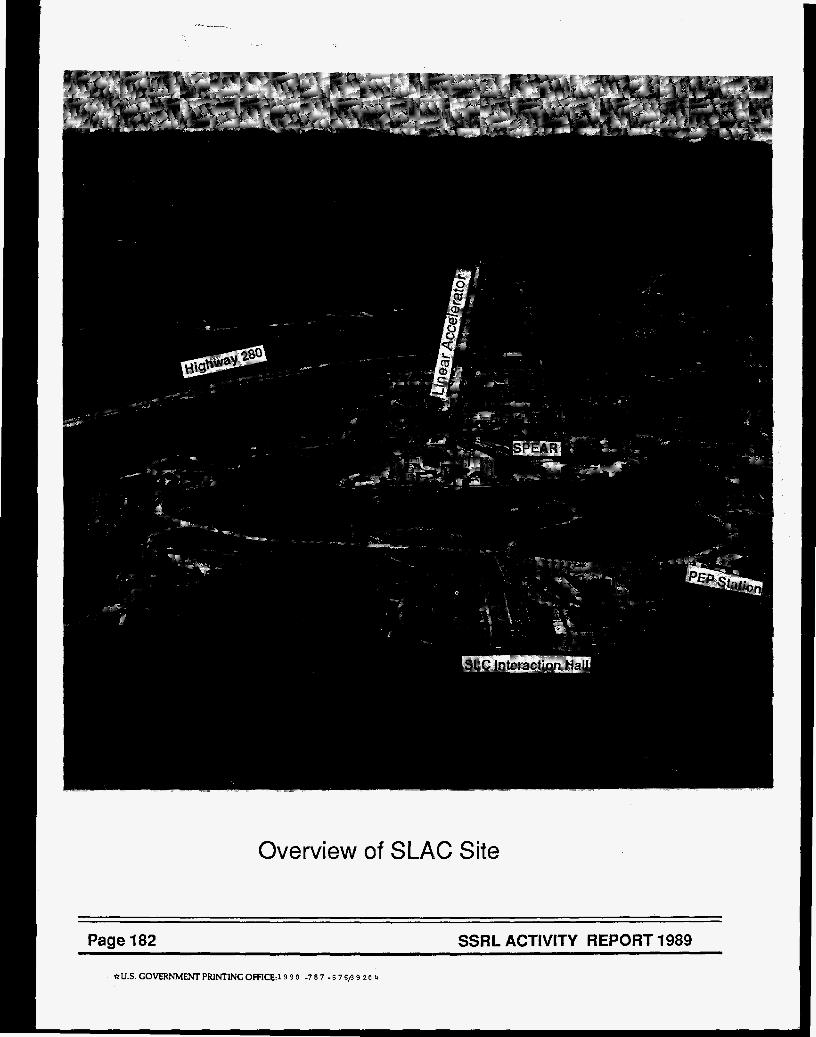
Overview of SLAC Site
Page 182 SSRL ACTIVITY REPORT 1989 ~~
* U S GOVERNMENT PRINTING OFFICE:19 9 0 -7 8 7 - 6 7 6/3 9 2 O 4