Standard Operating Procedures Manual Clinical ... - OSF
61
Standard Operating Procedures Manual Clinical procedures and specimen collection
-
Upload
khangminh22 -
Category
Documents
-
view
0 -
download
0
Transcript of Standard Operating Procedures Manual Clinical ... - OSF

Standard Operating Procedures Manual
Clinical procedures and
specimen collection

HOPE-SAM SOP 1
Anthropometric measurements in children
Page: 1 of 17
Compiled by: Dr Andrew Prendergast
Version: 1.0
Checked by: Dr Mutsa Bwakura
Approved by: Dr. Andrew Prendergast, Chief Investigator
Date: September 28, 2016
Signature:
VERSION HISTORY
CONTENTS 1.1 Purpose 1.2 Scope 1.3 Responsibilities 1.4 Principle/Overview 1.5 Weight 1.6 Mid-upper arm circumference 1.7 Head circumference 1.8 Length in children below 2 years 1.9 Height in children above 2 years
Version number Date Summary of changes

HOPE-SAM SOP 1
Anthropometric measurements in children
Page: 2 of 17
Compiled by: Dr Andrew Prendergast
Version: 1.0
Checked by: Dr Mutsa Bwakura
Approved by: Dr. Andrew Prendergast, Chief Investigator
Date: September 28, 2016
Signature:
1.1 PURPOSE This SOP describes the methods used to undertake anthropometry in the HOPE-SAM study. 1.2 SCOPE This SOP applies to the measurement of children in the HOPE-SAM study. 1.3 RESPONSIBILITIES The Principal Investigator shall ensure the implementation of this procedure. Study nurses and doctors will be responsible for undertaking these procedures.
1.4 PRINCIPLE / OVERVIEW Anthropometry is criticial to assess the nutritional status of children. It enables identification of wasting (low weight-for-height or mid-upper arm circumference), stunting (low height-for-age) and underweight (low weight-for-age). In the HOPE-SAM study, it allows us to identify both cases (children with SAM) and healthy controls; it is also a critical marker of risk of mortality. Anthropometry needs to be conducted accurately and precisely using standardized methods.

HOPE-SAM SOP 1
Anthropometric measurements in children
Page: 3 of 17
Compiled by: Dr Andrew Prendergast
Version: 1.0
Checked by: Dr Mutsa Bwakura
Approved by: Dr. Andrew Prendergast, Chief Investigator
Date: September 28, 2016
Signature:
1.5 WEIGHT
1.5.1 Weight in young children / infants
Equipment
• Seca 384 scales
• Alcohol surface wipes
• Clinical paper roll
Procedure
1. This scale can be used for children below 20kg in weight. For older children, or those too large to fit on this scale, use the mother-baby scale.
2. Explain to the mother that you will take the child’s weight measurement and would like their assistance.
3. Ask the mother to remove all clothing from the child (including their nappy). To avoid the child moving too much ask, the mother to help distract the child while you take the weight measurement; for example, jangling keys lightly can be quite effective.

HOPE-SAM SOP 1
Anthropometric measurements in children
Page: 4 of 17
Compiled by: Dr Andrew Prendergast
Version: 1.0
Checked by: Dr Mutsa Bwakura
Approved by: Dr. Andrew Prendergast, Chief Investigator
Date: September 28, 2016
Signature:
4. Check there is nothing on the scales. Press the green Start key to switch on the scales. The display will read SECA and then quickly run through all elements of the display. The scales are ready for operation when the display reads 0.000.
5. Choose correct weighing mode by pressing the grey button showing two arrows. Mode [1] is for babies below 10kg; mode [2] is for babies 10-20kg. Note that if the child exceeds that weight the display will read “STOP”
6. Place a double layer of clinical paper on the tray of the scales and if the mother has a suitable blanket with her that can be placed in the tray over the clinical roll then do this, as this is more comfortable for the child and may help to keep them still. It is important that the blanket / clinical roll does not hang over the edge of the tray.
7. Hold down the hold/tare key until "NET" appears in the display. Wait until the display stops flashing and is replaced by 0.000 NET.
8. Lay the child naked on the scales. The preference is for babies to be lying on the scales but if older babies are too long or will not stay in this position and they can sit unsupported, then they can be sat on the scales. The mother should stay immediately beside the baby whilst they are on the scales; they should not be left unattended at any time to avoid them falling off or out of the scales. Ideally, the baby should be as still as possible.
9. Read off the measured weight to 2 decimal places. Note that the scale reads to 3 decimal places, but the final number displayed is always zero. When transferring data to the CRF, ignore the final digit (eg 11.460 kg should be recorded as 11.46 kg on the CRF. Note that if the weight is below 10kg, you should record a zero at the front, eg 8.36kg is recorded on the CRF as 08.36 kg)
10. If the infant moves excessively while the scales are stabilising you may get a false reading. If you think this is the case, reweigh the infant.
11. Ask the mother to lift the infant off the scales.
12. Ask the mother to place the infant back on the scales. Repeat the steps to take a second and third measurement and record that too.
13. Throw away the clinical paper and wipe down the scales with an antibacterial surface wipe after each use.

HOPE-SAM SOP 1
Anthropometric measurements in children
Page: 5 of 17
Compiled by: Dr Andrew Prendergast
Version: 1.0
Checked by: Dr Mutsa Bwakura
Approved by: Dr. Andrew Prendergast, Chief Investigator
Date: September 28, 2016
Signature:
14. If the baby passes urine whilst their weight is being measured:
a. If you do not have any weight measurements, dispose of wet clinical roll in the clinical waste bins, clean the scales and start again from the beginning of the weight protocol.
b. If you already have one weight measurement and all the urine is inside the tray, then you can take a second weight reading before cleaning up (assuming the mother is happy with that).
c. If you already have one weight measurement and some of the urine goes outside of the tray, clean up and start the weight protocol from the beginning.

HOPE-SAM SOP 1
Anthropometric measurements in children
Page: 6 of 17
Compiled by: Dr Andrew Prendergast
Version: 1.0
Checked by: Dr Mutsa Bwakura
Approved by: Dr. Andrew Prendergast, Chief Investigator
Date: September 28, 2016
Signature:
1.5.2 Weight in older children
Equipment
• Seca 874 mother-baby scales
Procedure
1. Press the start key with no load on the scale. SECA will appear and then toggle through various displays before 0.00 appears. The scale is then ready to use.

HOPE-SAM SOP 1
Anthropometric measurements in children
Page: 7 of 17
Compiled by: Dr Andrew Prendergast
Version: 1.0
Checked by: Dr Mutsa Bwakura
Approved by: Dr. Andrew Prendergast, Chief Investigator
Date: September 28, 2016
Signature:
2. Ask the mother to stand on the scale without the child.
3. Press the 2-in-1 key. The scale will display 0.00 NET as shown below.
4. Hand the child to the mother to hold while she remains on the scale. If necessary, she can step off the scale to pick up the child, which will display ----- while she is off the scale.
5. The child’s weight will display, after a few seconds of stabilizing the reading, together with HOLD and NET, as shown below.
6. Transfer the reading to the CRF and repeat 2 more times.
7. The scale switches itself off automatically.

HOPE-SAM SOP 1
Anthropometric measurements in children
Page: 8 of 17
Compiled by: Dr Andrew Prendergast
Version: 1.0
Checked by: Dr Mutsa Bwakura
Approved by: Dr. Andrew Prendergast, Chief Investigator
Date: September 28, 2016
Signature:
1.6 MID-UPPER ARM CIRCUMFERENCE 1.6.1 Principle Mid-upper arm circumference is an anthropometric measure providing information on muscle mass and subcutaneous fat. Changes in arm circumference are relatively easy to detect and as such the mid-upper arm circumference is a key indicator of the nutritional status of children. 1.6.2 Equipment
• UNICEF/WHO MUAC measuring tape • White eyeliner pencil (to mark the measuring position on the infant’s arm) • Alcohol swabs to clean the tape
1.6.3 Procedure 1. Explain the procedure to the mother. 2. The baby must have a bare arm and shoulder for this measurement. Position the
baby in a sitting position on the mother’s lap so that you can easily access the left arm. The baby will need to have their elbow bent at a right angle. The mother should hold the left hand to prevent the baby from pulling their arm away.
3. Identify the process of the Acromion (this is the end of the shoulder bone, Arrow 2) and the tip of the elbow (olecranon process, Arrow 3).

HOPE-SAM SOP 1
Anthropometric measurements in children
Page: 9 of 17
Compiled by: Dr Andrew Prendergast
Version: 1.0
Checked by: Dr Mutsa Bwakura
Approved by: Dr. Andrew Prendergast, Chief Investigator
Date: September 28, 2016
Signature:
4. Using the measuring tape, measure the distance between these points. Divide this measurement in half, this is the mid-point of the upper arm; mark this mid-point with the white eyeliner pen.
5. Pass the tape over the baby’s hand and slip it up the baby’s arm, to the mid-point that you have marked. The mother should hold the baby’s arm loosely, and the arm should be straight.
6. The tape should lie on top of the mark, covering it. Ensure the tape is passing horizontally around the arm, not sloping, and is in contact with the skin but does not compress tissue underneath (Arrow 7). It should not be puckering the skin (Arrow 8) or too loose (Arrow 9).
7. Read the measurement to the nearest 0.1cm. If the arrow falls between millimetres always read to the nearest whole millimetre. Write this down or enter directly onto CRF.
8. Loosen and then reposition the tape, and repeat the measurement two more times.
9. Ensure all the measurements are entered onto the CRF. 10. Offer a non-alcohol wipe to the mother so that they can wipe off the white
eyeliner marks on the baby’s arm, if they wish to. 11. Clean the MUAC tape after use with the antibacterial surface wipe.

HOPE-SAM SOP 1
Anthropometric measurements in children
Page: 10 of 17
Compiled by: Dr Andrew Prendergast
Version: 1.0
Checked by: Dr Mutsa Bwakura
Approved by: Dr. Andrew Prendergast, Chief Investigator
Date: September 28, 2016
Signature:
1.6.4 Summary

HOPE-SAM SOP 1
Anthropometric measurements in children
Page: 11 of 17
Compiled by: Dr Andrew Prendergast
Version: 1.0
Checked by: Dr Mutsa Bwakura
Approved by: Dr. Andrew Prendergast, Chief Investigator
Date: September 28, 2016
Signature:
1.7 HEAD CIRCUMFERENCE 1.7.1 Principle Measurement of head circumference is a routine part of a child’s nutritional assessment, because it provides important information about growth and development. 1.7.2 Equipment Non-stretchable measuring tape
Alcohol surface wipes
1.7.3 Procedure 1. Explain the procedure to the mother. 2. Position the child on the mother’s lap. 3. Ask the mother to remove any hair clips, bobbles, hair bands or hats from the
child’s hair/head. 4. Place the measuring tape around the child’s head at its largest diameter midway
between the eyebrows and the hairline at the front of the head, above the ears and around the occipital prominence at the back of the head. Your aim is to always measure the largest circumference possible.

HOPE-SAM SOP 1
Anthropometric measurements in children
Page: 12 of 17
Compiled by: Dr Andrew Prendergast
Version: 1.0
Checked by: Dr Mutsa Bwakura
Approved by: Dr. Andrew Prendergast, Chief Investigator
Date: September 28, 2016
Signature:
5. Ask the mother to distract the infant with toys as they may try to pull the tape off. 6. Pull the tape snugly to compress the hair. Make sure that the tape passes the
occipito-frontal plane (as described above) and does not slip when getting the reading. It may be helpful to ask the mother to hold the tape in place on the back of the head with a finger.
7. Read the measurement to the nearest 0.1 cm. 8. Write down the measurement or enter it directly into the CRF. 9. Repeat the measurement two more times. 10. Wipe the measuring tape with the antibacterial surface wipes after each child and
store to avoid damage and creasing.

HOPE-SAM SOP 1
Anthropometric measurements in children
Page: 13 of 17
Compiled by: Dr Andrew Prendergast
Version: 1.0
Checked by: Dr Mutsa Bwakura
Approved by: Dr. Andrew Prendergast, Chief Investigator
Date: September 28, 2016
Signature:
1.8 LENGTH IN CHILDREN BELOW 2 YEARS 1.8.1 Principle The length measurement, when taken in conjunction with other growth parameters, can be used as an indicator of an infant’s nutritional status, and is used to calculate weight-for-height.
1.8.2 Equipment
• Height/length board
• Antibacterial surface wipes
1.8.3 Procedure
1. Shoes should be removed before height/length is measured. 2. Length using a measuring board lying on a flat surface should be used for children
below the age of 2 years. 3. Explain to the mother the reason for taking the length measurement. Explain that
you will need their assistance in taking this measure and how they can help. The infant needs to be undressed apart from a nappy, no foot or headwear should be worn.
4. An adult must stay immediately beside the infant at all times, even if the baby has never previously rolled over.
5. Place the child on the bed of the measuring board with his/her crown touching the headpiece and the mother standing at the head.
6. Straighten the infant’s legs by holding the legs by the ankles with one hand and applying a gentle downward pressure over the legs with the other hand. It is very important that this pressure should be GENTLE. Young infants cannot fully straighten their legs in the way that older age groups can, and damage and pain could result if this is done too rigorously. Make sure the infant’s hips are not twisted.
7. Move the infant’s head into the Frankfurt Plane position. The Frankfurt Plane should be at right angles to the surface of the table. The Frankfurt Plane is an imaginary line passing through the external ear canal and across the top of the lower bone of the eye socket, immediately under the eye (see picture). This

HOPE-SAM SOP 1
Anthropometric measurements in children
Page: 14 of 17
Compiled by: Dr Andrew Prendergast
Version: 1.0
Checked by: Dr Mutsa Bwakura
Approved by: Dr. Andrew Prendergast, Chief Investigator
Date: September 28, 2016
Signature:
position is important if an accurate reading is to be obtained. Ask the mother to hold the infant’s head in this position and make sure their head is in contact with the headpiece. Encourage the mother to keep her baby distracted by talking and keeping eye contact.
8. Releasing your hand from the baby’s ankles, move the footrest on which the measurement reader is mounted to touch the soles of the child’s feet, toes should be pointing directly upwards. If the baby is pointing the toes downward you will need help to get the feet straight. Once the footrest has been brought up it locks in position so the infant can move their legs again whilst you note the reading.
9. Height/length should be recorded on the CRF in cm to the nearest mm (e.g. 64.2cm)
10. Wipe the height board with an antibacterial surface wipe after taking the measurements.

HOPE-SAM SOP 1
Anthropometric measurements in children
Page: 15 of 17
Compiled by: Dr Andrew Prendergast
Version: 1.0
Checked by: Dr Mutsa Bwakura
Approved by: Dr. Andrew Prendergast, Chief Investigator
Date: September 28, 2016
Signature:
1.8.4 Summary

HOPE-SAM SOP 1
Anthropometric measurements in children
Page: 16 of 17
Compiled by: Dr Andrew Prendergast
Version: 1.0
Checked by: Dr Mutsa Bwakura
Approved by: Dr. Andrew Prendergast, Chief Investigator
Date: September 28, 2016
Signature:
1.9 HEIGHT IN CHILDREN ABOVE 2 YEARS
1.9.1 Principle The height measurement, when taken in conjunction with other growth parameters, can be used as an indicator of a child’s nutritional status, and is used to calculate weight-for-height.
1.9.2 Equipment
• Height/length board
• Antibacterial surface wipes
1.9.3 Procedure
1. For wall-mounted measuring devices, the child should stand with his/her back to the device. The child should be standing with the device centred down the middle of the body and should be standing with heels together and heels, buttocks, and shoulders touching the wall. The child should tuck his/her chin down into the chest and stand as tall as possible. If the measuring device has a horizontal bar to assist with the measurement, the bar should be raised above the child's head and lowered until it just touches the head (the skull; not just the hair). It is important to make certain that the bar is completely horizontal. If the bar is at an angle greater or less than 90° to the wall, the measurement of height will be inaccurate.

HOPE-SAM SOP 1
Anthropometric measurements in children
Page: 17 of 17
Compiled by: Dr Andrew Prendergast
Version: 1.0
Checked by: Dr Mutsa Bwakura
Approved by: Dr. Andrew Prendergast, Chief Investigator
Date: September 28, 2016
Signature:
1.9.4 Summary

HOPE-SAM SOP 2
Body Composition Assessment in children
Page: 1 of 7
Compiled by: Dr Andrew Prendergast
Version: 1.0
Checked by: Dr Mutsa Bwakura
Approved by: Dr. Andrew Prendergast, Chief Investigator
Date: September 28, 2016
Signature:
VERSION HISTORY
1.1 Purpose 1.2 Scope 1.3 Responsibilities 1.4 Overview 1.5 Method 1.6 Important guidelines for equipment care 1.7 Data collection 1.8 Quality Control 1.9 References
Version number Date Summary of changes

HOPE-SAM SOP 2
Body Composition Assessment in children
Page: 2 of 7
Compiled by: Dr Andrew Prendergast
Version: 1.0
Checked by: Dr Mutsa Bwakura
Approved by: Dr. Andrew Prendergast, Chief Investigator
Date: September 28, 2016
Signature:
1.1 PURPOSE This Standard Operating Protocol describes the method used by HOPE-SAM nurses and doctors to measure body composition. 1.2 SCOPE This procedure applies to the use of the Bodystat 1500 MDD instrument to perform bioelectrical impedance analysis.
1.3 RESPONSIBILITIES The Principal Investigator will ensure the implementation of this procedure. HOPE-SAM nurses and doctors will be responsible for undertaking this procedure.
1.4 OVERVIEW This project investigates the short- and long-term outcomes of children with severe acute malnutrition. We want to understand how malnutrition affects not only affect weight and length, but also body composition, and whether a child’s body composition predicts their long-term outcome. Measurement of body composition aims to differentiate lean mass (which represents functional organs and tissues) and fat mass, which represents a store of energy that may fuel immune function or future growth[1]. Bioelectrical impedance analysis (BIA) analyses the bioelectrical properties of the body to elucidate lean tissue and hydration[2]. It has also recently been used in children with malnutrition[3]. Bio-impedance involves placing 2 electrodes each on the hand and feet of a child once they are lying down. An imperceptible current is then run for about 5 seconds after placing electrodes on a child and the machine records several parameters: Z5 (impedance at 5kHz), Z50 (impedance at 50 kHz), R50 (resistance at 50kHz), Xc50 (reactance at 50kHz) and phase angle. This is then repeated so that the impedance measurement at 50 kHz is within 5 Ohms. Using these measurements, a measure of the proportion of lean mass (which conducts low frequencies of electricity well), and fat mass (which has a higher resistance) can be obtained.

HOPE-SAM SOP 2
Body Composition Assessment in children
Page: 3 of 7
Compiled by: Dr Andrew Prendergast
Version: 1.0
Checked by: Dr Mutsa Bwakura
Approved by: Dr. Andrew Prendergast, Chief Investigator
Date: September 28, 2016
Signature:
1.5 METHODOLOGY 1.5.1 Calibration procedure
1. Once a day, calibrate the machine.
2. Open the case, switch on the machine for at least 1 minute.
3. Attach the lead wires to the calibration block as shown and record the Z50 reading.
4. The Z50 reading should be between 497 and 503 Ohms but if it is outside this range, conduct the test anyway.
5. Record the calibration reading on the Body Composition Proforma – the same value can be used for all tests done that day.

HOPE-SAM SOP 2
Body Composition Assessment in children
Page: 4 of 7
Compiled by: Dr Andrew Prendergast
Version: 1.0
Checked by: Dr Mutsa Bwakura
Approved by: Dr. Andrew Prendergast, Chief Investigator
Date: September 28, 2016
Signature:
1.5.2 Test procedure
6. Insert the two main cable leads to the connectors at the back of the unit, with the sticker facing upwards.
7. Cut the electrodes in half as shown below:
8. Attach the cable leads to them with the crocodile clips. The tab should be facing the BIA machine (ie where there is tension in the leads).
9. To ensure good contact between electrodes and skin, use wipes or swabs with alcohol rub if there is excessive oil/lotion/sweat/dust
10. On the dorsal surface of the left hand, place the red electrode just behind the knuckles, and the black electrode at least 3cm (ideally 5cm) away in the direction of the wrist.
11. On the dorsal surface of the left foot, place the red electrode just behind the toes and the black electrode at least 3cm (ideally 5cm) away on the ankle between medial and lateral malleoli

HOPE-SAM SOP 2
Body Composition Assessment in children
Page: 5 of 7
Compiled by: Dr Andrew Prendergast
Version: 1.0
Checked by: Dr Mutsa Bwakura
Approved by: Dr. Andrew Prendergast, Chief Investigator
Date: September 28, 2016
Signature:
(There should always be a minimum of 3cm (preferably 5cm) between the placement of the black and red leads.
1.5.3 Measurement
1. With the child lying supine, legs slightly apart and arms on the side of the body with
palms facing down and away from the body, perform measurements on the right hand side of the body.
2. Check final position of the child and take readings. If necessary use distraction toys to keep them lying down. Ensure nothing else is touching the child.
3. Switch on and record the test number displayed on the screen. Press the middle button
(enter) on the machine, it says “Connect electrodes”.
4. Press enter, it will say “Measuring” for about 5 seconds
5. It will then say “Results not available”; ignore this and press enter again.
6. You will then get the data on Z5 (impedance at 5kHz), Z50 (impedance at 50 kHz), R50 (resistance at 50kHz), Xc50 (reactance at 50kHz) and phase angle, each time pressing the enter button. Write down each value as it appears on the screen using the Body Composition Proforma before pressing the button.
7. Repeat one more time unless there is a “WARNING ILLNESS MARKER” indication in
which case check the connections and repeat.

HOPE-SAM SOP 2
Body Composition Assessment in children
Page: 6 of 7
Compiled by: Dr Andrew Prendergast
Version: 1.0
Checked by: Dr Mutsa Bwakura
Approved by: Dr. Andrew Prendergast, Chief Investigator
Date: September 28, 2016
Signature:
1.5.4 Data interpretation
8. Calculate the difference of impedance at 50 Ohms (Z50) between readings 1 and 2 (highlighted row of table on proforma). If the difference is >5 Ohms, then undertake additional recordings until 2 readings are obtained which have a difference of less than 5 Ohms between the Z50 measurements.
9. Transfer the data obtained from the 2 recordings that have a Z50 value within 5 Ohms of
each other onto the CRF.
10. If it is not possible to obtain 2 acceptable readings after 5 tests, abandon the procedure and do not transfer data to the CRF. Instead, record “No” to the question “Was an adequate body composition assessment undertaken?”
11. File the proforma regardless of the test results.
12. Switch off machine to preserve the battery life.
1.6 IMPORTANT GUIDELINES FOR EQUIPMENT CARE Measurement of bio-impedance involves measuring highly sensitive values. Therefore, care should be exercised when handling the equipment, in particular the cable leads.
1. The leads should be folded neatly after use and
placed in a plastic zip bag. They should not be tangled together and avoid rolling them tightly when not in use.
2. Keep machine in bag when not in use.
3. Cleaning instructions: the hardware unit should be periodically wiped with a damp clean cloth.
4. Only use Duracell batteries, NEVER USE RECHARGABLE BATTERIES

HOPE-SAM SOP 2
Body Composition Assessment in children
Page: 7 of 7
Compiled by: Dr Andrew Prendergast
Version: 1.0
Checked by: Dr Mutsa Bwakura
Approved by: Dr. Andrew Prendergast, Chief Investigator
Date: September 28, 2016
Signature:
1.7 DATA COLLECTION All data will be collected using the data collection form, and then data entered onto the HOPE-SAM database. The Body Composition Proforma should be filed at the site. 1.8 QUALITY CONTROL The study team will be trained and supported with periodic revalidation and refresher training. The BIA system will be calibrated for each visit. 1.9 REFERENCES 1. Friis, H., K.F. Michaelsen, and J.C. Wells, Choice of design and outcomes in trials
among children with moderate acute malnutrition. Food Nutr Bull, 2015. 36(1 Suppl): p. S35-40.
2. Kyle, U.G., A. Piccoli, and C. Pichard, Body composition measurements: interpretation finally made easy for clinical use. Curr Opin Clin Nutr Metab Care, 2003. 6(4): p. 387-93.
3. Girma, T., et al., Bioimpedance index for measurement of total body water in severely malnourished children: Assessing the effect of nutritional oedema. Clinical Nutrition, 2015.
4. Prendergast, A.J. and J.H. Humphrey, The stunting syndrome in developing countries. Paediatr Int Child Health, 2014. 34(4): p. 250-65.

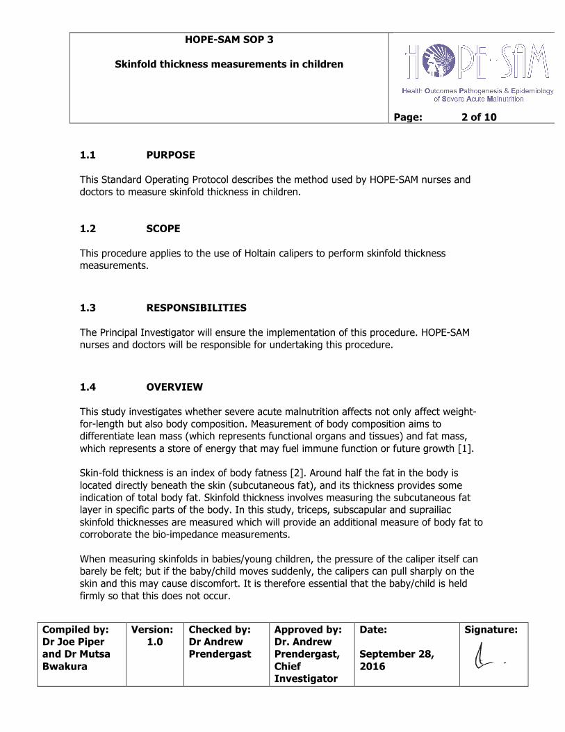
HOPE-SAM SOP 3
Skinfold thickness measurements in children
Page: 1 of 10
Compiled by: Dr Joe Piper and Dr Mutsa Bwakura
Version: 1.0
Checked by: Dr Andrew Prendergast
Approved by: Dr. Andrew Prendergast, Chief Investigator
Date: September 28, 2016
Signature:
VERSION HISTORY
CONTENTS 1.1 Purpose 1.2 Scope 1.3 Responsibilities 1.4 Principle/Overview 1.5 Method 1.5.1 General notes 1.5.2 Exclusion criteria 1.5.3 Equipment 1.5.4 General procedures 1.5.5 Subscapular skinfold thickness 1.5.6 Triceps skinfold thickness 1.5.7 Suprailiac skinfold thickness 1.6 Data collection 1.7 Quality control 1.8 References
Version number Date Summary of changes
HOPE-SAM SOP 3
Skinfold thickness measurements in children
Page: 2 of 10
Compiled by: Dr Joe Piper and Dr Mutsa Bwakura
Version: 1.0
Checked by: Dr Andrew Prendergast
Approved by: Dr. Andrew Prendergast, Chief Investigator
Date: September 28, 2016
Signature:
1.1 PURPOSE This Standard Operating Protocol describes the method used by HOPE-SAM nurses and doctors to measure skinfold thickness in children. 1.2 SCOPE This procedure applies to the use of Holtain calipers to perform skinfold thickness measurements.
1.3 RESPONSIBILITIES The Principal Investigator will ensure the implementation of this procedure. HOPE-SAM nurses and doctors will be responsible for undertaking this procedure.
1.4 OVERVIEW This study investigates whether severe acute malnutrition affects not only affect weight-for-length but also body composition. Measurement of body composition aims to differentiate lean mass (which represents functional organs and tissues) and fat mass, which represents a store of energy that may fuel immune function or future growth [1]. Skin-fold thickness is an index of body fatness [2]. Around half the fat in the body is located directly beneath the skin (subcutaneous fat), and its thickness provides some indication of total body fat. Skinfold thickness involves measuring the subcutaneous fat layer in specific parts of the body. In this study, triceps, subscapular and suprailiac skinfold thicknesses are measured which will provide an additional measure of body fat to corroborate the bio-impedance measurements. When measuring skinfolds in babies/young children, the pressure of the caliper itself can barely be felt; but if the baby/child moves suddenly, the calipers can pull sharply on the skin and this may cause discomfort. It is therefore essential that the baby/child is held firmly so that this does not occur.

HOPE-SAM SOP 3
Skinfold thickness measurements in children
Page: 3 of 10
Compiled by: Dr Joe Piper and Dr Mutsa Bwakura
Version: 1.0
Checked by: Dr Andrew Prendergast
Approved by: Dr. Andrew Prendergast, Chief Investigator
Date: September 28, 2016
Signature:
1.5 METHOD 1.5.1 General notes In order to reduce inter-operator and inter-visit variability, a standardised procedure following Lohman et al in the Anthropometric Standardization Manual with pre-specified positions for callipers and multiple measurements should be followed. Holtain callipers should be used.
1.5.2 Exclusion Criteria
Do not measure skinfolds over skin which is broken, swollen, inflamed / infected or sore / sensitive – for example, children with dermatosis related to SAM. If appropriate measure on the other side of the body, otherwise exclude altogether.
1.5.3 Equipment
• Holtain Skin-fold Caliper1
• White Eyeliner pencil
• Antibacterial surface wipes (to clean the calipers after use)

HOPE-SAM SOP 3
Skinfold thickness measurements in children
Page: 4 of 10
Compiled by: Dr Joe Piper and Dr Mutsa Bwakura
Version: 1.0
Checked by: Dr Andrew Prendergast
Approved by: Dr. Andrew Prendergast, Chief Investigator
Date: September 28, 2016
Signature:
1.5.4 General Procedures
1. Explain the procedure to the mother. You may need to demonstrate the procedure on their forearm. Have the baby/child seated sideways on the mother’s lap facing away from you. The mother may need to hold the baby’s shoulders/arms to prevent movement and also help with providing a distraction.
2. Once the site has been located and marked with a white eyeliner pencil, the thumb and forefinger of the left hand are used to elevate a fold of skin and subcutaneous fat about 1cm away from the measurement point. If necessary, the skin can be lifted using both hands and then held by the left hand whilst measuring with the right.
3. The thumb and finger must be far enough away from the point of measurement so that the fingers are not compressing the point of measurement and the skinfold is pulled away from the underlying muscles in order to form a fold with almost parallel skin surfaces. It may not be possible to achieve parallel sides when the skinfold is large. Care must be taken to only grasp skin and subcutaneous fat, and not muscle. The right hand is used to open the calipers and place them over the skinfold at right angles to the long axis (length) of the fold, approximately half way between the top of the fold and the body surface.
4. A reading is taken once the dial first slows to almost a stop. The time taken varies depending on the thickness of the skinfold. The caliper should not be left in position too long (ie more than 4 seconds), as they will start to compress the skinfold and give an inaccurate measurement.
5. Keep grasping the skinfold with the left hand while the reading is being taken. Once a reading is taken release the calipers FIRST before releasing the fold held by the left hand.
6. When taking a reading the operator’s head should be positioned so as to avoid errors due to parallax. (This means that the operator’s head should be in a direct line with the dial on the caliper so that they can clearly see the exact position of the needle on the dial, to avoid inaccurate readings).
7. A measurement is recorded to the nearest 0.2mm and the measurement is repeated, so that three comparable readings are obtained.
8. Record all measurements on the CRF.

HOPE-SAM SOP 3
Skinfold thickness measurements in children
Page: 5 of 10
Compiled by: Dr Joe Piper and Dr Mutsa Bwakura
Version: 1.0
Checked by: Dr Andrew Prendergast
Approved by: Dr. Andrew Prendergast, Chief Investigator
Date: September 28, 2016
Signature:
1.5.5 Subscapular skinfold (on left side)
2. The operator finds the left scapula (left shoulder blade) and moves the fingers down until the inferior angle at the lower margin of the left scapula. (If this is difficult to locate then gently take the left forearm and place behind the back, mark the scapular angle and then release the arm). The subscapular skinfold is picked up on a diagonal, inclined infero-laterally approximately 45° to the spine in the natural cleavage lines of the skin (see left). Mark a point roughly 1cm diagonally down from the bone just below this angle.
3. The skinfold is grasped diagonally so that the point of measurement is just inferior to this point and the fold is inclined infero-laterally at approximately 45 0 to the horizontal and in the natural cleavage line of the skin. The site is about 1 cm below the inferior angle of the scapula as shown.
1. The baby is seated sideways on the mother’s lap facing away from you. The mother may need to hold the baby’s shoulders/arms to prevent movement. Avoid having the baby seated facing the mother and held by both arms such that the skin of the back is taut as it will be difficult and uncomfortable to raise a skinfold.

HOPE-SAM SOP 3
Skinfold thickness measurements in children
Page: 6 of 10
Compiled by: Dr Joe Piper and Dr Mutsa Bwakura
Version: 1.0
Checked by: Dr Andrew Prendergast
Approved by: Dr. Andrew Prendergast, Chief Investigator
Date: September 28, 2016
Signature:
5. Repeat the measurement so that three readings are obtained. There should be a brief pause between measurements to prevent sustained compression of the tissue. Ensure that all measurements are entered onto the CRF.
6. Offer a non-alcohol wipe to the mother so that she can wipe off the white eyeliner marks on the baby’s back.
4. The caliper is placed approximately 1 cm from the fingers of the left hand, perpendicular to the long axis of the skinfold. Once the calipers are in place release the calipers slowly and gently whilst STILL HOLDING the skinfold between finger and thumb. Wait until the dial slows to almost a stop before reading. Record the measurement to the nearest 0.2mm. Write this down or enter directly into the CRF.

HOPE-SAM SOP 3
Skinfold thickness measurements in children
Page: 7 of 10
Compiled by: Dr Joe Piper and Dr Mutsa Bwakura
Version: 1.0
Checked by: Dr Andrew Prendergast
Approved by: Dr. Andrew Prendergast, Chief Investigator
Date: September 28, 2016
Signature:
1.5.6 Triceps skinfold (on left side)
2. The operator finds the level of the mid-upper arm circumference measurement (see HOPE-SAM SOP 1) and marks the point on the posterior aspect of the arm. Another line is drawn perpendicular to that line to form a cross.
3. The skinfold is measured with the arm hanging loosely and comfortably at the child’s side and palm facing the thigh or supported at 900 in front. The operator places the palm of his/ her hand on the child’s arm above the marked level. The operator gently pinches the skin and adipose tissue 1 cm above the cross and pulls it away from the underlying muscle. Sometimes a gentle shake can help the tissues align easier.
1. The baby is seated sideways on the mother’s lap facing away from you. The mother may need to hold the baby’s shoulders/arms to prevent movement.

HOPE-SAM SOP 3
Skinfold thickness measurements in children
Page: 8 of 10
Compiled by: Dr Joe Piper and Dr Mutsa Bwakura
Version: 1.0
Checked by: Dr Andrew Prendergast
Approved by: Dr. Andrew Prendergast, Chief Investigator
Date: September 28, 2016
Signature:
5. The measurement is repeated and recorded three times on the CRF.
1.5.7 Supra-iliac skinfold thickness (on left side)
4. The caliper is then placed at the exact level on the mid-upper arm plane. The site of measurement must be in the midline posteriorly. The skinfold must be parallel to the long axis of the upper arm and the caliper must be applied at the mark 90° to the long axis of the upper arm (see left).
2. The operator palpates the top of the iliac crest and marks a cross aligned with the mid-axillary line.
1. The infant is seated sideways on the mother’s lap facing away from you. The mother may need to hold the infant’s shoulders/arms to prevent movement and also to distract the child.

HOPE-SAM SOP 3
Skinfold thickness measurements in children
Page: 9 of 10
Compiled by: Dr Joe Piper and Dr Mutsa Bwakura
Version: 1.0
Checked by: Dr Andrew Prendergast
Approved by: Dr. Andrew Prendergast, Chief Investigator
Date: September 28, 2016
Signature:
5. The measurement is repeated and recorded three times on the CRF.
4. The caliper is placed on the fold at the mid-axillary point.
3. An oblique skinfold is grasped just above the centre of the marked cross and following the natural cleavage line of the skin so that the fold is approximately 1cm above the iliac crest at the mid-axillary point. It should be aligned inferomedially at approximately 450 to the horizontal. Two hands may be used to grasp the skinfold initially and then the right hand removed to use the caliper.

HOPE-SAM SOP 3
Skinfold thickness measurements in children
Page: 10 of 10
Compiled by: Dr Joe Piper and Dr Mutsa Bwakura
Version: 1.0
Checked by: Dr Andrew Prendergast
Approved by: Dr. Andrew Prendergast, Chief Investigator
Date: September 28, 2016
Signature:
1.6 DATA COLLECTION All data will be collected using the data collection form, and then data entered. 1.7 QUALITY CONTROL Nurses and doctors will be trained and supported with periodic revalidation and refresher training. 1.8 REFERENCES 1. Friis, H., K.F. Michaelsen, and J.C. Wells, Choice of design and outcomes in trials
among children with moderate acute malnutrition. Food Nutr Bull, 2015. 36(1 Suppl): p. S35-40.
2. Reilly, J.J., J. Wilson, and J.V. Durnin, Determination of body composition from skinfold thickness: a validation study. Archives of Disease in Childhood, 1995. 73(4): p. 305-310.

HOPE-SAM SOP 4
Venepuncture in children
Page: 1 of 5
Compiled by: Dr Andrew Prendergast
Version: 1.0
Checked by: Dr Mutsa Bwakura
Approved by: Dr. Andrew Prendergast, Chief Investigator
Date: September 28, 2016
Signature:
VERSION HISTORY
CONTENTS 1.1 Purpose 1.2 Scope 1.3 Responsibilities 1.4 Overview 1.5 Methodology 1.5.1 Materials 1.5.2 Procedure 1.6 Sample transport 1.7 Quality control
Version number Date Summary of changes

HOPE-SAM SOP 4
Venepuncture in children
Page: 2 of 5
Compiled by: Dr Andrew Prendergast
Version: 1.0
Checked by: Dr Mutsa Bwakura
Approved by: Dr. Andrew Prendergast, Chief Investigator
Date: September 28, 2016
Signature:
1.1 PURPOSE This chapter describes the method used by HOPE-SAM research nurses and physicians to collect child blood samples and transport samples to the laboratory. 1.2 SCOPE This procedure applies to child blood collection to obtain whole blood, plasma and RNA.
1.3 RESPONSIBILITIES The Principal Investigator will ensure the implementation of this procedure. Research nurses and physicians will be responsible for undertaking this procedure.
1.4 OVERVIEW
Venepuncture is an invasive procedure, which requires both knowledge and skill to perform. The following techniques and procedures are used to minimize these risks. In addition, procedures for specimen handling should be followed in order to preserve specimen integrity. Blood must be collected in a ‘closed circuit’ for two reasons – i) for safety to prevent exposure to blood-borne pathogens; ii) to ensure an uncontaminated sample is collected for subsequent lab analysis.
Blood collection tubes must be drawn in a specific order to avoid cross-contamination of additives between tubes. The recommended order of draw for plastic Vacutainer tubes is (for the enteropathy substudy) the EDTA (purple top), then the RNA PAXgene tube which contains an RNA fixing solution. In the observational study, only EDTA blood is collected.
Adverse events that can occur whilst carrying out this procedure include accidental needle stick injury and minor bruising. In the event of a needle stick injury, follow the local PEP policy. Warn the mother that the child may cry and that there may be some mild bruising, but this is transient.
HOPE-SAM SOP 4
Venepuncture in children
Page: 3 of 5
Compiled by: Dr Andrew Prendergast
Version: 1.0
Checked by: Dr Mutsa Bwakura
Approved by: Dr. Andrew Prendergast, Chief Investigator
Date: September 28, 2016
Signature:
1.5 METHOD 1.5.1 Materials
(a) 23g or 21g butterfly needle. (b) 5ml syringe. (c) RNA PAXgene tube and 2.7ml EDTA tube (enteropathy substudy) (d) 2 x 2.7ml EDTA tubes (observational study) (e) Alcohol swab (or cotton wool and methylated spirit) (f) Dry swab. (g) Adhesive plaster. (h) Sharps bins. (i) Latex gloves. (j) Biohazard waste collection bag. (k) Patient barcode labels. (l) Sample transmittal form. (m) Cooler box with cold packs.
1.5.2 Procedure
(a) Explain procedure to the caregiver and gain verbal consent.
(b) Find a suitable part of the ward or clinic to undertake the procedure, where the
caregiver and child will feel comfortable.
(c) Ensure that the caregiver is seated comfortably with the child appropriately restrained to prevent movement during blood taking.
(d) Have everything arranged before you start.
(e) Wash your hands with soap and water.
(f) Put on powder free gloves (provided by the study).

HOPE-SAM SOP 4
Venepuncture in children
Page: 4 of 5
Compiled by: Dr Andrew Prendergast
Version: 1.0
Checked by: Dr Mutsa Bwakura
Approved by: Dr. Andrew Prendergast, Chief Investigator
Date: September 28, 2016
Signature:
(g) Hold the hand of the child so that you are flexing the hand and pulling the skin taut. Your fingers should be wrapped around the child’s wrist so that you are gently applying a tourniquet effect. If there is an assistant, that person can use their hand to form a tourniquet around the child’s wrist and keep the arm steady.
(h) Clean the area with a spirited swab, or with cotton wool and methylated spirit. Ensure that the vein is prominent. If not, ‘pump’ the hand gently with your fingers to help show up the veins.
(i) Open the butterfly and connect the 5ml syringe to the end of the tubing.
(j) Warn the mother that you are going to insert the needle, and insert needle into
vein at an angle less than 30°.
(k) Once you are in the vein, blood will start to flow along the tubing of the butterfly needle. You may need to gently pump the hand to fill and refill the vein so that it continues to fill the tubing. Apply gentle pressure on the syringe to help the blood to flow.
(l) Collect just over 5ml blood into the syringe.
(m) Apply gentle pressure on the site with a dry swab and remove the needle and put
the needle and tubing in the portable sharps bin.
(n) Apply adhesive plaster on the puncture site and reassure the mother and infant.
(o) Carefully fill the different tubes from the syringe in the correct order and ensure sufficient volumes of blood:
Enteropathy substudy: 2.7ml in the EDTA tube 2.7ml in the RNA PAXgene tube Observational study: 2.7ml in the 1st EDTA tube 2.7ml in the 2nd EDTA tube
(p) Put the lids on the tubes and mix gently 5-10 times.
HOPE-SAM SOP 4
Venepuncture in children
Page: 5 of 5
Compiled by: Dr Andrew Prendergast
Version: 1.0
Checked by: Dr Mutsa Bwakura
Approved by: Dr. Andrew Prendergast, Chief Investigator
Date: September 28, 2016
Signature:
(q) Label the specimen tubes immediately with barcode labels or with plain label that includes PID and date of collection.
(r) Record specimen collection details on the Sample Transmittal Form.
(s) Put the EDTA and RNA PAXgene tubes in a Ziplock bag and leave them at room temperature.
(t) Dispose of used gloves and other waste material in the designated biohazard waste bag.
(u) Ensure the room temperature sample is safely packed and transported to the laboratory.
1.6 SAMPLE TRANSPORT During sample transport to the laboratory:
(a) Ensure that the driver/porter signs the specimen transmittal form. (b) Do not allow anyone to touch or otherwise handle the blood sample. (c) Do not leave the blood samples unattended during travel to the laboratory. (d) Take blood samples immediately to the laboratory on arrival (e) Hand over both the samples and the sample transmittal form to the laboratory
technician and ensure s/he signs the form as proof of receipt of specimens. 1.7 QUALITY CONTROL For sample integrity ensure the following:
(a) Blood tubes have not expired. (b) All blood samples are correctly labeled. (c) Blood samples are stored and transported at room temperature as instructed.

HOPE-SAM SOP 5
Collection of gastric aspirates in children
Page: 1 of 6
Compiled by: Dr Mutsa Bwakura
Version: 1.0
Checked by: Dr Andrew Prendergast
Approved by: Dr. Andrew Prendergast, Chief Investigator
Date: September 28, 2016
Signature:
VERSION HISTORY
CONTENTS 1.1 Purpose 1.2 Scope 1.3 Responsibilities 1.4 Overview 1.5 Methodology 1.5.1 Materials 1.5.2 Procedure 1.6 Sample transport 1.7 Quality control
Version number Date Summary of changes

HOPE-SAM SOP 5
Collection of gastric aspirates in children
Page: 2 of 6
Compiled by: Dr Mutsa Bwakura
Version: 1.0
Checked by: Dr Andrew Prendergast
Approved by: Dr. Andrew Prendergast, Chief Investigator
Date: September 28, 2016
Signature:
1.1 PURPOSE This SOP describes the methods used to collect a nasogastric (NG) aspirate and measure gastric pH from children in the HOPE-SAM study. 1.2 SCOPE This SOP applies to collection of gastric aspirate samples and measurement of pH in hospitalized children with severe acute malnutrition in the HOPE-SAM study. 1.3 RESPONSIBILITIES The Principal Investigator shall ensure the implementation of this procedure. Study nurses, study physician and laboratory technicians will be responsible for implementing those parts of this procedure that pertain to them.
1.4 PRINCIPLE / OVERVIEW In severe acute malnutrition, the stomach contents are less acidic (achlorhydria) which means that the ‘gastric acid barrier’ is less effective, allowing overgrowth of bacteria in the upper gut. By collecting gastric aspirate samples, we can measure the pH of the stomach juice, and collect samples for bacterial culture and detection using sensitive molecular methods. This procedure is only conducted on children in the enteropathy substudy of the HOPE-SAM study, who already have an NG tube in place for other reasons (either feeding, or for collection of gastric aspirates for TB diagnosis). It is only performed at baseline. It is very important to check correct NG tube placement because inadvertent passage of the NG tube into the lungs is dangerous.

HOPE-SAM SOP 5
Collection of gastric aspirates in children
Page: 3 of 6
Compiled by: Dr Mutsa Bwakura
Version: 1.0
Checked by: Dr Andrew Prendergast
Approved by: Dr. Andrew Prendergast, Chief Investigator
Date: September 28, 2016
Signature:
1.5 METHODOLOGY 1.5.1 Materials
(a) Gloves (b) Nasogastric tubes, 8F to 10F (c) 2 plain screw-top containers (d) 1 screw-top container with glycerol (e) 1 screw-top container with preservative (f) 5ml, 10ml or 20ml sterile syringes (g) Sodium bicarbonate (8.4%) (h) Sterile water (i) pH test strips and reference colour chart (j) Alcohol wipe, or cotton wool and methylated spirit (k) Specimen transmittal form
1.5.2 Procedure 1. Instruct the caregiver and ward nurses regarding overnight fasting of at least 4
hours before early morning gastric aspirate (GA). The procedure is preferably performed early in the morning. The procedure may also be performed during the daytime, as long as the child has been kept nil per os (NPO) for minimum 4 hours.
It may be useful to put up a sign by the bedside or nurses’ station saying “Bed xx HOPE-SAM Gastric aspirate on (Date), please do not feed”.
2. Use an assistant to help as this procedure requires 2-3 people.
3. Prepare all the materials for the procedure.
4. Position the child lying on the back
5. Measure the distance of the nasogastric tube to the stomach (from tip of the nose to earlobe, then to xiphisternum): this estimates the distance that will be required to insert the tube (see picture below).

HOPE-SAM SOP 5
Collection of gastric aspirates in children
Page: 4 of 6
Compiled by: Dr Mutsa Bwakura
Version: 1.0
Checked by: Dr Andrew Prendergast
Approved by: Dr. Andrew Prendergast, Chief Investigator
Date: September 28, 2016
Signature:
6. Place the child’s face in the “sniffing air” position, and pass the nasogastric tube from the nose into the stomach (see picture below).

HOPE-SAM SOP 5
Collection of gastric aspirates in children
Page: 5 of 6
Compiled by: Dr Mutsa Bwakura
Version: 1.0
Checked by: Dr Andrew Prendergast
Approved by: Dr. Andrew Prendergast, Chief Investigator
Date: September 28, 2016
Signature:
7. If the child develops severe coughing, choking or cyanosis, withdraw the tube immediately.
8. Withdraw gastric contents using the syringe attached to the nasogastric tube and wet each coloured section on the pH test strip.
9. Test the pH, by comparing the colour of each part of the strip to the reference colour chart. Find the combination of colours that forms the best match and read off the pH. Record the result on the CRF.
10. If no gastric aspirate is obtained, then other checks need to be conducted to ensure that the NG tube is in position before proceeding to step 11.
11. Draw up 10ml sterile water into an oral syringe, attach the syringe to the NG tube and inject down the tube, leave for three minutes, and then aspirate. Repeat if needed until 10ml aspirate is obtained. Do not repeat more than 3 times.
12. Divide the aspirate obtained equally between the 4 screw-top containers (i.e. 2.5ml into each; two tubes are plain, one contains glycerol and one contains preservative)
13. Label the three sterile containers for the HOPE-SAM study with the PID and date (or with a barcode label) and record the samples on a Specimen Transmittal Form.
14. Label one of the plain screw-cap tubes with appropriate hospital identifiers and send to the clinical laboratory for TB microscopy and culture. To this sample only, add an equal volume of sodium bicarbonate solution (in order to neutralize the acidic gastric contents and so prevent destruction of tubercle bacilli).
15. Tightly secure the lids and wipe the containers with an alcohol wipe, or with cotton wool and methylated spirit, to prevent cross-infection.
16. Transport the research specimens (in a cooler box) to the laboratory for processing as soon as possible (within 4 hours).
17. Place the specimens in a refrigerator (4-8°C) if it is likely to take more than 4 hours for the specimens to be taken to the laboratory.

HOPE-SAM SOP 5
Collection of gastric aspirates in children
Page: 6 of 6
Compiled by: Dr Mutsa Bwakura
Version: 1.0
Checked by: Dr Andrew Prendergast
Approved by: Dr. Andrew Prendergast, Chief Investigator
Date: September 28, 2016
Signature:
1.6 SAMPLE TRANSPORT During sample transport to the laboratory:
(a) Ensure that the driver/porter signs the specimen transmittal form. (b) Do not allow anyone to touch or otherwise handle NG sample. (c) Do not leave the NG samples unattended during travel to the laboratory. (d) Take NG samples immediately to the laboratory on arrival at the field office/Hub. (e) Hand over both the samples and the sample transmittal form to the laboratory
technician and ensure s/he signs the form as proof of receipt of specimens. 1.7 QUALITY CONTROL For sample integrity ensure the following:
(a) All NG samples are correctly labeled. (b) NG samples are stored and transported at the correct temperature as instructed. (c) Cooler packs are adequately frozen and remain cold until samples are transmitted
to the laboratory. Ref: Guidance for national programmes on the management of tuberculosis in children. Geneva, World Health Organization, WHO/HTM/2014.03

HOPE-SAM SOP 6
Stool collection in children
Page: 1 of 6
Compiled by: Dr Andrew Prendergast
Version: 1.0
Checked by: Dr Mutsa Bwakura
Approved by: Dr. Andrew Prendergast, Chief Investigator
Date: September 28, 2016
Signature:
VERSION HISTORY
CONTENTS 1.1 Purpose 1.2 Scope 1.3 Responsibilities 1.4 Principle/Overview 1.5 Methodology 1.5.1 Materials 1.5.2 Procedure 1.5.2.1 Procedure for hospitalized children 1.5.2.2 Procedure for discharged children 1.6 Sample transport 1.7 Quality control
Version number Date Summary of changes

HOPE-SAM SOP 6
Stool collection in children
Page: 2 of 6
Compiled by: Dr Andrew Prendergast
Version: 1.0
Checked by: Dr Mutsa Bwakura
Approved by: Dr. Andrew Prendergast, Chief Investigator
Date: September 28, 2016
Signature:
1.1 PURPOSE This chapter describes the method used to collect and transport stool specimens from children in the HOPE-SAM study. 1.2 SCOPE This SOP applies to stool sample collection and transport in children. 1.3 RESPONSIBILITIES The Principal Investigator will ensure the implementation of this procedure. Study nurses will be responsible for undertaking this procedure.
1.4 PRINCIPLE / OVERVIEW Stool samples will be collected to investigate the composition of the intestinal microbiota, and to measure markers of intestinal inflammation and repair. Proteins will degrade in the sample over time, and microbes will proliferate, so specimens must reach the laboratory as soon as possible after passage using a cold chain, ideally within 4 hours. Stool samples are collected from children in the enteropathy substudy of the HOPE-SAM study at baseline, discharge, then 12, 24 and 48 weeks. A diarrhoeal specimen can be collected if the child has current diarrhea. If the specimen is diarrhoeal, this should be recorded on the specimen transmittal form.

HOPE-SAM SOP 6
Stool collection in children
Page: 3 of 6
Compiled by: Dr Andrew Prendergast
Version: 1.0
Checked by: Dr Mutsa Bwakura
Approved by: Dr. Andrew Prendergast, Chief Investigator
Date: September 28, 2016
Signature:
1.5 METHODOLOGY 1.5.1 Materials
(a) Screw capped fecal container with applicator stick in the lid (b) Linen saver (c) Potty (for older children) (d) Latex gloves (e) Biohazard bag (f) Sample barcode labels or plain labels (g) Sample transmittal form (h) Cooler box with cold packs
1.5.2 Procedure 1.5.2.1 Procedure for hospitalized children 1. Note that the stool sample has to be collected before the LM test is
conducted. If this is not possible, stool collection should be deferred until one week after the LM test.
2. For children who are sick, or who are not continent, use a nappy/diaper or a cut-down linen saver to help collect the sample. Put the diaper on the child inside-out (shiny surface up) as this will help to collect a sample without it soaking into the nappy (especially if the child has diarrhea).
3. For children who are continent and well enough to pass stool voluntarily, a sample can be passed into a potty.
4. Leave the container with the mother. Show her how to transfer a sample of stool into the screw-cap tube using the applicator stick. The sample should ideally fill half the container, but any amount of stool should be collected if less than this.
5. Make sure sample container is tightly closed and avoid contaminating the outside of the container.
6. Dispose of the used gloves and wash your hands.

HOPE-SAM SOP 6
Stool collection in children
Page: 4 of 6
Compiled by: Dr Andrew Prendergast
Version: 1.0
Checked by: Dr Mutsa Bwakura
Approved by: Dr. Andrew Prendergast, Chief Investigator
Date: September 28, 2016
Signature:
7. Label the sample with a barcode label and place the sample in the dedicated cooler box.
8. Record stool collection details on the Sample Transmittal Form. Record if the sample is diarrhoeal in the comments box.
9. Ensure that the cooler box is properly closed, and arrange transport to the laboratory.
1.5.2.2 Procedure for discharged children 10. Give the caregiver a stool collection kit, which contains a screw-capped container
with an applicator stick in the lid, a cut-down linen saver, a pair of gloves and a biohazard waste bag.
11. Explain to the caregiver that a stool sample needs to be collected from the child. The mother should be given a stool pack and instructions and asked to bring the sample to the next visit.
12. Ask when the child normally passes stool. The caregiver should ideally collect a sample from the child on the morning of the outpatient visit. If this is not possible, she can collect a sample the night before the visit and try to keep it in a cool part of the house.
13. If the caregiver has not been able to collect a specimen from the child, then it may be collected at the outpatient visit before administration of the LM solution (if due). If it is still not possible to collect a specimen, the sample collection should be deferred for 1 week after LM administration.
14. Using a demonstration stool collection kit and showing the caregiver the items, tell the caregiver to wear the gloves provided and transfer a sample from the child’s nappy into the container using the applicator stick. Children can pass stool onto a linen server and the caregiver can collect the sample using the applicator stick.
15. The sample should ideally fill half the container – mark the container to make this clear for the caregiver. If she cannot obtain that much stool, she should collect whatever is possible.

HOPE-SAM SOP 6
Stool collection in children
Page: 5 of 6
Compiled by: Dr Andrew Prendergast
Version: 1.0
Checked by: Dr Mutsa Bwakura
Approved by: Dr. Andrew Prendergast, Chief Investigator
Date: September 28, 2016
Signature:
16. Tell the caregiver to make sure sample container is tightly closed and avoid contaminating the outside of the container.
17. Ask the caregiver to place the sample in the ziplock bag and keep it in a cool part of the home.
18. Tell the caregiver to dispose of the used gloves and other waste material wherever the household usually disposes of faecal material.
19. Advise the caregiver to wash her hands after the procedure.
20. When the caregiver brings the specimen to clinic, label the sample with a barcode label (or plain label with PID and date clearly recorded) and place the sample in the dedicated cooler box for transport to the laboratory.
21. Record stool collection details on the Sample Transmittal Form. Record if the sample is diarrhoeal in the comments box.
22. Ensure that the cooler box is properly closed, and arrange transport to the laboratory.
1.6 SAMPLE TRANSPORT During sample transport to the laboratory:
(a) Ensure that the driver/porter signs the specimen transmittal form. (b) Do not allow anyone to touch or otherwise handle stool sample. (c) Do not leave the stool samples unattended during travel to the laboratory. (d) Take stool samples immediately to the laboratory on arrival at the field office/Hub. (e) Hand over both the samples and the sample transmittal form to the laboratory
technician and ensure s/he signs the form as proof of receipt of specimens. 1.7 QUALITY CONTROL For sample integrity ensure the following:
(a) All stool collection containers are clean. (b) All stool samples are correctly labeled.

HOPE-SAM SOP 6
Stool collection in children
Page: 6 of 6
Compiled by: Dr Andrew Prendergast
Version: 1.0
Checked by: Dr Mutsa Bwakura
Approved by: Dr. Andrew Prendergast, Chief Investigator
Date: September 28, 2016
Signature:
(c) Stool samples are stored and transported at the correct temperature as instructed. (d) Cooler packs are adequately frozen and remain cold until samples are transmitted
to the laboratory.

HOPE-SAM SOP 7
Lactulose-Mannitol testing in children
Page: 1 of 9
Compiled by: Dr Andrew Prendergast
Version: 1.0
Checked by: Dr Mutsa Bwakura
Approved by: Dr. Andrew Prendergast, Chief Investigator
Date: September 28, 2016
Signature:
VERSION HISTORY
CONTENTS 1.1 Purpose 1.2 Scope 1.3 Responsibilities 1.4 Principle/Overview 1.5 Methodology 1.5.1 Materials 1.5.2 Procedure 1.5.2.1 Preparations in advance 1.5.2.2 LM solution administration and urine collection 1.6 Sample transport 1.7 Quality control 1.8 Summary Flow Chart 1.9 Urine collection bag instructions
Version number Date Summary of changes
HOPE-SAM SOP 7
Lactulose-Mannitol testing in children
Page: 2 of 9
Compiled by: Dr Andrew Prendergast
Version: 1.0
Checked by: Dr Mutsa Bwakura
Approved by: Dr. Andrew Prendergast, Chief Investigator
Date: September 28, 2016
Signature:
1.1 PURPOSE This SOP describes the methods used to administer lactulose:mannitol (L:M) solution and to collect urine from children in the HOPE-SAM study. 1.2 SCOPE This SOP applies to administration of L:M solution and collection and transport of urine samples. 1.3 RESPONSIBILITIES The Principal Investigator shall ensure the implementation of this procedure. Study nurses, study physician and laboratory technicians will be responsible for implementing those parts of this procedure that pertain to them.
1.4 PRINCIPLE / OVERVIEW Urine specimens will be collected from children after ingestion of a combination of two sugars (lactulose and mannitol). A urine specimen is collected at the start of the test (pre-LM urine). Urine is collected for at least 2 hours after ingestion of L:M solution to assess the ratio of lactulose:mannitol excretion (LM urine). The lactulose:mannitol (LM) test is considered a sensitive method to measure small intestinal epithelial area, paracellular and transcellular transport, damage and permeability. Children must be fasted for at least 30 minutes before ingesting a solution containing mannitol (50 mg/ml) and lactulose (250 mg/ml). The child will ingest 2ml/kg of the LM solution after being weighed, and can return to regular feeding 30 minutes after ingestion. Total urine is collected in a urine bag for two hours, during which time the mother should be encouraged to feed the child regularly in order to permit collection of an adequate volume of urine. Collected urine is preserved using chlorhexidine to prevent overgrowth of bacteria which would metabolize sugars. Total urine volume is collected and transported back to the laboratory, for storage in a -80 °C freezer until lactulose and mannitol concentration is measured by mass spectrometry.

HOPE-SAM SOP 7
Lactulose-Mannitol testing in children
Page: 3 of 9
Compiled by: Dr Andrew Prendergast
Version: 1.0
Checked by: Dr Mutsa Bwakura
Approved by: Dr. Andrew Prendergast, Chief Investigator
Date: September 28, 2016
Signature:
1.5 METHODOLOGY 1.5.1 Materials
(a) Oral L:M Solution (kept refrigerated prior to use) (b) 20mL oral syringe (c) Weighing scales (d) 90ml or 100ml screw cap urine containers x 2 (one for pre-LM urine, one with a
yellow label for LM urine) (e) Baby wipes (f) Urine collection bags x 3 (g) Sample barcode labels (pre-LM and LM urine) (h) Sample transmittal form (i) Latex gloves (j) Biohazard waste collection bag (k) Sample cooler box with cold packs (l) Chlorhexidine preservative solution in a dropper bottle (2.35% = 40 mg
chlorohexidine/ml water) (Sigma Chemical Co., St. Louis, MO)
1.5.2 Procedure 1.5.2.1 Preparations in advance
1. In advance, the study nurse explains to the mother that the L:M test will be
undertaken at the next visit (conducted in the enteropathy substudy only, at baseline, discharge, 12 and 48 weeks). The study nurse pre-warns the mother that we will ask her not to feed her infant for 1 hour in total (ie. no eating, drinking or breastfeeding) on the day – 30 minutes before ingesting LM solution, and 30 minutes after ingesting LM solution. This has to be carefully timed when children with severe acute malnutrition are inpatients, to fit in with their feed requirements.
2. The study nurse must liaise with the study physician to ensure that hospitalized children are clinically stable prior to conducting the test. In general, this is a child with no cardiorespiratory instability. Children with diarrhea can have an L:M test conducted unless they have signs of shock or dehydration, but need to be watched carefully afterwards to ensure diarrhea does not worsen.

HOPE-SAM SOP 7
Lactulose-Mannitol testing in children
Page: 4 of 9
Compiled by: Dr Andrew Prendergast
Version: 1.0
Checked by: Dr Mutsa Bwakura
Approved by: Dr. Andrew Prendergast, Chief Investigator
Date: September 28, 2016
Signature:
3. The L:M solution is manufactured and labeled in the central laboratory, transported by cold chain to the hospital site and kept refrigerated in a dedicated clean fridge until the test is conducted. The vial contains 20mL of L:M solution.
1.5.2.2 L:M solution administration and urine collection
1. The stool sample for the enteropathy substudy must be collected before the LM
test is conducted. Once LM solution has been administered, stool cannot be collected for one week (because the LM solution affects the laboratory tests). If stool has not been collected, defer the LM test until the stool has been obtained.
2. Find a suitable part of the ward to undertake the procedure, where the mother
and child will feel comfortable.
3. Complete the top of the proforma with date, PID and staff name.
4. Wash your hands before examining the child.
5. Check whether the child has any perineal skin infection or dermatosis which would prevent attachment of the urine bag. If so, record this on the proforma and delay the L:M test until this has resolved.
6. Weigh the child and record the weight on the proforma.
7. Ask the mother if the child is continent and can urinate on demand. The child
needs to pass urine regularly during the 2 hour test and not ‘hang on’ to urine. In sick children, those who are not continent, or children who are unlikely to cooperate with passing urine regularly into a potty, it is advisable to use a urine bag. Clean the child’s nappy area using a baby wipe then allow the area to air-dry fully before attaching a urine bag as shown in section 1.9. Record the time the bag was applied on the proforma.
8. Continue to feed the child while waiting for the pre-LM urine sample to be
collected, to encourage passing of urine.
9. Check the urine bag every 15 minutes.

HOPE-SAM SOP 7
Lactulose-Mannitol testing in children
Page: 5 of 9
Compiled by: Dr Andrew Prendergast
Version: 1.0
Checked by: Dr Mutsa Bwakura
Approved by: Dr. Andrew Prendergast, Chief Investigator
Date: September 28, 2016
Signature:
10. Once the child has passed urine, empty the urine bag into the 90mL screw top container. Label this pre-L:M urine, seal it in a ziplock bag and place into the specimen cool box. Keep the urine bag to reattach again later, or if the adhesive is not working, discard the bag. Record the time pre-LM urine was collected on the proforma.
11. If the child has not passed a pre-LM urine after 1 hour of waiting, then proceed
to step 12.
12. Inform the mother that the child should now stop feeding (ie. no eating, drinking or breastfeeding) for 30 minutes before the LM solution is ingested. The study nurse should supervise and time this fasting period, recording it on the proforma.
13. Calculate the start time of the L:M test, which should be 30 minutes after the
last witnessed ingestion of food, drink or breast milk.
14. Explain the procedure to the mother and gain verbal consent. Explain that the child will be given a measured dose of L:M solution according to his/her weight. Explain that L:M solution can sometimes lead to temporary loose stools for a day after ingestion.
15. Calculate the dose of L:M solution to be given, as 2mL/kg body weight (eg a 6.5kg
baby should receive 13mL of L:M solution). Round up to the nearest 0.5mL (eg a 5.2kg baby should receive 10.5ml, not 10.4ml) – the maximum dose is 20.0ml.
16. Draw up the solution in a 20mL oral syringe and give to the child. The easiest
way to give the L:M solution in young babies is using the syringe, giving a few ml each time. In older children, the L:M solution can be poured into a drinking cup. Ensure that the child ingests the whole calculated dose.
17. Record the time of ingestion of L:M solution on the proforma.
18. Attach a new urine bag to the child (see section 1.9) immediately to collect the
LM urine sample.
19. Continue to fast the child for 30 minutes after the complete ingestion of the L:M solution. After 30 minutes, the mother should be encouraged to feed her infant

HOPE-SAM SOP 7
Lactulose-Mannitol testing in children
Page: 6 of 9
Compiled by: Dr Andrew Prendergast
Version: 1.0
Checked by: Dr Mutsa Bwakura
Approved by: Dr. Andrew Prendergast, Chief Investigator
Date: September 28, 2016
Signature:
regularly (eg fluids, breastfeeding, therapeutic milk depending on age and current management protocol) to ensure the infant passes urine.
20. If the child vomits within an hour of ingesting the L:M solution then the test
should be stopped and rescheduled.
21. Check the urine bag every 15 minutes. If there is urine in the bag, empty it into the 90ml urine container labeled “LM” with a yellow sticker and affix a new urine bag immediately if needed. If the bag has become contaminated with stool, then clean the child and affix a new urine bag immediately.
22. Record any urine collection in the table on the proforma, recording time collected
and whether the specimen had to be discarded for contamination with stool.
23. Add one (1) drop of chlorhexidine to the urine each time you empty the urine bag into the 90ml urine container.
24. Put the 90ml screw-cap urine container labeled “LM” with a yellow sticker in the
specimen cool box and keep it there for the duration of the procedure. (Take care to ensure you do not confuse the pre-LM urine with the LM urine).
25. For 2 hours, collect all urine, following steps 21-24 above.
26. At 2 hours, check the urine bag one final time, and repeat steps 21-24 above.
27. If the child has passed no urine at all at 2 hours, then encourage further feeding
and wait until the child passes urine, then end the test. The test must be continued until some urine is obtained.
28. Record the time that the test ended on the proforma.
29. KEEP ALL THE URINE COLLECTED AND PLACE THE LM CONTAINER INTO
THE COOLER BOX FOR THE LAB TECHNICIAN TO PROCESS. DO NOT DISCARD ANY OF THE URINE.
30. Label the LM urine sample with the barcode label provided or hand-write the PID
and date on a blank label.
31. Place the tube into the Ziplock plastic bag in the specimen cool box.

HOPE-SAM SOP 7
Lactulose-Mannitol testing in children
Page: 7 of 9
Compiled by: Dr Andrew Prendergast
Version: 1.0
Checked by: Dr Mutsa Bwakura
Approved by: Dr. Andrew Prendergast, Chief Investigator
Date: September 28, 2016
Signature:
32. Dispose of the used gloves, and other waste material.
33. Wash your hands.
34. Record details of the samples collected on the sample transmittal form.
35. Record on the proforma whether the infant experienced any problems during the
procedure.
36. Ensure that the cooler box is properly closed, and arrange for transport to the laboratory for processing.
1.6 SAMPLE TRANSPORT During sample transport to the laboratory:
(a) Ensure that the driver signs the specimen transmittal form. (b) Do not allow anyone to touch or otherwise handle urine sample. (c) Do not leave the urine samples unattended during travel to the laboratory. (d) Take urine samples immediately to the laboratory on arrival at the field office/Hub. (e) Hand over both the samples and the sample transmittal form to the laboratory
technician and ensure s/he signs the form as proof of receipt of specimens. 1.7 QUALITY CONTROL For sample integrity ensure the following:
(a) All urine collection containers are clean. (b) All urine samples are correctly labeled. (c) Urine samples are stored and transported at the correct temperature as instructed. (d) Cooler packs are adequately frozen and remain cold until samples are transmitted
to the laboratory.

HOPE-SAM SOP 7
Lactulose-Mannitol testing in children
Page: 8 of 9
Compiled by: Dr Andrew Prendergast
Version: 1.0
Checked by: Dr Mutsa Bwakura
Approved by: Dr. Andrew Prendergast, Chief Investigator
Date: September 28, 2016
Signature:
1.8 SUMMARY FLOW CHART

HOPE-SAM SOP 7
Lactulose-Mannitol testing in children
Page: 9 of 9
Compiled by: Dr Andrew Prendergast
Version: 1.0
Checked by: Dr Mutsa Bwakura
Approved by: Dr. Andrew Prendergast, Chief Investigator
Date: September 28, 2016
Signature:
1.9 URINE COLLECTION BAG INSTRUCTIONS