Stabilization of α-chymotrypsin upon PEGylation correlates with reduced structural dynamics
17
Stabilization of α-Chymotrypsin upon PEGylation Correlates with Reduced Structural Dynamics José A. Rodríguez-Martínez 1 , Ricardo J. Solá 1 , Betzaida Castillo 1 , Héctor R. Cintrón- Colón 1 , Izarys Rivera-Rivera 1 , Gabriel Barletta 2 , and Kai Griebenow 1,* 1 Department of Chemistry, University of Puerto Rico-Río Piedras, PO Box 23346 San Juan Puerto Rico, 00931-3346 2 Department of Chemistry, University of Puerto Rico-Humacao, CUH Station, Humacao, 00791 Abstract Protein stability remains one of the main factors limiting the realization of the full potential of protein therapeutics. Poly(ethylene glycol) (PEG) conjugation to proteins has evolved into an important tool to overcome instability issues associated with proteins. The observed increase in thermodynamic stability of several proteins upon PEGylation has been hypothesized to arise from reduced protein structural dynamics, although experimental evidence for this hypothesis is currently missing. To test this hypothesis, the model protein α-chymotrypsin (α-CT) was covalently modified with PEGs with molecular weights (M W ) of 700, 2000 and 5000 and the degree of modification was systematically varied. The procedure did not cause significant tertiary structure changes. Thermodynamic unfolding experiments revealed that PEGylation increased the thermal transition temperature (T m ) of α-CT by up to 6°C and the free energy of unfolding (ΔG U (25°C)) by up to 5 kcal/mol. The increase in stability was found to be independent of the PEG M W and it leveled off after an average of four PEG molecules were bound to α-CT. Fourier- transformed infrared (FTIR) H/D exchange experiments were conducted to characterize the conformational dynamics of the PEG-conjugates. It was found that the magnitude of thermodynamic stabilization correlates with a reduction in protein structural dynamics and was independent of the PEG M W . Thus, the initial hypothesis proved positive. Similar to the thermodynamic stabilization of proteins by covalent modification with glycans, poly(ethylene glycol) thermodynamically stabilizes α-CT by reducing protein structural dynamics. These results provide guidance for the future development of stable protein formulations. Keywords H/D exchange; PEGylation; protein engineering; protein thermodynamic stability; protein dynamics Introduction Advances in biotechnology have accelerated the discovery and development of many proteins as therapeutic agents. The increasing use of recombinant proteins by the pharmaceutical industry has highlighted the important issue of protein stability (Frokjaer and Otzen 2005). The inherent physical and chemical instabilities of proteins can result in protein conformational changes, denaturation, aggregation, precipitation, and/or adsorption to surfaces, events that cause loss of biopharmaceutical efficacy (Manning et al. 1989). * Corresponding author. Telephone: (787) 764-0000 x4781, Fax: (787) 756-8242, Email: [email protected]. NIH Public Access Author Manuscript Biotechnol Bioeng. Author manuscript; available in PMC 2009 December 15. Published in final edited form as: Biotechnol Bioeng. 2008 December 15; 101(6): 1142–1149. doi:10.1002/bit.22014. NIH-PA Author Manuscript NIH-PA Author Manuscript NIH-PA Author Manuscript
Transcript of Stabilization of α-chymotrypsin upon PEGylation correlates with reduced structural dynamics
Stabilization of α-Chymotrypsin upon PEGylation Correlateswith Reduced Structural Dynamics
José A. Rodríguez-Martínez1, Ricardo J. Solá1, Betzaida Castillo1, Héctor R. Cintrón-Colón1, Izarys Rivera-Rivera1, Gabriel Barletta2, and Kai Griebenow1,*
1 Department of Chemistry, University of Puerto Rico-Río Piedras, PO Box 23346 San JuanPuerto Rico, 00931-33462 Department of Chemistry, University of Puerto Rico-Humacao, CUH Station, Humacao, 00791
AbstractProtein stability remains one of the main factors limiting the realization of the full potential ofprotein therapeutics. Poly(ethylene glycol) (PEG) conjugation to proteins has evolved into animportant tool to overcome instability issues associated with proteins. The observed increase inthermodynamic stability of several proteins upon PEGylation has been hypothesized to arise fromreduced protein structural dynamics, although experimental evidence for this hypothesis iscurrently missing. To test this hypothesis, the model protein α-chymotrypsin (α-CT) wascovalently modified with PEGs with molecular weights (MW) of 700, 2000 and 5000 and thedegree of modification was systematically varied. The procedure did not cause significant tertiarystructure changes. Thermodynamic unfolding experiments revealed that PEGylation increased thethermal transition temperature (Tm) of α-CT by up to 6°C and the free energy of unfolding (ΔGU(25°C)) by up to 5 kcal/mol. The increase in stability was found to be independent of the PEG MWand it leveled off after an average of four PEG molecules were bound to α-CT. Fourier-transformed infrared (FTIR) H/D exchange experiments were conducted to characterize theconformational dynamics of the PEG-conjugates. It was found that the magnitude ofthermodynamic stabilization correlates with a reduction in protein structural dynamics and wasindependent of the PEG MW. Thus, the initial hypothesis proved positive. Similar to thethermodynamic stabilization of proteins by covalent modification with glycans, poly(ethyleneglycol) thermodynamically stabilizes α-CT by reducing protein structural dynamics. These resultsprovide guidance for the future development of stable protein formulations.
KeywordsH/D exchange; PEGylation; protein engineering; protein thermodynamic stability; proteindynamics
IntroductionAdvances in biotechnology have accelerated the discovery and development of manyproteins as therapeutic agents. The increasing use of recombinant proteins by thepharmaceutical industry has highlighted the important issue of protein stability (Frokjaer andOtzen 2005). The inherent physical and chemical instabilities of proteins can result inprotein conformational changes, denaturation, aggregation, precipitation, and/or adsorptionto surfaces, events that cause loss of biopharmaceutical efficacy (Manning et al. 1989).
*Corresponding author. Telephone: (787) 764-0000 x4781, Fax: (787) 756-8242, Email: [email protected].
NIH Public AccessAuthor ManuscriptBiotechnol Bioeng. Author manuscript; available in PMC 2009 December 15.
Published in final edited form as:Biotechnol Bioeng. 2008 December 15; 101(6): 1142–1149. doi:10.1002/bit.22014.
NIH
-PA Author Manuscript
NIH
-PA Author Manuscript
NIH
-PA Author Manuscript

Protein stability challenges can be encountered during purification, separation, storage,delivery, and administration. Several stabilizing strategies have been pursued to resolvethese instability issues. These include the design of mutant proteins, the use of stabilizingexcipients during formulation (e.g., sugars and polyols), and/or chemical modifications (e.g.,by glycosylation or PEGylation) (Wang 1999; Wang 2000; Solá et al. 2006; Solá et al.2007).
In recent years, several PEGylated protein products have been developed and approved bythe FDA for therapeutic use in humans (Harris and Chess 2003). Approved PEGylatedprotein drugs include PEG-L-asparaginase (Oncaspar®), PEG-adenosine deaminase(Adagen®), PEG-interferon α2a (Pegasys®), PEG-interferon α2b (PEG-Intron®), PEG-granulocyte-colony stimulating factor (Neulasta®), and PEG-growth hormone receptorantagonist (Somavert®). Other PEG conjugated proteins are currently undergoing clinicaltrial, including PEG-arginine deiminase and PEG-uricase.
PEGylation refers to the attachment of one or several PEG molecules to the protein orpeptide surface. PEGylation chemistry can involve several functionalities of the availableamino acid side chains at the protein surface (i.e., NH2, COOH, SH, and OH). Historically,PEGylation has mostly been performed to improve protein half-life in the blood serum andto reduce immune responses. The prolonged circulating time is achieved by reducingglomerular filtration of the protein drug and by enhancing its proteolytic resistance(Abuchowski et al. 1977a; Abuchowski et al. 1977b). Reduced immunogenicity is a result ofepitope shielding by the PEG molecules (Soares et al. 2002).
An additional advantage of PEGylated proteins is their increased in vitro thermodynamicstability. The increased stability is beneficial for product development and storage, whereproteins can be submitted to several denaturing stresses (Cromwell et al. 2006; Perez et al.2002). Increased melting temperatures (Tm) have been reported for several PEGylatedproteins when compared to the unmodified protein. López-Cruz et al. reported an increase of2°C in the Tm of Myceliophthora thermophila laccase upon PEG conjugation to 8 of its 14available surface exposed lysine residues (López-Cruz et al. 2006). The Tm of recombinanthuman endostatin was increased from 47.6 to 62.6°C upon modification with a single PEGmolecule of 5 kDa at the N-terminus (Nie et al. 2006). An increment of 2°C in the Tm wasobserved for human interferon-beta when it was modified with two PEG molecules of 40kDa (Basu et al. 2006). It has been reported that after modification of α-chymotrypsin (α-CT) with PEG of 1.9 and 5 kDa the Tm is shifted to higher temperatures by as much as 5°C(Castellanos et al. 2005; Topchieva et al. 1995).
The molecular mechanism by which PEGylation causes increased thermodynamic stabilityremains uncertain. Several researchers have proposed that the protein structure becomesmore rigid upon PEGylation, yet no experimental evidence has been provided to support thishypothesis (García-Arellano et al. 2002; López-Cruz et al. 2006; Soares et al. 2002; Yang etal. 1996). This is due to the lack of systematic studies regarding the effect of PEGylationparameters on the biophysical properties of proteins (i.e., structure, structural dynamics, andstability) which have to involve both, variation in the MW and number of PEG moleculesattached to the protein. Understanding the mechanism by which PEGylation influencesprotein biophysical properties is vital to the development of PEG-based stabilizing strategiesin the future.
In the present study, we employed a series of PEG-α-CT conjugates with varyingPEGylation degree (1 to 9 PEG molecules bound to α-CT) and using PEG of different MW(700, 2000, and 5000 Da) as an experimental system to systematically study the correlationbetween changes in protein thermodynamic stability and structural dynamics.
Rodríguez-Martínez et al. Page 2
Biotechnol Bioeng. Author manuscript; available in PMC 2009 December 15.
NIH
-PA Author Manuscript
NIH
-PA Author Manuscript
NIH
-PA Author Manuscript

Materials and MethodsChemicals
All chemicals were of the highest purity grade available from commercial sources. Bovineα-CT was purchased from Sigma-Aldrich (St. Louis, MO). Activated linear PEGs with MWof 2 and 5 kDa were purchased from Nektar Technologies (Huntsville, AL). Methyl-PEO12-NHS ester (685.71 g/mol, PEG700) and 2, 4, 6-trinitrobenzene sulfonic acid (TNBSA) werefrom Pierce (Rockford, IL).
PEGylation of α-CTChemical modification of the surface exposed lysine amino groups of α-CT with theactivated PEG was carried out as previously reported (Castellanos et al. 2005). In brief,different PEG:α-CT molar ratios were employed during synthesis to obtain PEG-α-CTconjugates with varying degrees of modification. Un-reacted PEG and buffer salts wereremoved by dialyzing against deionized water at 4°C. The PEG-α-CT conjugates weresubsequently lyophilized. The degree of protein modification was determined by titration ofthe free amino groups with TNBSA (Habeeb 1966).
Circular dichroism (CD) spectroscopyNear-UV CD spectra were measured using an Olis DSM-10 UV-Vis CD spectrophotometer.For all measurements the protein concentration was adjusted to 0.6 mg/mL in 10 mMpotassium phosphate buffer (pH 7.1 and 25°C). Spectra were recorded from 260 to 310 nmusing 10 mm path length quartz cells. Each spectrum was obtained by averaging 6 scans at 2nm resolution. Blank spectra were subtracted from the protein CD spectra.
Differential Scanning CalorimetryAll thermal denaturation experiments were performed using a CSC 6100 Nano II differentialscanning calorimeter (Calorimetry Sciences Corp., Lindon, UT). Protein samples wereprepared at a concentration of 1 mg/ml in 10 mM potassium phosphate buffer, pH 7.1. Heatcapacity thermograms were determined at a 2°C/min scan rate from 20°C to 90°C. Baselineswere run with the same buffer used for the samples. Analysis of heat capacity thermogramsfor α-CT was carried out as previously reported (Solá et al. 2006).
Kinetics of hydrogen/deuterium (H/D) exchangeAmide H/D exchange kinetics were measured with a ThermoNicolet Nexus 470 FTIRspectrometer and the exchange kinetics analyzed as previously reported (Solá andGriebenow 2006a; Zavodszky et al. 1998). α-CT and PEG-α-CT conjugates (4 mg) weredissolved in 10 mM potassium phosphate buffer, pH 7.1, and subsequently lyophilized. Thelyophilized samples were dissolved in 200 μL of D2O and immediately transferred to aFTIR cell with CaF2 windows and a 25 μm Teflon spacer. Spectra (2000 – 1000 cm−1) werecollected at 2 min intervals until 10 min, 5 min intervals until 30 min, and every 30 min until360 min. For the first 30 min, 10 scans were averaged, and after 30 min, 56 scans, all at 2cm−1 resolution. Spectra of buffer blanks were subtracted from all sample spectra. H/Dexchange spectra were processed for quantitati ve analysis in the form of hydrogen exchangedecay plots (X vs. time). The fraction of unexchanged backbone hydrogen atoms at time twas determined by the following equation:
Rodríguez-Martínez et al. Page 3
Biotechnol Bioeng. Author manuscript; available in PMC 2009 December 15.
NIH
-PA Author Manuscript
NIH
-PA Author Manuscript
NIH
-PA Author Manuscript

where w(t) is the ratio of the baseline corrected absorbance of amide II (1550 cm−1) toamide I (1637.5 cm−1) at time t, w(0) is the amide II/amide I ratio of the undeuteratedprotein and w(∞) is the amide II/amide I ratio for the fully deuterated protein.
ResultsPEGylation of α-CT and characterization of the PEG-α-CT conjugates
To study the effects of the PEGylation degree and of the PEG MW on protein stability, PEGswith three different MW were employed to synthesize PEG-α-CT conjugates with varyingamount of bound PEG. PEG molecules were covalently linked to α-CT surface accessiblelysine residues. The PEG employed was activated with a succimidyl ester group, which isdisplaced by amines via nucleophilic substitution. By controlling the PEG-to-α-CT molarratio during the reaction, it was possible to prepare PEG-α-CT conjugates with up to 9bound PEG molecules. α-CT has 14 surface reactive lysine residues. The degree ofmodification was determined by measuring the amount of unreacted amino groups using thetrinitrobenzene sulfonic acid assay (Figure 1) (Habeeb 1966).
To assess the effect of PEGylation on the protein tertiary structure, the PEG-α-CTconjugates were characterized by near-UV CD spectroscopy. Previous work showed thatperturbations of α-CT tertiary structure cause a significant decrease in the spectral intensityin the near-UV CD spectra. The near-UV CD spectrum of non-modified α-CT displays twomaxima at 288 and 297 nm, which completely disappear upon thermal unfolding(Castellanos et al. 2005). No major spectral changes are observed as a result of PEGylation,which rules out large perturbations to the tertiary structure of the bioconjugates. It was foundthat neither the molecular weight of the PEG nor the degree of PEGylation adverselyimpacted structural integrity of α-CT (Figure 2). These results agree with previouslypublished work which revealed no significant changes in secondary and tertiary structure ofα-CT after PEGylation (Castellanos et al. 2005; Castillo et al. 2006).
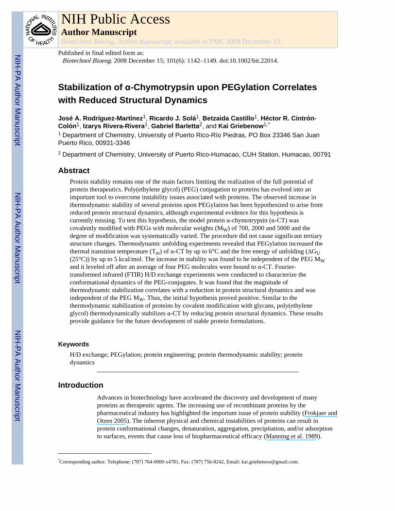
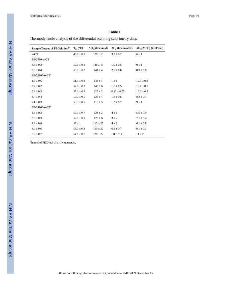
Effect of PEGylation on protein thermodynamic stabilityTo determine the role of the extent of PEGylation and PEG MW on protein thermodynamicstability, thermal unfolding experiments using differential scanning calorimetry (DSC) werecarried out. Figure 3 shows the thermograms for unmodified α-CT and for three PEG-α-CTconjugates with PEGs of 700, 2000, and 5000 Da. Unmodified α-CT displays a two-statethermally induced unfolding transition with the denaturation midpoint (Tm) at 48.0°C.Modification of α-CT with PEG resulted in a shift in the endothermic denaturation transitionto higher temperatures. This resulted in increased Tm values for the PEG-α-CT conjugates atincreasing PEG molar content until a plateau was reached (Figure 4). The effect of thePEGylation degree on the Tm levels off at around 53.0°C after the 4th PEG molecule isbound to the protein. A maximum increase of 6°C in the Tm was achieved. Analysis of thedenaturation endotherms permitted the calculation of the enthalpy (ΔHm) and heat capacitychanges (ΔCp) associated with the unfolding event (Table I). These values allowed thecalculation of the Gibbs free energy of unfolding at 25°C (ΔGU(25°C)) by using the Gibbs-Helmholtz equation (ΔGU(T)= ΔHm(1 − T/Tm) − ΔCp[(Tm − T) + Tln(T/Tm)]) (Pace1990;Solá et al. 2006). Similar to the Tm, the free energy of protein unfolding (ΔGU(25°C))increased for the PEGylated proteins. Stabilization of up to 5 kcal/mol was achieved for themost modified PEG-protein conjugates. Interestingly, it was observed that the MW of thePEG did not influence the magnitude of thermodynamic stabilization of the protein. Similarto the stabilization effects of protein glycosylation, stabilization by PEGylation was solelydependent on the amount of PEG bound to the protein.
Rodríguez-Martínez et al. Page 4
Biotechnol Bioeng. Author manuscript; available in PMC 2009 December 15.
NIH
-PA Author Manuscript
NIH
-PA Author Manuscript
NIH
-PA Author Manuscript

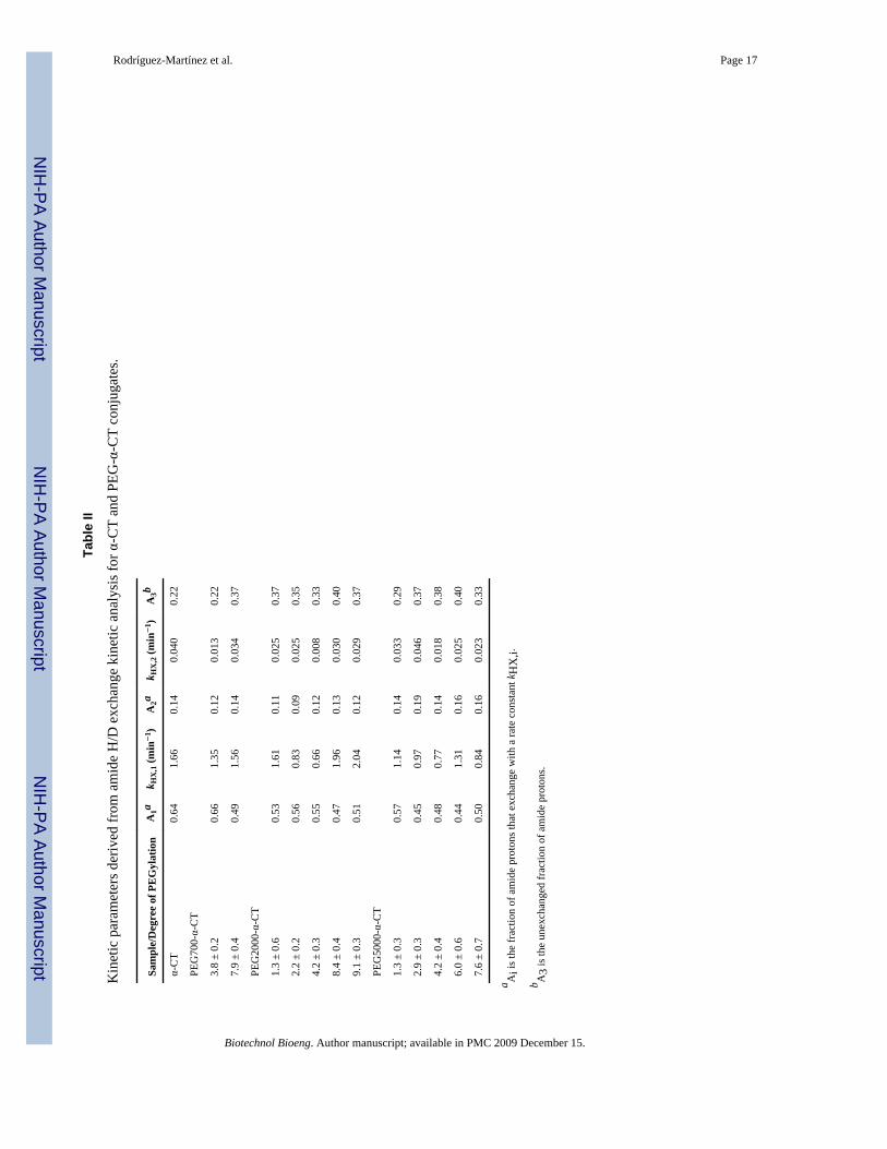
Effects of PEGylation on structural dynamicsHydrogen/deuterium exchange is a versatile method for obtaining information on thestructural dynamics of macromolecular systems (Ferraro and Robertson 2004; Hvidt andNielsen 1966; Privalov and Tsalkova 1979). Measuring the time course of H/D exchangewith FTIR allows to asses changes in global protein structural dynamics (Li et al. 2002;Zavodszky et al. 1998). H/D exchange experiments were carried out following the protocolpreviously reported (Solá and Griebenow 2006a). Figure 5 shows the spectroscopic resultsfrom a typical FTIR H/D exchange experiment for α-CT including both, the spectra of theundeuterated and completely deuterated protein. H/D exchange kinetics were determined byfollowing the decrease in the absorbance of the amide II band (N–H, 1500–1600 cm−1)relative to the amide I band (C=O, 1600–1700 cm−1) (Hvidt and Nielsen 1966). Theexchange data were plotted in the form of decay plots, where the fraction of unexchangedamide hydrogen atoms (X) decreases over time due to the H/D exchange process (for detailssee Materials and Methods section). Quantitative analysis of the decay plots was carried outby fitting a two-exponential decay model of the following form:
where A1, A2, and A3 are the fractions of the fast, slow, non-exchanging amide protons andkHX,1 and kHX,2 are the apparent exchange rate constants for the fast and slow amideprotons. Under experimental conditions, A3 refers to the unexchanged fraction of amideprotons, which is an indicator of structural dynamics; reduction of protein structuraldynamics results in an increase in unexchanged amide protons (Castillo et al. 2008).
In figure 6A, the H/D exchange decay plots for the PEG5000-α-CT conjugates with varyingamount of bound PEG are presented. As the amount of PEG per mol of enzyme is increased,both the rate constant and the fraction of fast exchanging amide hydrogens are reduced.Conversely, there is an increase in the fraction of non-exchanging amide hydrogens (A3)(Figure 6, Table II). This observation is indicative of a more rigid protein core as a result ofPEGylation. The structural dynamics of the protein become reduced as the PEGylationdegree increases until it levels off after the 4th PEG molecule is attached. The effect of thePEG MW on H/D exchange rates is presented in figure 6B for PEG-α-CT conjugates withcomparable degrees of PEGylation (50% of modified lysines). Similar to the effects onthermodynamic stability, the MW of the PEG chain is not a significant factor in reducing theprotein structural. Therefore, the reduction in protein structural dynamics only depended onthe degree of modification, not the molecular weight of the PEG.
DiscussionIn recent years, protein PEGylation has become a widely employed technology to improvethe in vivo lifetime of protein and peptide drugs. Accordingly, the number of PEG-proteinconjugates approved by the FDA and the amount of research articles on the topic aresteadily increasing (Veronese and Pasut 2005). A benefit of protein PEGylation is theincrease in protein thermodynamic stability which can result in a more robust proteinproduct that can better withstand the denaturing conditions encountered in productformulation and has a longer shelf-life.
The primary goal of this work was to determine the mechanism by which PEGylationincreases the thermodynamic stability of proteins in order to advance the understanding ofprotein biophysics and protein stabilization. Our results show that the magnitude ofstabilization increased proportionally with the degree of modification before leveling off(Figure 4). There is no significant difference between the stabilization effects of PEG of
Rodríguez-Martínez et al. Page 5
Biotechnol Bioeng. Author manuscript; available in PMC 2009 December 15.
NIH
-PA Author Manuscript
NIH
-PA Author Manuscript
NIH
-PA Author Manuscript

different MW (Table 1). This is important since PEG polymers of less than 1 kDa can besynthesized with uniform molecular weight, thus avoiding polydispersity issues. The in vivoimplications of this finding, however, will have still to be established.
To account for the observed stabilization effect of PEGylation, several researchers proposedthat chemical modification causes reduced protein structural dynamics. By measuring H/Dexchange for the different PEG-α-CT conjugates, we demonstrate that after PEGylation thekinetics of H/D exchange and extent of hydrogen exchange are indeed reduced (Figure 6).This observation demonstrates a reduction in protein structural dynamics and a more rigidprotein core. The increase in protein thermodynamic stability correlates with this reductionin protein structural dynamics.
A group of authors has suggested that the reduction of protein dynamics upon PEGmodification might be a consequence of the amphiphilic nature of PEG. Water moleculessolvate the hydrophilic regions of bound PEG, while the hydrophobic regions interact withhydrophobic clusters on the protein surface. The net effect of this is the removal of watermolecules from the surface of the protein to produce a more rigid protein structure (García-Arellano et al. 2002; Longo and Combes 1999; Solá and Griebenow 2006b). Computermodels suggest that the PEG chains fold upon themselves and form loosely folded domainson the surface of proteins, possibly excluding water from the protein surface (Manjula et al.2003). Using small-angle X-ray scattering, Svergun et al solved the first solution structurefor a PEG-protein conjugate (Svergun et al. 2008). This low resolution structure ofPEGylated hemoglobin shows that part of the PEG polymer sits on the surface of the proteinand the other part extends away of the protein. Their structure also revealed a more compactand rigid protein and partial dehydration of the protein surface.
Previous investigations have correlated reduced protein dynamics with an increase in proteinthermodynamic stability. Solá and Griebenow reported increased thermodynamic stability ofα-CT upon chemical glycosylation (Solá and Griebenow 2006a). Therein, the increasedthermodynamic stability by glycosylation correlated with a reduction in the proteindynamics. Reduced protein structural dynamics have been proposed as the main factoraccounting for the high thermostability of proteins of extremophiles. For example, adenylatekinase from the thermophilic archaeon Sulfolobus acidocaldarius posses a more rigid andcompact structure than its homologue from porcine muscle cytosol (Bonisch et al. 1996).Measuring H/D exchange with FTIR, Petsko showed that the more stable thermophilicenzyme, 3-isopropylmalate dehydrogenase (IPMDH) from Thermus thermophilus HB8, hasreduced conformational flexibility when compared to its homologous IPMDH fromEscherichia coli (Zavodszky et al. 1998). Stabilization of proteins by ligand binding alsocorrelates with a loss in structural dynamics. For example, the Tm of bovine serum albuminincreased from 59.0°C to 79.8°C when bound to anilinonaphthalene sulphonate, which wasattributed to a reduction in protein dynamics (Celej et al. 2003). On the other hand, achemical modification that increased protein dynamics, as measured by H/D exchange, leadto the destabilization of cytochrome c of around 4 kcal/mol (de Jongh et al. 1995).
In this report we show that the increase in protein thermodynamic stability was accompaniedby a reduction of protein structural dynamics. PEGylation is currently a well establishedtechnique for improving the pharmacokinetics and pharmacodynamics of protein drugsbecause of its in vivo stabilization effects. It can also be used to improve protein productsshelf-life, a current limitation of protein pharmaceuticals (Wang 1999; Wang 2000).Understanding the mechanism by which proteins become stabilized by chemicalmodifications will greatly benefit biopharmaceutical products since protein drugs canpotentially be modified with infinitely diverse chemical structures, whose limit is solelydictated by the chemist inventiveness.
Rodríguez-Martínez et al. Page 6
Biotechnol Bioeng. Author manuscript; available in PMC 2009 December 15.
NIH
-PA Author Manuscript
NIH
-PA Author Manuscript
NIH
-PA Author Manuscript

AcknowledgmentsThis publication was made possible by grant number S06 GM08102 from the National Institute for GeneralMedical Sciences (NIGMS) at the National Institutes of Health (NIH) through the Support of Competitive Research(SCORE) Program. Its contents are solely the responsibility of the authors and do not necessarily represent theofficial views of NIGMS. JARM was supported by a fellowship from the NIH Research Initiative for ScientificEnhancement (RISE) Program (contract number R25 GM061151) and by the Fellowship Program of the PuertoRico Development Company (PRIDCO).
ReferencesAbuchowski A, McCoy JR, Palczuk NC, van Es T, Davis FF. Effect of covalent attachment of
polyethylene glycol on immunogenicity and circulating life of bovine liver catalase. J Biol Chem.1977a; 252(11):3582–3586. [PubMed: 16907]
Abuchowski A, van Es T, Palczuk NC, Davis FF. Alteration of immunological properties of bovineserum albumin by covalent attachment of polyethylene glycol. J Biol Chem. 1977b; 252(11):3578–3581. [PubMed: 405385]
Basu A, Yang K, Wang ML, Liu S, Chintala R, Palm T, Zhao H, Peng P, Wu DC, Zhang ZF, et al.Structure-function engineering of interferon-beta-1b for improving stability, solubility, potency,immunogenicity, and pharmacokinetic properties by site-selective mono-PEGylation. BioconjugateChem. 2006; 17(3):618–630.
Bonisch H, Backmann J, Kath T, Naumann D, Schafer G. Adenylate kinase from Sulfolobusacidocaldarius: Expression in Escherichia coli and characterization by Fourier Transform InfraredSpectroscopy. Arch Biochem Biophys. 1996; 333(1):75–84. [PubMed: 8806756]
Castellanos IJ, Al-Azzam W, Griebenow K. Effect of the covalent modification with poly(ethyleneglycol) on alpha-chymotrypsin stability upon encapsulation in poly(lactic-co-glycolic)microspheres. J Pharm Sci. 2005; 94(2):327–340. [PubMed: 15570602]
Castillo B, Mendez J, Al-Azzam W, Barletta G, Griebenow K. On the relationship between the activityand structure of PEG-α-chymotrypsin conjugates in organic solvents. Biotech Bioeng. 2006; 94(3):565–574.
Castillo B, Solá RJ, Ferrer A, Barletta G, Griebenow K. Effect of PEG modification on subtilisinCarlsberg activity, enantioselectivity, and structural dynamics in 1,4-dioxane. Biotech Bioeng.2008; 99(1):9–17.
Celej MS, Montich GG, Fidelio GD. Protein stability induced by ligand binding correlates withchanges in protein flexibility. Protein Sci. 2003; 12(7):1496–1506. [PubMed: 12824495]
Cromwell MEM, Hilario E, Jacobson F. Protein aggregation and bioprocessing. AAPS J. 2006;8(3):E572–E579. [PubMed: 17025275]
DeJongh HHJ, Goormaghtigh E, Ruysschaert JM. Tertiary Stability of Native And Methionine-80Modified Cytochrome-C Detected By Proton Deuterium-Exchange Using Online Fourier-Transform Infrared Spectroscopy. Biochemistry. 1995; 34(1):172–179. [PubMed: 7819193]
Ferraro DM, Robertson AD. EX1 hydrogen exchange and protein folding. Biochemistry. 2004; 43(3):587–594. [PubMed: 14730962]
Frokjaer S, Otzen DE. Protein drug stability: A formulation challenge. Nat Rev Drug Discovery. 2005;4(4):298–306.
García-Arellano H, Valderrama B, Saab-Rincon G, Vázquez-Duhalt R. High temperature biocatalysisby chemically modified cytochrome c. Bioconjugate Chem. 2002; 13(6):1336–1344.
Habeeb AFSA. Determination of free amino groups in protein by trinitrobenzene sulfonic acid. AnalBiochem. 1966; 14:328–336. [PubMed: 4161471]
Harris JM, Chess RB. Effect of pegylation on pharmaceuticals. Nat Rev Drug Discovery. 2003; 2(3):214–221.
Hvidt A, Nielsen SO. Hydrogen exchange in proteins. Adv Prot Chem. 1966; 21:287–386.Li J, Cheng X, Lee JC. Structure and Dynamics of the Modular Halves of Escherichia coli Cyclic
AMP Receptor Protein. Biochemistry. 2002; 41(50):14771–14778. [PubMed: 12475225]Longo MA, Combes D. Thermostability of modified enzymes: a detailed study. J Chem Technol
Biotechnol. 1999; 74(1):25–32.
Rodríguez-Martínez et al. Page 7
Biotechnol Bioeng. Author manuscript; available in PMC 2009 December 15.
NIH
-PA Author Manuscript
NIH
-PA Author Manuscript
NIH
-PA Author Manuscript

López-Cruz JI, Viniegra-Gonzalez G, Hernandez-Arana A. Thermostability of native and pegylatedMyceliophthora thermophila laccase in aqueous and mixed solvents. Bioconjugate Chem. 2006;17(4):1093–1098.
Manjula BN, Tsai S, Upadhya R, Perumalsamy K, Smith PK, Malavalli A, Vandegriff K, WinslowRM, Intaglietta M, Prabhakaran M, et al. Site-specific PEGylation of hemoglobin at cys-93(beta):Correlation between the colligative properties of the PEGylated protein and the length of theconjugated PEG chain. Bioconjugate Chem. 2003; 14(2):464–472.
Manning MC, Patel K, Borchardt RT. Stability of Protein Pharmaceuticals. Pharm Res. 1989; 6(11):903. [PubMed: 2687836]
Nie YJ, Zhang X, Wang XC, Chen JH. Preparation and stability of N-terminal mono-PEGylatedrecombinant human endostatin. Bioconjugate Chem. 2006; 17(4):995–999.
Pace CN. Measuring and increasing protein stability. Trends Biotechnol. 1990; 8(4):93–8. [PubMed:1367432]
Perez C, Castellanos IJ, Costantino HR, Al-Azzam W, Griebenow K. Recent trends in stabilizingprotein structure upon encapsulation and release from bioerodible polymers. J Pharm Pharmacol.2002; 54(3):301–313. [PubMed: 11902796]
Privalov PL, Tsalkova TN. Micro- and macro-stabilities of globular proteins. Nature. 1979; 280(23):693–696. [PubMed: 224319]
Soares AL, Guirmaraes GM, Polakiewicz B, Pitombo RND, Abrahao-Neto J. Effects of polyethyleneglycol attachment on physicochemical and biological stability of E. coli L-asparaginase. Int JPharm. 2002; 237(1–2):163–170. [PubMed: 11955814]
Solá RJ, Al-Azzam W, Griebenow K. Engineering of protein thermodynamic, kinetic, and colloidalstability: Chemical glycosylation with monofunctionally activated glycans. Biotech Bioeng. 2006;94(6):1072–1079.
Solá RJ, Griebenow K. Chemical glycosylation: New insights on the interrelation between proteinstructural mobility, thermodynamic stability, and catalysis. FEBS Lett. 2006a; 580:1685–1690.
Solá RJ, Griebenow K. Influence of modulated structural dynamics on the kinetics of alpha-chymotrypsin catalysis - Insights through chemical glycosylation, molecular dynamics and domainmotion analysis. FEBS J. 2006b; 273(23):5303–5319.
Solá RJ, Rodríguez-Martínez JA, Griebenow K. Modulation of protein biophysical properties bychemical glycosylation: biochemical insights and biomedical implications. Cell Mol Life Sci.2007; 64(16):2133–2152. [PubMed: 17558468]
Svergun DI, Ekstrom F, Vandegriff KD, Malavalli A, Baker DA, Nilsson C, Winslow RM. Solutionstructure of poly(ethylene) glycol-conjugated hemoglobin revealed by small-angle x-rayscattering: Implications for a new oxygen therapeutic. Biophys J. 2008; 94(1):173–181. [PubMed:17827244]
Topchieva IN, Efremova NV, Khvorov NV, Magretova NN. Synthesis and Physicochemical Propertiesof Protein Conjugates with Water-Soluble Poly(Alkylene Oxides). Bioconjugate Chem. 1995;6(4):380–388.
Veronese FM, Pasut G. PEGylation, successful approach to drug delivery. Drug Discovery Today.2005; 10(21–24):1451–1458. [PubMed: 16243265]
Wang W. Instability, stabilization, and formulation of liquid protein pharmaceuticals. Int J Pharm.1999; 185(2):129–188. [PubMed: 10460913]
Wang W. Lyophilization and development of solid protein pharmaceuticals. Int J Pharm. 2000; 203(1–2):1–60. [PubMed: 10967427]
Yang Z, Domach M, Auger R, Yang FX, Russell AJ. Polyethylene glycol-induced stabilization ofsubtilisin. Enzyme Microb Technol. 1996; 18(2):82–89.
Zavodszky P, Kardos J, Svingor A, Petsko GA. Adjustment of conformational flexibility is a key eventin the thermal adaptation of proteins. Proc Natl Acad Sci USA. 1998; 95(13):7406–7411.[PubMed: 9636162]
Rodríguez-Martínez et al. Page 8
Biotechnol Bioeng. Author manuscript; available in PMC 2009 December 15.
NIH
-PA Author Manuscript
NIH
-PA Author Manuscript
NIH
-PA Author Manuscript

Figure 1.Degree of PEG-modification of α-CT accomplished during synthesis using various amountsof activated PEG with a molecular weight of 700 Da (open circles), 2000 Da (solid downtriangles), and 5000 Da (open triangles).
Rodríguez-Martínez et al. Page 9
Biotechnol Bioeng. Author manuscript; available in PMC 2009 December 15.
NIH
-PA Author Manuscript
NIH
-PA Author Manuscript
NIH
-PA Author Manuscript

Figure 2.Near UV-CD spectra of α-CT (solid line) and PEG-α-CT conjugates: (PEG700)8-α-CT(dashed line), (PEG2000)6-α-CT (dash-dot), and (PEG5000)8-α-CT (dotted line).
Rodríguez-Martínez et al. Page 10
Biotechnol Bioeng. Author manuscript; available in PMC 2009 December 15.
NIH
-PA Author Manuscript
NIH
-PA Author Manuscript
NIH
-PA Author Manuscript

Figure 3.Thermograms for α-CT and PEG-α-CT conjugates obtained by differential scanningcalorimetry: (PEG700)8-α-CT (dashed line), (PEG2000)6-α-CT (dash-dot), and(PEG5000)8-α-CT (dotted line).
Rodríguez-Martínez et al. Page 11
Biotechnol Bioeng. Author manuscript; available in PMC 2009 December 15.
NIH
-PA Author Manuscript
NIH
-PA Author Manuscript
NIH
-PA Author Manuscript
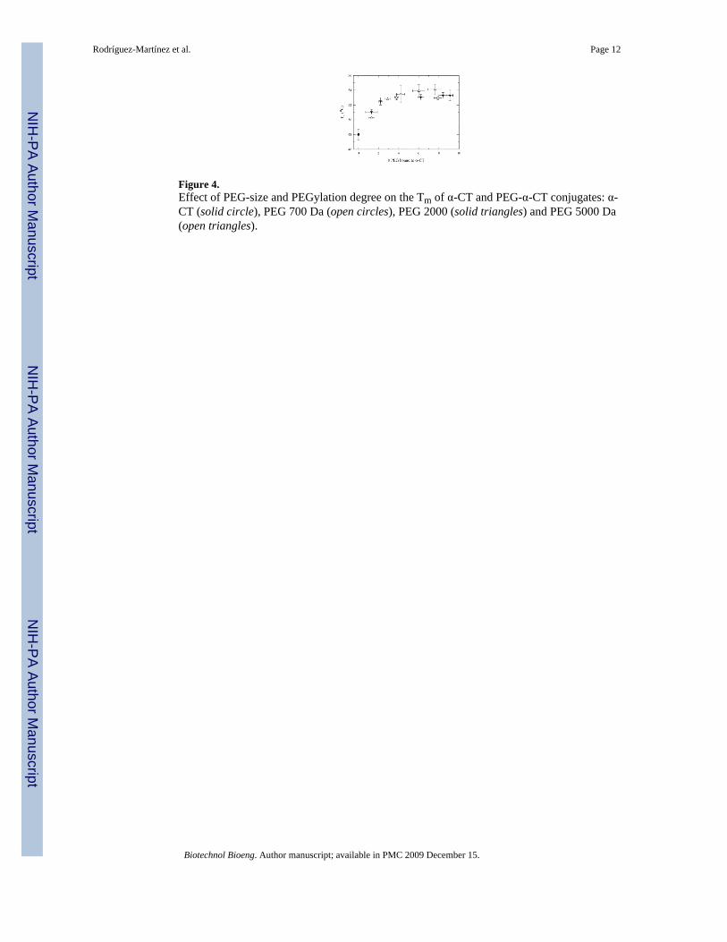
Figure 4.Effect of PEG-size and PEGylation degree on the Tm of α-CT and PEG-α-CT conjugates: α-CT (solid circle), PEG 700 Da (open circles), PEG 2000 (solid triangles) and PEG 5000 Da(open triangles).
Rodríguez-Martínez et al. Page 12
Biotechnol Bioeng. Author manuscript; available in PMC 2009 December 15.
NIH
-PA Author Manuscript
NIH
-PA Author Manuscript
NIH
-PA Author Manuscript
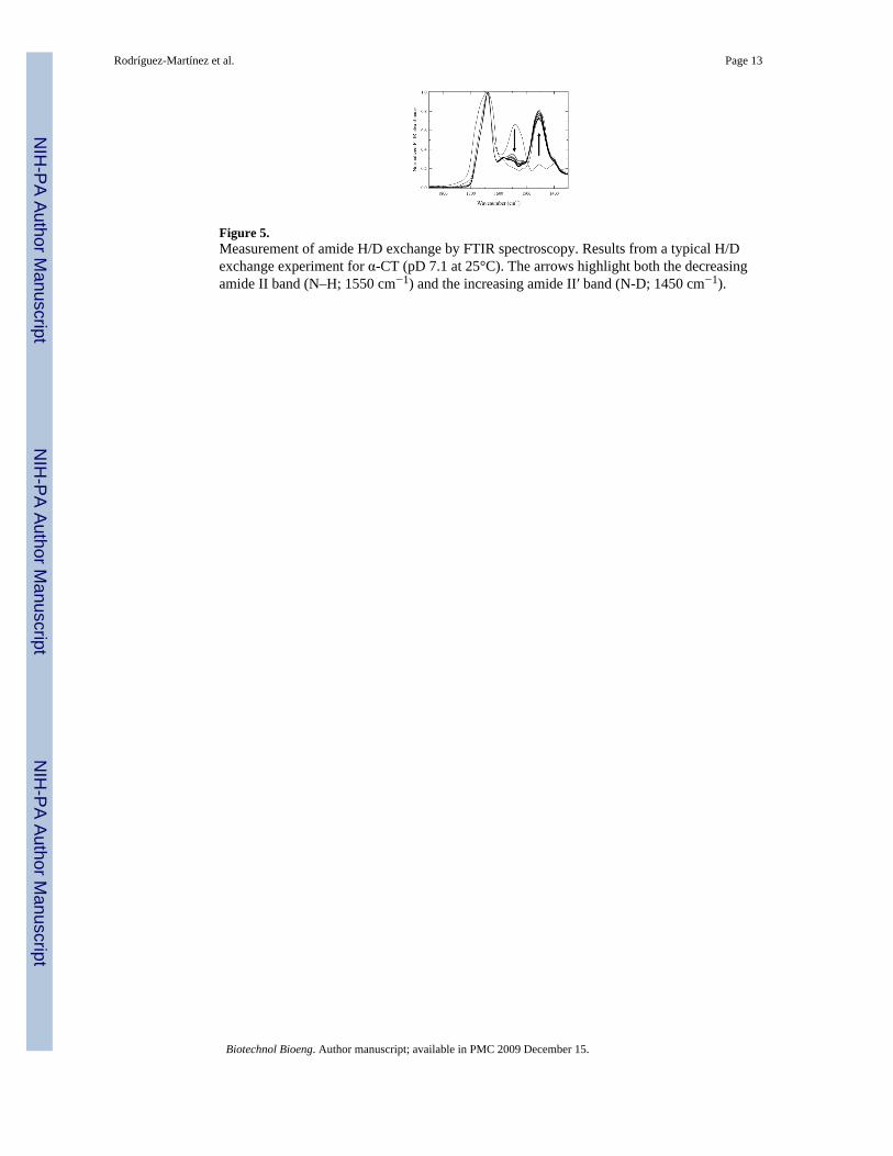
Figure 5.Measurement of amide H/D exchange by FTIR spectroscopy. Results from a typical H/Dexchange experiment for α-CT (pD 7.1 at 25°C). The arrows highlight both the decreasingamide II band (N–H; 1550 cm−1) and the increasing amide II′ band (N-D; 1450 cm−1).
Rodríguez-Martínez et al. Page 13
Biotechnol Bioeng. Author manuscript; available in PMC 2009 December 15.
NIH
-PA Author Manuscript
NIH
-PA Author Manuscript
NIH
-PA Author Manuscript
Figure 6.H/D exchange decay plots for α-CT and PEG-α-CT conjugates. (A) Effect of PEGylationdegree on H/D exchange rates of α-CT (solid circle), (PEG5000)1.3-α-CT (open circles),(PEG5000)2.9-α-CT (solid down triangles), (PEG5000)4.2-α-CT (open triangles), and(PEG5000)6.0-α-CT (solid squares). (B) Effect of the PEG size on H/D exchange rates forα-CT (solid circles), (PEG700)8-α-CT (open circles), (PEG2000)6-α-CT (solid downtriangles), and (PEG5000)8-α-CT (open triangles).
Rodríguez-Martínez et al. Page 14
Biotechnol Bioeng. Author manuscript; available in PMC 2009 December 15.
NIH
-PA Author Manuscript
NIH
-PA Author Manuscript
NIH
-PA Author Manuscript

Figure 7.Correlation between α-CT’s structural dynamics and its thermodynamic stability. α-CT(solid circle), PEG 2000 (solid down triangles) and PEG 5000 Da (open triangles). Thelinear regression was performed with the program Sigma Plot (R2 = 0.71; p < 0.01).
Rodríguez-Martínez et al. Page 15
Biotechnol Bioeng. Author manuscript; available in PMC 2009 December 15.
NIH
-PA Author Manuscript
NIH
-PA Author Manuscript
NIH
-PA Author Manuscript
NIH
-PA Author Manuscript
NIH
-PA Author Manuscript
NIH
-PA Author Manuscript
Rodríguez-Martínez et al. Page 16
Table I
Thermodynamic analysis of the differential scanning calorimetry data.
Sample/Degree of PEGylationa Tm (°C) ΔHm (kcal/mol) ΔCp (kcal/mol K) ΔGu(25 °C) (kcal/mol)
α-CT 48.0 ± 0.8 120 ± 18 3.2 ± 0.2 6 ± 1
PEG700-α-CT
3.8 ± 0.2 53.1 ± 0.4 128 ± 18 1.9 ± 0.5 9 ± 1
7.9 ± 0.4 53.0 ± 0.2 141 ± 4 2.6 ± 0.6 8.9 ± 0.8
PEG2000-α-CT
1.3 ± 0.6 51.1 ± 0.4 144 ± 4 1 ± 1 10.3 ± 0.8
2.2 ± 0.2 52.5 ± 0.8 149 ± 8 1.5 ± 0.5 10.7 ± 0.5
6.2 ± 0.3 53.1 ± 0.8 126 ± 2 0.13 ± 0.05 10.0 ± 0.5
8.4 ± 0.4 53.3 ± 0.5 123 ± 4 1.8 ± 0.5 8.3 ± 0.4
9.1 ± 0.3 53.3 ± 0.5 118 ± 5 1.2 ± 0.7 9 ± 1
PEG5000-α-CT
1.3 ± 0.3 50.3 ± 0.7 128 ± 2 4 ± 1 5.9 ± 0.9
2.9 ± 0.3 52.8 ± 0.8 127 ± 8 3 ± 2 7.1 ± 0.2
4.2 ± 0.4 53 ± 1 113 ± 25 4 ± 2 6.1 ± 0.8
6.0 ± 0.6 53.9 ± 0.8 119 ± 22 0.2 ± 0.7 9.1 ± 0.1
7.6 ± 0.7 54.1 ± 0.7 120 ± 22 −0.3 ± 2 11 ± 2
aIn mol of PEG/mol of α-chymotrypsin.
Biotechnol Bioeng. Author manuscript; available in PMC 2009 December 15.
NIH
-PA Author Manuscript
NIH
-PA Author Manuscript
NIH
-PA Author Manuscript
Rodríguez-Martínez et al. Page 17
Tabl
e II
Kin
etic
par
amet
ers d
eriv
ed fr
om a
mid
e H
/D e
xcha
nge
kine
tic a
naly
sis f
or α
-CT
and
PEG
-α-C
T co
njug
ates
.
Sam
ple/
Deg
ree
of P
EG
ylat
ion
A1a
k HX
,1 (m
in−
1 )A
2ak H
X,2
(min
−1 )
A3b
α-C
T0.
641.
660.
140.
040
0.22
PEG
700-α-
CT
3.8
± 0.
20.
661.
350.
120.
013
0.22
7.9
± 0.
40.
491.
560.
140.
034
0.37
PEG
2000
-α-C
T
1.3
± 0.
60.
531.
610.
110.
025
0.37
2.2
± 0.
20.
560.
830.
090.
025
0.35
4.2
± 0.
30.
550.
660.
120.
008
0.33
8.4
± 0.
40.
471.
960.
130.
030
0.40
9.1
± 0.
30.
512.
040.
120.
029
0.37
PEG
5000
-α-C
T
1.3
± 0.
30.
571.
140.
140.
033
0.29
2.9
± 0.
30.
450.
970.
190.
046
0.37
4.2
± 0.
40.
480.
770.
140.
018
0.38
6.0
± 0.
60.
441.
310.
160.
025
0.40
7.6
± 0.
70.
500.
840.
160.
023
0.33
a Ai i
s the
frac
tion
of a
mid
e pr
oton
s tha
t exc
hang
e w
ith a
rate
con
stan
t kH
X,i.
b A3
is th
e un
exch
ange
d fr
actio
n of
am
ide
prot
ons.
Biotechnol Bioeng. Author manuscript; available in PMC 2009 December 15.