Stability study of the human G-protein coupled receptor, Smoothened
11
Stability study of the human G-protein coupled receptor, Smoothened Rony Nehmé a , Olivier Joubert a , Michel Bidet a , Benoît Lacombe a , Ange Polidori b , Bernard Pucci b , Isabelle Mus-Veteau a, ⁎ a Université de Nice - Sophia Antipolis, CNRS UMR 6543, Institute of Developmental Biology and Cancer Research, Parc Valrose, F-06108 Nice Cedex 2, France b Université d'Avignon et des Pays du Vaucluse, Laboratoire de Chimie Bioorganique et des Systèmes Moléculaires Vectoriels, F-84000 Avignon, France abstract article info Article history: Received 25 September 2009 Received in revised form 10 February 2010 Accepted 12 February 2010 Available online 23 February 2010 Keywords: Heterologous expression GPCR Smoothened Hedgehog pathway Fluorinated surfactant Thermostability Smoothened is a member of the G-protein coupled receptor (GPCR) family responsible for the transduction of the Hedgehog signal to the intracellular effectors of the Hedgehog signaling pathway. Aberrant regulation of this receptor is implicated in many cancers but also in neurodegenerative disorders. Despite the pharmacological relevance of this receptor, very little is known about its functional mechanism and its physiological ligand. In order to characterize this receptor for basic and pharmacological interests, we developed the expression of human Smoothened in the yeast Saccharomyces cerevisiae and Smoothened was then purified. Using Surface Plasmon Resonance technology, we showed that human Smoothened was in a native conformational state and able to interact with its antagonist, the cyclopamine, both at the yeast plasma membrane and after purification. Thermostability assays on purified human Smoothened showed that this GPCR is relatively stable in the classical detergent dodecyl-β-D-maltoside (DDM). The fluorinated surfactant C 8 F 17 TAC, which has been proposed to be less aggressive towards membrane proteins than classical detergents, increased Smoothened thermostability in solution. Moreover, the replacement of a glycine by an arginine in the third intracellular loop of Smoothened coupled to the use of the fluorinated surfactant C 8 F 17 TAC during the mutant purification increased Smoothened thermostability even more. These data will be very useful for future crystallization assays and structural characterization of the human receptor Smoothened. © 2010 Elsevier B.V. All rights reserved. 1. Introduction G-protein coupled receptors (GPCRs) are transmembrane proteins that mediate most of their intracellular actions through pathways involving activation of G-proteins and arrestin [1,2]. G-proteins transmit intracellular signals to effector proteins such as enzymes and ion channels, resulting in rapid changes in the concentration of intracellular signaling molecules, cAMP, cGMP, inositol phosphates, diacylglycerol, arachidonic acid, and cytosolic ions [3,4]. The super- family of GPCRs comprises receptors for many hormones, paracrines (local hormones), neurotransmitters, and neuromodulators with important physiological functions. GPCRs represent about 30% of current drug targets and their dysfunction causes many human diseases. In the GPCR database [5], GPCRs are divided into six classes. These are the Class A Rhodopsin-like receptors, which account for over 80% of all GPCRs, Class B Secretin-like receptors, Class C Metabotropic glutamate receptors, Class D Pheromone receptors, Class E cAMP receptors and the Class F Frizzled/Smoothened family. This latter class comprises Frizzled and Smoothened receptors, which are key regulators of animal development that signal through the Wnt and Hedgehog signaling pathways, respectively. The Hedgehog (Hh) signaling pathway regulates body patterning and organ development during embryogenesis. In adults, the Hh pathway is mainly quiescent, with the exception of roles in tissue maintenance and repair, and its inappropriate reactivation has been linked to several disparate human cancers such as basal cell carcinomas and medulloblastomas [6]. The Hh pathway involves two integral membrane proteins, Patched (Ptc) and Smoothened (Smo). Ptc, a 12-transmembrane domain protein, is the receptor of the secreted protein Hedgehog (Hh). In the absence of Hh, Ptc inhibits the activity of Smo through an unknown mechanism. Hh binding to Ptc releases Smo inhibition. Activated Smo is stabilized on the plasma membrane and transduces the Hh signal to the intracellular components of the pathway, culminating in the activa- tion of the transcription factor Gli in mammals or Ci in Drosophila. Despite the important pharmacological interest of this receptor, very little is known about its functional mechanism or its physiological ligand and partners in mammals [7]. In order to better understand signaling via Smo in mammals, structural data are essential. Smo, like many other GPCRs, is present in very low amounts in native sources. In order to undergo biochemical and structural characterization, we over expressed this receptor in Biochimica et Biophysica Acta 1798 (2010) 1100–1110 Abbreviations: GPCR, G-Protein Coupled Receptor; hSmo, Smoothened; MAP, Multitag Affinity Purification; DDM, dodecyl-β-D-maltoside; FS, fluorinated surfactant; SPR, surface plasmon resonance; RU, resonance unit; Hh, Hedgehog; HA, hemagglu- tinin; CPN, cyclopamine ⁎ Corresponding author. Tel.: +33 4 92 07 68 71; fax: +33 4 92 07 68 35. E-mail address: [email protected] (I. Mus-Veteau). 0005-2736/$ – see front matter © 2010 Elsevier B.V. All rights reserved. doi:10.1016/j.bbamem.2010.02.015 Contents lists available at ScienceDirect Biochimica et Biophysica Acta journal homepage: www.elsevier.com/locate/bbamem
-
Upload
independent -
Category
Documents
-
view
3 -
download
0
Transcript of Stability study of the human G-protein coupled receptor, Smoothened

Biochimica et Biophysica Acta 1798 (2010) 1100–1110
Contents lists available at ScienceDirect
Biochimica et Biophysica Acta
j ourna l homepage: www.e lsev ie r.com/ locate /bbamem
Stability study of the human G-protein coupled receptor, Smoothened
Rony Nehmé a, Olivier Joubert a, Michel Bidet a, Benoît Lacombe a, Ange Polidori b,Bernard Pucci b, Isabelle Mus-Veteau a,⁎a Université de Nice - Sophia Antipolis, CNRS UMR 6543, Institute of Developmental Biology and Cancer Research, Parc Valrose, F-06108 Nice Cedex 2, Franceb Université d'Avignon et des Pays du Vaucluse, Laboratoire de Chimie Bioorganique et des Systèmes Moléculaires Vectoriels, F-84000 Avignon, France
Abbreviations: GPCR, G-Protein Coupled ReceptoMultitag Affinity Purification; DDM, dodecyl-β-D-maltosSPR, surface plasmon resonance; RU, resonance unit; Htinin; CPN, cyclopamine⁎ Corresponding author. Tel.: +33 4 92 07 68 71; fax
E-mail address: [email protected] (I. Mu
0005-2736/$ – see front matter © 2010 Elsevier B.V. Adoi:10.1016/j.bbamem.2010.02.015
a b s t r a c t
a r t i c l e i n f oArticle history:Received 25 September 2009Received in revised form 10 February 2010Accepted 12 February 2010Available online 23 February 2010
Keywords:Heterologous expressionGPCRSmoothenedHedgehog pathwayFluorinated surfactantThermostability
Smoothened is a member of the G-protein coupled receptor (GPCR) family responsible for the transductionof the Hedgehog signal to the intracellular effectors of the Hedgehog signaling pathway. Aberrant regulationof this receptor is implicated in many cancers but also in neurodegenerative disorders. Despite thepharmacological relevance of this receptor, very little is known about its functional mechanism and itsphysiological ligand. In order to characterize this receptor for basic and pharmacological interests, wedeveloped the expression of human Smoothened in the yeast Saccharomyces cerevisiae and Smoothened wasthen purified. Using Surface Plasmon Resonance technology, we showed that human Smoothened was in anative conformational state and able to interact with its antagonist, the cyclopamine, both at the yeastplasma membrane and after purification. Thermostability assays on purified human Smoothened showedthat this GPCR is relatively stable in the classical detergent dodecyl-β-D-maltoside (DDM). The fluorinatedsurfactant C8F17TAC, which has been proposed to be less aggressive towards membrane proteins thanclassical detergents, increased Smoothened thermostability in solution. Moreover, the replacement of aglycine by an arginine in the third intracellular loop of Smoothened coupled to the use of the fluorinatedsurfactant C8F17TAC during the mutant purification increased Smoothened thermostability even more. Thesedata will be very useful for future crystallization assays and structural characterization of the human receptorSmoothened.
r; hSmo, Smoothened; MAP,ide; FS, fluorinated surfactant;h, Hedgehog; HA, hemagglu-
: +33 4 92 07 68 35.s-Veteau).
ll rights reserved.
© 2010 Elsevier B.V. All rights reserved.
1. Introduction
G-protein coupled receptors (GPCRs) are transmembrane proteinsthat mediate most of their intracellular actions through pathwaysinvolving activation of G-proteins and arrestin [1,2]. G-proteinstransmit intracellular signals to effector proteins such as enzymesand ion channels, resulting in rapid changes in the concentration ofintracellular signaling molecules, cAMP, cGMP, inositol phosphates,diacylglycerol, arachidonic acid, and cytosolic ions [3,4]. The super-family of GPCRs comprises receptors for many hormones, paracrines(local hormones), neurotransmitters, and neuromodulators withimportant physiological functions. GPCRs represent about 30% ofcurrent drug targets and their dysfunction causes many humandiseases. In the GPCR database [5], GPCRs are divided into six classes.These are the Class A Rhodopsin-like receptors, which account forover 80% of all GPCRs, Class B Secretin-like receptors, Class CMetabotropic glutamate receptors, Class D Pheromone receptors,
Class E cAMP receptors and the Class F Frizzled/Smoothened family.This latter class comprises Frizzled and Smoothened receptors, whichare key regulators of animal development that signal through theWntand Hedgehog signaling pathways, respectively. The Hedgehog (Hh)signaling pathway regulates body patterning and organ developmentduring embryogenesis. In adults, the Hh pathway is mainly quiescent,with the exception of roles in tissue maintenance and repair, and itsinappropriate reactivation has been linked to several disparate humancancers such as basal cell carcinomas and medulloblastomas [6]. TheHh pathway involves two integral membrane proteins, Patched (Ptc)and Smoothened (Smo). Ptc, a 12-transmembrane domain protein, isthe receptor of the secreted protein Hedgehog (Hh). In the absence ofHh, Ptc inhibits the activity of Smo through an unknown mechanism.Hh binding to Ptc releases Smo inhibition. Activated Smo is stabilizedon the plasma membrane and transduces the Hh signal to theintracellular components of the pathway, culminating in the activa-tion of the transcription factor Gli in mammals or Ci in Drosophila.Despite the important pharmacological interest of this receptor, verylittle is known about its functional mechanism or its physiologicalligand and partners in mammals [7].
In order to better understand signaling via Smo in mammals,structural data are essential. Smo, like many other GPCRs, is present invery low amounts in native sources. In order to undergo biochemicaland structural characterization, we over expressed this receptor in

1101R. Nehmé et al. / Biochimica et Biophysica Acta 1798 (2010) 1100–1110
several heterologous systems [8,9]. Heterologously overproduced andpurified proteins provide a starting point for biochemical, biophysicaland structural studies, but the lack of sufficient quantities offunctional and stable membrane proteins is frequently a bottleneckhindering such studies. Recurring problems include i) recoveringadequate amounts of functional protein from the heterologousexpression system used, ii) solubilizing and purifying these proteins,iii) maintaining stability and functionality in solution after extractionfrom membranes and purification.
We show in the present study that human Smo could be expressedin the plasma membrane of the yeast Saccharomyces cerevisiaeand purified in a native and stable conformation, as assessed byantagonist-binding and Surface Plasmon Resonance studies. Ourresults indicate that human Smo is relatively stable in solution withthe classical detergent dodecyl-β-D-maltoside (DDM), and that thefluorinated surfactant C8F17TAC increases Smo thermostability insolution. We also observed a synergistic effect of the fluorinatedsurfactant C8F17TAC and the replacement of a glycine by an arginine inthe Smo third intracellular loop on the receptor thermostability.
2. Materials and methods
2.1. Construction of expression vectors
For expression in S. cerevisiae, the cDNAs from human Smo and amutant form of human Smo in which the guanine in position 1303was replaced by an adenine (giving the mutation G435R in the poly-peptide sequence) were inserted in the YEpPMA-MAP vector contain-ing the constitutive plasma membrane proton ATPase promoterand the Multitag Affinity Purification sequence as described in [8],giving the expression vectors YEpPMA-hSmo-MAP and YEpPMA-hSmoG435R-MAP respectively.
For expression inDrosophila Schneider 2 cells, thewild type and themutant human Smo cDNAs were inserted in the pAc-MAP vectorcontaining the strong constitutive actine promoter and the MultitagAffinity Purification sequence as described in [9], giving the expressionvectors pAc-hSmo-MAP and pAC-hSmo-G435R-MAP respectively.
2.2. Yeast culture
S. cerevisiae strain K699 (Mata, ura3, and leu 2–3), which wasgenerously given by R. Arkowitz, was transformed with YEpPMA-hSmo-MAP or YEpPMA-hSmo-G435R-MAP expression vector by thelithium acetate procedure [10] and grown on plates containingminimal medium and an amino-acid mixture lacking leucine. Cloneswere grown at 30 °C to an OD600 of 2 in minimal medium (0.67% yeastnitrogen base without amino acids, 0.3 mM adenine, 0.5 mM uracil,0.3 mM tyrosine and an amino acid mixture lacking leucine)supplemented with 2% D-glucose. Yeasts were diluted to an OD600 of0.1 in complete YEP medium (1.1% yeast extract, 2.2% bactopeptone,0.3 mM adenine) supplemented with 2% D-glucose, grown at 18 °Cunder 200 rpm shaking until the OD600 was 7, and centrifuged for10 min at 2000 g and 4 °C (see detailed protocol in [11].
Large-scale production of hSmo-MAP was carried out in a 40-Lfermentor (BIOSTAT C-PLUS Sartorius BBI Systems, Unité de fermen-tation IMM/IFR88 CNRS Marseille France). One liter of yeast with anOD600 of 1.5 served to inoculate 30 L of YEP medium. Yeasts weregrown at 18 °C for 24 h (2.5 OD600) under 200 rpm shaking with 30 Lof air perminute. The culturewas then oxygenated with 45 L of air perminute, kept for 6 h at 18 °C and 250 rpm shaking until the OD600 was7, and centrifuged in a Sharples AS16 centrifuge (100 L/h).
2.3. Yeast membrane preparation
All steps were performed at 4 °C. Yeast cells were washed in coldwater, re-suspended in ice-cold disintegration buffer (50 mM Tris–
HCl [pH 7.4], 500 mM NaCl, 2.5 mM EDTA, 4 mM Benzamidine and1 mM PMSF), and broken with glass beads (425–600 μm, Sigma) for15 min in a shaker at 2000 rpm (Heidolph Multi Reax shaker).Unbroken cells and nuclei were pelleted for 10 min at 3000 g andcrude extract was obtained from the supernatant. Plasma membraneswere prepared by centrifugation of crude extract for 1 h at 18,000 g.The membrane fraction was then washed twice with purificationbuffer (50 mM Tris–HCl pH (7.4), 500 mM NaCl, 10% glycerol, 4 mMBenzamidine and 1 mM PMSF). Finally, the membrane fraction wasre-suspended in purification buffer and frozen at −80 °C (for adetailed protocol, see [11]).
2.4. Solubilization and purification
Membrane fractions were thawed and simultaneously diluted to3 mg/mL with ice-cold purification buffer supplemented with 20 mMof the non-ionic detergent DDM (dodecyl-β-D-maltoside,CMC=0.15 mM). Solubilization of hSmo from the membrane wasperformed at 4 °C with gentle agitation for 1 h. The solution wascentrifuged at 100,000 g for 1 h to pellet unsolubilized material. ForSPR experiments, Smo-MAPwas purified on a calmodulin column. Thesupernatant containing solubilized hSmo was incubated with a pre-equilibrated calmodulin column in the presence of 2 mM CaCl2overnight at 4 °C on a rotating wheel. Non-specifically bound materialwas removed from the resin with several washes with purificationbuffer supplemented with 2 mM DDM and 2 mM CaCl2. In order toexchange DDM with the fluorinated surfactants C6F13C2H4-S-poly-Tris-(hydroxymethyl) aminomethane (C6F13TAC) (CMC=0.3 mM),or C8F17C2H4-S-poly-Tris-(hydroxymethyl) aminomethane(C8F17TAC) (CMC=0.03 mM) [12,13], the resin was washed 3 timeswith 5-column volumes of purification buffer supplemented with2 mMCaCl2 and either 2 mMC6F13TAC or 2 mMC8F17TAC. Elutionwascarried out with purification buffer supplemented with 5 mM EGTAand the appropriate surfactant. Aliquots were collected throughoutthe purification process, and analyzed by western-blot. For massspectrometry analysis, hSmoG435R-MAP was purified on Ni/NTAagarose (Qiagen) and on Streptavidin sepharose (Amersham). Thesolubilized fraction supplemented with 10 mM imidazole wasincubated overnight at 4 °C on Ni/NTA resin equilibrated withpurification buffer containing 2 mM of DDM and 10 mM of imidazole.The column was washed 3 times with purification buffer containing50 mM of imidazole, and hSmoG435R-MAP was eluted with a buffercontaining 400 mM of imidazole. Eluted fractions were pooled andloaded on the streptavidin column, for 1 h at 4 °C. Finally,hSmoG435R-MAP was eluted and loaded on SDS-PAGE for western-blotting and Coomassie blue staining. The band corresponding tohSmoG435R-MAP was cut and analyzed by mass spectrometry(MALDI-TOF-TOF 4800 Applied Biosystems, IPMC Sophia Antipolis).
2.5. MAP peptide expression and purification
The MAP sequence was inserted into the XhoI and NheI restrictionsites of the pBAD vector containing the inducible arabinose promoter(kind gift from P. Demange). The Escherichia coli strain MC1061 wastransformed with the vector pBAD-MAP. Bacteria were grown in LBmedium supplemented with ampicillin at 37 °C. At an OD600 of 0.8,0.4% of L-arabinose was added to the culture for 4 h at 37 °C to induceMAP peptide expression. The bacteria were then centrifuged and thepellet was washed with ice-cold water and then with ice-cold PBSbuffer supplemented with 1 mM PMSF and 4 mM Benzamidine. Thebacteria were sonicated for 5 min and centrifuged. CaCl2 was thenadded to the supernatant to a final concentration of 2 mM prior toincubation for 1 h with calmodulin resin pre-equilibrated in PBSbuffer supplemented with 1 mM PMSF, 4 mM benzamidine and 2 mMCaCl2. After several washes with equilibration buffer, theMAP peptidewas eluted with PBS buffer supplemented with 1 mM PMSF, 4 mM

1102 R. Nehmé et al. / Biochimica et Biophysica Acta 1798 (2010) 1100–1110
Benzamidine and 5 mM EGTA. Purified MAP peptide was used as astandard to quantify hSmo-MAP.
2.6. Protein quantification
The proteins were quantified using the Bio-Rad protein assay.
2.7. SDS-PAGE and western-blotting
Protein samples were separated on SDS-PAGE (acrylamide/bis-acrylamide: 8% for hSmo-MAP or hSmoG435R-MAP and 15% for MAPpeptide). For western-blot analysis, gels were transferred to nitrocel-lulose using standard techniques. Blots were first incubated for 1 h atroom temperature in the blocking buffer (20 mM Tris–HCl (pH 7.4),450 mM NaCl, 0.1% Tween-20 supplemented with 4% non-fat milk)and probed overnight at 4 °C with monoclonal mouse anti-HA anti-bodies (laboratory-made, dilution 1:20). The blots were then washedthree times in the blocking buffer. Secondary polyclonal goat anti-mouse immunoglobulins coupled to horseradish peroxidase (Dako)(dilution 1:5000) were then applied for 2 h at 4 °C. Revelation wascarried out using ECL kit (Amersham Biosciences), a CCD camera andthe Las 3000 system® (Fuji).
2.8. Native gel
Protein samples were separated on native gel under non-denaturing conditions in which acrylamide gels, running, loadingand transfer buffers lacked SDS. The gels were then transferred tonitrocellulose membrane and probed with anti-HA antibodies.
2.9. Fluorescent ligand binding measurements
100 µg of membrane preparation were incubated in PBS buffer andincreasing concentrations of Bodipy-cyclopamine (Bo-CPN) (between 0and 25 nM) 4 h at RT, centrifuged for 1 h at 25,000 g and re-suspendedin 1 mL of PBS buffer. The fluorescence measurements were realized ona spectrofluorimeter SAFAS FLX-Xenius. Thefluorescence variationsΔF/Fwere calculated for each concentration of Bo-CPN and analyzed usingOrigin software. The Bo-CPN standard curve was realized bymeasuringthe fluorescence variations before centrifugation.
2.10. Surface plasmon resonance (SPR) experiments
SPR experiments were performed on a Biacore 3000 (Biacore/GEHealthcare Uppsala, Sweden) with a CM5 sensor chip from Biacore/GE Healthcare. A stock solution of N-(2-Aminoethyl) cyclopamine inDMSO (5 mg/mL) was prepared and diluted 1:10 in sodium acetatebuffer pH 5.5 for covalent amine-coupling to the CM5 sensor chip.Flow cell 2 (Fc2) was activated with 200 mM N-ethyl-N-(3-dimethy-laminopropyl)-carbodiimide (EDC) and 50 mM N-hydroxysuccini-mide (NHS), and 100 μL (50 µg) of N-(2-Aminoethyl) cyclopaminewere injected at 10 μL/min. The surface was deactivated withethanolamine. Flow cell 1 (Fc1) was activated and deactivated, andused as a reference flow cell. The surfaces were then washed withthree pulses of 50 mM NaOH (15 μL at 5 μL/min).
Experiments on membrane preparations were carried out at 25 °C.50 µL of membrane preparation from hSmo-expressing yeasts or fromyeasts that do not express hSmo, were diluted in HBS-N runningbuffer (10 mM HEPES (pH 7.4) and 150 mM NaCl, GE Healthcare) at100 µg/mL and injected over both the reference (Fc1) and the CPN-coupled (Fc2) flow cells at a flow rate of 5 µL/min. The differencebetween sensorgrams recorded on Fc2 and Fc1 was reported. Thesensor chip surfacewas regenerated twicewith 10 μL of 50 mMNaOH,0.05% SDS at 20 μL/min.
Experiments on purified hSmo were performed at 10 °C. Purifica-tion buffer containing 2 mM of surfactant was used as a running
buffer, otherwise the specific buffer was stated. 40 μL (80 ng) ofpurified hSmowere injected on both the reference (Fc1) and the CPN-coupled (Fc2) flow cells at a flow rate of 10 μL/min. The differencebetween the sensorgrams recorded on Fc2 and Fc1 was reported. Thesensor chip surfacewas regenerated twicewith 10 μL of 50 mMNaOH,0.05% SDS at 20 μL/min.
To determine the stability of hSmo in surfactants over time,purified hSmo fractionswere stored at 4 °C and bindingwasmeasured10, 20 and 30 days after purification. The percentages of remainingbinding compared to initial binding measured 1 day after purificationwere calculated from at least two independent experiments.
For thermostability assays, sensorgrams were recorded afterincubating purified Smo for 30 min at temperatures ranging from 4to 70 °C prior to injection on the reference (Fc1) and the CPN-coupled(Fc2) flow cells at a flow rate of 10 μL/min. The percentage of re-maining binding after incubation was determined with respect to itsown unheated control. In order to determine the inflection pointof the melting curves, which was assumed to equal the meltingtemperature (Tm), a Boltzmann sigmoid equation was fitted to theraw data with the Origin software.
To determine the kinetic constants of the CPN-Smo binding, Fc2, 3and 4 of a new CM5 sensor chip were coupled with different amountsof N-(2-Aminoethyl) cyclopamine, Fc1 being the reference flow cell.Increasing concentrations of purified Smo were injected onto the fourflow cells and the differences between the sensorgrams recorded oneach CPN-coupled flow cell and Fc1 were reported and analyzed usingBiaEva software.
2.11. Surface Immunofluorescent labeling of Drosophila Schneider 2 cells
S2 cells were cultured and transiently transfected with 0.5 μg ofpAc-hSmo-MAP or pAc-hSmo-G435R-MAP as described in [9]. Threedays after transfection, the cells were treated or not with Hh-conditioned medium, and cell surface labeling was performed asdescribed in [9] by incubating cells on ice for 1 h with a rabbitpolyclonal antibody raised against amino acids 488–787 of humanSmo (1:100; N-300; Santa Cruz Biotechnology) in S2 mediumcontaining 10% of fetal bovine serum. After dilution and centrifuga-tion, the cells were incubated on ice for 30 min with a rhodaminefluorescent secondary antibody (Alexa 568 goat anti-rabbit IgG(1:500; Molecular Probes)) in S2 medium containing 10% of fetalbovine serum. After dilution and centrifugation, the cells were re-suspended in phosphate-buffered saline plus 1% formaldehyde.Rhodamine fluorescence was analyzed by fluorescence microscopyand image acquisition was performed using a confocal system (ZeissLSM510 Meta) with an objective Plan Apochromat ×63/1.4 oildifferential interference contrast. Cell fluorescence was analyzedusing Image J software.
3. Results
3.1. Expression, optimization and large-scale production ofhuman Smoothened
Human Smoothenedwas expressed in the yeast S. cerevisiaewith amultitag affinity purification sequence of 165 amino acids fused at itsC-terminus (hSmo-MAP, Fig. 1A) allowing detection of hSmo bywestern-blotting using antibodies directed against the hemagglutinin(HA) tag, and purification of hSmo using three affinity resins(calmodulin, streptavidin, Ni–NTA), as already described in [8].Expression of hSmo-MAP was improved and subsequently scaledup.We evaluated the impact of aeration on the hSmo-MAP expressionlevel by growing the yeast cells with different medium/air volumeratios of 1/10, 1/7, 1/5 and 1/4 in a 2-L flask. Yeasts were grown untilthe OD600 was 7 and plasmamembranes were prepared. Expression ofhSmo-MAP in each culture condition was checked by western-blot

Fig. 1. Expression of human Smoothened in S. cerevisiae. A. MAP sequence fused at hSmo C-terminus. The MAP sequence is composed of 165 amino acids: three affinity purificationtags (a calmodulin binding domain (CBD), a streptavidin tag and a hexa-histidine tag), the hemagglutin A epitope, and cleavage sites for factor Xa, TEV and thrombin. B. Effect of theyeast culture oxygenation on hSmo-MAP expression level. Western-blot with plasma membrane preparations from S. cerevisiae expressing hSmo-MAP cultured with indicatedmedium/air ratio (v/v): 1/10 (lane 1), 1/7 (lane 2), 1/5 (lane 3) and 1/4 (lane 4) (respectively 200 mL, 300 mL, 400 mL and 500 mL of YEPmedium in a 2-L flask). Samples were runon SDS-PAGE, transferred to a nitrocellulose membrane and probed with anti-HA antibodies. C. Large-scale production of hSmo-MAP in a 40-L fermentor. Plasma membranes wereprepared from hSmo-MAP-expressing yeast grown in 2-L flasks (lane 1) or in 40-L fermentors (lane 2). 40 μg of membranes were loaded on SDS-PAGE, western-blotted and probedwith anti-HA antibodies.
Fig. 2. Quantification of human Smoothened expression using the MAP peptide as astandard. MAP was expressed in E. coli and purified on calmodulin resin. Differentquantities of purified MAP peptide (lane 1: 10 ng, lane 2: 25 ng, lane 3: 50 ng, lane 4:100 ng, lane 5: 200 ng) and 5 μg of membrane fraction from yeast expressing hSmo-MAP (lane 6) were loaded on SDS-PAGE, transferred to a membrane and probed withanti-HA antibodies. The intensities of the bands corresponding to theMAP peptide wereestimated by densitometry using the ImageJ software and used to draw the MAPstandard curve. The intensity of hSmo-MAP band was reported on this standard curveto estimate the quantity of hSmo-MAP produced.
1103R. Nehmé et al. / Biochimica et Biophysica Acta 1798 (2010) 1100–1110
analysis (Fig. 1B) which showed a major band around 90 kDa close tothe predicted size of hSmo-MAP (100 kDa) and a weaker band around65 kDa. The highest expression level of hSmo-MAP was obtained inthe higher oxygenation conditions (medium/air volume ratios of 1/10 and 1/7). We used the ratio 1/7 for yeast cultures grown in flasks.This experiment allowed air/volume adaptation in the 40-L fermentorfor hSmo-MAP scale-up production. Western-blot analysis (Fig. 1C)shows that the quantity of hSmo-MAP expressed at the yeast plasmamembrane was comparable in 2-L flask and 40-L fermentor cultures.
To quantify the amounts of hSmo-MAP expressed on the yeastplasma membrane, the production of the MAP tag alone was neces-sary. Therefore we carried out the expression of the MAP peptide in E.coli and the peptide was then purified using calmodulin affinity resin.Purified MAP peptide was used as a standard to quantify hSmo-MAPon western-blots with anti-HA antibodies (Fig. 2). The expressionlevel of hSmo-MAPwas estimated at about 50 pmol/mg of membraneprotein, which in mass represents 150 μg of hSmo-MAP per liter ofyeast culture. However, due to the fact that the transfer of thehydrophobic protein hSmo-MAP was surely less efficient than that ofthe soluble peptide MAP, this value could have been underestimated.
3.2. Antagonist-binding properties of Smoothened expressed on the yeastplasma membrane
In order to find out if hSmo-MAP was expressed in S. cerevisiaein a native conformation, its ability to interact with its antagonistcyclopamine (CPN) was measured.
Binding experiments using a fluorescent derivative of thecyclopamine, the Bodipy-cyclopamine (Bo-CPN) (Fig. 3A) werecarried out on membrane prepared from yeast expressing hSmo-MAP and control yeast. The fluorescence variations associated withhSmo-MAP-containing membranes as a function of the concentrationof Bo-CPN allowed the calculation of apparent KD values between1.7 nM and 5.7 nM, and of the number of active hSmo-MAP per mg ofmembrane proteins between 20 and 50 pmol depending on mem-brane preparation (Fig. 3A).
Biacore surface plasmon resonance (SPR) technology forms thebasis of a rapid and reliable automated biosensor system and allowsreal-time detection of binding without labeling [14]. One of thebinding partners is immobilized on the sensor chip surface and theother is passed over it in solution. Binding causes a change in therefractive index at the biosensor surface which is transformed into a
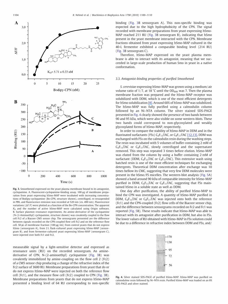
Fig. 3. Smoothened expressed on the yeast plasma membrane bound to its antagonist,cyclopamine. A. Fluorescent-cyclopamine-binding assay. 100 µg of membrane prepa-ration from yeast expressing hSmo-MAP were incubated with increasing concentra-tions of Bodipy-cyclopamine (Bo-CPN, structure shown), centrifuged, re-resuspendedin PBS, and fluorescence emission was recorded at 520 nm (ex. 490 nm). Fluorescencevariations (ΔF/F) were plotted as a function of the Bo-CPN concentration. The apparentKD and the number of active hSmo-MAP were calculated using Origin software.B. Surface plasmon resonance experiments. An amine-derivative of the cyclopamine(N-(2-Aminoethyl) cyclopamine, structure shown) was covalently coupled to the flowcell Fc2 of a Biacore CM5 sensor chip. The sensorgrams presented are the differencebetween signals recorded on the CPN-coupled flow cell Fc2 and on the reference flowcell. 50 μL of membrane fraction (100 μg/mL) from control yeasts that do not expresshSmo (sensorgram A), from 2 L flask-cultured yeast expressing hSmo-MAP (sensor-gram B), and from fermentor-cultured yeast expressing hSmo-MAP (sensorgram C),were injected over both Fc1 and Fc2.
Fig. 4. Silver stained SDS-PAGE of purified hSmo-MAP. hSmo-MAP was purified oncalmoduline resin followed by Ni–NTA resin. Purified hSmo-MAP was loaded on an 8%SDS-PAGE and silver stained.
1104 R. Nehmé et al. / Biochimica et Biophysica Acta 1798 (2010) 1100–1110
measurable signal by a light-sensitive detector and expressed asresonance units (RU) on the recorded sensorgram. An amine-derivative of CPN, N-(2-aminoethyl) cyclopamine (Fig. 3B) wascovalently immobilized by amine-coupling on the flow cell 2 (Fc2)of a CM5 sensor chip producing a change of the refractive index of theFc2 surface of 3600 RU. Membrane preparations from yeast that do ordo not express hSmo-MAP were injected on both the reference flowcell (Fc1), and the measure flow cell (Fc2) coupled to CPN (Fig. 3B).Membrane preparations from yeasts that do not express hSmo-MAPpresented a binding level of 64 RU corresponding to non-specific
binding (Fig. 3B sensorgram A). This non-specific binding wasexpected due to the high hydrophobicity of the CPN. The signalrecorded with membrane preparations from yeast expressing hSmo-MAP reached 211 RU (Fig. 3B sensorgram B), indicating that hSmopresent in the yeast membrane interacted with the CPN. Membranefractions obtained from yeast expressing hSmo-MAP cultured in the40-L fermentor exhibited a comparable binding level (214 RU)(Fig. 3B sensorgram C).
Therefore, hSmo-MAP expressed on the yeast plasma mem-brane is able to interact with its antagonist, meaning that we suc-ceeded in large-scale production of human Smo in yeast in a nativeconformation.
3.3. Antagonist-binding properties of purified Smoothened
S. cerevisiae expressing hSmo-MAPwas grown using amedium/airvolume ratio of 1/7, at 18 °C until the OD600 was 7. Then the plasmamembrane fraction was prepared and the hSmo-MAP receptor wassolubilized with DDM, which is one of the most efficient detergentsfor hSmo solubilization [8]. Around 60% of hSmo-MAPwas solubilized.The hSmo-MAP was fully purified using a calmodulin columnfollowed by an Ni–NTA column. The silver stained SDS-PAGEpresented in Fig. 4 clearly showed the presence of two bands between90 and 95 kDa, which were also visible on some western-blots. Thesetwo bands could correspond to non-glycosylated and weaklyglycosylated forms of hSmo-MAP, respectively.
In order to compare the stability of hSmo-MAP in DDM and in thefluorinated surfactants (FSs) C6F13TAC, or C8F17TAC [12,13], DDMwasexchangedwith FSs on the calmodulin resin during thewashing steps.The resin was incubated with 5 volumes of buffer containing 2 mM ofC6F13TAC or C8F17TAC, slowly centrifuged and the supernatantremoved. This step was repeated 3 times before elution. hSmo-MAPwas eluted from the column by using a buffer containing 2 mM ofsurfactant (DDM, C6F13TAC or C8F17TAC). This extensive wash usingbatched resin is one of the most efficient techniques for exchangingdetergents. Theoretical DDM concentration after exchange was 10times bellow its CMC, suggesting that very few DDM molecules werepresent in the hSmo/FS micelles. The western-blot analysis (Fig. 5A)showed a band around 90 kDa of comparable intensity for hSmo-MAPpurified in DDM, C6F13TAC or C8F17TAC, suggesting that FSs main-tained hSmo in a soluble state as well as DDM.
One day after purification, the ability of purified hSmo-MAP tobind the CPN was investigated. A quantity of hSmo-MAP purified inDDM, C6F13TAC or C8F17TAC was injected onto both the reference(Fc1) and the CPN-coupled (Fc2) flow cells of the Biacore sensor chip,and the difference between sensorgrams recorded on Fc2 and Fc1 wasreported (Fig. 5B). These results indicate that hSmo-MAP was able tointeract with its antagonist after purification in DDM, but also in FSs.The lower values of RU obtainedwith hSmo-MAP in FSs solution couldbe due to a difference in refractive index between DDM and FSs, and/

1105R. Nehmé et al. / Biochimica et Biophysica Acta 1798 (2010) 1100–1110
or to the difference of hydrophilicity of the surfactant polymer heads.The fact that the specific refractive index increments (dñ/dc) of thesurfactants used to purify hSmo were different (0.14 for DDM and0.08 for FSs) and that these surfactants may have an effect on thebinding response recorded by SPR, made it difficult to comparebetween the signals recorded in the presence of the differentsurfactants. However, in order to clarify this point, different amountsof cyclopamine were immobilized on Fc2 (11825 RU), Fc3 (8345 RU)and Fc4 (2971 RU) of a new CM5 sensor chip, the Fc1 being thereference flow cell. Using buffer containing 2 mM DDM as runningbuffer, we observed that the injection of buffer containing 2 mM ofC8F17TAC had a different effect depending on the amount of CPN-coupled: it was positive in the absence of CPN (on Fc1) and negative inthe presence of CPN, the negative response being proportional to theamount of coupled-CPN (Fig. S1A). This result suggests that eachsurfactant interacts with the CPN coupled on the flow cells and has adifferent effect on the SPR response. To further consider surfactanteffects a second experiment was carried out in which hSmo purified inDDM, C6F13TAC, C8F17TAC was diluted ten times in the DDM-containing running buffer and injected on Fc1 and Fc2. Thesensorgrams resulting from the difference between signals recordedon Fc2 and Fc1 were very different, suggesting that the surfactantsurrounding hSmo had an effect on the CPN and the SPR response.
Fig. 5.DDM and fluorinated surfactants preserved Smoothened integrity after purification. A.on SDS-PAGE and western-blotted with anti-HA antibodies. B. SPR measurements showingMAP in DDM, in C8F17TAC or in C6F13TAC were injected over both Fc1 (reference flow cell) anin resonance units (RU) are the differences between sensorgrams recorded on Fc2 and Fcresponses of increasing concentrations of hSmo-MAP purified in DDM. 50 µL of increasing coCPN-coupled Fc2. The differences between sensorgrams recorded on Fc2 and Fc1 at each hSpurified in DDM and in C8F17TAC. Sensorgrams recorded at increasing concentrations of hSmcalculated kinetic constants were reported.
However, after dissociation time, recorded sensorgrams showedsimilar RU values suggesting a recovery of DDM signals when FS-purified Smo was diluted in DDM-containing buffer (Fig. S1B).
As a direct comparison of binding responses of hSmo purified inthe different surfactants was not possible, kinetic constants of hSmo/CPN binding were determined. Increasing concentrations of hSmopurified in DDM or FSs were injected on the flow cells coupled todifferent quantities of CPN and the sensorgrams recorded from thedifference between CPN-coupled Fc and the reference Fc1 weresuperposed and analyzed using BiaEva software (Fig. 5C and D). Theaffinity constants of hSmo purified in DDM or C8F17TAC for itsantagonist were of the same order (5.8 nM and 2 nM respectively).We also calculated from the Rmax values that the same amounts ofhSmo-MAP purified in DDM or in C8F17TAC (0.9 nmol) interactedwiththe CPN coupled to the sensor chip per mg of protein injected. Thesevalues, which represent about 10% of the injected samples, werecalculated considering that the totality of the injected sample couldinteract with the cyclopamine coupled to the sensor chip. However,since the experiments were carried out with a continuous flow, it wasimpossible to know the volume of sample that was really in contactwith the cyclopamine. Unfortunately, the technique used did notpermit calculation of the number of active receptors, but just allowedus to compare the amount of hSmo that interacted with cyclopamine
hSmo-MAP purified in DDM (lane 1), C8F17TAC (lane 2) or C6F13TAC (lane 3) was loadedthat purified hSmo-MAP-bound to its antagonist, cyclopamine. 40 μL of purified hSmo-d Fc2 (on which CPNwas immobilized by amine-coupling). The SPR responses reported1, and correspond to the specific binding of hSmo-MAP on immobilized CPN. C. SPRncentrations of hSmo-MAP purified in DDM were injected over both reference Fc1 andmo-MAP concentration are presented. D. CPN-binding kinetic constants for hSmo-MAPo-MAP purified in DDM and in C8F17TAC were analyzed using BiaEva software and the

1106 R. Nehmé et al. / Biochimica et Biophysica Acta 1798 (2010) 1100–1110
in each injected sample. Taking into account this point and the factthat the samples injected onto the sensor chip were purified on thecalmodulin column and only gave a purity yield of about 60 to 70%, theamount of active hSmo after purification in DDM and in C8F17TACshould be clearly higher than 10%.
3.4. Smoothened stability in solution
To compare the stability of purified detergent solubilized hSmoreceptor, we developed two stability assays based on SPR.
Time-dependent stability was determined from SPR bindingassays 10, 20 and 30 days after purification in the different surfactantswith hSmo-MAP samples kept at 4 °C. Results showed that 1 monthafter purification, 45% of the initial hSmo still bound to the CPN inDDM, and that this value was slightly higher in FSs (55% and 50% forC6F13TAC and C8F17TAC respectively) (Fig. 6A). Our results indicatedthat FSs were at least as efficient as DDM in maintaining hSmo insolution and in preserving hSmo–cyclopamine binding over time afterpurification.
The thermostability of hSmo-MAP purified in DDM and in FSs wasdetermined from SPR binding assays on hSmo-MAP incubated for30 min at increasing temperatures ranging from 10 to 70 °C prior toinjection on the reference (Fc1) and the CPN-coupled (Fc2) flow cellsof the Biacore sensor chip. The difference between sensorgramsrecorded on Fc2 and Fc1 was reported, giving the binding level foreach sample. Themaximal level of binding was determined on controlsamples kept on ice. The percentage of remaining native receptorswas determined by comparing the amount of binding (in RU) afterheating with its unheated control (Fig. 6B). The apparent Tm, definedas the temperature at which 50% of the binding activity remained aftera 30-min incubation was calculated and reported. The hSmo-MAP inDDM showed apparent Tm values of 40.7±2.3 °C. The apparent Tm in
Fig. 6. Smoothened stability in solution. A. Time-dependent stability of hSmo-MAP indifferent surfactants. SPR responses were recorded 10, 20 and 30 days after purification,samples being kept at 4 °C. Remaining hSmo-MAP binding is represented aspercentages of the initial binding recorded 1 day after purification. B. Thermostabilityof purified hSmo-MAP. Samples of hSmo-MAP purified in DDM, C6F13TAC, or C8F17TACwere heated for 30 min at increasing temperatures and injected on the Biacore sensorchip to determine remaining hSmo-MAP binding activity, which was normalizedagainst the unheated control in each surfactant (100%). Data points are from duplicatemeasurements and are representative of at least two independent experiments.Apparent Tm values determined from the curves by nonlinear regression are indicated.
C6F13TAC was lower (33.6±2.4 °C) than that in DDM, whereas the Tmwas shifted to 45.1±0.8 °C when hSmo-MAP was purified inC8F17TAC.
Samples of hSmo-MAP purified in DDM, C8F17TAC and C6F13TACheated at increasing temperatures were mixed with loading buffer,loaded on SDS-PAGE and western-blotted with anti-HA antibodies(Fig. S2A). The western-blots showed that heating Smo did not inducethe appearance of new bands, suggesting that the loss of antagonist-binding was not due to proteolysis. The band at 90 kDa was quantifiedusing Image J software and the percentage of remaining Smo at90 kDa was plotted as a function of the temperature using Originsoftware (Fig. S2B). For hSmo-MAP purified in DDM, the 90-kDa bandbegan to decrease at 40 °C, the Tm value being around 60 °C. ForhSmo-MAP in C8F17TAC, the 90-kDa band decreased by 20% at 20 °C,was stable until 40 °C, and then decreased as for Smo in DDM with aTm value around 60 °C. These data suggest that the 20% decreaseobserved at 20 °C in the antagonist-binding activity could be due to aprecipitation of Smo: it is possible that some of the Smo proteins didnot get enough C8F17TAC molecules to stabilize them. This could alsoexplain why the thermal transition was less cooperative in C8F17TACthan in DDM. For hSmo-MAP purified in C6F13TAC, the Tm valuecalculated from the intensity of the 90-kDa band was around 35 °C,which was in good agreement with the Tm value calculated fromthe antagonist-binding activity. These data suggest that the loss ofantagonist-binding after heating could be due to a progressive un-folding followed by the precipitation of Smo in DDM and in C8F17TAC,and to a more rapid precipitation in C6F13TAC.
SPR and western-blot analyses clearly indicated that hSmo-MAPwas at least as stable in the FS C8F17TAC as in the classical detergentDDM, and less stable in C6F13TAC. The 12 °C shift in thermostabilitybetween the two FSs could be due to the length of the fluorocarbonchain, which is longer for the C8F17TAC and would better protectmembrane proteins.
3.5. Characterization of the Smoothened mutant G435R
This mutant is the result of an error introduced during the PCRcarried out to modify the 5′ and 3′ extremities of hSmo cDNA forcloning into the YEpPMA-MAP expression vector. The guanine inposition 1303 was substituted for adenine giving an arginine insteadof a glycine in position 435 of the third intracellular loop of hSmo.
Yeasts expressing hSmo-MAP or hSmoG435R-MAP were grown inthe same conditions, membrane proteins were prepared and loadedon SDS-PAGE for western-blotting with anti-HA antibodies (Fig. 7A).The first observation was that, in the line where membranes preparedfrom yeast expressing hSmoG435R-MAP were loaded, anti-HA anti-bodies did not detect a band at 90 kDa as for hSmo-MAP but only theband at 65 kDa. The second observation was that the densitometricmeasure of the 65-kDa band corresponding to hSmoG435R-MAP was2.5 times that of the 90-kDa corresponding to hSmo-MAP. Using thepurified MAP peptide as a standard, we could estimate that 120 pmolof hSmoG435R-MAP was produced per mg of membrane proteins,which corresponded to 360 μg/L of yeast culture. The western-blotpresented in Fig. 7A also shows that the 90-kDa band disappearedafter heating at 95 °C in the loading buffer whereas the 65-kDa bandwas not affected by heating. This suggests that the 90-kDa protein andthe 65-kDa protein have differences in their folding. Membranepreparations from yeast expressing hSmo-MAP and yeast expressinghSmo-G435R-MAP were loaded on a native gel and the western-blotrevealed with anti-HA antibodies showed that both proteins migratedidentically on this native gel, with a stronger signal for hSmoG435R-MAP than for hSmo-MAP (Fig. 7B). Moreover, we carried outdigestions with increasing amounts of trypsine on purified hSmo-MAP and hSmoG435R-MAP. The western-blot analysis showed thattrypsine did not enhance the 65-kDa band for hSmo-MAP butproduced only one C-terminal fragment around 35 kDa both with

Fig. 7. Expression of the G435R-mutant Smoothened. A. Plasma membranes prepared from yeasts expressing hSmoG435R-MAP (lanes 1 and 2) or hSmo-MAP (lanes 3 and 4). 80 μgof membranes were heated for 2 min at 95 °C in loading buffer (lanes 2 and 4) or not (lanes 1 and 3) and loaded on SDS-PAGE, western-blotted and probed with anti-HA antibodies.Arrows indicate the 90-kDa and the 65-kDa bands. B. Membranes prepared from yeasts expressing hSmo-MAP (lane 1) or hSmoG435R-MAP (lane 2) were loaded on a native gel,western-blotted and probed with anti-HA antibodies. C. Coomassie blue stained gel of purified hSmoG435R-MAP. hSmoG435R-MAPwas purified in DDM onNi/NTA and streptavidincolumns successively, loaded on a SDS-PAGE and stained by Coomassie blue. The 65-kDa band indicated by an arrow was cut for mass spectrometry analysis. D. Amino-acidsequence of human Smoothened (Homo sapiens) (from NP 005622; NCBI, http://www.ncbi.nlm.nih.gov.gate1.inist.fr). Peptides detected by mass spectrometry from the cut 65-kDaband are in bold and underlined. They occur in the N-terminal domain, the first extracellular loop, the 2nd intracellular loop, the 3rd intracellular loop, the 3rd extracellular loop, the7th TMS and the C-terminal domain of the human receptor Smoothened. E. CPN-binding of membrane-bound G435R-mutant Smoothened measured by SPR. 50 μL of membranes(100 μg/mL) prepared from control yeasts that do not express hSmo, from yeasts expressing hSmo-MAP, and from yeasts expressing hSmoG435R-MAP were injected over both thereference flow cell Fc1 and the CPN-coupled flow cell Fc2 of a CM5 sensor chip. The sensorgrams presented show the difference between signals recorded on Fc2 and Fc1.
1107R. Nehmé et al. / Biochimica et Biophysica Acta 1798 (2010) 1100–1110
wild-type and mutant Smo (Fig. S3). This result suggests that the 65-kDa band observed on hSmo-MAP western-blot does not correspondto a proteolysed form of Smo. Taken together, these observationssuggested that the mutant receptor hSmo-G435R-MAP could have amore stable conformation making it less denatured by SDS whichcould explain the faster running on SDS-PAGE.
However, in order to verify that the special migration ofhSmoG435R-MAP was not due to a truncated protein, hSmoG435R-MAP was purified in DDM on an Ni/NTA column followed by astreptavidin column. The 65-kDa band observed on the Coomassieblue stained SDS-PAGE (Fig. 7C) was cut and analyzed byMALDI-TOF–TOF mass spectrometry. Several peptides were identified between theamino acids number 66 and 575 (Fig. 7D). Moreover, the 65-kDa bandbeing detected by anti-HA antibodies on western-blots indicated thatthe MAP sequence was present at the C-terminal extremity of mutantSmo. The 65 N-terminus residues could have been truncated resultingin 8 kDa deletion but this cannot explain the running at 65 kDa of theprotein. Therefore, the data suggested that the 65-kDa band wouldcorrespond to the full-length MAP-tagged hSmoG435R protein andwould result from atypical running behavior.
3.5.1. Antagonist-binding properties of the Smoothened mutant G435RWe tested the ability ofmembrane-boundhSmoG435R-MAP to bind
to the Smo antagonist cyclopamine by SPR studies. Membranepreparations from yeast either expressing hSmo-MAP or hSmoG435R-MAP, or from a control yeast strain that did not express hSmo, wereinjected onto the reference flow cell (Fc1) and the measure flow cell(Fc2) coupled to the CPN.We observed thatmembranes prepared fromyeast expressinghSmoG435R-MAPgavea comparablebinding responseto membranes prepared from yeast expressing hSmo-MAP (Fig. 7E),indicating that themutation G435R did not prevent antagonist-binding.However, we expected a higher binding activity with hSmoG435R-MAP-bound membranes since western-blotting suggested that themutant receptor was more strongly expressed than the wild-type.
3.5.2. Thermostability of the Smoothened mutant G435R in solutionWe also studied the thermostability of hSmoG435R-MAP after
purification in different surfactants, as was done for the wild-typereceptor (Fig. 8). Tm values obtained for hSmoG435R-MAP in DDMwere relatively close to that of hSmo-MAP (38±1.3 °C and 40.7±2.3 °C respectively, Table 1). In C8F17TAC, however, we obtained Tm

Fig. 8. Thermostability of the purified G435R-mutant Smoothened. Samples ofhSmoG435R-MAP purified in DDM or C8F17TAC were heated for 30 min at increasingtemperatures and injected on the Biacore sensor chip to determine remaining bindingactivity which was normalized against the unheated control in each surfactant (100%).Data points are from duplicate measurements and are representative of at least twoindependent experiments. Apparent Tm values were determined from the curves bynonlinear regression.
1108 R. Nehmé et al. / Biochimica et Biophysica Acta 1798 (2010) 1100–1110
values of about 51 °C for hSmoG435R-MAP, which was about 6 °Chigher than Tm values obtained with hSmo-MAP in the samesurfactant (Table 1).
3.5.3. Activation of the Smoothened mutant G435R by HedgehogTo find out if the mutation G435R had an effect on the localization
of hSmo in response to Hedgehog, we performed surface immuno-fluorescence labeling on Drosophila Schneider 2 (S2) cells transientlytransfected with pAc-hSmo-MAP or pAc-hSmoG435R-MAP. The cellswere incubated with a rhodamine fluorescent polyclonal antibodyraised against the 300 last amino acids of hSmo that recognized the3rd extracellular loop of hSmo, and analyzed by confocal fluorescentmicroscopy. In agreement with our previously published results [9],hSmo-MAP expressed in S2 cells was present on the plasma mem-brane in the absence of Hh, and treatment of cells with Hh enrichedhSmo-MAP at the cell surface. Cell surface immunofluorescentlabeling on hSmoG435R-MAP gave comparable results (Fig. 9A). Theamount of hSmoG435R-MAP present on the plasmamembrane beforeHh treatment was slightly lower than that for the wild-type, andincubation of the cells with Hh increased the amount of hSmoG435R-MAP on the plasma membrane 1.8 times instead of 1.5 times for thewild-type (Fig. 9B).
4. Discussion
This study reports the large-scale expression, the purification, andthe stabilization of the human GPCR Smoothened.
Table 1Tm values obtained for Smoothened wild type and G435R purified in DDM andC8F17TAC. hSmo-MAP and hSmoG435R-MAP purified in DDM or C8F17TAC were heated30 min at increasing temperatures and injected on the Biacore sensor chip to determineremaining CPN binding activity which was normalized against the unheated control ineach surfactant (100%). Apparent Tm values were determined from thermostabilitycurves by nonlinear regression using Origin software.
Tm (°C)
hSmo-MAP hSmoG435R-MAP
DDM 40.7±2.3 38±1.3C8F17TAC 45.1±0.8 51±1.5
hSmo was expressed in the yeast S. cerevisiae with a multitagaffinity purification sequence of 165 amino acids fused at its C-terminus. We showed in a previous study that hSmo-MAP expressedin Drosophila Schneider 2 cells was inhibited by Patched in theabsence of Hh, stabilized at the surface of Drosophila cells in responseto Hh, and bound its antagonist cyclopamine as wild-type Smo [9],meaning that the presence of the MAP sequence at the C-terminalextremity of hSmo did not affect its activity. We estimated theamount of MAP-tagged hSmo expressed on the plasma membrane ofthe yeast S. cerevisiae to be around 150 μg of hSmo per L of yeastculture, 20 to 50 pmol/mg of membrane proteins (meaning 50 to100% of expressed hSmo) being able to bind cyclopamine. The latter isa steroidal alkaloid extracted from the lily corn plant Veratrumcalifornicum that binds to the heptahelical bundle of mammalianSmoothened, inhibiting the Hedgehog pathway [15], and does notrecognize Drosophila Smoothened. The expression level obtained isnot very high, but the 40-L fermentor production should be sufficientto perform biochemical and structural studies.
Surface plasmon resonance experiments were developed tomeasure the interaction of hSmowith its antagonist, the cyclopamine,covalently coupled to the sensor chip flow cells. The binding ofmembrane-bound hSmo-MAP to the cyclopamine indicated thathSmo-MAP was expressed in a native conformational state inS. cerevisiae grown in 2-L flasks as well as in a 40-L fermentor. Aftersolubilization of hSmo-MAP from the membrane with the non-ionicdetergent DDM, we showed that hSmo-MAP could be purified in DDMbut also in fluorinated surfactants using the affinity tags present in theMAP. Solubilization of membrane proteins often results in destabili-zation, unfolding and subsequent aggregation. To investigate whetherpurified hSmo-MAP kept its integrity and antagonist-binding abilityin surfactant solution, we performed SPR experiments and checkedtime-dependent stability and thermostability. Biacore SPR experi-ments showed that DDM preserved the ability of hSmo-MAP tointeract with its antagonist in solution at 4 °C for at least 1 month afterpurification. Thermostability assays allowed the melting temperature(Tm) of hSmo-MAP in DDM to be estimated at about 40 °C, indicatingthat hSmo is a relatively stable GPCR.
Because fluorinated surfactants (FSs) have been shown to be lessaggressive towards certainmembraneproteins thanclassical detergents[16,17], and because we have recently shown that these FSs preservedthe integrity and stability of human Patched, the other membraneprotein of the Hedgehog pathway [18], we studied the ability of FSs tomaintain hSmo-MAP integrity in solution. Our results showed that theamount of hSmo-MAP obtained after purification was comparable inDDMand in FSs. However, the cyclopamine-binding signals observedbySPRwere lower in FSs than in DDM. It has to be recalled that FSs possessthe samegeneral structure as classical detergents, i.e. a hydrophilic headgroup and a hydrophobic tail, but the latter, rather than being ahydrogenated aliphatic chain, is a fluorocarbon chain [12,13]. Thepolymer head of FSs is much more hydrophilic than that of DDM. Thiscould induce a repulsion effect between hSmo/FS micelles and thehighly hydrophobic cyclopamine bound to the flow cell surface, whichcould be compared to the brush effect observed with the stealthliposomes [19]. This property could explain the lower binding of hSmo-MAP with the cyclopamine in the presence of FSs. Kinetic analysisallowed us to calculate that affinity for cyclopamine was of the sameorder in DDM or in C8F17TAC and comparable to the values obtained forSmo in membrane preparations, and that the same amount of activeSmo was present in DDM or in C8F17TAC solution. The amount ofremaining binding activity 1 month after purification in FSs suggestedthat FSs were at least as efficient as the classical detergent DDM inmaintaining hSmo-MAP in solution and in preserving hSmo–cyclopa-mine binding. It is noticeable that the apparent Tm of hSmo-MAP inC8F17TACwas 4 to 5 °C higher than that of hSmo-MAP inDDM. This shiftin Tm could be important for crystallization, as reported for the β1adrenergic receptor [20] and the adenosine receptor [21].

Fig. 9. Activation of the G435R-mutant Smoothened by Hedgehog in the Drosophila Schneider 2 cells. Drosophila S2 cells transfected with 0.5 μg of pAc-hSmo-MAP or pAc-hSmoG435R-MAP, treated (+) or not (−) with Hh-conditioned medium, were incubated on ice for 1 h with rabbit anti-hSmo antibody and 30 min with a rhodamine fluorescentsecondary antibody. A. The rhodamine fluorescence corresponding to the hSmo amount at the cell surface was analyzed by confocal microscopy with an objective ×63. A. B. Theintensity of rhodamine fluorescence of ∼20 hSmo-MAP or hSmoG435R-MAP-expressing cells, normalized by cell area, was quantified using Image J software (arbitrary units; AU)and reported in the histogram.
1109R. Nehmé et al. / Biochimica et Biophysica Acta 1798 (2010) 1100–1110
The β1AR atomic structure was obtained with a thermostabilizedmutant [22], and the use of a mutant that improves thermostabiliza-tion and/or conformational homogeneity of the receptor seems to bea good strategy for the structure determination of GPCRs [21]. One ofthe hSmo mutant clones generated by PCR, in which a neutral glycineresiduewas replaced by a positively charged arginine residue, showeda higher expression level than the wild-type hSmo in the yeast S.cerevisiae. Moreover, this mutation was located in the third intracel-lular loop of hSmo which had been shown to be very important forSmoothened activity [23]. Therefore, expecting that this mutationcould improve hSmo stability, we decided to further characterize thismutant and to study its thermostability. Surprisingly, this mutantprotein ran only under the 65-kDa form instead of the expected 90-kDa major form and the 65-kDa weak band obtained with the wild-type receptor on SDS-PAGE. However, mass spectrometry analysisand a running profile on native gel strongly suggested thathSmoG435R-MAP was expressed in S. cerevisiae along its full-length.The fact that the 65-kDa band did not disappear after heating inloading buffer, contrary to the 90-kDa band, suggested that therunning behavior of hSmoG435R-MAP could be due to a more stableconformation, less denatured by SDS-containing loading buffer. Itmust also be mentioned that membrane proteins frequently run atlower apparent molecular weight than their theoretical value due totheir high hydrophobicity [24,25]. Quantification of hSmoG435R-MAP on western-blot suggested that the level of expression ofhSmoG435R-MAP was 2.5 times higher than that of hSmo-MAP;however, the cyclopamine-binding properties were comparableto those of hSmo-MAP. If the denatured state of hSmoG435R-MAPis different from that of hSmo-MAP, we cannot exclude a higherreactivity of anti-HA antibodies with themutant protein thanwith thewild-type, which could explain the higher level of expressionestimated. However, if G435R mutation had an effect on Smoothenedconformation, surface immunofluorescent labeling showed that thismutation did not affect Smoothened enrichment on theDrosophila cellplasmamembrane in response to Hh activation, which is in agreementwith our previous data [9] and other studies [26].
Thermostability of hSmoG435R-MAP in the classical detergentdodecyl maltoside is comparable to that of hSmo-MAP with a Tmof about 40 °C. However, in the fluorinated surfactant C8F17TAC,the thermostability of the mutant is higher than that of wild-typereceptor in the same surfactant, and reached 51 °C. These resultsshowed that the FS C8F17TAC increased the thermostability ofhSmoG435R-MAP by about 13 °C compared to dodecyl maltoside,and confirmed the stabilizing effect of the FS C8F17TAC on hSmo. Themutation G435R neither affected antagonist-binding nor the responseof hSmo to Hh activation, but strongly stabilized the receptor in thefluorinated surfactant C8F17TAC solution. There is a synergistic effectof the mutation G435R and the FS C8F17TAC on the stability ofSmoothened in solution that could be very helpful for crystallizationand structural characterization [21,27].
In summary, this is the first study reporting conditions in whichthe human receptor Smoothened is in a native stable conformationalstate and interacts with its antagonist cyclopamine in solution afterpurification. The apparent melting temperatures obtained for hSmo inthe classical detergent DDM and in the fluorinated surfactantC8F17TAC suggest that Smo is relatively stable in solution. Moreover,the results obtained with a mutant form of Smoothened presentingthe replacement of a glycine by an arginine in the third intracellularloop showed that this mutation strongly stabilized the receptor in thefluorinated surfactant C8F17TAC solution.
Acknowledgements
We thank Dr. David Pauron and the UMR Interactions Biotiques etSanté Végétale (INRA/CNRS/Université de Nice Sophia Antipolis,Sophia Antipolis, France) for access to the Biacore 3000, MarielleBauzan (Unité de fermentation IMM/IFR88 CNRSMarseille France) forfermentor cultures, and Sabine Scarzello (Institut de PharmacologieMoléculaire et Cellulaire (IPMC) Sophia Antipolis, France) for massspectrometry analyses. We also thank Dr. Joel Chopineau (InstitutCharles Gerhardt UMR 5253, Montpellier, France) and Dr. CécileBreyton (IBS Grenoble France) for helpful discussions, and

1110 R. Nehmé et al. / Biochimica et Biophysica Acta 1798 (2010) 1100–1110
Dr. Christopher G. Tate (MRC Laboratory of Molecular BiologyCambridge UK) for critical reading of the manuscript. RN wassupported by the European Community Specific Targeted ResearchProject grant “Innovative Tools for Membrane Protein StructuralProteomics” (STREP-IMPS). This work was supported by the CNRS, theUniversity of Nice Sophia Antipolis, the foundation France Cancer, theConseil Général des Alpes Maritimes and the European Community.
Appendix A. Supplementary data
Supplementary data associated with this article can be found, inthe online version, at doi:10.1016/j.bbamem.2010.02.015.
References
[1] U. Gether, F. Asmar, A.K. Meinild, S.G. Rasmussen, Structural basis for activation ofG-protein-coupled receptors, Pharmacol. Toxicol. 91 (2002) 304–312.
[2] T. Schoneberg, A. Schulz, T. Gudermann, The structural basis of G-protein-coupledreceptor function and dysfunction in human diseases, Rev. Physiol. Biochem.Pharmacol. 144 (2002) 143–227.
[3] Y. Landry, J.P. Gies, Heterotrimeric G proteins control diverse pathways oftransmembrane signaling, a base for drug discovery, Mini Rev. Med. Chem. 2(2002) 361–372.
[4] T.M. Cabrera-Vera, J. Vanhauwe, T.O. Thomas, M. Medkova, A. Preininger, M.R.Mazzoni, H.E. Hamm, Insights into G protein structure, function, and regulation,Endocr. Rev. 24 (2003) 765–781.
[5] F. Horn, E. Bettler, L. Oliveira, F. Campagne, F.E. Cohen,G.Vriend,GPCRDB informationsystem for G protein-coupled receptors, Nucleic Acids Res. 31 (2003) 294–297.
[6] S.J. Scales, F.J. de Sauvage, Mechanisms of Hedgehog pathway activation in cancerand implications for therapy, Trends Pharmacol. Sci. 30 (2009) 303–312.
[7] P.W. Ingham, A.P. McMahon, Hedgehog signaling in animal development:paradigms and principles, Genes Dev. 15 (2001) 3059–3087.
[8] M. De Rivoyre, F. Bonino, L. Ruel, M. Bidet, P. Therond, I. Mus-Veteau, Humanreceptor Smoothened, a mediator of Hedgehog signalling, expressed in its nativeconformation in yeast, FEBS Lett. 579 (2005) 1529–1533.
[9] M. De Rivoyre, L. Ruel, M. Varjosalo, A. Loubat, M. Bidet, P. Therond, I. Mus-Veteau,Human receptors patched and smoothened partially transduce hedgehog signalwhen expressed in Drosophila cells, J. Biol. Chem. 281 (2006) 28584–28595.
[10] R. Elble, A simple and efficient procedure for transformation of yeasts,Biotechniques 13 (1992) 18–20.
[11] O. Joubert, R. Nehmé, M. Bidet, I. Mus-Veteau, Heterologous expression of humanmembrane receptors in the yeast Saccharomyces cerevisiae, in: Mus-Veteau (Ed.),
Heterologous Expression of Membrane Proteins: Methods and Protocols, Series:Methods in Molecular Biology, vol. 601, Humana Press, 2010, pp. 87–103.
[12] A.A. Pavia, B. Pucci, J.G. Riess, L. Zarif, New perfluoro alkylated telomeric non ionicsurfactants synthesis physicochemical and biological properties, Makromol.Chem. 193 (1992) 2505–2517.
[13] E. Chabaud, P. Barthelemy, N. Mora, J.L. Popot, B. Pucci, Stabilization of integralmembrane proteins in aqueous solution using fluorinated surfactants, Biochimie80 (1998) 515–530.
[14] S. Sen, V.P. Jaakola, P. Pirila, M. Finel, A. Goldman, Functional studies withmembrane-bound and detergent-solubilized alpha2-adrenergic receptorsexpressed in Sf9 cells, Biochim. Biophys. Acta 1712 (2005) 62–70.
[15] J.K. Chen, J. Taipale, K.E. Young, T. Maiti, P.A. Beachy, Small molecule modulation ofSmoothened activity, Proc. Natl. Acad. Sci. U. S. A. 99 (2002) 14071–14076.
[16] C. Breyton, E. Chabaud, Y. Chaudier, B. Pucci, J.L. Popot, Hemifluorinatedsurfactants: a non-dissociating environment for handling membrane proteins inaqueous solutions? FEBS Lett. 564 (2004) 312–318.
[17] K.H. Park, C. Berrier, F. Lebaupain, B. Pucci, J.L. Popot, A. Ghazi, F. Zito, Fluorinatedand hemifluorinated surfactants as alternatives to detergents for membraneprotein cell-free synthesis, Biochem. J. 403 (2007) 183–187.
[18] O. Joubert, R. Nehme, D. Fleury, M. De Rivoyre, M. Bidet, A. Polidori, M. Ruat, B.Pucci, P. Mollat, I. Mus-Veteau, Functional studies of membrane-bound andpurified human Hedgehog receptor Patched expressed in yeast, Biochim. Biophys.Acta 1788 (2009) 1813–1821.
[19] D. Needham, K. Hristova, T.J. McIntosh, M. Dewhirst, N. Wu, D.D. Lasic, Polymer-grafted liposomes: physical basis for the “stealth” property, J. Liposome Res. 2(1992) 411–430.
[20] M.J. Serrano-Vega, F. Magnani, Y. Shibata, C.G. Tate, Conformational thermo-stabilization of the beta1-adrenergic receptor in a detergent-resistant form, Proc.Natl. Acad. Sci. U. S. A. 105 (2008) 877–882.
[21] F. Magnani, Y. Shibata, M.J. Serrano-Vega, C.G. Tate, Co-evolving stability andconformational homogeneity of the human adenosine A2a receptor, Proc. Natl.Acad. Sci. U. S. A. 105 (2008) 10744–10749.
[22] T. Warne, M.J. Serrano-Vega, J.G. Baker, R. Moukhametzianov, P.C. Edwards, R.Henderson, A.G. Leslie, C.G. Tate, G.F. Schertler, Structure of a beta1-adrenergic G-protein-coupled receptor, Nature 454 (2008) 486–491.
[23] Y. Zhao, C. Tong, J. Jiang, Hedgehog regulates smoothened activity by inducing aconformational switch, Nature 450 (2007) 252–258.
[24] L.A. de Jong, S. Grunewald, J.P. Franke, D.R. Uges, R. Bischoff, Purification andcharacterization of the recombinant human dopamine D2S receptor from Pichiapastoris, Protein Expr. Purif. 33 (2004) 176–184.
[25] H.M. Weiss, R. Grisshammer, Purification and characterization of the humanadenosine A(2a) receptor functionally expressed in Escherichia coli, Eur. J.Biochem. 269 (2002) 82–92.
[26] K.C. Corbit, P. Aanstad, V. Singla, A.R. Norman, D.Y. Stainier, J.F. Reiter, VertebrateSmoothened functions at the primary cilium, Nature 437 (2005) 1018–1021.
[27] A.I. Alexandrov,M.Mileni, E.Y. Chien,M.A.Hanson, R.C. Stevens,Microscalefluorescentthermal stability assay for membrane proteins, Structure 16 (2008) 351–359.