Ouellette J. Black bodies and quantum cats. Tales of pure ...
Upload
independentCategory
view
0download
0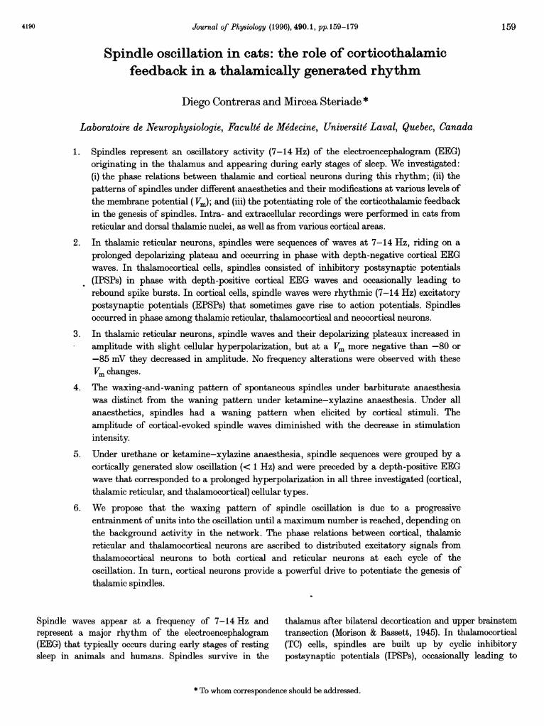
Journal of Physiology (1996), 490.1, pp. 159-179
Spindle oscillation in cats: the role of corticothalamicfeedback in a thalamically generated rhythm
Diego Contreras and Mircea Steriade *
Laboratoire de Neurophysiologie, Faculte de Medecine, Universite Laval, Quebec, Canada
1. Spindles represent an oscillatory activity (7-14 Hz) of the electroencephalogram (EEG)originating in the thalamus and appearing during early stages of sleep. We investigated:(i) the phase relations between thalamic and cortical neurons during this rhythm; (ii) thepatterns of spindles under different anaesthetics and their modifications at various levels ofthe membrane potential (Vm); and (iii) the potentiating role of the corticothalamic feedbackin the genesis of spindles. Intra- and extracellular recordings were performed in cats fromreticular and dorsal thalamic nuclei, as well as from various cortical areas.
2. In thalamic reticular neurons, spindles were sequences of waves at 7-14 Hz, riding on aprolonged depolarizing plateau and occurring in phase with depth-negative cortical EEGwaves. In thalamocortical cells, spindles consisted of inhibitory postsynaptic potentials(IPSPs) in phase with depth-positive cortical EEG waves and occasionally leading torebound spike bursts. In cortical cells, spindle waves were rhythmic (7-14 Hz) excitatorypostsynaptic potentials (EPSPs) that sometimes gave rise to action potentials. Spindlesoccurred in phase among thalamic reticular, thalamocortical and neocortical neurons.
3. In thalamic reticular neurons, spindle waves and their depolarizing plateaux increased inamplitude with slight cellular hyperpolarization, but at a Vm more negative than -80 or-85 mV they decreased in amplitude. No frequency alterations were observed with theseVm changes.
4. The waxing-and-waning pattern of spontaneous spindles under barbiturate anaesthesiawas distinct from the waning pattern under ketamine-xylazine anaesthesia. Under allanaesthetics, spindles had a waning pattern when elicited by cortical stimuli. Theamplitude of cortical-evoked spindle waves diminished with the decrease in stimulationintensity.
5. Under urethane or ketamine-xylazine anaesthesia, spindle sequences were grouped by acortically generated slow oscillation (< 1 Hz) and were preceded by a depth-positive EEGwave that corresponded to a prolonged hyperpolarization in all three investigated (cortical,thalamic reticular, and thalamocortical) cellular types.
6. We propose that the waxing pattern of spindle oscillation is due to a progressiveentrainment of units into the oscillation until a maximum number is reached, depending onthe background activity in the network. The phase relations between cortical, thalamicreticular and thalamocortical neurons are ascribed to distributed excitatory signals fromthalamocortical neurons to both cortical and reticular neurons at each cycle of theoscillation. In turn, cortical neurons provide a powerful drive to potentiate the genesis ofthalamic spindles.
Spindle waves appear at a frequency of 7-14 Hz and thalamus after bilateral decortication and upper brainstemrepresent a major rhythm of the electroencephalogram transection (Morison & Bassett, 1945). In thalamocortical(EEG) that typically occurs during early stages of resting (TC) cells, spindles are built up by cyclic inhibitorysleep in animals and humans. Spindles survive in the postsynaptic potentials (IPSPs), occasionally leading to
* To whom correspondence should be addressed.
1594190
D. Contreras and M. Steriade
rebound spike bursts which are transferred to the cerebralcortex (Andersen & Andersson, 1968; Steriade, Jones &Llinas, 1990).
Two different hypotheses were advanced to explain theorigin of spindle-related IPSPs in TC cells. The first one
was a model based on intranuclear recurrent inhibitionthrough excitation of local-circuit neurons by collateralaxons of TC cells (Andersen & Andersson, 1968). In mostdorsal thalamic nuclei, including the ventroposterior (VP)complex where Andersen's experiments were conducted, theaxons of TC cells do not give rise to intranuclear recurrentcollaterals (Yen & Jones, 1983; Steriade & Deschenes, 1984).The other hypothesis proposed that it is the thalamicreticular (RE) nucleus, consisting of neurons that use
y-aminobutyric acid (GABA) as transmitter, that plays thedecisive role in the genesis of rhythmic IPSPs in TC cells(Steriade, Deschenes, Domich & Mulle, 1985). This ideastemmed from experimental data showing the abolition ofspindle oscillations in TC neurons disconnected from REinputs (Steriade et al. 1985) and the preservation ofspindles within the rostral pole of the RE nucleus isolatedfrom the dorsal thalamus and cortex (Steriade, Domich,Oakson & Deschenes, 1987). That RE neurons imposerhythmic IPSPs onto TC cells was further supported byintracellular recordings showing that, during the IPSPs ofTC cells, GABAergic RE neurons discharge prolonged spikebarrages within the spindle frequency, superimposed over a
depolarizing plateau (Steriade & Deschenes, 1988). It was
also demonstrated that, during natural sleep, the shortpostinhibitory spike bursts that are occasionally fired byTC cells stand in contrast with the discharges of REneurons throughout spindle sequences (Steriade, Domich& Oakson, 1986). While all these results seemed to justifythe claim that RE neurons play a pacemaking role in theproduction of spindles, it was emphasized that, in theintact brain, any excitatory input may effectively drivethe conditional RE pacemaker. Such inputs could includefocal hyperpolarizations followed by rebound spike burstsin TC cells that would trigger the RE neuron networkwhich, by virtue of widely distributed thalamic projections,may contribute to the synchronization of the wholethalamus (Steriade et al. 1987).
It is now recognized that spindle oscillations depend on
both intrinsic properties of thalamic cells and complexnetwork operations in thalamocorticothalamic loops. TheCa2+-dependent low-threshold spike (LTS) crowned byhigh-frequency Na+ action potentials is a basic intrinsicproperty of TC cells (Llinas & Jahnsen, 1982) which isuncovered by membrane potential (Vm) hyperpolarizationduring sleep (see Steriade et al. 1990). In vitro studiesrevealed the intrinsic properties and ionic conductances ofsingly oscillating RE cells (Avanzini, De Curtis, Panzica &Spreafico, 1989; Huguenard & Prince, 1992, 1994; Bal &McCormick, 1993) and supported the notion of a thalamicpacemaker. However, without network operations, spindles
would not be observed synchronously in many thalamic fociand widespread cortical areas (Andersen & Andersson,1968; Steriade et al. 1990). Moreover, an oscillation at< 1 Hz, generated within the cerebral cortex (Steriade,Nufiez & Amzica, 1993 b,c) and reflected in RE and TC cells(Steriade, Contreras, Curro Dossi & Nufiez, 1993a),contributes to the grouping and synchronization of sleeprhythms, including spindles, within slowly recurring wave-sequences (Contreras & Steriade, 1995).
Although spindles are generated in the thalamus even inthe absence of the cerebral cortex, cortical inputs potentiatespindles as this oscillation is elicited by corticothalamicvolleys, even by stimulating the contralateral cortex toavoid the antidromic invasion of TC axons (Steriade,Wyzinski & Apostol, 1972). Intracellular recordingsshowed that corticothalamic stimuli are much more efficientthan prethalamic volleys in eliciting spindles in TC neurons(see Fig. 1.3 in Steriade et al. 1990).
The present work was undertaken to study the patterns ofspontaneous and evoked spindles under differentanaesthetics, the dependency of spindles upon the intrinsicelectrophysiological properties of RE and TC cells, and therole of corticothalamic volleys in the elicitation andsynchronization of spindles. We used simultaneous intra-and extracellular recordings of cortical, RE and TC neuronsbelonging to sensory, motor and association systems.Throughout this article we define 'spindle' as a sequence ofrhythmic waves at 7-14 Hz and we use the term 'depolariz-ing plateau' to designate the prolonged depolarization overwhich spindle waves are riding in RE cells recorded in vivo.
METHODSExperiments were conducted on adult cats anaesthetized withpentobarbitone (35 mg kg-, i.P.), ketamine-xylazine (10-15 and2-3 mg kg-', respectively, I.M.) or urethane (IP8 g kg-, i.P.). Inaddition, all pressure points and tissues to be incised wereinfiltrated with lidocaine. The animals were paralysed withgallamine triethiodide only after the EEG showed the typicalpatterns of deep general anaesthesia. The animals were artificiallyventilated to an end-tidal C02 of 3 5-3 7%. The heart beat wascontinuously monitored and showed rates (between 90 and110 min-) that assessed the depth of anaesthesia. Bodytemperature was maintained at 37-39 0C. Saline glucose wassupplemented as a fluid therapy, 1-2 times during the experiment.The EEG was continuously recorded throughout the experiments.To monitor the depth of anaesthesia, we maintained a constantpicture of high-amplitude and low-frequency EEG waves. Lowerdoses of the same anaesthetic were administered (2-3 timesduring an experiment) at the slightest changes in EEG patterns,i.e. a tendency towards increased frequency and decreasedamplitudes ofEEG waves. Experiments lasted for 8-10 h.
Intracellular recordings were performed with glass micropipettesfilled with a solution of 3 M potassium acetate and DC resistancesof 35-45 Mil. (i) Cortical intracellular recordings were performedafter resection of the bone and dura. Mineral oil was used toprevent desiccation. The pipettes for intracellular recordings in thecortex were placed in the vicinity of the coaxial EEG recording
160 J Physiol.490.1
Role of corticothalamic feedback in spindles
electrodes (see below). (ii) For thalamic intracellular recordings, thecortex and white matter over the recorded thalamic territory were
removed by suction. Micropipettes were then lowered through thehead of the caudate nucleus in order to reach the rostrolateralsector of the RE nucleus and the ventrolateral (VL) nucleus. Thestability of intracellular recordings was improved by performing a
bilateral pneumothorax, as well as by the drainage of the cisternamagna, hip suspension, and by filling the holes made for recordingwith a solution of 4% agar. A high-impedance amplifier (bandpassof 0-5 kHz) with active bridge circuitry was used to record andinject current into the cells. Extracellular recordings were done bymeans of tungsten electrodes with resistances of 1-5 MQ. Signalswere recorded on an eight-channel tape with bandpass of 0-9 kHzand thereafter were digitized at 20 kHz for off-line computeranalysis.
The gross EEG was recorded monopolarly by means of a screw
inserted in the bone. The focal EEG was recorded by means ofcoaxial electrodes with the tip placed in deep cortical layers andthe ring over the pial surface. The EEG recording electrodes were
inserted in the lateral part of the precruciate motor cortex (area 4),the postcruciate somatosensory cortex (areas 3b, 1 and 2), and therostral suprasylvian association cortex (area 5). The depth was
adjusted to obtain an inverse polarity of the thalamic-evokedpotential. In all monopolar recordings the indifferent electrode wasplaced in the neck muscles. Besides, the electrothalamogram(EThG) was bipolarly recorded through the same electrodes as usedfor stimulation.
Stimulating coaxial electrodes were stereotaxically placed in themotor VL thalamic nucleus and the brachium conjunctivum (BC),the somatosensory VP thalamic nuclear complex and the dorsalcolumn nuclei (DCN), or the association lateroposterior (LP)thalamic nucleus, according to the system under study. Corticalstimulation was performed by means of the coaxial electrodes usedfor EEG recording.
At the end of experiments the cats were given a lethal dose ofpentobarbitone.
RESULTSData base and neuronal identificationThe results are based on recordings from 208 corticalneurons, 233 RE neurons, and 129 TC neurons. Corticalcells (n = 143 intra, n = 65 extra) were recorded from themotor, primary somatosensory, and associationsuprasylvian cortices. RE cells (n = 70 intra, n = 163extra) were recorded from the rostral pole, rostrolateral(peri-VL), and lateral (peri-VP and peri-LP) sectors of thenucleus. TC neurons (n = 26 intra, n = 103 extra) were
recorded from the VL, VP and LP nuclei. The intra-cellularly recorded neurons that were used for the data baseand analyses had stable Vm, more negative than -60 mVfor cortical cells or more negative than -55 mV for thalamiccells, and overshooting action potentials.
(i) Cortical cells were located in layers II to VI. Motorcortical neurons were identified by an excitatorypostsynaptic potential (EPSP) evoked by VL and BCstimulation. Cells from the somatosensory cortex respondedto stimulation of the appropriate body region. Theydisplayed EPSPs in response to stimulation of VP andDCN (see Fig. 2B). (ii) Neurons from the RE nucleus were
identified by their prolonged spike bursts and accelerando-decelerando bursting pattern during spontaneous oscillatoryactivity (Domich, Oakson & Steriade, 1986; see Fig. 5Ad)and burst responses to stimulation of cortical and thalamicinput sources. (iii) TC cells from the VL nucleus wereidentified by antidromic invasion and EPSPs in response tomotor cortex stimulation, followed by a sequence of IPSPsat the spindling frequency, occasionally giving rise torebound spike bursts. TC cells recorded extracellularly wereidentified by their short, high-frequency spike bursts witha decelerando pattern in response to motor cortexstimulation (VL cells) or to DCN or receptive fieldstimulation (VP cells).
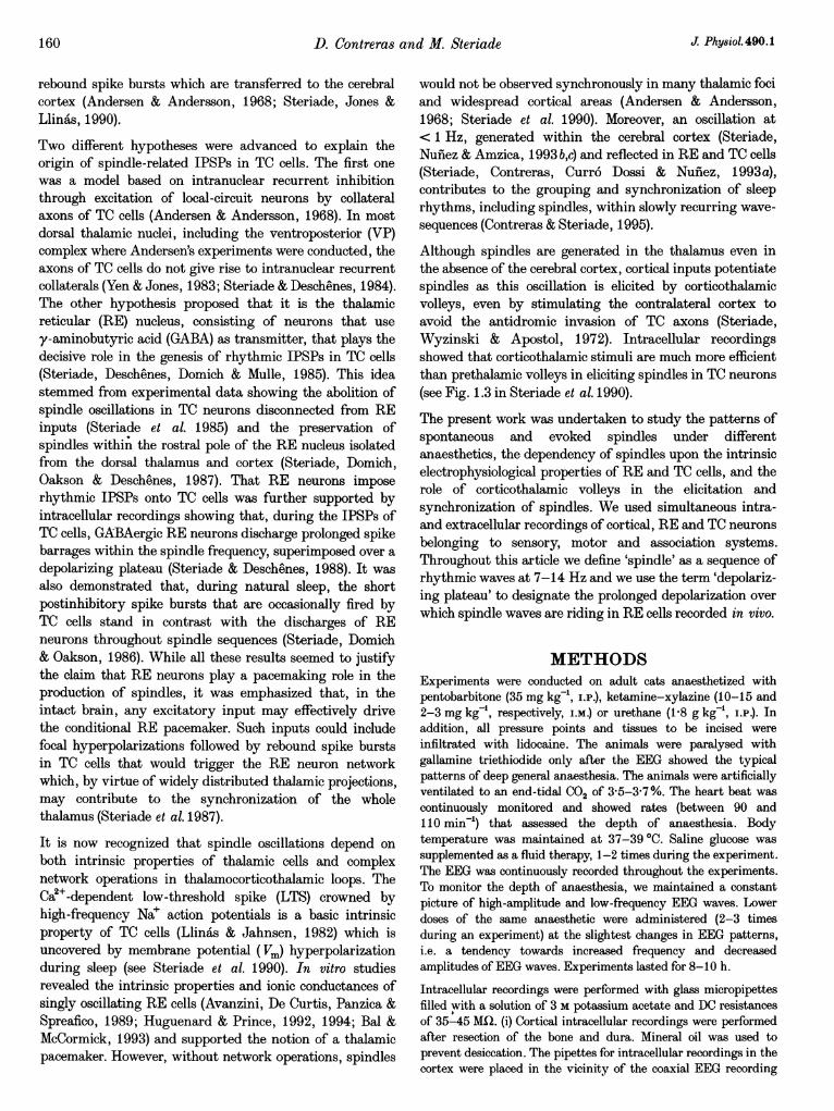
Cortical cellsUnder barbiturate anaesthesia, cortical cells spontaneouslydisplayed oscillations at 7-14 Hz, grouped in spindlesequences that recurred periodically with a slow rhythm of0-1-0-3 Hz. Spindling in cortical cells was characterized byrhythmic EPSPs (occasionally leading to action potentials)which increased in amplitude towards the middle of thespindle sequence and thereafter decreased in amplitude(Figs IA and 2Aa). The cellular oscillatory activity wasreflected in the EEG, most commonly as depth-negative(surface-positive) waves. In some instances, the EEG waveswere more complex, with a mixture of positive andnegative components that changed their shape during thespindle sequence (see Fig. 2Aa).
During spindles, cortical cells that were simultaneouslyrecorded within a distance of 1-1 5 mm exhibitedsynchronized oscillatory activity (n = 15). Figure 1 depictstwo intracellularly recorded neurons at depths of 0 5 and0 7 mm, simultaneously with the surface and depth EEG intheir vicinity, an extracellularly recorded cell populationfrom VP thalamus and focal waves recorded through thesame thalamic microelectrode. The six traces shown in Awere averaged around the negative peaks of depth EEGwaves (Fig. 1B) in order to assess the phase relation duringthe oscillatory activity. The averaged oscillation shows thatthe depth-negative waves of the cortical EEG correspondedto depolarizations of both cortical cells as well as to firing ofTC cells (peri-event histogram, bottom, Fig. 1B) andnegative field potentials in the focal EThG recorded fromthe VP nucleus. The depolarizations of cortical neuronsstarted before the peak negativity of depth EEG waves andwere related to the field thalamic waves.
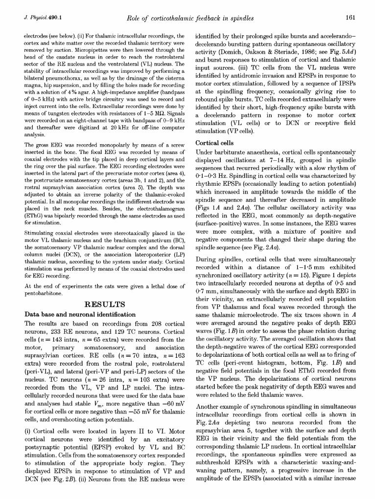
Another example of synchronous spindling in simultaneousintracellular recordings from cortical cells is shown inFig. 2Aa depicting two neurons recorded from thesuprasylvian area 5, together with the surface and depthEEG in their vicinity and the field potentials from thecorresponding thalamic LP nucleus. In cortical intracellularrecordings, the spontaneous spindles were expressed assubthreshold EPSPs with a characteristic waxing-and-waning pattern, namely, a progressive increase in theamplitude of the EPSPs (associated with a similar increase
J Phy-siol.490.1 161
D. Contreras and M. Steriade
in cortical and thalamic field potentials towards the middleof the spindle sequence), followed by a progressive decreasein the amplitudes of oscillatory events. Upon thalamic LPstimulation (Fig. 2A b), the evoked spindle sequence showeda waning pattern, with the EPSPs and EEG wavesshowing only a progressive decrease in amplitude.
Similar phase relations and prevalent waning pattern ofthe evoked spindling were observed in the series of intra-cellular responses of two simultaneuously recorded SIneurons to DCN stimulation, depicted in Fig. 2B togetherwith a cell from the corresponding region in the thalamicVP nuclear complex. The two SI cells, separated by about1-3 mm, displayed DCN-evoked spindle sequences in whichthe first three waves were in phase. The following EPSPswithin the spindle sequence were of smaller amplitude and
were not synchronized. The peristimulus histogram of VPcell population firing (bottom trace of Fig. 2B) shows twoclear oscillatory peaks, followed by irregular discharges.The fact that the second peak was delayed by 20 ms afterthe EPSPs of cortical cells suggests that this VP neuronlagged slightly the main thalamic population impingingupon the recorded cortical cells. These data suggest that thewaning pattern of cortical spindling is due to a process ofdesynchronization of the TC input.
Spindle oscillations under ketamine-xylazine anaesthesia(Fig. 3) were grouped by a slow rhythm (< 1 Hz),characterized by long-lasting positive waves in the depthEEG, followed by sharp negative deflections leading to aspindle oscillation. By contrast to the waxing-and-waningpattern of spindles under barbiturate anaesthesia,
BA
EEG-surlace SI
EEG-,depth SI
Av
Intra-cell SI-63 mV
Focal waves VP
Extra-cell VI
-J L A .IS-1$11 1 V'41*---r----l 7p 0
Time (s)0-5 s
Figure 1. Waxing-and-waning spindle oscillations occur in phase in related thalamic andcortical territoriesBarbiturate anaesthesia. Two cells from the primary somatosensory cortex (SI), separated by about 1 mm,were impaled and simultaneously recorded together with the EEG from the surface (EEG-surface) anddepth (EEG-depth) in the cortical vicinity. In addition, a neuronal population was extracellularly recordedfrom the VP nucleus; the two bottom traces illustrate the focal waves (field potentials) through the samemicroelectrode that was used to record action potentials. A, spontaneous waxing-and-waning spindlesequence at around 8 Hz, consisting of EEG depth-negative deflections that reversed polarity at thesurface. Both cells displayed EPSPs in phase with the EEG deflections. *expanded in inset. B, average(Av, n = 15) of traces shown in A centred on the depth-negative peak of EEG potentials. In this andfollowing figures, Vm is indicated; field potentials are averaged whenever single cell activity is averaged;and the polarity of EEG is the same as for intracellular recordings (positivity up).
-R -- r --l w - w -, _
162 J Physiol.490.1
Role of corticothalamic feedback in spindles
Intra-cell (area 5)
Intra-cell (area 5)-65 mV A A
t1,~~~~~~~~~~~~~
Intra-cell SI ADCN I
Extra-cell VP i0 200 400 600
Time (ms)
Figure 2. Spindle oscillations preferentially display a waning pattern when elicited bysynchronous stimuli to central pathwaysBarbiturate anaesthesia. A, two cells were simultaneously recorded intracellularly from area 5 (1P5 mmapart). In Aa, they displayed spontaneously occurring, synchronized spindle oscillations, together withthe surface and depth EEG from area 5 in the vicinity of cells and with the EThG from the LP nucleus. InAb, average (Av, n = 15) of LP-evoked responses in the same two cortical cells and EEG activities.B, intracellular activities of two SI cortical cells, simultaneously recorded with extracellular activities froma thalamic VP nucleus. The two cortical cells responded to DCN stimulus with a spindle sequence thatdisplayed a waning pattern. In both intracellular recordings, the 10 traces were displaced vertically forclarity and represent the responses of two cells (at a constant Vm) to DCN shocks of constant parameters.The bottom trace represents the peristimulus histogram from the thalamic VP cells' population andsimilarly shows prevalent waning of the spindle sequence in response to DCN stimuli.
Aa b
0-2 s
800 1000 1200
J Physiol. 490.1 163
D. Contreras and M. Steriade
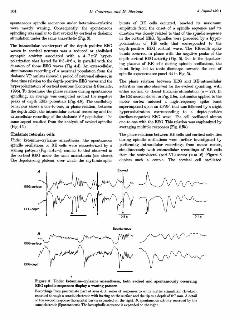
spontaneous spindle sequences under ketamine-xylazinewere mostly waning. Consequently, the spontaneousspindling was similar to that evoked by cortical or thalamicstimulation under the same anaesthetic (Fig. 3).
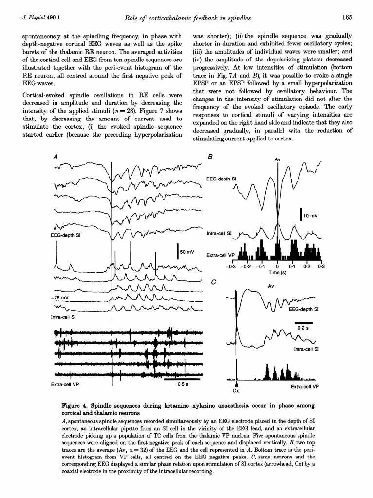
The intracellular counterpart of the depth-positive EEGwaves in cortical neurons was a reduced or abolishedsynaptic activity associated with a 4-7 mV hyper-polarization that lasted for 03-06 s, in parallel with theduration of those EEG waves (Fig. 4A). An extracellular,simultaneous recording of a neuronal population from thethalamic VP nucleus showed a period of neuronal silence, inclose time relation to the depth-positive EEG waves and thehyperpolarization of cortical neurons (Contreras & Steriade,1995). To determine the phase relation during spontaneousspindling, an average was computed around the negativepeaks of depth EEG potentials (Fig. 4B). The oscillatorybehaviour shows a one-to-one, in phase relation, betweenthe depth EEG, the intracellular cortical recording and theextracellular recording of the thalamic VP population. Thesame aspect resulted from the analysis of evoked spindles(Fig. 4C).
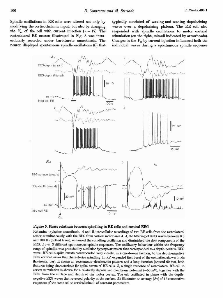
Thalamic reticular cellsUnder ketamine-xylazine anaesthesia, the spontaneousspindle oscillations of RE cells were characterized by awaning pattern (Fig. 5Aa-c), similar to that observed inthe cortical EEG under the same anaesthesia (see above).The depolarizing plateau, over which the rhythmic spike
bursts of RE cells occurred, reached its maximumamplitude from the onset of a spindle sequence and itsduration was closely related to that of the spindle sequencein the cortical EEG. Spindles were preceded by a hyper-polarization of RE cells that corresponded to thedepth-positive EEG cortical wave. The RE-cell's spikebursts occurred in phase with the negative peaks of thedepth-cortical EEG activity (Fig. 5). Due to the depolariz-ing plateau of RE cells during spindle oscillations, theburst firing led to tonic discharge towards the end ofspindle sequences (see panel A b in Fig. 5).
The phase relation between EEG and RE-intracellularactivities was also observed for the evoked spindling, witheither cortical or dorsal thalamic stimulation (n = 22). Inthe RE neuron shown in Fig. 5Ba, a stimulus applied to themotor cortex induced a high-frequency spike burstsuperimposed upon an EPSP, that was followed by a slighthyperpolarization corresponding to a depth-positive(surface-negative) EEG wave. The cell oscillated almostone-to-one with the EEG. This relation was emphasized byaveraging multiple responses (Fig. 5Bb).
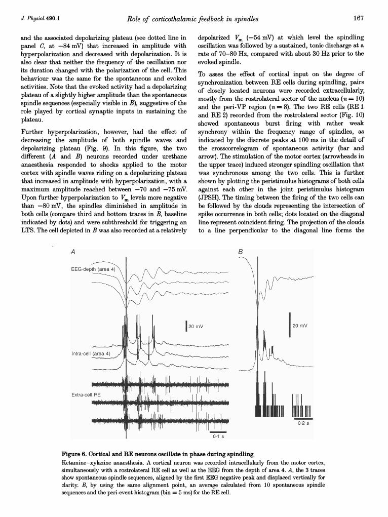
The phase relations between RE cells and cortical activitiesduring spindle oscillations were further investigated byperforming intracellular recordings from motor cortex,simultaneously with extracellular recordings of RE cellsfrom the rostrolateral (peri-VL) sector (n = 16). Figure 6depicts such a couple. The cortical cell oscillated
Evoked
0-5 s
Spontaneous
0*1 s
B
EEG-surface
EEG-depth
Figure 3. Under ketamine-xylazine anaesthesia, both evoked and spontaneously occurringEEG spindle sequences display a waning patternRecordings from precruciate part of area 4. A, series of responses to white matter stimulation (Evoked),recorded through a coaxial electrode with its ring on the surface and the tip at a depth of 07 mm. A detailof the second response (horizontal bar) is expanded on the right. B, spontaneous activity recorded by thesame electrode (Spontaneous). The last spindle sequence is expanded on the right.
164 J Physiol.490.1
I
Role of corticothalamic feedback in spindles
spontaneously at the spindling frequency, in phase withdepth-negative cortical EEG waves as well as the spikebursts of the thalamic RE neuron. The averaged activitiesof the cortical cell and EEG from ten spindle sequences areillustrated together with the peri-event histogram of theRE neuron, all centred around the first negative peak ofEEG waves.
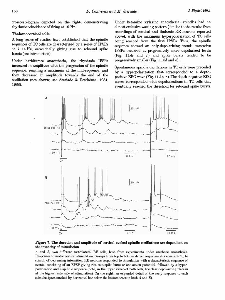
Cortical-evoked spindle oscillations in RE cells weredecreased in amplitude and duration by decreasing theintensity of the applied stimuli (n = 28). Figure 7 showsthat, by decreasing the amount of current used tostimulate the cortex, (i) the evoked spindle sequencestarted earlier (because the preceding hyperpolarization
was shorter); (ii) the spindle sequence was graduallyshorter in duration and exhibited fewer oscillatory cycles;(iii) the amplitudes of individual waves were smaller; and(iv) the amplitude of the depolarizing plateau decreasedprogressively. At low intensities of stimulation (bottomtrace in Fig. 7A and B), it was possible to evoke a singleEPSP or an EPSP followed by a small hyperpolarizationthat were not followed by oscillatory behaviour. Thechanges in the intensity of stimulation did not alter thefrequency of the evoked oscillatory episode. The earlyresponses to cortical stimuli of varying intensities areexpanded on the right hand side and indicate that they alsodecreased gradually, in parallel with the reduction ofstimulating current applied to cortex.
A
.EEG-depth SI
-76 mV
Intra-cell SI
LI
Extra-cell VP
B
EEG-depth SI
Af'x~~-V -A
r
Intra-cell SI
Extra-cell VP
Av
=11
I10 mV
I I I I-0-2 -0-1 0 01
Time (s)
C
A
0-5 s ACx
Av
G-depth SI
0.2 s
,1
AULExtra-cell VP
Figure 4. Spindle sequences during ketamine-xylazine anaesthesia occur in phase among
cortical and thalamic neurons
A, spontaneous spindle sequences recorded simultaneously by an EEG electrode placed in the depth of SIcortex, an intracellular pipette from an SI cell in the vicinity of the EEG lead, and an extracellularelectrode picking up a population of TC cells from the thalamic VP nucleus. Five spontaneous spindlesequences were aligned on the first negative peak of each sequence and displaced vertically. B, two toptraces are the average (Av, n = 32) of the EEG and the cell represented in A. Bottom trace is the peri-event histogram from VP cells, all centred on the EEG negative peaks. C, same neurons and thecorresponding EEG displayed a similar phase relation upon stimulation of SI cortex (arrowhead, Cx) by a
coaxial electrode in the proximity of the intracellular recording.
I I0-2 0-3
165J Physiol.490.1
F,, I
- Xd v
I
50 mv
I
i
-0-3
11II ot;a-1 %-.71
166 D. Contreras
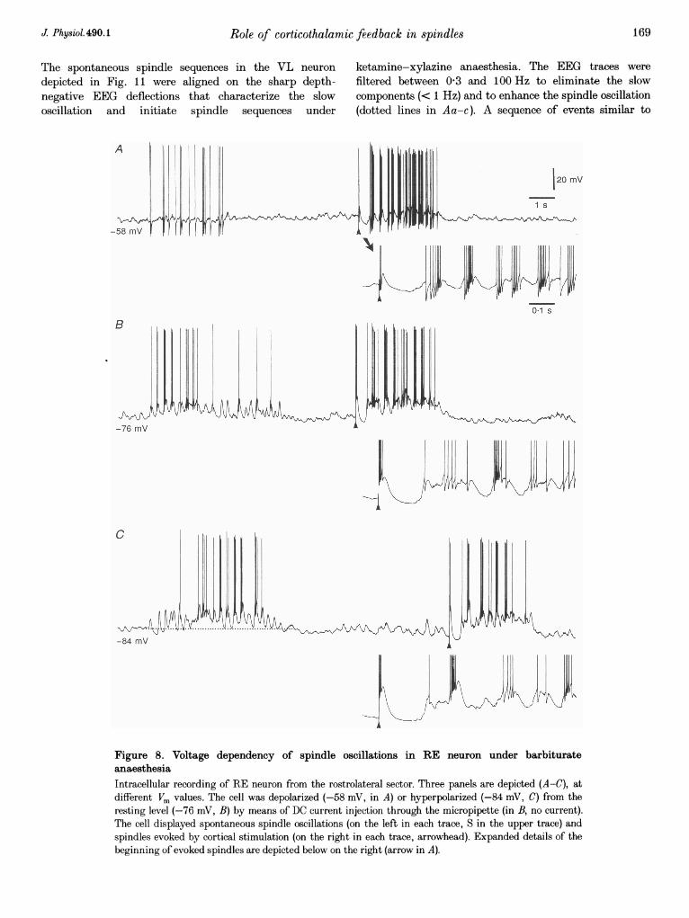
Spindle oscillations in RE cells were altered not only bymodifying the corticothalamic input, but also by changingthe Vm of the cell with current injection (n = 17). Therostrolateral RE neuron illustrated in Fig. 8 was intra-cellularly recorded under barbiturate anaesthesia. Theneuron displayed spontaneous spindle oscillations (S) that
an4 M. Steriade J. Physiol. 490.1
typically consisted of waxing-and-waning depolarizingwaves over a depolarizing plateau. The RE cell alsoresponded with spindle oscillations to motor corticalstimulation (on the right, stimuli indicated by arrowheads).Changes in the Vm by current injection influenced both theindividual waves during a spontaneous spindle sequence
b
02 s
d
20 mV
20 ms
Ba b
Cx
Figure 5. Phase relations between spindling in RE cells and cortical EEGKetamine-xylazine anaesthesia. A and B, intracellular recordings of two RE cells from the rostrolateralsector, simultaneously with the EEG from cortical motor area 4. A, the filtering ofEEG waves between 03and 100 Hz (dotted trace), enhanced the spindling oscillation and diminished the slow components of theEEG. Aa-c, 3 different spontaneous spindle sequences. The oscillatory behaviour within the frequencyrange of spindles was preceded by a cellular hyperpolarization that corresponded to a depth-positive EEGwave. RE cell's spike bursts corresponded very closely, in a one-to-one fashion, to the depth-negativeEEG cortical waves that characterize spindling. In Ad, expanded first burst of the oscillation shown in Aa(horizontal bar). It shows an accelerando-decelerando pattern and a long duration (around 60 ms), bothfeatures being characteristic for spike bursts of RE cells. B, a single response of rostrolateral RE cell tocortex stimulation is shown for a relatively depolarized membrane potential (-56 mV), together with theEEG from the surface and depth of the motor cortex. The cell oscillated in phase with the depth-negative EEG waves that reversed polarity at the surface. Bb illustrates an average (Av) of 15 consecutiveresponses of the same cell to cortical stimuli of constant parameters.
Role of corticothalamic
and the associated depolarizing plateau (see dotted line inpanel C, at -84 mV) that increased in amplitude withhyperpolarization and decreased with depolarization. It isalso clear that neither the frequency of the oscillation nor
its duration changed with the polarization of the cell. Thisbehaviour was the same for the spontaneous and evokedactivities. Note that the evoked activity had a depolarizingplateau of a slightly higher amplitude than the spontaneousspindle sequences (especially visible in B), suggestive of therole played by cortical synaptic inputs in sustaining theplateau.
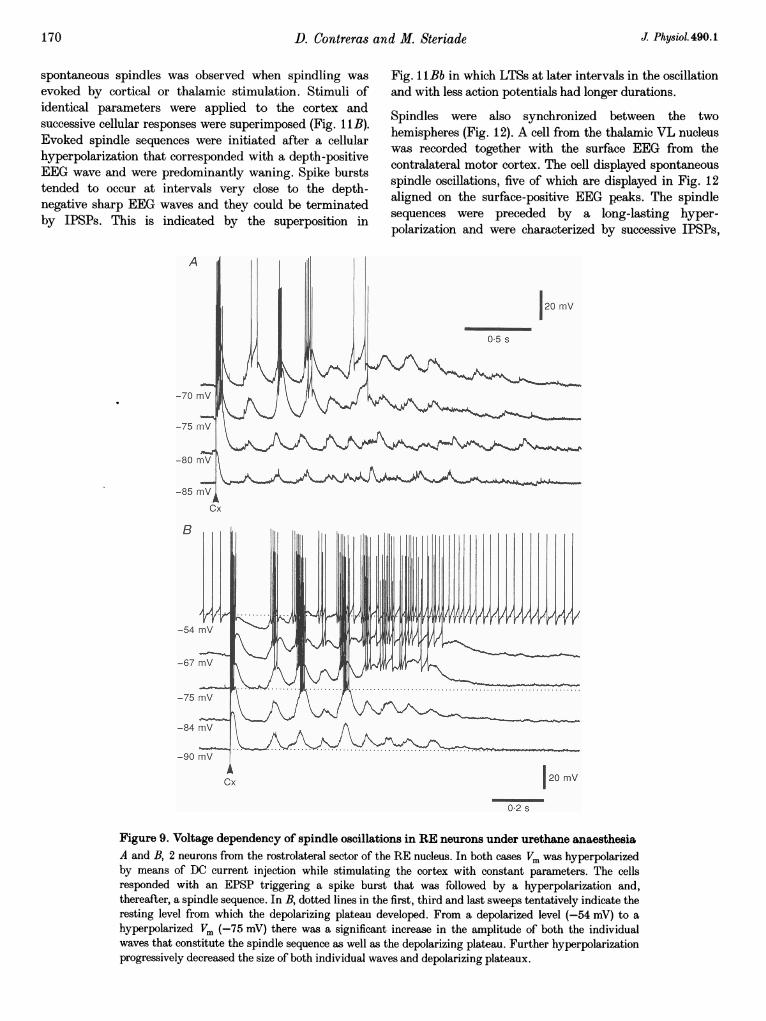
Further hyperpolarization, however, had the effect ofdecreasing the amplitude of both spindle waves anddepolarizing plateau (Fig. 9). In this figure, the twodifferent (A and B) neurons recorded under urethaneanaesthesia responded to shocks applied to the motorcortex with spindle waves riding on a depolarizing plateauthat increased in amplitude with hyperpolarization, with a
maximum amplitude reached between -70 and -75 mV.Upon further hyperpolarization to Vm levels more negativethan -80 mV, the spindles diminished in amplitude inboth cells (compare third and bottom traces in B, baselineindicated by dots) and were subthreshold for triggering an
LTS. The cell depicted in B was also recorded at a relatively
A
feedback in spindles 167
depolarized Vm (-54 mV) at which level the spindlingoscillation was followed by a sustained, tonic discharge at arate of 70-80 Hz, compared with about 30 Hz prior to theevoked spindle.
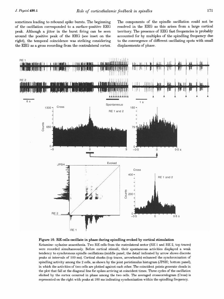
To asses the effect of cortical input on the degree ofsynchronization between RE cells during spindling, pairsof closely located neurons were recorded extracellularly,mostly from the rostrolateral sector of the nucleus (n = 10)and the peri-VP region (n = 8). The two RE cells (RE 1and RE 2) recorded from the rostrolateral sector (Fig. 10)showed spontaneous burst firing with rather weaksynchrony within the frequency range of spindles, as
indicated by the discrete peaks at 100 ms in the detail ofthe crosscorrelogram of spontaneous activity (bar andarrow). The stimulation of the motor cortex (arrowheads inthe upper trace) induced stronger spindling oscillation thatwas synchronous among the two cells. This is furthershown by plotting the peristimulus histograms of both cellsagainst each other in the joint peristimulus histogram(JPSH). The timing between the firing of the two cells canbe followed by the clouds representing the intersection ofspike occurrence in both cells; dots located on the diagonalline represent coincident firing. The projection of the cloudsto a line perpendicular to the diagonal line forms the
B
20 mV
ill Ml Iiilli2
Figure 6. Cortical and RE neurons oscillate in phase during spindlingKetamine-xylazine anaesthesia. A cortical neuron was recorded intracellularly from the motor cortex,simultaneously with a rostrolateral RE cell as well as the EEG from the depth of area 4. A, the 3 tracesshow spontaneous spindle sequences, aligned by the first EEG negative peak and displaced vertically forclarity. B, by using the same alignment point, an average calculated from 10 spontaneous spindlesequences and the peri-event histogram (bin = 5 ms) for the RE cell.
J Physiol.490.1
D. Contreras and M. Steriade
crosscorrelogram depicted on the right, demonstratingrhythmic coincidence of firing at 10 Hz.
Thalamocortical cellsA long series of studies have established that the spindlesequences of TC cells are characterized by a series of IPSPsat 7-14 Hz, occasionally giving rise to rebound spikebursts (see introduction).
Under barbiturate anaesthesia, the rhythmic IPSPsincreased in amplitude with the progression of the spindlesequence, reaching a maximum at the mid-sequence, andthey decreased in amplitude towards the end of theoscillation (not shown; see Steriade & Deschenes, 1984,1988).
-68 mV A -_-_-
Cx 01 s
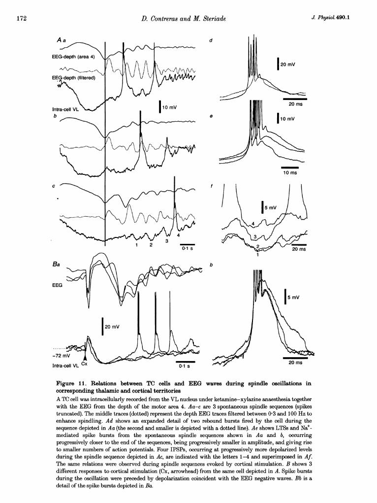
Under ketamine-xylazine anaesthesia, spindles had analmost exclusive waning pattern (similar to the results fromrecordings of cortical and thalamic RE neurons reportedabove), with the maximum hyperpolarization of TC cellsbeing reached from the first IPSPs. Thus, the spindlesequence showed an only-depolarizing trend: successiveIPSPs occurred at progressively more depolarized levels(Fig. ilAc and f) and spike bursts tended to beprogressively smaller (Fig. 1 Ad and e).
Spontaneous spindle oscillations in TC cells were precededby a hyperpolarization that corresponded to a depth-positive EEG wave (Fig. llAa-c). The depth-negative EEGwaves corresponded with depolarizations in TC cells thateventually reached the threshold for rebound spike bursts.
220 mTs
Figure 7. The duration and amplitude of cortical-evoked spindle oscillations are dependent onthe intensity of stimulationA and B, two different rostrolateral RE cells, both from experiments under urethane anaesthesia.Responses to motor cortical stimulation. Sweeps from top to bottom depict responses at a constant Vm tostimuli of decreasing intensities. RE neurons responded to stimulation with a characteristic sequence ofevents, consisting of an EPSP giving rise to a spike burst or one action potential, followed by a hyper-polarization and a spindle sequence (note, in the upper sweep of both cells, the clear depolarizing plateauat the highest intensity of stimulation). On the right, an expanded detail of the early response to eachstimulus (part marked by horizontal bar below the bottom trace in both A and B).
I
I
J Physiol.490.1168
.'I.l
Role of corticothalamic feedback in spindles
The spontaneous spindle sequences in the VL neurondepicted in Fig. 11 were aligned on the sharp depth-negative EEG deflections that characterize the slowoscillation and initiate spindle sequences under
A
ketamine-xylazine anaesthesia. The EEG traces werefiltered between 0 3 and 100 Hz to eliminate the slowcomponents (< 1 Hz) and to enhance the spindle oscillation(dotted lines in Aa-c). A sequence of events similar to
120 mV
1 s
-58 mv
0-1 s
B
-76 mV
C
-84 mV
Figure 8. Voltage dependency of spindle oscillations in RE neuron under barbiturateanaesthesiaIntracellular recording of RE neuron from the rostrolateral sector. Three panels are depicted (A-C), atdifferent Vm values. The cell was depolarized (-58 mV, in A) or hyperpolarized (-84 mV, C) from theresting level (-76 mV, B) by means of DC current injection through the micropipette (in B, no current).The cell displayed spontaneous spindle oscillations (on the left in each trace, S in the upper trace) andspindles evoked by cortical stimulation (on the right in each trace, arrowhead). Expanded details of thebeginning of evoked spindles are depicted below on the right (arrow in A).
J Physiol.490.1 169
D. Contreras and M. Steriade
spontaneous spindles was observed when spindling wasevoked by cortical or thalamic stimulation. Stimuli ofidentical parameters were applied to the cortex andsuccessive cellular responses were superimposed (Fig. 11B).Evoked spindle sequences were initiated after a cellularhyperpolarization that corresponded with a depth-positiveEEG wave and were predominantly waning. Spike burststended to occur at intervals very close to the depth-negative sharp EEG waves and they could be terminatedby IPSPs. This is indicated by the superposition in
Fig. 11Bb in which LTSs at later intervals in the oscillationand with less action potentials had longer durations.
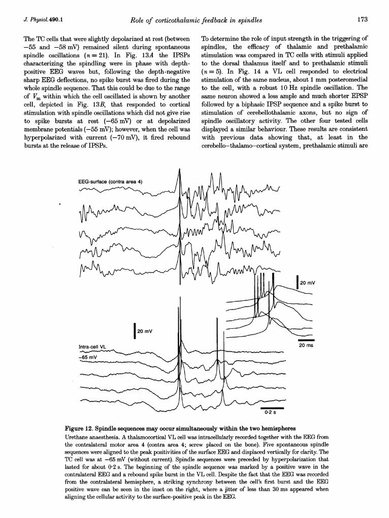
Spindles were also synchronized between the twohemispheres (Fig. 12). A cell from the thalamic VL nucleuswas recorded together with the surface EEG from thecontralateral motor cortex. The cell displayed spontaneousspindle oscillations, five of which are displayed in Fig. 12aligned on the surface-positive EEG peaks. The spindlesequences were preceded by a long-lasting hyper-polarization and were characterized by successive IPSPs,
B
-54
-67
-75
-84
-90 mvAICx 120 mV
02 s
Figure 9. Voltage dependency of spindle oscillations in RE neurons under urethane anaesthesiaA and B, 2 neurons from the rostrolateral sector of the RE nucleus. In both cases Vm was hyperpolarizedby means of DC current injection while stimulating the cortex with constant parameters. The cellsresponded with an EPSP triggering a spike burst that was followed by a hyperpolarization and,thereafter, a spindle sequence. In B, dotted lines in the first, third and last sweeps tentatively indicate theresting level from which the depolarizing plateau developed. From a depolarized level (-54 mV) to ahyperpolarized Vm (-75 mV) there was a significant increase in the amplitude of both the individualwaves that constitute the spindle sequence as well as the depolarizing plateau. Further hyperpolarizationprogressively decreased the size of both individual waves and depolarizing plateaux.
170 J Physiol.490.1
Role of corticothalamic
sometimes leading to rebound spike bursts. The beginningof the oscillation corresponded to a surface-positive EEGpeak. Although a jitter in the burst firing can be seen
around the positive peak of the EEG (see inset on theright), the temporal coincidence was striking consideringthe EEG as a gross recording from the contralateral cortex.
feedback in spindles 171
The components of the spindle oscillation could not beresolved in the EEG as this arises from a large corticalterritory. The presence of EEG fast frequencies is probablyaccounted for by multiples of the spindling frequency dueto the convergence of different oscillating spots with smalldisplacements of phase.
RE 1I i l hIL ihhW UII IIh
hi ,I I[IL*[iE - l§§ili"r I I IIILIENIDl n. Ii1A 1IIIIIIUI 1~~~~~U10 .s -
RE 2
s w I ' ' w r - I r I~~~~~~~~111AA iAAAA iA
_s
6 s
1300 CrossSpontaneous
RE 1 and 2
Li1. i ll i w li II~~~b~~~I -~~~~~I I j -~i I-
L IIiI[I
ikL:2411 liISP, II PI', T..Mr -T',r' wTlr- ! VARM11R
1 s
A
IIF'J Dll PUVA A A
150
U)
0 6500
0
0o--5 0 5 -0.5 0
T- _ _ 4
05 s
Evoked
Cross400 -
U)
= 200-0
.-q
-05
. RE 1 and 2
i
0 05 s
Figure 10. RE cells oscillate in phase during spindling evoked by cortical stimulationKetamine-xylazine anaesthesia. Two RE cells from the rostrolateral sector (RE 1 and RE 2, top traces)were recorded simultaneously. Before cortical stimuli, their spontaneous activities displayed a weaktendency to synchronous spindle oscillations (middle panel; the detail indicated by arrow shows discretepeaks at intervals of 100 ms). Cortical shocks (top traces, arrowheads) enhanced the synchronization ofspindling activity among the 2 cells, as shown by the joint peristimulus histogram (JPSH; bottom panel),in which the activities of two cells are plotted against each other. The coincident points generate clouds inthe plot that fall at the diagonal line for spikes arriving at coincident times. Three cycles of the oscillationelicited by the cortex occurred in phase among the two cells. The averaged crosscorrelogram (Cross) isrepresented on the right with peaks at 100 ms indicating synchonization within the spindling frequency.
J Physiol.490.1
---,T-Ir
00
---T -17,711-FpyI
D. Contreras and M. Steriade
d
1'''- %-,4.'
I10 mV
e
I _ f I
b
J Physiol.490.1
20 mV
10 ms
5 mV
20 ms
Figure 1. Relations between TC cells and EEG waves during spindle oscillations incorresponding thalamic and cortical territoriesA TC cell was intracellularly recorded from the VL nucleus under ketamine-xylazine anaesthesia togetherwith the EEG from the depth of the motor area 4. Aa-c are 3 spontaneous spindle sequences (spikestruncated). The middle traces (dotted) represent the depth EEG traces filtered between 03 and 100 Hz toenhance spindling. Ad shows an expanded detail of two rebound bursts fired by the cell during thesequence depicted in Aa (the second and smaller is depicted with a dotted line). Ae shows LTSs and Nae-mediated spike bursts from the spontaneous spindle sequences shown in Aa and b, occurringprogressively closer to the end of the sequences, being progressively smaller in amplitude, and giving riseto smaller numbers of action potentials. Four IPSPs, occurring at progressively more depolarized levelsduring the spindle sequence depicted in Ac, are indicated with the letters 1-4 and superimposed in Af.The same relations were observed during spindle sequences evoked by cortical stimulation. B shows 3different responses to cortical stimulation (Cx, arrowhead) from the same cell depicted in A. Spike burstsduring the oscillation were preceded by depolarization coincident with the EEG negative waves. Bb is adetail of the spike bursts depicted in Ba.
172
Aa
Role of corticothalamic
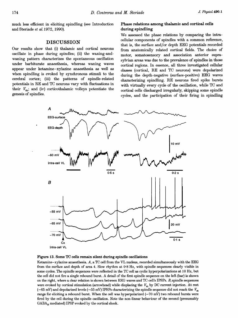
The TC cells that were slightly depolarized at rest (between-55 and -58 mV) remained silent during spontaneousspindle oscillations (n = 21). In Fig. 13A the IPSPscharacterizing the spindling were in phase with depth-positive EEG waves but, following the depth-negativesharp EEG deflections, no spike burst was fired during thewhole spindle sequence. That this could be due to the range
of Vm within which the cell oscillated is shown by anothercell, depicted in Fig. 13B, that responded to corticalstimulation with spindle oscillations which did not give riseto spike bursts at rest (-65 mV) or at depolarizedmembrane potentials (-55 mV); however, when the cell washyperpolarized with current (-70 mV), it fired reboundbursts at the release of IPSPs.
EEG-surface (contra area 4)
'I
feedback in spindles 173
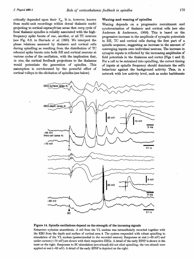
To determine the role of input strength in the triggering ofspindles, the efficacy of thalamic and prethalamicstimulation was compared in TC cells with stimuli appliedto the dorsal thalamus itself and to prethalamic stimuli(n = 5). In Fig. 14 a VL cell responded to electricalstimulation of the same nucleus, about 1 mm posteromedialto the cell, with a robust 10 Hz spindle oscillation. Thesame neuron showed a less ample and much shorter EPSPfollowed by a biphasic IPSP sequence and a spike burst tostimulation of cerebellothalamic axons, but no sign ofspindle oscillatory activity. The other four tested cellsdisplayed a similar behaviour. These results are consistentwith previous data showing that, at least in thecerebello-thalamo-cortical system, prethalamic stimuli are
0-2 s
Figure 12. Spindle sequences may occur simultaneously within the two hemispheresUrethane anaesthesia. A thalamocortical VL cell was intracellularly recorded together with the EEG fromthe contralateral motor area 4 (contra area 4; screw placed on the bone). Five spontaneous spindlesequences were aligned to the peak positivities of the surface EEG and displaced vertically for clarity. TheTC cell was at -65 mV (without current). Spindle sequences were preceded by hyperpolarization thatlasted for about 0-2 s. The beginning of the spindle sequence was marked by a positive wave in thecontralateral EEG and a rebound spike burst in the VL cell. Despite the fact that the EEG was recordedfrom the contralateral hemisphere, a striking synchrony between the cell's first burst and the EEGpositive wave can be seen in the inset on the right, where a jitter of less than 30 ms appeared whenaligning the cellular activity to the surface-positive peak in the EEG.
J Physiol.490.1
174 D. Contreras anl
much less efficient in eliciting spindling (see Introductionand Steriade et al. 1972, 1990).
DISCUSSIONOur results show that (i) thalamic and cortical neuronsoscillate in phase during spindles; (ii) the waxing-and-waning pattern characterizes the spontaneous oscillationunder barbiturate anaesthesia, whereas waning wavesappear under ketamine-xylazine anaesthesia as well aswhen spindling is evoked by synchronous stimuli to thecerebral cortex; (iii) the patterns of spindle-relatedpotentials in RE and TC neurons vary with fluctuations intheir Vm; and (iv) corticothalamic volleys potentiate thegenesis of spindles.
A
EEG-surface
EEG-depth
d M. Steriade J Phy8iol. 490.1
Phase relations among thalamic and cortical cellsduring spindlingWe assessed the phase relations by comparing the intra-cellular components of spindles with a common reference,that is, the surface and/or depth EEG potentials recordedfrom anatomically related cortical fields. The choice ofmotor, somatosensory and association anterior supra-sylvian areas was due to the prevalence of spindles in thosecortical regions. In essence, all three investigated cellularclasses (cortical, RE and TC neurons) were depolarizedduring the depth-negative (surface-positive) EEG wavescharacterizing spindling. RE neurons fired spike burstswith virtually every cycle of the oscillation, while TC andcortical cells discharged irregularly, skipping some spindlecycles, and the participation of their firing in spindling
.......m-60 mV
Intra-cell VL
05s 0-2s
B
CxIntra-cell VL
Figure 13. Some TC cells remain silent during spindle oscillationsKetamine-xylazine anaesthesia. A, a TC cell from the VL nucleus, recorded simultaneously with the EEGfrom the surface and depth of area 4. Slow rhythm at 0-8 Hz, with spindle sequences clearly visible insome cycles. The spindle sequences were reflected in the TC cell as cyclic hyperpolarizations at 10 Hz, butthe cell did not fire a single rebound burst. A detail of the first spindle sequence on the left (bar) is shownon the right, where a clear relation is shown between EEG waves and TC-cell's IPSPs. B, spindle sequenceswere evoked by cortical stimulation (arrowhead) while displacing the Vm by DC current injection. At rest(-65 mV) and depolarized levels (-55 mV) IPSPs characterizing the spindle sequence did not reach the Vmrange for eliciting a rebound burst. When the cell was hyperpolarized (-70 mV) two rebound bursts werefired by the cell during the spindle oscillation. Note the non-linear behaviour of the second (presumablyGABAB mediated) IPSP evoked by the cortical shock.
Iq

Diego Contreras and Mircea Steriade
Journal of Physiology 490, 159-179 (1996)
On page 175, the labelling in Figure 14 should read:
Intra-cell VL and not Intra-cell (area 4).
The legend i correct.
Role of corticothalamic feedback in spindles
critically depended upon their Vm. It is, however, knownfrom multi-unit recordings within dorsal thalamic nucleiprojecting to cortical suprasylvian areas that every cycle offocal thalamic spindles is reliably associated with the high-frequency spike bursts of one, another, or all TC neurons(see Fig. 6.8. in Steriade et al. 1990). We interpret thephase relations assumed by thalamic and cortical cellsduring spindling as resulting from the distribution of TCrebound spike bursts onto both RE and cortical neurons atvarious cycles of the oscillation, with the implication that,in vivo, the cortical feedback projections to the thalamuswould potentiate the generation of spindles. Thisassumption is corroborated by the powerful effect ofcortical volleys in the elicitation of spindles (see below).
Waxing-and-waning of spindlesWaxing depends on a progressive recruitment andsynchronization of thalamic and cortical cells (see alsoAndersen & Andersson, 1968). This is based on theprogressive increase in the amplitude of synaptic potentialsin RE, TC and cortical cells during the first part of aspindle sequence, suggesting an increase in the amount ofconverging inputs onto individual neurons. The increase insynaptic inputs is reflected by the increasing amplitudes offield potentials in the thalamus and cortex (Figs 1 and 2).For a cell to be entrained into spindling, the correct timingof inputs at spindle frequency should dominate the cell'sbehaviour against the background activity. Thus, in anetwork with low activity level, such as under barbiturate
Figure 14. Spindle oscillations depend on the strength of the incoming signalsKetamine-xylazine anaesthesia. A cell from the VL nucleus was intracellularly recorded together withthe EEG from the depth and surface of cortical area 4. The system responded with robust spindling tostimulation of the VL nucleus (posteromedial to the recorded neuron). Responses at rest (-65 mV) andunder current (-70 mV) are shown with their respective EEGs. A detail of the early EPSP is shown in theinset on the right. Responses to BC stimulation (arrowhead) did not elicit spindling; the two stimuli wereapplied at rest (-65 mV). A detail of the early EPSP is depicted on the right.
J Physiol.490.1 175
D. Contreras and M. Steriade
anaesthesia, the threshold for spindle generation is low,with the consequence that synchronous inputs, initiallytriggering spike bursts in a relatively small number ofthalamic neurons, would eventually lead to a full-blownspindle sequence. It is conceivable that, in a thalamic slicewith an even lower background activity, a burst of a singlethalamic cell may trigger a whole spindle sequence in therelated network. On the contrary, when the thalamic andcortical background activities are high, as is the case underketamine-xylazine anaesthesia, the threshold of spindlegeneration is higher, but once a critical neuronal mass is setinto the oscillatory mode, there is little further entrainmentof cells and the waxing process is unlikely. Spontaneousspindle sequences are observed in those cases in which highsynchronization is achieved from the very onset of theoscillation, thus giving rise to preferentially waningspindles. The slow oscillation (< 1 Hz), with its sharpdepth-negative component (Fig. 3) reflecting thesynchronous excitation of a considerable number of corticalneurons (Contreras & Steriade, 1995), represents afavourable oondition for triggering a waning spindlesequence in the pacemaking RE cells and in their TCtargets, with a shorter duration than that observed underbarbiturate anaesthesia (see Figs 6 and 12, compared toFigs 1 and 2).
The above comments also apply to data from thalamicslices in which succinimide-induced small changes in theprobability of burst firing in thalamic cells caused animportant reduction in intrathalamic rhythmicity(Huguenard & Prince, 1994). This implies that slight orseemingly negligible alterations in the proportion ofthalamic neurons participating in the organization ofnetwork activity may be critical for the development of theoscillation. We propose that the background noise in aspontaneously active network, as in vivo, plays the role ofdiminishing the probability of spike bursts in thalamiccells, thus increasing the requirements for converginginputs in order to effectively trigger spindle oscillations. Inthe same vein, we think that the long-lasting hyper-polarizations, generating widespread neuronal silence,create favourable conditions for generation of synchronizedspindling (Contreras & Steriade, 1995).
Although the barbiturate-induced spindling is waxing-and-waning, cortical stimulation gives rise to mostly waningspindles under the same anaesthetic condition (Fig. 2A).This leads us to the conclusion that synchronous stimulientrain, right from the start, a great number of neuronsparticipating in a spindle sequence, which would explainthe absence of the waxing process. Evoked spindles occurfollowing a long-lasting hyperpolarization triggered by thecortical stimulus in TC cells. The cellular hyperpolarizationalso occurs simultaneously in cortical, RE and TC cells, andis reflected as a prolonged depth-positive EEG wave(Contreras & Steriade, 1995). The spontaneously occurring
spindling under ketamine-xylazine anaesthesia is mostlywaning and preceded by a hyperpolarization having thesame characteristics as the evoked one (see Figs 5, 7 and11). In both cortical and thalamic cells, prolonged hyper-polarizations have been shown to be produced byGABAA,B-mediated IPSPs (Connors, Malenka & Silva,1988; Crunelli, Haby, Jassik-Gerschenfeld, Leresche &Pirchio, 1988). Although the spontaneous long-lastinghyperpolarization of the slow oscillation under ketamine-xylazine anaesthesia is not mainly due to the activation ofGABAergic receptors (Contreras & Steriade, 1995), thesame principle seems to be valid, i.e. prolonged hyper-polarizations, either spontaneous or stimulus elicited, areeffective in setting the scene for widespread synchroniz-ation in thalamocortical networks. The synchronizingprocess includes the appearance of spindle oscillationswhich follow the sharp depth-negative cortical deflectioncharacteristic of the slow oscillation.
The waning of the oscillations may require, besides thedesynchronization of the network (Andersen & Andersson,1968), the intervention of cellular intrinsic propertiescapable of diminishing cells' probability of firing spikebursts. Otherwise, the oscillation would be self-sustainedand generalized, as is the case with the development fromsleep rhythms to epileptic patterns (Avoli, Gloor,Kostopoulos & Gotman, 1983; Steriade & Amzica, 1994;Steriade & Contreras, 1995). The slow inward rectifier Ih,present in TC cells (McCormick & Pape, 1990), may playsuch a role (Bal, von Krosigk & McCormick, 1995a) bydepolarizing the cells and bringing them into the range ofLTS inactivation. Thus, with the progressive dropout ofneurons generating spike bursts, converging inputs ontonetwork elements become smaller and the oscillation fades.This possibility is supported by the present results showingthat IPSPs related to the waning of spindles occur on abackground of progressive depolarization (Fig. 11A). Thatcellular desynchronization contributes to the waning ofspindles (see Andersen & Andersson, 1968) is suggested bythe fact that, during waning, EPSPs in cortical neuronsbecome not only smaller but also scattered (Fig. 2B).
Voltage dependency of spindling in thalamic reticularand thalamocortical cellsSpindle sequences in RE cells recorded in vivo arecharacterized by prolonged spike bursts at 7-14 Hz, withan acceleration followed by a deceleration of intraburstfrequency (Domich et al. 1986; Steriade et al. 1986). Thelong duration and peculiar burst patterns of RE cells aredue to a special type of It (Huguenard & Prince, 1992),probably located in the dendrites. The spindle-relateddischarges of RE neurons are superimposed on adepolarizing plateau (Mulle, Madariaga & Deschenes, 1986;Contreras, Curr6 Dossi & Steriade, 1993). The present datashow that the depolarizing plateau of RE neuronaldischarges throughout a spindle sequence is increased with
176 J Physiol.490.1
Role of corticothalamic feedback in spindles
hyperpolarization down to -75 or -80 mV and thendecreases in amplitude with further hyperpolarization,together with the individual spindle waves (Figs 8 and 9).
A series of arguments point to the participation ofsynaptic inputs in the generation of the depolarizingplateau upon which individual spindle waves develop inRE cells. (a) The components of this depolarizing plateaubehave as postsynaptic excitatory events when neuronsare hyperpolarized to -75 mV. (b) The amplitude of thedepolarizing plateau is related to the presumed degree ofsynchronization in the network. Indeed, during spon-taneous waxing-and-waning spindle sequences (such asunder barbiturate anaesthesia) the depolarizing plateauslowly rises and falls, whereas during evoked spindles thedepolarizing plateau reaches its maximum from the onset(Fig. 8). (c) With hyperpolarization at Vm levels morenegative than -75 mV, the decreased amplitude of thedepolarizing plateau and of the associated phasic eventsmay be explained by the activation of inward rectificationin RE cells that are able to reduce their input resistance byup to 5004 (Contreras et al. 1993). It should also beemphasized that the constant frequency of spindles withina broad range of Vm values is indicative for the over-whelming of intrinsic oscillatory properties of RE cells(Mulle et al. 1986; Avanzini et al. 1989; Bal & McCormick,1993) by network operations during spindles.
The depolarizing plateau seen in vivo is absent in peri-geniculate RE cells recorded from ferret slices, a conditionunder which RE cells undergo a progressive hyper-polarization (von Krosigk, Bal & McCormick, 1993; Bal,von Krosigk & McCormick, 1995 b). This difference may beexplained by several network factors, including the actionsof some brainstem neuromodulatory systems as well ascorticothalamic inputs that are absent in the slice. Theclaim might also be made that the depolarizing plateauobserved in vivo is due to a lower membrane inputresistance of RE neurons, as a consequence of theimpalement. That the latter explanation is not valid isdemonstrated by the pattern of extracellularly recordeddischarges during natural sleep, when RE neurons firerhythmic spike bursts followed by a prolonged tonic tail ofaction potentials, extending throughout the duration of aspindle sequence (see Fig. 5A2 in Steriade et at. 1986). Thisaspect, seen in an extracellular position that precludes acompromised integrity of membrane input resistance, isindicative for the depolarization of RE cells during spindlesequences. The same pattern was described in the presentexperiments in which the depolarizing envelope of RE cellsassociated with a tail of tonic single-spike discharges wasonly seen at a relatively depolarized Vm (Fig. 9B), as isprobably the case in the behaving animal in whichneuromodulatory systems and corticothalamic projectionsare intact. The tonic discharges of RE cells after spindlewaves may explain the tail hyperpolarization, associated
with an increased conductance, in TC neurons after thecompletion of a spindle sequence in vivo (see Fig. 8 inNuniez, Curro Dossi, Contreras & Steriade, 1992).
An important requirement for triggering of spike bursts byTC cells following their rhythmic IPSPs during spindleoscillation is an adequate Vm. Our results show that slighthyperpolarizations of TC cells, which may otherwise besilent during spindling (Fig. 13A; see also Nufiez et al.1992), bring the IPSPs into the range of LTS de-inactivation and thus, spike bursts are triggered when theVm returns to resting values (Fig. 13B).
Corticothalamic feedback during spindlingOur results suggest a multiple role for the corticothalamicinput during spindle oscillations.
(a) Cortical inputs proved much more efficient for elicitingspindle sequences in extra- and intracellularly recorded TCcells than stimulation of prethalamic pathways (Steriadeet al. 1972, 1990). Moreover, during the cortically generatedslow sleep oscillation, inputs from antidromically identifiedcorticothalamic neurons trigger and pace spindle sequencesin thalamic neurons with a frequency lower than 1 Hz (seeFig. 5 in Steriade et al. 1993c). Under ketamine-xylazineanaesthesia, sequences of spindle waves recur with a higherfrequency, 0-6-0-9 Hz, than under urethane anaesthesia,0-3-0-6 Hz (Steriade et al. 1993b). Under barbiturateanaesthesia and in thalamic slices, when the cortical slowrhythm is not present, spindle sequences recur with arhythm of 0-1-0-2 Hz (Steriade & Deschenes, 1984, 1988;von Krosigk et al. 1993; Bal et al. 1995a, b) whose origin isnot yet determined, but that does not depend on the cortexas it also appears in the isolated RE nucleus (Steriade et al.1987).
(b) Spindle sequences could be synchronized among the twohemispheres from the outset (Fig. 12). This phenomenoncan hardly be explained by ascribing to the cortex the roleof a passive receiver of TC spike bursts at the spindlefrequency. Rather, what probably underlies the simultaneityof spindle sequences between the two hemispheres is a wideintracortical synchronization of the slow oscillation(Amzica & Steriade, 1995), coupled with the power ofcorticothalamic drives to elicit spindle sequences. Thereticulo-reticular thalamic commissural pathways (Battaglia,Lizier, Colacitti, Princivalle & Spreafico, 1994) should alsobe taken in consideration among the possible factorsaccounting for the synchronization of spindles between thetwo hemispheres.
(c) Cortical stimulation proved quite efficient to increase thesynchrony between RE neurons (Fig. 10). Synchronizedvolleys of cortical origin trigger an EPSP followed by long-lasting hyperpolarizations in RE cells (Contreras &Steriade, 1995) from which they are released in phase andoriginate synchronized spindle sequences.
J Phy8iol. 490.1 177
D. Contreras an
Concluding remarks on spindle generationSleep spindles appear when cortical and brainstem neurons,with activating actions on thalamic neurons, have alreadybegun to slow down their discharge frequencies (seeSteriade & McCarley, 1990). The decreased release ofexcitatory neurotransmitters leads to progressive hyper-polarizations that may be followed by rebound spike burstsin thalamic neurons. These events are hallmark electro-physiological signs during the behavioural state of restingsleep. Similar disfacilitation processes, leading to rhythmicspike bursts within the frequency range of spindles, occur
in a subset of corticothalamic neurons (Steriade, 1978) andare probably due to the diminished firing rates of afferentbasal forebrain cholinergic and monoamine-containingneurons. The hyperpolarizations and spike bursts thatappear in thalamic and cortical cells with transition fromwaking to sleep are decisive factors to the development ofsynchronized spindle oscillations. In thalamic slices,spindles may be initiated by spike bursts in thalamicreticular cells, imposing IPSPs that de-inactivate reboundbursts in target-relay cells, followed by synaptic excitationof reticular neurons, with progressive recruitment ofneurons in the network (Bal et al. 1995b), as similarlyenvisioned by Andersen & Andersson (1968) within theconceptual framework of a hypothetical intranuclearrecurrent inhibitory circuit. In brain-intact animals, thediminished activity in afferent cortical, brainstem, andhypothalamic neurons achieves the incipient conditionstowards the hyperpolarizations and spike bursts ofreticular and cortical-projecting thalamic cells (Steriade etal. 1987), but the synchronization of spindles throughoutthalamocortical systems is greatly potentiated by cortico-thalamic volleys, as generated periodically during the slowoscillation. Although the thalamic reticular nucleus can
generate spindles in clustered neuronal pools or some of itssectors, even after complete disconnection, modellingstudies show that the full synchronization of this nucleusand, by consequence, of thalamocortical systems, is helpedby extrinsic excitatory projections (Destexhe, Contreras,Sejnowski & Steriade, 1994a; Golomb, Wang & Rinzel,1994). The present data show that among the afferentexcitatory inputs, the cortex plays a very potent role. Thepresence of modulatory systems, including the cortico-thalamic projection, may also explain, at least partially, thedifference between the presence of spindles in the isolatedrostral pole of reticular nucleus in vivo (Steriade et al. 1987)and the absence of this oscillation in perigeniculatereticular cells in vitro (von Krosigk et al. 1993). Indeed, a
recent study (Destexhe, Contreras, Sejnowski & Steriade,1994b) introduced the actions of depolarizing neuro-
modulators in model thalamic reticular neurons organizedwith dense proximal connectivity, and demonstrated thatwaxing-and-waning spindles appear at a level of membranepolarization that would correspond to a weak activity inactivating systems, as is the case under anaesthesia or
sleep, but are absent at more hyperpolarized levels, as may
d M. Steriade J. Physiol. 490.1
be the case in slices. Thus, the scenario of spindlinggeneration is quite complex in vivo when the thalamus isembedded in networks that include the cerebral cortex anda series of neuromodulatory brainstem and hypothalamicsystems.
AMZICA, F. & STERIADE, M. (1995). Short- and long-range neuronalsynchronization of the slow (< 1 Hz) cortical oscillation. Journal ofNeurophysiology 73, 20-38.
ANDERSEN, P. & ANDERSSON, S. (1968). Physiological Basis of theAlpha Rhythm. Appleton-Century-Crofts, New York.
AVANZINI, G., DE CURTIS, M., PANZICA, F. & SPREAFICO, R. (1989).Intrinsic properties of nucleus reticularis thalami neurones of the ratstudied in vitro. Journal of Physiology 416, 111-122.
AVOLI, M., GLOOR, P., KOSTOPOULOS, G. & GOTMAN, J. (1983). Ananalysis of penicillin-induced spike and wave discharges usingsimultaneous recordings of cortical and thalamic neurons. Journalof Neurophysiology 50, 819-837.
BAL, T. & MCCORMICK, D. A. (1993). Mechanisms of oscillatoryactivity in guinea-pig nucleus reticularis thalami in vitro: amammalian pacemaker. Journal of Physiology 468, 669-691.
BAL, T., VON KROSIGK, M. & MCCORMICK, D. A. (1995a). Synaptic andmembrane mechanisms underlying synchronized oscillations in theferret lateral geniculate nucleus in vitro. Journal of Physiology 483,641-663.
BAL, T., VON KROSIGK, M. & MCCORMICK, D. A. (1995b). Role of theferret perigeniculate nucleus in the generation of synchronizedoscillations in vitro. Journal of Physiology 483, 665-685.
BATTAGLIA, G., LIZIER, C., COLACITTI, C., PRINCIVALLE, A. &SPREAFICO, R. (1994). Reticuloreticular commissural pathway in therat thalamus. Journal of Comparative Neurology 347, 127-138.
CONNORS, B. W., MALENKA, R. C. & SILVA, L. R. (1988). Twoinhibitory postsynaptic potentials, and GABAA and GABABreceptor-mediated responses in neocortex of rat and cat. Journal ofPhysiology 406, 443-468.
CONTRERAS, D., CURR6 DossI, R. & STERIADE, M. (1993).Electrophysiological properties of cat reticular thalamic neurons invivo. Journal of Physiology 470, 273-294.
CONTRERAS, D. & STERIADE, M. (1995). Cellular basis of EEG slowrhythms: a study of dynamic corticothalamic relationships. Journalof Neuroscience 15, 604-622.
CRUNELLI, V., HABY, M., JASSIK-GERSCHENFELD, D., LERESCHE, N. &PIRCHIO, M. (1988). C1-- and K+-dependent inhibitory postsynapticpotentials evoked by interneurones of the rat lateral geniculatenucleus. Journal of Physiology 399, 153-176.
DESTEXHE, A., CONTRERAS, D., SEJNOWSKI, T. J. & STERIADE, M.(1994a). A model of spindle rhythmicity in the isolated thalamicreticular nucleus. Journal of Neurophysiology 72, 803-818.
DESTEXHE, A., CONTRERAS, D., SEJNOWSKI, T. J. & STERIADE, M.(1994b). Modeling the control of reticular thalamic oscillations byneuromodulators. Neuroreport 5, 2217-2220.
DOMICH, L., OAKSON, G. & STERIADE, M. (1986). Thalamic burstpatterns in the naturally sleeping cat: a comparison betweencortically projecting and reticularis neurones. Journal of Physiology379,429-450.
GOLOMB, D., WANG, X.-J. & RINZEL, J. (1994). Synchronizationproperties of spindle oscillations in a thalamic reticular model.Journal of Neurophysiology 72, 1109-1126.
Role of corticothalamic feedback in spindles
HUGUENARD, J. R. & PRINCE, D. A. (1992). A novel T-type currentunderlies prolonged Ca2+-dependent burst firing in GABAergicneurons of rat thalamic reticular nucleus. Journal of Neuroscience12, 3804-3817.
HUGUENARD, J. R. & PRINCE, D. A. (1994). Intrathalamic rhythmicitystudied in vitro: nominal T current modulation causes robust anti-oscillatory effects. Journal of Neuroscience 14, 5485-5502.
LLINAS, R. R. & JAHNSEN, H. (1982). Electrophysiology ofmammalian neurones in vitro. Nature 297, 406-408.
MCCORMICK, D. A. & PAPE, H.-C. (1990). Properties of a hyper-polarization-activated cation current and its role in rhythmicoscillation in thalamic relay neurones. Journal of Physiology 431,291-318.
MORISON, R. S. & BASSETT, D. L. (1945). Electrical activity of thethalamus and basal ganglia in decorticated cats. Journal ofNeurophysiology 8, 309-314.
MULLE, C., MADARIAGA, A. & DESCHENES, M. (1986). Morphology andelectrophysiological properties of reticularis thalami neurons in cat:in vivo study of a thalamic pacemaker. Journal of Neuroscience 6,2134-2145.
NUNEZ, A., CURR6 DOSSI, R., CONTRERAS, D. & STERIADE, M. (1992).Intracellular evidence for incompatibility between spindle and deltaoscillations in thalamocortical neurons of cat. Neuroscience 48,75-85.
STERIADE, M. (1978). Cortical long-axoned cells and putativeinterneurons. Behavioral and Brain Sciences 3, 465-514.
STERIADE, M. & AMZICA, F. (1994). Dynamic coupling amongneocortical neurons during evoked and spontaneous spike-waveseizure activity. Journal of Neurophysiology 72, 2051-2069.
STERIADE, M. & CONTRERAS, D. (1995). Relations between corticaland thalamic cellular events during transition from sleep patterns toparoxysmal activity. Journal of Neuroscience 15, 623-642.
STERIADE, M., CONTRERAS, D., CURR6 DOSSI, R. & NUNEZ, A.(1993a). The slow (< 1 Hz) oscillation in reticular thalamic andthalamocortical neurons: scenario of sleep rhythm generation ininteracting thalamic and neocortical networks. Journal ofNeuroscience 13, 3284-3299.
STERIADE, M. & DESCHENES, M. (1984). The thalamus as a neuronaloscillator. Brain Research Reviews 8, 1-63.
STERIADE, M. & DESCHENES, M. (1988). Intrathalamic and brainstem-thalamic networks involved in resting and alert states. In CellularThalamic Mechanisms, ed. BENTIVOGLIO, M. & SPREAFICO, R.,pp. 51-76. Elsevier, Amsterdam.
STERIADE, M., DESCHENES, M., DOMICH, L. & MULLE, C. (1985).Abolition of spindle oscillations in thalamic neurons disconnectedfrom nucleus reticularis thalami. Journal of Neurophysiology 54,1473-1497.
STERIADE, M., DOMICH, L. & OAKSON, G. (1986). Reticularis thalamicneurons revisited: activity changes during shifts in states ofvigilance. Journal of Neuroscience 6, 68-81.
STERIADE, M., DOMICH, L., OAKSON, G. & DESCHENES, M. (1987). Thedeafferented reticularis thalami nucleus generates spindlerhythmicity. Journal of Neurophysiology 57, 260-273.
STERIADE, M., JONES, E. G. & LLINAS, R. R. (1990). ThalamicOscillations and Signaling. John Wiley and Sons, New York.
STERIADE, M. & MCCARLEY, R. W. (1990). Brainstem Control ofWakefulness and Sleep. Plenum, New York.
STERIADE, M., NUNEZ, A. & AMZICA, F. (1993b). A novel slow(< 1 Hz) oscillation of neocortical neurones in vivo: depolarizing andhyperpolarizing components. Journal of Neuroscience 13,3252-3265.
STERIADE, M., NUNEZ, A. & AMZICA, F. (1993c). Intracellular analysisof relations between the slow (< 1 Hz) neocortical oscillation andother sleep rhythms of the electroencephalogram. Journal ofNeuroscience 13, 3266-3283.
STERIADE, M., WYZINSKI, P. & APOSTOL, V. (1972). Corticofugalprojections governing rhythmic thalamic activity. In CorticothalamicProjections and Sensorimotor Activities, ed. FRIGYESI, T. L.,RINVIK, E. & YAHR, M. D., pp. 221-272. Raven Press, New York.
VON KROSIGK, M., BAL, T. & MCCORMICK, D. A. (1993). Cellularmechanisms of a synchronized oscillation in the thalamus. Science261,361-364.
YEN, C. T. & JONES, E. G. (1983). Intracellular staining ofphysiologically identified neurons and axons in the somatosensorythalamus of the cat. Brain Research 280, 148-154.
AcknowledgementsThis work was supported by the Medical Research Council ofCanada (grant MT-3689). D.C. is a PhD student and is partiallysupported by a fellowship from the Savoy Foundation. We thankP. Giguere and D. Drolet for technical assistance.
Received 17 January 1995; accepted 6 July 1995.
J. Physiol.490.1 179
Copyright © 2022 FDOKUMEN