Spike timing dependent plasticity promotes synchrony in inhibitory networks in presence of...
20
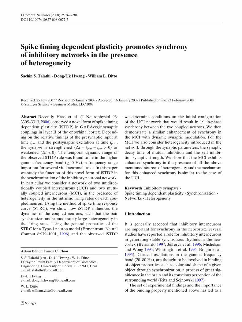
J Comput Neurosci (2008) 25:262–281 DOI 10.1007/s10827-008-0077-7 Spike timing dependent plasticity promotes synchrony of inhibitory networks in the presence of heterogeneity Sachin S. Talathi · Dong-Uk Hwang · William L. Ditto Received: 25 July 2007 / Revised: 15 January 2008 / Accepted: 16 January 2008 / Published online: 23 February 2008 © Springer Science + Business Media, LLC 2008 Abstract Recently Haas et al. (J Neurophysiol 96: 3305–3313, 2006), observed a novel form of spike timing dependent plasticity (iSTDP) in GABAergic synaptic couplings in layer II of the entorhinal cortex. Depend- ing on the relative timings of the presynaptic input at time t pre and the postsynaptic excitation at time t post , the synapse is strengthened (t = t post − t pre > 0) or weakened (t < 0). The temporal dynamic range of the observed STDP rule was found to lie in the higher gamma frequency band (≥40 Hz), a frequency range important for several vital neuronal tasks. In this paper we study the function of this novel form of iSTDP in the synchronization of the inhibitory neuronal network. In particular we consider a network of two unidirec- tionally coupled interneurons (UCI) and two mutu- ally coupled interneurons (MCI), in the presence of heterogeneity in the intrinsic firing rates of each cou- pled neuron. Using the method of spike time response curve (STRC), we show how iSTDP influences the dynamics of the coupled neurons, such that the pair synchronizes under moderately large heterogeneity in the firing rates. Using the general properties of the STRC for a Type-1 neuron model (Ermentrout, Neural Comput 8:979–1001, 1996) and the observed iSTDP Action Editor: Carson C. Chow S. S. Talathi (B ) · D.-U. Hwang · W. L. Ditto J Crayton Pruitt Family Department of Biomedical Engineering, University of Florida, FL 32611, USA e-mail: [email protected]fl.edu D.-U. Hwang e-mail: [email protected]fl.com W. L. Ditto e-mail: [email protected]fl.com we determine conditions on the initial configuration of the UCI network that would result in 1:1 in-phase synchrony between the two coupled neurons. We then demonstrate a similar enhancement of synchrony in the MCI with dynamic synaptic modulation. For the MCI we also consider heterogeneity introduced in the network through the synaptic parameters: the synaptic decay time of mutual inhibition and the self inhibi- tion synaptic strength. We show that the MCI exhibits enhanced synchrony in the presence of all the above mentioned sources of heterogeneity and the mechanism for this enhanced synchrony is similar to the case of the UCI. Keywords Inhibitory synapses · Spike timing dependent plasticity · Synchronization · Networks · Heterogeneity 1 Introduction It is generally accepted that inhibitory interneurons are important for synchrony in the neocortex. Several studies have reported a role for inhibitory interneurons in generating stable synchronous rhythms in the neo- cortex (Bernardo 1997; Jefferys et al. 1996; Michelson and Wong 1994; Whittington et al. 1995; Bragin et al. 1995). Cortical oscillations in the gamma frequency band (20–80 Hz), are thought to be involved in binding of object properties such as color and shape of a given object through synchronization, a process of great sig- nificance in the brain and its conscious perception of the surrounding world (Ritz and Sejnowski 1997). The set of experimental findings and the importance of the binding property mentioned above has led to a
-
Upload
independent -
Category
Documents
-
view
0 -
download
0
Transcript of Spike timing dependent plasticity promotes synchrony in inhibitory networks in presence of...
J Comput Neurosci (2008) 25:262–281DOI 10.1007/s10827-008-0077-7
Spike timing dependent plasticity promotes synchronyof inhibitory networks in the presenceof heterogeneity
Sachin S. Talathi · Dong-Uk Hwang · William L. Ditto
Received: 25 July 2007 / Revised: 15 January 2008 / Accepted: 16 January 2008 / Published online: 23 February 2008© Springer Science + Business Media, LLC 2008
Abstract Recently Haas et al. (J Neurophysiol 96:3305–3313, 2006), observed a novel form of spike timingdependent plasticity (iSTDP) in GABAergic synapticcouplings in layer II of the entorhinal cortex. Depend-ing on the relative timings of the presynaptic input attime tpre and the postsynaptic excitation at time tpost,the synapse is strengthened (�t = tpost − tpre > 0) orweakened (�t < 0). The temporal dynamic range ofthe observed STDP rule was found to lie in the highergamma frequency band (≥40 Hz), a frequency rangeimportant for several vital neuronal tasks. In this paperwe study the function of this novel form of iSTDP inthe synchronization of the inhibitory neuronal network.In particular we consider a network of two unidirec-tionally coupled interneurons (UCI) and two mutu-ally coupled interneurons (MCI), in the presence ofheterogeneity in the intrinsic firing rates of each cou-pled neuron. Using the method of spike time responsecurve (STRC), we show how iSTDP influences thedynamics of the coupled neurons, such that the pairsynchronizes under moderately large heterogeneity inthe firing rates. Using the general properties of theSTRC for a Type-1 neuron model (Ermentrout, NeuralComput 8:979–1001, 1996) and the observed iSTDP
Action Editor: Carson C. Chow
S. S. Talathi (B) · D.-U. Hwang · W. L. DittoJ Crayton Pruitt Family Department of BiomedicalEngineering, University of Florida, FL 32611, USAe-mail: [email protected]
D.-U. Hwange-mail: [email protected]
W. L. Dittoe-mail: [email protected]
we determine conditions on the initial configurationof the UCI network that would result in 1:1 in-phasesynchrony between the two coupled neurons. We thendemonstrate a similar enhancement of synchrony inthe MCI with dynamic synaptic modulation. For theMCI we also consider heterogeneity introduced in thenetwork through the synaptic parameters: the synapticdecay time of mutual inhibition and the self inhibi-tion synaptic strength. We show that the MCI exhibitsenhanced synchrony in the presence of all the abovementioned sources of heterogeneity and the mechanismfor this enhanced synchrony is similar to the case ofthe UCI.
Keywords Inhibitory synapses ·Spike timing dependent plasticity · Synchronization ·Networks · Heterogeneity
1 Introduction
It is generally accepted that inhibitory interneuronsare important for synchrony in the neocortex. Severalstudies have reported a role for inhibitory interneuronsin generating stable synchronous rhythms in the neo-cortex (Bernardo 1997; Jefferys et al. 1996; Michelsonand Wong 1994; Whittington et al. 1995; Bragin et al.1995). Cortical oscillations in the gamma frequencyband (20–80 Hz), are thought to be involved in bindingof object properties such as color and shape of a givenobject through synchronization, a process of great sig-nificance in the brain and its conscious perception of thesurrounding world (Ritz and Sejnowski 1997).
The set of experimental findings and the importanceof the binding property mentioned above has led to a
J Comput Neurosci (2008) 25:262–281 263
number of theoretical studies of synchrony among in-hibitory interneurons (Ernst et al. 1995; vanVreeswijket al. 1994; Wang and Rinzel 1992). The result of thesestudies can in general be summarized as: depending onthe decay time of the inhibitory synaptic coupling, mu-tually coupled inhibitory neurons exhibit in-phase syn-chrony (zero phase difference) or out-phase synchrony(phase difference of π). However much of the aboveinvestigations did not explore the effects of hetero-geneity in the intrinsic firing rates on synchronizationnor did they take into account noise, which is invari-ably present in neuronal systems. In another set oftheoretical investigations, White et al. (1998) exploredthe effects of small heterogeneities on the degrada-tion of synchrony of fast spiking inhibitory neurons,and the mechanism by which the degradation occurs.They found that introduction of even small amountsof heterogeneity in the external dc current that drivesthe firing rate of the neuron, resulted in a significantreduction in the coherence of neuronal spiking. Theyattributed this loss in synchrony to the failure of aheterogeneous network to entrain the frequency offiring. It is important then to understand what medi-ates observed in vivo synchrony of inhibitory neuronalnetworks under biologically realistic conditions of noiseinduced unpredictability and intrinsic heterogeneity inthe spiking rates of the neuronal ensemble.
In this paper we study the issue of sensitivity of syn-chrony to heterogeneity in neuronal firing rates, in thecontext of recently observed spike timing dependentplasticity in inhibitory synapses (iSTDP; Haas et al.2006). We begin our analysis by considering a pairof unidirectionally coupled interneuron’s (UCI) withdissimilar intrinsic firing rates. We study the influenceof iSTDP on the synchronization properties of thesetwo coupled neurons. We observe that iSTDP modu-lates the synaptic coupling strength such that the drivenneuron fires synchronously in-phase with the drivingneuron. The stability of this in-phase synchronous so-lution is then studied in terms of the stability of thefixed point of spike time evolution map for the coupledneurons using the spike time response method (Ackeret al. 2004).
We then explore the function of iSTDP in enhancingsynchronization between mutually coupled interneu-rons (MCI) with self inhibition in the presence ofheterogeneity in the intrinsic firing rates. We considerthe following set of heterogeneity in the MCI: (a) het-erogeneity in external dc current, IDC, (b) heterogene-ity in the synaptic decay time, τD and (c) heterogeneityin the self inhibition strength, gs.
Earlier work (Nowotny et al. 2003; Zhigulin et al.2003) has explored the function of synaptic plasticity
at an excitatory synapse in improving synchronizationof a unidirectionally coupled neuronal network. Theydemonstrated that STDP of excitatory synapses withthe property that the synaptic strength decreases whenpostsynaptic spike occurs after the presynaptic spike(�t = tpost − tpre > 0) and vice-versa, result in increasedsynchronization. In addition the role of gap junctioncoupling between interneurons in enhancement of syn-chronization has also been well studied (Skinner et al.1999). It was shown in Kopell and Ermentrout (2004),that gap junction coupling plays a complementary rolewith respect to chemical synapses in synchrony of in-hibitory neuronal network. The authors demonstratedthat while, inhibition through GABAergic synapsesis important for mitigating the effect of the initialconditions, it is the gap junction coupling that signif-icantly improves synchronization in the presence ofheterogeneity. Our results in this work demonstrate thatinhibitory plastic synapses can serve similar role bysignificantly improving synchronization in the presenceof heterogeneity.
The paper is organized as follows: In the methodssection we present the mathematical model for theneuron, the synapse and the network studied. We thendefine the spike time response curve (STRC) for anisolated neuron with self inhibition. We then presentthe empirical iSTDP rule, observed by Haas et al.(2006) and use it in this paper to study the synchro-nization properties of the inhibitory network in thepresence of heterogeneity. In the results section, webegin with the demonstration of the influence of iSTDPon synchronization of the two unidirectionally coupledinterneurons. We then derive an analytic expressionfor the evolution of spike times for each neuron usingSTRC and then demonstrate how the iSTDP modulatesthe synaptic strength to synchronize the driven neuronto fire in-phase with the driving neuron for a broadrange of heterogeneity in the firing rates. A similar en-hancement in synchronization brought about by iSTDPis observed in the MCI with different intrinsic firingrates.
We have also done some investigation on the in-fluence of noise in the presence of iSTDP on thesynchronization property of the MCI. We consideredtwo potential sources of noise through the milieu ofthe neuronal environment which might influence thedynamics of the network. The first source of noiseconsidered was in the intrinsic firing frequency of eachneuron and the second source of noise was consideredin the modulation strength of the iSTDP. We foundthat even in the presence of mild noise, iSTDP playsa critical role in maintaining the synchronous stateunder heterogeneity. All the details on iSTDP induced
264 J Comput Neurosci (2008) 25:262–281
synchrony in the UCI and the MCI in the presence ofnoise will be presented in future work.
2 Methods
2.1 Model neuron
Each neuron is modeled based on Hodgkin Huxleyframework as a single compartment model with fastsodium channel, delayed rectifier potassium channeland a leak channel. The parameters of the model areset such that it represents a cortical neuron model oftype I (Ermentrout 1996). Each neuron is self inhibitedthrough a GABAergic synaptic model which obeyssecond order kinetics. The dynamical equation for themodel neuron is given by,
CdV(t)
dt= IDC + gNam3(t)h(t)(ENa − V(t))
+gKn4(t)(EK − V(t)) + gL(EL − V(t))
+IM(t) + IS(t) (1)
where C = 1 μF/cm2. V(t) is the membrane potential,IDC: external DC current drive, is set such that theneuron spikes at a given intrinsic frequency F(IDC).IS(t) = gsSS(t)(EI − V(t)), is the synaptic current dueto self inhibition and IM(t) = gM(t)SM(t)(EI − V(t)) isthe synaptic current from external inhibition. gs is thesynaptic strength of self inhibition and gM(t) is thedynamic synapse, whose strength is determined bythe inhibitory synaptic plasticity rule discussed below.Er (r = Na, K, L) are reversal potentials of the sodiumand potassium ion channels and the leak channel re-spectively. EI , is the reversal potential of the inhibitorysynapse. gr (r = Na, K, L) represent the conductance ofsodium, potassium and the leak channel respectively.
The gating variables X = {m, h, n} satisfy the fol-lowing first order kinetic equation: dX(t)
dt = αX(V(t))(1 − X(t)) − βX(V(t))X(t), where αX and βX aregiven by
αm = 0.32(13−(V(t)−Vth))
e(13−(V(t)−Vth))
4.0 −1βm = 0.28((V(t)−Vth)−40)
e((V(t)−Vth)−40)
5 −1
αh = 0.128e17−(V(t)−Vth)
18 βh = 4
e40−(V(t)−Vth)
5 +1
αn = 0.032(15−(V(t)−Vth))
e(15−(V(t)−Vth))
5 −1βn = 0.5
e(V(t)−Vth)−10
40
with Vth = −65 mV.SY(t), (Y = {S, M}) gives the fraction of bound re-
ceptors and satisfy the following first order kineticequation (Abarbanel et al. 2003),
SY(t) = S0(θ(t)) − SY(t)τ (SI − S0(θ(t)))
where Y={S, M}, and θ(t)=∑i �(t−ti).�((ti + τR)−t).
�(X) is the heaviside function satisfying �(X) = 1if X > 0 else �(X) = 0 and ti is the time of the ithpresynaptic neuronal spike. In the case of self inhibi-tion the presynaptic neuron is the same as the postsynaptic neuron. The kinetic equation for S(t) involvestwo time constants, τR = τ (SI − 1), the docking timefor the neurotransmitter and τD = τ SI , the undock-ing time constant for the neurotransmitter binding. Fi-nally, S0(θ) is the sigmoidal function given by, S0(θ) =0.5(1 + tanh(120(θ − 0.1))).
In Table 1 we list all the parameters for the modelconsidered, unless otherwise stated in the text andfigure captions.
All the model parameters given above are withinphysiological range and give high spike rates typicalof the fast spiking interneurons (Lacaille and Williams1990; McCormick et al. 1985).
All the simulations were done using 4th orderRunge–Kutta method for differential equations withtime step δt = 0.01 ms, on a 2 GHz Intel Core DuoMac OS X. The source code is available from authorson request.
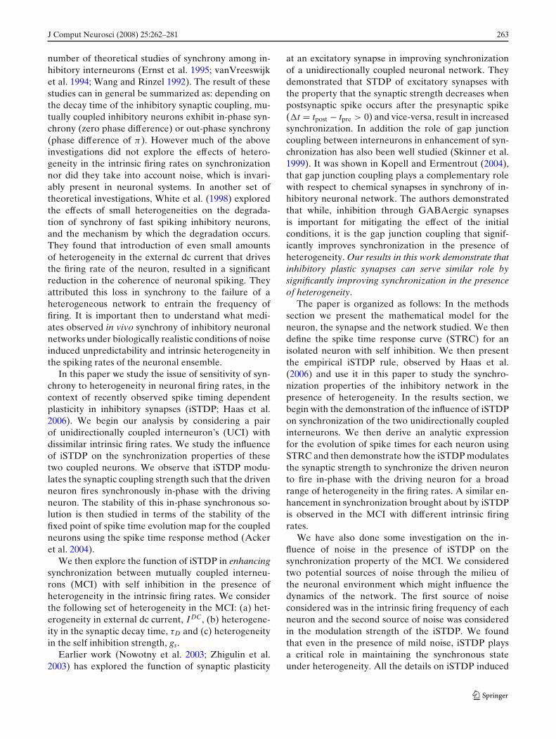
2.2 Spike time response curve (STRC)
As a measure of the influence of synaptic input onthe firing times of a neuron, we define the spiketime response curve (STRC) (t, τR, τD, g, T0) = T −T0 (Acker et al. 2004; Oprisan et al. 2004), where T0 isthe intrinsic period of spiking for a given neuron modelobtained by driving the neuron with a fixed DC currentIDC and T is the time at which the neuron fires a spikeafter it has received a perturbation through synapticinput at time t < T, in its spiking cycle. The key tothe computation of STRC is that the perturbation isthrough a model GABAergic input with synaptic para-meters, τR: the synapse rise time, τD: the synaptic decaytime and g: the synaptic strength. In general this input
Table 1 List of all the parameters for the model considered
Neuron model ENa 50 mVEK −95 mVEL −64 mVgNa 215 mS/cm2
gK 43 mS/cm2
gL 0.813 mS/cm2
Synapse model EI −82 mVgS 0.2 mS/cm2
gM (static synapse) 0.1 mS/cm2
τR 0.2 msτD 5 ms
J Comput Neurosci (2008) 25:262–281 265
need not be weak. The STRC is obtained numericallyhere using the direct method of STRC computation asshown in the schematic diagram of STRC calculation inFig. 1(a). The neuron firing regularly with period T0,is perturbed through inhibitory synapse at time t afterthe neuron has fired a spike at reference time zero. Thespiking time for neuron is considered to be the timewhen the membrane voltage V, crosses a threshold (setto 0 mV in all the calculations presented here). As a
result of this perturbation, the neuron fires the nextspike at time T which is different from T0, the time atwhich the neuron would have fired a spike in absenceof any perturbation through inhibitory synapse. TheSTRC measures this shift in firing time of the neuronT − T0 as a function of the time of perturbation inthe neuronal firing period through the synaptic input.As shown in Fig. 1(b) the STRC is obtained by vary-ing the perturbation time t, over the entire cycle of
Fig. 1 Spike time responsecurve (STRC). (a) Schematicdiagram demonstrating theperturbation effect ofsynaptic input to neuronfiring at given period T0.A perturbation is deliveredto the neuron through theinhibitory synapse at time tafter the last neuronal spikeat reference time t. Theperturbation results in thenext spike of neuronoccurring at time T which isdifferent from the time ofspike for neuron T0 in theabsence of any synapticperturbation. The STRCmeasures this shift in spiketime as function of the time t,when the perturbation isdelivered (b) STRC ofneuron firing with periodT0 = 10.6 ms. The synapticparameters are: τR = 0.2 ms,τD = 5 ms, and g = 0.1mS/cm2
(a)
(b)
0 2 4 6 8 10
1.5
2
2.5
3
3.5
4
t (ms)
ST
RC
Φ (
t) m
s
266 J Comput Neurosci (2008) 25:262–281
spiking (T0 = 10.6 ms, for the example considered) andplotting versus the perturbation time t. The synapticparameters for computation in Fig. 1(b) are, τR = 0.2ms, τD = 5 ms, g = 0.2 mS/cm2 .
STRC is analogous to the phase response curve(PRC; Ermentrout 1996). Similar to the PRC for typeI neurons, the STRC results in the subsequent spike ofthe neuron receiving an inhibitory input to be delayedin time. For brevity of notation, in all further calcu-lations, unless otherwise mentioned, we suppress thedependence of on τR, τD and define (t, τR, τD, g) ≡(t, g).
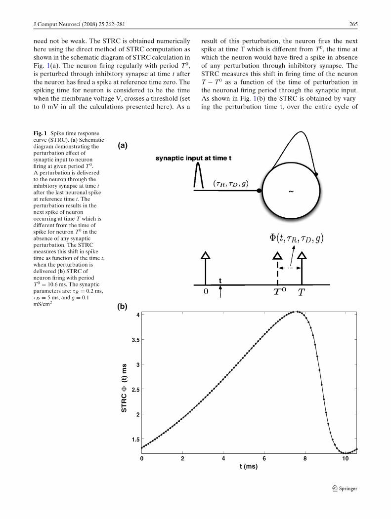
2.3 Spike timing dependent plasticity of inhibitorysynapses
A spike timing dependent plasticity rule for inhibitorysynapses (iSTDP) has been recently reported in Haaset al. (2006) and an empirical fit to the observed experi-mental data was obtained with the following functionalform
�g(�t) = g0
gnormαβ |�t|�tβ−1e−α|�t| (2)
where �t = tpost − tpre. tpre is the time of presynapticspike input arrival and tpost is the time of a spike
generated by the postsynaptic neuron. g0 is the scalingfactor accounting for the amount of change in in-hibitory conductance induced by the synaptic plasticityrule and is set to g0 = 0.02 in all the calculations pre-sented here. gnorm = βe−β is the normalizing constant.With parameter values α = 1 and β = 10 (Haas et al.2006), we obtain a window of ±20 ms over which theefficacy of synaptic plasticity is non zero. In Fig. 2 weshow the iSTDP rule fit with functional form given inEq. (2). Four key properties of the iSTDP rule aresummarized below:
1. �g(�t) > 0 for �t > 02. �g(�t) < 0 for �t < 03. �g(�t) ≈ 0 for �t ≈ 0 and4. �g(−�t) = −�g(�t)
Properties 1 and 2 above imply that a pre-synaptic spikeoccurring before post-synaptic excitation will alwaysenhance the strength of inhibitory synaptic input andvice-versa. Property 3 imply that the synaptic strengthof self inhibition is not modified by the spiking neuronas the pre and the post synaptic spikes for the selfinhibitory synapse occur at the same time, i.e., �t = 0.Property 4, emerges from our choice of same values forparameters α and β for both positive and negative �tregions. We have also explored the effect of asymmetry
Fig. 2 STDP rule forinhibitory synapses. Theparameters are α = 1,β = 10 and g0 = 1
–30 –25 –20 –15 –10 –5 0 5 10 15 20 25 30–1
–0.75
–0.5
–0.25
0
0.25
0.5
0.75
1
Δ t = tpost
– tpre
(ms)
Δ g
/g0
J Comput Neurosci (2008) 25:262–281 267
in the empirical rule on the synchronization propertiesof the inhibitory network. We find that for level ofasymmetry as presented in Haas et al. (2006), where for�t > 0, α = 0.94 and for �t < 0, α = 1.1, with β = 10,the enhancement in synchronization window throughiSTDP remains essentially the same as presented inFig. 6. In all the calcutions presented in this work,unless otherwise mentioned, the parameters for theempirical fit for iSTDP define in Eq. (2) are, α = 1,β = 10 and g0 = 0.02.
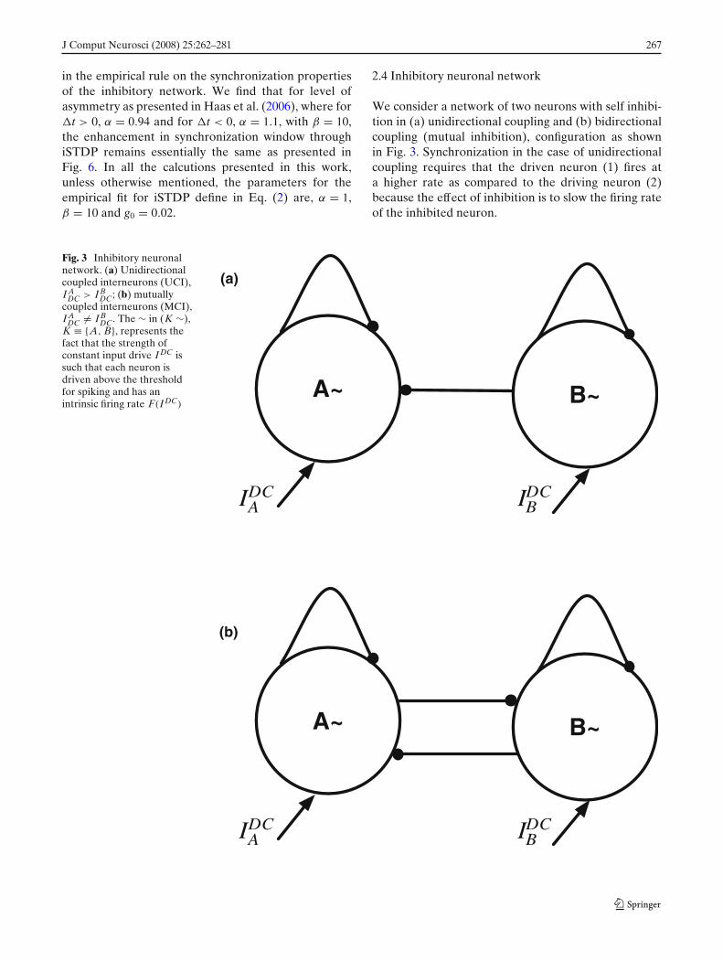
2.4 Inhibitory neuronal network
We consider a network of two neurons with self inhibi-tion in (a) unidirectional coupling and (b) bidirectionalcoupling (mutual inhibition), configuration as shownin Fig. 3. Synchronization in the case of unidirectionalcoupling requires that the driven neuron (1) fires ata higher rate as compared to the driving neuron (2)because the effect of inhibition is to slow the firing rateof the inhibited neuron.
Fig. 3 Inhibitory neuronalnetwork. (a) Unidirectionalcoupled interneurons (UCI),I A
DC > I BDC; (b) mutually
coupled interneurons (MCI),I A
DC �= I BDC. The ∼ in (K ∼),
K ≡ {A, B}, represents thefact that the strength ofconstant input drive I DC issuch that each neuron isdriven above the thresholdfor spiking and has anintrinsic firing rate F(I DC)
(b)
(a)
IDCA IDC
B
A~ B~
IDCA IDC
B
A~ B~

268 J Comput Neurosci (2008) 25:262–281
3 Results
3.1 Single self inhibited neuron
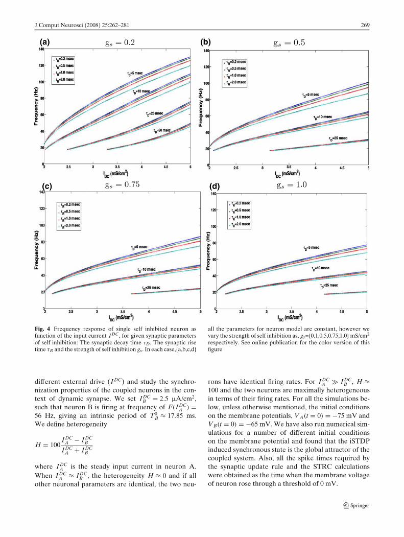
We begin with the study of the firing characteristics ofsingle self inhibited neuron dependent on the synapticparameters: the rise time τR, the decay time τD and thestrength of self inhibition, gs. Spike based adaptation,dependent on self inhibition is considered for thefollowing reasons,
• Biological neural networks often have local in-hibitory interneurons which deliver feedback inhi-bition to the cells that activate those interneurons(Sheperd 1990).
• It has been shown by Traub et al. (2001), that thefrequency in the gamma regime in a distributednetwork of inhibitory interneurons is highly depen-dent on synaptic decay time. This effect has beensimulated through self inhibition in our model.
• The iSTDP learning rule considered in this work,has a zero at �t = 0, which implies, the strengthof self inhibition is not modulated through theiSTDP rule. As a result, the synchronization in theUCI and the MCI, is not modulated by changesin self-inhibition strength of through iSTDP. Inaddition, self-inhibition provides a control over fre-quency range of operation of the neuron throughthe synaptic parameters.
In Fig. 4, we show the frequency response of theneuron for the following set of synaptic parameters,gs = {0.2, 0.5, 0.75, 1.0} mS/cm2, τR = {0.1, 0.5, 1.1, 2.0}ms and τD = {5, 10, 25, 50} ms. We see that while thebiologically realistic time scales for synaptic rise time donot have a significant effect on the firing characteristicsof the self inhibited neuron, the synaptic decay time andthe strength of self inhibition do significantly decreasethe firing frequency of the neuron for a fixed levelof input drive IDC. This results from the fact that forslower synaptic decay times the effect of inhibitionpersists longer. As a result the neuron takes longertime to recover from hyper-polarization to produce aspike again. Also if the strength of inhibition is high, theneuron is strongly inhibited and it takes a longer time torecover back to produce the next spike. Thus τD, the de-cay time of self inhibition and gs, the synaptic strengthof self inhibition determines the frequency regime ofoperation of the neuron. Heterogeneity in one of theseparameters will significantly affect the synchronizationproperties of network of inhibitory neurons.
3.2 Spike time scheme for the analysis of iSTDPinduced synchrony
For the analysis of iSTDP induced synchrony in theUCI and the MCI in the presence of heterogeneityusing STRC, we adopt the following representation forspike times from neuron A and B:
Let WA =[wA(1),wA(2), · · ·wA(i), · · ·wA(NA)], wherewA(i) = ti
A and tiA represents the time of ith spike from
neuron A and NA is the total number of spikesfrom neuron A. Similarly we define the set WB =[wB(1), wB(2),· · · wB( j ),· · · , wB(NB)] with wB( j )= t j
B,where t j
B again represents the time of jth spike fromneuron B. We now define a new set WE comprising ofall spike times from the coupled system (UCI/MCI)arranged in monotonically increasing order as follows,WE = WA ⊕ WB = [wE(1), wE(2), · · · wE(NA + NB)].For example, if t1
A < t1B < t2
A, the first two elements inthe set WE are, wE(1) = t1
A and wE(2) = t1B res-
pectively. In the case when neurons A and Bfires at the same time, we have for some i, and j,tiA = t j
B. These two simultaneous spike events areadded to WE such that, for some n, wE(n) = ti
A andwE(n + 1) = t j
B. We next define two new sets, WA
and WB, which represents a modified spike timeseries for neurons A and B defined by, WA/B =[wA/B(1), wA/B(2), · · · wA/B(NA + NB)],where eachelement wA/B(n) is obtained from WA/B and WE asfollows,
wA/B(n) = max(wA/B(i) ≤ wE(n)
) (0 < i ≤ NA/B
)
We now define an inter spike interval (ISI) time se-ries s(n), for the coupled system as s(n) = wE(n + 1) −wE(n). When the two neurons show in-phase synchronywith period T, we have s(2n − 1) = 0 and s(2n) = T,∀ n > 0. For the case of dynamic synapse, the synapticstrength g(t) is modulated by the pre-post synapticspike pairs through iSTDP [Eq. (2)]. We define g(t) =g(n) when wE(n) ≤ t < wE(n + 1). In Fig. 5 we showthe schematic diagram for the spike time scheme usedfor the analysis of the iSTDP induced synchrony aspresented in this section.
3.3 iSTDP induced synchrony in the UCI
3.3.1 Numerical simulations
We consider two self inhibited neurons with unidirec-tional coupling [Fig. 3(a)] in the presence of hetero-geneity in their intrinsic firing rates introduced through
J Comput Neurosci (2008) 25:262–281 269
(a) (b)
(d)(c)
Fig. 4 Frequency response of single self inhibited neuron asfunction of the input current I DC, for given synaptic parametersof self inhibition: The synaptic decay time τD, The synaptic risetime τR and the strength of self inhibition gs. In each case,{a,b,c,d}
all the parameters for neuron model are constant, however wevary the strength of self inhibition as, gs={0.1,0.5,0.75,1.0} mS/cm2
respectively. See online publication for the color version of thisfigure
different external drive (IDC) and study the synchro-nization properties of the coupled neurons in the con-text of dynamic synapse. We set IDC
B = 2.5 μA/cm2,such that neuron B is firing at frequency of F(IDC
A ) =56 Hz, giving an intrinsic period of T0
B ≈ 17.85 ms.We define heterogeneity
H = 100IDC
A − IDCB
IDCA + IDC
B
where IDCA is the steady input current in neuron A.
When IDCA ≈ IDC
B , the heterogeneity H ≈ 0 and if allother neuronal parameters are identical, the two neu-
rons have identical firing rates. For IDCA IDC
B , H ≈100 and the two neurons are maximally heterogeneousin terms of their firing rates. For all the simulations be-low, unless otherwise mentioned, the initial conditionson the membrane potentials, VA(t = 0) = −75 mV andVB(t = 0) = −65 mV. We have also run numerical sim-ulations for a number of different initial conditionson the membrane potential and found that the iSTDPinduced synchronous state is the global attractor of thecoupled system. Also, all the spike times required bythe synaptic update rule and the STRC calculationswere obtained as the time when the membrane voltageof neuron rose through a threshold of 0 mV.

270 J Comput Neurosci (2008) 25:262–281
Fig. 5 The set WA. WB, WE corresponding to the spike times ofneuron A, neuron B and the coupled system are shown. We alsoshow the inter-spike interval s between the spikes in the coupled
system and the evolution of the dynamic synapse gBA modulatedby the iSTDP rule
We now consider two situations: one in which theconductance of the inhibitory synapse from B to A, gS
BAis static, with gS
BA = 0.1 mS/cm2 (gij is the conductanceof inhibitory synapse from neuron i onto neuron j),and a second situation in which the inhibitory synapticstrength is dynamic and is governed by the iSTDP rulegiven in Fig. 2, such that
gBA(n) = gBA(n − 1) + �g(wA(n) − wB(n)) (3)
where gAB(n) is synaptic strength from B to A for dura-tion, wE(n) ≤ t < wE(n + 1) (see Results, Section 3.2).
In defining the synaptic update rule defined above,we make two key assumptions
• The iSTDP update is considered to be depen-dent only on the two nearest spike neighbors, andthe effects of spike pairs is assumed to sum lin-early, following the conditions under which (Haaset al. 2006) experimentally observed the iSTDP. Weignore multi-spike interactions, which have beenshown to play a significant role in STDP of ex-citatory synapses (Froemke and Dan 2002) and
also any dependence of iSTDP on the frequency offiring of the pre-synaptic neuron.
• The iSTDP update is assumed to happen instanta-neously, as a result we ignore the delay that existsbetween the pairing of the pre and post-synapticspikes and the resultant induction of synaptic mod-ification as suggested by the experiments.
The implication of above assumptions regarding theiSTDP update rule, are explored in more details in thediscussion section. We begin with the initial synapticstrength gBA(t = 0) ≡ gBA(0) = gS
BA = 0.1 mS/cm2. Inorder to determine synchrony in the firing of the UCI,in Fig. 6(a) we plot the ratio of the period for spikingfor neuron B (the driver neuron) T0
B to the averageperiod for spiking for neuron A, 〈TA〉 as a function ofheterogeneity H, for the two cases considered above:Static synapse, and Dynamic synapse, modulated byiSTDP rule defined in Eq. (2). We see that in the case ofstatic synapse, neuron B is able to entrain neuron A tofire at its frequency for mild levels of heterogeneity H(from about 2 to 12). But as the heterogeneity increasesbeyond H ≈ 12, neuron B with a static synapse is no
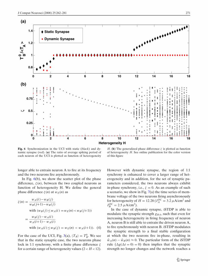
J Comput Neurosci (2008) 25:262–281 271
0 2 4 6 8 10 12 14 16 180
0.25
0.5
0.75
Heterogeneity H
0 2 4 6 8 10 12 14 16 18
0.8
1
1.2
1.4
Tζ
0 B/<
T A>
Static Synapse
Dynamic Synapse
(a)
(b)
Fig. 6 Synchronization in the UCI with static (black) and dy-namic synapse (red). (a) The ratio of average spiking period ofeach neuron of the UCI is plotted as function of heterogeneity
H. (b) The generalized phase difference ζ is plotted as functionof heterogeneity H. See online publication for the color versionof this figure
longer able to entrain neuron A to fire at its frequencyand the two neurons fire asynchronously.
In Fig. 6(b), we show the scatter plot of the phasedifference, ζ(n), between the two coupled neurons asfunction of heterogeneity H. We define the generalphase difference ζ(n) at wE(n) as
ζ(n) = wA(i )−wB( j )wB( j+1)−wB( j )
with (wB( j )≤wA(i ) =wE(n)<wB( j+1))
= wB( j )−wA(i )wA(i+1)− wA(i )
with (wA(i )≤wB( j ) = wE(n) < wA(i+1)). (4)
For the case of the UCI, Fig. 3(a), 〈TB〉 = T0B. We see
that in the static synaptic case, the two neurons phaselock in 1:1 synchrony, with a finite phase difference ζ
for a certain range of heterogeneity values (2< H <12).
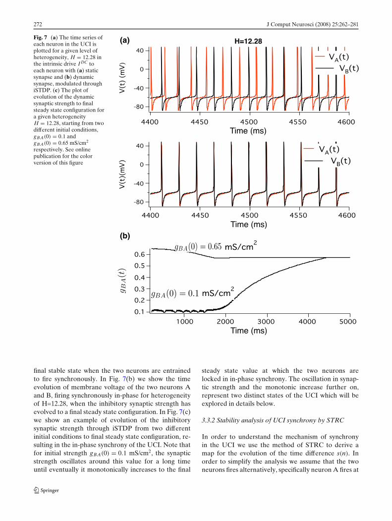
However with dynamic synapse, the region of 1:1synchrony is enhanced to cover a larger range of het-erogeneity and in addition, for the set of synaptic pa-rameters considered, the two neurons always exhibitin-phase synchrony, i.e., ζ = 0. As an example of sucha scenario, we show in Fig. 7(a) the time series of mem-brane voltage of the two neurons firing asynchronouslyfor heterogeneity of H = 12.28 (IDC
A = 3.2 μA/cm2 andIDC
B = 2.5 μA/cm2).In the case of dynamic synapse, iSTDP is able to
modulate the synaptic strength gBA, such that even forincreasing heterogeneity in firing frequency of neuronA, neuron B is still able to entrain the driven neuron A,to fire synchronously with neuron B. iSTDP modulatesthe synaptic strength to a final stable configurationat which the two neurons fire in-phase, resulting inwA(n) − wB(n) ≈ 0. The particular form of the iSTDPrule (�g(�t = 0) = 0) then implies that the synapticstrength no longer changes and the network reaches a
272 J Comput Neurosci (2008) 25:262–281
Fig. 7 (a) The time series ofeach neuron in the UCI isplotted for a given level ofheterogeneity, H = 12.28 inthe intrinsic drive I DC toeach neuron with (a) staticsynapse and (b) dynamicsynapse, modulated throughiSTDP. (c) The plot ofevolution of the dynamicsynaptic strength to finalsteady state configuration fora given heterogeneityH = 12.28, starting from twodifferent initial conditions,gBA(0) = 0.1 andgBA(0) = 0.65 mS/cm2
respectively. See onlinepublication for the colorversion of this figure
(a)
(b)
final stable state when the two neurons are entrainedto fire synchronously. In Fig. 7(b) we show the timeevolution of membrane voltage of the two neurons Aand B, firing synchronously in-phase for heterogeneityof H=12.28, when the inhibitory synaptic strength hasevolved to a final steady state configuration. In Fig. 7(c)we show an example of evolution of the inhibitorysynaptic strength through iSTDP from two differentinitial conditions to final steady state configuration, re-sulting in the in-phase synchrony of the UCI. Note thatfor initial strength gBA(0) = 0.1 mS/cm2, the synapticstrength oscillates around this value for a long timeuntil eventually it monotonically increases to the final
steady state value at which the two neurons arelocked in in-phase synchrony. The oscillation in synap-tic strength and the monotonic increase further on,represent two distinct states of the UCI which will beexplored in details below.
3.3.2 Stability analysis of UCI synchrony by STRC
In order to understand the mechanism of synchronyin the UCI we use the method of STRC to derive amap for the evolution of the time difference s(n). Inorder to simplify the analysis we assume that the twoneurons fires alternatively, specifically neuron A fires at
J Comput Neurosci (2008) 25:262–281 273
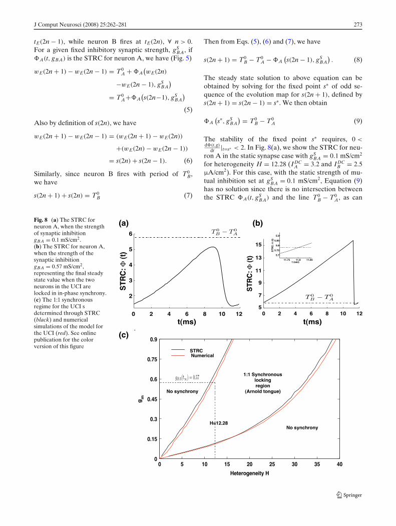
tE(2n − 1), while neuron B fires at tE(2n), ∀ n > 0.For a given fixed inhibitory synaptic strength, gS
BA, ifA(t, gBA) is the STRC for neuron A, we have (Fig. 5)
wE(2n + 1) − wE(2n − 1) = T0A + A
(wE(2n)
−wE(2n − 1), gSBA
)
= T0A+A
(s(2n−1), gS
BA
)
(5)
Also by definition of s(2n), we have
wE(2n + 1) − wE(2n − 1) = (wE(2n + 1) − wE(2n))
+(wE(2n) − wE(2n − 1))
= s(2n) + s(2n − 1). (6)
Similarly, since neuron B fires with period of T0B,
we have
s(2n + 1) + s(2n) = T0B (7)
Then from Eqs. (5), (6) and (7), we have
s(2n + 1) = T0B − T0
A − A(s(2n − 1), gS
BA
). (8)
The steady state solution to above equation can beobtained by solving for the fixed point s∗ of odd se-quence of the evolution map for s(2n + 1), defined bys(2n + 1) = s(2n − 1) = s∗. We then obtain
A(s∗, gS
BA
) = T0B − T0
A (9)
The stability of the fixed point s∗ requires, 0 <d(t,g)
dt |t=s∗ < 2. In Fig. 8(a), we show the STRC for neu-ron A in the static synapse case with gS
BA = 0.1 mS/cm2
for heterogeneity H = 12.28 (IDCA = 3.2 and IDC
B = 2.5μA/cm2). For this case, with the static strength of mu-tual inhibition set at gS
BA = 0.1 mS/cm2, Equation (9)has no solution since there is no intersection betweenthe STRC A(t, gS
BA) and the line T0B − T0
A, as can
Fig. 8 (a) The STRC forneuron A, when the strengthof synaptic inhibitiongBA = 0.1 mS/cm2.(b) The STRC for neuron A,when the strength of thesynaptic inhibitiongBA = 0.57 mS/cm2,representing the final steadystate value when the twoneurons in the UCI arelocked in in-phase synchrony.(c) The 1:1 synchronousregime for the UCI sdetermined through STRC(black) and numericalsimulations of the model forthe UCI (red). See onlinepublication for the colorversion of this figure
(a)
(c)
(b)

274 J Comput Neurosci (2008) 25:262–281
be seen from Fig. 8(a) and therefore the two neuronscannot lock in synchronous state.
In Fig. 8(b), we similarly show the STRC computedwith dynamic synapse in the asymptotic state when thesystem has locked into in-phase synchronous solutionand the inhibitory synaptic strength gBA(t) has reacheda final stable value, gBA(t = ∞) ≡ gBA(∞) = 0.57. Wesee that the solution to Eq. (9) exists as the STRC curveA(t, gBA(∞)) intersects the line T0
B − T0A at s∗ = 11.8
and the condition for stability for this fixed point is alsosatisfied.
In above example we considered a specific examplefor the synaptic strength, gBA = 0.1 mS/cm2 and de-termined that the lack in the existence of stable fixedpoint solution to Eq. (9), results in the absence of syn-chrony between the firing of the two coupled neuronsin the UCI. We also showed that iSTDP modulatedsynaptic strength in the asymptotic state (gBA(∞) =0.57 mS/cm2) provides a unique stable solution to theEq. (9), and the two coupled neurons in the UCI fire inin-phase synchrony.
3.3.3 Arnold tongue of UCI
We next solve Eq. (9) for different levels of hetero-geneity H, thereby modulating T0
A to determine the setof inhibitory synaptic strength gBA which will result inunique stable solution for Eq. (9) to exist. The solutionto Eq. (9) is obtained by estimating the STRC using thedirect method for STRC computation as explained inthe methods section, for each value of the inhibitorysynaptic strength gBA, and determining, whether thereexists a stable fixed point solution to Eq. (9). In Fig. 8(c)we present the results of these calculations. For givenH, the curve in black gives the lower and upper boundon the set of inhibitory synaptic strengths, for which aunique stable solution for Eq. (9) exists. For examplewith H=10, the range of values for gBA which result inan unique stable solution to exist for Eq. (9) is 0.09 <
gBA < 0.49. For this range of values for inhibitorysynaptic strength gBA, at H = 10 the driver neuronB is able to entrain the driven neuron A to oscillatein synchrony with it. This region of synchronous 1:1locking between the two coupled neurons is analogousto the classical Arnold tongue (Kurths et al. 2001)obtained for synchrony between two coupled nonlinearoscillators. Arnold tongues are typical signature of syn-chrony in coupled nonlinear oscillators. In general thewidth of synchrony between two heterogeneous neuraloscillators (heterogeneity in intrinsic firing period ofthe oscillators) is dependent on the strength of couplingbetween the driver and the driven oscillator. Arnoldtongues provide a two dimensional visualization of this
dependence of the width of synchrony on the strengthof coupling between the two oscillators. Naturally in theabsence of any coupling the two oscillators are firingat their intrinsic oscillation frequency and the widthof synchrony is zero. As the strength of coupling isincreased, the width of synchrony increases, resultingin an tongue shaped two dimensional area. In Fig. 8(c),this general feature of Arnold tongue is represented bythe area bounded by the two black curves, obtainedthrough STRC by solving Eq. (9) as discussed above.
In Fig. 8(c), shown in red is a similar bound on thelevels of heterogeneity leading to synchronous oscil-lation in the UCI, obtained by numerically integrat-ing Eq. (1), for the dynamics of evolution of the twocoupled neurons. The curves in red are obtained byfixing the firing period of the driver neuron B at T0
Band varying the intrinsic firing period for neuron A,T0
A by changing the dc current IDCA , thereby varying
the level of heterogeneity H in the firing of the twocoupled neurons, and then determining the synaptic
strength gBA, that will result in T0B
<T A>≈ 1, so that the
two neurons are locked in 1:1 synchrony. We find theresults of numerical simulation match the results fromSTRC calculations in Fig. 8(c). We also see that for H =12.28, the asymptotic value gBA(t) obtained through themodulation of the synapse from B to A through iSTDPis gBA(∞) = 0.57 mS/cm2 which is in the region of 1:1synchrony for the two coupled neurons. We thereforeconclude that iSTDP modulates the synaptic strengthgBA such that Eq. (9) is satisfied and the two coupledneurons lock into 1:1 synchronous in-phase oscillation.
3.3.4 In-phase synchrony in the UCI induced by iSTDP
iSTDP however not only modulates gBA(t) such the twoneurons are locked in 1:1 synchrony, but the strength ismodulated such that the two neurons exhibit in-phasesynchrony with the phase difference ζ being identicallyzero irrespective of H and initial gBA(0), as can be seenfrom Fig. 6(b). In order to understand the function ofiSTDP in producing this in-phase synchrony betweenthe two coupled neurons, we consider the followingtwo scenarios with the case, H = 12.28 and gBA(0) =0.1 mS/cm2 as an example. The initial strength of gBA
is outside and below the region of 1:1 synchronouslocking for the two coupled neurons for given hetero-geneity, as can be seen in Fig. 9.
In this situation, with H > 0, neuron A is firing ata higher rate than neuron B. Therefore more oftenthan not, neuron A will fire more than once for everyperiod of firing of neuron B. Each firing of neuronA (the postsynaptic neuron), results in correspondingincrease in synaptic strength gBA through iSTDP. For
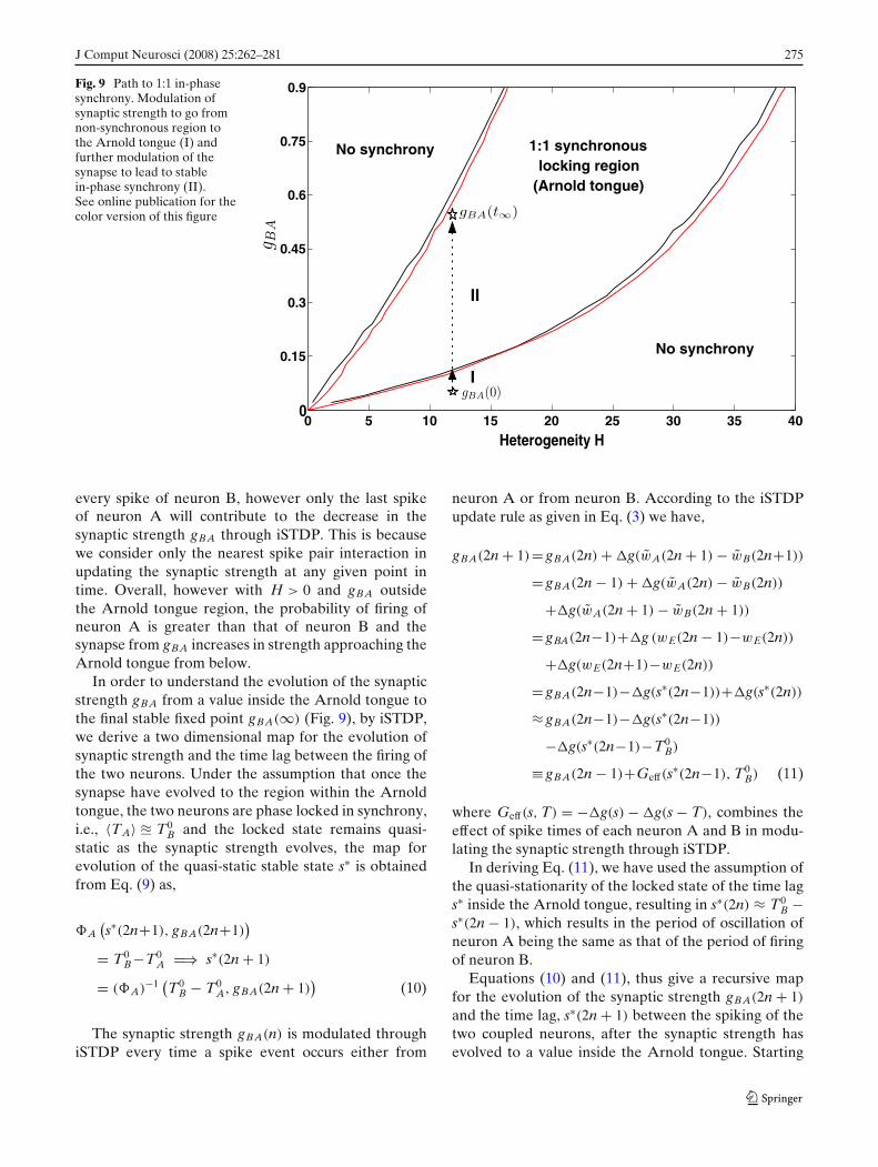
J Comput Neurosci (2008) 25:262–281 275
Fig. 9 Path to 1:1 in-phasesynchrony. Modulation ofsynaptic strength to go fromnon-synchronous region tothe Arnold tongue (I) andfurther modulation of thesynapse to lead to stablein-phase synchrony (II).See online publication for thecolor version of this figure
0 5 10 15 20 25 30 35 400
0.15
0.3
0.45
0.6
0.75
0.9
Heterogeneity H
1:1 synchronouslocking region
(Arnold tongue)
No synchrony
No synchrony
I
II
every spike of neuron B, however only the last spikeof neuron A will contribute to the decrease in thesynaptic strength gBA through iSTDP. This is becausewe consider only the nearest spike pair interaction inupdating the synaptic strength at any given point intime. Overall, however with H > 0 and gBA outsidethe Arnold tongue region, the probability of firing ofneuron A is greater than that of neuron B and thesynapse from gBA increases in strength approaching theArnold tongue from below.
In order to understand the evolution of the synapticstrength gBA from a value inside the Arnold tongue tothe final stable fixed point gBA(∞) (Fig. 9), by iSTDP,we derive a two dimensional map for the evolution ofsynaptic strength and the time lag between the firing ofthe two neurons. Under the assumption that once thesynapse have evolved to the region within the Arnoldtongue, the two neurons are phase locked in synchrony,i.e., 〈TA〉 � T0
B and the locked state remains quasi-static as the synaptic strength evolves, the map forevolution of the quasi-static stable state s∗ is obtainedfrom Eq. (9) as,
A(s∗(2n+1), gBA(2n+1)
)
= T0B−T0
A =⇒ s∗(2n + 1)
= (A)−1 (T0
B − T0A, gBA(2n + 1)
)(10)
The synaptic strength gBA(n) is modulated throughiSTDP every time a spike event occurs either from
neuron A or from neuron B. According to the iSTDPupdate rule as given in Eq. (3) we have,
gBA(2n + 1)= gBA(2n) + �g(wA(2n + 1) − wB(2n+1))
= gBA(2n − 1) + �g(wA(2n) − wB(2n))
+�g(wA(2n + 1) − wB(2n + 1))
= gBA(2n−1)+�g (wE(2n − 1)−wE(2n))
+�g(wE(2n+1)−wE(2n))
= gBA(2n−1)−�g(s∗(2n−1))+�g(s∗(2n))
≈ gBA(2n−1)−�g(s∗(2n−1))
−�g(s∗(2n−1)−T0B)
≡ gBA(2n − 1)+Geff(s∗(2n−1), T0B) (11)
where Geff(s, T) = −�g(s) − �g(s − T), combines theeffect of spike times of each neuron A and B in modu-lating the synaptic strength through iSTDP.
In deriving Eq. (11), we have used the assumption ofthe quasi-stationarity of the locked state of the time lags∗ inside the Arnold tongue, resulting in s∗(2n) ≈ T0
B −s∗(2n − 1), which results in the period of oscillation ofneuron A being the same as that of the period of firingof neuron B.
Equations (10) and (11), thus give a recursive mapfor the evolution of the synaptic strength gBA(2n + 1)
and the time lag, s∗(2n + 1) between the spiking of thetwo coupled neurons, after the synaptic strength hasevolved to a value inside the Arnold tongue. Starting
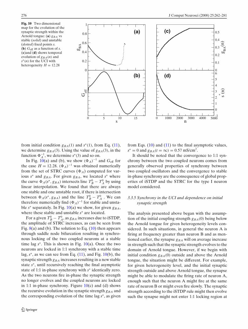
276 J Comput Neurosci (2008) 25:262–281
Fig. 10 Two dimensionalmap for the evolution of thesynaptic strength within theArnold tongue: (a) gBA vsstable (solid) and unstable(dotted) fixed points s.(b) Geff as a function of s.(c)and (d) shows temporalevolution of gBA(n) ands∗(n) for the UCI withheterogeneity H = 12.28
0
0.2
0.4
0.6
0.8
1
g BA
0 5 10 15s
Gef
f
0 1000 2000 3000 4000 5000n
-1
0
1
2
3
4
5
6
s* (n)
0
0.1
0.2
0.3
0.4
0.5
g BA
(n)
(a)
(b)
(c)
(d)
from initial condition gBA(1) and s∗(1), from Eq. (11),we determine gBA(3). Using the value of gBA(3), in thefunction −1
A , we determine s∗(3) and so on.In Fig. 10(a) and (b), we show (A)−1 and Geff for
the case H = 12.28. (A)−1 was obtained numericallyfrom the set of STRC curves (A) computed for var-ious s∗ and gBA. For given gBA, we located s∗ wherethe curve A(s∗, gBA) intersects line T0
B − T0A by using
linear interpolation. We found that there are alwaysone stable and one unstable root, if there is intersectionbetween A(s∗, gBA) and the line T0
B − T0A . We can
therefore numerically find (A)−1 for stable and unsta-ble s∗ separately. In Fig. 10(a) we show, for given gBA,where these stable and unstable s∗ are located.
For a given T0B − T0
A, as gBA increases due to iSTDP,the amplitude of STRC increases, as can be seen fromFig. 8(a) and (b). The solution to Eq. (10) then appearsthrough saddle node bifurcation resulting in synchro-nous locking of the two coupled neurons at a stabletime lag s∗. This is shown in Fig. 10(a). Once the twoneurons are locked in 1:1 synchrony with a stable timelag, s∗, as we can see from Eq. (11), and Fig. 10(b), thesynaptic strength gBA increases resulting in a new stablestate s∗, until recursively reaching the final asymptoticstate of 1:1 in-phase synchrony with s∗ identically zero.As the two neurons fire in-phase the synaptic strengthno longer evolves and the coupled neurons are lockedin 1:1 in-phase synchrony. Figure 10(c) and (d) showsthe recursive evolution in the synaptic strength gBA andthe corresponding evolution of the time lag s∗, as given
from Eqs. (10) and (11) to the final asymptotic values,s∗ = 0 and gBA(t = ∞) = 0.57 mS/cm2.
It should be noted that the convergence to 1:1 syn-chrony between the two coupled neurons comes fromgenerally observed properties of synchrony betweentwo coupled oscillators and the convergence to stablein-phase synchrony are the consequence of global prop-erties of iSTDP and the STRC for the type I neuronmodel considered.
3.3.5 Synchrony in the UCI and dependence on initialsynaptic strength
The analysis presented above began with the assump-tion of the initial coupling strength gBA(0) being belowthe Arnold tongue for given heterogeneity levels con-sidered. In such situations, in general the neuron A isfiring at frequency greater than neuron B and as men-tioned earlier, the synapse gBA will on average increasein strength such that the synaptic strength evolves to thedomain of Arnold tongue. However, if we begin withinitial condition gBA(0) outside and above the Arnoldtongue, the situation might be different. For example,for given heterogeneity level, and the initial synapticstrength outside and above Arnold tongue, the synapsemight be able to modulate the firing rate of neuron A,enough such that the neuron A might fire at the samerate of neuron B or might even fire slowly. The synapticstrength according to the iSTDP rule might then evolvesuch the synapse might not enter 1:1 locking region at
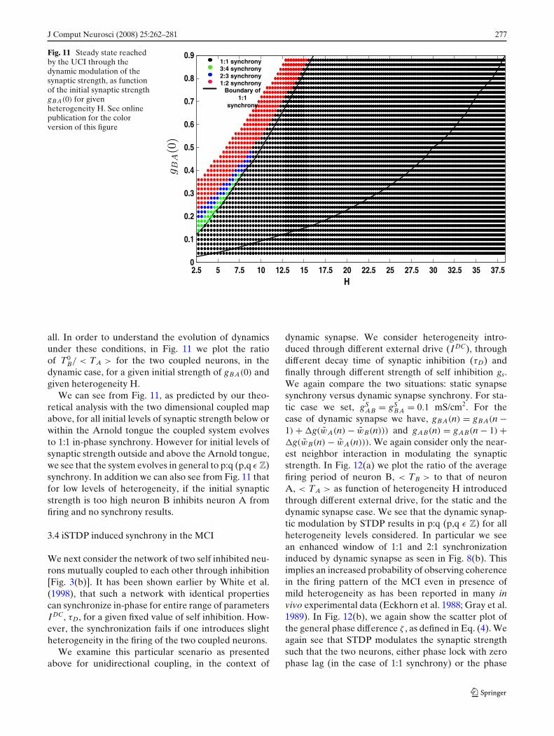
J Comput Neurosci (2008) 25:262–281 277
Fig. 11 Steady state reachedby the UCI through thedynamic modulation of thesynaptic strength, as functionof the initial synaptic strengthgBA(0) for givenheterogeneity H. See onlinepublication for the colorversion of this figure
2.5 5 7.5 10 12.5 15 17.5 20 22.5 25 27.5 30 32.5 35 37.50
0.1
0.2
0.3
0.4
0.5
0.6
0.7
0.8
0.9
H
1:1 synchrony3:4 synchrony2:3 synchrony1:2 synchrony
Boundary of 1:1
synchrony
all. In order to understand the evolution of dynamicsunder these conditions, in Fig. 11 we plot the ratioof T0
B/ < TA > for the two coupled neurons, in thedynamic case, for a given initial strength of gBA(0) andgiven heterogeneity H.
We can see from Fig. 11, as predicted by our theo-retical analysis with the two dimensional coupled mapabove, for all initial levels of synaptic strength below orwithin the Arnold tongue the coupled system evolvesto 1:1 in-phase synchrony. However for initial levels ofsynaptic strength outside and above the Arnold tongue,we see that the system evolves in general to p:q (p,q ε Z)synchrony. In addition we can also see from Fig. 11 thatfor low levels of heterogeneity, if the initial synapticstrength is too high neuron B inhibits neuron A fromfiring and no synchrony results.
3.4 iSTDP induced synchrony in the MCI
We next consider the network of two self inhibited neu-rons mutually coupled to each other through inhibition[Fig. 3(b)]. It has been shown earlier by White et al.(1998), that such a network with identical propertiescan synchronize in-phase for entire range of parametersIDC, τD, for a given fixed value of self inhibition. How-ever, the synchronization fails if one introduces slightheterogeneity in the firing of the two coupled neurons.
We examine this particular scenario as presentedabove for unidirectional coupling, in the context of
dynamic synapse. We consider heterogeneity intro-duced through different external drive (IDC), throughdifferent decay time of synaptic inhibition (τD) andfinally through different strength of self inhibition gs.We again compare the two situations: static synapsesynchrony versus dynamic synapse synchrony. For sta-tic case we set, gS
AB = gSBA = 0.1 mS/cm2. For the
case of dynamic synapse we have, gBA(n) = gBA(n −1) + �g(wA(n) − wB(n))) and gAB(n) = gAB(n − 1) +�g(wB(n) − wA(n))). We again consider only the near-est neighbor interaction in modulating the synapticstrength. In Fig. 12(a) we plot the ratio of the averagefiring period of neuron B, < TB > to that of neuronA, < TA > as function of heterogeneity H introducedthrough different external drive, for the static and thedynamic synapse case. We see that the dynamic synap-tic modulation by STDP results in p:q (p,q ε Z) for allheterogeneity levels considered. In particular we seean enhanced window of 1:1 and 2:1 synchronizationinduced by dynamic synapse as seen in Fig. 8(b). Thisimplies an increased probability of observing coherencein the firing pattern of the MCI even in presence ofmild heterogeneity as has been reported in many invivo experimental data (Eckhorn et al. 1988; Gray et al.1989). In Fig. 12(b), we again show the scatter plot ofthe general phase difference ζ , as defined in Eq. (4). Weagain see that STDP modulates the synaptic strengthsuch that the two neurons, either phase lock with zerophase lag (in the case of 1:1 synchrony) or the phase
278 J Comput Neurosci (2008) 25:262–281
(c) (d)
(b)
(a)
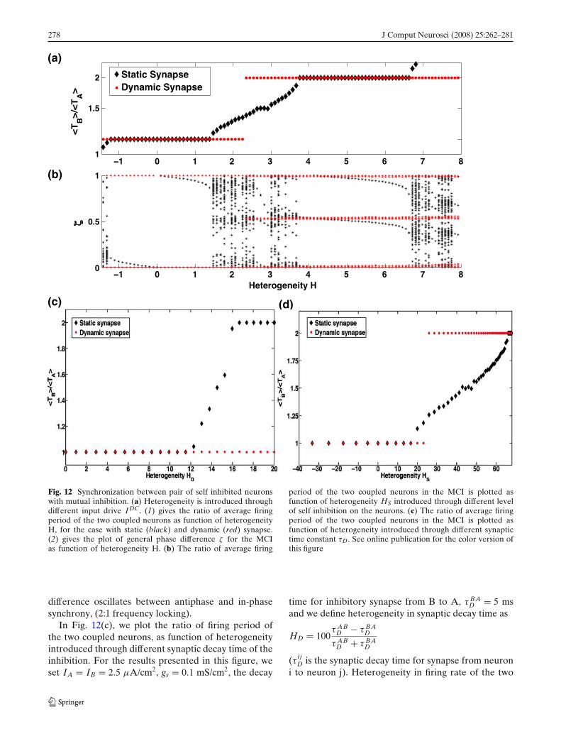
Fig. 12 Synchronization between pair of self inhibited neuronswith mutual inhibition. (a) Heterogeneity is introduced throughdifferent input drive I DC. (1) gives the ratio of average firingperiod of the two coupled neurons as function of heterogeneityH, for the case with static (black) and dynamic (red) synapse.(2) gives the plot of general phase difference ζ for the MCIas function of heterogeneity H. (b) The ratio of average firing
period of the two coupled neurons in the MCI is plotted asfunction of heterogeneity HS introduced through different levelof self inhibition on the neurons. (c) The ratio of average firingperiod of the two coupled neurons in the MCI is plotted asfunction of heterogeneity introduced through different synaptictime constant τD. See online publication for the color version ofthis figure
difference oscillates between antiphase and in-phasesynchrony, (2:1 frequency locking).
In Fig. 12(c), we plot the ratio of firing period ofthe two coupled neurons, as function of heterogeneityintroduced through different synaptic decay time of theinhibition. For the results presented in this figure, weset IA = IB = 2.5 μA/cm2, gs = 0.1 mS/cm2, the decay
time for inhibitory synapse from B to A, τ BAD = 5 ms
and we define heterogeneity in synaptic decay time as
HD = 100τ AB
D − τ BAD
τ ABD + τ BA
D
(τ ijD is the synaptic decay time for synapse from neuron
i to neuron j). Heterogeneity in firing rate of the two
J Comput Neurosci (2008) 25:262–281 279
neurons is introduced by different synaptic decay timeas, larger the decay time of synapse, longer is the inhi-bition and the neuron tends to fire at slower rate. Weagain see that the dynamic synapse is able to modulatethe strength of inhibition on each neuron, such that thetwo neurons are able to synchronize over a broaderrange of heterogeneity in the synaptic decay time.
We finally consider a source of heterogeneity intro-duced by different self inhibition strength gs. We defineheterogeneity in self inhibition
HS = 100gB
S − gAS
gBS + gA
S
The parameters for the MCI in this configuration are,IDC
A = IDCB = 2.5 μA/cm2, τD = 5 ms, τR = 0.1 ms and
gAS = 0.1 mS/cm2. In Fig.12(d), we show the results
comparing synchrony between the two coupled neu-rons with static synapse and dynamic synapse. Againthe coupled neurons with dynamic synapse show agreater robustness in synchrony as compared to thestatic case.
In order to understand synchrony in the MCI, withdynamic modulation of both synaptic strength gAB andgBA, we numerically simulated the MCI with the evo-lution rule applied to the synapse from slower firingneuron B to the faster firing neuron A, gBA as inthe unidirectional case studied above, and fixed thesynaptic strength in opposite direction gAB fixed atmoderately lower strength. Depending on the initialcondition on the synaptic strength gBA, the two neuronseither phase locked in in-phase 1:1 synchrony or thesystem evolved to 2:1 synchrony. We thus conclude thatsynchrony in the MCI is brought about by the modula-tion of the synapse from the slower neuron to the fasterneuron and that the modulation in the synapse fromfaster neuron to the slower neuron, simply controlsthe firing rate of the slower neuron and prevents theeffective inhibition of the slower neuron by the fasterneuron.
4 Discussion
In this work we have analyzed the functional signif-icance of spike timing dependent plasticity, recentlyobserved for inhibitory synapses (Haas et al. 2006) insynchronizing a pair of neurons with self inhibition intwo coupling configurations: (a) uni-directional cou-pling and (b) bi-directional coupling. We begin withthe study of a single self inhibited neuron and showhow the firing frequency of the neuron is dependenton the decay time of the synapse and the strength of
the self synapse. Slower synaptic decay time results inprolonged influence of inhibition and it takes longertime for neuron to recover from inhibition to fire again,there by decreasing the firing frequency of neuron forthe same level of input drive through IDC. The presenceof self inhibition which results in spike based adaptationin firing of the neuron was considered because it hasbeen shown that the frequency in the gamma band ina distributed network of inhibitory neurons is highlydependent on the synaptic decay time. This effect canbe simulated through self inhibition. Moreover theiSTDP learning rule, considered in this study has aninteresting zero at �t = 0 implying that iSTDP doesnot modulate the self inhibition synaptic strength. Thusthe analysis of synchrony through STRC is unaffectedby the presence of self inhibition. In addition (as canbe seen from results in Section 3.1) the parametersof the self inhibition synapse, provide control overthe frequency range of the operation of the neuronand provide an additional source of heterogeneity thatmight influence synchrony between mutually coupledinterneurons.
Network of mutually coupled neurons with intrinsicheterogeneity in firing frequency has been studied ear-lier in White et al. (1998). These authors demonstratedthat even a mild introduction of heterogeneity in thenetwork results in disruption of synchrony in the net-work as the coupling not only has to align the phasefor synchrony but also has to entrain the frequency offiring of the two neurons. They showed that synchronyis achieved only when inhibition is strong enough sothat the firing period is dominated by the synaptic decaytime. However a very strong inhibition results in lossof synchrony through suppression whereby the fasterspiking neuron inhibits the slow neuron so much so thatit stops firing.
In this work we show that a possible route to achievestable synchronous oscillations in the presence of het-erogeneity is through spike timing dependent plasticityof inhibitory synapses. Recently Haas et al. (2006), havereported spike timing dependent plasticity of inhibitorysynapses (iSTDP) in layer II of the entorhinal cortex. Inthis work we have utilized the functional form for thisrecently observed synaptic plasticity rule to study itsinfluence on the synchronization of inhibitory neuronalnetwork in the presence of heterogeneity. The empiri-cal fit to iSTDP data observed by Haas et al. (2006), ispresented in Fig. 2. We have shown that in the presenceof heterogeneity, the dynamic synapse through iSTDPresults in significant enhancement of neural synchrony.The iSTDP modulates the synaptic strength such thatthe faster spiking neuron slows down through increasein inhibition on it and vice versa. We would like to
280 J Comput Neurosci (2008) 25:262–281
note that STDP of inhibitory synapses has also beenobserved in acute hippocampal slices (Woodin et al.2003). The authors report a functional form for iSTDPin hippocampal slices that is symmetric with respect tothe timing of the pre- and postsynaptic spikes and isnon-zero at zero time delay, which is distinctly differentfrom the functional form for iSTDP rule observed inthe entorhinal cortex by Haas et al. (2006). The sig-nificance of this form of iSTDP in the enhancement ofsynchrony in inhibitory neuronal network remains to beexplored.
In all our calculations for the dynamic modulationof the inhibitory synaptic strength and the theoreticalanalysis thereof, we have made two key assumptionsfor the update rule governed by iSTDP. We consideredonly the neighboring spike pair interaction for iSTDPand assumed that the effect of the iSTDP modulationsum linearly. This assumption allowed us to obtainan analytic expression for the evolution of the synap-tic strength, after the two coupled neurons are phaselocked and have evolved to the region within the arnoldtongue. It has also been shown by Froemke and Dan(2002), that in the presence of natural spike trains,the contribution to synaptic modification is primarilythrough the timing of the first spike in each burst.It would be interesting to see how the dynamics ofsynchrony between the MCI would be affected throughsuch multi-spike interaction. The second key assump-tion we made was that the iSTDP update happensinstantaneously, thereby we ignore the actual delay ofseveral minutes that exists between the pairing of thepre and post-synaptic spikes and the results inductionof synaptic modification. Although our assumption ofinstantaneous iSTDP update implies that the synapticmodification has time scale much faster than the firingrate of the neuron, which is contrary to the observedexperimental results, the mere introduction of delay insynaptic update, as seen in experiments, has no conse-quence for our results. In order to verify that it is indeedthe case, we varied the intensity of change in synapticstrength through iSTDP by modulating g0, such that theincrement in the synaptic strength induced by iSTDP ismuch smaller in the UCI. We observed no changes inthe results from our simulations.
In Fig. 6 we compared the synchrony in the UCI withheterogeneity in the presence and absence of dynamicsynapse. We see that iSTDP results in 1:1 synchronybetween the two neurons for all levels of heterogeneityconsidered. We analyzed this network synchrony withthe method of STRC and demonstrate that iSTDPmodulates the synaptic strength such that there existsa unique stable synchronous solution to Eq. (9) for thelevels of heterogeneity considered. In addition, once
the synaptic strength has evolved within the region of1:1 synchronous locking, iSTDP further modulates thesynaptic strength such that it approaches monotonicallyto a final stable configuration wherein the two neuronsare locked in in-phase 1:1 synchrony. We also demon-strated, the influence of the initial synaptic strength inachieving the final 1:1 in-phase synchronous solution.
We next show through numerical simulations, thatthe enhancement in synchronization persists for mu-tually coupled pair of neurons in the presence ofincreasing levels of heterogeneity. For both intrinsicheterogeneity through different external drive and ex-trinsic heterogeneity through different decay time ofthe synapse and different strength of self inhibition,iSTDP is able to maintain synchrony between the cou-pled neurons. Although not presented in this work, thestability of the synchronous state for the MCI can alsobe studied using the STRC method (Acker et al. 2004).
This effect of the iSTDP in enhancement of syn-chrony in the UCI and the MCI in the presence ofheterogeneity in the firing rates of the two coupledneurons is an interesting result from neuroscience per-spective as it suggests that neural system through STDPis very robust against any external perturbation andthat system always phase locks in in-phase synchrony.While such a robust state of neural synchrony is es-sential for memory consolidation, it might not be anoptimal scenario for sensory information processingwherein sensory information is encoded in the form ofa temporal code and a robust neural synchrony mightresult in loss of all sensory information.
We have also done numerical simulations with mildnoise in the intrinsic firing rate of each coupled neuronand the iSTDP rule to study the influence of noise in thecoupled system on the synchrony induced by iSTDP.We found that the synchrony remains enhanced withiSTDP for all the levels of heterogeneity considered.However under similar noise conditions, synchrony iscompletely lost with static synapse. Details on the influ-ence of iSTDP learning on synchrony in the presence ofnoise will be presented in the forthcoming work.
Higher frequency synchronous oscillations havebeen reported experimentally in behaving animals(Ylinen et al. 1995). Our study above suggest thatiSTDP might in fact work better at such high frequen-cies, in maintaining synchronous network oscillations.It has been suggested in Haas et al. (2006), that plas-ticity of inhibitory synapses may play an important rolein balancing the effect of excitatory synapse preventingrun away behavior typically observed in epileptogen-esis. Also recently Abarbanel and Talathi (2006), usedthe model for STDP used in this work to design a neuralcircuitry for spike pattern recognition. In this work we

J Comput Neurosci (2008) 25:262–281 281
present yet another important function for STDP ininhibitory synapses: its role in maintaining synchronyin networks of coupled interneurons, under biologicallyrealistic situation of mild heterogeneity and noise.
Acknowledgements This work was performed under thesponsorship of the Office of Naval Research (Grant N00014-02-1-1019) and the National Institute of Health Collab-orative Research in Computational Neuroscience program(1R01EB004752).
References
Abarbanel, H., Gibb, L., Huerta, R., & Rabinovich, M. (2003).Biophysical model of synaptic plasticity dynamics. BiologicalCybernetics, 89, 214–226.
Abarbanel, H., & Talathi, S. (2006). Neural circuitry for recog-nizing interspike interval sequences. Physical Review Letters,96, 148104.
Acker, C., Kopell, N., & White, J. (2004). Synchronizationof strongly coupled excitatory neurons: Relating networkbehavior to biophysics. Journal of Computational Neuro-science, 15, 71–90.
Benardo, L. (1997). Recruitment of GABAergic inhibition andsynchronization of inhibitory interneurons in rat neocortex.Journal of Neurophysiology, 77, 3134–3144.
Bragin, A., Jando, G., Nadasdy, Z., Hetke, J., Wise, K., et al.(1995). Gamma (40–100 Hz) oscillations in the hippocampusof the behaving rat. Journal of Neuroscience, 15, 47–60.
Eckhorn, R., Bauer, B., Jordan, W., Brosch, M., Kruse, W.,et al. (1988). Coherent oscillations: A mechanism of featurelinking in the visual cortex. Biological Cybernetics, 60,121–130.
Ermentrout, B. (1996). Type 1 membranes, phase resettingcurves and synchrony. Neural Compute, 8, 979–1001.
Ernst, U., Pawelzik, K., & Geisel, T. (1995). Synchronizationinduced by temporal delays in pulse-coupled oscillators.Physical Review Letters, 74, 1570–1573.
Froemke, R., & Dan, Y. (2002). Spike-timing-dependent synapticmodification induced by natural spike trains. Nature, 416,433–438.
Gray, C., Koenig P, Engel, K., & Singer, W. (1989). Oscillatoryresponses in cat visual cortex exhibit intercolumnar synchro-nization which reflect global stimulus properties. Nature,338, 334–337.
Haas, J., Nowotny, T., & Abarbanel, H. (2006). Spike-timing-dependent plasticity of inhibitory synapses in the entorhinalcortex. Journal of Neurophysiology, 96, 3305–3313.
Jefferys, J., Traub, R., & Whittington, M. (1996). Neuronal net-works for induced 40 Hz rhythms. Trends in Neuroscience,19, 202–208.
Kopell, N., & Ermentrout, B. (2004). Chemical and Electricalsynapses perform complementary roles in the synchroniza-tion of interneuronal networks. Proceedings of the NationalAcademy of Sciences, 101, 15482–15487.
Kurths, J., Pikovsky, A., & Rosenblum, M. (2001). Synchroniza-tion, a universal concept in non-linear science. CambridgeUniversity Press.
Lacaille, J., & Williams, S. (1990). Membrane properties ofinterneurons in stratum oriens-alveus of the CA1 region ofrat hippocampus in vitro. Neuroscience, 36, 349–359.
McCormick, D., Connors, B., Lighthall, J., & Prince, D. (1985).Comparative electrophysiology of pyramidal and sparselyspiny stellate neurons of the neocortex. Journal of Neuro-physiology, 54, 782–806
Michelson, H., & Wong, R. (1994). Syncrhonization of inhibitoryneurones in the guinea-pig hippocampus in vitro. Journal ofPhysiology, 477, 35–45.
Nowotny, T., Zhigulin, V., Selverston, A., Abarbanel, H., &Rabinovich, M. (2003). Enhancement of synchronization inhybrid neural circuit by spike timing dependent plasticity.Journal of Neuroscience, 23, 9776–9785.
Oprisan, S., Prinz, A., & Canavier, C. (2004). Phase resettingand phase locking in hybrid circuits of one model and onebiological neuron. Biophysical Journal, 87, 2283–2298.
Ritz, R., & Sejnowski, T. (1997). Synchronous oscillatory activ-ity in sensory systems: New vistas on mechanisms. CurrentOpinion in Neurobiology, 7, 536–546.
Sheperd, G. (1990). The synaptic organization of the brain. NewYork: Oxford University Press.
Skinner, F., Zhang, L., Velazquez, P., & Carlen, P. (1999).Bursting inhibitory interneuronal networks: A role forgap-junctional coupling. Journal of Neurophysiology, 81,1274–1283.
Traub, R., Kopell, N., Bibbig, A., Buhl, E. H., le Beau, F.,et al. (2001). Gap junctions between interneuron dendritescan enhance synchrony of gamma oscillatios. Journal of Neu-roscience, 21, 9478–9486.
vanVreeswijk, C., Abbott, L., & Ermentrout, B. (1994). Wheninhibition and not excitation synchronizes neural firing.Journal of Computational Neuroscience, 1, 313–321.
Wang, X., & Rinzel, J. (1992). Alternating and synchronousrhythms in reciprocally inhibitory model neurons. NeuralComputation, 4, 84–97.
White, A., Chow, C., Ritt, J., Trevino, C., & Kopell, N. (1998).Synchronization and oscillatory dynamics in heterogeneous,mutually inhibited neurons. Journal of Computational Neu-roscience, 5, 5–16.
Whittington, M., Traub, R., & Jefferys, J. (1995). Synchronizedoscillations in interneuron networks driven by metabotropicglutamate receptor activation. Nature, 373, 612–615.
Wilkie, J. (2004). Numerical methods for stochastic differentialequations. Physical Review E, 70, 017701.
Woodin, M., Ganguly, K., & Poo, M. (2003). Coincident Pre-and Postsynaptic activity modifies GABAergic synapses bypostsynaptic changes in Cl Transporter activity. Neuron, 39,807–820
Ylinen, A., Bragin, A., Nadasdy, Z., Jando, G., Szabo, I., et al.(1995). Sharp wave-associated high-frequency oscillation(200 Hz) in the intact hippocampus: network and intracel-lular mechanisms. Journal of Neuroscience, 15, 30–46.
Zhigulin, V., Rabinovich, M., Huerta, R., & Abarbanel, H.(2003). Robustness and enhancement of neural synchroniza-tion by activity-dependent coupling. Physical Review E, 67.