Spectral properties and bio-activity of copper(II) clofibriates, part III: crystal structure of...
12
Spectral properties and bio-activity of copper(II) clofibriates, part III: crystal structure of Cu(clofibriate) 2 (2-pyridylmethanol) 2 , Cu(clofibriate) 2 (4-pyridylmethanol) 2 (H 2 O) dihydrate, and Cu 2 (clofibriate) 4 (N,N-diethylnicotinamide) 2 q,qq Jan Moncol a, * , Barbora Kalinakova b , Jozef Svorec a , Miroslava Kleinova c , Marian Koman a , Daniela Hudecova b , Milan Melnik a , Milan Mazur c , Marian Valko c a Department of Inorganic Chemistry, Slovak Technical University, Radlinskeho 9, SK-81237 Bratislava, Slovakia b Department of Biochemistry and Microbiology, Slovak Technical University, SK-81237 Bratislava, Slovakia c Department of Physical Chemistry, Slovak Technical University, SK-81237 Bratislava, Slovakia Received 22 December 2003; accepted 28 March 2004 Available online 10 May 2004 Abstract New copper(II) clofibriates (clof, {2-(4-chlorophenoxy)-2-methylpropionic or 2-(4-chlorophenoxy)isobutyric acid}) of compo- sition Cu(clof) 2 L 2 (where L ¼ 2-pyridylmethanol (2-pymeth) (1), N-methylnicotinamide (Menia) (4), N,N-diethylnicotinamide (Et 2 nia) (5), isonicotinamide (isonia) (7) or methyl-3-pyridylcarbamate (mpc) (8)), [Cu(clof) 2 (4-pymeth) 2 (H 2 O)] 2H 2 O (4-py- meth ¼ 4-pyridylmethanol) (2 2H 2 O) and Cu(clof) 2 L (where L ¼ 4-pymeth (3) or Et 2 nia (6)) have been prepared and spectro- scopically characterized. All the Cu(clof) 2 L 2 compounds seem to possess distorted octahedral copper(II) stereochemistry with differing tetragonal distortions. An X-ray analysis of 1 was carried out and it featured a tetragonal-bipyramidal geometry around the copper(II) atom. X-ray analysis of 2 2H 2 O featured a square-pyramidal geometry around copper(II) atom. Both the Cu(clof) 2 L compounds seem to consist of a binuclear unit of tetracarboxylate type bridging. An X-ray analysis of 6 revealed typical binuclear paddle-wheel type structure, consisting of two copper(II) atoms in square-pyramidal geometry bridged by four carboxylate anions in the xy-plane. All complexes under study were characterized by EPR and electronic spectroscopy. The antimicrobial effects have been tested on various strains of bacteria, yeasts and filamentous fungi. Ó 2004 Elsevier B.V. All rights reserved. Keywords: Copper; Carboxylate complexes; Crystal structure; Spectral properties; Biological activity; Hydrogen bonding network 1. Introduction The interaction of the copper ions, which plays a vital role in a number of quite differential processes [2], and their interaction with drugs administered for therapeutic reasons is of considerable interest [3]. Phenoxyalkanoic acids also play an important role in biological processes as commercial auxin herbicide and/or anti-inflammatory agents [4]. Copper(II) phenoxyalkanoate complexes have been shown to have diverse stereochemistry with mononuclear [5,6], dinuclear [7] or polynuclear [8] structures. Clofibric acid {2-(4-chlorophenoxy)-2-methylprop- ionic or 2-(4-chlorophenoxy)isobutyric acid} (Scheme 1) is a very interesting anti-inflammatory agent. Five cop- per(II) clofibriate complexes were studied by X-ray dif- fraction methods. X-ray diffraction analysis of [Cu 2 (clof) 4 (ampy) 2 ] [9] and [Cu 2 (clof) 4 (MeOH) 2 ] [10] shows that the compounds are binuclear with square py- ramidal geometry at each copper(II) centre. The two copper(II) atoms are bridged by four carboxylate groups in syn–syn configuration of four clofibriate anions, while q In memory of Prof. Tadeusz Glowiak. qq For part II, see [1]. * Corresponding author. Tel.: +4212593-25-186; fax: +4212524-93- 198. E-mail address: [email protected] (J. Moncol). 0020-1693/$ - see front matter Ó 2004 Elsevier B.V. All rights reserved. doi:10.1016/j.ica.2004.03.043 Inorganica Chimica Acta 357 (2004) 3211–3222 www.elsevier.com/locate/ica
Transcript of Spectral properties and bio-activity of copper(II) clofibriates, part III: crystal structure of...

Inorganica Chimica Acta 357 (2004) 3211–3222
www.elsevier.com/locate/ica
Spectral properties and bio-activity of copper(II) clofibriates, partIII: crystal structure of Cu(clofibriate)2(2-pyridylmethanol)2,Cu(clofibriate)2(4-pyridylmethanol)2(H2O) dihydrate, and
Cu2(clofibriate)4(N,N-diethylnicotinamide)2q,qq
Jan Moncol a,*, Barbora Kalinakova b, Jozef Svorec a, Miroslava Kleinova c,Marian Koman a, Daniela Hudecova b, Milan Melnik a, Milan Mazur c, Marian Valko c
a Department of Inorganic Chemistry, Slovak Technical University, Radlinskeho 9, SK-81237 Bratislava, Slovakiab Department of Biochemistry and Microbiology, Slovak Technical University, SK-81237 Bratislava, Slovakia
c Department of Physical Chemistry, Slovak Technical University, SK-81237 Bratislava, Slovakia
Received 22 December 2003; accepted 28 March 2004
Available online 10 May 2004
Abstract
New copper(II) clofibriates (clof, {2-(4-chlorophenoxy)-2-methylpropionic or 2-(4-chlorophenoxy)isobutyric acid}) of compo-
sition Cu(clof)2L2 (where L¼ 2-pyridylmethanol (2-pymeth) (1), N-methylnicotinamide (Menia) (4), N,N-diethylnicotinamide
(Et2nia) (5), isonicotinamide (isonia) (7) or methyl-3-pyridylcarbamate (mpc) (8)), [Cu(clof)2(4-pymeth)2(H2O)] Æ 2H2O (4-py-
meth¼ 4-pyridylmethanol) (2 Æ 2H2O) and Cu(clof)2L (where L¼ 4-pymeth (3) or Et2nia (6)) have been prepared and spectro-
scopically characterized. All the Cu(clof)2L2 compounds seem to possess distorted octahedral copper(II) stereochemistry with
differing tetragonal distortions. An X-ray analysis of 1 was carried out and it featured a tetragonal-bipyramidal geometry around the
copper(II) atom. X-ray analysis of 2 Æ 2H2O featured a square-pyramidal geometry around copper(II) atom. Both the Cu(clof)2L
compounds seem to consist of a binuclear unit of tetracarboxylate type bridging. An X-ray analysis of 6 revealed typical binuclear
paddle-wheel type structure, consisting of two copper(II) atoms in square-pyramidal geometry bridged by four carboxylate anions in
the xy-plane. All complexes under study were characterized by EPR and electronic spectroscopy. The antimicrobial effects have been
tested on various strains of bacteria, yeasts and filamentous fungi.
� 2004 Elsevier B.V. All rights reserved.
Keywords: Copper; Carboxylate complexes; Crystal structure; Spectral properties; Biological activity; Hydrogen bonding network
1. Introduction
The interaction of the copper ions, which plays a vitalrole in a number of quite differential processes [2], and
their interaction with drugs administered for therapeutic
reasons is of considerable interest [3]. Phenoxyalkanoic
acids also play an important role in biological processes
as commercial auxin herbicide and/or anti-inflammatory
q In memory of Prof. Tadeusz Glowiak.qqFor part II, see [1].
* Corresponding author. Tel.: +4212593-25-186; fax: +4212524-93-
198.
E-mail address: [email protected] (J. Moncol).
0020-1693/$ - see front matter � 2004 Elsevier B.V. All rights reserved.
doi:10.1016/j.ica.2004.03.043
agents [4]. Copper(II) phenoxyalkanoate complexes
have been shown to have diverse stereochemistry with
mononuclear [5,6], dinuclear [7] or polynuclear [8]structures.
Clofibric acid {2-(4-chlorophenoxy)-2-methylprop-
ionic or 2-(4-chlorophenoxy)isobutyric acid} (Scheme 1)
is a very interesting anti-inflammatory agent. Five cop-
per(II) clofibriate complexes were studied by X-ray dif-
fraction methods. X-ray diffraction analysis of
[Cu2(clof)4(ampy)2] [9] and [Cu2(clof)4(MeOH)2] [10]
shows that the compounds are binuclear with square py-ramidal geometry at each copper(II) centre. The two
copper(II) atoms are bridged by four carboxylate groups
in syn–syn configuration of four clofibriate anions, while

Fig. 1. Perspective view of complex [Cu(clof)2(2-pymeth)2] (1), with the
atom numbering scheme. Thermal ellipsoids are drawn at the 50%
probability level.
Scheme 1. Clofibric acid.
Table 1
Selected bond lengths (�A) and angles (�) for 1, 2 Æ 2H2O and 6
1
Cu–O1 1.998(2) Cu–N1 1.981(2)
Cu–O4 2.388(2)
O1–Cu–N1 89.30(7) O4–Cu–N1 76.81(7)
O1–Cu–O4 94.11(6) O1–Cu–O1i 180.0
O4–Cu–O4i 180.0 N1–Cu–N1i 180.0
2 Æ 2H2O
Cu–O1 1.967(1) Cu–N1 2.010(2)
Cu–O1W 2.281(3)
O1–Cu–N1 87.86(6) O1W–Cu–N1 95.11(4)
O1–Cu–O1W 91.00(4) N1–Cu–N1ii 169.74(9)
O1–Cu–O1ii 177.98(7)
6
Cu–O21 1.968(2) Cu–O22 1.985(2)
Cu–O11 1.980(2) Cu–O12iii 1.987(2)
Cu–N1 2.179(3) Cu � � �Cuiii 2.6481(9)
O11–Cu–N1 94.8(1) O12iii–Cu–N1 97.1(1)
O21–Cu–N1 95.7(1) O22–Cu–N1 95.7(1)
3212 J. Moncol et al. / Inorganica Chimica Acta 357 (2004) 3211–3222
the apical ligands are 2-aminopyrimidine in [Cu2(clof)4(ampy)2] [9] and methanol molecules in [Cu2(clof)4(MeOH)2] [10], respectively. The structures of
[Cu(clof)2L2], where L¼ pyridine (py) [10] or nicotin-
amide (nia) [11], are mononuclear and each copper(II)
atom has a tetragonal-bipyramidal environment with the
CuO4N2 chromophore. Two-dimensional coordination
polymer is a [Cu(clof)2(3-pymeth)2]n complex [1] (where3-pymeth¼ 3-pyridylmethanol); the coordination envi-
ronment about the copper atom is tetragonal bipyramidal
with CuO4N2 chromophore.
The antibacterial and antifungal activities of a range
of copper(II) carboxylates with N-donor or O-donor
ligands have been evaluated against several fungi and
bacteria by our group [11,12] and other authors [13]. In
this paper, we investigate the preparation of 2-pyridyl-methanol (2-pymeth), 4-pyridylmethanol (4-pymeth), N-
methylnicotinamide (Menia), N,N-diethylnicotinamide
(Et2nia), isonicotinamide (isonia) and methyl-3-pyr-
idylcarbamate (mpc) compounds of copper(II) clofibri-
ate. The complexes were characterized by elemental
analysis, electronic and EPR spectra. The crystal and
molecular structure of [Cu(clof)2(2-pymeth)2], [Cu(clof)2(4-pymeth)2(H2O)] Æ 2H2O and [Cu2(clof)4 (Et2nia)2] wasdetermined. The X-ray data are compared and discussed
with those found for similar copper(II) carboxylates.
The role of different types of hydrogen bond interactions
in crystal packing is discussed as well. Bioactivity of the
complexes was tested on Staphylococcus aureus, Esche-
richia coli, Candida parapsilosis, Rhizopus oryzae, Al-
ternaria alternata, Botrytis cinerea, and Microsporum
gypseum.
O11–Cu–O21 87.9(1) O11–Cu–O22 90.5(1)O11–Cu–O12 167.9(1) O21–Cu–O22 168.5(1)
O12iii–Cu–O21 88.9(1) O12iii–Cu–O22 90.4(1)
Symmetry code: (i) �xþ 1=2;�y þ 1=2;�z; (ii) �x; y;�zþ 1=2;
(iii) �xþ 1;�y;�z.
2. Results and discussion2.1. Crystal and molecular structures
The structure of the complex [Cu(clof)2(2-pymeth)2]
(1): The molecular structure of 1 is shown in Fig. 1 andselected bond distances and bond angles are given in
Table 1. The crystal structure consists of discrete units
of [Cu(clof)2(2-pymeth)2]. The stereochemistry around
the central copper(II) atom is axially elongated octahe-
dral. The molecule of 1 is centrosymmetric with the
copper(II) atom being surrounded by two nitrogen at-
oms coming from two different 2-pyridylmethanol
molecules [Cu–N1¼ 1.981(2) �A] and two carboxylate
oxygen atoms from two different clofibriate anions [Cu–
O1¼ 1.998(2) �A] in a square-planar arrangement. The
methanol oxygen atoms of two 2-pyridylmethanol
molecules complete the distorted octahedral coordina-tion of the copper(II) atom [Cu–O4¼ 2.388(2) �A]. The
intramolecular hydrogen bond O4–H17� � �O2 (Table 2)
creates a six-membered metalocycle and stabilizes the
molecular structure. The complex [Cu(salicylato)2(2-
pymeth)2] has a very similar structure, as well as struc-
tural parameters to complex 1 [14].

Table 2
Parameters in (�A) and (�) of hydrogen bonds within the structure 1, 2 Æ 2H2O and 6
D–H� � �A d(D–H) d(H� � �A) d(D� � �A) \(DHA) Symmetry code
1
O3–H17� � �O2 0.81(3) 1.80(3) 2.595(2) 169(3)
C13–H12. . .O2 0.93(3) 2.58(2) 3.195(3) 124(2) �xþ 1=2; y � 1=2;�zþ 1=2
C14–H13� � �O1 0.92(3) 2.55(2) 3.412(3) 157(2) �xþ 1=2;�y � 1=2
C10–H5� � �Cl 0.92(3) 3.07(3) 3.877(3) 147(2) �xþ 1;�y þ 1;�z
2 Æ 2H2O
O2W–H19� � �O1 0.83(3) 1.97(3) 2.799(2) 177(2) x; y þ 1; zO2W–H20� � �O2 0.77(3) 2.28(3) 2.935(2) 142(2)
O2W–H20� � �O3 0.77(3) 2.42(3) 3.103(2) 147(2)
O1W–H18� � �O2 0.83(3) 2.00(3) 2.808(2) 166(3) x; y � 1; zO4–H17� � �O2W 0.81(3) 1.92(3) 2.715(2) 166(3)
C15-H14� � �O2 0.93(2) 2.51(2) 3.311(2) 144(2)
6
C1–H1� � �O22 0.91(3) 2.59(3) 3.170(5) 122(2)
C5–H5� � �O21 0.92(3) 2.66(3) 3.225(5) 120(2)
C18–H18� � �O11 0.86(3) 2.41(3) 3.059(5) 132(3)
C24–H24� � �O21 0.92(4) 2.47(4) 3.122(5) 128(3)
C14–H14� � �O1 0.97(3) 2.69(3) 3.386(5) 129(2) �xþ 3=2; y � 1=2
C8–H8C� � �O13 0.91(6) 2.65(6) 3.538(7) 166(5) �xþ 2;�y;�z
J. Moncol et al. / Inorganica Chimica Acta 357 (2004) 3211–3222 3213
In Fig. 2 is drawn the crystal packing of 1 in the cell,
viewed perpendicular to the bc-plane. In the crystal
structure, weak C–H� � �O hydrogen bonds [15] [C13–
H12� � �O2; C14–H13� � �O1] and weak C–H� � �Cl hydro-gen bond [16] [C10–H5� � �Cl] (Table 2) dominate the
packing of molecules 1. A relatively short Cl� � �Cl in-teraction [17] [Cl� � �Cl¼ 3.773(2) �A ð�xþ 1; y;�z� 1=2Þwas also observed.
The structure of the complex [Cu(clof)2(4-py-
meth)2(H2O)] Æ 2H2O (2 Æ 2H2O): The molecular structure
of 2 Æ 2H2O is shown in Fig. 3 with selected bond dis-
tances and bond angles given in Table 1. Themolecules of
Fig. 2. View of the packing 1 in the cell, viewed perpendicular to the
bc-plane; hydrogen atoms are omitted for clarity.
Fig. 3. Perspective view of complex [Cu(clof)2(4-pymeth)2(H2O)] Æ2H2O (2 Æ 2H2O), with the atom numbering scheme. Thermal ellipsoids
are drawn at the 50% probability level.
2 are monomeric, five coordinate with square pyramidal
stereochemistry (Fig. 3). The coordination sphere around
each copper(II) ion consists of two oxygen atoms from
the carboxyl groups of unidentate clofibriate anions [Cu–
O1¼ 1.967(1) �A] and two nitrogen atoms from 4-pyr-
idylmethanols [Cu–N1¼ 2.010(2) �A] forming the basal
plane. The fifth coordination position is provided by awater oxygen atom [Cu–O1W¼ 2.281(3) �A]. The Cu
atom is 0.107(1) �A above the equatorial plane O1–N1–
O1i–N1i towards the water oxygen atom O1W. For this
type of five-coordinated structure, the parameter s[s ¼ ða� bÞ=60], (where a and b are the equatorial an-
gles) was introduced [18]. The value s is 0 for perfectly
tetragonal geometry and 1 for perfectly trigonal-bipyra-
midal geometry. In this case s ¼ 0:137. The square-py-ramidal stereochemistry is known among nitrogen base
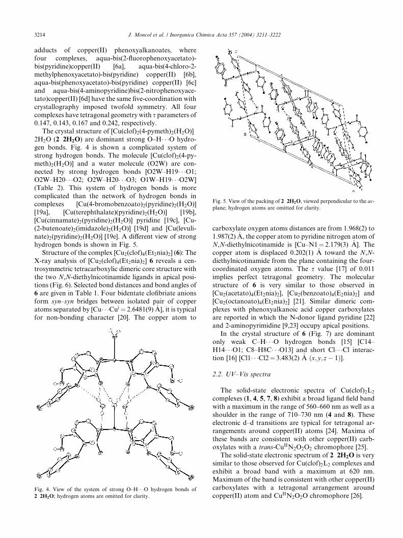
Fig. 5. View of the packing of 2 Æ 2H2O, viewed perpendicular to the ac-
plane; hydrogen atoms are omitted for clarity.
3214 J. Moncol et al. / Inorganica Chimica Acta 357 (2004) 3211–3222
adducts of copper(II) phenoxyalkanoates, where
four complexes, aqua-bis(2-fluorophenoxyacetato)-
bis(pyridine)copper(II) [6a], aqua-bis(4-chloro-2-
methylphenoxyacetato)-bis(pyridine) copper(II) [6b],
aqua-bis(phenoxyacetato)-bis(pyridine) copper(II) [6c]and aqua-bis(4-aminopyridine)bis(2-nitrophenoxyace-
tato)copper(II) [6d] have the same five-coordination with
crystallography imposed twofold symmetry. All four
complexes have tetragonal geometry with s parameters of
0.147, 0.143, 0.167 and 0.242, respectively.
The crystal structure of [Cu(clof)2(4-pymeth)2(H2O)] Æ2H2O (2 Æ 2H2O) are dominant strong O–H� � �O hydro-
gen bonds. Fig. 4 is shown a complicated system ofstrong hydrogen bonds. The molecule [Cu(clof)2(4-py-
meth)2(H2O)] and a water molecule (O2W) are con-
nected by strong hydrogen bonds [O2W–H19� � �O1;
O2W–H20� � �O2; O2W–H20� � �O3; O1W–H19� � �O2W]
(Table 2). This system of hydrogen bonds is more
complicated than the network of hydrogen bonds in
complexes [Cu(4-bromobenzoato)2(pyridine)2(H2O)]
[19a], [Cu(terephthalate)(pyridine)2(H2O)] [19b],[Cu(cinnamate)2(pyridine)2(H2O)] Æ pyridine [19c], [Cu-
(2-butenoate)2(imidazole)2(H2O)] [19d] and [Cu(levuli-
nate)2(pyridine)2(H2O)] [19e]. A different view of strong
hydrogen bonds is shown in Fig. 5.
Structure of the complex [Cu2(clof)4(Et2nia)2] (6): The
X-ray analysis of [Cu2(clof)4(Et2nia)2] 6 reveals a cen-
trosymmetric tetracarboxylic dimeric core structure with
the two N,N-diethylnicotinamide ligands in apical posi-tions (Fig. 6). Selected bond distances and bond angles of
6 are given in Table 1. Four bidentate clofibriate anions
form syn–syn bridges between isolated pair of copper
atoms separated by [Cu� � �Cui ¼ 2.6481(9) �A], it is typical
for non-bonding character [20]. The copper atom to
Fig. 4. View of the system of strong O–H� � �O hydrogen bonds of
2 Æ 2H2O; hydrogen atoms are omitted for clarity.
carboxylate oxygen atoms distances are from 1.968(2) to
1.987(2) �A, the copper atom to pyridine nitrogen atom of
N,N-diethylnicotinamide is [Cu–N1¼ 2.179(3) �A]. Thecopper atom is displaced 0.202(1) �A toward the N,N-
diethylnicotinamide from the plane containing the four-
coordinated oxygen atoms. The s value [17] of 0.011
implies perfect tetragonal geometry. The molecular
structure of 6 is very similar to those observed in
[Cu2(acetato)4(Et2nia)2], [Cu2(benzoato)4(E2nia)2] and
[Cu2(octanoato)4(Et2nia)2] [21]. Similar dimeric com-
plexes with phenoxyalkanoic acid copper carboxylatesare reported in which the N-donor ligand pyridine [22]
and 2-aminopyrimidine [9,23] occupy apical positions.
In the crystal structure of 6 (Fig. 7) are dominant
only weak C–H� � �O hydrogen bonds [15] [C14–
H14� � �O1; C8–H8C� � �O13] and short Cl� � �Cl interac-tion [16] [Cl1� � �Cl2¼ 3.483(2) �A ðx; y; z� 1Þ].
2.2. UV–Vis spectra
The solid-state electronic spectra of Cu(clof)2L2
complexes (1, 4, 5, 7, 8) exhibit a broad ligand field band
with a maximum in the range of 560–660 nm as well as a
shoulder in the range of 710–730 nm (4 and 8). These
electronic d–d transitions are typical for tetragonal ar-
rangements around copper(II) atoms [24]. Maxima of
these bands are consistent with other copper(II) carb-oxylates with a trans-CuIIN2O2O2 chromophore [25].
The solid-state electronic spectrum of 2 Æ 2H2O is very
similar to those observed for Cu(clof)2L2 complexes and
exhibit a broad band with a maximum at 620 nm.
Maximum of the band is consistent with other copper(II)
carboxylates with a tetragonal arrangement around
copper(II) atom and CuIIN2O2O chromophore [26].

Fig. 6. Perspective view of complex [Cu2(clof)4(Et2nia)2] (6), with the atom numbering scheme. Thermal ellipsoids are drawn at the 50% probability
level.
Fig. 7. View of the packing of 6 in the cell, viewed perpendicular to the ac-plane; hydrogen atoms are omitted for clarity.
J. Moncol et al. / Inorganica Chimica Acta 357 (2004) 3211–3222 3215
The solid-state electronic spectra of both complexes
Cu(clof)2L show a broad absorption band (band I) in
the visible region (757 nm for 3 and 717 nm for 6).These bands were assigned to dxz;yz–dx2�y2 transitions
[27]. Moreover, the spectra of both complexes display
shoulders at 429 and 400 nm (band II). This band is
broad due to a shoulder observed at lower energies
originating from the dz2–dx2�y2 transition, which is
typical of Jahn-Teller distorted Cu(II) complexes [28].
The other strong band and shoulder around 230–380
nm (band III) has been assigned to a charge-transfer
LMCT absorption [29] and is believed to be indicativeof a dimeric complex [29] even though a number of
monomeric and polymeric Cu(II) compounds exhibit
this band [30]. Finally, both complexes display bands I,
II and III in the usual range observed for Cu(II)
compounds in a square-pyramidal CuIIO4O0 and
CuIIO4N environment [12,31].

3216 J. Moncol et al. / Inorganica Chimica Acta 357 (2004) 3211–3222
2.3. EPR spectra
2.3.1. Monomeric strucutures
The EPR spectra of monomeric complexes are of the
normal axial type, exhibiting allowed transitions(DMs ¼ 1) characteristic of species with S ¼ 1=2. The gtensor components obtained from these spectra exhibit
1000 2000 3000 4000
Bmon
BZ
1
B(x2
Magnetic Field /
2000 2500 3000 3
exp
sim
Magnetic Field /
2000 2500 3000
1500 2000 2500 3000 3500 4000
Magnetic Fiel
(a)
(b)
(c)
Fig. 8. First-derivative EPR powder spectra at room temperature of: (a) [C
using spin Hamiltonian parameters in Table 3, (b) [Cu2(clof)4(4-pymeth)2spectrum; inset: forbidden transitions measured with increased sensitivity (5
g? < gk; in agreement with the elongated pseudo-octa-
hedral geometry having a dx2�y2 ground state [32].
For complexes 4 and 5, copper hyperfine splitting
structure (ICu ¼ 3/2) is nicely resolved (Fig. 8(a)). This
points to reduced copper–copper dipolar interaction in 4and 5 and an effective diamagnetic shielding of copper
atom by Menia and Et2nia ligands, respectively.
5000 6000 7000
Bz
2
,y2)
Gauss
500 4000 4500 Gauss
3500 4000 4500
d / Gauss
u(clof)2(Et2nia)2] (5) experimental spectrum and computer simulation
] (3), (c) [Cu(clof)2(4-pymeth)2(H2O)] Æ 2H2O (2 Æ 2H2O) experimental
00·).
Table 3
EPR spin Hamiltonian parameters for studied complexesa
Complex g? gk g3 Ak (G) D (cm�1) gk=jAkj (cm) a2
g1 g2
1 2.074 2.079 2.314
2 Æ 2H2O 2.054 2.058 2.271
3 2.076 2.364 0.3938
4 2.031 2.081 2.276 175 139 0.86
5 2.053 2.289 165 149 0.85
6 2.061 2.330 0.3727
7 2.087 2.285 �170b �144 �0.85
8 2.075 2.265a The EPR spectra were measured in polycrystalline form at room temperature.b The parallel hyperfine splitting structure not satisfactorily resolved.
J. Moncol et al. / Inorganica Chimica Acta 357 (2004) 3211–3222 3217
EPR spectra were evaluated by assuming an effective
D4h local symmetry of the complexes. For the anti-
bonding molecular orbital in this symmetry group con-
taining the unpaired electron, we may write the
following equation [33]:
wb1g ¼ adx2�y2 � a0vðx2 � y2Þ; ð1Þ
where d and v functions represent copper(II) 3d orbitals
and ligand-group orbitals of the appropriate symmetry,
respectively [33].
The molecular orbital coefficient a can be related to
the parallel component of the hyperfine splitting tensor
A according to the following equation [34]:
a2 ¼ �ðAk=P Þ þ ðgk � 2Þ þ ð3=7Þðg? � 2Þ þ 0:04; ð2Þwhere P is the dipole coefficient of the free ion and other
symbols have usual meaning [35].
The coefficient a2 characterizes the covalency of the
r-in plane bond and was calculated from the above Eq.(2) for both complexes 4 and 5, which show resolved
parallel hyperfine splitting structure in their EPR spec-
tra. The calculated values are summarized in Table 3.
They imply that the nature of the bonding in th com-
plexes 4, 5 and 7 is not purely ionic and that there is
appreciable in-plane covalent r bonding.
2.3.2. Dimeric structures
The EPR spectra of complexes 3 and 6 are typical of
the triplet state type (Fig. 8(b)) and were interpreted
using the spin Hamiltonian for S ¼ 1 [35]
H ¼ bB�gS þ D S2z
�� 1
2SðS þ 1Þ
�þ EðS2
x � S2y Þ; ð3Þ
where D and E are the zero-field splitting (ZFS) parame-
ters; x, y, and z are the principal-axes coordinate system
fixed with respect to the Cu–Cu bond, and the other
symbols have their usual meaning [35]. The calculated
EPR data for these complexes are given together with the
monomeric structures in Table 3. The EPR spectra of the
studied complexes 3 and 6 revealed axial symmetry; the
ZFS parameter E is therefore equal to zero.
Of specific type is the EPR spectrum of complex
2 Æ 2H2O. The EPR spectrum recorded under typical
spectrometer settings (gain and modulation field) shows
axially resolved spectrum pointing to a monomeric
structure, in agreement with the X-ray data. However,
EPR scan with high gain (500·) shows weak features
typical for half-field,DMs ¼ 2, forbidden transitions. This
indicates that there is an appreciable interaction betweencopper(II) centres (copper–copper distance¼ 6.6 �A),
most probably through space [36]. The calculated values
of gk=Ak summarized in Table 3 for 4, 5 and 7 indicated
marked tetrahedral distortions around copper atom.
2.4. Antimicrobial activities
Results of the quantitative determination of antimi-crobial activity, characterized by IC50 and MIC values,
of the selected copper(II) clofibriates are summarized in
Table 4. As can be seen, the inhibition activities increase
in the sequences: 4> 1> 7> 8> 2 Æ 2H2O > 6 against
model microorganisms. Complexes 4 and 7 had the
highest antibacterial inhibition activity. The sensitivity
of Gþ S. aureus to the tested compounds was two times
higher than that of G� E. coli. Copper(II) complexesvary greatly in their antifungal activity. C. parapsilosis
was greatly inhibited by 4 and 7. Complexes 1, 2 Æ 2H2O,
7 and 8 influenced the colony morphology of A. alter-
nata. Changes of colony colour were probably caused by
defects in biosynthesis of melanin. Ramification effect
on growing hyphal tips of B. cinerea was observed by
complexes 1, 2 Æ 2H2O, 4, 6 and 7, a fungus most sensi-
tive to tested compounds was M. gypseum.
3. Experimental
3.1. Preparation of the complexes
3.1.1. [Cu(clof)2(2-pymeth)2] (1)The complex 1 was prepared by addition of 2-pyridyl-
methanol (0.02 mol) to copper(II) clofibriate (0.01 mol) in

Table
4
AntimicrobialactivityofCu(II)
complexes
characterized
bythenumericalvalues
ofIC
50(m
moll�
1)andMIC
(mmoll�
1)
Bacteria
Yeasts
Filamentousfungi
S.aureus
E.coli
C.parapsilosis
R.oryzae
A.alternata
B.cinerea
M.gypseum
IC50
MI
IC50
MI
IC50
MI
IC50
MI
IC50
MI
IC50
MI
IC50
MI
12.00
5a
3.30
>5
2.70
>5
inact
inact
3.00
>5
1.45
2.5
a
2Æ2H
2O
4.25
>5
3.80
>5
3.20
>5
3.40
>5
4.80
>5
3.70
>5
2.70
5a
41.65
2.5
b3.75
>5
2.20
5b
2.80
>5
2.50
>5
1.70
5b
1.85
2.5
a
63.05
>5
inact
inact
inact
3.35
5a
3.30
>5
2.5
5a
71.55
2.5
b2.95
5b
2.40
5b
2.30
5a
2.25
5a
3.20
>5
3.30
5a
81.60
2.5
binact
3.30
5b
2.05
5b
2.70
5b
inact
2.15
5a
aConcentrationinducingamicrobicidaleffect(M
MC),inact
–inactivecompound(IC
50>5mmoll�
1).
bConcentrationinducingamicrobistaticaleffect.
3218 J. Moncol et al. / Inorganica Chimica Acta 357 (2004) 3211–3222
hot acetone. The mixture was stirred, filtered and left to
cool and stand at room temperature. A blue product pre-
cipitated, which was collected; recrystallization from ace-
tone solution gave air stable crystals. Anal. Calc. for
[Cu(clof)2(2-pymeth)2] (1): C, 54.20; H, 4.83; N, 3.95; Cu,8.96. Found: C, 54.4; H, 4.6; N, 3.8; Cu, 8.95%. IR
masym(COO)1609, msym(COO)1402 cm�1.UV–Vis: 620nm.
3.1.2. [Cu(clof)2(4-pymeth)2(H2O)] � 2H2O (2 � 2H2O)
The complex 2 Æ 2H2O was prepared by addition of
methanol solution of 4-pyridylmethanol (0.02 mol) to
copper(II) clofibriate (0.01 mol) in hot acetone. The
mixture was stirred, filtered and left to cool and stand atroom temperature. A blue product precipitated, which
was collected; recrystallization from acetone solution
gave air stable crystals. Anal. Calc. for [Cu(clof)2(4-py-
meth)2(H2O)] Æ 2H2O (2 Æ 2H2O): C, 50.37; H, 5.28; N,
3.67; Cu, 8.33. Found: C, 50.4; H, 5.4; N, 3.6; Cu,
8.28%. IR m(OH) 3348, masym(COO) 1618, msym(COO)
1398 cm�1. UV–Vis: 620 nm.
3.1.3. [Cu2(clof)4(4-pymeth)2] (3)The complex 3 was prepared by addition of methanol
solution of 4-pyridylmethanol (0.01 mol) to copper(II)
clofibriate (0.01 mol) in hot methanol. The mixture was
stirred, filtered and left to cool and stand at room
temperature. A green product precipitated, which was
collected; recrystallization from acetone solution gave
air stable crystals. Anal. Calc. for [Cu2(clof)4(4-py-meth)2] (3): C, 52.05; H, 4.54; N, 2.33; Cu, 10.59. Found:
C, 51.9; H, 4.4; N, 2.4; Cu, 10.35%. IR m(OH) 3472,
masym(COO) 1620, msym(COO) 1418 cm�1. UV–Vis: 235,
257, 429 (sh), 757 nm.
3.1.4. [Cu(clof)2(Menia)2] (4)The complex 4 was prepared by addition of N-
methylnicotinamide (0.02 mol) to copper(II) clofibriate(0.01 mol) in hot acetone. The mixture was stirred, fil-
tered and left to cool and stand at room temperature. A
blue product precipitated, which was collected; recrys-
tallization from aceton–N,N-diethylnicotinamide (8:1)
solution gave air stable crystals. Anal. Calc. for
[Cu(clof)2(Menia)2] (4): C, 53.51; H, 4.75; N, 7.34; Cu,
8.33. Found: C, 53.2; H, 4.9; N, 4.7; Cu, 8.41%. IR
m(NH) 3248, masym(COO) 1604, msym(COO) 1409 cm�1.UV–Vis: 562, 725 (sh) nm.
3.1.5. [Cu(clof)2(Et2nia)2] (5)The complex 5 was prepared by addition of methanol
solution of N-methylnicotinamide (0.02 mol) to cop-
per(II) clofibriate (0.01mol) in hotmethanol. Themixture
was stirred, filtered and left to cool and stand at room
temperature. A violet product precipitated, which wascollected; recrystallization from acetone solution gave air

J. Moncol et al. / Inorganica Chimica Acta 357 (2004) 3211–3222 3219
stable crystals. Anal. Calc. for [Cu(clof)2(Et2nia)2] (5): C,
56.70; H, 5.71; N, 6.61; Cu, 7.50. Found: C, 56.7; H, 5.9;
N, 6.5; Cu, 7.36%. IR masym(COO) 1607, msym(COO) 1394
cm�1. UV–Vis: 603 nm.
3.1.6. [Cu2(clof)4(Et2nia)2] (6)The complex 6 was prepared by addition of N,N-di-
ethylnicotinamide (0.01 mol) to copper(II) clofibriate
(0.01 mol) in hot methanol. The mixture was stirred,
filtered and left to cool and stand at room temperature.
A green product precipitated, which was collected; re-
crystallization from methanol solution gave air stable
crystals. Anal. Calc. for [Cu2(clof)4(Et2nia)2] (6): C,53.86; H, 5.12; N, 4.19; Cu, 9.50. Found: C, 53.9; H, 4.9;
N, 4.2; Cu, 9.43%. IR masym(COO) 1628, msym(COO) 1414
cm�1. UV–Vis: 230, 273, 400 (sh) 717 nm.
3.1.7. [Cu(clof)2(isonia)2] (7)The complex 7 was prepared by addition of methanol
solution of isonicotinamide (0.02 mol) to copper(II)
clofibriate (0.01 mol) in hot acetone. The mixture wasstirred, filtered and left to cool and stand at room
temperature. A blue product precipitated, which was
collected; recrystallization from acetone solution gave
air stable crystals. Anal. Calc. for [Cu(clof)2(isonia)2]
(7): C, 52.29; H, 4.39; N, 7.62; Cu, 8.64. Found: C, 52.4;
H, 4.5; N, 7.7; Cu, 8.59%. IR masym(NH2) 3400,
masym(NH2) 3169, masym(COO) 1610, msym(COO) 1397
cm�1. UV–Vis: 660 nm.
3.1.8. [Cu(clof)2(mpc)2] (8)The complex 8 was prepared by addition of methanol
solution of methyl-3-pyridylcarbamate (0.02 mol) to
copper(II) clofibriate (0.01 mol) in hot acetone–metha-
nol solution (4:1). The mixture was stirred, filtered and
left to cool and stand at room temperature. A violet
product precipitated, which was collected; recrystalliza-tion from acetone solution gave air stable crystals. Anal.
Calc. for [Cu(clof)2(mpc)2] (8): C, 51.36; H, 4.56; N,
7.05; Cu, 7.99. Found: C, 51.1; H, 4.6; N, 7.1; Cu,
8.09%. IR m(NH) 3243, masym(COO) 1617, msym(COO)
1403 cm�1. UV–Vis: 579, 719 (sh) nm.
3.2. Chemical reagents, analysis and spectral studies
All the chemicals used were of analytical grade
(Aldrich or Sigma) and were used without further
purification.
Copper was determined by electrolysis after miner-
alization of the complexes; carbon, hydrogen, nitrogen
and sulfur were determined by microanalytical methods
(Carlo Erba Instruments EA 1108). Besides the ele-
mental analysis, the water content in hydrates was de-termined from thermogravimetrical curves.
Electronic spectra of the powdered samples in nujol
mulls were recorded at room temperature on Specord
M40 and Specord 200, respectively. IR spectra were
recorded on a FT Magna 750 spectrophotometer at
room temperature. Spectra of solid samples were ob-tained in nujol mulls, KBr and polyethylene pellets at a
resolution of 1 cm�1.
The X-band (�9.4 GHz) EPR spectra in polycrys-
talline solid state were recorded on a Bruker EMX
spectrometer equipped with a Bruker variable temper-
ature unit. Line positions were measured accurately
using internal field markers generated by an NMR
gaussmeter, while the microwave frequency was mea-sured by a microwave frequency counter; 100 kHz
magnetic field modulation (peak-to-peak amplitude� 3
G) was used. All EPR measurements were done using
cylindrical quartz sample tubes with 3.5 mm o.d. (ca. 3.0
mm i.d.). Additional details were published elsewhere
[37].
The simulations of the EPR spectra were performed
using the commercially available program SimFonia(Bruker) [38] and/or program QPOWQPOW developed by Prof.
Belford, University of Illionois [39].
3.3. Structure determination
Data collection at 100 K and cell refinement were
carried out using a a-axis diffractometer Kuma KM-4
CCD with graphite monochromated MoKa radiation.The diffraction intensities were corrected for Lorentz
and polarization factors. The structures were solved
by Patterson methods for 1 using DIRDIFDIRDIF-99 [40] or
direct methods for 2 Æ 2H2O and 6 using SIRSIR-97 [41].
Geometrical analysis was performed using SHELXLSHELXL-97
[42]. Empirical absorption correction was made by
using XABSXABS-2 [43]. The structure of the complexes 1,
2 Æ 2H2O and 6 was drawn by ORTEPORTEP-3 for Windows[44]. The Single Crystal Suite WINGXWINGX [45] was used as
an integrated system for all the crystallographic pro-
grams and software used to prepare material for
publication. Crystal data and conditions of data col-
lection and refinement are reported in Table 5.
3.4. Bio-tests
The antimicrobial activity of the compounds under
investigation was evaluated by a microdilution method
[46] using Gþ bacteria S. aureus (CCM 3824) and G�
bacteria E. coli (CCM 3988). The effects of these com-
pounds on the yeasts C. parapsilosis (Laboratory of
Medical Mycology, Postgraduate Medical Institute,
Bratislava, Slovakia) were determinated by the macro-
dilution method in L-shaped tubes [47]. The cultures ofthe bacteria and yeasts were incubated under vigorous
shaking.

Table 5
Crystallographic data for compounds 1, 2 Æ 2H2O and 6
Complex 1 2 Æ 2H2O 6
Formula C32H34Cl2N2O8Cu C32H40Cl2N2O11Cu C30H34Cl2N2O7Cu
Crystal dimensions (mm) 0.25· 0.20· 0.15 0.20· 0.10· 0.10 0.20· 0.15· 0.10M 709.05 763.10 669.03
T (K) 100(2) 100(2) 100(2)
Crystal system monoclinic monoclinic monoclinic
Space group C2=c C2=c P21=na (�A) 26.698(2) 30.399(5) 13.8410(14)
b (�A) 7.2399(5) 6.638(5) 12.8140(14)
c (�A) 16.790(1) 21.248(5) 18.9998(18)
b (�) 101.120(7) 101.98(3) 111.537(9)
V (�A3) 3184.4(4) 3527(3) 3134.5(5)
Z 4 4 4
Absorption corrections XABS-2 XABS-2 XABS-2
Tmin, Tmax 0.4940, 0.9248 0.7430, 0.9264 0.7567, 0.9105
Dcalc (Mg m�3) 1.479 1.437 1.418
l (Mo Ka) (mm�1) 0.907 0.831 0.915
F ð000Þ 1468 1588 1388
h Range (�) 3.66–25.00 3.54–25.00 3.38–25.00
Reflections collected 8868 9773 10783
Unique reflections (Rint) 2757 (0.0282) 3086 (0.0254) 2884 (0.0309)
S 1.095 1.085 1.079
R1a (observed reflections) 0.0382 0.0283 0.0302
wR2b (all reflections) 0.09779 0.0619 0.0791
aR1 ¼P
ðjFoj � jFcjÞ=P
jFoj:bwR2 ¼
PwðF 2
o � F 2c Þ
2h i
=P
wðF 2o Þ
2n o1=2
, w ¼ 1=½r2ðF 2o Þ þ ðaPÞ2 þ bp�, where P ¼ ðF 2
o þ 2F 2c Þ=3.
3220 J. Moncol et al. / Inorganica Chimica Acta 357 (2004) 3211–3222
The efficiency of prepared derivatives on filamentous
fungi R. oryzae (Collection of Microorganisms of De-
partment of Biochemistry and Microbiology, Faculty of
Chemical and Food Technology, Slovak University of
Technology), B. cinerea (CCMF-16); A. alternata
(CCMF-128) and M. gypseum (Laboratory of Medical
Mycology, Postgraduate Medical Institute, Bratislava,Slovakia), was tested by macrodilution technique on
solidified broth medium during static culturing [47].
Chromatographically pure compounds were dis-
solved in dimethyl sulfoxide, its final concentration
never exceeded 1.0% (v/v) in either the control or the
treated samples. The concentration of the tested com-
pounds ranging from 0.63 to 5 mmol l�1 for filamentous
fungi, yeasts and bacteria was used in experiments.The antimicrobial activity was characterized by IC50
values (concentration of the compound which, in com-
parison to the control, inhibits the growth of microor-
ganisms to 50%) and MIC values (minimal inhibitory
concentration of the compound which inhibits microbial
growth by 100%). The IC50 and MIC values were ob-
tained from the toxicity curves.
MIC experiments on subculture dishes were used toassess the minimal microbicidal concentration (MMC).
Subcultures were prepared separately in Petri dishes
containing complete agar medium and incubated at 30
�C for 48 h (bacteria, yeasts) and at 25 �C for 96 h
(filamentous fungi). The MMC values were taken as the
lowest concentration that showed no visible growth of
microbial colonies in the subculture dishes.
4. Conclusion
A series of eight new copper(II) clofibriates with
pyridylmethanol, N-methylnicotinamide, N,N-diethyl-
nicotinamide, isonicotinamide and methyl-3-pyridylc-
arbamate have been prepared and spectroscopically
characterized. X-ray analysis of two monomeric com-plexes ([Cu(clof)2(4-pymeth)2(H2O)] Æ 2H2O and [Cu
(clof)2(2-pymeth)2]) and a dimeric complex ([Cu2(clof)4(Et2nia)2]) was determined. In [Cu(clof)2(4-pymeth)2-
(H2O)] Æ 2H2O, the copper atom has five coordinate
square pyramidal stereochemistry, while in [Cu(clof)2(2-
pymeth)2], the copper atom has six-coordinate elongated
tetragonal bipyramidal stereochemistry. An X-ray
analysis of [Cu2(clof)4(Et2nia)2] revealed typical binu-clear paddle-wheel type structure, consisting of two
copper(II) atoms in square-pyramidal geometry bridged
by four carboxylate anions in the xy-plane. Similar type
of structure has been proposed for Cu2(clof)4(4-py-
meth)2. The crystal structure of [Cu(clof)2(4-pymeth)2-
(H2O)] Æ 2H2O revealed a complicated system of strong
O–H� � �O hydrogen bonds connecting [Cu(clof)2(4-py-
meth)2(H2O)] moiety via the non-coordinating watermolecules. All the studied Cu(clof)2L2 (L¼N-methyl-
nicotinamide, N,N-diethylnicotinamide, isonicotinamide
or methyl-3-pyridylcarbamate) compounds seem to
possess distorted octahedral copper(II) stereochemistry
with differing tetragonal distortions, similarly to pyri-
dine [10] and nicotinamide [11] copper clofibriate ana-
logues. The antimicrobial effect of selected complexes
J. Moncol et al. / Inorganica Chimica Acta 357 (2004) 3211–3222 3221
has been tested on various strains of bacteria, yeasts and
filamentous fungi.
5. Supplementary material
Crystallographic data (excluding structure factors)
for the structures reported in this paper have been de-
posited with the Cambridge Crystallographic Data
Centre as supplementary publication nos. CCDC-
217665 (1), CCDC-217666 (2 Æ 2H2O) and CCDC-
217667 (6). Copies of the data can be obtained free of
charge on application to the CCDC, 12 Union Road,Cambridge CB2 1EZ, UK [fax: (internat.) +44-0-1223/
336033; e-mail: [email protected]].
Acknowledgements
We thank the Slovak Grant Agency for financial
support (Grant Nos. 1/9251/02 to M.M., 1/9256/02 toM.V. and 1/0109/03 to L.M.).
References
[1] J. Moncol, M. Koman, M. Melnik, T. Glowiak, Cryst. Eng.
Commun. 3 (2001) 262.
[2] D. Kovala-Demertzi, A. Theodorou, M.A. Demertzis, C.P.
Raptoulou, A. Terzis, J. Inorg. Biochem. 65 (1997)
151.
[3] (a) J.R.J. Sorenson, Prog. Med. Chem. 26 (1989) 437;
(b) J.R.J. Sorenson, J. Med. Chem. 19 (1976) 135.
[4] (a) C. Dendrinou-Samara, G. Psomas, K. Christophorou, V.
Tangoulis, C.P. Raptopoulou, A. Terzis, P. Kessissoglou, J.
Chem. Soc., Dalton Trans. (1996) 3737;
(b) G. Smith, E.J. O’Reilly, C.H.L. Kennard, K. Stadnicka, B.
Oleskyn, Inorg. Chim. Acta 47 (1981) 111.
[5] (a) G. Psomas, C.P. Raptopoulou, L. Iordanidis, C. Dendrinou-
Samara, V. Tangoulis, D.P. Kessissoglou, Inorg. Chem. 39 (2000)
3042;
(b) G.V. Goebel, R.J. Doedens, Inorg. Chem. 10 (1971)
2607;
(c) G. Smith, E.J. O’Reilly, C.H.L. Kennard, Inorg. Chim. Acta
62 (1981) 241;
(d) T.C.W. Mak, W.-H. Yip, G. Smith, E.J. O’Reilly, C.H.L.
Kennard, Inorg. Chim. Acta 112 (1986) 53;
(e) R. Stomberg, K. Lundquist, Acta Chem. Scand. 43 (1989)
160;
(f) G. Smith, C.H.L. Kennard, Inorg. Chim. Acta 161 (1989)
3.
[6] (a) C.H.L. Kennard, E.J. O’Reilly, S. Schiller, G. Smith, A.H.
White, Aust. J. Chem. 39 (1986) 1823;
(b) G. Smith, E.J. O’Reilly, C.H.L. Kennard, T.C.W. Mak, Inorg.
Chim. Acta 62 (1982) L219;
(c) C.K. Prout, M.J. Barrow, F.J.C. Rossotti, J. Chem. Soc. A
(1971) 3326;
(d) C.H.L. Kennard, S.W. Steward, E.J. O’Reilly, G. Smith, A.H.
White, Polyhedron 4 (1985) 697.
[7] (a) Y. Kani, M. Tsuchimoto, S. Ohba, H. Matsushima, M.
Noguchi, T.S. Tokii, Acta Crystallogr., Sect. C 55 (1999)
316;
(b) Shova, G. Novitchi, M. Gdaniec, Y.A. Simonov, C. Turta,
Russ. J. Inorg. Chem. 46 (2001) 1685;
(c) T.C.W. Mak, W.-H. Yip, C.H.L. Kennard, Polyhedron 9
(1990) 1667;
(d) G. Smith, D.E. Lynch, T.C.W. Mak, W.-H. Yip, Polyhedron
12 (1993) 203.
[8] (a) G. Smith, E.J. O’Reilly, H.L. Carrell, C.J. Carrell, C.H.L.
Kennard, Polyhedron 15 (1996) 1995;
(b) G. Smith, E.J. O’Reilly, C.H.L. Kennard, A.H. White, J.
Chem. Soc., Dalton Trans. (1985) 243;
(c) S. Shova, G. Novitchi, M. Gdaniec, A. Caneschi, D. Gatteschi,
L. Korobchenko, V.K. Voronkova, Y.A. Simonov, C. Turta, Eur.
J. Inorg. Chem. (2002) 3313;
(d) G. Plesch, M. Blahova, J. Kratsmar-Smogrovic, S. Surka, Z.
Naturforsch., Teil B 28 (1973) 521;
(e) G. Plesch, O. Svajlenova, J. Kratsmar-Smogrovic, M. Kohut-
ova, Chem. Zvesti 37 (1983) 169.
[9] T.C.W. Mak, C.H.L. Kennard, G. Smith, E.J. O’Reilly, D.S.
Sagatys, J.C. Fulwood, Polyhedron 6 (1987) 855.
[10] Y. Kani, S. Ohba, H. Matsushima, T. Tokii, Acta Crystallogr.,
Sect. C 54 (1998) 193.
[11] M. Melnik, M. Koman, D. Hudecova, J. Moncol, B. Dudova, T.
Glowiak, J. Mrozinski, C.E. Holloway, Inorg. Chim. Acta 308
(2000) 1.
[12] (a) J. Moncol, M. Koman, M. Melnik, M. Cernakova, T.
Glowiak, Polyhedron 19 (2000) 2573;
(b) M. Palicova, P. Segla, D. Miklos, M. Kopcova, M. Melnik,
B. Dudova, D. Hudecova, T. Glowiak, Polyhedron 19 (2000)
2689.
[13] (a) C. Dendrinou-Samara, G. Psomas, C.P. Raptopoulou, D.P.
Kessissoglou, J. Inorg. Biochem. 83 (2001) 7;
(b) G. Psomas, C. Densrinou-Samara, P. Philippakopoulos, V.
Tangoulis, C.P. Raptopoulou, E. Samaras, D.P. Kessissoglou,
Inorg. Chim. Acta 272 (1998) 24;
(c) B. Kozlevcar, I. Leban, I. Turel, P. Segedin, M. Petric, F.
Pohleven, A.J.P. White, D.J. Williams, J. Sieler, Polyhedron 18
(1999) 755;
(d) G. Plesch, M. Blahova, J. Kratsmar-Smogrovic, C. Friebel,
Inorg. Chim. Acta 136 (1987) 117.
[14] N.N. Hoang, F. Valach, M. Dunaj-Jurco, M. Melnik, Acta
Crystallogr., Sect. C 48 (1992) 443.
[15] G.R. Desiraju, T. Steiner, The Weak Hydrogen Bond, Oxford
University Press, Oxford, 1999.
[16] (a) P.K. Thallapally, A. Nangia, Cryst. Eng. Commun. 3 (2001)
114;
(b) L. Brammer, E.A. Brutton, P. Sherwood, Cryst. Growth Des.
1 (2001) 277.
[17] G.R. Desiraju, Angew. Chem., Int. Ed. Engl. 34 (1995) 3211.
[18] (a) A.W. Addison, T.N. Rao, J. Reedijk, J. Rija, G.C. Verchoors,
J. Chem. Soc., Dalton Trans. (1984) 1349;
(b) G.A. van Albada, S.A. Komaei, H. Kooijman, A.L. Spek, J.
Reedijk, Inorg. Chim. Acta 287 (1999) 226.
[19] (a) R.E. Del Sesto, A.M. Arif, J.S. Miller, Inorg. Chem. 39 (2000)
4894;
(b) T. Ohmura, W. Mori, M. Hasegawa, T. Takei, A. Yoshizawa,
Chem. Lett. 32 (2003) 34;
(c) M.G.B. Drew, A.P. Mullins, D.A. Rice, Polyhedron 13 (1994)
1631;
(d) E. Escriva, M. Sanau, J.-V. Folgado, J. Garcia-Lozano, A.M.
Arif, Polyhedron 15 (1996) 3276;
(e) J.D. Zubkowski, D. Washington, N. Njoroge, E.J. Valente, T.
Cannon, C.D. Parks, P. Perdahl, D.L. Perry, Polyhedron 16
(1997) 2341.
[20] (a) F. Valach, M. Tokarcik, T. Maris, D.J. Watkin, C.K. Prout, J.
Organomet. Chem. 622 (2001) 166;
(b) F. Valach, M. Tokarcik, A. Sauders, A. Cowley, D.J. Watkin,
Polyhedron 20 (2001) 1933.

3222 J. Moncol et al. / Inorganica Chimica Acta 357 (2004) 3211–3222
[21] (a) L.Kh. Minacheva, T.S. Khodashova, M.A. Porai-Koshits,
A.Yu. Tsivadze, Sov. J. Coord. Chem. 7 (1981) 217;
(b) T. Hokelek, H. Necefoglu, M. Balci, Acta Crystallogr., Sect. C
51 (1995) 2020;
(c) B. Kozlevcar, N. Lah, I. Leban, F. Pohleven, P. Segedin,
Croat. Chem. Acta 73 (2000) 733.
[22] G. Smith, E.J. O’Reilly, C.H.L. Kennard, K.E. Brown, Inorg.
Chim. Acta 49 (1981) 53.
[23] E.J. O’Reilly, G. Smith, C.H.L. Kennard, Inorg. Chim. Acta 90
(1984) 63.
[24] Dubicky, R.L. Martin, Inorg. Chem. 5 (1966) 2203.
[25] (a) L. Antolini, L.P. Battaglia, A. Bonamartini-Corradi, G.
Macrotrigiano, L. Menabur, G.C. Pellacani, M. Saladini, Inorg.
Chem. 21 (1982) 1391;
(b) A.L. Abuhijleh, C. Woods, J. Chem. Soc., Dalton Trans.
(1992) 1249;
(c) Y.R. Morgan, P. Turner, B.J. Kennedy, T.W. Hambley, P.A.
Lay, J.R. Biffin, H.L. Regtop, B. Warwick, Inorg. Chim. Acta 324
(2002) 150.
[26] C. Dendrinou-Samara, D.P. Kessissoglou, P.D. Jannakoudakis,
G.E. Manoussakis, D. Mentzafos, A. Terzis, J. Chem. Soc.,
Dalton Trans. (1992) 3259.
[27] M. Kato, Y. Muto, Coord. Chem. Rev. 92 (1988) 45.
[28] (a) G.W. Reimann, G.F. Kokoszka, G. Gordon, Inorg. Chem. 4
(1965) 1082;
(b) J.E. Weder, T.W. Hambley, B.J. Kennedy, P.A. Lay, D.
MacLachland, R. Bramley, C.D. Delfs, K.S. Murray, B. Mouba-
raki, B. Warwick, J.R. Biffin, H.L. Regtop, Inorg. Chem. 38
(1999) 1736.
[29] J. Catterick, P. Thornton, Adv. Inorg. Chem. Radiochem. 20
(1977) 291.
[30] B.J. Hathaway, D.E. Billing, Coord. Chem. Rev. 5 (1970) 143.
[31] (a) F.P.W. Agterberg, H.A.J. Provo Kluit, W.L. Driessen, J.
Reedijk, H. Oevering, W. Buijs, N. Veldman, M.T. Lakin, A.L.
Spek, Inorg. Chim. Acta 267 (1998) 183;
(b) F.P.W. Agterberg, H.A.J. Prov�o Kluit, W.L. Driessen, M.T.
Lakin, A.L. Spek, J. Reedijk, Inorg. Chem. 36 (1997) 4321.
[32] M. Valko, M. Meln�ık, P. Pelik�an, F. Valach, M. Maz�ur, Chem.
Phys. Lett. 174 (1990) 591.
[33] D. Kivelson, R. Neiman, J. Chem. Phys. 35 (1961) 149.
[34] G. Maki, B. McGarvey, J. Chem. Phys. 29 (1958) 31.
[35] F.E. Mabbs, D. Collison, Electron Paramagnetic Resonance of d-
Transition Metal Compounds, Elsevier, Amsterdam, 1992.
[36] J. Kohout, M. Hvastijova, J. Kozisek, J. Garcia Diaz, M. Valko,
L. Jager, I. Svoboda, Inorg. Chim. Acta 287 (1999) 186.
[37] M. Valko, H. Morris, M. Mazur, J. Telser, E.J.L. McInnes, F.E.
Mabbs, J. Phys. Chem. B 103 (1999) 5591.
[38] SIMFONIASIMFONIA: Simulation Program, Shareware, Bruker, 1996.
[39] (a) R.L. Belford, M.J. Nilges, Program QPOWQPOW: ‘Computer
Simulation of Powder spectra’, EPR Symposium, 21st Rocky
Mountain Conference, Denver, Colorado (August, 1979);
(b) M.J. Nilges, Ph.D. Thesis, University of Illinois, Urbana,
IL.
[40] P.T. Beurskens, G. Beurskens, R. de Gelder, S. Garcia-Granda,
R.O. Gould, R. Israel, J.M.M. Smith, The DIRDIFDIRDIF-99 Program
System, Crystallography Laboratory, University of Nijmegen,
The Netherlands, 1999.
[41] A. Altomare, M.C. Burla, M. Camalli, G.L. Cascarano, C.
Giacovazzo, A. Guagliardi, A.G.G. Moliterni, G. Polidori, R.
Spagna, J. Appl. Crystallogr. 32 (1999) 115.
[42] G.M. Sheldrick, SHELXLSHELXL-97: Program for Refinement of
Crystal Structure, University of G€ottingen, G€ottingen, Ger-
many, 1997.
[43] S. Parkin, B. Moezzi, H. Hope, J. Appl. Crystallogr. 28 (1995) 53.
[44] L.J. Farrugia, J. Appl. Crystallogr. 30 (1997) 565.
[45] L.J. Farrugia, J. Appl. Crystallogr. 32 (1999) 837.
[46] S. Jantova, D. Hudecova, S. Stankovsky, K. Spirkova, L.
Ruzekova, Folia Microbiol. 40 (1995) 611.
[47] D. Hudecova, S. Jantova, M. Melnik, M. Uher, Folia Microbiol.
41 (1996) 473.

![Stereochemistry of imidazolate-bridged copper(II) complexes: [Cu2bpim(im)]2(NO3)4.3H2O, C44H50Cu4N20O15, [Cu(pip)]2(im)(NO3)3.2.5H2O, C29H34Cu2N11O11.5,[Cu(pmdt)]2(2-Meim)(ClO4)3,](https://static.fdokumen.com/doc/165x107/6314c248fc260b71020fb3eb/stereochemistry-of-imidazolate-bridged-copperii-complexes-cu2bpimim2no343h2o.jpg)


![Nanotubes of Core/Shell Cu/Cu[sub 2]O as Anode Materials for Li-Ion Rechargeable Batteries](https://static.fdokumen.com/doc/165x107/63443760f474639c9b0445b5/nanotubes-of-coreshell-cucusub-2o-as-anode-materials-for-li-ion-rechargeable.jpg)