Spatio–Temporal Reconstruction of Bilateral Auditory Steady-State Responses Using MEG Beamformers
11
1092 IEEE TRANSACTIONS ON BIOMEDICAL ENGINEERING, VOL. 55, NO. 3, MARCH 2008 Spatio–Temporal Reconstruction of Bilateral Auditory Steady-State Responses Using MEG Beamformers Mihai Popescu*, Elena-Anda Popescu, Tszping Chan, Shannon D. Blunt, and Jeffrey D. Lewine Abstract—A rapidly growing number of neuromagnetic studies focus on the analysis of auditory steady-state responses (ASSR) in relation to a diverse array of factors including age, selective at- tention, and presence of psychopathology. The objectives of these studies require accurate spatio–temporal estimation of the under- lying neural generators, a challenging task due to the relatively low signal strength and high correlation between bilateral audi- tory cortical sources. This paper evaluates the performance of two beamforming schemes that can potentially overcome such difficul- ties: 1) the linearly constrained minimum variance beamformer with partial sensor coverage (LCMV-PSC), and 2) the multiple constrained minimum-variance beamformer with coherent source region suppression (MCMV-CSRS). Simulation experiments are conducted to assess the impact of source parameters on the re- construction accuracy. The results indicate that the LCMV-PSC method is prone to localization errors that essentially occur along medio–lateral directions, increase with source depth, and are as- sociated to amplitude and phase distortions of the estimated time courses of activity. Comparatively, the MCMV-CSRS method ex- hibits precise localization and minimal amplitude and phase distor- tion for a broad range of relative interferer’s positions within the suppression region. The results from the numerical experiments are validated on real magnetoencephalographic (MEG) data col- lected from a 40-Hz ASSR paradigm. Index Terms—Auditory steady-state response (ASSR), beam- formers, magnetoencephalography (MEG), spatial filtering. I. INTRODUCTION A UDITORY STEADY-STATE RESPONSES (ASSR) elicited by trains of brief auditory stimuli presented at short intervals [1], [2] or by amplitude/frequency modulated Manuscript received February 15, 2007; revised June 28, 2007. Asterisk in- dicates corresponding author. *M. Popescu is with the Department of Molecular and Integrative Physi- ology and the Hoglund Brain Imaging Center, The University of Kansas Med- ical Center, 2021 West 38th Avenue, Kansas City, KS 66160-7401 USA (e-mail: [email protected]). E.-A. Popescu is with the Hoglund Brain Imaging Center, Univer- sity of Kansas Medical Center, Kansas City, KS 66103 USA (e-mail: [email protected]). T. Chan and S. D. Blunt are with the Electrical Engineering and Computer Science Department, Kansas University, Lawrence, KS 66045-7621 USA (e-mail: [email protected]; [email protected]). J. D. Lewine was with the Hoglund Brain Imaging Center and the Depart- ment of Neurology, The University of Kansas Medical Center, Kansas City, KS 66160-7401 USA. He is now with the Alexian Neurosciences Institute, Elk Grove Village, IL 60007 USA (e-mail: [email protected]). Color versions of one or more of the figures in this paper are available online at http://ieeexplore.ieee.org. Digital Object Identifier 10.1109/TBME.2007.906504 sounds [3]–[5] have been extensively utilized to study the brain response to periodically changing sounds of relatively long duration. The ASSR recorded by electroencephalographic (EEG) or magnetoencephalographic (MEG) techniques consist of oscillatory waveforms that show an increased amplitude in adults when the stimulus or modulation rate is around 40 Hz [6]–[8]. Early studies considered the 40-Hz ASSR as a potential tool for assessing hearing thresholds [1], [6], and for evaluating the level of consciousness during general anesthesia [9], with more recent studies focusing on spatio–temporal changes of the response induced by a variety of factors including selective attention [10], normal development [11], and training on pitch discrimination tasks [12]. With the development of whole-head MEG sensing devices, there has been increased interest in the evaluation of interhemispheric differences in source locations and dynamics of activity [8], [13]. Finally, there is renewed interest in how disease processes modify the morphology of the 40-Hz ASSR and the generators locations, with relevant studies in patients with schizophrenia [14] and Alzheimer’s dementia [15]. The accuracy of the spatio–temporal reconstruction of the neuronal activities underlying the ASSR hinges on the incor- poration of realistic constraints that render a mathematically tractable solution to the inverse electromagnetic problem. The most common assumption is that small active cortical areas can be modeled by equivalent current dipoles [16]. Dipole fitting, relying on iterative optimization algorithms, is generally used to estimate the parameters (location, direction, strength) of the source model that best fits the data recorded at the array of sensors. These algorithms are vulnerable to misspecification of the model order and they become less stable with an increased number of unknown parameters [17]. To minimize these effects, the use of additional constraints has been considered, such as positing that ASSR sources in the two hemispheres are sym- metrical with respect to the midsagital plane [12], [18], [19] or by estimating sequentially a single dipolar source in each hemi- sphere, e.g., by using a reduced number of sensors unilaterally covering the parieto–temporal area and assuming that they re- ceive only the contribution from a single auditory source [7], [11], [20]. Although these approaches have provided insights into the underlying ASSR generators, their accuracy is limited by either the artificial nature of the constraints (e.g., assuming symmetry of the source locations biases the solutions towards being as much model driven as data driven), or by the assump- tion that subsets of sensors are exclusively sensitive to a single source [21]. 0018-9294/$25.00 © 2008 IEEE
Transcript of Spatio–Temporal Reconstruction of Bilateral Auditory Steady-State Responses Using MEG Beamformers

1092 IEEE TRANSACTIONS ON BIOMEDICAL ENGINEERING, VOL. 55, NO. 3, MARCH 2008
Spatio–Temporal Reconstruction of BilateralAuditory Steady-State Responses
Using MEG BeamformersMihai Popescu*, Elena-Anda Popescu, Tszping Chan, Shannon D. Blunt, and Jeffrey D. Lewine
Abstract—A rapidly growing number of neuromagnetic studiesfocus on the analysis of auditory steady-state responses (ASSR) inrelation to a diverse array of factors including age, selective at-tention, and presence of psychopathology. The objectives of thesestudies require accurate spatio–temporal estimation of the under-lying neural generators, a challenging task due to the relativelylow signal strength and high correlation between bilateral audi-tory cortical sources. This paper evaluates the performance of twobeamforming schemes that can potentially overcome such difficul-ties: 1) the linearly constrained minimum variance beamformerwith partial sensor coverage (LCMV-PSC), and 2) the multipleconstrained minimum-variance beamformer with coherent sourceregion suppression (MCMV-CSRS). Simulation experiments areconducted to assess the impact of source parameters on the re-construction accuracy. The results indicate that the LCMV-PSCmethod is prone to localization errors that essentially occur alongmedio–lateral directions, increase with source depth, and are as-sociated to amplitude and phase distortions of the estimated timecourses of activity. Comparatively, the MCMV-CSRS method ex-hibits precise localization and minimal amplitude and phase distor-tion for a broad range of relative interferer’s positions within thesuppression region. The results from the numerical experimentsare validated on real magnetoencephalographic (MEG) data col-lected from a 40-Hz ASSR paradigm.
Index Terms—Auditory steady-state response (ASSR), beam-formers, magnetoencephalography (MEG), spatial filtering.
I. INTRODUCTION
AUDITORY STEADY-STATE RESPONSES (ASSR)elicited by trains of brief auditory stimuli presented at
short intervals [1], [2] or by amplitude/frequency modulated
Manuscript received February 15, 2007; revised June 28, 2007. Asterisk in-dicates corresponding author.
*M. Popescu is with the Department of Molecular and Integrative Physi-ology and the Hoglund Brain Imaging Center, The University of Kansas Med-ical Center, 2021 West 38th Avenue, Kansas City, KS 66160-7401 USA (e-mail:[email protected]).
E.-A. Popescu is with the Hoglund Brain Imaging Center, Univer-sity of Kansas Medical Center, Kansas City, KS 66103 USA (e-mail:[email protected]).
T. Chan and S. D. Blunt are with the Electrical Engineering and ComputerScience Department, Kansas University, Lawrence, KS 66045-7621 USA(e-mail: [email protected]; [email protected]).
J. D. Lewine was with the Hoglund Brain Imaging Center and the Depart-ment of Neurology, The University of Kansas Medical Center, Kansas City,KS 66160-7401 USA. He is now with the Alexian Neurosciences Institute, ElkGrove Village, IL 60007 USA (e-mail: [email protected]).
Color versions of one or more of the figures in this paper are available onlineat http://ieeexplore.ieee.org.
Digital Object Identifier 10.1109/TBME.2007.906504
sounds [3]–[5] have been extensively utilized to study thebrain response to periodically changing sounds of relativelylong duration. The ASSR recorded by electroencephalographic(EEG) or magnetoencephalographic (MEG) techniques consistof oscillatory waveforms that show an increased amplitude inadults when the stimulus or modulation rate is around 40 Hz[6]–[8]. Early studies considered the 40-Hz ASSR as a potentialtool for assessing hearing thresholds [1], [6], and for evaluatingthe level of consciousness during general anesthesia [9], withmore recent studies focusing on spatio–temporal changes ofthe response induced by a variety of factors including selectiveattention [10], normal development [11], and training on pitchdiscrimination tasks [12]. With the development of whole-headMEG sensing devices, there has been increased interest in theevaluation of interhemispheric differences in source locationsand dynamics of activity [8], [13]. Finally, there is renewedinterest in how disease processes modify the morphology ofthe 40-Hz ASSR and the generators locations, with relevantstudies in patients with schizophrenia [14] and Alzheimer’sdementia [15].
The accuracy of the spatio–temporal reconstruction of theneuronal activities underlying the ASSR hinges on the incor-poration of realistic constraints that render a mathematicallytractable solution to the inverse electromagnetic problem. Themost common assumption is that small active cortical areas canbe modeled by equivalent current dipoles [16]. Dipole fitting,relying on iterative optimization algorithms, is generally usedto estimate the parameters (location, direction, strength) of thesource model that best fits the data recorded at the array ofsensors. These algorithms are vulnerable to misspecification ofthe model order and they become less stable with an increasednumber of unknown parameters [17]. To minimize these effects,the use of additional constraints has been considered, such aspositing that ASSR sources in the two hemispheres are sym-metrical with respect to the midsagital plane [12], [18], [19] orby estimating sequentially a single dipolar source in each hemi-sphere, e.g., by using a reduced number of sensors unilaterallycovering the parieto–temporal area and assuming that they re-ceive only the contribution from a single auditory source [7],[11], [20]. Although these approaches have provided insightsinto the underlying ASSR generators, their accuracy is limitedby either the artificial nature of the constraints (e.g., assumingsymmetry of the source locations biases the solutions towardsbeing as much model driven as data driven), or by the assump-tion that subsets of sensors are exclusively sensitive to a singlesource [21].
0018-9294/$25.00 © 2008 IEEE

POPESCU et al.: SPATIO–TEMPORAL RECONSTRUCTION OF BILATERAL ASSRS USING MEG BEAMFORMERS 1093
Recent studies focusing on the estimation of the ASSR gen-erators have also tested spatial filtering approaches based eitheron the subspace projection methods [8], [22], [23] or on beam-forming strategies [24], [25]. The signal subspace methodsaim to obtain estimates of the time courses of regional brainactivity, though this requires a reliance on a priori informationregarding the source locations and directions acquired from apreceding dipole-fitting procedure. Hence, subspace methodsare generally prone to the inherited pitfalls of the preprocessingstep. In contrast, the beamformers comprise a collection ofspatial filters, each tuned so as to enhance the signal generatedby a source at a distinct location while suppressing the sensornoise and interferences produced by sources at other locations[26]–[28], all without the requirement of prior assumptionsabout the number and/or spatial distribution of the active brainareas. MEG beamforming methods are also considered attrac-tive since they can potentially estimate the brain activity onraw data or in single trials. However, a restrictive assumptionof beamforming is that source moments from spatially distinctbrain areas are temporally uncorrelated. The well-known can-cellation effect of spatially separate yet covariant sources [29]has been mitigated in various MEG applications by selectinglong segments of raw data for the adaptive beamformers esti-mation, because distinct brain areas are less likely to exhibitsynchronous activities over extended time intervals [27], [30].However, the estimation of the bilateral ASSR generators isa generic example in which conventional beamformers arelikely to fail due to the relatively long lasting, stimulus-drivenoscillatory activity of bilateral sources, which can be highlysynchronized on both averaged and raw data. Acknowledgingthis drawback and in line with the strategies adopted for thedipole fitting, it has been proposed to employ beamformersthat estimate the activity in each hemisphere using a subset ofMEG sensors [or partial sensor coverage (PSC)], i.e., utilizethose sensors that are positioned over the temporoparietal areaof that hemisphere [24]. Although the PSC strategy reducesthe interference from the contralateral auditory cortex, it stillhas a twofold vulnerability. First, PSC inherently reduces thenumber of degrees of freedom available for the adaptation ofthe beamformer coefficients, which may become particularlyimportant when seeking to analyze unaveraged data. Second,in the same manner as for the dipole fitting algorithms, thereis no guarantee of a complete removal of the interference fromthe contralateral hemisphere [21].
This study seeks to gain additional insights into the efficacyof the linearly constrained minimum variance beamformer withpartial sensor coverage (LCMV-PSC) as a means to estimatethe bilateral ASSR generators. An alternative technique for aug-menting beamformer performance is also considered, in whicha priori functional information is incorporated into the beam-former design in order to impose hard null constraints over ex-tended brain volumes that encompass the locations of the in-terferers [31]. The latter approach uses a multiple constrainedminimization of the output power [32] to achieve better suppres-sion of the interference. This spatial filter belongs to the classof multiple constrained minimum variance beamformers withcoherent source region suppression (MCMV-CSRS). The twobeamforming strategies are tested in simulation experiments toassess the impact of the source parameters on the accuracy ofthe estimators. Subsequently, the two beamformers are applied
to real MEG data collected from a 40-Hz ASSR paradigm tocross validate the theoretical findings.
II. THEORETICAL BACKGROUND
A. LCMV Beamformer
This section reviews the general principle of the MEG beam-forming algorithms; unless otherwise mentioned throughout themathematical formalism, scalars are denoted by plain italics let-ters, vectors are denoted by lower case bold letters, and matricesare denoted by upper case bold letters.
Assuming that the primary current source configuration con-sists of dipolar sources with respective mo-ments , and that the recording is performed using
sensors, then the instantaneous data vector canbe expressed by the linear mixture model
(1)
where is the leadfield matrix for source at location ,is the transpose operator, and is the additive noise,
considered henceforth to be a temporally and spatially zero-mean, white process.
In a spherically symmetric volume conductor, currentsoriented along the radial direction do not produce any fieldoutside the volume conductor [33]. This implies that theleadfields and source moments can be expressed in a spher-ical coordinate system by two tangential components as
and , respec-tively. The LCMV beamformer estimates the moment of thesource by applying the following linear operator:
(2)where is an matrix of the spatial filter coefficients.To achieve a distortionless spatial filter, a linear constraint ofunit loop gain at is imposed [26], [27]. The LCMV beam-former allocates spatial nulls so as to minimize the contributionto the filter output from sources at locations other than . Underthe assumption that source moments associated with differentsources are temporally uncorrelated, the solution to this mini-mization problem is given by [26]
(3)
where is the data covariance matrix. The poweremitted by the source is estimated as [26]
(4)
where is the trace operator. Since the sensor noise isnonuniformly projected throughout the source space, this metricis normalized by the noise power (given an estimate of the noisecovariance matrix) thus leading to the neural activity index [26]or pseudo- metrics [27].
Spatially separate sources that are temporally coherent aresubject to cancellation [29]. Partial temporal correlation yields
1094 IEEE TRANSACTIONS ON BIOMEDICAL ENGINEERING, VOL. 55, NO. 3, MARCH 2008
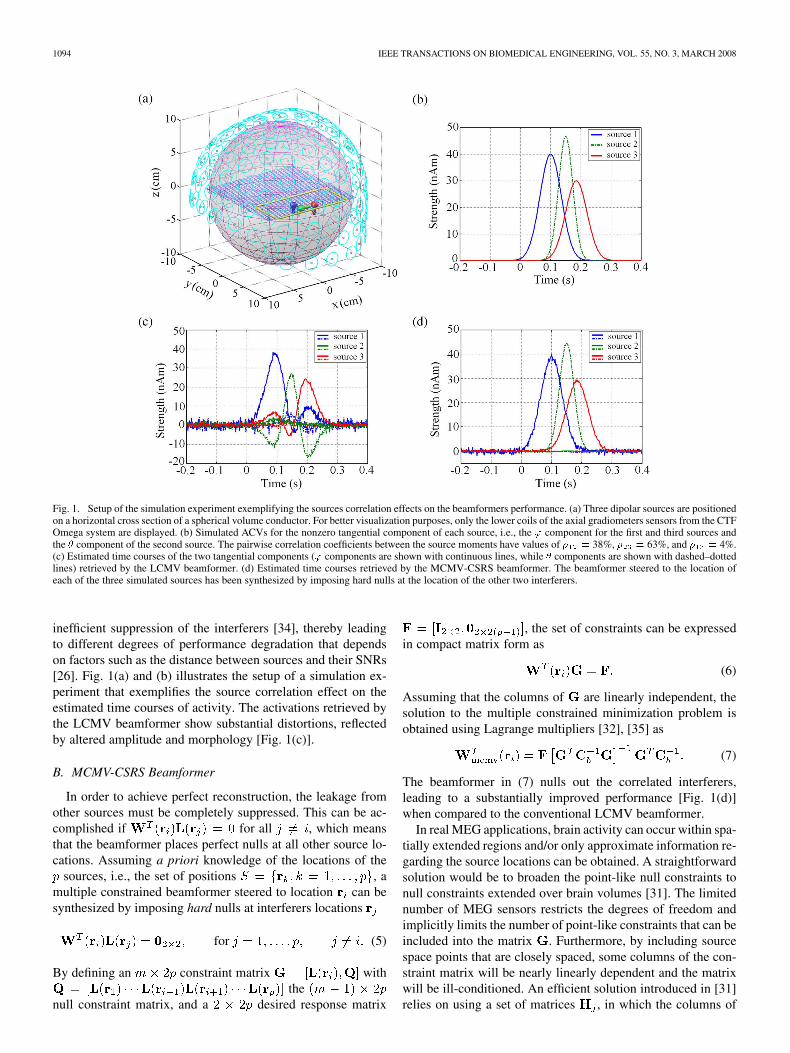
Fig. 1. Setup of the simulation experiment exemplifying the sources correlation effects on the beamformers performance. (a) Three dipolar sources are positionedon a horizontal cross section of a spherical volume conductor. For better visualization purposes, only the lower coils of the axial gradiometers sensors from the CTFOmega system are displayed. (b) Simulated ACVs for the nonzero tangential component of each source, i.e., the ' component for the first and third sources andthe � component of the second source. The pairwise correlation coefficients between the source moments have values of � = 38%, � = 63%, and � = 4%.(c) Estimated time courses of the two tangential components (' components are shown with continuous lines, while � components are shown with dashed–dottedlines) retrieved by the LCMV beamformer. (d) Estimated time courses retrieved by the MCMV-CSRS beamformer. The beamformer steered to the location ofeach of the three simulated sources has been synthesized by imposing hard nulls at the location of the other two interferers.
inefficient suppression of the interferers [34], thereby leadingto different degrees of performance degradation that dependson factors such as the distance between sources and their SNRs[26]. Fig. 1(a) and (b) illustrates the setup of a simulation ex-periment that exemplifies the source correlation effect on theestimated time courses of activity. The activations retrieved bythe LCMV beamformer show substantial distortions, reflectedby altered amplitude and morphology [Fig. 1(c)].
B. MCMV-CSRS Beamformer
In order to achieve perfect reconstruction, the leakage fromother sources must be completely suppressed. This can be ac-complished if for all , which meansthat the beamformer places perfect nulls at all other source lo-cations. Assuming a priori knowledge of the locations of the
sources, i.e., the set of positions , amultiple constrained beamformer steered to location can besynthesized by imposing hard nulls at interferers locations
for (5)
By defining an constraint matrix withthe
null constraint matrix, and a desired response matrix
, the set of constraints can be expressedin compact matrix form as
(6)
Assuming that the columns of are linearly independent, thesolution to the multiple constrained minimization problem isobtained using Lagrange multipliers [32], [35] as
(7)
The beamformer in (7) nulls out the correlated interferers,leading to a substantially improved performance [Fig. 1(d)]when compared to the conventional LCMV beamformer.
In real MEG applications, brain activity can occur within spa-tially extended regions and/or only approximate information re-garding the source locations can be obtained. A straightforwardsolution would be to broaden the point-like null constraints tonull constraints extended over brain volumes [31]. The limitednumber of MEG sensors restricts the degrees of freedom andimplicitly limits the number of point-like constraints that can beincluded into the matrix . Furthermore, by including sourcespace points that are closely spaced, some columns of the con-straint matrix will be nearly linearly dependent and the matrixwill be ill-conditioned. An efficient solution introduced in [31]relies on using a set of matrices , in which the columns of

POPESCU et al.: SPATIO–TEMPORAL RECONSTRUCTION OF BILATERAL ASSRS USING MEG BEAMFORMERS 1095
each matrix are the normalized leadfield components at loca-tions of the point-like nulls in each suppression region
(8)
Singular value decomposition is subsequently performed oneach matrix
(9)
and the singular vectors that correspond to a subsetof the largest singular values are collected in the matrix
. The reduced rank constraint matrix is then built as, and is used in conjunc-
tion with the corresponding reduced rank response matrixin (7) to synthesize the
beamformer for location . Typically, a low number of singularvectors can account for relatively large brain regions [36]. Thethresholding criterion used throughout this study required thecumulative fractional contribution of the largest singularvalues to exceed 97.5%.
In the presence of closely spaced sources, the simultaneousrequirement of unit pass band response and perfect nulls at theinterferers’ locations may lead to a large norm for the filter,which thereby increases its sensitivity to white noise. Onesolution to this problem is diagonal loading [37], [38], whichinvolves replacing the matrix in (7) with its regularizedinverse , with the diagonal loading factor. Thisstrategy increases the robustness to forward modeling errorsas well as to errors introduced by using the sample covariancematrix. The regularization parameter adjusts the tradeoff be-tween the spatial resolution and the noise magnitude in thereconstructed time courses of activity [39]. An alternativestrategy consists of projecting the beamformer coefficients ontothe signal subspace of , which preserves the null constraintsimposed on the interferers [28].
III. METHODS
A. Simulation Experiments
Simulation experiments were conducted to assess the impactof the source parameters on the beamformers’ ability to esti-mate the bilateral ASSR generators. One oscillatory dipolarsource was considered in each hemisphere and the sources po-sitions were varied independently in the medio–lateral directionwithin a horizontal cross section of a spherically symmetricvolume conductor [Fig. 2(a)]. The variation was performed in2-mm increments, over a range from 3.4 to 5.4 cm from thecross-section center. The dipole orientations were set alongthe (declination) tangential direction at each location. The40-Hz oscillatory moments were simulated for the two sources[Fig. 2(b)], and the relative phase shift between sources wasvaried in steps of radians. The magnetic field was com-puted at the sensor positions of the 151 channel CTF Omegasystem (VSM MedTech, Vancouver, Canada) using the Sarvasequations for nonradial magnetic measurements [40]. The lead-fields were computed for axial gradiometers with 5-cm distancebetween lower and upper coils. White noise with root meansquare (rms) value of 10 fT was added to the simulated data
Fig. 2. (a) Setup of the simulation experiments designed to assess the effectsof ASSR sources correlation on the beamformers performance. (b) Generic ex-ample of simulated ACVs.
and a noise-only temporal window of 0.5 s was appended to thesimulated data segment to allow the estimation of the samplenoise covariance. The LCMV-PSC beamformer was evaluatedwith respect to two sensor setups: first, by considering a fullhemispherical coverage of 75 sensors (setup 2), and second byconsidering a reduced number of sensors (namely 40, setup3) selected from those exhibiting the largest signals across thesimulated conditions. The LCMV-PSC beamformers were syn-thesized at every source space point of one hemisphere, whichwill be henceforth referred to as the hemisphere of the probedsource. The MCMV-CSRS beamformer was evaluated usingthe whole set of sensors and by considering a square-shapedregion of null constraints around the location of the neglectedsource (interferer) that extends from 3 to 6 cm in the medio-lat-eral direction and from 2.1 to 0.9 cm in the posterior–anteriordirection. The null-constraints region contains a lattice of 210source space points with 2-mm spacing in each direction. Theregion of null constraints was characterized by ten singularvectors, corresponding to the largest singular values of theregional leadfield matrix. The MCMV-CSRS beamformer wassynthesized at every source space point from the hemisphere ofthe probed source. The normalized power maps were computedand displayed within the horizontal cross section of the sources.Localization errors were reported as the Euclidian distancebetween the peak of the power map and the actual (known)source position. The temporal courses of activity at the localpeak of power were compared to the simulated signals to assessamplitude and phase distortions. A normalized signal-to-noiseratio (SNR) measure for the accuracy of the reconstructed

1096 IEEE TRANSACTIONS ON BIOMEDICAL ENGINEERING, VOL. 55, NO. 3, MARCH 2008
Fig. 3. The 100-ms temporal segments of the sound stimuli used in the auditoryexperiment. The two types of stimuli have opposite phases of the AM compo-nent (shown in dashed line).
signal amplitude was defined as the difference between theestimated signal and noise powers, divided by the power of thesimulated signal. All numerical experiments and data analysiswere performed using in-house developed MATLAB (TheMathWorks, Inc., Natick, MA) software.
B. MEG Data Acquisition and Processing
The performance of the two beamformers was evaluated onMEG data from 40-Hz ASSR to amplitude modulated (AM)sounds recorded on a right-handed male subject. The stimuliwere pure tones with 1-kHz frequency, 1.2-s duration, and co-sine-modulated amplitude (40-Hz modulation frequency, 90%modulation depth). The sounds were presented binaurally fora total of 200 trials, with 2-s interstimulus interval. In half ofthe trials, the sounds had 180 phase shift of the AM compo-nent (Fig. 3). The MEG signals were acquired at 600-Hz sam-pling rate, and were bandpass filtered between 1 and 100 Hz,with a powerline filter applied at 60 Hz (3-Hz width). The AMphase shift between the two stimuli types allows separating thephase-independent from the phase-dependent components ofthe evoked responses. This was achieved by separate averagingwith respect to each stimulus type, followed by the average and
average, respectively, of the resulting data sets. The ASSRsignals were further bandpass filtered between 30 and 50 Hz toenhance the SNR, and then were subjected to the beamformeranalysis.
The outer brain compartment was segmented from magneticresonance imaging (MRI) data. The source space was set as aregular grid of points spaced at 4 mm throughout the brain. Theleadfield matrices were computed assuming a spherical volumeconductor fitted to the subject’s head. The beamformers wereapplied on the ASSR data using 1.3-s-long signal covariancewindows following the stimulus onset, and 1-s long noise-onlycovariance windows on the prestimulus segment. The outputpower was computed at each point within the source space, andit was normalized by the noise power. A regularization param-eter derived from the average variance of the sensor noise hasbeen used with each beamforming scheme.
Fig. 4. (a) and (b) Regions of null constraints used with the MCMV-CSRSalgorithm for the left and right hemispheres, respectively. (c) Singular values ofthe leadfield matrices that correspond to the regions of null constraints.
A priori approximate information has been incorporated intothe MCMV-CSRS beamformer design by using knowledge ofthe anatomical localization of the auditory cortex. Localizationof the generators of auditory evoked potentials obtained via in-tracerebral recordings in the humans auditory cortex [41], [42]have indicated that regions of the auditory cortex are confinedto the medial and intermediate part of Heschl’s gyrus (HG), thelateral part of HG and planum temporale, the posterior part ofthe supra temporal gyrus (STG), and the anterior part of STGin front of the HG. To account for this, relatively large spher-ical null regions (5 cm in diameter) were defined for each hemi-sphere to ensure they encompass all areas of the auditory cortex[Fig. 4(a) and (b)]. The null constraints region defined on theleft and right hemispheres contained 460 and 398 source spacepoints, respectively. The singular values of the regional lead-field matrices exhibit a fast decay [Fig. 4(c)], allowing for thecharacterization of each region by a number of 15 singular vec-tors. The MCMV beamformer was synthesized at every sourcespace point according to (7). For a given point in one hemi-sphere, the matrix of null constraints comprises the singularvectors that correspond to the null region defined in the oppo-site hemisphere. Using this strategy prevents the beamformer’snorm from becoming excessively large due to the proximityof the null region and does not necessarily require additionaleigenspace projection to increase robustness to the white noise.
IV. RESULTS
A. Results From the Simulation Experiments
Fig. 5 compares the localization performance of the LCMVbeamformer with full sensor coverage [Fig. 5(a)] versus theLCMV-PSC beamformer [Fig. 5(b)] and the MCMV-CSRSbeamformer [Fig. 5(c)] for the condition of perfectly correlatedsources. The LCMV-PSC beamformer is applied by dividingthe array of sensors into two subarrays, each covering onehemisphere. A neural power map is separately computed in
POPESCU et al.: SPATIO–TEMPORAL RECONSTRUCTION OF BILATERAL ASSRS USING MEG BEAMFORMERS 1097
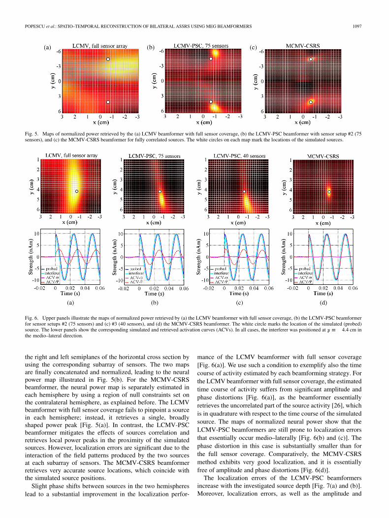
Fig. 5. Maps of normalized power retrieved by the (a) LCMV beamformer with full sensor coverage, (b) the LCMV-PSC beamformer with sensor setup #2 (75sensors), and (c) the MCMV-CSRS beamformer for fully correlated sources. The white circles on each map mark the locations of the simulated sources.
Fig. 6. Upper panels illustrate the maps of normalized power retrieved by (a) the LCMV beamformer with full sensor coverage, (b) the LCMV-PSC beamformerfor sensor setups #2 (75 sensors) and (c) #3 (40 sensors), and (d) the MCMV-CSRS beamformer. The white circle marks the location of the simulated (probed)source. The lower panels show the corresponding simulated and retrieved activation curves (ACVs). In all cases, the interferer was positioned at y = �4.4 cm inthe medio–lateral direction.
the right and left semiplanes of the horizontal cross section byusing the corresponding subarray of sensors. The two mapsare finally concatenated and normalized, leading to the neuralpower map illustrated in Fig. 5(b). For the MCMV-CSRSbeamformer, the neural power map is separately estimated ineach hemisphere by using a region of null constraints set onthe contralateral hemisphere, as explained before. The LCMVbeamformer with full sensor coverage fails to pinpoint a sourcein each hemisphere; instead, it retrieves a single, broadlyshaped power peak [Fig. 5(a)]. In contrast, the LCMV-PSCbeamformer mitigates the effects of sources correlation andretrieves local power peaks in the proximity of the simulatedsources. However, localization errors are significant due to theinteraction of the field patterns produced by the two sourcesat each subarray of sensors. The MCMV-CSRS beamformerretrieves very accurate source locations, which coincide withthe simulated source positions.
Slight phase shifts between sources in the two hemisphereslead to a substantial improvement in the localization perfor-
mance of the LCMV beamformer with full sensor coverage[Fig. 6(a)]. We use such a condition to exemplify also the timecourse of activity estimated by each beamforming strategy. Forthe LCMV beamformer with full sensor coverage, the estimatedtime course of activity suffers from significant amplitude andphase distortions [Fig. 6(a)], as the beamformer essentiallyretrieves the uncorrelated part of the source activity [26], whichis in quadrature with respect to the time course of the simulatedsource. The maps of normalized neural power show that theLCMV-PSC beamformers are still prone to localization errorsthat essentially occur medio–laterally [Fig. 6(b) and (c)]. Thephase distortion in this case is substantially smaller than forthe full sensor coverage. Comparatively, the MCMV-CSRSmethod exhibits very good localization, and it is essentiallyfree of amplitude and phase distortions [Fig. 6(d)].
The localization errors of the LCMV-PSC beamformersincrease with the investigated source depth [Fig. 7(a) and (b)].Moreover, localization errors, as well as the amplitude and

1098 IEEE TRANSACTIONS ON BIOMEDICAL ENGINEERING, VOL. 55, NO. 3, MARCH 2008
Fig. 7. (a) Localization errors (upper panel) and SNR (lower panel) for the LCMV-PSC beamformer with sensor setup #2 (75 sensors). Results are displayedas a function of the probed source position (in the medio–lateral direction) and relative phase shift between sources in the two hemispheres. (b) Results for theLCMV-PSC beamformer with sensor setup #3 (40 sensors). In all cases, the interferer was positioned at y = �4.4 cm in the medio–lateral direction. (c) Local-ization errors and SNR, respectively, for the MCMV-CSRS beamformer. Results in this case are displayed as a function of the probed and interferer locations.
phase distortions, are maximal when sources are in phase anddecay with increasing phase shifts. All measures show little de-pendence on the contralateral source position. Observations arevalid irrespective of the type of partial sensor coverage underinvestigation. This is explained by the fact that the sensorswhich are most sensitive to the probed source are also likely toreceive a significant contribution from the interferer. Overall,the results indicate that the LCMV-PSC beamformer perfor-mance is still affected by the contribution of the contralateralinterferer to the signals recorded around the temporoparietalarea of one hemisphere. In contrast, the MCMV-CSRS per-formance is very robust with respect to the source locations[Fig. 7(c)]. Since the interferer location was changed in themedio–lateral direction across the simulated conditions whilethe suppression region was fixed, the results implicitly show thatthe performance is essentially independent of the medio–lateralinterferer location relative to the region of null constraints. In afollow-up experiment, we extended the suppression region to acubical volume by including source space points from adjacentcross sections within 1.5 cm above and below the plane of thesimulated sources. The MCMV-CSRS beamformer exhibitedessentially the same performance, demonstrating robustness tothese changes in the suppression region definition.
B. Results on Real MEG Data
The phase-independent components of the evoked responses[Fig. 8(a)] consisted mainly of the transient onset and offset re-sponses, as well as a distinguishable component that immedi-ately precedes the offset response and can presumably reflect the
Fig. 8. (a) Phase-independent evoked responses obtained by averaging the re-sponses to each stimulus type. (b) Phase-dependent evoked responses obtainedby the +=� average of the responses to each stimulus type.
anticipatory effects on stimulus duration. The phase-dependentcomponent comprises the ASSR to the AM sounds [Fig. 8(b)].
Fig. 9(a) and (c) illustrates the maps of normalized neuralpower retrieved by the LCMV-PSC and MCMV-CSRS beam-formers, respectively, for the bilateral ASSR signals. TheLCMV-PSC method retrieves local peaks that are locatedwithin the lateral part of the HG. In contrast, the MCMV-CSRS

POPESCU et al.: SPATIO–TEMPORAL RECONSTRUCTION OF BILATERAL ASSRS USING MEG BEAMFORMERS 1099
Fig. 9. Spatio–temporal estimation of the ASSR retrieved by (a) and (c)LCMV-PSC and (b) and (d) MCMV-CSRS beamformers. Upper panelsshow the 3-D maps of normalized neural power (each clipped at 60% of itsmaximum). Lower panels illustrate the temporal course of activity (on themain direction of variance) at the local power peaks within the right and lefthemispheres.
beamformer shows local peaks of activity within regions of themedial and intermediate part of HG, i.e., within the primaryauditory cortex of each hemisphere. This result agrees withfindings reported in fMRI studies of auditory SSR response toAM stimuli [43], which demonstrated that for this range of AMfrequencies the activity is confined to focal areas of the pri-mary auditory cortex that are characterized by responsivenessat a high temporal resolution thus allowing the encoding ofrelatively fast variations of signal amplitude. The lateral shiftof the local power peaks retrieved by the LCMV-PSC methodagrees with the simulation results and is due to the contributionof the correlated interferer to sensors placed around the con-tralateral hemisphere. Activation curves (ACVs) retrieved byboth methods [Fig. 9(b) and (d)] clearly reveal the oscillatorySSR in each hemisphere. However, the ACVs estimated bythe LCMV-PSC show substantial amplitude attenuation, aswell as phase shifts when compared with ACVs retrieved bythe MCMV-CSRS beamformer. The correlation coefficientbetween the left and right generators was estimated from thereconstructed activity in the main direction of variance of eachsource yielding a value of 96%.
V. DISCUSSION
The neuromagnetic ASSR reflect oscillatory brain activitieswithin the auditory cortex of each hemisphere. Even when usingmonaurally presented sounds, right and left hemisphere profilescan be highly correlated, leading to the failure of conventionalLCMV beamformers to provide accurate source estimators. Al-though partial sensor coverage can reduce the contribution ofthe neural generators from the contralateral auditory cortex, ourresults show that it does not guarantee a complete removal of theinterfering signal effect. When compared to the LCMV beam-former with full sensor coverage, the LCMV-PSC beamformershows a better localization performance for the case of perfectlysynchronized sources, i.e., when the LCMV beamformer com-pletely fails to pinpoint the bilateral auditory sources. Moreover,the LCMV-PSC beamformer exhibits relatively smaller distor-tions of the estimated time courses of activity. However, thenumerical experiments presented in this study indicate that theLCMV-PSC beamformer is still prone to systematic localiza-tion errors, as well as amplitude and phase distortions of the es-timated ACVs. Its performance degradation becomes maximalwhen left and right auditory sources are perfectly synchronized,but errors persist for small relative phase shifts, often to thepoint of rendering derived solutions to be of little utility whenseeking the precise spatio–temporal reconstruction of the under-lying generators. The localization errors are explained by thecontribution of a contralateral source to the field pattern mea-sured at a subarray of sensors covering one hemisphere, whichcan be reflected by changes of the largest spatial derivative ofthe field across sensors in the vicinity of the investigated source,as well as by the decrease of the absolute magnetic field valuesin sensors away from the investigated dipole [21]. This specifictype of interaction leads to a dipolar field pattern confined tosmaller regions of the subarray of sensors, thus resembling thetopographic map that would be generated by a more superficialsource. The increase of the medio–lateral errors with the sourcedepth is particularly important when experiments are performedon children, as auditory sources are closer to the center of thevolume conductor [44].
In contrast, the MCMV-CSRS beamformer has been shownto be very robust to correlations between activities in spatiallydistinct brain areas. The MCMV-CSRS beamformer is synthe-sized by imposing hard null constraints over relatively largebrain regions that encompass the location of the interferers. Asa result, the interferers’ contribution to the beamformer’s outputcan be efficiently blocked, leading to precise localizations andhighly accurate reconstructions of the time courses of activity,i.e., free of amplitude and phase distortions. For suitable sup-pression region definitions and appropriate selections of the sin-gular vectors of the regional leadfield matrix, its performance isnot sensitive to the relative phase shift between sources withinthe left and right hemispheres. Note that underestimating thenumber of singular vectors used to characterize the suppres-sion region may lead to an inefficient blocking of the interferer.On the other hand, overestimating that number may reduce theavailable degrees of freedom for the adaptive beamformer thatcan be used to suppress other interferers. This can become a sig-nificant issue when analyzing single trials or raw data.
1100 IEEE TRANSACTIONS ON BIOMEDICAL ENGINEERING, VOL. 55, NO. 3, MARCH 2008
Analysis of real 40-Hz ASSR data indicates that the MCMV-CSRS beamformer retrieves activity confined to regions of theprimary auditory cortex of each hemisphere. Furthermore, theestimated amplitude of the sources is not attenuated as in thecase of the LCMV-PSC beamformer. A potential disadvantageof this strategy is the requirement of a priori information usedto define suitable suppression volumes that encompass the in-terferer locations. Misspecifications of the suppression regions,such as when the interferer position is outside or too close to theedges of the suppression volume, may increase the likelihood oflocalization errors [31]. To decrease such a risk, the informationon the approximate location of the ASSR sources retrieved bythe LCMV-PSC beamformer can be used to aid the appropriatedefinition of the suppression regions. In such a case, the sizeand position of the suppression volume must take into consid-eration the possible localization bias of the LCMV-PSC beam-former reported in this study. The computational efficiency ofthe MCMV-CSRS beamformers adds to the fact that their recon-struction accuracy is not dependent on the degree of source cor-relation, and compares favorably with the recently introduceddual source beamformer [45]. This latter strategy requires largecomputational resources to perform exhaustive searches for thepositions of two correlated sources that best account for inde-pendent magnetic field topographies, and show a decrease inperformance with decreasing source correlation.
Overall, the results of this study indicate that theMCMV-CSRS beamformer can emerge as a reliable methodto improve accuracy of the spatio–temporal reconstruction ofthe underlying ASSR generators, particularly, when addressingsubtle interhemispheric differences in sources locations [14],[46], amplitudes, and phase shifts [47]. The strategy is par-ticularly appealing for complex ASSR paradigms (that canpotentially involve additional brain activity outside the auditorycortex), or when studying the ASSR dynamical changes duringongoing stimulation, which poses overwhelming difficultiesto alternative parametric methods such as dipole fitting. Theseobservations may extend to studies seeking to estimate the timecourse of activity in auditory cortical regions in response tononstationary auditory stimuli such as speech [48] or music[49], which has recently generated a growing interest in cog-nitive neuroscience.
REFERENCES
[1] R. Galambos, S. Makeig, and P. J. Talmachoff, “A 40-Hz auditory po-tential recorded from the human scalp,” Proc. NY Acad. Sci., vol. 78,no. 4, pp. 2643–2647, Apr. 1981.
[2] J. P. Mäkelä and R. Hari, “Evidence for cortical origin of the 40 Hzauditory evoked response in man,” Electroencephalogr. Clin. Neuro-physiol., vol. 66, no. 6, pp. 539–546, Jun. 1987.
[3] A. Rees, G. G. Green, and R. H. Kay, “Steady-state evoked responsesto sinusoidally amplitude-modulated sounds recorded in man,” Hear.Res., vol. 23, no. 2, pp. 123–133, 1986.
[4] T. W. Picton, C. R. Skinner, S. C. Champagne, A. J. Kellett, and A. C.Maiste, “Potentials evoked by the sinusoidal modulation of the ampli-tude or frequency of a tone,” J. Acoust. Soc. Amer., vol. 82, no. 1, pp.165–178, Jul. 1987.
[5] F. A. Boettcher, E. A. Poth, J. H. Mills, and J. R. Dubno, “The am-plitude-modulation following response in young and aged human sub-jects,” Hear. Res., vol. 153, no. 1–2, pp. 32–42, Mar. 2001.
[6] D. R. Stapells, D. Linden, J. B. Suffield, G. Hamel, and T. W. Picton,“Human auditory steady state potentials,” Ear Hear., vol. 5, no. 2, pp.105–113, Mar.–Apr. 1984.
[7] R. Hari, M. Hämäläinen, and S. R. Joutsiniemi, “Neuromagneticsteady-state responses to auditory stimuli,” J. Acoust. Soc. Amer., vol.86, no. 3, pp. 1033–1039, Sep. 1989.
[8] B. Ross, C. Borgmann, R. Draganova, L. Roberts, and C. Pantev, “Ahigh precision magnetoencephalographic study of human auditorysteady-state responses to amplitude-modulated tones,” J. Acoust. Soc.Amer., vol. 108, no. 2, pp. 679–691, Aug. 2000.
[9] G. Plourde and T. W. Picton, “Human auditory steady-state responseduring general anesthesia,” Anesth. Analg., vol. 71, no. 5, pp. 460–468,Nov. 1990.
[10] H. T. Tiitinen, J. Sinkkonen, K. Reinikainen, K. Alho, J. Lavikainen,and R. Näätänen, “Selective attention enhances the auditory 40-Hztransient response in humans,” Nature, vol. 364, pp. 59–60, Jul. 1993.
[11] D. C. Rojas, K. Maharajh, P. D. Teale, M. R. Kleman, T. L. Benkers,J. P. Carlson, and M. L. Reite, “Development of the 40 Hz steady stateauditory evoked magnetic field from ages 5 to 52,” Clin. Neurophysiol.,vol. 117, no. 1, pp. 110–117, Jan. 2006.
[12] D. J. Bosnyak, R. A. Eaton, and L. E. Roberts, “Distributed auditorycortical representations are modified when nonmusicians are trained atpitch discrimination with 40 Hz amplitude modulated tones,” CerebralCortex, vol. 14, no. 10, pp. 1088–1099, 2004.
[13] B. Ross, T. W. Picton, and C. Pantev, “Temporal integration in thehuman auditory cortex as represented by the development of the steadystate magnetic field,” Hear. Res., vol. 165, no. 1–2, pp. 68–84, Mar.2002.
[14] P. Teale, J. Carlson, D. Rojas, and M. Reite, “Reduced laterality ofthe source locations for generators of the auditory steady-state field inschizophrenia,” Biol. Psychiatry, vol. 54, no. 11, pp. 1149–1153, Dec.2003.
[15] D. Osipova, E. Pekkonen, and J. Ahveninen, “Enhanced magnetic au-ditory steady-state response in early Alzheimer’s disease,” Clin. Neu-rophysiol., vol. 117, no. 9, pp. 1990–1995, Sep. 2006.
[16] M. Scherg and D. von Cramon, “Two bilateral sources of the late AEPas identified by a spatio-temporal dipole model,” Electroencephalogr.Clin. Neurophysiol., vol. 62, pp. 32–44, 1985.
[17] S. Supek and C. J. Aine, “Simulation studies of multiple dipole neu-romagnetic source localization: Model order and limits of source res-olution,” IEEE Trans. Biomed. Eng., vol. 40, no. 6, pp. 529–540, Jun.1993.
[18] R. Schoonhoven, C. J. R. Boden, J. P. A. Verbunt, and J. C. de Munck,“A whole head MEG study of the amplitude-modulation-followingresponse: Phase coherence, group delay and dipole source analysis,”Clin. Neurophysiol., vol. 114, no. 11, pp. 2096–2106, Nov. 2003.
[19] A. T. Herdman, O. Lins, P. Van Roon, D. R. Stapells, M. Scherg, andT. W. Picton, “Intracerebral sources of human auditory steady stateresponses,” Brain Topography, vol. 15, no. 2, pp. 69–86, 2002.
[20] N. Weisz, A. Keil, C. Wienbruch, S. Hoffmeister, and T. Elbert, “Oneset of sounds, two tonotopic maps: Exploring auditory cortex withamplitude-modulated tones,” Clin. Neurophysiol., vol. 115, no. 6, pp.1249–1258, Jun. 2004.
[21] J. Vrba, T. Cheung, D. Cheyne, S. E. Robinson, and A. Starr, “Errorsin ECD localization with partial sensor coverage,” in Recent Advancesin Biomagnetism, T. Yoshimoto, M. Kotani, S. Kuriki, H. Karibe, andN. Nakasato, Eds. Sendai, Japan: Tohoku Univ. Press, 1999, pp.109–112.
[22] B. Ross and C. Pantev, “Auditory steady-state responses reveal ampli-tude modulation gap detection thresholds,” J. Acoust. Soc. Amer., vol.115, no. 5, pp. 2193–2206, May 2004.
[23] M. I. G. Simpson, A. Hadjipapas, G. R. Barnes, P. L. Furlong, andC. Witton, “Imaging the dynamics of the auditory steady-state evokedresponse,” Neurosci. Lett., vol. 385, no. 3, pp. 195–197, Sep. 2005.
[24] A. T. Herdman, A. Wollbrink, W. Chau, R. Ishii, B. Ross, C. Pantev,and R. Ross, “Determination of activation areas in the human auditorycortex by means of synthetic aperture magnetometry,” NeuroImage,vol. 20, no. 2, pp. 995–1005, Oct. 2003.
[25] A. T. Herdman, A. Wollbrink, W. Chau, R. Ishii, and C. Pantev,“Localization of transient and steady-state auditory evoked responsesusing synthetic aperture magnetometry,” Brain Cogn., vol. 54, no. 2,pp. 149–151, Mar. 2004.
[26] B. D. Van Veen, W. van Drongelen, M. Yuchtman, and A. Suzuki, “Lo-calization of brain electrical activity via linearly constrained minimumvariance spatial filtering,” IEEE Trans. Biomed. Eng., vol. 44, no. 9, pp.867–880, Sep. 1997.
[27] S. E. Robinson and J. Vrba, “Functional neuroimaging by syntheticaperture magnetometry (SAM),” in Recent Advances in Biomag-netism. Sendai, Japan: Tohoku Univ. Press, 1999, pp. 302–305.

POPESCU et al.: SPATIO–TEMPORAL RECONSTRUCTION OF BILATERAL ASSRS USING MEG BEAMFORMERS 1101
[28] K. Sekihara, S. Nagarajan, D. Poeppel, and Y. Miyashita, “Recon-structing spatio-temporal activities of neural sources using an MEGvector beamformer technique,” IEEE Trans. Biomed. Eng., vol. 48, no.7, pp. 760–771, Jul. 2001.
[29] B. Widrow, K. M. Duvall, P. P. Gooch, and W. C. Newman, “Signalcancellation phenomena in adaptive arrays: Causes and cures,” IEEETrans. Antennas Propag., vol. AP-30, no. 5, pp. 469–478, May 1982.
[30] G. R. Barnes and A. Hillebrand, “Statistical flattening of MEG beam-former images,” Human Brain Mapping, vol. 18, no. 1, pp. 1–12, 2003.
[31] S. S. Dalal, K. Sekihara, and S. S. Nagarajan, “Modified beamformersfor coherent source region suppression,” IEEE Trans. Biomed. Eng.,vol. 53, no. 7, pp. 1357–1363, Jul. 2006.
[32] H. L. Van Trees, “Optimum array processing,” in Part IV of Detection,Estimation and Modulation Theory. New York: Wiley-Interscience,2002, p. 526.
[33] R. J. Ilmoniemi, M. S. Hämäläinen, and J. Knuutila, “The forward andinverse problems in the spherical model,” in Biomagnetism: Applica-tions and Theory, Weinberg, Stroink, and Katila, Eds. New York:Pergamon, 1985, pp. 278–282.
[34] K. Sekihara, S. Nagarajan, D. Poeppel, and A. Marantz, “Performanceof an MEG adaptive-beamformer technique in the presence of corre-lated neural activities: Effects on signal intensity and time-course es-timates,” IEEE Trans. Biomed. Eng., vol. 49, no. 12, pp. 1534–1546,Dec. 2002.
[35] O. L. Frost, “An algorithm for linearly constrained adaptive array pro-cessing,” Proc. IEEE, vol. 60, no. 8, pp. 926–935, Aug. 1972.
[36] X. L. Xu, B. Xu, and B. He, “An alternative subspace approach toEEG dipole source localization,” Phys. Med. Biol., vol. 49, no. 2, pp.327–343, Jan. 2004.
[37] H. Cox, R. M. Zeskind, and M. M. Owen, “Robust adaptive beam-forming,” IEEE Trans. Signal Process., vol. SP-35, no. 10, pp.1365–1376, Oct. 1987.
[38] B. D. Carlson, “Covariance matrix estimation errors and diagonalloading in adaptive arrays,” IEEE Trans. Aerosp. Electron Syst., vol.ASSP-24, no. 4, pp. 397–401, Jul. 1988.
[39] K. Sekihara, S. Nagarajan, D. Poeppel, A. Marantz, and Y. Miyashita,“Application of an MEG eigenspace beamformer to reconstructingspatio-temporal activities of neural sources,” Human Brain Mapping,vol. 15, no. 4, pp. 199–215, Apr. 2002.
[40] J. Sarvas, “Basic mathematical and electromagnetic concepts of thebio-magnetic inverse problems,” Phys. Med. Biol., vol. 32, no. 1, pp.11–22, Jan. 1987.
[41] C. Liégeois-Chauvel, A. Musolino, and P. Chauvel, “Localization ofthe primary auditory area in man,” Brain, vol. 114, pp. 139–153, Feb.1991.
[42] C. Liégeois-Chauvel, A. Musolino, J. M. Badier, P. Marquis, and P.Chauvel, “Evoked potentials recorded from the auditory cortex in man:Evaluation and topography of the middle latency components,” Elec-troencephalogr. Clin. Neurophysiol., vol. 92, no. 3, pp. 204–214, May1994.
[43] A. L. Giraud, C. Lorenzi, J. Ashburner, J. Wable, I. Johnsrude, R.Frackowiak, and A. Kleinschmidt, “Representation of the temporalenvelope of sounds in the human brain,” J. Neurophysiol., vol. 84, no.3, pp. 1588–1598, Sep. 2000.
[44] E. W. Pang, W. Gaetz, H. Otsubo, S. Chuang, and D. Cheyne, “Lo-calization of auditory N1 in children using MEG: Source modellingissues,” Int. J. Psychophysiol., vol. 51, pp. 27–35, 2003.
[45] M. J. Brookes, C. M. Stevenson, G. R. Barnes, A. Hillebrand, M. I. G.Simpson, S. T. Francis, and P. G. Morrisa, “Beamformer reconstructionof correlated sources using a modified source model,” Neuroimage, vol.34, no. 4, pp. 1454–1465, Feb. 2007.
[46] B. Ross, A. T. Herdman, and C. Pantev, “Right hemispheric lateralityof human 40 Hz auditory steady-state responses,” Cerebral Cortex, vol.15, no. 12, pp. 2029–2039, Dec. 2005.
[47] A. D. Patel and E. Balaban, “Human auditory cortical dynamics duringperception of long acoustic sequences: Phase tracking of carrier fre-quency by the auditory steady-state response,” Cerebral Cortex, vol.14, no. 1, pp. 35–46, Jan. 2004.
[48] E. Ahissar, S. Nagarajan, M. Ahissar, A. Protopapas, H. Mahncke, andM. M. Merzenich, “Speech comprehension is correlated with temporalresponse patterns recorded from auditory cortex,” Proc. Nat. Acad. Sci.USA, vol. 98, no. 23, pp. 13367–13372, Nov. 2001.
[49] M. Popescu, A. Otsuka, and A. A. Ioannides, “Dynamics of brainactivity in motor and frontal cortical areas during music listening:A magnetoencephalographic study,” NeuroImage, vol. 21, no. 4, pp.1622–1638, Apr. 2004.
Mihai Popescu received the B.Sc. degree in elec-trical engineering from the “Politehnica” Universityof Bucharest, Bucharest, Romania, in 1994, and theM.Sc. and Ph.D. degrees in biomedical engineeringfrom the University of Patras, Patras, Greece, in1996 and 2001, respectively.
He worked as a Scientific Researcher in the MEGLaboratory, RIKEN Brain Science Institute, Saitama,Japan (2001–2003), focusing on studying the neuralmechanisms of auditory processing. Since 2003 hehas been with the MEG Laboratory, Hoglund Brain
Imaging Center, University of Kansas Medical Center, Kansas City, where heis currently also a Research Assistant Professor at the Department of Molec-ular and Integrative Physiology. His research interests include the biomagneticinverse problems, statistical estimation theory, and wavelet transform analysiswith applications to measurements of brain and cardiac functions, and the neuralmechanisms of auditory processing and audiovisual interactions.
Elena-Anda Popescu received the B.Sc. degree inphysics from the University of Bucharest, Bucharest,Romania, in 1995, and the M.Sc. degree in medicalphysics from the University of Patras, Patras, Greece,in 1998.
She was a Monbusho Research Student in theGraduate School of Life Science and SystemsEngineering, Kyushu Institute of Technology, Japan,and a student trainee at the Brain Science Institute,RIKEN, Japan during 2001–2003. Since 2004 shehas been a Research Associate at the Hoglund Brain
Imaging Center, University of Kansas Medical Center, Kansas City. Herresearch interest in biosignal analysis focuses on the linear transformationsof multivariate data, such as independent component analysis and sphericalharmonics analysis.
Tszping Chan was born in Hong Kong in 1984. Shereceived the B.S. degree with highest distinction inelectrical engineering in 2006 from the University ofKansas, Lawrence, where she is currently working to-wards the M.S. degree in electrical engineering.
She has been working as a graduate research assis-tant at the Radar Systems and Remote Sensing Lab-oratory at KU since 2006. She is a student memberof Eta Kappa Nu and Tau Beta Pi. Her research inter-ests include adaptive signal processing and functionalimaging.
Shannon D. Blunt (S’96–M’02) received the B.S.,M.S., and Ph.D. degrees in electrical engineeringfrom the University of Missouri-Columbia (MU) in1999, 2000, and 2002, respectively.
From 2002 to 2005, he was with the RadarDivision of the U.S. Naval Research Laboratory,Washington, DC. In 2005, he joined the facultyof the Department of Electrical Engineering andComputer Science at the University of Kansas,Kansas City. He is a member of Eta Kappa Nu andTau Beta Pi. His research interests are in adaptive
signal processing for radar and communications with an emphasis on waveformdiversity techniques.
Dr. Blunt received the Donald K. Anderson Graduate Student Teaching awardin electrical engineering from MU in 2000 and the MU Outstanding GraduateStudent award in electrical engineering in 2001. He also received Naval Re-search Laboratory Alan Berman Research Publication awards in 2004 and 2006.

1102 IEEE TRANSACTIONS ON BIOMEDICAL ENGINEERING, VOL. 55, NO. 3, MARCH 2008
Jeffrey D. Lewine received the B.Sc. degree,cum laude with distinction, from the University ofRochester, Rochester, NY, in 1982, and the M.Sc.and Ph.D. degrees in neuroscience from the Centerfor Brain Research, University of Rochester, in 1986and 1989, respectively.
He served as a Director’s Fellow in Neuroscienceand Biophysics at Los Alamos National Laboratory,Los Alamos, NM. After fellowship, Dr. Lewinestayed on as a scientific staff member with thebiophysics group at Los Alamos before taking
faculty appointments at the University of New Mexico School of Medicinethrough 1996 and then at the University of Utah School of Medicine in 1997.Since 2002 he was an Associate Professor at the Department of Neurology,University of Kansas Medical Center (UKMC), Kansas City, and the Directorof the Biomagnetism Laboratory, Hoglund Brain Imaging Center, UKMC.He is now the Executive Director of the Illinois MagnetoencephalographyCenter, Alexian Neurosciences Institute, Elk Grove Village, IL. His currentmain research interests include the application of biomagnetic techniquesfor evaluation of neurological disorders including autism, autistic spectrumdisorders, and epilepsy.