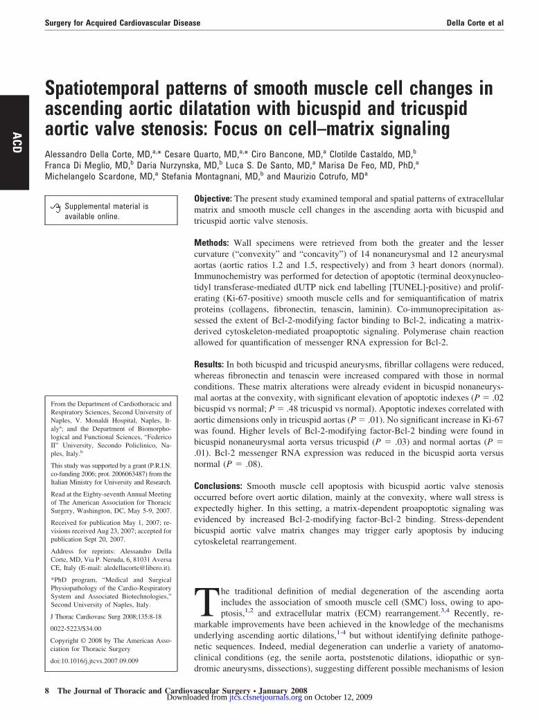
Spatiotemporal patterns of smooth muscle cell changes in ascending aortic dilatation with bicuspid...
15
DOI: 10.1016/j.jtcvs.2007.09.009 2008;135:8-18 J Thorac Cardiovasc Surg Stefania Montagnani and Maurizio Cotrufo Meglio, Daria Nurzynska, Luca S. De Santo, Marisa De Feo, Michelangelo Scardone, Alessandro Della Corte, Cesare Quarto, Ciro Bancone, Clotilde Castaldo, Franca Di signaling dilatation with bicuspid and tricuspid aortic valve stenosis: Focus on cellmatrix Spatiotemporal patterns of smooth muscle cell changes in ascending aortic http://jtcs.ctsnetjournals.org/cgi/content/full/135/1/8 located on the World Wide Web at: The online version of this article, along with updated information and services, is 2008 American Association for Thoracic Surgery Association for Thoracic Surgery and the Western Thoracic Surgical Association. Copyright © is the official publication of the American The Journal of Thoracic and Cardiovascular Surgery on October 12, 2009 jtcs.ctsnetjournals.org Downloaded from
Transcript of Spatiotemporal patterns of smooth muscle cell changes in ascending aortic dilatation with bicuspid...
DOI: 10.1016/j.jtcvs.2007.09.009 2008;135:8-18 J Thorac Cardiovasc Surg
Stefania Montagnani and Maurizio Cotrufo Meglio, Daria Nurzynska, Luca S. De Santo, Marisa De Feo, Michelangelo Scardone, Alessandro Della Corte, Cesare Quarto, Ciro Bancone, Clotilde Castaldo, Franca Di
signalingdilatation with bicuspid and tricuspid aortic valve stenosis: Focus on cell�matrix
Spatiotemporal patterns of smooth muscle cell changes in ascending aortic
http://jtcs.ctsnetjournals.org/cgi/content/full/135/1/8located on the World Wide Web at:
The online version of this article, along with updated information and services, is
2008 American Association for Thoracic Surgery Association for Thoracic Surgery and the Western Thoracic Surgical Association. Copyright ©
is the official publication of the AmericanThe Journal of Thoracic and Cardiovascular Surgery
on October 12, 2009 jtcs.ctsnetjournals.orgDownloaded from
SaaAFM
Surgery for Acquired Cardiovascular Disease Della Corte et al
8
ACD
patiotemporal patterns of smooth muscle cell changes inscending aortic dilatation with bicuspid and tricuspidortic valve stenosis: Focus on cell–matrix signaling
lessandro Della Corte, MD,a,* Cesare Quarto, MD,a,* Ciro Bancone, MD,a Clotilde Castaldo, MD,b
ranca Di Meglio, MD,b Daria Nurzynska, MD,b Luca S. De Santo, MD,a Marisa De Feo, MD, PhD,a
ichelangelo Scardone, MD,a Stefania Montagnani, MD,b and Maurizio Cotrufo, MDa
Omt
McaItepsda
Rwcmbawb.n
Coeebc
Tmunc
Supplemental material isavailable online.
From the Department of Cardiothoracic andRespiratory Sciences, Second University ofNaples, V. Monaldi Hospital, Naples, It-alya; and the Department of Biomorpho-logical and Functional Sciences, “FedericoII” University, Secondo Policlinico, Na-ples, Italy.b
This study was supported by a grant (P.R.I.N.co-funding 2006; prot. 2006063487) from theItalian Ministry for University and Research.
Read at the Eighty-seventh Annual Meetingof The American Association for ThoracicSurgery, Washington, DC, May 5-9, 2007.
Received for publication May 1, 2007; re-visions received Aug 23, 2007; accepted forpublication Sept 20, 2007.
Address for reprints: Alessandro DellaCorte, MD, Via P. Neruda, 6, 81031 AversaCE, Italy (E-mail: [email protected]).
*PhD program, “Medical and SurgicalPhysiopathology of the Cardio-RespiratorySystem and Associated Biotechnologies,”Second University of Naples, Italy.
J Thorac Cardiovasc Surg 2008;135:8-18
0022-5223/$34.00
Copyright © 2008 by The American Asso-ciation for Thoracic Surgery
ddoi:10.1016/j.jtcvs.2007.09.009
The Journal of Thoracic and CardiovaDownloa
bjective: The present study examined temporal and spatial patterns of extracellularatrix and smooth muscle cell changes in the ascending aorta with bicuspid and
ricuspid aortic valve stenosis.
ethods: Wall specimens were retrieved from both the greater and the lesserurvature (“convexity” and “concavity”) of 14 nonaneurysmal and 12 aneurysmalortas (aortic ratios 1.2 and 1.5, respectively) and from 3 heart donors (normal).mmunochemistry was performed for detection of apoptotic (terminal deoxynucleo-idyl transferase-mediated dUTP nick end labelling [TUNEL]-positive) and prolif-rating (Ki-67-positive) smooth muscle cells and for semiquantification of matrixroteins (collagens, fibronectin, tenascin, laminin). Co-immunoprecipitation as-essed the extent of Bcl-2-modifying factor binding to Bcl-2, indicating a matrix-erived cytoskeleton-mediated proapoptotic signaling. Polymerase chain reactionllowed for quantification of messenger RNA expression for Bcl-2.
esults: In both bicuspid and tricuspid aneurysms, fibrillar collagens were reduced,hereas fibronectin and tenascin were increased compared with those in normal
onditions. These matrix alterations were already evident in bicuspid nonaneurys-al aortas at the convexity, with significant elevation of apoptotic indexes (P � .02
icuspid vs normal; P � .48 tricuspid vs normal). Apoptotic indexes correlated withortic dimensions only in tricuspid aortas (P � .01). No significant increase in Ki-67as found. Higher levels of Bcl-2-modifying factor-Bcl-2 binding were found inicuspid nonaneurysmal aorta versus tricuspid (P � .03) and normal aortas (P �
01). Bcl-2 messenger RNA expression was reduced in the bicuspid aorta versusormal (P � .08).
onclusions: Smooth muscle cell apoptosis with bicuspid aortic valve stenosisccurred before overt aortic dilation, mainly at the convexity, where wall stress isxpectedly higher. In this setting, a matrix-dependent proapoptotic signaling wasvidenced by increased Bcl-2-modifying factor-Bcl-2 binding. Stress-dependenticuspid aortic valve matrix changes may trigger early apoptosis by inducingytoskeletal rearrangement.
he traditional definition of medial degeneration of the ascending aortaincludes the association of smooth muscle cell (SMC) loss, owing to apo-ptosis,1,2 and extracellular matrix (ECM) rearrangement.3,4 Recently, re-
arkable improvements have been achieved in the knowledge of the mechanismsnderlying ascending aortic dilations,1-4 but without identifying definite pathoge-etic sequences. Indeed, medial degeneration can underlie a variety of anatomo-linical conditions (eg, the senile aorta, poststenotic dilations, idiopathic or syn-
romic aneurysms, dissections), suggesting different possible mechanisms of lesionscular Surgery ● January 2008 on October 12, 2009 jtcs.ctsnetjournals.orgded from
dcftu
EdiedpiaStstpocaaa“Mccfksssa
PSTam(dieaBgtatadgbid
SAplTs(1sirbctatmmat
ISdsapaCwwrsLfl
Della Corte et al Surgery for Acquired Cardiovascular Disease
ACD
evelopment.1 In particular, aortic dilations associated withongenital bicuspid aortic valve (BAV) are known to differrom those with tricuspid aortic valve (TAV) disease inerms of histopathology,2 anatomic configuration,5 and nat-ral history.6
In our preliminary investigation, we4 defined medialCM changes in BAV-associated dilations, observing aecrease in collagen content versus normal aorta and anncrease in fibronectin and the expression of tenascin. How-ver, no comparison with TAV-associated dilations and noistinction between early changes and mature lesions wereerformed. The present study was undertaken as a moren-depth continuation of the previous one,4 with the aim tossess spatial and temporal patterns of ECM protein andMC changes in BAV- and TAV-associated aortic dila-
ions. Moreover, inasmuch as changes in the ECM compo-ition, including those previously observed in BAV aortopa-hy,3,4 are known to possibly influence SMC phenotype,roliferation, survival, and synthetic activity,7,8 we focusedn the hypothesis that SMC apoptosis in ascending dilationsould be provoked by matrix-derived signaling. Anoikis (thencient Greek word for homelessness) is the term indicatingpoptosis caused by the loss of normal adhesion of cells to
normally organized ECM, occurring via altered celltensegrity,” the tensional integrity of the cytoskeleton.9
atrix disarray and/or cell detachment can induce loss ofytoskeletal integrity, cell shape changes, and eventuallyell death9,10: thus, “amorphosis” is considered as a typicaleature/modality of anoikis.10,11 Proapoptotic cues arenown to be transmitted to cells by the ECM in both theettings of vascular physiology and disease.9 In the presenttudy, we hypothesized that matrix-derived proapoptoticignals could be involved in the development of BAV
Abbreviations and AcronymsAI � apoptotic indexBAV � bicuspid aortic valveBmf � Bcl-2-modifying factorDAPI � 4=,6-diamidino-2=-phenylindoleECM � extracellular matrixGAPDH � glyceraldehyde-3-phosphate dehydrogenaseMMP � matrix metalloproteinasesmRNA � messenger RNAPBS � phosphate-buffered salinePCR � polymerase chain reactionSMC � smooth muscle cellTAV � tricuspid aortic valveTUNEL � terminal deoxynucleotidyl transferase-
mediated dUTP nick end labeling
ortopathy. I
The Journal of Thorajtcs.ctsnetjourDownloaded from
atients and Methodstudy Patientshe study included 26 patients with aortic valve stenosis (meange 59 � 15 years, 65% male) undergoing aortic valve replace-ent and/or ascending aorta replacement. Valve morphology
BAV/TAV) was defined on the basis of concordant echocar-iographic diagnosis, surgical inspection, and pathologic exam-nation. In case of discordances, patients were considered non-ligible. Twelve patients had degenerative stenosis of a TAV,nd 14 had a stenotic congenital BAV. Respective TAV andAV subgroups were comparable for clinical and echocardio-raphic data (Table E1). No patient had inherited connectiveissue abnormalities, atherosclerotic aneurysm, or associatedortic regurgitation (more than mild). To define temporal pat-erns of medial change progression, we distinguished subgroupsccording to aortic dimensions: nonaneurysmal aortas (mildilations) were defined as an aortic ratio (between echocardio-raphically measured maximal diameter and expected diameter,ased on age and body surface area) less than 1.4 (correspond-ng, on average, to a diameter of 4.5 cm), and aneurysms wereefined as a ratio greater than 1.4.
ample Retrieval Protocolt surgical inspection, all BAV-associated aortic aneurysms ap-eared asymmetric, with predominant bulging of the right antero-ateral side of the vessel. This configuration was found also in 3AV aneurysms (60%), whereas the other 2 showed a fusiform,ymmetric dilation at both the outer (convexity) and inner curvesconcavity). The pattern of cusp fusion was right-left coronary in2 BAV patients and right-noncoronary in the other 2. To definepatial patterns of lesion expression, according to a previouslyntroduced protocol,4 soon after the aortotomy was performed, weetrieved 2 full-thickness aortic wall samples distal to the sinotu-ular junction (maximal dilation level) in each patient: one at theonvexity and the other at the concavity (from the two ends of theransverse aortotomy in patients without indication to concomitantscending aorta replacement). Wall specimens were retrieved athe same levels also from 3 normotensive heart donors duringultiorgan procurement (2 men, 1 woman; mean age 40 � 3 years;ean diameter 3.4 � 0.15 cm). Thus, 58 specimens overall were
vailable for examination. The study was approved by our insti-ution’s ethical committee and patients gave informed consent.
mmunohistochemistry of Matrix Proteinspecimens (n � 52) were fixed in buffered 10% formalin, embed-ed in paraffin, and sectioned. Serial 4 �m-thick sections of aorticpecimens were deparaffinized, covered with primary monoclonalntibodies against type I collagen, tenascin-C, and laminin orolyclonal antibody for fibronectin (Sigma-Aldrich, St Louis, Mo),nd type III collagen (Santa Cruz Biotechnology Inc., Santa Cruz,alif), and incubated in a moist chamber for 1 hour at 37°C. Afterashings in phosphate-buffered saline (PBS), they were coveredith fluoresceinated secondary antibodies (Sigma-Aldrich) and
eincubated. Nuclei were stained with propidium iodide, and thenections were mounted in Vectashield mounting medium (Vectoraboratories, Burlingame, Calif) and observed with Leica DMLBuorescence microscope (Leica Microsystems, Inc, Bannockburn,
ll). Moreover, a Zeiss LSM-510 confocal microscope (Karl Zeisscic and Cardiovascular Surgery ● Volume 135, Number 1 9 on October 12, 2009 nals.org
Irpr0fcCAttawdteimqtit
APWafAAaptwcbifps3cm
Surgery for Acquired Cardiovascular Disease Della Corte et al
1
ACD
nternational, Jena, Germany) was used to obtain full-thicknesseconstructions of the aortic wall to describe spatial distribution ofroteins. To obviate the possible interference of elastin autofluo-escence, however, quenched by pretreatment with toluidine blue.1% solution, we also performed the immunoperoxidase methodor countercheck, using the standard avidin-biotin-peroxidaseomplex technique and the L.V. Dako LSAB kit (DAKO A/S,arpinteria, Calif) after antigen retrieval by pressure cooking.fter quenching in 3% hydrogen peroxide and blocking, the sec-
ions were incubated with primary antibodies for the aforemen-ioned ECM proteins overnight at 4°C. Biotinylated secondaryntibody and streptavidin conjugated to horseradish peroxidaseere subsequently applied with color development with 3,3=-iaminobenzidine and hematoxylin counterstaining. Negative con-rol slides in the absence of primary antibody were included forach staining. Samples were evaluated with both methods by 3ndependent observers blinded to the register that matched speci-ens with patient clinical data, using a grading scale for semi-
uantification of staining, from none (0) to intense (4). Owing tohe overall good correspondence between fluorescence and perox-dase assessments, each specimen was assigned 1 score (averaging
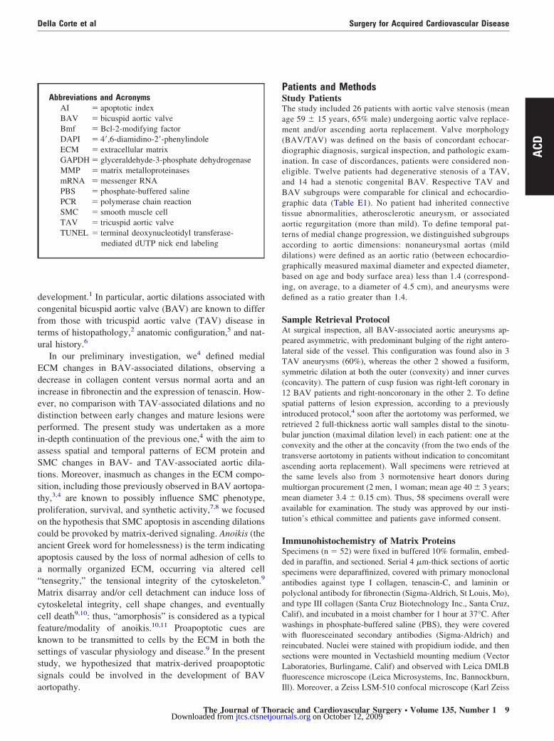
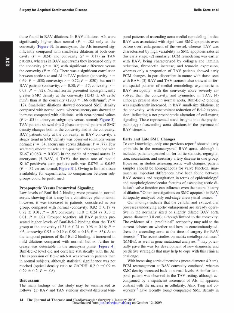
Figure 1. Immunostaining for laminin is shown as achanges. A, Immunofluorescence method, confocal mis� propidium iodide (nuclei). B, Immunoperoxidase memethods indicated that in BAV patients laminin decreaand more asymmetrical (ie, more severe at the convexwere from the inner portion of the media. ECM, Extraaortic valve.
he 6 scores from the 2 methods). t
0 The Journal of Thoracic and Cardiovascular Surgery ● Januajtcs.ctsnetjournaDownloaded from
ssessment of SMC Density, Apoptosis, androliferationall fragments from the concavity and convexity of the ascending
orta (n � 54) were washed in PBS, fixed in 10% bufferedormalin, paraffin-embedded, and sliced in 4-�m serial sections.fter deparaffinization, the ApopTag Plus Fluorescein In Situpoptosis Detection Kit (Chemicon, Temecula, Calif) was used,
ccording to the manufacturer protocol. In brief, sections wereretreated with proteinase K for 15 minutes. Pre-equilibrated sec-ions were covered with terminal deoxynucleotidyl transferaseorking solution and incubated for 1 hour at 37°C in a humidified
hamber. The reaction was stopped by incubation with stop/washuffer. After three washes with PBS, antidigoxigenin fluoresceinsothiocyanate conjugate (Sigma-Aldrich) was applied to the slidesor 30 minutes at room temperature. For characterization of apo-totic cells, sections were subsequently incubated with anti-mooth muscle �-actin antibody (Sigma-Aldrich) for 1 hour at7°C in a humidified chamber. After washes in PBS, rhodamine-onjugated secondary immunoglobulin G antibody (Jackson Im-unoResearch Laboratories, Inc, West Grove, Pa) was applied to
sentative example of the observed patterns of ECMcopy. Green fluorescence � laminin; red fluorescencelight microscopy, 200�; laminin stained brown). Bothas earlier (ie, already evident in small-size dilations)an at the concavity) than in TAV patients. All images
lar matrix; BAV, bicuspid aortic valve; TAV, tricuspid
reprecrosthod,se wity thcellu
he sections for 1 hour at 37°C. Cell nuclei were counterstained
ry 2008 on October 12, 2009 ls.org

ACD
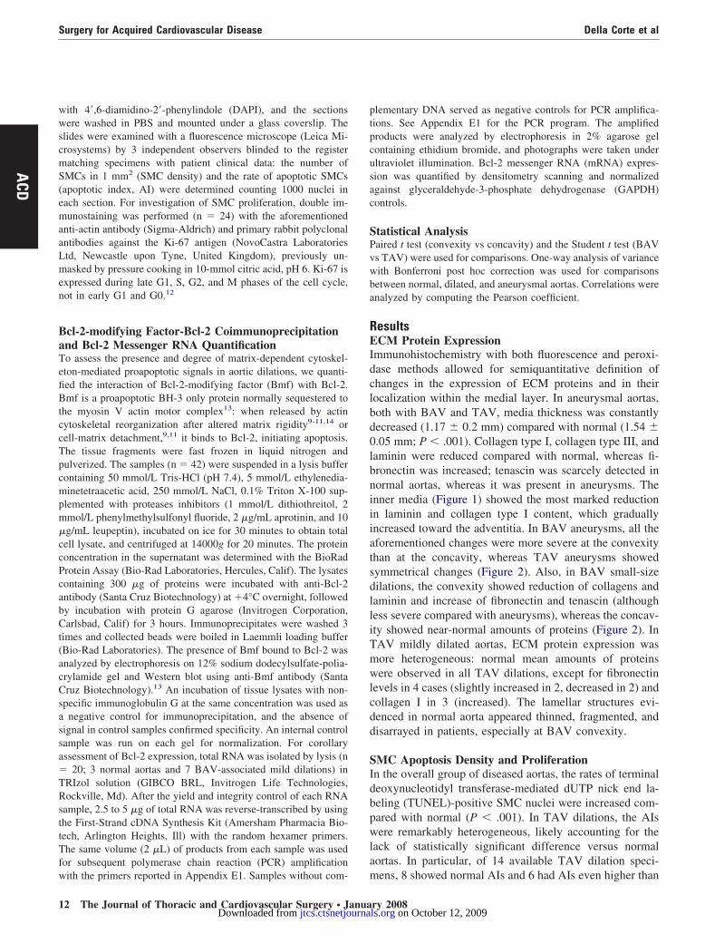
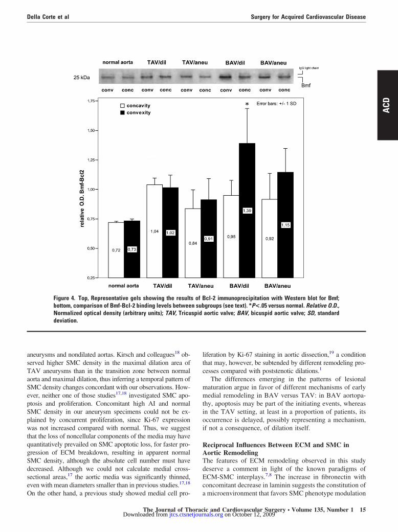
Figure 2. Patterns of ECM changes by semiquantitative assessment of collagen type I, fibronectin, and tenascinimmunostainings. *P � .05 BAV versus TAV dilation; §P < .05 BAV versus normal (P � .016 for collagen type I; P� .008 for fibronectin) and versus TAV dilation (P � .008 for collagen type I; P � .037 for fibronectin); °P < .05aneurysm versus respective dilation (P � .007 for collagen type I in BAV; P � .03 and P � .01 for collagen typeI in TAV; P�.002 for tenascin in BAV); P < .01 for all differences between convexity and concavity in BAV. P <.05 (not significant) for all other comparisons. ECM, Extracellular matrix; TAV, tricuspid aortic valve; BAV, bicuspid
aortic valve.The Journal of Thoracic and Cardiovascular Surgery ● Volume 135, Number 1 11 on October 12, 2009 jtcs.ctsnetjournals.orgDownloaded from
wwscmS(emaaLmen
BaTefiBtccTpcmpm�ccPcabCt(acCsassa�TRsttTfw
ptpcusac
SPvwba
REIdclbd0lbniiiatsdlliTmwlcdd
SIdbpwla
Surgery for Acquired Cardiovascular Disease Della Corte et al
1
ACD
ith 4=,6-diamidino-2=-phenylindole (DAPI), and the sectionsere washed in PBS and mounted under a glass coverslip. The
lides were examined with a fluorescence microscope (Leica Mi-rosystems) by 3 independent observers blinded to the registeratching specimens with patient clinical data: the number ofMCs in 1 mm2 (SMC density) and the rate of apoptotic SMCsapoptotic index, AI) were determined counting 1000 nuclei inach section. For investigation of SMC proliferation, double im-unostaining was performed (n � 24) with the aforementioned
nti-actin antibody (Sigma-Aldrich) and primary rabbit polyclonalntibodies against the Ki-67 antigen (NovoCastra Laboratoriestd, Newcastle upon Tyne, United Kingdom), previously un-asked by pressure cooking in 10-mmol citric acid, pH 6. Ki-67 is
xpressed during late G1, S, G2, and M phases of the cell cycle,ot in early G1 and G0.12
cl-2-modifying Factor-Bcl-2 Coimmunoprecipitationnd Bcl-2 Messenger RNA Quantificationo assess the presence and degree of matrix-dependent cytoskel-ton-mediated proapoptotic signals in aortic dilations, we quanti-ed the interaction of Bcl-2-modifying factor (Bmf) with Bcl-2.mf is a proapoptotic BH-3 only protein normally sequestered to
he myosin V actin motor complex13: when released by actinytoskeletal reorganization after altered matrix rigidity9-11,14 orell-matrix detachment,9,11 it binds to Bcl-2, initiating apoptosis.he tissue fragments were fast frozen in liquid nitrogen andulverized. The samples (n � 42) were suspended in a lysis bufferontaining 50 mmol/L Tris-HCl (pH 7.4), 5 mmol/L ethylenedia-inetetraacetic acid, 250 mmol/L NaCl, 0.1% Triton X-100 sup-
lemented with proteases inhibitors (1 mmol/L dithiothreitol, 2mol/L phenylmethylsulfonyl fluoride, 2 �g/mL aprotinin, and 10g/mL leupeptin), incubated on ice for 30 minutes to obtain totalell lysate, and centrifuged at 14000g for 20 minutes. The proteinoncentration in the supernatant was determined with the BioRadrotein Assay (Bio-Rad Laboratories, Hercules, Calif). The lysatesontaining 300 �g of proteins were incubated with anti-Bcl-2ntibody (Santa Cruz Biotechnology) at �4°C overnight, followedy incubation with protein G agarose (Invitrogen Corporation,arlsbad, Calif) for 3 hours. Immunoprecipitates were washed 3
imes and collected beads were boiled in Laemmli loading bufferBio-Rad Laboratories). The presence of Bmf bound to Bcl-2 wasnalyzed by electrophoresis on 12% sodium dodecylsulfate-polia-rylamide gel and Western blot using anti-Bmf antibody (Santaruz Biotechnology).13 An incubation of tissue lysates with non-
pecific immunoglobulin G at the same concentration was used asnegative control for immunoprecipitation, and the absence of
ignal in control samples confirmed specificity. An internal controlample was run on each gel for normalization. For corollaryssessment of Bcl-2 expression, total RNA was isolated by lysis (n
20; 3 normal aortas and 7 BAV-associated mild dilations) inRIzol solution (GIBCO BRL, Invitrogen Life Technologies,ockville, Md). After the yield and integrity control of each RNA
ample, 2.5 to 5 �g of total RNA was reverse-transcribed by usinghe First-Strand cDNA Synthesis Kit (Amersham Pharmacia Bio-ech, Arlington Heights, Ill) with the random hexamer primers.he same volume (2 �L) of products from each sample was used
or subsequent polymerase chain reaction (PCR) amplification
ith the primers reported in Appendix E1. Samples without com- m2 The Journal of Thoracic and Cardiovascular Surgery ● Januajtcs.ctsnetjournaDownloaded from
lementary DNA served as negative controls for PCR amplifica-ions. See Appendix E1 for the PCR program. The amplifiedroducts were analyzed by electrophoresis in 2% agarose gelontaining ethidium bromide, and photographs were taken underltraviolet illumination. Bcl-2 messenger RNA (mRNA) expres-ion was quantified by densitometry scanning and normalizedgainst glyceraldehyde-3-phosphate dehydrogenase (GAPDH)ontrols.
tatistical Analysisaired t test (convexity vs concavity) and the Student t test (BAVs TAV) were used for comparisons. One-way analysis of varianceith Bonferroni post hoc correction was used for comparisonsetween normal, dilated, and aneurysmal aortas. Correlations werenalyzed by computing the Pearson coefficient.
esultsCM Protein Expression
mmunohistochemistry with both fluorescence and peroxi-ase methods allowed for semiquantitative definition ofhanges in the expression of ECM proteins and in theirocalization within the medial layer. In aneurysmal aortas,oth with BAV and TAV, media thickness was constantlyecreased (1.17 � 0.2 mm) compared with normal (1.54 �.05 mm; P � .001). Collagen type I, collagen type III, andaminin were reduced compared with normal, whereas fi-ronectin was increased; tenascin was scarcely detected inormal aortas, whereas it was present in aneurysms. Thenner media (Figure 1) showed the most marked reductionn laminin and collagen type I content, which graduallyncreased toward the adventitia. In BAV aneurysms, all theforementioned changes were more severe at the convexityhan at the concavity, whereas TAV aneurysms showedymmetrical changes (Figure 2). Also, in BAV small-sizeilations, the convexity showed reduction of collagens andaminin and increase of fibronectin and tenascin (althoughess severe compared with aneurysms), whereas the concav-ty showed near-normal amounts of proteins (Figure 2). InAV mildly dilated aortas, ECM protein expression wasore heterogeneous: normal mean amounts of proteinsere observed in all TAV dilations, except for fibronectin
evels in 4 cases (slightly increased in 2, decreased in 2) andollagen I in 3 (increased). The lamellar structures evi-enced in normal aorta appeared thinned, fragmented, andisarrayed in patients, especially at BAV convexity.
MC Apoptosis Density and Proliferationn the overall group of diseased aortas, the rates of terminaleoxynucleotidyl transferase-mediated dUTP nick end la-eling (TUNEL)-positive SMC nuclei were increased com-ared with normal (P � .001). In TAV dilations, the AIsere remarkably heterogeneous, likely accounting for the
ack of statistically significant difference versus normalortas. In particular, of 14 available TAV dilation speci-
ens, 8 showed normal AIs and 6 had AIs even higher thanry 2008 on October 12, 2009 ls.org
Della Corte et al Surgery for Acquired Cardiovascular Disease
ACD
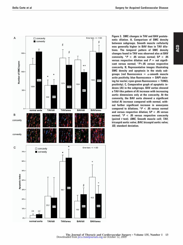
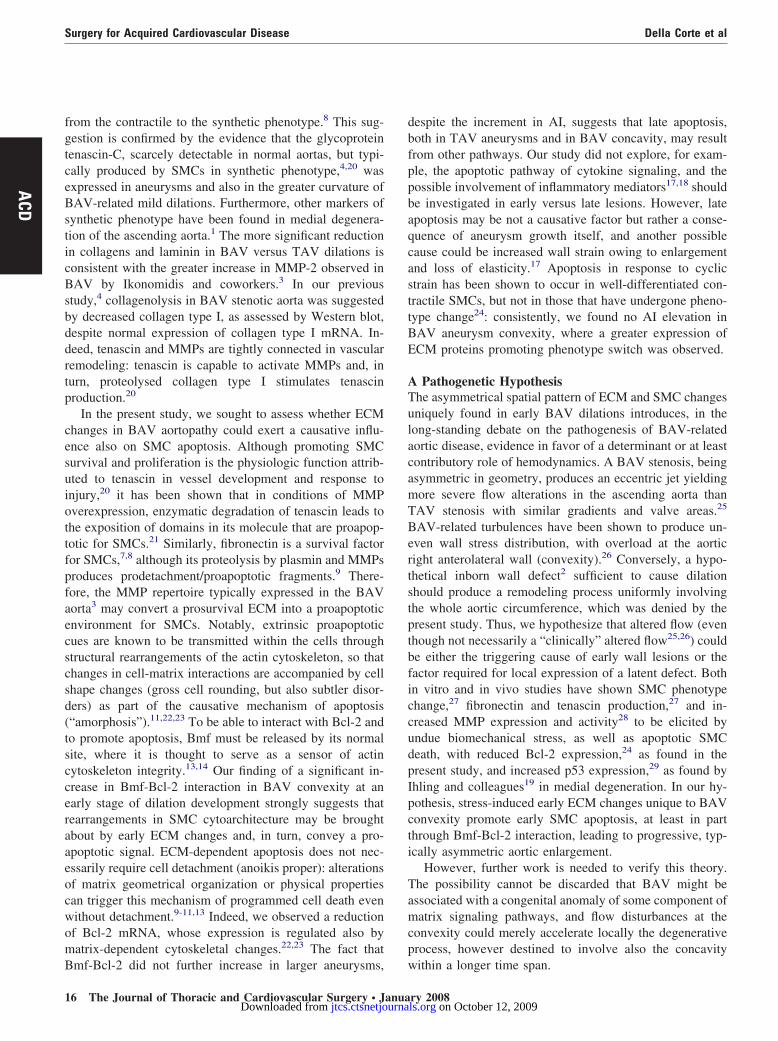
Figure 3. SMC changes in TAV and BAV postste-notic dilation. A, Comparison of SMC densitybetween subgroups. Smooth muscle cellularitywas generally higher in BAV than in TAV dila-tions. The temporal pattern of SMC densitychanges found in TAV was observed also at BAVconvexity. *P < .05 versus normal; §P < .05versus respective dilation and P � not signifi-cant versus normal; °P<.05 versus respectiveconcavity. B, Representative images illustratingSMC density and apoptosis in the study sub-groups (red fluorescence � �-smooth muscleactin positivity; blue fluorescence � DAPI stain-ing for nuclei; cyan-green fluorescence � TUNELpositivity). C, Comparative graph of apoptotic in-dexes (AI) in the subgroups. BAV aortas showeda TAV-like pattern of AI increase with increasingaortic dimensions only at the concavity. At theconvexity, the BAV aorta showed a significantinitial AI increase compared with normal, with-out further significant increase in aneurysmscompared to dilations. *P < .05 versus normaland versus respective dilation; §P < .05 versusnormal; °P < .05 versus respective concavity(paired t test). SMC, Smooth muscle cell; TAV,tricuspid aortic valve; BAV, bicuspid aortic valve;SD, standard deviation.
The Journal of Thoracic and Cardiovascular Surgery ● Volume 135, Number 1 13 on October 12, 2009 jtcs.ctsnetjournals.orgDownloaded from
tscncpttb0B0gm.ci(TdBsnsKaK(ag
PLahg00sg.tmcBTir0
DTf
pBbctwrwEweBvawtsspB
ETaitHsmBaloa
pt(Tcds(tpc
ESpcc
Surgery for Acquired Cardiovascular Disease Della Corte et al
1
ACD
hose found in BAV dilations. In BAV dilations, AIs wereignificantly higher than normal (P � .02) only at theonvexity (Figure 3). In aneurysms, the AIs increased sig-ificantly compared with small-size dilations at both con-avity (P � .024) and convexity (P � .017) in TAVatients, whereas in BAV aneurysms they increased only athe concavity (P � .02) with significant difference versushe convexity (P � .02). There was a significant correlationetween aortic size and AI in TAV patients (concavity: r �.69; P � .038; convexity: r � 0.72; P � .030), but not inAV patients (concavity: r � 0.50; P � .17; convexity: r �.03; P � .92). Normal aortas presented nonsignificantlyreater SMC density at the convexity (1543 � 69 cells/m2) than at the concavity (1200 � 166 cells/mm2; P �
12). Small-size dilations showed decreased SMC densityompared with normal aorta, whereas aneurysms showed anncrease compared with dilations, with near-normal valuesP � .05 in aneurysm subgroups versus normal; Figure 3).AV patients showed this 2-phase temporal pattern of SMCensity changes both at the concavity and at the convexity,AV patients only at the convexity: in BAV concavity, a
teady trend in SMC density was observed (dilations versusormal: P � .84; aneurysms versus dilations: P � .77). Fewcattered smooth muscle actin-positive cells co-stained withi-67 (0.06% � 0.03%) in the media of normal aortas. In
neurysms (5 BAV, 4 TAV), the mean rate of mediali-67-positive/�-actin-positive cells was 0.07% � 0.05%
P � .32 versus normal; Figure E1). Owing to limited tissuevailability for experiments, no comparison between sub-roups could be performed.
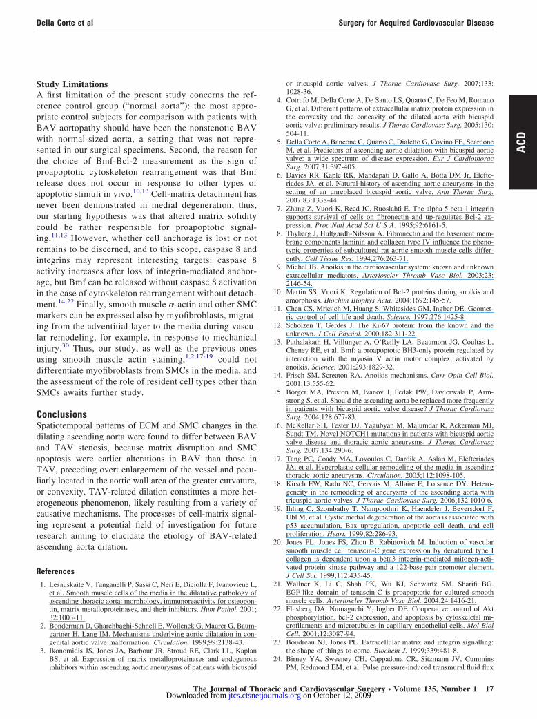
roapoptotic Versus Prosurvival Signalingow levels of Bmf-Bcl-2 binding were present in normalortas, showing that it may be a constitutive phenomenon;owever, it was increased in patients, considered as oneroup, compared with normal (concavity: 0.92 � 0.17 vs.72 � 0.01; P � .07; convexity: 1.10 � 0.24 vs 0.73 �.01; P � .02). Grouped together, all BAV patients pre-ented higher levels of Bmf-Bcl-2 binding than the TAVroup at the convexity (1.21 � 0.24 vs 0.96 � 0.16; P �03; concavity: 0.93 � 0.19 vs 0.90 � 0.16; P � .83). As tohe temporal patterns of Bmf-Bcl-2 binding, it increased inild dilations compared with normal, but no further in-
rease was detectable in the aneurysm phase (Figure 4).mf-Bcl-2 level did not correlate statistically with the AI.he expression of Bcl-2 mRNA was lower in patients than
n normal subjects, although statistical significance was noteached (optical density ratio to GAPDH: 0.2 0 �0.09 vs.29 � 0.2; P � .08).
iscussionhe main findings of this study may be summarized as
ollows: (1) BAV and TAV stenosis showed different tem- w
4 The Journal of Thoracic and Cardiovascular Surgery ● Januajtcs.ctsnetjournaDownloaded from
oral patterns of ascending aorta medial remodeling, in thatAV was associated with significant SMC apoptosis evenefore overt enlargement of the vessel, whereas TAV washaracterized by high variability in SMC apoptosis rates athis early stage; (2) similarly, ECM remodeling was earlierith BAV, being characterized by collagen and laminin
eduction, fibronectin increase, and tenascin expression,hereas only a proportion of TAV patients showed earlyCM changes, in part discordant in nature with those seenith BAV; (3) BAV and TAV stenosis also showed differ-
nt spatial patterns of medial remodeling: asymmetric inAV aortopathy, with the convexity more severely in-olved than the concavity, and symmetric in TAV; (4)lthough present also in normal aorta, Bmf-Bcl-2 bindingas significantly increased, in BAV small-size dilations, at
he convexity, with concomitant reduction of Bcl-2 expres-ion, indicating a net proapoptotic alteration of cell-matrixignaling. These represented novel insights into the physio-athology of ascending aorta dilations in the presence ofAV stenosis.
arly and Late SMC Changeso our knowledge, only one previous report2 showed earlypoptosis in the nonaneurysmal BAV aorta, although itncluded patients operated on for aortic stenosis, regurgita-ion, coarctation, and coronary artery disease in one group.owever, in studies assessing aortic wall changes, patient
amples should be homogeneous for valve function, inas-uch as important differences have been found betweenAV stenosis and regurgitation in terms of epidemiology5
nd morphologic/molecular features of ascending aortic di-ation4: valve function can influence even the natural historyf dilation.6 Other investigations on SMC apoptosis in BAVortopathy analyzed only end-stage aneurysmal tissues.1,2
Our findings indicate that the cellular and extracellularrocesses underlying aortic enlargement are already opera-ive in the normally sized or slightly dilated BAV aortamean diameter 3.8 cm), although limited to the convexity.he evidence of a “preclinical” aortopathy may add to theurrent debates on whether and how to concomitantly ad-ress the ascending aorta at the time of surgery for BAVtenosis.15 The recent studies on matrix metalloproteinases3
MMPs), as well as gene mutational analyses,16 may poten-ially pave the way for development of new diagnostic andredictive strategies that may help to cope with this clinicalhallenge.
With increasing aortic dimensions (mean diameter 4.9 cm),CM rearrangement at BAV convexity continued, whereasMC density increased back to normal levels. A similar tem-oral pattern was observed in the TAV setting, although ac-ompanied by a significant increment of AIs, in apparentontrast with the increase in cellularity. Also, Tang and co-
orkers17 have recently found comparable SMC density inry 2008 on October 12, 2009 ls.org
asTaSepSpwtqgSdseO
ltc
mmtioi
RATdEc
Della Corte et al Surgery for Acquired Cardiovascular Disease
ACD
neurysms and nondilated aortas. Kirsch and colleagues18 ob-erved higher SMC density in the maximal dilation area ofAV aneurysms than in the transition zone between normalorta and maximal dilation, thus inferring a temporal pattern ofMC density changes concordant with our observations. How-ver, neither one of those studies17,18 investigated SMC apo-tosis and proliferation. Concomitant high AI and normalMC density in our aneurysm specimens could not be ex-lained by concurrent proliferation, since Ki-67 expressionas not increased compared with normal. Thus, we suggest
hat the loss of noncellular components of the media may haveuantitatively prevailed on SMC apoptotic loss, for faster pro-ression of ECM breakdown, resulting in apparent normalMC density, although the absolute cell number must haveecreased. Although we could not calculate medial cross-ectional areas,17 the aortic media was significantly thinned,ven with mean diameters smaller than in previous studies.17,18
Figure 4. Top, Representative gels showing the resultbottom, comparison of Bmf-Bcl-2 binding levels betweeNormalized optical density (arbitrary units); TAV, Tricudeviation.
n the other hand, a previous study showed medial cell pro- a
The Journal of Thoracjtcs.ctsnetjourDownloaded from
iferation by Ki-67 staining in aortic dissection,19 a conditionhat may, however, be subtended by different remodeling pro-esses compared with poststenotic dilations.1
The differences emerging in the patterns of lesionalaturation argue in favor of different mechanisms of earlyedial remodeling in BAV versus TAV: in BAV aortopa-
hy, apoptosis may be part of the initiating events, whereasn the TAV setting, at least in a proportion of patients, itsccurrence is delayed, possibly representing a mechanism,f not a consequence, of dilation itself.
eciprocal Influences Between ECM and SMC inortic Remodelinghe features of ECM remodeling observed in this studyeserve a comment in light of the known paradigms ofCM-SMC interplays.7,8 The increase in fibronectin withoncomitant decrease in laminin suggests the constitution of
cl-2 immunoprecipitation with Western blot for Bmf;groups (see text). *P<.05 versus normal. Relative O.D.,aortic valve; BAV, bicuspid aortic valve; SD, standard
s of Bn subspid
microenvironment that favors SMC phenotype modulation
ic and Cardiovascular Surgery ● Volume 135, Number 1 15 on October 12, 2009 nals.org
fgtceBsticBsbddrtp
cesuiottfpfaecscsd(tscceraaeocwomB
dbfppbaqcasttBE
ATulacamTBertstptbficcudpIpcti
Tamcp
Surgery for Acquired Cardiovascular Disease Della Corte et al
1
ACD
rom the contractile to the synthetic phenotype.8 This sug-estion is confirmed by the evidence that the glycoproteinenascin-C, scarcely detectable in normal aortas, but typi-ally produced by SMCs in synthetic phenotype,4,20 wasxpressed in aneurysms and also in the greater curvature ofAV-related mild dilations. Furthermore, other markers of
ynthetic phenotype have been found in medial degenera-ion of the ascending aorta.1 The more significant reductionn collagens and laminin in BAV versus TAV dilations isonsistent with the greater increase in MMP-2 observed inAV by Ikonomidis and coworkers.3 In our previous
tudy,4 collagenolysis in BAV stenotic aorta was suggestedy decreased collagen type I, as assessed by Western blot,espite normal expression of collagen type I mRNA. In-eed, tenascin and MMPs are tightly connected in vascularemodeling: tenascin is capable to activate MMPs and, inurn, proteolysed collagen type I stimulates tenascinroduction.20
In the present study, we sought to assess whether ECMhanges in BAV aortopathy could exert a causative influ-nce also on SMC apoptosis. Although promoting SMCurvival and proliferation is the physiologic function attrib-ted to tenascin in vessel development and response tonjury,20 it has been shown that in conditions of MMPverexpression, enzymatic degradation of tenascin leads tohe exposition of domains in its molecule that are proapop-otic for SMCs.21 Similarly, fibronectin is a survival factoror SMCs,7,8 although its proteolysis by plasmin and MMPsroduces prodetachment/proapoptotic fragments.9 There-ore, the MMP repertoire typically expressed in the BAVorta3 may convert a prosurvival ECM into a proapoptoticnvironment for SMCs. Notably, extrinsic proapoptoticues are known to be transmitted within the cells throughtructural rearrangements of the actin cytoskeleton, so thathanges in cell-matrix interactions are accompanied by cellhape changes (gross cell rounding, but also subtler disor-ers) as part of the causative mechanism of apoptosis“amorphosis”).11,22,23 To be able to interact with Bcl-2 ando promote apoptosis, Bmf must be released by its normalite, where it is thought to serve as a sensor of actinytoskeleton integrity.13,14 Our finding of a significant in-rease in Bmf-Bcl-2 interaction in BAV convexity at anarly stage of dilation development strongly suggests thatearrangements in SMC cytoarchitecture may be broughtbout by early ECM changes and, in turn, convey a pro-poptotic signal. ECM-dependent apoptosis does not nec-ssarily require cell detachment (anoikis proper): alterationsf matrix geometrical organization or physical propertiesan trigger this mechanism of programmed cell death evenithout detachment.9-11,13 Indeed, we observed a reductionf Bcl-2 mRNA, whose expression is regulated also byatrix-dependent cytoskeletal changes.22,23 The fact that
mf-Bcl-2 did not further increase in larger aneurysms, w6 The Journal of Thoracic and Cardiovascular Surgery ● Januajtcs.ctsnetjournaDownloaded from
espite the increment in AI, suggests that late apoptosis,oth in TAV aneurysms and in BAV concavity, may resultrom other pathways. Our study did not explore, for exam-le, the apoptotic pathway of cytokine signaling, and theossible involvement of inflammatory mediators17,18 shoulde investigated in early versus late lesions. However, latepoptosis may be not a causative factor but rather a conse-uence of aneurysm growth itself, and another possibleause could be increased wall strain owing to enlargementnd loss of elasticity.17 Apoptosis in response to cyclictrain has been shown to occur in well-differentiated con-ractile SMCs, but not in those that have undergone pheno-ype change24: consistently, we found no AI elevation inAV aneurysm convexity, where a greater expression ofCM proteins promoting phenotype switch was observed.
Pathogenetic Hypothesishe asymmetrical spatial pattern of ECM and SMC changesniquely found in early BAV dilations introduces, in theong-standing debate on the pathogenesis of BAV-relatedortic disease, evidence in favor of a determinant or at leastontributory role of hemodynamics. A BAV stenosis, beingsymmetric in geometry, produces an eccentric jet yieldingore severe flow alterations in the ascending aorta thanAV stenosis with similar gradients and valve areas.25
AV-related turbulences have been shown to produce un-ven wall stress distribution, with overload at the aorticight anterolateral wall (convexity).26 Conversely, a hypo-hetical inborn wall defect2 sufficient to cause dilationhould produce a remodeling process uniformly involvinghe whole aortic circumference, which was denied by theresent study. Thus, we hypothesize that altered flow (evenhough not necessarily a “clinically” altered flow25,26) coulde either the triggering cause of early wall lesions or theactor required for local expression of a latent defect. Bothn vitro and in vivo studies have shown SMC phenotypehange,27 fibronectin and tenascin production,27 and in-reased MMP expression and activity28 to be elicited byndue biomechanical stress, as well as apoptotic SMCeath, with reduced Bcl-2 expression,24 as found in theresent study, and increased p53 expression,29 as found byhling and colleagues19 in medial degeneration. In our hy-othesis, stress-induced early ECM changes unique to BAVonvexity promote early SMC apoptosis, at least in parthrough Bmf-Bcl-2 interaction, leading to progressive, typ-cally asymmetric aortic enlargement.
However, further work is needed to verify this theory.he possibility cannot be discarded that BAV might bessociated with a congenital anomaly of some component ofatrix signaling pathways, and flow disturbances at the
onvexity could merely accelerate locally the degenerativerocess, however destined to involve also the concavity
ithin a longer time span.ry 2008 on October 12, 2009 ls.org
SAepBwstpranociriaaimmiliudtS
CSdaaTloecira
R
1
1
1
1
1
1
1
1
1
1
2
2
2
2
Della Corte et al Surgery for Acquired Cardiovascular Disease
ACD
tudy Limitationsfirst limitation of the present study concerns the ref-
rence control group (“normal aorta”): the most appro-riate control subjects for comparison with patients withAV aortopathy should have been the nonstenotic BAVith normal-sized aorta, a setting that was not repre-
ented in our surgical specimens. Second, the reason forhe choice of Bmf-Bcl-2 measurement as the sign ofroapoptotic cytoskeleton rearrangement was that Bmfelease does not occur in response to other types ofpoptotic stimuli in vivo.10,13 Cell-matrix detachment hasever been demonstrated in medial degeneration; thus,ur starting hypothesis was that altered matrix solidityould be rather responsible for proapoptotic signal-ng.11,13 However, whether cell anchorage is lost or notemains to be discerned, and to this scope, caspase 8 andntegrins may represent interesting targets: caspase 8ctivity increases after loss of integrin-mediated anchor-ge, but Bmf can be released without caspase 8 activationn the case of cytoskeleton rearrangement without detach-ent.14,22 Finally, smooth muscle �-actin and other SMCarkers can be expressed also by myofibroblasts, migrat-
ng from the adventitial layer to the media during vascu-ar remodeling, for example, in response to mechanicalnjury.30 Thus, our study, as well as the previous onessing smooth muscle actin staining,1,2,17-19 could notifferentiate myofibroblasts from SMCs in the media, andhe assessment of the role of resident cell types other thanMCs awaits further study.
onclusionspatiotemporal patterns of ECM and SMC changes in theilating ascending aorta were found to differ between BAVnd TAV stenosis, because matrix disruption and SMCpoptosis were earlier alterations in BAV than those inAV, preceding overt enlargement of the vessel and pecu-
iarly located in the aortic wall area of the greater curvature,r convexity. TAV-related dilation constitutes a more het-rogeneous phenomenon, likely resulting from a variety ofausative mechanisms. The processes of cell-matrix signal-ng represent a potential field of investigation for futureesearch aiming to elucidate the etiology of BAV-relatedscending aorta dilation.
eferences
1. Lesauskaite V, Tanganelli P, Sassi C, Neri E, Diciolla F, Ivanoviene L,et al. Smooth muscle cells of the media in the dilatative pathology ofascending thoracic aorta: morphology, immunoreactivity for osteopon-tin, matrix metalloproteinases, and their inhibitors. Hum Pathol. 2001;32:1003-11.
2. Bonderman D, Gharehbaghi-Schnell E, Wollenek G, Maurer G, Baum-gartner H, Lang IM. Mechanisms underlying aortic dilatation in con-genital aortic valve malformation. Circulation. 1999;99:2138-43.
3. Ikonomidis JS, Jones JA, Barbour JR, Stroud RE, Clark LL, Kaplan
BS, et al. Expression of matrix metalloproteinases and endogenousinhibitors within ascending aortic aneurysms of patients with bicuspid2
The Journal of Thoracjtcs.ctsnetjourDownloaded from
or tricuspid aortic valves. J Thorac Cardiovasc Surg. 2007;133:1028-36.
4. Cotrufo M, Della Corte A, De Santo LS, Quarto C, De Feo M, RomanoG, et al. Different patterns of extracellular matrix protein expression inthe convexity and the concavity of the dilated aorta with bicuspidaortic valve: preliminary results. J Thorac Cardiovasc Surg. 2005;130:504-11.
5. Della Corte A, Bancone C, Quarto C, Dialetto G, Covino FE, ScardoneM, et al. Predictors of ascending aortic dilatation with bicuspid aorticvalve: a wide spectrum of disease expression. Eur J CardiothoracSurg. 2007;31:397-405.
6. Davies RR, Kaple RK, Mandapati D, Gallo A, Botta DM Jr, Elefte-riades JA, et al. Natural history of ascending aortic aneurysms in thesetting of an unreplaced bicuspid aortic valve. Ann Thorac Surg.2007;83:1338-44.
7. Zhang Z, Vuori K, Reed JC, Ruoslahti E. The alpha 5 beta 1 integrinsupports survival of cells on fibronectin and up-regulates Bcl-2 ex-pression. Proc Natl Acad Sci U S A. 1995;92:6161-5.
8. Thyberg J, Hultgardh-Nilsson A. Fibronectin and the basement mem-brane components laminin and collagen type IV influence the pheno-typic properties of subcultured rat aortic smooth muscle cells differ-ently. Cell Tissue Res. 1994;276:263-71.
9. Michel JB. Anoikis in the cardiovascular system: known and unknownextracellular mediators. Arterioscler Thromb Vasc Biol. 2003;23:2146-54.
0. Martin SS, Vuori K. Regulation of Bcl-2 proteins during anoikis andamorphosis. Biochim Biophys Acta. 2004;1692:145-57.
1. Chen CS, Mrksich M, Huang S, Whitesides GM, Ingber DE. Geomet-ric control of cell life and death. Science. 1997;276:1425-8.
2. Scholzen T, Gerdes J. The Ki-67 protein: from the known and theunknown. J Cell Physiol. 2000;182:311-22.
3. Puthalakath H, Villunger A, O’Reilly LA, Beaumont JG, Coultas L,Cheney RE, et al. Bmf: a proapoptotic BH3-only protein regulated byinteraction with the myosin V actin motor complex, activated byanoikis. Science. 2001;293:1829-32.
4. Frisch SM, Screaton RA. Anoikis mechanisms. Curr Opin Cell Biol.2001;13:555-62.
5. Borger MA, Preston M, Ivanov J, Fedak PW, Davierwala P, Arm-strong S, et al. Should the ascending aorta be replaced more frequentlyin patients with bicuspid aortic valve disease? J Thorac CardiovascSurg. 2004;128:677-83.
6. McKellar SH, Tester DJ, Yagubyan M, Majumdar R, Ackerman MJ,Sundt TM. Novel NOTCH1 mutations in patients with bicuspid aorticvalve disease and thoracic aortic aneurysms. J Thorac CardiovascSurg. 2007;134:290-6.
7. Tang PC, Coady MA, Lovoulos C, Dardik A, Aslan M, ElefteriadesJA, et al. Hyperplastic cellular remodeling of the media in ascendingthoracic aortic aneurysms. Circulation. 2005;112:1098-105.
8. Kirsch EW, Radu NC, Gervais M, Allaire E, Loisance DY. Hetero-geneity in the remodeling of aneurysms of the ascending aorta withtricuspid aortic valves. J Thorac Cardiovasc Surg. 2006;132:1010-6.
9. Ihling C, Szombathy T, Nampoothiri K, Haendeler J, Beyersdorf F,Uhl M, et al. Cystic medial degeneration of the aorta is associated withp53 accumulation, Bax upregulation, apoptotic cell death, and cellproliferation. Heart. 1999;82:286-93.
0. Jones PL, Jones FS, Zhou B, Rabinovitch M. Induction of vascularsmooth muscle cell tenascin-C gene expression by denatured type Icollagen is dependent upon a beta3 integrin-mediated mitogen-acti-vated protein kinase pathway and a 122-base pair promoter element.J Cell Sci. 1999;112:435-45.
1. Wallner K, Li C, Shah PK, Wu KJ, Schwartz SM, Sharifi BG.EGF-like domain of tenascin-C is proapoptotic for cultured smoothmuscle cells. Arterioscler Thromb Vasc Biol. 2004;24:1416-21.
2. Flusberg DA, Numaguchi Y, Ingber DE. Cooperative control of Aktphosphorylation, bcl-2 expression, and apoptosis by cytoskeletal mi-crofilaments and microtubules in capillary endothelial cells. Mol BiolCell. 2001;12:3087-94.
3. Boudreau NJ, Jones PL. Extracellular matrix and integrin signalling:the shape of things to come. Biochem J. 1999;339:481-8.
4. Birney YA, Sweeney CH, Cappadona CR, Sitzmann JV, CumminsPM, Redmond EM, et al. Pulse pressure-induced transmural fluid flux
ic and Cardiovascular Surgery ● Volume 135, Number 1 17 on October 12, 2009 nals.org
2
2
2
2
2
3
DJCcasT
gpwdtmaboa
cirn
f
tfic
pbr
admes
tHpstrttrhdpc
t
wwcv
p
BwT
eyH
Surgery for Acquired Cardiovascular Disease Della Corte et al
1
ACD
increases bovine aortic smooth muscle cell apoptosis in a mitogenactivated protein kinase dependent manner. J Vasc Res. 2004;41:364-74.
5. Donal E, Novaro GM, Deserranno D, Popovic ZB, Greenberg NL,Richards KE, et al. Planimetric assessment of anatomic valve areaoverestimates effective orifice area in bicuspid aortic stenosis. Am JSoc Echocardiogr. 2005;18:1392-8.
6. Robicsek F, Thubrikar MJ, Cook JW, Fowler B. The congenitallybicuspid aortic valve: How does it function? Why does it fail? AnnThorac Surg. 2004;77:177-85.
7. Williams B. Mechanical influences on vascular smooth muscle cellfunction. J Hypertens. 1998;16:1921-9.
8. Grote K, Flach I, Luchtefeld M, Akin E, Holland SM, Drexler H, et al.Mechanical stretch enhances mRNA expression and proenzyme re-lease of matrix metalloproteinase-2 (MMP-2) via NAD(P)H oxidase-derived reactive oxygen species. Circ Res. 2003;92:80-6.
9. Wernig F, Mayr M, Xu Q. Mechanical stretch-induced apoptosis insmooth muscle cells is mediated by �-1-integrin signalling pathways.Hypertension. 2003;41;903-11.
0. Chambers RC, Leoni P, Kaminski N, Laurent GJ, Heller RA. Globalexpression profiling of fibroblast responses to transforming growthfactor-beta1 reveals the induction of inhibitor of differentiation-1 andprovides evidence of smooth muscle cell phenotypic switching. Am JPathol. 2003;162:533-46.
iscussionohn S. Ikonomidis (Charleston, SC). I congratulate Dr Dellaorte for an outstanding study, which showed temporospatialhanges in extracellular proteins, vascular smooth muscle density,nd indexes of apoptosis in normal or aneurysmal ascending aorticpecimens taken from patients with normal or stenotic BAVs orAVs.
In particular, and in keeping with previous studies from thisroup, extracellular matrix protein changes were particularly im-ortant in the convexity of aneurysmal ascending aorta associatedith BAVs. In addition, evidence of early SMC apoptosis wasemonstrated. Of particular relevance here is evidence presentedhat anoikis, or apoptosis, which is induced by the loss of integrin-ediated cellular attachments, participates in the process of SMC
ttrition as the aneurysm develops. This, of course, makes senseecause extracellular matrix remodeling and its attendant changesf cellular matrix interactions are universal features of aorticneurysm development.
I have 4 questions: Your extracellular protein assessments wereonducted by immunohistochemical techniques. Elastin was notncluded in these results but is known to cause significant autofluo-escence in these studies. How did you correct for this phenome-on when you were assessing the other proteins?
Dr Della Corte. Thank you, Dr Ikonomidis. My coworkers
rom the biomorphology department used to pre-tract tissues with o8 The Journal of Thoracic and Cardiovascular Surgery ● Januajtcs.ctsnetjournaDownloaded from
oluidine blue solution and then they used a very selective greenlter for fluorescence microscopy to obviate elastin autofluores-ence.
We have already published our preliminary results with ECMrotein assessment using both immunohistochemistry and Westernlot, which is not affected by elastin autofluorescence, and theesults of the two methods were highly consistent.
Dr Ikonomidis. Second question: Your data suggest that SMCpoptosis is primed and occurs early in the course of aneurysmevelopment. However, in the later phase of aneurysm develop-ent, you demonstrate an increase in SMC density. How do you
xplain these findings and did you examine fibroblast profiles as aecond resident cell within the aortic aneurysm wall?
Dr Della Corte. We didn’t look at other resident cell types inhis study, and this may be an interesting point for future research.owever, both the evidence of smooth muscle cell loss in an earlyhase of dilation with bicuspid valves and that of an increase inmooth muscle cell density in aneurysms compared to small dila-ions (resulting in near-normal cell density) have been alreadyeported in two different studies by other authors. Here we haveried to suggest a common interpretation for those two findings inhe light of cell-matrix interactions. Cell-matrix signaling may beesponsible for early apoptosis with the anoikis mechanism, as weave demonstrated it, but also, in the later phase of aneurysmevelopment, the effect of increased morphogenetic pro-survivalroteins, like fibronectin and tenascin, may explain smooth muscleell density increase.
Dr Ikonomidis. Were you able to stratify these data relative tohe degree of aortic stenosis?
Dr Della Corte. We tried to compare subgroups of patientsith different degrees of valve stenosis, but no difference in aorticall changes emerged. However, we expected such results, be-
ause other factors may be important besides the degree of aorticalve stenosis, as for example the time duration of valve disease.
Dr Ikonomidis. Last, have you related any of your changes torofiles of known protease systems such as MMPs?
Dr Della Corte. We observed a greater decrease in collagen inAV compared with TAV patients, and this may be consistentith the increase in MMP-2 expression that is quite unique to theAV setting, or at least greater in BAV than in TAV patients.
Similarly, our finding of increased tenascin and fibronectinxpression may be consistent with the decrease in MMP-3 thatour group has demonstrated in the setting of bicuspid aortopathy.owever, we should keep in mind that protein amount is the result
f a fine balance between production and degradation of proteins.ry 2008 on October 12, 2009 ls.org
APl
hw
Della Corte et al Surgery for Acquired Cardiovascular Disease
ACD
ppendix E1olymerase chain reactions (PCRs) were performed with the fol-
owing program:1. Initial denaturation for 5 minutes at 95°C2. Denaturation for 45 seconds at 95°C3. Annealing for 1 minute at 59°C4. Extension for 2 minutes at 68°C5. Final extension at 68°C for 5 minutesThe following primers of the Bcl-2 gene and the glyceralde-
yde-3-phosphate dehydrogenase (GAPDH) housekeeping gene
ere used for PCR (expected size of the amplification product):The Journal of Thoracicjtcs.ctsnetjourDownloaded from
● Bcl-2: 5=-ATGCCAAGGGGGAAACACCAG-3= (forwardprimer)
● 5=-AGACAGCCAGGAGAAATCAAA-3’ (reverse primer)(831 bp)
● GAPDH: 5=-CACCATCTTCCAGGAGCGAG-3= (forwardprimer)
● 5=-TCACGCCACAGTTTCCCGGA-3= (reverse primer)(372 bp)
and Cardiovascular Surgery ● Volume 135, Number 1 18.e1 on October 12, 2009 nals.org
Th
AHMMEMASSA
TBa
Surgery for Acquired Cardiovascular Disease Della Corte et al
1
ACD
ABLE E1. Within groups of aortic dimensions (small-somogeneous in terms of age, prevalence of hypertension
TAV/dilation (n � 7) BAV/dilatio
ge (y) 60 � 9 54 �ypertension (rate) 57% 43aximum gradient (mm Hg) 114 � 44 95 �ean gradient (mm Hg) 76 � 32 63 �
OAI (cm2/m2) 0.31 � 0.02 0.35 �ild aortic regurgitation (rate) 29% 43
scending aorta (cm) 3.8 � 0.5 3.8 �inotubular ridge (cm) 3.4 � 0.5 3.5 �inuses (cm) 3.2 � 0.4 3.5 �ortic ratio 1.19 � 0.15 1.15 �
AV, Tricuspid aortic valve; BAV, bicuspid aortic valve; EOAI, effectiveAV/aneurysm. Additionally, no significant difference (P � .05) was founneurysms in terms of age and stenosis severity.
Figure E1. Representative images of Ki-67 staining (abetween a normal aorta, a BAV aneurysm, and a TAV a
ize dilations and aneurysms), BAV and TAV patients were, aortic valve stenosis parameters, and aortic dimensionsn (n � 7) P* TAV/aneurysm (n � 5) BAV/aneurysm (n � 7) P†
14 .37 67 � 11 60 � 15 .47% .50 60% 57% .69
16 .43 62 � 8 79 � 16 .1312 .47 40 � 5 47 � 9 .110.1 .58 0.42 � 0.03 0.37 � 0.07 .39
% .50 60% 57% .690.6 .98 5.0 � 0.3 4.9 � 0.4 .820.4 .68 4.3 � 1.1 3.7 � 0.7 .380.4 .21 3.9 � 0.6 3.8 � 0.7 .910.17 .68 1.54 � 0.14 1.55 � 0.12 .92
orifice area index. *TAV/dilation versus BAV/dilation; †TAV/aneurysm versusd when comparing BAV dilations versus aneurysms and TAV dilations versus
rrows) showing no difference in SMC proliferative indexneurysm. TAV, Tricuspid aortic valve; BAV, bicuspid aortic
valve.
8.e2 The Journal of Thoracic and Cardiovascular Surgery ● January 2008 on October 12, 2009 jtcs.ctsnetjournals.orgDownloaded from
DOI: 10.1016/j.jtcvs.2007.09.009 2008;135:8-18 J Thorac Cardiovasc Surg
Stefania Montagnani and Maurizio Cotrufo Meglio, Daria Nurzynska, Luca S. De Santo, Marisa De Feo, Michelangelo Scardone, Alessandro Della Corte, Cesare Quarto, Ciro Bancone, Clotilde Castaldo, Franca Di
signalingdilatation with bicuspid and tricuspid aortic valve stenosis: Focus on cell�matrix
Spatiotemporal patterns of smooth muscle cell changes in ascending aortic
Continuing Medical Education Activities
http://cme.ctsnetjournals.org/cgi/hierarchy/ctsnetcme_node;JTCSSubscribers to the Journal can earn continuing medical education credits via the Web at
Subscription Information
http://jtcs.ctsnetjournals.org/cgi/content/full/135/1/8#BIBLThis article cites 30 articles, 20 of which you can access for free at:
Citations
http://jtcs.ctsnetjournals.org/cgi/content/full/135/1/8#otherarticlesThis article has been cited by 2 HighWire-hosted articles:
Subspecialty Collections
http://jtcs.ctsnetjournals.org/cgi/collection/valve_disease Valve disease http://jtcs.ctsnetjournals.org/cgi/collection/molecular_biology
Molecular biologyThis article, along with others on similar topics, appears in the following collection(s):
Permissions and Licensing
http://www.elsevier.com/wps/find/obtainpermissionform.cws_home/obtainpermissionformreceipt, is available at: An on-line permission request form, which should be fulfilled within 10 working days of
. http://www.elsevier.com/wps/find/supportfaq.cws_home/permissionusematerialcan be found online at: General information about reproducing this article in parts (figures, tables) or in its entirety
on October 12, 2009 jtcs.ctsnetjournals.orgDownloaded from