
Solution Structure and Antiestrogenic Activity of the Unique C-terminal, NR-box Motif-containing...
12
Solution Structure and Antiestrogenic Activity of the Unique C-terminal, NR-box Motif-containing Region of MTA1s * Received for publication, May 10, 2006, and in revised form, June 26, 2006 Published, JBC Papers in Press, June 27, 2006, DOI 10.1074/jbc.M604444200 Rajesh R. Singh ‡1 , Kumaralal Kaluarachchi §1 , Mingzhi Chen ¶2 , Suresh K. Rayala ‡ , Seetharaman Balasenthil ‡ , Jianpeng Ma ¶ ** 3 , and Rakesh Kumar ‡ ‡‡4 From the ‡ Department of Molecular and Cellular Oncology and § Experimental Therapeutics, University of Texas M.D. Anderson Cancer Center, Houston, Texas 77030, the ¶ Graduate Program of Structural and Computational Biology and Molecular Biophysics, Baylor College of Medicine, Houston, Texas 77030, the **Department of Bioengineering, Rice University, Houston, Texas 77251, the Verna and Marrs McLean Department of Biochemistry and Molecular Biology, Baylor College of Medicine, Houston, Texas 77030, and the ‡‡ Department of Molecular and Cellular Biology, Baylor College of Medicine, Houston, Texas 77030 Metastasis tumor-associated 1 short form (MTA1s) is a natu- rally occurring, alternatively spliced variant of MTA1 that func- tions as a repressor of estrogen receptor (ER) transcriptional functions, at least in part by binding and sequestering ER in the cytoplasm. A unique C-terminal 33-amino acid region contain- ing a nuclear receptor (NR)-box motif (-LRILL-) mediates bind- ing of MTA1s with ER and is indispensable in this interaction. Here, we elucidated the solution structure of this 33-amino acid region by NMR spectroscopy. We found a predominance of the -helical region toward the N-terminal region, which includes the NR-box motif. In silico docking and comparison studies showed similarities between the NR-box motif of MTA1s and a similar motif of coregulators, both in structure and mode of ER binding. In MCF-7 breast cancer cells, the MTA1s peptide effec- tively repressed ER transactivation function, as evidenced by the estrogen response element-luc assay and down-regulation of estrogen-induced genes. In mechanistic studies, we found that the antiestrogenic effects of the MTA1s peptide were due to its ability to compete with the coactivator recruitment to ER. Fur- thermore, the peptide efficiently repressed estrogen-induced proliferation and anchorage-independent growth of MCF-7 cells. In addition, the MTA1s peptide blocked the progression of tumors formed by MCF-7 cells overexpressing an ER coactiva- tor in a xenograft-based assay. In brief, the characterization of structure and antiestrogenic activity of MTA1s peptide high- light its therapeutic potential. Protein-protein interactions play an important role in com- municating signals in the cell, which involves the conductance and amplification of the message from a master hormonal sig- nal. In this context, the proteins involved in the downstream convergence of the signals are equipped with specialized domains that help establish specific inter-protein interactions to achieve high signal transduction efficiency. In recent years, extensive research on proteins such as the nuclear receptor (NR) 5 family of hormone-induced transcription factors has identified their mode of action and their binding partners, pro- viding novel insights into the significance of specialized pro- tein-protein interactions in hormone action. The NR family is composed of 48 structurally related transcription factors, which require their cognate ligand for activity. Upon ligand binding, the NR homodimers or het- erodimers selectively bind to the specific short stretches of DNA, referred to as hormone response elements, and stim- ulate transcription of the target genes responsible for vital biological processes such as cell survival, proliferation, dif- ferentiation, and apoptosis (1). NRs are composed of structurally and functionally similar molecular domains, which reflect their common mode of action. The N-terminal domain is referred to as “activation function 1” (AF1), which shows a higher degree of sequence variation. NRs also have a highly conserved central DNA-bind- ing domain composed of two zinc finger motifs. The ligand- binding domain to which the hormones bind is localized in the C-terminal half of the protein and is referred to as “activation function 2” (AF2). The extensive structural characterization of the ligand-binding domain of several receptors, such as estro- gen receptor (ER) (2), progesterone receptor (PR) (2), and androgen receptor (3), has improved our understanding of the binding of the hormone, the resulting conformational changes in the receptor, and the basis of acute specificity of hormone binding to its receptor. The DNA binding of ligand-activated NR represents the delivery of the signal to the genome, whereas further amplifica- tion of the signal is brought about by the ability of NRs to recruit a family of proteins broadly referred to as “coactivators.” One of * This work was supported in part by National Institutes of Health Grants CA98823 and CA65746 (to R. K.). The costs of publication of this article were defrayed in part by the payment of page charges. This article must there- fore be hereby marked “advertisement” in accordance with 18 U.S.C. Sec- tion 1734 solely to indicate this fact. 1 Both authors contributed equally to this work. 2 Supported by a pre-doctoral fellowship from the W. M. Keck Foundation of the Gulf Coast Consortia through the Keck Center for Computational and Structural Biology. 3 Support by the Robert A. Welch Foundation (Grant JM Q-1512). 4 To whom correspondence should be addressed: Dept. of Molecular and Cellular Oncology, University of Texas M.D. Anderson Cancer Center, 1515 Holcomb St., Houston, TX 77030. Tel.: 713-745-3558; Fax: 713-745-3792; E-mail: [email protected]. 5 The abbreviations used are: NR, nuclear receptor; MTA1s, metastasis tumor- associated 1 short form; PELP1, proline-, glutamic acid-, and leucine-rich protein 1; E 2 , estrogen (17--estradiol); ER, estrogen receptor ; TFE, trif- luoroethanol; ERE, estrogen response element; PR, progesterone receptor; NOESY, nuclear Overhauser effect spectroscopy; TOCSY, total correlation spectroscopy; dqf-COSY, double-quantum-filtered correlation spectrosco- py; GST, glutathione S-transferase; AIB1 amplified in breast cancer-1. THE JOURNAL OF BIOLOGICAL CHEMISTRY VOL. 281, NO. 35, pp. 25612–25621, September 1, 2006 © 2006 by The American Society for Biochemistry and Molecular Biology, Inc. Printed in the U.S.A. 25612 JOURNAL OF BIOLOGICAL CHEMISTRY VOLUME 281 • NUMBER 35 • SEPTEMBER 1, 2006 by guest on October 20, 2016 http://www.jbc.org/ Downloaded from by guest on October 20, 2016 http://www.jbc.org/ Downloaded from by guest on October 20, 2016 http://www.jbc.org/ Downloaded from
Transcript of Solution Structure and Antiestrogenic Activity of the Unique C-terminal, NR-box Motif-containing...
Solution Structure and Antiestrogenic Activity of the UniqueC-terminal, NR-box Motif-containing Region of MTA1s*
Received for publication, May 10, 2006, and in revised form, June 26, 2006 Published, JBC Papers in Press, June 27, 2006, DOI 10.1074/jbc.M604444200
Rajesh R. Singh‡1, Kumaralal Kaluarachchi§1, Mingzhi Chen¶2, Suresh K. Rayala‡, Seetharaman Balasenthil‡,Jianpeng Ma¶�**3, and Rakesh Kumar‡ ‡‡4
From the ‡Department of Molecular and Cellular Oncology and §Experimental Therapeutics, University of Texas M.D. AndersonCancer Center, Houston, Texas 77030, the ¶Graduate Program of Structural and Computational Biology and Molecular Biophysics,Baylor College of Medicine, Houston, Texas 77030, the **Department of Bioengineering, Rice University, Houston, Texas 77251, the�Verna and Marrs McLean Department of Biochemistry and Molecular Biology, Baylor College of Medicine, Houston, Texas 77030,and the ‡‡Department of Molecular and Cellular Biology, Baylor College of Medicine, Houston, Texas 77030
Metastasis tumor-associated 1 short form (MTA1s) is a natu-rally occurring, alternatively spliced variant ofMTA1 that func-tions as a repressor of estrogen receptor (ER) � transcriptionalfunctions, at least in part by binding and sequesteringER� in thecytoplasm. A unique C-terminal 33-amino acid region contain-ing a nuclear receptor (NR)-boxmotif (-LRILL-) mediates bind-ing of MTA1s with ER� and is indispensable in this interaction.Here, we elucidated the solution structure of this 33-amino acidregion by NMR spectroscopy. We found a predominance of the�-helical region toward the N-terminal region, which includesthe NR-box motif. In silico docking and comparison studiesshowed similarities between the NR-box motif of MTA1s and asimilarmotif of coregulators, both in structure andmodeof ER�binding. InMCF-7 breast cancer cells, theMTA1s peptide effec-tively repressed ER� transactivation function, as evidenced bythe estrogen response element-luc assay anddown-regulationofestrogen-induced genes. In mechanistic studies, we found thatthe antiestrogenic effects of the MTA1s peptide were due to itsability to competewith the coactivator recruitment to ER�. Fur-thermore, the peptide efficiently repressed estrogen-inducedproliferation and anchorage-independent growth of MCF-7cells. In addition, theMTA1s peptide blocked the progression oftumors formed byMCF-7 cells overexpressing an ER� coactiva-tor in a xenograft-based assay. In brief, the characterization ofstructure and antiestrogenic activity of MTA1s peptide high-light its therapeutic potential.
Protein-protein interactions play an important role in com-municating signals in the cell, which involves the conductanceand amplification of the message from a master hormonal sig-
nal. In this context, the proteins involved in the downstreamconvergence of the signals are equipped with specializeddomains that help establish specific inter-protein interactionsto achieve high signal transduction efficiency. In recent years,extensive research on proteins such as the nuclear receptor(NR)5 family of hormone-induced transcription factors hasidentified their mode of action and their binding partners, pro-viding novel insights into the significance of specialized pro-tein-protein interactions in hormone action.The NR family is composed of �48 structurally related
transcription factors, which require their cognate ligand foractivity. Upon ligand binding, the NR homodimers or het-erodimers selectively bind to the specific short stretches ofDNA, referred to as hormone response elements, and stim-ulate transcription of the target genes responsible for vitalbiological processes such as cell survival, proliferation, dif-ferentiation, and apoptosis (1).NRs are composed of structurally and functionally similar
molecular domains, which reflect their common mode ofaction. The N-terminal domain is referred to as “activationfunction 1” (AF1), which shows a higher degree of sequencevariation. NRs also have a highly conserved central DNA-bind-ing domain composed of two zinc finger motifs. The ligand-binding domain to which the hormones bind is localized in theC-terminal half of the protein and is referred to as “activationfunction 2” (AF2). The extensive structural characterization ofthe ligand-binding domain of several receptors, such as estro-gen receptor (ER) (2), progesterone receptor (PR) (2), andandrogen receptor (3), has improved our understanding of thebinding of the hormone, the resulting conformational changesin the receptor, and the basis of acute specificity of hormonebinding to its receptor.The DNA binding of ligand-activated NR represents the
delivery of the signal to the genome, whereas further amplifica-tion of the signal is brought about by the ability ofNRs to recruita family of proteins broadly referred to as “coactivators.” One of
* This work was supported in part by National Institutes of Health GrantsCA98823 and CA65746 (to R. K.). The costs of publication of this article weredefrayed in part by the payment of page charges. This article must there-fore be hereby marked “advertisement” in accordance with 18 U.S.C. Sec-tion 1734 solely to indicate this fact.
1 Both authors contributed equally to this work.2 Supported by a pre-doctoral fellowship from the W. M. Keck Foundation of
the Gulf Coast Consortia through the Keck Center for Computational andStructural Biology.
3 Support by the Robert A. Welch Foundation (Grant JM Q-1512).4 To whom correspondence should be addressed: Dept. of Molecular and
Cellular Oncology, University of Texas M.D. Anderson Cancer Center, 1515Holcomb St., Houston, TX 77030. Tel.: 713-745-3558; Fax: 713-745-3792;E-mail: [email protected].
5 The abbreviations used are: NR, nuclear receptor; MTA1s, metastasis tumor-associated 1 short form; PELP1, proline-, glutamic acid-, and leucine-richprotein 1; E2, estrogen (17-�-estradiol); ER�, estrogen receptor �; TFE, trif-luoroethanol; ERE, estrogen response element; PR, progesterone receptor;NOESY, nuclear Overhauser effect spectroscopy; TOCSY, total correlationspectroscopy; dqf-COSY, double-quantum-filtered correlation spectrosco-py; GST, glutathione S-transferase; AIB1 amplified in breast cancer-1.
THE JOURNAL OF BIOLOGICAL CHEMISTRY VOL. 281, NO. 35, pp. 25612–25621, September 1, 2006© 2006 by The American Society for Biochemistry and Molecular Biology, Inc. Printed in the U.S.A.
25612 JOURNAL OF BIOLOGICAL CHEMISTRY VOLUME 281 • NUMBER 35 • SEPTEMBER 1, 2006
by guest on October 20, 2016
http://ww
w.jbc.org/
Dow
nloaded from
by guest on October 20, 2016
http://ww
w.jbc.org/
Dow
nloaded from
by guest on October 20, 2016
http://ww
w.jbc.org/
Dow
nloaded from
the functions of coactivator proteins is to directly or indirectlyremodel the local chromatin structure through covalent mod-ification of histones (acetylation, phosphorylation, or methyla-tion), resulting in the opening of chromatin, greater accessibil-ity of the target gene promoter to transcriptionmachinery, andultimately, gene transcription (4). Numerous families of NRcoactivators have been identified. The best studied includep300/CBP (CREB-binding protein), p300/CBP-associated fac-tor, members of the SRC family (5), and proline, glutamic acid,and leucine-rich protein 1 (PELP1) (6).Coactivator proteins have multiple signature motifs referred
to as NR-boxes, which are short stretches of leucine-richsequences with consensus sequence of LXXLL (where Xdenotes any amino acid residue). The binding of the cognateligand to the ligand-binding domain of the NR results in a con-formational change involving the rearrangement of the posi-tions of helices H3, H4, H5, and H12, leading to the formationof a hydrophobic pocket. TheNR-boxes bind to this hydropho-bic pocket in a helical conformation with a high degree of affin-ity, thus leading to the recruitment of coactivators to the acti-vated NR. The interaction of a NR with a specific coactivator isdetermined by the specificity of the binding interactionsbetween the NR-box motif and the ligand-binding domain oftheNR. In general, each coactivator hasmore than oneNR-box,and the differences in spacing of the NR-boxes, combined withthe variable flanking sequences, have been implicated in con-ferring the specificity of this interaction (7, 8).Estrogen receptor � (ER�) belongs to a subfamily of NRs,
namely steroid hormone receptors. It functions as an estrogen-activated transcription factor and is involved in the up-regula-tion of downstream target genes, most of which are involved inthe control of cell cycle progression and growth of humanbreast epithelium. Excessive stimulation of the ER pathway inthe cell due to increased hormonal secretion or increased acti-vation of the receptor can lead to augmentation of cell prolifer-ation and thus a higher risk of growth deregulation, possiblyresulting in breast cancer (4). In this context, themode of actionof ER�, its potential deregulation in cancer, and its coregulatoryprotein are subjects of intense study.In a previous report from this laboratory, we identified and
characterized a unique coregulatory protein of ER�, namelyMTA1s, which is a naturally occurring splice variant of MTA1(9). MTA1s localizes in the cytoplasm, and its C-terminalstretch of 33 amino acids bears no resemblance to any of thepreviously reported protein sequences. Situated in the earlyportion of this 33-amino acid region is anNR-boxmotif bearingthe sequence -LRILL- (Fig. 1A).MTA1s uses thismotif to inter-act and bind with the AF2 domain of ER�, leading to its cyto-plasmic sequestration, decrease in the genomic functions ofER�, and augmentation of the aggressive ER-negative pheno-types in cells like increased metastasis, invasiveness, and resist-ance to antiestrogen therapy. In the present investigation, weelucidated the NMR solution structure of this stretch of 33amino acids ofMTA1s. Then, in an in silicomodeling study, wesimulated the binding of the NR-box motif to the AF2 domainof ER�. We determined the effects of MTA1s peptide on ER�transactivation function, estrogen-stimulated proliferation,and tumorigenesis of breast cancer cells.
MATERIALS AND METHODS
Cell Culture and Reagents—Human breast cancer cells werecultured in Dulbecco’s modified Eagle’s medium/F-12 mediumsupplemented with 10% fetal bovine serum. For estrogen treat-ment experiments, the regular medium was replaced withmedium containing 5% dextran-charcoal-stripped medium.The C-terminal 33-amino acid peptide of MTA1s was pur-chased fromSynPepCorp. (Dublin, CA) and fully characterizedby reverse phase high-performance liquid chromatography,amino acid sequencing, and mass spectrometry.Far-UV Circular Dichroism Spectroscopy—Far UV CD spec-
tra of the peptide dissolved in different concentrations of trif-luoroethanol (TFE) (30, 40, 50, and 70%) were recorded from260 to 210 nm using a JASCO J-810 spectropolarimeter using0.1-cm quartz cuvettes. Each spectrum was an average of fivescans.NMR Spectroscopy—Perdeuterated TFE was supplied by
Cambridge Isotope Inc. NMR samples were prepared by dis-solving 4 mg of MTA1s peptide in 550 �l of a 1:1 mixture ofperdeuterated TFE and H2O or perdeuterated TFE and 99.96%D2O. Proton NMR spectra were recorded either on a VarianUnityPlus spectrometer (Palo Alto, CA) operating at 750 MHzor a Bruker Avance (Billerica, MA) spectrometer operating at500 MHz equipped with 1H/13C/15N triple resonance probeswith pulse-field gradients. All experiments were performed at25 °C, and thewater peakwas suppressed using eitherWET (10,11) or WATERGATE (12) solvent suppression sequences.Two-dimensional Nuclear Overhauser effect spectroscopyNOESY, total correlation spectroscopy (TOCSY), and double-quantum-filtered correlation spectroscopy (dqf-COSY) spectrawere collected with the states-time proportional phase incre-ment method. Double-quantum-filtered two-dimensional cor-relation (13) spectra were collected with 4096 points in the t2dimension, 400 real t1 increments, 32 transients per free induc-tion decay, and a 2-s recycle delay. The data were zero-filled to1024 points in the t1 dimension and multiplied by a 20°-shiftedsine bell function in both dimensions prior to Fourier transfor-mation. Estimates of NH-C�H coupling constants (3JNH) wereobtained from the peak separation of multiplets in the dqf-COSY spectrum. TOCSY spectra (14) were recorded at mixingtimes of 30, 60, and 75mswith 4096 data points in the t2 dimen-sion and 320 t1 increments. NOESY spectra (15) were collectedwith 4096 data points in the t2 dimension and 320 real t1 incre-ments using the states-time proportional phase incrementmethod of phase cycling. Thirty-two transients per free induc-tion decay were collected, with a relaxation delay time of 3.5 s.Mixing times of 60, 180, 250, and 400mswere used. All NOESYspectra were zero-filled to 2048 in the t1 dimension prior toFourier transformation and apodized with a 90°-shifted sinebell function. A NOESY spectrum with a 400-min mixing timewas used only for resonance assignments. All spectra were pro-cessed on an SGI workstation using Felix version 2000 (Accel-rys, Inc., San Diego, CA) and internally referenced to the resid-ual CDH resonance of perdeuterated TFE (3.9 ppm).Structure Calculations—Inter-proton distances were esti-
mated fromNOESY cross-peak intensities using Felix. NOESYintensities were classified as strong, medium, weak, or very
Structure and Activity of the C-terminal Region of MTA1s
SEPTEMBER 1, 2006 • VOLUME 281 • NUMBER 35 JOURNAL OF BIOLOGICAL CHEMISTRY 25613
by guest on October 20, 2016
http://ww
w.jbc.org/
Dow
nloaded from
weak and were assigned distance ranges of 1.8–2.5, 1.8–3.5,1.8–4.5, or 1.8–5.5 Å, respectively, to be used as distancerestraints in molecular dynamics simulation with XPLOR 3.1(16). This produced a total of 310 inter-proton distance con-straints. A linear peptide chain of the sequence was generatedusing XPLOR and was subjected to 10 ps of unrestrainedmolecular dynamics in the absence of any Van derWaal repul-sion to generate a family of 30 initial randomized structures.Each of these structureswas subjected to 500 steps of restrainedenergy minimization using the steepest descent algorithm andsubsequently subjected to restrained molecular dynamics. Therestrained molecular dynamics protocol consisted of 10 ps ofrestrainedmolecular dynamics at 1000K, duringwhich theVander Waals’ force constant was gradually increased. Each struc-ture was subjected to an additional 5 ps of molecular dynamicsat the maximum Van der Waals’ force constant and was fol-lowed by a cooling step to 300 K over 7 ps. Finally, each struc-turewas subjected to 1000 steps of energyminimization. A final20 structures with the lowest energies and the least NOE viola-tions were retained for structural analysis. The progress ofstructure refinement was visually monitored using the molec-ular modeling program Insight II (2000) (Accelrys, Inc.).Glutathione S-Transferase Pulldown Assay—In vitro tran-
scription and translation of proteins was performed using aT7-TNT kit (Promega), where 1 �g of cDNA in pcDNA 3.1vector was translated in the presence of [35S]methionine (forfull-length ER�) and unlabeled, coldmethionine (for the PELP1N-terminal 1- to 400-amino acid region) in a reaction volumeof50 �l. The reaction mixture was diluted to 1 ml with NonidetP-40 lysis buffer (25 mM Tris, 50 mM NaCl, and 1% NonidetP-40). An equal aliquot was used for each GST pulldown assay.Translation and product size were verified by subjecting 5% ofthe reaction mixture to and autoradiography. The GST pull-down assays were performed by incubating equal amounts ofthe GST-tagged C-terminal 33-amino acid region of MTA1s,immobilized on glutathione-Sepharose beads (Amersham Bio-sciences) with the in vitro-translated 35S-labeled protein whosebindingwas being tested. Bound proteinswere isolated by incu-bating the mixture for 3 h at 4 °C, after which the proteins werewashed five times with Nonidet P-40 lysis buffer, eluted with2� SDS buffer, and separated by SDS-PAGE. The bound pro-teins were then visualized by fluorography.Immunofluorescence and Confocal Microscopy Studies—We
determined the cellular localization of proteins by indirectimmunofluorescence. In brief, cells grown on glass coverslipswere fixed in 4% phosphate-buffered paraformaldehyde for 15min. Cells were permeabilized inmethanol at�20 °C for 4min.Cells were then incubated with primary antibodies for 2 h atambient temperature, washed three times in phosphate-buff-ered saline, and incubated with secondary antibodies conju-gated with 546-Alexa (red) or 488-Alexa (green) fromMolecu-lar Probes (Eugene, OR). The DNA dye Topro-3 (MolecularProbes) was used to co-stain the DNA (blue). Confocal scan-ning analysis was performed with an Olympus FV300 laserscanning confocal microscope in accordance with establishedmethods, using sequential laser excitation tominimize the pos-sibility of fluorescent emission bleed-through. Each image is athree-dimensional reconstructed image of stacks of serial Z
sections at the same cellular level and magnification. Co-local-ization of two proteins is shown as yellow for red and greenfluorescence.Liposomal Encapsulation of the Peptide—A liposomal for-
mulation of a 33-amino acid peptide was prepared as follows.Tenmilliliters of t-butanol was added to 10mgMTA1s peptide.Ddimyristoylphosphatidylcholine (Aventi Lipids, Alabaster,AL) was added to t-butanol (125 mg of dimyristoylphosphati-dylcholine:25ml of t-butanol). After lipid was dissolved, Tween20 (187 �l) was added to the lipid solution. The peptide andlipid solutions were then combined and mixed. After freezingthe solution was lyophilized for 24 h. The resulting white pow-der was dissolved by addition of saline, and the contents wereshaken by hand for 5 min. This process was repeated in a sepa-rate sample with addition of no peptide and was considered ascontrol liposome.Transfection and Promoter Assays—Cells weremaintained in
Dulbecco’s modified Eagle’s medium/F-12 (1:1) supplementedwith 10% fetal calf serum. For reporter assays, the required plas-mids were transiently transfected using the FuGENE 6 kit fromRoche Applied Science as per the manufacturer’s instructions.Cells were cotransfected with �-galactosidase, and a luciferaseassay was performed using the Luc assay kit (Promega).Reverse Transcription-PCR Analysis—Reverse transcrip-
tion-PCR was performed using the Access RT-PCR kit (Pro-mega) and specific primers for c-myc, total PR, and actin;the sequence details are as follows: PR, CAAATGAAA-GCCAAGCCCTA and TGCCTCTCGCCTAGTTGATT;c-myc, CTCCTGGCAAAAGGTCAGAG and AGCTTTT-GCTCCTCTGCTTG; and actin, GGACTTCGAGCAA-GAGATGG and ACATCTGCTGGAAGGTGGAC.Cell Proliferation, Soft Agar, and Tumorigenesis Assays—For
cell proliferation assays, cells were grown in phenol red-freemedium supplemented with 5% dextran-charcoal-strippedserum for 48 h and then treated with estrogen. The prolifer-ation rate of the cells was measured by counting them in aBeckman Coulter Counter as previously described (17). Soft-agar colony-growth assays were performed as previouslydescribed (18). In brief, 1 ml of 0.6% Difco agar in Dulbecco’smodified Eagle’s medium supplemented with 5% dextran-charcoal-stripped serum and insulin was layered onto tissueculture plates. Test cells (1 � 104) mixed with 1 ml of 0.36%Bacto agar solution in Dulbecco’s modified Eagle’s mediumwere layered on top of the 0.6% Bacto agar layer. The plateswere incubated at 37 °C in 5% CO2 for 21 days. For tumori-genesis studies, 5 � 107 cells were implanted into the mam-mary fat pads of six nude mice as previously described (19).Female athymic mice, 4–6 weeks of age (NCI, NationalInstitutes of Health, Frederick, MD) were housed in sterileenvironmental condition. All animal procedures were per-formed in compliance with the Institute Animal Care andUse Committee and National Institutes of Health Policy onHumane Care and Use of Laboratory Animals.
RESULTS
Far-UV CD Analysis of MTA1s Peptide—The 33-amino acidpeptide had a high degree of hydrophobicity because of anabundance of hydrophobic amino acids (five leucines, three
Structure and Activity of the C-terminal Region of MTA1s
25614 JOURNAL OF BIOLOGICAL CHEMISTRY VOLUME 281 • NUMBER 35 • SEPTEMBER 1, 2006
by guest on October 20, 2016
http://ww
w.jbc.org/
Dow
nloaded from
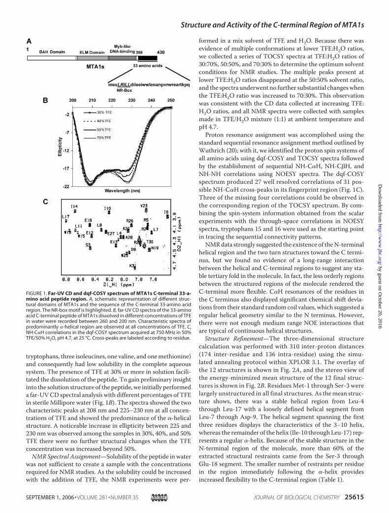
tryptophans, three isoleucines, one valine, and onemethionine)and consequently had low solubility in the complete aqueoussystem. The presence of TFE at 30% or more in solution facili-tated the dissolution of the peptide. To gain preliminary insightinto the solution structure of the peptide,we initially performeda far-UVCD spectral analysis with different percentages of TFEin sterile Millipore water (Fig. 1B). The spectra showed the twocharacteristic peaks at 208 nm and 225–230 nm at all concen-trations of TFE and showed the predominance of the �-helicalstructure. A noticeable increase in ellipticity between 225 and230 nmwas observed among the samples in 30%, 40%, and 50%TFE there were no further structural changes when the TFEconcentration was increased beyond 50%.NMRSpectral Assignment—Solubility of the peptide inwater
was not sufficient to create a sample with the concentrationsrequired for NMR studies. As the solubility could be increasedwith the addition of TFE, the NMR experiments were per-
formed in a mix solvent of TFE and H2O. Because there wasevidence of multiple conformations at lower TFE:H2O ratios,we collected a series of TOCSY spectra at TFE:H2O ratios of30:70%, 50:50%, and 70:30% to determine the optimum solventconditions for NMR studies. The multiple peaks present atlower TFE:H2O ratios disappeared at the 50:50% solvent ratio,and the spectra underwent no further substantial changeswhenthe TFE:H2O ratio was increased to 70:30%. This observationwas consistent with the CD data collected at increasing TFE:H2O ratios, and all NMR spectra were collected with samplesmade in TFE/H2O mixture (1:1) at ambient temperature andpH 4.7.Proton resonance assignment was accomplished using the
standard sequential resonance assignment method outlined byWuthrich (20); with it, we identified the proton spin systems ofall amino acids using dqf-COSY and TOCSY spectra followedby the establishment of sequential NH-C�H, NH-C�H, andNH-NH correlations using NOESY spectra. The dqf-COSYspectrum produced 27 well resolved correlations of 31 pos-sible NH-C�H cross-peaks in its fingerprint region (Fig. 1C).Three of the missing four correlations could be observed inthe corresponding region of the TOCSY spectrum. By com-bining the spin-system information obtained from the scalarexperiments with the through-space correlations in NOESYspectra, tryptophans 15 and 16 were used as the starting pointin tracing the sequential connectivity patterns.NMRdata strongly suggested the existence of theN-terminal
helical region and the two turn structures toward the C termi-nus, but we found no evidence of a long-range interactionbetween the helical and C-terminal regions to suggest any sta-ble tertiary fold in the molecule. In fact, the less orderly regionsbetween the structured regions of the molecule rendered theC-terminal more flexible. C�H resonances of the residues inthe C terminus also displayed significant chemical shift devia-tions from their standard randomcoil values, which suggested aregular helical geometry similar to the N terminus. However,there were not enough medium range NOE interactions thatare typical of continuous helical structures.Structure Refinement—The three-dimensional structure
calculation was performed with 310 inter-proton distances(174 inter-residue and 136 intra-residue) using the simu-lated annealing protocol within XPLOR 3.1. The overlay ofthe 12 structures is shown in Fig. 2A, and the stereo view ofthe energy-minimized mean structure of the 12 final struc-tures is shown in Fig. 2B. Residues Met-1 through Ser-3 werelargely unstructured in all final structures. As themean struc-ture shows, there was a stable helical region from Leu-4through Leu-17 with a loosely defined helical segment fromLeu-7 through Asp-9. The helical segment spanning the firstthree residues displays the characteristics of the 3–10 helix,whereas the remainder of the helix (Ile-10 through Leu-17) rep-resents a regular �-helix. Because of the stable structure in theN-terminal region of the molecule, more than 60% of theextracted structural restraints came from the Ser-3 throughGlu-18 segment. The smaller number of restraints per residuein the region immediately following the �-helix providesincreased flexibility to the C-terminal region (Table 1).
FIGURE 1. Far-UV CD and dqf-COSY spectrum of MTA1s C-terminal 33-a-mino acid peptide region. A, schematic representation of different struc-tural domains of MTA1s and the sequence of the C-terminal 33-amino acidregion. The NR-box motif is highlighted. B, far-UV CD spectra of the 33-aminoacid C-terminal peptide of MTA1s dissolved in different concentrations of TFEin water were recorded between 260 and 200 nm. Characteristic spectra ofpredominantly �-helical region are observed at all concentrations of TFE. C,NH-C�H correlations in the dqf-COSY spectrum acquired at 750 MHz in 50%TFE/50% H2O, pH 4.7, at 25 °C. Cross-peaks are labeled according to residue.
Structure and Activity of the C-terminal Region of MTA1s
SEPTEMBER 1, 2006 • VOLUME 281 • NUMBER 35 JOURNAL OF BIOLOGICAL CHEMISTRY 25615
by guest on October 20, 2016
http://ww
w.jbc.org/
Dow
nloaded from
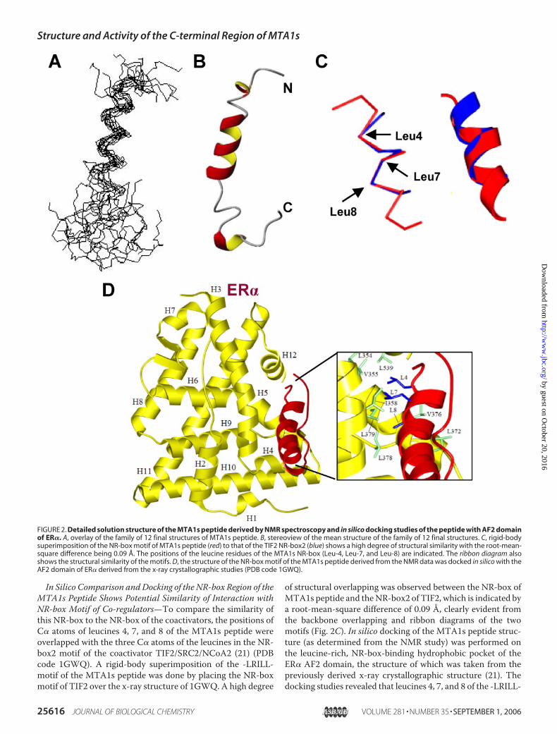
In Silico Comparison andDocking of the NR-box Region of theMTA1s Peptide Shows Potential Similarity of Interaction withNR-box Motif of Co-regulators—To compare the similarity ofthis NR-box to the NR-box of the coactivators, the positions ofC� atoms of leucines 4, 7, and 8 of the MTA1s peptide wereoverlapped with the three C� atoms of the leucines in the NR-box2 motif of the coactivator TIF2/SRC2/NCoA2 (21) (PDBcode 1GWQ). A rigid-body superimposition of the -LRILL-motif of the MTA1s peptide was done by placing the NR-boxmotif of TIF2 over the x-ray structure of 1GWQ. A high degree
of structural overlapping was observed between the NR-box ofMTA1s peptide and theNR-box2 of TIF2, which is indicated bya root-mean-square difference of 0.09 Å, clearly evident fromthe backbone overlapping and ribbon diagrams of the twomotifs (Fig. 2C). In silico docking of the MTA1s peptide struc-ture (as determined from the NMR study) was performed onthe leucine-rich, NR-box-binding hydrophobic pocket of theER� AF2 domain, the structure of which was taken from thepreviously derived x-ray crystallographic structure (21). Thedocking studies revealed that leucines 4, 7, and 8 of the -LRILL-
FIGURE 2. Detailed solution structure of the MTA1s peptide derived by NMR spectroscopy and in silico docking studies of the peptide with AF2 domainof ER�. A, overlay of the family of 12 final structures of MTA1s peptide. B, stereoview of the mean structure of the family of 12 final structures. C, rigid-bodysuperimposition of the NR-box motif of MTA1s peptide (red) to that of the TIF2 NR-box2 (blue) shows a high degree of structural similarity with the root-mean-square difference being 0.09 Å. The positions of the leucine residues of the MTA1s NR-box (Leu-4, Leu-7, and Leu-8) are indicated. The ribbon diagram alsoshows the structural similarity of the motifs. D, the structure of the NR-box motif of the MTA1s peptide derived from the NMR data was docked in silico with theAF2 domain of ER� derived from the x-ray crystallographic studies (PDB code 1GWQ).
Structure and Activity of the C-terminal Region of MTA1s
25616 JOURNAL OF BIOLOGICAL CHEMISTRY VOLUME 281 • NUMBER 35 • SEPTEMBER 1, 2006
by guest on October 20, 2016
http://ww
w.jbc.org/
Dow
nloaded from
motif of the MTA1s peptide could interact with Leu-354, Val-355, and Ile-358; Leu-372, Val-376, Leu-378, and Leu-379; andLeu-539, Leu-540, Leu-541, and Leu-544 of the ER� AF2domain, respectively (Fig. 2D, enlarged in the inset). Thesestudies showed the potential similarity of the NR-box motif ofthe MTA1s peptide to the similar motif of a coregulator andexplained the strong binding ability of the MTA1s 33-aminoacid region to ER�.Overexpression of MTA1s Peptide Represses ER� Transacti-
vation Function—The C-terminal 33-amino acid region ofMTA1s has a unique sequence and an NR-box motif (-LRILL-)that interacts strongly with the AF2 domain of ER�. MTA1smutant with the 33-amino acid region or NR-boxmotif deletedwas incapable of binding to ER� and sequestering it in the cyto-plasm losing its efficacy as an inhibitor of ER� transactivationfunctions (9).Next, we determined the effect of the 33-amino acid peptide
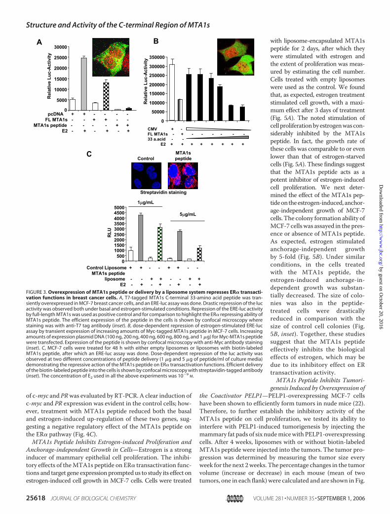
on the ER� nuclear functions by an ERE-luc assay, with tran-sient overexpression of theT7-tagged peptide inMCF-7 cells. Adrastic repression of ER� was observed, in the ERE-luc assayboth in the presence and absence of estrogen (Fig. 3A). Therepression caused by overexpression of full-length MTA1s wasused as a positive control. Overexpression of the T7-taggedpeptide in the MCF-7 cells was shown by confocal immunoflu-orescence microscopy (Fig. 3A, inset).
In a similar experiment, different concentrations of thetagged peptide were transiently expressed in MCF-7 cells, andER� transactivation functions were repressed in a dose-dependent manner (Fig. 3B). In another assay, MCF-7 cellswere treated with two different doses of liposome-encapsu-lated, biotin-labeled peptide, and an ERE-luc assay was per-formed. At a peptide concentration of 1 �g/ml, a substantialrepression of ER transactivation was observed, and this effectwas further enhanced at the concentration of 5�g/ml (Fig. 3C).The successful delivery of the biotin-labeled peptide into thecells was evident by streptavidin staining of the cells, which isshown in the figure inset along with cells treated with controlempty liposomes for comparison. In brief, we found that the33-amino acid region of MTA1s strongly repressed ER� trans-activation functions.
MTA1s 33-Amino Acid Region Binds to ER�, Competes forBinding Sites with Coregulators, and Negatively Regulates ER�Target Genes—In cells, MTA1s is localized to the cytoplasmand functions as a repressor of ER� transactivation by bindingand sequestering it in the cytoplasm. The strong repression ofER transactivation by the 33-amino acid peptide, which waslocalized in both the cytoplasm and the nucleus (as evident bythe staining shown in Fig. 3), indicated that the MTA1s 33-a-mino acid peptide acts as a repressor of ER through a mecha-nism different from that employed by the full-length MTA1s.As mentioned earlier, deletion of the NR-box motif present
in the 33-amino acid region resulted inMTA1s losing its abilityto bind and inhibit ER� (9). Comparison studies showed thattheNRbox of theMTA1s peptide had similar potential to inter-act with the AF2 domain of ER� (Fig. 2D), which raised thepossibility that the peptide inhibits ER� by binding to the AF2domain and potentially competes for binding sites with thecoactivator proteins.To explore this possibility, we next performed an ER trans-
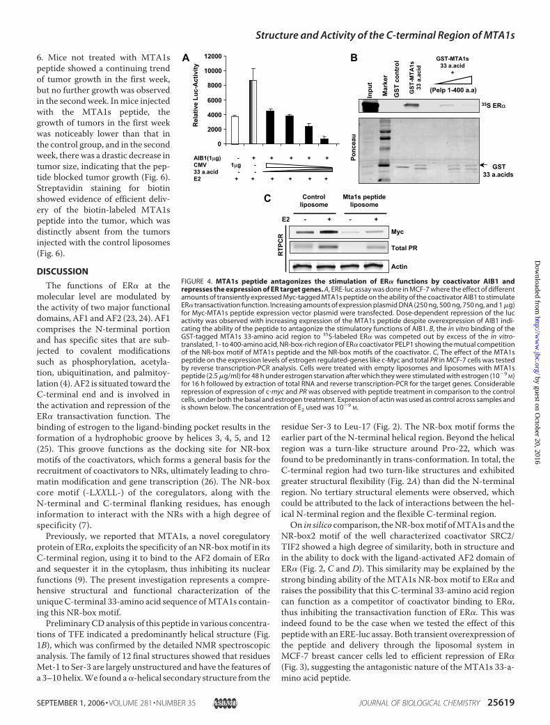
activation assay in MCF-7 cells and evaluated the effect of theMTA1s peptide on the stimulation of ER� transactivation func-tion by AIB1, a coactivator belonging to the p160 family. Tran-sient overexpression of AIB1 in MCF-7 cells stimulated ER�activity by �2-fold (Fig. 4A). Transient expression of Myc-tagged MTA1s peptide, along with overexpression of AIB1,blocked the effect of AIB1 and resulted in a dose-dependentrepression of ERE-luc activity (Fig. 4A), suggesting that theMTA1s peptide may antagonize the effect of AIB1 by compet-ingwith it for binding sites on ER�. This hypothesis was furthersupported by in vitro binding studies, where the binding of theGST-tagged MTA1s 33-amino acid peptide to ER� was com-peted out by the NR-box-rich region of the recently character-ized coactivator of ER, PELP1.PELP1 was initially identified as a novel coactivator of ER�
and found to be a powerful potentiator of ER� transactivationfunctions. PELP1 has a total of nine NR-box motifs, seven ofwhich are present in the N-terminal 400-amino acid region (6).TheGST-taggedMTA1s 33-amino acid peptide showed strongbinding to full-length in vitro-translated 35S-labeled-ER� (Fig.4B). This binding could be competed out by introducing thein vitro-translated, unlabeled, NR-box-rich, 1- to 400-aminoacid N-terminal region of PELP1. The presence of the PELP1N-terminal region blocked the binding of the GST-taggedMTA1s peptide to ER� (Fig. 4B). The direct in vitro effect of thepeptide on the binding of coactivators to ER� could not betested because of the low solubility of the peptide in solution, asmentioned earlier. In the in vitro binding study, the binding ofGST-tagged MTA1s to ER� was abrogated by the presence ofthe PELP1 NR-box-rich region, which indicated the mutualcompetition of the MTA1s peptide with the NR-box motifs ofthe coregulator for the binding site.The repressive effect of MTA1s peptide on the transactiva-
tion activity of ER� was further evidenced by the effect of thepeptide on the transcription of ER� target genes. The expres-sion of two target genes, c-myc and PRwas examined inMCF-7cells (Fig. 4C). Cells were treated with liposome-encapsulatedMTA1s peptide (2.5�g/ml) or empty liposomes for 2 days priorto estrogen treatment. Total RNAwas extracted, and the status
TABLE 1Summary of input restraint for MD calculations and structuralstatistics of the final structureThe parameters are given for the average structure of ensemble of 20 lowest energystructures. There were no NOE violations �0.2 Å or dihedral angle restraint viola-tions of �5°.
Inter proton distance restraintsIntra-residue 136Sequential 113Medium range 61Total 310
Root-mean-square deviation from ideal geometryBond lengths (Å) 0.021Bond angles (°) 2.4
Ramachandran plot analysisResidues in most favorable regions (%) 62.1Residues in additional allowed regions (%) 17.2Residues in generously allowed regions (%) 13.8Residues in disallowed regions (%) 6.9
Root-mean-square deviation (residues 4–16 to the mean)Backbone (Å) 1.01Heavy atoms (Å) 1.67
Structure and Activity of the C-terminal Region of MTA1s
SEPTEMBER 1, 2006 • VOLUME 281 • NUMBER 35 JOURNAL OF BIOLOGICAL CHEMISTRY 25617
by guest on October 20, 2016
http://ww
w.jbc.org/
Dow
nloaded from
of c-myc and PRwas evaluated by RT-PCR. A clear induction ofc-myc and PR expression was evident in the control cells; how-ever, treatment with MTA1s peptide reduced both the basaland estrogen-induced up-regulation of these two genes, sug-gesting a negative regulatory effect of the MTA1s peptide onthe ER� pathway (Fig. 4C).MTA1s Peptide Inhibits Estrogen-induced Proliferation and
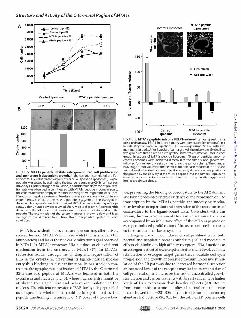
Anchorage-independent Growth in Cells—Estrogen is a stronginducer of mammary epithelial cell proliferation. The inhibi-tory effects of theMTA1s peptide on ER� transactivation func-tions and target gene expressionpromptedus to study its effect onestrogen-induced cell growth in MCF-7 cells. Cells were treated
with liposome-encapsulated MTA1speptide for 2 days, after which theywere stimulated with estrogen andthe extent of proliferation was meas-ured by estimating the cell number.Cells treated with empty liposomeswere used as the control. We foundthat, as expected, estrogen treatmentstimulated cell growth, with a maxi-mum effect after 3 days of treatment(Fig. 5A). The noted stimulation ofcell proliferationbyestrogenwascon-siderably inhibited by the MTA1speptide. In fact, the growth rate ofthese cells was comparable to or evenlower than that of estrogen-starvedcells (Fig. 5A). These findings suggestthat the MTA1s peptide acts as apotent inhibitor of estrogen-inducedcell proliferation. We next deter-mined the effect of the MTA1s pep-tideontheestrogen-induced, anchor-age-independent growth of MCF-7cells.The colony formation ability ofMCF-7 cells was assayed in the pres-ence or absence of MTA1s peptide.As expected, estrogen stimulatedanchorage-independent growthby 5-fold (Fig. 5B). Under similarconditions, in the cells treatedwith the MTA1s peptide, theestrogen-induced anchorage-in-dependent growth was substan-tially decreased. The size of colo-nies was also in the peptide-treated cells were drasticallyreduced in comparison with thesize of control cell colonies (Fig.5B, inset). Together, these studiessuggest that the MTA1s peptideeffectively inhibits the biologicaleffects of estrogen, which may bedue to its inhibitory effect on ERtransactivation activity.MTA1s Peptide Inhibits Tumori-
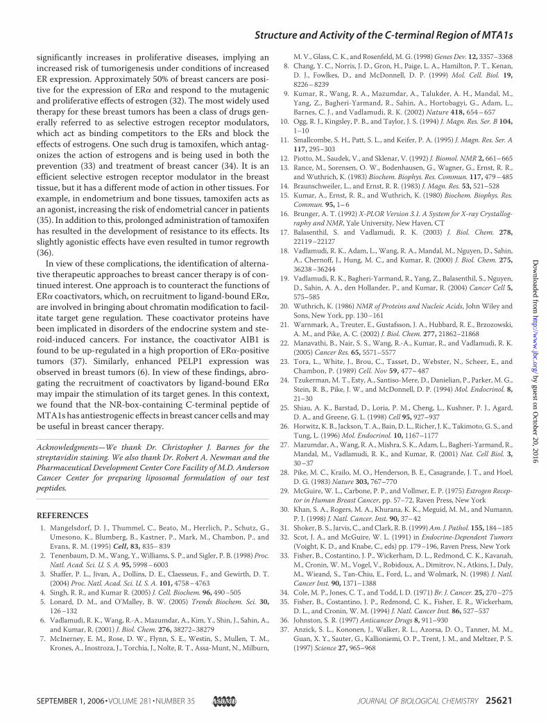
genesis Induced byOverexpression ofthe Coactivator PELP1—PELP1-overexpressing MCF-7 cellshave been shown to efficiently form tumors in nude mice (22).Therefore, to further establish the inhibitory activity of theMTA1s peptide on cell proliferation, we tested its ability tointerfere with PELP1-induced tumorigenesis by injecting themammary fat pads of six nudemicewith PELP1-overexpressingcells. After 4 weeks, liposomes with or without biotin-labeledMTA1s peptide were injected into the tumors. The tumor pro-gression was determined by measuring the tumor size everyweek for the next 2weeks. The percentage changes in the tumorvolume (increase or decrease) in each mouse (mean of twotumors, one in each flank)were calculated and are shown in Fig.
FIGURE 3. Overexpression of MTA1s peptide or delivery by a liposome system represses ER� transacti-vation functions in breast cancer cells. A, T7-tagged MTA1s C-terminal 33-amino acid peptide was tran-siently overexpressed in MCF-7 breast cancer cells, and an ERE-luc assay was done. Drastic repression of the lucactivity was observed both under basal and estrogen-stimulated conditions. Repression of the ERE-luc activityby full-length MTA1s was used as positive control and for comparison to highlight the ER� repressing ability ofMTA1s peptide. The efficient expression of the peptide in the cells is shown by confocal microscopy wherestaining was with anti-T7 tag antibody (inset). B, dose-dependent repression of estrogen-stimulated ERE-lucassay by transient expression of increasing amounts of Myc-tagged MTA1s peptide in MCF-7 cells. Increasingamounts of expression plasmid DNA (100 ng, 200 ng, 400 ng, 600 ng, 800 ng, and 1 �g) for Myc-MTA1s peptidewere transfected. Expression of the peptide is shown by confocal microscopy with anti-Myc antibody staining(inset). C, MCF-7 cells were treated for 48 h with either empty liposomes or liposomes with biotin-labeledMTA1s peptide, after which an ERE-luc assay was done. Dose-dependent repression of the luc activity wasobserved at two different concentrations of peptide delivery (1 �g and 5 �g of peptide/ml of culture media)demonstrating the repressive action of the MTA1s peptide on ER� transactivation functions. Efficient deliveryof the biotin-labeled peptide into the cells is shown by confocal microscopy with streptavidin-tagged antibody(inset). The concentration of E2 used in all the above experiments was 10�9
M.
Structure and Activity of the C-terminal Region of MTA1s
25618 JOURNAL OF BIOLOGICAL CHEMISTRY VOLUME 281 • NUMBER 35 • SEPTEMBER 1, 2006
by guest on October 20, 2016
http://ww
w.jbc.org/
Dow
nloaded from
6. Mice not treated with MTA1speptide showed a continuing trendof tumor growth in the first week,but no further growth was observedin the second week. Inmice injectedwith the MTA1s peptide, thegrowth of tumors in the first weekwas noticeably lower than that inthe control group, and in the secondweek, therewas a drastic decrease intumor size, indicating that the pep-tide blocked tumor growth (Fig. 6).Streptavidin staining for biotinshowed evidence of efficient deliv-ery of the biotin-labeled MTA1speptide into the tumor, which wasdistinctly absent from the tumorsinjected with the control liposomes(Fig. 6).
DISCUSSION
The functions of ER� at themolecular level are modulated bythe activity of two major functionaldomains, AF1 andAF2 (23, 24). AF1comprises the N-terminal portionand has specific sites that are sub-jected to covalent modificationssuch as phosphorylation, acetyla-tion, ubiquitination, and palmitoy-lation (4). AF2 is situated toward theC-terminal end and is involved inthe activation and repression of theER� transactivation function. Thebinding of estrogen to the ligand-binding pocket results in theformation of a hydrophobic groove by helices 3, 4, 5, and 12(25). This groove functions as the docking site for NR-boxmotifs of the coactivators, which forms a general basis for therecruitment of coactivators to NRs, ultimately leading to chro-matin modification and gene transcription (26). The NR-boxcore motif (-LXXLL-) of the coregulators, along with theN-terminal and C-terminal flanking residues, has enoughinformation to interact with the NRs with a high degree ofspecificity (7).Previously, we reported that MTA1s, a novel coregulatory
protein of ER�, exploits the specificity of anNR-boxmotif in itsC-terminal region, using it to bind to the AF2 domain of ER�and sequester it in the cytoplasm, thus inhibiting its nuclearfunctions (9). The present investigation represents a compre-hensive structural and functional characterization of theunique C-terminal 33-amino acid sequence ofMTA1s contain-ing this NR-box motif.Preliminary CD analysis of this peptide in various concentra-
tions of TFE indicated a predominantly helical structure (Fig.1B), which was confirmed by the detailed NMR spectroscopicanalysis. The family of 12 final structures showed that residuesMet-1 to Ser-3 are largely unstructured and have the features ofa 3–10helix.We found a�-helical secondary structure from the
residue Ser-3 to Leu-17 (Fig. 2). The NR-box motif forms theearlier part of the N-terminal helical region. Beyond the helicalregion was a turn-like structure around Pro-22, which wasfound to be predominantly in trans-conformation. In total, theC-terminal region had two turn-like structures and exhibitedgreater structural flexibility (Fig. 2A) than did the N-terminalregion. No tertiary structural elements were observed, whichcould be attributed to the lack of interactions between the hel-ical N-terminal region and the flexible C-terminal region.On in silico comparison, theNR-boxmotif ofMTA1s and the
NR-box2 motif of the well characterized coactivator SRC2/TIF2 showed a high degree of similarity, both in structure andin the ability to dock with the ligand-activated AF2 domain ofER� (Fig. 2, C and D). This similarity may be explained by thestrong binding ability of the MTA1s NR-box motif to ER� andraises the possibility that this C-terminal 33-amino acid regioncan function as a competitor of coactivator binding to ER�,thus inhibiting the transactivation function of ER�. This wasindeed found to be the case when we tested the effect of thispeptidewith anERE-luc assay. Both transient overexpression ofthe peptide and delivery through the liposomal system inMCF-7 breast cancer cells led to efficient repression of ER�(Fig. 3), suggesting the antagonistic nature of the MTA1s 33-a-mino acid peptide.
FIGURE 4. MTA1s peptide antagonizes the stimulation of ER� functions by coactivator AIB1 andrepresses the expression of ER target genes. A, ERE-luc assay was done in MCF-7 where the effect of differentamounts of transiently expressed Myc-tagged MTA1s peptide on the ability of the coactivator AIB1 to stimulateER� transactivation function. Increasing amounts of expression plasmid DNA (250 ng, 500 ng, 750 ng, and 1 �g)for Myc-MTA1s peptide expression vector plasmid were transfected. Dose-dependent repression of the lucactivity was observed with increasing expression of the MTA1s peptide despite overexpression of AIB1 indi-cating the ability of the peptide to antagonize the stimulatory functions of AIB1. B, the in vitro binding of theGST-tagged MTA1s 33-amino acid region to 35S-labeled ER� was competed out by excess of the in vitro-translated, 1- to 400-amino acid, NR-box-rich region of ER� coactivator PELP1 showing the mutual competitionof the NR-box motif of MTA1s peptide and the NR-box motifs of the coactivator. C, The effect of the MTA1speptide on the expression levels of estrogen regulated-genes like c-Myc and total PR in MCF-7 cells was testedby reverse transcription-PCR analysis. Cells were treated with empty liposomes and liposomes with MTA1speptide (2.5 �g/ml) for 48 h under estrogen starvation after which they were stimulated with estrogen (10�9
M)for 16 h followed by extraction of total RNA and reverse transcription-PCR for the target genes. Considerablerepression of expression of c-myc and PR was observed with peptide treatment in comparison to the controlcells, under both the basal and estrogen treatment. Expression of actin was used as control across samples andis shown below. The concentration of E2 used was 10�9
M.
Structure and Activity of the C-terminal Region of MTA1s
SEPTEMBER 1, 2006 • VOLUME 281 • NUMBER 35 JOURNAL OF BIOLOGICAL CHEMISTRY 25619
by guest on October 20, 2016
http://ww
w.jbc.org/
Dow
nloaded from
MTA1s was identified as a naturally occurring, alternativelyspliced form of MTA1 (715 amino acids) that is smaller (430amino acids) and lacks the nuclear localization signal observedin MTA1 (9). MTA1s represses ER� but does so via a differentmechanism from the one used by MTA1 (27). In MTA1s,repression occurs through the binding and sequestration ofER� in the cytoplasm, preventing its ligand-induced nuclearentry thus blocking its nuclear function. In our study, in con-trast to the cytoplasmic localization of MTA1s, the C-terminal33-amino acid peptide of MTA1s was localized in both thecytoplasm and nucleus (Fig. 3), where nuclear entry might beattributed to its small size and passive accumulation in thenucleus. The efficient repression of ERE-luc by this peptide ledus to speculate whether this could be brought about by thepeptide functioning as a mimetic of NR-boxes of the coactiva-
tor, preventing the binding of coactivators to the AF2 domain.We found proof-of-principle evidence of the repression of ER�transcription by the MTA1s peptide; the underlying mecha-nism involves competition andprevention of the recruitment ofcoactivators to the ligand-bound ER�. Consistent with thisnotion, the down-regulation of ER� transactivation activitywasaccompanied by an inhibitory effect of the MTA1s peptide onestrogen-induced proliferation of breast cancer cells in tissueculture- and animal-based systems.Estrogens are a major inducer of cell proliferation in both
normal and neoplastic breast epithelium (28) and mediate itseffects via binding to high affinity receptors. ER� functions asan estrogen-activated transcription factor and is involved in thestimulation of estrogen target genes that modulate cell cycleprogression and growth of breast epithelium. Excessive stimu-lation of the ER pathway due to increased hormonal secretionor increased levels of the receptor may lead to augmentation ofcell proliferation and increases the risk of uncontrolled growthstimulation and cancer. Patients with breast cancer have higherlevels of ER� expression than healthy subjects (29). Resultsfrom immunohistochemical studies of normal and canceroustissue showed that �20–30% of cells in the normal mammarygland are ER-positive (30, 31), but the ratio of ER-positive cells
FIGURE 5. MTA1s peptide inhibits estrogen-induced cell proliferationand anchorage-independent growth. A, the estrogen-stimulated prolifer-ation of MCF-7 cells treated with empty or MTA1s peptide liposomes (5 �g/mlpeptide) was tested by estimating the total cell count every 24 h for 4 consec-utive days. Under estrogen stimulation, a considerable decrease of prolifera-tion rate was observed in cells treated with MTA1s peptide in comparison tothe cells treated with empty liposomes showing down-regulation of cell pro-liferation on peptide treatment. Results shown are an average of two differentexperiments. B, effect of the MTA1s peptide (5 �g/ml) on the estrogen-in-duced anchorage-independent growth of MCF-7 cells was tested by soft agarassay. Colony numbers were counted after 3 weeks of growth. A considerabledecrease of the colony size and number was observed in cells treated with thepeptide. The quantitation of the colony number is shown below and is anaverage of five different fields from three independent plates for eachcondition.
FIGURE 6. MTA1s peptide inhibits PELP1-induced tumor growth in axenograft assay. PELP1-induced tumors were generated by xenograft in 6female athymic mice by injecting PELP1-overexpressing MCF-7 cells intomammary fat pads. After 4 weeks of tumor growth the mice were divided intotwo groups of three each so as to get the same total tumor volumes in eachgroup. Injections of MTA1s peptide liposome (40 �g of peptide/tumor) orempty liposomes were delivered directly into the tumors, and growth wasfollowed for the next 2 weeks by measuring the tumor volume. The changesin average tumor volume from the two tumors in each mouse for the first andsecond week after the liposomal injections clearly show a down-regulation ofthe growth by the delivery of the MTA1s peptide into the tumors. Represent-ative pictures of the tumor sections stained with streptavidin-tagged anti-bodies are shown above.
Structure and Activity of the C-terminal Region of MTA1s
25620 JOURNAL OF BIOLOGICAL CHEMISTRY VOLUME 281 • NUMBER 35 • SEPTEMBER 1, 2006
by guest on October 20, 2016
http://ww
w.jbc.org/
Dow
nloaded from
significantly increases in proliferative diseases, implying anincreased risk of tumorigenesis under conditions of increasedER expression. Approximately 50% of breast cancers are posi-tive for the expression of ER� and respond to the mutagenicand proliferative effects of estrogen (32). The most widely usedtherapy for these breast tumors has been a class of drugs gen-erally referred to as selective estrogen receptor modulators,which act as binding competitors to the ERs and block theeffects of estrogens. One such drug is tamoxifen, which antag-onizes the action of estrogens and is being used in both theprevention (33) and treatment of breast cancer (34). It is anefficient selective estrogen receptor modulator in the breasttissue, but it has a different mode of action in other tissues. Forexample, in endometrium and bone tissues, tamoxifen acts asan agonist, increasing the risk of endometrial cancer in patients(35). In addition to this, prolonged administration of tamoxifenhas resulted in the development of resistance to its effects. Itsslightly agonistic effects have even resulted in tumor regrowth(36).In view of these complications, the identification of alterna-
tive therapeutic approaches to breast cancer therapy is of con-tinued interest. One approach is to counteract the functions ofER� coactivators, which, on recruitment to ligand-bound ER�,are involved in bringing about chromatin modification to facil-itate target gene regulation. These coactivator proteins havebeen implicated in disorders of the endocrine system and ste-roid-induced cancers. For instance, the coactivator AIB1 isfound to be up-regulated in a high proportion of ER�-positivetumors (37). Similarly, enhanced PELP1 expression wasobserved in breast tumors (6). In view of these findings, abro-gating the recruitment of coactivators by ligand-bound ER�may impair the stimulation of its target genes. In this context,we found that the NR-box-containing C-terminal peptide ofMTA1s has antiestrogenic effects in breast cancer cells andmaybe useful in breast cancer therapy.
Acknowledgments—We thank Dr. Christopher J. Barnes for thestreptavidin staining. We also thank Dr. Robert A. Newman and thePharmaceutical Development Center Core Facility of M.D. AndersonCancer Center for preparing liposomal formulation of our testpeptides.
REFERENCES1. Mangelsdorf, D. J., Thummel, C., Beato, M., Herrlich, P., Schutz, G.,
Umesono, K., Blumberg, B., Kastner, P., Mark, M., Chambon, P., andEvans, R. M. (1995) Cell, 83, 835–839
2. Tenenbaum, D.M.,Wang, Y.,Williams, S. P., and Sigler, P. B. (1998) Proc.Natl. Acad. Sci. U. S. A. 95, 5998–6003
3. Shaffer, P. L., Jivan, A., Dollins, D. E., Claesseus, F., and Gewirth, D. T.(2004) Proc. Natl. Acad. Sci. U. S. A. 101, 4758–4763
4. Singh, R. R., and Kumar R. (2005) J. Cell. Biochem. 96, 490–5055. Lonard, D. M., and O’Malley, B. W. (2005) Trends Biochem. Sci. 30,
126–1326. Vadlamudi, R. K., Wang, R.-A., Mazumdar, A., Kim, Y., Shin, J., Sahin, A.,
and Kumar, R. (2001) J. Biol. Chem. 276, 38272–382797. McInerney, E. M., Rose, D. W., Flynn, S. E., Westin, S., Mullen, T. M.,
Krones, A., Inostroza, J., Torchia, J., Nolte, R. T., Assa-Munt, N., Milburn,
M. V., Glass, C. K., and Rosenfeld,M. G. (1998)Genes Dev. 12, 3357–33688. Chang, Y. C., Norris, J. D., Gron, H., Paige, L. A., Hamilton, P. T., Kenan,
D. J., Fowlkes, D., and McDonnell, D. P. (1999) Mol. Cell. Biol. 19,8226–8239
9. Kumar, R., Wang, R. A., Mazumdar, A., Talukder, A. H., Mandal, M.,Yang, Z., Bagheri-Yarmand, R., Sahin, A., Hortobagyi, G., Adam, L.,Barnes, C. J., and Vadlamudi, R. K. (2002) Nature 418, 654–657
10. Ogg, R. J., Kingsley, P. B., and Taylor, J. S. (1994) J. Magn. Res. Ser. B 104,1–10
11. Smallcombe, S. H., Patt, S. L., and Keifer, P. A. (1995) J. Magn. Res. Ser. A117, 295–303
12. Piotto, M., Saudek, V., and Sklenar, V. (1992) J. Biomol. NMR 2, 661–66513. Rance, M., Sorensen, O. W., Bodenhausen, G., Wagner, G., Ernst, R. R.,
and Wuthrich, K. (1983) Biochem. Biophys. Res. Commun. 117, 479–48514. Braunschweiler, L., and Ernst, R. R. (1983) J. Magn. Res. 53, 521–52815. Kumar, A., Ernst, R. R., and Wuthrich, K. (1980) Biochem. Biophys. Res.
Commun. 95, 1–616. Brunger, A. T. (1992) X-PLOR Version 3.1. A System for X-ray Crystallog-
raphy and NMR, Yale University, New Haven, CT17. Balasenthil, S. and Vadlamudi, R. K. (2003) J. Biol. Chem. 278,
22119–2212718. Vadlamudi, R. K., Adam, L., Wang, R. A., Mandal, M., Nguyen, D., Sahin,
A., Chernoff, J., Hung, M. C., and Kumar, R. (2000) J. Biol. Chem. 275,36238–36244
19. Vadlamudi, R. K., Bagheri-Yarmand, R., Yang, Z., Balasenthil, S., Nguyen,D., Sahin, A. A., den Hollander, P., and Kumar, R. (2004) Cancer Cell 5,575–585
20. Wuthrich, K. (1986) NMR of Proteins and Nucleic Acids, John Wiley andSons, New York, pp. 130–161
21. Warnmark, A., Treuter, E., Gustafsson, J. A., Hubbard, R. E., Brzozowski,A. M., and Pike, A. C. (2002) J. Biol. Chem. 277, 21862–21868
22. Manavathi, B., Nair, S. S., Wang, R.-A., Kumar, R., and Vadlamudi, R. K.(2005) Cancer Res. 65, 5571–5577
23. Tora, L., White, J., Brou, C., Tasset, D., Webster, N., Scheer, E., andChambon, P. (1989) Cell. Nov 59, 477–487
24. Tzukerman,M. T., Esty, A., Santiso-Mere, D., Danielian, P., Parker, M. G.,Stein, R. B., Pike, J. W., and McDonnell, D. P. (1994) Mol. Endocrinol. 8,21–30
25. Shiau, A. K., Barstad, D., Loria, P. M., Cheng, L., Kushner, P. J., Agard,D. A., and Greene, G. L. (1998) Cell 95, 927–937
26. Horwitz, K. B., Jackson, T. A., Bain, D. L., Richer, J. K., Takimoto, G. S., andTung, L. (1996)Mol. Endocrinol. 10, 1167–1177
27. Mazumdar, A.,Wang, R. A.,Mishra, S. K., Adam, L., Bagheri-Yarmand, R.,Mandal, M., Vadlamudi, R. K., and Kumar, R. (2001) Nat. Cell Biol. 3,30–37
28. Pike, M. C., Krailo, M. O., Henderson, B. E., Casagrande, J. T., and Hoel,D. G. (1983) Nature 303, 767–770
29. McGuire, W. L., Carbone, P. P., and Vollmer, E. P. (1975) Estrogen Recep-tor in Human Breast Cancer, pp. 57–72, Raven Press, New York
30. Khan, S. A., Rogers, M. A., Khurana, K. K., Meguid, M. M., and Numann,P. J. (1998) J. Natl. Cancer. Inst. 90, 37–42
31. Shoker, B. S., Jarvis, C., andClark, R. B. (1999)Am. J. Pathol. 155, 184–18532. Scot, J. A., and McGuire, W. L. (1991) in Endocrine-Dependent Tumors
(Voight, K. D., and Knabe, C., eds) pp. 179–196, Raven Press, New York33. Fisher, B., Costantino, J. P., Wickerham, D. L., Redmond, C. K., Kavanah,
M., Cronin, W. M., Vogel, V., Robidoux, A., Dimitrov, N., Atkins, J., Daly,M., Wieand, S., Tan-Chiu, E., Ford, L., and Wolmark, N. (1998) J. Natl.Cancer Inst. 90, 1371–1388
34. Cole, M. P., Jones, C. T., and Todd, I. D. (1971) Br. J. Cancer. 25, 270–27535. Fisher, B., Costantino, J. P., Redmond, C. K., Fisher, E. R., Wickerham,
D. L., and Cronin, W. M. (1994) J. Natl. Cancer Inst. 86, 527–53736. Johnston, S. R. (1997) Anticancer Drugs 8, 911–93037. Anzick, S. L., Kononen, J., Walker, R. L., Azorsa, D. O., Tanner, M. M.,
Guan, X. Y., Sauter, G., Kallioniemi, O. P., Trent, J. M., and Meltzer, P. S.(1997) Science 27, 965–968
Structure and Activity of the C-terminal Region of MTA1s
SEPTEMBER 1, 2006 • VOLUME 281 • NUMBER 35 JOURNAL OF BIOLOGICAL CHEMISTRY 25621
by guest on October 20, 2016
http://ww
w.jbc.org/
Dow
nloaded from

VOLUME 281 (2006) PAGES 25612–25621DOI 10.1074/jbc.A113.604444
Solution structure and antiestrogenic activity of theunique C-terminal, NR-box motif-containing regionof MTA1s.Rajesh R. Singh, Kumaralal Kaluarachchi, Mingzhi Chen, Suresh K. Rayala,Seetharaman Balasenthil, Jianpeng Ma, and Rakesh Kumar
PAGE 25620:
The original inset in Fig. 6 did not originate from the correct experi-ment. It has been replaced by the results fromanew staining experimentfrom the same paraffin blocks. The replacement does not change theresults or interpretation of the data.
THE JOURNAL OF BIOLOGICAL CHEMISTRY VOL. 288, NO. 38, p. 27518, September 20, 2013© 2013 by The American Society for Biochemistry and Molecular Biology, Inc. Published in the U.S.A.
27518 JOURNAL OF BIOLOGICAL CHEMISTRY VOLUME 288 • NUMBER 38 • SEPTEMBER 20, 2013
ADDITIONS AND CORRECTIONS
Authors are urged to introduce these corrections into any reprints they distribute. Secondary (abstract) services are urged to carry notice ofthese corrections as prominently as they carried the original abstracts.

Seetharaman Balasenthil, Jianpeng Ma and Rakesh KumarRajesh R. Singh, Kumaralal Kaluarachchi, Mingzhi Chen, Suresh K. Rayala,
Motif-containing Region of MTA1sSolution Structure and Antiestrogenic Activity of the Unique C-terminal, NR-box
doi: 10.1074/jbc.M604444200 originally published online June 27, 20062006, 281:25612-25621.J. Biol. Chem.
10.1074/jbc.M604444200Access the most updated version of this article at doi:
Alerts:
When a correction for this article is posted•
When this article is cited•
to choose from all of JBC's e-mail alertsClick here
http://www.jbc.org/content/281/35/25612.full.html#ref-list-1
This article cites 31 references, 12 of which can be accessed free at
by guest on October 20, 2016
http://ww
w.jbc.org/
Dow
nloaded from