
sokratskapredavanja socraticlectures - Zdravstvena fakulteta
246
S O K R A T S K A P R E D A V A N J A S O C R A T I C L E C T U R E S 4. MEDNARODNI MINISIMPOZIJ, LJUBLJANA, 11.-12. DECEMBER 2020 4TH INTERNATIONAL MINISYMPOSIUM, LJUBLJANA, 11.-12. DECEMBER 2020 ZBORNIK RECENZIRANIH PRISPEVKOV PEER REVIEWED PROCEEDINGS UREDILA: VERONIKA KRALJ-IGLIČ EDITED BY: VERONIKA KRALJ-IGLIČ ZDRAVSTVENA FAKULTETA, UNIVERZA V LJUBLJANI FACULTY OF HEALTH SCIENCES, UNIVERSITY OF LJUBLJANA
-
Upload
khangminh22 -
Category
Documents
-
view
3 -
download
0
Transcript of sokratskapredavanja socraticlectures - Zdravstvena fakulteta

S O K R A T S K A P R E D A V A N J AS O C R A T I C L E C T U R E S
4. MEDNARODNI MINISIMPOZIJ, LJUBLJANA, 11.-12. DECEMBER 20204TH INTERNATIONAL MINISYMPOSIUM, LJUBLJANA, 11.-12. DECEMBER 2020
ZBORNIK RECENZIRANIH PRISPEVKOVPEER REVIEWED PROCEEDINGS
UREDILA: VERONIKA KRALJ-IGLIČEDITED BY: VERONIKA KRALJ-IGLIČ
ZDRAVSTVENA FAKULTETA, UNIVERZA V LJUBLJANIFACULTY OF HEALTH SCIENCES, UNIVERSITY OF LJUBLJANA

I
Sokratska predavanja 4. Mednarodni minisimpozij, Ljubljana, 11.-12. december, 2020 Zbornik recenziranih prispevkov Zbornik uredila: prof.dr. Veronika Kralj-Iglič, univ.dipl.fiz. Recenzenta: prof.dr.Rok Vengust, dr. med. in Karin Schara, dr. med. Izdala in založila: Univerza v Ljubljani, Zdravstvena fakulteta Oblikovanje in fotografije: Anna Romolo Slika na naslovnici: Samo Penič Ilustracije: Stran 78: Niko Kralj, stran 123: ulična slikarka iz Firenc, ostale: Marguerite de Saint Champs Socratic lectures 4th International Minisymposium, Ljubljana, December 11.-12., 2020 Peer Reviewed Proceedings Edited by Prof. Veronika Kralj-Iglič, Ph.D. Reviewers: Prof. Rok Vengust, M.D., Ph.D. and Karin Schara, M.D. Published by: University of Ljubljana, Faculty of Health Sciences Design and photos: Anna Romolo Image on the front page: Samo Penič Illustrations: Page 78: Niko Kralj, Page 123: Street artist from Florence, others: Marguerite de Saint Champs Publikacija je dostopna v PDF formatu na spletni strani: https://www.zf.uni-lj.si/images/stories/datoteke/Zalozba/Sokratska_2021.pdf Publication is available online in PDF format at: https://www.zf.uni-lj.si/images/stories/datoteke/Zalozba/Sokratska_2021.pdf Ljubljana, 2021 To delo je dosegljivo pod licenco Creative Commons Priznanje avtorstva 4.0
Mednarodna
This work is available under a Creative Commons Attribution 4.0 International
____________________
Kataložni zapis o publikaciji (CIP) pripravili v Narodni in univerzitetni knjižnici v Ljubljani
COBISS.SI-ID=51797507
ISBN 978-961-7112-02-3 (pdf)
_____________________

II
The members of the Organizing Committee of Socratic Lectures: Antonella Bongiovanni, Tjaša Griessler Bulc, Aleš Iglič, Veronika Kralj Iglič, Laura Sesma, Polonca Trebše
Program
Socratic Symposium December 11, 2020, 09:00 – 12:00 (Ljubljana time) Parallel Sections
Section 1: Emergent Environmental Polution Problems
organized and moderated by prof. Tjaša Griessler-Bulc and prof. Polonca Trebše
9.00 - 9.20 Franja Prosenc, Faculty of Health Sciences, University of Ljubljana, Slovenia: Microplastics in soil, problematics and quantification 9.20 – 9.40 Petra Procházková, Brno University of Technology, The Czech Republic: Effects of microplastics to aquatic enviromnent 9.40 – 10.00 Urška Šunta, Faculty of Health Sciences, University of Ljubljana, Slovenia Insights into microplastics: from physical and chemical characterisation to its potential as a vector 10.00 – 10.20 Nevena Antić, Institute of Chemistry, Technology and Metallurgy, Department of Chemistry, University of Belgrade, Serbia: Extreme climate condition and acid rain impact on land degradation 10.20 – 11.40 Irina Vasileva, M.V. Lomonosov Moscow State University, Organic Chemistry Department, Moscow, Russia: Peculiarities of the de novo mass spectrometry sequencing of bioactive peptides secreted by Slovenian Rana temporaria 10.40 – 11.00 Valentina Polanc Rutar, Faculty of Civil and Geodetic Engineering, University of Ljubljana, Slovenia, Green wall systems for greywater treatment
Section 2: Membrane Biophysics organized and moderated by prof. Aleš Iglič
9:00-9:30 Mitja Drab, Žiga Pandur, Samo Penič, Aleš Iglič, Veronika Kralj-Iglič, David Stopar. Monte Carlo studies of detergent solubilization of lipid bilayers 9:30-10:00 Luka Mesarec, Wojciech Góźdź, Iglič Aleš, Kralj-Iglič Veronika, Epifanio Virga, Samo Kralj: Stability of normal red blood cells explained by membrane's in-plane ordering 10:00-10:30 Jeel Raval, Aleš Iglič, Wojciech Góźdź: Study of shapes and shape transformations of vesicles induced by their adhesion to a rigid surface 10:30-11:00 Break 11:00-11:30 Raj Kumar Sadhu, Nir Gov, Aleš Iglič, Samo Penič. Modelling cellular spreading in the presence of curved membrane proteins and active cytoskeleton forces 11:30-12:00 Metka Benčina, Ita Junkar, Eva Levičnik, Veronika Kralj-Iglič, Miran Mozetič, Aleš Iglič: Improved hemocompatibility of Ti-based nanostructures

III
Section 3: Cellular Nanovesicles organized by partners of the VES4US project (a project funded by the FET Open Call of the Horizon2020) and moderated by prof. Veronika Kralj-Iglič
9:00-9:20 Ramila Mammadova, Gabriella Pocsfalvi - Nanovesicles from tomatoes: focus on the molecular cargo 9:20 – 9:40 Paulina Ramos Juarez, Gabriella Pocsfalvi - The human EV membranome: more ligand and adhesion proteins than receptors, what are the implications? 9:40-10:00 Marija Holcar, Jana Ferdin, Simona Sitar, Magda Tušek-Žnidarič, Vita Dolžan, Ana Plemenitaš, Ema Žagar, Metka Lenassi: Method for reliable enrichment of plasma extracellular vesicles for biomarker discovery
10:00-10:30 Break 10:30-10:50 Zala Jan, Mitja Drab, Damjana Drobne, Apolonija Bedina Zavec, Mojca Benčina, Barbara Drašler, Matej Hočevar, Judita Lea Krek, Ljubiša Pađen, Manca Pajnič, Neža Repar, Boštjan Šimunič, Roman Štukelj, Veronika Kralj-Iglič: Impact of physical effort on cellular nanovesicles concentration in blood isolates 10:50-11.10 Domen Vozel, Nejc Steiner, Darja Božič, Zala Jan, Marko Jeran, Manca Pajnič, Ljubiša Pađen, Aleš Iglič, Veronika Kralj-Iglič, Saba Battelino: Preparation of platelet- and extracellular vesicle-rich gel and its role in management of cerebrospinal fluid leak in skull-base surgery 11.10-11:30 Ryan Garry, Dave Weitz: Harnessing nature communication system: Engineering exosomes
12:00 – 12:15 Cultural program Slovenian folk song, I have made up something, trombone: Emil Somun, piano: Elena Startseva Somun Elvis Presley, Love me tender: trombone: Emil Somun, piano: Elena Starteseva Somun Johann Sebastian Bach, Sarabande from from Partita for Flute Solo in A minor BWV 1013, flute: Anita Prelovšek Carlos Gardel: Poe una cabesa, violin: Vittorio Sbordone Christina Perry: A thousand years, piano: Elena Startseva Somun
12:15 – 14:00 Joint Section Cross-Domain Communication for all participants of the Symposium and Lectures, with discussion of posters
Socratic Lectures, December 12, 2020, 16:00 – 18:00 (Ljubljana time)
https://uni-lj-si.zoom.us/j/97287958341 Meeting ID: 972 8795 8341 Find your local number: https://uni-lj-si.zoom.us/u/aIClELqVM 16:00-16:30 Leonid Margolis, National Institute of Health, Bathesda, U.S.A.: Viruses and extracellular vesicles – similarities and differences 16:30-17:00 Gabriella Pocsfalvi, National Research Council of Italy, Naples, Italy: Use of extracellular vesicles in COVID-19 17:00-17:30 Vesna Spasovski, Institute of Molecular Biology, Belgrade, Serbia: Stem cell extracellular vesicles 17:30-18:00 Duško Spasovski, Institute for Othopaedic Surgery, Clinics Banjica, Belgrade, Serbia: Mechanisms of cartilage degeneration 18.00-18.30 Bojana Beović, University Medical Centre Ljubljana, Slovenia: Management of COVID-19 in Slovenia 18.30-19.00 Cultural program Pjotr Iljič Čajkovski, Seasons, June, piano: Elena Startseva Somun Johann Sebastian Bach, Badinerie from Suite in B major, BWV 1067, flute: Anita Prelovšek Nicola Piovani, La vita e bella, violin: Vittorio Sbordone

IV
EDITORIAL
Year 2020 was especially fruitful for Socratic lectures. Traditionally, held each year in April, in
2020, the lectures took place twice – on April 17 and on December 11 and 12. This happened
because the elective course entitled Biomechanics of Joints at the Faculty of Medicine,
University of Ljubljana leading to Socratic lectures, was moved to the first academic
semester.
It was a pleasure an an honour to attend excellent lectures of Prof. Leonid Margolis, National
Institute of Health, Bethesda, U.S.A., Dr. Gabriella Pocsfalvi, National Research Council of
Italy, Naples, Italy, Prof. Duško Spasovski, Institute of Orthopaedy, Clinics Banjica, Belgrade,
Serbia, Dr. Vesna Spasovski, Institute of Molecular Biology, Belgrade, Serbia and Prof. Bojana
Beović, University Medical Centre Ljubljana, Ljubljana, Slovenia. This year’s hot themes were
COVID-19, viruses, extracellular vesicles and degenerative joint diseases.
Each year, Socratic lectures presented also the results of postgraduate students of the
Doctoral school of the University of Ljubljana, Biosciences and Biomedicine, of young
researchers and fellows. This time, due to increased interest, the lectures held on December
12 were appended by a Symposium a day before featuring the results of young sicentists.
Three sections were organized: Emergent environmental pollution problems, Biophysics of
membranes and Cellular nanovesicles. The last section was organized by the members of the
EU Commission – funded project Ves4us (Extracellular vesicles from a natural source for
tailor-made nanomaterials) from Palermo and Naples, Italy and Ljubljana, Slovenia. Poster
section to accompany lectures and symposium was organized at the homepage of the
Laboratory of Clinical Biophysics, Faculty of Health Sciences, University of Ljubljana,
www.lkbf.si. For the events, the homepage of the laboratory was re-designed by EnBit.d.o.o.
It was inspiring to acnowledge the results of emerging new science. We are especially
grateful for the gifts of musicians Elena Startseva-Somun, Vittorio Sbordone, Anita Prelovšek
and Emil Somun, who supported the scientific part with the cultural program on both days.
Music attracted to the computer also the family members of the participants, including pets.
The array with pictures of everyone who celebrated a moment of science and art, that
appeared at the monitor was memorable. In that moment, our thoughts included those who
have lost and suffered for COVID-19 directly or indirectly – remembering that no man is an
island.
We are thankful to everyone who contributed to the events and the proceedings and hope
to meet at the next Socratic lectures.
Veronika Kralj-Iglič and Anna Romolo

V
CONTENTS
INVITED LECTURES
1. Procházková P, Zlámalová Gargošová H : Effects of microplastics to aquatic environment………..…2
2. Vasileva Irina D. : Peculiarities of the mass spectrometry de novosequencing of bioactive
peptides secreted by Slovenian Rana temporaria ……………………………………….………………………………8
3. Drab M, Pandur Ž, Penič S, Iglič A, Kralj-Iglič V, Stopar D: Monte Carlo studies of
detergent solubilization of lipid bilayers ………….…………………………….………………………………………….14
4. Mesarec L, Góźdź W, Iglič A, Kralj-Iglič V, Virga EG and Kralj S : Stability of normal red blood
cells explained by membrane's in-plane ordering ………………………………………………….…..…………….21
5. Penič S, Drab M, Kralj-Iglič V, Iglič A: Towards implementation of numerical model for
anisotropic inclusions into the phospholipid bilayer for Monte Carlo simulations ……………………..29
6. Jan Z, Bedina Zavec A, Benčina M, Drab M, Drašler B, Drobne D,Hočevar M, Krek JL,
Pađen L, Pajnič M, Repar N, Šimunič B,Štukelj R, Kralj-Iglič V: Impact of Physical Effort
on Cellular Nanovesicle Concentration ………………………………………………………..………………..………….38
7. Vozel D, Battelino S: Preparation of platelet- and extracellular vesicle-rich gel and its role
in the management of cerebrospinal fluid leak in anterior and lateral skull-base surgery ..……….47
SCIENTIFIC CONTRIBUTIONS
8. Rugelj D, Vidovič M: Evaluation the effect of frequency modulated transcutaneous
electrical nerve stimulation on postural sway in healthy young adults. A pilot study ………………..60
9. Svete AN, Erjavec V: Routine coagulation parameters in brachycephalic dogs with
brachycephalic obstructive airway syndrome …………………………………………………………………………...70
10. Jeran M, Barrios-Francisco R, Sedušak Kljakič A, Remškar H, Novak U: Non-destructive
characterisation of natural materials: quantitative determination of borneol and
limonene in european spruce needles (picea abies) by ftir spectroscopy ………………………….………79
11. Paunovic O, Sabolc P, Prosen H, Krasevec I,Trebse P, Turk Sekulic M : Removal process
optimisation for emerging pollutants onto two biochars synthesised with classic and
microwave induced pyrolysis ………………………………………………………………………………………………….…88
12. Radovic S, Sabolc Pap, Prodanovic J, Bremner B, Turk Sekulic M: Challenges in removal
of emerging contaminants from the wastewater through hybrid treatment system:
An eco-friendly approach …………………………………………………………………………………………………………98
13. Tomšič R, Heath D, Heath E, Markelj J and Prosen H: Development of an analytical method
to determine contamination of propolis with neonicotinoid pesticides …………………………………..108
14. Tršek A, Smerkolj N, Jeran M: Pharmaceutical and financial aspect of research and development
in nine pharmaceutical industry giants ……………………………………………………………………………………114

VI
REVIEWS
15. Šuligoj A: Mental health during COVID-19 ……………………………………………………………………………….124
16. Smajila M, Celin D, Kovačič D: Testing for SARS-CoV-2: a review of current methods ………………134
17. Pevec V, Černe ŽP, Dobrin E, Beović B: Experience with glucocorticoids and other
treatments on COVID-19 so far ……………………………………………………..……………………………………….142
18. Zore LA, Stražar K: Problem of hip dysplasia in adults …………………………………………………………....158
19. Steiner N, Battelino S : Extracellular vesicles and their use in inner ear ………………..…………………167
20. Schara K, Kralj Iglič V: Charcot neuroarthropathy: What lies beneath? …………………………………...174
REFLECTIONS
21. Romolo A.: Post- COVID-19 Experiences ………………………………………………………………………………….182
22. Amon M, Kresal F: COVID-19 Health consequences management: proposed health
rehabilitation of long-term care for older adults ………………………………………..………………………..…187
23. Mustar E: Regulation of gender verification in sport and future sustainable development ……..196
24. Kralj-Iglič V: Exams at Socratic lectures in the time of COVID-19 ……………………………………………..203
25. Prelovšek A: Fyodor Mikhailovich Dostoevsky and his relationship with music ……………..………..210
POSTERS
26. Antenen N, Mežek K, Junge R: A green wall system for laundry greywater treatment …………......P1
27. Kranjc Požar A, Istenič D, Žagar D: Pathways of microplastics from inland polluters
in the Gulf of Trieste: determining the beaches for deposition ……………………………….............…..P2
28. Tomšič R, Heath D, Heath E, Markelj J, Prosen H: Development of an analytical
method to determine contamination of propoliswith neonicotinoidpesticides …………..............…P3
29. Resnik N, Griessler Bulc T, Žgajnar Gotvajn A: Does microplastics in waste biological
sludge impact biogas production? .................................................................................................P4
30. Czarny K, Krawczyk B, Szczukocki D: Inhibition of growth of cyanobacteria anabaena
variabilis and microcystis aeruginosa by single and mixed bisphenol analogues ……………………....P5
31. Leban P, Prosenc F, Bavcon Kralj M, Griessler Bulc T: Extraction of pet microplastics
from soil and different soil and compost mixtures …………………………………………………………….………P6
32. Paunovic O, Pap S, Prosen H, Krasevec I , Polonca T,Turk Sekulic M: Removal process
optimisation for emerging pollutants onto two biochars synthesised with classic and
microwave induced pyrolysis …………………………………………………………………………………………............P7
33. Radovic S, Pap S, Prodanovic J, Bremner B, Maja Turk Sekulic M: challenges in
removal of emerging contaminants from the wastewater through hybrid treatment
system: an eco-friendly approach ……………………………………………………………………………………………..P8
34. Goršak T, Drab M, Križaj D, Jeran M, Genova J, Kralj S, Lisjak D, Kralj-Iglič V, Iglič A,
Makovec D: Disruption of phospholipid membranes with magneto-mechanical
actuation using barium-hexaferrit nanoplatelets …………………..………………………………………………....P9

VII
35. Rawat N, Benčina M, Junkar I, Iglič A: Anodised Microflowers On The Surface
of Titanium ……………………………………………………………………………………………………………………………..P10
36. Schara P, Schara K: A novel approach to predicting hip dislocation in children with
cerebral palsy a case report ………………………………….…………………………………………………………….…..P11
37. Steiner N, Battelino S : Exctracellular vesicles and their use in inner ear ……………………………......P12
38. Mitić J, Daniel M, Ponorac S, Gošnak Dahmane R, Kralj-Iglič V: Determination of
resultant hip force and contact hip stress from magnetic resonance images ………………….……….P13
39. Kisslinger A, Božič D, Adamo G, Gai M, Picciotto S, Marko Jeran M, Stanly C, Pamela Santonicola
P, Raccosta S, Paganini C, Capasso U, Cusimano A, Romancino D, Carrotta R, Martorana V, Noto
R, Touzet N, Arosio P, Di Schiavi E, Manno M, Pocsfalvi G, Morsbach S, Landfester K, Iglic A, Kralj-
Iglic V, Bongiovanni A, Liguori GL: Quality management tools
and research activities: an innovative cross-contamination …………………………………………………….P14
40. Žunko H, Vauhnik R: Ankle dorisflexion range of motion measurement tools ………………………….P15
41. Nemec AS, Erjavec V: coagulation profile of brachycephalic dogs with brachycephalic
obstructive airway syndrome ………………………………………………………………………………………………….P16
42. Moubarak M, Chiaiese P, Pocsfalvi G: Towards the Development of a novel continuous
Extracellular Vesicle production system in plants ……………………………………………………………..…….P17
43. Paterna A, Rao E, Raccosta S, Adamo G, Picciotto S : Extracellular vesicles production
from microalgae: a renewable bioprocess ………………………………………………………………………………P18
44. Picciotto S, Adamo A, Romancino D, Rao E, Paterna A, Raccosta S, Noto R, Carrotta R,
Touzet N, Manno M, Antonella Bongiovanni A: Nanoalgosomes: analyses of the
cellular uptake……………………………………………..……………………………………………………………………….…P19
45. Božič D, Hočevar M, Pajnič M, Jeran M, Iglič A, Kralj Iglič V: The importance of considering
sample specificities in optimization of centrifugation based vesicle harvesting ………………………P20
1

2
___________________
Effects of microplastics to aquatic environment
Procházková P1,*, Zlámalová Gargošová H1
1Brno University of Technology, Faculty of Chemistry, Institute of Chemistry and Technology
of Environmental Protection, Brno, Czech republic
Abstract
Plastics with their pervasive distribution are gradually becoming a global threat to the
environment. Plastic items undergo slow degradation and fragmentation to smaller particles
called microplastics. Microplastics can be defined as solid synthetic particles or polymer
matrices with regular or irregular shape and with a size in the range of 1 µm to 5 mm. These
particles are insoluble in water. The contamination of microplastic particles occurs across all
ecosystems at different trophic levels. Microplastics may have direct ecotoxicological effects
as well as vector effects through the adsorption of co-contaminants. These days, we deal
with influence of PHB microparticles to aquatic organism Daphnia magna via acute and
reproductive ecotoxicity tests.
___________________

3
1. Introduction
As plastic particles are an increasing enviromental problem, new more easily degradable
polymers are being developed. These biodegradable plastics can be decomposed in the
environment by microorganisms and fungi, which convert plastic materials into natural
substances like water, carbon dioxide, methane and biomass. These substances do net
represent a danger to the enviroment. The biodegradation process depend on the ambient
conditions (temperature, pH, humidity) and can be both anaerobic (without oxygen) and
aerobic (in the presence of oxygen) [1,2].
Two types of biodegradable materials are known – plastics made from renewable raw
material and plastics from petrochemical with additives that increase their biodegradability.
In this work, we focused on polyhydroxybutrate (PHB). PHB was discovered by Lemoigne in
1925 as a product of biosynthesis Bacillus megaterium. It is a fully biodegradable polyester
with optical activity, piezoelectricity, and very good barrier properties. PHB is a
thermoplastic and belongs to the group of polyhydroxyalkanoate PHAs. It has physical and
mechanical properties comparable to those of isotactic popypropylene [3,4,5].
Ecotoxicological studies of microplastics are focused on two types of compounds – to
polymers and their additives and then to chemical compounds adsorbed on plastic particles
from the environment (e.g. metals, PCBs). These compounds may be toxic, mutagenic or
have effect as endocrine disruptors. Some of these compounds may be released again either
in the environment or in living organisms. In our work, we use crustacean D. magna to study
effects of PHB on representative of invertebrates in aquatic environment [6].
2. Methods
Culturing of D. magna The D. magna came from our laboratory breeding and has been
cultured continuously at 21±2°C and 16:8 h (light/dark) photoperiod. Animals were cultured
in M4 medium (OECD – Test No. 211: Daphnia Magna Reproduction Test); medium was
renewed, and organisms were fed with cultured green algae Desmodesmus subcapitatus
three times a week.
Preparation of samples Samples of PHB were at first wet sieved using Milli-Q water
on 63 and 125 µm sieves. Subsequently, they were dried in a hood at room temperature.
After drying, PHB suspensions were prepared. A medium suitable for D. magna was used to
prepare the suspensions, ultrasound was used to separate the aggregated particles. All the
test concentrations were prepared immediately before the beginning of each experiment.
Acute experiments Two acute experiments were conducted: the first with particles of PHB
smaller than 63 µm and the second with particles smaller than 125 µm.
In both experiments, newborn neonates of D. magna < 24 h old were exposed to 6 different
concetrations of PHB microparticles (0, 6.25, 12.5, 25, 50 and 100 mg·L-1).

4
These suspensions were prepared by sonication of corresponding amount of PHB in medium
immediately before the beginning of the experiments. The test took place in glass beakers.
The immobilization of the individual D. magna was monitored visually after 24 and 48 h.
According to the OECD 202 guideline, the animals were not fed, and the medium was not
changed. No new PHB particles were added during the experiment [7].
Chronic experiments As well as the acute test, the chronic test was performed twice – with
particles of PHB smaller than 63 µm and with particles smaller than 125 µm. We examined
the effects of chronic exposure via total offspring number of test organism D. magna after
exposure to different concentrations of PHB particles. The experiments desing was based on
the standard OECD 21 days Daphnia reproduction test (OECD 211). Animals were exposed as
neonates, < 24 h old. Five nominal concentrations of PHB particles were used: 6.25, 12.5, 25,
50 and 100 mg·L-1, plus the control group (without particles). Neonates were incubated
individually in 100 ml of PHB suspension at a temperature of 22°C under a 16:8 light/dark
cycle. We had one neonate per test beaker and n=10 replicate for each concentration. Test
suspensions were renewed 3 times a week, organisms were fed with 1 ml of the alga D.
subcapitatus at the same time. The experiment lasted 21 days. The number of neonates was
checked every day [8].
3. Results
Acute experiments First acute test with PHB particles smaller than 63 µm has shown that the
presence of these particles in the test suspension has a negligible effect on the test
organism D. magna (mortality only 5% of organisms at the highest concentration).
The results are shown in Figure 1, which express the dependence of mortality of test
organisms on concentration of PHB particles in suspension.
Figure 1. Results of acute test, PHB particles < 63 µm
The results of acute test with PHB particles smaller than 125 µm are shown in Figure 2
(dependence of mortality to concentration of PHB in suspension). After 24 h, the highest
mortality (10%) could be observed at the highest tested concentration. After 48 h, mortality
of a 15% was observed for concentrations from 12.5 to 100 mg·L-1.
0
5
10
15
20
0 6.25 12.5 25 50 100
mo
rtal
ity
[%]
c [mg·L-1]
24 h
48 h

5
Figure 2. Results of acute test, PHB particles <125 µm
Chronic experiments
The results of both chronic test are shown in Figure 3, which is dependence of mean number
of juvenils for mother organism on concentration of PHB particles in test suspension. With
increasing concentration of PHB particles in the suspension, the birth rate of organism D.
magna gradually decreased. At the end of the test, the birth rate of the organisms at the
highest concentration was almost half of that the lowest concentration (6.25 mg·l-1). This
effect was observed for both size fractions of PHB. In addition, significantly lower birth rates
were observed for particles smaller than 125 µm than for particles smaller than 63 µm.
Figure 3. Results of reproduction tests.
0
5
10
15
20
0 6.25 12.5 25 50 100m
ort
alit
y[%
]
c [mg·L-1]
24 h
48 h
y = -0,5843x + 114,59R² = 0,9627
y = -0,4271x + 85,892R² = 0,9308
40
50
60
70
80
90
100
110
120
0 12,5 25 37,5 50 62,5 75 87,5 100
mea
nn
um
ber
of
juve
nils
for
mo
ther
org
anis
m
c [mg·L-1]
< 63 µm

6
4. Discussion
Given the first results we recieved from acute toxicity tests, it can be concluded that PHB has
very small acute toxic effect on the organism D. magna. The higest observed mortality was
15%. This value is not statistically significant because the OECD guidline allows 10% mortality
in the control. More interesting are the results of reproduction tests, when the average
number of juveniles dropped almost on half from the lowest to the highest concentration.
There results are preliminary, all performed tests must be repeated to confirm the obtained
data.
Acknowledgements
This work was supported by the institution research number FCH-S-20-6446 from the
Ministry of Education and by Youth and Sports of the Czech Republic.
References
1. Zhu J, Wang C, Biodegradable plastics: Green hope or greenwashing?. Marine Pollution Bulletin 2020, 161, Part B: 111774. doi: 10.1016/j.marpolbul.2020.111774
2. Kroisová D, Biodegradovatelné polymery – úvod do problematiky. Technická univerzita v Liberci, Liberec, 2009. ISBN: 978-80-7372-468-9
3. Huang JC, Shetty AS, Wang MS, Biodegradable plastics: A review. Advances in Polymer Technology 1990, 10: 23-30. doi: 10.1002/adv.1990.060100103
4. Hankermeyer CR, Tjeerdema RS, Polyhydroxybutyrate: Plastic Made and Degraded by
Microorganisms. Rev of Environ Contam Toxicol 1999, 159:1-24. doi: 10.1007/978-1-4612-
1496-0_1 5. Kim YB, Lenz RW, Polyesters from Microorganisms. Babel W., Steinbüchel A. (eds)
Biopolyesters. Advances in Biochemical Engineering/Biotechnology, 2001. 71: 51-79. doi: 10.1007/3-540-40021-4_2
6. Anbumani S, Kakkar P, Ecotoxicological effects of microplastics on biota: a review. Environ Sci and Pollut Res 2018, 25:14373-14396. doi: 10.1007/s11356-018-1999-x
7. OECD (2004), Test No. 202: Daphnia sp. Acute Immobilisation Test, OECD Guidelines for the Testing of Chemicals, Section 2, OECD Publishing, Paris, doi: 10.1787/9789264069947-en
8. OECD (2012), Test No. 211: Daphnia magna Reproduction Test, OECD Guidelines for the Testing of Chemicals, Section 2, OECD Publishong, Paris, doi: 10.1787/9789264185203-en

7
8
__________________ Peculiarities of the mass spectrometry de novosequencing of bioactive
peptides secreted by Slovenian Rana temporaria
Irina D. Vasileva1
1Lomonosov Moscow State University, Organic Chemistry Department, Leninskie Gory 1/3,
Moscow, 119991, Russia
Abstract
Temporins represent a family of short peptides (10-17 аа), possessing wide spectrum of
biological activities. They belong to the most perspective group of peptides-antibiotics for
the development on their basis pharmaceuticals of new generation. The search of new
representatives of that class is an important scientific task. Skin secretion of Rana
temporaria from Slovenian population was investigated, manual interpretation of CID and
HCD tandem mass spectra allowed estimating the sequences of 13 temporins including 4
novel ones, peculiar for Slovenian population.
__________________

9
1. Introduction
Nowadays mass spectrometric peptide sequencing became the most efficient tool to
establish the primary sequence of peptides demonstrating much better results than
alternative Edman degradation. However, in some cases even MS sequencing can be
difficult. For temporins , which structural featues involve the presence of proline in the third
position of the sequence, the presence of at least one basic amino acid (Lys or Arg) and
amidated C-terminus, MS sequencing is complicated by secondary fragmentation, lack of
cleavages Gly-Arg sites and possible cyclization of short fragment ions – scrambling.
This work deals with peptide determination in sample of skin secretion of Rana temporaria
from Slovenian population. We demonstrated efficiency of new approach, using HCD with
normalized collision energy (NCE) 28 for manual de novo sequencing of short peptides. Its
combination with mass chromatographic approachshowed high usefulness for targeted
peptide searching followed by their identification.
5. Methods
Secretion obtaining:
The procedure is described in detail in [1]. The moisturized frog back was stimulated with
pulsed currents for 40 s using laboratory electrostimulator. The stimulation parameters
were as follows: voltage, 10 V; pulse duration, 5 ms; pulse frequency, 50 Hz. The secretion
was washed with 25 mL of MilliQ water into container with equal volume of methanol to
deactivate proteases.
Mass spectrometric de novo sequencing:
LC-MS/MS experiments were carried out using nano column with Easy nano-LC 1000
(Thermo Scientific, USA) chromatograph combined to OrbiTrap Elite ETD (Thermo Scientific,
Germany) mass spectrometer. Solution A - 0.1% formic acid in MilliQ water, В – 80% of
acetonitrile and 20% 0.1% formic acid in MilliQ water. The separation was achieved with the
gradient of В from 5% to 60 % in 120 min with eluent current 150 nL/min. The details of
experiments were as follows: inlet capillar voltage – 1.6 kV, inlet capillar temperature – 200
°С, normalized cell energy (NCE) in CID mode was 35 and in HCD mode28 and 40.
6. Results
The sequences of 13 temporins identified in the skin secretion of Slovenian common frog
Rana temporaria are presented in Table 1. Sequences of temporins 10-13 are reported for
the first time. Sequences of all thirteen temporins, include four novel, were determined by
manual interpretation of sum of spectra [HCDNCE 28 + HCDNCE 40 + CIDNCE 35] (Figure 1).

10
(a)
(b)
(c)
Figure 1. Tandem mass spectra of novel temporin 3,(a) – CIDNCE35, (b) – HCDNCE40,
(c)–HCDNCE28. LVPFLGRTLGGLLARF-NH2 = Σ [HCDNCE 28 + HCDNCE 40 + CIDNCE 35]. Fragment
ions of y14 ion marked as b*.

11
Table 1. Temporins identified in the skin secretion of Slovenian R. temporaria
Peptide name or family Sequence MM, Da
1 Temporin A FLPLIGRVLSGIL−NH2 1395.897
2 Temporin B LLPIVGNLLKSLL−NH2 1390.928
3 Temporin C LLPILGNLLNGLL−NH2 1360.881
4 Temporin D LLPIVGNLLNSLL−NH2 1376.876
5 Temporin E VLPIIGNLLNSLL−NH2 1376.876
6 Temporin F FLPLIGKVLSGLL−NH2 1367.890
7 Temporin G FFPVIGRILNGIL−NH2 1456.892
8 Temporin L FVQWFSKFLGRIL−NH2 1638.940
9 Temporin N FLGALGNALSRVL−NH2 1328.793
10 Temporin 1 (new) LVPFLGKTLGGLLARF-NH2 1700.050
11 Temporin 2 (new) LVPFLGRTLGGLLARL-NH2 1694.072
12 Temporin 3 (new) LVPFLGRTLGGLLARF-NH2 1728.056
13 Temporin 4 (new) (LV)PLLGNLLSGLL-NH2 1319.854
7. Discussion
Mass chromatographic approach allowed us performing targeted searching of
temporinsusing neutral loss of 130.111 Da from the protonated molecule equal to sum of
the masses of ammonia and leucine.HCD and CID fragment spectra are complicated by
secondary fragmentation of уn-2ions of Pro-containing temporins. Fornovel sixteen-
membered temporins with N-terminus motif LARX (X = F or L) HCD NCE 40spectra are more
informative than HCD NCE 28because in their case fragmentation occurs in a proton deficit
conditions due to two basic amino acids in sequences. On the contrary, in case of the
presence of one basic amino acid in the sequence, such as in short thirteen-membered
temporins, HCD NCE 28 become more informative than HCD NCE 40.
Acknowledgements
Author acknowledge prof. Polonca Trebše (Faculty of Health Sciences, University of
Ljubljana, Slovenia) and prof. Gregor Torkar (Faculty of Education, University of Ljubljana,
Slovenia) for providing skin secretion and Dr Alexey K. Surin (Pushchino Branch, Shemyakin–
Ovchinnikov Institute of Bioorganic Chemistry, Russia), Prof.Albert Lebedev and Dr. Tatiana
Samgina (Chemistry Department of the Moscow State University, Russia) for the help in
spectral interpretation, and Dr Roman A. Zubarev (Department of Medical Biochemistry and
Biophysics, Karolincka Institutet, Stockholm, Sweden) for instruments provided for this
research.

12
References
1. Tyler MJ, Stone DJ, Bowie JH. A Novel Method for the Release and Collection of dermal, glandular secretions from the skin of frogs. J PharmacolToxicol Methods. 1992;28(4):199˗200. Doi: 10.1016/1056-8719(92)90004-k.
2. Simmaco M, Mignogna G, Canofeni S, Miele R, Mangoni ML, Barra D. Temporins, antimicrobial peptides from the European red frog Rana temporaria. Eur J Biochem, 1996, 242(3):788-92. Doi: 10.1111/j.1432-1033.1996.0788r.x
3. Pukala TL, Bowie JH, Maselli, VM, Musgrave IF, Tyler MJ. Host-defence peptides from the glandular secretions of amphibians: structure and activity. Nat Prod Rep, 2006, 23 (3):368−393. Doi: 10.1039/b512118n
4. Romero SM, Cardillo AB, Martínez Ceron MC, Camperi SA, Giudicessi SL. Temporins: An Approach of Potential Pharmaceutic Candidates. Surg Infect (Larchmt). 2020, 21(4):309-322.https://doi.org/10.1089/sur.2019.266

13

14
___________________
Monte Carlo studies of detergent solubilization of lipid bilayers
M. Drab1,*, Ž. Pandur2, S. Penič3, A. Iglič1,4, V. Kralj-Iglič5, D. Stopar2
1 University of Ljubljana, Faculty of Electrical Engineering, Laboratory of Physics, Ljubljana, Slovenia; 2 University of Ljubljana, Biotechnical Faculty, Department of Food Science and Technology,
Ljubljana, Slovenia; 3 University of Ljubljana, Faculty of Electrical Engineering, Laboratory of Bioelectromagnetics,
Ljubljana, Slovenia; 4 University of Ljubljana, Faculty of Medicine, Laboratory of Clinical Biophysics, Ljubljana, Slovenia; 5 University of Ljubljana, Faculty of Health Sciences, Laboratory of Clinical Biophysics, Ljubljana,
Slovenia.
Abstract
Giant unilamellar vesicles (GUVs) undergo dynamic morphological changes when exposed to
a detergent such as Triton X-100 (TR). The beginning stages of membrane solubilization have
been studied in the past empirically, with the underlying mechanisms of the first stage,
where the two amphiphiles coexist, remaining largely unknown. In this work we present
results of fluorescence microscopy of a binary mixture of DOPC GUVs and TR and construct a
simple numerical simulation aimed at explaining the possible underlying mechanisms. A
three-dimensional Monte Carlo scheme emulating the non-equilibrium conditions of the
beginning stages of solubilization shows to be a good predictor of vibrant morphological
changes of lipid dynamics when exposed to a detergent.
___________________

15
1. Introduction
Lipid vesicles are soft spherical structures. Under shear flow conditions a fascinating vesicle
dynamic behavior has been observed such as: (i) tumbling, where a vesicle undergoes a
periodic flipping motion, (ii) trembling, where vesicle shape fluctuates and the orientation
oscillates in time, and (iii) tank-treading, where an ellipsoid vesicle's major axis maintains a
fixed orientation with respect to the flow direction while the membrane rotates about the
vorticity axis [1].
In this work we will show that extensive lipid vesicle reshaping can be induced in the
absence of shear flow with the addition of detergent molecules. The detergent reshaping of
lipid vesicle can lead to vesiculation and solubilization. Lipid vesicle is composed of two
flexible layers of phospholipids where in an aqueous solution polar headgroups are oriented
outward facing the solution, while hydrophobic tails of the two layers are facing each other.
Two factors primarily govern whether a lipid will form a stable bilayer: solubility and
molecular shape. For a self-assembled structures such as a bilayers the lipid should have low
solubility in water, which can be described as a low critical micelle concentration [2, 3].
2. Experimental results
Giant DOPC unilamellar lipid vesicles were prepared as described by Moscho et al. [4]. Giant
DOPC lipid vesicles were exposed to TR detergent under the microscope and monitored
online to capture the early lipid vesicle dynamics. In the experiments, the concentration of
GUV were between 106 and 107 vesicles/mL. All experiments were made under ambient
conditions (room temperature, ambient air pressure). The binary solutions were prepared
with 9 µL of vesicle solution pipetted onto #1,5 microscope cover glass to form hemispheric
drop, after positioning and focusing the solution on the microscope, approximately 1 µL of
appropriate TR solution final concentrations of TR were approx. 0,2 mM. Image acquisition
started right after the start of addition of detergent. Dynamics of lipid solubilization with
detergent was visualized with laser microscope fluorescence microscope Zeiss Axio Observer
Z1 equipped with confocal unit LSM 800 (Figure 1).
Figure 1. Time evolution snapshots of binary mixtures of DOPC GUVs and TR. Slight
undulations are caused by the intrinsic curvature of TR rafts that group together and bend the
membrane locally and with time cause elongated protrusions from the vesicle before
completely solubilizing into many smaller micelles.

16
3. Theoretical background
Amphiphiles such as phospholipids self-assemble in a way that prevents exposure of their
hydrophobic moieties to water. In absence of detergent, DOPC amphiphiles are likely to
form self-assembled flat bilayers because such packing entails a minimal exposure of
hydrophobic chains to water. The molecular structure of TR, like most detergents, can be
idealized as a cone. The volume of such a conical molecule is less than the product of the
polar surface cross-area and the length of the extended chain, therefore its packing
parameter is less than 1. When phospholipid bilayers are mixed with a detergent, the two
components are forced by entropy to reside in mixed aggregates. Prior to being solubilized,
the bilayers retain their lamellar structure, but as the detergent: lipid ratio in the bilayers
increases, detergent molecules agglomerate leading to local membrane undulations.
4. Monte Carlo simulation results
The membrane is represented by a set of N vertices that are linked by tethers of variable
length l to form a closed, dynamically triangulated, self-avoiding two-dimensional network
(as described in [5-7]). The microstates of the membrane are sampled according to the
Metropolis algorithm. The probability of accepting the change of the microstate due to
vertex move or bond flip is min[1, exp(−𝛥𝐸/𝑘𝑇)], where 𝛥𝐸 is the energy change, 𝑘 is the
Boltzmann constant and 𝑇 is absolute temperature. The energy for a given microstate is
specified by the standard Helfrich equation [8]:
𝑊𝑏 =𝜅
2∫ (𝑐1 + 𝑐2 − 𝑐0)
2𝑑𝐴,𝐴
(1)
where the integral runs over the whole area of the membrane with bending stiffness𝜅,
𝑐1and 𝑐2are principal curvatures and 𝑐0 the spontaneous curvature of the detergent
inclusions. The detergent inclusions on the membrane are therefore modeled as patches of
the membrane with given spontaneous curvature𝑐0. The patches occupied by the
detergents we set 𝑐0 > 0 and elsewhere we assume a symmetric membrane 𝑐0 = 0.
Additionally, to account for associative nature of membrane inclusions, a step potential
between neighboring curved inclusions is taken into account by an additional energy term:
𝑊𝑑 = −𝑤∑ ℋ(𝑟0 − 𝑟𝑖𝑗),𝑖<𝑗 (2)
where 𝑤 is a direct interaction constant, the sum runs over all detergent-detergent pairs, 𝑟𝑖𝑗
are their mutual in-plane distances, ℋ(𝑟) is the Heaviside step function and 𝑟0 is the range
of the direct interaction. We consider here attractive interactions𝑤 > 0 that induce phase-
separation of the lipid bilayer.
In this work we set 𝑁𝑑 of the total 𝑁 = 1447 vertices to represent detergent domains (curved
inclusions), which have spontaneous curvature 𝑐0 that can be described well by the discrete
mesh. All other vertices represent symmetric membrane and have zero spontaneous
curvature. The positive sign of 𝑐0 for curved inclusions indicates a tendency to curve the
17
membrane outwards. The density of curved inclusions on the membrane is given by a
fraction:
𝜌 =𝑁𝑑
𝑁. (3)
We presume that the detergent binds to the membrane in a gradual process, which is
accounted for in the simulations by adding only a fraction of 𝑁𝑑 into the mesh every 5
iterations and then succeeded by 2000 MC steps before the same fraction of curved
inclusions are added. In each such step, the total energy 𝑊 = 𝑊𝑏 +𝑊𝑑 is numerically
minimized.
Simulation results shown in Figure 2 reveal that detergents have a major effect on the lipid
vesicle shape. At low interactions between detergent molecules (i.e. 𝑤 = 0.5) the increasing
detergent concentration decreased the flat membranes patches and the vesicle shape
became irregular. The vertices of the irregular vesicle shape were composed mainly of the
detergent molecules. At higher detergent concentrations (𝜌 > 0.6) the percolation threshold
has been reached and the majority of the detergent molecules were interconnected.
When the association between the detergent molecules was high (i.e. 𝑤 = 2) the shape of
the lipid vesicle become distorted already at much lower detergent concentrations. The
vesicle structure evolved quickly into lobed structure with increasing detergent
concentration and vesicles had significantly increased surface-to-volume ratio. The
detergent density was simulated from 0.28 to 0.8. Below 𝜌 = 0.28 there was not enough
curved detergent inclusions to have a significant effect on morphology and vesicles generally
retain their quasi-spherical shape. On the other hand, we found that at 𝜌 > 0.8 vesicle
shapes become pronouncedly spiculated and branched, with high local curvature.
5. Discussion
Solubilization mechanisms are not completely understood and their use remains empirical
even for systems of liposomes. The structural changes of liposomes induced by detergent
solutions are known experimentally for some time and reveal that liposomes take various
types of solubilization pathways depending on the types of lipids and detergents [9, 10]. In
the present work, a possible mechanism of membrane structural changes seen in
experiments with DOPC liposomes and TR detergent is presented within a simple Monte
Carlo model of curved inclusions that can move over the membrane laterally and induce
local curvature changes due to their molecular shape. This leads to a “rock-and-roll”
dynamics seen in experiments before total solubilization and micellization of liposomes
takes place. Morphology changes observed in experiments and simulations alike encompass
symmetry breaking, resulting in protrusion growth and undulating geometries.

18
Figure 2. Final microstates of vesicles for gradual adding of curved inclusions (𝑁𝑑) every 5
iterations for 𝑐0 = 0.5. The patches of flat membrane with no spontaneous curvature are
shown in dark blue, while the grey areas correspond to positive spontaneous curvature 𝑐0
where curved inclusions are present.
6. Conclusions
In this work the interaction between DOPC GUVs and non-ionic detergent Triton X-100 was
studied with an emphasis on the processes prior to solubilization. An intensive and dynamic
changes of DOPC liposomes morphology were observed. A possible mechanism for such a
dynamic process was proposed that is based on the geometrical and associative properties
of the detergent molecules that are adsorbed and laterally diffuse across the lipid vesicle. A
3D Monte Carlo numerical simulations were used to study the phase space of metastable
shapes and their dependence on detergent inclusion spontaneous curvature and the
attraction between detergent molecules under non equilibrium detergent concentrations. It
was found that the gradual addition of curved detergent inclusions predicts very well
morphological shapes observed in experiment (spheroids, pears, undulations, lobes,

19
spicules, budding vesicles with thin necks connecting regions of quasi-spherical vesicles). The
results are in line with the existing literature and shed a new light on the mechanical and
dynamical aspects of the early stages of the solubilization process.
Acknowledgements
Authors acknowledge support of ARRS, grant J1-9162, P2-0232, P3-0388 and P4-0116.
References
1. Kumar, D., C.M. Richter, and C.M. Schroeder, Conformational dynamics and phase behavior of lipid vesicles in a precisely controlled extensional flow. Soft Matter, 2020. 16(2): p. 337-347.
2. Heerklotz, H. and R.M. Epand, The enthalpy of acyl chain packing and the apparent water-accessible apolar surface area of phospholipids. Biophysical journal, 2001. 80(1): p. 271-279.
Doi: 10.1016/S0006-3495(01)76012-2 3. Israelachvili, J., Interactions of Biological Membranes and Structures. Intermolecular Surface
Forces, 3rd ed., Academic Press, San Diego, 2011. 4. Moscho, A., O. Orwar, D.T. Chiu, B.P. Modi, and R.N. Zare, Rapid preparation of giant
unilamellar vesicles. Proceedings of the National Academy of Sciences, 1996. 93(21): p. 11443-11447. Doi: 10.1073/pnas.93.21.11443
5. Gompper, G. and D. Kroll, Random surface discretizations and the renormalization of the bending rigidity. Journal de Physique I, 1996. 6(10): p. 1305-1320. Doi: 10.1051/jp1:1996246
6. Gompper, G. and D. Kroll, Triangulated-surface models of fluctuating membranes, in Statistical mechanics of membranes and surfaces. 2004, World Scientific. p. 359-426. Doi: 10.1142/9789812565518_0012
7. Fošnarič, M., S. Penič, A. Iglič, V. Kralj-Iglič, M. Drab, and N.S. Gov, Theoretical study of vesicle shapes driven by coupling curved proteins and active cytoskeletal forces. Soft Matter, 2019. 15(26): p. 5319-5330. Doi: 10.1039/c8sm02356e
8. Helfrich, W., Elastic properties of lipid bilayers: theory and possible experiments. Zeitschrift für Naturforschung C, 1973. 28(11-12): p. 693-703. Doi: 10.1515/znc-1973-11-1209
9. Arnulphi, C., J. Sot, M. García-Pacios, J.-L.R. Arrondo, A. Alonso, and F.M. Goñi, Triton X-100 partitioning into sphingomyelin bilayers at subsolubilizing detergent concentrations: effect of lipid phase and a comparison with dipalmitoylphosphatidylcholine. Biophysical journal, 2007. 93(10): p. 3504-3514. https://doi.org/10.1021/la011381c
10. Tomita, T., T. Sugawara, and Y. Wakamoto, Multitude of morphological dynamics of giant multilamellar vesicles in regulated nonequilibrium environments. Langmuir, 2011. 27(16): p. 10106-10112. Doi: 10.1021/la2018456

20

21
________________ Stability of normal red blood cells explained by membrane's in-plane
ordering
1Mesarec L*, 2Góźdź W, 1,3,4Iglič A, 3,5Kralj-Iglič V, 6Virga E G and 7,8Kralj S
1Laboratory of Biophysics, Faculty of Electrical Engineering, University of Ljubljana, 1000 Ljubljana, Slovenia 2Institute of Physical Chemistry, Polish Academy of Sciences, 01‑224 Warsaw, Poland 3Laboratory of Mass Spectrometry and Proteomics, Institute of Biosciences and BioResources, National Research Council of Italy, Napoli 80132, Italy 4Laboratory of Clinical Biophysics, Faculty of Medicine, University of Ljubljana, 1000 Ljubljana, Slovenia 5Laboratory of Clinical Biophysics, Faculty of Health Sciences, University of Ljubljana, 1000 Ljubljana, Slovenia 6Department of Mathematics, University of Pavia, Via Ferrata 5, 27100 Pavia, Italy 7Department of Physics, Faculty of Natural Sciences and Mathematics, University of Maribor, 2000 Maribor, Slovenia 8Condensed Matter Physics Department, Jožef Stefan Institute, 1000 Ljubljana, Slovenia *[email protected]
Abstract
Red blood cells (RBCs) are present in almost all vertebrates. Their main function is the
transport of oxygen to the body tissues. In almost all mammals, RBCs adopt discocyte
(oblate) shape, which optimizes their flow properties in vessels and capillaries. As observed
in experiments, stable discocytes range in a relatively broad window of relative volume
values between 𝑣~0.58 and 𝑣~0.8. However, these observations are not supported by
existing theoretical models, which predict stable discocyte RBC shapes only in a quite
narrow interval between 𝑣~0.59 and 𝑣~0.65. We demonstrate that this interval is
broadened if we take into account membrane's in-plane ordering. In our study, we model
RBCs by using a hybrid Helfrich-Landau mesoscopic approach. We show that an extrinsic
(deviatoric) curvature free energy term is crucial for explaining experimentally observed
wide stability range of discocyte RBC shapes.
__________________

22
1. Introduction
Red blood cells (RBCs) play a vital role in all vertebrates. In mammals, their main role is to
transport oxygen to all parts of a body’s tissue. In normal conditions, RBCs have a biconcave
(discocyte) shape, which can be transformed into other shapes, such as stomatocytes or
spiculated echinocytes [1-8]. Optimal RBCs flow and their transport capabilities in “healthy”
conditions depend on discocyte RBC shape [9], while in pathological conditions, a larger
number of RBCs may have also stomatocyte or echinocyte shapes.
The key geometric parameter controlling the stability of closed membrane shapes such as
cells and vesicles is the reduced volume 𝑣 = 𝑉/𝑉0. Here 𝑉 stands for the cell/vesicle volume
and 𝑉0 = 4𝜋𝑅3/3 represents the volume of a sphere with the same surface area, where
𝑅 = √𝐴/4𝜋 is the radius of the sphere and 𝐴 the surface area of the cell/vesicle. In different
mammals, the reduced volume values in healthy cells possess a relatively broad range
[1,5,10,11]. In humans, the reduced volumes of discocyte RBCs range within the interval 𝑣 ∈
[0.58, 0.81] [1]. However, experimentally observed range of 𝑣 values for which disk-like
RBC shapes are stable cannot be reproduced using the existing theoretical approaches
[4,7,12-15].
The Helfrich model predicts three qualitatively different RBC shapes upon varying of the
reduced volume 𝑣, i.e. stomatocytes, oblate discocytes and prolate shapes [4,12]. Within
that model, discocyte red blood cell shapes are stable only within a relatively narrow
interval 𝑣 ∈ [0.59, 0.65]. This window could be slightly widened by adding additional free
energy contributions, for instance, by taking into account the bi-layer structure of the
membrane [15-17]. However, the experimentally observed stability range of 𝑣 for discocyte
shapes has so far not been reproduced.
We will show that by taking into account in-plane ordering of membranes, it is possible to
explain the experimentally observed broad stability window of 𝑣 for discocyte shapes [18].
Biological membranes likely possess some degree of in-plane ordering, especially in highly
and anisotropically curved membrane regions [19-21]. For example, nematic ordering might
be present due to two flexible hydrocarbon chains of lipids [22-25] or to anisotropic Band 3
proteins embedded within membranes [19,20,26-28]. Nematic ordering in membranes may
also occur due to membrane attached rod-like BAR domains [29]. Furthermore, the tails of
lipid molecules in the bilayer may tilt relative to the surface normal and develop tilt and
hexatic orientational ordering [30-32].
The effect of orientational ordering is primarily determined by intrinsic and extrinsic [33-35]
curvature contributions to the elastic free energy. We show that the extrinsic term is crucial
for explaining the broad stability window of discocyte RBC shapes [18]. In studies of
biological cells, such terms were considered already previously and referred to as deviatoric
terms [19-23,36-38].

23
2. Methods
We study axisymmetric closed surfaces, which exhibit nematic in-plane ordering. We refer
to them as nematic vesicles. We use a 2D mesoscopic model in which the shape of the
closed membrane is determined by the curvature tensor 𝐶 [4,12] and the orientational
ordering is described by the 2D nematic tensor order parameter 𝑄 [39,40]. These tensors
are expressed as:
𝐶 = 𝐶1𝑒1⨂𝑒1 + 𝐶2𝑒2⨂𝑒2, (1)
𝑄 = 𝜆(�⃗⃗�⨂�⃗⃗� − �⃗⃗�⊥⨂�⃗⃗�⊥). (2)
The unit vectors {𝑒1, 𝑒2} determine a local principal curvature frame with principal
curvatures {𝐶1, 𝐶2}, 𝜆 ∈ [0, 1/2] is the orientational order parameter, and �⃗⃗� is the nematic
director field indicating the direction of a local in-plane ordering (states ±�⃗⃗� are physically
equivalent). The total free energy density per surface area is expressed as [18]
𝑓 = 𝑓𝐻 + 𝑓𝑐 + 𝑓𝑒. (3)
The first term 𝑓𝐻 = 𝜅
2(𝑇𝑟𝐶)2 stands for the Helfrich curvature contribution [12], where 𝜅
represents a bending modulus. The second term 𝑓𝑐 =𝛼0(𝑇 − 𝑇∗) 𝑇𝑟𝑄2 +𝛽
4 (𝑇𝑟𝑄2)
2
is
the nematic condensation contribution, which enforces nematic orientational order below a
critical temperature 𝑇∗. The quantities 𝛼0 and 𝛽 are positive phenomenological constants.
Below the critical temperature, the equilibrium degree of order is 𝜆0 = √𝛼0(𝑇∗ − 𝑇)/𝛽. The
elastic contribution 𝑓𝑒 consists of the intrinsic, 𝑓𝑖𝑛𝑡 =1
2𝑘𝑖 |∇𝑠𝑄|
2
, and extrinsic, 𝑓𝑒𝑥𝑡 = 𝑘𝑒𝑄 ∙
𝐶2, contributions. Here, 𝑘𝑖 and 𝑘𝑒 represent intrinsic and extrinsic elastic constants, which
we set to be positive, and ∇𝑠 stands for the surface gradient operator [41]. The extrinsic
(deviatoric) term has similar impact as an external ordering field, which is present in regions
where 𝐶1 ≠ 𝐶2. The essential characteristic material dependent length of the model is the
nematic order parameter correlation length, which we express in the nematic phase as 𝜉 =
√𝑘𝑖/(𝛼0(𝑇∗ − 𝑇)) [18].
3. Results
The stability range of oblate and prolate namatic vesicles within our model as a function of 𝑣
is shown in Fig. 1. When the extrinsic (deviatoric) elasticity is neglected (dashed line),
discocytes are stable within a relatively narrow window of 𝑣 for all ratios 𝑘𝑖/𝜅. The stability
window of discocyte and oblate shapes significantly widens when the extrinsic contribution
is switched on (solid line in Fig. 1). For example, for 𝑘𝑖
𝜅= 1.4, discocyte shapes of red blood
cells are stable up to the value 𝑣 ∼ 0.83, thus recovering the experimentally observed
regime [18]. Broader stability range of discocytes in the presence of the extrinsic (deviatoric)
term is a consequence of their unique shape.

24
Figure 1. Phase diagram of nematic vesicles. The solid and dashed lines separate the stability
regions of oblates and prolates for 𝑘e = 𝑘i/2 and 𝑘e = 0, respectively. Stable shapes
presented in the diagram are shown for different values of the reduced volume 𝑣 and 𝑘i/к.
𝑅/𝜉 = 7. Adapted from [18].
In equatorial region of discocytes, there is a large difference between the principal
curvatures 𝐶1 and 𝐶2. Consequenlty, the extrinsic term enforces strong orientational order
in that region, which contributes to the lower total free energy [18]. Such surface patches
are therefore energetically favorable in the presence of the extrinsic (deviatoric) term. For
this reason, discocyte shapes of red blood cells become energetically more favorable than
prolate shapes in a wider window of 𝑣, which is clearly visible in Fig. 1 [18].
4. Discussion
We considered theoretically the stability of red blood cells focusing on the competition
between prolates and oblates (discocytes). Existent theoretical models fail to explain a
relatively broad range of relative volumes for which oblate discocyte shapes of red blood
cells are experimentally observed. We demonstrate that taking into account the in-plane
orientational ordering and extrinsic (deviatoric) curvature elasticity, the stability window of
stable discocytes becomes comparable with experimentally observed values [18].

25
Acknowledgements
L.M., V.K.I., A.I. and S.K. acknowledge the financial support from the grants No. P2-0232, P3-
0388, J5-7098, L7-7566, J2-8166 and P1–0099 from the Slovenian Research Agency (ARRS).
V.K.I. and A.I. also acknowledge the funding from the European Union’s Horizon 2020
research and innovation programme VES4US No. 801338. E.G.V. acknowledges the kind
hospitality of the Oxford Centre for Nonlinear PDE, where part of this work was done while
he was visiting the Mathematical Institute at the University of Oxford.
References
1. Canham, P. B. & Burton, A. C. Distribution of size and shape in populations of normal human red cells. Circ. Res. 22(3), 405-422 (1968). https://doi.org/10.1161/01.RES.22.3.405
2. Deuticke, B. Transformation and restoration of biconcave shape of human erythrocytes induced by amphiphilic agents and changes of ionic environment. Biochim. Biophys. Acta 163, 494–500 (1968). https://doi.org/10.1016/0005-2736(68)90078-3
3. Brecher, G. & Bessis, M. Present status of spiculated red cells and their relationship to the discocyte-echinocyte transformation: critical review. Blood 40, 333–344 (1972).
https://doi.org/10.1182/blood.V40.3.333.333 4. Deuling, H. J. & Helfrich, W. Red blood cell shapes as explained on the basis of curvature
elasticity. Biophys. J 16(8), 861-868 (1976). Doi: 10.1016/S0006-3495(76)85736-0 5. Fung, Y. C., Tsang, W. C. & Patitucci, P. High-resolution data on the geometry of red blood
cells. Biorheology 18(3-6), 369-385 (1981). Doi: 10.3233/BIR-1981-183-606 6. Iglič, A. A possible mechanism determining the stability of spiculated red blood cells. J.
Biomech. 30(1), 35-40 (1997).https://doi.org/10.1016/S0021-9290(96)00100-5 7. Gerald Lim, H. W, Wortis, M. & Mukhopadhyay, R. Stomatocyte–discocyte–echinocyte
sequence of the human red blood cell: Evidence for the bilayer–couple hypothesis from membrane mechanics. PNAS 99(26), 16766-16769 (2002). https://doi.org/10.1073/pnas.202617299
8. Kumar, G., Ramakrishnan, N. & Sain, A. Tubulation pattern of membrane vesicles coated with biofilaments. Phys. Rev. E 99(2), 022414 (2019). https://doi.org/10.1103/PhysRevE.99.022414
9. Bryngelson, S. H. & Freund, J. B. Global stability of flowing red blood cell trains. Phys. Rev. Fluids 3(7), 073101 (2018). https://doi.org/10.1103/PhysRevFluids.3.073101
10. Gruber, W. & Deuticke, B. Comparative aspects of phosphate transfer across mammalian erythrocyte membranes. J. Membr. Biol. 13(1), 19-36 (1973).
https://doi.org/10.1007/BF01868218 11. Emmons, W. F. The interrelation of number, volume, diameter and area of mammalian
erythrocytes. J. Physiol. 64(3), 215-228 (1927). Doi: 10.1113/jphysiol.1927.sp002431 12. Helfrich, W. Elastic properties of lipid bilayers: theory and possible experiments. Z.
Naturforsch. C 28(11-12), 693-703 (1973).https://doi.org/10.1515/znc-1973-11-1209 13. Seifert, U., Berndl, K. & Lipowsky, R. Shape transformations of vesicles: Phase diagram for
spontaneous-curvature and bilayer-coupling models. Phys. Rev. A 44(2), 1182 (1991). https://doi.org/10.1103/PhysRevA.44.1182 14. Canham, P. B. The minimum energy of bending as a possible explanation of the biconcave
shape of the human red blood cell. J. Theor. Biol. 26(1), 61-81 (1970). https://doi.org/10.1016/S0022-5193(70)80032-7

26
15. Evans, E. A. Bending resistance and chemically induced moments in membrane bilayers. Biophys. J. 14(12), 923-931 (1974). https://doi.org/10.1016/S0006-3495(74)85959-X
16. Sheetz, M. P. & Singer, S. J. Biological membranes as bilayer couples. A molecular mechanism of drug-erythrocyte interactions. PNAS 71(11), 4457-4461 (1974).
https://doi.org/10.1073/pnas.71.11.4457 17. Helfrich, W. Blocked lipid exchange in bilayers and its possible influence on the shape of
vesicles. Z. Naturforsch. C 29(9-10), 510-515 (1974). https://doi.org/10.1515/znc-1974-9-1010
18. Mesarec, L., Góźdź, W., Iglič, A., Kralj-Iglič, V., Virga, E. G., & Kralj, S. Normal red blood cells’ shape stabilized by membrane’s in-plane ordering. Sci. Rep. 9(1), 1-11 (2019).
https://doi.org/10.1038/s41598-019-56128-0 19. Kralj-Iglič, V., Svetina, S. & Žeks, B. Shapes of bilayer vesicles with membrane embedded
molecules. Eur. Biophys. J. 24, 311-321 (1996). https://doi.org/10.1007/BF00180372 20. Fournier, J. B. Nontopological saddle-splay and curvature instabilities from anisotropic
membrane inclusions. Phys. Rev. Lett. 76(23), 4436 (1996). https://doi.org/10.1103/PhysRevLett.76.4436 21. Kralj-Iglič V., Heinrich V., Svetina S. & Žeks B. Free energy of closed membrane with
anisotropic inclusions. Eur. Phys. J. B 10, 5-8 (1999). https://doi.org/10.1007/s100510050822 22. Kralj-Iglič, V., Babnik, B., Gauger, D. R., May, S. & Iglič, A. Quadrupolar ordering of
phospholipid molecules in narrow necks of phospholipid vesicles. J. Stat. Phys. 125, 727–752 (2006). https://doi.org/10.1007/s10955-006-9051-9
23. Mareš, T. et al. Role of phospholipid asymmetry in the stability of inverted hexagonal mesoscopic phases. J. Phys. Chem. B 112(51), 16575-16584 (2008).
https://doi.org/10.1021/jp805715r 24. Perutková, Š. et al. Elastic deformations in hexagonal phases studied by small-angle X-ray
diffraction and simulations. Phys. Chem. Chem. Phys. 13(8), 3100-3107 (2011). https://doi.org/10.1039/C0CP01187H 25. Alimohamadi, H., Vasan, R., Hassinger, J., Stachowiak, J. & Rangamani, P. The role of traction
in membrane curvature generation. Biophys. J. 114(3), 600a (2018). https://doi.org/10.1091/mbc.E18-02-0087 26. Wang, D. N. Band 3 protein: structure, flexibility and function. FEBS Lett. 346(1), 26-31
(1994). https://doi.org/10.1016/0014-5793(94)00468-4 27. Delaunay, J. The molecular basis of hereditary red cell membrane disorders. Blood Rev.
21(1), 1-20 (2007). https://doi.org/10.1016/j.blre.2006.03.005 28. Reithmeier, R. A. et al. Band 3, the human red cell chloride/bicarbonate anion exchanger
(AE1, SLC4A1), in a structural context. BBA Biomembranes 1858(7), 1507-1532 (2016). https://doi.org/10.1016/j.bbamem.2016.03.030 29. Mesarec, L., Góźdź, W., Kralj-Iglič, V., Kralj, S. & Iglič, A. Closed membrane shapes with
attached BAR domains subject to external force of actin filaments. Colloids Surf., B 141, 132-140 (2016). https://doi.org/10.1016/j.colsurfb.2016.01.010
30. Smith, G. S., Sirota, E. B., Safinya, C. R. & Clark, N. A. Structure of the L β phases in a hydrated phosphatidylcholine multimembrane. Phys. Rev. Lett. 60(9), 813 (1988).
https://doi.org/10.1103/PhysRevLett.60.813 31. Helfrich, W. & Prost, J. Intrinsic bending force in anisotropic membranes made of chiral
molecules. Phys. Rev. A 38(6), 3065 (1988). https://doi.org/10.1103/PhysRevA.38.3065 32. Lubensky, T. C. & Prost, J. Orientational order and vesicle shape. J. Phys. II 2(3), 371-382
(1992). https://doi.org/10.1051/jp2:1992133

27
33. Kamien, R. D. The geometry of soft materials: a primer. Rev. Mod. Phys. 74, 953 (2002). https://doi.org/10.1103/RevModPhys.74.953 34. Selinger, R. L. B., Konya, A., Travesset, A. & Selinger, J. V. Monte Carlo studies of the XY
model on two-dimensional curved surfaces. J. Phys. Chem. B 115, 13989-13993 (2011). https://doi.org/10.1021/jp205128g 35. Napoli, G. & Vergori, L. Extrinsic curvature effects on nematic shells. Phys. Rev. Lett. 108,
207803 (2012). https://doi.org/10.1103/PhysRevLett.108.207803 36. Kralj-Iglič, V., Iglič, A., Hägerstrand, H. & Peterlin, P. Stable tubular microexovesicles of the
erythrocyte membrane induced by dimeric amphiphiles. Phys. Rev. E 61, 4230 (2000). https://doi.org/10.1103/PhysRevE.61.4230 37. Kralj-Iglič, V., Remškar, M., Vidmar, G., Fošnarič, M. & Iglič, A. Deviatoric elasticity as a
possible physical mechanism explaining collapse of inorganic micro and nanotubes. Phys. Lett. A 296, 151–155 (2002). https://doi.org/10.1016/S0375-9601(02)00265-7
38. Iglič, A., Babnik, B., Gimsa, U. & Kralj-Iglič, V. On the role of membrane anisotropy in the beading transition of undulated tubular membrane structures. J. Phys. A: Math. Gen. 38, 8527 (2005). https://doi.org/10.1088/0305-4470/38/40/004
39. Kralj, S., Rosso, R. & Virga, E. G. Curvature control of valence on nematic shells. Soft Matter 7, 670–683 (2011). https://doi.org/10.1039/C0SM00378F
40. Rosso, R., Virga, E. G. & Kralj, S. Parallel transport and defects on nematic shells. Continuum Mech. Therm. 24, 643–664 (2012). https://doi.org/10.1007/s00161-012-0259-4
41. Virga, E. G. Curvature potentials for defects on nematic shells. Lecture notes, Isaac Newton Institute for Mathematical Sciences, Cambridge. (2013).

28

29
___________________
Towards implementation of numerical model for anisotropic inclusions
into the phospholipid bilayer for Monte Carlo simulations
Penič S1,*, Drab M2, Kralj-Iglič V3, Iglič A2 1University of Ljubljana, Faculty of Electrical Engineering, Laboratory of Bioelectromagnetics,
Ljubljana, Slovenia 2University of Ljubljana, Faculty of Electrical Engineering, Laboratory of Physics, Ljubljana,
Slovenia 3University of Ljubljana, Faculty of Health Sciences, Laboratory of Clinical Biophysics,
Ljubljana, Slovenia
Abstract
Monte Carlo simulations software of triangulated network phospholipid bilayer membrane
model, called Trisurf is used for investigation of biophysical phenomena in biological cells.
One of the important features is capability of modelling rigid membrane inclusions (such as
proteins) that have bent shape (curved proteins). They induce local change in curvature of
the bilayer and can influence the properties and working of the membrane. There is a
limitation however, that is the inclusion curvature must be isotropic, meaning that it has
same values for both principal curvatures. To overcome the limitation a new model for
energy calculation is proposed and discussed. With changed method of calculating local
principal curvatures of the membrane, the simulations of the anisotropic curved inclusions
will be possible with some performance degradation.
___________________
30
1. Introduction
The main building block of the biological membrane is the lipid bilayer with embedded
inclusions like proteins, glycolipids and many other biologically active components [1, 2].
These inclusions induce local curvature changes of the membrane resulting in a global
change of cell shape [3,4,5]. Membrane shape depends on the intrinsic shape of membrane
constituents and their interaction with other constituents. Many proteins and other
molecules that have anisotropic intrinsic curvature can be found in membranes [6,7].
Monte Carlo simulations based on triangulated surface model of the membrane is usefull
tool for investigation of phenomena of the membrane physics. Current model of simulation
software Trisurf currently in use, allow modelling of inclusion that have only isotropic
intrinsic curvature [8]. This limitation greatly lowers usability of the simulator. In this article,
the advanced theoretical model for energy calculations for anisotropic inclusions is
presented. With this addition to the energy terms calculation, the usability of the simulator
will be extended, where many of the current features will not be changed or removed.
Drawbacks of the proposed approach will be discussed.
2. Methods
Monte Carlo (MC) triangulated mesh was used to numerically model and investigate the
vesicle shape and the lateral distribution of membrane inclusions by means of computer
simulations [8]. Phospholipid bilayer membranes can be treated due to their small thickness
in the first approximation as a two dimensional surface, allowing the continuum approach in
the theoretical description of membrane surfaces [9]. In the model we discretize the
membrane into patches consisting of many molecules (Figure 1). A single patch is
represented by a vertex in a triangulated surface model. The main model parameter that
defines mechanical bilayer properties is bending stiffness.
The vesicle is represented by a set of N vertices that are linked by bonds of flexible length d
to form a closed, randomly triangulated, self-avoiding network [10, 11]. The lengths of the
tethers can vary between a minimal (dmin) and a maximal (dmax) value. The self-avoidance
of the network can be implemented by ensuring that no vertex can penetrate through the
triangular network. The maximal possible random displacement of the vertex in a single step
(s), should be small enough so that the fourth vertex cannot move through the plane of the
other three to the minimal allowed distance, dmin, from the three vertices. In our scenario,
we use s=0.15 dmin and dmax=1.7 dmin. For details about the expressions to calculate self-
avoidance constraint dmax, see [12].

31
Figure 1: Triangulated network of vertices with their connection for thermalized semi-
spherical vesicle. The color represents the vertex index. This allows visual following the
mixing of the vertices.
The initial state of triangulated surface is a pentagonal bipyramid with all the edges divided
into equilateral bonds so that the network is composed of 3(N-2) bonds forming 2(N-2)
triangles. Nc randomly selected vertices are given non-zero isotropic intrinsic curvature of c0,
thus they become the model of the membrane inclusion. The rest of the vertices have zero
intrinsic curvature. Positive curvature means that the membrane will locally bulge towards
the exterior, negative curvature will force the membrane to bulge towards the interior
compartment of the vesicle.
The system is developed into the thermal equilibrium state. The evolution of the system is
measured in Monte Carlo sweeps (mcs). One mcs consists of individual attempts to displace
each of the N vertices by a random increment in the sphere with radius s – the action we will
refer to as a vertex move. Membrane fluidity is maintained by flipping bonds within the
triangulated network.
In each mcs, the vertex move attempts are followed by a 3N attempts to flip a randomly
chosen bond. A single bond flip involves the four vertices of two neighboring triangles. The
tether between the two vertices is cut and re-established between the other two, previously

32
unconnected vertices (a detailed description was published elsewhere [13]). Each individual
Monte Carlo step (vertex moveshown in Figure 2 or bond flip shown in Figure 3) is accepted
with probability according to Metropolis Hastings algorithm, based on free energy change due
to the Monte Carlo step.
Figure 2. Part of the Monte Carlo step: moving a vertex. The 𝑖-th vertex is moved within the
limits of size of the step 𝑠.
Figure 3. Part of the Monte Carlo step: flipping a bond. The chosen bond is flipped. The
connection between 𝑖𝑡 and 𝑘-th vertex is severed and a new connection between 𝑘𝑚 and 𝑘𝑝
is generated to keep the topology constant. Triangles that are annotated with 𝑙 change their
neighbor relationships.
For the bending energy Wb of the membrane, we use the standard Helfrich expression for a
tensionless membrane with a term that represents intrinsic curvature of isotropic inclusions
[14].
The membrane keeps fixed topology, thus the contribution of the Gaussian curvature to the
change of bending energy is cancelled out.
𝑊𝑏 =𝜅
2∮ (𝑐1 + 𝑐2 − 𝑐0)
2𝐴
d𝐴, (1)
where κ is the bending stiffness of the membrane, c1, c2 and c0 are the two principal
curvatures and the isotropic intrinsic curvature of the vesicle membrane at the point under
consideration. The integration is performed over the membrane area A.

33
3. Results
Currently the simulator permits only calculations of the vesicle where the inclusions have
isotropic intrinsic curvatures. There is a major deficiency in the algorithm that can acquire
only the sum of principal curvatures (𝑐1 + 𝑐2) and not individual values of 𝑐1 and 𝑐2. This
design choice was made in the past, because in this way the energy calculations can be done
much faster than evaluating each principal curvature individually.
The proposed change in the algorithm will extend its capabilities to calculations of both
principal curvatures local to the given vertex and will lead towards simulations. The Helfrich
expression given in eq. 2 can now be rewritten:
𝑊𝑏 =𝜅
2∮ ((c1 − c0
′ )2 + (𝑐2 − 𝑐0′′)2)
𝐴d𝐴. (3)
The intrinsic currvatures 𝑐0′ and 𝑐0
′′ in the equation above represents the two principal
curvatures of the inclusion.
Figure 4: Patch of the membrane with triangles around vertex 𝑣. Red vectors represent
normal to the triangle and green vector is normal at the vertex, whereas binormal is colored
purple.
The proposed algorithm works in three parts. In first part, the chosen vertex 𝑣 is taken into
the consideration together with neighboring vertices and triangles that the vertex 𝑣 belongs
to (see Figure 4). The bond (or edge) vectors from vertex 𝑣 to each of its neighbors 𝑖 is
defined with their local vectors as
𝑟𝑒(𝑖)
= �⃗⃗�𝑣 − �⃗⃗�(𝑖). (4)
The normal of the edge can furthermore be expressed as averaged and normalized normal
of corresponding triangles �̂�𝑡 that share this edge (triangles are denoted with indexes 𝑘 and
𝑘 + 1, as they are listed in the simulator internal data structure in this order)
�̂�𝑒(𝑖)
=�̂�𝑡(𝑘,𝑖)+�̂�𝑡(𝑘+1,𝑖)
‖�̂�𝑡(𝑘,𝑖)+�̂�𝑡(𝑘+1,𝑖)‖. (5)

34
Edge binormal (see figure 4) is expressed by using vector along the bond 𝑟𝑒(𝑖)
and �̂�𝑒(𝑖)
�̂�𝑒(𝑖)
= �̂�𝑒(𝑖)
× 𝑟𝑒(𝑖). (6)
Last expression of the first part is the normal defined at the vertex 𝑣
�̂�𝑣 =∑ 𝐴𝑡
(𝑘)�̂�𝑡(𝑘)
𝑘
‖∑ 𝐴𝑡(𝑘)
�̂�𝑡(𝑘)
𝑘 ‖, (7)
where 𝐴𝑡(𝑘)
is the area of the 𝑘-th triangle that belong to the vertex 𝑣 and �̂�𝑡(𝑘)
triangle’s
normal.
With expressions above, the shape operators are defined in second step of the procedure.
Firstly the tensor that is shape operator of the edge is expressed using the angle Φ as shown
in Figure 5:
𝑆𝑒(𝑖)
= 2‖𝑟𝑒(𝑖)‖cos(Φ(𝑒)(𝑖)/2) ∙ [�̂�𝑒
(𝑖)⊗ �̂�𝑒
(𝑖)]. (8)
Additionally, the projection to tangent plane is expressed as inner product of vertex normal
by itself
𝑃𝑣 = 1 − [�̂�𝑣 ⊗ �̂�𝑣], (9)
and finally the vertex vector operator as
𝑆𝑣 =1
𝐴𝑣∑ �̂�𝑣 ⋅ �̂�𝑒
(𝑖)𝑖 ⋅ 𝑃𝑣
𝑇 ⋅ 𝑆𝑒(𝑖) ⋅ 𝑃𝑣, (10)
Where 𝐴𝑣 is the area belonging to the vertex 𝑣 that is expressed as 1
3 of a sum of all the
triangles touching the vertex
𝐴𝑣 =1
3∑ 𝐴𝑡
(𝑘)𝑘 . (11)
In the last step, the principal curvatures are acquired as eigenvalues of the vertex shape
operator 𝑆𝑣.

35
Figure 5: Two neighboring triangles that share same vertex 𝑣. The angle Φ used to
calculate edge operator is the angle between two triangle normals.
4. Discussion
The described algorithm for acquiring individual local principal curvatures at each vertex in
the triangulated network will extend the functionally and usability of the simulation
software. The result will be used for bending energy calculations in Monte Carlo, allowing
simulations of the inclusions (such as proteins) with anisotropic intrinsic curvatures.
Its major drawback is in its complexness which will result in prolonged simulation times,
therefore it is not recommended in situations where the inclusion have isotropic shapes or
in situations the inclusions are not present. In this two cases the existing algorithm is
proposed.
Acknowledgements
This project has received funding from the European Union’s Horizon 2020 research and innovation
programme under grant agreement No. 801338 (VES4US project). The authors also acknowledge the
financial support from the grants No. P2-0232, P3-0388, J1-9162 and J2-8166 from the Slovenian
Research Agency (ARRS).
References
1. Cevc G, Marsh D. Phospholipid bilayers: physical principles and models. Wiley, 1987. 2. Israelachvili JN. Intermolecular and surface forces. Academic press, 1997. 3. Szleifer I, Kramer D, Ben-Shaul A, Gelbart WB, Safran SA. Molecular theory of curvature
elasticity in surfactant films. J Chem Phys 1990, 92: 6800–6817. https://doi.org/10.1063/1.458267
4. C. Nielsen C, Goulian M, Andersen OS. Energetics of inclusion-induced bilayer deformations. Biophys J 1998, 74: 1966–1983. DOI: 10.1016/S0006-3495(98)77904-4
5. Fošnarič M, Bohinc K, Gauger DR, Iglič A, Kralj-Iglič V, May S. The influence of anisotropic membrane inclusions on curvature elastic properties of lipid membranes. J Chem Inf Model 2005, 45: 1652–1661. https://doi.org/10.1021/ci050171t.
6. Singer SJ, Nicolson GL. The fluid mosaic model of the structure of cell membranes. Science 1972, 175: 720–731. DOI: 10.1126/science.175.4023.720
7. Gómez-Llobregat J, Elías-Wolff F, Lindén M. Anisotropic membrane curvature sensing by amphipathic peptides. Biophys J 2016, 110: 197–204. DOI: 10.1016/j.bpj.2015.11.3512
8. Fosnarič M, Penič S, Iglič A, Kralj-Iglič V, Drab M, Gov N. Theoretical study of vesicle shapes driven by coupling curved proteins and active cytoskeletal forces. Soft Matter 2019, 15: 5319-5330. DOI: 10.1039/C8SM02356E

36
9. Noguchi H. Membrane simulation models from nanometer to micrometer scale. J. Phys. Soc. Japan 2009, 78:041007-. https://doi.org/10.1143/JPSJ.78.041007
10. Gompper G, Kroll DM. Triangulated-surface models of fluctuating membranes. In: Nelson D, Piran T, Weinberg S editors. Statistical mechanics of membranes and surfaces. Singapore, World Scientific, 2004, pp. 359-426.
11. Gompper G, Kroll DM. Random surface discretizations and the renormalization of the bending rigidity. J de Physique I 1996, 6:1305-20. https://hal.archives-ouvertes.fr/jpa-00247247
12. Fošnarič M, Penič S, Iglič A, Bivas I. Thermal Fluctuations of Phospholipid Vesicles Studied by Monte Carlo Simulations. In: Iglič A, Genova J editors. Advances in Planar Lipid Bilayers and Liposomes. Academic Press, Elsevier, 2013, pp. 331-357.
13. Penič S, Iglič A, Bivas I, Fošnarič M. Bending elasticity of vesicle membranes studied by Monte Carlo simulations of vesicle thermal shape fluctuations. Soft matter 2015, 11:5004-9.DOI: 10.1039/C5SM00431D
14. Helfrich W. Elastic properties of lipid bilayers: theory and possible experiments. Z Naturforsch C 1973: 28:693-703. DOI: 10.1515/znc-1973-11-1209

37

38
___________________
Impact of Physical Effort on Cellular Nanovesicle Concentration
Jan Z1,*, Bedina Zavec A2, Benčina M3, Drab M4, Drašler Barbara5, Drobne D5, Hočevar M6,
Krek JL1, Pađen L1, Pajnič M1, Repar N5, Šimunič B7, Štukelj R1, Kralj-Iglič V1
1University of Ljubljana Faculty of Health Sciences, Laboratory of Clinical Biophysics,
Ljubljana, Slovenia
2National Institute of Chemistry, Department of Molecular Biology and Nanobiotechnology,
Ljubljana, Slovania 3National Institute of Chemistry, Department of Synthetic Biology and Immunology,
Ljubljana, Slovenia 4University of Ljubljana, Faculty of Electrical Engineering. Laboratory of Physics, Ljubljana,
Slovenia 5University of Ljubljana, Biotechnical Faculty, Nanobiology and Nanotoxicology group,
Ljubljana, Slovenia 6 Institute of Metals and Technology, Ljubljana, Slovenia 7 Institute for Kinesiology Research, Science and Research Centre Koper, Koper, Slovenia
Abstract
Cellular nanovesicles (CNVs), that are shed from cells, have been recognized as promising
indicators of health status. In the study presented here we studied possible change of CNV
concentration in isolates from peripheral blood samples and concomitant change of some
physiological state-related blood parameters that reflect response to inflammation and
stress (cholinesterase (ChE) and Glutathione S-transferase (GST) activity, interleukin 6 (IL-6)
and tumor necrosis factor alpha(TNF-alpha) concentrations). We compared these quantities
two days before and >15 hours after long distance running. We discuss the applicably of
CNVs concentration and ChE activity as indicators of the response of human body to
physiological stress and inflammation after physical effort. Understanding these
mechanisms and using the knowledge in planning of physical activities will decrease risk for
adverse effects of physical effort and improve physical performance in various fields,
including recreational and competitive sport. Also, under- standing the effects of physical
activity on the assessment of CNVs in isolates will improve the repeatability and accuracy of
CNV-based diagnostic and therapeutic methods by instructing the blood donors how to
prepare for blood sampling.
___________________

39
1. Introduction
Regular physical effort, especially aerobic exercise, offers protection against all-cause
morbidity and mortality, elevates the metabolic rate and increases oxygen consumption by
the entire body, particularly by locomotive muscles [1]. Physical effort also results in
adaptation of endocrine, cardiovascular and immune systems to oxidative stress [2] and
inflammation [3,4] caused by physical effort. Due to stress and immune response
mechanisms during the exercise, several tissues release molecules into the blood stream,
with the aim to mediate stress-related effects throughout the whole body [5].
“Cellular Nanovesicles” is the term for sub-micron sized membrane-enclosed fragments that
can be found in isolates from different body fluids, in our work [6] isolates from blood. A
related term “Extracellular Vesicles (EVs)” is defined as a generic term for particles naturally
released from the cell that are delimited by a lipid bilayer and cannot replicate [7]. EVs,
discharged into the circulation could assist with disposal of cellular waste products
generated under stress conditions and help the cells to preserve homeostasis [8].
EV formation by cells is considered a physiological process [9] that can be accelerated by
oxidative stress [10] and by inflammation process [4]. EVs may have an important role in
microvascular [11], skeletal muscle and systemic [9] adaptation to physical effort.
Physical effort enhances also other physiological state-related parameters that reflect
inflammation and stress response of human body [12], such as ChE [13] and GST [2] activity
as well as the concentrations of cytokines [4,14] and C-reactive protein (CRP) [3] in the blood
samples. Lipogram parameters and blood cell parameters are also of interest, in particular
due to possible interference with concentration of EVs isolated from peripheral blood and
possible risk for thromboembolism. It has been previously shown that the concentration of
EVs in peripheral blood correlates with total blood cholesterol and the prandial state of the
individuals [15].
2. Materials and methods
27 volunteers (15 females, 12 males), aged from 24 to 62, were included in the study. Blood
was collected twice: First, two days before the marathon, when all of the participants were
pre-prandial for at least 8 hours, and second >15 and <22 hours after the marathon, when
the participants were pre-prandial for at least 10 hours. Cellular nanovesicles were isolated
by repetitive centrifugation and washing of samples and assessed by flow cytometer with
405 nm, 488 nm, and 640 nm air-cooled lasers. Flow cytometer was calibrated using the
MACSQuant Calibration Beads, 2 μm and 3 μm in size. The presence of CNVs and residual
cells was determined within the forward scatter parameters and side scatter parameters
(FFC and SSC axes on the scatterplot, respectively, Figure 1).
To adjust the channels and set the gate, 460 nm beads were used as a reference to the CNVs
in the plasma sample. For all channel settings, the hyper log (hlog) settings were selected.
The threshold or trigger on side scatter parameters (SSC) was set to 4 and secondary trigger
was shut off. Events in buffer (PBS citrate) were measured and excluded as the background.
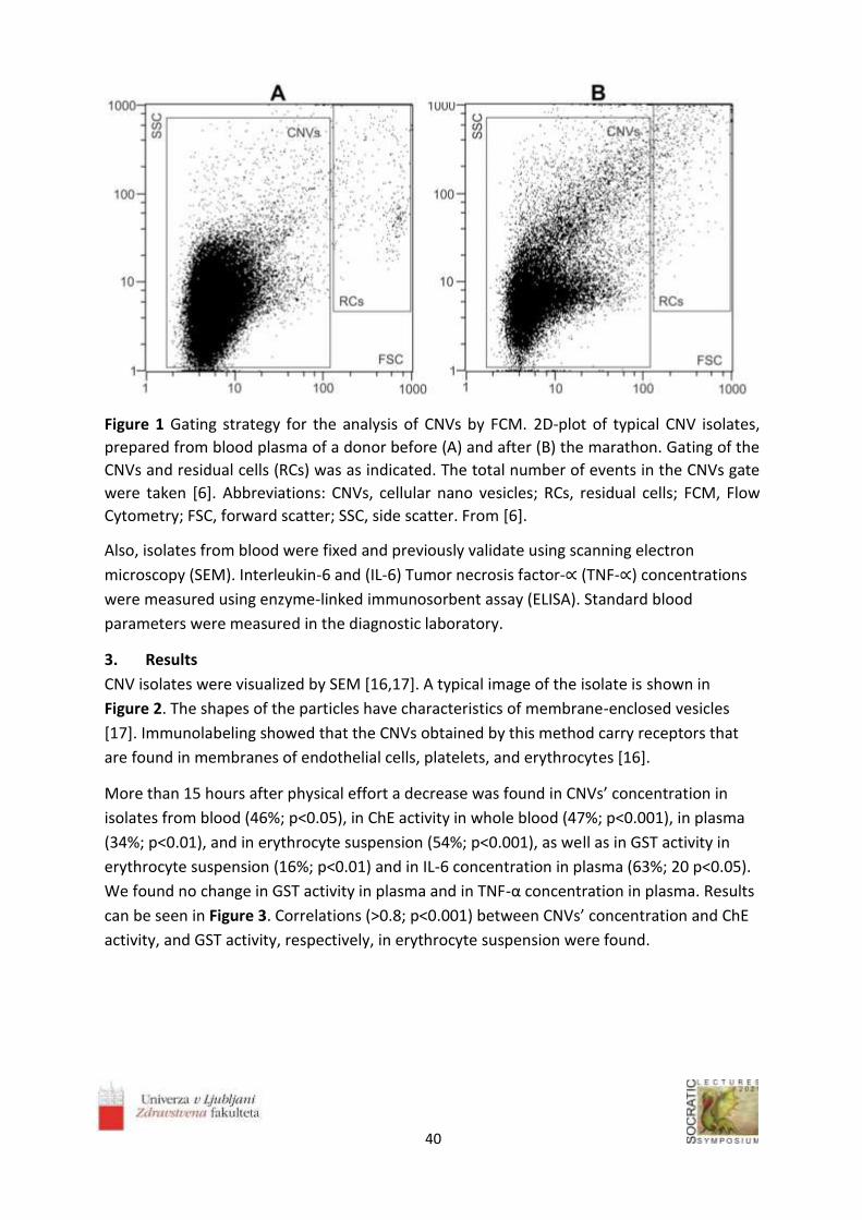
40
Figure 1 Gating strategy for the analysis of CNVs by FCM. 2D-plot of typical CNV isolates,
prepared from blood plasma of a donor before (A) and after (B) the marathon. Gating of the
CNVs and residual cells (RCs) was as indicated. The total number of events in the CNVs gate
were taken [6]. Abbreviations: CNVs, cellular nano vesicles; RCs, residual cells; FCM, Flow
Cytometry; FSC, forward scatter; SSC, side scatter. From [6].
Also, isolates from blood were fixed and previously validate using scanning electron
microscopy (SEM). Interleukin-6 and (IL-6) Tumor necrosis factor-∝ (TNF-∝) concentrations
were measured using enzyme-linked immunosorbent assay (ELISA). Standard blood
parameters were measured in the diagnostic laboratory.
3. Results
CNV isolates were visualized by SEM [16,17]. A typical image of the isolate is shown in
Figure 2. The shapes of the particles have characteristics of membrane-enclosed vesicles
[17]. Immunolabeling showed that the CNVs obtained by this method carry receptors that
are found in membranes of endothelial cells, platelets, and erythrocytes [16].
More than 15 hours after physical effort a decrease was found in CNVs’ concentration in
isolates from blood (46%; p<0.05), in ChE activity in whole blood (47%; p<0.001), in plasma
(34%; p<0.01), and in erythrocyte suspension (54%; p<0.001), as well as in GST activity in
erythrocyte suspension (16%; p<0.01) and in IL-6 concentration in plasma (63%; 20 p<0.05).
We found no change in GST activity in plasma and in TNF-α concentration in plasma. Results
can be seen in Figure 3. Correlations (>0.8; p<0.001) between CNVs’ concentration and ChE
activity, and GST activity, respectively, in erythrocyte suspension were found.

41
Figure 2. Scanning electron micrograph of a typical isolate of CNVs from the blood plasma
sample with characteristic shapes of particles without internal structures including spheres,
tubules (black arrows), and tori (white arrows). No residual cells are observed in the figure.
From [6].

42
Figure 3. The effect of physical effort on concentration of microparticles and measured
physiological state-related parameters in blood. (A) CNVs concentration in 1 mL plasma
samples two days before and >15 hours after physical effort. (B) Activity of ChE (n=24) (in
mM/min/mL of sample) two days before and >15 hours after physical effort in plasma,
erythrocyte suspension and whole blood samples. (C) Activity of GST (n=24) (in mM/min/mL
of sample) in plasma and erythrocyte suspension samples two days before and >15 hours after
physical effort. Scale on the left represents values of plasma GST activity and scale on the right
represents values of erythrocyte suspension GST activity. (D) Concentration of IL-6 and TNF-α
(n=26) (in pg/mL of plasma) two days before and >15 hours after physical effort. Statistical
differences before and after physical effort at: * p<0.05, ** p<0.01 and *** p<0.001. From [6].
Abbreviations: CNVs, cellular nanovesicles; ChE, cholinesterase; GST, Glutathione S-
transferase; TNF-α, tumor necrosis factor; IL-6 -interleukin 6.

43
4. Discussion
According to most of the previous reports, physical effort causes an increase in
concentration of CNVs during or immediately after the effort [8, 18-20]. Yet, according to
our results, the concentration of Cellular nanovesicles in isolates decreases after the rest
and may reach values lower than initial. Evidence on GST in connection with physical effort
is limited in previous studies. Connection between GST activity in erythrocyte suspension
and concentration of CNVs in blood isolates, as shown in our study, can indicate a decrease
in oxidative stress >15 hours after recovery period [21].
Connection between ChE activity in erythrocyte suspension and concentration of CNV was
previously considered in other studies [22-25]. Our results suggest that ChE is a potential
indicator of the response of human body to physiological stress and inflammation after
physical effort, since ChE activity was lower after >15 hours of rest concomitantly with CNVs.
We found decrease in IL-6 concentration in blood plasma samples after >15 hours of rest.
Previous studies [3,4,26] report elevated plasma IL-6 concentration and connect the
elevation with running intensity, duration and level of fitness. Plasma TNF- concentration
was not changed after >15 hours of rest and that is similar to previously reported results
[27].
5. Conclusion
We obtained lower concentration of CNVs in isolates from peripheral blood >15 hours after
physical effort with lower values of other measured physiological state-related parameters:
whole blood, plasma and erythrocyte suspension ChE activity, erythrocyte suspension GST
activity and IL-6 plasma concentrations. This suggests that CNVs concentration and ChE
activity have the potential to indicate impact of physical effort on inflammation in the
human body. Also, concentration of the measured parameters depends on the duration of
recovery period. According to previous reports, physical effort causes an increase in
concentration of CNVs, but according to our results, the concentration of CNVs in isolates
decreases after rest and may reach values lower than initial ones. With this, our study
provides novel insights into the beneficial effects of physical effort on the human body and
guidance about physical effort for donors before blood collection in procedures focusing on
CNV-containing diagnostic and therapeutic compounds.
Acknowledgments
Authors acknowledge support from EU Commission (Ves4us) and Slovenian Research 770
Agency (P3-0388, P2-9232, P1-0132, J1-9162, L3-2621, J5-7098).

44
References
1. Whitham M, Parker BL, Friedrichsen M, et al. Extracellular Vesicles Provide a Means for Tissue Crosstalk during Exercise.Cell Metab. 01 2018;27(1):237-251.e4. doi:10.1016/j.cmet.2017.12.001
2. Evelo CT, Palmen NG, Artur Y, Janssen GM. Changes in blood glutathione concentrations, and in erythrocyte glutathione reductase and glutathione S-transferase activity after running training and after participation in contests. Eur J Appl Physiol Occup Physiol. 1992;64(4):354-8. doi:10.1007/bf00636224
3. Mendham AE, Donges CE, Liberts EA, Duffield R. Effects of mode and intensity on the acute exercise-induced IL-6 and CRP responses in a sedentary, overweight population. Eur J Appl Physiol. Jun 2011;111(6):1035-45. doi:10.1007/s00421-010-1724-z
4. Chaar V, Romana M, Tripette J, et al. Effect of strenuous physical exercise on circulating cell-derived microparticles. Clin Hemorheol Microcirc. 2011;47(1):15-25. doi:10.3233/CH-2010-1361
5. Brahmer A, Neuberger E, Esch-Heisser L, et al. Platelets, endothelial cells and leukocytes contribute to the exercise-triggered release of extracellular vesicles into the circulation. J Extracell Vesicles. 2019;8(1):1615820. doi:10.1080/20013078.2019.1615820
6. Jan Z, Drab M, Drobne D, et al. Decrease in Cellular Nanovesicles Concentration in Blood of Athletes More Than 15 Hours After Marathon. Int J Nanomedicine. 2021; 6:1-14. doi: 10.2147/IJN.S282200.
7. Witwer KW, Théry C. Extracellular vesicles or exosomes? On primacy, precision, and popularity influencing a choice of nomenclature. J Extracell Vesicles. 2019;8(1):1648167. doi:10.1080/20013078.2019.1648167
8. Frühbeis C, Helmig S, Tug S, Simon P, Krämer-Albers EM. Physical exercise induces rapid release of small extracellular vesicles into the circulation. J Extracell Vesicles. 2015;4:28239. doi:10.3402/jev.v4.28239
9. Morshed A, Karawdeniya BI, Bandara YMND, Kim MJ, Dutta P. Mechanical characterization of vesicles and cells: A review. Electrophoresis. Jan 2020;doi:10.1002/elps.201900362
10. Borras C, Mas-Bargues C, Sanz-Ros J, et al. Extracellular vesicles and redox modulation in aging. Free Radic Biol Med. Mar 2020;149:44-50. doi:10.1016/j.freeradbiomed.2019.11.032
11. Rakobowchuk M, Ritter O, Wilhelm EN, et al. Divergent endothelial function but similar platelet microvesicle responses following eccentric and concentric cycling at a similar aerobic power output. J Appl Physiol (1985). Apr 2017;122(4):1031-1039. doi:10.1152/japplphysiol.00602.2016
12. Liem RI, Onyejekwe K, Olszewski M, et al. The acute phase inflammatory response to maximal exercise testing in children and young adults with sickle cell anaemia. Br J Haematol. Dec 2015;171(5):854-61. doi:10.1111/bjh.13782
13. Chamera T, Spieszny M, Klocek T, et al. Post-Effort Changes in Activity of Traditional Diagnostic Enzymatic Markers in Football Players' Blood. J Med Biochem. Apr 2015;34(2):179-190. doi:10.2478/jomb-2014-0035
14. Petersen AM, Pedersen BK. The anti-inflammatory effect of exercise. J Appl Physiol (1985). Apr 2005;98(4):1154-62. doi:10.1152/japplphysiol.00164.2004
15. Eichner NZM, Gilbertson NM, Musante L, et al. An Oral Glucose Load Decreases Postprandial Extracellular Vesicles in Obese Adults with and without Prediabetes. Nutrients. Mar 2019;11(3)doi:10.3390/nu11030580

45
16. Štukelj R, Schara K, Bedina-Zavec A, et al. Effect of shear stress in the flow through the sampling needle on concentration of nanovesicles isolated from blood. Eur J Pharm Sci. Feb 2017;98:17-29. doi:10.1016/j.ejps.2016.10.007
17. Suštar V, Bedina-Zavec A, Stukelj R, et al. Post-prandial rise of microvesicles in peripheral blood of healthy human donors. Lipids Health Dis. Mar 2011;10:47. doi:10.1186/1476-511X-10-47
18. Augustine D, Ayers LV, Lima E, et al. Dynamic release and clearance of circulating microparticles during cardiac stress. Circ Res. Jan 2014;114(1):109-13. doi:10.1161/CIRCRESAHA.114.301904
19. Wilhelm EN, González-Alonso J, Parris C, Rakobowchuk M. Exercise intensity modulates the appearance of circulating microvesicles with proangiogenic potential upon endothelial cells. Am J Physiol Heart Circ Physiol. 11 2016;311(5):H1297-H1310. doi:10.1152/ajpheart.00516.2016
20. Sossdorf M, Otto GP, Claus RA, Gabriel HH, Lösche W. Cell-derived microparticles promote coagulation after moderate exercise. Med Sci Sports Exerc. Jul 2011;43(7):1169-76. doi:10.1249/MSS.0b013e3182068645
21. Neefjes VM, Evelo CT, Baars LG, Blanco CE. Erythrocyte glutathione S transferase as a marker of oxidative stress at birth. Arch Dis Child Fetal Neonatal Ed. Sep 1999;81(2):F130-3. doi:10.1136/fn.81.2.f130
22. Yarana C, St Clair DK. Chemotherapy-Induced Tissue Injury: An Insight into the Role of Extracellular Vesicles-Mediated Oxidative Stress Responses. Antioxidants (Basel). Sep 2017;6(4)doi:10.3390/antiox6040075
23. Cantin R, Diou J, Bélanger D, Tremblay AM, Gilbert C. Discrimination between exosomes and HIV-1: purification of both vesicles from cell-free supernatants. J Immunol Methods. Sep 2008;338(1-2):21-30. doi:10.1016/j.jim.2008.07.007
24. Johnstone RM, Adam M, Hammond JR, Orr L, Turbide C. Vesicle formation during reticulocyte maturation. Association of plasma membrane activities with released vesicles (exosomes). J Biol Chem. Jul 1987;262(19):9412-20.
25. Park IW, He JJ. HIV-1 is budded from CD4+ T lymphocytes independently of exosomes. Virol J. Sep 2010;7:234. doi:10.1186/1743-422X-7-234
26. Liao Z, Jaular LM, Soueidi E, et al. Acetylcholinesterase is not a generic marker of extracellular vesicles. J Extracell Vesicles. 2019;8(1):1628592. doi:10.1080/20013078.2019.1628592
27. Timmerman KL, Flynn MG, Coen PM, Markofski MM, Pence BD. Exercise training-induced lowering of inflammatory (CD14+CD16+) monocytes: a role in the anti-inflammatory influence of exercise? J Leukoc Biol. Nov 2008;84(5):1271-8. doi:10.1189/jlb.0408244

46

47
___________________
Preparation of platelet- and extracellular vesicle-rich gel and its role in the
management of cerebrospinal fluid leak in anterior and lateral skull-base
surgery
Vozel D1,2,*, Battelino S1,2
1University Medical Centre Ljubljana, Department of Otorhinolaryngology and Cervicofacial
Surgery, Ljubljana, Slovenia 2University of Ljubljana, Faculty of Medicine, Ljubljana, Slovenia
Abstract
Platelet- and extracellular vesicle-rich plasma (PVRP) is a blood-derived product with the
concentrations of platelets and extracellular vesicles (EVs) above the blood levels.
Moreover, EVs are believed to possess the PVRP's leading regenerative roles since they are
essential mediators of inter-and intracellular communication. PVRP has been named
inconsistently, e.g. as platelet-rich plasma (PRP), platelet-rich fibrin (PRF), platelet
concentrate, platelet-rich growth factors, platelet-rich fibrin matrix and platelet-rich gel due
to different methods of preparation, activation and analyses. Taken nomenclature aside,
autologous PVRP has been applied in various medical specialities, including in
otorhinolaryngology, head & neck surgery (ORL-HNS). Despite that, there is a lack of
comparable randomised controlled clinical studies. This article presents autologous PVRP
and platelet- and extracellular vesicle-rich gel (PVRG) preparation protocols and autologous
PVRG's role in skull-base surgery, performed by ORL-HNS. Since the autologous PVRG can
provide a watertight seal and induce osteogenesis, the mini-review presents PVRG's role in
skull-base reconstruction via anterior and lateral approaches.
___________________
48
1. Platelet- and extracellular vesicle-rich plasma and gel
1.1 Definitions
Platelet- and extracellular vesicle-rich plasma (PVRP) is defined as a blood-derived product
with platelet and EV concentrations higher than in blood. In a strict sense, PVRP is a liquid
preparation that can be transformed into a PVRG (i.e. platelet- and extracellular vesicle-rich
gel) via the coagulation cascade's activation with different substances 1.
Despite the strict definition, PVRP's terminology is inconsistent due to different preparation
methods, activation and analyses. Accordingly, it has been named as platelet-rich plasma,
platelet concentrate, platelet-rich fibrin, platelet-rich fibrin matrix, platelet-rich growth
factors, platelet gel etc. 1
The PVRP's main regenerative functions are believed to be exhibited by platelets and
especially extracellular vesicles (EVs) 2. These roles enable introducing PVRP and PVRG in
the treatment of various diseases covered by otorhinolaryngology, head & neck surgery
(ORL-HNS).
If the blood donor and PVRP acceptor is the same person, the PVRP is autologous.
Otherwise, it is heterologous. Adverse reactions to PVRP-based treatment are infrequent.
There is a minute risk of malignant or infectious disease transmission or causing a rejection
reaction, especially by heterologous PVRP 3.
1.2 Preparation basics
PVRP preparation protocols can be classified according to the blood fraction from which the
PVRP is prepared. Namely, the PVRP preparation protocol can be plasma-based or buffy
coat-based. Plasma-based protocols yield higher PVRP volume, lower leukocyte and lower
platelet concentrations compared to buffy coat-based protocols (Figure 1) 1.
Moreover, PVRP can be prepared by one-stepped or multiple-stepped centrifugation of
blood fractions. The majority of commercially available kits utilise one-stepped plasma-
based PVRP preparation protocols. These kits advantageously provide the closed system to
prevent sample contamination. However, non-commercial multiple-stepped plasma-based
centrifugation methods can yield higher platelet concentrations compared to commercial
kits. To prevent contamination, strict measures need to be undertaken.
To prepare PVRP, anticoagulated tubes must be used to collect blood. If not, the blood clots,
but the preparation cannot be defined as PVRG. ACD (acid citrate dextrose solution) or
sodium citrate can be used as anticoagulants in PVRP preparation. The sodium citrate is
recommended since it stimulates the proliferation of mesenchymal stromal cells without
interfering with their phenotype and enables more concentrated PVRP 4. It is not
recommended to collect blood in EDTA tubes, because EDTA irreversibly inactivates
thrombocytes 5). After PVRP is prepared, it can be administered to the target tissue where
it activates endogenously via interaction with type I collagen. The activation process results
in the release of numerous growth factors from platelet and EVs granules 1.

49
On the other hand, PVRP can be activated before administration with different substances
(i.e., activators) which activate coagulation cascade. This results in PVRG formation, which
can be laid over or under the target tissue or defect. The activators are components of
coagulation cascade: e.g. CaCl2, thrombin and other coagulation factors. CaCl2 is used to
calcify blood but can lower the pH if administered in too high concentration. Therefore, it is
important to titrate the CaCl2. The activation can be achieved with the blood serum, which
contains the components of the coagulation cascade. It is essential to keep in mind that the
glass activates this cascade, also. Anticoagulant or antithrombotic drugs hamper PVRP gel
production 1.
Instant freezing and storing of PVRP at -80 °C for up to 1 month do not decrease some
growth factors after thawing according to the McClain and McCarrel (2019) 6. On the other
side, freezing at -20 °C for up to two weeks reduces some growth factor levels according to
the Hosnuter et al. (2017) 7. Roffi et al. (2014) concluded that one freeze/thaw cycle at -30
°C (for 2 hours) and 37 °C, respectively, sufficiently preserves PVRP quality and ability to
promote regeneration via stimulation of chondrocytes and synoviocytes 8. To sum up,
PVRP cryopreservation remains controversial, so far, and clinical studies are required to
determine the efficacy of PVRP and PVRG after different freeze/thaw cycles. Nevertheless,
PVRP freezing enables administration of PVRP during surgical procedures with the expected
gross blood volume loss, which restricts any additional blood collection.
Since the PVRG preparation protocol is an extension of the PVRP preparation protocol, the
latter is described first.
1.3 Platelet- and extracellular vesicle-rich plasma preparation protocol
Our multiple-stepped plasma-based PVRP preparation protocol is as follows:
1. Blood is drawn into four 4.5 mL tubes with sodium citrate anticoagulant (9 NC sodium citrate 0.105 M, BD Vacutainer, Becton Dickinson, USA, stored at room temperature) (9 NC sodium citrate 0.105 M, BD Vacutainer, Becton Dickinson, USA, stored at room temperature) using a 21 G wing needle (Safety-Lok Blood Collection Set, BD Vacutainer, Becton Dickinson, USA). Tubes must be full.
2. Soft spin centrifugation (i.e., 5 minutes, 300×g, 18 °C) of blood tubes to distinguish two fractions: haematocrit (red layer in the bottom) and plasma with platelets (yellow upper layer). Occasionally a buffy coat (thin opaque whitish layer of leukocytes on top of haematocrit could be observed.
3. Plasma directly above the buffy coat is transferred by a sterile pipette into a sterile polypropylene tube. Caution is required to prevent mixing layers, i.e., disruption of haematocrit and buffy coat and consequent leukocyte and erythrocyte harvesting. 3.1. Haematocrit can be centrifuged with soft spin (i.e., additional soft spin) if higher
PVRP volume is required. 3.2. The harvested plasma yielded with soft spins is merged
4. Harvested plasma is equally distributed among two sterile polypropylene tubes (i.e., plasma from two blood tubes is distributed in one sterile polypropylene tubes) 5. Hard spin centrifugation (i.e., 17 minutes, 700×g, 18 °C) to sediment the platelets in
plasma.

50
6. The upper half of the centrifuged plasma (i.e., platelet-poor plasma) is carefully removed and discarded without disruption of layers.
7. The remaining pellet is resuspended in the remaining volume of plasma (Fig. 3, Panel 5B) in the same tube to produce PVRP
8. The whole PVRP is pooled in a larger tube. Then, PVRP is administered to the target tissue or activated to produce the PVRG.
1.4 Platelet- and extracellular vesicle-rich gel preparation protocol
Our PVRP preparation protocol is an extension of the PVRP preparation protocol (i.e., step 8)
and follows these steps:
9. 1M CaCl2 (14.7%, Pharmacy of University Medical Centre Ljubljana, stored at room
temperature) in the ratio with PVRP 1: 100 (i.e., 1%; e.g., 17 μL to activate 1.7 mL of
PVRP) is administered with the sterile pipette to the plastic petri dish
10. Autologous serum in the ratio with PVRP 1: 5 (i.e., 20 %; e.g., 0.34 mL to activate 1.7
mL of PVRP) is administered with the sterile pipette on the top of CaCl2.
a. The autologous serum is prepared by centrifugation (i.e., 10 min, 1260×g, 18°C) of
blood collected into one 4 mL plastic blood tube without anticoagulant (i.e., red
cap, Z Serum, Vacutube, LT Burnik, d.o.o., Slovenia, stored at room temperature).
The blood serum (i.e., supernatant above the red layer) is transferred by a sterile
pipette into a sterile polypropylene tube. Caution is required to prevent the
mixing of layers and consequential clot formation.
11. Autologous PVRP is administered with the sterile pipette on the top of CaCl2 and
autologous serum mixture.
12. Waiting for the PVRP to transform to the PVRG
13. PVRG trimming (if necessary) and application to the target tissue.
13.1. Alternatively, the PVRP can be transformed into PVRG (with CaCl2 and
autologous serum) in the target tissue
13.2. Alternatively, the PVRP can be mixed with CaCl2 and autologous serum in the
petri dish and then immediately (i.e., without step 12) administered to the target
tissue, transforming into a gel.
PVRG preparation with different concentrations of CaCl2 and autologous serum and the effect
of the freeze-thawing cycle are depicted in Table 1.

51
Table 1: Preparation of PVRG from the PVRP of four healthy donors (A-D).
Freeze/thaw VPVRP (mL)
CaCl2 Serum
Time (minutes) μL
% of
VPVRP μL
% of
VPVRP
A None 1.5 15
1 300
20 4
Yes 1 10 200 2
B None
0.5 5 1 100 20 2
250 50 2
0.5 none 250 50
none 100 20
Yes 0.5 5 1 100 20 2
C
None
0.5 5 1 none
20
0.5 10 2 20
0.5 5 1 250 50
2
0.5 10 2 2
Yes
0.5 5 1 none
8
0.5 10 2 6
0.5 5 1 250 50
1
0.5 10 2 4
D
None
0.5 2.5 0.5 50 10
none 25 5
0.5 5 1 50 10 5
25 5 5
Yes 0.5 5 1 50 10 2
25 5 2
Remarks: Different activation methods and freeze-thaw cycle were tested in PVRG
preparation. Both PVRP and serum samples were instantly frozen at -80 °C and thaw at
room temperature before the activation and gel formation; PVRP – platelet- and
extracellular vesicle-rich plasma; PVRG – platelet- and extracellular vesicle-rich gel; VPVRP –
Volume of PVRP; Time – the time from mixing of PVRP with an activator(s) to gel formation.
2. Platelet- and extracellular vesicle-rich gel and skull-base surgery
The main hallmark of skull-base surgery is a meticulous dissection of skull-base, which is
intimately related with vital neurovascular structures and special senses (i.e., vision, hearing,
balance, smell and taste). The anterior skull-base surgery denotes anterior transnasal
endoscopic and/or transfacial approach, and lateral skull-base surgery indicates lateral (i.e.,
otosurgical microscopic) approach to the skull-base. The anterior, middle and posterior
cranial fossa can be dissected via these approaches.

52
Anterior and lateral skull-base surgery involves treatment of numerous acquired or
congenital disorders involving the bones of the skull-base (i.e., frontal, ethmoid, sphenoid,
temporal, occipital bone): e.g. resection of neoplasms (e.g. meningioma,
esthesioneuroblastoma, middle ear cancer) or inflamed tissue (e.g., invasive fungal disease,
cholesteatoma), decompression of cranial nerves (e.g., traumatic facial or optic nerve
compression) and closure of cerebrospinal fluid (CSF) leak.
The CSF leak is caused when the communication between arachnoid space, which contains
CSF, and an external environment (e.g., nasal cavity) is created due to the dural and
arachnoid tear. The tear results from either accidental trauma or iatrogenically (deliberately
during the resection of skull-base or inadvertently). If there is no identifiable cause, a
spontaneous CSF leak is diagnosed, typically associated with idiopathic intracranial
hypertension. The hallmark of CSF leak is a rhinoliquorrhea or otoliquorrhea. These patients
suffer from headache, visual and hearing loss, disequilibrium, vertigo and are prone to
develop meningitis 9.
2.1 Transnasal endoscopic repair of the cerebrospinal fluid leak using platelet- and
extracellular vesicle-rich gel
According to the international consensus, it is essential to close the CSF leak as soon as
possible without delay. The skull-base defect closure is necessary already at the primary
surgery, later at the revision surgery or in the treatment of spontaneous CSF leaks. A multi-
layered closure is required, but there is no consensus which material to be used 9. The
main goal of closure is to provide watertight closure which can be achieved with different
autologous (e.g. lat. fascia lata, abdominal fat, mucosal flap, pericranial flap), or comercial
heterologous materials.
According to the application of PVRP and PVRG in ORL-HNS so far, there is a lack of clinical
studies in the field of skull-base surgery, including CSF leak closure (Table 2).
The role of PVRP or PVRG has been extensively studied in in-vitro and in animal studies. For
instance, in the recent study of Vasilikos et al. (2020), the hermetic effect of PVRG has been
elucidated.
In-vitro created dural tears reconstructed with PVRG and suture withheld higher water
pressure than those reconstructed with suture only [49]. In addition to the PVRG's hermetic
properties, an increased cranial bone formation has been reported to reconstruct rabbits'
cranial defects with PVRG in a prospective randomised controlled study by Kim et al. (2014)
50.
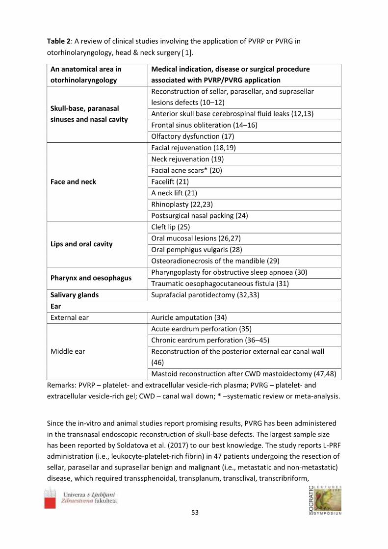
53
Table 2: A review of clinical studies involving the application of PVRP or PVRG in
otorhinolaryngology, head & neck surgery 1.
An anatomical area in
otorhinolaryngology
Medical indication, disease or surgical procedure
associated with PVRP/PVRG application
Skull-base, paranasal
sinuses and nasal cavity
Reconstruction of sellar, parasellar, and suprasellar
lesions defects (10–12)
Anterior skull base cerebrospinal fluid leaks (12,13)
Frontal sinus obliteration (14–16)
Olfactory dysfunction (17)
Face and neck
Facial rejuvenation (18,19)
Neck rejuvenation (19)
Facial acne scars* (20)
Facelift (21)
A neck lift (21)
Rhinoplasty (22,23)
Postsurgical nasal packing (24)
Lips and oral cavity
Cleft lip (25)
Oral mucosal lesions (26,27)
Oral pemphigus vulgaris (28)
Osteoradionecrosis of the mandible (29)
Pharynx and oesophagus Pharyngoplasty for obstructive sleep apnoea (30)
Traumatic oesophagocutaneous fistula (31)
Salivary glands Suprafacial parotidectomy (32,33)
Ear
External ear Auricle amputation (34)
Middle ear
Acute eardrum perforation (35)
Chronic eardrum perforation (36–45)
Reconstruction of the posterior external ear canal wall
(46)
Mastoid reconstruction after CWD mastoidectomy (47,48)
Remarks: PVRP – platelet- and extracellular vesicle-rich plasma; PVRG – platelet- and
extracellular vesicle-rich gel; CWD – canal wall down; * –systematic review or meta-analysis.
Since the in-vitro and animal studies report promising results, PVRG has been administered
in the transnasal endoscopic reconstruction of skull-base defects. The largest sample size
has been reported by Soldatova et al. (2017) to our best knowledge. The study reports L-PRF
administration (i.e., leukocyte-platelet-rich fibrin) in 47 patients undergoing the resection of
sellar, parasellar and suprasellar benign and malignant (i.e., metastatic and non-metastatic)
disease, which required transsphenoidal, transplanum, transclival, transcribriform,

54
transethmoid or transpterygoid approach. L-PRF was prepared without an anticoagulant,
which results in gel formation during the centrifugation. For that reason, this is not PVRP or
PVRG in its strict sense, according to the 4. L-PRF has been administered as inlay and
overlay with mucoperiosteal graft or flap. Although the reported incidence of CSF's
postoperative leak is low and crusting and scar formation was satisfactory, more studies
need to be conducted. Namely, this study's main limitations are a retrospective type of
analysis, no control group, and group heterogeneity 10.
L-PRF has been used by Khafagy et al. (2018) in the transnasal endoscopic treatment of
spontaneous CSF leak in underlay and overlay fashion as an adjunct to autologous grafts and
flaps. None statistically significant differences between CSF pressures and defect sizes and in
CSF leak recurrence have been identified between the control and intervention group 13.
The study by Fredes et al. (2017) reported the neoosteogenesis visible on the CT in the
region of skull-base, which was reconstructed with L-PRF membranes and autologous flaps
or grafts 12.
Rasmussen et al. (2018) administered L-PRF, to reconstruct defects after transsphenoidal
endoscopic approach to the sellar region. Although the L-PRF has been administered as
overlay solely, there was no CSF leak and favourable crusting 11.
2.2 Repair of the cerebrospinal fluid leak using platelet- and extracellular vesicle-rich gel in
otological surgical procedures
PVRP and PVRG have been effectively used in the treatment of skull-base defects and long-
bone fractures. Moreover, its use has been described in treating calvarial defects, e.g.
frontal sinus obliteration with PVRG and bone chips via bicoronal approach 14–16.
Since the otological surgical procedures routinely expose dura of middle cranial fossa (i.e.,
via lateral microscopically-based approaches), PVRP and PVRG could be used to prevent CSF
leak or meningocele formation via mechanisms of neosteogenesis and remucosalisation.
Moreover, PVRG's obliterative properties could place it as an adjunct to the fat
inobliteration after subtotal petrosectomy. Since the EVs can readily cross the hemato-
encephalic barrier 51and have been proposed as vectors in the treatment of hearing loss
52, PVRP and PVRG could have the potential role in the treatment of inner ear disorders
due to its high concentrations of EVs [53].
3. Conclusion
PVRG prepared by our protocol possesses good therapeutic properties due to the
regenerative roles of platelets and extracellular vesicles. It has already been described as
effective in treating the spectrum of diseases, including those covered by
otorhinolaryngology, head & neck surgery. Despite its great potential, there is a lack of
clinical evidence to establish this therapeutic modality adjuvant to the skull-base surgery.
Nevertheless, there is some evidence of PVRG application in anterior transnasal endoscopic
skull-base surgery, including cerebrospinal fluid leak closure, which provides the basis for

55
further research. Some parallels could also be drawn with skull-base reconstruction during
otological surgery, where the PVRG's potential remains unexploited.
Acknowledgements
Authors acknowledge support of EU Commission (Ves4us) and Slovenian Research Agency
(grants P3-0388, J1-9162, J2-8166, L3-2621 and J2-8169).
References
1. Vozel D, Božič D, Jeran M, et al. Treatment with platelet- and extracellular vesicle-rich plasma in otorhinolaryngology-a review and future perspectives. In: Advances in Biomembranes and Lipid Self-Assembly [Internet]. Academic Press; 2020. Available from: http://www.sciencedirect.com/science/article/pii/S2451963420300224
2. Guo S-C, Tao S-C, Yin W-J, Qi X, Yuan T, Zhang C-Q. Exosomes derived from platelet-rich plasma promote the re-epithelisation of chronic cutaneous wounds via activation of YAP in a diabetic rat model. Theranostics, 2017, 7(1):81–96.
3. Dhillon RS, Schwarz EM, Maloney MD. Platelet-rich plasma therapy - future or trend? Arthritis Res Ther, 2012, 14(4):219.
4. do Amaral RJFC, da Silva NP, Haddad NF, et al. Platelet-Rich Plasma Obtained with Different Anticoagulants and Their Effect on Platelet Numbers and Mesenchymal Stromal Cells Behavior In Vitro. Fan J, editor. Stem Cells Int, 2016, 2016:7414036.
5. White JG, Escolar G. EDTA-induced changes in platelet structure and function: adhesion and spreading. Platelets, 2000, 11(1):56–61.
6. McClain AK, McCarrel TM. The effect of four different freezing conditions and time in frozen storage on the concentration of commonly measured growth factors and enzymes in equine platelet-rich plasma over six months. BMC Vet Res, 2019, 15(1):292.
7. Hosnuter M, Aslan C, Isik D, Caliskan G, Arslan B, Durgun M. Functional assessment of autologous platelet-rich plasma (PRP) after long-term storage at −20 °C without any preservation agent. J Plast Surg Hand Surg, 2017, 51(4):235–9.
8. Roffi A, Filardo G, Assirelli E, et al. Does Platelet-Rich Plasma Freeze-Thawing Influence Growth Factor Release and Their Effects on Chondrocytes and Synoviocytes? Sánchez M, editor. BioMed Res Int, 2014, 2014:692913.
9. Georgalas C, Oostra A, Ahmed S, et al. International Consensus Statement: Spontaneous Cerebrospinal Fluid Rhinorrhea. Int Forum Allergy Rhinol [Internet]. n/a(n/a). Available from: https://onlinelibrary.wiley.com/doi/abs/10.1002/alr.22704
10. Soldatova L, Campbell RG, Elkhatib AH, et al. Role of Leukocyte-Platelet-Rich Fibrin in Endoscopic Endonasal Skull Base Surgery Defect Reconstruction. J Neurol Surg Part B Skull Base, 2017, 78(1):59–62.
11. Rasmussen J, Ruggeri C, Ciraolo C, Baccanelli M, Yampolsky C, Ajler P. Application of Fibrin Rich in Leukocytes and Platelets in the Reconstruction of Endoscopic Approaches to the Skull Base. World Neurosurg, 2018, 118:32–41.
12. Fredes F, Pinto J, Pinto N, et al. Potential Effect of Leukocyte-Platelet-Rich Fibrin in Bone Healing of Skull Base: A Pilot Study. Nathan C-AO, editor. Int J Otolaryngol, 2017, 2017:1231870.
13. Khafagy YW, Abd Elfattah AM, Moneir W, Salem EH. Leukocyte- and platelet-rich fibrin: a new graft material in endoscopic repair of spontaneous CSF leaks. Eur Arch Otorhinolaryngol, 2018, 275(9):2245–52.

56
14. Mendonça-Caridad JJ, Juiz-Lopez P, Rubio-Rodriguez JP. Frontal sinus obliteration and craniofacial reconstruction with platelet rich plasma in a patient with fibrous dysplasia. Int J Oral Maxillofac Surg, 2006, 35(1):88–91.
15. Mendonça-Caridad JJ, Juiz-Lopez P, Fayos FV, Miery G. A novel approach to human cranial tissue regeneration and frontal sinus obliteration with an autogenous platelet-rich/fibrin-rich composite matrix: 10 patients with a 6-10 year follow-up. J Tissue Eng Regen Med, 2013, 7(6):491–500.
16. Taglialatela Scafati C, Taglialatela Scafati S, Aveta A, Cassese M, Vitale C. Chronic frontal sinus disease: combined use of platelet-rich plasma and calvarial bone grafts for sinus obliteration in aggressive and secondary cases. Rev Stomatol Chir Maxillofac, 2010, 111(4):216–20.
17. Yan CH, Mundy DC, Patel ZM. The use of platelet-rich plasma in treatment of olfactory dysfunction: A pilot study. Laryngoscope Investig Otolaryngol, 2020; 5(2):187–93.
18. Maisel-Campbell AL, Ismail A, Reynolds KA, et al. A systematic review of the safety and effectiveness of platelet-rich plasma (PRP) for skin aging. Arch Dermatol Res [Internet], 2019, [cited 2020 Jan 25]; Available from: https://doi.org/10.1007/s00403-019-01999-6
19. Gawdat HI, Tawdy AM, Hegazy RA, Zakaria MM, Allam RS. Autologous platelet-rich plasma versus readymade growth factors in skin rejuvenation: A split face study. J Cosmet Dermatol, 2017, 16(2):258–64.
20. Hsieh T-S, Chiu W-K, Yang T-F, Wang H-J, Chen C. A Meta-analysis of the Evidence for Assisted Therapy with Platelet-Rich Plasma for Atrophic Acne Scars. Aesthetic Plast Surg, 2019, 43(6):1615–23.
21. Man D, Plosker H, Winland-brown JE. The Use of Autologous Platelet-rich Plasma (platelet Gel) and Autologous Platelet-poor Plasma (fibrin Glue) in Cosmetic Surgery. Plast Reconstr Surg, 2001, 107(1):229–36.
22. Gode S, Ozturk A, Kısmalı E, Berber V, Turhal G. The Effect of Platelet-Rich Fibrin on Nasal Skin Thickness in Rhinoplasty. Facial Plast Surg FPS, 2019, 35(4):400–3.
23. Gode S, Ozturk A, Berber V, Kısmalı E. Effect of Injectable Platelet-Rich Fibrin on Diced Cartilage's Viability in Rhinoplasty. Facial Plast Surg FPS, 2019, 35(4):393–6.
24. Kuzucu I, Beriat GK, Ezerarslan H, Ozdemir S, Kocaturk S. Effects of the Autologous Platelet-Rich Plasma in Nasal Pack on Postoperative Quality of Life. J Craniofac Surg, 2017, 28(3):e299–302.
25. Refahee SM, Aboulhassan MA, Abdel Aziz O, et al. Is PRP Effective in Reducing the Scar Width of Primary Cleft Lip Repair? A Randomized Controlled Clinical Study. Cleft Palate-Craniofacial J Off Publ Am Cleft Palate-Craniofacial Assoc, 2019, 57(5):581–8.
26. Pathak H, Mohanty S, Urs AB, Dabas J. Treatment of Oral Mucosal Lesions by Scalpel Excision and Platelet-Rich Fibrin Membrane Grafting: A Review of 26 Sites. J Oral Maxillofac Surg, 2015, 73(9):1865–74.
27. Mahajan M, Gupta MK, Bande C, Meshram V. Comparative Evaluation of Healing Pattern After Surgical Excision of Oral Mucosal Lesions by Using Platelet-Rich Fibrin (PRF) Membrane and Collagen Membrane as Grafting Materials—A Randomized Clinical Trial. J Oral Maxillofac Surg, 2018, 76(7):1469.e1-1469.e9.
28. EL-Komy MHM, Saleh NA, Saleh MA. Autologous platelet-rich plasma and triamcinolone acetonide intralesional injection in the treatment of oral erosions of pemphigus vulgaris: a pilot study. Arch Dermatol Res, 2018, 310(4):375–81.
29. Scala M, Gipponi M, Mereu P, et al. Regeneration of mandibular osteoradionecrosis defect with platelet rich plasma gel. Vivo Athens Greece, 2010, 24(6):889–93.

57
30. Elkahwagi M, Elokda M, Elghannam D, Elsobki A. Role of autologous platelet-rich fibrin in relocation pharyngoplasty for obstructive sleep apnoea. Int J Oral Maxillofac Surg, 2020, 49(2):200–6.
31. Joudi M, Hamidi Alamdari D, Hyradfar M, et al. Lateral Traumatic Esophago-Cutaneous fistula in a Child; Platelet-Rich Fibrin Glue Challenge. Iran Red Crescent Med J, 2013, 15(3):256–9.
32. Ricci E, Riva G, Dagna F, Cavalot AL. The use of platelet-rich plasma gel in superficial parotidectomy. Acta Otorhinolaryngol Ital Organo Uff Della Soc Ital Otorinolaringol E Chir Cerv-facc, 2019, 39(6):363–6.
33. Scala M, Mereu P, Spagnolo F, et al. The use of platelet-rich plasma gel in patients with mixed tumour undergoing superficial parotidectomy: a randomised study. Vivo Athens Greece, 2014, 28(1):121–4.
34. Lee SK, Lim YM, Lew DH, Song SY. Salvage of Unilateral Complete Ear Amputation with Continuous Local Hyperbaric Oxygen, Platelet-Rich Plasma and Polydeoxyribonucleotide without Micro-Revascularization. Arch Plast Surg, 2017, 44(6):554–8.
35. Habesoglu M, Oysu C, Sahin S, et al. Platelet-Rich Fibrin Plays a Role on Healing of Acute-Traumatic Ear Drum Perforation. J Craniofac Surg, 2014, 25(6):2056–8.
36. Yadav SPS, Malik JS, Malik P, Sehgal PK, Gulia JS, Ranga RK. Studying the result of underlay myringoplasty using platelet-rich plasma. J Laryngol Otol, 2018, 132(11):990–4.
37. Sharma D, Mohindroo S, Azad RK. Efficacy of platelet rich fibrin in myringoplasty. Int J Otorhinolaryngol Head Neck Surg, 2018, 4(3):677–81.
38. Sankaranarayanan G, Prithiviraj V, Kumar RV. A Study on Efficacy of Autologous Platelet Rich Plasma in Myringoplasty. Otolaryngol Online J, 2013, 3(3):1–15.
39. Saeedi M, Ajalloueian M, Zare E, et al. The Effect of PRP-enriched Gelfoam on ChronicTympanic Membrane Perforation: A Double-blind Randomised Clinical Trial. Int Tinnitus J, 2017, 21(2):108–11.
40. Fawzy T, Hussein M, Eid S, Guindi S. Effect of adding platelet-rich plasma to fat grafts in myringoplasty. Egypt J Otolaryngol, 2018, 34(4):224–8.
41. El-Anwar MW, Elnashar I, Foad YA. Platelet-rich plasma myringoplasty: A new office procedure for the repair of small tympanic membrane perforations. Ear Nose Throat J, 2017, 96(8):312–26.
42. Nair NP, Alexander A, Abhishekh B, Hegde JS, Ganesan S, Saxena SK. Safety and Efficacy of Autologous Platelet-rich Fibrin on Graft Uptake in Myringoplasty: A Randomized Controlled Trial. Int Arch Otorhinolaryngol, 2019, 23(1):77–82.
43. El-Anwar MW, El-Ahl MAS, Zidan AA, Yacoup MA-RA-S. Topical use of autologous platelet rich plasma in myringoplasty. Auris Nasus Larynx, 2015, 42(5):365–8.
44. Ersözlü T, Gultekin E. A Comparison of the Autologous Platelet-Rich Plasma Gel Fat Graft Myringoplasty and the Fat Graft Myringoplasty for the Closure of Different Sizes of Tympanic Membrane Perforations. Ear Nose Throat J, 2020, 145561319900388.
45. Shiomi Y, Shiomi Y. Surgical outcomes of myringoplasty using platelet-rich plasma and evaluation of the outcome-associated factors. Auris Nasus Larynx, 2020, 47(2):191–7.
46. Elbary M, Nasr W, Sorour S. Platelet-Rich Plasma in Reconstruction of Posterior Meatal Wall after Canal Wall Down Mastoidectomy. Int Arch Otorhinolaryngol, 2018, 22(2):103–7.
47. Jang CH, Choi CH, Cho YB. Effect of BMP2–Platelet-rich Plasma–Biphasic Calcium Phosphate Scaffold on Accelerated Osteogenesis in Mastoid Obliteration, In Vivo, 2016 30(6):835–40.

58
48. Askar SM, Saber IM, Omar M. Mastoid Reconstruction With Platelet-Rich Plasma and Bone
Pate After Canal Wall Down Mastoidectomy: A Preliminary Report. Ear Nose Throat J, 2019, 145561319879789.
49. Vasilikos I, Beck J, Ghanaati S, et al. Integrity of dural closure after autologous platelet rich fibrin augmentation: an in vitro study. Acta Neurochir (Wien), 2020, 162(4):737–43.
50. Kim T-H, Kim S-H, Sándor GK, Kim Y-D. Comparison of platelet-rich plasma (PRP), platelet-rich fibrin (PRF), and concentrated growth factor (CGF) in rabbit-skull defect healing. Arch Oral Biol, 2014, 59(5):550–8.
51. Banks WA, Sharma P, Bullock KM, Hansen KM, Ludwig N, Whiteside TL. Transport of Extracellular Vesicles across the Blood-Brain Barrier: Brain Pharmacokinetics and Effects of Inflammation. Int J Mol Sci, 2020, 21(12):4407.
52. György B, Sage C, Indzhykulian AA, et al. Rescue of Hearing by Gene Delivery to Inner-Ear Hair Cells Using Exosome-Associated AAV. Mol Ther, 2017, 25(2):379–91.
53. Steiner N, Battelino S, Extracellular vesicles and their use in the inner ear, In Kralj-Iglič V ed., Proceeding of the 4th Symposium Socratic Lectures, Ljubljana, 2021, P

59

60
__________________
Evaluation the effect of frequency modulated transcutaneous electrical nerve
stimulation on postural sway in healthy young adults. A pilot study.
Rugelj D1,*, Vidovič M2
1University of Ljubljana, Faculty of Health Sciences, Biomechanical laboratory, Ljubljana, Slovenia 2University Institute of Rehabilitation - Soča, Ljubljana, Slovenia *[email protected]
Abstract
Transcutaneous electrical nerve stimulation (TENS) is a therapeutic modality that can elicit
afferent flow from somatosensory afferents. There are ambiguous results of previous
research on TENS effect on postural sway. Some authors have reported the effect while
others found no effect. Purpose of the study was to evaluate the effect of frequency
modulated TENS applied through the soles and posterior aspect of shanks on postural sway.
Methods: In a group of 13 young adults postural sway was measured with force platform
during 60 s standing balance task in four conditions (eyes opened and eyes closed with TENS
and with sham TENS). Sub-sensory threshold TENS with sweep of frequencies between 5 and
180 Hz was delivered to the soles and shanks of both legs. Results indicated that TENS with
the chosen frequencies and electrode positions had no immediate effect on postural sway in
both vision and no vision conditions. Conclusion: within the setup used (regarding type of
stimuli parameters and type of electrode placement) we observed no attenuation of
postural sway in a group of young persons with no balance impairments. A further research
with different stimulation frequencies and intensities as well as different electrodes
placements is needed.
_______________________

61
1. Introduction
Regulation of human postural control depends on accurate processing of visual, vestibular
and various somatosensory stimuli [1, 2]. During standing and purposeful movements
persons constantly and subconsciously use this information for the correction of posture or
ongoing movements. Additional somatosensory stimuli from skin mechanoreceptors are
believed to enhance the signals coming from muscle and joint proprioceptors, thus enabling
better perception and integration of stimuli into the ongoing movement [2]. There are
different methods that specifically enhance somatosensory flow, among them light touch by
fingertip of stationary [3] or movable surface [2] or even self-touching [4] have been shown
to decrease postural sway in elderly as well as in young persons [3]. Other types of sensory
stimulations that were used for the enhancement of sensory information are stochastic
resonance stimulation, textured material stimulation and garment stimulation usually
applied over lower limbs. They all exhibit stabilising effect on postural sway resulting in
decrease of the centre of pressure (CoP) path length or its mean velocity [5].
Transcutaneous electrical nerve stimulation (TENS) is a therapeutic modality with the main
purpose to modulate pain intensity and is routinely used in physiotherapy. With TENS
additional somatosensory flow can be provided. It is of importance to investigate whether
TENS can add information to the sensory processing to attenuate postural sway similarly to
other somatosensory stimulation. These signals are usually composed of short pulses (50-
200 µs) administered with fixed or varying frequency in the range of 50 to 200 Hz.
To date, few investigators have studied the effect of conventional TENS on postural sway
and reported inconclusive results. For instance, Dickstein et al. [6] and Laufer and Dickstein
[7] used conventional TENS with constant frequency of 100 Hz and reported a decrease of
one of five CoP variables in a group of young healthy persons. While Saadat et al. [8] found
no effect of TENS stimulation in the knee region in a group of diabetic neuropathy patients.
The disagreement between the results of the previous reports of TENS on the postural sway
led to the purpose of the present work which is to evaluate the effect of sensory sub-
threshold TENS applied directly on the soles of the feet and on the posterior aspect of the
shanks below the knee on postural sway in a group of healthy young persons. We
hypothesised that sub-threshold TENS would attenuate postural sway.
2. Methods
13 young persons (21 ± 2 years; body mas 67.1 ± 14.7; body height 168.1 ± 7.5) were
invited to participate in the study. Inclusion criteria were no prior lower leg injuries or
conditions, which could affect their balance. The study was approved by the Slovenian
National Medical Ethic Committee (0120-309/2018/3) and prior to any measurements, all
participants read the information about the testing protocol, received additional verbal
explanations when required, and provided written informed consent.

62
2.1 Sensory threshold testing
Before the application of the TENS and measurements of postural sway the sensory
threshold of the soles was measured with Sammens-Weinstein mono-filaments (Baseline
Tactile Sensory evaluator, USA) ranging from 0,007g to 300g. The sensory threshold testing
is valid and reliable procedure for evaluating the cutaneous threshold sensitivity [9,10]. A
point beneath the first metatarsal was tested. The initial side of the body was randomly
chosen. To determine the sensory threshold a stepping algorithm was used according to
testing protocol described by Snyder et al. [10].
2.2. Experimental procedure
Participants were instructed to stand barefooted as still as possible with their feet close
together on the force platform, with their eyes opened or closed with or without sub-
threshold TENS. Arms were relaxed beside the body, head was held upright with gaze
forward to the anchor point at the eye height approximately 2 metres away.
Measurements of postural sway were performed under four different conditions during
standing on force platform: eyes opened without TENS, eyes opened with sub-threshold
TENS, eyes closed without TENS, eyes closed with sub-threshold TENS. The order of testing
was randomised in two blocks: first eyes opened or closed and then in each vision condition
the sub-sensory TENS or sham TENS. The random number generator was used to determine
the order of tests. The participants and the assessor were blinded for the presence or
absence of the sub-threshold electrical stimulation. Electrical stimulation was operated by
research assistant.
2.3. Instrumentation and data processing
The force platform Kistler 9286AA (Winthertur, Switzerland), with the corresponding data
acquisition software BioWare was used for data acquisition. A non-slip insulating rubber pad
was installed on the force platform to avoid electrical interference. Data acquisition lasted
60 seconds at 200 Hz sampling rate. All the analysis was later done on a Linux server (Fedora
24) with a StabDat-V2.0 software [11].
Four sway parameters in the time domain were chosen for the analysis of postural sway: (1)
CoP velocity (2), medio-lateral (3) and antero-posterior path lengths, (4), sway area
calculated as the best area outline represented by the first 20 Fourier coefficients (FAO) as
described elsewhere [12]. The fractal dimensions of the ML and AP CoP positions time series
were determined by the Higutchi method [13] described in detail previously [14]. The fractal
dimensions were calculated from the unfiltered ML and AP time series of the CoP positions
for the two time intervals from 0 to 0.3 s and from 0.8 to 12 s. These short and long time
intervals were chosen to exclude the transition region and to assure two predominantly
linear regions in the log/log plot of the curve length vs. sampling time. The fractal

63
dimensions were then determined by linear regression from the slopes of these two lines.
The test-retest reliability of the used postural sway measurements on the force platform in
opened and closed eyes has been established for older and young group as excellent to very
good (ICC from 0.68 – 0.85 and 0.61 – 0.93 respectively [15].
For the electrical stimulation, Sono Plus 6920 (Enraf Nonius, the Nederlands) device was
used. The self-adhesive electrodes (9 x 5 cm Axelgaard PALS, Axelgaard Manufecturing Co.
Ltd., CA, USA) were placed on the soles over distal heads of metatarsals (-) and posterior
shanks below the knee (+) to stimulate direct skin receptors on the soles and posterior
aspect of the shanks. Before applying the electrodes, the skin of the soles and shanks was
cleaned with alcohol to decrease the surface impedance. The stimulation parameters were
set at 300 µs pulse duration, frequency range from 5 to 180 Hz in a 12 seconds ramp up and
12 seconds ramp down sweeping between the two set frequencies. Detailed testing
protocol is described elsewhere (16).
2.4. Statistical analysis
The Statistical Package for Social Sciences (SPSS 26, SPSS Inc., Chicago, IL USA) was used for
the statistical analysis. A 2 x 2 repeated measures analysis of variance (ANOVA) was
performed to identify the effect of the vision conditions and TENS on postural sway
dependent variables in time and fractal domain. The significance level was set at p < 0.05.
3. Results
3.1. Pressure threshold test
The median value for the pressure threshold test was at monofilament no. 3.61 or 0.4g. This
result revealed that all participants were slightly more pressure sensitive compared to
normative values [17].
3.2. Postural sway
Data distribution was assessed for normality using histograms of the raw data and with the
Kolmogorov-Smirnov test. Since data showed normal distribution, data was analysed using
parametric tests. Postural sway variables were analysed in two domains, firstly the time
domain followed by the fractal dimension.
3.3. Time domain
A repeated measures 2 x 2 ANOVA was calculated comparing the postural sway variables for
the four different experimental conditions. A significant main effect for the vision condition
was found for all the analysed postural sway variables: mean CoP velocity (F1 = 32.491, p <
0.001; medio-lateral path length (F1 = 32.765, p < 0.001), antero-posterior path length (F1 =
21.682, p = 0.001), and sway area (F1 = 20.76, p = 0.001) indicating an increase of postural
sway in no vision conditions. The main effect of TENS was not significant for all the analysed

64
postural sway variables: mean CoP velocity (F1 = 0.267, p = 0.615), medio-lateral path length
(F1 = 0.458, p = 0.511), antero-posterior path length (F1 = 0.006, p = 0.940), and sway area
(F1 = 2.121, p = 0.171) indicating no effect of TENS on postural sway variables. Also the
interaction between eyes open or closed with TENS and no TENS condition was not
significant for all four postural sway variables p ranged between 0.155 and 0.506.
Table 1. The mean values of the time domain variables of postural sway for different sensory conditions.
Mean velocity
(SD) (cm/s)
ML path
(SD) (cm)
AP path
(SD) (cm)
Sway area
(SD) (cm2)
Opened eyes 1.14 (0.18) 46.96 (7.06) 39.88 (7.11) 3.05 (1.09)
Opened eyes with TENS 1.12 (0.20) 46.37 (8.36) 38.48 (8.03) 3.32 (12.29)
Closed eyes 1.68 (0.43) 68.26 (16.87) 59.47 (17.98) 4.94 (1.85)
Closed eyes with TENS 1.74 (0.56) 71.45 (22.41) 61.09 (22.89) 5.40 (2.40)
ML – medio-lateral, AP – antero-posterior, SD – standard deviation
Table 2. The mean values of the fractal dimensions of medio-lateral and antero-posterior time series in four different sensory conditions.
ML-short (SD) ML-long (SD) AP-short (SD) AP-long (SD)
Opened eyes 1.21 (0.03) 1.83 (0.12) 1.39 (0.10) 1.78 (0.09)
Opened eyes with TENS 1.21 (0.04) 1.78 (0.14) 1.38 (0.10) 1.73 (0.15)
Closed eyes 1.16 (0.04) 1.92 (0.07) 1.29 (0.07) 1.89 (0.75)
Closed eyes with TENS 1.17 (0.04) 1.89 (0.10) 1.29 (0.08) 1.86 (0.09)
ML - medio-lateral, AP - antero-posterior, SD – standard deviation
3.4. Fractal dimension
A 2 x 2 repeated measures ANOVA was calculated comparing the fractal dimension of time
series for the four different experimental conditions. A significant main effect for the vision
condition was found for all the analysed time series: medio-lateral short (F1 = 53.581, p <
0.001) medio-lateral long (F1 = 22.736, p < 0.001), antero-posterior short (F1 = 27.511, p =
0.001), and antero-posterior long (F1 = 19.028, p = 0.001) indicating an increase in fractal
dimension in eyes closed conditions. The main effect of TENS was not significant for all the
analysed time series: medio-lateral short (F1 = 0.590, p = 0.457), medio-lateral long (F1 =
2.786, p = 0.121), antero-posterior short (F1 = 0.211, p = 0.653), and antero-posterior long
(F1 = 2.494, p = 0.140) indicating no effect of TENS on postural sway.
The interaction between eyes open or closed with TENS and no TENS condition was not
significant for all analysed time series p ranged between 0.421 and 0.782.

65
4. Discussion
The purpose of the present work was to evaluate the effect of TENS applied through the
soles and posterior aspect of shanks on postural sway for the variables in time and fractal
domain. Our results indicated that TENS with chosen frequencies and electrode positions
had no statistically significant effect on postural sway in the group of young adults in both
vision and no vision conditions. We investigated the response to TENS in time domain and
in fractal dimensions of postural sway time series of the acquired postural sway data. The
results showed that dynamic complexity of measured data did not change with added
somatosensory stimuli.
The reports regarding the effect of TENS on postural sway are inconclusive. In the present
study, no influence of TENS on postural sway variables was found. This is in agreement with
Saadat et al [8]. On the other hand, Dicksten et al. [6] and Laufer and Dickstein [15] reported
the influence of TENS on one of the three reported postural sway variables. In those two
studies the reported difference between stimulation and no stimulation was in a range of
5%, which is very small and probably not clinically important.
There are several features of the TENS such as intensity, frequency settings, irregularity of
the stimuli and targeted tissue that contribute to the efficacy of TENS and offer possible
explanations for the obtained results. Additionally, TENS used as a pain control tool has a
high placebo effect [18], therefore sham stimulation and double blind design is required to
control for the placebo effect. In our study care was taken to apply sub-threshold intensity
to produce stimuli to elicit noise-like frequency-modulated stimulation instead of
perceivable stimuli. It could be that the intensity of the stimuli was too low to be able to
attenuate the postural sway response.
Breen et al. [19] reported an optimal level of stimulation intensity where subjects
demonstrated the highest percentage of the improvement of vibratory threshold
perception. Only one of the previous studies had a placebo group [20] and TENS had no
effect on postural sway whereas studies without control group or sham TENS [ 6,7] reported
a mild effect on postural sway. We can conclude that the stimulation intensity could played
a role for the effect of TENS on postural sway. The other possible explanation for the
differences between the studies with perceivable ES and sub-threshold ES might be the
effect of stiffening of the posture as a consequence of the perceived stimulation. Increased
stiffness as a result of environmental conditions and perception [21] indicates an
anticipatory motor control strategy.
TENS frequency settings could have also play a role. In our study the frequency sweep
between 5 – 180 Hz was used to avoid adaptation to regular stimuli, while Saadat et al. [8]
reported applying constant frequency of 100 Hz as well as Dickstein et all., and Laufer et al.
[6,7]. Since there was no response in our study and no response in the study of Saadat et al.
[8] we might conclude that low frequencies in the range up to 200 Hz are not sufficient to
enhance detection or processing of pressure stimuli. Furthermore, the irregularity of the

66
stimuli could play an important role in the sub threshold stimulation. For TENS it is known
that the nervous system habituates to constant stimuli [22]. To avoid habituation to TENS
stimuli, we introduced frequency modulation from 5 to 180 Hz in a 12 seconds ramp up and
12 seconds ramp down interval. The sweep of different frequencies (5 – 1000 Hz) was used
also by Kimura et al. [20] to produce noise like stimuli with much higher frequencies and
resulted in decrease in three of eight CoP variables. While noise stimulation such as
stochastic resonance with irregular pulses reported a decrease of postural sway [23,24] and
Toledo et al., [25] reported a decrease of postural sway only in more demanding conditions.
The chosen stimulation area in our study was the sole and posterior aspect of the shank. We
assumed that the stimulation applied directly over the target tissue (in our case the skin of
the sole) could contribute to the processing of the cutaneous afferents as well as
proprioceptive ones arising from soleus muscle. The enhanced sensory flow could support
faster response to the small changes in the pressure on the sole skin and/or soleus muscle
length that are a consequence of the postural sway and thus detection of smaller changes.
5. Conclusion
In conclusion, the basic assumption for the use of TENS-type of electrical stimulation for
facilitation of information processing was that regular or frequency modulated signal
produced by low frequency TENS could alter the regulatory mechanisms of postural sway.
The electrical stimulation was expected to improve the information processing and thus
allow the detection of weaker signals and respective responses.
We can conclude that low frequencies and predictable sweep of frequencies with sub-
threshold intensity are not the type of stimuli to improve the detection of movements on
the soles or around ankle joint in response to postural sway in a group of young participants.
Although our results did not support the hypothesis we recommend further investigation of
different parameters as well as intensities of TENS along with different electrode
placements.
Acknowledgements
Authors acknowledge support of ARRS, grant P3-0388.
References
1. Maurer C, Mergner T, Peterka RJ, Multisensory control of human upright stance. Exp Brain Res 2006, 171: 231-250. DOI: 10.1007/s00221-005-0256-y.
2. Albertsen IM, Temprado JJ, Berton E, Effect of haptic suplementation on postural stabilization: A combination of fixed and mobile support conditions. Hum Mov Sci 2010, 29: 999–1010. Doi: 10.1016/j.humov.2010.07.013.
3. Jeka JJ, Lackner JR, Fingertip contact influences human postural control. Exp Brain Res 1994, 100: 495–502. Doi: 10.1007/bf02738408.
4. Nagano A, Yoshioka S, Hay DC, Fukashiro S, Light finger touch on the upper legs reduces postural sway during quasi static standing. Motor Control, 2006, 10: 348–358. Doi: 10.1123/mcj.10.4.348.

67
5. Woo MT, Davids K, Liukkonen J, Orth D, Chow JY, Jaakkola T, Effects of different lower-limb
sensory stimulation strategies on postural regulation-A systematic review and meta-analysis. PLoS One 2017, 12: e0174522. Doi: 10.1371/journal.pone.0174522.
6. Dickstein R, Laufer Y, Katz M, TENS to the posterior aspect of the legs decreases postural sway during stance. Neurosci Lett, 2006, 393: 51-55. Doi: 10.1016/j.neulet.2005.09.039.
7. Laufer Y, Dickstein R, TENS to the lateral aspect of the knees during stance attenuates postural sway in young adults. ScientificWorldJournal 2007, 7: 1904-1911. Doi: 10.1100/tsw.2007.279.
8. Saadat Z, Rojhani-Shirazi Z, Abbasi L, Dose postural control improve following application of transcutaneous electrical nerve stimulation in diabetic peripheral neuropathic patients? A randomized placebo control trial. Diabetes Metab Syndr 2017, 11: S755-S757. Doi: 10.1016/j.dsx.2017.05.011.
9. Collins A, Visscher P, de Vet HC, Zuurmond W, Perez R, Reliability of the Semmes Weinstein Monofilaments measure coetaneous sensibility in the feet of healthy subjects. Disabil Rehabil 2010, 32: 2019 – 2027. Doi: 10.3109/09638281003797406.
10. Snyder BA, Munter AD, Houston MN, Hoch JM, Hoch MC, Interrater and intrarater reliability of the Semmes-Weinstein monofilament 4-2-1 stepping algorithm. Muscle Nerve, 2016; 53: 918 – 924. Doi: 10.1002/mus.24944.
11. Sevšek F. StabDat V 2.0. 2014, Faculty of health sciences, Ljubljana. Accessed 18.1.2021. Available from http://manus.zf.uni-lj.si/stabdat.
12. Rugelj D, Sevšek F, The effect of load mass and its placement on postural sway. Appl Erg 2011, 42: 860-866. Doi: 10.1016/j.apergo.2011.02.002.
13. Higuchi T, Approach to an irregular time series of the basis of the fractal theory. Phys D 1988, 31: 277–283. https://doi.org/10.1016/0167-2789(88)90081-4.
14. Rugelj D, Gomišček G, Sevšek F, The influence of very low illumination on the postural sway of young and elderly Adults. PLoS One 2014, 9:e103903. Doi: 10.1371/journal.pone.0103903.
15. Rugelj D, Hrastnik A, Sevšek F, Vauhnik R, Reliability of modified sensory interaction test as measured with force platform. Med Biol Eng Comput 2015, 53:525–534. Doi: 10.1007/s11517-015-1259-x.
16. Rugelj D, Vidovič M, Vauhnik R, Sensory Sub- and Suprathreshold TENS Exhibit No Immediate Effect on Postural Steadiness in Older Adults with No Balance Impairments. Biomed Res Int 2020, 2020:2451291. Doi: 10.1155/2020/2451291.
17. Rinkel WD, Aziz MH, Van Deelen MJM, Willemsen SP, Cabezas MC, Van Neck JW, et al. Normative data for cutaneous threshold and spatial discrimination in the feet. Muscle Nerve 2017, 56: 399-407. Doi: 10.1002/mus.25512.
18. Daguet I, Bergeron-Vézina K, Harvey MP, Martel M, Léonard G, Transcutaneous electrical nerve stimulation and placebo analgesia: is the effect the same for young and older individuals? Clin Interv Aging 2018, 13: 335-342.
19. Breen PP, OLaighin G, McIntosh C, Dinneen SF, Quinlan LR, Serrador JM, A new paradigm of electrical stimulation to enhance sensory neural function. Med Eng Phys, 2014, 36: 1088-1091. Doi: 10.2147/CIA.S152906.
20. Kimura T, Kouzaki M, Electrical noise to a knee joint stabilizes quiet bipedal stance. Gait & posture 2013, 37: 634-636. Doi: 10.1016/j.gaitpost.2012.09.013

68
21. Stins JF, Roerdink M, Beek P. To freeze or not to freeze? Affective and cognitive perturbations
have markedly different effects on postural control. Hum Mov Sci. 2011; 30: 190–202. Doi: 10.1016/j.humov.2010.05.013
22. Spielholz NI, Nolan MF, Conventional TENS and the phenomena of accommodation, adaptation, habituation, and electrode polarization. J Clin Electrophysiol 1995, 7: 16-19.
23. Magalhaes FH, Kohn AF, Imperceptible electrical noise attenuates isometric plantar flexion force fluctuations with correlated reductions in postural sway. Exp Brain Res 2012, 217: 175-186. Doi: 10.1007/s00221-011-2983-6.
24. Magalhaes FH, Kohn AF, Effectiveness of electrical noise in reducing postural sway: a comparison between imperceptible stimulation applied to the anterior and to the posterior leg muscles. Eur J Appl Physiol 2014, 114: 1129-1141. Doi: 10.1007/s00421-014-2846-5.
25. Toledo DR, Barela JA, Kohn AF, Improved proprioceptive function by application of subsensory electrical noise: effects of aging and task-demand. Neuroscience 2017, 358: 103-114. Doi: 10.1016/j.neuroscience.2017.06.045.

69
70
_________________
Routine coagulation parameters in brachycephalic dogs with brachycephalic
obstructive airway syndrome
Svete AN1, Erjavec V1*
1 University of Ljubljana, Veterinary Faculty, Small Animal Clinic, Ljubljana, Slovenia
Abstract
Brachycephalic dogs, such as English and French Bulldogs, Boston Terriers, and Pugs, are
prone to a conformation-related respiratory disorder known as brachycephalic obstructive
airway syndrome (BOAS), which is associated with a variety of clinical signs and dysfunction
of other systems (e.g., pulmonary, digestive, cardiovascular, coagulation, and immune). The
results of routine coagulation tests, such as prothrombin time (PT), activated partial
thromboplastin time (aPTT), and D-dimer concentration, have not been reported in BOAS
patients. Therefore, the aim of the present study was to determine these parameters in
dogs with different grades of BOAS and a group of healthy non-brachycephalic dogs (control
group). All BOAS patients were client-owned dogs admitted to the small animal clinic for
surgical treatment of BOAS. We included 74 patients with BOAS and 8 healthy age-matched
non-brachycephalic dogs. According to the severity of the disease, BOAS patients were
classified into grade 1 (13 dogs), grade 2 (28 dogs), and grade 3 (33 dogs). The median values
of PT were slightly above the upper value of the reference range in the grade 1 patients and
control dogs; however, the dogs were clinically healthy. Other parameters remained within
the reference ranges in all grades of BOAS and the control dogs. We did not find a significant
difference between BOAS patients and control dogs in any of the coagulation parameters
measured. We can conclude that routine coagulation parameters are not altered in BOAS
patients. However, further studies including larger groups of BOAS patients and control dogs
and other haemostatic parameters are warranted.
__________________

71
1. Introduction Brachycephaly, or foreshortening of the facial skeleton, is a common condition in dogs that
is aggravated by selective breeding. In recent years, brachycephalic dogs have become
increasingly popular, possibly due to the similarity between the head shape of
brachycephalic dogs and that of human infants and their generally less fearful nature toward
strangers compared to dolichocephalic breeds 1,2,3.
Brachycephalic dogs, such as English and French Bulldogs, Pugs and Boston Terriers, belong
to a group of breeds characterized by a severe shortening of the muzzle and thus of the
underlying bones, as well as a more modest shortening and widening of the skull.
Brachycephalic dogs are prone to a conformation-related respiratory disorder known as
brachycephalic obstructive airway syndrome (BOAS) 2-7. Breeding selection for extreme
brachycephaly has resulted in the deformation of the upper airway tract leading to
obstruction because the soft tissues have not reduced proportionally with the length of the
skull. Affected dogs show clinical signs that may include inspiratory dyspnoea, snoring,
stertor and stridor, panting, stress, exercise and heat intolerance, cyanosis, and even
syncopal episodes, respiratory distress, gastrointestinal problems, and disturbed sleep
patterns 2-8. Due to skull conformation, several other medical complications occur in
brachycephalic dogs, including skin fold dermatitis, malocclusion, hydrocephalus, globe
proptosis, and facial nerve paralysis 4. In addition, BOAS is associated with dysfunction of
other systems (e.g., pulmonary, digestive, cardiovascular, coagulation, and immune) and
should be considered a systemic disorder 4,9. Furthermore, BOAS has potentially serious
welfare consequences 3,8.
Most systemic consequences of BOAS are similar to those of obstructive sleep apnoea
syndrome (OSAS) 9. Brachycephalic obstructive airway syndrome shares features of OSAS
(10,11), which is the most common type of sleep-disordered breathing in humans 12. A
useful animal model for OSAS is the English Bulldog, as no surgical interventions or genetic
manipulation is required 10. Obstructive sleep apnoea syndrome is caused by repetitive
collapse of the narrow upper airway during sleep. Patients with OSAS experience repeated
episodes of cessation of breathing, leading to hypoxia and reoxygenation. These events can
cause increased formation of reactive oxygen species/reactive nitrogen species and thus
oxidative stress. The latter may further lead to endothelial dysfunction, vascular
inflammation, and atherosclerosis, which plays an important role in the progression of
cardiovascular disease in OSAS patients 12,13,17.
A hypercoagulable or prothrombotic state is characterized by an imbalance between
procoagulant and anticoagulant molecules that occurs before the appearance of thrombotic
phenomena 14. Epidemiological studies have shown that OSAS is associated with
hypercoagulability. Moreover, hypercoagulability has been proposed as a link between OSAS
and cardiovascular complications, as it contributes to both inflammation and thrombosis
15,16,17,18. In human OSAS patients, the presence of a hypercoagulable state has been

72
assessed using global coagulation assays (thromboelastography), which reflect the dynamics
of the complete coagulation process in whole blood 19, as well as traditional (routine)
coagulation tests (coagulation times: prothrombin time (PT) in seconds and as International
Normalized Ratio (INR) and activated partial thromboplastin time (aPTT); D-dimer, a final
degradation product of cross-linked fibrin) 15,16,17,18. On the other hand, there is a lack
of data on coagulation abnormalities in brachycephalic dogs with BOAS. Only two studies
have been published reporting hypercoagulability in small groups of clinically healthy
Bulldogs 20 and severely affected BOAS patients 21 using a global coagulation assay,
thromboelastography.
In veterinary medicine, the availability of thromboelastography for the diagnosis of
hypercoagulability is often limited to academic institutions and large referral centres
because of the expense of equipment and the time sensitivity of sample processing 22.
Widely used routine coagulation parameters such as PT, aPTT and D-dimer concentration
have not been reported in brachycephalic dogs with BOAS. However, shortened PT and aPTT
and increased D-dimer concentrations have been found to be associated with a
hypercoagulable state in dogs 23. We were interested in whether there are similar changes
in routine coagulation parameters in BOAS patients as in OSAS patients. Therefore, our
study aimed to determine PT, aPTT and D-dimer concentration in brachycephalic dogs with
different severity of BOAS and healthy non-brachycephalic dogs.
2. Material and Methods 2.1. Dogs
A total of 74 client-owned dogs diagnosed with BOAS and 8 healthy dogs of non-
brachycephalic breeds that served as controls were included in the study. The control dogs
were considered healthy based on normal history, normal clinical examination, and results
of haematological and biochemical analyses.
All BOAS patients were admitted to the Small Animal Clinic for surgical treatment of BOAS.
At initial presentation, the history of BOAS patients was obtained using a questionnaire
about behaviour, health, and lifestyle. All dogs that showed signs of
concurrent disease or had received any type of therapy or vaccination within the last month
were excluded from evaluation. A preoperative owner questionnaire was completed for
each dog with BOAS to investigate a wide range of clinical signs, i.e., respiratory signs,
gastrointestinal signs, exercise intolerance, and sleep disorders. The health status of the
patients was assessed by history, physical examination, and results of blood tests including
complete blood count with white blood cell differential count (data not shown) and serum
biochemical analyses (data not shown).
The diagnosis of BOAS was based on clinical signs of upper airway obstruction and
anatomical anomalies, as described elsewhere 5,24. According to the severity of the
disease, BOAS patients were classified into grade 1 (13 dogs), grade 2 (28 dogs), and grade 3
(33 dogs). The severity of the disease was classified based on the anatomical anomalies of
73
the airways. Patients were classified as grade 1, grade 2, and grade 3 based on the decrease
in the radius of the airway at the level of the nasopharynx, oropharynx, laryngopharynx, and
larynx after soft palate surgery. Grade 1 patients had no or very mild narrowing of the
airways, grade 2 patients had 50% decrease in airways radius, and grade 3 patients had
almost complete airway obstruction at the level of nasopharynx, oropharynx,
laryngopharynx, and larynx.
Formal written informed consent was obtained from the owner. All procedures complied
with the relevant Slovenian governmental regulations (Animal Protection Act, Official
Gazette of the Republic of Slovenia, 43/2007). The study was evaluated and approved by the
Ethical Committee on Animal Research of the Veterinary Faculty, University of Ljubljana.
2.2. Blood samples collection and coagulation analyses
Venous blood samples for coagulation profile determination and other routine blood
analyses were collected once from the cephalic vein on the day of surgical treatment in
BOAS patients and at the time of inclusion in the study in control dogs. Blood samples for
determination of coagulation parameters were collected into tubes containing the
anticoagulant sodium citrate (Sarstedt, Germany). The tubes were centrifuged immediately
after blood collection at 2000 × g for 15 minutes at room temperature (21°C). The obtained
citrated plasma was harvested and stored at - 80°C until batch analysis. The coagulation
parameters, PT, aPTT and D-dimer concentration, were determined using an automated
coagulation analyser STA Satellite (Diagnostica Stago, France) and commercially available
reagents (all Diagnostica Stago, France), NeoPTimal® (PT), Cephascreen® (aPTT) and Liatest®
(D-dimer concentration) according to the manufacturer's instructions.
2.3. Statistical analysis
Data were analysed using commercial software (IBM® SPSS 25.0, USA). The Shapiro-Wilk test
was performed to test whether the data were normally distributed. According to the results
of the normality test, a non-parametric test, the Kruskal-Wallis test, followed by multiple
comparisons, was used to compare the coagulation parameters as well as age and weight
between the dog groups. The significance level was set at 5%.
3. Results The demographic data of the brachycephalic and non-brachycephalic dogs are shown in
Table 1. There were no significant weight differences between non-brachycephalic dogs and
BOAS patients; however, patients in BOAS grade 3 were significantly (p = 0.028) older than
patients in BOAS grade 1. Regardless of BOAS grade, French Bulldogs were the most
common breed. These dogs accounted for 38 of the 74 cases.

74
Table 1: Demographic data of brachycephalic dogs with different grades of brachycephalic obstructive airway syndrome (BOAS) and healthy non-brachycephalic dogs (Control)
Grade 1 Grade 2 Grade 3 Control
Number 13 28 33 8
Sex (F/M) 6/7 12/16 11/22 5/3
Age (months)
Median (IQR)
13 (8.5 – 28.5)
33.5 (16.5 – 52.8)
36.0 (19.5 – 67.0)*
45.0 (27.0 – 55.5)
Weight (kg)
Median (IQR)
8.5 (6.5 – 12.1)
10.0 (8.5 – 12.6)
12.2 (9.5 – 15.3)
12.2 (9.5 – 15.3)
*grade 3 patients significantly older than grade 1 (p = 0.028); IQR: interquartile range
(interval from 25th quartile to 75th quartile); F: female; M: male
Table 2: Coagulation parameters of brachycephalic dogs with different grades of BOAS and healthy non-brachycephalic dogs (control)
Grade 1 Grade 2 Grade 3 Control Reference ranges
PT (s)
Median
IQR
9.35
8.93 – 9.85
8.90
8.50 – 9.25
8.70
8.35 – 9.05
9.25
8.63 – 9.60
6.9 – 9.0a
APTT (s)
Median
IQR
15.5
14.3 – 17.3
14.3
13.5 – 17.5
13.6
12.9 – 15.5
14.3
13.5 – 15.5
13.1 – 17.7a
D-dimer
(µg/L)
Median
IQR
120
98 – 165
175
120 – 210
190
120 – 240
150
140 – 180
23 – 654b
a reference ranges of the Diagnostic laboratory, Small Animal Clinic, Veterinary Faculty, University of Ljubljana; b Reference range for D-dimer concentration in dogs (25); IQR:
interquartile range (interval from 25th quartile to 75th quartile)
The coagulation parameters are shown in Table 2. The median values of coagulation
parameters were within the reference ranges except PT in grade 1 of BOAS and control
dogs. The median values of PT were slightly above the upper value of the reference range
for grade 1 of BOAS patients and control dogs; however, the dogs were clinically healthy.
We found no significant difference in any of the coagulation parameters between the
different grades of BOAS and control group. In addition, there was no significant difference
in any of the coagulation parameters when comparing three grades of BOAS.

75
4. Discussion
In this study, routine coagulation parameters, PT, aPTT and D-dimer concentration, were
determined in brachycephalic dogs with different severity of BOAS and healthy non-
brachycephalic dogs. Our results cannot be compared with the results of similar studies
performed in BOAS patients because no such papers have been published. Therefore, our
results were partially discussed with the results obtained in OSAS patients, as the two
syndromes are similar 10,11.
Contrary to our expectations, we found no alteration in any of the measured coagulation
parameters in brachycephalic dogs with different grades of BOAS and no significant
difference between BOAS patients and control dogs. Because prolongation of coagulation
times, PT and aPTT, is an indicator of coagulation factor deficiencies and
a hypocoagulable state, it has been suggested that shortening of these same times indicates
a hypercoagulable state 23. There is no single conventional test for hypercoagulability, so
current diagnostics also focus on measuring D-dimer, a final degradation product of cross-
linked fibrin 23. Hypercoagulability 26 has been detected in clinically healthy Bulldogs
20 and severely affected BOAS patients 21 using the thromboleastography method.
Therefore, we expected a shortened PT and aPTT and increased D-dimer concentration in
BOAS patients, as such changes have been associated with a hypercoagulable state in dogs
23. In severe and moderate OSAS patients, a significantly shortened PT, but not a
shortened aPTT was found compared with the control group (18) or patients with mild OSAS
(27), which is partly in agreement with our results. In the study by Hong and colleagues (18),
the shortening of PT in OSAS patients was attributed to underlying chronic inflammatory
processes. The latter are also present in BOAS patients (28). D-dimer concentration is
commonly used as hypercoagulability marker. Since D-dimers are formed secondary to
fibrinolysis after thrombus formation, it is expected that hypercoagulable dogs would have
increased thrombus formation and consequently increased D-dimer concentration (23,26).
In addition, Song and colleagues (23) reported that dogs with shortened coagulation times
were significantly more likely to have increased D-dimer concentration than normal dogs. As
mentioned above, we found no significant difference in D-dimer concentration in BOAS
patients compared with control dogs. In OSAS patients, hypoxic challenge increased D-dimer
concentration (16); however, it has been reported that D-dimer concentration at baseline
was lower in OSAS patients than in healthy subjects (29).
In interpreting our results, some limitations must be taken into account. The most important
limitation of our study is the lack of thromboelastography measurements, which would
allow comparison of the results obtained by two different methods. The next limitation is
the fact that patients in BOAS grade 3 were significantly older than patients in BOAS grade 1
and that more of the BOAS patients were male dogs.

76
5. Conclusion
We may conclude that in BOAS patients the routine coagulation parameters, PT, aPTT and D-
dimer concentration, are not altered. However, further studies that include larger groups of
BOAS patients and control dogs as well as other haemostasis parameters are warranted.
Acknowledgments
The authors acknowledge the financial support of the Slovenian Research Agency (research
programm P4-0053). The authors also thank Rebeka Turk and Luka Šparaš, undergraduate
students, for the processing of the collected blood samples.
References
1. McGreevy PD, Georgevsky D, Carrasco J, Valenzuela M, Duffy DL, Serpell JA. Dog behavior co-varies with height, bodyweight and skull shape. PlosOne. 2013;8(12):e80529. doi: 10.1371/journal.pone.0080529.
2. O'Neill DG, Jackson C, Guy JH, Church DB, McGreevy PD, et al. Epidemiological associations between brachycephaly and upper respiratory tract disorders in dogs attending veterinary practices in England. Canine Genet Epidemiol. 2015;2:10.
3. Packer RM, Hendricks A, Tivers MS, Burn CC. Impact of Facial Conformation on Canine Health: Brachycephalic Obstructive Airway Syndrome. PLoS One. 2015;10(10):e0137496. https://doi.org/10.1371/journal.pone.0137496.
4. Meola SD. Brachycephalic airway syndrome. Top Companion Anim Med. 2013;28:91-96. doi: 10.1053/j.tcam.2013.06.004.
5. Dupre G, Heidenreich D. Brachycephalic Syndrome. Vet Clin North Am Small Anim Pract. 2016;46:691–707. doi: 10.1016/j.cvsm.2016.02.002.
6. Fasanella FJ, Shivley JM, Wardlaw JL, Givaruangsawat S. (2010) Brachycephalic airway obstructive syndrome in dogs: 90 cases (1991-2008). J Am Vet Med Assoc. 2010;237:1048–1051. doi: 10.2460/javma.237.9.1048.
7. Liu NC, Troconis EL, Kalmar L, Kalmar L, Price DJ, et al. Conformational risk factors of brachycephalic obstructive airway syndrome (BOAS) in pugs, French bulldogs, and bulldogs. PLoS One. 2017;12:e0181928. https://doi.org/10.1371/journal.pone.0181928.
8. Roedler FS, Pohl S, Oechtering GU. How does severe brachycephaly affect dog's lives? Results of a structured preoperative owner questionnaire. Vet J. 2013;198:606–610. doi: 10.1016/j.tvjl.2013.09.009
9. Mellema MS, Hoareau GL. Brachycephalic syndrome. In: Silverstein DC, Hopper K, eds. Small animal critical care medicine. 2nd ED. St. Louis: Elsevier, 2014:104–106.
10. Hendricks JC, Kline LR, Kovalski RJ, O’Brien JA, Morrison AR, et al. The English bulldog: a natural model of sleep-disordered breathing. J Appl Physiol. 1987 (1985);63:1344–1350. doi: 10.1152/jappl.1987.63.4.1344.
11. Hendricks JC (1992) Brachycephalic airway syndrome. Vet Clin North Am Small Anim Pract. 1992;22:1145–1153. https://doi.org/10.1016/S0195-5616(92)50306-0.
12. Eisele HJ, Markart P, Schulz R. Obstructive Sleep Apnea, Oxidative Stress, and Cardiovascular Disease: Evidence from Human Studies. Oxid Med Cell Longev. 2015:608438. doi: 10.1155/2015/608438.
13. Lavie L. Oxidative stress in obstructive sleep apnea and intermittent hypoxia--revisited--the bad ugly and good: implications to the heart and brain. Sleep Med Rev. 2015;20:27–45. doi: 10.1016/j.smrv.2014.07.003.

77
14. Bauer KA, Rosenberg RD. The pathophysiology of the prethrombotic state in humans: insights gained from studies using markers of hemostatic system activation. Blood. 1987;70:343–350. https://pubmed.ncbi.nlm.nih.gov/3607275/.
15. Guardiola JJ, Matheson PJ, Clavijo LC, Wilson MA, Fletcher EC. Hypercoagulability in patients with obstructive sleep apnea. Slepp Med. 2001;2(6):517–523.
doi: 10.1016/s1389-9457(01)00088-0 16. von Känel R, Le DT, Nelesen RA, Mills PJ, Ancoli-Israel S, Dimsdale JE. The hypercoagulable
state in sleep apnea is related to comorbid hypertension. J Hypertens. 2001;19(8):1445–51. doi: 10.1097/00004872-200108000-00013.
17. Toraldo DM, De Benedetto M, Scoditti E, De Nuccio F. Obstructive sleep apnea syndrome: coagulation anomalies and treatment with continuous positive airway pressure. Sleep Breath. 2016;20(2):457–65. Doi: 10.1007/s11325-015-1227-6.
18. Hong SN, Yun HC, Yoo JH, Lee SH. Association between hypercoagulability and severe obstructive sleep apnea. JAMA Otolaryngol Head Neck Surg. 2017;143(10):996–1002. doi: 10.1001/jamaoto.2017.1367.
19. Lancé MD. A general review of major global coagulation assays: thrombelastography, thrombin generation test and clot waveform analysis. Thromb J. 2015;13:1. doi:10.1186/1477-9560-13-1.
20. Hoareau GL, Mellema M. Pro-coagulant thromboelastographic features in the bulldog. J Small Anim Pract. 2015;56(2):103–107. https://doi.org/10.1111/jsap.12299.
21. Crane C, Rozanski EA, Abelson AL, deLaforcade A. Severe brachycephalic obstructive airway syndrome is associated with hypercoagulability in dogs. J Vet Diagn Invest. 2017;29 (4):570–573. doi: 10.1177/1040638717703434.
22. Wiinberg B, Kristensen AT, Thromboelastography in veterinary medicine. Semin Thromb Hemost. 2010;36(7):747–756. doi: 10.1055/s-0030-1265291.
23. Song J, Drobatz KJ, Silverstein DC. Retrospective evaluation of shortened prothrombin time or activated partial thromboplastin time for the diagnosis of hypercoagulability in dogs: 25 cases (2006–2011). J Vet Emerg Crit Care. 2016;26:398–405. doi: 10.1111/vec.12478.
24. Hedlund CS. Surgery of the upper respiratory system. In: Fossum TW, ed, Small animal surgery. Mosby, St.Luis, USA, 2007, pp: 817–866. doi:10.1016/j.cvfa.2008.02.003.
25. Bauer N, Eralp O, Moritz A. Reference intervals and method optimization for variables reflecting hypocoagulatory and hypercoagulatory states in dogs using the STA Compact automated analyzer. J Vet Diagn Invest. 2009;21:803–814. doi: 10.1177/104063870902100606.
26. Kittrell D, Berkwitt L. Hypercoagulability in dogs: pathophysiology. Compend Contin Educ Pract Vet. 2012;34(4):E1–5. https://pubmed.ncbi.nlm.nih.gov/22488600/.
27. Basavaraj PB, Tejaswini JS, Samanvaya S. Hemostatic alterations in patients with obstructive sleep apnea: an observational study. Int J Otorhinolaryngol Head Neck Surg. 2019;5(4):990-96. doi: 10.18203/issn.2454-5929.ijohns20192718.
28. Rancan L, Romussi S, Garcia P, Albertini M, Vara E, et al. Assessment of circulating concentrations of proinflammatory and anti-inflammatory cytokines and nitric oxide in dogs with brachycephalic airway obstruction syndrome. Am J Vet Res. 2013;74:155–160. doi: 10.2460/ajvr.74.1.155.
29. von Kanel R, Natarajan L, Ancoli-Israel S, Mills PJ, Loredo JS, Dimsdale JE. Day/night rhythm of hemostatic factors in obstructive sleep apnea. Sleep. 2010;33:371–7. doi: 10.1093/sleep/33.3.371.

78

79
__________________
NON-DESTRUCTIVE CHARACTERISATION OF NATURAL MATERIALS:
QUANTITATIVE DETERMINATION OF BORNEOL AND LIMONENE IN EUROPEAN
SPRUCE NEEDLES (PICEA ABIES) BY FTIR SPECTROSCOPY
Jeran M1,2,*, Barrios-Francisco R3, Sedušak Kljakič A4, Remškar H4, Novak U5
1University of Ljubljana, Faculty of Health Sciences, Laboratory of Chlinical Biophysics,
Ljubljana, Slovenia 2University of Ljubljana, Faculty of Electrical Engineering, Laboratory of Physics, Ljubljana,
Slovenia 3División Ingeniería Química, Tecnológico Nacional de México/TES de San Felipe del
Progreso, San Felipe del Progreso, Mexico 4University of Ljubljana, Biotechnical Faculty, Department of Agronomy, Slovenia 5National Institute of Chemistry, Laboratory for Biomolecular Structure, Ljubljana, Slovenia
Abstract
Plants have an incredible power ability to synthesize many of the active ingredients on
which today’s medicines and therapies are based. They are able to defend themselves
against external influences (enemies) by releasing the appropriate molecules into individual
parts of plants. In this work we studied the maceration process of isolation and quantitative
identification of biologically active compounds limonene and borneol from European spruce
(Picea abies) needles. Ethanol and n-hexane macerate was prepared from needles of healthy
spruce. Limonene and borneol were quantitatively determined by FTIR spectroscopy. The
results have shown the high yield of limonene and borneol in the isolate. It was interpreted
that high yield was achieved also due to polarity of the solvent (ethanol). Limonene was
macerated better in n-hexane while borneol was macerated better in ethanol. It was
concluded that the solvent (and its chemical and physical properties) plays key role in
maceration process.
___________________
80
1. Introduction
In many natural sources, a variety of chemical compounds showing desirable biological
properties such as antimicrobial, antitumor, antioxidants activity, etc, can be found. One of
them is spruce (Picea Abies), which it is an abundant and common tree. The spruce tree
contains different composition of biologically active and complex substances. The
composition of such substances depends on several factors (e.g. climate, period of collection
in the year, accessibility to nutrients, etc.).
1.1 European spruce (Picea abies)
The European spruce (Picea abies) belongs to the pine family (Pinaceae), the most numerous
family of conifers with more than 200 species. Home of origin is the European mountains of
the central and northern regions 1,2.
The spruce is a versatile plant. In the past, it was used for heating, for example. Its light-
coloured, relatively light and soft wood is mainly used in construction, in the furniture,
paper industry and manufacturing musical instruments. Many spruce preparations have
served in medicinal purposes. It is worthy to note its particular its essential oils from needles
and resin as an ointment. Needles and tips are mainly used for the preparation of teas,
baths and vitaminated drinks. The active substances contained in spruce help for coughing
up phlegm in colds, and they have an antibacterial effect and relieve rheumatism 1.
1.2 Biologically active substances in European spruce (P. abies)
We focused on substances that have effects on model organisms or produce effects that
could later be used for further applications. The activity of the target substance itself
depends on the dose (amount) of a substance we use. The active substance must comply
with the rules of ADME (absorption, distribution, metabolism and excretion) to be truly
biologically active 3.
Biologically active substances in spruce known so far are: p-hydroxybenzoic acid (PHBA) and
its glucosides, piceol (and its glucoside picein): which is used in the pharmaceutical industry
for the production of active ingredients, piceatannol (and astringin who is stilbenoid, the 3-
β-D-glucoside of piceatannol), ferulic acid, which is the basis for the production of aromatic
compounds, isorhapontin, catechin: which is a natural antioxidant and a secondary
metabolite of plants. Vitamin C, which is one of the most important antioxidants in the
extracellular fluid and taken orally, is also present in spruce tops 1, 4-7. Limonene (by
IUPAC nomenclature 1-methyl-4-(1-methylethenyl)-cyclohexene), is a liquid and colorless
hydrocarbon at room temperature. It is classified as a cyclic terpene with a ring (Figure 1A).
Its name comes from from its considerable content in lemon peel, and peel of other citrus
fruits, which also gives their characteristic aroma. The most common form of limonene is D-
limonene, an enantiomer with a strong orange odor. This compoundis extracted from the
peel of citrus fruits by centrifugation and steam distillation.
The primary natural source of limonene is citrus fruits, where D-limonene can be easily
obtained. Racemic mixture of limonene is known as dipentene. Limonene as an active
ingredient is also found in resin of coniferous trees, and it is used to repel insects. In addition

81
to carvone production, limonene is also used in cosmetics as a citrus fragrance additive. D-
limonene is used in the food industry and in some medicines to mask the bitter taste of
alkaloids and as a fragrance in perfumes, detergents, and hand sanitizers. Lemon is also a
natural plant insecticide 8,10.
Figure1. Structure of the borneol (A) and limonene (B).
Borneol (Figure 1B) exists under normal conditions in the form of partially transparent white
crystals, which smell strongly of camphor and spruce resin. Due to its non-polarity, it is
relatively insoluble in water, slightly soluble in ethers and related non-polar solvents such as
chloroform, toluene, n-hexane and dichloromethane. It has a higher density compared to
water, a boiling point of 212 °C, a melting point of 202 °C, and an ignition temperature or
flash point of 60 °C. Due to its flammability, it can ignite by friction or heat. Borneol is used
as a flavouring agent for food, fragrances, and for conversion into chemicals. It is also used
in traditional Chinese medicine. Contact with the skin and eyes can cause irritation. Some
people may also develop skin allergies. Headaches, shortness of breath, vomiting, dizziness,
and unconsciousness may occur if high amounts of borneol are inhaled. Inhalation of
extremely high concentrations may cause restlessness, difficulty concentrating, irritability
and seizures 11-14.
2. Isolation of biological agents from plants
Like most other plants, the essential oils of spruce are complex mixtures of many
compounds. There are several methods of distillation for making essential oils: with water,
with steam, with both water and steam, and distillation with steam under pressure. Many of
the essential oils have a boiling point between 150 and 200 °C, so they must be heated to
the boiling point to separate them from the rest of the non-volatile plant material. Since the
plant would burn at such high temperatures, the plant material is not heated directly, but
with hot water or steam. The active ingredients must be stable enough to withstand heating
processes at operating temperatures and must not react chemically with water. The
simplest and most common process for obtaining plant extracts is maceration or "solid-
liquid" extraction 1,15.The crushed plant material is mixed with the solvent in a specific
ratio and allowed to stand at room temperature, with occasional shaking or stirring. After a
few days, the mixture is drained or filtered.

82
The process of isolation on an industrial scale is often carried out with some modifications:
increased temperature, constant stirring and/or shorter time. The main disadvantages of the
method are the long periods of the process and the entrapment of the active ingredient in
the plant material. Maceration is most commonly used to obtain tinctures and oil extracts.
This group of extracts can also include infusions and decoctions prepared with water, but
much shorter than classical macerations: the whole or crushed plant is covered with boiling
water for 5 to 15 minutes (infusion) or crushed is covered with cold water and brought to a
boil, then boiled for another 15 to 30 minutes 1,15.
3. Aim and hypothesis of research work
The aim of the research work in hands consists on describing a method to carry out the
maceration or "solid/liquid" extraction technique of extraction from a large amount of
needles of spruce and to study the role of two maceration solvents – ethanol and n-hexane.
Then followed quantitative analysis/characterization of macerates and evaluation of
limonene and borneol content by means of FTIR spectroscopy (IR spectroscopy with Fourier
transform).
A larger amount of the extract is quantified in a polar solvent (ethanol), since we expect that
many other components of the real sample are also isolated. In n-hexane, the mass of the
extract will be lower, since hexane is a weakly polar (non-polar) solvent. In characterizing
the macerates, quantitative FTIR spectroscopy will show a higher content of limonene
isolated in n-hexane and a higher content of borneol in ethanol.
4. Methods
4.1 Preparation of macerate from spruce needles
50.0 g of dried needles of healthy spruce were weighed into a 1000 mL conical flask
(Erlenmeyer flask). 500 mL of the selected solvent was then added to each conical flask. For
the needles in the first Erlenmeyer flask, 500 mL of ethanol was added and for the needles in
the second Erlenmeyer flask, the same amount of n-hexane was added. The samples were
heated at 50 °C for 30 minutes and then allowed to cool spontaneously. The mixture in the
Erlenmeyer flasks was then shaken on a laboratory shaker at room temperature (22 °C) at
250 rpm for 7 days (168 hours). The procedure is shown in Figure 2.
Figure 2. (A) Maceration mixture in solvents. (B) Mixture on the laboratory shaker. (C)
Maceration mixture after filtration. (D) Evaporation of solvent on rotary evaporator.

83
At the end of the procedure, the contents were filtered. The solid plant residue was washed
with 3 times 80 mL each one of the respective solvent and the contents were quantitatively
transferred into a weighing flask. The solvent was removed under reduced pressure and the
solid residue was weighed. The isolated material was stored in an airtight flask in a
refrigerator at 5 °C until analysis and use.
4.2 Quantitative determination of limonene and borneol in spruce macerates by FTIR-
spectroscopy
FTIR spectra were recorded on a Bruker Tensor 27 FTIR spectrometer with Specac's Golden
Gate ATR (Attenuated Total Reflection method) cell (Figure 3). The spectral range of the
recorded spectra was between 4000 and 600 cm-1, spectra resolution was 4 cm-1. Each final
spectrum was averaged from 64 individual spectra, improving the spectral signal and noise
ratio. Using the same measurement parameters, the background was subtracted from each
individual spectrum. The concentration of limonene and borneol in macerates was
determined by the difference between the individual macerate spectra and standard spectra
of limonene/borneol.
Figure 3. FTIR spectra: (A) Standard spectra of limonene and borneol. (B) Spectra of
macerated samples – isolates.

84
4. Results and Discussion
4.1 Results of the maceration of spruce needles
Table 1 presents the masses of the extracts obtained after separating the plant material
from the maceration solvent with the sample components. The yield was better when
ethanol instead of n-hexane was used as a solvent. We interpreted this as a consequence of
polarity of solvent molecules, namely, ethanol has a higher dielectric constant (ε) [16] – a
measure of polarity (ε = 22.4) compared to n-hexane (ε = 1.9).
Table 1. Macerate quantities.
Solvent Macerate Mass [g] Isolated Oily Yield (%)
Ethanol 5.75 11.5
n-Hexane 0.23 0.46
4.2 Results of the quantitative determinations of limonene and borneol by FTIR
spectroscopy
Using FTIR spectroscopy, we quantified the content of two biologically active components
that we expected to be present in the sample (Table 2). Borneol and limonene could be
defined as integral parts of the aromatic components of the sample.
Table 2. Quantitative contents of components in macerates by FTIR spectroscopy.
Solvent Limonene (%) Borneol (%)
Ethanol n. d. 4.0
n-Hexane 3.5 1.0
Quantitative evaluation of limonene and borneol in the isolates was performed by
subtracting the spectrum of pure limonene and borneol from the spectrum of the isolate.
The bands at 1644 cm-1 and 886 cm-1 were used as an indicator of limonene. The band at
1644 cm-1 is attributed to the stretching vibration of cyclic C=C groups. The band at 886 cm-1
belongs to the out of plane bending vibrations of the C-H and CH2 groups in the cyclohexane
ring. As an indicator of borneol, we have used bands at 1068 and 1004 cm-1 attributed to C-
O stretching vibration. It can be seen in Table 2 that limonene was not detected in ethanol
macerate. In n-hexane macerate, both substances were detected and the sample was rich
with limonene. Limonene was macerated better in n-hexane and borneol in ethanol.
Spruce needles were macerated in two kinds of solvents. In polar ethanol, a higher mass of
the final macerate was obtained. Ethanol leached from plant tissue ethanol-soluble
substances, which can be salts of organic compounds, dyes, and alike. A more selective
solvent was reflected in the lower polarity of n-hexane. The mass of macerate is expected to
be low, as it selectively leached only non-polar components. The difference between the
maceration process can be seen in the colour of the solvent. Ethanol has a distinctly darker
shade, and the n-hexane phase has a lighter shade.

85
The characterization was performed using FTIR spectroscopy, which proved to be fast, easy
to use and efficient. It is also a non-destructive technique, with wich samples in all aggregate
states can be anylised. In addition when measuring solids or liquids only small amount of
sample is needed (the top of lab spatula is already sufficient).
We found that ethanol macerate is enriched in borneol because this compound is readily
soluble in ethanol. As we can see, the solvents (and their chemical and physical properties)
play key role in maceration process.
Acknowledgements
This research work was supported by the Slovenian Research Agency (ARRS) (Grants P3-
0388, P1-0010, L3-2621, J3-9262, J1-9162, J1-1705).
References
1. Jeran M, Navadna smreka (Picea abies) kot vir aktivnih učinkovin/ Engl. European Spruce (Picea abies) as biologically active compounds source. Bilten Trdoživ, 2020, IX(2): 22-24.
2. Skrøppa T (2003), Euforgen technical guidelines for genetic conservation and use for Norway spruce (Picea abies). International Plant Genetic Resources Institute, Rome, Italy. Accessed 10.1.2021. Available from: http://www.euforgen.org/uploads/tx_news/856_Technical_guidelines_for_genetic_conservation_and_use_for_Norway_spruce__Picea_abies_.pdf
3. Balani SK, Miwa GT, Gan L-S, et al. Strategy of utilizing in vitro and in vivo ADME tools for lead optimization and drug candidate selection. Curr Top Med Chem, 2005, 5: 1033-1038. DOI: https://doi.org/10.2174/156802605774297038
4. Metsämuuronen S, Sirén H, Bioactive phenolic compounds, metabolism and properties: a review on valuable chemical compounds in Scots pine and Norway spruce. Phytochem Rev, 2019, 18: 623-664. DOI: https://doi.org/10.1007/s11101-019-09630-2
5. Løkke H, Picein and piceol concentrations in Norway spruce. Ecotoxicol Environ Saf, 1990, 19: 301-309. DOI: https://doi.org/10.1016/0147-6513(90)90032-Z
6. Gabaston J, Richard T, Biais B, Stilbenes from common spruce (Picea abies) bark as natural antifungal agent against downy mildew (Plasmopara viticola). Ind Crops Prod, 2017, 103: 267-273. DOI: https://doi.org/10.1016/j.indcrop.2017.04.009
7. Osswald WF, Senger H, Elstner EF, Ascorbic acid and glutathione contents of spruce needles from different locations in Bavaria. Z Naturforsch C, 1987, 42: 879-884. DOI: https://doi.org/10.1515/znc-1987-7-825
8. Fahlbusch K-G, Hammerschmidt F-J, Panten J at al. Flavors and fragrances. Ullmann's Encyclopedia of Industrial Chemistry, 2003, 15: 73-198. Wiley-VCH Verlag GmbH & Co. KGaA. DOI: https://doi.org/10.1002/14356007.a11_141
9. Burnham PM, Limonene (2008): The industrial degreasing agent found in orange peel (Molecule of the month – March 2008). Updated 2017. Accessed 19.1.2020. Available from: https://figshare.com/articles/online_resource/Limonene_-_Molecule_of_the_Month_March_2008_Archived_version_/5427154 DOI: 10.6084/m9.figshare.5427154
10. Ciriminna R, Lomeli-Rodrigues M, Demma Carà P, et al. Limonene: a versatile chemical of the bioeconomy. Chem Comm, 2014, 50: 15288-15296. DOI: https://doi.org/10.1039/C4CC06147K

86
11. Gril M, Šivavec J (menthors: Jeran M, Susič R, Kralj-Cigić), Priprava ekstraktov navadne smreke (Picea abies), bogatih z aktivnim borneolom ter njihova aktivnost in delovanje na kvasovke (Saccharomyces cerevisiae)/ Engl. Preparation of the Norway Spruce extracts (Picea abies), rich with active borneol and its effect on yeast (Saccharomyces cerevisiae). Research work, 2019. Ljubljana, Faculty of Health Sciences and Faculty of Electrical Engineering, University of Ljubljana, Slovenia, and General Upper Secondary School and Veterinary Technician School, Biotechnical Educational Centre Ljubljana.
12. Sheng LS, Du LD, Qiang GF, et al. Borneol. In: Natural small molecule drugs from plants. Springer, Singapore, 2018. DOI: https://doi.org/10.1007/978-981-10-8022-7_30
13. Sedušak Kljakič A, Remškar H (menthors: Jeran M, Oder M), Protimikrobno delovanje macerata iglic navadne smreke (Picea abies) na bakterije Legionella pneumophila/ Engl. Antimicrobial activity of Norway spruce (Picea abies) needls macerate on baceteria Legionella pneumophila. Research work, 2020. Ljubljana, Faculty of Health Sciences and Faculty of Electrical Engineering, University of Ljubljana, Slovenia, and General Upper Secondary School and Veterinary Technician School, Biotechnical Educational Centre Ljubljana.
14. Wang Y, Wang A, Tian H, et al. Determination of (-)-borneol, camphor and isoborneol in Blumea balsamifera (L.) DC. Leaves by simultaneous ultrasonic and microwave assisted extraction and gas chromatography. Asian J Chem, 2014, 26: 997-1001. DOI: http://dx.doi.org/10.14233/ajchem.2014.15737
15. Kočevar Glavač N., Pridobivanje in vrednotenje rastlinskih izvlečkov/ Engl. Production and evaluation of herbal extracts. Farm Vestn, 2018, 69: 259-264. Available from http://www.sfd.si/uploads/datoteke/kocevar_glavac.pdf
16. Smallwood IM, Handbook of organic solvent properties. Arnold, London, 1996 (1st edition).

87

88
__________________
Removal process optimisation for emerging pollutants onto two biochars
synthesised with classic and microwave induced pyrolysis
Paunovic O1,*; Pap S1,2; Prosen H3; Krasevec I3; Trebše P4; Turk Sekulic M1
1 University of Novi Sad, Faculty of Technical Sciences, Novi Sad, Serbia
2 Environmental Research Institute, North Highland College, University of the Highlands and Islands, Thurso, UK
3 University of Ljubljana, Faculty of Chemistry and Chemical Technology, Ljubljana, Slovenia
4University of Ljubljana, Faculty of Health Sciences, Ljubljana, Slovenia
Abstract
Emerging pollutants, such as benzotriazole and its derivatives 4-hydroxy-1H-benzotriazole
(OHBZ), 4-methyl-1H-benzotriazole (4MBZ), 5-methyl-1H-benzotriazole (5MBZ), 5-chloro-1H-
benzotriazole (ClBZ), 5,6-dimethyl-1H-benzotriazole (DMBZ) are usually present in trace
concentrations in industrial and municipal wastewater effluents. This study shows the
removal efficiencies of adsorption of these pollutants onto two functionalised biochars with
the variation of different process parameters (e.g., pH, contact time, and initial
concentration). Microwave induced and classic pyrolysis were used to synthesise the
biochars from wild plum (WpOH) and apricot kernels (AsPhA), respectively. Removal
efficiency was optimal at pH 4 for WpOH and at pH 6 for AsPhA and equilibrium was reached
for both adsorbents after 240 min. Maximum adsorption capacity showed ClBZ (98.1 mg/g)
for WpOH and DMBZ (120.41 mg/g) for AsPhA. It was shown that both adsorbents were
efficient for the removal of benzotriazole and its derivatives. AsPhA was more suitable at
neutral pH conditions.
___________________

89
1. Introduction
Due to the intensive development of industry and civilization in recent decades, there is a
growing problem created by wastewaters. Emerging pollutants, usually present in trace
concentrations in industrial and municipal wastewater effluents, have become subject of
interest due to potentially toxic effects on the environment and human health 1.
Benzotriazole and its derivatives are heterocyclic compounds that are produced in high
volumes and widely used in different products and industrial aplications 2,3. Considering
their polar character and solubility in water, they easily reach different environmental
compartments and can be found in different concentrations: in surface and ground water in
ng/L levels, while in wastewater up to µg/L 4. Adsorption with low-cost adsorbents is an
economical, simple method, that does not require expensive equipment and does not
generate toxic or harmful by-products.
Efficiency of adsorption has been shown as very high, with numerous advantages 5. The
aim of this study is to compare the efficiency of two different alternative low-cost biochars
for removal of benzotriazole (1H-benzotriazole, BTZ) and its derivatives 4-hydroxy-1H-
benzotriazole (OHBZ), 4-methyl-1H-benzotriazole (4MBZ), 5-methyl-1H-benzotriazole
(5MBZ), 5-chloro-1H-benzotriazole (ClBZ), 5,6-dimethyl-1H-benzotriazole (DMBZ), and to
optimise the operational parameters.
2. Methods
2.1. Materials and chemicals
Solid standards of the analytes in this study were purchased from Sigma-Aldrich, USA (OHBZ,
ClBZ, and BTZ), or from Fluka, Switzerland (4MBZ, 5MBZ, and DMBZ). Ultrapure (MQ) water
was prepared by Milli-Q water system (Millipore, USA). HPLC grade solvents acetonitrile
(Fisher Chemical, UK) and methanol (J. T. Baker, UK) were used. All other chemicals were of
p.a. or higher purity. Wild plum kernels were collected from Zrenjanin city, Serbia. Apricot
kernels came from private orchard in Novi Becej, Serbia.
2.2. Biochar preparation
Synthesis of wild plum biochar (WpOH) was done in few steps. First, the wild plum kernels
were washed in water and after that they were crushed in a mechanical mill. Ground
biomass was placed in ceramic crucibles and then into an electric furnace, where pyrolysis
was performed. There were two phases of pyrolysis: heating the raw material on 180 °C for
35 min (rate of 10 °C min-1) and then the temperature was set up on 500 °C for 60 min (rate
of 10 °C min-1). After pyrolysis, the material was crushed to <2 mm and impregnated with a
KOH aqueous solution (50% w/v), the impregnation ratio was 0.4 g KOH/g biochar. Material
was soaked for 24 h at a room temperature and then it was dried in an oven at 110 °C for 2
h. After that, it was placed in ceramic crucibles and introduced into a microwave oven
(Samsung ME71A) for 12 min at 700 W. Obtained product was rinsed several times with an
acidic solution (0.1 mol/L HCl) and then with MQ water until the filtrate pH was 7. Finally,

90
the prepared biochar was dried in an oven at 110 °C for at least 3 h. The final biochar is
referred to as WpOH.
First step of apricot kernel biochar (AsPhA) preparation was the raw material pre-treatment.
Apricot kernels were washed and crushed in mechanical mill. Milled material was washed
with water and dried at 105 °C in electrical furnace. Dried material (<2 mm) was soaked in
50% w/v phosphoric acid with the aim of chemical activation. After 24 h of impregnation the
material was filtered to remove excess acid, placed in ceramic crucibles and introduced into
an electric furnace. Pyrolysis was done in two phases, likewise as with WpOH. Thereafter,
the material was cooled in a desiccator to the room temperature and washed with MQ
water until the filtrate pH was above 4. The adsorbent thus prepared was dried in an oven at
110 °C for at least 3 h to obtain biochar AsPhA.
2.3. Adsorption experiment
Experiment was begun with four different biochars: the two mentioned biochars (WpOH and
AsPhA) and biochars synthesised from plum and mixed cherry and sweet cherry kernels
(PPhA 6 and CScPhA 7, respectively). PPhA and CScPhA were prepared in the same way
as AsPhA. Removal of benzotriazole and its derivatives was performed in batch experiments.
Specified mass of biochar was mixed with 50 mL aqueous solution of mixed OHBZ, BTZ,
4MBZ, 5MBZ, ClBZ and DMBZ in an Erlenmeyer flask with a stopper. For this purpose, a
mechanical shaker was used at 90 rpm at 22 ± 1 °C. Initial screening was done under the
same conditions: pH 6-7, concentration of benzotriazoles was 0.01 mg/L, adsorbent mass
was 20 mg. The effect of initial pH (2-9), contact time (5-420 min) and initial concentration
of compounds (1-50 mg/L) were studied. pH was adjusted using 0.1 mol/L HCl or 0.1 mol/L
NH3(aq). When the initial concentrations were lower than 10 mg/L, an extraction step was
implemented using the method described in chapter 2.4.
The removal efficiency, R (%), and the adsorption capacity of OHBZ, BTZ, 4MBZ, 5MBZ, ClBZ,
and DMBZ, qe (mg/g), were calculated using equations:
𝑅(%) = 𝐶0−𝐶𝑒
𝐶0 ∙ 100 , (1)
𝑞𝑒 =𝐶0−𝐶𝑒
𝑚 ∙ 𝑉 , (2)
where C0 is the initial and Ce is the residual concentration of OHBZ, BTZ, 4MBZ, 5MBZ, ClBZ
and DMBZ (mg/L), V is the volume of solution (L) and m is the mass of the biochar (g).
2.4. Solid-phase extraction
For the purpose of solid-phase extraction Oasis MAX cartridges (500 mg) supplied by Waters
(Milford, MA, USA) were used. The optimal conditions were: loading volume of sample from
adsorption experiment was 50 mL. Oasis MAX cartridges were conditioned with 10 mL of
MeOH, followed by 10 mL of MQ water. After conditioning, cartridges were loaded with

91
sample, and then washed first with 5 mL of 5% solution of NH3 in methanol and dried for 5
min, until they were completely dry. At the end, elution was done with 5 mL of 5% HCOOH
in MeOH. Eluates were evaporated and taken to a final volume of 0.5 mL with MQ water.
2.5. Benzotriazoles analysis
Benzotriazoles solutions from the adsorption experiments with concentrations above
10 mg/L were quantified by High Performance Liquid Chromatography (HPLC) coupled to a
diode-array detector (DAD). Conditions were optimised using an HPLC-DAD method with the
following parameters: an Agilent 1100 Series HPLC-DAD instrument (Agilent, USA) equipped
with autosampler. LC separation was performed on a Kinetex XB-C18 column (Phenomenex,
USA, 150 × 4.6 mm, 5 µm) at room temperature and flow rate of 0.7 mL/min. The mobile
phase was composed of acetonitrile (A) and 0.1% HCOOH in MQ water (B), with the
following gradient profile: 5% A, increased to 15% A in 0.5 min, then to 35% A in 12.5 min,
then to 100% A in next 7 min. The injection volume was 10 µL. UV spectra were recorded in
200–400 nm range, and detection wavelengths for quantification were set at 260, 304, and
350 nm.
Samples with concentrations lower than 10 mg/L were quantified by LC-MS/MS method
where Perkin Elmer LC system, coupled with TurboSpray ESI ionisation and 3200 QTRAP
mass analyser (Sciex, USA) was used. Same column and parameters as previously described
for HPLC-DAD were used for LC separation, except for the mobile phase gradient. Mobile
phase gradient was as follows: 5% A, increased to 15% A in 0.5 min, then to 35% A in 12.5
min, then to 100% A in next 7 min. The injection volume was 10 µL.
Ionisation was performed with the electrospray in positive mode with the curtain gas
pressure at 30 psi, ion spray voltage of 4 kV, drying gas temperature at 450 °C, sheath gas 1
at 50 psi and sheath gas 2 at 50 psi. Nitrogen, supplied by Messer (Germany), was used both
as drying and collision cell gas. The mass spectrometer was operated in selected reaction
monitoring (SRM) mode; the declustering potential, entrance potential and collision cell exit
potential were fixed at 40 V, 10 V, and 3 V, respectively. Transition parameters were
optimised for each analyte separately by injecting separate solutions directly into the ion
source by flow-injection; two fragment ions were monitored for each compound.
Quantification was performed as analyte signal, using the first transition for each analyte,
while the second transition and the fragment ion ratio were used for identity confirmation.
During sample analysis, the calibration standard at 0.1 mg/L level and a MQ blank were
injected every 12 samples to check for drift in response and carryover effect.

92
3. Results
3.1. Initial screening
At the beginning, four different biochars were tested. Their characteristics are shown in
Table 1.
Table 1. Characteristics of four biochars used in the initial screening
Biochar Activation process SBET (m2/g) pHpzc Total pore volume
(cm3/g)
AsPhA Chemical (H3PO4, 500 °C, 2 h) 1098.9 3.98 0.505
PPhA Chemical (H3PO4, 500 °C, 2 h) 829.0 4.12 0.418
WpOH Chemical (KOH, 500 °C 2 h, 700 W MW 12 min)
601.9 7.20 0.260
CScPA Chemical (H3PO4, 500 °C, 2 h) 657.1 3.14 0.315
Figure 1 shows efficiency of the removal of benzotriazole and its derivatives under the
uniform conditions. It can be noticed that all biochars have efficiency over 50%, some of
them even over 90%. As might be expected, considering the highest SBET value, AsPhA was
the most efficient.
Efficiency of the removal was over 90% for all pollutants, except for ClBZ, where it was over
85%. CScPA was also highly efficient with high percentage of pollutant removal, while PPhA
was the least efficient, but still had over 70% efficiency. WpOH had the lowest efficiency, but
was still considered as satisfactory because of over 60% removal of most pollutants. Only
OHBZ was adsorbed around 50%. AsPhA, PPhA and CScPA were considered as similar,
because of the same activation process, so AsPhA was chosen for further experiments, as
the most efficient. WpOH, although the least efficient, was studied further because of
different impregnation agent and activation process.
Figure 1. Initial screening of removal of mixed OHBZ, BTZ, 4MBZ, 5MBZ, ClBZ, and DMBZ
with AsPhA, PPhA, WpOH, and CScPA
AsPhA PPhA WpOH CScPA
0
20
40
60
80
100
Rem
ova
l effic
iency
(%
)
Adsorbents
OHBZ
BTZ
4MBZ
5MBZ
ClBZ
DMBZ

93
3.2. pH influence on adsorption
The influence of pH on adsorption was demonstrated as a function of adsorption capacity (in
mg/g) with respect to the solution pH (Figure 2). Very high removal efficiency can be noticed
at lower pH values. For WpOH the efficiency is very high (65% to over 90%) at pH value 2-4,
and then decreases rapidly; except for OHBZ, where it is around 70% at pH 2, then decreases
until pH 5 and increases again until pH 9. As it was shown in a previously study 8, pHpzc of
WpOH is neutral or slightly basic (7.2). Since pKA of studied compounds ranges between 7.25
and 8.92, electrostatic interaction is not expected to occur, but adsorption is still present.
Although the removal efficiency at these pH values is very high, pH 4 was decided to be used
in the further experiments due to the less extreme acidity of the medium, in order to create
a matrix closest to the realistic conditions.
Figure 2. Influence of pH on removal efficiency for mixed compounds OHBZ, BTZ, 4MBZ,
5MBZ, ClBZ, and DMBZ with adsorbent WpOH (left) and AsPhA (right).
For the other adsorbent, AsPhA, the situation is a little bit different. Efficiency is very high
(over 90%) at a low pH (2-4), but it does not decrease until pH 6. At neutral pH efficiency is
still high (over 80%), but after that it decreases rapidly down to 70% to even less than 5%.
pHpzc of AsPhA is 3.98 5, so strong electrostatic interactions may be present, since between
4 and 7 biochar is negatively charged and pollutants are positively charged. In the further
experiments for AsPhA, pH 6 was decided to be used due to the highest efficiency and
almost neutral medium.
3.3. Contact time influence The data regarding contact time and adsorption are presented in Figure 3 (WpOH and AsPhA dose 0.4 g/L, pH 4 and pH 6, respectively, for contact time of 5, 10, 15, 30, 60, 120, 180, 240, and 300 min).
2 4 6 8 10
0
20
40
60
80
100
Re
mova
l eff
icie
ncy (
%)
pH
OHBZ
BTZ
4MBZ
5MBZ
CIBZ
DMBZ
2 4 6 8 10
0
20
40
60
80
100
Re
mova
l eff
icie
ncy (
%)
pH
OHBZ
BTZ
4MBZ
5MBZ
CIBZ
DMBZ

94
Figure 3. Contact time influence on removal efficiency for mixed compounds OHBZ, BTZ,
4MBZ, 5MBZ, ClBZ, and DMBZ with adsorbent WpOH (left) and AsPhA (right).
At first, adsorption was rapid and spontaneous (during first 60 min) for both adsorbents.
After that, adsorption became slower, and reached the equilibrium at 240 min. Processes
like diffusion and physical adsorption may need extended time needed to establish the
equilibrium.
3.4. Initial adsorbate concentration influence
Adsorption of benzotriazole and its derivatives onto WpOH and AsPhA was investigated for
individual compounds OHBZ, BTZ, 4MBZ, 5MBZ, ClBZ, and DMBZ separately at various initial
concentrations, from 1 to 50 mg/L (at pH 4 for WpOH and pH 6 for AsPhA, dose 0.4 g/L,
reaction temperature 22 ± 1 °C and contact time 240 min). Results of this study are shown
on Figure 4. Adsorption capacity was highly dependent on initial concentration of
compounds for both adsorbents. Maximum adsorption capacity is different for every
compound, but it is highest for initial concentration 50 mg/L. For the adsorption with WpOH
the highest adsorption capacity was for ClBZ (98.10 mg/g) with removal efficiency of 78.4%
and the lowest was for 4MBZ (34.62 mg/g), efficiency 27.7%. AsPhA was shown as more
efficient so far, so it was expected to have higher maximum adsorption capacity.
The highest adsorption capacity was for DMBZ (120.41 mg/g) with very high efficiency of
96.3%, while the lowest was for OHBZ (72.24 mg/g) with removal efficiency of 57.6%.
Compared to other study9, maximum adsorption capacity of metal-organic framework,
MAF-5(Co) for BTZ was higher (175,00 mg/g) than it was achieved with WpOH and AsPhA,
but this adsorbent was synthesized from commercial activated carbon, which is not
economic. Compared to a different type of adsorbent (surfactant modified Ca-
montmorillonite) 10, where adsorption capacity for BTZ and 5MBZ was 18.31 and
16.24 mg/g, respectively, results obtained for WpOH and AsPhA were much higher.
0 100 200 300
20
40
60
80
100
Re
mo
va
l e
ffic
ien
cy (
%)
Contact time (min)
OHBZ
BTZ
4MBZ
5MBZ
CIBZ
DMBZ
0 100 200 300
0
20
40
60
80
100
Re
mo
va
l e
ffic
ien
cy (
%)
Contact time (min)
OHBZ
BTZ
4MBZ
5MBZ
CIBZ
DMBZ

95
Figure 4. Initial adsorbate concentration influence for individual compounds OHBZ, BTZ,
4MBZ, 5MBZ, ClBZ, and DMBZ with adsorbent WpOH (left) and AsPhA (right).
4. Conclusion
We investigated the adsorption of emerging pollutants benzotriazole and its derivatives
(OHBZ, 4MBZ, 5MBZ, ClBZ, and DMBZ) onto two novel functionalised biochars WpOH and
AsPhA made from wild plum and apricot kernels, respectively. Removal was the most
efficient at lower pH values (2-4), but optimal pH values for the process were 4 and 6 for
WpOH and AsPhA, respectively. Adsorption was faster and more spontaneous for the first
60 min, and then it started to be slower until equilibrium. Equilibrium was reached after 240
min for both adsorbents. Maximum adsorption capacity was shown for ClBZ (98.1 mg/g) on
WpOH and for DMBZ (120.41 mg/g) on AsPhA. This study showed that both adsorbents
were efficient for removal of benzotriazole and its derivatives, especially in acidic
environment. AsPhA was more efficient and suitable for neutral environment. Both of these
biochars should be investigated further on real wastewater samples and pilot-scale, so they
can be implemented in a real wastewater treatment systems.
Acknowledgements
This study has been financially supported by CEEPUS student exchange program and by the
Ministry of Education, Science and Technological Development through the project no. 451-
03-68/2020-14/200156: “Innovative scientific and artistic research from the FTS (activity)
domain“. The financial support of the Slovenian Research Agency, namely Programme
Group P1-0153, is acknowledged as well.
0 10 20 30 40 50
0
20
40
60
80
100
qe (
mg
/g)
Ci (mg/L)
OHBZ
BTZ
4MBZ
5MBZ
CIBZ
DMBZ
0 10 20 30 40 50
0
20
40
60
80
100
120
qe (
mg
/g)
Ci (mg/L)
OHBZ
BTZ
4MBZ
5MBZ
CIBZ
DMBZ

96
References
1. Rueda-Marquez J, Levchuk I, Fernandez Ibanez P, Sillanpaa M, A critical review on application of photocatalysis for toxicity reduction of real wastewaters. Journal of Cleaner Production, 2020, 258: 120694. https://doi.org/10.1016/j.jclepro.2020.120694.
2. Maceira A, Marce RM, Borrull F, Occurrence of benzothiazole, benzotriazole and benzenesulfonamide derivates in outdoor air particulate matter samples and human exposure assessment. Chemosphere 2020, 193: 557-566.
3. Doi: 10.1016/j.chemosphere.2017.11.073. 4. Shi ZQ, Liu YS, Xiong Q, Cai WW, Ying GG, Occurrence, toxicity and transformation of six
typical benzotriazoles in the environment: A review. Science of the Total Environment 2020, 661: 407-421. Doi: 10.1016/j.scitotenv.2019.01.138.
5. Kraševec I, Prosen H, Development of a Dispersive Liquid-Liquid Microextraction Followed by LC-MS/MS for Determinationof Benzotriazoles in Environmental Waters, Acta Chim. Slov. 2019, 66: 247–254. Doi: 10.17344/acsi.2018.4914.
6. Turk Sekulić M, Pap S, Stojanović Z, Bošković N, Radonić J, Šolević Knudsen T, Efficient removal of priority, hazardous priority and emerging pollutants with Prunus armeniaca functionalized biochar from aqueous wastes: Experimental optimization and modelling. Science of the Total Environment 2018, 613–614: 736–750. Doi:10.1016/j.scitotenv.2017.09.082.
7. Turk Sekulic M, Boskovic N, Slavkovic A, Garunovic J, Kolakovic S, Pap S, Surface functionalised adsorbent for emerging pharmaceutical removal: Adsorption performance and mechanisms. Process Safety and Environmental Protection 2016, 125: 50-63.
8. Doi: 10.1016/j.psep.2019.03.007. 9. Pap S, Radonic J, Trifunovic S, Adamovic D, Mihajlovic I, Vojinovic Miloradov M, Turk Sekulic
M, Evaluation of the adsorption potential of eco-friendly activated carbon prepared from cherry kernels for the removal of Pb2+, Cd2+ and Ni2+ from aqueous wastes. Journal of Environmental Management 2016, 184: 279-306. Doi: 10.1016/j.jenvman.2016.09.089.
10. Paunovic O, Pap S, Maletic S, Taggart M, Boskovic N, Turk Sekulic M, Ionisable emerging pharmaceutical adsorption onto microwave functionalised biochar derived from novel lignocellulosic waste biomass. Journal of Colloid and Interface Science 2019, 547: 350–360. Doi: 10.1016/j.jcis.2019.04.011.
11. Sarker M, Bhadra BN, Seo PW, Jhung SH, Adsorption of benzotriazole and benzimidazole from water over a Co-based metal azolate framework MAF-5(Co). Journal of Hazardous Materials 2017, 324: 131–138. Doi: 10.1016/j.jhazmat.2016.10.042.
12. Li P, Khan MA, Xia M, Lei W, ZHU S, Wang F, Efficient preparation and molecular dynamic (MD) simulations of Gemini surfactant modified layered montmorillonite to potentially remove emerging organic contaminants from wastewater. Ceramics International 2019, 45: 10782–10791. Doi: 10.1016/j.ceramint.2019.02.152.

97

98
_________________
Challenges in removal of emerging contaminants from the wastewater
through hybrid treatment system: An eco-friendly approach
Radović S1,*, Pap S1,2, Jelena Prodanović3, Bremner B3, Turk Sekulic M1
1University of Novi Sad, Faculty of Technical Sciences, Department of Environmental
Engineering and Occupational Safety and Health, Trg Dositeja Obradovica 6, 21 000 Novi Sad,
Serbia 2Environmental Research Institute, North Highland College, University of the Highlands and
Islands, Thurso, Scotland, KW14 7JD, UK 3University of Novi Sad, Faculty of Technology Novi Sad, Bulevar Cara Lazara 1, 21 000 Novi
Sad, Serbia
Abstract
In order to improve performances and reduce negative effects of the sole technologies
effective for the removal of different pollutants from wastewater, hybrid processes have
been investigated. This work proposes an eco-friendly hybrid technology for the removal of
target emerging water pollutants, namely non-steroidal anti-inflammatory drugs (NSAIDs) –
diclofenac (DCF) and naproxen (NPX). Hybrid technology consists of coagulation and
adsorption processes, successively applied in the mentioned order. Eco-friendly coagulant
obtained from the common bean seeds (Phaseolus vulgaris) was used as an alternative
coagulant (prepared by conventional solid/liquid and ultrasound extraction and spray drying
process), whereas low-cost adsorbent obtained from apricot kernels additionally
magnetised with FeSO4 was used in adsorption process. Coagulation alone achieved
promising results in terms of turbidity removal (over 60%), but it insignificantly reduced DCF
and NPX concentrations within defined conditions (pH 6, coagulant dose: 0.4 mL/L, turbidity:
200 NTU, DCF and NPX initial concentration: 1 mg/L). However, after hybrid process,
removal of DCF and NPX was upgraded to 27.5-99.9% and 15.7-99.9%, respectively. The
results variation originated from the type of coagulant and adsorbent dose (1.25 to 12.5
mg/mL). Additionally, potential benefits of the hybrid technology as well as suggestions for
the process optimisation were discussed in the paper. An importance and justification for
the application of hybrid systems in wastewater treatment was also emphasised.
__________________

99
1. Introduction
Non-steroidal anti-inflammatory drugs (NSAIDs) are a group of pharmaceuticals (PhCs) that
became popular for treating inflammatory and painful conditions 1. Among NSAID group,
diclofenac (DCF) and naproxen (NPX) were detected as highly consumed NSAIDs that are
abundantly found in water and wastewater streams 2. In such aquatic environments they
could pose a serious health and environmental issue, even in small but constantly present
concentrations (ranging from ng/L to several µg/L) 3. The main source of NSAIDs in aquatic
environments is inadequate treatment by conventional technologies in wastewater
treatment plants (WWTPs) 4. Advanced technologies were tested for the removal of
different PhCs and although they achieved high removal rates, they were proved to be
economically demanding. Their high cost makes large scale application cost-prohibitive
which is a problem especially in poor regions. In order to overcome the economical barrier
and make wastewater treatment more affordable to all countries, alternative, cost-
beneficial technologies have been investigated 5.
Adsorption is recognised as one of the most widely used technologies for the reduction of
organic and inorganic pollutants from wastewater. It could be attributed to its easy
operation, simple design, low-cost, high efficiency and absence of hazardous products
6. In order to further lower the cost of adsorption, adsorbent produced from low-cost
precursors could be used instead of commercially used adsorbent (i.e., activated carbon).
Low-cost adsorbents utilised for the PhCs removal were made from different precursors
such as agricultural waste (e.g., lignocellulosic biomass) 7,8, industrial waste, chitosan,
clays, fly ash, organic resins etc. 9,6. Difficult segregation of adsorbent from the treated
medium is one of the obstacles that should be overcome so that the adsorption process
would be unconditionally adopted by the industries. This could be done by adsorbent
magnetisation10. In order to magnetise adsorbent, magnetic particles (i.e., oxides of
different metals such as Fe, Mg, etc.) are incorporated in the adsorbent structure11.
Hence, their magnetic properties would facilitate separation and recovery of spent
adsorbent which will further reduce capital investments10.
Although adsorption was proved as an efficient method, their material and energy demand
could be optimised by coupling adsorption with a different technology. Integration of
technologies could be implemented to achieve the treatment goals by utilising the strength
of each process, while minimising the shortcoming of others 12 . One potential promising
technology that could be coupled with adsorption in order to enhance reduction of different
pollutants from wastewater is coagulation. Previous studies on such hybrid processes
included removal of NPX, acetaminophen 13, caffeine, benzophenone, benzophenone-3
14 and dyes 15.
As the primal role of coagulation is to remove colloid and suspended solids, it could be
assumed that in hybrid system coagulation it would enhance adsorption of target pollutants
by removing potentially interfering impurities (adsorption barriers) from the treated water
13.

100
Additionally, conventional coagulants (aluminium (Al) and ferrous (Fe) salts), which are
problematic because of the potential negative effects of Al and Fe on human health, could
be replaced by safer coagulants of ''green'' origin16. Conventional coagulants could
increase ionic strength and hinder reduction of PhCs in hybrid (coagulation-adsorption)
system 13, hence, application of natural coagulants could be beneficial in this area.
Justification for natural coagulant implementation could also be found in their numerous
advantages such as their non-toxicity, biodegradability, less sludge production, non-
corrosivity etc.17. Precursors for natural coagulant production such as chitosan and
different plants (e.g. Moringa Oleifera, cactus) are renewable and abundantly available
which is another benefit of their utilisation 12.
In this work hybrid system, which consists of coagulation and adsorption technology, the
removal of DCF and NPX was tested from the model water in lab-conditions. For that
purpose, novel eco-friendly coagulant made from the common bean was introduced,
whereas apricot kernels were utilised as precursors for low-cost magnetised adsorbent
preparation. The novelty of this work comparing to the others published in this area of
research is that it includes a completely alternative and eco-friendly approach for the design
of treatment technology.
2. Methods
DCF and NPX standards (VWR Chemicals) were used for preparing 500 mg/L stock solution of
each pollutant. NaCl (Centrohem, Stara Pazova) was used as an extracting agent. HCl and
33% NaOH (Sigma Aldrich) were used for adjusting pH. Iron (II) sulphate heptahydrate
(FeSO4×7H2O) (Sigma Aldrich) was utilised for the adsorbent magnetisation.
Seeds from the common bean (Phaseolus vulgaris) were purchased from the local market
and used as a precursor for an eco-friendly coagulant production. The coagulant was
produced by a three-step procedure, including: mechanical treatment (milling and sieving),
extraction phase and spray drying. After milling, bean seeds were sieved and a smaller
fraction (<0.4 mm in diameter) was collected and used in extraction step. Two types of the
coagulant from the same precursor were obtained. The difference was in the extraction
step. The one produced by conventional solid/liquid extraction (10 min stirring on magnetic
stirrer at room temperature, followed by filtration on Büchnerُs funnel) will be denoted in
further text as KNO. The other coagulant produced by ultrasound extraction (50 min
treatment in ultrasonic bath at 25°C, followed by filtration in Büchnerُs funnel) will be
denoted as UNO. The extraction agent in both cases was 0.5 mol/L NaCl aqueous solution.
Obtained liquid extracts were dried in a spray dryer at 120°C.
Powdered coagulants were kept in well-sealed plastic tubes in an exicator until further use.
Apricot kernels used as starting material for adsorbent production were obtained from the
local plantation in Novi Becej, province of Vojvodina, Serbia. First of all, the impurities were
removed from obtained kernels by washing in distilled water. After washing, kernels were
milled in a mechanical mill and then dried for 2 h at 105°C. 20 g of FeSO4×7H2O was diluted

101
in 100 mL of Milli-Q distilled water. Apricot kernels were immersed in FeSO4 solution and the
obtained suspension was treated in ultrasonic bath (30 min in ultrasonic bath followed by 10
min of mixing on magnetic stirrer – the procedure repeated four times). The treated
suspension was left in a dryer on 105°C overnight. Following that, the drying material was
placed in crucibles and heated in a furnace at 500°C for 1 h. The cooled adsorbent was
washed until pH 7 was achieved. Recommendation is to wash adsorbent under a digestor,
because of the strong ferrous smell. Adsorbents were dried at 105°C overnight and packed
in zip-bags until further use.
For the experiments, model water made from previously prepared 1% kaolin suspension,
was made on the day of experiments. To obtain turbidity of 200 NTU, kaolin suspension was
diluted with tap water. pH 6 was adjusted by HCl and 33% NaOH. 1 mg/L of each DCF and
NPX were spiked in model water. For that purpose, stock solution of 500 mg/L for DCF and
NPX were used.
Based on the previously conducted coagulation/flocculation experiments, a coagulant dose
of 0.4 mL/L was determined as an optimal dose for turbidity reduction. Hence, this dose was
used in hybrid experiments. Coagulation/flocculation experiments were done in Jar Tester.
Firstly 200 mL of model water was divided in the glass beakers of 600 mL in volume. The
experiments were done in the following dynamic/regime: 1 min of rapid mixing (200 rpm)
and 30 min of slow mixing (60 rpm). This was followed by 1 h of settling after which 50 mL of
precipitate was decanted and prepared for HPLC-MS/MS and turbidity measurements.
Preparation for HPLC-MS/MS included filtration through PTFE filter (0.45 µm pore diameter,
Fisherbrand). Samples were additionally diluted (2x) before HPLC/MS/MS analysis.
For adsorption experiments, model water was divided into plastic tubes of 20 mL in volume
and prepared for the adsorption experiments. In each sample different mass of adsorbent
was added so that the final adsorbent concentration would be 0, 1.25, 2.5 and 12.5 mg/mL.
Adsorption experiments were done on pH 6 in batch mode (2 h mixing on magnetic stirrer,
room temperature, 150 rpm). After adsorption, samples were prepared for the HPLC-MS/MS
analysis in the previously described method.
Hybrid experiments consisted of described technologies in the mentioned order
(coagulation followed by adsorption). Precipitate from the coagulation experiments was, in
hybrid experiments, used for the adsorption phase. Except from the type of water used as a
medium for adsorption process, everything else was the same in separate as well as within
hybrid systems. pH, DCF and NPX concentrations were measured after each process.
The removal efficiency (%) was estimated using the equation below:
𝑅𝑒𝑚𝑜𝑣𝑎𝑙(%) =(𝐶𝑜−𝐶𝑒)
𝐶𝑜𝑥100% , (1)
where C0 and Ce (mg/L) represent initial and final concentration of the PhCs, respectively, in
the treated model water.

102
3. Results
Preliminary results showed promising potential of hybrid coagulation-adsorption system for
the removal of relatively low initial DCF and NPX concentrations. The system had the highest
efficiency (>98%) when higher adsorbent dosages (5 and 12.5 mg/mL) are applied.
Additionally, application of UVO coagulant was preferable. However, on lower adsorbent
dosages (1.25 and 2.5 mg/mL) efficiency dropped significantly. This trend can also be
noticed in the results of sole adsorption experiments, but to a lesser extent.
Coagulation alone did not present any significant removal of DCF and NPX under applied
conditions, hence, the results were not graphically presented herein. On the other hand,
adsorption was effective (>60% of removal rate for all applied dosages). Removal efficiency
was increased with adsorbent dosage and showed best efficiency (99.86±0.13 and
99.76±0.07 % for DCF and NPX, respectively) for the dose of 12.5 mg/L (Figure 1).
Figure 1. Removal of DCF and NPX under batch adsorption experiments (pH 6, 120 min contact
time, 180 rpm, initial DCF and NPX concentration: 1 mg/L)
Concerning DCF and NPX removal within the hybrid system, it could be seen from Figure 2
that combination of coagulant obtained by ultrasound extraction and magnetised adsorbent
(UVO&FeSO4) achieved higher removal of both compounds compared to experiments where
coagulant obtained by conventional solid/liquid extraction was combined with magnetised
adsorbent (KNO&FeSO4). The largest difference could be observed for the adsorbent dose of
2.5 mg/mL (removal was around 20% lower for DCF and around 10% lower for NPX). The
mentioned differences could be attributed to higher efficiency of UVO coagulant for
turbidity removal. From the previously conducted experiments it was proved that for the
same coagulant dose (0.4 mL/L), UVO reduced turbidity for 68.29%, whereas KNO removed

103
44.04%. However, differences in DCF and NPX removal were significant only for lower doses
of adsorbent (1.25 and 2.5 mg/mL). DCF and NPX removal rates were not hindered by
different coagulants on higher adsorbent doses (5 and 12.5 mg/mL). As for the adsorption
alone, the highest removal by hybrid system was achieved at the highest adsorbent doses
(removal rate was >99.9 and >99.8% for DCF and NPX, respectively). The similar can be
observed for the adsorbent dose of 5 mg/mL. Adsorption alone and hybrid system were
almost equally efficient for the DCF and NPX removal on mentioned higher doses. However,
for lower adsorbent doses, DCF and NPX removal was higher in adsorption experiments
compared to experiments with hybrid system.
Figure 2. Removal of DCF and NPX by successively applied coagulation and adsorption process
(hybrid system)
4. Discussion
The differences between adsorption and hybrid system (successive coagulation and regime)
were in accordance with previous study conducted by Jung et al. (2015). They assumed that
failure of hybrid system to remove acetaminophen and NPX was due to the increase in ionic
strength. Furthermore, they emphasised the difference between the experiments where
coagulation and adsorption were conducted simultaneously and successive regime.
Successive coagulation-adsorption was more efficient for the pollutant removal as pre
coagulation removed adsorption barriers and in that way facilitate adsorption of targeted
compounds (Jung et al., 2015). Similar results can be observed in the study of Ivančev-
tumbas et al. 14 who also emphasised an importance of total suspended solids (TSS)
concentration in treated medium. The performances and activities of simultaneous and
successive hybrid system differ with TSS concentration (<2 mg/L and equal to 68 mg/L).

104
Hence, it could be assumed that whether simultaneous or successive design of hybrid
system would be preferable depends on TSS concentration in treated water.
Bearing in mind all previously said, ionic strength should be monitored so that the system
behaviour could be better understand. In order to optimise and rise an efficiency of hybrid
technology in future experiments more operational parameters could be variated, with
special attention on TSS content and pH of the model water. Successive and simultaneous
performance of technologies should also be compared (with additional TSS concentration
monitoring), in order to deduce which one is more adequate and effective for the removal
of DCF and NPX under optimal conditions. Optimal conditions, for instance, could be
determined or re-evaluated by response surface methodology. Hoong and Ismail (2018)15
used it to determine optimal operating conditions for the removal of dye from Congo red
dye wastewater by simultaneous adsorption-coagulation system. The only possible
drawback is that this methodology requests higher number of experiment set-ups (27) 15.
Another possible interfering factor in the evaluated research could be utilisation of kaolin
for model water preparation, as kaolin as well have adsorption properties. This could be the
reason for the deviations in the removal efficiencies between the sole and hybrid systems.
Kaolin replacement should be taken into consideration for the future experiments.
Inefficiency of sole coagulation technology for DCF and NPX removal could be explained by
the model water used for the experiments. In this work coagulation was purposely used for
the turbidity removal, hence experiments were set according to that. If coagulation would
be investigated only for DCF and NPX removal, more explanations would be gathered by
experiments on distilled water rather than kaolin suspension spiked with target pollutants.
By and large, hybrid systems have advantages that should not be neglected, such as lower
sludge production, lower resources and energy consumption, but to achieve them, the
system must be optimised. Preliminary results could direct future research and lead toward
system optimisation. In order to achieve that, more work should be done.
Acknowledgements
This research has been supported by Innovation Fund, Republic of Serbia, ID 5156 through
project Proof of Concept, by the Ministry of Education, Science and Technological
Development through the project no. 451-03-68/2020-14/200156: “Innovative scientific and
artistic research from the FTS (activity) domain“ and mobility funding which facilitated study
at Environmental Research Institute in Scotland provided through ERASMUS + Higher
Education International Credit Mobility - Project 2018-1_UK01-KA107-047241 between
North Highland College, Thurso and Faculty of Technical Sciences, University of Novi Sad,
Serbia.

105
References
1. Rainsford K.D, Kean W.F, Ehrlich G.E, Review of the pharmaceutical properties and clinical effects of the topical NSAID formulation, diclofenac epolamine. Curr. Med. Res. Opin. 2008, 24: 2967–2992. DOI: 10.1185/03007990802381364
2. Elizalde-velázquez A, Subbiah S, Anderson T.A, Green M.J, Zhao X, Cañas-carrell J.E, Sorption of three common nonsteroidal anti-in fl ammatory drugs ( NSAIDs ) to microplastics. Sci. Total Environ. 2020, 715: 136974. DOI: 10.1016/j.scitotenv.2020.136974
3. Wang T, Liu S, Gao G, Zhao P, Lu N, Lun X, Hou X, Magnetic solid phase extraction of non-steroidal anti-inflammatory drugs from water samples using a metal organic framework of type Fe3O4/MIL-101(Cr), and their quantitation by UPLC-MS/MS. Microchim. Acta 2017, 184: 2981–2990. DOI: 10.1007/s00604-017-2319-8
4. Stancová V, Ziková A, Svobodová Z, Kloas W, Effects of the non-steroidal anti-inflammatory drug(NSAID) naproxen on gene expression of antioxidant enzymes in zebrafish (Danio rerio). Environ. Toxicol. Pharmacol. 2015, 40: 343–348. DOI: 10.1016/j.etap.2015.07.009
5. Li Y, Zhu G, Ng W.J, Tan S.K, A review on removing pharmaceutical contaminants from wastewater by constructed wetlands: Design, performance and mechanism. Sci. Total Environ. 2014, 468–469, 908–932. DOI: 10.1016/j.scitotenv.2013.09.018
6. Quesada H.B, Baptista A.T.A, Cusioli L.F, Seibert D, de Oliveira Bezerra C, Bergamasco R, Surface water pollution by pharmaceuticals and an alternative of removal by low-cost adsorbents: A review. Chemosphere 2019, 222: 766–780. DOI: 10.1016/j.chemosphere.2019.02.009
7. Pap S, Taggart M.A, Shearer L, Li Y, Radovic S, Sekulic M.T, Removal behaviour of NSAIDs from wastewater using a P-functionalised microporous carbon. Chemosphere 2020, 264: 128439. DOI: 10.1016/j.chemosphere.2020.128439
8. Turk Sekulic M, Boskovic N, Slavkovic A, Garunovic J, Kolakovic S, Pap S, Surface functionalised adsorbent for emerging pharmaceutical removal: Adsorption performance and mechanisms. Process Saf. Environ. Prot. 2019, 125: 50–63. DOI: 10.1016/j.psep.2019.03.007
9. De Andrade J.R, Oliveira M.F, Da Silva M.G.C, Vieira M.G.A, Adsorption of Pharmaceuticals from Water and Wastewater Using Nonconventional Low-Cost Materials: A Review. Ind. Eng. Chem. Res. 2018, 57: 3103–3127. DOI: 10.1021/acs.iecr.7b05137
10. Mehta D, Mazumdar S, Singh S.K, Magnetic adsorbents for the treatment of water/wastewater-A review. J. Water Process Eng. 2015, 7: 244–265. DOI: 10.1016/j.jwpe.2015.07.001
11. Philippova O, Barabanova A, Molchanov V, Khokhlov A, Magnetic polymer beads: Recent trends and developments in synthetic design and applications. Eur. Polym. J. 2011, 47: 542–559. DOI: 10.1016/j.eurpolymj.2010.11.006
12. Ang W.L, Mohammad A.W, State of the art and sustainability of natural coagulants in water and wastewater treatment. J. Clean. Prod. 2020, 262. DOI: 10.1016/j.jclepro.2020.121267
13. Jung C, Oh J, Yoon Y, Removal of acetaminophen and naproxen by combined coagulation and adsorption using biochar: influence of combined sewer overflow components. Environ. Sci. Pollut. Res. 2015, 22: 10058–10069. DOI: 10.1007/s11356-015-4191-6
14. Ivančev-tumbas I, Bogunović M, Vasić Vet al., ‘ Green ’ coagulant application with activated carbon : dosing sequence and removal of selected micropollutants and effluent organic matter from municipal wastewater. Environ. Technol. 2020, 0: 1–7. DOI: 10.1080/09593330.2020.1821788

106
15. Hoong H.N.J, Ismail N, Removal of Dye in Wastewater by Adsorption-Coagulation Combined System with Hibiscus sabdariffa as the Coagulant. MATEC Web Conf. 2018, 152. DOI: 10.1051/matecconf/201815201008
16. Yin C.Y, Emerging usage of plant-based coagulants for water and wastewater treatment. Process Biochem. 2010, 45: 1437–1444. DOI: 10.1016/j.procbio.2010.05.030
17. Maurya S, Daverey A, Evaluation of plant-based natural coagulants for municipal wastewater treatment. 3 Biotech 2018, 8: 1–4. DOI: 10.1007/s13205-018-1103-8

107

108
__________________
Development of an analytical method to determine contamination of
propolis with neonicotinoid pesticides
Tomšič R1, Heath D2, Heath E2,3, Markelj J1, and Prosen H1*
1 University of Ljubljana, Faculty of Chemistry and Chemical Technology, Ljubljana, Slovenia 2 Jožef Stefan Institute, Department of Environmental Sciences, Ljubljana, Slovenia 3 Jožef Stefan International Postgraduate School, Ljubljana, Slovenia
Abstract
Neonicotinoid insecticides in the environment are one of the possible reasons for the
diminishing number of honeybees. They are frequently found in different bee products. We
developed a method for the determination of five neonicotinoids in propolis samples. Two
sample preparation methods were tested: solid-phase extraction (SPE) and quick, easy,
cheap, effective, rugged, and safe (QuEChERS). The extracts were analysed with liquid
chromatography-tandem mass spectrometry (LC-MS/MS) in selected reaction monitoring
mode. Cleaner extracts were obtained by solid-phase extraction. SPE-LC-MS/MS method
was validated at two spiking levels and good recoveries were obtained for all neonicotinoids
(61-101%), except for clothianidin, as well as low LODs (0.2-4.4 µg/L) and LOQs (0.8-14.7
µg/L). Matrix-matched calibration was used with good accuracy and linearity (R2 > 0.991).
Thirty real propolis samples (raw propolis and alcohol tinctures) from Slovenia and other
countries were analysed and low contamination with some neonicotinoids was detected.
__________________

109
1. Introduction
Neonicotinoid insecticides became commercially available by early 1990s 1 and are
nowadays commonly used as systemic insecticides for the protection of economic plants
during the entire growing season2.
They show low toxicity for the mammals, but are more problematic for bees. Within plant,
they are present in pollen and nectar, which are the primary food source for bees3. Those
neonicotinoids that exhibit the lowest LD50 for bees (clothianidin, imidacloprid, and
thiamethoxam) are currently banned in the European Union. Even sub-lethal exposure of
bees could lead to colony collapse disorder (CCS)4.
Most often, LC-MS or LC-MS/MS in combination with a suitable extraction method are used
for the determination of neonicotinoids in honeybee products, such as pollen, honey, and
beeswax 5-11. Different extraction methods are used, such as liquid-liquid extraction (LLE)
12,6, dispersive liquid-liquid microextraction (DLLME)13,6, QuEChERS14,7,9,10 and
solid-phase extraction – SPE 15,5,16,10. Residues of any type of pesticides are rarely
determined in propolis because of its complexity. Only one study was found that included
determination of thiacloprid residues along with other pesticides in propolis 17.
We developed and validated a method for the determination of five neonicotinoid
pesticides in propolis. It was applied to the analysis of thirty propolis samples from Slovenia
and other countries. To the best of our knowledge, this is the first study on the
contamination of propolis with neonicotinoid insecticides.
2. Methods
Standard stock solutions of acetamiprid, clothianidin, imidacloprid, thiacloprid, and
thiamethoxam (1 g/L) and standard stock solution of the internal standard (IS) clothianidin-
d3 (1 g/L) were prepared in acetonitrile. Multicomponent working standard (5.5 mg/L) was
prepared in acetonitrile-0.1% HCOOH (1:9, V/V). Matrix-matched solutions were prepared
by adding the multicomponent working standard to blank propolis extracts. Spiked propolis
samples were prepared by adding multicomponent working standard and internal standard
to the blank propolis sample prior to extraction.
Thirty propolis samples (18 raw propolis and 12 tinctures) were collected from Europe and
Canada. Propolis tinctures were diluted with MilliQ water, while raw propolis samples were
dissolved in 10% ethanol.
Solid-phase extraction was optimised using aqueos samples with approximately 10% of
ethanol. Oasis HLB extraction cartridges were used.
The QuECHERS method was adapted from Anastassiades, Lehotay, Stajnbaher, and Schenck
18.
LC-MS/MS analysis was performed on a Perkin Elmer Series 200 HPLC instrument, coupled
to a triple quadropole MS (Applied Biosystems/MDS Sciex 3200 QTRAP) with TurboIonSpray
(ESI) source. Electrospray ionization (ESI) with positive polarity was used. The mass analyzer
was run in the selected reaction monitoring (SRM) mode.

110
Final SPE-LC-MS/MS method was validated at two spiking levels 10 and 50 ng/g of raw
propolis,. Matrix-matched calibration was used to determine LOD, LOQ, and linearity.
Identification was based on a precursor and two product ions (SRM method), the ratio
between the signals for product ions (product ion 1/product ion 2) and on retention time.
Method accuracy was calculated as the relative error and method eficiency as average
recovery. Repeatability of the method was calculated as RSD of the parallel determinations.
5. Results and Discussion
For SPE method propolis samples with different ethanol content (1-20%) were tested.
Ethanol content in the above range did not significantly affect recoveries of neonicotinoids,
except for clothianidin. in the final method, samples with 10% ethanol content were used.
The highest sample volume to be loaded on the cartridge was 50 mL. The optimum elution
solvent was acetonitrile (5 mL).
For QuEChERS, a modified and umodified EN method18 (Anastassiades et al., 2003) were
tested with reconstitution of dried ethanol tinctures in either acetonitrile or ethanol. Higher
recoveries were obtained by using acetonitrile (95-117%). However, the QuEChERS extracts
were very complex and clogging of MS ion source was observed. Additional clean-up by SPE
resulted in very low recoveries. For this reason, SPE was selected as the better sample
preparation method.
SPE-LC-MS/MS method was validated and it was found that the method gives good linearity
(R2>0.99) for all analytes, low LODs (0.2-4.4 ng/g), and LOQs (0.7-14.7 ng/g). The LOQs were
bellow the MRLs in honey for all analytes, while for propolis, no MRLs are currently
established. Method gave satisfactory recoveries for acetamiprid, imidacloprid, and
thiacloprid at both spiking levels (91-101%). Lower recovery was obtained for thiametoxam
(61%) and clothianidin (10-20%). Overall accuracy (expressed as relative error) was below
11% at both spiking levels. Matrix effect was low for clothianidin and thiamethoxam and
higher for the other three analytes. Detailed method optimization and validation data are
available in the paper by 19.
Analyis of 30 propolis samples from Europe and Canada confirmed the presence of
acetamiprid, thiacloprid and imidacloprid, with concentrations between LOD and LOQ.
Acetamiprid was detected in three samples, thiacloprid in two samples, and imidacloprid in
two samples as well (Figure 1).

111
Figure 1. Chromatogram of a contaminated sample (m/z transition for imidacloprid).
Acknowledgements
The study was performed with the financial support of the Slovenian Research Agency,
namely Program Groups P1-0143 and P1-0153, and Projects L1-9191 and N1-0143. This
study has received funding from the EMPIR programme co-financed by the Participating
States and from the European Union’s Horizon 2020 research and innovation programme
(18NMR01).
References
1. Lundin, O., Rundlöf, M., Smith, H. G., Fries, I., and Bommarco, R. Neonicotinoid insecticides and their impacts on bees: A systematic review of research approaches and identification of knowledge gaps. PLoS One 2015, 10: 1–20. DOI:10.1371/journal.pone.0136928.
2. Jeschke, P., Nauen, R., Schindler, M., and Elbert, A. Overview of the status and global strategy for neonicotinoids. J. Agric. Food Chem. 2011, 59: 2897–2908. DOI:10.1021/jf101303g.
3. Elbert, A., Haas, M., Springer, B., Thielert, W., and Nauen, R. Applied aspects of neonicotinoid uses in crop protection. Pest Manag. Sci. 2008, 64: 1099–1105. DOI:10.1002/ps.1616.
4. Lu, C., Warchol, M.-K., and Callahan, A. R. Sub-lethal exposure to neonicotinoids impaired honey bees winterization before proceeding to colony collapse disorder. Bull. Insectology 2014, 67: 125–130.
5. Fidente, P., Seccia, S., Vanni, F., and Morrica, P. Analysis of nicotinoid insecticides residues in honey by solid matrix partition clean-up and liquid chromatography-electrospray mass spectrometry. J. Chromatogr. A 2005, 1094: 175–178. DOI:10.1016/j.chroma.2005.09.012.
6. Jovanov, P., Guzsvány, V., Franko, M., et al. Multi-residue method for determination of selected neonicotinoid insecticides in honey using optimized dispersive liquid-liquid microextraction combined with liquid chromatography-tandem mass spectrometry. Talanta 2013, 111: 125–133. DOI:10.1016/j.talanta.2013.02.059.

112
7. Jovanov, P., Guzsvány, V., Franko, M., et al. Development of multiresidue DLLME and QuEChERS based LC-MS/MS method for determination of selected neonicotinoid insecticides in honey liqueur. Food Res. Int. 2014, 55: 11–19. DOI:10.1016/j.foodres.2013.10.031.
8. Gbylik-Sikorska, M., Sniegocki, T., and Posyniak, A. Determination of neonicotinoid insecticides and their metabolites in honey bee and honey by liquid chromatography tandem mass spectrometry. J. Chromatogr. B Anal. Technol. Biomed. Life Sci. 2015, 990: 132–140. DOI:10.1016/j.jchromb.2015.03.016.
9. Daniele, G., Giroud, B., Jabot, C., and Vulliet, E. Exposure assessment of honeybees through study of hive matrices: analysis of selected pesticide residues in honeybees, beebread, and beeswax from French beehives by LC-MS/MS. Environ. Sci. Pollut. Res. 2018, 25: 6145–6153. DOI:10.1007/s11356-017-9227-7.
10. Mrzlikar, M., Heath, D., Heath, E., Markelj, J., Kandolf, A., and Prosen, H. LWT - Food Science and Technology Investigation of neonicotinoid pesticides in Slovenian honey by LC-MS/MS. LWT 2015, 104: 45–52. DOI:10.1016/j.lwt.2019.01.017.
11. Yáñez, K. P., Martín, M. T., Bernal, J. L., Nozal, M. J., and Bernal, J. Trace Analysis of Seven Neonicotinoid Insecticides in Bee Pollen by Solid-Liquid Extraction and Liquid Chromatography Coupled to Electrospray Ionization Mass Spectrometry. Food Anal. Methods 2014, 7: 490–499. DOI:10.1007/s12161-013-9710-9.
12. Liu, S., Zheng, Z., Wei, F., et al. Simultaneous determination of seven neonicotinoid pesticide residues in food by ultraperformance liquid chromatography tandem mass spectrometry. J. Agric. Food Chem. 2010, 58: 3271–3278. DOI:10.1021/jf904045j.
13. Campillo, N., Viñas, P., Férez-Melgarejo, G., and Hernández-Córdoba, M. Liquid chromatography with diode array detection and tandem mass spectrometry for the determination of neonicotinoid insecticides in honey samples using dispersive liquid-liquid microextraction. J. Agric. Food Chem. 2013, 61: 4799–4805. DOI:10.1021/jf400669b.
14. Kamel, A. Refined methodology for the determination of neonicotinoid pesticides and their metabolites in honey bees and bee products by liquid chromatography-tandem mass spectrometry (LC-MS/MS). J. Agric. Food Chem. 2010, 58: 5926–5931. DOI:10.1021/jf904120n.
15. Albero, B., Sánchez-Brunete, C., and Tadeo, J. L. Analysis of pesticides in honey by solid-phase extraction and gas chromatography-mass spectrometry. J. Agric. Food Chem. 2004, 52: 5828–5835. DOI:10.1021/jf049470t.
16. Jones, A., and Turnbull, G. Neonicotinoid concentrations in UK honey from 2013. Pest Manag. Sci. 2016, 72: 1897–1900. DOI:10.1002/ps.4227.
17. Oellig, C. Acetonitrile extraction and dual-layer solid phase extraction clean-up for pesticide residue analysis in propolis. J. Chromatogr. A 2016, 1445: 19–26. DOI:10.1016/j.chroma.2016.03.082.
18. Anastassiades, M., Lehotay, S. J., Štajnbaher, D., and Schenck, F. J. Fast and easy multiresidue method employing acetonitrile extraction/partitioning and “dispersive solid-phase extraction” for the determination of pesticide residues in produce. J. AOAC Int. 2003, 86: 412–431. DOI:10.1093/jaoac/86.2.412.
19. Tomšič, R., Heath, D., Heath E., Markelj, J., Kandolf Borovšak, A.,4 and Prosen, H. Determination of neonicotinoid pesticides in propolis with liquid chromatography coupled to tandem mass spectrometry. Molecules 2020, 25: 5870. DOI:10.3390/molecules25245870

113

114
__________________
PHARMACEUTICAL AND FINANCIAL ASPECT OF RESEARCH AND DEVELOPMENT
IN NINE PHARMACEUTICAL INDUSTRY GIANTS
Tršek A1, Smerkolj N2, Jeran M3,4,*
1University of Ljubljana, Faculty of Pharmacy, Ljubljana, Slovenia 2University of Ljubljana, Faculty of Economics, Ljubljana, Slovenia 3University of Ljubljana, Faculty of Electrical Engineering, Laboratory of Physics, Ljubljana,
Slovenia 4University of Ljubljana, Faculty of Health Sciences, Laboratory of Clinical Biophysics, Ljubljana,
Slovenia
Abstract
In the time of writing this article, the first dosages of vaccine against COVID-19 are being
redistributed around Europe. Although much desired and needed, the vaccine had to go
through a number of processes before it was approved by the overseeing authorities.
However, the development of a regular medicine is considerably longer. In this article, we
describe the research and development process that pharmaceutical companies have to go
through in order to register a new drug. The article shows just how crucial it is for the
companies to invest proper amounts in R&D and it further illustrates what some of the
most well-known companies have achieved in the last twenty years. The article also includes
financial data and analysis with the focus on so-called R&D intensity.
__________________

115
1. Drug discovery and development
Industrial research and development (R&D) is a fundamental segment of innovation-led
growth as it helps generate higher value-added products, processes and services 1. Studies
suggest that there is a positive correlation between the expenditure for R&D and
productivity (output per unit of input) 2,3. High-tech industries have a stronger correlation
between R&D spending and productivity; however, low-tech industries likewise enjoy the
benefits of R&D spending, but to a smaller degree 2. In sectors where R&D is especially
important, investments also have a positive correlation with sales growth and share price,
moreover, with sustained R&D investment, businesses can more easily achieve and maintain
a competitive advantage on the market, ceteris paribus 1. Johansson and Lööf
demonstrated that firms with a continual R&D investing outperform companies with
irregular or no R&D investment program 4. Among industries with known high R&D
intensity (R&D expenses as a ratio of total revenue) are IT hardware, Automotive,
Biotechnology, Aerospace and defence, Electronics, Engineering and chemicals and
Pharmaceuticals 1.
In the pharmaceutical industry drug discovery and development is a lengthy, complex and
expensive process. The journey from when a new drug molecule is discovered to when an
innovative medical product is available on the market for treating a medical condition may
take up to 15 years 5,6. The average R&D costs for each drug ranges from $900 million to
$2 billion and comprises the cost of all failures 5.
The process of drug discovery and development is presented in Figure 1. Before a drug is
approved for use, rigorous testing is performed to determine its safety, efficacy, the correct
dosage and the optimum administration route 6,7. Firstly, scientists identify and validate
an enzyme, receptor, ion channel, protein or another biological target in the human body,
related to a certain disease 8,9. The ultimate goal of drug discovery is to find molecules
that elicit the desired activity on the target 7. With the help of high throughput screening
assays, this wide range of candidates is narrowed down to the most promising
ones. Identified hits are then validated and optimized to achieve adequate target selectivity,
absorption, distribution, metabolism and excretion properties. Their pharmacokinetics and
toxicity must also be evaluated 9. All of these studies are performed in vitro and/or in
animal models, simulating in vivo conditions 6. At this stage, companies also have to
consider cost-effectiveness, as it impacts the long-term profitability of the drug 6.
Companies seek to find more than just one candidate molecule, since many of them fail
further down in the development process on account of factors such as safety and potency
issues 8. Detailed research methods are needed in order to eliminate unsuitable
candidates, since gaps in knowledge at this stage often lead to difficulties later on 8. Both
in vitro and in vivo tests are usually used for exploring the drugs’ pharmacodynamics,
pharmacokinetics and safety 6,10. The purpose of formulation is to deliver the drug to the
target site at the right time and in the right concentration. Formulation optimization begins
in preclinical testing and continues throughout clinical research 11.

116
Figure 1. The process of drug discovery and development in the pharmaceutical industry.
Adapted from 6, 10.
After the viable candidates are defined, regulatory authorities, such as Food and Drug
Administration (FDA) in the U. S. and European Medicines Agency (EMA) in the European
Union, require the submission of a clinical trials application 9. If the regulatory agency
deems the information gathered during preliminary testing suitable, clinical trials may begin.
The objective of clinical trials is to answer specific research questions. All four phases must
follow a protocol 6. Phase I trials are performed on 20-100 healthy volunteers and take
several months 10. They are intended to determine the drug’s safety in humans and gather
information on the best administration route 6,10. At least half of compounds exhibit
adequate safety to progress 7. Phase II trials are conducted on the patient population and
include several hundred volunteers with the disease the drug is intended to treat. These
trials last from several months to two years and gather additional data on safety as well as
determine efficacy and the appropriate therapeutic dose 6. This is the stage in which most
clinical candidates fail due to lack of efficacy or safety 9,10. Larger scale phase III trials
include 300-3000 patients and last from one to four years. They further discover the drug’s
efficacy and acquire the greatest amount of safety information 6,10.
The greater number of participants enables the detection of long-term and rarer side
effects 6. Clinical trials account for a large share of the drug development budget.
Originating from the complexity of medical conditions new drugs are being developed for,
the extent and duration of clinical trials is increasing. This of course also contributes to the
growth of R&D costs 12.
If clinical research is successful in terms of drug’s efficacy and safety, the pharmaceutical
company submits an application for marketing authorization. For FDA, the application is
called New Drug Application (NDA), whereas EMA requests a Marketing Authorization
Application (MAA) 6. The regulatory authority scientifically evaluates the submitted
documentation and decides to approve or not to approve the drug 6. If the application is

117
granted, the company will then take steps towards manufacturing and launching the
product 6,7. The drug development process, however, does not stop here. After the drug
reaches the market and is being widely used among the patient population, post-marketing
surveillance is performed. Phase IV clinical trials obtain information on the drug’s long-term
advantages and disadvantages 6,10. The scope for further development of the drug may
be determined. On the other hand, if the drug’s risks outweigh the benefits, more severe
measures are taken and the drug may even be taken off the market 9.
R&D clearly plays a significant role in drug discovery and development, as it allows scientists
to gain information on the safety, efficacy and quality of the drug, which are the basic
requirements for every medical product. R&D costs differ substantially, depending on the
kind of drug being developed and the prospect of failure 12. Furthermore, all R&D
investments bear a high risk and no guarantee of positive returns. Therefore, companies
need to alter their R&D strategies and costs with respect to the expected profit from future
drug sales 12.
2. Financial Analysis
In this paper, we analyze the R&D expenses of nine pharmaceutical groups (for the purpose
of objective and unaffiliated research work we changed their names to letters A to I). They
were specifically selected based on financial criteria, such as market share (they are among
the biggest companies in the industry), generated revenue and reliance of their financial
reports (compliance with International Accounting Standards). The article does not analyze
trends of the pharmaceutical industry as a whole, but solely focuses on the aforementioned
nine giants. The selected timeframe for the analysis is the period from 2000-2019 and all
financial data was taken from the official consolidated financial statements. Whole
statements are available online on respective websites of the company. Data used for the
analysis is available in the Appendix.
In the observed period the aforementioned companies invested a total of 1014,3 billion USD
in R&D1. In the analysis 158 values were taken into consideration, as some companies were
created by a merger or were founded later than in 2000. The mean value of R&D intensity
for the selected companies and period is 16.737 percentage points with a standard deviation
of 3.57 percentage points2. In Figure 2 we present revenue (in millions), net income (in
millions) and research and development expenditure (in millions) for each selected
companies individually. Additionally, RD intensity (represented by the red line) is added to
each graph.
1. Sum of all R&D investments was calculated using the USD values in the reported year. For companies reporting in
foreign currency, the annual mean value of exchange rate was used to determine the USD value of their investments.
2. There were two major mergers in the selected companies, namely company E and G. One company was founded
later than the beginning of our selected period (company H).

118
Figure 2. Operating revenue, R&D Investments, Net earnings and R&D Intensity for the
selected companies.
Figure 3. Variation of R&D Intensity. For the analysis of absolute values invested in R&D we
divided the total expenditure by the number of companies (since not all were operating
during the whole selected period). The mean value of R&D expenditure in 2000 was 3037.7
million USD, while the mean value of R&D expenditure in 2019 was 8359.3 million USD. The
slope of our model linear regression function is positive, meaning that the absolute values of
R&D investments are increasing over time. In absolute terms, throughout the last 20 years,
company C and company B contributed the most to the R&D with investments of 156 and
150 billion USD (see footnote 1 in previous page) respectively. If we compare yearly absolute
investments in R&D over the years, companies C, A and E invested the most (7.8, 7.7 and 7.5
billion USD (see footnote 1 in previous page) per year).

119
We can observe an increasing trend of RD intensity in the majority of companies, however,
the differences between individual companies are present (see Figure 3). A joint graph of all
RD intensities of the selected companies is presented in Figure 4.
Figure 4. R&D Intensity for the selected companies and period with mean values added.
In the observed period three companies stand out with the high R&D intensity. Companies
E, C and I had above-average investments in R&D for 15, 16 and 18 years, respectively. In
our model we also analyzed the changes of R&D intensity over the observed period. The
slope of the linear regression line is positive, meaning that throughout the years, the mean
R&D intensity was increasing. Periods 2006-2010 and 2014-2019 were above-average in
R&D intensity, with the highest value in 2018 (18.70 %).
Figure 5. Mean value of yearly R&D intensity for the selected nine companies.

120
Although this financial analysis has its limitations, namely the usage of data gathered from consolidated yearly reports, it still offers an interesting insight into the finances of big pharmaceutical companies. With increasing absolute and relative R&D investments, companies will be able to adapt to the market demands and will provide better and safer products for the consumers.
Acknowledgements
The authors are in no way connected to the below-mentioned companies nor they hold any personal interest in them. The article’s purpose is solely to illustrate research and development expenditure. Nothing in the article constitutes financial advice. This research work was supported by the Slovenian Research Agency (ARRS) (Grants P3-0388, L3-2621, J3-9262, J1-9162).
References
1. The Department of Trade and Industry UK (2004), Research & Development. Updated 6. 3. 2011. Accessed 7. 1 .2021. Available from https://web.archive.org/web/20051016024614/http://www.innovation.gov.uk/randd/index.asp?lvl1=1&lvl2=1&lvl3=0&lvl4=0
2. Ortega-Argilés R, Potters L, Vivarelli M, R&D and productivity: testing sectoral peculiarities using micro data. Empir Econ, 2011, 41: 817–839. DOI: https://doi.org/10.1007/s00181-010-0406-3
3. Ortega-Argilés R, Piva M, Vivarelli M, The productivity impact of R&D investment: are high-tech sectors still ahead?. Econ Innov New Technol, 24: 204-222. DOI:
https://doi.org/10.1080/10438599.2014.918440
4. Johansson B, Lööf H, The impact of firm’s R&D strategy on profit and productivity. Working Paper Series in Economics and Institutions of Innovation (CESIS Electronic Working Paper Series), 2008, 156: 1-28. Accessed 25.12.2019. Available from
https://static.sys.kth.se/itm/wp/cesis/cesiswp156.pdf 5. Deore AB, Dhumane JR, Wagh R, et al. The Stages of drug discovery and development
process. Asian J Pharm Res Develop, 2019, 7: 62-67. DOI: https://doi.org/10.22270/ajprd.v7i6.616
6. Lansdowne LE (2020), Exploring the Drug Development Process. Updated 13. 3. 2020. Accessed 1. 12. 2020. Available from https://www.technologynetworks.com/drug-discovery/articles/exploring-the-drug-development-process-331894
7. Torjesen I (2015), Drug development: the journey of a medicine from lab to shelf. Updated 12. 5. 2015. Accessed 1. 12. 2020. Available from https://www.pharmaceutical-journal.com/publications/tomorrows-pharmacist/drug-development-the-journey-of-a-medicine-from-lab-to-shelf/20068196.article?firstPass=false
8. Mohs RC, Greig NH, Drug discovery and development: Role of basic biological research. Alzheimer’s Dement 2017, 3: 651-57. DOI: https://doi.org/10.1016/j.trci.2017.10.005
9. Sinha S, Vohora D, Drug discovery and development: an overview (Chapter 2). In: Vohora D, Singh G, editors. Pharmaceutical Medicine and Translational Clinical Research. Amsterdam, The Netherlands, Elsevier, 2018, pp. 19-32. DOI: https://doi.org/10.1016/B978-0-12-802103-3.00002-X

121
10. U. S. Food and Drug Administration (2018), The Drug Development Process. Updated 4. 1. 2018. Accessed 1. 12. 2020 Available from https://www.fda.gov/patients/learn-about-drug-and-device-approvals/drug-development-process
11. Padney A (2020). Drug Discovery and Development Process. Updated 2020. Accessed 2. 12. 2020. Available from https://www.nebiolab.com/drug-discovery-and-development-process/
12. Austin DH (2006), Research and Development in the Pharmaceutical Industry. The Congress of the United States, Congressional Budget Office. Updated 2006. Accessed 7. 12. 2020. Available from https://www.cbo.gov/sites/default/files/109th-congress-2005-2006/reports/10-02-drugr-d.pdf

122
APPENDIX
Table A1

123

124
_______________
Mental health during COVID-19
Šuligoj Ariana1*
1Faculty of Medicine, University of Ljubljana, Ljubljana, Slovenia
Abstract
Outbreaks of infectious diseases remain a major problem worldwide. They have substantial
impacts not only on medical treatment, the economy, and society, but also on the
psychological health of healthcare workers and people, which has become a prominent
public health problem. In this article, we review some of the studies conducted during the
epidemic of COVID-19 regarding mental health issues of different population groups. We
believe that it is essential to introduce time-oriented policy, and implement care monitoring
plans, which may help managing the pandemic as well as nurturing the public mental health
to combat COVID-19 related psychological challenges. Governments must address public
health needs by developing and implementing strategic plans to meet these needs during
the COVID-19 pandemic and for any other pandemic outbreaks in the future.
________________

125
1. Introduction
Outbreaks of infectious diseases remain a major problem worldwide. They have substantial
impact not only on medical treatment, the economy, and society, but also on the
psychological health of healthcare workers, which has become a prominent public health
problem [1]. The novel coronavirus designated COVID-19, is one of the most challenging
threats to public health around the world. Due to its high incidence, strong risk of contagion
and asymptomatic transmission, the epidemic situation is severe, and control is difficult,
leading to the strictest prevention and control measures [2]. With widespread work stop-
offs, school suspensions and the shortage of protective materials, this COVID-19 epidemic
exceeds the coping ability of individuals and society, causing anxiety and panic around the
world [3].
2. Recent studies
The COVID-19 pandemic has aggregated mental health sufferings throughout the entire
world. Suicide completions are the extreme consequences of COVID-19 related
psychological burdens, which was reported in many countries. However, there is a lack of
studies assessing COVID-19 related human stress and its associations with other relevant
factors affecting quality of life [4]. Some groups may be more vulnerable than others to the
psychosocial effects of pandemics. In particular, people who contract the disease, those at
heightened risk for it (including the elderly, people with compromised immune function, and
those living or receiving care in congregate settings), and people with pre-existing medical,
psychiatric, or substance use problems are at increased risk for adverse psychosocial
outcomes. Health care providers are also particularly vulnerable to emotional distress in the
current pandemic, given their risk of exposure to the virus, concern about infecting and
caring for their loved ones, shortages of personal protective equipment (PPE), longer work
hours, and involvement in emotionally and ethically fraught resource-allocation decisions.
Prevention efforts such as screening for mental health problems, psychoeducation, and
psychosocial support should focus on these and other groups at risk for adverse
psychosocial outcomes [5].
2.1 University students
University students may constitute a particularly vulnerable population for mental health
problems in light of challenges commonly associated with transitions to adulthood and the
frequent economic and material difficulties of this population. French respondents to a
World mental Health survey of university students completed questions concerning COVID-
19 confinement. The sample experienced increased anxiety as well as moderate to severe
stress during confinement.
Respondents who did not relocate to live with parents were disproportionately affected.
Knowledge of confinement effects may be used to reduce its negative impact in vulnerable
populations. However, very recent research conducted in China among medical school

126
undergraduates demonstrated that only a minority of students reported moderate (2.7%) or
severe anxiety (0.9%), although the specific timing of data collection is unclear. This latter
study further reported that living with parents was associated with significantly lower rates
of severe anxiety in students, while living in rural areas, not having a steady income and
knowing someone infected with COVID-19 increased the risk of severe anxiety.
Nevertheless, the important differences between the cultural and political characteristics in
China compared with those of western nations requires information concerning country-
specific data addressing this question [6].
Nearly half of the present student sample from the French respondents to a World mental
Health survey were relocated from their usual place of residence to more rural areas
immediately before confinement began. Individuals who changed residences were more
likely to be living in a house, to share their living quarters with their parents, and to have
access to an exterior space such as a yard or garden compared to students who did not
exhibit mobility. The overall sample also experienced significant psychological distress, with
the majority of participants reporting that an increase in anxiety since the beginning of the
confinement period as well as moderate to severe levels of stress. These findings are in stark
contrast with the recent statistics reporters for medical school students in China [7] where
less than 4% reported at least moderate levels of anxiety during the COVID-19 pandemic.
While methodological and cultural factors may explain a portion of differences relative to
the current findings, it is notable that these reported rates for psychological distress in
Chinese university students are also far below estimates for this same population observed
just before the pandemic began [8]. No differences in alcohol use were noted, consistent
with prior reports that invoked restrictions in the access to alcohol during quarantine as one
possible explanation [9]. However, it remained possible in France to purchase alcohol at any
supermarket or liquor store for the duration of confinement, although restaurants, bars and
nightclubs were closed [6].
The differences in estimates for university students who relocated versus those who did not
suggest how public health crises may magnify inequalities that exist within specific
populations, as a function of the individual's financial situation, family resources or other
factors. These inequalities are well-documented risk factors for mental disorder onset and
symptom severity. Importantly, the transition from high school to university is a major life
transition in itself and source of stress that can be associated with adjustment problems [10,
6]. However, the present findings should be interpreted in light of several limitations of the
methodology, notably the inability and no information was collected concerning their
housing conditions prior to changing locations.The specific nature of the sample may also
restrict the generalization of conclusions.
The findings nevertheless suggest that public health strategies used to combat COVID-19, as
well as specific psychological interventions (e.g. cognitive behaviour therapy, mindfulness-
based therapy) may correlate with mental health indicators [11, 6].

127
2.2 Medical staff and medical students
In China, a study was aimed to investigate the mental state of medical staff and medical
students in the early stages of the COVID-19 outbreak, as well as analyse the risk factors of
serious mental illness (SMI), to provide a scientific basis for further psychological
intervention and management [3].
In total, 505 respondents completed the online survey. Only 7.90% of all respondents
showed relaxed in current mental state. Respectively, 14.14% of 304 medical staff and
72.14% of 201 medical students reported high risk of SMI. At the same time, those at high
risk spent more time watching outbreaks per day and were younger and more likely to be
medical students, to have a history of contact with people from Wuhan, to have
experienced an outbreak in their residential community, and to have suspected that they
themselves or their relatives were infected with the new coronavirus. High-risk individuals
were also more likely to report difficulty in controlling their emotions during the epidemic,
to have dreams related to the SARS.Cov-2, to be more concerned about media reports of the
epidemic, to experience more anxiety about the epidemic, and to agree with the idea that
mental state is important in dealing with the epidemic [3].
These findings highlight that medical staff and medical students were vulnerable to SMI
during the early stages of COVID-19 outbreak and identify the factors associated with SMI,
which can be used to formulate psychological interventions to improve the mental health.
The independent risk factors for SMI among them are suspicion that they or relatives were
infected with the COVID-19, greater interest in media reports about the epidemic, frequency
of recent dreams related to COVID-19, difficulty in controlling emotions during the epidemic,
and hours spent watching outbreaks per day. This suggests that medical students and staff,
especially who pay more attention to the epidemic, should be taught active coping
strategies [3].
2.3 Population research
In Bangladesh an online survey was carried out among 340 Bangladesh adult populations
(65.90 % male; mean age 26.23 +- 6.39) by utilizing the socio-demographics, possible human
stress due to COVID-19 pandemic and its consequences. About 85.60% of the participants
are in COVID-19-related stress, which results in sleep shortness, short temper, and chaos in
family. Fear of COVID-19 infection (i.e., self and/or family members, and/or relatives),
hampering scheduled study plan and future career, and financial difficulties are identified as
the main cause of human stress [4].
In America, concern was raised about how people will react individually and collectively
because beyond stresses inherent in the illness itself, mass home-confinement directives
(including stay-at-home orders, quarantine, and isolation) are new to Americans. A recent
review of psychological sequelae in samples of quarantined people and of health care
providers may be instructive. It revealed numerous emotional outcomes, including stress,
depression, irritability, insomnia, fear, confusion, anger, frustration, boredom, and stigma

128
associated with quarantine, some of which persisted after the quarantine was lifted. Specific
stressors included greater duration of confinement, having inadequate supplies, difficulty
securing medical care and medications, and resulting financial losses. In the current
pandemic, the home confinement of large swaths of the population for indefinite periods,
differences among the stay-at-home orders issued by various jurisdictions, and conflicting
messages from government and public health authorities will most likely intensify distress. A
study conducted in communities affected by severe acute respiratory syndrome (SARS) in
the early 2000s revealed that although community members, affected individuals, and
health care workers were motivated to comply with quarantine to reduce the risk of
infecting others and to protect the community's health, emotional distress tempted some to
consider violating their orders [5].
An online survey of a representative sample of Canadian adults (n = 1055) was also made, in
which they explored whether perceptions of threat from COVID-19 and efficacy to follow
government recommendations for preventing COVID-19 would mediate the relationships
between personality traits (e.g., neuroticism, conscientiousness-goal-striving, extroversion-
activity and sociability) and perceived stress. They found that higher neuroticism and
extroversion were associated with higher levels of stress during the pandemic and a greater
increase in stress levels compared to levels before the pandemic. Individuals with higher
neuroticism experienced higher levels of stress due to higher levels of perceived threat and
lower levels of efficacy. Perceived threat did not mediate the relationship between
extroverts and stress, like between the neuroticism and stress, which suggested that the
source of stress might stem from elsewhere (e.g., inability to socialize). This study
highlighted that personality traits could be an important factor in identifying stress-prone
individuals during a pandemic and that stress management interventions need to be
personality-specific [12].
3. Stressors
Along with the fear of infection of this highly contagious virus, fear of losing beloved ones,
the COVID-19 related misinformation spreading, the lack of medical treatment, and the
shortage of properly equipped units to treat the patients, the lockdown-related issues (e.g.
prolonged home isolation, social distancing, food insecurity, fear of unemployment, loss of
income etc.) are being sought to be associated with mental distresses like depression,
anxiety, phobia, insomnia, trauma etc [13]. But, as the world is being arguably failed to
combat with the physical treatments of the COVID-19, the mental health portion is
somewhat being neglected or overseen [14]. Consequently, suicide rate increment is
common during and aftermath of the pandemics that was also reported in the COVID-19
pandemic context, where the mental health problems and its mediators aggravate the
suicide risk [15].
Individual physical performance as well as immunological stability are somewhat correlated
with psychological states [16]. But studies reported elevated psychological problems and

129
decreased quality of life across nations and occupations, which are reflecting the
forthcoming worst mental as well physical health situations to the risky people. Besides,
most of the global COVID-19 studies are arguably concerned with infection control, effective
vaccine or treatment, its spread, and forecasting [17].
While still largely undocumented, the COVID-19 pandemic may affect mental health through
direct threats to the individual's health but also through indirect effects of public health
policies and containment efforts. It is important to note that the most common strategies
applied to combat the spread of COVID-19 involve travel restriction, social distancing and
confinement to one's residence. While necessary from a public health standpoint,
understanding their impact on vulnerable individuals requires extensive investigation [6].
Uncertain prognoses, looming severe shortages of resources for testing and treatment and
for protecting responders and health care providers from infection, imposition of unfamiliar
public health measures that infringe on personal freedoms, large and growing financial
losses, and conflicting messages from authorities are among the major stressors that
undoubtedly will contribute to widespread emotional distress and increased risk for
psychiatric illness associated with COVID-19. Health care providers have an important role in
addressing these emotional outcomes as part of the pandemic response [5].
Public health emergencies may affect the health, safety, and well-being of both individuals
(causing, for example, insecurity, confusion, emotional isolation, and stigma) and
communities (e.g. owing to economic loss, work and school closures, inadequate resources
for medical response, and deficient distribution of necessities). These effects may translate
into a range of emotional reactions (e.g. distress or psychiatric conditions), unhealthy
behaviours (such as excessive substance use), and noncompliance with public health
directives (e.g. home confinement and vaccination) in people who contract the disease and
in the general population. Extensive research in disaster mental health has established that
emotional distress is ubiquitous in affected populations – a finding certain to be echoed in
populations affected by the COVID-19 pandemic [5].
4. How to cope?
After disasters, most people are resilient and do not succumb to psychopathology. Indeed,
some people find new strengths. Nevertheless, in “conventional” natural disasters,
technological accidents, and intentional acts of mass destruction, a primary concern is post-
traumatic stress disorder (PTSD) arising from exposure to trauma. Medical conditions from
natural causes such as life-threatening viral infection do not meet the current criteria for
trauma required for a diagnosis of PTSD, but other psychopathologies, such as depressive
and anxiety disorders, may ensue [5].
Opportunities to monitor psychosocial needs and deliver support during direct patient
encounters in clinical practice are greatly curtailed in this crisis by large-scale home
confinement. Psychosocial services, which are increasingly delivered in primary care

130
settings, are being offered by means of telemedicine. In the context of COVID-19,
psychosocial assessment and monitoring should include queries about COVID-19 related
stressors, such as exposures to infected sources, infected family members, loss of loved
ones, and physical distancing; secondary adversities, i.e. economic loss, psychosocial effects,
i.e. depression, anxiety, psychosomatic preoccupations, insomnia, increased substance use,
and domestic violence, and indicators of vulnerability, i.e. pre-existing physical or
psychological conditions. Some patients will need referral for formal mental health
evaluation and care, while others may benefit from supportive interventions designed to
promote wellness and enhance coping, i.e. psychoeducation or cognitive behavioural
techniques. In light of the widening economic crisis and numerous uncertainties surrounding
this pandemic, suicidal ideation may emerge and necessitate immediate consultation with a
mental health professional or referral for possible emergency psychiatric hospitalization [5].
5. Conclusion
Considering the present findings, it is essential to introduce time-oriented policy, and
implement care monitoring plans, which may help managing the pandemic as well as
nurturing the public mental health to combat COVID-19 related psychological challenges [4].
During any outbreak of an infectious disease, the population's psychological reactions play a
critical role in shaping both spread of the disease and the occurrence of emotional distress
and social disorder during and after the outbreak [18]. Given lessons learned from past
outbreaks in the world, public mental health interventions should be formally integrated
into public health preparedness and emergency response plans to effectively curb all
outbreaks. The World Health Organization's strategic preparedness and response plan for
COVID-19, however, has not yet specified any strategies to address mental health needs of
any kind [19]. Governments must address public health needs by developing and
implementing strategic plans to meet these needs during the COVID-19 pandemic and for
any other pandemic outbreaks in the future.
References
1. Holloway HC, Norwood AE, Fullerton CS, Engel CC, Ursano RJ. The threat of biological weapons: prophylaxis and mitigation of psychological and social consequences. Jama,1997. 278(5), 425-427. doi:10.1001/jama.1997.03550050087038
2. Xiang YT, Yang Y, Li W, Zhang L, Zhang Q, et al. Timely mental health care for the 2019 novel coronavirus outbreak is urgently needed. The Lancet Psychiatry, 2020. 7(3), 228-229. doi: 10.1016/S2215-0366(20)30046-8
3. Wu S, Li Z, Li Z, Xiang W, Yuan Y, et al. The mental state and risk factors of Chinese medical staff and medical students in early stages of the COVID-19 epidemic. Comprehensive psychiatry, 2020. 102, 152202. doi: 10.1016/j.comppsych.2020.152202
4. Islam SDU, Bodrud-Doza M, Khan RM, Haque MA, Mamun MA. Exploring COVID-19 stress and its factors in Bangladesh: a perception-based study. Heliyon, 2020. 6(7), e04399. doi: 10.1016/j.heliyon.2020.e04399

131
5. Pfefferbaum B, North C S . Mental health and the Covid-19 pandemic. New England Journal of Medicine, 2020. 383(6), 510-512. doi: 10.1056/NEJMp2008017
6. Husky MM, Kovess-Masfety V, Swendsen JD. Stress and anxiety among university students in France during Covid-19 mandatory confinement. Comprehensive Psychiatry, 2020. 102, 152191. doi: 10.1016/j.comppsych.2020.152191
7. Cao W, Fang Z, Hou G, Han M, Xu X, et al. The psychological impact of the COVID-19 epidemic on college students in China. Psychiatry research, 2020. 287, 112934. doi: 10.1016/j.psychres.2020.112934
8. Chau S W, Lewis T, Ng R, Chen JY, Farrell S M, el al. Wellbeing and mental health amongst medical students from Hong Kong. International Review of Psychiatry, 2019. 31(7-8), 626-629. doi: 10.1080/09540261.2019.1679976
9. Hao F, Tan W, Jiang L, Zhang L, Zhao X, et al. Do psychiatric patients experience more psychiatric symptoms during COVID-19 pandemic and lockdown? A case-control study with service and research implications for immunopsychiatry. Brain, behavior, and immunity,2020. 87, 100-106. doi: 10.1016/j.bbi.2020.04.069
10. Fisher S, Hood B. The stress of the transition to university: A longitudinal study of psychological disturbance, absent-mindedness and vulnerability to homesickness. British journal of psychology, 1987. 78(4), 425-441. doi: 10.1111/j.2044-8295.1987.tb02260.x
11. Ho CS, Chee CY, Ho RC. Mental health strategies to combat the psychological impact of COVID-19 beyond paranoia and panic. Ann Acad Med Singapore, 2020. 49(1), 1-3. https://pubmed.ncbi.nlm.nih.gov/32200399/
12. Liu S, Lithopoulos A, Zhang CQ, Garcia-Barrera MA, Rhodes RE. Personality and perceived stress during COVID-19 pandemic: Testing the mediating role of perceived threat and efficacy. Personality and individual differences, 2021. 168, 110351. doi: 10.1016/j.paid.2020.110351
13. Tasnim S, Rahman M, Pawar P, Chi X, Yu Q, et al. Epidemiology of sleep disorders during COVID-19 pandemic: A systematic scoping review. MedRxiv, 2020. doi: 10.1101/2020.05.17.20104794
14. Usman N, Mamun MA, Ullah I. COVID-19 infection risk in pakistani health-care workers: The cost-effective safety measures for developing countries. Social Health and Behavior, 2020. 3(3), 75. doi: 10.4103/SHB.SHB_26_20
15. Jahan S, Araf K, Gozal D, Griffiths MD, Mamun MA. Depression and suicidal behaviors among Bangladeshi mothers of children with Autistic Spectrum Disorder: a comparative study. Asian journal of psychiatry, 2020. doi: 10.1016/j.ajp.2020.101994
16. Naser AY, Dahmash EZ, Al-Rousan R, Alwafi H, Alrawashdeh HM, et al. Mental health status of the general population, healthcare professionals, and university students during 2019 coronavirus disease outbreak in Jordan: A cross-sectional study. Brain and behavior, 2020. 10(8), e01730. doi: 10.1002/brb3.1730
17. Ahorsu DK, Lin CY, Imani V, Saffari M, Griffiths MD, Pakpour AH. The fear of COVID-19 scale: development and initial validation. International journal of mental health and addiction, 2020. 1-9. doi: 10.1007/s11469-020-00270-8
18. Cullen W, Gulati G, Kelly BD. Mental health in the Covid-19 pandemic. QJM: An International Journal of Medicine, 2020. 113(5), 311-312. doi: 10.1093/qjmed/hcaa110

132
19. World Health Organization. (2019). Novel coronavirus (2019-nCoV): strategic preparedness and response plan Feb 3, 2020 [cited 2020 Feb 7]. https://www.who.int/publications/i/item/strategic-preparedness-and-response-plan-for-the-new-coronavirus

133

134
______________
Testing for SARS-CoV-2: A review of current methods Maša Smajila¹, Desiree Celin¹, Domen Kovačič¹.
¹ University of Ljubljana, Faculty of Medicine, Ljubljana, Slovenia
Abstract
More than a year ago a quick-spreading respiratory virus emerged in Wuhan, China. After
sequencing its genome it was named severe acute respiratory syndrome-coronavirus type 2
or SARS-CoV-2. Due to its quick spread and mortality, scientists designed several tests for
identifying a SARS-CoV-2 infection as well as post-infection immunity and its duration. These
include RT-qPCR, antigen and serological tests. In this article we aimed to briefly explain
these testing methods, which are currently being used in the clinical environment, their
advantages and their disadvantages.
______________

135
1. Introduction
In December 2019, a newly emerged respiratory virus began spreading in Wuhan city, Hubei
Province, China. The causal agent was initially called 2019-novel coronavirus and the
associative respiratory illness Coronavirus Disease 2019 (COVID-19). After sequencing its
genome, the virus was then renamed to severe acute respiratory syndrome-coronavirus
type 2 (SARS-CoV-2). This virus is a positive-sense, single-stranded RNA virus in the genus
Betacoronavirus, family Coronaviridae 1. As of 21 Januar, 2021, there have been
101.378.379 confirmed cases and 2.188.287 global deaths due to COVID-19 2.
Because the infection spreads quickly, there have been established protective measures to
limit the spread, e.g. social distancing and wearing face masks. Furthermore, scientists have
designed several tests aimed at diagnosing a SARS-CoV-2 infection as quickly and accurately
as possible to limit its spread. There are three major groups of test currently available. The
first one is the real-time reverse transcriptase-polymerase chain reaction (RT-qPCR) test, the
second are antigen tests and the third are serological tests. The first two detect viral RNA or
viral protein(s) in the sample, whereas the third measures a person's immune response to
the virus. In this review we will briefly explain each test and their use in the clinical
environment.
2. RT-qPCR testing
When performing a test, we must consider its specificity and its sensitivity. Sensitivity is the
percentage of persons with the disease who are correctly identified by the test. This means
that all persons with the disease get a positive result on the test. Specificity is the
percentage of persons without the disease who are correctly excluded by the test. Ideally,
we want a test with both high sensitivity and high specificity 3.
The gold standard in diagnosing COVID-19 is a nucleic acid test (NAT) using reverse-
transcriptase polymerase-chain-rection (RT-PCR) technology. This test dectects SARS-CoV-2
RNA in the specimen using PCR technology 4. The RNA is first reversely transcribed into
cDNA (complimentary DNA sequence) and then amplified using primer sets and detected
using specific probes. These are unique to SARS-CoV-2 in order to evade a positive result
with other closely related coronaviruses 5.
A positive PCR result indicates the presence of SARS-CoV-2 nucleic acid. To interpret it, we
need to combine it with patient clinical history and additional diagnostic tools. It is
important to note that even though this test is highly specific, its sensitivity is not ideal 5.
The sensitivity differs between different testing kits. Furthermore, the reporting of positive
results is not unified.
The cycle threshold value (Ct), which is the number of the amplification cycle in which viral
RNA becomes detectable, is used to differentiate between a positive and a negative result.
Ct values below 40 are generally recommended as a positive result, although some reports
consider values greater than 35 as negative because a detection of non-specifically
precipitated sequences could occur 1. To add, this also means that if there is not enough

136
viral RNA in the specimen, the test will not detect it and will be falsely negative 6. A
negative PCR result therefore does not rule out a SARS-CoV-2 infection. It is also important
to note that viral RNA can be present after the virus fragments, therefore the results can
continue to be positive after the resolution of symptoms 5.
Due to low viral load specimens and previously decribed limitations of RT-qPCR, we are
confronted with false negative results, which are inevitable. Because they impose a risk due
to failure in timely diagnosing the disease and cutting off transmission, there is another
method being tested. It is called droplet digital PCR (ddPCR). In ddPCR , the sample is
randomly distributed into thousands of droplets or partitions, some of which contain no
template and other contain one or more of them. These partitions or droplets are then
amplified and a droplet reader afterwards determines the number of positive partitions. An
estimated concentration is then calculated by modelling as a Poisson distribution. In ddPCR,
the quantification is therefore less affected by poor amplification efficiency and inhibitors of
amplification that may be present in the samples. This method is not only applicable to
analysing absolute viral loads from clinical samples, but also to analyse gene copy number
variations, gene expressions, microRNA analysis and so on 7.
When comparing the limit of detection (LoD) between ddPCR and RT-qPCR, Suo et al (2020)
7 found that ddPCR is 500 times more sensitive than RT-qPCR in a low level analyte. This
means that the use of ddPCR could help in early treatment and control of viral transmission,
when the viral load is still too low for RT-qPCR to detect 7.
Even though ddPCR provides a number of clear advantages over RT-qPCR, there are also a
few technical disadvantages to the method. These include limited sample volume per
reaction, small dynamic range due to a limited number of partitions, and falsely low
quantification due to molecular dropout, in which not all templates in a partition are
amplified. The workflow is also more complex, requires more time with multiple steps,
which increases the time to result 8.
3. Antigen testing
In contrast to reverse transcription polymerase chain reaction (RT-PCR), which amplifies the
virus target sequences, rapid antigen tests detect the presence of a viral antigen in the
patient’s specimen without amplification. As a result, most currently available rapid antigen
tests show a lower sensitivity compared to the standard RT-PCR test, while their specificity is
generally reported to be high (above 98%). It is important to note that rapid antigen tests
may be sensitive enough to detect cases with a high viral load, i.e. pre-symptomatic and
early symptomatic cases (up to five days from symptom onset; or low RT-PCR cycle thres-
hold (Ct) value <25), which likely account for a significant proportion of transmissions 9.
Most antigen-detecting rapid diagnostic tests (Ag-RDTs) for COVID-19 use a sandwich
immunodetection method employing a simple-to-use lateral flow test format commonly
used for HIV, malaria, and influenza testing. Lateral flow tests use capillary action in order to
spread the specimen containing liquid along the pad, which has a test line and a control line.

137
Ag-RDTs are usually comprised of a plastic cassette with sample and buffer wells, a
nitrocellulose matrix strip with a test line with bound antibody specific for conjugated target
antigen-antibody complexes and a control line with bound antibody specific for conjugated-
antibody. Antibodies are conjugated with larger molecules to allow the visualization of the
results. In the case of SARS-CoV-2 rapid diagnostic tests the target analyte is often the virus’
nucleocapsid (N) protein, preferred because of its relative abundance. Typically, all materials
that are required to perform the test, including sample collection materials, are provided in
the commercial kit, except for a timer (specialized type of clock used for measuring specific
time intervals). After collecting the respiratory specimen and applying it to the test strip,
results are read by the operator within 10 to 30 minutes with or without the aid of a reader
instrument. The use of a reader standardizes interpretation of test results, reducing variance
in assay interpretation by different operators, but requires ancillary equipment. Most of the
currently manufactured tests require nasal or nasopharyngeal swab samples, but companies
are carrying out studies to assess the performance of their tests using alternative sample
types such as saliva, oral fluid and sample collection systems to potentially expand options
for use and to facilitate safe and efficient testing 10.
Rapid antigen tests are easy to use and offer rapid results at low cost but show a sensitivity
of 80%, which is lower than the 98% sensitivity of the RT-PCR test. Rapid antigen tests offer
multiple benefits in comparison to RT-PCR tests for the detection of SARS-CoV-2. They have
been developed as both laboratory-based tests and for near-patient use (point-of-care or
PoC), and results are normally generated in 10 to 30 minutes after the start of the analysis,
and at low cost. Due to the timeliness of results, rapid antigen tests can provide added value
e.g. in the patient triage process in healthcare settings at admission. In the context of
contact tracing, rapid antigen tests can allow for a more rapid identification of infectious
contacts 9.
4. Serological testing
Even though the forementioned PCR testing is considered a gold standard in detecting SARS-
CoV-2 infection, serological and antigen testing are also used in laboratory and point-of-care
(PoC) testing.
As with other infections in non-immunocompromised patients, the immunity against a
pathogen consists of innate immunity (rapid response), adaptive immunity (slow response)
and passive immunity 11.
Serological, or antibody, tests detect immunoglobulins produced by the host's plasma B cells
following exposure to foreign antigens, which is part of adaptive immunity. In a SARS-CoV-2
infection the antigens that we are exposed to are approximately 25 proteins that are
required for its infection and replication 12. The main structural proteins are spike (S)
membrane (M), envelope (E) and nucleocapsid (N) proteins 13.

138
When a person starts producing antibodies against these proteins, they seroconvert. Most
patients with COVID-19 seroconvert by day 10–14 (~80%) following the onset of symptoms,
with almost 100% seroconversion by day 20 14.
The evidence suggests that seroconversion in SARS-CoV-2 is similar to other acute viral
infections. IgM antibodies are the first to form, whereas IgG concentration begins to rise as
IgM levels reach a plateau 13. IgMs are therefore interpreted as an indicator of acute
infection, whereas IgG represent previous infection and possible immunity 15. There are
also IgA, predominantly present in the mucosal tissue, which may also play an important
role in the immune response and disease progression in COVID-19 12. Antibodies primarily
neutralize SARS-CoV-2 cell fusion and entry and facilitate recognition and phagocytosis. It
has been suggested that host-neutralising antibodies primarily work against SARS-CoV-2 S
(spike) and N (nucleocapsid) proteins 12.
When testing for host antibodies, we can use different serological tests. They can be vaguely
divided into (I) point-of-care tests (PoCTs), (II) tests, that can be performed in routine
diagnostic laboratories and (III) those, that can only be performed in specialised reference
laboratories 14.
The PoCT is a rapid diagnostic test based on lateral flow immunoassay (LFIA) technology,
that is used also in pregnancy test kits for home use. In this test, we have an adhesive
membrane (e. g. nitrocellulose), in which host antibodies migrate across due to capillary
forces. On this membrane, we have two lines, one consisting of conjugated SARS-CoV-2-
specific antigens and the other of secondary antibodies (antihuman IgG/IgM antibodies). We
also have a control line using rabbit IgG conjugates and their corresponding anti-rabbit IgG
antibodies. The test is valid, when we have a positive result in the control line. The test is
positive, when the positive result appears in the IgM or IgG line as well 12. Tests that can
be routinely performed in diagnostic laboratories, include ELISA (enzyme-linked
imunosorbent assay) and CLIA (chemiluminescence immunoassay). ELISA has a test-to-result
time of 2.5 hours. It uses a surface coated with specific viral antigens on which bind the
corresponding patient antibodies (IgG, IgM, IgA). The antigen-antibody complex is then
detected using a second antibody and a substrate that produces a color- or fluorescent-
based signal. It is important to note we have different formats of ELISA, including direct,
competitive and sandwich ELISA tests 12. CLIA uses a similar concept to ELISA but uses
chemical probes that yield light emission through a chemical reaction to generate a positive
signal. An average time-to-result is 1-2 hours 12. Due to longer time-to-result time and
specific reagents and steps that need to be followed, this test can not be used as a PoCT.
Neutralisation tests can only be performed in specialised reference laboratories.
Neutralisation tests determine the ability of antibodies to inhibit live virus replication. This
test is highly specific but technically demanding and involves a live virus, hence it needs
special handling and protective equipment. It has a slow turnaround time 14.

139
If we compare all the tests descibed above, we can see that PoCTs are the least technically
demanding, give results in 10 to 30 minutes and can be therefore deployed in large-scale
serological surveys. They may be less sensitive and specific than laboratory-based assays,
but are the only ones that can be taken by non-laboratory staff. The most demanding are of
course neutralisation tests, whereas CLIA and ELISA tests represent a midpoint between the
PoCT and neutralisation condisering the time-to-result and required expertise to perform.
As given in the COVID-19 Testing Toolkit, developed by The Johns Hopkins Center for Health
Security, most PoCT tests have a sensitivity of above 98 % 15 days after symptom onset and
a specificity above 98%. It is important to note that sensitivity is much lower in the earliest
days of symptom onset, which is expected due to seroconversion time. Reported CLIA and
ELISA test specificities and sensitivities 15 days after symptom onset are above 99% for the
majority of tests. It is important to note that these are specificities and sensitivities as stated
by the manufacturers 16.Neutralisation assays are used to establish the ability of detected
antibodies to neutralize the virus itself in a cell culture, hence we don't talk about specificity
and sensitivity.
5. Conclusions
With the pandemic still emerging, the use of previously described tests is of great
importance not only in determining SARS-CoV-2 infection but in evaluating population
immunity and its duration as well. This is why apart from COVID-19 vaccines, which are
already being used and distributed, the use of specific tests in different environments
equally contributs to limiting the pandemic. Rapid antigen tests are of great value in
screening testing of critical staff and people of greater risk of infection, whereas RT-qPCR
and especially ddPCR enable confirmation of results given by rapid antigen tests if needed.
RT-qPCR tests are still the gold standard in diagnosing infection due to their specificity and
greater sensitivity than antigen tests. With the use of vaccines, serological tests are not only
used to evaluate immune response after infection, but after vaccination as well. We see a
future in an even broader and synchronised use of the forementioned tests, possibly
enabling the use of rapid antigen tests by the general population as well. We also hope for
an implementation of ddPCR tests for negative RT-qPCR samples that have a great
epidemiological and clinical risk of having the infection and might have a false negative
result due to low viral load.
References
1. Sule WF, Oluwayelu DO, Real-time RT-PCR for COVID-19 diagnosis: challenges and prospects. Pan Afr Med J. 2020, 35(Suppl 2): 121. DOI: 10.11604/pamj.supp.2020.35.24258
2. Dong E, Du H, Gardner L, An interactive web-based dashboard to track COVID-19 in real time. Lancet Infect Dis, Published online 2020. Updated 28. 1. 2021. Accessed 28. 1. 2021. Available from: https://doi.org/10.1016/S1473-3099(20)30120-1

140
3. Boyce D, Orthopaedic Physical Therapy Secrets, Third Edition. In: Placzek J, Boyce D, editors. Evaluation of Medical Laboratory Tests. St Louis, United states, Elsevier, 2017. pp. 125–134.
4. Li X, Zeng W, Li X, Chen H, et al. CT imaging changes of corona virus disease 2019(COVID-19): a multi-center study in Southwest China. J Transl Med. 2020, 18: 154. DOI: 10.1186/s12967-020-02324-w
5. Ward S, Lindsley A, Courter J, Assa'ad A, Clinical testing for COVID-19. J Allergy Clin Immunol. 2020, 1: 23-34. DOI: 10.1016/j.jaci.2020.05.012
6. Liu C, Shi Q, Peng M, et al. Evaluation of droplet digital PCR for quantification of SARS-CoV-2 virus in discharged COVID-19 patients. Aging (Albany NY). 2020, 12: 20997-21003. DOI: 10.18632/aging.104020
7. Suo T, Liu X, Feng J, et al. ddPCR: a more accurate tool for SARS-CoV-2 detection in low viral load specimens. Emerg Microbes Infect. 2020, 9: 1259–1268. DOI: 10.1080/22221751.2020.1772678
8. Kuypers J, Jerome KR, Applications of Digital PCR for Clinical Microbiology. J Clin Microbiol. 2017, 55: 1621–1628. DOI: 10.1128/JCM.00211-17
9. ECDC (European centre for disease prevention and control), Options for the use of rapid antigen tests for COVID-19 in the EU/EEA and the UK [internet]. Accessed 3. 1. 2021. Available from: https://www.ecdc.europa.eu/sites/default/files/documents/Options-use-of-rapid-antigen-tests-for-COVID-19.pdf
10. WHO (World Health Organization), Antigen-detection in the diagnosis of SARS-CoV-2 infection using rapid immunoassays [internet]. Accessed 3. 1. 2021. Available from: https://apps.who.int/iris/rest/bitstreams/1302653/retrieve
11. Chowdhury MA, Hossain N, Kashem MA, Shahid MA, Alam A, Immune response in COVID-19: A review. J Infect Public Health. 2020, 13(11): 1619-1629. DOI: 10.1016/j.jiph.2020.07.001
12. Ghaffari A, Meurant R, Ardakani A, COVID-19 serological tests: how well do they actually perform?. Diagnostics (Basel). 2020, 10(7): 453. DOI: 10.3390/diagnostics10070453
13. La Marca A, Capuzzo M, Paglia T, Roli L, Trenti T, Nelson S, Testing for SARS-CoV-2 (COVID-19): a systematic review and clinical guide to molecular and serological in-vitro diagnostic assays. Reproductive BioMedicine Online. 2020, 41(3): 483–499 DOI: 10.1016/j.rbmo.2020.06.001
14. Bond K, Williams E, Howden BP, Williamson DA, Serological tests for COVID-19. Med. J. Aust. 2020, 213: 397-399.e1. DOI: 10.5694/mja2.50766
15. Moura D, McCarty T, Ribeiro IB, et al. Diagnostic characteristics of serological-based COVID-19 testing: a systematic review and meta-analysis. Clinics, 75, e2212. Epub. 2020 Aug 10. DOI: 10.6061/clinics/2020/e2212
16. Kobokovich A, West R, Gronvall G, Serology-based tests for COVID-19. COVID-19 Testing Toolkit. Johns Hopkins Center for Health Security. Published online 2020. Updated 27. 1. 2021. Accessed 28. 1. 2021. Available from: https://www.centerforhealthsecurity.org/covid-19TestingToolkit/serology/Serology-based-tests-for-COVID-19.html

141

142
______________
Experience with glucocorticoids and other treatments on COVID-19 so far
Pevec V1, Černe ŽP1, Dobrin E1, Beović B1,2
1University of Ljubljana, Faculty of Medicine, Ljubljana, Slovenia
2University Medical Centre Ljubljana, Department for Infectious Diseases, Ljubljana, Slovenia
Abstract
The Coronavirus disease 2019 (COVID-19) is an infectious respiratory disease caused by a
newly discovered coronavirus which was recognized in December 2019 and quickly spread
around the world causing a global pandemic by March of 2020. There is currently no cure for
the infection with the virus as there is not enough knowledge. However, many treatments
are being studied and a lot of physicians are working on their own clinical trials. There are
quite a few different hypotheses of how to proceed with the treatment, yet none of them
have been conclusively approved. Currently corticosteroids, remdesivir, monoclonal
antibodies and some other agents are being used, mostly in the context of various clinical
trials. Intravenous methylprednisolone pulse as a treatment of patients receiving
supplemental oxygen showed to be significantly beneficial as treatment of the disease. The
RECOVERY study used dexamethasone, hydroxychloroquine or lopinavir-ritonavir at random
and the results were in favor of the corticosteroid for the patients who were also on invasive
mechanical ventilation. However, data on the side effects are sparse. Further studies are
needed, corticosteroids have been shown as the most effective therapy in COVID-19, but we
are still far from effective antiviral treatment.
_______________

143
1. Introduction
In late December 2020, Severe acute respiratory syndrome coronavirus 2 (SARS-CoV-2) first
appeared in Wuhan, Hubei province, China 1. It is most likely the origin is Wuhan’s wet
animal wholesale market, where viruses could be transferred from a bat to humans 2.
SARS-CoV-2 is the cause of Coronavirus disease 2019 (COVID-19), which has, in a matter of
few months, became a global problem due to its high transmissibility 3. In most cases the
disease is asymptomatic or shows as a mild upper respiratory tract infection, however, an
important percentage of infected individuals develop more severe form of the disease,
which requires a hospital care. Approximately 10 percent of the infected require hospital
care and approximately 10 percent of those require admission to Intensive care unit (ICU),
including mechanical ventilation 4. Morbidity of hospitalized patients is high, 26% of all
patients admitted to hospitals in UK died and the percentage was even higher among those
in ICU receiving mechanical ventilation, 37% of them died. Independent risk factors for
terminal outcome appear to be increasing age, male sex, and chronic comorbidity, including
obesity 5.
Pathological findings in patients who died as a result of COVID-19 are similar to those found
in SARS and MERS; apart from clinical and radiological signs of interstitial pneumonia,
autopsies revealed diffuse alveolar damage, inflammatory infiltrate, and microvascular
thrombosis 6. Highly elevated inflammation markers such as C-reactive protein, ferritin,
interleukin-1, and interleukin-6 were observed in patients with severe form of the disease,
meaning Cytokine release syndrome (CRS) could be the cause behind ARDS, therefore the
search for potential drugs targeting exaggerated immune response was addressed 7.
Several drugs that do not target patient’s immune response, but rather aim directly at
virions or their replication pathway, were also considered and some of them still have not
been ruled out as ineffective 8.
2. Review of different pharmacological treatment options on COVID-19
Hydroxychloroquine was considered as a treatment option due to its various
immunomodulatory effects and even in vitro inhibition of SARS-Cov-19 9,10. However the
clinical trials have failed to show any significant decrease in mortality, moreover, adverse
effects such as arrhythmias were more frequent among individuals receiving
hydroxychloroquine 8. There were also considerations of treatment, combining
hydroxychloroquine with azithromycin, an antibiotic which has in-vitro antiviral effect and
same anti-inflammatory activity 11, 12. Again, treatment did not manage to provide
significant improvement of patients’ outcomes and therefore is not advised 8.One of the
most promising therapeutic agents is broad spectrum antiviral remdesivir, which has shown
its efficacy against various coronaviruses, such as SARS-CoV both in-vitro as in animal models
10, 13.
Significant results were observed in a trial regarding hospitalized patients with saturation
under 94% on room air, including ones receiving supplemental oxygen, mechanical
ventilation or ECMO, group receiving remdesivir had statistically significant shorter time of

144
recovery and lower need for mechanical ventilation compared to patients that did not
receive remdesivir, the difference was most apparent in the group only receiving
supplemental oxygen 14. Other trials failed to prove that remdesivir reduces mortality,
however they did, as well as the trial mentioned before, prove patients receiving remdesivir
did not experience significantly more side effects than patients in the placebo groups
14,15. No statistically significant data was obtained from trials regarding patients with
oxygen saturation over 94% on room air 8. Therefore, usage of remdesivir is advised in
hospitalized patients with saturation under 94% on room air, receiving supplemental
oxygen, rather than in ones receiving mechanical ventilation or ECMO 8.
Convalescent plasma has been successfully used as a passive immunization therapy for over
100 years and was therefore also considered as a treatment option, its main mechanism of
action is supposed to be neutralization of virions via antibodies 8,16]. The treatment has
shown to be more effective if administered shortly after the initial diagnosis, effect also
varied depending on titre level of transfused plasma 17. Naturally, transfusion can cause
serious complications such as thrombosis, thromboembolism and cardiac events, however,
most of the described events in COVID-19 patients occurring after transfusion do not seem
to be connected to convalescent plasma therapy 18. Due to lack of evidence, usage of
convalescent plasma is only advised in a context of clinical trials 8].
As mentioned above, cytokine storm plays an important role in pathogenesis of COVID-19
and as Il-6 is associated with it the storm could potentially be suppressed by monoclonal
antibody against Il-6 receptor, tocilizumab 7, 8. Clinical studies did not show decreased
mortality among patients receiving tocilizumab compared to patients that received standard
care, however, number of patients that experienced clinical deterioration, need for
mechanical ventilation or admission to ICU was significantly lower among those who
received tocilizumab 8, 19-23. However, a recent study including tocilizumab and
sarilumab, another Il-6 receptor antagonist showed that both drugs significantly decrease
duration of respiratory or cardiovascular organ support as well as decrease mortality among
critically ill patients, though the described trial still awaits for peer review (24). Routine
usage of tocilizumab is still not advised, as some of the studies have a serious risk of bias,
since CRP levels in patients receiving treatment were significantly lower, which could have
effect on assessment of patient’s outcomes, nonetheless recommendations could change
shortly due to new evidence 8, 24.
Another way of using antibodies could be by directly blocking SARS-CoV-2 spike protein, for
this purpose, antibodies targeting different parts of the receptor binding domain of spike
protein were identified 8, 25-26.
Compared to convalescent plasma, commercial products containing monoclonal antibodies
offer significantly better control over administered dosage, which may vary greatly in case of
convalescent plasma 8. Bamlanivimab did show to reduce frequency of hospitalization and
reduce the burden of symptoms among non-hospitalized patients with recently diagnosed
mild or moderate disease, however another study among hospitalized patients without end
organ failure showed that bamlanivimab offers no significant effect when combined with

145
standard care 27, 28. Casirivimab and Imdevimab, which must be administered together in
a combined cocktail REGN-COV2, did show to reduce viral load compared to the placebo
group, especially in seronegative patients with higher viral loads, side effects reported in the
test group were similar to those reported in placebo group 26, 29. Both Bamlanivimab and
REGN-COV2 are currently authorised by FDA under Emergency Use Authorisation (EUA) for
treatment of mild to moderate COVID-19 in adults and children at least 12 years old
weighing at least 40 kg 30, 31. In order to give clear recommendation on usage of
monoclonal antibodies against COVID-19, again, further clinical trials need to be carried out
8.
Surprisingly, famotidine, a H2 histamine receptor antagonist, usually used in conditions such
as GERD, is also being considered as a potential treatment for COVID-19 8, 32, 33. An
analysis of potential therapeutic targets showed that famotidine may successfully inhibit
one of the viral enzymes crucial for its replication 34. Till now, only one clinical trial was
completed, results showed that usage of famotidine was associated with lower chance for
clinical deterioration, proceeding to death or mechanical ventilation, however the study was
designed as a retrospective cohort study, therefore further research is needed in order to
ensure effectiveness of famotidine and advise its usage 8, 33.
A protease inhibitor lopinavir was also studied as a possible treatment for COVID-19,
alongside with ritonavir, which enhances lopinavir’s effect by inhibiting cytochrome P450
3A4 metabolic pathway, described combination is usually prescribed to HIV-1 positive
patients 35. The described combination has even shown to be efficient in treatment of
SARS-CoV-1 and MERS induced illness 36-38. Randomized controlled trials failed to prove
combination of lopinavir and ritonavir as effective treatment against COVID-19, mortality or
need for mechanical ventilation, although it might enhance clinical improvement, however
evidence remains uncertain 8, 15, 39, 40. There are also some concerns regarding side
effects such as anorexia, diarrhoea, abdominal discomfort and acute gastritis, these findings
raised concerns about higher dosages in potential future trials 8, 39, 40. Due to all noted
data, usage of lopinavir and ritonavir against COVID-19 is not advised 8.
Routinely used antiparasitic ivermectin is another candidate drug which might be suitable
for treatment of COVID-19, as it has been proven to inhibit viral replication in-vitro 41.
Nevertheless the drug concentrations in-vitro were 50 fold higher than concentrations
achieved in the standard administration regime used against parasites such as strongyloides
42.
Currently available results from trials using standard dosing regimes offer diverse results,
from failing to show any significant data at all, to statistically significant earlier viral
clearance or even decrease in mortality, unfortunately these trials were all relatively small
and mostly retrospective 42- 44. These results call for additional research, and with a fair
number of trials underway, we should expect more reliable information about ivermectin
effectiveness in the following months, however at the moment, the evidence for it’s usage
against COVID-19 are insufficient 45, 46.
In the study in Iran they did a single-blind, randomized, controlled clinical trial randomly on
68 patients in the early pulmonary phase of the illness from April to June 2020. They

146
investigated the methylprednisolone pulse effects as a glucocorticoid therapy on the
treatment, clinical symptoms, and laboratory signs of hospitalized severe COVID-19 patients
since in the phase of the severe inflammatory response there is a cytokine storm which
glucocorticoid administration suppresses. All patients received oxygen support and neither
of them received mechanical ventilation. Half the patients received standard care with
methylprednisolone pulse, intravenous injection of 250mg/day for 3 days and the other half
the standard care alone (Hydroxychloroquine sulfate, Lopinavir, and Naproxen). The
improvement was higher in the methylprednisolone group to the standard care group
(94.1% vs 57.1%; median, 11.84 ± 4.88 days vs 16.44 ± 6.93 days; P=0.003), they had
reduced time to event (discharge, or death) compared to patients assigned to the standard
care group (median, 11.62 ± 4.81 days vs 17.61 ± 9.84 days; P=0.006). The mortality rate was
also lower (5.9% vs 42.9%; P<0.001). The patients with methylprednisolone treatment had a
significantly increased survival time compared with the standard care group (Log rank test:
P<0.001; Hazard ratio: 0.293; 95% CI: 0.154-0.555). Other measurements: blood saturation
O2 level, BORG score, blood oxygen saturation level, heart rate, temperature, respiratory
rate, other clinical characteristics of COVID-19 patients and the CT scan were also
significantly better in the treated group. The laboratory findings showed significantly better
results for white blood cells, hemoglobin and lymphocytes and the platelet count,
inflammatory markers such as CRP and IL-6 serum levels in the treated group. The
administration time and pulmonary phase of patients are key factors in the corticosteroid
treatment efficacy. The patients in the methylprednisolone group were less likely to receive
invasive ventilation. Nevertheless, there are several limitations in this study, including the
possible existing bias, single-blind design of the study, lack of follow-up to identify late
adverse events, such as hip osteonecrosis or tuberculosis reactivation, and limited sample
size 47.
The RECOVERY is a controlled, open-label Randomized Evaluation of COVID-19 Therapy trial
of dexamethasone in patients hospitalized with COVID-19 in the United Kingdom where the
hospitalized patients (6425) with no other medical history that could cause risk received
dexamethasone, hydroxychloroquine, or lopinavir–ritonavir at random from March to June
2020. 2:1 patients received the usual standard of care alone or the usual standard of care
plus oral or intravenous dexamethasone (at a dose of 6 mg once daily) for up to 10 days (or
until hospital discharge if sooner) or to receive one of the other suitable and available
treatments that were being evaluated in the trial. Mortality at 28 days was significantly
lower in the dexamethasone group than in the usual care group, with deaths reported in 482
of 2104 patients (22.9%) and in 1110 of 4321 patients (25.7%), respectively (rate ratio, 0.83;
95% con- fidence interval [CI], 0.75 to 0.93; P<0.001). In the dexamethasone group, the
incidence of death was lower than that in the usual care group among patients receiving
invasive mechanical ventilation (29.3% vs. 41.4%; rate ratio, 0.64; 95% CI, 0.51 to 0.81) and
in those receiving oxygen without invasive mechanical ventilation (23.3% vs. 26.2%; rate
ratio, 0.82; 95% CI, 0.72 to 0.94). However, there was no clear effect of dexamethasone
among patients who were not receiving any respiratory support at randomization (17.8% vs.
14.0%; rate ratio, 1.19; 95% CI, 0.91 to 1.55). Patients with a longer duration of symptoms
147
had a greater mortality benefit in response to treatment with dexamethasone. The receipt
of dexamethasone was associated with a reduction in 28-day mortality among those with
symptoms for more than 7 days but not among those with a more recent symptom onset
who are receiving respiratory support. Patients in the dexamethasone group had a shorter
duration of hospitalization than those in the usual care group (median, 12 days vs. 13 days)
and a greater probability of discharge alive within 28 days (rate ratio, 1.10; 95% CI, 1.03 to
1.17). The greatest effect regarding discharge within 28 days was seen among patients who
were receiving invasive mechanical ventilation at randomization.The risk of progression to
invasive mechanical ventilation was lower in the dexamethasone group than in the usual
care group. They also continue to check for cause-specific mortality, the need for renal
hemodialysis or hemofiltration, major cardiac arrhythmia, and the duration of ventilation.
They did not collect information on physiologic, laboratory, or virologic measures 48.
The data also indicated that dexamethasone might increase mortality in hospitalised
patients who were not receiving oxygen. Because the design of the largest trial, RECOVERY,
was pragmatic, data were scarce in some domains. The benefit–risk profile of
corticosteroids across the full spectrum of patients with critical COVID-19 and a range of
comorbidities remains uncertain. There is no data available on the level of oxygen support,
the need for higher levels of oxygen might have been a good indicator that SARS-CoV-2
infection had caused some degree of lung injury and that these patients were likely to
benefit from dexamethasone therapy. A third limitation is that the vast majority of patients
in these trials were not receiving remdesivir. A fourth limitation is the lack of any data on
viral clearance, which is likely to be an important factor in determining harm versus benefit
of dexamethasone 49.
3. Guidelines for the treatment of patients infected with SARS-CoV-2
Countries have different guidelines for the treatment of patients infected with SARS-CoV-2.
We sum up some of the available guidelines for the USA, UK and the World Health
Organisation. Usually the recommendations in guidelines are based on evidence from the
best available clinical studies with patient-important endpoints.
During the pandemic, there is a need to frequently update practice guidelines, based on
critical evaluation of rapidly emerging publications. It is important to know that guidelines
cannot always account for individual variation among patients 8.
In hospitalized critically ill patients with COVID-19, the Infectious Diseases Society of
America (IDSA) guideline panel recommends dexamethasone rather than no dexamethasone
– graded as strong recommendation, moderate certainty of evidence. IDSA guideline panel
made a remark that if dexamethasone is unavailable, equivalent total daily doses of
alternative glucocorticoids may be used. Physicians should prescribe 6 mg of
dexamethasone IV or PO for 10 days or until discharge. Equivalent total daily doses of
alternative glucocorticoids to dexamethasone 6 mg daily are methylprednisolone 32 mg and
prednisone 40 mg.
Among hospitalized patients with severe, but non-critical, COVID-19, the IDSA guideline
panel suggests dexamethasone rather than no dexamethasone – graded as conditional

148
recommendation, moderate certainty of evidence. Physicians should prescribe 6 mg of
dexamethasone IV or PO for 10 days or until discharge or equivalent glucocorticoid dose
may be substituted if dexamethasone is unavailable. Equivalent total daily doses of
alternative glucocorticoids to dexamethasone 6 mg daily are methylprednisolone 32 mg and
prednisone 40 mg.
Among hospitalized patients with non-severe COVID-19 without hypoxemia requiring
supplemental oxygen, the IDSA guideline panel suggests against the use of glucocorticoids –
graded as conditional recommendation, low certainty of evidence 8.
Guidelines published by National Institute for Health and Care Excellence (NICE) recommend
to offer dexamethasone or hydrocortisone to people with severe or critical COVID-19 (this is
also in line with updated WHO guidance). Recommended use of dexamethasone is 6 mg
once a day orally (tablets or oral solution) or intravenously for 7 to 10 days. For
hydrocortisone the recommended dosage is 50 mg every 8 hours intravenously (solution,
powder for solution for injection/infusion is also available). The treatment may be continued
for up to 28 days for patients with septic shock and it should stop if the person is discharged
from hospital before the 10 day course is completed. Treated should be people with ARDS,
sepsis or septic shock, other conditions that would normally need life- sustaining therapies
such as ventilation or vasopressor therapy, sign of severe respiratory distress, oxygen
saturation <90% (or deteriorating) on room air and increased respiratory rate (more than 30
breaths per minute in adults and children over 5 years). They emphasised that
corticosteroids should not normally be used in people with COVID-19 that is not severe or
critical, because there is the possibility of harm to such people. They also stated that you
should consider following local policies on gastroprotection during treatment with
corticosteroids and that if a person is pregnant or breastfeeding, the benefits of treatment
with corticosteroids outweigh the risk 50.
The WHO in their recommendations, they recommend systemic corticosteroids rather than
no systemic corticosteroids for the treatment of patients with severe and critical COVID-19
(graded as strong recommendation, based on moderate certainty evidence). This
recommendation was achieved after a vote, which concerned the strength of the
recommendation in favour of systemic corticosteroids. Of the 23 voting panel members, 19
(83%) voted in favour of a strong recommendation, and 4 (17%) voted in favour of a
conditional recommendation.
They also suggest not to use corticosteroids in the treatment of patients with non-severe
COVID-19 (graded as conditional recommendation, based on low certainty evidence). This
recommendation they achieved by consensus.
Systemic corticosteroids may be administered both orally and intravenously. When
considering the use of systemic corticosteroids, it is important to keep in mind that the bio-
availability of dexamethasone is very high (i.e. similar concentrations are achieved in plasma
after oral and intravenous intake), however critically ill patients may be unable to absorb
any nutrients or medications due to intestinal dysfunction. Clinicians therefore may consider
administering systemic corticosteroids intravenously rather than orally if intestinal
dysfunction is suspected. In terms of glucocorticoid effect, a dose of 6 mg of dexamethasone

149
is equivalent to 150 mg of hydrocortisone (e.g. 50 mg every 8 hours), or 40 mg of
prednisone, or 32 mg of methylprednisolone (e.g. 8 mg every 6 hours or 16 mg every 12
hours). It would be reasonable to monitor glucose levels in patients with severe and critical
COVID-19, regardless of whether the patient is known to have diabetes 51.
Published literature described important side-effects of drugs used, for example
azithromycin and hydroxychloroquine can cause QT prolongation and potentially life
threatening arrhythmias. Corticosteroids may increase the risk of death when used in non-
severe COVID-19 patients. The RECOVERY trial suggests that the relative effects of systemic
corticosteroids varied as a function of the level of respiratory support received at
randomization. The studies showed that the therapy is likely to increase the incidence of
hyperglycemia (moderate certainty evidence; absolute effect estimate 46 more per 1000
patients, 95% confidence interval 23 more to 72 more) and hypernatraemia (moderate
certainty evidence; 26 more per 1000 patients, 95% confidence interval 13 more to 41 more)
51. Patients receiving a short course of steroids may experience hyperglycemia,
neurological side effects (e.g., agitation/confusion), adrenal suppression, and risk of
bacterial and fungal infection. Steroids can be immunosuppressive and potentially increase
risk of secondary infections (e.g., hepatitis B virus, herpesvirus infections, strongyloidiasis,
tuberculosis 52. Steroids may produce long term side effect such as osteonecrosis 8.
Prolonged use of dexamethasone (I.e., used for more than two weeks) may be associated
with adverse events such as glaucoma, cataract, fluid retention, hypertension, psychological
effects (e.g., mood swings, memory issues, confusion or irritation), weight gain, or increased
risk of infections and osteoporosis 53.
Dexamethasone is a moderate cytochrome P450 (CYP) 3A4 inducer. As such, it may reduce
the concentration and potential efficacy of concomitant medications that are CYP3A4
substrates 54. One study suggested that corticosteroid might have a negative effect on
lung injury recovery, as patients in the corticosteroid group were estimated to have a lower
CT score than in non-corticosteroid group on day 7 after admission 55. The
dexamethasone dose was chosen based on a previous trial showing the benefit of
dexamethasone to patients with non–COVID-19 ARDS. Previous data suggest that high doses
of corticosteroids (the equivalent of 30 mg/d of dexamethasone) in viral pneumonia may be
associated with unfavorable outcomes. However, there is no currently available data from
patients with COVID-19 to determine if higher doses are harmful. There are no written
protocols on how to proceed when patients experience any of the side effects, whether the
steroid treatment was terminated or gradually reduced. We did not find any cases where
they would explain in the study how they managed the side effects. It could be that the
studies are focused on benefits and the usage of steroids and there has not been enough
time to focus on. Common practice with steroid treatment is to slowly lower the dosage
administered for the patient to prevent withdrawal side effects. It is also important to
acknowledge that the side effects of steroids take time to show so researchers should
perform monitoring of their patients for a few months after the discharge and make a
record of any possible side effects that may occur.

150
4. Discussion
During epidemics like the current, caused by SARS-CoV-2, a major challenge is that for rapid
information on best treatment for patients there are no clinically proven treatments.
Physicians are most likely to be instructed to use drugs based on in vitro studies, in this case
their antiviral activity or on anti-inflammatory effects or based on observational studies. For
example, in vitro activity of hydroxychloroquine, its use for multiple other conditions and
widespread availability made it one of the first possible treatments for COVID-19. In the end
studies can show that such promising drugs for treatment can be clinically ineffective. Rapid
information comes first from observational studies that are not necessarily objectively and
thoroughly made [8]. Observational studies were reported that were not conducted
sufficiently well, for example included analyses without adjustment, had no proper control
group, and used cases and controls from different patient populations 56. Some factors
that can influence the trustworthiness of evidence are avoidance of some of the usual
research steps, limited peer-review and increased potential for publication bias. There is
increased potential for publication bias because there may be added inclination to publish
positive results and disregard negative ones 8. On the other hand, randomized control trial
(RCTs) require considerable advance planning and approval processes and are expensive.
Performing RCTs is thus reserved for the pharmaceutical industry and research institutions
with strong funding. This poses a problem during the COVID-19 pandemic.
The economic crisis that will be the result of a pandemic may cause a reduction of research
funding. However, the RCTs provide reliable evidence and should be the preferred method
of study design 57.
Due to the understandable urgency in producing, synthesizing and disseminating data during
the current pandemic, there has been an evident increase in fast track publication of studies.
This process is not based on the usual thoroughness from editors and reviewers and this
could lead to unnoticed errors in data and calculations, incomplete reporting of methods
and results. It’s also important that the study limitations could be underestimated 8.
As the term pandemic suggests, SARS-CoV2 is causing damage in both developed and
undeveloped parts of the world. Large proportion of patients requiring hospitalization or
even admission to the ICU will without any doubt significantly increase operating costs of
healthcare systems 58, 59. Nevertheless, chances for reducing treatment costs still haven't
been completely exploitet, for example, minimal estimated generic production costs and
actual prices for dexamethasone have already varied greatly in years prior to pandemic 60.
However even with that data in mind, corticosteroids are still considered as an affordable
treatment option. Newer repurposed agents such as remdesivir are far more problematic,
with estimated cost of daily dose under 1 dollar and actual cost of five days treatment at 2
340 dollars in the US 61, 62. Luckily, Gilead Sciences allowed several other pharmaceutical
companies to produce generic remdesivir for mostly low income countries without paying
any royalties until WHO declares end of the pandemic, a new drug or vaccine is developed
for treatment or prevention of COVID-19, whichever comes first 63. Hopefully, other
producers will also decide to add their share to the ongoing fight against global COVID
pandemic.

151
The clinical effects of systemic corticosteroids in patients with non-severe COVID-19 (i.e.
pneumonia without hypoxaemia) remain unclear and may be studied further. The impact of
the treatment on immunity and the risk of a subsequent infection, which may impact the
risk of death after 28 days, remains uncertain 51. In instances where the doctors could not
make a determination whether the benefits outweigh harms, it is ethical and prudent to
enroll patients with COVID-19 in clinical trials, rather than use clinically unproven therapies.
There are multiple ongoing trials, some with adaptive designs, which potentially can quickly
answer pressing questions on efficacy and safety of drugs in the treatment of patients with
COVID-19 8. The benefit–risk profile of corticosteroids across the full spectrum of patients
with critical COVID-19 and a range of comorbidities remains uncertain as they excluded
certain patients with the conditions on which dexamethasone has effect on in The
RECOVERY trial [49]. Co-administration of remdesivir and dexamethasone has not been
formally studied, but a clinically significant pharmacokinetic interaction is not predicted.
Very few pediatric or pregnant patients with COVID-19 were included in the RECOVERY trial;
therefore, the safety and efficacy of dexamethasone for the treatment of COVID-19 in
children or in pregnant individuals are unknown 52.
There are still many limitations to the studies about side effects of steroids as treatment for
COVID-19 so far. Some studies use retrospective methods which resulted in few missing
values despite the effort in data collection, and might also bring many confounders, such as
confounding by indications of corticosteroid treatment. Future analyses of evidence will
consider long-term effects of systemic corticosteroids on functional outcomes and mortality
in COVID-19 survivors and the clinical effects of systemic corticosteroids in patients with
non-severe COVID-19. It would be very valuable if researchers compared all investigational
therapies for severe and critical COVID-19 with systemic corticosteroids or evaluated in
combination with systemic corticosteroids vs. systemic corticosteroids alone. It is also
necessary to evaluate how additional therapies (i.e. novel immunomodulators) interact with
systemic corticosteroids 51.
Despite multiple studies made, there are still a lot of ongoing uncertainties and unanswered
questions for the future. Really important is that the safety of drugs used for the treatment of
COVID-19, especially in patients with cardiovascular disease and immunosuppressive conditions
or those who are critically ill with multi-organ failure also needs to be studied 8.Important
issue is the generalizability of clinical study results to populations that were under-represented
in the trials, especially children and immuno- compromised patients. WHO encounters difficulty
in making recommendations for the low- and middle-income countries where there are limited
resources to provide expensive drugs 51. There are also unanswered questions on the effect
on viral replication and viral clearance 49. The potential of effective treatment brings high
levels of publicity to the medicines and incidents of substandard and/or falsified
dexamethasone products occur. The same happened when hydroxychloroquine was thought to
be a potential treatment for COVID-19 and falsified chloroquine products occured. There have
been reports on the falsified dexamethasone being sold on the international market. The WHO
records events like this in their Global Surveillance and Monitoring System database 64.

152
5. Conclusion
Our findings from reviewing many different studies confirm the beneficial effects of
corticosteroids used up to 12 days on short-term mortality and a reduction in the need for
mechanical ventilation. Optimal timing, dose and duration of corticosteroids, in relation to
safety, remain a subject of further investigation. Since corticosteroids are affordable and
easily accessible in healthcare systems quivering under the pressure of the global outbreak
of this rapidly spreading coronavirus, this field of research should be a universal priority. It is
important to emphasise that until there is sufficient evidence, physicians and medical
associations should not recommend or administer unproven treatments to patients with
COVID-19 or people self-medicate with them. There are still many aspects to the best
treatment of COVID-19 to be studied and hopefully we get there soon and stop this world
pandemic.
Acknowledgements
Authors acknowledge kind support of prof. Veronika Kralj-Iglič. Special thanks to Tanja Kunej
for English language editing.
References
1. Zhu N, Zhang D, Wang W, et al. A Novel Coronavirus from Patients with Pneumonia in China, 2019. N Engl J Med. 2020;382(8):727-733. doi:10.1056/NEJMoa2001017
2. Andersen KG, Rambaut A, Lipkin WI, Holmes EC, Garry RF. The proximal origin of SARS-CoV-2. Nat Med. 2020;26(4):450-452. doi:10.1038/s41591-020-0820-9
3. World Health Organization. Coronavirus disease 2019 (COVID-19) Situation Report - 75. Geneva: World Health Organization, 2020 4 April.
4. Wu Z, McGoogan JM. Characteristics of and Important Lessons From the Coronavirus Disease 2019 (COVID-19) Outbreak in China: Summary of a Report of 72 314 Cases From the Chinese Center for Disease Control and Prevention. JAMA. 2020;323(13):1239-1242. doi:10.1001/jama.2020.2648
5. Docherty AB, Harrison EM, Green CA, et al. Features of 20 133 UK patients in hospital with covid-19 using the ISARIC WHO Clinical Characterisation Protocol: prospective observational cohort study. BMJ 2020. 2020;369:m1985. doi:10.1136/bmj.m1985
6. Carsana L, Sonzogni A, Nasr A, Rossi RS, Pellegrinelli A, Zerbi P, Rech R, Colombo R, Antinori S, Corbellino M, Galli M, Catena E, Tosoni A, Gianatti A, Nebuloni M. Pulmonary post-mortem findings in a series of COVID-19 cases from northern Italy: a two-centre descriptive study. Lancet Infect Dis. 2020 Oct;20(10):1135-1140. doi: 10.1016/S1473-3099(20)30434-5. Epub 2020 Jun 8. PMID: 32526193; PMCID: PMC7279758.
7. Moore JB, June CH. Cytokine release syndrome in severe COVID-19. Science. 2020 May 1;368(6490):473-474. doi: 10.1126/science.abb8925. Epub 2020 Apr 17. PMID: 32303591.
8. Bhimraj A, Morgan RL, Shumaker AH. Infectious Diseases Society of America Guidelines on the Treatment and Management of Patients with COVID-19. IDSA 2020. Last updated January 2021. Available on: https://www.idsociety.org/COVID19guidelines.
9. Ben-Zvi I, Kivity S, Langevitz P, Shoenfeld Y. Hydroxychloroquine: from malaria to autoimmunity. Clin Rev Allergy Immunol 2012; 42(2): 145-53.

153
10. Wang M, Cao R, Zhang L, Yang X, Liu J, Xu M, Shi Z, Hu Z, Zhong W, Xiao G. Remdesivir and chloroquine effectively inhibit the recently emerged novel coronavirus (2019-nCoV) in vitro. Cell Res. 2020 Mar;30(3):269-271. doi: 10.1038/s41422-020-0282-0. Epub 2020 Feb 4. PMID: 32020029; PMCID: PMC7054408.
11. Gielen V, Johnston SL, Edwards MR. Azithromycin induces anti-viral responses in bronchial epithelial cells. Eur Respir J. 2010 Sep;36(3):646-54. doi: 10.1183/09031936.00095809. Epub 2010 Feb 11. PMID: 20150207.
12. Takizawa H, Desaki M, Ohtoshi T, et al. Erythromycin suppresses interleukin 6 expression by human bronchial epithelial cells: a potential mechanism of its anti-inflammatory action. Biochem Biophys Res Commun. 1995;210(3):781-786. doi:10.1006/bbrc.1995.1727
13. Sheahan TP, Sims AC, Graham RL, et al. Broad-spectrum antiviral GS-5734 inhibits both epidemic and zoonotic coronaviruses. Sci Transl Med. 2017;9(396):eaal3653. doi:10.1126/scitranslmed.aal3653
14. Beigel JH, Tomashek KM, Dodd LE, et al. Remdesivir for the Treatment of Covid-19 - Final Report. N Engl J Med. 2020;383(19):1813-1826. doi:10.1056/NEJMoa2007764
15. WHO Solidarity Trial Consortium, Pan H, Peto R, et al. Repurposed Antiviral Drugs for Covid-19 - Interim WHO Solidarity Trial Results [published online ahead of print, 2020 Dec 2]. N Engl J Med. 2020;NEJMoa2023184. doi:10.1056/NEJMoa2023184
16. Casadevall A, Dadachova E, Pirofski LA. Passive antibody therapy for infectious diseases. Nat Rev Microbiol. 2004;2(9):695-703. doi:10.1038/nrmicro974
17. Joyner MJ, Senefeld JW, Klassen SA, et al. Effect of Convalescent Plasma on Mortality among Hospitalized Patients with COVID-19: Initial Three-Month Experience. Preprint. medRxiv. 2020;2020.08.12.20169359. Published 2020 Aug 12. doi:10.1101/2020.08.12.20169359
18. Joyner MJ, Bruno KA, Klassen SA, et al. Safety Update: COVID-19 Convalescent Plasma in 20,000 Hospitalized Patients. Mayo Clin Proc. 2020;95(9):1888-1897. doi:10.1016/j.mayocp.2020.06.028
19. Hermine O, Mariette X, Tharaux PL, et al. Effect of Tocilizumab vs Usual Care in Adults Hospitalized With COVID-19 and Moderate or Severe Pneumonia: A Randomized Clinical Trial. JAMA Intern Med. 2021;181(1):32-40. doi:10.1001/jamainternmed.2020.6820
20. Rosas I, Bräu N, Waters M, et al. Tocilizumab in hospitalized patients with COVID-19 pneumonia. MedRxiv 2020: Available at: https://doi.org/10.1101/2020.08.27.20183442 [Preprint 12 September 2020].
21. Salama C, Han J, Yau L, et al. Tocilizumab in Patients Hospitalized with Covid-19 Pneumonia. N Engl J Med. 2021;384(1):20-30. doi:10.1056/nejmoa2030340
22. Spinner CD, Gottlieb RL, Criner GJ, et al. Effect of Remdesivir vs Standard Care on Clinical Status at 11 Days in Patients With Moderate COVID-19: A Randomized Clinical Trial. JAMA. 2020;324(11):1048-1057. doi:10.1001/jama.2020.16349
23. Stone JH, Frigault MJ, Serling-Boyd NJ, et al. Efficacy of Tocilizumab in Patients Hospitalized with Covid-19. N Engl J Med. 2020;383(24):2333-2344. doi:10.1056/NEJMoa2028836
24. The REMAP-CAP Investigators, Gordon AC, Mouncey PR, et al. Interleukin-6 receptor antagonists in critically ill patients with Covid-19 – preliminary report. bioRxiv. Published online 2021. doi:10.1101/2021.01.07.21249390

154
25. Jones BE, Brown-Augsburger PL, Corbett KS, et al. LY-CoV555, a rapidly isolated potent neutralizing antibody, provides protection in a non-human primate model of SARS-CoV-2 infection. Preprint. bioRxiv. 2020;2020.09.30.318972. Published 2020 Oct 1. doi:10.1101/2020.09.30.318972
26. Wolf J, Abzug MJ, Wattier RL, et al. Initial Guidance on Use of Monoclonal Antibody Therapy for Treatment of COVID-19 in Children and Adolescents [published online ahead of print, 2021 Jan 3]. J Pediatric Infect Dis Soc. 2021;piaa175. doi:10.1093/jpids/piaa175
27. Chen P, Nirula A, Heller B, et al. SARS-CoV-2 Neutralizing Antibody LY-CoV555 in Outpatients with Covid-19. N Engl J Med. 2021;384(3):229-237. doi:10.1056/NEJMoa2029849
28. ACTIV-3/TICO LY-CoV555 Study Group, Lundgren JD, Grund B, et al. A Neutralizing Monoclonal Antibody for Hospitalized Patients with Covid-19 [published online ahead of print, 2020 Dec 22]. N Engl J Med. 2020;NEJMoa2033130. doi:10.1056/NEJMoa2033130
29. Weinreich DM, Sivapalasingam S, Norton T, et al. REGN-COV2, a Neutralizing Antibody Cocktail, in Outpatients with Covid-19. N Engl J Med. 2021;384(3):238-251. doi:10.1056/NEJMoa2035002
30. Food and Drug Administration. Bamlanivimab EUA Letter of Authorization. Published November 10, 2020. Available at: https://www.fda.gov/media/143602/download.
31. Food and Drug Administration. Casirivimab and Imdevimab EUA Letter of Authorization. Published November 21, 2020. Available at: https://www.fda.gov/media/143891/download.
32. Zhao F, Wang S, Liu L , Wang Y. Comparative effectiveness of histamine-2 receptor antagonists as short-term therapy for gastro-esophageal reflux disease: a network meta-analysis. Int J Clin Pharmacol Ther. 2016;54(10):761-770. doi:10.5414/CP202564
33. Freedberg DE, Conigliaro J, Wang TC, et al. Famotidine Use Is Associated With Improved Clinical Outcomes in Hospitalized COVID-19 Patients: A Propensity Score Matched Retrospective Cohort Study. Gastroenterology. 2020;159(3):1129-1131.e3. doi:10.1053/j.gastro.2020.05.053
34. Wu C, Liu Y, Yang Y, et al. Analysis of therapeutic targets for SARS-CoV-2 and discovery of potential drugs by computational methods. Acta Pharm Sin B. 2020;10(5):766-788. doi:10.1016/j.apsb.2020.02.008
35. Chandwani A, Shuter J. Lopinavir/ritonavir in the treatment of HIV-1 infection: a review. Ther Clin Risk Manag. 2008;4(5):1023-1033. doi:10.2147/tcrm.s3285
36. Chu CM, Cheng VC, Hung IF, et al. Role of lopinavir/ritonavir in the treatment of SARS: initial virological and clinical findings. Thorax. 2004;59(3):252-256. doi:10.1136/thorax.2003.012658
37. Spanakis N, Tsiodras S, Haagmans BL, et al. Virological and serological analysis of a recent Middle East respiratory syndrome coronavirus infection case on a triple combination antiviral regimen. Int J Antimicrob Agents. 2014;44(6):528-532. doi:10.1016/j.ijantimicag.2014.07.026
38. Kim UJ, Won EJ, Kee SJ, Jung SI, Jang HC. Combination therapy with lopinavir/ritonavir, ribavirin and interferon-α for Middle East respiratory syndrome. Antivir Ther. 2016;21(5):455-459. doi:10.3851/IMP3002
39. Cao B, Wang Y, Wen D, et al. A Trial of Lopinavir-Ritonavir in Adults Hospitalized with Severe Covid-19. N Engl J Med. 2020;382(19):1787-1799. doi:10.1056/NEJMoa2001282

155
40. RECOVERY Collaborative Group. Lopinavir-ritonavir in patients admitted to hospital with COVID-19 (RECOVERY): a randomised, controlled, open-label, platform trial [published online ahead of print, 2020 Oct 5]. Lancet. 2020;396(10259):1345-1352. doi:10.1016/S0140-6736(20)32013-4
41. Caly L, Druce JD, Catton MG, Jans DA, Wagstaff KM. The FDA-approved drug ivermectin inhibits the replication of SARS-CoV-2 in vitro. Antiviral Res. 2020;178:104787. doi:10.1016/j.antiviral.2020.104787
42. Camprubí D, Almuedo-Riera A, Martí-Soler H, et al. Lack of efficacy of standard doses of ivermectin in severe COVID-19 patients. PLoS One. 2020;15(11):e0242184. Published 2020 Nov 11. doi:10.1371/journal.pone.0242184
43. Ahmed S, Karim MM, Ross AG, et al. A five-day course of ivermectin for the treatment of COVID-19 may reduce the duration of illness [published online ahead of print, 2020 Dec 2]. Int J Infect Dis. 2020;103:214-216. doi:10.1016/j.ijid.2020.11.191
44. Rajter JC, Sherman MS, Fatteh N, et al. ICON (ivermectin in COvid nineteen) study: Use of ivermectin is associated with lower mortality in hospitalized patients with COVID19. bioRxiv. Published online 2020. doi:10.1101/2020.06.06.20124461
45. Pandey S, Pathak SK, Pandey A, et al. Ivermectin in COVID-19: What do we know?. Diabetes Metab Syndr. 2020;14(6):1921-1922. doi:10.1016/j.dsx.2020.09.027
46. COVID-19 Treatment Guidelines Panel. Coronavirus Disease 2019 (COVID-19) Treatment Guidelines. National Institutes of Health. Available at https://www.covid19treatmentguidelines.nih.gov/. Accessed [26.1.2021].
47. Edalatifard M, Akhtari M, Salehi M, et al. Intravenous methylprednisolone pulse as a treatment for hospitalised severe COVID-19 patients: results from a randomised controlled clinical trial. Eur Respir J 2020; in press (https://doi.org/10.1183/13993003.02808-2020).
48. RECOVERY Collaborative Group. Dexamethasone in Hospitalized Patients with Covid-19 — Preliminary Report (RECOVERY): a randomised, controlled, open-label, platform trial, published on July 17, 2020, at NEJM.org.
49. Lancet Respir Med 2020. Dexamethasone in hospitalised patients with COVID-19: addressing uncertainties; Published online October 29, 2020. Available from: https://doi.org/10.1016/S2213-2600(20)30503-8
50. Yusuf E, Maiwald M. COVID-19, equipoise and observational studies: a reminder of forgotten issues [published online ahead of print, 2020 Jun 25]. Infection. 2020;1-3. doi:10.1007/s15010-020-01466-9
51. Diaz J, Bertagnolio S, Emiroglu N, et al. Corticosteroids for COVID-19: Living guidance. 2 September 2020. Reference number: WHO/2019-nCoV/Corticosteroids/2020.1
52. NIH COVID-19 Treatment Guidelines. Updated 3rd November 2020. Available from: https://www.covid19treatmentguidelines.nih.gov/immune-based-therapy/immunomodulators/corticosteroids/
53. WHO: Solidarity” clinical trial for COVID-19 treatments. Accessed in January 2021. Available from: https://www.who.int/emergencies/diseases/novel-coronavirus-2019/global-research-on-novel-coronavirus-2019-ncov/solidarity-clinical-trial-for-covid-19-treatments
54. NIH COVID-19 Treatment Guidelines. Updated 3rd November 2020. Available from: https://www.covid19treatmentguidelines.nih.gov/immune-based-therapy/immunomodulators/corticosteroids/

156
55. Yuan, Mingli; Xu, Xiaoxiao; Xia, Dongping; Tao, Zhaowu; Yin, Wen; Tan, Weijun; Hu, Yi; Song, Cheng. Effects of Corticosteroid Treatment for Non-Severe COVID-19 Pneumonia: A Propensity Score-Based Analysis. SHOCK: November 2020 - Volume 54 - Issue 5 - p 638-643. doi: 10.1097/SHK.0000000000001574
56. Alexander PE, Debono VB, Mammen MJ, Iorio A, Aryal K, Deng D, et al. COVID-19 research has overall low methodological quality thus far: case in point for chloroquine/hydroxychloroquine. J Clin Epidemiol. 2020;123:120–126. doi: 10.1016/j.jclinepi.2020.04.016.
57. Yusuf E, Maiwald M. COVID-19, equipoise and observational studies: a reminder of forgotten issues [published online ahead of print, 2020 Jun 25]. Infection. 2020;1-3. doi:10.1007/s15010-020-01466-9
58. Bartsch SM, Ferguson MC, McKinnell JA, et al. The Potential Health Care Costs And Resource Use Associated With COVID-19 In The United States. Health Aff (Millwood). 2020;39(6):927-935. doi:10.1377/hlthaff.2020.00426
59. Youngji Jo, Lise Jamieson, Ijeoma Edoka, Lawrence Long, Sheetal Silal, Juliet R.C. Pulliam,et all. Cost-effectiveness of remdesivir and dexamethasone for COVID-19 treatment in South Africa. medRxiv 2020. Posted September 27, 2020. doi: https://doi.org/10.1101/2020.09.24.20200196
60. Hill AM, Barber MJ, Gotham D. Estimated costs of production and potential prices for the WHO Essential Medicines List. BMJ Glob Health. 2018;3(1):e000571. Published 2018 Jan 29. doi:10.1136/bmjgh-2017-000571
61. Hill A, Wang J, Levi J, Heath K, Fortunak J. Minimum costs to manufacture new treatments for COVID-19. J Virus Erad. 2020;6(2):61-69. Published 2020 Apr 30. doi:10.1016/S2055-6640(20)30018-2
62. O'Day D. An Open Letter from Daniel O'Day, Chairman & CEO, Gilead Sciences . Gilead Sciences. 29 June 2020. Available from: https://www.gilead.com/news-and-press/press-room/press-releases/2020/6/an-open-letter-from-daniel-oday-chairman--ceo-gilead-sciences
63. Voluntary Licensing Agreements for Remdesivir. Gilead Sciences. 24 May 2020. https://www.gilead.com/purpose/advancing-global-health/covid-19/voluntary-licensi
64. WHO: Coronavirus disease (COVID-19): Dexamethasone. Last updated 16 October 2020. Available from: https://www.who.int/news-room/q-a-detail/coronavirus-disease-covid-19-dexamethasone#:~:text=Dexamethasone%20is%20a%20corticosteroid%20used,for%20critically%20ill%20patients.

157

158
______________
Problem of hip dysplasia in adults Zore LA1,*, Stražar K1,2
1 University Medical Centre Ljubljana, Department for Orthopaedic Surgery, Ljubljana, Slovenia 2 University of Ljubljana, Faculty of Medicine, Chair of Orthopaedics, Ljubljana, Slovenia *[email protected]
Abstract
Hip dysplasia has long been recognized as one of main risk factors for pain and early
degenerative changes in the hip joint. Therefore it is important for the patient to be
diagnosed early and treated before initiation of adverse arthritic changes. In this article we
present different types of hip dysplasia and its variations in diagnostic criteria. Further we
discuss on timing and different options of treatment of this relatively common
developmental disorder. We stress the significance of novel approach to 3D visualization of
dysplastic hip joint and surgical planning which is the treatment of choice in adult
population.
______________

159
1. Introduction
Hip dysplasia is a developmental disorder of variable pattern predominantly of the pelvic
acetabulum and can present at various time points in the developing human skeleton. It can
be congenital or can develop later during skeletal growth [1]. Primary, it affects the
acetabulum which is shallow, steep and often anteverted. Possible concomitant pathology
on the femoral side includes femoral neck valgus and femoral head anteversion. Such
athoanatomy can result in instability and uneven transfer of weight (distribution of stress) in
the hip joint, which can produce mechanical overloading of specific areas of acetabular
surfaces and its edges [2]. Patients with dysplastic deformities have pathologic transfer of
forces through their hip joints and are therefore more prone to develop injury and
premature osteoarthritis [3].
2. Diagnosing hip dysplasia
Historically, diagnosis of hip dysplasia was made by clinical exam together with certain
projections of X-ray imaging [4]. Knowledge of pathoanatomy and natural history of the hip
joint has improved significantly over the last two decades due to development of advanced
imaging modalities such as Computed Tomography (CT) with 3D reconstruction and high-
resolution Magnetic Resonance Imaging (MRI) together with their better accessibility [5].
There are still many different definitions of hip dysplasia but most researchers and clinicians
agree to consist of the clinical exam of the patient and radiological findings [6] (Bali, 2020).
Clinical diagnosis can be made already in newborns when testing stability of hips (the Barlow
and Ortolani tests) in which case we talk about developmental dysplasia of the hips (DDH)
[7-9]. When there is suspicion of hip dysplasia (positive Barlow and Ortolani tests), diagnosis
is confirmed with ultrasound screening. In later childhood and in adults it is hard to make
clinical diagnosis by clinical exam alone. Patients usually seek help due to unspecific pain in
the (dysplastic) hip joint which is triggered by overload injury or early degeneration. We
differentiate pre-arthritic and arthritic stages. First, symptoms are usually tiredness after
longer activities. Later, pain is triggered by compensation of hip movements and occurs in
the surrounding soft tissues (muscles) or by the labral lesion which is usually hypertrophied
in dysplastic hip due to increased axial loading [10].
Acetabular coverage is classified in the literature into borderline hip dysplasia and (true) hip
dysplasia depending on the two measurements - lateral center-edge angle of Wiberg (LCEA)
and acetabular index (AI) on the standing anteroposterior (AP) x-ray of the whole pelvis with
both hips [11] (Figure 1). LCE angle of Wiberg should range from 25° to 39°. Angle less than
20° defines structural instability due to acetabular dysplasia (under-coverage), 20°–24°
indicate borderline acetabular dysplasia and 40° or more indicates acetabular over coverage
and diagnosis of pincer type of femoroacetabular impingement (FAI) [12].
Acetabular Index is also measured from (AP) x-ray of the whole pelvis with both hips and is
normal from 0° - 10°. If angle is negative, we talk about pincer type of FAI and when the
angle is more than 10°, we define the hip as dysplastic. However, both measurements, LCE
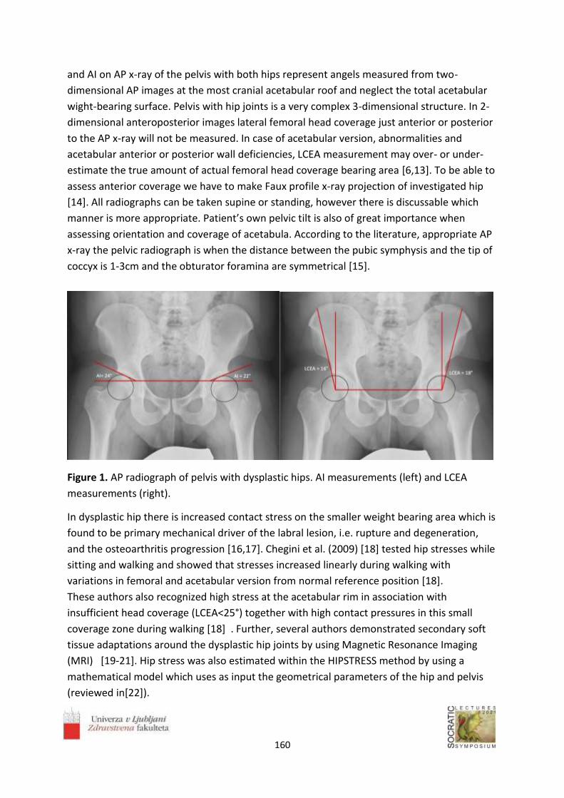
160
and AI on AP x-ray of the pelvis with both hips represent angels measured from two-
dimensional AP images at the most cranial acetabular roof and neglect the total acetabular
wight-bearing surface. Pelvis with hip joints is a very complex 3-dimensional structure. In 2-
dimensional anteroposterior images lateral femoral head coverage just anterior or posterior
to the AP x-ray will not be measured. In case of acetabular version, abnormalities and
acetabular anterior or posterior wall deficiencies, LCEA measurement may over- or under-
estimate the true amount of actual femoral head coverage bearing area [6,13]. To be able to
assess anterior coverage we have to make Faux profile x-ray projection of investigated hip
[14]. All radiographs can be taken supine or standing, however there is discussable which
manner is more appropriate. Patient’s own pelvic tilt is also of great importance when
assessing orientation and coverage of acetabula. According to the literature, appropriate AP
x-ray the pelvic radiograph is when the distance between the pubic symphysis and the tip of
coccyx is 1-3cm and the obturator foramina are symmetrical [15].
Figure 1. AP radiograph of pelvis with dysplastic hips. AI measurements (left) and LCEA
measurements (right).
In dysplastic hip there is increased contact stress on the smaller weight bearing area which is
found to be primary mechanical driver of the labral lesion, i.e. rupture and degeneration,
and the osteoarthritis progression [16,17]. Chegini et al. (2009) [18] tested hip stresses while
sitting and walking and showed that stresses increased linearly during walking with
variations in femoral and acetabular version from normal reference position [18].
These authors also recognized high stress at the acetabular rim in association with
insufficient head coverage (LCEA<25°) together with high contact pressures in this small
coverage zone during walking [18] . Further, several authors demonstrated secondary soft
tissue adaptations around the dysplastic hip joints by using Magnetic Resonance Imaging
(MRI) [19-21]. Hip stress was also estimated within the HIPSTRESS method by using a
mathematical model which uses as input the geometrical parameters of the hip and pelvis
(reviewed in[22]).

161
3. Types of acetabular dysplasia
Researchers tend to differentiate three types of acetabular under coverage depending on
the deficient area of acetabulum. Different types of dysplasia give different clinical pictures
and radiological findings. The most common is lateral (global) type where LCEA and AI angles
measured on the standard AP x-ray of pelvis show superolatelar undercoveage of acetabuli
[6],[23]. Anterosuperior type is measured by LCEA angle on faux profile of acetabular x-ray.
Third type is the posteriorsuperior type of acetabular undercoverage [24,25]. Anterior and
posterior types of dysplasia can have normal values of LCEA and AI on standard AP x-ray of
pelvis [25]. Differentiating different types of dysplasia is important since each has specific
clinical presentation.
4. Symptoms of different types of acetabular dysplasia
Lateral (global) dysplasia is the main type of acetabular instability with deficiency of
superolatelar part of acetabulum. Further it can also have different degrees of anterior or
posterior acetabular deficiency at the same time [25]. Patients often have symptoms of
static overload such as pain with prolonged standing and/or abductor fatigue such as pain
with prolonged walking or jogging. On anterior-posterior x-ray of the whole pelvis with both
hips, the LCEA will be reduced (<25°) and there may be also features of anterior or posterior
deficiency [7,23].
Anterior dyslasia represents anterior under coverage of acetabulum and can be suspected
when patient has shortened stride length, anterior hip pain in late stance phase and
increased pain while wearing high heels. Pain resembles to full extension and external
rotation of the involved hip joint where femoral head is pushed anteriorly from behind. On
physical exam, patient has increased internal rotation of the hip, may have a positive
anterior apprehension test and/or pain with anterior pressure on the femur with the hip in
an extended position, during so called prone apprehension-relocation test. The anterior
instability may include a loss of anterior acetabular coverage or excessive anteversion of the
acetabulum and/or the proximal femur. Radiographically, anterior acetabular deficiency is
suggested by decreased anterior wall index (AWI), reduced anterior acetabular sector angle
(AASA) by CT, and many times normal LCEA [23].
Posterior dysplasia results from posterior under coverage of the acetabulum [24]. Patients
can be recognized by posterior hip pain and posterior instability symptoms (increased
external rotation), however anterior symptoms does not exclude posterior dysplasia since
there can be anterior impingement simultaneously due to increased acetabular retroversion
[25,26]. Patients would complain of increased pain when climbing stairs or walking on an
incline. Steeper the incline, bigger the pressure of femoral head towards posterior
acetabular wall and greater the instability (pain). Patients may have a history of treating
piriformis syndrome and may have vague neurologic symptoms in the distribution of the
sciatic nerve. Clinically, the posterior apprehension test may be positive. Radiographically,

162
posterior acetabular deficiency is suggested by a positive posterior wall sign, positive ischial
spine sign, positive crossover sign, a decreased posterior wall index (PWI), reduced posterior
acetabular sector angle (PASA) by CT, and normal LCEA [23].
5. Treatment of Acetabular Dysplasia
Current treatment depends on age of the patient. Complex reorientational periacetabular
osteotomies (PAOs) remain the golden standard for treatment of adult hip dysplasia. There
are many types of PAOs described. The goal of PAO is to correct and to optimize acetabular
alignment for improved hip biomechanics. The most commonly performed PAOs are triple
pelvic osteotomy (e.g. Tönnis type PAO) rotational (curved) acetabular osteotomy (RAO) and
Bernese PAO.
6. Conclusion
Hip dysplasia is a complex three-dimensional deformity. Traditional diagnostic criteria based
on 2-D imaging are not optimal to accurately capture all cases of acetabular dysplasia. The
future of treatment of acetabular dysplasia is to fully comprehend pathoanatomy and to
make surgical plan which would provide correction of acetabular position in order to
optimize the pressure distribution in the hip joint.
As the three-dimensionally-derived CT reconstruction is the new standard we can not only
better understand different types of acetabular dysplasia but also make simulations of
individual hip movements (Figure 2). In addition to 3D planning there is a great need to
precisely execute the surgical plan. Intraoperative navigation is the next expected
technological advance in order to provide safer osteotomies and to obtain more reliable and
accurate correction. Technology is already used in some centers and enables real-time
tracking of osteotomes in relation to 3-D anatomy of the pelvis followed by guided
reposition of the osteotomized acetabular fragment. Surgical navigation systems have been
developed for surgical treatment of hip dysplasia based on real-time biomechanical
feedback (Lepistö J, 2008).
Dedicated software system provides measurement of contact surface angles in three
dimensions and simultaneously estimates biomechanical loading pattern in means of joint
pressure at weight bearing area during distinct activities of daily living.
Surgical navigation systems enable real-time tracking of instruments and full 3-D
intraoperative assessment during execution of surgical plan. Despite limited scientific
evidence, advanced 3-D planning and intraoperative navigation seem to be sufficiently
reliable and accurate for clinical application in hip preservation surgery. Evolution of
planning systems based on MR imaging and optimization of imageless navigation systems is
expected to eliminate potential harmful effect of preoperative and intraoperative radiation
exposure on patient`s health. At the moment, 3-D planning and surgical navigation are time
consuming, require supportive technical personnel and are relatively expensive. Before
becoming widely accepted, automatization of certain steps, e.g. motion analysis and

163
intraoperative registration, is necessary. Ultimate intention is to incorporate elements of
robotic assistance into surgical navigation systems. Robotic surgery may also enable
surgeons to perform more complex and precise tasks in restricted spaces as they are already
used in some other industries [5].
Figure 2. Planning pelvic cuts of pelvic acetabular osteotomy by on CT-based 3-D model
utilizing EBS software (Ekliptik, Slovenia) From [5].
References
1. Klisic, PJ. Congenital dislocation of the hip--a misleading term: brief report. J Bone Joint Surg
[Br] (1989), 71(1):136.
2. Mavčič, B., Antolič V., Brand R. et al. Weight bearing area during gait in normal and
dysplastic hips. Pflügers Arch (2000), r213–r214.
3. Henak CR., Abraham CL., Anderson AE., Maas SA, Ellis BJ, Peters CL, et al. Patient-specific
analysis of cartilage and labrum mechanics in human hips with acetabular dysplasia.
Osteoarthritis Cartilage (2014), 22(2):210-7.
4. Van Bosse H., Wedge JH., Babyn P. How are dysplastic hips different? A three-dimensional
CT study. Clin Orthop Rel Res (2015), 473(5):1712-23.
5. Stražar, K. Computer assistance in hip preservation surgery—current status and introduction
of our system. Int Orthop (2020).

164
6. Bali, K., K. Smit, M. Ibrahim, S. Poitras, G. Wilkin, R. Galmiche, E. Belzile, P. E. Beaulé. ottawa
classification for symptomatic acetabular dysplasia: assessment of interobserver and
intraobserver reliability. Bone & Joint Research (2020), 242–249.
7. TG, Barlow. Early diagnosis and treatment of congenital dislocationof the hip. Proc Royal Soc
Med (1963), 804-6.
8. Barlow, TG. CONGENITAL DISLOCATION OF THE HIP. EARLY DIAGNOSIS AND 438 TREA
TMENT. London Clinic Med J (1964), 47-58.
9. Ortolani, M. Congenital hip dysplasia in the light of early and very early diagnosis. Clin
Orthop Rel Res (1976), 6-10.
10. Nunley RM, Prather H, Hunt D, Schoenecker PL, Clohisy JC. Clinical presentation of
symptomatic acetabular dysplasia in skeletally mature patients. J Bone Joint Surg [Am]
(2011), 17-21.
11. Ciglič M, Stražar K. Plain radiography in femoroacetabular impingement syndrome.
Zdravniški vestnik, 83, 6 (2014), 475-483.
12. Tönnis, D. Normal values of the hip joint for the evaluation of X-rays in children and adults.
Clin Orthop Relat Res (1976), 39–47.
13. Agricola R., Heijboer MP., Roze RH., Reijman M., Bierma-Zeinstra SM., Verhaar JA., et al.
Pincer deformity does not lead to osteoarthritis of the hip whereas acetabular dysplasia
does: acetabular coverage and development of osteoarthritis in a nationwide prospective
cohort study (CHECK). Osteoarthritis Cartilage (2013), 1514-21.
14. LequesneMG, LaredoJD. Thefauxprofil(oblique view) of the hip in the standing position.
Contribution to the evaluation of osteoarthritis of the adult hip. Ann Rheum Dis, 57 (1998),
676–81.
15. Clohisy JC, Carlisle JC, Beaulé PE, et al. A systematic approach to the plain radiographic
evaluation of the young adult hip. J Bone Joint Surg [Am], 90 (2008), 47-66.
16. Wyles CC, Heidenreich MJ, Jeng J, Larson DR, Trousdale RT, Sierra RJ. The John Charnley
Award: redefining the natural history of osteoarthritis in patients with hip dysplasia and
impingement. Clin Orthop Rel Res (2017), 336-50.
17. Chegini S, Beck M, Ferguson SJ. The effects of impingement and dysplasia on stress
distributions in the hip joint during sitting and walking: a finite element analysis. J Orthop
Res (2009), 195-201.
18. KS, Rakhra. Magnetic resonance imaging of acetabular labral tears. J Bone Joint Surg [Am]
(2011), 28-34.
19. Leunig M, Podeszwa D, Beck M, Werlen S, Ganz R. Magnetic resonance arthrography of
labral disorders in hips with dysplasia and impingement. Clin Orthop Relat Res (2004), 74-80.
20. Haefeli PC, Steppacher SD, Babst D, Siebenrock KA, Tannast M. An increased iliocapsularis-
to-rectus-femoris ratio is suggestive for instability in borderline hips. Clin Orthop Rel Res
(2015), 3725-34.
21. Kralj-Iglič V. Validation of mechanical hypothesis of hip arthritis development by HIPSTRESS
method. In: Osteoarthritis: progress in basic research and treatment, Intech (2015), 131-156.

165
22. Wilkin GP, Ibrahim MM, Smit KM, Beaulé PE. A Contemporary Definition of Hip Dysplasia
and Structural Instability: Toward a Comprehensive Classification for Acetabular Dysplasia. J
Arthroplasty (2017), S20-S27.
23. Nepple JJ, Wells J, Ross JR, Bedi A, Schoenecker PL, Clohisy JC. Three Patterns of Acetabular
Deficiency Are Common in Young Adult Patients With Acetabular Dysplasia. Clin Orthop Rel
Res (2016).
24. Dandachli W, Islam SU, Liu M, Richards R, Hall-Craggs M, Witt J. Three-dimensional CT
analysis to determine acetabular retroversion and the implications for the management of
femoro- 412 acetabular impingement. J Bone Joint Surg [Br] (2009), 1031-6.
25. Cross MB, Fabricant PD, Maak TG, Kelly BT. Impingement (acetabular side). Clin Sports Med
(2011), 379-90.
26. Steppacher SD, Albers CE, Siebenrock KA, Tannast M, Ganz R. Femoroacetabular
impingement predisposes to traumatic posterior hip dislocation. Clin Orthop Rel Res (2013),
1937-43.
27. Lepistö J, Armand M, Armiger RS. Periacetabular osteotomy in adult hip dysplasia -
developing a computer aided real-time bio- mechanical guiding system (BGS). Suom Ortoped
Traumatol, 31 (2008), 186–190.
28. Mayer SW, Abdo JC, Hill MK, Kestel LA, Pan Z, Novais EN. Femoroacetabular Impingement Is
Associated With Sports-Related Posterior Hip Instability in Adolescents: A Matched-Cohort
Study. Am J Sports Med (2016), 2299-303.
bone 347 and joint surgery British volume (1989), 71(1):136.

166

167
______________
EXTRACELLULAR VESICLES AND THEIR USE IN INNER EAR
Nejc Steiner1,*, Saba Battelino1
1University Medical Centre Ljubljana, Slovenia, Department of Otorhinolaryngology and
Cervicofacial Surgery
Abstract
Hearing loss is a substantial health issue that affects approximately 18.5% of the world
population. Currently there is no medical treatment that would cure cochlear malfunction.
Once the hearing deteriorates only hearing rehabilitation with hearing aids and implantable
hearing devices can be offered to the patients. To overcome this global health problem
novel methods of treatment are being studied. Extracellular vesicles could have regenerative
and therapeutical effect on the inner ear. Gene therapy is another new developing
treatment. The delivery of therapeutic agents to the inner ear presents an obstacle and a lot
of studies are investigating the best way to overcome it. Studies show that extracellular
vesicles may serve as nanocarriers for the delivery of different agents that will act against
preventing and treating hearing loss.
___________________

168
1. Introduction
Extracellular vesicles (EVs) are a heterogeneous group of nanometrically sized cell-derived
membranous structures which can originate from any cell, including plant cell or bacteria.
They were found in many body fluids including platelet and extracellular vesicle rich plasma
(PVRP) which is a blood-derived product with immune, haemostatic and regenerative
effects. PVRP is expected to contain important concentrations of EVs which could be
important contributors to PVRPs effects 1.
2. Classification Classification of EVs is done based on physical characteristics, biochemical composition and
by descriptions of conditions or cell of origin. But mostly they are classified as (i)
microvesicles, formed by budding, 50–500 nm in size, and (ii) exosomes, formed by
exocytosis of intracellular compartments, 50–150 nm in size [1].
3. Mode of action of extracellular vesicles Platelets and their EVs have an essential role in hemostasis, immune response and tissue
regeneration. Their shape and size enable them to travel near the wall of the blood vessels
and detect endothelial damage 2. They secrete bioactive molecules contained in granules,
cytoplasm or cellular organelles. The largest and the most abundant are alpha granules,
which contain approximately 280 different types of proteins. Delta granules are smaller and
contain smaller molecules. Lambda, the least abundant granules, are lysosomes and contain
mainly enzymes. T-granules contain molecules which can be either simply translocated to
the extracellular surface of the plasma membrane or they can be secreted with exocytosis
3. EVs exert a physiological or pathological response when they reach a target cell, which
can be adjacent, distant or an origin cell.
The latter implies an important role of EVs in autoregulation. There are three modes of
action of EVs 1: (i) Binding with plasma cell membranous receptors (cellular signalling), (ii):
Fusion of EV and plasma membrane which causes a cytosolic release of EV cargo, (iii):
Uptake of EV by plasma membrane with endocytosis.
4. Use of extracellular vesicles in inner ear Only a few studies regarding the use of EVs in inner have been published. Platelet-derived
exosomes isolated from PVRP possess a substantial role in the proliferation, migration and
vessel formation. These roles are mediated by composition of exosomes, which are rich in
growth factors, especially bFGF, PDGF, TFG-β and VEGF 4. Many pathological processes,
immunological or degenerative disorders cause ischemia, inflammation and cause damage
to the hair cells of inner ear.
Growth factors, angiogenetic substances and immunomodulators from EVs could have
positive influence on restoration of cochlear function.
In principle, EVs could also serve as potential carriers for inner ear delivery of different
effectors. EVs could be delivered to the inner ear systemically, through round and oval
windows which are natural openings to the cochleae or through the cochleostomy, which is
an opening drilled into cochlea. 5 To access the inner ear from the external ear canal, a

169
tympano-meatal flap needs to be lifted, like in procedure called tympanoplasty 6. The
round window membrane is the only delivery portal to the inner ear that does not require
bone perforation. Recent advances in microneedle fabrication enable the safe membrane
perforation with polymeric and gold-coated metallic microneedles as a mean to enhance the
rate of agent delivery from the middle ear to the inner ear 7.
The agents can be either injected into the inner ear or they could be potentially released
from cochlear implant electrode which is inserted into cochlea. To our knowledge the
delivery of EVs with cochlear implant electrode has not been studied yet but cochlear
implant electrode has already been used for delivery of different medications such as
glucocorticosteroids, antiapoptotic substances, or neurotrophins to the inner ear and thus
might be used for delivery of EVs in the future 8. Studies of delivering other agents
including neurotrophins, antiapoptotics, cell therapy, gene therapy, and anti-inflammatory
drugs are mostly in preclinical stages 9.
Disabling hearing loss is a big health issue that affects approximately 6.1% of the world
population 10. It can be present at birth or it can be acquired at any time afterwards.
About half of all congenital hearing losses, which incidence is 1:1,000 births 11, have a
defined genetic cause. Currently there is no medical treatment that would cure deafness.
Once the hearing deteriorates there is not much that we can do except to offer hearing
rehabilitation with hearing aids and implantable hearing devices. Novel methods of
treatment are being studied to treat this global health problem. One of the new methods is
gene therapy.
Cochlea is the part of inner ear involved in hearing. It is surgically accessible and local
application of agents into a relatively immune-protected environment is possible, thus gene
therapy using viral vectors is an attractive approach for treating hearing loss. For congenital
recessive deafness, gene addition is possible and for congenital dominant forms of deafness,
silencing or correcting the mutated gene should be done 10. Gene therapy could also be
used in the future for age-related hearing loss by targeting pathways involved in hair cell or
spiral ganglion neuron survival (for example - neurotrophic factors 12 or antioxidant
proteins 13.
The cochlea has two types of hair cells. Inner hair cells and outer hair cells 14. Most
deafness genes that are known to affect hair cell function are expressed in both cell types,
so gene therapy strategy should target both inner and outer hair cells 15.
Currently, gene therapy for hearing disorders is still not yet as advanced because the
delivery of agents to the inner ear is still inefficient. The major limitation of gene therapy for
the cochlea is the relative inefficiency of vectors that mediate transgene expression in hair
cells. In order to achieve better delivery to the inner ear B György et. al. (2017)[5]
demonstrated that a vector, exosome-associated AAV, is a potent carrier of transgenes to all
inner ear hair cells and may be useful for gene therapy of deafness. They injected the agent
in vivo into cochlea through round window membrane and through cochleostomy. Injection
through round window membrane resulted in higher transduction of inner hair cells and
injection through cochleostomy resulted in higher transduction of outer hair cells.
Cochleostomy results were more variable and there were more instances with very low

170
expression 5. Promising results of gene therapy have been showed in studies regarding
Usher syndrome, where not only hearing but also vision is deteriorating over time 16.
Acquired hearing loss can happen due to trauma, infection, ototoxic medication and loud
noises. In the past, noise induced hearing loss was thought to be caused primarily by noise,
but today more and more children and adolescents are suffering from hearing loss caused
by exposure to loud music. We could say that the world we live in is getting louder. In
addition to all the loud machines, the development of a louder environment was mainly
influenced by the technological development of loud speakers, headphones, portable music
players and smartphones. Their invention simplified the way we listen to music, but it also
brought with it a great risk, namely hearing impairment among young people. Many people
are unaware of the dangers posed by audio devices. They should be aware of the fact that
these seemingly innocent devices can cause irreparable hearing loss 17. Data from studies
conducted in countries with a medium and high standard of living, analysed by the WHO,
show that among teenagers and young adults aged 12 to 35, almost 50% are exposed to the
dangerous volume of music from personal audio devices. However, around 40% are exposed
to the potentially harmful intensity of music in entertainment venues 18.
Drug and noise related hearing loss are associated with inflammatory responses in the inner
ear 19. Inflammation is a normal biological reaction aimed at restoring tissue functionality
and its homeostasis. It is mostly divided into two phases: initiation and resolution 20. The
start of inflammation is characterized by the up-regulation of pro-inflammatory mediators.
The resolution phase, which starts at the time when the inflammatory response peaks, is an
active process achieved mostly by the action of specialized protein and lipid pro-resolving
mediators 21. Stimulation of these pro-resolving pathways associated with cochlear
inflammatory processes could be an important therapeutic approach for preventing drug
and noise related hearing loss 22.
Kalinec et. al. (2019) [23] provided evidence that auditory hair cells generated abundant EVs
that can be loaded with cocktail of molecules (a combination of pro-resolving mediators,
specialized proteins and lipids) aimed at accelerating inflammation resolution and improving
the organ response to inflammation damage and thus prevent or alleviate drug and noise
related hearing loss 23. They accelerate the return to homeostasis by modifying the
immune response rather than by inhibiting inflammation 24.
Hair cell death and consequent hearing loss are common results of treatment with ototoxic
drugs, including the widely used aminoglycoside antibiotics. Breglio et. al. (2020) [25]
studied how to decrease the negative effect of ototoxic drugs. They showed that in response
to heat stress, inner ear tissue releases exosomes that improved the survival of hair cells
exposed to the aminoglycoside antibiotic neomycin, whereas inhibition or depletion of
exosomes from the extracellular environment abolished the protective effect of heat
shock 25.
171
5. Conclusion EVs have a role in intercellular communication of the inner ear and can mediate
nonautonomous hair cell survival. Their delivery to the inner ear currently present one of
the challenges that researchers are trying to overcome. Anatomical position, microscopic
dimensions and very sensitive cell structures of the inner ear present challenges that need
to be faced in research. In the future, extracellular vesicles may also serve as nano-carriers
for the delivery of therapeutic agents for treating hearing loss.
References 1. Vozel D, Božič D, Jeran M, Jan Z, Pajnič M, Pađen L, et al. Treatment with platelet- and
extracellular vesicle-rich plasma in otorhinolaryngology-a review and future perspectives. In: Advances in Biomembranes and Lipid Self-Assembly [Internet]. Academic Press; 2020 [cited 2020 Sep 22].
Available from: http://www.sciencedirect.com/science/article/pii/S2451963420300224 2. Holinstat M. Normal platelet function. Cancer Metastasis Rev. 2017 Jun;36(2):195–8.
doi: 10.1007/s10555-017-9677-x 3. Morrell CN, Aggrey AA, Chapman LM, Modjeski KL. Emerging roles for platelets as immune
and inflammatory cells. Blood. 2014 May 1;123(18):2759–67. doi: 10.1182/blood-2013-11-462432 4. Guo S-C, Tao S-C, Yin W-J, Qi X, Yuan T, Zhang C-Q. Exosomes derived from platelet-rich
plasma promote the re-epithelization of chronic cutaneous wounds via activation of YAP in a diabetic rat model. Theranostics. 2017;7(1):81–96. doi: 10.7150/thno.16803
5. György B, Sage C, Indzhykulian AA, Scheffer DI, Brisson AR, Tan S, et al. Rescue of Hearing by Gene Delivery to Inner-Ear Hair Cells Using Exosome-Associated AAV. Mol Ther. 2017 01;25(2):379–91. doi: 10.1016/j.ymthe.2016.12.010
6. Steiner N, Battelino S. Techniques of tympanoplasty in otorhinolaryngology. In Ljubljana; 2020 [cited 2021 Jan 30]. p. 153–4.
Available from: https://www.zf.uni-lj.si/images/stories/datoteke/Zalozba/Sokratska_2020.pdf 7. Aksit A, Rastogi S, Nadal ML, Parker AM, Lalwani AK, West AC, et al. Drug delivery device for
the inner ear: ultra-sharp fully metallic microneedles. Drug Deliv Transl Res. 2021 Feb;11(1):214–26. doi: 10.1007/s13346-020-00782-9
8. Plontke SK, Götze G, Rahne T, Liebau A. Intracochlear drug delivery in combination with cochlear implants : Current aspects. HNO. 2017 Jan;65(Suppl 1):19–28.
doi: 10.1007/s00106-016-0285-9 9. Chin OY, Diaz RC. State-of-the-art methods in clinical intracochlear drug delivery. Curr Opin
Otolaryngol Head Neck Surg. 2019 Oct;27(5):381–6. doi: 10.1097/MOO.0000000000000566 10. Global, regional, and national incidence, prevalence, and years lived with disability for 310
diseases and injuries, 1990–2015: a systematic analysis for the Global Burden of Disease Study 2015. Lancet. 2016 Oct 8;388(10053):1545–602.
Doi:https://doi.org/10.1016/S0140-6736(16)31678-6 11. Shearer AE, Hildebrand MS, Smith RJ. Hereditary Hearing Loss and Deafness Overview. In:
Adam MP, Ardinger HH, Pagon RA, Wallace SE, Bean LJ, Mirzaa G, et al., editors. GeneReviews® [Internet]. Seattle (WA): University of Washington, Seattle; 1993 [cited 2021 Jan 27]. Available from: http://www.ncbi.nlm.nih.gov/books/NBK1434/

172
12. Deafness | Britannica [Internet]. [cited 2021 Jan 27]. Available from: https://www.britannica.com/science/deafness
13. Deafness and hearing loss [Internet]. [cited 2021 Jan 27]. Available from: https://www.who.int/news-room/fact-sheets/detail/deafness-and-hearing-loss
14. Flint PW, Haughey BH, Robbins KT, Thomas JR, Niparko JK, Lund VJ, et al. Cummings Otolaryngology - Head and Neck Surgery. Elsevier Health Sciences; 2014. 4198 p.
15. Schilder AG, Chong LY, Ftouh S, Burton MJ. Bilateral versus unilateral hearing aids for bilateral hearing impairment in adults. Cochrane Database Syst Rev [Internet]. 2017 Dec 19 [cited 2021 Jan 27];2017(12). Available from: https://www.ncbi.nlm.nih.gov/pmc/articles/PMC6486194/
16. Géléoc GGS, El-Amraoui A. Disease mechanisms and gene therapy for Usher syndrome. Hearing Research. 2020 Sep 1;394:107932. doi: 10.1016/j.heares.2020.107932
17. eSiNAPSA | Kako nam lahko glasna glasba »vzame« sluh in povzroči tinitus [Internet]. [cited 2021 Jan 30]. Available from: https://www.sinapsa.org/eSinapsa/stevilke/2015-10/183/kako_nam_lahko_glasna_glasba_vzame_sluh_in_povzroci_tinitus
18. Shrivastava SR, Shrivastava PS, Ramasamy J. Joining hands with World Health Organization initiative Make Listening Safe. Noise and Health. 2015 Jan 5;17(76):173.
doi: 10.4103/1463-1741.155854 19. Kaur T, Borse V, Sheth S, Sheehan K, Ghosh S, Tupal S, et al. Adenosine A1 Receptor Protects
Against Cisplatin Ototoxicity by Suppressing the NOX3/STAT1 Inflammatory Pathway in the Cochlea. J Neurosci. 2016 Apr 6;36(14):3962–77. doi: 10.1523/JNEUROSCI.3111-15.2016
20. Lai P, Weng J, Guo L, Chen X, Du X. Novel insights into MSC-EVs therapy for immune diseases. Biomark Res [Internet]. 2019 Mar 18 [cited 2021 Jan 28];7. Available from: https://www.ncbi.nlm.nih.gov/pmc/articles/PMC6423844/
21. Perretti M, Leroy X, Bland EJ, Montero-Melendez T. Resolution Pharmacology: Opportunities for Therapeutic Innovation in Inflammation. Trends Pharmacol Sci. 2015 Nov;36(11):737–55. doi: 10.1016/j.tips.2015.07.007
22. Kalinec GM, Lomberk G, Urrutia RA, Kalinec F. Resolution of Cochlear Inflammation: Novel Target for Preventing or Ameliorating Drug-, Noise- and Age-related Hearing Loss. Front Cell Neurosci. 2017;11:192. doi: 10.3389/fncel.2017.00192
23. Kalinec GM, Gao L, Cohn W, Whitelegge JP, Faull KF, Kalinec F. Extracellular Vesicles From Auditory Cells as Nanocarriers for Anti-inflammatory Drugs and Pro-resolving Mediators. Front Cell Neurosci [Internet]. 2019 [cited 2021 Jan 26];13. Available from: https://www.frontiersin.org/articles/10.3389/fncel.2019.00530/full
24. Dalli J, Serhan CN. Identification and structure elucidation of the pro-resolving mediators provides novel leads for resolution pharmacology. Br J Pharmacol. 2019 Apr;176(8):1024–37. doi: 10.1111/bph.14336
25. Breglio AM, May LA, Barzik M, Welsh NC, Francis SP, Costain TQ, et al. Exosomes mediate sensory hair cell protection in the inner ear. J Clin Invest. 130(5):2657–72.
doi: 10.1172/JCI128867

173

174
_______________
Charcot neuroarthropathy: What lies beneath?
Schara K 1, Kralj Iglič V 2
1University Medical center Ljubljana, Department of Orthopaedic Surgery, Ljubljana,
Slovenia
2University of Ljubljana, Faculty of Health Sciences, Laboratory of Clinical Biophysics,
Ljubljana, Slovenia
Abstract
This work describes the mechanisms for the development of diabetic Charcot
neuroarthropathy (CNA) syndrome. CNA is a chronic, progressive disease causing severe foot
and ankle deformities which present a serious and potentially limb-threatening lower-
extremity complication of diabetes. One lower limb is lost to diabetes every 30 seconds in
the world. It is currently believed that once the disease is triggered, it is mediated through a
process of uncontrolled inflammation in the foot leading to osteolysis and progressive
destruction of bone and joint structures. Until the intertwining of several interrelated
factors and mechanisms initiating bone and joint destruction is confirmed, challenges of
early recognition and treatment of the disease remain crucial for the limb preservation.
__________________

175
1. Introduction
CNA is a chronic, progressive disease causing severe foot and ankle deformities which
present a serious and potentially limb-threatening lower-extremity complication of diabetes
1 It is characterized by painful or painless bone and joint destruction in limbs that have lost
sensory innervation. Peripheral neuropathy in diabetes is usually presenting as a distal,
symmetric polyneuropathy therefore CNA is predominately affecting the joints of the foot
and ankle 2CNA is considered a rare disease with estimated prevalence of 1 to 2% of
patients with diabetic neuropathy but the true prevalence in patients with diabetes mellitus
is not known because it is likely that many cases remain undiagnosed due to a lack of
recognition of the disease 1.
The Charcot foot can be classified in terms of clinical stage, anatomical localization and stage
of natural history. In clinical practice, the Charcot foot can be classified into the acute
(active) and chronic (inactive) stage while other classifications are based on anatomical
localization and staging of natural history 3.
The earliest manifestation of active, acute CNA is persistent unilateral swelling with only
mild to modest pain or discomfort. On clinical examination, the foot is warm, swollen and
erythematous, loss of sensation is diagnosed (Figure 1). This initial clinical picture resembles
various conditions like cellulitis, osteomyelitis, deep vein thrombosis, lymphoedema,
arthritis or acute gout and can be misdiagnosed as such 1. Bone and joint involvement
during active disease present on the X ray with a variety of changes including
demineralization, joint dislocation, bone destruction, fractures, bone fragmentation and
periosteal reaction 4.
All these changes can occur very rapidly with a normal x-ray deteriorating to grossly abnormal
within a few weeks 3 (Figure 2). If the condition is correctly diagnosed and the patient is
appropriately treated, the local inflammation will subside and further bony destruction can
be minimized or avoided 5. If treatment is delayed or if left untreated mechanical stress may
lead to ligament strain, fractures and dislocations resulting in severe foot deformity and/or
joint instability 5,1.
The mainstay of treatment of Charcot neuroarthropathy is immobilization in a total contact
cast, which increases the total surface area of contact to the entire lower extremity,
distributing pressure away from the foot. In order to avoid weight bearing of the affected
foot in active stage of CNA, the patient should use crutches or wheelchair 6.

176
Figure 1. Clinical presentation of CNA (patient’s left).
Figure 2. A: Early stage of CNA. On the X-ray midfoot joint anatomy is still preserved, ossifications marked in the circle indicate the disease. B: Progressive stage of CNA. Bone

177
anatomy is destructed, abundant ossification above the joints of dorsal midfoot can be observed. Progressive X-ray changes are marked in the circle.
2. Mechanisms of CNA disease
There is no singular cause for the development of the Charcot foot therefore our knowledge
on pathogenesis and treatment options remain limited. Initially the condition was explained
by neurotraumatic and neurovascular theories, recently active CNA is considered to be an
uncontrolled inflammatory syndrome affecting bone metabolism 7. CNA occurs as a
consequence of an imbalance of activity of two types of cells: osteoblasts and osteoclasts
8. This imbalance leads to enhanced bone lysis and fracture and is driven by inflammation
9,1. Increased amounts of the pro-inflammatory cytokines TNF-alpha, IL-1 and IL-6,
decreased amounts of the anti-inflammatory cytokines IL-4 and IL-10 and increased
amounts of the surface molecules CD40, CD80, and CD86 were found in monocytes of
patients with CNA 10. Increased concentrations of TNF-alpha and IL-6 were found also in
serum of patients with CAN 11.
CN has been associated with an increase in advanced glycation end products (AGEs) in blood
that is often caused by poorly controlled diabetes and/or inflammation due to
hyperglycemia and oxidative stress 9. It was found that AGEs stimulate apoptosis of human
mesenchymal stem cells 12 and osteoblasts 13 by binding to receptor for AGE (RAGE) on
their membranes 9. It was also observed that the concentration of soluble RAGE in blood
plasma of patients with CN was lower than in healthy subjects and also lower than in
patients with diabetes mellitus 14. Possibly due to a deficiency of soluble RAGE, binding of
AGE to soluble RAGE was decreased with a deleterious consequence of AGE binding to
membrane fraction of RAGE 14. As these processes are regulated by many pathways
involving the osteoblast and the osteoclast lineages 15,16,9 it is evident that intercellular
communication is crucial for bone remodeling.
Extracellular vesicles (EVs) and nanotubules were reported as important participants in
cellular processes 17 (Figure 3). Experimental evidence indicates that EV concentration is
increased in some diseases e.g. cardiovascular diseases, autoimmune disorders,
inflammation and infection 18. Increased levels of extracellular vesicles have been found in
the peripheral blood of patients with diabetic neuropathy 19. Studies confirmed that EVs
derived from patients with diabetic neuropathy display a higher level of leukocytes- and
monocytes-derived EVs 19.
Osteoclasts, cells responsible for bone resorption in CF are members of the monocyte and
macrophage group. A high content of inflammatory cytokines G-CSF, GM-CSF, IL-1-ra and IL-
2 which may enhance osteoclasts differentiation was detected in CNA disease 20. It was
shown that levels of EVs in active CNA were elevated and associated with inflammatory
parameters and increased temperature of the foot 21. Therefore it was suggested that EVs
may be useful in monitoring and predicting clinical progression of the disease 21.

178
Figure 3. Cell to cell communication: EVs and nanotubules in bone remodeling process.
3. Conclusions Few studies have included patients with active stage of CNA, therefore it is difficult to draw
conclusions on causation and the relationship factors connected with the development and
progression of the condition. A final common pathway for the pathogenesis of CNA is yet to
be determined and promising therapeutic targets have yet to be identified. Until the
intertwining of several interrelated factors and mechanisms initiating bone and joint
destruction is confirmed, challenges of early recognition and treatment of the disease
remain crucial for treatment and limb preservation.
Acknowledgements
Authors acknowledge support of Research Agency of the Republic Slovenia ARRS: program
P3-0388 and projects J1-6728 and L3-2621.
References
1. Rogers LC, Frykberg RG, Armstrong DG, et al. The Charcot foot in diabetes. Diabetes Care
2011; 34:2123. DOI: 10.7547/1010437
2. Rajbhandari S, Jenkins R, Davies, C et al. Charcot neuroarthropathy in diabetes mellitus.
Diabetologia, 2002, 45: 1085–1096. DOI:10.1007/s00125-002-0885-7
179
3. Armstrong DG, Todd WF, Lavery LA, Harkless LB, Bushman TR, The natural history of acute
Charcot's ar-thropathy in a diabetic foot specialty clinic. Diabet Med. 1997;14(5):357–363.
DOI: 10.1002/(SICI)1096-9136(199705)14:5<357::AID-DIA341>3.0.CO;2-8
4. Marmolejo VS, Arnold JF, Ponticello M, Anderson CA, Charcot Foot: Clinical clues, diagnostic
strategies, and treatment principles. Am Fam Physician. 2018, 97: 594-599.
5. Papanas N, Maltezos E, Etiology, pathophysiology and classifications of the diabetic Charcot
foot, Diabetic Foot & Ankle, 2013, 4: 1-5. DOI:10.3402/dfa.v4i0.20872
6. Frykberg RG, Eneroth M, Principles of conservative management. In The Diabetic Charcot
Foot: Principles and Management. Frykberg RG, Ed. Brooklandville, MD, Data Trace
Publishing Company, 2010, p. 93–116.
7. Jeffcoate WJ. Charcot neuro-osteoarthropathy. Diabetes Metab Res Rev. 2008, 24: S62-S65.
DOI: 10.1002/dmrr.837
8. Jeffcoate WJ. Charcot foot syndrome. Diabetic Medicine. 2015, 32: 760–770. DOI:
10.1111/dme.12754
9. Petrova NL, Shanahan CM, Neuropathy and the vascular-bone axis in diabetes: lessons from
Charcot osteoarthropathy. Osteoporos Int 2014, 25: 1197–1207. DOI: 10.1007/s00198-013-
2511-6
10. Uccioli L, Sinistro A, Almerighi C, et al., Pro-inflammatory modulation of the surface and
cytokine phenotype of monocytes in patients with acute Charcot foot. Diabetes Care 2010,
33: 350–355. DOI:10.2337/dc09-1141
11. Petrova NL, Dew TK, Musto RL, et al., Inflammatory and bone turnover markers in a cross-
sectional and prospective study of acute Charcot osteoarthropathy. Diabet Med 2015, 32:
267-273. DOI:10.1111/dme.12590
12. Kume S, Kato S, Yamagishi S, et al., Advanced glycation end-products attenuate human
mesenchymal stem cells and prevent cognate differentiation into adipose tissue, cartilage,
and bone. J Bone Miner Res 2005, 20: 1647–1658. DOI:10.1359/JBMR.050514
13. Alikhani M, Alikhani Z, Boyd C, et al., Advanced glycation end products stimulate osteoblast
apoptosis via the MAP kinase and cytosolic apoptotic pathways. Bone 2007, 40: 345–353.
DOI: 10.1016/j.bone.2006.09.011
14. Witzke KA, Vinik AI, Grant LM, Loss of RAGE Defense: A Cause of Charcot Neuroarthropathy?
Diabetes Care 2011, 34: 1617–1621. DOI:10.2337/dc10-2315
15. Nomura S, Significance of chemokines and activated platelets in patients with diabetes. Clin
Exp Immunol 2000, 121: 437-443. DOI: 10.1046/j.1365-2249.2000.01324.x
16. Schmidt AM, Yan SD,Yan SF, Stern DM, The multiligand receptor RAGE as a progression
factor amplifying immune and inflammatory responses. J Clin Invest 2001, 108: 949–955.
DOI: 10.1172/JCI14002
17. Schara K, Janša V, Šuštar V, et al., Mechanisms for the formation of membranous
nanostructures in cell-to-cell communication. Cell Mol Biol Lett 2009, 14: 636-656. DOI:
10.2478/s11658-009-0018-0

180
18. Boulanger CM, Amabile N, Tedgui A, Circulating microparticles: a potential prognostic
marker for atherosclerotic vascular disease. Hypertension 2006, 48: 180 -186. DOI:
10.1161/01.HYP.0000259667.22309.df
19. Wang Y, Chen L, Liu M, Microvesicles and diabetic complications — novel mediators,
potential biomarkers and therapeutic targets. Acta Pharmacol Sin 2014, 35: 433–443.
DOI:10.1038/aps.2013.188
20. Pasquier J, Thomas B, Hoarau-Véchot, J et al., Circulating microparticles in acute diabetic
Charcot foot exhibit a high content of inflammatory cytokines, and support monocyte-to-
osteoclast cell induction. Sci Rep 2017, 7, 16450. DOI: 10.1038/s41598-017-16365-7
21. Schara K, Štukelj R, Krek JL et al., A study of extracellular vesicle concentration in active
diabetic Charcot neuroarthropathy, European Journal of Pharmaceutical Sciences, 2017, 98:
58-63. DOI: 10.1016/j.ejps.2016.09.009

181

182
__________________
POST-COVID-19 EXPERIENCES
Romolo A1*
1University of Ljubljana, Faculty of Electrical Engineering, Laboratory of Physics, Ljubljana,
Slovenia,
Abstract
A year since the outbreak of the Corona Virus Disease 2019 (COVID-19) pandemic, the
epidemiological and clinical characteristics, pathogenesis and complications of acute stage of
the disease have been described extensively in the scientific literature. However, the disease
might have long-term effects on patients, in those that underwent severe disease as well as
in those with milder forms. In this contribution, we explored the existing evidences of the
post-COVID-19 one year after the outbreak. A search was performed in the Google Scholar
base, using selected keywords. Both patients that were hospitalized and those that were not
hospitalized may show persistent symptoms such as fatigue, wheezing and reduced lung
function at the 6-months follow-up after infection with SARS-CoV-2 indicating that in some
cases COVID-19 is not a disease from which one heals quickly.
__________________

183
Survey of the literature
SARS-CoV-2 virus affects different organs and systems in the body 1. The underlying
mechanisms are not yet completely understood. Its long-term consequences after the acute
phase of infection exhibited in COVID-19 are now the subject of scientific research by
following up of those who seem to be cured. We surveyed the literature by searching
through the Google Scholar database. Inserting the keyword Effect long term COVID-19 into the
browser returned 2565 articles. Changing the keyword to Consequence effect long term COVID-19
shrank the number of articles to 383. After screening the abstracts of these articles we modified the
keyword to: Consequences pulmonary long term COVID-19. We obtained 48 articles. We screened
the articles and outlined 6 articles which in our opinion represented best the hitherto gathered
evidences on post-COVID effects.
The outlined reports
One of the first published works on the post-COVID-19 effects was a study by Cortinovis et
al., (2020) 2. These authors found that the clinical spectrum of SARS-CoV-2 infection is
wide: from asymptomatic infection, fever, fatigue, myalgias, mild upper respiratory tract
illness to severe life-threatening viral pneumonia and death. Persisting symptoms as well as
unexpected ones were observed after the disease similar to organ dysfunction after
infection with SARS-CoV-2 2. This was supported by the findings of the physicians from the
Policlinico Gemelli in Rome (2020)3 and by Huang et al.,(2021) 4 who showed that many
people infected with SARS-Cov-2, those were hospitalized and those who were not
hospitalized, had different disorders for weeks and even six months after the disease.
Carfì et al. (2020) 3 described the outcome of 143 patients in a period of time ranging from
21 April to 29 May 2020. The average hospital stay was 13.5 days; 21 patients (15%) received
non-invasive ventilation and 7 patients (5%) received invasive ventilation. During
hospitalization, 72.7% of participants had evidence of interstitial pneumonia. Patients
meeting the World Health Organisation criteria for the cessation of quarantine (absence of
fever for 3 consecutive days, improvement of other symptoms and 2 negative test results
taken 24 hours apart) were included in the follow-up. At the release from the hospital,
patients were subjected to a full medical evaluation with detailed medical history and
physical examination. Data on all clinical features, including clinical and pharmacological
history, lifestyle factors, vaccination status and body measurements have been grouped
together in a structured electronic data collection system. In particular, data on specific
symptoms potentially related to COVID-19 were obtained using a standardised questionnaire
administered to enlistment. In the waning phase of the pandemic, starting April 21, 2020,
the Fondazione Policlinico Universitario Agostino Gemelli IRCCS in Rome, Italy, established
an outpatient post office service for patients dismissed from the hospital after recovery from
COVID-19. Patients were asked to report retrospectively the presence or absence of
symptoms during the acute phase of COVID-19 and whether each symptom persisted at the
time of the examination.

184
Patients were rated at an average of 60.3 days after the start of the first COVID-19 symptom;
at the time of evaluation, only 18 (12.6%) were completely free of any COVID-19 related
symptom, while 32% had 1 or 2 symptoms and 55% had 3 or more. None of the patients had
a fever or any signs or symptoms of acute illness. A deterioration in the quality of life was
observed in 44.1% of patients: a high percentage of individuals reported fatigue (53.1%),
dyspnoea (43.4%), joint pain (27.3%) and chest pain (21.7%). The study found that in
patients who recovered from COVID-19, 87.4% reported persistence of at least 1 symptom,
particularly fatigue and wheezing.
Huang et al., (2021) 4 conducted the study focused on the course of 1733 patients (average
age 57, 52 percent male) that were hospitalized due to COVID-19 in the Jinyintan Hospital in
Wuhan between January 7 and May 29, 2020. Six months after the onset of the first
symptoms, 76 percent of patients released from the hospital reported that they still had at
least one symptom that have persisted longer in women. The most frequently reported
symptoms were fatigue and muscle pain (63 percent) and sleep disorders (26 percent).
Nearly a quarter of patients (23 percent) said that they experienced anxiety or depression
4. In addition, changes in lung function persisting as long as six months after the onset of
the symptoms were more frequent in patients with more severe forms of the. Persistent
kidney disorders were also observed which were not detected during the hospitalization.
The most severely ill patients have also achieved worse results in a six-minute walking test.
Almost a quarter failed to walk at least 5 minutes 4. The study also considered the
antibody load at the time of maximum infection in 94 patients. Six months later, the level of
antibodies against the virus was split to less than half. To our best knowledge the study of
Huang et al. (2021) 3 considered the largest cohort of hospitalized patients. However the
authors suggested that "It is necessary to have a larger sample to better study these
postums and to measure the rate of antibodies against SARS-CoV-2".
Greenhalgh T. et al. (2020) 5 highlighted in an article published in British Medical Journal
that the consequences of COVID-19 can be extremely varied. They created an infographic
entitled “Long COVID in primary care” (https://www.bmj.com/content/370/bmj.m3026 ) for
assessment and initial management of patients with persistent problems after COVID-19.
They illustrated the symptoms with lots of drawings in a detailed summary with indications
for physicians as well as for patients.
More specifically, evidences on autoimmune diseases (often atypical) arising after the acute
phase are multiplying. Reyes Gil M. et al. (2020)6 from Montefiore Medical Center in New
York, have observed, on a hundred patients hospitalized that in 44% the presence of
markers of systemic Lupus erythematosus greatly increased in those who were previously
positive for these markers. In addition, the presence of Lupus Anticoagulant antibodies was
associated with incidence of thrombosis in patients with COVID-19 6.
185
Restivo et al., (2020) 7 reported that some patients infected with SARS-CoV-2 later
exhibited neurologic symptoms. The authors argued that these symptoms were caused by
autoimmune mechanisms. They described 3 cases without previous neurologic or
autoimmune disorders (a 64-year-old male, a 68-year-old male and a 71-year old female)
who developed diplopia and muscular fatigability some days after having high fever. The
chest CT was normal in males while in female, it revealed bilateral interstitial pneumonia.
Repetitive stimulation showed less than 60% of the neuromuscular transmission of the
facial/ulnar nerve indicating involvement of the postsynaptic neuromuscular junction. Serum
AChR antibody levels were elevated in all three patients and the diagnosis of myasthenia
gravis was indicated.
Conclusions
Although there are many patients recovered from COVID-19, systematic reports on post-
COVID-19 manifestations are yet scarce. The existing ones focus mostly on patients who
were dismissed from the hospital, as there exist records of their disease. Many cases that
were cured at home and were not subjected to testing are missing from the records. As
COVID-19 involves different organs and systems, caution is necessary that multiple and
complex symptoms may occur after the symptoms of the primary disease have ceased and
that post-COVID-19 rehabilitation is indicated 8.
References
1. Kralj-Iglič V, Gošnak Dahmane R, Griessler Bulc T, et al., From extracellular vesicles to
global environment: A cosmopolite SARS-CoV-2 Virus. Int J Clin Stud Med Case Rep
2020. 4:1-23. https://ijclinmedcasereports.com/pdf/IJCMCR-CR-00079.pdf
2. Cortinovis M, Perico N, Remuzzi G. Long-term follow-up of recovered patients with
COVID-19. Lancet 2021. 16;397(10270):173-175. doi: 10.1016/S0140-6736(21)00039-8
3. Carfì A, Bernabei R, Landi F, et al. Persistent symptoms in patients after acute COVID-
19. JAMA, 2020. 324(6):603-605. doi:10.1001/jama.2020.12603
4. Huang C, Huang L, Wang Y, Li X, Ren L, Gu X, Kang L, et all. 6-month consequences of
COVID-19 in patients discharged from hospital: a cohort study. The Lancet 2021.
397(10270):220-232. doi: 10.1016/S0140-6736(20)32656-8.
5. Greenhalgh T, Knight M, A’Court C, Buxton M, Husain L. Management of post-acute
COVID-19 in primary care. BMJ 2020; 370 doi: https://doi.org/10.1136/bmj.m3026
6. Reyes Gil M, Barouqa M, Szymanski J, et al. Assessment of lupus anticoagulant positivity
in patients with coronavirus disease 2019 (COVID-19). JAMA Netw Open 2020
3(8):e2017539. doi:10.1001/jamanetworkopen.2020.17539.
7. Restivo DA, Centonze D, Alesina A, Marchese-Ragona R, Myasthenia gravis associated
with SARS-CoV-2 infection. Ann Intern Med 2020. 73(12):1027-1028.
https://doi.org/10.7326/L20-0845
8. Romolo A, Kralj-Iglič V, Rehabilitation after COVID-19 : A review of recent experiences.
Physis 2020. 1: 11-18.

186

187
__________________
COVID-19 Health consequences management: proposed health rehabilitation
of long-term care for older adults
1Amon Mojca*, Friderika Kresal
1Institution of Higher Education Fizioterapevtika, Ljubljana, Slovenia
Abstract
Healthcare professionals must work in a balance of sustainable development with a
comprehensive way of working that emphasizes a systemic approach. The pandemic
situation underscores the need for health promotion in public health. An appropriate
integrated health strategy, which includes intensive preventive interventions at all levels of
health care, represents an opportunity to reduce health absenteeism and strengthen bio-
psycho-social health. The improved uniform health explanation and understanding can
contribute to the awareness of the individual and society. Unity of action is achieved
through common understanding and action, which can also successfully contribute to limit
the expansion of pandemic disease. The purpose of the investigated content is to encourage
social development through rehabilitation as a physiotherapeutic long-term support, which
will enable and connect long-term conditions for human health, well-being and quality of
life. The strategy of the proposal is to unite at least two or three generations and can
contribute to maintaining intergenerational understanding, empathy and coexistence.
Appropriate targeted development and research-educational activities are needed, which
can encourage a development of society that enables the preservation of the living
environment and functional independence of older adults. To conclude, the proposal can
contribute to the development of future generations in rural areas and rural development.
_________________

188
1. Introduction
Protecting the environment from increasing burdens is the fundamental condition for a
sustainable development of society. The sustainable development strategy has three pillars,
namely, environmental protection, economic development and social development. The
central element of the development of society is human. The health status of society is also
closely linked to the potential of environmental, economic and social development.
Sustainable development is the idea of developing a human society based on the
preservation of the environment and biodiversity. The concept of sustainable development
encompasses several aspects, but above all the possibility of developing each person or
society in a way that does not harm others. The oft-cited definition is that sustainable
development meets the needs of the current generations without compromising the ability
of future generations to meet their own needs 1. The exceptional global health situation in
the COVID-19 (SARS-CoV-2) period 2019-2021 reminds us of the importance of health. The
basic factors with which we can influence health are, above all, clean air, food, proper
exercise, recreation and the overall bio-psycho-social quality of life.
The aging process is accompanied by an increased risk of functional limitations, disability
and handicaps, social isolation and dependence on the help of others. Strengthening health
status with increasing longevity is essential and also a decisive factor in maintaining the
activities of several generations. The proposed measures are therefore focused on solutions
to strengthen the physical capacity of the rural population, strengthen long-term
rehabilitation physiotherapy support and intergenerational associations to maintain a
physically active lifestyle.
Over the years, the immune system changes and this affects the acquired and innate
response of the immune system. Infections, cancers and autoimmune diseases are more
common in older adults (over 65 years of age) and many factors are responsible for this
phenomenon. Age-related changes and the weakening of the innate and acquired immune
systems play the greatest role.
Stable health in old age is not only the result of inflammatory mechanisms, but also the
effectiveness of the entire system acquired over a lifetime. For this reason, fragility is also
the result of an inflammatory condition. Together with hormonal changes, nutritional
deficiencies and physical inactivity, it can lead to an essential element of aging - fragility or
sarcopenia. Immunity plays an important role in the regulation of aging mechanisms and the
occurrence of age-related diseases 2. The immune system is associated with the aging of
the endocrine, nervous, digestive, cardiovascular, and musculoskeletal systems. New
infectious diseases may shape the way of life in the future, as many epidemics and
pandemics have done in the past. Controlling this danger depends on understanding how to
make the most of the potential of our immune system. A fundamental property of the
immune system is that it has control over the whole body. An effective immune system
adapts to changes in the environment 3.

189
The literature indisputably describes in detail the many advantages of physical activity in the
period of growth and adolescence, as the movement has an exceptional educational and
developmental potential 4.
Health is also a reflection of time and space. The living quality and bio-psycho-social
condition of the individual in the home environment are important. We assume that helping
older adults usually means an unexpected change in the way of life of the entire living
structure or that it is completely focused on the female members of the family.
Dissemination of the conclusions of rehabilitation geriatrics can be a starting point for
proposals for bio-psycho-social rehabilitation assistance to older adults.
2. Methods
The purpose of the studied content is to present the importance of promoting the
prevention of disease, illness and health discomfort as a form of long-term rehabilitation
and the deinstitutionalization of older adults.
2.1 Suggested methods
Many adult individuals may be overwhelmed due to constant activities and sudden involvement
in additional nursing or rehabilitation support for older adults. In case of sudden health
weakness, movement impairment and incapacity, they take care of elderly relatives without
prior education. We assume that the proposed measures of long-term health rehabilitation
support may lead to a change that significantly affects the health of the wider society.
2.2 Educational and developmental support of long-term rehabilitation support
To develop an appropriate strategy, it will be necessary to study the field situation,
examples of good practices, long-term care options and examples of pilot projects for the
formalization of long-term care in Slovenia. Based on the joint assessment, we will examine
the obstacles and possibilities of long-term care in terms of the development of new staff,
employment opportunities, additional knowledge and, last but not least, intergenerational
coexistence.
In the proposal of measures for long-term rehabilitation support, we propose the creation of
educational and developmental support for long-term rehabilitation care. Physiotherapists
are obliged to contribute to the development of those activities that strengthen and
improve physical performance and motor skills and quality of life. In addition, the weakened
ability to communicate and cooperate between three different generations is increasingly
present in Slovenia. The ability to recognize and express oneself in relationships and
empathize with others is also insufficiently developed, which undermines the development
of healthy, cooperative and satisfied relationships, which are crucial for living together and
well-being.
Such mutual bio-psycho-social expert assistance can contribute to improving the
understanding, empathy and cooperation of several generations.

190
Results
Expected results – the possibilities of formal health rehabilitation care for older adults
We believe that the form of formal long-term health rehabilitation support for older adults
can significantly affect the quality of aging of many individuals.
3. Discussion
The exceptional global health situation in the period 2019-2021 5 reminds us of the
importance of maintaining a healthy narrower and wider living environment for health
promotion. The basic factors with which we can influence health are, above all, clean air,
food and proper exercise or recreation. The basics of clinical physiotherapy exercise
emphasize the laws of therapeutically safe exercise 6,7. The evolution of rehabilitation
exercise shifts the boundaries of rehabilitation spaces to the external environment. Fresh
air, regardless of weather conditions, with the right equipment, is the optimal choice for
physical activity, which can contribute to maintaining bio-psycho-social capacity and quality
of life 8. After all, home is home. Usually, the attachment to the home environment only
deepens with age. Changing the living environment is burdensome or even fatal for many.
3.1 Educational and support options for long-term health rehabilitation support
Geriatrics is a demanding medical science due to its multimorbidity. It is right that older
adults have the help they really need. The informal long-term care does not allow a
professional help that a person would really need. Lower secondary health education can
offer certain knowledge and health care options in the form of formal long-term care. Post-
vocational additional education would provide additional realistic solutions to the problem.
Moreover, the possibilities are promising in the development of special health conditions
that individuals could gain through shorter forms of education to provide some non-
essential health services. Healthcare requires medical education, but it is not necessarily
present or feasible. It is worth considering the possibilities of developing short-term
education in terms of maintaining functional support that can be provided by individuals
with or without health education to help older adults. The nature of work in this area is not
defined and it is necessary to draw clear boundaries between what is medical, clinical,
physiotherapeutic, rehabilitation, therapeutic or long-term nursing care at home and to
express the individual guidelines. In the future, it is necessary to study the duties and rights
and the way of work of individual care.
Implementation options are probably allowed by contract work or a form of additional /
supplementary work. It is also important for the field to create minimum needs either for
the implementation of nursing or rehabilitation activities at home (home study, standards,
equipment) or the need for field work (work at home for older adults). Field analyses and
case studies can be the basis for proposals for the measure of “deinstitutionalisation” of
older adults and intergenerational participation of young and older adults on farms.

191
3.2 Promotion of clinical physical activity for all generations
Morbidity prevention also includes the responsibility of maintaining a physically active
society 9-13. The mission of the health professions is to restore, maintain and strengthen
the health of the individual. Improving society's health can contribute to efficiency, safety
and quality of life. The purpose of the studied content was to present the importance of
promoting the prevention of disease, illness and health discomfort as a form of long-term
rehabilitation or deinstitutionalization of older adults. The content of the proposal refers to
educational and developmental opportunities and activities that highlight physical activity as
a central component for maintaining the health of the wider community.
The starting point of the proposal initiates from the legality of clinical exercise, which is a
central element of rehabilitation physiotherapy 6,7,12-14. We believe that the health
promotion of physical activity is an exceptional opportunity to develop the intergenerational
understanding that conditions the bio-psycho-social health of society and rural development.
Historical records describe disease as the result of mystical forces and according to early
scientific descriptions disease encompasses environmental factors and living habits in life
(Hippocrates, 460-370 BC; Galen, 129-210 AD). Hippocrates emphasized the benefits of
physical activity by stating, "If we can devote an adequate amount of nutrients and physical
activity to an individual, not too much and not too little, we will find a safe path to health."
Current epidemiological data suggest that adolescents are already insufficiently physically
active15. The prevalence of overweight individuals and the presence of obesity is
becoming a widespread public health problem 16-18. The irreversible pathological effects
of physical inactivity, especially during development and adolescence, call for the
importance of tracking physical activity. The sedentary lifestyle is becoming a widespread
health problem, so health promotion also includes measures to prevent the consequences
of the sedentary lifestyle of modern society. The scientific conclusions were the basis for
health awareness of the wider social environment from kindergartens, schools to young and
older adults about the importance of health. The central proposal of measures for the
medical rehabilitation of older adults relates to the education of young adults for the
preservation of motor abilities and abilities of older adults in the home living environment.
4.3 Intergenerational connection and strengthening of physically active everyday life in
the wider environment
Children as well as adults are not immune to the dangers of the modern sedentary lifestyle
and the consequences of physical inactivity. Therefore, we emphasize the importance of
promoting health promotion as an opportunity for identification and initial encouragement
for an active way of life, which directly or indirectly includes a group of older adults. We
agree with researchers 19,7,12 that physical activity must be suitable for the individual and
consistent with medical and health guidelines.

192
As part of health promotion, we emphasize the central measure, which includes the importance
of physical activity in childhood and adulthood, as it is an important window of opportunity that
opens opportunities for greater interest in a physically active lifestyle and the associated lower
risk of many diseases. Furthermore, we point out that in many cases there is a need for wider
professional cooperation of medical and health professions with experts in the field of sports
science in the focused development of integrated strategic programs for health quality and well-
being of society. We emphasize the sustainable development of optimal effectiveness of health
prevention in the broader context and structure of work to strengthen community health, relieve
the health system, prevent health absenteeism or absence from work due to health problems
and increase work efficiency. The proposed health rehabilitation of long-term care for older
adults can also contribute to new staff development and employment opportunities. Further-
more, if we start from health as a whole, health promotion can connect intergenerationally,
ensure empathetic cooperation and sober decisions of young and older adults.
4. Conclusion
The mission of health professions is to restore, maintain and strengthen the health of the
individual. Improving society's health can contribute to efficiency, safety and quality of life.
The aim of the proposed project is to examine the promotion of health promotion and
highlight physical activity as a central component for maintaining the health of the general
population. We believe that the proposal of long-term physiotherapeutical care as a form of
deinstitutionalization assistance to older adults is an exceptional opportunity to develop
intergenerational understanding that conditions the bio-psycho-social health of society and
rural development.
Prevention of illness, injury and health discomfort includes the responsibility of maintaining
a physically active society. To address the consequences of the COVID-19 health emergency,
an increased need for appropriate clinical activity is expected as a rehabilitation of
individuals' weakness after infection.
To conclude, we strongly agree with the numerous researchers that suggested physical
exercise as a type of therapy to fight the mental and physical consequences of COVID-19 20.
References
1. Brundtland GH (1987), Brundtland Report (admin.ch). Accessed 12.11.2020. Available from https://www.are.admin.ch/are/en/home/sustainable-development/international-cooperation/2030agenda/un-milestones-in-sustainable-development/1987--brundtland-report.html
2. Soo-Jin O, Lee JK, Shin OS, Aging and the Immune System: the Impact of Immunosenescence on Viral Infection, Immunity and Vaccine Immunogenicity. Immune Netw 2019, 19(6):e37. DOI: 10.4110/in.2019.19.e37.
3. Nicholson LB, The immune system. Essays Biochem 2016, 31;60(3):275-301. DOI: 10.1042/EBC20160017

193
4. Škof B, Bočanac L, Bratina N, et al. Šport po meri otrok in mladostnikov. Pedagoški, didaktični, psiho-socialni, biološki in zdravstveni vidiki športne vadbe mladih, Fakulteta za šport Univerze v Ljubljani, Inštitut za kineziologijo, 2016, pp. (1-50) (1st Chapter in a book).
5. WHO, 2020. Available from: https://covid19.who.int/, Accessed: 16.09.2020) 6. Dean E, et al., 2009. Exercise specialists and the health priorities of the 21st century: A new
perspective on knowledge translation for the physical therapist. Physiotherapy 16: 3–7. DOI: 10.1080/09593980802668027
7. Dean E, et al., 2019. Health Competency Standards in Physical Therapist Practice. Phys Ther. Sep 1;99(9):1242-1254. DOI: 10.1093/ptj/pzz087
8. Pedersen BK, Saltin B, Exercise as medicine—Evidence for prescribing exercise as therapy in 26 different chronic diseases. Scand J Med Sci Sport 2015, 25: 1–72. 10.1111/sms.12581 DOI: 10.1111/sms.12581
9. Hubley J, Copeman J, Woodall J, Practical Health Promotion Paperback. 2013, pp. 14-50. 10. World Health Organization, Final draft of the NCD action plan 2013–2020, 2013. Accessed:
10.11.2020, Available from: http://www.ncdalliance.org/final-draft-global-ncd-actionplan-2013-2020
11. Porta M, A Dictionary of Epidemiology. 6th ed, New York: Oxford University Press 2014, ISBN 978-0-19-997673-7.
12. Bezner JR, 2015. Promoting health and wellness: implications for physical therapist practice. Phys Ther. 95:1433-1444. Phys Ther. 2016 Jan;96(1):123. DOI: 10.2522/ptj.20140271
13. Magnusson DM, Eisenhart Gorman MI, Kennedy VK, Davenpor TE, 2019. Adopting Population Health Frameworks in Physical Therapist Practice, Research, and Education: The Urgency of Now. Physical Therapy, 2019, Volume 99, Issue 8, August, Pages 1039–1047. DOI:10.1093/PTJ/PZZ048
14. Fizioterapija, 2017. Kodeks etike fizioterapevtov. 25(1): 75-8. Available from: https://www.physio.si/kodeks-etike/
15. Faigenbaum A, Myer G, Exercise deficit disorder in youth: Play now or pay later. Curr Sports Med Reports 2012, 11:196–200. DOI: 10.1249/JSR.0b013e31825da961
16. Nader P, Bradley R, Houts R, McRitchie S, O’Brien M, Moderate to vigorous physical activity from ages 9 to 15 years. JAMA 2008, 300:295–305. DOI: 10.1001/jama.300.3.295
17. Belcher B, Berrigan D, Dodd K, Emken A, Chou C, Spruijt-Metz D, Physical activity in US youth: Effect of race/ethnicity, age, gender, and weight status. Med Sci Sports Exerc 2010, 42:2211–2221. . DOI: 10.1249/MSS.0b013e3181e1fba9
18. Tudor-Locke C, Bassett DR Jr, How many steps/day are enough? Preliminary pedometer indices for public health. Sports Med 2004, 34(1):1-8. DOI: 10.2165/00007256-200434010-00001
19. Kruk J, Physical activity and health, Asian Pac J Cancer Prev 2009, 10(5):721-8. 20. Jimenez-Pavon D, Carborell-Baeza A, Physical exercise as a therapy to fight the mental and
physical consequences of COVID-19 quarantine: Special focus in older people, Progress in Cardiovascular Diseases 2020, 63:386-388. DOI: 10.1016/j.pcad.2020.03.00

194

195
__________________ REGULATION OF GENDER VERIFICATION IN SPORT AND FUTURE SUSTAINABLE DEVELOPMENT
Mustar E1,*, Trofenik D1
1 Physiotherapeutics, Ljubljana, Slovenia *[email protected] Abstract Regulations concerning gender verification in sport is still a controversial topic. For the last 30 years legislators and scientists have been trying different types of testing that could be least discomfiting. The IAAF’s (International Association of Athletics Federation) policy requires testosterone levels below 5 nmol/L for continuous period of at least six months (that could be achieved e.g. by use of hormonal contraceptives) which can present bias as regards human rights and ethics. These regulations have hostile impact on female dignity and right to compete in a body they are born in. The following article deals with historical view on gender verification regulation development and classification of athletes with differences in sex development.
___________________

196
Introduction
Gender verification in sport was first implemented in 1900 at the Olympic Games in Paris.
Before that time in many cultures, women were prohibited from competing in sport.
Women athletes started participating in grass tennis and golf, even though they had actively
been involved as participants or supporters by the end of the 20th century 1. According to
the International Olympic Committee (IOC), first full gender-balanced Olympic games took
place in Buenos Aires in 2018 (Youth Olympic Games) 2.
1. Barr body chromosomal test
In respect to the eliminated extremely discomforting visual test to verify the gender, where
women were exposed nude in front of a group of doctors, the IOC enforced new type of
testing, the so-called Barr body chromosomal test, also known as buccal smear 3. To put it
into more simple terms, cells are collected by scraping the cheek with cotton swab. It is a
painless procedure and much less embarrassing than physical verification of the gender,
which was earlier in practice 4. The Barr body test was introduced in 1968 during the City
Olympic Games in Mexico City and it has been used until 1992. Besides the test results that
show only chromosomal state of the individual (XX for female or XY for male) and categorize
person as man or woman, it is not practical. Information about chromosomal pictures does
not give us details about hormone levels and body sensitivity on hormonal state. Polish
sprinter, Ewa Klobukowska was the first woman who failed the test and was disqualified
from the competition for having an “extra” chromosome and “internal, man-like
characteristics” 5. Analyses showed that the Barr body test did not take into consideration
complexity of sex determination itself. Despite the fact that women have an XY
chromosome, they could still have entirely female physique because of the Androgen
Insensitive Syndrome (AIS) 6. AIS was discovered after a sex verification in Maria Jose
Martinez Patio at World University Games in Kobe, Japan in 1985. The athlete found out that
she lacked a second X chromosome. Spanish Athletic Federation released the results to the
press and made her condition public. The discovery devastated the Spanish athlete, who felt
embarrassed and humiliated [7]. According to the author, it was documented that spreading
the news into the public ruined her personal life [7].
2. Polymerase chain reaction (PCR) test
After 1992, the IAAF adopted a fairer, PCR (Polymerase Chain Reaction) test of the SYR gene.
The buccal smears samples were taken by female officials from both side of the mouth of
each women competitor with sterile filtered tip and deposited in 100ml of sterile water 21.
During the Summer Olympics in Atlanta, 8 out of 3000 female athletes were tested positive
for the Y chromosome, however, they were allowed to compete 3. Information about the
positive athletes were confidential and they never came out in public 6. The results were
given to the team physicians, and in some cases, some further medical research has been
197
recommended after the Games. Further, researches showed AIS (Androgen insensitivity
syndrome) or other syndromes lower production of the testosterone. During Atlanta
Summer Olympics, 4 out of 8 athletes had an incomplete while 3 of them had a complete
AIS 5.
3. Gender verification test; “suspicious” athletes only
By the year 2000 most of the international sport associations suspended gender verification
test. However, the IOC still had the authority to verify the gender if there were any
suspicions of determining the gender identity. In 2006 IAAF’s policy on gender verification
worked similarly as IOC’s 8. Gender verification was not mandatory. In a paper written by
the IAAF in 2006 called “Medical and Anti-doping Commission” it was stated that “There will
be no compulsory, standard or regular gender verification during IAAF sanctioned
championships. But, if there is any ‘suspicion’ or if there is a ‘challenge’ then the athlete
concerned can be asked to attend a medical evaluation before a panel comprising
gynecologist, endocrinologist, psychologist, internal medicine specialist, expert on
gender/transgender issues. The medical delegate can do an initial check”. In the same paper
IAAF allows certain conditions that have no advantages over the female athletes such as AIS,
gonadal dysgenesis or Turner’s syndrome, and examples of conditions that may cause some
advantages but are, nevertheless, acceptable. New regulations and policies by the IAAF
came into force in 2011, the so-called regulations governing the eligibility of females with
hyperandrogenism (the term describes the excessive production of androgen). According to
these rules, if an individual is recognized as a female by law, she is able to compete if her
androgen level is below the male range or has androgen level within the male rage. The
androgen resistance is not a condition for exclusion from one’s own gender 9.
These guidelines were challenged by Indian sprinter Dutee Chand. She filed a lawsuit at the
Court of Arbitration for Sport (CAS), because she was not able to compete in 2014
Commonwealth Games in Glasgow due to her natural testosterone levels, which were in the
male range. She would have been allowed to return to track only if she had lowered her
testosterone levels below the male level. In other words, she could have been taking
hormone-suppressing drugs or had surgery that would affect body’s testosterone
production 10. CAS gave IAAF 2 years to provide evidence that would show testosterone
level in females which would increase their athletic performance 4.
4. Open letter to IAAF criticizing their new regulations
IAAF changed the rules in April 2018 and developed new document Eligibility regulations for
The Female Classification – Athletes with Differences of Sex Development 11. In the same
year the United Nation adept for human rights wrote an open letter to the president of
IAAF, expressing serious concerns related to the new regulations 12. Special Procedure of
the Human Rights Council criticized IAAF for violation of “human rights norms and standards

198
including the right to equality and non-discrimination, the right to the highest attainable
standard of physical and mental health, the right to physical and bodily integrity and the
right to freedom from torture, and other cruel, inhuman or degrading treatments and
harmful practices.” 13.
5. IAAF regulations at the european court of human rights
Caster Semenya, the 800 meters runner from South Africa, winner of two gold medals (the
World Championship, 2009 and the Olympic Games in London, 2012) filed a lawsuit against
the World Athletics at the Court of Arbitration, and also at the Swiss Federal Supreme Court
in September 2020 14. Since she lost both lawsuits, she disputed IAAF regulations at the
European Court of Human Rights 15. Her lawyer revealed the news to the public on 17
November 2020 16. Her lawsuit deals with IAAF’s regulations which do not allow female
athletes who suffer from Differences in Sexual Development (DSD) to compete in a race
from 400m to 1600m without reducing the testosterone levels below 5 nmol/L for
continuous period of at least six months (e.g.by use of hormonal contraceptives) 11. The
final ECHR’s decision is expected to exert an impact on further development of IAAF rules.
Eventually, it could even bring the annulment of gender verification in sport.
Female athletes with high blood testosterone levels are basically left with only two decisions
– to undergo hormonal treatment or to suspend their sport career. In general, physicians are
expected to value the patients’ well-being over exposing them to unnecessary hormonal
therapy (which can have a negative impact on a person). Moreover, it is not necessary that
athletes with high levels of testosterone have changes in physical composition. The
president of WMA (Word Medical Association) dr. Leonid Eidelman said that: “They have
strong reservation about the ethical validity of these regulations. They are based on weak
evidence from a single study, which is currently being widely debated by the scientific
community” [17]. According to WMA Declaration of Geneva, the modern equivalent of the
Hippocratic Oath, physicians shall take the patient health as their first consideration and
prove medical service in full technical and moral independence, even though there are
affiliated with some other commercial entities 18.
In addition, it would be interesting to analyze athletes with particularities in sex
development and advantages they have upon the other competitors in their gender group.
DSD (Disorders of Sex Development) can be classified into several different types of
disorders including chromosomal, anatomic or gonadal abnormalities. Those individuals
have atypical development of internal and external genital structures [19]. The IAAF
decision to control women athletes’ testosterone level in 400m, 800m and 1500m races is
based on unverified assumption that higher testosterone level is huge advantage for
women. Jurisdiction is discriminatory towards athletes with disorder of androgen
insensitivity. Regardless of significantly elevated testosterone it may not be functional and
does not necessary affect body development [20].

199
It can be interpreted that some individuals are privileged. They can be born with a privilege
or can develop it during maturation. For example, some basketball players are being taller
than everyone else which is, also, another advantage towards the others.
6. Conclusion
After taking everything into consideration, sport institutions are being active in developing
fairer and more human regulations in order to determine athlete’s gender and eligibility to
compete in a competition. Rules are important for sport integrity and the entire sport
community and should not be discriminating towards the athletes. However current rules
stigmatize women instead of empower them for being different and celebrate their
naturally occurring diversity [20]. Total abandonment of eligibility regulations is not a
solution. In conclusion, the ECHR’s ruling in Caster Semenya’s case is a monumental
milestone when gender verification is concerned [16].
References
1. Anastasovski, I. The beginnings of the participation of women in sport. 2019. https://www.sportanddev.org/en/article/news/beginnings-participation-women-sport
2. Promotion of women in sport through time. (b. d.). Olympic. https://www.olympic.org/women-in-sport/background
3. Cooky C and Dworkin SL. ( Policing the Boundaries of Sex: A Critical Examination of Gender Verification and the Caster Semenya Controversy. Journal of Sex Research, 2013. 50:2, 103-111.
4. Mohamed S and Dhai A. Global injustice in sport: The Caster Semenya ordeal prejudice, discrimination and racial bias. SAMJ, 2019. Vol. 109, No. 8.
5. Elsas Lj, Ljungqvist AR, Ferguson-Smith MA, Simpson Jl, Genel M, Carlson AS, Ferris E, Chapelle A and Ehrhardt AA. (2000). Gender Verification of female athletes. Genetics in Medicine, Vol. 2, No. 4, 249-254.
6. Ritchie R, Reynard J, Lewis T. Intersex and the Olympic Games. Journal of the Royal Society of Medicine, 2008. Vol. 101, No. 8.
7. Transas city. (b. d.). Cross-Training- The History and Future od Transgender and Intersex Athletes. http://transascity.org/cross-training-the-history-and-future-of-transgender-and-intersex-athletes-3/
8. International Association of Athletics Federations. (2006). IAAF policy on gender verification. http://bolandathletics.com/sitefiles/wp-content/uploads/2015/11/IAAF-Gender-Verification-Policy.pdf
9. World Athletics. (b. d). IAAF to introduce eligibility rules for females with hyperandrogenism. https://www.worldathletics.org/news/iaaf-news/iaaf-to-introduce-eligibility-rules-for-femal-1
10. Macur J. (6. 10. 2014). Fighting for the Body She Was Born With. https://www.nytimes.com/2014/10/07/sports/sprinter-dutee-chand-fights-ban-over-her-testosterone-level.html
11. International Association of Athletics Federations. (23. 4. 2018). Eligibility regulations for the female classification - athletes with differences of sex development. http://www.femede.es/documentos/IAAF_Eligibility_Regulations_DSD-2018.pdf
http://transascity.org/cross-training-the-history-and-future-of-transgender-and-intersex-athletes-3/

200
12. Sports Integrity Initiative. (24. 9. 2018). UN urges IAAF to withdraw DSD Regulations.
https://www.sportsintegrityinitiative.com/un-urges-iaaf-to-withdraw-dsd-regulations/ 13. Pūras D, Melzer N, Radačić I. (18. 9. 2018). Mandates of the Special Rapporteur on the right
of everyone to the enjoyment of the highest attainable standard of physical and mental health; the Special Rapporteur on torture and other cruel, inhuman or degrading treatment or punishment; and the Working Group on the issue of discrimination against women in law and in practice. https://ohchr.org/Documents/Issues/Health/Letter_IAAF_Sept2018.pdf
14. Olympic Channel. (8. 9. 2020). Caster Semenya loses Swiss Supreme Court appeal.
https://www.olympicchannel.com/en/stories/news/detail/caster-semenya-loses-swiss-
supreme-court-appeal-cas-defeat/
15. BBC. (17. 11. 2020). Caster Semenya takes appeal to European Court of Human Rights.
https://www.bbc.com/sport/athletics/54973244
16. Goh. ZK. Caster Semenya disputing World Athletics regulations at European Court of Human Rights. Olympic Channel 2020 https://www.olympicchannel.com/en/stories/news/detail/caster-semenya-world-athletics-
dsd-regulations-european-court-human-rights/
17. World Medical Association. (25. 4. 2019). WMA urges physicians not to implement IAAF
rules on classifying women athletes. https://www.wma.net/news-post/wma-urges-
physicians-not-to-implement-iaaf-rules-on-classifying-women-athletes/
18. Human Rights Watch. (4. 12. 2020). They’re Chasing US Away from Sport.
https://www.hrw.org/node/377076/printable/print
19. Feldman Witchel, S. Disorders of Sex Development. Best Practice & Research Clinical
Obstetrics & Gynaecology, 2018. Vol. 48.
20. Khattab, A. Marshall, I. Radovick, S. Controversies surrounding female athletes with
differences in sexual development. The Journal of Clinical Investigation. 2020. Vol. 130.
21. Serrat, A. Garcia de Herreros, A. Gender verification in sports by PCR amplification of SRY
and DYZ1 Y chromosome specific sequences: presence of DYZ1 repeat in female athletes.
Sport medicine. 1996. Vol. 30.

201

202
__________________
Exams at Socratic lectures in the time of COVID-19
Veronika Kralj-Iglič1
1University of Ljubljana, Faculty of Health Sciences, Laboratory of Clinical Biophysics,
Ljubljana, Slovenia
Abstract
Development of communication tools has implied changes of the university teaching and
learning process. Scholars (teachers and students) have exerted resistance to changes,
however, necessity of faster adjustment to lockdown due to COVID-19 has accelerated
exploration of possibilities of use of internet and social interactions in problem solving. From
the initiation in 2008, it was our intention to use modern communication tools and social
interactions to thier full extent within a course Biomechanics of hip at the Faculty of
Medicine, University of Ljubljana. Socratic lectures were introduced into this course as a way
of expanded examination. Here we report on performing the examination at the course
Biomechanics of joints in academic year 2020/2021. Students have used internet and social
interactions to their advantage and produced quality answers to emerging questions. In
conclusion, including internet and social interactions hints at a next generation teaching
process. Adjustment to new technologies and connections seems inevitable.
___________________
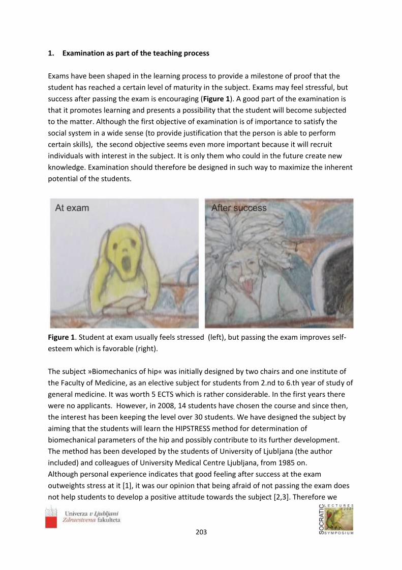
203
1. Examination as part of the teaching process
Exams have been shaped in the learning process to provide a milestone of proof that the
student has reached a certain level of maturity in the subject. Exams may feel stressful, but
success after passing the exam is encouraging (Figure 1). A good part of the examination is
that it promotes learning and presents a possibility that the student will become subjected
to the matter. Although the first objective of examination is of importance to satisfy the
social system in a wide sense (to provide justification that the person is able to perform
certain skills), the second objective seems even more important because it will recruit
individuals with interest in the subject. It is only them who could in the future create new
knowledge. Examination should therefore be designed in such way to maximize the inherent
potential of the students.
Figure 1. Student at exam usually feels stressed (left), but passing the exam improves self-
esteem which is favorable (right).
The subject »Biomechanics of hip« was initially designed by two chairs and one institute of
the Faculty of Medicine, as an elective subject for students from 2.nd to 6.th year of study of
general medicine. It was worth 5 ECTS which is rather considerable. In the first years there
were no applicants. However, in 2008, 14 students have chosen the course and since then,
the interest has been keeping the level over 30 students. We have designed the subject by
aiming that the students will learn the HIPSTRESS method for determination of
biomechanical parameters of the hip and possibly contribute to its further development.
The method has been developed by the students of University of Ljubljana (the author
included) and colleagues of University Medical Centre Ljubljana, from 1985 on.
Although personal experience indicates that good feeling after success at the exam
outweights stress at it [1], it was our opinion that being afraid of not passing the exam does
not help students to develop a positive attitude towards the subject [2,3]. Therefore we

204
focused more on the learning part hinting at the possibility that the students will find the
subject interesting. We tried to achieve positive effects of the examination with minimally
stressing the students.
The exam was designed in a way to enable recruitment of positive feelings in forming a
social network for problem solving. Internet and other technical platforms (mobile phones
etc.) provided excellent tools for this purpose. Instead of banning mobile phones and social
networks from the examination process as cheeting tools, we have encouraged their use
and therefore eradicated cheeting from examination to a great extent.
2. Socratic lectures as an exam
Socratic lectures were intended to present to the students excellent scientists who would
talk about their own contributions to the world science. Students should consider the work
of the scientists and create a dialogue at the lectures. Supporting the lecturers with their
interest was the main role of the students. The foreign scientists were invited to Ljubljana
within ongoing scientific collaboration and have donated their lectures to the students. Also
domestic scientists donated lectures. A minisymposium was created on informal basis. In
particular, because there was no funding for these teaching activities, this symposium was
called Socratic lectures. The students could feel that the lecturers focused on the subject of
their dedication as well as on the general idea that transmission of knowledge to the youth
is essential, and have responded with equal excellence. The idea was to link examination
with these positive effects. In examination, students should reconsider emergent questions
and try to solve actual problems. The lecturers were available to help them find the best
answers to the questions posed. At the same time, students had a possibility to devote a
considerable part of the day to the problems addressed in the course.
This design of the subject proved rewarding as the method HIPSTRESS [4] has been
developing to meet the needs of the students. Some students have been awarded Prešeren
awards for students for the work that initiated with the subject of Biomechanics of hip and
later Biomechanics of joints. Some students have been coauthors of papers published in
journals with impact factor. As there was an increasing interest to publish student work, we
decided to make Socratic lectures formal and enable publication of excellent student work.
This was recently supported by the activities of postgraduate students as well as by scientific
work of teachers of University of Ljubljana and their partners abroad, in the shape of
Socratic symposium that took place a day before the lectures. In 2019, the first Proceeding
of the Socratic Lectures was published. The course Biomechanics of joints was this year
moved to the winter semester, therefore in 2020, the Socratic lectures took part twice.
205
3. Exam questions in Socratic Lectures 2020/2021
Due to the lockdown, the Socratic lectures with the exam took place online (through the
Zoom platform). We were therefore unlimeted as regards the travel expences and could
invite the best experts in the world. The lectures were donated by prof. Leonid Margolis,
National Institute of Health, Bethesda, U.S.A., dr. Gabriella Pocsfalvi, National Research
Council of Italy, Naples, Italy, Prof. Duško Spasovski, Institute of Orthopaedy, Clinics Banjica,
Belgrade, Serbia, Dr. Vesna Spasovski, Institute of Molecular Biology, Belgrade, Serbia and
Prof. Bojana Beović from University Medical Centre Ljubljana, Slovenia. Prof. Margolis and
dr. Pocsfalvi are experts in extracellular vesicles and viruses, which was of special interest
because of COVID-19, colleagues from Serbia have implemented the stem cell therapy into
healing of degeneration of joints in clinical practice and prof. Beović is the Head of the
COVID-19 Counceling Group at the Governament of Republic Slovenia. The questions were
from the subjects of their lectures as well as from the lectures that took place during the
course. There were 10 questions at the exam:
1. Explain what you mean by the following terms: Extracellular vesicles; Viruses.
2. What are the common properties of viruses and extracellular vesicles?
3. Can a virus infection affect joint function? Explain the answer.
4. How can we distinguish viruses from extracellular vesicles in samples?
5. Are viruses more stable than extracellular vesicles in ultracentrifugation? Explain the
answer.
6. What is the order of magnitude of the number of protein species in the membrane of
extracellular vesicles?
7. Indicate at least one mechanism through which extracellular vesicles could treat joint
disease. Is it introduced into clinical practice? If so, give an example.
8. How do extracellular vesicles from stem cells differ from extracellular vesicles of
differentiated cells?
9. How can we distinguish extracellular vesicles from stem cells from extracellular vesicles
from differentiated cells in samples?
10. Do extracellular vesicles and viruses also bud from microorganisms? Explain the answer.
11. What are the strengths and weaknesses of COVID-19 control in Slovenia?
12. How can you explain silent hypoxia with your knowledge of pathophysiology?
Students have formed their own forum to discuss the issues. They used Chat in the Zoom
platform to pose questions to the lecturers. Also they posed questions directly to the
lecturers who provided answers from their experiences and their opinions. Students have
decided to formulate answers jointly. Below we show as an example the answer to the
question no. 4: How can we distinguish viruses from extracellular vesicles in samples?

206
Students answered:
“Separation of viruses and extracellular vesicles (EVs) is a complex process that cannot be
performed to 100% with current technology. The biggest challenge is posed by viruses
whose size and density are similar to the size and density of extracellular vesicles (50-100nm
and 1.13-1.18 g/L)(1). According to these parameters, the viruses most similar to EVs are
from the family of Retroviruses (mostly about 100 nm in size and 1.15-1.18 g/L in density).
Viruses (in the case of HIV research) are thought to migrate faster in the gradient fluid, also
expressing the p24 protein on the surface while acetylcholinesterase and CD45 can be found
on extracellular vesicles. Although acetylcholinesterase and CD45 are not expressed on
virions, it is not known whether they are expressed on all extracellular vesicles (1),(2). Only
non-encapsulated viruses can be distinguished from extracellular vesicles with great
certainty, since neutralizing antibodies bind to the viruses. The same technique cannot be
used in the case of encapsulated viruses, as their proteins can also be expressed on the
surface of extracellular vesicles. Thus, accurate differentiation between viruses and
extracellular vesicles is practically impossible. Differentiation would be possible in the
future using new discoveries in the field of flow cytometry, which allows counting and
description of particles of the order of 550-200m, but the technique needs to be further
improved (1,2). The problem is the very definition of what is a virus and what is an
extracellular vesicle that may have viral proteins (99% of HIV in an infected person are not
infectious). Precisely because of this, we can reliably distinguish only e.g. vesicles that come
from healthy cells and vesicles (viruses, including defective ones) that derive from infected
cells and have viral proteins.
1. Nolte-'t Hoen E, Cremer T, Gallo RC, Margolis LB. Extracellular vesicles and viruses: Are
they close relatives? Proc Natl Acad Sci U S A. 2016; 113 (33): 9155-61.https:
//pubmed.ncbi.nlm.nih.gov/27432966/
2. Cantin R, Diou J, Bélanger D, Tremblay AM, Gilbert C. Discrimination between exosomes
and HIV-1: purification of both vesicles from cell-free supernatants. J Immunol Methods.
2008 Sep 30; 338.”
As there is no decisive answer to the question that students could learn and reproduce, they
were challenged with taking a standpoint. It can be seen from the answer that they were
able to do this and express their standpoint in such way to participate in a scientific
discourse. Also they used citations and presented the references.
New ways of teaching and new tools present challenges in the teaching process. With data
easily available reproducing facts in great details could be replaced by consideration of
emergent problems which are not yet well understood.
Also, new problems emerge. Allowing formation of the social network in creating the
answers increases the quality of the answers to such extent that the pieces of evidence can
be presented for publication, especially if the students involve their friends and relatives
who are experts on the issues. A question can then be posed who is the author of possible
deliverables of these activities? Should students be stating the disclosure and conflict of
interest on the examination form?

207
Conclusions
Online university teaching challenges existing methods of examination. It can be expected
that new minimally frustrating methods will be developed which will enable better
motivation, increase quality of answers and promote scientific work of students and
teachers.
Acknowledgements
Authors acknowledge support of EU Commission H2020, grant Ves4us 80331 and ARRS,
grant P3-0388.
References
1. Zajenkowska A, Zajenkowski M, Jankowski, KS, The relationship between mood experienced
during an exam, proneness to frustration and neuroticism. Learn Ind Diff 2015, 37: 237-240,
http://doi: 10.1016/j.lindif.2014.11.014
2. Liu S, An Investigation into the Emotional Barriers of College Students in English Learning. In:
International symposium on engineering technology, education and management (ISETEM
2014), 2014: 587-597.
3. Artino AR, Hemmer PA, Durning SJ, Using self-regulated learning theory to understand the
beliefs, emotions, and behaviors of struggling medical students, Acad Med 2011, 86: S36-
S39, http://doi: 10.1097/ACM.0b013e31822a603d
4. Kralj-Iglič V, Understanding Hip Biomechanics: From simple equilibrium to personalized
HIPSTRESS method, In: Developmental Diseases of the Hip (Spasovski D, ed.), InTech, 2017,
http://doi: 10.5772/66753

208

209
________________
Fyodor Mikhailovich Dostoevsky and his relationship with music ˡ Prelovšek, A* *[email protected] Abstract
This article describes the musical world of the great Russian writer, the bicentenary of whose
birth is celebrated this year. Music played an important part in his life, and in his free time he
spent much time attending concerts and operas. During the period of his forced labour in
Siberia he made a collection of a number of folk songs, and his musical bent was also
reflected in his literary work. The last part of the article talks of Dostoevsky's descendants'
relationship with music and some of the influence his works had on later musical production.
___________________

210
1. Introduction
Fyodor Mikhailovich Dostoevsky, born in Moscow on 11 November 1821, is, in the opinion of
the contemporary Russian writer Dmitry Bykov [1], »the most Western of the Russian
writers« and therefore the most popular in Europe and America. A. Gozenpud, the author of
the monography Dostoevsky i muzyka, says that »Dostoevsky loved and understood music
and felt deeply about it« 2. The same author adds that »Dostoevsky’s literary art was
closely tied to music« 2, his works frequently containing quotations from tunes he had
heard in the concerts, folk songs or Russian romances he knew, but also reflecting the effect
which the musical experiences and impressions of great artists had had on him. As well as
literature, theatre, music and musicians; Dostoevsky also loved painting; he enjoyed visiting
galleries and was equally inspired by painting.
2. Music in Dostoevsky’s life and literary work
Dostoevsky considered music as a way of expressing a man’s inner state and uses it to
describe the emotional condition of his heroes. In his story Another Man's Wife or The
Husband under the Bed, he articulated this statement as follows: »It is maintained that what
is good in music is that musical impressions can be made to fit any mood. The man who
rejoices finds joy in its strains, while he who grieves finds sorrow in it.« 3. Further, he
describes with sounds (»a tempest«) the distraction and jealousy of the hero Ivan
Andreyitch: »a regular tempest was howling in Ivan Andreyitch’s ears« 3.
His novel The Insulted and Injured includes a passage in which the heroine Katia compares
her feelings to those in Beethoven’s third piano concerto 4. For Dostoevsky, Beethoven’s
music was all about love: in 1975 he wrote to his wife Anna Grigoryevna: »in Beethoven,
there is everywhere passion and love. This is the poet of love, happiness and love's
torments« 2. Dostoevsky often connects singing with happiness. He wrote in White Nights:
»I walked along singing, for when I am happy I am always humming to myself like every
happy man who has no friend or acquaintance with whom to share his joy.« 5.
In her memoirs, Dostoevsky’s daughter recalls that when he was in a good mood, the writer
would sing to himself Varlamov's romance ‘Na zare ty ee ne budi’ (’Don’t wake her up at
sunrise’)1 2. He told his later wife that at the time when his death penalty was cancelled at
the last minute before its execution and commuted to forced labour in Siberia, Dostoevsky
walked around his cell in the Peter and Paul Fortress in St. Petersburg and was »singing,
loudly singing, because I was so happy for the life given to me« 2.
3. Dostoevsky and Russian folk music
In his early childhood, Dostoevsky had an opportunity to hear folk songs and see folk
traditions during the festivities that accompanied traditional holiday days or at fairs. Later in
1 A Russian romance song by Aleksandr Varlamov, words by Afanasy Fet, written in 1842.

211
his life, when he was exiled to the Siberian town of Omsk (from 1849 to 1853), he was
collecting the folklore of the Russian nation, such as texts of songs, funny stories and
expressions that arrested people were using in their everyday language 2. In The House of
the Dead he mentioned that singing songs in the forced labour camp imbued the inmates
with a sense of optimism and consolation. Some of them would sing accompanying
themselves on balalaikas and a small choir was also formed. As Dostoevsky writes, some of
those songs were laments but there were also songs with a humorous text 2. In Chapter XI
of the first part of the novel quoted above, Dostoevsky describes the preparations for the
performance of the spectacle with music that was to be put on by the inmates. In that little
improvised orchestra, about eight instruments would play dance music.
4. Dostoevsky at concerts and operas in St. Petersburg
Dostoevsky was a regular visitor to concert halls; he also loved opera and frequented the
salons of St. Petersburg’s aristocracy, where music was regularly performed,2 but music was
often present also at the meetings of the Petrashevsky Circle.3 One of the strongest musical
impressions in Dostoevsky’s life was his meeting with the composer Mikhail Glinka. They met
at an evening organized by the Palm-Durov Circle4 in 1849. On this occasion, Glinka was
supposed to be playing on the piano his romance ‘K ney’, excerpts from his opera ‘Ruslan
and Ludmila’, his ‘Kamarinskaya’ and pieces by Gluck and Chopin 2. Later in his literary
work, Dostoevsky remembered Glinka: ‘Kamarinskaya’ is mentioned in The House of the
Dead,5 while many years later, Glinka’s romance ‘K ney’is sung by a hero in a short story The
Eternal Husband.
All the musical impressions which left their mark on Dostoevsky (but before the meeting
with Glinka in the same year) were concentrated in his most »musical« novel, Netochka
Nezvanova, published in 1849. The characters in this novel are a failed hero, the violinist
2 In this context, the house of Prince V. Odoevsky, a writer, musician and one of the first Russian musicologists,
was an important musical venue for Dostoevsky [2]. In the 1830s and 1840s, one of the centres of musical life in St. Petersburg, and one where Dostoevsky was also
present, was the salon of Mikhail Vielgorsky [2]. Wikipedia [6] states that Vielgorsky, a diplomat, composer and
amateur musician, was a great admirer of Beethoven and hosted at his home all the most famous Russian and
foreign musicians of the time, such as Berlioz, Liszt, Rubinstein, Viardo and Glinka. 3 [7] The members of this intellectual circle, including Dostoevsky, were in 1849 condemned to death. Many of
them – also Dostoevsky – were condemned only because they had disseminated the text of Belinsky’s letter to
Gogol. 4 [7] An intellectual group, a branch of the Petrashevsky Circle, from which it had become independent.
5 ‘Kamarinskaya’ is likewise mentioned in the novel The Village of Stepanchikovo but as a traditional Russian
folk dance, not the one of Glinka.

212
Efimov, and his opposite, a brilliant violin player named »S.«. According to Gozenpud (1971)
2, Dostoevsky got the inspiration for this talented musician from concerts given by the
leading musicians of the time which he had the opportunity to listen to, such as the pianist
Franz Liszt, the Norwegian violin virtuoso Ole Bull and especially Heinrich Wilhelm Ernst - a
Moravian-Jewish violinist, an outstanding virtuoso and Paganini’s successor 2. Dostoevsky
was also greatly impressed by a concert in which Hector Berlioz conducted his ‘Symphonie
dramatique Romeo and Juliet’ 2. Concerts by internationally acclaimed musicians at that
time were a real sensation: »The whole musical world of St. Petersburg was astir. Singers,
actors, poets, artists, musical people, and even those who were not at all musical but with
modest pride declared that they did not know one note from another, rushed with eager
enthusiasm to buy tickets. The hall could not seat a tenth of the enthusiasts who were able
to pay twenty-five roubles for a ticket; but the European fame of S., his old age crowned with
laurels, the unflagging freshness of his talent […] all produced an effect.« 8].
In 1846, during the period in which Dostoevsky was working on Netochka Nezvanova, he
wrote to his brother Mikhail about his passion for the opera: »After seven p.m. I am going to
the Italian opera, in the gallery, to listen to our incomparable singers.« 2. The monography
about Dostoevsky and music states that the operas Dostoevsky saw in the 1840s included
‘Askold’s Grave’, an opera, very popular at the time, by the romantic Russian composer
Alexei Verstovsky, Glinka’s ‘Ivan Susanin’(or ‘A Life for the Tsar’) and ‘Ruslan and Ludmila’.
This opera was a particular favourite of Dostoevsky's throughout his life and he also wanted
his children to appreciate this work of art 2. Among the operas of foreign composers, he
saw and much appreciated ‘Robert le diable’ by Giacomo Meyerbeer and Rossini’s ‘The
Barber of Seville’. In Meyerbeer’s opera he was able to admire one of the most famous ballet
dancers of the romantic era, Marie Taglioni, while in Rossini, he could hear the famous singer
Pauline Viardot (the younger sister of one of the most adored opera singers of her time,
Maria Malibran). In a short story he mentions Mayerbeer's cemetery scene in which he saw
Marie Taglioni. In the same book, he also mentions Rosina’s aria from Rossini.
As described by A. Gozenpud 2 with reference to Italian opera in St. Petersburg in the
1840s, there were two opposing ways of musical interpretation which divided the public into
two parties. The first one preferred a more dramatic, expressive musical interpretation while
the second one admired pure virtuosity.
The representative of the first approach was a dramatic soprano, Teresa De Giuli Borsi, and
that of the second a coloratura soprano, Erminia Frezzolini. Dostoevsky favoured the
supporters of De Giuli Borsi [2] and he ironically described the relationship between the two
groups of opera enthusiasts and the general atmosphere in the opera theatre in that time in
his story Another Man's Wife or The Husband under the Bed: »The following evening there
was a performance of some sort at the Italian opera. […] It was known for a positive fact,
anyway, that Ivan Andreyitch used to like taking a nap for an hour or two at the Italian opera;
he even declared on several occasions how sweet and pleasant it was. “Why, the prima

213
donna,” he used to say to his friends, “mews a lullaby to you like a little white kitten.” […] It
must be observed that there were at that time two parties, each supporting the superior
claims of its favourite prima donna. They were called the -sists and the –nists. Both parties
were so devoted to music that the conductors actually began to be apprehensive of some
startling manifestation of the passion for the good and the beautiful embodied in the two
prima donnas.« [3].
Figure 1. An excerpt (first page of ‘Rondo russo’) of the manuscript of Mercadante’s ‘Flute
concerto in E minor’, kept in the library of the Neapolitan Conservatory of music (photo:
Anita Prelovšek).
In St. Petersburg in the 1830s and 1840s, theatrical programmes included many operas by
Saverio Mercadante, an Italian composer of the Neapolitan school. Dostoevsky thought
highly of his work, as can be concluded from the words he put into the mouth of his hero in
The Village of Stepanchikovo. To demonstrate his wide general culture, his hero cites the
names of important people who came to mind at that moment, so he puts Mercadante near
to Machiavelli: »[…] perhaps Macchiavelli himself or some Mercadante was sitting before
him and only to blame for being poor and nondescript...« 8. Mercadante was a prolific
composer of operas, writing more than sixty of them, but he is nowadays mainly appreciated
by flute players because of his ‘Concerto in E minor’ (Figure 1). Written in 1819, when the

214
composer was twenty-four years old, it is faintly redolent of Russia, the last movement even
being called ‘Rondo russo’, which also testifies to the protracted influence that Italy had on
Russian music6.
5. Dostoevsky and music after the exile in Siberia
After ten years of exile in Siberia,6 he finally came back to live in St. Petersburg as a free
citizen. So from the 1860s, Dostoevsky’s musical impressions were again varied. He attended
concerts and went to Italian and Russian operas; he went to concerts given by Anton
Rubinstein and sometimes they performed at the same cultural evenings 2, Rubinstein as a
pianist and Dostoevsky as a reader of literary works, in which he was known to excel. The
circle of the writer’s musical acquaintances was growing. As editor of the literary magazines
Vremya and Epoch, he collaborated among others with the music critic and composer
Alexander Serov.7
Music was present in Dostoevsky's home: his second wife, Anna Grigoryevna, played the
piano and his library contained Mendelssohn's ‘Wedding March’ and Rossini’s ‘Stabat Mater’
2.
During his journeys abroad,8 Dostoevsky continued going to concerts. He couldn’t afford
expensive tickets and in opera-houses he had to take the cheapest, standing places. Where
possible, as in Dresden and Bad Ems, he was always happy to seize the opportunity to listen
to the free concerts given by symphony orchestras and brass bands in parks. The repertoire
of such concerts included overtures (Mozart, Beethoven or Boieldieu,9 for instance) or single
movements from symphonies, marches, dance music and potpourris of famous opera
themes (Anna Grigoryevna mentions, among others, a potpourri on themes of Franz von
Suppé 12. Dostoevsky didn’t like all the music that was being performed there. In one
letter from 1875 he complained about the musical programme in Bad Ems: »They rarely play
something interesting, everything is some kind of potpourri or German marches, some
Strauss, Offenbach […]« 2. Anna Grigoryevna said that »he liked Mozart, Beethoven’s
‘Fidelio’ and Mendelssohn’s ‘Wedding March’, but couldn't stand Richard Wagner's music.«
12.
6 The first four years he spent as a prisoner in a forced labour camp in Omsk without being allowed to write and
read. From 1854 he worked as a soldier, when, thanks to some influential protectors, he recovered the right to
attend local intellectual circles, and later he was allowed to publish his work again. 7 A. Serov was known especially for his operas, which were quite successful at the time. Dostoevsky was
present at the premieres of Serov’s operas ‘Judith’ and ‘Rogneda’ [2]. 8 During his first journey abroad in 1862, he visited Berlin, Dresden, Lucerne, Wiesbaden, Baden-Baden,
Cologne, Paris, London, Düsseldorf, Geneva, Genoa, Livorno, Florence, Milan, Venice and Vienna. From 1867
to 1871, immediately after their marriage, Dostoevsky and his wife Anna Grigoryevna spent time in Dresden,
Baden-Baden, Geneva, Vevey and Florence [11]. 9 François Adrien Boieldieu was a French composer of operas. The overture to his comic opera ‘The White
Lady’ was often played at concerts and Dostoevsky particularly liked it because of its attractive romantic
melodies and the fact that the libretto is based on a novel by his beloved Sir Walter Scott.

215
His favourite composers were Mozart and Beethoven. According to his diaries, another piece
he particularly loved was Rossini’s ‘Stabat Mater’ 2.
6. Dostoevsky’s heritage and music today
Among contemporary authors, Haruki Murakami, a great Japanese admirer of Dostoevsky’s
work who also sometimes quotes Dostoevsky in his novels, such as his reference to The
Brothers Karamazov in the novel 18Q4, frequently cites music of different genres, especially
classical music and jazz. Music in Murakami’s work is used to describe the inner state of his
heroes, being related to their reminiscences, or sometimes his heroes just enjoy listening to
it or it is in some way relevant to their life.
7. Dostoevsky’s descendants and music
Dostoevsky’s nephews Maria and Fyodor were both good pianists who studied with Anton
and Nikolai Rubinstein. His niece Maria Ivanova graduated from the Moscow Conservatory in
the class of N. Rubinstein 2.
A love of music has also been inherited by those of Dostoevsky’s descendants who are living
today. The son of a great-grandson of Fyodor Mikhailovich, Aleksei Dmitrievich Dostoevsky
works for the Monastery of Valaam as a captain of the boats and boat-driver. In his free time
he is also a musician, playing bass-guitar in a folk group. In an interview he gave in 2018, he
said that it was thanks to this hobby that he met his wife, Natalia, who is a flautist. Their
children are also musicians. Their elder daughter Anna is, as her father said in the interview,
very artistic and plays the flute like her mother. Their second daughter, Vera, is studying the
tamboura at the Rimsky-Korsakov musical institute and at the time of the interview was
planning to enrol at the musical university. The youngest daughter, Masha, plays the cello,
while their son Fyodor had taken up percussion in a music school. In 2018, daughter Vera
gave a concert there on the occasion of its annual conference devoted to research into
Dostoevsky's works10 14.
8. Influence of Dostoevsky's work on music
The most famous music inspiration that Dostoevsky's novels provided for composers
resulted in operas: Sergei Prokofiev adapted Dostoevsky's novel The Gambler [15] for the
libretto of his opera of the same name, written between 1915 and 1917 and premiered in
1929.
The Czech composer Leoš Janáček adapted Dostoevsky's The House of the Dead for the
libretto of his last opera The House of the Dead,[16] premiered in 1930.
There are now more recent musical adaptations of Dostoevsky's novels. For instance, 2016,
10 [13] This conference is taking place in Staraya Russa, which is home to a Dostoevsky museum. His wife Anna
Grigoryevna recalled the winter of 1874-75 as being one of her most cherished memories. Dostoevsky was in
good spirits and always cheerful; in the evenings he would partner her and the children in various different dances.
He was particularly fond of the mazurka, which he danced enthusiastically »like a passionate Pole« [12].

216
the year of the 150th anniversary of the publication of Crime and Punishment, was marked
by the premiere of a rock-opera of the same name by a Russian composer of mostly film
music, Eduard Artemyev, [17] and the occasion of the writer's jubilee in 2021 is likely to see
some new settings of his work to music.
References
1. Bykov, D. »Открытый урок с Дмитрием Быковым. "Преступление и наказание" - странный русский детектив.« (»An open lecture with Dmitry Bykov. "Crime and Punishment" – a strange Russian detective novel.« Internet source: https://www.youtube.com/watch?v=kxwcA9NAEcY Accessed on 7 January 2021.
2. Gozenpud, A. Dostoevsky i muzyka. Leningrad: Muzyka, 1971. 3. Dostoevsky, F. Another Man's Wife or The Husband under the Bed. Adelaide: The University
of Adelaide Library, 2016. Internet source: https://www.holybooks.com/wp-content/uploads/Another-Mans-Wife.pdf Accessed on 10 January 2021.
4. Dostoevsky, F. Ponižani in razžaljeni. Translation of Borut Kraševec. Ljubljana: Mladinska knjiga, 2017.
5. Dostoevsky, F. Translation by Constance Garnett. White Nights and Other Stories from Fyodor Dostoevsky. New York: The Macmillan Company, 1918. Internet source: https://www.holybooks.com/wp-content/uploads/White-Nights-And-Other-Stories.pdf Accessed on 9 January 2021.
6. »Vielgorsky, Mikhail« Wikipedia. Internet source: https://en.wikipedia.org/wiki/Mikhail_Vielgorsky Accessed on 11 January 2021.
7. »Petrashevsky Circle«. Wikipedia. Internet source: https://en.wikipedia.org/wiki/Petrashevsky_Circle Accessed on 12 January 2021.
8. Dostoevsky, F. Translation by Constance Garnett. Netochka Nezvanova. London: Heinemann, 1912. Internet source: https://archive.org/details/novelsoffyodordo12dost/page/244/mode/2up Accessed on 12 January 2021.
9. Dostoevsky, F. Translation by Constance Garnett. The Village of Stepanchikovo. (The Friend of the Family). London: Heinemann, 1912. Internet source: https://archive.org/details/novelsoffyodordo12dost/page/106/mode/2up Accessed on 18 January 2021.
10. Ivashkin, A. »Italiani in Russia: Presenza fisica e influenza virtuale sulla musica.« in: Rinascimento e antirinascimento, Gabinetto scientifico letterario G. P. Vieusseux. Studi, vol. 22: 209-218. Firenze: Casa editrice Leo S. Olschki, 2012.
11. Dostoevsky, F. Editor: Prina, S. Le notti bianche. La cronaca di Pietroburgo. Milano: Feltrinelli, 2018.
12. Dostoevskaya, A. Življenje z genijem (Spomini). Kranj: Zelolepo, 2007. 13. »Staraya Russa. Wikipedia. Internet source: «https://en.wikipedia.org/wiki/Staraya_Russa
Accessed on 12 January 2021. 14. Kotov, V. and Pogodina-Kuzmina, O. »Как живут потомки Достоевского.« Internet source:
https://www.sobaka.ru/omsk/city/society/76489 August, 2018. Accessed on 12 November 2020.
15. »The Gambler.« Wikipedia. Internet source: https://en.wikipedia.org/wiki/The_Gambler_(Prokofiev) Accessed on 7 January 2021.

217
16. »From the House of the Dead.« Wikipedia. Internet source: https://en.wikipedia.org/wiki/From_the_House_of_the_Dead Accessed on 7 January 2021.
17. Artemyev, E. »Ария из рок-оперы Эдуарда Артемьева "Преступление и наказание".«
(»Aria from the rock-opera »Crime and Punishment.«) Internet source:
https://www.youtube.com/watch?v=MKJn5XX4SKk Accessed on 7 January 2021

218

219
P1

220
P2

221
P3
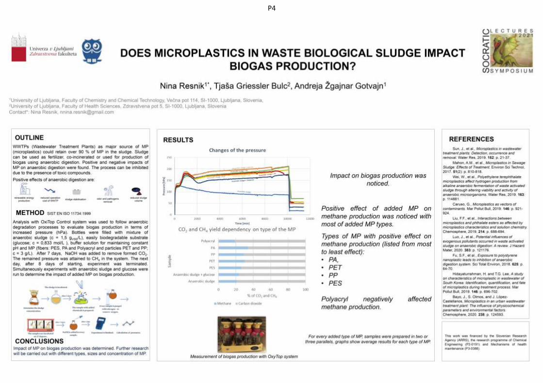
222
P4

223
P5

224
P6

225
P7

226
P8

227
P9

228
P10

229
P11

230
P12

231
P13

232
P14

233
P15

234
P16

235
P17

236
P18
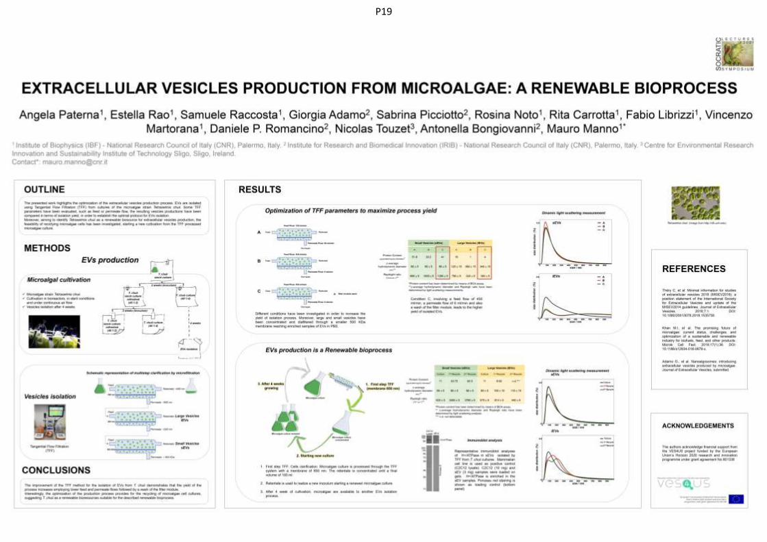
237
P19

238
P20