Stimuli-responsive nanoparticles from ionic cellulose derivatives
Upload
independentCategory
view
1download
0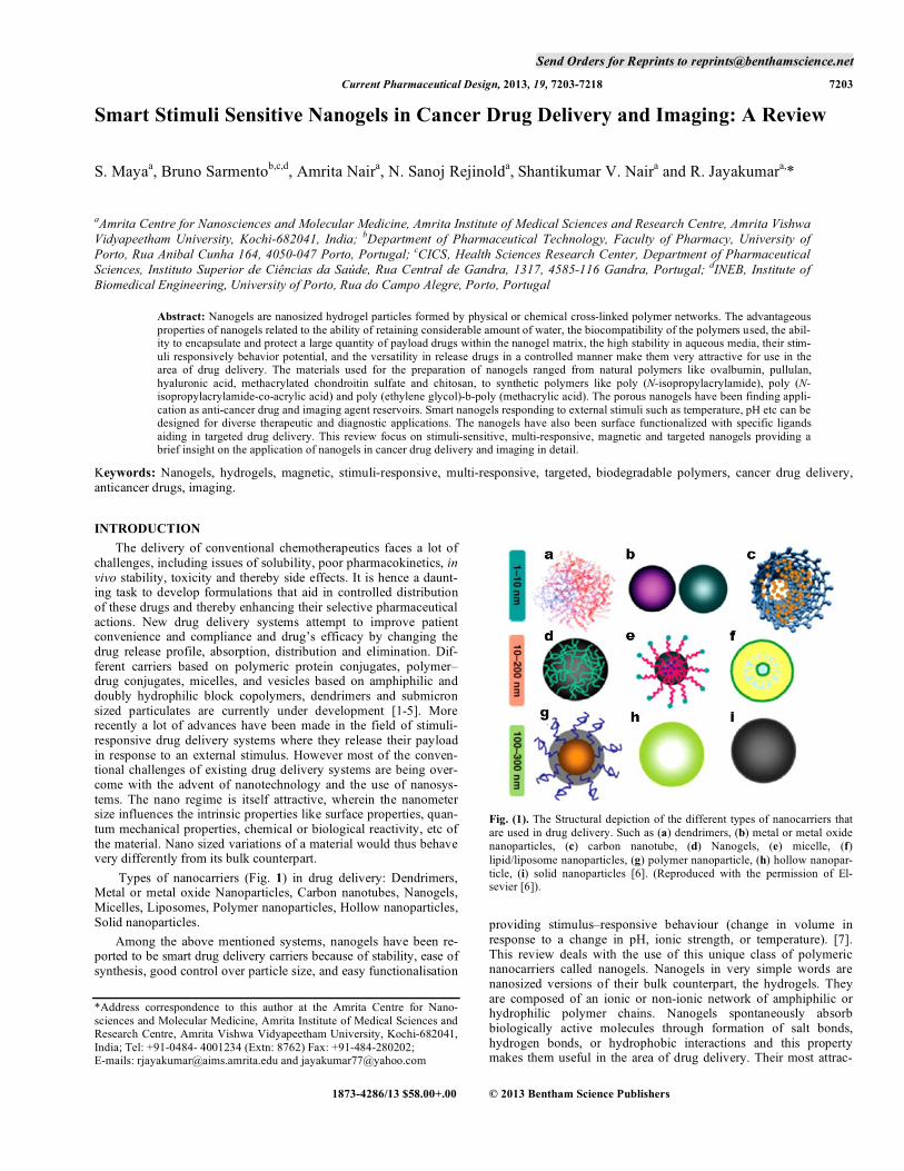
Send Orders for Reprints to [email protected]
Current Pharmaceutical Design, 2013, 19, 7203-7218 7203
Smart Stimuli Sensitive Nanogels in Cancer Drug Delivery and Imaging: A Review
S. Mayaa, Bruno Sarmentob,c,d, Amrita Naira, N. Sanoj Rejinolda, Shantikumar V. Naira and R. Jayakumara,*
aAmrita Centre for Nanosciences and Molecular Medicine, Amrita Institute of Medical Sciences and Research Centre, Amrita Vishwa Vidyapeetham University, Kochi-682041, India; bDepartment of Pharmaceutical Technology, Faculty of Pharmacy, University of Porto, Rua Anibal Cunha 164, 4050-047 Porto, Portugal; cCICS, Health Sciences Research Center, Department of Pharmaceutical Sciences, Instituto Superior de Ciências da Saúde, Rua Central de Gandra, 1317, 4585-116 Gandra, Portugal; dINEB, Institute of Biomedical Engineering, University of Porto, Rua do Campo Alegre, Porto, Portugal
Abstract: Nanogels are nanosized hydrogel particles formed by physical or chemical cross-linked polymer networks. The advantageous properties of nanogels related to the ability of retaining considerable amount of water, the biocompatibility of the polymers used, the abil-ity to encapsulate and protect a large quantity of payload drugs within the nanogel matrix, the high stability in aqueous media, their stim-uli responsively behavior potential, and the versatility in release drugs in a controlled manner make them very attractive for use in the area of drug delivery. The materials used for the preparation of nanogels ranged from natural polymers like ovalbumin, pullulan,hyaluronic acid, methacrylated chondroitin sulfate and chitosan, to synthetic polymers like poly (N-isopropylacrylamide), poly (N-isopropylacrylamide-co-acrylic acid) and poly (ethylene glycol)-b-poly (methacrylic acid). The porous nanogels have been finding appli-cation as anti-cancer drug and imaging agent reservoirs. Smart nanogels responding to external stimuli such as temperature, pH etc can be designed for diverse therapeutic and diagnostic applications. The nanogels have also been surface functionalized with specific ligands aiding in targeted drug delivery. This review focus on stimuli-sensitive, multi-responsive, magnetic and targeted nanogels providing a brief insight on the application of nanogels in cancer drug delivery and imaging in detail.
Keywords: Nanogels, hydrogels, magnetic, stimuli-responsive, multi-responsive, targeted, biodegradable polymers, cancer drug delivery, anticancer drugs, imaging.
INTRODUCTION The delivery of conventional chemotherapeutics faces a lot of challenges, including issues of solubility, poor pharmacokinetics, in vivo stability, toxicity and thereby side effects. It is hence a daunt-ing task to develop formulations that aid in controlled distribution of these drugs and thereby enhancing their selective pharmaceutical actions. New drug delivery systems attempt to improve patient convenience and compliance and drug’s efficacy by changing the drug release profile, absorption, distribution and elimination. Dif-ferent carriers based on polymeric protein conjugates, polymer–drug conjugates, micelles, and vesicles based on amphiphilic and doubly hydrophilic block copolymers, dendrimers and submicron sized particulates are currently under development [1-5]. More recently a lot of advances have been made in the field of stimuli-responsive drug delivery systems where they release their payload in response to an external stimulus. However most of the conven-tional challenges of existing drug delivery systems are being over-come with the advent of nanotechnology and the use of nanosys-tems. The nano regime is itself attractive, wherein the nanometer size influences the intrinsic properties like surface properties, quan-tum mechanical properties, chemical or biological reactivity, etc of the material. Nano sized variations of a material would thus behave very differently from its bulk counterpart. Types of nanocarriers (Fig. 1) in drug delivery: Dendrimers, Metal or metal oxide Nanoparticles, Carbon nanotubes, Nanogels, Micelles, Liposomes, Polymer nanoparticles, Hollow nanoparticles, Solid nanoparticles. Among the above mentioned systems, nanogels have been re-ported to be smart drug delivery carriers because of stability, ease of synthesis, good control over particle size, and easy functionalisation
*Address correspondence to this author at the Amrita Centre for Nano-sciences and Molecular Medicine, Amrita Institute of Medical Sciences and Research Centre, Amrita Vishwa Vidyapeetham University, Kochi-682041, India; Tel: +91-0484- 4001234 (Extn: 8762) Fax: +91-484-280202; E-mails: [email protected] and [email protected]
Fig. (1). The Structural depiction of the different types of nanocarriers that are used in drug delivery. Such as (a) dendrimers, (b) metal or metal oxide nanoparticles, (c) carbon nanotube, (d) Nanogels, (e) micelle, (f)lipid/liposome nanoparticles, (g) polymer nanoparticle, (h) hollow nanopar-ticle, (i) solid nanoparticles [6]. (Reproduced with the permission of El-sevier [6]).
providing stimulus–responsive behaviour (change in volume in response to a change in pH, ionic strength, or temperature). [7]. This review deals with the use of this unique class of polymeric nanocarriers called nanogels. Nanogels in very simple words are nanosized versions of their bulk counterpart, the hydrogels. They are composed of an ionic or non-ionic network of amphiphilic or hydrophilic polymer chains. Nanogels spontaneously absorb biologically active molecules through formation of salt bonds, hydrogen bonds, or hydrophobic interactions and this property makes them useful in the area of drug delivery. Their most attrac-
1873-4286/13 $58.00+.00 © 2013 Bentham Science Publishers
7204 Current Pharmaceutical Design, 2013, Vol. 19, No. 41 Maya et al.
tive features are their high water content, biocompatibility, and desirable mechanical properties [7].
HYDROGELS Hydrogels find immense application in tissue engineering, biomedical implants and drug delivery due to these potential properties. These properties of hydrogels tissue engineering, bio-medical implants, and drug delivery [8]. The first time a hydro-philic gel was put to biological use was in the early 1960’s by Wichterle and Lim [9]. By definition, hydrogels are three-dimensional networks of hydrophilic polymers held together by the cross-links of covalent bonds or ionic bonds or hydrogen bonds or hydrophobic interactions. These materials when placed in water swells and retain large amount of water maintaining its structure [10]. Polymers with hydrophilic pendant groups –OH, –CONH–, –CONH2–, and –SO3H tend to swell and absorb water, thus becoming candidates for hydrogels [11]. The nanostructures which is cross linked by physical, chemical, covalent, hydrogen bonding, van der Waals interactions, or physical entanglements provides the ability to absorb water, swell and remain undissolved [12]. One of the most interesting features of hydrogels is their ability to resemble living tissue. This unique property has been attributed to their high water content, their soft and rubbery consistency, and low interfa-cial tension with water or biological fluids [13, 14]. Drug release from hydrogels can be categorized as: Diffusion-controlled, Swelling-controlled, and Chemical-controlled. Hydrogels exhibit diffusion-controlled release which in turn dependent on the mesh size within gel matrix. Mesh size depend upon the monomers, degree of cross-linking and the intensity of external stimuli (pH, temperature) and also is responsible for the mechanical strength, diffusivity, and biodegradability of the network [15, 16]. Hydrogels have become a very popular candidate in the race to design an ideal drug delivery system owing to their exceptional physicochemical and biological characteristics such as a) ability to swell and retain large amount of water b) protects the drug from hostile environments (enzymes, pH etc) c) undergo volume phase transition with respect to the environmental conditions d) control the drug release in response to the external stimuli like pH, tem-perature, ionic strength and electric field [17]. One of the earliest works dealing with the use of hydrogels in drug delivery was done by Morimoto et al. [18]. Their work focused on using polyvinyl alcohol hydrogel for rectal administration of indomethacin. Table 1summarizes some of the initial studies done in the field of hydro-gels used for drug delivery. Jeyanthi et al. [19] conducted their research on an implantable collagen-poly (HEMA) hydrogel loaded with 5-FU with an aim to treat solid tumors. The hydrogel showed
enhanced antitumor effect against fibrosarcoma in wistar rats over free 5-flurouracil (5-FU) indicating the ability of the hydrogel to release 5-FU in a controlled sustained manner. Later, in 1996, Patel et al. used freeze-dried chitosan-poly (ethylene oxide) (PEO) hy-drogels for site-specific antibiotic delivery in the stomach. They synthesized semi inter penetrating networks (semi-IPN) by cross-linking chitosan with PEO and more than 65% of the loaded drugs were released from the freeze dried hydrogel. This study proved the application of semi IPN for localized delivery of antibiotics in acidic environment of the gastric fluid [20]. Vervoort et al. worked on inulin hydrogels as carriers for colonic drug targeting [21]. J. Wu et al. have dealt with a thermo-sensitive hydrogel based on quaternized chitosan and poly (ethylene glycol) (PEG) for nasal drug delivery applications for peptide drugs. They found that the blood glucose concentration was apparently reduced after 4-5 h of the nasal administration of hydrogel [22]. K. Nomura et al. used danazol-loaded hyaluronic acid hydrogel to treat endometriosis in a rat model. There has hence been a lot of work on the utilization of hydrogels in the field of drug delivery [23].
NANOGELS All these attractive properties and characteristics resulted in research being conducted into the nanoscale variations of hydro-gels. Although very popular, these hydrogels faced a main drawback when used in stimuli-responsive applications where the transduction of signals will be limited by the rate of diffusion. Later research proved that formation of capillary networks by inter-connecting the pores in the polymeric structure and downsizing the hydrogels would decrease the diffusion path which in turn resulted in the development of nanogels [24]. The various advantages that nanogels provide are as listed below [7, 11, 16]: • High biocompatibility: This is by virtue of the high water con-
tent and low surface tension • High Loading Capacity • Controlled Release of payload • Flexibility in design • Versatility in drug loading and release • High Water absorptivity: Nanogels in the unloaded swollen
state contain a considerable amount of water • The rapid response to external stimuli: This property is by
virtue of its nanoscale dimensions • Increased and prolonged circulation time: They hence have a
better chance of targeting the site of interest
Table 1. Early research done in the field of hydrogels used for drug delivery applications.
Type of Hydrogel Payload Significant outcome Reference
Poly vinyl alcohol (PVA) Indomethacin Thermosensitive
Rectal administration
[18]
Collagen-poly(HEMA) 5-FU Implanatable hydrogels
Treatment of solid tumors like fibrosarcoma
[19]
Chitosan-PEO Amoxicillin & Metronidazole pH sensitive
Localized delivery of antibiotic in stomach
[20]
Quarternized chitosan-PEG Insulin Thermosensitive
Mucoadhesive, Non toxic
Facilitate nasal drug delivery of peptide drugs
[22]
Hyaluronic acid (HA) Danazol Treatment of endometriosis [23]
Smart Stimuli Sensitive Nanogels in Cancer Drug Delivery and Imaging Current Pharmaceutical Design, 2013, Vol. 19, No. 41 7205
• High Stability in aqueous solution: Nanogels with polycore structure (hydrophobic polymeric core and hydrophilic shell) make zero Gibb’s energy enabling the enclosure of a water-insoluble drug in the polycore thereby protecting from interactions with the surrounding biological fluids.
• The nanoscale size of nanogels also leads to a high specific surface area that is available for the bioconjugation of active targeting agents.
The incorporation of high-affinity functional groups, stimuli-responsive conformations or biodegradable bonds into the polymer network aid in protecting the drug molecules and regulating its release from the nanogels [25]. The loaded drug would interact with the polymer matrix through electrostatic, van-der Waals and/or hydrophobic interactions. The hydrophilicity of the nanogels en-ables them to be easily dispersed in aqueous media and hence easily administered in liquid dosage form [26, 27]. Introduction of hydrophilic polymer such as PEG provides a protective coating around the nanogels, preventing their uptake by the mononuclear phagocytic system thereby increasing the circulation time in the bloodstream and thus enhancing their stability. Targeting of these nanogels is made possible by functional modification of the many functional groups exposed on its surface, with targeting moieties like folic acid (FA), monoclonal antibodies (mAb), transferrins etc. Research does show that various nanogels deliver their payload inside cells and across biological barriers by protecting them from degradation by cells metabolic systems and hence they exhibit high stability [7]. Thus, nanogels provide excellent potential for enhanc-ing oral and brain bioavailability and systemic drug delivery of low molecular weight drugs [7]. Currently there are many approaches for the preparation of nanogels. They can be categorized as below [28]: Chemical Nanogels consist of covalently bonded crosslinking points formed during (eg: a bifunctional crosslinking agent such as methylenebis(acrylamide) ( BIS ) or divinyl benzene ( DVB )) and after polymerization. Chemical gels are usually stable and rigid and difficult to exchange. Polymerization in templates: Polymerization of the monomer requires nanospaces. • Mini-emulsion polymerization (surfactant dispersed oil-in-
water). Surfactants such as sodium dodecyl sulphate (SDS) can be used. eg: PNIPAAm nanogels with potassium persulfate as initiator, BIS as crosslinker and SDS as surfactant.
• Reverse mini-emulsion polymerization (surfactant dispersed water-in-oil). This method is used for polymerizing electrolyte monomers or PEG containing monomers. Surfactants such as sorbitan monooleate (Span - 80) and sodium bis(2 - ethyl-hexyl) sulfosuccinate (AOT). eg: cationic nanogels interacting with anionic DNA: Cationic monomer, (3 - acrylamidopropyl) - trimethylammonium chloride, was polymerized with BIS in inverse miniemulsion using both L - � - phosphatidylcholine (lecithin) and AOT surfactants.
• Suspension polymerization: Simple approach for the polym-erization of hydrophobic monomers, but difficult to control the nanogel size. eg: poly(2 - vinylpyridine) (P2VP ) nanogels were prepared by the copolymerization of VP and DVB in wa-ter under stirring.
• Polymerization in lipososmes: Hydrophilic nanogels could be prepared by using liposomes as nanoscale reactors. eg: polym-erization of dex-HEMA containing 1-stearoyl-2- oleoyl - sn -glycero - 3 -phosphocholine liposomes.
• Polymerization on inorganic nanoparticles: Inorganic nanopar-ticles act as a template to forming hollow nanogels in the form of capsules). eg: Gold nanoparticles (Au Nps) were used as atemplate for the crosslinking of diblock copolymers with an
active ester and later on the Au Nps will be removed via etch-ing.
• Polymerization in polymer solutions. eg: DVB nanogels were synthesized by polymerizing in solution of poly(4 - vinylpyri-dine) ( P4VP).
• Dispersion and precipitation polymerization: For dispersion polymerization, monomers and polymers are soluble in a spe-cific solvent and dispersion stabilizers are required. But pre-cipitation polymerization is characterized by in soluble mono-mers and polymers without any dispersion stabilizers. The methods are advantageous because no surfactant is required and hence purification is easy.
Crosslinked micelles and star polymers: This method enables easy preparation of smaller nanogels <10 nm. A crosslinked micelle consists of a nanogel core and shell arms, where the shell arms prevent aggregation. • Arm-first method: The monomers are initially polymerized to
a linear chain, followed by the chain end corsslinking by a bifunctional monomer. A controlled/living polymerization procedure (radical, cationic, and ring opening polymerizations) enables the formation of nanogels.
• Core-first method: The nanogel core is formed in the first step, after which the arms are synthesized by polymerization from the nanogels to form hairy nanogels.
• Micelle crosslinking method: This method provides crosslinked micelles (side chain cross linking or chain end crosslinking), whereby surfactants or diblock copolymers are self - assembled in any solvent, and the micelles then crosslinked using any method.
• Block copolymer crosslinking method: Diblock co-polymers undergo crosslinking without any surfactant.
Polymer reaction of linear chain, nanoparticles and nano-complexes: The crosslinking points are formed after polymeriza-tion. • Crosslinking of linear chain: Linear chain crosslinks via either
the irradiation of a beam to generate a radical; or the chemical reaction of crosslinkable groups in the polymer side chains.
• Crosslinking on nanoparticles: a) Side chain (eg: Maleinimido group containing PNIPAM was prepared by the radical copolymerization of NIPAM and 2 - dimethylmaleinimido ethylacrylamide followed by a photo - crosslinking reaction of the maleinimido group resulting in the formation of nanogels. B) Main chain (eg: Polymerization was performed by poly-condensation under suspension to obtain polyester nanoparti-cles with the cinnamic group in the main chain. Following UV irradiation, the crosslinking reaction proceeded to form nano-gels.) c) Surface crosslinking to coat the molecules with thin gel films.
• Crosslinking of nanocomplexes: The polymer complexes are cross linked in situ inorder to conjugate and immobilize the biomolecules into nanogels.
Physical Nanogels are those whose crosslinking points consist of hydrogen, coordination and ionic bonds formed after polymeriza-tion. Physical nanogels find their wide application in the field of drug delivery owing to its reversibility with external stimulus. Hydrogen bonding: Linear polymer chaind with hydrogen bond forming units could form nanogels. eg: PMMA nanogels with dendritic benzamide groups was synthesized by RAFT polymeriza-tion in toluene. Ionic bonding: The driving force for nanogel formation can be the electrostatic interaction ie, anionic-cationic polymer complexes (eg: PEG -block - poly( L - lysine) and PEG - block - poly( � , � - aspartic acid) or anionic polymer-metal ion complexes (eg: sodium alginate was encapsulated into a liposome template, while a subse-
7206 Current Pharmaceutical Design, 2013, Vol. 19, No. 41 Maya et al.
quent by ion exchange to calcium ions under heating produced ionic nanogels.) Cordination bonding: In the case of coordination bonding, the crosslinking points consist of coordination bonds, such as a metal complex. eg: cisplatin loaded nanogels were prepared using car-boxylic functionalized poly( � - aminoester)s with PEG side chains. Hydrophobic interaction: Intermolecular interaction where hydrophobic molecules in aqueous solution assemble to form nano-gels for preventing entropy loss. eg: Nanogels based on cholesterol bearing pullulan CHP, amphiphilic graft copolymers. Protein denaturation: It is a technique which could be used while developing a green delivery system. Nanogels could be formed by heating oppositely charged proteins where the crosslink-ing points cosist hydrogen bond, intermolecular interaction and disulphide bonds. eg: Stable structured nanogels were prepared by mixing ovalbumin and lysozyme at pH 5.5 followed by heating at 80 °C for 90 min. Hydrophilic nanogels when swollen, comprise of large amount of water providing larger cargo space for incorporation of drug and biomacromolecules and hence is expected for high drug loading capacities compared to other pharmaceutical nano carriers such as polymeric micelles, liposomes and biodegradable nanoparticles. Also, the loading in nanogels is done under relatively mild condi-tions when compared to other carriers, which is very important for preservation of biological activity of labile drugs and biomacro-molecules, such as proteins and polypeptides. Also the release of the loaded moiety could be in response to various stimuli. (Fig. 2)represents the release of biological agents from these stimuli sensi-tive nanogels. Thus, nanogels are this very distinct class of drug vehicles of-fering many benefits over existing drug carriers including very good encapsulation of small biologically active agents, simplicity of formulation with the drugs, high loading capacity and stability of the resulting formulation in dispersion. Unique property of swelling and collapsing of nanogels enables optimal drug loading and release. In response to external environmental factors, nanogels can undergo rapid volume changes, and allow for stimuli-controlled release of encapsulated biologically active compounds including charged or hydrophobic drugs and biopolymers. External stimuli cause changes in the particles properties, dimensions, structure, and interactions, and lead to their rearrangement or changes in their aggregation state and hence can be exploited and biomedical appli-cations [29].
NANOGELS IN CANCER DRUG DELIVERY Nanogels, cross-linked and swellable hydrophilic polymer nanoparticles, have recently gained much interest as promising nanoparticulate carriers due to their nanoscale size (50-200 nm) and high stability favorable for intravenous and intracellular drug deliv-ery [30]. Nanogels have opened new approaches for cancer chemo-therapy offering advantageous delivery of anticancer drugs with non-specific toxicity and a narrow therapeutic window. The respon-siveness of smart nanogels to temperature and pH and also the pro-vision for attaching more functional groups for targeting the cancer cells made this nanoscopic carrier a better platform for delivering anticancer agents.
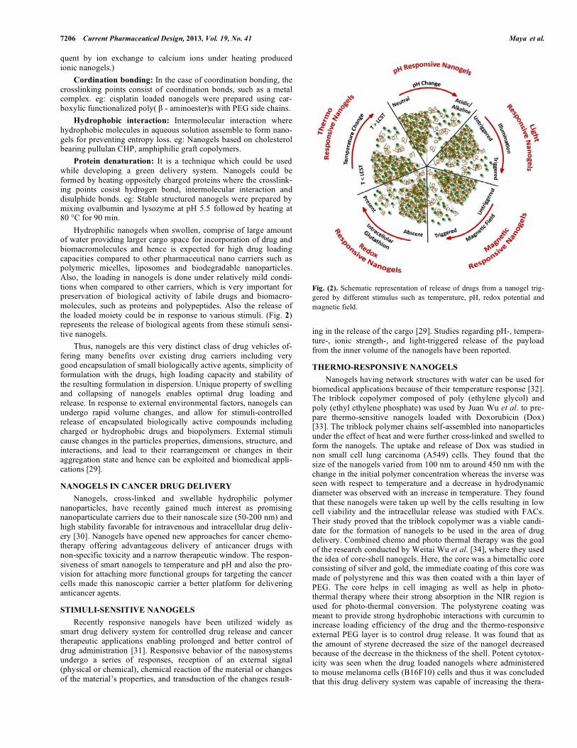
STIMULI-SENSITIVE NANOGELS Recently responsive nanogels have been utilized widely as smart drug delivery system for controlled drug release and cancer therapeutic applications enabling prolonged and better control of drug administration [31]. Responsive behavior of the nanosystems undergo a series of responses, reception of an external signal (physical or chemical), chemical reaction of the material or changes of the material’s properties, and transduction of the changes result-
Fig. (2). Schematic representation of release of drugs from a nanogel trig-gered by different stimulus such as temperature, pH, redox potential and magnetic field.
ing in the release of the cargo [29]. Studies regarding pH-, tempera-ture-, ionic strength-, and light-triggered release of the payload from the inner volume of the nanogels have been reported.
THERMO-RESPONSIVE NANOGELS Nanogels having network structures with water can be used for biomedical applications because of their temperature response [32]. The triblock copolymer composed of poly (ethylene glycol) and poly (ethyl ethylene phosphate) was used by Juan Wu et al. to pre-pare thermo-sensitive nanogels loaded with Doxorubicin (Dox) [33]. The triblock polymer chains self-assembled into nanoparticles under the effect of heat and were further cross-linked and swelled to form the nanogels. The uptake and release of Dox was studied in non small cell lung carcinoma (A549) cells. They found that the size of the nanogels varied from 100 nm to around 450 nm with the change in the initial polymer concentration whereas the inverse was seen with respect to temperature and a decrease in hydrodynamic diameter was observed with an increase in temperature. They found that these nanogels were taken up well by the cells resulting in low cell viability and the intracellular release was studied with FACs. Their study proved that the triblock copolymer was a viable candi-date for the formation of nanogels to be used in the area of drug delivery. Combined chemo and photo thermal therapy was the goal of the research conducted by Weitai Wu et al. [34], where they used the idea of core-shell nanogels. Here, the core was a bimetallic core consisting of silver and gold, the immediate coating of this core was made of polystyrene and this was then coated with a thin layer of PEG. The core helps in cell imaging as well as help in photo-thermal therapy where their strong absorption in the NIR region is used for photo-thermal conversion. The polystyrene coating was meant to provide strong hydrophobic interactions with curcumin to increase loading efficiency of the drug and the thermo-responsive external PEG layer is to control drug release. It was found that as the amount of styrene decreased the size of the nanogel decreased because of the decrease in the thickness of the shell. Potent cytotox-icity was seen when the drug loaded nanogels where administered to mouse melanoma cells (B16F10) cells and thus it was concluded that this drug delivery system was capable of increasing the thera-
Smart Stimuli Sensitive Nanogels in Cancer Drug Delivery and Imaging Current Pharmaceutical Design, 2013, Vol. 19, No. 41 7207
peutic efficiency of the drug. You-Yong Yuan et al. reported bio-compatible and biodegradable nanogels with a branched structure by the reaction of 3, 6-dioxaoctan-1, 8-diyl bis(ethylene phosphate) (TEGDP) with tris(2-aminoethyl)amine (TREN) in an ionic liquid containing mini-emulsion. The nanogels efficiently loaded doxorubicin with an enzyme-responsive drug release behavior and also proved to be uptaken by the human breast cancer cell line (MDA-MB-231) [35]. Y. Lee et al. synthesized super-expandable nanogels and util-ized them for thermally triggered cell death. The nanogels were fabricated by light cross-linking between oligo (L-lactic acid)-poly(ethylene oxide)-poly(propylene oxide)-poly(ethylene oxide)-oligo (L-lactic acid) and poly(ethylene glycol) grafted poly(L-lysine). The drastic and rapid volume transition exhibited by the nanogels (reversible 800-fold nano- to micro-scale volume transi-tion upon cold shock ie, 150 nm at 37 °C and 1.4 �mat 15 °C) in-duced cell death in cervical cancer cells (Hela) via necrosis. This nanogels could also used for carrying anticancer agents within the hydrophobic interior of the collapsed nanogels at 37 °C to increase the cell lethality [36]. Thermo-responsive nanogels from poly(l-lactide)-g-pullulan (PLP1 and 2) copolymers with phase transisition temperature of 35 ºC were investigated as an anticancer drug deliv-ery carrier using Dox. The cytotoxicity assay performed on Hela cells found that the IC50 values of Dox released from PLP 1 were approximately 5.9 and 9.3 mg/mL at 37 and 42 ºC, respectively indicating 1.6 times enhanced cytotoxicity at 42 ºC than 37 ºC due to higher Dox release at this temperature [37].
pH-RESPONSIVE NANOGELS pH-sensitive nanogels that create switching carriers in release kinetics from slow release while circulating to rapid release at the targets have been considered as promising anticancer drug carriers. Shaoyong Yu et al. used chitosan and ovalbumin to prepare stable nanogels using a green method for its synthesis [38]. Their nanogels consisted of ovalbumin nanospheres containing some chitosan chains interspersed in the nanogel structure and the rest forming its shell. The 100 nm sized nanogels were pH sensitive and the hydro-dynamic radius was constant in the pH range of 4.3-5, increases in the pH range of 5-5.3 and strongly increases in the pH range of 5.3-5.8. They also reported a change in the hydrophobicity/hy-drophilicity with respect to pH. They are hydrophilic at neutral and acidic pH and hydrophobic at alkaline pH. The nanogels they pre-pared thus have the potential to be used in drug delivery where their pH dependent properties could be utilized. Motoi Oishi et al., [39] came out with a pH sensitive PEGylated nanogel loaded with Dox which showed good release at endosomal pH which is 1.4-2.4 units lower than physiological pH. Volume phase transition is observed at endosomal pH where the size changed from 145 to 165 nm. However, the collapse of the nanogel was observed at alkaline pH. These nanogels where prepared by emulsion copolymerization of 2-(N, N-diethylamino) ethyl methacrylate (EAMA) with heterobifunc-tional poly (ethylene glycol) bearing a 4-vinylbenzyl group at the �-end and a carboxylic acid group at the �-end (CH2-LCH–Ph–PEG–COOH) in the presence of potassium persulfate and ethylene glycol dimethacrylate (1.0 mol %) as cross-linker. They carried out in vitro cytotoxicity assays on human breast cancer MCF-7 cells and the Dox ressistant human hepatoma cell line (HuH-7) and found that the activity of the drug is a bit lower than when com-pared to the bare drug in the case of MCF-7 cells. However the Dox loaded nanogels exhibited higher antitumor activity against HuH-7 cells [39]. Table 2 summarizes the pH sensitive nanogels with the pay load and their significant characteristics. pH responsive chitosan based nanogels have been prepared by the inverse micro-emulsion method for loading Dox [40] and also by the gelation method based on chemical cross-linking to encapsu-late hydrophobic anticancer drugs like camptothecin (CPT) [41]. To avoid the cardiotoxicity of Dox, it has been coupled with dextran
(DXR) and then encapsulated within chitosan nanogels. The nano-system showed enhanced tumor regression compared to that of drug conjugate and free drug in Balb/c mice implanted with J774A.1 macrophage tumor cells [39]. Chitosan based nanogels have been reported to exhibit temperature dependant reversible surface switch-ing which makes a viable option for targeted drug delivery [42, 43]. Chitosan was modified with glycidyltrimethylammonium chloride and used to synthesize the nanogels, which showed pH dependent volume phase transition from 180 nm to 450 nm. Targeting was made possible by conjugation with transferrin. Methotrexate was the anticancer drug chosen for this study and release from the nano-gels was carried out under acidic conditions. In another study, gly-col chitosan (Glycol-CS) was modified with cholanic acid and self-assembled with hydrophobic anticancer drug docetaxel (DTX) us-ing a dialysis method. The resulting hydrophobically modified gly-col-chitosan (HGC) nanogels had a diameter of 350 nm and were stable in the presence of bovine serum albumin and also showed a sustained drug release profile in vitro and also enhanced antitumor activity in vivo in lung cancer model, suggesting a promising drug delivery for cancer therapy [44]. Previous studies indicated that drug loaded nanoparticles composed of NIPAAm and chitosan could achieve a pH-sensitive drug release and an enhanced anti-cancer activity under a slightly acidic environment. NI-PAAm/chitosan nanoparticles were used for loading anticancer drugs CPT [43] and paclitaxel (PTX) [42]. CPT and PTX showed a pH responsive release pattern. In addition, CPT showed pH de-pendant cytotoxicty in human colon carcinoma cells SW480 (sig-nificant toxicity at pH 6.8 than at 7.4). The mice treated with PTX-NIPAAm/chitosan nanoparticles showed enhanced tumor regres-sion and also complete regression in more than 50% mice. [45, 46]. Duan et al. reported the development of self-assembled chitosan-graft-poly (N-isopropylacrylamide) (CS-g-PNIPAm) nanogels for the pH dependant release of a potent anticancer agent, Oridonin (ORI) to cancer cells. ORI-loaded nanogels could enhance the anti-tumor activity towards Hep G2 liver cancer cell lines under an acidic environment due to its fast pH-triggered (acidic condition) drug release. This particular study proposed that this nanogels-based delivery system might be exploitable in tumoral acidic ex-tracellular pH targeting for hydrophobic anticancer drugs [47]. pH responsive Dox loaded chitin nanogels was developed and its in vitro anticancer efficacy was evaluated in different cancer cell lines. The study reported that 130-160 nm sized nanogels showed pH sensitive controlled release of Dox [48]. They also showed the cel-lular uptake of these nanogels and toxicity towards variety of can-cer cell lines. The fluorescent Dox enabled the imaging of the can-cer cells (Fig. 3). Nanogels sensitive to endosomal pH with Dox prodrug were studied investigated for triggered intracellular drug release in cancer cells. Sequential treatment of poly(ethylene glycol)-b-poly(2-hydroxyethyl methacrylate-co-ethyl glycinate methacryl-amide) (PEG-b-P(HEMA-co-EGMA)) copolymers with hydrazine result in Dox prodrugs with grafting contents of 3.9, 5.7, and 11.7 wt %. The pH release of Dox prodrug from nanogels was proved by the enhanced release at endosomal pH and 37 °C in 48 h, and lesser amount of drug was released at pH 7.4 under the same conditions. The internalization of Dox prodrug nanogels and the released Dox was observed in the cytosol and cell nuclei of RAW 264.7 by Confocal laser scanning microscope (CLSM). Thus the study proved endosomal pH-activatable Dox prodrug nanogels with features of water-soluble macromolecular prodrugs and nanogels put forward a potential platform for targeted cancer therapy [49]. A virus-mimetic nanogel (VM-nanogel) was developed with a hydro-phobic polymer core (poly(L-histidine-co-phenylalanine) (poly(His32-co-Phe6)) and two layers of hydrophilic shell loaded with Dox. PEG forms the inner shell whose one end is linked to the polymeric core and another end to bovine serum albumin (BSA). The VM-nanogel is reported to be pH sensitive whereby reversible
7208 Current Pharmaceutical Design, 2013, Vol. 19, No. 41 Maya et al.
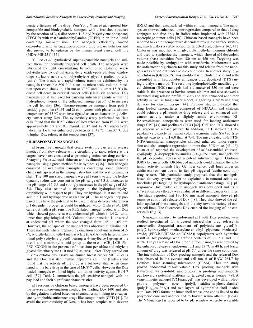
Fig. (3). Fluorescent microscopy images of doxorubicin loaded chitin nano-gels taken up by (A) MCF7, (B) PC3, (C) A549 and (D) HepG2 cells after 4 h of incubation period [48]. (Reproduced with the permission of Elsevier [48]). swelling and shrinking of the nanogel was observed when the pH was cycled between the typical cytosolic pH range (7.4 and 6.8). The release rate of Dox is influenced by the pH-induced reversible swelling/deswelling of the core as a result of which enhanced amount of Dox was released at endosomal pH (pH 6.4) compared to that of cytosolic pH (pH 6.8–7.4). These VM-nanogels were found to kill human ovarian cancer cells (A2780) [50]. Another study reported a pH sensitive polysaccharide/drug conjugate penetrating Hela tumors in mice and it has also found to have the potential to be used in photodynamic therapy specifically targeting tumors [51]. Weitai Wu et al. developed a pH sensitive chitosan based nano-gel. 3.2-3.8 nm CdSe quantum dots were in-situ immobilized in the 100 nm nanogels, which consisted of chitosan chains semi-interpenetrating in the cross-linked poly (methacrylic acid) (PMAA) networks. They did observe that the pH responsiveness of the nanogel changed when the polymer chains were submitted to a non-covalent physical association with each other. They studied the cytotoxicity and uptake in B16F10 cells and the in vitro release of anti-cancer drug temozolomide under acidic conditions. They also found a non-reversible pH- sensitive PL property and cytotoxicity after 24 h [52]. Sabitha M et al. developed biodegradable pH sensitive chitin nanogels loaded with anticancer drug curcumin (CCNGs) and used for the transdermal delivery of curcumin to melanoma. The release profile showed prominent release of curcumin at acidic pH than at neutral pH referring to the pH-responsive behavior of the nanogels. The pH-responsiveness of CCNGs could be due to the the protona-tion of its free amine groups in acidic pH. The CCNGs were inter-nalized by the melanoma cell lines A375 and showed enhanced cytotoxic effect compared to normal human dermal fibroblast cell lines. They also reported that CCNGs possess excellent skin pene-tration and retention properties. The cationic chitin interact more with the negatively charged skin lipids and the hydrophobic moie-ties on the chitin nanogels could favor van der Waals interactions with the hydrophobic areas of the keratin present in skin melano-
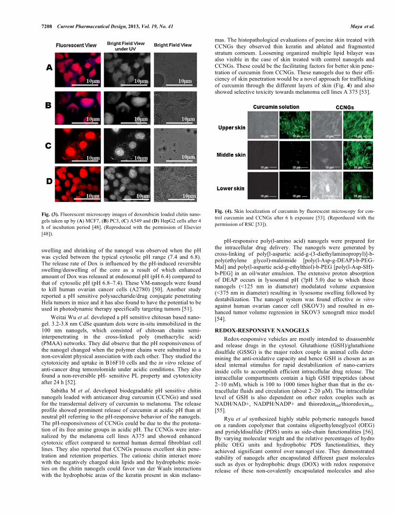
mas. The histopathological evaluations of porcine skin treated with CCNGs they observed thin keratin and ablated and fragmented stratum corneum. Loosening organized multiple lipid bilayer was also visible in the case of skin treated with control nanogels and CCNGs. These could be the facilitating factors for better skin pene-tration of curcumin from CCNGs. These nanogels due to their effi-ciency of skin penetration would be a novel approach for trafficking of curcumin through the different layers of skin (Fig. 4) and also showed selective toxicity towards melanoma cell lines A 375 [53].
Fig. (4). Skin localization of curcumin by fluorescent microscopy for con-trol curcumin and CCNGs after 6 h exposure [53]. (Reporduced with the permission of RSC [53]). pH-responsive poly(l-amino acid) nanogels were prepared for the intracellular drug delivery. The nanogels were generated by cross-linking of poly[l-aspartic acid-g-(3-diethylaminopropyl)]-b-poly(ethylene glycol)-maleimide [poly(l-Asp-g-DEAP)-b-PEG-Mal] and poly(l-aspartic acid-g-ethylthiol)-b-PEG [poly(l-Asp-SH)-b-PEG] in an oil/water emulsion. The extensive proton absorption of DEAP occurs in lysosomal pH (?pH 5.0) due to which these nanogels (<125 nm in diameter) modulated volume expansion (<375 nm in diameter) resulting in \lysosome swelling followed by destabilization. The nanogel system was found effective in vitro against human ovarian cancer cell (SKOV3) and resulted in en-hanced tumor volume regression in SKOV3 xenograft mice model [54].
REDOX-RESPONSIVE NANOGELS Redox-responsive vehicles are mostly intended to disassemble and release drugs in the cytosol. Glutathione (GSH)/glutathione disulfide (GSSG) is the major redox couple in animal cells deter-mining the anti-oxidative capacity and hence GSH is chosen as an ideal internal stimulus for rapid destabilization of nano-carriers inside cells to accomplish efficient intracellular drug release. The intracellular compartments contain a high GSH tripeptides (about 2–10 mM), which is 100 to 1000 times higher than that in the ex-tracellular fluids and circulation (about 2–20 �M). The intracellular level of GSH is also dependent on other redox couples such as NADH/NAD+, NADPH/NADP+ and thioredoxinred/thioredoxinox. [55]. Ryu et al synthesized highly stable polymeric nanogels based on a random copolymer that contains oligoethyleneglycol (OEG) and pyridyldisulfide (PDS) units as side-chain functionalities [56]. By varying molecular weight and the relative percentages of hydro philic OEG units and hydrophobic PDS functionalities, they achieved significant control over nanogel size. They demonstrated stability of nanogels after encapsulated different guest molecules such as dyes or hydrophobic drugs (DOX) with redox responsive release of these non-covalently encapsulated molecules and also
Smart Stimuli Sensitive Nanogels in Cancer Drug Delivery and Imaging Current Pharmaceutical Design, 2013, Vol. 19, No. 41 7209
Table 2. Different pH sensitive nanogel systems loaded with anticancer drugs with its significant characteristics and the cancercells treated by them
Drug loaded nanogels Important characteristic Type of cancer cells Reference
Dox-EAMA-PEG Volume phase transition at endosomal pH with a size change (145 to 165 nm) and also enhanced drug release
Human breast adenocarcinoma (MCF-7) & Human hepatoma
(Huh-7)
[39]
Dox-DXR-chitosan Long circulating hydrogel nanoparticles
Enhanced regression in tumor volumes in vivo
50% survival rate for 9 days
Reduced toxicity compared to free drug
Macrophage tumor cells (J774A.1) [40]
DTX-HGC 350 nm sized nanogels
Stable for 2 weeks under physiological conditions (pH 7.4 & 37ºC)
Enhanced antitumor activity in lung cancer models
Non small cell lung carcinoma (A549)
[44]
CPT-PNIPAAm /chitosan Sensitive to tumor pH
Optimal CPT release at pH 6.8
Enhanced cytotoxicity at pH 6.8 than 7.4
Human colon carcinoma (SW480) [45]
PTX-PNIPAAm /chitosan Sensitive to tumor pH
High cumulative release of PTX at pH 6.8 and decreased above pH 6.9
Complete tumor regression in more than 50% mice bearing lung cancer
Non small cell lung carcinoma (A549)
[46]
ORI-CS-g-PNIPAm pH-triggered fast drug release under a slightly acidic condition Human hepato-
cellular carcinoma (Hep G2)
[47]
Dox-chitin 130-160 nm sized pH sensitive nanogels
60% Dox released under acidic environment and only 40% release at neutral pH
MCF-7, A549, Hep G2, Human prostate cancer (PC-3)
[48]
VM-nanogel: Dox- (poly(L-histidine-co-phenylalanine)
(poly(His32-co-Phe6))-PEG-BSA
pH-induced reversible swelling/deswelling of the core
Enhanced Dox released at endosomal pH (pH 6.4) and the release rate was reduced at the cytosolic pH (pH 6.8–7.4).
Human ovarian carcinoma (A2780) [50]
Temozolomide-chitosan-PMAA Excellent collidal and structural stability pH responsive re-versible property change sense the environmental pH change
regulate the release of anticancer drug pH range of 5-7.4 foundin pathological zone
Mouse melanoma (B16F10) [52]
Curcumin-chitin Transdermal delivery of anticancer agents
Excellent skin permeation and retention
Ctionically charged CCNGs interact with negatively charged skin lipids
Interaction between the hydrophobic moieties
Human melanoma (A375) [53]
Poly(l-amino acid) volume expansion in a lysosomal pH (<pH 5.0)
Enhanced tumor volume regression in SKOV3 xenograft
Human ovarian carcinoma (SKOV3)
[54]
efficient cell-uptake efficiencies (in MCF-7 cells) [56]. Matyjaszewski et al reported reduction-sensitive functional nanogels using inverse mini-emulsion atom transfer radical polym-erization (ATRP) and the disulfide-thiol exchange reaction. Hela cells treated with Dox-loaded disulfide-crosslinked nanogels upon addition of 20wt % GSH the growth was significantly inhibited [57].
Lee et al., developed HA nanogels (200-500 nm) physically encapsulating small interfering RNA (siRNA) by an inverse water-in-oil emulsion method where in thiol-conjugated HA in aqueous emulsion droplets was ultrasonically crosslinked via the formation of disulfide linkages. CD44 +ve HCT-116 cells readily took up HA/siRNA nanogels. Release rates of siRNA from the HA nanogels was regulated by the GSH concentration, indicating intracellular reductive agent triggered cleavage of disulfide
7210 Current Pharmaceutical Design, 2013, Vol. 19, No. 41 Maya et al.
crosslinked HA nanogels. Similarly HA nanogels with the disulphide linkages could be used for the specific intracellular delivery of anticancer agents and could be exploited for cancer therapy [58].
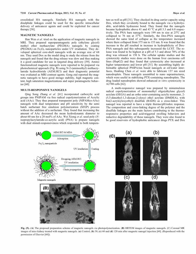
MAGNETIC NANOGELS Han Wen et al. tried on the application of magnetic nanogels in MRI. They prepared superparamagnetic poly (ethylene glycol) methyl ether methacrylate (PEGMA) nanogels by coating (PEGMA) on Fe3O4 nanoparticles under UV irradiation. They de-veloped spherical core-shell nanogels with an average size of 68 nm. They used Dox as the model drug to study the release from the nanogels and found that the drug release was slow and thus making it a good candidate for use in targeted drug delivery [59]. Amino functionalized magnetic nanogels were reported to be prepared via photochemical approach (Fig. 5) using N-2-(aminoethyl) methacry-lamide hydrochloride (AEM.HCl) and their diagnostic potential was evaluated as MRI contrast agents. Gong etal reported the mag-netic nanogels to have good storage stability, high magnetic con-tent, high saturation magnetizations and super paramagnetic behav-ior [60]
MULTI-RESPONSIVE NANOGELS Qing Song Zhang et al. [61] incorporated carboxylic acid groups into PNIPAM via free radical copolymerization of Acrylic acid (AAc). They thus prepared transparent poly (NIPAMco-AAc) nanogels with dual temperature and pH sensitivity by the semi batch surfactant free emulsion polymerization (SFEP) process without the addition of a surfactant. They found that increasing the amount of AAc decreased the mean hydrodynamic diameter to about 60 nm for a 20 mol% of AAc. Wei Xiong et al. used poly (N-isopropylacrylamide-co-acrylic acid) (PNA) to prepare nanogels with dual stimuli-responsiveness which responded to both tempera-
ture as well as pH [31]. They checked its drug carrier capacity using Dox, which they covalently bound to the nanogels via a hydrolys-able, acid-labile hydrazone bond. They found that the nanogels became hydrophobic above 41 and 43 oC at pH 5.3 and 6.8 respec-tively. The PNA bare nanogels were 199 nm in size at 25oC and collapsed to 76 nm at 37oC. Similarly, the Dox-PNA nanogels showed the same kind of collapse as the temperature increased where there collapsed from 375 nm to 153 nm. It was found that the increase in the pH resulted in increase in hydrophilicity of Dox-PNA nanogels and this subsequently increased the LCST. The re-lease was found to be highest at a pH of 5.3 and about 70% of the drug was released in 150 h. The cellular uptake studies and the cytotoxicity studies were done on the human liver carcinoma cell lines (HepG2) and they found that cytotoxicity also increased at higher temperatures and lower pH [31]. By assembling highly de-formable spherical PNIPAAm based nanogels at oil/water inter-faces, Huabing Chen et al. were able to fabricate 137 nm sized nanodroplets. These nanogels assembled to nano suprastructures, which were useful in stabilizing PTX-containing nanodroplets. The drug loaded nanodroplets showed enhanced in vitro cytotoxicity in A2780 cells [62]. A multi-responsive nanogel was prepared by miniemulsion radical copolymerization of monomethyl oligo(ethylene glycol) acrylate (OEGA) and an ortho ester-containing acrylic monomer, 2-(5,5-dimethyl-1,3-dioxan-2-yloxy) ethyl acrylate (DMDEA), with bis(2-acryloyloxyethyl) disulfide (BADS) as a cross-linker. This nanogel was reported to have a triple thermo/pH/redox response. The composition and cross-linking degree of the polymer and the disulfide linkages are the main factors contributing to the thermo-responsive properties and acid-triggered swelling behaviors and reductive degradability of these nanogels. They were also found to be good reservoirs of hydrophobic anticancer drugs PTX and Dox
Fig. (5). (A) The proposed preparation scheme of magnetic nanogels via photopolymerization. (B) HRTEM images of magnetic nanogels. (C) Coronal MR images of mice kidney treated with magnetic nanogels. (a) Control, (b) 30, (c) 60 and (d) 120 min after magnetic nanogel injection [60]. (Reporduced with the permission of Elsevier [60]).
Smart Stimuli Sensitive Nanogels in Cancer Drug Delivery and Imaging Current Pharmaceutical Design, 2013, Vol. 19, No. 41 7211
and also for a dye [63]. Fan et al. reported thermo and pH respon-sive magnetic hydrogel nanosphere poly (Nisopropylacrylamide-co-acrylic acid)/Fe3O4 (poly(NIPAAm-co-AA)/Fe3O4). The drug load-ing capacities and the releasing behavior of the magnetic hydrogel nanospheres were investigated with Dox as an anticancer drug model. The Dox-loaded magnetic hydrogel nanospheres showed an enhanced anti-tumor effect towards Hela cancer cell lines. They suggested the enhanced cytotoxicity by enhanced cellular uptake of magnetic nanogel and the release of Dox within the acidic cellular compartments [64]. A magnetic pH responsive nanogel was ex-ploited for the delivery of anti-GFP siRNA and superparamagnetic IONPs to HeLa-GFP cells. The siRNA release via pH-mediated endosomal escape was demonstrated. The IONPs played dual roles: initially as a magnetofection agent for enhanced cellular uptake later as release monitoring probe [65].
TARGETED NANOGELS Targeted drug delivery has been developed as new chemotherapeutic strategies for cancer treatment. Drug loaded nanogels can be passively targeted within the tumor tissue due to the enhanced permeation and retention effect (EPR), the specific pathophysiology of cancers. EPR effect aids in the localization of these drug loaded nanogels in tumor sites. In addition to this, en-hanced specificity can be endowed by actively targeting these sytems by the introduction of targeting moieties like folic acid (FA), transferrin or monoclonal antibodies [66]. Folic acid (FA) is one among the prominent targeting moiety capable of specific in-teraction with cells expressing the folate receptor (FR) [67]. Many of the human tumors over express FR providing the tumor cells with more FA required for DNA synthesis aiding in aggressive tumor growth. A simple strategy was reported for delivering Dox specifically to cancer cells by direct coupling of FA to Pullulan (Pul) backbone via ester linkages. The nanogels possessed two size distributions in aqueous solution, which were around 70 and 270 nm. It was found that with variations in the degree of substitution of FA, the release rate of Dox changed slower release with higher substitution. KB cells treated by FA4/Pul containing Dox showed statistically significant loss of viability [66]. Pluronic® F127 (PF127) was used to produce amphiphilic nanocarriers for Dox. Stabilized CS-PF127 nanogel was developed by acrylating and reacting both the terminal hydroxyl group of PF127 with methacrylated chondroitin sulfate (CSMA). The carboxylic group of CSMA was reacted with a FA-polyethylene glycol (FA-PEG) thereby forming FA decorated nanogels. Dox was selectively delivered to FR-positive KB cells because of the high binding affinity of FA to tumor-associated folate receptors (FR). They proved better cellular internalization of 140nm sized FA-CS-PF127 into the FR over expressing KB cells by CLSM and flow cytometry [68]. Controlled template synthesis of nanogels has been reported by polyion complexation and cross-linking of doubly hydrophilic block ionomers, such as PEO-b-PMA [69]. Nukolova et al. evalu-ated the drug therapy efficacy of FA decorated nanogels loaded cisplatin and doxorubicin against ovarian cancer. FA conjugation was done via EDC activation of FA. FA positive ovarian cancer cells showed an enhanced uptake of the FA-nanogels with respect to FA negative cells. The study reported folate specificity and elevated activity by FA-nanogel through a specific mechanism involving in vivo FR. Anti-cancer drug, cisplatin (CDDP) was delivered to a xenograft tumor using these FR-targeted nanogels and they observed increased Pt accumulation in all organs,superior anti-tumor efficacy and decreased renal toxicity. This study pro-jected the possibility of targeted nanogels against ovarian cancer [69]. Monoclonal antibodies (mAb) are now a days becoming an efficient agent for targeted cancer therapy. Diblock copolymers of poly(ethylene glycol)-b-poly(methacrylic acid) (PEG-b-PMA) with PEG terminal aldehyde functionality were used to prepare nanogels via condensation of PEG-b-PMA with Ca2+ ions into micelle-like
aggregates, cross-linking of the PMA/Ca2+ cores and removal of Ca2+ ions. mAb CC49 was effectively conjugated to nanogels utilizing a reductive amination reaction between aldehyde groups and amino groups of mAb resulting activity against tumor-associated glycoprotein 72 (TAG-72). The nanogels represented hydrophilic, highly swollen, soft particles, which were composed of cross-linked ionic cores and hydrophilic protective PEG shells with active functional groups. The ionic PMA cores can immobilize various therapeutic and imaging agents, while aldehyde-functionalized PEG chains allow for simple and efficient conjuga-tion of nanogels with proteins [70]. Some studies reported the use of integrins �v�3-targeted lipid-coated nanogels with cross-linked human serum albumin in the core for carrying therapeutic cargoes. They developed lipid-coated nanogels using lipid bilayer surrounding cross-linked pro-tein/polymeric core. The lipid bilayer incorporates targeting ligands (integrin �v�3) and polymeric coatings such as polyethylene glycol (PEG), whereas the cross-linked core consists of proteins such as human serum albumin (HSA) or �1-acid glycoprotein, which serve as carriers for the drug cargoes. The nanogels were used as a carrier for variety of hydrophobic drugs. The taxane loaded nanogels ex-hibited 15 fold more potent antitumor activity in the treatment of orthotopic breast and pancreatic tumors in mice. They proved that nanogels as a flexible platform for choosing different drug cargoes and targeting ligands so as to improve the efficacy of therapeutic agents with dose limiting toxicities and their pharmacokinetics [71]. Table 3 describes actively targeted nanogels and their application in cancer therapy with its outcome. Active targeting with nanogels is a subject of study and Wooram Park et al. went step further preparing nanogels with the material possessing affinity towards cell surafce receptors. Hyaluronic acid (HA) is expressed on cell surface and extracellular matrix of human tissues and HA interacts with cells via receptor CD44. Various carcinoma, lymphoma, breast, colorectal, and lung tumor cells express elevated levels of CD44 and hence HA are se-lected as a promising targeting moiety for the specific delivery of anticancer agents. Park et al. reported the preparation of self as-sembled nanogels using acetylated low molecular weight HA (AC-HALM). The potential for cancer cell selectivity was analyzed by loading the nanogels with Dox and comparing the effect on CD44 positive cancer cells (Hela) and CD44 negative normal cells (Vero). Dox loaded AC-HALM nanogels showed selective cytotoxicity towards cancer cells with HA-binding receptors, and not to normal cells without HA binding receptors [72].
OTHER NANOGELS WITH ANTICANCER AGENTS Na Li et al. prepared nanogels using Pluronic micelles; one of Pluronic F127 micelles cross-linked with polyethylenimine (PEI) and the other had a penetrating network of poly (butylcyanoacry-late) (PBCA) in Pluronic F127 micelles. The cytotoxicity and re-lease studies of the 2 model anticancer drugs PTX and 10-Hydroxycamptothecin (HCPT) was studied. The 200 nm sized drug loaded nanogels resulted in a higher toxicity towards Human hepa-toma Hep G2 cell lines than the free drug [73]. Another interesting material that has been used for the synthesis of nanogels is cyclo-dextrin. Samia et al. studied self-assembly of cyclodextrin into nanogels and used them to entrap two hydrophobic molecules namely benzophenone (BZ) and tamoxifen (TM). The spontaneous formation of these nanogels was aided by the addition of hydropho-bically modified dextran forming 100-200 nm nanogels. The test molecules were loaded into the nanogels by solubilizing them in the associative polymers. However, their studies showed that the high-est loading efficiency was obtained when the molecules were dis-solved in both the polymers before mixing which resulted in the formation of very stable nanogel suspensions. It was observed that the BZ loaded nanogels were more stable than the TM loaded nanogels, which agglomerated very fast [74]. Heparin, a well
7212 Current Pharmaceutical Design, 2013, Vol. 19, No. 41 Maya et al.
known anticoagulant is also reported to have various anti-cancer activities in the processes of tumor progression and metastasis [75, 76]. Bae et al. reported efficient delivery of free heparin using hepa-rin nanogels [77]. They chemically modified heparin with thiol groups and then cross-linked with disulfide linkages to produce reducible heparin nanogels. The anti-cancer effects of the heparin nanogels were carefully investigated by examining their cellular uptake and apoptosis-inducing effects using mouse melanoma B16F10 cells. Heparin nanogels exhibited enhanced cellular caspase-3 activity compared to that of heparin and hence they reported caspase-dependent apoptotic cancer cell death induced by heparin nanogels. In short the study demonstrated that the heparin nanogels resulted intracellular delivery of free heparin, triggered caspase activation, and consequently induced the apoptotic cancer cells death [77]. W. Park et al reported a self organized acetylated chondroitin sulfate (AC-CS3) and analysed its potential for deliver-ing Dox to Hela cells. AC-CS3 nanogel was readily internalized into Hela cells via sugar receptors, and localized in the cytoplasm and showed an IC50 of 1100 ng/ml [78]. Nucleoside analogues (NA) and nucleobases such as pyrimidine analogue arabinofuranosylcytosine (cytarabine, araC) for acute myeloid leukemia, the fluorinated pyrimidine analogue gemcitabine (dFdC) for lung, breast, pancreatic, and bladder cancers, the fluoro-pyrimidine 5-fluorouracil (5-FU) are widely used for the treatment of cancer. Nanogels are suggested as a vehicle for the delivery of nucleoside analog 5’-triphosphates based anticancer drugs. Solutions of FATP and nanogels spontaneously form polyplexes by ionic interactions between polyphosphate groups of the drug and protonated polyethylenimine (PEI) chains in nanogel network. Vinogradov et al. reported the formation of polyplex nanogel formulations with the 5’-triphosphate of cytotoxic 5-fluoroadenine arabinoside (fludarabine) (FATP). The polyplexes have formed spontaneously by mixing solutions of FATP and nanogels because of ionic interactions between protonated polyethylenimine (PEI) chains in nanogel network with polyphosphate groups of the drug.
These nanogels were attached with folate and showed 10-fold in-crease of the carrier’s internalization in human breast carcinoma MCF-7 cells [79]. Stable lyophilized formulations of 5’-triphosphates of cytarabine (araCTP), gemcitabine (dFdCTP), and floxuridine (FdUTP) encapsulated in biodegradable PEG-cl-PEI or F127-cl-PEI nanogel networks (NGC and NGM, respectively) were prepared by a self-assembly procedure. The study showed that the nanogels could deliver the cytotoxic analogues into breast (MCF-7) and colorectal (HCT-116) cancer cell lines and also induced en-hanced cytotoxicity in these cancer cell lines. Tumor growth inhibi-tion was also observed in human breast carcinoma MCF-7 xenograft mouse model [80]. Nanohydrogels are also being used for delivering exogenous proteins. Fusion proteins containing protein transduction domain (PTD) mediated delivery require expression of heparan sulfate on the surface of the target cells [81-83]. A study demonstrated efficient delivery of proteins to myeloma cells and primary CD4+ T lymphocytes using nanosized hydrogels formed by cationic cholesteryl group-bearing pullulans (cCHP) [84]. Shimizu et al.synthesized recombinant murine interleukin-12 (rmIL-12) loaded cholesterol-bearing pullulan (CHP)-based hydrogel nanoparticles (CHP/. rmIL-12 nanogels). They studied the release property and the stability of these nanogels in vivo in mice with fibrosarcoma. Subcutaneously injected CHP/rmIL-12 nanogels resulted in a prolonged increase in serum concentration of rmIL-12 which inturn retarded growth of preestablished subcutaneous fibrosarcoma [85].
NANOGELS FOR CANCER CELL IMAGING Quantom dots (QDs) with bright fluorescence, narrow emission, broad excitation, and high photo-stability has been developed as potential fluorescent probes for long-term cellular and molecular imaging [86, 87]. It has already been reported that cho-lesterol bearing pullulan (CHP) self assemble into stable nanogel. Hasegawa et al. investigated the ability of CHP and amino modified CHP (CHPNH2) to deliver QDs into cells. They reported that posi-tively charged amino group interact well with negatively charged QD and they were used to image human cervical cancer cell lines
Table 3. Actively targeted nanogels and their application in cancer therapy with its outcome
Drug Loaded Nanogels Targeting Ligand Important Out Come Reference
Dox-Pullulan
Dox-CS-PF127 (Chondroitin sulphate- pluro-nic F127)
Folic acid (FA)
Folic acid receptor (FR) mediated active targeting
Enhanced inetranalization by FR +ve KB cells
Significant cell viability loss for oral cancer cells (KB)
[66,68]
Cisplatin- PEO-b-PMA
(poly(ethylene oxide)-b-poly(methacrylic acid) Folic acid (FA)
pH sensitive nanogels
Enhanced inetranalization by FR +ve ovarian cancer cells (A2780) compared to that of FR –ve A549.
Superior antitumor effect in A2780 xenograft models.
[69]
Monoclonal Tumor-associated glycoprotein 72 (TAG-72)
PEO-b-PMA-PEG antibody (mAb CC49)
mediated active targeting
Targeted delivery of anticancer drugs and imaging agents to-wards primary and metastatic human adenocarcinomas
[70]
Taxane-Lipid-Human serum albumin (HSA)-PEG
RGD peptide Integrin �v�3 mediated targeted delivery of DTX
Supressed the tumor growth in orthotopic breast and pancreatic cancer model compared to that of Abraxane
[71]
Dox-AcHALM (acetylated low molecular weight hyaluronic acid)
HA CD44 receptor mediated targeted delivery of Dox.
Superior cytotoxicity in CD44 +ve cervical cancer (Hela) com-pared to that of CD44 –ve Vero cells.
[72]
Smart Stimuli Sensitive Nanogels in Cancer Drug Delivery and Imaging Current Pharmaceutical Design, 2013, Vol. 19, No. 41 7213
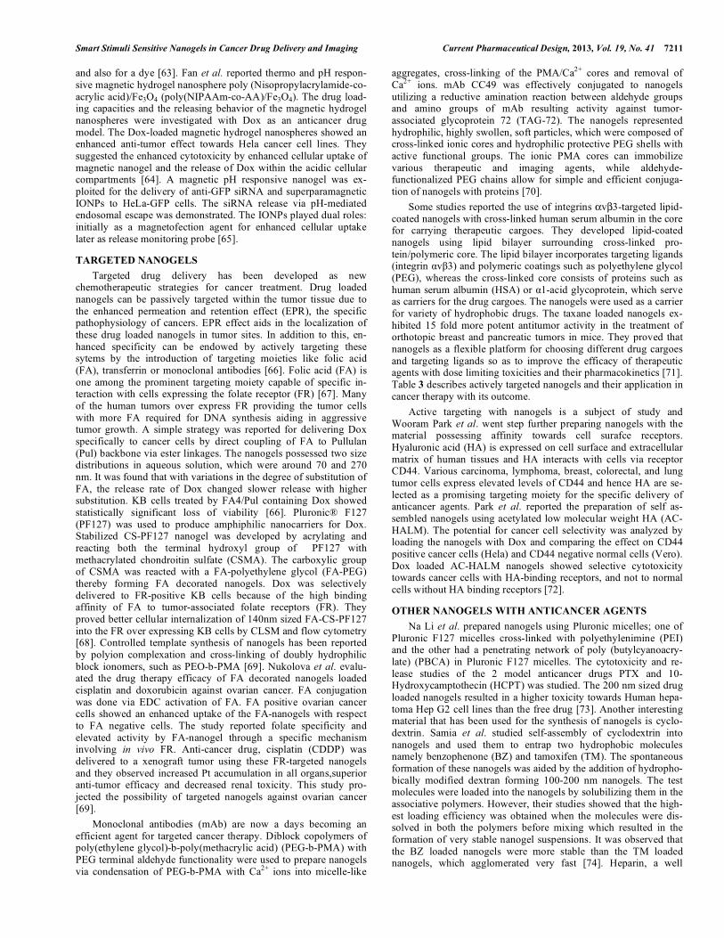
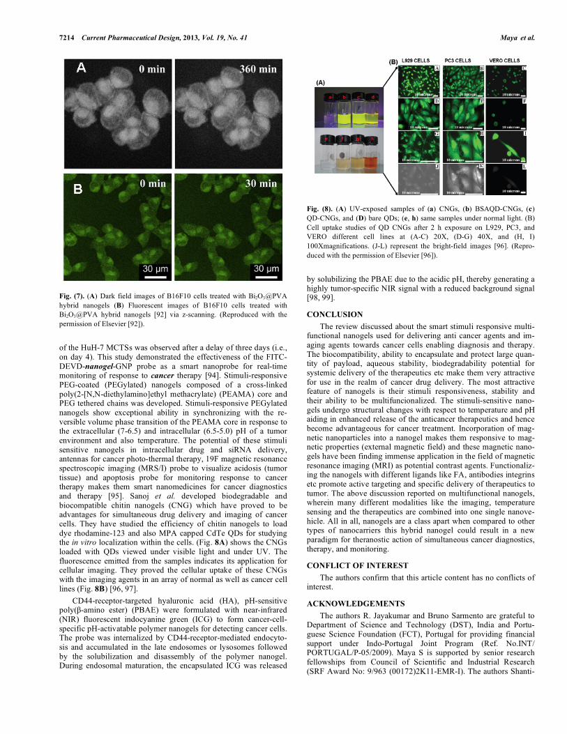
(Fig. 6) Here they have shown the QDs capped with protein A so as to generate stable negatively charged moiety for the interaction with positively charged amine group on the nanogels and also neutral nanogels could self complex with the protein capped QDs [88]. Table 4 represents the nanogels loaded with different imaging agents used for cancer cell imaging. Weitai Wu et al. [89], reported the incorporation of the imaging modality, CdSe quantum dots (~3.2 nm), by the process of in-situ immobilization in dual temperature and pH responsive hydroxypro-pylcellulose-poly (acrylic acid) (HPC-PAA) nanogels. Here they used mouse melanoma B16F10 cells to study the cell imaging, drug release and in vitro toxicity of the temozolomide-loaded nanogels. It was found that the size of the nanogels was dependent on the concentration of the cross-linker MBAAm thus making their size tunable. The nanogels sized less than 100 nm consisted of HPC chains to stabilize the CdSe QDs and pH-sensitive PAA network chains to control the swelling of the nanogelsresulting in CdSe QDs for optical pH-sensing. HPC-PAA-CdSe hybrid nanogels is advantageous for bioimaging applications.as they emit at two dif-ferent fluorescence wavelengths, combining a pH-sensitive NIR emission (741 nm) and a less sensitive visible emission (592 nm). Thus these multifunctional nanogels form the basis for sensing, imaging, and controlled drug delivery and offer opportunities for combined diagnosis and therapy. Nanogels, which could be used simultaneously for temperature sensing, cancer cell imaging, fluorescence imaging, and chemo-photo thermal treatment, was the topic of research for Weitai Wu etal. [90]. Their nanogels had a bimetallic core consisting of gold and silver along with a coating of thermo-responsive non-linear poly-mer PEG based hydrogel and semi-interpenetrating targeting ligands of HA. Their theory was that the bimetallic core could be used for cell imaging, the temperature dependent volume transition could be utilized for efficient drug loading and controlled release, the targeting ligands were obviously for active targeting of the nanogel to the effect site. They found that temperature sensing was a possibility and this was because the thermo-responsive volume phase transition of the PEG shell modifies the physico-chemical environment around the bimetallic core and changes the fluores-cence intensity. The model drug chosen here was temozolomide and its release was found to be induced by both the heat generated from external NIR irradiation and local temperature increase. Xue-qin Zhao et al. [91] fabricated photothermo-responsive nanogels for use in simultaneous optical temperature-sensing, cell imaging, and combined chemo-photothermal treatment. The nano-gels were composed of an interpenetrating network of poly (N-isopropylacrylamide) (pNIPAAm) hydrogel with a Gold nanoparti-cle (50 nm) at its core. The model drug chosen here for studying the temperature controlled release was 5-FU and cytotoxicity studies were done on Hela cells. Hela cells significantly took up these nanogels and got localized to the lysozomes. The scattering proper-ties of the gold nanoparticles have been used for cell imaging using dark-field microscopy. They thus proved to be a novel contrast agent for cell imaging. Photothermally triggered drug release was studied by irradiating them. The 5-FU loaded nanogels alone did not show much of cytotoxicity in Hela cells when compared to that which was shown when they were irradiated with a 515nm laser. Zhu et al. came up with a thermo-responsive Bi2O3@PVA (Bi2O3QDs in PVA nanogel) hybrid nanogel for theranostic actions. They reported the preparation of temperature-responsive hybrid nanogels by immobilization of Bi2O3 quantum dots (QDs) in the interior of a nanogel of poly (vinyl alcohol) (PVA). Bi2O3@PVA hybrid nano-gels stimulated by the surrounding fluid temperature (37-40ºC) convert the disruptions in homeostasis of environmental tempera-ture into high-sensitive fluorescent signals which was exploited for dark-field and fluorescence dual-modal imaging of mouse melanoma B16F10 cells (Fig. 7) indicates the bright field and fluo-
Fig. (6). (A) Formation of CHPNH2-QD hybrid nanoparticle (B) CLSFM images of HeLa cells labeled with (a) QD, (b) CHPNH2(15)-QD, and (c)liposome-QD after treating the cells with 1 nM QD, CHPNH2(15)-QD hy-brid nanoparticles, and liposome-QD complexes for 3 h [88]. (Reproduced with the permission of Elsevier [88]).
rescent images of melanoma cells treated with the hybrid nanogels [92]. Core-shell structured hybrid nanogels composed of a Ag nanoparticle (NP) as core and smart gel of poly(N-isopropylacryl-amide-co-acrylic acid) as shell was developed and it facilitated the simultaneous cancer cell imaging and adequate local delivery to tumor site. The study reported that smart hybrid nanogels developed by the incorporation of functional building blocks with optical and therapeutic functio-nality, overcome the cellular barriers of the mouse melanoma B16F10 cells and light up intracellular region, thus making it suitable for biomedical applications [93]. Oishi M et al reported a nanoprobe based on a PEGylated nanogel containing gold nanoparticies (GNPs) (fluorescence quenchers) in the cross-linked polyamine gel core and fluorescein isothiocyanate (FITC)-labeled DEVD peptides at the tethered PEG chain ends. The nanogel-nanoprobe system is reported to be bio-compatible, caspase-3-responsive, and fluorescence-quenching suitable for monitoring the cancer response to therapy. Apoptotic cells were detected in human hepatocyte (HuH-7) multicellular tumor spheroids (MCTSs) as early as one day post-treatment with staurosporine (an apoptosis inducing agent) while growth inhibition
7214 Current Pharmaceutical Design, 2013, Vol. 19, No. 41 Maya et al.
Fig. (7). (A) Dark field images of B16F10 cells treated with Bi2O3@PVA hybrid nanogels (B) Fluorescent images of B16F10 cells treated with Bi2O3@PVA hybrid nanogels [92] via z-scanning. (Reproduced with the permission of Elsevier [92]).
of the HuH-7 MCTSs was observed after a delay of three days (i.e., on day 4). This study demonstrated the effectiveness of the FITC-DEVD-nanogel-GNP probe as a smart nanoprobe for real-time monitoring of response to cancer therapy [94]. Stimuli-responsive PEG-coated (PEGylated) nanogels composed of a cross-linked poly(2-[N,N-diethylamino]ethyl methacrylate) (PEAMA) core and PEG tethered chains was developed. Stimuli-responsive PEGylated nanogels show exceptional ability in synchronizing with the re-versible volume phase transition of the PEAMA core in response to the extracellular (7-6.5) and intracellular (6.5-5.0) pH of a tumor environment and also temperature. The potential of these stimuli sensitive nanogels in intracellular drug and siRNA delivery, antennas for cancer photo-thermal therapy, 19F magnetic resonance spectroscopic imaging (MRS/I) probe to visualize acidosis (tumor tissue) and apoptosis probe for monitoring response to cancer therapy makes them smart nanomedicines for cancer diagnostics and therapy [95]. Sanoj et al. developed biodegradable and biocompatible chitin nanogels (CNG) which have proved to be advantages for simultaneous drug delivery and imaging of cancer cells. They have studied the efficiency of chitin nanogels to load dye rhodamine-123 and also MPA capped CdTe QDs for studying the in vitro localization within the cells. (Fig. 8A) shows the CNGs loaded with QDs viewed under visible light and under UV. The fluorescence emitted from the samples indicates its application for cellular imaging. They proved the cellular uptake of these CNGs with the imaging agents in an array of normal as well as cancer cell lines (Fig. 8B) [96, 97]. CD44-receptor-targeted hyaluronic acid (HA), pH-sensitive poly(�-amino ester) (PBAE) were formulated with near-infrared (NIR) fluorescent indocyanine green (ICG) to form cancer-cell-specific pH-activatable polymer nanogels for detecting cancer cells. The probe was internalized by CD44-receptor-mediated endocyto-sis and accumulated in the late endosomes or lysosomes followed by the solubilization and disassembly of the polymer nanogel. During endosomal maturation, the encapsulated ICG was released
Fig. (8). (A) UV-exposed samples of (a) CNGs, (b) BSAQD-CNGs, (c)QD-CNGs, and (D) bare QDs; (e, h) same samples under normal light. (B) Cell uptake studies of QD CNGs after 2 h exposure on L929, PC3, and VERO different cell lines at (A-C) 20X, (D-G) 40X, and (H, I) 100Xmagnifications. (J-L) represent the bright-field images [96]. (Repro-duced with the permission of Elsevier [96]).
by solubilizing the PBAE due to the acidic pH, thereby generating a highly tumor-specific NIR signal with a reduced background signal [98, 99].
CONCLUSION The review discussed about the smart stimuli responsive multi-functional nanogels used for delivering anti cancer agents and im-aging agents towards cancer cells enabling diagnosis and therapy. The biocompatibility, ability to encapsulate and protect large quan-tity of payload, aqueous stability, biodegradability potential for systemic delivery of the therapeutics etc make them very attractive for use in the realm of cancer drug delivery. The most attractive feature of nanogels is their stimuli responsiveness, stability and their ability to be multifuncionalized. The stimuli-sensitive nano-gels undergo structural changes with respect to temperature and pH aiding in enhanced release of the anticancer therapeutics and hence become advantageous for cancer treatment. Incorporation of mag-netic nanoparticles into a nanogel makes them responsive to mag-netic properties (external magnetic field) and these magnetic nano-gels have been finding immense application in the field of magnetic resonance imaging (MRI) as potential contrast agents. Functionaliz-ing the nanogels with different ligands like FA, antibodies integrins etc promote active targeting and specific delivery of therapeutics to tumor. The above discussion reported on multifunctional nanogels, wherein many different modalities like the imaging, temperature sensing and the therapeutics are combined into one single nanove-hicle. All in all, nanogels are a class apart when compared to other types of nanocarriers this hybrid nanogel could result in a new paradigm for theranostic action of simultaneous cancer diagnostics, therapy, and monitoring.
CONFLICT OF INTEREST The authors confirm that this article content has no conflicts of interest.
ACKNOWLEDGEMENTS The authors R. Jayakumar and Bruno Sarmento are grateful to Department of Science and Technology (DST), India and Portu-guese Science Foundation (FCT), Portugal for providing financial support under Indo-Portugal Joint Program (Ref. No.INT/ PORTUGAL/P-05/2009). Maya S is supported by senior research fellowships from Council of Scientific and Industrial Research (SRF Award No: 9/963 (00172)2K11-EMR-I). The authors Shanti-

Smart Stimuli Sensitive Nanogels in Cancer Drug Delivery and Imaging Current Pharmaceutical Design, 2013, Vol. 19, No. 41 7215
Table 4. Hybrid nanogels loaded with imaging agents used for cancer cell imaging.
Nanogels Cancer cell imaging Important characteristics Reference
QD-CHPNH2 Human cervical cancer cells
(Hela)
QD-nanogel complex formed by electrostatic interaction
Uniformly internalized within Hela cells
Promising candidate for live cell imaging
[88]
Dual temperature and pH responsive
Emit at two different fluorescence wavelengths, combining a pH-sensitive NIR emission (741 nm) and a less sensitive visible emission
(592 nm).
[89] Mouse melanoma (B16F10)
Mouse melanoma (B16F10)
Simultaneous temperature sensing, cancer cell imaging, fluorescence imaging, and chemo-photo thermal treatment.
[90]
CdSe QD- HPC-PAA
(hydroxypropylcellulose-poly (acrylic acid)
Bimetallic core of gold and coated with PEG and targeting ligands of
HA.
PNIPAAm-Gold nanoparticle Human cervical cancer
cells
(Hela)
Simultaneous optical temperature-sensing, cell imaging, and com-bined chemo-photothermal treatment.
Superior Hela cell death were observed when they were irradiated with a 515nm laser.
[91]
Thermoresponsive
Adapted to the surrounding fluids physiologically important range of 37-40 ºC
Enable dark-field and fluorescence dual-modal imaging
[92]
[93]
Biocompatible, caspase-3-responsive
Enables cancer cell response to therapy monitoring [96,97]
Bi2O3 QDs -PVA
PEG-Gold NPs-FITC labeled DEVD peptides
Rhodamine 123-chitin
MPA capped CdTe QD-chitin
Mouse melanoma (B16F10)
Human Hepatoma cells (Huh-7)
Human prostate cancer cells (PC-3)
pH responsive
ICG-HA-PBAE
(Indocyanin green loaded hya-luronic acid- poly(�-amino ester)
pH sensitive
CD44 receptor mediated endocytosis
pH activated release of
[98, 99]
ICG during endosomal maturation
Fig. (9). Schematic structure showing multifunctional nanogels that have the potential for carrying therapeutic moities, diagnostic moities and also both the moities together, that can be actively and passively targeted and resulting in the specific and selective delivery of the pay load in response to different external and internal stimuli.
7216 Current Pharmaceutical Design, 2013, Vol. 19, No. 41 Maya et al.
kumar V. Nair and R. Jayakumar are grateful to Nanomission, DST, India, which partially supported this work, under a “Theragnostics” grant of the Nanoscience and Nanotechnology Initiative program. The author R. Jayakumar is also grateful to Department of Biotech-nology (DBT), Govt. of India for providing research support.
ABBREVIATIONS AAc = Acrylic acid AEM.HCl = N-2-(aminoethyl) methacrylamide hydro-
chloride A375 = Human melanoma cell lines A2780 = Human Ovarian Cancer cell lines BADS = bis(2-acryloyloxyethyl) disulfide Bi2O3 = Bismuth Oxide B16F10 = Mouse Melanoma cell lines BZ = benzophenone CCNGs = Curcumin loaded chitin nanogels cCHP = cationic cholesteryl group-bearing pullu-
lans CdSe QD = Cadmium selenide quantum dot CDDP = cis-diamminechloroplatinum CS-g-PNIPAm = chitosan-graft-poly (N-isopropylacryl-
amide) CSMA = methacrylated chondroitin sulfate DEVD = Amino acid sequence Asp-Glu-Val-Asp DMDEA = 2-(5,5-dimethyl-1,3-dioxan-2-yloxy) ethyl
acrylate Dox = Doxorubicin DTX = Docetaxel EAMA = 2-(N, N-diethylamino) ethyl methacrylate 5-FU = 5-fluorouracil FATP = 5-fluoroadenine arabinoside (fludarabine) Glycol-CS = Glycol chitosan HA = Hyaluronic acid HCPT = Hydroxycamptothecin Hep G2 = Human Liver Carcinoma cell line Hela = Human Cervical Cancer cell lines HEMA = 2-hydroxyethyl methacrylate HPC-PAA = hydroxypropylcellulose-poly (acrylic acid) HuH-7 = Human Hepatoma cell line MBAAm = N,N-methyl- enebis(acrylamide) MCF-7 = Human Breast Cancer cell line MPA = mercaptopropionic acid NIPAAm = N-isopropylacrylamide OEG = oligoethyleneglycol OEGA = oligo(ethylene glycol) acrylate ORI = Oridonin PBCA = poly (butylcyanoacrylate) PDS = pyridyldisulfide PEG = Poly ethylene Glycol PEGMA = poly (ethylene glycol) methyl ether
methacrylate PEI = polyethylenimine PEG-b-P (HEMA-co-EGMA) = poly(ethylene glycol)-b-poly(2-
hydroxyethyl methacrylate-co-ethyl glycinate methacrylamide)
PF-127 = Pluronic F-127 PMMA = poly (methacrylic acid) PNA = poly (N-isopropylacrylamide-co-acrylic
acid) PTX = Paclitaxel Pul = Pullulan RAW 264.7 = Mouse Leukaemic Monocyte Macrophage
cell line SFEP = Surfactant Free Emulsion Polymerization TEGDP = 3, 6-dioxaoctan-1, 8-diyl bis(ethylene
phosphate) TREN = tris(2-aminoethyl)amine TM = tamoxifen VERO = Monkey Kidney Epithelial cell lines
REFERENCES[1] Charman WN, Chan HK, Finnin BC, Charman SA. Drug Delivery:
A Key Factor in Realizing the Full Therapeutic Potential of Drugs. Drug Develop Res 1999; 46: 316-27.
[2] Kopecek J. Smart and genetically engineered biomaterials and drug delivery systems. Eur J Pharm Sci 2003; 20: 1-16.
[3] Torchilin VP. Structure and design of polymeric surfactant-based drug delivery systems. J Control Release 2001; 73: 137-72.
[4] Muller-Goymann CC. Physicochemical characterization of colloi-dal drug delivery systems such as reverse micelles, vesicles, liquid crystals and nanoparticles for topical administration. Eur J Phar Biopharm 2004; 58: 343-56.
[5] Soppimath KS, Aminabhavi TM, Kulkarni AR, Rudzinski WE. Biodegradable polymeric nanoparticles as drug delivery devices. J Control Release 2001; 70: 1-20.
[6] Murali MY, Meena J, Subhash CC. Design and engineering of nanogels for cancer treatment. Drug Discovery Today 2011; 16: 457-63.
[7] Alexander VK, Serguei VV. Nanogels as pharmaceutical carriers: finite networks of infinite capabilities. Angew Chem Int Ed Engl 2009; 48: 5418-29.
[8] Jung KO, Ray D, Daniel JS, Krzysztof M. The development of microgels/nanogels for drug delivery applications. Prog Polym Sci 2008; 33:448-77.
[9] Wichterle O, Lim D. Hydrophilic gels for biological use. Nature 1960; 185: 117-8.
[10] Nhoa YC, Limb YM, Leeb YM. Preparation, properties and bio-logical application of pH-sensitive poly(ethylene oxide) (PEO) hy-drogels grafted with acrylic acid(AAc) using gamma-ray irradia-tion. Radiati Phy Chem 2004; 71: 237-40.
[11] Mehrdad H, Amir A, Pedram R. Hydrogel nanoparticles in drug delivery. Adv Drug Delivery Rev 2008; 60: 1638-49.
[12] Peppas NA, Khare AR. Preparation, structure and diffusional be-havior of hydrogels in controlled release. Adv Drug Del Rev 1993; 11: 1-35.
[13] Ratner BD, Hoffman AS. Synthetic hydrogels for biomedical ap-plications, in J.D. Andrade (Ed.), Hydrogels for Medical and Re-lated Applications, ACS Symposium Series. J Am Chem Soc 1976; 31: 1-36.
[14] Blanco MD, Garcia O, Trigo RM, Teijon JM, Katime I. 5-Fluorouracil release from copolymeric hydrogels of itaconic acid monoester: I. Acrylamide-comonomethyl itaconate. Biomaterials 1996; 17: 1061-7.
[15] Amsden B. Solute diffusion within hydrogels. Mechanisms and models, Macromolecules 1998; 31: 8382-95.
[16] Canal T, Peppas NA. Correlation between mesh size and equilib-rium degree of swelling of polymeric networks. J Biomed Mater Res 1989; 23: 1183-93.
[17] Vashista A, Gupta YK, Ahmada S. Interpenetrating biopolymer network based hydrogels for an effective drug delivery system. Carbohydr Polym 2010; 87: 1433-9.
[18] Morimoto K, Nagayasu A, Fukanoki S, Morisaka K, Ikada Y. Evaluation of polyvinyl alcohol hydrogel as a sustained-release ve-hicle for rectal administration of indomethacin. Pharm Res 1989; 6: 338-41.
Smart Stimuli Sensitive Nanogels in Cancer Drug Delivery and Imaging Current Pharmaceutical Design, 2013, Vol. 19, No. 41 7217
[19] Jeyanthi R, Nagarajan B, Panduranga KR. Solid tumor chemother-apy using implantable collagen-poly (HEMA) hydrogel containing 5-fluorouracil. J Pharm Pharmacol 1991; 43: 60-2.
[20] Patel VR, Amiji MM. Preparation and characterization of freeze dried chitosanpoly (ethylene oxide) hydrogels for site-specific anti-biotic delivery in the stomach. Pharm Res 1996; 13: 588-93.
[21] Vervoort L, Vinckier I, Moldenaers P, Van den Mooter G, Augustijns P, Kinget R. Inulin hydrogels as carriers for colonic drug targeting. Rheological characterization of the hydrogel forma-tion and the hydrogel network. J Pharm Sci 1999; 88: 209-14.
[22] Wu J, Wei W, Wang LY, Su ZG, Ma GH. A thermosensitive hy-drogel based on quaternized chitosan and poly(ethylene glycol) for nasal drug delivery system Biomaterials 2007; 28: 2220-32.
[23] Nomura K, Murakami K, Shozu M, Nakama T, Yui N, Inoue M. Local application of danazol-loaded hyaluronic acid hydrogel to endometriosis in a rat model. Fertil Steril 2006; 85: 1157-67.
[24] Somali C, Keun K, Kinam P. Smart Polymeric Gels: Redefining the Limits of Biomedical Devices. Prog Polym Sci 2007; 32: 1083-122.
[25] Moya-Ortegaa D, Alvarez-Lorenzob C, Concheirob A, Loftssona T. Cyclodextrin-based nanogels for pharmaceutical and biomedical applications. Int J Pharm 2012; 428: 152-63.
[26] Samah NA, Williams N, Heard CM. Nanogel particulates located within diffusion cell receptor phases following topical application demonstrates uptake into and migration across skin. Int J Pharm 2010; 401: 72-8.
[27] Sawada S, Sasaki Y, Nomura Y, Akiyoshi K. Cyclodextrin-responsive nanogel as an artificial chaperone for horseradish per-oxidase. Colloid Polym Sci 2011; 289: 685-91.
[28] Amamoto Y, Otsuka H, Takahara A. Synthesis and Characteriza-tion of Polymeric Nanogels. Nanomaterials Life Sci 2012; 10: 27-57
[29] Motornov M, Roiter Y, Tokarev I, Minko S. Stimuli-responsive nanoparticles, nanogels and capsules for integrated multifunctional intelligent systems Progress in Polymer Science 2010; 35: 174-211.
[30] Raemdonck K, Demeester J, Smedt S. Advanced nanogel engineering for drug delivery. Soft Matter 2009; 5: 707-715.
[31] Wei X, Wei W, Yi W, et al, Dual temperature/pH-sensitive drug delivery of poly(N- isopropylacrylamide-co-acrylic acid) nanogels conjugated with doxorubicin for potential application in tumor hy-perthermia therapy. Colloids Surfaces B 2011; 84: 447-53.
[32] Pelton R. Temperature-sensitive aqueous microgels. Adv Colloid Interface Sci 2000; 85: 1-33.
[33] Juan W, Xi-Qiu L, Yu-Cai W, Jun W. Template-free synthesis of biodegradable nanogels with tunable sizes as potential carriers for drug delivery. J Mater Chem 2009; 19: 7856-63.
[34] Weitai Wu, Jing Shen, Probal, Shuiqin Zhou. Water-dispersible multifunctional hybrid nanogels for combined curcumin and pho-tothermal therapy. Biomaterials 2011; 32: 598-609.
[35] Yuan YY, Du JZ, Song WJ, et al. Biocompatible and functionalizable polyphosphate nanogel with a branched structure. J Mater Chem 2012; 22: 9322-9.
[36] Lee Y, Park SY, Kim C, Park TG. Thermally triggered intracellular explosion of volume transition nanogels for necrotic cell death. J Control Release 2009; 135: 89-95.
[37] Seo S, Lee CS, Jung YS, Na K. Thermo-sensitivity and triggered drug release of polysaccharide nanogels derived from pullulan-g-poly(l-lactide) copolymers. Carbohydr Polym 2012; 87: 1105-11.
[38] Shaoyong Y, Jinhua H, Xiaoyun P, Ping Y, Ming J. Stable and pH-sensitive nanogels repared by self-assembly of chitosan and oval-bumin. Langmuir 2006; 2: 2754-9.
[39] Motoi O, Hisato H, Michihiro I, Yukio N. Endosomal release and intracellular delivery of anticancer drugs using pH-sensitive PEGy-lated nanogels. J Mater Chem 2007; 17: 3720-5.
[40] Mitra S, Gaur U, Ghosh PC, Maitra AN. Tumor targeted delivery of encapsulated dextran-doxorubicin conjugate using chitosan nanoparticles as carrier. J Control Release 2001; 74: 317-23.
[41] Shen X, Zhang L, Jiang X, Hu Y, Guo J. Reversible surface switch-ing of nanogel triggered by external stimuli. Angew Chem Int Ed 2007; 46: 7104-7.
[42] Zhang H, Oh M, Allen C, Kumacheva E. Monodisperse chitosan nanoparticles for mucosal drug delivery. Biomacromolecules 2004; 5:2461-8.
[43] Zhang H, Mardyani S, Chan WCW, Kumacheva E. Design of bio-compatible chitosan microgels for targeted pH-mediated intracellu-lar release of cancer therapeutics. Biomacromolecules 2006; 7: 1568-72.
[44] Hwang H-Y, Kim I-S, Kwon IC, Kim Y-H. Tumor targetability and antitumor effect of docetaxel-loaded hydrophobically modified glycol chitosan nanoparticles. J Control Release 2008; 128: 23-31.
[45] Fan L, Wu H, Zhang, H, Li F, Yang T, Gu C, Yang Q. Novel super pHsensitive nanoparticles responsive to tumor extracellular pH. Carbohydr Polym 2008; 73: 390-400.
[46] Li F, Wu H, Zhang H, Li F, Gu C, Yang Q. Antitumor drug Pacli-taxel-loaded pH-sensitive nanoparticles targeting tumor extracellu-lar pH. Carbohydr Polym 2009; 77: 773-8.
[47] Cunxian D, Dianrui Z, Feihu W, et al, Chitosan-g-poly(N-isopropylacrylamide) based nanogels for tumor extracellular target-ing. Int J Pharm 2011; 409: 252-9.
[48] Jayakumar R, Amrita Nair, Sanoj RN, Maya S, Nair SV. Doxoru-bicin-loaded pH-responsive chitin nanogels for drug delivery to cancer cells. Carbohydr Polym 2012; 87: 2352-6.
[49] Zhan F, Chen W, Wang Z, et al. Acid-activatable prodrug nanogels for efficient intracellular doxorubicin release. Biomacromolecules2011; 10: 3612-20.
[50] Kim D, Youn YS, Oh KT, Bae YH. A virus-mimetic nanogel vehi-cle. Angew Chem Int Ed Engl 2008; 47: 2418-21.
[51] Park SY, Baik HJ, Oh YT, Oh KT, Youn YS, Lee ES. A Smart Polysaccharide/Drug Conjugate for Photodynamic Therapy. Angew Chem Int Ed Engl 2011; 50: 1644-7.
[52] Weitai Wu, Jing Shen, Probal Banerjee, Shuiqin Zhou. Chitosan-based responsive hybrid nanogels for integration of optical pH-sensing, tumor cell imaging and controlled drug delivery. Biomate-rials 2010; 31: 8371-81.
[53] Sabitha M, Sanoj RN, Amrita N, Vinoth-Kumar L, Nair SV, Jaya-kumar R. Curcumin loaded chitin nanogels for skin cancer treat-ment via the transdermal route. Nanoscale 2012; 4: 239-50.
[54] Oh NM, Oh KT, Youn YS, et al. Poly(l-aspartic acid) nanogels for lysosome-selective antitumor drug delivery. Colloids Surf B: Bio-interfaces 2013; 101: 298-306.
[55] Cheng R, Feng F, Meng F, Deng C, Feijen J, Zhong Z. Glu-tathione-responsive nano-vehicles as a promising platform for tar-geted intracellular drug and gene delivery. J.Control. Release 2011; 152: 2-12.
[56] Ryu JH, Chacko RT, Jiwpanich S, Bickerton S, Babu RP, Thayu-manavan S. Self-cross-linked polymer nanogels: A versatile nano-scopic drug delivery platform. J Am Chem Soc 2010; 132: 17227-35.
[57] Oh JK, Bencherif SA, Matyjaszewski K. Atom transfer radical polymerization in inverse miniemulsion: a versatile route toward preparation and functionalization of microgels/nanogels for tar-geted drug delivery applications. Polymer 2009; 50: 4407-23.
[58] Lee H, Mok H, Lee S, Oh YK, Park TG. Target-specific intracellu-lar delivery of siRNA using degradable hyaluronic acid nanogels. J Control Release 2007; 119: 245-52.
[59] HanWen S, LianYing Z, XinJun Z, ChunYan K, CunLan Z, Side Y. Poly (PEGMA) magnetic nanogels: Preparation via photo-chemical method, characterization and application as drug carrier. Sci China Ser B-Chem 2009; 52: 169-75.
[60] Yanbao G, Mingxia F, Feng G, et al. Preparation and characteriza-tion of amino-functionalized magnetic nanogels via photopolym-erization for MRI applications Colloids and Surfaces B 2009; 71: 243-7.
[61] Qingsong Z, Liusheng Z, Jinghong M, Borun L. A novel route to prepare pH- and temperature-sensitive nanogels via a semibatch process. J Colloid Interf Sci 2009; 330: 330-6.
[62] Huabing C, Hongda Z, Jingdong H, et al, Suprastructures and their application in nanomedicine. ACS Nano 2011; 5: 2671-80.
[63] Zeng-Ying Q, Rui Z, Fu-Sheng D, De-Hai L, Zi-Chen L. Multi-responsive nanogels containing motifs of ortho ester, oligo(ethylene glycol) and disulfide linkage as carriers of hydro-phobic anti-cancer drugs. J Control Release 2011; 152: 57-66.
[64] Fan T, Li M, Wu X, Li M, Wu Y. Preparation of thermoresponsive and pH-sensitivity polymer magnetic hydrogel nanospheres as anti-cancer drug carriers. Colloids Surfaces B 2011; 88: 593-600.
[65] Curcio A, Marotta R, Riedinger A, Palumberi D, Falqui A, Pelle-grino T. Magnetic pH-responsive nanogels as multifunctional de-
7218 Current Pharmaceutical Design, 2013, Vol. 19, No. 41 Maya et al.
livery tools for small interfering RNA (siRNA) molecules and iron oxide nanoparticles (IONPs). Chem Com 2012; 48: 2400-2.
[66] Sungwon K, Kyong MP, Jin YK, et al. Minimalism in fabrication of self-organized nanogels holding both anti-cancer drug and tar-geting moiety. Colloids Surfaces B 2008; 63: 55-63.
[67] Low PS, Kularatne SA. Folate-targeted therapeutic and imaging agents for cancer. Curr Opin Chem Biol 2009; 13: 256-62.
[68] Shih-Jer H, Shuo-Li S, Tzu-Hua F, Kuo-Hsun S, Wen-Ling L, Li-Fang W. Folate-mediated chondroitin sulfate-Pluronic® 127 nano-gels as a drug carrier. Eur J Pharm Sci 2009; 38: 64-73.
[69] Natalia VN, Hardeep SO, Samuel MC, Alexander VK, Tatiana KB. Folate-decorated nanogels for targeted therapy of ovarian cancer. Biomaterials 2011; 32: 5417-26.
[70] Nataliya VN, Zigang Y, Jong OK, Alexander VK, Tatiana KB. Polyelectrolyte nanogels decorated with monoclonal antibody for targeted drug delivery. React Func Polym 2011; 71: 315-23.
[71] Eric A, Murphy, Bharat K. Majeti, Rajesh Mukthavaram, Lisette M. Targeted nanogels: A versatile platform for drug delivery to tumors. Mol Cancer Ther 2011; 10: 972-82.
[72] Wooram P, Kyoung K, Byoung-chan B, Young-Heui K, Kun N. Cancer cell specific targeting of nanogels from acetylated hyaluronic acid with low molecular weight. Eur J Pharm Sci 2010; 40: 367-75.
[73] Na Li, Jinli W, Xingguo Y, Lingbing L. Novel nanogels as drug delivery systems for poorly soluble anticancer drugs. Colloids Sur-faces B 2011; 83: 237-44.
[74] Samia DM, Patrick C, Kawthar B, et al. Cyclodextrin and polysac-charide-based nanogels: Entrapment of two hydrophobic mole-cules, benzophenone and tamoxifen. Biomacromolecules 2009; 10: 547-54.
[75] Sasisekharan R, Shriver Z, Venkataraman G, Narayanasami U. Roles of heparansulfate glycosaminoglycans in cancer. Nat Rev Cancer 2002; 2: 521-8.
[76] Thorpe PE, Derbyshire EJ, Andrade SP, et al. Heparin-steroid conjugates: New angiogenesis inhibitors with antitumor activity in mice. Cancer Res 1993; 53: 3000-7.
[77] Bae KH, Mok H, Park TG. Synthesis, characterization, and intra-cellular delivery of reducible heparin nanogels for apoptotic cell death. Biomaterials 2008; 29: 3376-83.
[78] Park W, Park SJ, Na K. Potential of self-organizing nanogel with acetylated chondroitin sulfate as an anti-cancer drug carrier. Col-loids Surf B: Biointerfaces 2010; 79: 501-8.
[79] Serguei VV, Arin DZ, Elena VB, Alexander VK. Polyplex Nanogel formulations for drug delivery of cytotoxic nucleoside analogs. J Control Release 2005; 107: 143-57.
[80] Carlos MG, Galya W, Ekta K, Arin Z, Anton M, Serguei VV. Po-lymeric nanogels containing the triphosphate form of cytotoxic nu-cleoside analogues show antitumor activity against breast and colo-rectal cancer cell lines. Mol Cancer Ther 2008; 7: 3373-80.
[81] Nakajima M, Irimura T, Di Ferrante D, Di Ferrante N, Nicolson GL. Heparan sulfate degradation: relation to tumor invasive and metastatic properties of mouse B16 melanoma sublines. Science 1983; 220: 611-3.
[82] Vlodavsky I, Friedmann Y, Elkin M, et al. Mammalian heparanase: gene cloning, expression and function in tumor progression and metastasis. Nat Med 1999; 5:793-802.
[83] Yang Y, Macleod V, Bendre M, et al. Heparanase promotes the spontaneous metastasis of myeloma cells to bone. Blood 2005; 105: 1303-9.
[84] Kozo W, Yumiko T, Yoshinori K, et al. The use of cationic nano-gels to deliver proteins to myeloma cells and primary T lympho-cytes that poorly express heparan sulfate. Biomaterials 2011; 32: 5900-5.
[85] Takeshi S, Tsunao K, Urara H, et al. Nanogel DDS enables sus-tained release of IL-12 for tumor immunotherapy. Biochem Bioph Res Co 2008; 367: 330-5.
[86] Andrew MS, Duan HW, Aaron MM, Nie SM. Bioconjugated quan-tum dots for in vivo molecular and cellular imaging. Adv Drug De-liv Rev 2008; 60: 1226-40.
[87] Park K, Lee S, Kang E, Kim K, Choi K, Kwon IC. New generation of multifunctional nanoparticles for cancer imaging and therapy. Adv Funct Mater 2009; 19: 1553-66.
[88] Urara H, Shin-ichiro M.N, Sunil C.K, Takashi H, Kazunari A. Nanogel-quantum dot hybrid nanoparticles for live cell imaging. Biochem Biophy Res Co 2005; 331: 917-21.
[89] Weitai Wu, Michael Aiello, Ting Zhou, Alexandra Berliner, Probal Banerjee, Shuiqin Zhou. In-situ immobilization of quantum dots in polysaccharide-based nanogels for integration of optical pH-sensing, tumor cell imaging, and drug delivery. Biomaterials 2010; 31: 3023-31.
[90] Weitai W, Jing S, Probal B, Shuiqin Z. Coreeshell hybrid nanogels for integration of optical temperature-sensing, targeted tumor cell imaging, and combined chemo-photothermal treatment. Biomateri-als 2010; 31: 7555-66.
[91] Xue-qin Z Tian-xiao W, Wen L, et al. Multifunctional Au@IPN-pNIPAAm nanogels for cancer cell imaging and combined chemo-photothermal treatment. J Mater Chem 2011; 21: 7240-7.
[92] Zhu H, Li Y, Qiu R, Shi L, Wu W, Zhou S. Responsive fluorescent Bi2O3@PVA hybrid nanogels for temperature-sensing, dual-modal imaging, and drug delivery. Biomaterials 2012; 33: 3058-69.
[93] Weitai W, Ting Z, Alexandra B, Probal B, Shuiqin Z. Smart Core-shell hybrid nanogels with Ag nanoparticle core for cancer cell im-aging and gel shell for pH-regulated drug delivery. Chem Mater 2010; 22: 1966-76.
[94] Oishi M, Tamura A, Nakamura T, Nagasaki Y. A smart nanoprobe based on fluorescence-quenching PEGylated nanogels containing gold nanopartlcles for monitoring the response to cancer therapy. Adv Func Mater 2009; 19: 827-34.
[95] Oishi M, Nagasaki Y. Stimuli-responsive smart nanogels for cancer diagnostics and therapy. Nanomedicine 2010; 5: 451-68.
[96] Rejinold NS, Chennazhi KP, H, Nair SV, Jayakumar R. Multifunctional chitin nanogels for simultaneous drug delivery, bioimaging, and biosensing. ACS Appl Mater Interfaces 2011; 3: 3654-65.
[97] Sanoj RN, Amrita N, Sabitha M, et al. Synthesis, characterization and in vitro cytocompatibility studies of chitin nanogels for bio-medical applications. Carbohydr Polym 2012; 87.
[98] Park HS, Lee JE, Cho MY, Hong JH, Cho SH, Lim YT. Hyaluronic acid/poly(�-amino ester) polymer nanogels for cancer-cell-specific NIR fluorescence switch. Macromol Rapid Commun 2012; 33: 1549-55.
[99] Mok H, Jeong H, Kim SJ, Chung BH. Indocyanine green encapsu-lated nanogels for hyaluronidase activatable and selective near in-frared imaging of tumors and lymph nodes. Chem Commun 2012; 48: 8628-30.
Received: February 1, 2013 Accepted: March 11, 2013
Copyright © 2022 FDOKUMEN