Smart Hydrogel Particles: Biomarker Harvesting: One-Step Affinity Purification, Size Exclusion, and...
32
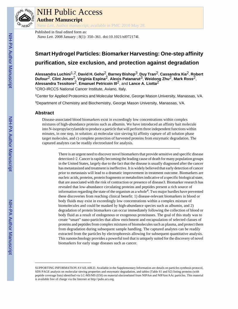
Smart Hydrogel Particles: Biomarker Harvesting: One-step affinity purification, size exclusion, and protection against degradation Alessandra Luchini 1,2 , David H. Geho 2 , Barney Bishop 3 , Duy Tran 2 , Cassandra Xia 2 , Robert Dufour 2 , Clint Jones 2 , Virginia Espina 2 , Alexis Patanarut 3 , Weidong Zhu 2 , Mark Ross 2 , Alessandra Tessitore 2 , Emanuel Petricoin III 2 , and Lance A. Liotta 2 1 CRO-IRCCS National Cancer Institute, Aviano, Italy. 2 Center for Applied Proteomics and Molecular Medicine, George Mason University, Manassas, VA. 3 Department of Chemistry and Biochemistry, George Mason University, Manassas, VA. Abstract Disease-associated blood biomarkers exist in exceedingly low concentrations within complex mixtures of high-abundance proteins such as albumin. We have introduced an affinity bait molecule into N-isopropylacrylamide to produce a particle that will perform three independent functions within minutes, in one step, in solution: a) molecular size sieving b) affinity capture of all solution phase target molecules, and c) complete protection of harvested proteins from enzymatic degradation. The captured analytes can be readily electroeluted for analysis. There is an urgent need to discover novel biomarkers that provide sensitive and specific disease detection1 , 2. Cancer is rapidly becoming the leading cause of death for many population groups in the United States, largely due to the fact that the disease is usually diagnosed after the cancer has metastasized and treatment is ineffective. It is widely believed that early detection of cancer prior to metastasis will lead to a dramatic improvement in treatment outcome. Biomarkers are nucleic acids, proteins, protein fragments or metabolites indicative of a specific biological state, that are associated with the risk of contraction or presence of disease3. Biomarker research has revealed that low-abundance circulating proteins and peptides present a rich source of information regarding the state of the organism as a whole 4 . Two major hurdles have prevented these discoveries from reaching clinical benefit: 1) disease-relevant biomarkers in blood or body fluids may exist in exceedingly low concentrations within a complex mixture of biomolecules and could be masked by high-abundance species such as albumin, and 2) degradation of protein biomarkers can occur immediately following the collection of blood or body fluid as a result of endogenous or exogenous proteinases. The goal of this study was to create “smart” nano-particles that allow enrichment and encapsulation of selected classes of proteins and peptides from complex mixtures of biomolecules such as plasma, and protect them from degradation during subsequent sample handling. The captured analytes can be readily extracted from the particles by electrophoresis allowing for subsequent quantitative analysis. This nanotechnology provides a powerful tool that is uniquely suited for the discovery of novel biomarkers for early stage diseases such as cancer. SUPPORTING INFORMATION AVAILABLE: Available in the Supplementary Information are details on particles synthesis protocol, SDS PAGE analysis on molecular sieving properties and enzymatic degradation, and tables (Table S1 and S2) listing proteins (with peptide coverage lists) identified via LC-MS/MS (ESI) on material electroeluted from NIPAm and NIPAm/AAc particles. This material is available free of charge via the Internet at http://pubs.acs.org. NIH Public Access Author Manuscript Nano Lett. Author manuscript; available in PMC 2010 May 28. Published in final edited form as: Nano Lett. 2008 January ; 8(1): 350–361. doi:10.1021/nl072174l. NIH-PA Author Manuscript NIH-PA Author Manuscript NIH-PA Author Manuscript
-
Upload
independent -
Category
Documents
-
view
3 -
download
0
Transcript of Smart Hydrogel Particles: Biomarker Harvesting: One-Step Affinity Purification, Size Exclusion, and...
Smart Hydrogel Particles: Biomarker Harvesting: One-step affinitypurification, size exclusion, and protection against degradation
Alessandra Luchini1,2, David H. Geho2, Barney Bishop3, Duy Tran2, Cassandra Xia2, RobertDufour2, Clint Jones2, Virginia Espina2, Alexis Patanarut3, Weidong Zhu2, Mark Ross2,Alessandra Tessitore2, Emanuel Petricoin III2, and Lance A. Liotta21CRO-IRCCS National Cancer Institute, Aviano, Italy.2Center for Applied Proteomics and Molecular Medicine, George Mason University, Manassas, VA.3Department of Chemistry and Biochemistry, George Mason University, Manassas, VA.
AbstractDisease-associated blood biomarkers exist in exceedingly low concentrations within complexmixtures of high-abundance proteins such as albumin. We have introduced an affinity bait moleculeinto N-isopropylacrylamide to produce a particle that will perform three independent functions withinminutes, in one step, in solution: a) molecular size sieving b) affinity capture of all solution phasetarget molecules, and c) complete protection of harvested proteins from enzymatic degradation. Thecaptured analytes can be readily electroeluted for analysis.
There is an urgent need to discover novel biomarkers that provide sensitive and specific diseasedetection1,2. Cancer is rapidly becoming the leading cause of death for many population groupsin the United States, largely due to the fact that the disease is usually diagnosed after the cancerhas metastasized and treatment is ineffective. It is widely believed that early detection of cancerprior to metastasis will lead to a dramatic improvement in treatment outcome. Biomarkers arenucleic acids, proteins, protein fragments or metabolites indicative of a specific biological state,that are associated with the risk of contraction or presence of disease3. Biomarker research hasrevealed that low-abundance circulating proteins and peptides present a rich source ofinformation regarding the state of the organism as a whole4. Two major hurdles have preventedthese discoveries from reaching clinical benefit: 1) disease-relevant biomarkers in blood orbody fluids may exist in exceedingly low concentrations within a complex mixture ofbiomolecules and could be masked by high-abundance species such as albumin, and 2)degradation of protein biomarkers can occur immediately following the collection of blood orbody fluid as a result of endogenous or exogenous proteinases. The goal of this study was tocreate “smart” nano-particles that allow enrichment and encapsulation of selected classes ofproteins and peptides from complex mixtures of biomolecules such as plasma, and protect themfrom degradation during subsequent sample handling. The captured analytes can be readilyextracted from the particles by electrophoresis allowing for subsequent quantitative analysis.This nanotechnology provides a powerful tool that is uniquely suited for the discovery of novelbiomarkers for early stage diseases such as cancer.
SUPPORTING INFORMATION AVAILABLE: Available in the Supplementary Information are details on particles synthesis protocol,SDS PAGE analysis on molecular sieving properties and enzymatic degradation, and tables (Table S1 and S2) listing proteins (withpeptide coverage lists) identified via LC-MS/MS (ESI) on material electroeluted from NIPAm and NIPAm/AAc particles. This materialis available free of charge via the Internet at http://pubs.acs.org.
NIH Public AccessAuthor ManuscriptNano Lett. Author manuscript; available in PMC 2010 May 28.
Published in final edited form as:Nano Lett. 2008 January ; 8(1): 350–361. doi:10.1021/nl072174l.
NIH
-PA Author Manuscript
NIH
-PA Author Manuscript
NIH
-PA Author Manuscript
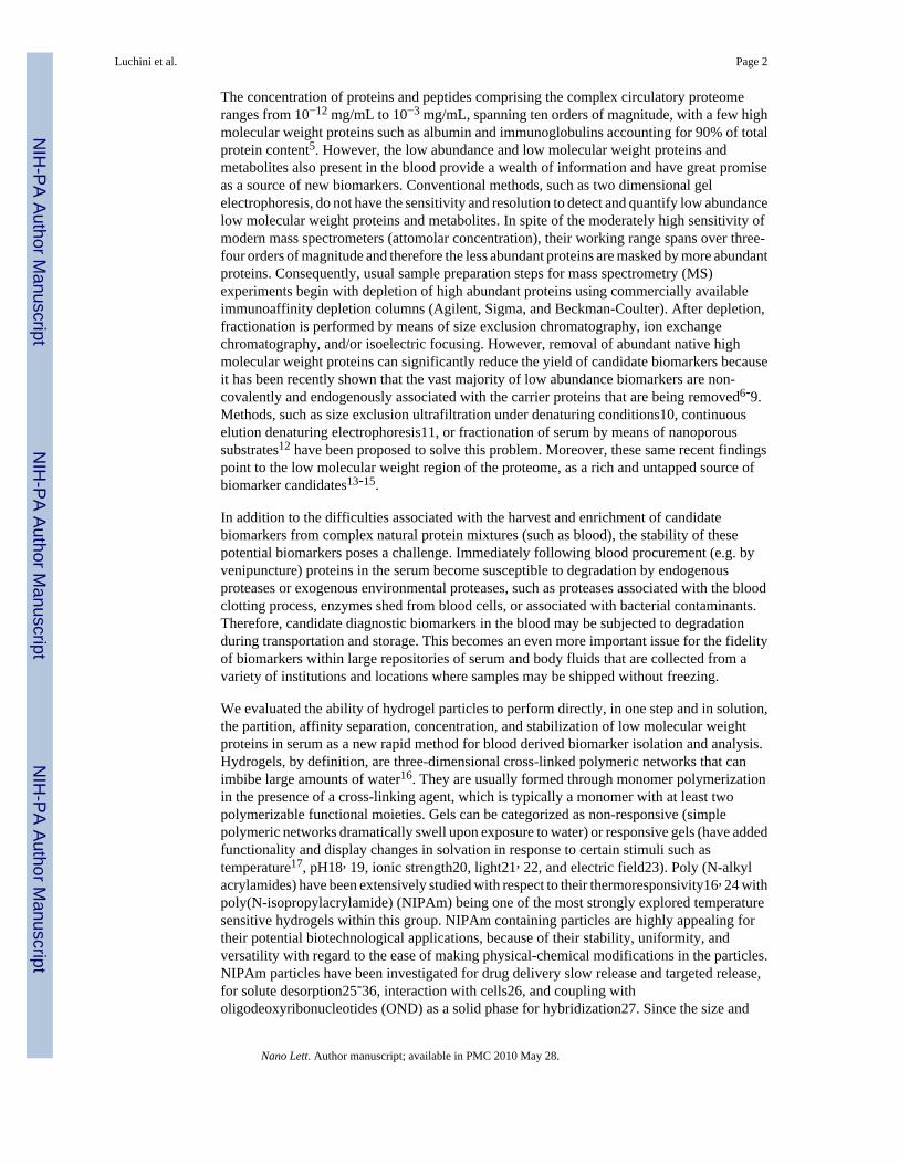
The concentration of proteins and peptides comprising the complex circulatory proteomeranges from 10−12 mg/mL to 10−3 mg/mL, spanning ten orders of magnitude, with a few highmolecular weight proteins such as albumin and immunoglobulins accounting for 90% of totalprotein content5. However, the low abundance and low molecular weight proteins andmetabolites also present in the blood provide a wealth of information and have great promiseas a source of new biomarkers. Conventional methods, such as two dimensional gelelectrophoresis, do not have the sensitivity and resolution to detect and quantify low abundancelow molecular weight proteins and metabolites. In spite of the moderately high sensitivity ofmodern mass spectrometers (attomolar concentration), their working range spans over three-four orders of magnitude and therefore the less abundant proteins are masked by more abundantproteins. Consequently, usual sample preparation steps for mass spectrometry (MS)experiments begin with depletion of high abundant proteins using commercially availableimmunoaffinity depletion columns (Agilent, Sigma, and Beckman-Coulter). After depletion,fractionation is performed by means of size exclusion chromatography, ion exchangechromatography, and/or isoelectric focusing. However, removal of abundant native highmolecular weight proteins can significantly reduce the yield of candidate biomarkers becauseit has been recently shown that the vast majority of low abundance biomarkers are non-covalently and endogenously associated with the carrier proteins that are being removed6-9.Methods, such as size exclusion ultrafiltration under denaturing conditions10, continuouselution denaturing electrophoresis11, or fractionation of serum by means of nanoporoussubstrates12 have been proposed to solve this problem. Moreover, these same recent findingspoint to the low molecular weight region of the proteome, as a rich and untapped source ofbiomarker candidates13-15.
In addition to the difficulties associated with the harvest and enrichment of candidatebiomarkers from complex natural protein mixtures (such as blood), the stability of thesepotential biomarkers poses a challenge. Immediately following blood procurement (e.g. byvenipuncture) proteins in the serum become susceptible to degradation by endogenousproteases or exogenous environmental proteases, such as proteases associated with the bloodclotting process, enzymes shed from blood cells, or associated with bacterial contaminants.Therefore, candidate diagnostic biomarkers in the blood may be subjected to degradationduring transportation and storage. This becomes an even more important issue for the fidelityof biomarkers within large repositories of serum and body fluids that are collected from avariety of institutions and locations where samples may be shipped without freezing.
We evaluated the ability of hydrogel particles to perform directly, in one step and in solution,the partition, affinity separation, concentration, and stabilization of low molecular weightproteins in serum as a new rapid method for blood derived biomarker isolation and analysis.Hydrogels, by definition, are three-dimensional cross-linked polymeric networks that canimbibe large amounts of water16. They are usually formed through monomer polymerizationin the presence of a cross-linking agent, which is typically a monomer with at least twopolymerizable functional moieties. Gels can be categorized as non-responsive (simplepolymeric networks dramatically swell upon exposure to water) or responsive gels (have addedfunctionality and display changes in solvation in response to certain stimuli such astemperature17, pH18, 19, ionic strength20, light21, 22, and electric field23). Poly (N-alkylacrylamides) have been extensively studied with respect to their thermoresponsivity16, 24 withpoly(N-isopropylacrylamide) (NIPAm) being one of the most strongly explored temperaturesensitive hydrogels within this group. NIPAm containing particles are highly appealing fortheir potential biotechnological applications, because of their stability, uniformity, andversatility with regard to the ease of making physical-chemical modifications in the particles.NIPAm particles have been investigated for drug delivery slow release and targeted release,for solute desorption25-36, interaction with cells26, and coupling witholigodeoxyribonucleotides (OND) as a solid phase for hybridization27. Since the size and
Luchini et al. Page 2
Nano Lett. Author manuscript; available in PMC 2010 May 28.
NIH
-PA Author Manuscript
NIH
-PA Author Manuscript
NIH
-PA Author Manuscript
porosity can be controlled by temperature, the use of temperature treatment to control uptakeand release of chemicals has been one of the most extensively characterized application ofNIPAm particles as vectors for controlled drug delivery28-34.
In the present study, hydrogel particles containing an affinity bait and a defined porosity weredeveloped and demonstrated to a) rapidly and in one step sequester the low molecular weightfraction of serum proteins, peptides and metabolites, b) remove and concentrate the targetmolecules from solution, and c) protect captured proteins from enzymatic degradation.
NIPAm based particles have been chosen because their high water content, broad range oftunable porosities, consistency and uniformity following synthesis, functional reconstitutionfollowing freeze-drying, and potential biocompatibility. By changing the percentage of crosslinking agent and temperature, it is possible to control the particles size and the effectiveporosity. A significant advantage for the application studied here is the ability of these particlesto rapidly uptake molecules because of their open structure, high water content, dualhydrophobic and hydrophilic chemical moieties that can be substituted in the polymer, andlarge surface area. This is a critical requirement for the goal of rapid harvesting of labile smallproteins in solution and protecting the proteins from degradation. The small size, uniformityof particle dimension, and reproducibility from batch to batch, of NIPAm provide a specialadvantage for applications in flow cytometry.
Hydrogel Particle Synthesis and CharacterizationGel particles incorporating N-isopropylacrylamide (NIPAm) were created and evaluated formolecular sieving properties. A second class of particles containing both NIPAm and acrylicacid (AAc), NIPAm/AAc, were fabricated to incorporate a charge-based affinity bait into theparticles16, 18, 35. Particle synthesis chemistry is described in the Supplementary Information.
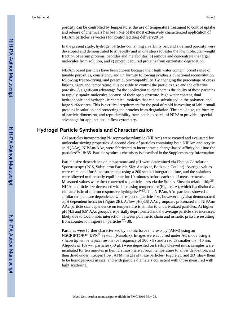
Particle size dependence on temperature and pH were determined via Photon CorrelationSpectroscopy (PCS, Submicron Particle Size Analyzer, Beckman Coulter). Average valueswere calculated for 3 measurements using a 200 second integration time, and the solutionswere allowed to thermally equilibrate for 10 minutes before each set of measurements.Measured values were then converted to particle sizes via the Stokes-Einstein relationship36.NIPAm particle size decreased with increasing temperature (Figure 2A), which is a distinctivecharacteristic of thermo responsive hydrogels28-33. The NIPAm/AAc particles showed asimilar temperature dependence with respect to particle size, however they also demonstrateda pH dependent behavior (Figure 2B). At low pH (3.5) AAc groups are protonated and NIPAm/AAc particle size dependence on temperature is similar to underivatized particles. At higherpH (4.5 and 6.5) AAc groups are partially deprotonated and the average particle size increases,likely due to Coulombic interaction between polymeric chain and osmotic pressure resultingfrom counter ion ingress in particles37, 38.
Particles were further characterized by atomic force microscopy (AFM) using anNSCRIPTOR™ DPN® System (NanoInk). Images were acquired under AC mode using asilicon tip with a typical resonance frequency of 300 kHz and a radius smaller than 10 nm.Aliquots of 1% w/v particles (50 μL) were deposited on freshly cleaved mica; samples wereincubated for ten minutes in humid atmosphere at room temperature to allow deposition, andthen dried under nitrogen flow. AFM images of these particles (Figure 2C and 2D) show themto be homogeneous in size, and with particle diameters consistent with those measured withlight scattering.
Luchini et al. Page 3
Nano Lett. Author manuscript; available in PMC 2010 May 28.
NIH
-PA Author Manuscript
NIH
-PA Author Manuscript
NIH
-PA Author Manuscript
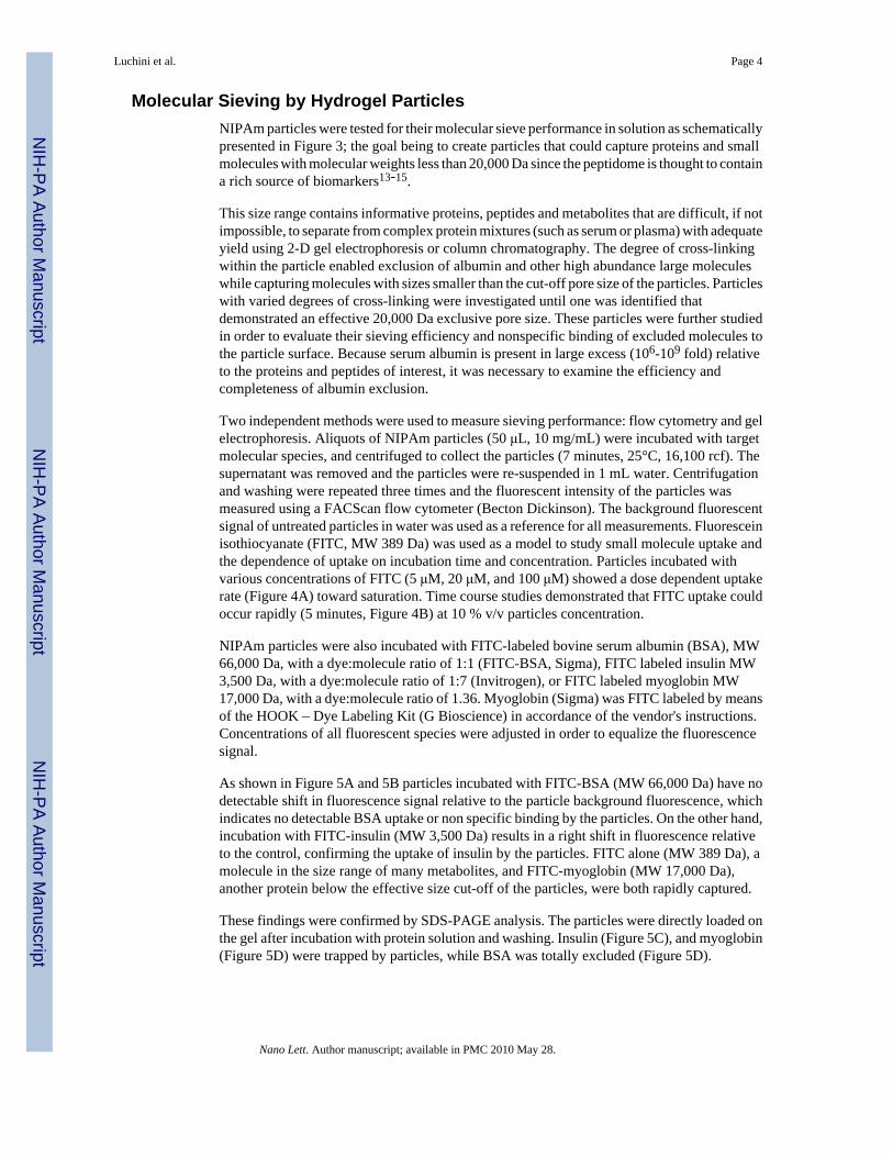
Molecular Sieving by Hydrogel ParticlesNIPAm particles were tested for their molecular sieve performance in solution as schematicallypresented in Figure 3; the goal being to create particles that could capture proteins and smallmolecules with molecular weights less than 20,000 Da since the peptidome is thought to containa rich source of biomarkers13-15.
This size range contains informative proteins, peptides and metabolites that are difficult, if notimpossible, to separate from complex protein mixtures (such as serum or plasma) with adequateyield using 2-D gel electrophoresis or column chromatography. The degree of cross-linkingwithin the particle enabled exclusion of albumin and other high abundance large moleculeswhile capturing molecules with sizes smaller than the cut-off pore size of the particles. Particleswith varied degrees of cross-linking were investigated until one was identified thatdemonstrated an effective 20,000 Da exclusive pore size. These particles were further studiedin order to evaluate their sieving efficiency and nonspecific binding of excluded molecules tothe particle surface. Because serum albumin is present in large excess (106-109 fold) relativeto the proteins and peptides of interest, it was necessary to examine the efficiency andcompleteness of albumin exclusion.
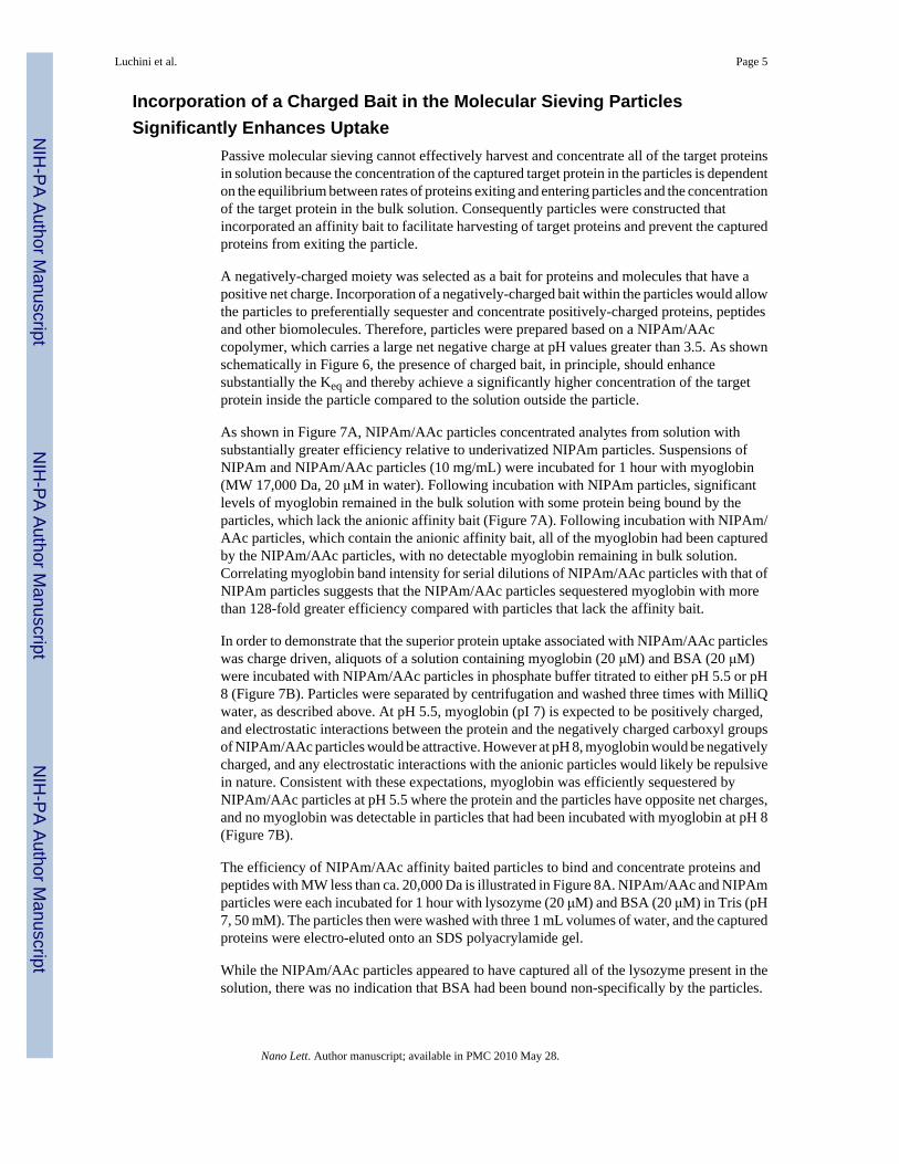
Two independent methods were used to measure sieving performance: flow cytometry and gelelectrophoresis. Aliquots of NIPAm particles (50 μL, 10 mg/mL) were incubated with targetmolecular species, and centrifuged to collect the particles (7 minutes, 25°C, 16,100 rcf). Thesupernatant was removed and the particles were re-suspended in 1 mL water. Centrifugationand washing were repeated three times and the fluorescent intensity of the particles wasmeasured using a FACScan flow cytometer (Becton Dickinson). The background fluorescentsignal of untreated particles in water was used as a reference for all measurements. Fluoresceinisothiocyanate (FITC, MW 389 Da) was used as a model to study small molecule uptake andthe dependence of uptake on incubation time and concentration. Particles incubated withvarious concentrations of FITC (5 μM, 20 μM, and 100 μM) showed a dose dependent uptakerate (Figure 4A) toward saturation. Time course studies demonstrated that FITC uptake couldoccur rapidly (5 minutes, Figure 4B) at 10 % v/v particles concentration.
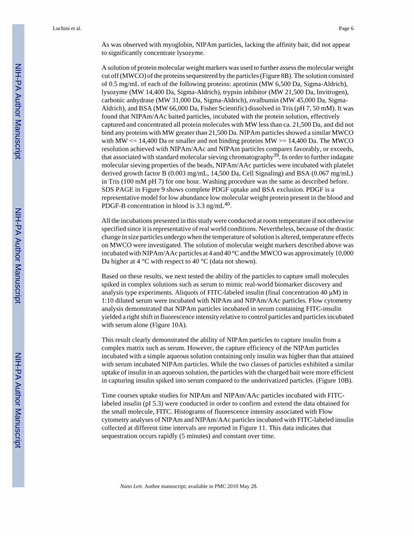
NIPAm particles were also incubated with FITC-labeled bovine serum albumin (BSA), MW66,000 Da, with a dye:molecule ratio of 1:1 (FITC-BSA, Sigma), FITC labeled insulin MW3,500 Da, with a dye:molecule ratio of 1:7 (Invitrogen), or FITC labeled myoglobin MW17,000 Da, with a dye:molecule ratio of 1.36. Myoglobin (Sigma) was FITC labeled by meansof the HOOK – Dye Labeling Kit (G Bioscience) in accordance of the vendor's instructions.Concentrations of all fluorescent species were adjusted in order to equalize the fluorescencesignal.
As shown in Figure 5A and 5B particles incubated with FITC-BSA (MW 66,000 Da) have nodetectable shift in fluorescence signal relative to the particle background fluorescence, whichindicates no detectable BSA uptake or non specific binding by the particles. On the other hand,incubation with FITC-insulin (MW 3,500 Da) results in a right shift in fluorescence relativeto the control, confirming the uptake of insulin by the particles. FITC alone (MW 389 Da), amolecule in the size range of many metabolites, and FITC-myoglobin (MW 17,000 Da),another protein below the effective size cut-off of the particles, were both rapidly captured.
These findings were confirmed by SDS-PAGE analysis. The particles were directly loaded onthe gel after incubation with protein solution and washing. Insulin (Figure 5C), and myoglobin(Figure 5D) were trapped by particles, while BSA was totally excluded (Figure 5D).
Luchini et al. Page 4
Nano Lett. Author manuscript; available in PMC 2010 May 28.
NIH
-PA Author Manuscript
NIH
-PA Author Manuscript
NIH
-PA Author Manuscript
Incorporation of a Charged Bait in the Molecular Sieving ParticlesSignificantly Enhances Uptake
Passive molecular sieving cannot effectively harvest and concentrate all of the target proteinsin solution because the concentration of the captured target protein in the particles is dependenton the equilibrium between rates of proteins exiting and entering particles and the concentrationof the target protein in the bulk solution. Consequently particles were constructed thatincorporated an affinity bait to facilitate harvesting of target proteins and prevent the capturedproteins from exiting the particle.
A negatively-charged moiety was selected as a bait for proteins and molecules that have apositive net charge. Incorporation of a negatively-charged bait within the particles would allowthe particles to preferentially sequester and concentrate positively-charged proteins, peptidesand other biomolecules. Therefore, particles were prepared based on a NIPAm/AAccopolymer, which carries a large net negative charge at pH values greater than 3.5. As shownschematically in Figure 6, the presence of charged bait, in principle, should enhancesubstantially the Keq and thereby achieve a significantly higher concentration of the targetprotein inside the particle compared to the solution outside the particle.
As shown in Figure 7A, NIPAm/AAc particles concentrated analytes from solution withsubstantially greater efficiency relative to underivatized NIPAm particles. Suspensions ofNIPAm and NIPAm/AAc particles (10 mg/mL) were incubated for 1 hour with myoglobin(MW 17,000 Da, 20 μM in water). Following incubation with NIPAm particles, significantlevels of myoglobin remained in the bulk solution with some protein being bound by theparticles, which lack the anionic affinity bait (Figure 7A). Following incubation with NIPAm/AAc particles, which contain the anionic affinity bait, all of the myoglobin had been capturedby the NIPAm/AAc particles, with no detectable myoglobin remaining in bulk solution.Correlating myoglobin band intensity for serial dilutions of NIPAm/AAc particles with that ofNIPAm particles suggests that the NIPAm/AAc particles sequestered myoglobin with morethan 128-fold greater efficiency compared with particles that lack the affinity bait.
In order to demonstrate that the superior protein uptake associated with NIPAm/AAc particleswas charge driven, aliquots of a solution containing myoglobin (20 μM) and BSA (20 μM)were incubated with NIPAm/AAc particles in phosphate buffer titrated to either pH 5.5 or pH8 (Figure 7B). Particles were separated by centrifugation and washed three times with MilliQwater, as described above. At pH 5.5, myoglobin (pI 7) is expected to be positively charged,and electrostatic interactions between the protein and the negatively charged carboxyl groupsof NIPAm/AAc particles would be attractive. However at pH 8, myoglobin would be negativelycharged, and any electrostatic interactions with the anionic particles would likely be repulsivein nature. Consistent with these expectations, myoglobin was efficiently sequestered byNIPAm/AAc particles at pH 5.5 where the protein and the particles have opposite net charges,and no myoglobin was detectable in particles that had been incubated with myoglobin at pH 8(Figure 7B).
The efficiency of NIPAm/AAc affinity baited particles to bind and concentrate proteins andpeptides with MW less than ca. 20,000 Da is illustrated in Figure 8A. NIPAm/AAc and NIPAmparticles were each incubated for 1 hour with lysozyme (20 μM) and BSA (20 μM) in Tris (pH7, 50 mM). The particles then were washed with three 1 mL volumes of water, and the capturedproteins were electro-eluted onto an SDS polyacrylamide gel.
While the NIPAm/AAc particles appeared to have captured all of the lysozyme present in thesolution, there was no indication that BSA had been bound non-specifically by the particles.
Luchini et al. Page 5
Nano Lett. Author manuscript; available in PMC 2010 May 28.
NIH
-PA Author Manuscript
NIH
-PA Author Manuscript
NIH
-PA Author Manuscript
As was observed with myoglobin, NIPAm particles, lacking the affinity bait, did not appearto significantly concentrate lysozyme.
A solution of protein molecular weight markers was used to further assess the molecular weightcut off (MWCO) of the proteins sequestered by the particles (Figure 8B). The solution consistedof 0.5 mg/mL of each of the following proteins: aprotinin (MW 6,500 Da, Sigma-Aldrich),lysozyme (MW 14,400 Da, Sigma-Aldrich), trypsin inhibitor (MW 21,500 Da, Invitrogen),carbonic anhydrase (MW 31,000 Da, Sigma-Aldrich), ovalbumin (MW 45,000 Da, Sigma-Aldrich), and BSA (MW 66,000 Da, Fisher Scientific) dissolved in Tris (pH 7, 50 mM). It wasfound that NIPAm/AAc baited particles, incubated with the protein solution, effectivelycaptured and concentrated all protein molecules with MW less than ca. 21,500 Da, and did notbind any proteins with MW greater than 21,500 Da. NIPAm particles showed a similar MWCOwith MW <= 14,400 Da or smaller and not binding proteins MW >= 14,400 Da. The MWCOresolution achieved with NIPAm/AAc and NIPAm particles compares favorably, or exceeds,that associated with standard molecular sieving chromatography39. In order to further indagatemolecular sieving properties of the beads, NIPAm/AAc particles were incubated with plateletderived growth factor B (0.003 mg/mL, 14,500 Da, Cell Signaling) and BSA (0.067 mg/mL)in Tris (100 mM pH 7) for one hour. Washing procedure was the same as described before.SDS PAGE in Figure 9 shows complete PDGF uptake and BSA exclusion. PDGF is arepresentative model for low abundance low molecular weight protein present in the blood andPDGF-B concentration in blood is 3.3 ng/mL40.
All the incubations presented in this study were conducted at room temperature if not otherwisespecified since it is representative of real world conditions. Nevertheless, because of the drasticchange in size particles undergo when the temperature of solution is altered, temperature effectson MWCO were investigated. The solution of molecular weight markers described above wasincubated with NIPAm/AAc particles at 4 and 40 °C and the MWCO was approximately 10,000Da higher at 4 °C with respect to 40 °C (data not shown).
Based on these results, we next tested the ability of the particles to capture small moleculesspiked in complex solutions such as serum to mimic real-world biomarker discovery andanalysis type experiments. Aliquots of FITC-labeled insulin (final concentration 40 μM) in1:10 diluted serum were incubated with NIPAm and NIPAm/AAc particles. Flow cytometryanalysis demonstrated that NIPAm particles incubated in serum containing FITC-insulinyielded a right shift in fluorescence intensity relative to control particles and particles incubatedwith serum alone (Figure 10A).
This result clearly demonstrated the ability of NIPAm particles to capture insulin from acomplex matrix such as serum. However, the capture efficiency of the NIPAm particlesincubated with a simple aqueous solution containing only insulin was higher than that attainedwith serum incubated NIPAm particles. While the two classes of particles exhibited a similaruptake of insulin in an aqueous solution, the particles with the charged bait were more efficientin capturing insulin spiked into serum compared to the underivatized particles. (Figure 10B).
Time courses uptake studies for NIPAm and NIPAm/AAc particles incubated with FITC-labeled insulin (pI 5.3) were conducted in order to confirm and extend the data obtained forthe small molecule, FITC. Histograms of fluorescence intensity associated with Flowcytometry analyses of NIPAm and NIPAm/AAc particles incubated with FITC-labeled insulincollected at different time intervals are reported in Figure 11. This data indicates thatsequestration occurs rapidly (5 minutes) and constant over time.
Luchini et al. Page 6
Nano Lett. Author manuscript; available in PMC 2010 May 28.
NIH
-PA Author Manuscript
NIH
-PA Author Manuscript
NIH
-PA Author Manuscript
Rapid time course of target protein uptake by bait containing particlesIn order to prove that the kinetics of protein uptake is very rapid, the amount of proteinremaining in bulk solution after incubation with NIPAm/AAc particles was measured byReverse Phase Protein Arrays (RPPA)41. RPPA was chosen as a means to determine proteinconcentration in particles supernatant since it is a very sensitive technique (more sensitive bythree orders of magnitude than methods like Bradford assays) and our goal was to use dilutedprotein solution in order to mimic the condition of low abundance low molecular weightproteins in the blood, and to demonstrate how complete protein removal from bulk solutionwas. Different amounts of NIPAm/AAc particles (1.15 and 11.5 million) were incubated withlysozyme (20 μM) in Tris (pH 7, 50 mM) in a total volume of 100 μL for periods of 1 and 10minutes. After centrifugation of the particles, aliquots of supernatant were spotted on anitrocellulose coated slide (FAST slide, Whatman) using an Aushon 2470 robotic arrayer(Aushon Biosystems). Arrays were stained with a colloidal gold solution, AuroDye Forte Kit(Amersham), images were acquired using a PowerLook 1120 scanner (Umax), and numericvalues were obtained from images with ImageQuant (GE Healthcare) and processed withSigmaPlot (Systat). The bulk solution after a one minute incubation with 1.15 million particlescontained 28% of the initial protein amount, and 15% after ten minutes. Moreover, the solutionrecovered from incubation with 11.5 million particles after 1 and 10 minutes contained 5% and9% of the initial amount, respectively (Figure 12A). To be noted is that, since in all the timecourse experiments the separation of particles from solution was obtained by centrifugation,reported time values refer to incubations time intervals only. Beyond that, particles were incontact with solution for additional 7 minutes required by centrifugation.
The kinetics of protein uptake by NIPAm/AAc particles was further investigated by incubatingparticles with BSA (20 μM) and lysozyme (20 μM) in Tris (pH 7, 50 mM) at room temperatureand using SDS-PAGE to monitor lysozyme uptake at time points of 1, 10, 20, 30, and 60minutes (Figure 12B). The results of this experiment showed that lysozyme sequestration wasnearly complete after 1 minute and was complete by 60 minutes, confirming that the processoccurs very quickly as indicated in the flow cytometry time course study described above. Asexpected, BSA was excluded by the particles, and none of the BSA was taken-up by theNIPAm/AAc throughout the duration of the experiment (60 minutes).
Demonstration of Isolation and Enrichment of Low Molecular Weight and LowAbundance Analytes from Serum
The ability of NIPAM and NIPAm/AAc particles to sequester and concentrate lowconcentration candidate protein biomarkers from serum for proteomic analysis was evaluatedby incubating the particles with a 1:10 v/v dilution of serum in water for 1 hour. The trappedproteins were electrophoretically eluted from the particles under denaturing conditions andthen trypsin digested. The particles were heated in SDS sample buffer for 5 minutes at 100 °C and loaded on a 4-20 % Tris Glicine gel (Invitrogen). Bands below 30 kDa were cut and in-gel trypsin digestion was performed 11. The resulting peptide fragments were analyzed byonline liquid chromatography/electrospray ionization tandem mass spectrometry (LC/ESI MS)using LTQ-Orbitrap mass spectrometer (Thermo Fisher). Reverse phase column was slurry-packed in-house with 5 μm, 20 Å pore size C18 resin (Michrom BioResources, CA) in 100mm i.d. × 10 cm long fused silica capillary (Polymicro Technologies, Phoenix, AZ) with alaser-pulled tip. After sample injection, the column was washed for 5 minutes with mobilephase A (0.1% formic acid) and peptides were eluted using a linear gradient of 0% mobilephase B (0.1 % formic acid, 80% acetonitrile) to 50% mobile phase B in 50 minutes at 200 nl/min, then to 100% B in an additional 5 minutes. The LTQ mass spectrometer was operated ina data-dependent mode in which each full MS scan was followed by five MS/MS scans werethe five most abundant molecular ions were dynamically selected and fragmented by collision-
Luchini et al. Page 7
Nano Lett. Author manuscript; available in PMC 2010 May 28.
NIH
-PA Author Manuscript
NIH
-PA Author Manuscript
NIH
-PA Author Manuscript
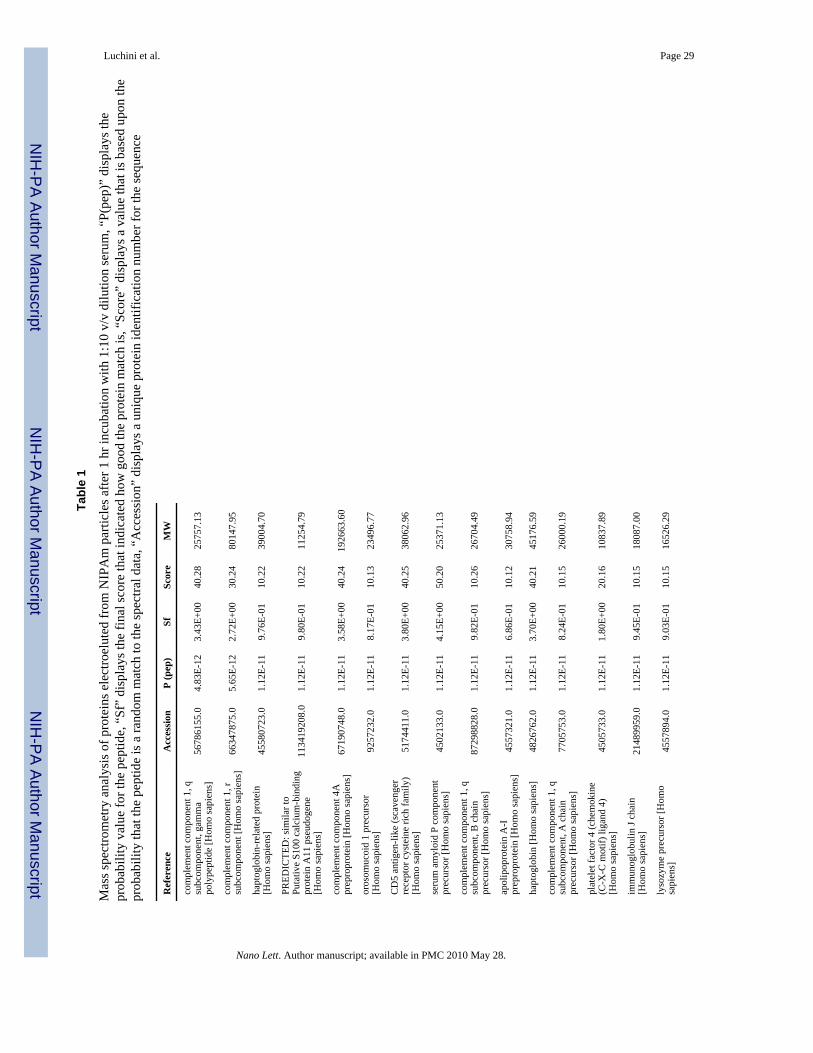
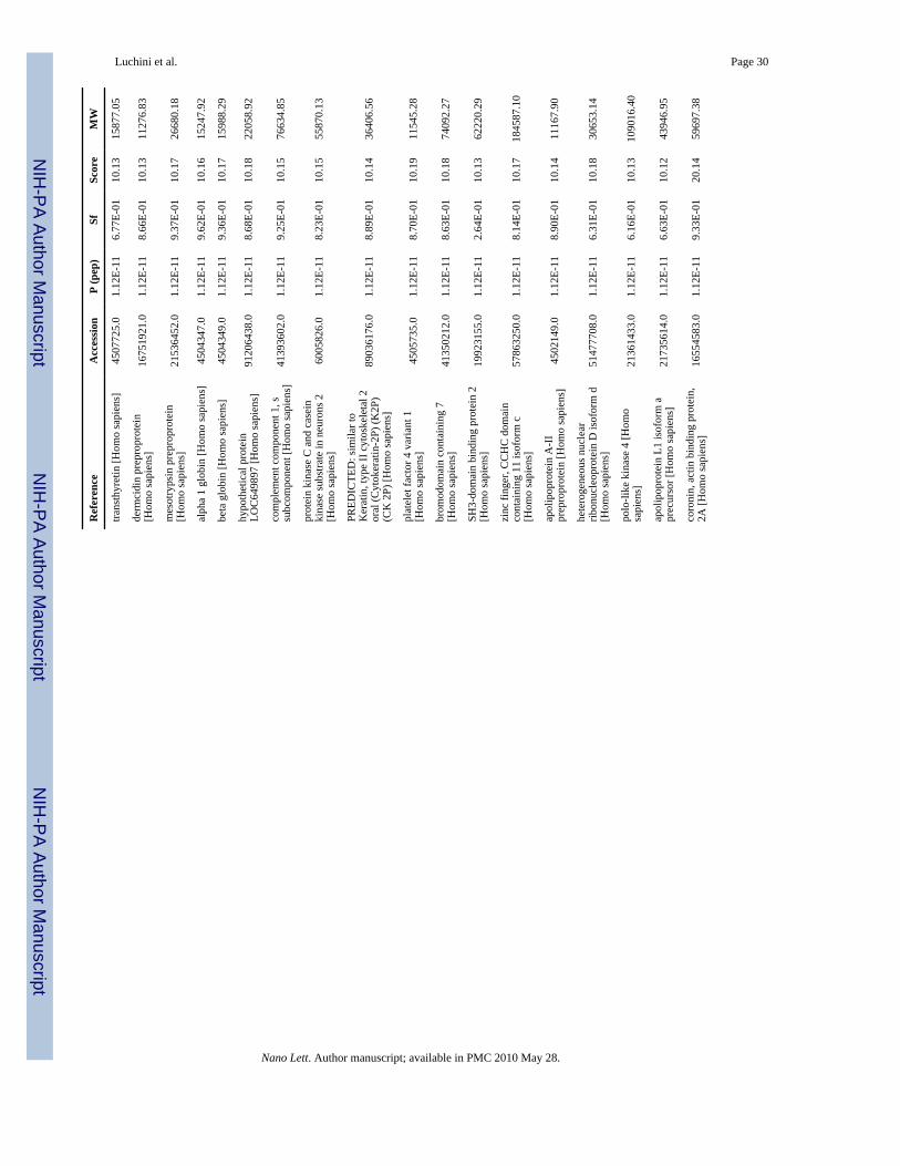
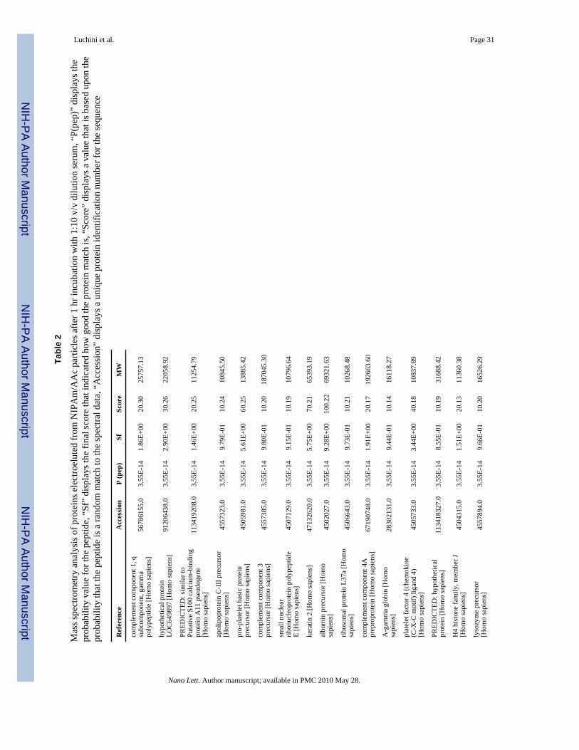
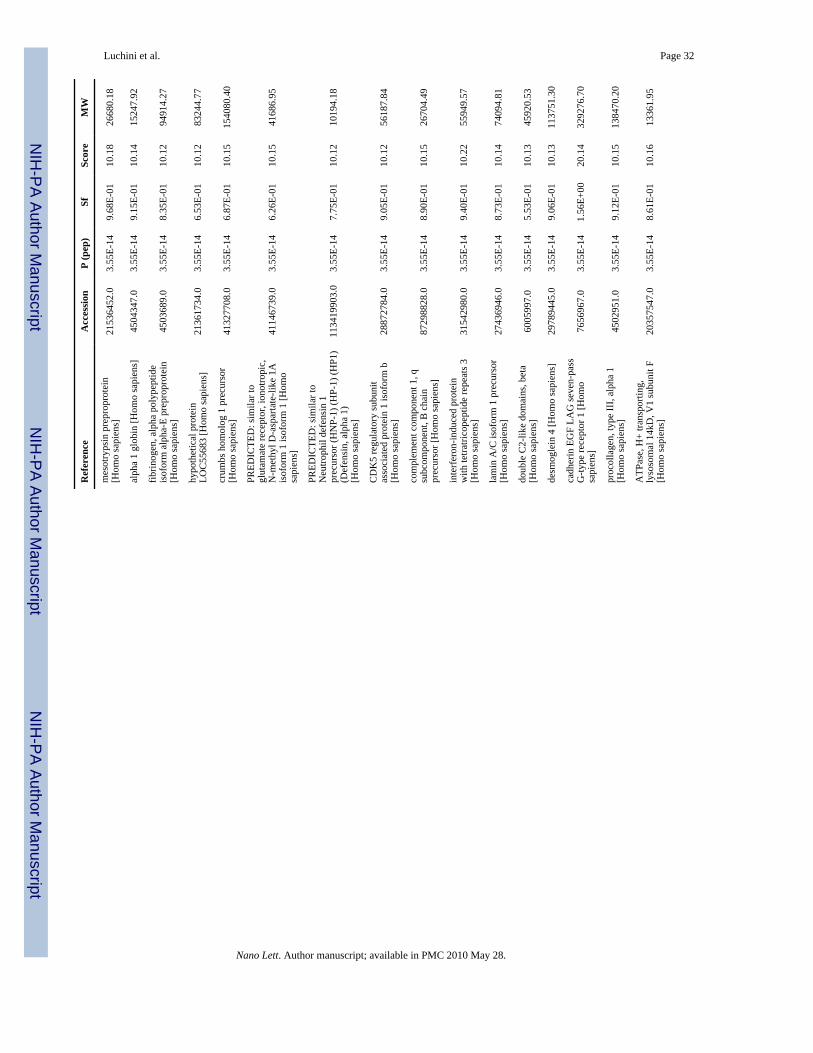
induced dissociation (CID) using a normalized collision energy of 35%. MS/MS data werematched against the NCBI (National Center for Biotechnology Information) human proteindatabase with the program SEQUEST (Bioworks software, Thermo) using full tryptic cleavageconstraints. High-confidence peptide identifications were obtained by applying the followingfilters to the search results: cross-correlation score (XCorr) >= 1.9 for 1+, 2.2 for 2+, 3.5 for3+, and a maximum probability for a random identification of 0.01. The list of identifiedproteins (Supplementary Information) demonstrated that albumin and other high abundanceserum proteins were not present in the particles. On the other hand, the list of identified proteinsindicates that the particles sequestered rare and small-sized serum proteins and peptides.
Protein Sequestration by Particle Blocks Protease DegradationOne of the major problems associated with biological fluids is the potential for sampledegradation during collection, transport, storage and analysis. Endogenous clotting cascadeenzymes, enzymes released from damaged cells, or exogenous enzymes (from contaminatingbacteria) can contribute to the degradation of diagnostically important proteins.
The lack of standardized preservation methods could result in bias in high-throughput analysisof serum and plasma42. While it was expected that proteases with MW greater than the MWCOof the particles (~20,000 Da) would be excluded from the interior space of the particles andthereby denied access to captured proteins, smaller proteases such as trypsin (23,800 Da) aremore likely to be able to enter the particles. Additionally, it was not known whether proteasesthat entered charged-bait particles would retain their enzymatic potency when both thesubstrate proteins and the enzyme were sequestered by the particles (Figure 12). Therefore,NIPAm/AAc particles were incubated at 37°C in a pH 7 NH4HCO3 (100 mM) solutioncontaining lysozyme (0.5 mg/mL) and trypsin (0.05 mg/mL, Promega). Trypsin was selectedfor these studies based on its small size and the fact that the tryptic digestion of lysozyme wouldproduce very characteristic cleavage products. The conditions used in this experiment wouldallow both lysozyme and trypsin to enter the particle. Analysis of the captured proteins by SDSPAGE after incubation for 1 hour and overnight showed only two bands - one correspondingto trypsin and the other to the full length lysozyme, indicating that no degradation of the proteinhad occurred (Figure 14A). Incubation of lysozyme (0.5 mg/mL) with trypsin (0.05 mg/mL)at 37°C in a pH 7 NH4HCO3 (100 mM) solution in the absence of NIPAm/AAc particlesresulted in degradation of lysozyme. SDS-PAGE analysis of the reaction after incubation for1 hour and overnight clearly indicated the presence of low molecular weight peptide fragments,which showed that lysozyme was proteolyzed by trypsin in the absence of NIPAm/AAcparticles. These results clearly indicate that sequestration of small proteins by affinity-baitparticles can effectively shield bound proteins from proteases including those that are capableof entering the particles interior.
In order to better understand the benefits associated with sequestration of proteins by NIPAm/AAc affinity-bait particles, NIPAm/AAc particles were incubated at 37°C with a combinationof BSA (0.5 mg/mL), lysozyme (0.5 mg/mL) and trypsin (0.05 mg/mL) in 100 mMNH4HCO3 (pH7).
As with the previous protection study, the reaction was analyzed using SDS-PAGE afterincubating 1 hr and overnight. In the absence of NIPAm/AAc particles, the majority of BSAhad been digested after 1 hr and the band corresponding to full-length BSA had disappearedafter incubating overnight (Figure 14B). As was noted earlier, the NIPAm/AAC particlesefficiently sequestered both lysozyme and trypsin, and protected lysozyme from proteolysisby trypsin. However, the particles did not bind BSA, and the presence of low molecular weightbands in the supernatant after 1 hour and overnight incubation accompanied by the decreasein intensity of the band corresponding to full-length BSA indicates that BSA was not protected
Luchini et al. Page 8
Nano Lett. Author manuscript; available in PMC 2010 May 28.
NIH
-PA Author Manuscript
NIH
-PA Author Manuscript
NIH
-PA Author Manuscript
from degradation by trypsin. Suppression of proteolytic activity by enzymes small enough toenter the particles, such as trypsin, may occur because immobilization of the enzymes by thecharge-bait particle prevents them from binding substrate proteins or may be the result of sterichindrance associated with trapping of the substrate by the affinity-bait groups in the particlethus preventing enzymes from productively binding target proteins inside the particle. Thus,the functional state of the proteins sequestered by the charge-bait may be similar to that ofproteins arrested using a precipitating fixative treatment.
In order to exclude that non specific interactions of analytes exist with the surface charge ofparticles we synthesized a batch of particles containing a NIPAm/AAc core covered by aNIPAm shell (details on protocol available in the Supplemental Information). We thenconfirmed that core shell particles have the same sieving properties as NIPAm/AAc particles(Figure S1).
The use of lysozyme in its native form is a model close to real physiological condition andmake it possible to detect partial products of degradation with SDS PAGE analysis. With thismodel it was proved that bait particles are able to protect the protein from enzymaticdegradation. Nevertheless, to verify how effective43 the protection from degradation ofparticles with bait is, we used reduced and alkylated lyzozyme and incubated it with trypsinand particles. Lysozyme was reduced by incubation with Dithiothreitol (DTT) (10 mM) inNH4HCO3 buffer (50 mM, pH 8) containing urea (2 M) for one hour at room temperature.Iodoacetamide was added to the solution to a final concentration of 50 mM and let react in thedark for 30 minutes. Lysozyme solution was diluted 1:2 with NH4HCO3 buffer (50 mM, pH8) to a final concentration of 0.5 mg/ml. Aliquots of 50 μl of reduced and alkylated lysozymewere added to 50 μl of particles (10 mg/ml) in order to demonstrate that lysozyme by itselfwas captured by particles. In addition, trypsin was added to lysozyme solution at a w/w ratioof 1:50 (0.01 mg/ml) and 1:100 (0.005 mg/ml). Besides, particles with and without bait, andcore shell particles were added to lysozyme-trypsin solution. All the solutions were incubatedfor 1 hour at 37°C. Particles were washed as described above and all samples loaded on SDSPAGE. In Figure 15 it is shown that a) reduced and alkylated lysozyme undergoes to trypsinmediated proteolysis and products of degradation are too small to be detected (lane 1-3), b)lysozyme is captured by plain particles (lane 4) but in the presence of trypsin it is not protectedfrom degradation as is evident from the fact that lysozyme band is absent in the particles (lane6) and highly reduced in intensity in the supernatant (lane 5), c) particles with bait harvest andconcentrate lysozyme (lane 7) and protect lysozyme from degradation (lane 9), d) likewise,core shell particles harvest, concentrate and protect reduced and alkylated lysozyme fromtryptic degradation. This suggests that the AAc bait plays a fundamental role in protectingproteins from degradation.
An incubation of trypsin (0.5 mg/ml in NH4HCO3 50 mM pH 8, 1 hour, 37°C) with particleswas performed to corroborate the entrance of the enzyme in the beads. Trypsin is captured byplain particles, and is captured and concentrated by NIPAm/AAc and core shell particles(Figure 16).
We have described the development and application of hydrogel bait-containing particles as anew tool for harvesting and concentrating small molecule analytes and biomarker candidatesfrom biological fluids, allowing high throughput analysis of low-abundance and low molecularweight components. These nanoparticles present a rapid and straightforward workflow fordirect utility in raw body fluids, while the work herein described the particles with a negativecharge that preferentially bind cationic species, positively charged particles such as a NIPAm/allylamine copolymer could be used to selectively harvest and concentrate anionic species frombiological fluids. Similarly, hydrophobic metabolites could be captured for comprehensivemetabolomic studies by using more hydrophobic particles such as NIPAm/styrene copolymers.
Luchini et al. Page 9
Nano Lett. Author manuscript; available in PMC 2010 May 28.
NIH
-PA Author Manuscript
NIH
-PA Author Manuscript
NIH
-PA Author Manuscript
Analyte-specific chemical or protein or nucleic acid affinity baits can be incorporated. Forexample, boronate-containing particles, which are known to bind saccharides, would beutilized to sequester glycoproteins from solution 44. Consequently, NIPAm-allylaminecopolymer are currently being synthesized that contain a bait for anionic proteins. Moreover,p-vinylphenylboronic acid (VPBA) is under consideration as a copolymer for harvesting ofsugars and nucleic acids. Further affinity baits such as triazinil-based reactive dyes (that haveaffinity towards proteins), hexadecylamine (for lipids uptake) and cyclodextrins (able toassociate small molecules) are being noncovalently or covalently immobilized within theparticles. In particular, we have used the bait chemistry described above to harvest the followingsmall metabolites L-Dopa, homogentisic acid, Dopamine, Dopac and 5-hydroxyindoleaceticacid. This extends the utility of the technology to the realm of metabolomics.
Combining a variety of affinity chemistries with a size-sieving tool in a one-step process couldhave enormous utility for disease marker discovery and analysis workflows.
In the workflow presented in this study, proteins are denatured when eluted out of particlesand then analyzed in mass spectrometry for biomarker discovery. Nevertheless, it is importantto note that the harvesting conditions are conducted with native protein mixtures. This permitsfuture applications that require the analytes of interest to be in their native state (immulite,radioimmunoassays). For these applications it would important that proteins are not denaturedwhen released from the particles. Ahmad and colleagues have demonstrated, using circulardichroism, that molecules for drug delivery released from temperature sensitive polymericparticles by temperature changes retained their native conformational state 45 Consequentlypossible means of eluting native proteins from the particles include modifying the temperatureor pH of the solution, increasing the ionic strength, or electroeluting the proteins under nondenaturing conditions, in the absence of detergent.
Supplementary MaterialRefer to Web version on PubMed Central for supplementary material.
AcknowledgmentsThe authors appreciate the generous support of Dr. Vikas Chandhoke and the Department of Life Sciences at GeorgeMason University. The authors also thank Mr. Tom Huff for facilitating experimental procedures. We would like toacknowledge stimulating discussions with Dr. Enrico Garaci, Dr. Alfonso Colombatti, Dr. Claudio Belluco, Dr. VictorMorozov, and Dr. Michele Signore. This work was partly supported by the Italian Istituto Superiore di Sanità in theframework Italy/USA cooperation agreement between the US Department of Health and Human Services and theItalian Ministry of Public Health.
REFERENCES1. Aebersold R, Anderson L, Caprioli R, Druker B, Hartwell L, Smith R. J Proteome Res 2005;4(4):1104–
9. [PubMed: 16083259]2. Srinivas PR, Verma M, Zhao Y, Srivastava S. Clin Chem 2002;48(8):1160–9. [PubMed: 12142368]3. Frank R, Hargreaves R. Nature reviews 2003;2(7):566–80.4. Espina V, Mehta AI, Winters ME, Calvert V, Wulfkuhle J, Petricoin EF 3rd, Liotta LA. Proteomics
2003;3(11):2091–100. [PubMed: 14595807]5. Anderson NL, Anderson NG. Mol Cell Proteomics 2002;1(11):845–67. [PubMed: 12488461]6. Lopez MF, Mikulskis A, Kuzdzal S, Golenko E, Petricoin EF 3rd, Liotta LA, Patton WF, Whiteley
GR, Rosenblatt K, Gurnani P, Nandi A, Neill S, Cullen S, O'Gorman M, Sarracino D, Lynch C, JohnsonA, McKenzie W, Fishman D. Clinical chemistry 2007;53(6):1067–74. [PubMed: 17463175]
7. Conrads TP, Hood BL, Veenstra TD. BioTechniques 2006;40(6):799–805. [PubMed: 16774124]
Luchini et al. Page 10
Nano Lett. Author manuscript; available in PMC 2010 May 28.
NIH
-PA Author Manuscript
NIH
-PA Author Manuscript
NIH
-PA Author Manuscript
8. Lowenthal MS, Mehta AI, Frogale K, Bandle RW, Araujo RP, Hood BL, Veenstra TD, Conrads TP,Goldsmith P, Fishman D, Petricoin EF 3rd, Liotta LA. Clin Chem 2005;51(10):1933–45. [PubMed:16099937]
9. Lopez MF, Mikulskis A, Kuzdzal S, Bennett DA, Kelly J, Golenko E, DiCesare J, Denoyer E, PattonWF, Ediger R, Sapp L, Ziegert T, Lynch C, Kramer S, Whiteley GR, Wall MR, Mannion DP, DellaCioppa G, Rakitan JS, Wolfe GM. Clinical chemistry 2005;51(10):1946–54. [PubMed: 16081505]
10. Zolotarjova N, Martosella J, Nicol G, Bailey J, Boyes BE, Barrett WC. Proteomics 2005;5(13):3304–13. [PubMed: 16052628]
11. Camerini S, Polci ML, Liotta LA, Petricoin EF, Zhou W. Proteomics Clin. Appl 2007;1:176–184.12. Geho D, Cheng MM, Killian K, Lowenthal M, Ross S, Frogale K, Nijdam J, Lahar N, Johann D,
Herrmann P, Whiteley G, Ferrari M, Petricoin E, Liotta L. Bioconjug Chem 2006;17(3):654–61.[PubMed: 16704202]
13. Tirumalai RS, Chan KC, Prieto DA, Issaq HJ, Conrads TP, Veenstra TD. Molecular & cellularproteomics 2003;2(10):1096–103. [PubMed: 12917320]
14. Merrell K, Southwick K, Graves SW, Esplin MS, Lewis NE, Thulin CD. Journal of biomoleculartechniques 2004;15(4):238–48. [PubMed: 15585820]
15. Orvisky E, Drake SK, Martin BM, Abdel-Hamid M, Ressom HW, Varghese RS, An Y, Saha D,Hortin GL, Loffredo CA, Goldman R. Proteomics 2006;6(9):2895–902. [PubMed: 16586431]
16. Pelton R. Adv Colloid Interface Sci 2000;85(1):1–33. [PubMed: 10696447]17. Li Y, Tanaka T. The Journal of Chemical Physics 1990;92(2):1365–1371.18. Jones CD, Lyon LA. Macromolecules 2000;33(22):8301–8306.19. Moselhy J, Wu XY, Nicholov R, Kodaria K. Journal of Biomaterials Science, Polymer Edition
2000;11(2):123–147. [PubMed: 10718475]20. Duracher D, Sauzedde F, Elaissari A, Perrin A, Pichot C. Colloid & Polymer Science 1998;276(3):
219–231.21. Sershen SR, Westcott SL, Halas NJ, West JL. Temperature-sensitive polymer-nanoshell composites
for photothermally modulated drug delivery 2000;51:293–298.22. Suzuki A, Tanaka T. Nature 1990;346(6282):345–347.23. Tanaka T, Nishio I, Sun S-T, Ueno-Nishio S. Collapse of Gels in an Electric Field 1982;218:467–
469.24. Inomata H, Goto S, Saito S. Phase transition of N-substituted acrylamide gels 1990;23:4887–4888.25. Kawaguchi H, Fujimoto K, Mizuhara Y. Colloid & Polymer Science 1992;270(1):53–57.26. Achiha K, Ojima R, Kasuya Y, Fujimoto K, Kawaguchi H. Interactions between temperature-sensitive
hydrogel microspheres and granulocytes 1995;6:534–540.27. Delair T, Meunier F, Elaissari A, Charles M-H, Pichot C. Colloids and Surfaces A: Physicochemical
and Engineering Aspects 1999;153(1-3):341–353.28. Sparnacci K, Laus M, Tondelli L, Bernardi C, Magnani L, Corticelli F, Marchisio M, Ensoli B,
Castaldello A, Caputo A. J Biomater Sci Polym Ed 2005;16(12):1557–74. [PubMed: 16366337]29. Haruyuki Hiratani YMCA-L. Macromolecular Bioscience 2005;5(8):728–733. [PubMed: 16082622]30. Nahar M, Dutta T, Murugesan S, Asthana A, Mishra D, Rajkumar V, Tare M, Saraf S, Jain NK. Crit
Rev Ther Drug Carrier Syst 2006;23(4):259–318. [PubMed: 17341200]31. Wu J-Y, Liu S-Q, Heng PW-S, Yang Y-Y. Journal of Controlled Release 2005;102(2):361–372.
[PubMed: 15653157]32. Zhang XZ, Jo Lewis P, Chu CC. Biomaterials 2005;26(16):3299–309. [PubMed: 15603825]33. Woo BH, Jiang G, Jo YW, DeLuca PP. Pharm Res 2001;18(11):1600–6. [PubMed: 11758769]34. Basinska T. Macromol Biosci 2005;5(12):1145–68. [PubMed: 16294370]35. Saunders BR, Vincent B. Advances in Colloid and Interface Science 1999;80(1):1–25.36. Pecora, R. Dynamic Light Scattering: Applications of Photo Correlation Spectroscopy. Springer;
1985. p. 43637. Fernandez-Nieves A, Fernandez-Barbero A, Vincent B, de las Nieves FJ. Macromolecules
2000;33:2114–2118.38. Ito S, Ogawa K, Suzuki H, Wang B, Yoshida R, Kokufuta E. Langmuir 1999;15(12):4289–4294.
Luchini et al. Page 11
Nano Lett. Author manuscript; available in PMC 2010 May 28.
NIH
-PA Author Manuscript
NIH
-PA Author Manuscript
NIH
-PA Author Manuscript
39. Boschetti E. Journal of Chromatography A 1994;658(2):207–236.40. Eppley BL, Woodell JE, Higgins J. Plastic and reconstructive surgery 2004;114(6):1502–8. [PubMed:
15509939]41. Gulmann C, Sheehan KM, Kay EW, Liotta LA, Petricoin EF 3rd. The Journal of pathology 2006;208
(5):595–606. [PubMed: 16518808]42. Ayache S, Panelli M, Marincola FM, Stroncek DF. American journal of clinical pathology 2006;126
(2):174–84. [PubMed: 16891190]43. Noda Y, Fujiwara K, Yamamoto K, Fukuno T, Segawa S-I. Specificity of trypsin digestion and
conformational flexibility at different sites of unfolded lysozyme 1994;34:217–226.44. Ivanov AE, Galaev IY, Mattiasson B. Journal of molecular recognition 2006;19(4):322–31. [PubMed:
16865663]45. Ahmad H, Okubo M, Kamatari YO, Minami H. Colloid & Polymer Science 2002;280(4):310–315.
Luchini et al. Page 12
Nano Lett. Author manuscript; available in PMC 2010 May 28.
NIH
-PA Author Manuscript
NIH
-PA Author Manuscript
NIH
-PA Author Manuscript
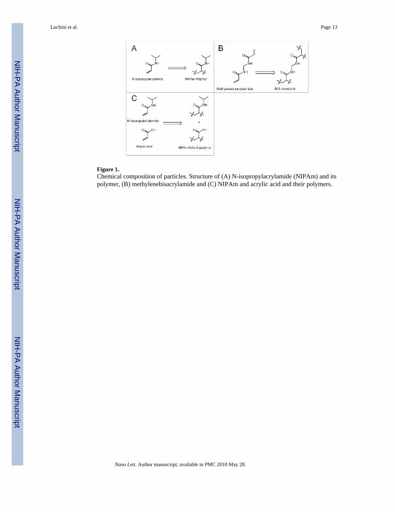
Figure 1.Chemical composition of particles. Structure of (A) N-isopropylacrylamide (NIPAm) and itspolymer, (B) methylenebisacrylamide and (C) NIPAm and acrylic acid and their polymers.
Luchini et al. Page 13
Nano Lett. Author manuscript; available in PMC 2010 May 28.
NIH
-PA Author Manuscript
NIH
-PA Author Manuscript
NIH
-PA Author Manuscript
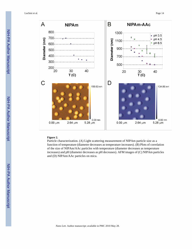
Figure 2.Particle characterization. (A) Light scattering measurement of NIPAm particle size as afunction of temperature (diameter decreases as temperature increases). (B) Plots of correlationof the size of NIPAm/AAc particles with temperature (diameter decreases as temperatureincreases) and pH (diameter decreases as pH decreases). AFM images of (C) NIPAm particlesand (D) NIPAm/AAc particles on mica.
Luchini et al. Page 14
Nano Lett. Author manuscript; available in PMC 2010 May 28.
NIH
-PA Author Manuscript
NIH
-PA Author Manuscript
NIH
-PA Author Manuscript
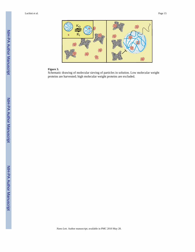
Figure 3.Schematic drawing of molecular sieving of particles in solution. Low molecular weightproteins are harvested; high molecular weight proteins are excluded.
Luchini et al. Page 15
Nano Lett. Author manuscript; available in PMC 2010 May 28.
NIH
-PA Author Manuscript
NIH
-PA Author Manuscript
NIH
-PA Author Manuscript
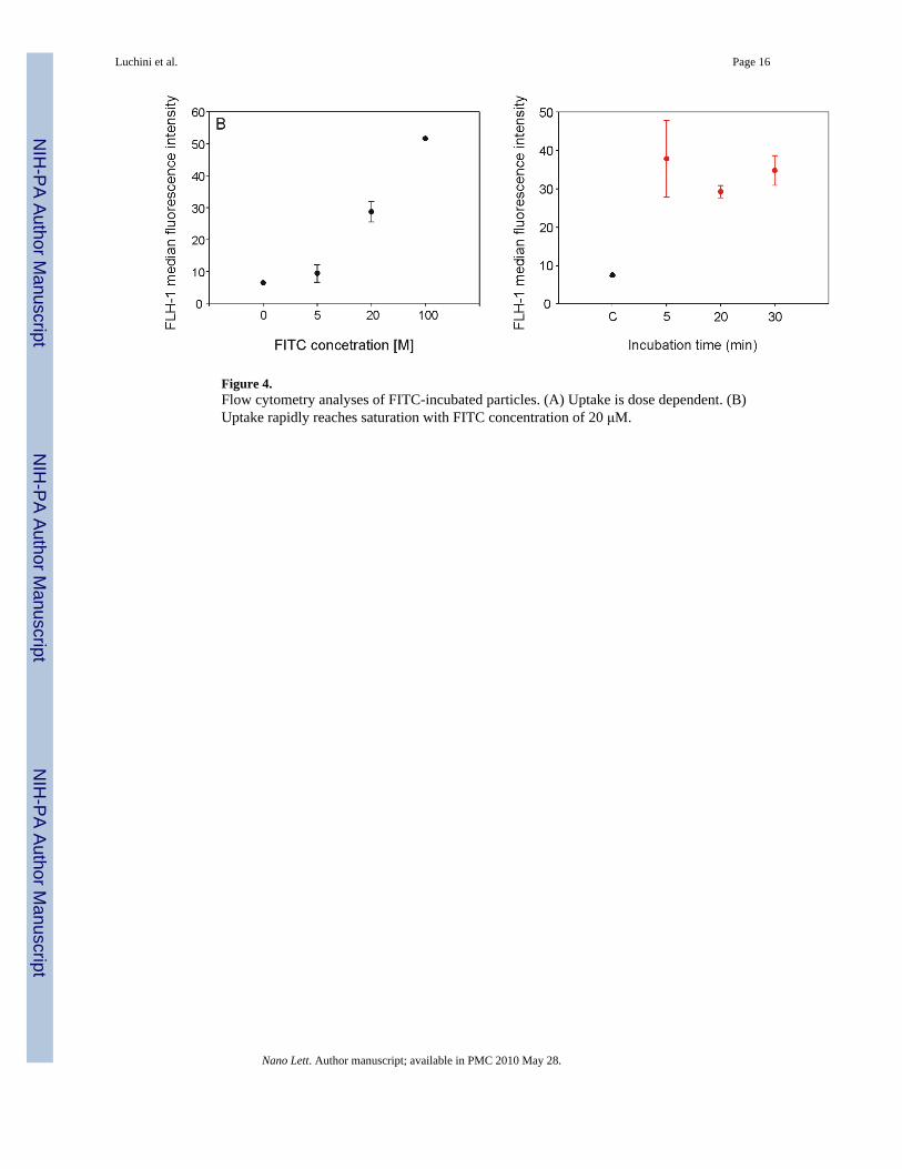
Figure 4.Flow cytometry analyses of FITC-incubated particles. (A) Uptake is dose dependent. (B)Uptake rapidly reaches saturation with FITC concentration of 20 μM.
Luchini et al. Page 16
Nano Lett. Author manuscript; available in PMC 2010 May 28.
NIH
-PA Author Manuscript
NIH
-PA Author Manuscript
NIH
-PA Author Manuscript
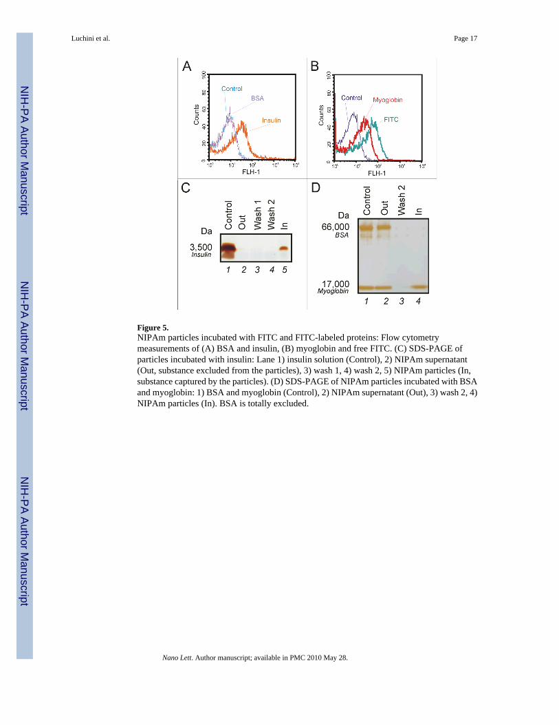
Figure 5.NIPAm particles incubated with FITC and FITC-labeled proteins: Flow cytometrymeasurements of (A) BSA and insulin, (B) myoglobin and free FITC. (C) SDS-PAGE ofparticles incubated with insulin: Lane 1) insulin solution (Control), 2) NIPAm supernatant(Out, substance excluded from the particles), 3) wash 1, 4) wash 2, 5) NIPAm particles (In,substance captured by the particles). (D) SDS-PAGE of NIPAm particles incubated with BSAand myoglobin: 1) BSA and myoglobin (Control), 2) NIPAm supernatant (Out), 3) wash 2, 4)NIPAm particles (In). BSA is totally excluded.
Luchini et al. Page 17
Nano Lett. Author manuscript; available in PMC 2010 May 28.
NIH
-PA Author Manuscript
NIH
-PA Author Manuscript
NIH
-PA Author Manuscript
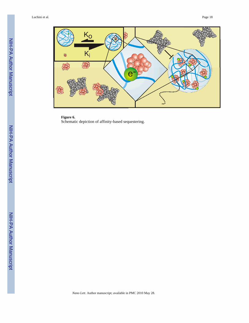
Figure 6.Schematic depiction of affinity-based sequestering.
Luchini et al. Page 18
Nano Lett. Author manuscript; available in PMC 2010 May 28.
NIH
-PA Author Manuscript
NIH
-PA Author Manuscript
NIH
-PA Author Manuscript
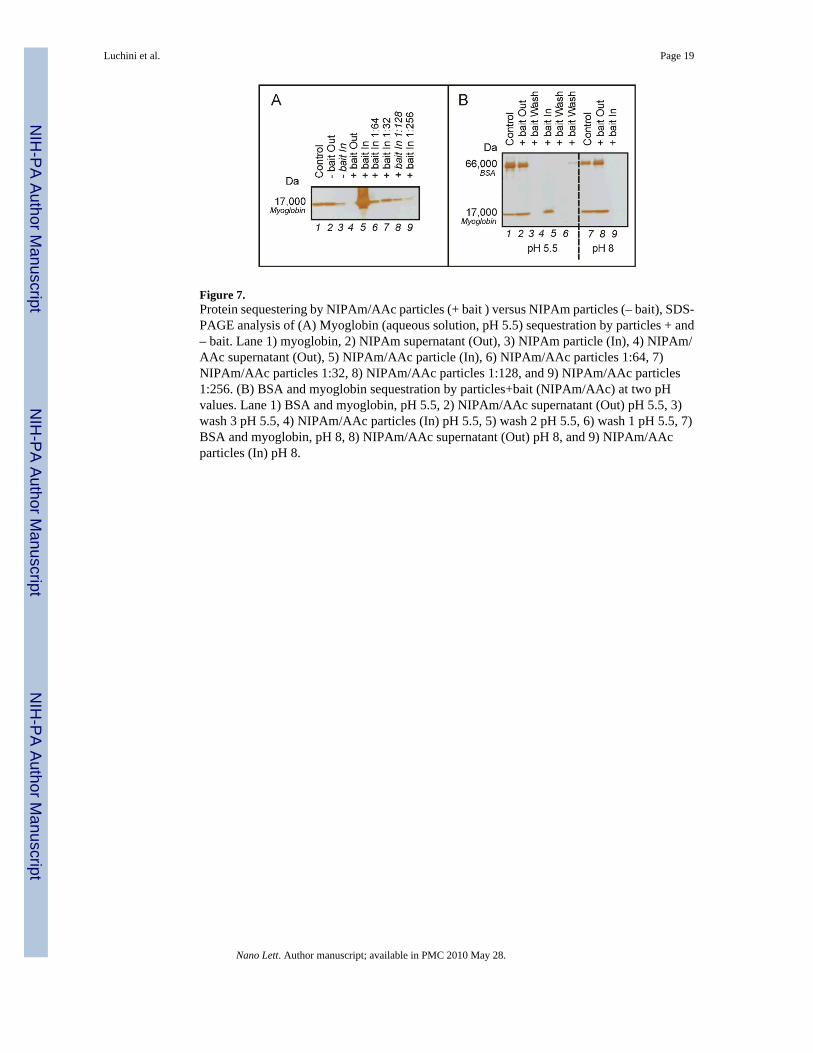
Figure 7.Protein sequestering by NIPAm/AAc particles (+ bait ) versus NIPAm particles (– bait), SDS-PAGE analysis of (A) Myoglobin (aqueous solution, pH 5.5) sequestration by particles + and– bait. Lane 1) myoglobin, 2) NIPAm supernatant (Out), 3) NIPAm particle (In), 4) NIPAm/AAc supernatant (Out), 5) NIPAm/AAc particle (In), 6) NIPAm/AAc particles 1:64, 7)NIPAm/AAc particles 1:32, 8) NIPAm/AAc particles 1:128, and 9) NIPAm/AAc particles1:256. (B) BSA and myoglobin sequestration by particles+bait (NIPAm/AAc) at two pHvalues. Lane 1) BSA and myoglobin, pH 5.5, 2) NIPAm/AAc supernatant (Out) pH 5.5, 3)wash 3 pH 5.5, 4) NIPAm/AAc particles (In) pH 5.5, 5) wash 2 pH 5.5, 6) wash 1 pH 5.5, 7)BSA and myoglobin, pH 8, 8) NIPAm/AAc supernatant (Out) pH 8, and 9) NIPAm/AAcparticles (In) pH 8.
Luchini et al. Page 19
Nano Lett. Author manuscript; available in PMC 2010 May 28.
NIH
-PA Author Manuscript
NIH
-PA Author Manuscript
NIH
-PA Author Manuscript
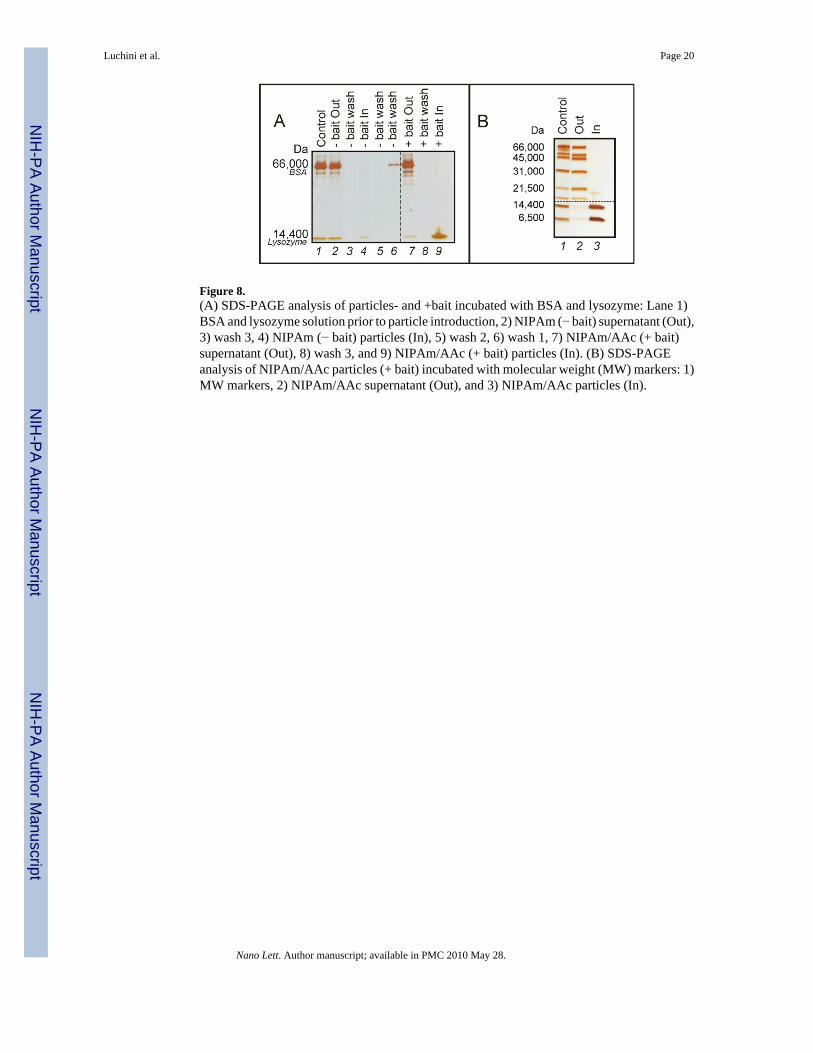
Figure 8.(A) SDS-PAGE analysis of particles- and +bait incubated with BSA and lysozyme: Lane 1)BSA and lysozyme solution prior to particle introduction, 2) NIPAm (− bait) supernatant (Out),3) wash 3, 4) NIPAm (− bait) particles (In), 5) wash 2, 6) wash 1, 7) NIPAm/AAc (+ bait)supernatant (Out), 8) wash 3, and 9) NIPAm/AAc (+ bait) particles (In). (B) SDS-PAGEanalysis of NIPAm/AAc particles (+ bait) incubated with molecular weight (MW) markers: 1)MW markers, 2) NIPAm/AAc supernatant (Out), and 3) NIPAm/AAc particles (In).
Luchini et al. Page 20
Nano Lett. Author manuscript; available in PMC 2010 May 28.
NIH
-PA Author Manuscript
NIH
-PA Author Manuscript
NIH
-PA Author Manuscript
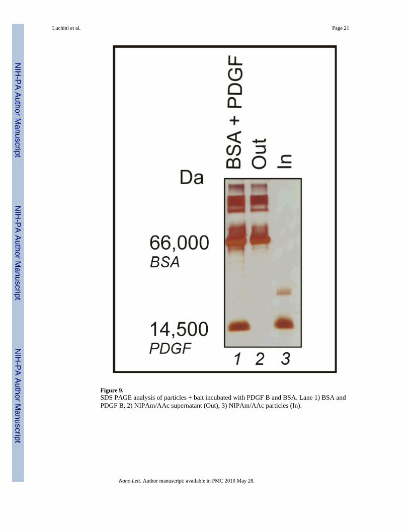
Figure 9.SDS PAGE analysis of particles + bait incubated with PDGF B and BSA. Lane 1) BSA andPDGF B, 2) NIPAm/AAc supernatant (Out), 3) NIPAm/AAc particles (In).
Luchini et al. Page 21
Nano Lett. Author manuscript; available in PMC 2010 May 28.
NIH
-PA Author Manuscript
NIH
-PA Author Manuscript
NIH
-PA Author Manuscript
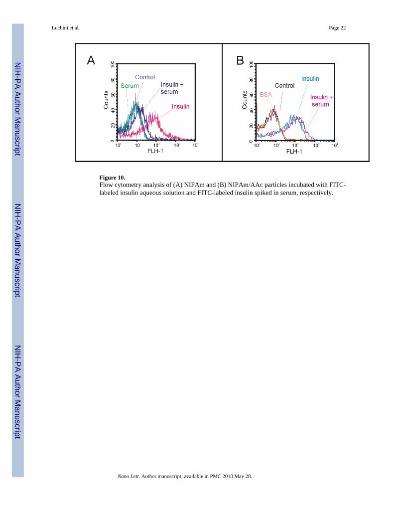
Figure 10.Flow cytometry analysis of (A) NIPAm and (B) NIPAm/AAc particles incubated with FITC-labeled insulin aqueous solution and FITC-labeled insulin spiked in serum, respectively.
Luchini et al. Page 22
Nano Lett. Author manuscript; available in PMC 2010 May 28.
NIH
-PA Author Manuscript
NIH
-PA Author Manuscript
NIH
-PA Author Manuscript
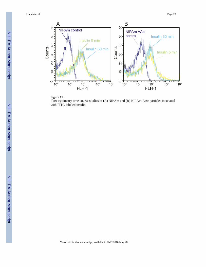
Figure 11.Flow cytometry time course studies of (A) NIPAm and (B) NIPAm/AAc particles incubatedwith FITC-labeled insulin.
Luchini et al. Page 23
Nano Lett. Author manuscript; available in PMC 2010 May 28.
NIH
-PA Author Manuscript
NIH
-PA Author Manuscript
NIH
-PA Author Manuscript
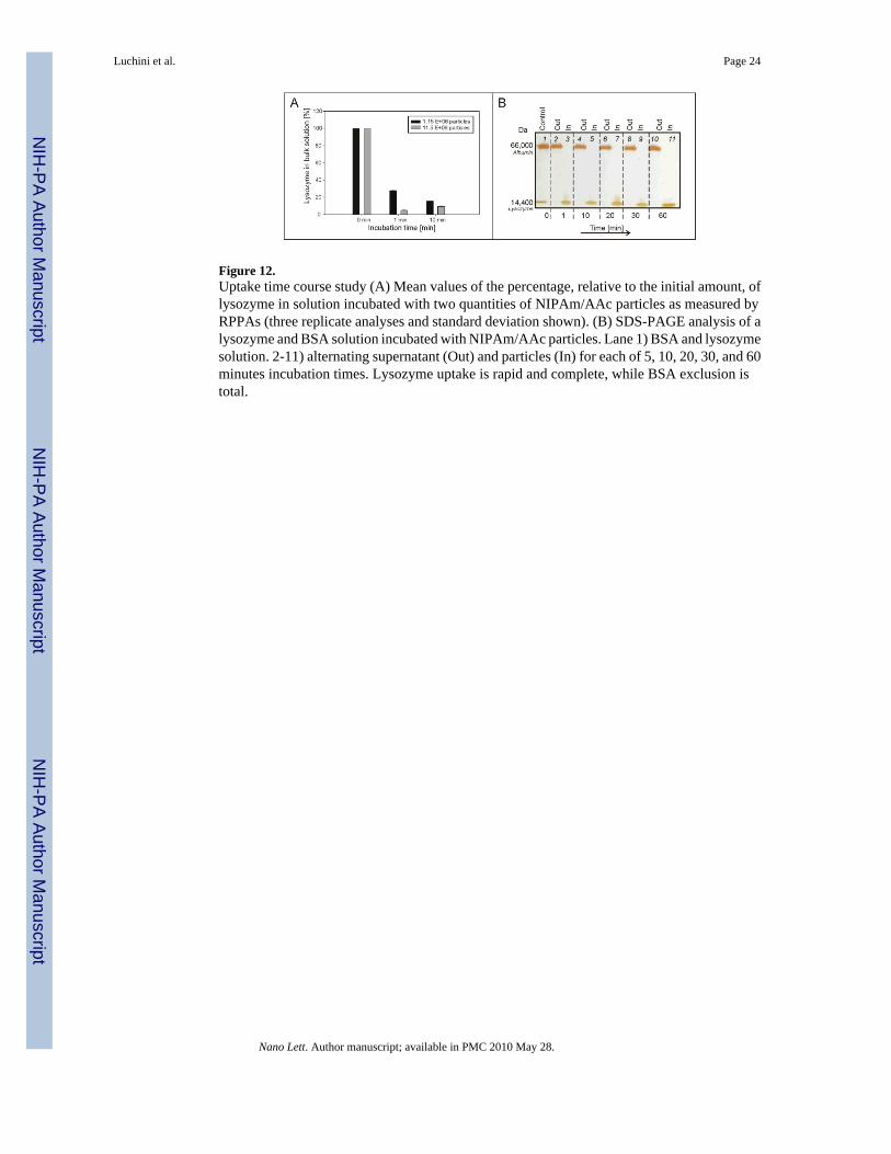
Figure 12.Uptake time course study (A) Mean values of the percentage, relative to the initial amount, oflysozyme in solution incubated with two quantities of NIPAm/AAc particles as measured byRPPAs (three replicate analyses and standard deviation shown). (B) SDS-PAGE analysis of alysozyme and BSA solution incubated with NIPAm/AAc particles. Lane 1) BSA and lysozymesolution. 2-11) alternating supernatant (Out) and particles (In) for each of 5, 10, 20, 30, and 60minutes incubation times. Lysozyme uptake is rapid and complete, while BSA exclusion istotal.
Luchini et al. Page 24
Nano Lett. Author manuscript; available in PMC 2010 May 28.
NIH
-PA Author Manuscript
NIH
-PA Author Manuscript
NIH
-PA Author Manuscript
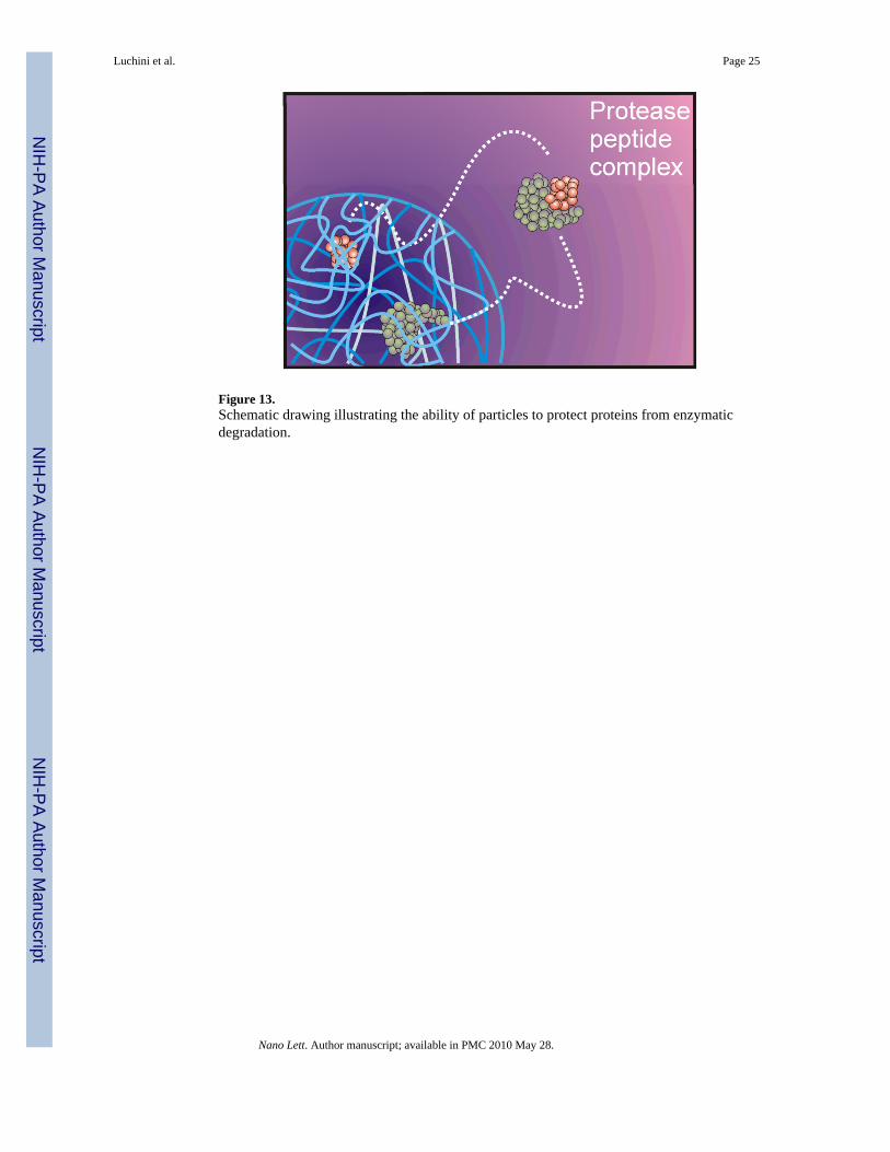
Figure 13.Schematic drawing illustrating the ability of particles to protect proteins from enzymaticdegradation.
Luchini et al. Page 25
Nano Lett. Author manuscript; available in PMC 2010 May 28.
NIH
-PA Author Manuscript
NIH
-PA Author Manuscript
NIH
-PA Author Manuscript
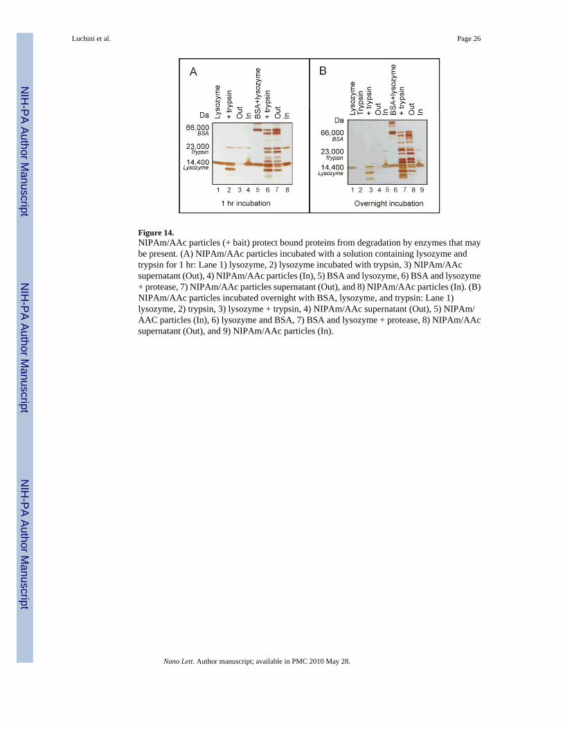
Figure 14.NIPAm/AAc particles (+ bait) protect bound proteins from degradation by enzymes that maybe present. (A) NIPAm/AAc particles incubated with a solution containing lysozyme andtrypsin for 1 hr: Lane 1) lysozyme, 2) lysozyme incubated with trypsin, 3) NIPAm/AAcsupernatant (Out), 4) NIPAm/AAc particles (In), 5) BSA and lysozyme, 6) BSA and lysozyme+ protease, 7) NIPAm/AAc particles supernatant (Out), and 8) NIPAm/AAc particles (In). (B)NIPAm/AAc particles incubated overnight with BSA, lysozyme, and trypsin: Lane 1)lysozyme, 2) trypsin, 3) lysozyme + trypsin, 4) NIPAm/AAc supernatant (Out), 5) NIPAm/AAC particles (In), 6) lysozyme and BSA, 7) BSA and lysozyme + protease, 8) NIPAm/AAcsupernatant (Out), and 9) NIPAm/AAc particles (In).
Luchini et al. Page 26
Nano Lett. Author manuscript; available in PMC 2010 May 28.
NIH
-PA Author Manuscript
NIH
-PA Author Manuscript
NIH
-PA Author Manuscript
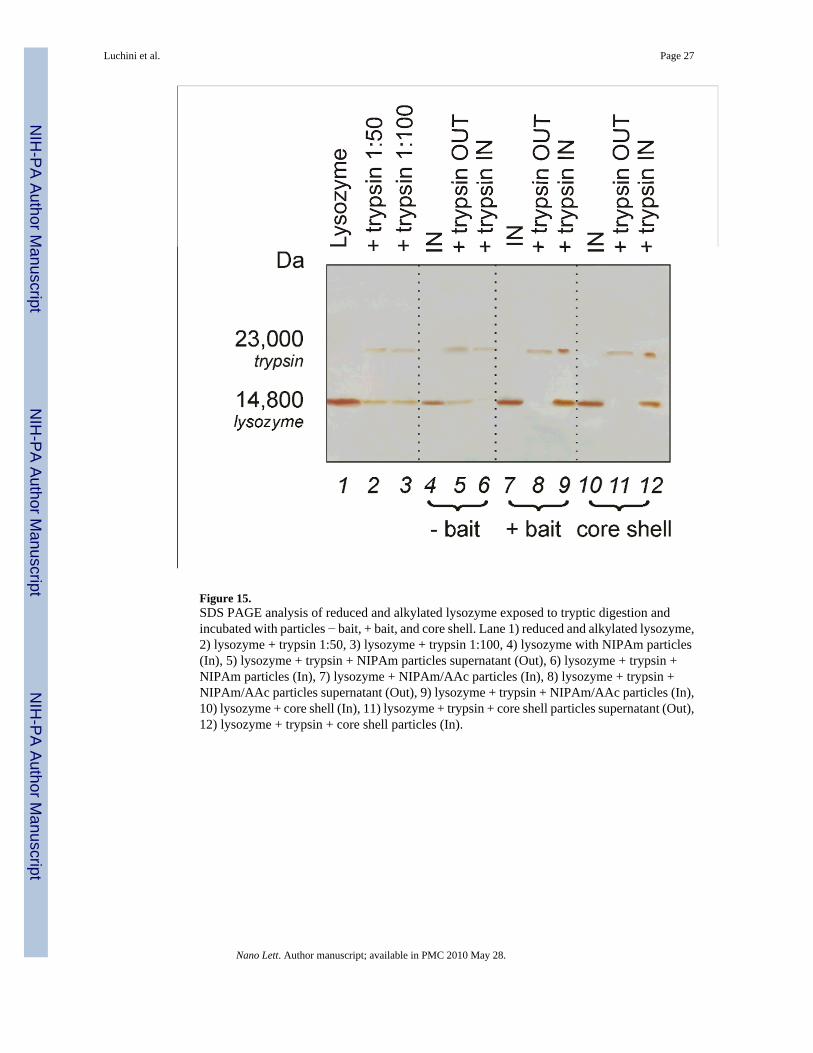
Figure 15.SDS PAGE analysis of reduced and alkylated lysozyme exposed to tryptic digestion andincubated with particles − bait, + bait, and core shell. Lane 1) reduced and alkylated lysozyme,2) lysozyme + trypsin 1:50, 3) lysozyme + trypsin 1:100, 4) lysozyme with NIPAm particles(In), 5) lysozyme + trypsin + NIPAm particles supernatant (Out), 6) lysozyme + trypsin +NIPAm particles (In), 7) lysozyme + NIPAm/AAc particles (In), 8) lysozyme + trypsin +NIPAm/AAc particles supernatant (Out), 9) lysozyme + trypsin + NIPAm/AAc particles (In),10) lysozyme + core shell (In), 11) lysozyme + trypsin + core shell particles supernatant (Out),12) lysozyme + trypsin + core shell particles (In).
Luchini et al. Page 27
Nano Lett. Author manuscript; available in PMC 2010 May 28.
NIH
-PA Author Manuscript
NIH
-PA Author Manuscript
NIH
-PA Author Manuscript
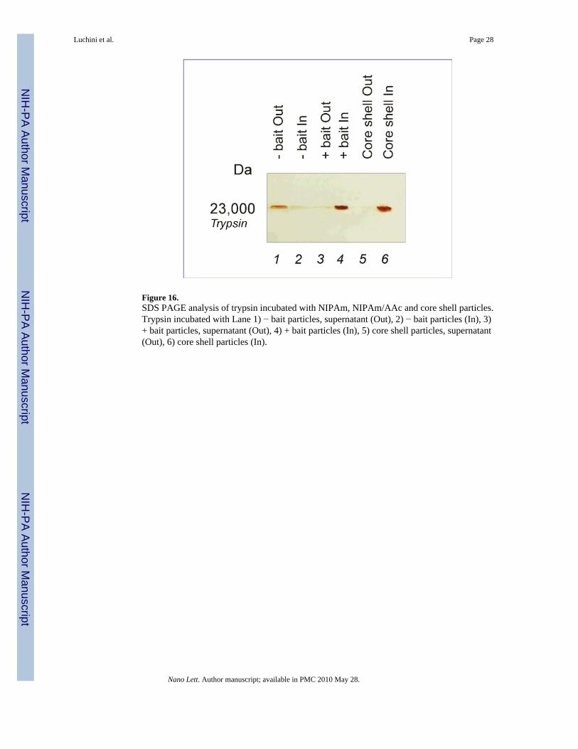
Figure 16.SDS PAGE analysis of trypsin incubated with NIPAm, NIPAm/AAc and core shell particles.Trypsin incubated with Lane 1) − bait particles, supernatant (Out), 2) − bait particles (In), 3)+ bait particles, supernatant (Out), 4) + bait particles (In), 5) core shell particles, supernatant(Out), 6) core shell particles (In).
Luchini et al. Page 28
Nano Lett. Author manuscript; available in PMC 2010 May 28.
NIH
-PA Author Manuscript
NIH
-PA Author Manuscript
NIH
-PA Author Manuscript
NIH
-PA Author Manuscript
NIH
-PA Author Manuscript
NIH
-PA Author Manuscript
Luchini et al. Page 29
Tabl
e 1
Mas
s spe
ctro
met
ry a
naly
sis o
f pro
tein
s ele
ctro
elut
ed fr
om N
IPA
m p
artic
les a
fter 1
hr i
ncub
atio
n w
ith 1
:10
v/v
dilu
tion
seru
m, “
P(pe
p)”
disp
lays
the
prob
abili
ty v
alue
for t
he p
eptid
e, “
Sf”
disp
lays
the
final
scor
e th
at in
dica
ted
how
goo
d th
e pr
otei
n m
atch
is, “
Scor
e” d
ispl
ays a
val
ue th
at is
bas
ed u
pon
the
prob
abili
ty th
at th
e pe
ptid
e is
a ra
ndom
mat
ch to
the
spec
tral d
ata,
“A
cces
sion
” di
spla
ys a
uni
que
prot
ein
iden
tific
atio
n nu
mbe
r for
the
sequ
ence
Ref
eren
ceA
cces
sion
P (p
ep)
SfSc
ore
MW
com
plem
ent c
ompo
nent
1, q
subc
ompo
nent
, gam
ma
poly
pept
ide
[Hom
o sa
pien
s]56
7861
55.0
4.83
E-12
3.43
E+00
40.2
825
757.
13
com
plem
ent c
ompo
nent
1, r
subc
ompo
nent
[Hom
o sa
pien
s]66
3478
75.0
5.65
E-12
2.72
E+00
30.2
480
147.
95
hapt
oglo
bin-
rela
ted
prot
ein
[Hom
o sa
pien
s]45
5807
23.0
1.12
E-11
9.76
E-01
10.2
239
004.
70
PRED
ICTE
D: s
imila
r to
Puta
tive
S100
cal
cium
-bin
ding
prot
ein
A11
pse
udog
ene
[Hom
o sa
pien
s]11
3419
208.
01.
12E-
119.
80E-
0110
.22
1125
4.79
com
plem
ent c
ompo
nent
4A
prep
ropr
otei
n [H
omo
sapi
ens]
6719
0748
.01.
12E-
113.
58E+
0040
.24
1926
63.6
0
oros
omuc
oid
1 pr
ecur
sor
[Hom
o sa
pien
s]92
5723
2.0
1.12
E-11
8.17
E-01
10.1
323
496.
77
CD
5 an
tigen
-like
(sca
veng
erre
cept
or c
yste
ine
rich
fam
ily)
[Hom
o sa
pien
s]51
7441
1.0
1.12
E-11
3.80
E+00
40.2
538
062.
96
seru
m a
myl
oid
P co
mpo
nent
prec
urso
r [H
omo
sapi
ens]
4502
133.
01.
12E-
114.
15E+
0050
.20
2537
1.13
com
plem
ent c
ompo
nent
1, q
subc
ompo
nent
, B c
hain
prec
urso
r [H
omo
sapi
ens]
8729
8828
.01.
12E-
119.
82E-
0110
.26
2670
4.49
apol
ipop
rote
in A
-Ipr
epro
prot
ein
[Hom
o sa
pien
s]45
5732
1.0
1.12
E-11
6.86
E-01
10.1
230
758.
94
hapt
oglo
bin
[Hom
o sa
pien
s]48
2676
2.0
1.12
E-11
3.70
E+00
40.2
145
176.
59
com
plem
ent c
ompo
nent
1, q
subc
ompo
nent
, A c
hain
prec
urso
r [H
omo
sapi
ens]
7705
753.
01.
12E-
118.
24E-
0110
.15
2600
0.19
plat
elet
fact
or 4
(che
mok
ine
(C-X
-C m
otif)
liga
nd 4
)[H
omo
sapi
ens]
4505
733.
01.
12E-
111.
80E+
0020
.16
1083
7.89
imm
unog
lobu
lin J
chai
n[H
omo
sapi
ens]
2148
9959
.01.
12E-
119.
45E-
0110
.15
1808
7.00
lyso
zym
e pr
ecur
sor [
Hom
osa
pien
s]45
5789
4.0
1.12
E-11
9.03
E-01
10.1
516
526.
29
Nano Lett. Author manuscript; available in PMC 2010 May 28.
NIH
-PA Author Manuscript
NIH
-PA Author Manuscript
NIH
-PA Author Manuscript
Luchini et al. Page 30
Ref
eren
ceA
cces
sion
P (p
ep)
SfSc
ore
MW
trans
thyr
etin
[Hom
o sa
pien
s]45
0772
5.0
1.12
E-11
6.77
E-01
10.1
315
877.
05
derm
cidi
n pr
epro
prot
ein
[Hom
o sa
pien
s]16
7519
21.0
1.12
E-11
8.66
E-01
10.1
311
276.
83
mes
otry
psin
pre
prop
rote
in[H
omo
sapi
ens]
2153
6452
.01.
12E-
119.
37E-
0110
.17
2668
0.18
alph
a 1
glob
in [H
omo
sapi
ens]
4504
347.
01.
12E-
119.
62E-
0110
.16
1524
7.92
beta
glo
bin
[Hom
o sa
pien
s]45
0434
9.0
1.12
E-11
9.36
E-01
10.1
715
988.
29
hypo
thet
ical
pro
tein
LOC
6498
97 [H
omo
sapi
ens]
9120
6438
.01.
12E-
118.
68E-
0110
.18
2205
8.92
com
plem
ent c
ompo
nent
1, s
subc
ompo
nent
[Hom
o sa
pien
s]41
3936
02.0
1.12
E-11
9.25
E-01
10.1
576
634.
85
prot
ein
kina
se C
and
cas
ein
kina
se su
bstra
te in
neu
rons
2[H
omo
sapi
ens]
6005
826.
01.
12E-
118.
23E-
0110
.15
5587
0.13
PRED
ICTE
D: s
imila
r to
Ker
atin
, typ
e II
cyt
oske
leta
l 2or
al (C
ytok
erat
in-2
P) (K
2P)
(CK
2P)
[Hom
o sa
pien
s]89
0361
76.0
1.12
E-11
8.89
E-01
10.1
436
406.
56
plat
elet
fact
or 4
var
iant
1[H
omo
sapi
ens]
4505
735.
01.
12E-
118.
70E-
0110
.19
1154
5.28
brom
odom
ain
cont
aini
ng 7
[Hom
o sa
pien
s]41
3502
12.0
1.12
E-11
8.63
E-01
10.1
874
092.
27
SH3-
dom
ain
bind
ing
prot
ein
2[H
omo
sapi
ens]
1992
3155
.01.
12E-
112.
64E-
0110
.13
6222
0.29
zinc
fing
er, C
CH
C d
omai
nco
ntai
ning
11
isof
orm
c[H
omo
sapi
ens]
5786
3250
.01.
12E-
118.
14E-
0110
.17
1845
87.1
0
apol
ipop
rote
in A
-II
prep
ropr
otei
n [H
omo
sapi
ens]
4502
149.
01.
12E-
118.
90E-
0110
.14
1116
7.90
hete
roge
neou
s nuc
lear
ribon
ucle
opro
tein
D is
ofor
m d
[Hom
o sa
pien
s]51
4777
08.0
1.12
E-11
6.31
E-01
10.1
830
653.
14
polo
-like
kin
ase
4 [H
omo
sapi
ens]
2136
1433
.01.
12E-
116.
16E-
0110
.13
1090
16.4
0
apol
ipop
rote
in L
1 is
ofor
m a
prec
urso
r [H
omo
sapi
ens]
2173
5614
.01.
12E-
116.
63E-
0110
.12
4394
6.95
coro
nin,
act
in b
indi
ng p
rote
in,
2A [H
omo
sapi
ens]
1655
4583
.01.
12E-
119.
33E-
0120
.14
5969
7.38
Nano Lett. Author manuscript; available in PMC 2010 May 28.
NIH
-PA Author Manuscript
NIH
-PA Author Manuscript
NIH
-PA Author Manuscript
Luchini et al. Page 31
Tabl
e 2
Mas
s spe
ctro
met
ry a
naly
sis o
f pro
tein
s ele
ctro
elut
ed fr
om N
IPA
m/A
Ac
parti
cles
afte
r 1 h
r inc
ubat
ion
with
1:1
0 v/
v di
lutio
n se
rum
, “P(
pep)
” di
spla
ys th
epr
obab
ility
val
ue fo
r the
pep
tide,
“Sf
” di
spla
ys th
e fin
al sc
ore
that
indi
cate
d ho
w g
ood
the
prot
ein
mat
ch is
, “Sc
ore”
dis
play
s a v
alue
that
is b
ased
upo
n th
epr
obab
ility
that
the
pept
ide
is a
rand
om m
atch
to th
e sp
ectra
l dat
a, “
Acc
essi
on”
disp
lays
a u
niqu
e pr
otei
n id
entif
icat
ion
num
ber f
or th
e se
quen
ce
Ref
eren
ceA
cces
sion
P (p
ep)
SfSc
ore
MW
com
plem
ent c
ompo
nent
1, q
subc
ompo
nent
, gam
ma
poly
pept
ide
[Hom
o sa
pien
s]56
7861
55.0
3.55
E-14
1.86
E+00
20.3
025
757.
13
hypo
thet
ical
pro
tein
LOC
6498
97 [H
omo
sapi
ens]
9120
6438
.03.
55E-
142.
90E+
0030
.26
2205
8.92
PRED
ICTE
D: s
imila
r to
Puta
tive
S100
cal
cium
-bin
ding
prot
ein
A11
pse
udog
ene
[Hom
o sa
pien
s]11
3419
208.
03.
55E-
141.
46E+
0020
.25
1125
4.79
apol
ipop
rote
in C
-III
pre
curs
or[H
omo
sapi
ens]
4557
323.
03.
55E-
149.
79E-
0110
.24
1084
5.50
pro-
plat
elet
bas
ic p
rote
inpr
ecur
sor [
Hom
o sa
pien
s]45
0598
1.0
3.55
E-14
5.61
E+00
60.2
513
885.
42
com
plem
ent c
ompo
nent
3pr
ecur
sor [
Hom
o sa
pien
s]45
5738
5.0
3.55
E-14
9.80
E-01
10.2
018
7045
.30
smal
l nuc
lear
ribon
ucle
opro
tein
pol
ypep
tide
E [H
omo
sapi
ens]
4507
129.
03.
55E-
149.
15E-
0110
.19
1079
6.64
kera
tin 2
[Hom
o sa
pien
s]47
1326
20.0
3.55
E-14
5.75
E+00
70.2
165
393.
19
albu
min
pre
curs
or [H
omo
sapi
ens]
4502
027.
03.
55E-
149.
28E+
0010
0.22
6932
1.63
ribos
omal
pro
tein
L37
a [H
omo
sapi
ens]
4506
643.
03.
55E-
149.
73E-
0110
.21
1026
8.48
com
plem
ent c
ompo
nent
4A
prep
ropr
otei
n [H
omo
sapi
ens]
6719
0748
.03.
55E-
141.
91E+
0020
.17
1926
63.6
0
A-g
amm
a gl
obin
[Hom
osa
pien
s]28
3021
31.0
3.55
E-14
9.44
E-01
10.1
416
118.
27
plat
elet
fact
or 4
(che
mok
ine
(C-X
-C m
otif)
liga
nd 4
)[H
omo
sapi
ens]
4505
733.
03.
55E-
143.
44E+
0040
.18
1083
7.89
PRED
ICTE
D: h
ypot
hetic
alpr
otei
n [H
omo
sapi
ens]
1134
1832
7.0
3.55
E-14
8.55
E-01
10.1
931
688.
42
H4
hist
one
fam
ily, m
embe
r J[H
omo
sapi
ens]
4504
315.
03.
55E-
141.
51E+
0020
.13
1136
0.38
lyso
zym
e pr
ecur
sor
[Hom
o sa
pien
s]45
5789
4.0
3.55
E-14
9.66
E-01
10.2
016
526.
29
Nano Lett. Author manuscript; available in PMC 2010 May 28.
NIH
-PA Author Manuscript
NIH
-PA Author Manuscript
NIH
-PA Author Manuscript
Luchini et al. Page 32
Ref
eren
ceA
cces
sion
P (p
ep)
SfSc
ore
MW
mes
otry
psin
pre
prop
rote
in[H
omo
sapi
ens]
2153
6452
.03.
55E-
149.
68E-
0110
.18
2668
0.18
alph
a 1
glob
in [H
omo
sapi
ens]
4504
347.
03.
55E-
149.
15E-
0110
.14
1524
7.92
fibrin
ogen
, alp
ha p
olyp
eptid
eis
ofor
m a
lpha
-E p
repr
opro
tein
[Hom
o sa
pien
s]45
0368
9.0
3.55
E-14
8.35
E-01
10.1
294
914.
27
hypo
thet
ical
pro
tein
LOC
5568
3 [H
omo
sapi
ens]
2136
1734
.03.
55E-
146.
53E-
0110
.12
8324
4.77
crum
bs h
omol
og 1
pre
curs
or[H
omo
sapi
ens]
4132
7708
.03.
55E-
146.
87E-
0110
.15
1540
80.4
0
PRED
ICTE
D: s
imila
r to
glut
amat
e re
cept
or, i
onot
ropi
c,N
-met
hyl D
-asp
arta
te-li
ke 1
Ais
ofor
m 1
isof
orm
1 [H
omo
sapi
ens]
4114
6739
.03.
55E-
146.
26E-
0110
.15
4168
6.95
PRED
ICTE
D: s
imila
r to
Neu
troph
il de
fens
in 1
prec
urso
r (H
NP-
1) (H
P-1)
(HP1
)(D
efen
sin,
alp
ha 1
)[H
omo
sapi
ens]
1134
1990
3.0
3.55
E-14
7.75
E-01
10.1
210
194.
18
CD
K5
regu
lato
ry su
buni
tas
soci
ated
pro
tein
1 is
ofor
m b
[Hom
o sa
pien
s]28
8727
84.0
3.55
E-14
9.05
E-01
10.1
256
187.
84
com
plem
ent c
ompo
nent
1, q
subc
ompo
nent
, B c
hain
prec
urso
r [H
omo
sapi
ens]
8729
8828
.03.
55E-
148.
90E-
0110
.15
2670
4.49
inte
rfer
on-in
duce
d pr
otei
nw
ith te
tratri
cope
ptid
e re
peat
s 3[H
omo
sapi
ens]
3154
2980
.03.
55E-
149.
40E-
0110
.22
5594
9.57
lam
in A
/C is
ofor
m 1
pre
curs
or[H
omo
sapi
ens]
2743
6946
.03.
55E-
148.
73E-
0110
.14
7409
4.81
doub
le C
2-lik
e do
mai
ns, b
eta
[Hom
o sa
pien
s]60
0599
7.0
3.55
E-14
5.53
E-01
10.1
345
920.
53
desm
ogle
in 4
[Hom
o sa
pien
s]29
7894
45.0
3.55
E-14
9.06
E-01
10.1
311
3751
.30
cadh
erin
EG
F LA
G se
ven-
pass
G-ty
pe re
cept
or 1
[Hom
osa
pien
s]76
5696
7.0
3.55
E-14
1.56
E+00
20.1
432
9276
.70
proc
olla
gen,
type
III,
alph
a 1
[Hom
o sa
pien
s]45
0295
1.0
3.55
E-14
9.12
E-01
10.1
513
8470
.20
ATP
ase,
H+
trans
porti
ng,
lyso
som
al 1
4kD
, V1
subu
nit F
[Hom
o sa
pien
s]20
3575
47.0
3.55
E-14
8.61
E-01
10.1
613
361.
95
Nano Lett. Author manuscript; available in PMC 2010 May 28.