Protein modification and replicative senescence of WI38 human embryonic fibroblasts
Upload
independentCategory
view
4download
0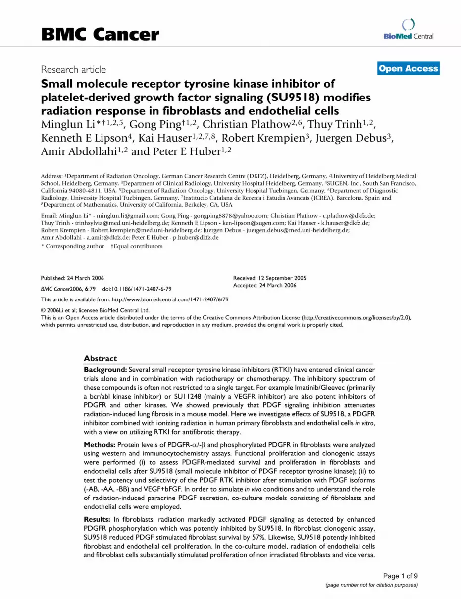
BioMed CentralBMC Cancer
ss
Open AcceResearch articleSmall molecule receptor tyrosine kinase inhibitor of platelet-derived growth factor signaling (SU9518) modifies radiation response in fibroblasts and endothelial cellsMinglun Li*†1,2,5, Gong Ping†1,2, Christian Plathow2,6, Thuy Trinh1,2, Kenneth E Lipson4, Kai Hauser1,2,7,8, Robert Krempien3, Juergen Debus3, Amir Abdollahi1,2 and Peter E Huber1,2Address: 1Department of Radiation Oncology, German Cancer Research Centre (DKFZ), Heidelberg, Germany, 2University of Heidelberg Medical School, Heidelberg, Germany, 3Department of Clinical Radiology, University Hospital Heidelberg, Germany, 4SUGEN, Inc., South San Francisco, California 94080-4811, USA, 5Department of Radiation Oncology, University Hospital Tuebingen, Germany, 6Department of Diagnostic Radiology, University Hospital Tuebingen, Germany, 7Institucio Catalana de Recerca i Estudis Avancats (ICREA), Barcelona, Spain and 8Department of Mathematics, University of California, Berkeley, CA, USA
Email: Minglun Li* - [email protected]; Gong Ping - [email protected]; Christian Plathow - [email protected]; Thuy Trinh - [email protected]; Kenneth E Lipson - [email protected]; Kai Hauser - [email protected]; Robert Krempien - [email protected]; Juergen Debus - [email protected]; Amir Abdollahi - [email protected]; Peter E Huber - [email protected]
* Corresponding author †Equal contributors
AbstractBackground: Several small receptor tyrosine kinase inhibitors (RTKI) have entered clinical cancertrials alone and in combination with radiotherapy or chemotherapy. The inhibitory spectrum ofthese compounds is often not restricted to a single target. For example Imatinib/Gleevec (primarilya bcr/abl kinase inhibitor) or SU11248 (mainly a VEGFR inhibitor) are also potent inhibitors ofPDGFR and other kinases. We showed previously that PDGF signaling inhibition attenuatesradiation-induced lung fibrosis in a mouse model. Here we investigate effects of SU9518, a PDGFRinhibitor combined with ionizing radiation in human primary fibroblasts and endothelial cells in vitro,with a view on utilizing RTKI for antifibrotic therapy.
Methods: Protein levels of PDGFR-α/-β and phosphorylated PDGFR in fibroblasts were analyzedusing western and immunocytochemistry assays. Functional proliferation and clonogenic assayswere performed (i) to assess PDGFR-mediated survival and proliferation in fibroblasts andendothelial cells after SU9518 (small molecule inhibitor of PDGF receptor tyrosine kinase); (ii) totest the potency und selectivity of the PDGF RTK inhibitor after stimulation with PDGF isoforms(-AB, -AA, -BB) and VEGF+bFGF. In order to simulate in vivo conditions and to understand the roleof radiation-induced paracrine PDGF secretion, co-culture models consisting of fibroblasts andendothelial cells were employed.
Results: In fibroblasts, radiation markedly activated PDGF signaling as detected by enhancedPDGFR phosphorylation which was potently inhibited by SU9518. In fibroblast clonogenic assay,SU9518 reduced PDGF stimulated fibroblast survival by 57%. Likewise, SU9518 potently inhibitedfibroblast and endothelial cell proliferation. In the co-culture model, radiation of endothelial cellsand fibroblast cells substantially stimulated proliferation of non irradiated fibroblasts and vice versa.
Published: 24 March 2006
BMC Cancer2006, 6:79 doi:10.1186/1471-2407-6-79
Received: 12 September 2005Accepted: 24 March 2006
This article is available from: http://www.biomedcentral.com/1471-2407/6/79
© 2006Li et al; licensee BioMed Central Ltd.This is an Open Access article distributed under the terms of the Creative Commons Attribution License (http://creativecommons.org/licenses/by/2.0), which permits unrestricted use, distribution, and reproduction in any medium, provided the original work is properly cited.
Page 1 of 9(page number not for citation purposes)
BMC Cancer 2006, 6:79 http://www.biomedcentral.com/1471-2407/6/79
Importantly, the RTK inhibitor significantly inhibited this paracrine radiation-induced fibroblast andendothelial cell activation.
Conclusion: Radiation-induced autocrine and paracrine PDGF signaling plays an important role infibroblast and endothelial cell proliferation. SU9518, a PDGFR tyrosine kinase inhibitor, reducesradiation-induced fibroblast and endothelial cell activation. This may explain therapeutic anticancereffects of Imatinib/Gleevec, and at the same time it could open a way of attenuating radiation-induced fibrosis.
BackgroundThe development of fibrosis is a frequent side effect thatmay be caused by a variety of inductors. Fibrosis especiallyhampers cancer treatment such as radiotherapy and chem-otherapy, thus severely limiting cancer therapy success.Furthermore, the development of fibrosis may reflect theintegrative and interdependent roles of various cell com-pounds in tumor biology calling into question reduction-ist approaches focus on individual tumor cellcompartment. Fibrosis occurs in many organs. Lung fibro-sis for example may be caused by radiotherapy, chemo-therapeutic drugs (such as bleomycin), amiodaron or,chemical substances (like asbestos fibers and silica parti-cles, etc [1]). With the progression of the disease patientsdevelop severe clinical symptoms limiting oxygenation ofthe blood and patients' survival. Treatment of fibrosisremains elusive given that the exact mediators and mech-anisms involved in fibrogenesis are unknown [2,3].
Fibrosis is characterized by the excessive proliferation ofmesenchymal cells (fibroblasts, myofibroblasts, andsmooth muscle cells) and the subsequent deposition ofextracellular matrix proteins [2]. Fibroblasts exhibitincreased chemotaxis, proliferation, and extracellularmatrix production in fibrotic lung and skin [2,4].Cytokines such as platelet-derived growth factor (PDGF),transforming growth factor (TGF)-α and -β, interleukin(IL)-1α and -β, and basic fibroblast growth factor (bFGF)have emerged as major stimulators of the fibroprolifera-tive process induced by diverse stimuli such as ionizingradiation [5-7].
Here we focus on PDGF which has been implicated in awide variety of pathological processes, including pulmo-nary fibrosis and skin fibrosis [8-10]. PDGF is a disulfide-linked dimer of two related polypeptide chains, desig-nated A, B, C and D, which are assembled as heterodimers(PDGF-AB) or homodimers (PDGF-AA, PDGF-BB, PDGF-CC and PDGF-DD) [11]. PDGF exerts its biological activ-ity by binding to structurally similar PDGF receptors(PDGFR-α and -β). It induces receptor dimerization andautophosphorylation of the receptor tyrosine kinase(RTK). Activated RTK phosphorylates numerous signalingmolecules that initiate the intracellular signaling cascadesleading to cell proliferation and survival [12]. These phos-
phorylation-dependent interactions are essential for theactivation of intracellular signaling pathways that can leadto tissue fibrosis. It seems reasonable to assume thatblocking phosphorylation of PDGFR could be a way ofpreventing biological effects of this cytokine and thusfibrogenesis.
Recently, we have demonstrated this concept in vivo in aradiation-induced lung fibrosis model in mice by showingthat inhibition of PDGF signaling functionally attenuatespulmonary fibrosis [13]. Here we analyze the underlyingmechanistic cell effects in vitro with respect to the cellularpathway of RTK inhibition and to radiation for the PDGF/PDGFR system in human primary fibroblasts andendothelial cells.
Materials and methodsEndothelial and fibroblast cell cultures and reagentsPrimary isolated human dermal microvascular veinendothelial cells (HDMVEC, Promocell, Heidelberg, Ger-many) and primary isolated human fibroblasts (Promo-cell, Heidelberg, Germany) were cultured up to passage 6.Cells were maintained in culture at 37°C with 5% CO2and 95% humidity in serum reduced (5% fetus calfserum) modified promocell media (MPM) supplementedwith 2 ng/ml vascular endothelial growth factor (VEGF)and 4 ng/ml basic fibroblast growth factor (bFGF). Thiscombination of VEGF and bFGF optimized growth kinet-ics [14]. Human recombinant VEGF and bFGF proteinswere purchased from Promocell.
Western blottingPrimary human lung fibroblasts were grown in modifiedmedia supplemented with 5% fetus calf serum containing2 ng/ml VEGF and 4 ng/ml bFGF in 125 cm2 flasks. Cellswere then treated either with 10 Gy or 5 µM SU9518exclusively or in combination with both (1 hour SU9518incubation prior to radiation). 1/2 hour after radiationfibroblasts were washed twice with ice-cold PBS, lysedwith 250 µl ice-cold cell lysis buffer (1 M Tris, 5 M NaCl,1 mM EDTA, 1 mM EGTA, pH 8.01). Lysates were kept onice, and centrifuged at 4°C for 10 minutes at 13000 rpm.Supernatant was collected for western blotting assay asdescribed [15]. Proteins were transferred from a polyacry-lamide gel to a nitrocellulose membrane with a Mini-
Page 2 of 9(page number not for citation purposes)
BMC Cancer 2006, 6:79 http://www.biomedcentral.com/1471-2407/6/79
Trans-Blotter (100 V for 90 minutes) in transfer buffer.After transfer, non-specific binding sites were blocked for1 hour with 5% non-fat milk powder in TBST buffer (15mM NaCl, 1 mM TRIS-HCl, 0.1% Tween 20, pH 8.0) atroom temperature. Each primary antibody (anti-PDGFR-α, anti-PDGFR-β and anti-phosphorylated PDGFR-β, CellSignaling Technology, Inc. Germany) was diluted inblocking buffer and incubated with the membrane over-night at 4°C. After washing in TBST, the nitrocellulosemembrane was incubated with horseradish peroxidase-conjugated antibody in blocking buffer at room tempera-ture for 60 minutes. Antibody binding was determinedusing the enhanced chemiluminescence detection systemaccording to the manufacturer's instructions. Exposureswere recorded on hyperfilms for 10 seconds to 3 minutes.
ImmunocytochemistryFibroblasts were cultured as in the western blotting assay.After trypsinization of fibroblasts in flasks, 20 µl cell sus-pensions (3 × 10⁴ cells/ml) were seeded on slips and incu-bated for 24 hour at 37°C with 5% CO2 and 95%humidity. After treatments with radiation (10 Gy),SU9518 (5 µM) or SU9518 + radiation (1 hour incuba-tion with SU9518 prior radiation), fibroblasts were incu-bated 1/2, 6, 12 or 24 hours in MPM. After stimulationfibroblasts were washed with phosphate-buffered saline(PBS) and fixed with methanol and acetone 5 minutes at-20°C respectively. After washing with PBS, fibroblastswere permeabilized for 30 minutes with PBS containing1% tween-20 (Merck, Darmstadt, Germany), and blockedfor 30 minutes with PBS blocking buffer (containing 5%FBS with 1% tween-20). The cells were incubated for 12hours at 18–20°C in blocking buffer with 3 primary anti-bodies: anti-PDGFR-α, anti-PDGFR-β and anti-phosphor-ylated PDGFR-β (Cell Signaling Technology, Inc.Germany). After washing with PBS, the cells were incu-bated for 1 hour at room temperature with 1:250 second-ary antibodies, goat anti-mouse IgG (Alexa Fluor 488) orgoat anti-rabbit IgG (Alexa Fluor 488), and mixed 1:200with bromphenolblue (Sigma, St. Louis, USA) solutionfor cell nucleus staining. After further washing slides weremounted in Citifluor mounting medium and were exam-ined with a fluorescence microscope (400X, softwareOpenlab 3.1.4, Leica, DMIRB).
Clonogenic survival assayTo investigate the effects of SU9518 on clonogenic sur-vival of fibroblast and endothelial cells, cells were platedwith increasing numbers (102 to 5 × 10⁴) in 25 cm2 flasks.Cells were incubated with 0–10 µM SU9518 in presenceof 10 ng/ml PDGF-AB. Flasks were returned to the incuba-tor for 14–17 days, after which they were stained withcrystal violet (Sigma, Germany). Colonies were countedand the surviving percentage was determined for clono-genic survival after correcting for plating efficiency.
Proliferation assayCells were harvested by trypsinization at 37°C and neu-tralized with trypsin-neutralizing solution. A suspensionof 50000 cells in MPM/DMEM was added to 25 cm2 flasks(Becton Dickinson, Heidelberg, Germany). Cells wereincubated with SU9518 for 1 hour in cytokine-freemedium at standard conditions and incubated foranother 72 hours in final mediums (either with 10 ng/mlPDGF-AB, or with 2 ng/ml VEGF + 4 ng/ml bFGF). Cellswere then dispersed in trypsin, resuspended, and countedin a coulter counter.
In fibroblasts, PDGF-isoform dependent proliferationassays were performed as follows: Fibroblasts were cul-tured and plated as in the clonogenic assay. Cells wereincubated for 72 hours either with PDGF (-AA, -AB, -BB,each isoform in 2 and 10 ng/ml) alone, or with additionaladministration of 5 µM SU9518. Cells were then dis-persed in trypsin, resuspended, and counted in a coultercounter.
Co-culture modelA proliferation assay in a co-culture model was used toassess the proliferation ability of cells in the upper com-partments after irradiation of cells in the lower compart-ments with or without SU9518 treatment. Human dermalmicrovascular endothelial cells (HDMVECs) were grownin 24 well plates. At confluence, the media were changed.Cells were irradiated with 10 Gy using a 6 MeV x-rays lin-ear accelerator at a dose rate of 118 cGy/min. Transwellinserts of upper compartments with a 0.6 µm pore sizewere plated with 20,000 cells. In SU9518 treated groupsand SU9518 + radiation groups, cells in upper compart-ments were preincubated with 5 µM SU9518 for 1 hour.After selective irradiation of cells in lower compartments,transwells of upper compartments containing cells weretransferred into the 24 well plates, incubated for 72 hoursat standard conditions, then dispersed in trypsin, resus-pended and counted. Three conditions were analyzed: (A)irradiated fibroblast cells in the lower compartment, asthe chemotactic "agent" after radiation, and (non irradi-ated) fibroblasts in the upper compartment; (B) irradiatedHDMVEC in the lower compartment and fibroblasts inthe upper compartment; (C) irradiated fibroblasts in thelower compartment and non irradiated HDMVEC in theupper compartment.
Statistical analysisStatistical analyses were performed with a statistical anal-ysis software system (Statistica 6.0). Student's t test wasused to compare means. For multiple comparisonsANOVA was used with Fisher's least-significant differencemethod. P-values less than 0.05 were considered statisti-cally significant.
Page 3 of 9(page number not for citation purposes)
BMC Cancer 2006, 6:79 http://www.biomedcentral.com/1471-2407/6/79
Page 4 of 9(page number not for citation purposes)
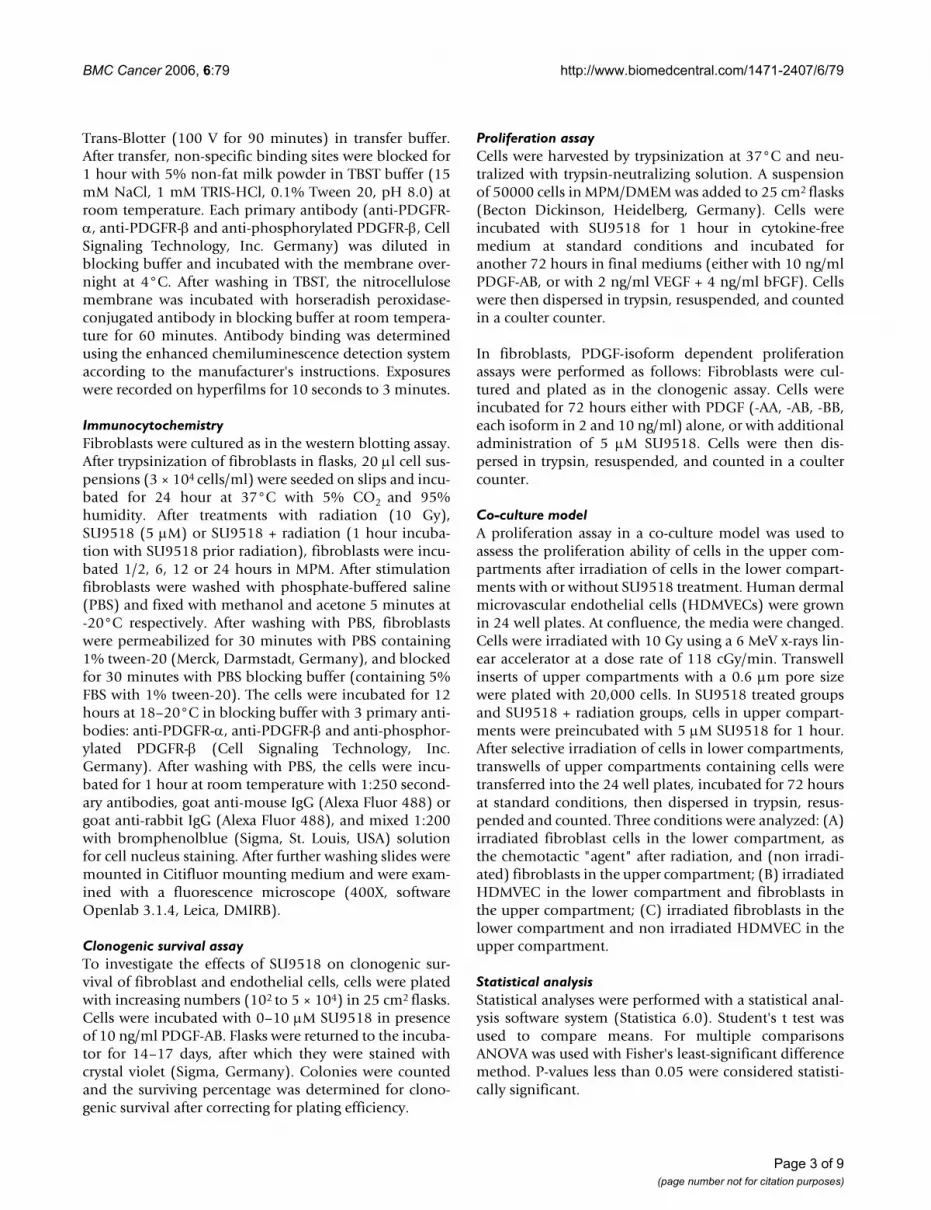
(a) Western-blotting analysis of PDGFR-α/-β and phosphorylated PDGFR-β (p-PDGFR-β)Figure 1(a) Western-blotting analysis of PDGFR-α/-β and phosphorylated PDGFR-β (p-PDGFR-β). Human dermal fibroblast cells were treated with or without 5 µM SU9518 for 1 hour before 10 Gy irradiation. Whole cell lysates of fibroblasts were collected for protein content and separated by SDS-PAGE and transferred to nitrocellulose. The amount of PDGFR and phosphorylated PDGFR was visualized with anti-PDGFR-α, anti-PDGFR-β or anti-phosphorylated PDGFR-β antibodies, respectively. Radiation-induced PDGFR phosphorylation was markedly inhibited by SU9518. (b) Immunocytochemistry of human primary dermal fibroblast cells. Fibroblasts were analyzed as untreated (control), after SU9518 (5 µM), irradiation (10Gy) or combined SU9518 and radiation treatment. The cells were incubated 6 hours post radiation and stained with the polyclonal rabbit anti-PDGFR-β/anti-p-PDGFR-β antibody followed by incubation with Alexa Fluor 488 goat anti-rabbit secondary antibody. For nuclear locali-zation the cells were counterstained with propidium jodide (red color). Immunofluorescence staining for PDGFR-β/p-PDGFR-β was indicated by green fluorescence. Irradiation-induced phosphorylation of PDGFR-β and that was substantially inhibited by SU9518. Scale bar presents 25 µm.

BMC Cancer 2006, 6:79 http://www.biomedcentral.com/1471-2407/6/79
ResultsPDGFR protein regulation in fibroblastsTo investigate the impact of radiation to PDGFR in fibrob-lasts and the effect of SU9518 on phosphorylation ofPDGF RTK in fibroblasts after radiation, human dermalfibroblast cells were pre-treated with 5 µM SU9518 onehour before 10 Gy irradiation and incubated for 30 min-utes (Figure 1a). PDGFR levels as well as PDGFR phos-
phorylation were determined by western-blotting usingprimary antibodies specific to PDGFR-α, PDGFR-β orphosphorylated PDGFR-β (p-PDGFR-β), respectively.Irradiation stimulated overexpression of both PDGFR-αand -β in fibroblasts, while expression of PDGFR-β wasmore upregulated than PDGFR-α. Moreover, irradiationresulted in significantly increased PDGFR-β phosphoryla-tion while SU9518 treatment particularly inhibited phos-phorylation of PDGFR-β.
ImmunocytochemistryTo investigate radiation-induced PDGF signaling infibroblasts in the presence or absence of SU9518, immu-nocytochemistry analyses were performed at various timepoints (1/2, 1, 6 hours) after irradiation. Irradiationresulted in enhanced phosphorylation of PDGFR-β (p-PDGFR-β) in fibroblasts. Representative microscopic pho-tos of fibroblasts with PDGFR-β or p-PDGFR-β stainingare presented at time points 1/2 and 6 hours after radia-tion, respectively (Fig. 2b). Importantly, the autophos-phorylation of PDGFR-β was markedly inhibited by 5 µMSU9518, consistent with the results in western-blottingassays.
Fibroblasts clonogenic survivalIn the clonogenic survival assay, stimulating effects ofPDGF (-AB isoform, 10 ng/ml) on fibroblast cells wereexamined in the absence or presence of SU9518 withincreasing concentrations (Figure 2a). Inhibition of PDGFsignaling by SU9518 reduced the clonogenic survival frac-tion of fibroblasts. The surviving fraction of fibroblastswas reduced by 10 µM SU9518 to 0.23 fold (Figure 3),suggesting that PDGF played a role for the clonogenic sur-vival of fibroblasts which in turn would explain why thesmall molecular RTK inhibitor SU9518 inhibits fibroblastclonogenic survival.
Endothelial cell clonogenic survivalIn the clonogenic survival assay (Figure 3a), the inhibitoryeffects of SU9518 on human dermal microvascularendothelial cells (HDMVECs) were examined in the pres-ence of PDGF (-AB isoform, 10 ng/ml). Inhibition ofPDGF signaling with SU9518 reduced the survival frac-tion of endothelial cells, which underscored the impor-tance of the PDGF pathway for the survival of endothelialcells. For example, using 5 µM SU9518, surviving fractionof endothelial cells decreased to 42% and further to 22%at 10 µM SU9518. Thus the PDGF-mediated clonogenicsurvival of endothelial cells was significantly inhibited bythe administration of SU9518 in a dose dependent man-ner.
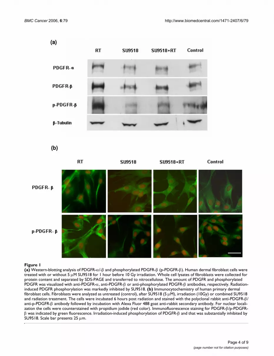
Endothelial cell proliferationTo further evaluate the specificity of SU9518, its ability toinhibit PDGF-stimulated endothelial proliferation was
(a) Clonogenic survival assay of dermal fibroblastsFigure 2(a) Clonogenic survival assay of dermal fibroblasts. Fibroblast cells (102 to 5 × 10⁴) were incubated with 0–10 µM SU9518 in presence of 10 ng/ml PDGF-AB. Cultures were returned to the incubator for 14–17 days, after which they were stained with crystal violet (Sigma, Germany). Colonies were counted and the surviving percentage was determined for clonogenic survival after correcting for plating efficiency. Bars are means of 5 experiments ± SD. (b) Clonogenic survival assay of human dermal microvascular endothelial cells (HDMVECs). Endothelial cells were treated with 0–10 µM SU9518 in the presence of 10 ng/ml PDGF-AB in the media. Surviving fraction was calculated based on the number of col-onies counted after 14–17 days. Bars are means of 5 experi-ments ± SD.
Page 5 of 9(page number not for citation purposes)
BMC Cancer 2006, 6:79 http://www.biomedcentral.com/1471-2407/6/79
measured and compared with its inhibitory potency toendothelial proliferation stimulated by VEGF and bFGF.We found that SU9518 inhibited 10 ng/ml PDGF-inducedendothelial proliferation with an IC50 of approximately0.9 µM. PDGF-induced proliferation was effectivelyblocked by SU9518, whereas endothelial proliferationstimulated by VEGF and bFGF was inhibited to a muchlesser degree by SU9518 (Figure 2b). For example, the cellnumber reduction by 1 µM SU9518 was 52% after PDGFstimulation versus 13% after stimulation by VEGF andbFGF.
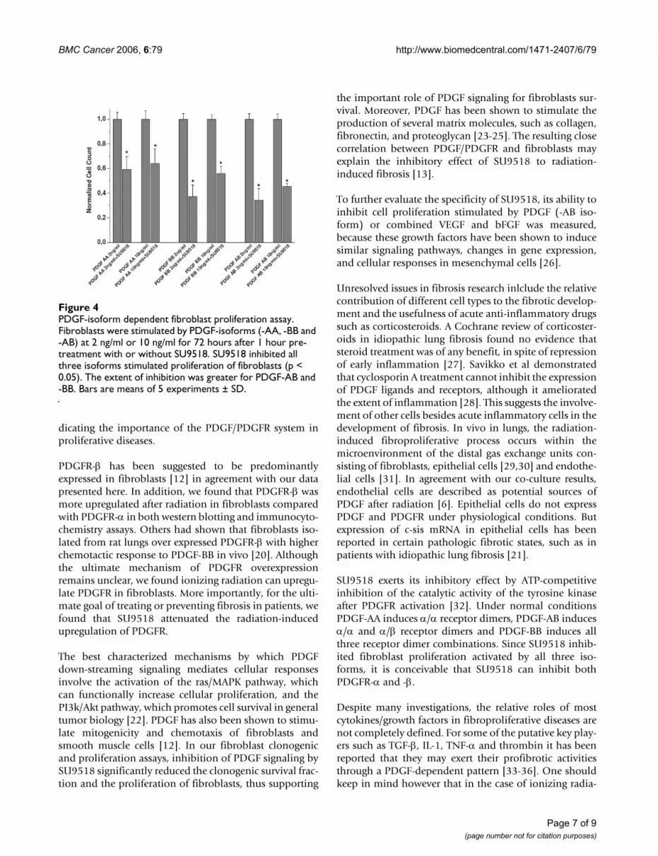
PDGF-isoforms dependent fibroblasts proliferationSeveral PDGF-isoforms are found to stimulate differentPDGFR subtypes. To further assess the effects of SU9518we investigated the PDGF-isoforms dependent inhibitionof fibroblast proliferation at 2 ng/ml or 10 ng/ml concen-trations of various PDGF-isoforms. We found thatSU9518 significantly inhibited fibroblast proliferationinduced by all three PDGF-isoforms at a concentration of5 µM (Figure 4). The inhibitory potency of SU9518 toPDGF-AB and -BB appeared to be slightly greater than -AA, while that to PDGF-AB was the most pronouncedamong the three isoforms.
Radiation-induced interactions between fibroblasts and endothelial cellsTo investigate the interactions between fibroblasts andendothelial cells a co-culture model was used. Irradiation
(10 Gy) of human dermal microvascular endothelial cells(HDMVECs) stimulated paracrine fibroblast proliferation(2.1 fold). Likewise, irradiation of fibroblast cells alsostimulated fibroblast cells and endothelial cells in the nonirradiated upper compartment up to 1.5 fold (Figure 5).Importantly, the paracrine fibroblast stimulation by eitherfibroblast or endothelial cells was markedly reduced withthe administration of SU9518 by approximately 50% (P <0.001). Thus SU9518 could significantly inhibit the acti-vation of radiation-induced direct and indirect fibroblastactivation.
DiscussionIn the context of fibrosis development in conventionalcancer therapy, including radio- and chemotherapy, westudied here the effects of SU9518, a receptor tyrosinekinase inhibitor of PDGF signaling on human primarydermal fibroblasts and endothelial cells. We had previ-ously shown that using this class of molecular targetedcompounds – including SU9518 and Imatinib (Gleevec)as well as SU11657 (similar to SU11248, a compoundcurrently in several clinical phase I and II anticancer trials)– can attenuate ionizing radiation-induced lung fibrosis invivo if it is given either prior to or after radiation insult[13]. Here we investigate in vitro whether primary humanendothelial and fibroblast cells are an important primarytarget of the RTK inhibitor in the context of PDGF signal-ing. We found that, in vitro, irradiation of fibroblasts andendothelium induced autocrine and paracrine PDGFexpression. Functionally, the isolated irradiation of pri-mary human endothelial cells stimulated proliferation inhuman fibroblasts in a co-culture model. Thus irradiationhas effects which can be described as an indirect promo-tion of fibroblast survival and proliferation. Importantly,RTK inhibitors could effectively attenuate this paracrineradiation-induced activation by blocking autophosphor-ylation of PDGFR and by downregulation of PDGFRexpression. Since fibroblasts are most likely key players inradiotherapy related fibrosis, and given the close correla-tion between PDGF signaling and fibroblasts activity (e.g.survival and proliferation), along with results in vivo [13]we conclude that fibroblasts may as well be a target of RTKinhibitors in the process of attenuating radiation-inducedfibrogenesis in vivo.
This conclusion is in agreement with earlier reports show-ing that PDGF is a potent mitogen and chemoattractantfor mesenchymal cells and also a chemoattractant for neu-trophils and monocytes [16-18]. Similarly, several studieshad reported that PDGF isoforms and their receptors wereupregulated during pathologic fibroproliferative diseasesin humans and in murine models by different types ofimpairment (e.g. radiation- [19,20]/bleomycin- [11]induced fibrosis, idiopathic pulmonary fibrosis [21]) vin-
Proliferation assay of HDMVECsFigure 3Proliferation assay of HDMVECs. Endothelial cells were plated in collagen I-coated flask and stimulated with 10 ng/ml PDGF (black line) or 2 ng/ml VEGF + 4 ng/ml bFGF (red line) in the absence or presence of SU9518 with increasing con-centration (0.1, 0.5, 1, 2 and 5 µM). 72 hours later cells were counted and normalized to respective controls without SU9518.
Page 6 of 9(page number not for citation purposes)
BMC Cancer 2006, 6:79 http://www.biomedcentral.com/1471-2407/6/79
dicating the importance of the PDGF/PDGFR system inproliferative diseases.
PDGFR-β has been suggested to be predominantlyexpressed in fibroblasts [12] in agreement with our datapresented here. In addition, we found that PDGFR-β wasmore upregulated after radiation in fibroblasts comparedwith PDGFR-α in both western blotting and immunocyto-chemistry assays. Others had shown that fibroblasts iso-lated from rat lungs over expressed PDGFR-β with higherchemotactic response to PDGF-BB in vivo [20]. Althoughthe ultimate mechanism of PDGFR overexpressionremains unclear, we found ionizing radiation can upregu-late PDGFR in fibroblasts. More importantly, for the ulti-mate goal of treating or preventing fibrosis in patients, wefound that SU9518 attenuated the radiation-inducedupregulation of PDGFR.
The best characterized mechanisms by which PDGFdown-streaming signaling mediates cellular responsesinvolve the activation of the ras/MAPK pathway, whichcan functionally increase cellular proliferation, and thePI3k/Akt pathway, which promotes cell survival in generaltumor biology [22]. PDGF has also been shown to stimu-late mitogenicity and chemotaxis of fibroblasts andsmooth muscle cells [12]. In our fibroblast clonogenicand proliferation assays, inhibition of PDGF signaling bySU9518 significantly reduced the clonogenic survival frac-tion and the proliferation of fibroblasts, thus supporting
the important role of PDGF signaling for fibroblasts sur-vival. Moreover, PDGF has been shown to stimulate theproduction of several matrix molecules, such as collagen,fibronectin, and proteoglycan [23-25]. The resulting closecorrelation between PDGF/PDGFR and fibroblasts mayexplain the inhibitory effect of SU9518 to radiation-induced fibrosis [13].
To further evaluate the specificity of SU9518, its ability toinhibit cell proliferation stimulated by PDGF (-AB iso-form) or combined VEGF and bFGF was measured,because these growth factors have been shown to inducesimilar signaling pathways, changes in gene expression,and cellular responses in mesenchymal cells [26].
Unresolved issues in fibrosis research inlclude the relativecontribution of different cell types to the fibrotic develop-ment and the usefulness of acute anti-inflammatory drugssuch as corticosteroids. A Cochrane review of corticoster-oids in idiopathic lung fibrosis found no evidence thatsteroid treatment was of any benefit, in spite of repressionof early inflammation [27]. Savikko et al demonstratedthat cyclosporin A treatment cannot inhibit the expressionof PDGF ligands and receptors, although it amelioratedthe extent of inflammation [28]. This suggests the involve-ment of other cells besides acute inflammatory cells in thedevelopment of fibrosis. In vivo in lungs, the radiation-induced fibroproliferative process occurs within themicroenvironment of the distal gas exchange units con-sisting of fibroblasts, epithelial cells [29,30] and endothe-lial cells [31]. In agreement with our co-culture results,endothelial cells are described as potential sources ofPDGF after radiation [6]. Epithelial cells do not expressPDGF and PDGFR under physiological conditions. Butexpression of c-sis mRNA in epithelial cells has beenreported in certain pathologic fibrotic states, such as inpatients with idiopathic lung fibrosis [21].
SU9518 exerts its inhibitory effect by ATP-competitiveinhibition of the catalytic activity of the tyrosine kinaseafter PDGFR activation [32]. Under normal conditionsPDGF-AA induces α/α receptor dimers, PDGF-AB inducesα/α and α/β receptor dimers and PDGF-BB induces allthree receptor dimer combinations. Since SU9518 inhib-ited fibroblast proliferation activated by all three iso-forms, it is conceivable that SU9518 can inhibit bothPDGFR-α and -β.
Despite many investigations, the relative roles of mostcytokines/growth factors in fibroproliferative diseases arenot completely defined. For some of the putative key play-ers such as TGF-β, IL-1, TNF-α and thrombin it has beenreported that they may exert their profibrotic activitiesthrough a PDGF-dependent pattern [33-36]. One shouldkeep in mind however that in the case of ionizing radia-
PDGF-isoform dependent fibroblast proliferation assayFigure 4PDGF-isoform dependent fibroblast proliferation assay. Fibroblasts were stimulated by PDGF-isoforms (-AA, -BB and -AB) at 2 ng/ml or 10 ng/ml for 72 hours after 1 hour pre-treatment with or without SU9518. SU9518 inhibited all three isoforms stimulated proliferation of fibroblasts (p < 0.05). The extent of inhibition was greater for PDGF-AB and -BB. Bars are means of 5 experiments ± SD.
Page 7 of 9(page number not for citation purposes)

BMC Cancer 2006, 6:79 http://www.biomedcentral.com/1471-2407/6/79
tion the signaling cascades in cells are complex and theresulting regulations may well be described as a networkof signaling [36-40]. Nevertheless, PDGF may be animportant node in this network and thus a key cytokineresponsible for the fibrotic development, suggesting thatperturbation of the PDGF/PDGFR system could providean effective means of inhibiting fibrosis.
ConclusionRadiation-induced autocrine and paracrine PDGF signal-ing plays an important role in fibroblast and endothelialcell activation and proliferation. PDGFR tyrosine kinaseinhibition reduces radiation-induced fibroblast activa-tion. This data supports the notion that SU9518 mightexert its antifibrotic activity in vivo via attenuation ofPDGF signaling. These results warrant further studiesinvestigating how tyrosine kinase inhibitors with anti-
PDGF property may attenuate cancer therapy relatedfibrosis in addition to their primary antitumoral activity.
Competing interestsThe author(s) declare that they have no competing inter-ests.
Authors' contributionsML participated in performing experiments, in data anal-ysis and in writing the manuscript. PG performed westernblots and immunocytochemistry. CP participated in writ-ing the manuscript. KEL participated in designing thestudy and in writing the manuscript. RK, KH and JD par-ticipated in writing the manuscript. TT performed the cellassays. AA and PEH designed the study, participated indata analyses and in writing the manuscript. All authorsedited and acknowledged the final version of the manu-script.
AcknowledgementsThis study was supported by the Research Program of the University of Heidelberg Medical School.
References1. Lasky JA, Coin PG, Lindroos PM, Ostrowski LE, Brody AR, Bonner JC:
Chrysotile asbestos stimulates platelet-derived growth fac-tor-AA production by rat lung fibroblasts in vitro: evidencefor an autocrine loop. Am J Respir Cell Mol Biol 1995, 12:162-170.
2. Trott KR, Herrmann T, Kasper M: Target cells in radiation pneu-mopathy. International Journal of Radiation Oncology*Biology*Physics2004, 58:463-469.
3. Plathow C, Li M, Gong P, Zieher H, Kiessling F, Peschke P, KauczorHU, Abdollahi A, Huber PE: Computed tomography monitoringof radiation-induced lung fibrosis in mice. Invest Radiol 2004,39:600-609.
4. Davis AM, Gerrand C, Griffin A, O'Sullivan B, Hill RP, Wunder JS,Abudu A, Bell RS: Evaluation of clinical utility of BTC-2000 formeasuring soft tissue fibrosis. International Journal of RadiationOncology*Biology*Physics 2004, 60:286-294.
5. Gridley DS, Bonnet RB, Bush DA, Franke C, Cheek GA, Slater JD,Slater JM: Time course of serum cytokines in patients receiv-ing proton or combined photon/proton beam radiation forresectable but medically inoperable non-small-cell lung can-cer. Int J Radiat Oncol Biol Phys 2004, 60:759-766.
6. Zerwes HG, Risau W: Polarized secretion of a platelet-derivedgrowth factor-like chemotactic factor by endothelial cells invitro. J Cell Biol 1987, 105:2037-2041.
7. Lindroos PM, Coin PG, Badgett A, Morgan DL, Bonner JC: Alveolarmacrophages stimulated with titanium dioxide, chrysotileasbestos, and residual oil fly ash upregulate the PDGF recep-tor-alpha on lung fibroblasts through an IL-1beta-dependentmechanism. Am J Respir Cell Mol Biol 1997, 16:283-292.
8. Iida H, Seifert R, Alpers CE, Gronwald RG, Phillips PE, Pritzl P, Gor-don K, Gown AM, Ross R, Bowen-Pope DF, Johson R: Platelet-derived growth factor (PDGF) and PDGF receptor areinduced in mesangial proliferative nephritis in the rat. ProcNatl Acad Sci USA 1991, 88:6560-6564.
9. Wilcox JN, Smith KM, Williams LT, Schwartz SM, Gordon D: Plate-let-derived growth factor mRNA detection in humanatherosclerotic plaques by in situ hybridization. J Clin Invest1988, 82:1134-1143.
10. Rice AB, Moomaw CR, Morgan DL, Bonner JC: Specific inhibitorsof platelet-derived growth factor or epidermal growth factorreceptor tyrosine kinase reduce pulmonary fibrosis in rats.Am J Pathol 1999, 155:213-221.
11. Zhuo Y, Zhang J, Laboy M, Lasky JA: Modulation of PDGF-C andPDGF-D expression during bleomycin-induced lung fibrosis.Am J Physiol Lung Cell Mol Physiol 2004, 286:L182-L188.
Co-culture experimentsFigure 5Co-culture experiments. Cells in lower compartments were selectively irradiated to induce paracrine proliferation of cells in upper compartments. Radiation (10Gy) of endothelial cells (HDMVECs) and fibroblast cells stimulated fibroblast prolif-eration (RT) in upper compartments. The administration of SU9518 significantly inhibited radiation-induced fibroblast proliferation. Likewise, irradiation of fibroblasts stimulated the proliferation of HDMVECs. The paracrine induction of endothelial cell proliferation was significantly inhibited by SU9518. The basal paracrine mitogenic stimulation of fibrob-lasts or endothelial cells in the upper compartment induced by co-culturing with non irradiated control cells in the lower compartment was also effectively inhibited by SU9518 (all SU9518 vs. control groups). Bars are mean ± SD. In all fig-ures asterisks indicate degree of statistical significance: *sig-nificant, P < 0.05; **highly significant P < 0.01.
Page 8 of 9(page number not for citation purposes)

BMC Cancer 2006, 6:79 http://www.biomedcentral.com/1471-2407/6/79
Publish with BioMed Central and every scientist can read your work free of charge
"BioMed Central will be the most significant development for disseminating the results of biomedical research in our lifetime."
Sir Paul Nurse, Cancer Research UK
Your research papers will be:
available free of charge to the entire biomedical community
peer reviewed and published immediately upon acceptance
cited in PubMed and archived on PubMed Central
yours — you keep the copyright
Submit your manuscript here:http://www.biomedcentral.com/info/publishing_adv.asp
BioMedcentral
12. Heldin CH, Westermark B: Mechanism of action and in vivo roleof platelet-derived growth factor. Physiol Rev 1999,79:1283-1316.
13. Abdollahi A, Li M, Ping G, Plathow C, Domhan S, Kiessling F, Lee LB,McMahon G, Grone HJ, Lipson KE, Huber PE: Inhibition of plate-let-derived growth factor signaling attenuates pulmonaryfibrosis. J Exp Med 2005, 201:925-935.
14. Abdollahi A, Lipson KE, Han X, Krempien R, Trinh T, Weber KJ, Hah-nfeldt P, Hlatky L, Debus J, Howlett AR, Huber PE: SU5416 andSU6668 attenuate the angiogenic effects of radiation-induced tumor cell growth factor production and amplify thedirect anti-endothelial action of radiation in vitro. Cancer Res2003, 63:3755-3763.
15. Luo J, Miller MW: Platelet-derived growth factor-mediated sig-nal transduction underlying astrocyte proliferation: site ofethanol action. J Neurosci 1999, 19:10014-10025.
16. Pierce GF, Vande BJ, Rudolph R, Tarpley J, Mustoe TA: Platelet-derived growth factor-BB and transforming growth factorbeta 1 selectively modulate glycosaminoglycans, collagen,and myofibroblasts in excisional wounds. Am J Pathol 1991,138:629-646.
17. Shure D, Senior RM, Griffin GL, Deuel TF: PDGF AA homodimersare potent chemoattractants for fibroblasts and neutrophils,and for monocytes activated by lymphocytes or cytokines.Biochem Biophys Res Commun 1992, 186:1510-1514.
18. Siegbahn A, Hammacher A, Westermark B, Heldin CH: Differentialeffects of the various isoforms of platelet-derived growth fac-tor on chemotaxis of fibroblasts, monocytes, and granulo-cytes. J Clin Invest 1990, 85:916-920.
19. Thornton SC, Walsh BJ, Bennett S, Robbins JM, Foulcher E, MorganGW, Penny R, Breit SN: Both in vitro and in vivo irradiation areassociated with induction of macrophage-derived fibroblastgrowth factors. Clin Exp Immunol 1996, 103:67-73.
20. Tada H, Ogushi F, Tani K, Nishioka Y, Miyata Jy, Sato K, Asano T,Sone S: Increased Binding and Chemotactic Capacities ofPDGF-BB on Fibroblasts in Radiation Pneumonitis. RadiationResearch 2003, 159:805-811.
21. Antoniades HN, Bravo MA, Avila RE, Galanopoulos T, Neville-GoldenJ, Maxwell M, Selman M: Platelet-derived growth factor in idio-pathic pulmonary fibrosis. J Clin Invest 1990, 86:1055-1064.
22. Franke TF, Yang SI, Chan TO, Datta K, Kazlauskas A, Morrison DK,Kaplan DR, Tsichlis PN: The protein kinase encoded by the Aktproto-oncogene is a target of the PDGF-activated phosphati-dylinositol 3-kinase. Cell 1995, 81:727-736.
23. Canalis E: Effect of platelet-derived growth factor on DNAand protein synthesis in cultured rat calvaria. Metabolism 1981,30:970-975.
24. Heldin P, Laurent TC, Heldin CH: Effect of growth factors onhyaluronan synthesis in cultured human fibroblasts. BiochemJ 1989, 258:919-922.
25. Blatti SP, Foster DN, Ranganathan G, Moses HL, Getz MJ: Inductionof fibronectin gene transcription and mRNA is a primaryresponse to growth-factor stimulation of AKR-2B cells. ProcNatl Acad Sci USA 1988, 85:1119-1123.
26. Fambrough D, McClure K, Kazlauskas A, Lander ES: Diverse signal-ing pathways activated by growth factor receptors inducebroadly overlapping, rather than independent, sets of genes.Cell 1999, 97:727-741.
27. Richeldi L, Davies HR, Ferrara G, Franco F: Corticosteroids for idi-opathic pulmonary fibrosis. Cochrane Database Syst Rev2003:CD002880.
28. Savikko J, Taskinen E, Von Willebrand E: Chronic allograft neph-ropathy is prevented by inhibition of platelet-derived growthfactor receptor: tyrosine kinase inhibitors as a potentialtherapy. Transplantation 2003, 75:1147-1153.
29. Lang DS, Jorres RA, Mucke M, Siegfried W, Magnussen H: Interac-tions between human bronchoepithelial cells and lungfibroblasts after ozone exposure in vitro. Toxicol Lett 1998, 96–97:13-24.
30. Demayo F, Minoo P, Plopper CG, Schuger L, Shannon J, Torday JS:Mesenchymal-epithelial interactions in lung developmentand repair: are modeling and remodeling the same process?Am J Physiol Lung Cell Mol Physiol 2002, 283:L510-L517.
31. Ward WF, Molteni A, Solliday NH, Jones GE: The relationshipbetween endothelial dysfunction and collagen accumulation
in irradiated rat lung. Int J Radiat Oncol Biol Phys 1985,11:1985-1990.
32. Yamasaki Y, Miyoshi K, Oda N, Watanabe M, Miyake H, Chan J, WangX, Sun L, Tang C, McMahon G, Lipson KE: Weekly dosing with theplatelet-derived growth factor receptor tyrosine kinaseinhibitor SU9518 significantly inhibits arterial stenosis. CircRes 2001, 88:630-636.
33. Allen JT, Spiteri MA: Growth factors in idiopathic pulmonaryfibrosis: relative roles. Respir Res 2002, 3:13.
34. Battegay EJ, Raines EW, Seifert RA, Bowen-Pope DF, Ross R: TGF-beta induces bimodal proliferation of connective tissue cellsvia complex control of an autocrine PDGF loop. Cell 1990,63:515-524.
35. Battegay EJ, Raines EW, Colbert T, Ross R: TNF-alpha stimulationof fibroblast proliferation. Dependence on platelet-derivedgrowth factor (PDGF) secretion and alteration of PDGFreceptor expression. J Immunol 1995, 154:6040-6047.
36. Raines EW, Dower SK, Ross R: Interleukin-1 mitogenic activityfor fibroblasts and smooth muscle cells is due to PDGF-AA.Science 1989, 20(243):393-396.
37. Abdollahi A, Hahnfeldt P, Maercker C, Grone HJ, Debus J, AnsorgeW, Folkman J, Hlatky L, Huber PE: Endostatin's antiangiogenicsignaling network. Mol Cell 2004, 13:649-663.
38. Abdollahi A, Griggs DW, Zieher H, Roth A, Lipson KE, Saffrich R,Grone HJ, Hallahan DE, Reisfeld RA, Debus J, Niethammer AG,Huber PE: Inhibition of alpha(v)beta3 integrin survival signal-ing enhances antiangiogenic and antitumor effects of radio-therapy. Clin Cancer Res 2005, 11:6270-6279.
39. Huber PE, Hauser K, Abdollahi A: Genome wide expression pro-filing of angiogenic signaling and the Heisenberg uncertaintyprinciple. Cell Cycle 2004, 3:1348-1351.
40. Huber PE, Bischof M, Jenne J, Heiland S, Peschke P, Saffrich R, GroneHJ, Debus J, Lipson KE, Abdollahi A: Trimodal cancer treatment:beneficial effects of combined antiangiogenesis, radiation,and chemotherapy. Cancer Res 2005, 65:3643-3655.
Pre-publication historyThe pre-publication history for this paper can be accessedhere:
http://www.biomedcentral.com/1471-2407/6/79/prepub
Page 9 of 9(page number not for citation purposes)
Copyright © 2022 FDOKUMEN