Small, Clonally Variant Antigens Expressed on the Surface of the Plasmodium falciparum- infected...
11
1393 J. Exp. Med. The Rockefeller University Press • 0022-1007/99/11/1393/11 $5.00 Volume 190, Number 10, November 15, 1999 1393–1403 http://www.jem.org Small, Clonally Variant Antigens Expressed on the Surface of the Plasmodium falciparum–infected Erythrocyte Are Encoded by the rif Gene Family and Are the Target of Human Immune Responses By Victor Fernandez,* Marcel Hommel, ‡ Qijun Chen,* Per Hagblom,* and Mats Wahlgren* From the *Microbiology and Tumor Biology Center, Karolinska Institutet, and the Swedish Institute for Infectious Disease Control, S-17177 Stockholm, Sweden; and the ‡ Department of Tropical Medicine and Infectious Diseases, Liverpool School of Tropical Medicine, Liverpool L3QA, United Kingdom Summary Disease severity in Plasmodium falciparum infections is a direct consequence of the parasite’s efficient evasion of the defense mechanisms of the human host. To date, one parasite-derived molecule, the antigenically variant adhesin P. falciparum erythrocyte membrane protein 1 (PfEMP1), is known to be transported to the infected erythrocyte (pRBC) surface, where it mediates binding to different host receptors. Here we report that multiple additional proteins are expressed by the parasite at the pRBC surface, including a large cluster of clonally variant antigens of 30–45 kD. We have found these antigens to be identical to the rifins, predicted polypeptides encoded by the rif multigene family. These parasite products, formerly called rosettins after their identification in rosetting para- sites, are prominently expressed by fresh isolates of P. falciparum. Rifins are immunogenic in natural infections and strain-specifically recognized by human immune sera in immunoprecipitation of surface-labeled pRBC extracts. Furthermore, human immune sera agglutinate pRBCs digested with trypsin at conditions such that radioiodinated PfEMP1 polypeptides are not detected but rifins are detected, suggesting the presence of epitopes in rifins targeted by agglutinating antibodies. When analyzed by two-dimensional electrophoresis, the rifins resolved into several isoforms in the pI range of 5.5–6.5, indicating molecular microheterogeneity, an additional potential novel source of antigenic diversity in P. falciparum. Prominent polypeptides of 20, 22, 76–80, 140, and 170 kD were also detected on the surfaces of pRBCs bearing in vitro–propagated or field-isolated parasites. In this report, we describe the rifins, the second family of clonally variant antigens known to be displayed by P. falciparum on the surface of the infected erythrocyte. Key words: P . falciparum • surface antigen • rif gene family • antibodies • strain-specific T he morbidity and mortality associated with Plasmodium falciparum malaria infections occurs exclusively during the erythrocytic phase of the parasite life cycle. Strategies used by P. falciparum for maximizing survival and prolifera- tive capacity in the bloodstream include the receptor-medi- ated sequestration of P. falciparum–infected erythrocytes (pRBCs) on the vascular endothelium and the constant vari- ation of antigenic epitopes on the pRBC surface to evade antiparasite mechanisms of protection in the human host (1). A polypeptide exported by the parasite to the outer face of the pRBC membrane, the 200–400-kD P. falciparum erythrocyte membrane protein 1 (PfEMP1), 1 is a cytoadher- ence ligand and undergoes clonal switching (2, 3). PfEMP1 polypeptides are encoded by the var gene family (4), and they are expressed on the infected cell surface as the para- site develops from the ring-shaped early forms into the pig- mented trophozoite stage, simultaneous with the onset of adhesive capacity and antigenicity of the pRBC (5). Studies of humoral immune responses in natural infec- tions of P. falciparum highlight the extreme diversity of anti- genic determinants on the infected erythrocytes (6–8). Al- though sera from individuals with a history of exposure to the disease may contain antibodies that react with epitopes shared by many parasite isolates, the bulk of the natural or experimentally induced immune response to surface deter- minants on the pRBC is strain/clone specific (9, 10). The variant antigen PfEMP1 has been postulated to be the sole target for specific antibodies that agglutinate pRBCs and confer protection against clinical disease (11, 12). 1 Abbreviations used in this paper: CHO, chinese hamster ovary; HUVECs, human umbilical vein endothelial cells; PECAM1, platelet/endothelial cell adhesion molecule 1; PfEMP1, P. falciparum erythrocyte membrane protein 1. on February 21, 2016 jem.rupress.org Downloaded from Published November 15, 1999
-
Upload
independent -
Category
Documents
-
view
3 -
download
0
Transcript of Small, Clonally Variant Antigens Expressed on the Surface of the Plasmodium falciparum- infected...
1393
J. Exp. Med.
The Rockefeller University Press • 0022-1007/99/11/1393/11 $5.00Volume 190, Number 10, November 15, 1999 1393–1403http://www.jem.org
Small, Clonally Variant Antigens Expressed on the Surface
of the
Plasmodium falciparum–
infected Erythrocyte Are Encoded by the
rif
Gene Family and Are the Target of HumanImmune Responses
By Victor Fernandez,
*
Marcel Hommel,
‡
Qijun Chen,
*
Per Hagblom,
*
and Mats Wahlgren
*
From the
*
Microbiology and Tumor Biology Center, Karolinska Institutet, and the Swedish Institute for Infectious Disease Control, S-17177 Stockholm, Sweden; and the
‡
Department of Tropical Medicine and Infectious Diseases, Liverpool School of Tropical Medicine, Liverpool L3QA, United Kingdom
Summary
Disease severity in
Plasmodium falciparum
infections is a direct consequence of the parasite’s efficientevasion of the defense mechanisms of the human host. To date, one parasite-derived molecule, theantigenically
variant adhesin
P. falciparum
erythrocyte membrane protein 1 (PfEMP1), is known tobe transported to the infected erythrocyte (pRBC) surface, where it mediates binding to differenthost receptors. Here we report that multiple additional proteins are expressed by the parasite at thepRBC surface, including a large cluster of clonally variant antigens of 30–45 kD. We have foundthese antigens to be identical to the rifins, predicted polypeptides encoded by the
rif
multigenefamily. These parasite products, formerly called rosettins after their identification in rosetting para-sites, are prominently expressed by fresh isolates of
P
.
falciparum
. Rifins are immunogenic in naturalinfections and strain-specifically recognized by human immune sera in immunoprecipitationof surface-labeled pRBC extracts. Furthermore, human immune sera agglutinate pRBCs digestedwith trypsin at conditions such that radioiodinated PfEMP1 polypeptides are not detected but rifinsare detected, suggesting the presence of epitopes in rifins targeted by agglutinating antibodies.When analyzed by two-dimensional electrophoresis, the rifins resolved into several isoforms in thepI range of 5.5–6.5, indicating molecular microheterogeneity, an additional potential novel sourceof antigenic diversity in
P
.
falciparum
. Prominent polypeptides of 20, 22, 76–80, 140, and 170 kDwere also detected on the surfaces of pRBCs bearing in vitro–propagated or field-isolated parasites.In this report, we describe the rifins, the second family of clonally variant antigens known to bedisplayed by
P
.
falciparum
on the surface of the infected erythrocyte.
Key words:
P
.
falciparum
• surface antigen •
rif
gene family • antibodies • strain-specific
T
he morbidity and mortality associated with
Plasmodiumfalciparum
malaria infections occurs exclusively duringthe erythrocytic phase of the parasite life cycle. Strategiesused by
P
.
falciparum
for maximizing survival and prolifera-tive capacity in the bloodstream include the receptor-medi-
ated sequestration of
P. falciparum–
infected erythrocytes(pRBCs) on the vascular endothelium and the constant vari-ation of antigenic epitopes on the pRBC surface to evadeantiparasite mechanisms of protection in the human host (1).
A polypeptide exported by the parasite to the outer face
of the pRBC membrane, the 200–400-kD
P
.
falciparum
erythrocyte membrane protein 1 (PfEMP1),
1
is a cytoadher-
ence ligand and undergoes clonal switching (2, 3). PfEMP1polypeptides are encoded by the
var
gene family (4), andthey are expressed on the infected cell surface as the para-site develops from the ring-shaped early forms into the pig-mented trophozoite stage, simultaneous with the onset ofadhesive capacity and antigenicity of the pRBC (5).
Studies of humoral immune responses in natural infec-tions of
P
.
falciparum
highlight the extreme diversity of anti-genic determinants on the infected erythrocytes (6–8). Al-though sera from individuals with a history of exposure tothe disease may contain antibodies that react with epitopesshared by many parasite isolates, the bulk of the natural orexperimentally induced immune response to surface deter-minants on the pRBC is strain/clone specific (9, 10). Thevariant antigen PfEMP1 has been postulated to be the soletarget for specific antibodies that agglutinate pRBCs andconfer protection against clinical disease (11, 12).
1
Abbreviations used in this paper:
CHO, chinese hamster ovary; HUVECs,human umbilical vein endothelial cells; PECAM1, platelet/endothelialcell adhesion molecule 1; PfEMP1,
P
.
falciparum
erythrocyte membraneprotein 1.
on February 21, 2016
jem.rupress.org
Dow
nloaded from
Published November 15, 1999
1394
P. falciparum
Exports Multiple Antigens to the Host Cell Surface
To date, PfEMP1 is the only molecularly characterizedprotein of
P
.
falciparum
shown to be located on the surfaceof the infected erythrocyte (2). PfEMP1 mediates bindingto vascular endothelial receptors such as CD36, intercellularadhesion molecule 1, and thrombospondin (13), as well as touninfected erythrocytes in the adhesion phenomenon knownas rosetting (14, 15). Polypeptides of low molecular masshave been radiolabeled on the pRBC surface and termedrosettins after their identification in rosetting strains (16).Based on this and additional observations suggesting thatthe parasite exports more than one set of polypeptides tothe host cell surface (reference 17 and our unpublished data),we set out to systematically reanalyze the surfaces of pRBCsharboring
P
.
falciparum
recently isolated from malaria pa-tients or strains and clones adapted to laboratory culture con-ditions. Here, we have focused the analysis and characteriza-tion to parasite-derived products with a molecular size
,
200kD, a size distinct from the known PfEMP1 antigens.
Materials and Methods
Parasites and Sera.
The
P
.
falciparum
lines FCR3S/b (K
2
) andFCR3S1/b (K
2
) were selected from parasites FCR3S (K
2
) andFCR3S1 (K
2
), respectively, for cytoadherence to the CD36receptor on C32 melanoma cells. FCR3S/a (K
2
) was obtainedfrom FCR3S by repeated selection for nonrosetting parasitesas described elsewhere (18). FCR3S1 was cloned by limitingdilution from FCR3S, which originated from the FCR3 strainisolated in The Gambia, West Africa. The subclones FCR3S1.2(K
2
) and FCR3S1.6 (K
2
) were obtained by micromanipulation fromFCR3S1. Clones TM284S2 (K
1
), TM284S3 (K
1
), TM284S11(K
1
), TM284S12 (K
1
), TM284S20 (K
1
), TM284S7 (K
2
),TM284S9 (K
2
), and TM284S19 (K
2
) were derived by micro-manipulation from the strain TM284 (K
1
), which, along withstrain TM180 (K
2
), were isolated from malaria patients in Thai-land. The F32 strain was isolated in Tanzania. The parasite 3D7was obtained by limiting dilution cloning of the isolate NF54,which was derived from a patient who acquired malaria in theairport area in Amsterdam, The Netherlands. R29 (K
1
) wascloned from ITOR, a parasite from the ITO strain selected forthe rosetting phenotype. The parasite Dd2 was originally clonedfrom the W2-MEF line of the Indochina III isolate. The
P
.
falci-parum
isolates 186, 199, 341, 347, 352, and 354 were part of alarger panel of field parasites collected from African children in-fected with malaria. Upon collection, the blood samples wereimmediately frozen according to standard techniques. For theiranalysis, the frozen blood samples were thawed and maintained inculture in their own blood for 24–30 h until parasites developedinto the mature trophozoite stage, at which time they were har-vested and further processed.
Sera were collected from (a) adults living in Yekepa, Liberia,an area characterized by high perennial malaria transmission (de-noted 022, 102, 119, 142, 163, 164, 169, 174, 179, 198, 241, and368), (b) adults from Fajara, The Gambia, a region with seasonalmalaria transmission (denoted 072, 100, and 136), and (c) chil-dren 1–15 yr old living in Saradidi, an area in western Kenya ho-loendemic for malaria (denoted 011, 039, 080, 118, and 209). Alldonors had had repeated malaria attacks; none had symptoms ofclinical malaria at the time of sampling. In all cases, informedconsent was obtained from the patients and/or their parents. Thesera were stored at
2
70
8
C and heat inactivated at 56
8
C before
the assays. Control sera were obtained from healthy Swedish blooddonors.
Culture of Parasites.
All of the laboratory-adapted parasitesused in this study were cultured in human group O Rh
1
erythro-cytes at 5% hematocrit with 10% AB
1
serum added to the malariaculture medium containing RPMI 1640, 25 mM Hepes, 25 mMsodium bicarbonate, and 50 mg/ml gentamycin, pH 7.4. In somecases, cultured parasites were enriched for the rosetting pheno-type (R
1
) as previously described (18).
Surface Analysis of pRBCs.
Surface iodination of pRBCs wasperformed by the lactoperoxidase/Na
125
I/H
2
O
2
method underconditions of minimal intracellular labeling. In brief, 2
3
10
9
cellsof a culture at 7–15% parasitemia with a majority of parasites in thetrophozoite stage were gently washed in PBS and resuspended to1 ml in PBS plus 1 mM KI. 1 mCi of Na
125
I (Amersham) and100
m
l of 2 mg/ml lactoperoxidase (Sigma Chemical Co.) wasadded, and the reaction was initiated by the addition of 25
m
l of0.03% H
2
O
2
. Four subsequent additions of 25
m
l of 0.03% H
2
O
2
were administered at 1-min intervals. Radioiodinated cells werewashed four times with ice-cold PBS containing 5 mM KI andresuspended in 1 ml of RPMI 1640 plus 5% sorbitol. Labeling ofintracellular hemoglobin accounted for
,
2% of total acid-precip-itable incorporated radiolabel. To disrupt rosettes/agglutinates,100 IU/ml of heparin (Løvens) was added to the cell suspension,and this was passed five times through a 23-gauge (0.6 mm inter-nal diameter) needle using a 1-ml syringe. The cell suspensionwas overlaid on top of a four-step (40, 60, 70, and 80%) Percollgradient in RPMI/5% sorbitol and centrifuged in a JA 20 rotor(Beckman Instruments, Inc.) at 10,000 rpm for 30 min at roomtemperature. Cells floating between the 40 and 60% Percoll layers(
.
95% mature parasite–containing erythrocytes) were recoveredand gently washed with PBS. Enriched pRBCs were first ex-tracted with 1% Triton X-100/PBS containing a cocktail of pro-tease inhibitors (1 mM EDTA-N
2
, 20
m
g/ml leupeptin, 0.7
m
g/ml pepstatin, 0.2 mM PMSF, and 50
m
g/ml aprotinin) and subse-quently extracted in a solution of 2% SDS and protease inhibitorsin PBS. The polypeptides in the Triton-insoluble fraction weresolubilized in 2% SDS-PAGE sample buffer and separated by agradient of 5–8.5 or 7.5–17.5% SDS-PAGE. The dried gels werescanned and analyzed using a PhosphorImager and ImageQuantanalysis software (Molecular Dynamics).
Immunoprecipitation.
SDS or Triton X-100 extracts of surface-radioiodinated pRBCs (25
m
l) were mixed with 375
m
l of NETT(50 mM Tris, pH 7.4, 150 mM NaCl, 5 mM disodium EDTA,20
m
g/ml leupeptin, 50
m
g/ml aprotinin, 0.02% sodium azide,and 0.5% Triton X-100) buffer/10 mg/ml IgG-free BSA (SigmaChemical Co.) and 100
m
l of 10% Triton X-100/PBS. 15
m
l of se-rum was added to 500
m
l of reconstituted extract, and the sampleswere incubated for 12 h at 4
8
C with rotation. Protein A–Sepharose(100
m
l of a 50% suspension in NETT buffer/10 mg/ml IgG-freeBSA) was added, and the samples were further incubated for 1 hat room temperature with rotation. The adsorbent was then washedtwice with 1 ml of NETT, twice with 1 ml of NETT/0.35 MNaCl, and twice with 1 ml of NETT. The Sepharose beads wereboiled for 4 min in the presence of 5% SDS-PAGE sample buffer,and the solubilized polypeptides were separated and analyzed asdescribed above.
Trypsin Treatment of the PRBC Surface.
Intact or radiolabeledpRBCs purified on Percoll gradients were digested with trypsin(Sigma Chemical Co.) at the indicated concentrations (see Fig. 5and Table I) for 10 min at 37
8
C. The reaction was terminated bythe addition of 1 ml soybean of trypsin inhibitor (Sigma ChemicalCo.) at 1 mg/ml in RPMI 1640/10% AB
1
serum. Cells were
on February 21, 2016
jem.rupress.org
Dow
nloaded from
Published November 15, 1999
1395
Fernandez et al.
washed with PBS and either resuspended in RPMI–Hepes/10%AB
1
serum for binding and agglutination assays or extracted withTriton X-100/SDS and analyzed by SDS-PAGE as described above.
Assay of Serum-induced Agglutination of pRBCs.
Agglutination ofparasitized red blood cells was assayed as previously described (19)with some modifications. pRBCs cultured to trophozoite and lateschizont stages were washed three times in malaria culture me-dium and then resuspended at 20% hematocrit. Aliquots of 25
m
lwere mixed with an equal volume of prediluted or nonpredilutedserum to a final dilution of 1:2, 1:5, or 1:10. The pRBC–serummixture was incubated at 37
8
C for 1 h under a constant rotationof 3 rpm. A 10-
m
l aliquot was mixed with 5
m
l of acridine orangeand mounted on a glass slide, and 50 consecutive fields of visionwere examined diagonally using a 40
3
lens in an incident UVlight microscope. Control sera from healthy Swedish blood donorswere used. Semiquantitative scoring was used for analysis. Theagglutination assay was scored negative when no agglutinates of4 pRBCs were detected; 11, 1–5 agglutinates of 4–10 pRBCs;21, .5 agglutinates of 4–10 pRBCs or 1–5 agglutinates of 11–20pRBCs; 31, .5 agglutinates of 11–20 pRBCs or 1–5 aggluti-nates of .20 pRBCs; 41, .5 agglutinates of .20 pRBCs.
Rosetting, Autoagglutination, and Cytoadherence. Rosetting of un-infected red cells and autoagglutination of parasitized erythrocyteswas assessed by direct staining of cultures with acridine orange(Sigma Chemical Co.) and examination using an epifluorescencemicroscope. Rosetting rates were measured as previously described(20). Spontaneous autoagglutination of pRBCs in the P. falci-parum cultures was measured as described elsewhere (18). Adher-ence of pRBCs to unfixed C32 melanoma cells, human umbilicalvein endothelial cells (HUVECs), chinese hamster ovary (CHO)cells transfected with CD36 or intercellular adhesion molecule 1,or L cells transfected with platelet/endothelial cell adhesion mol-ecule (PECAM)1/CD31 was performed as described (21). In somecytoadherence assays on HUVECs, C32, and L cells, rosetteswere first disrupted by adding 100 IU/ml of heparin (Løvens) tothe culture and passage (five times) through a 23-gauge (0.6 mminternal diameter) needle using a 1-ml syringe. The cell suspen-sion was overlaid on a four-step (40, 60, 70, and 80%) Percollgradient, and the trophozoite-bearing red cells were selected asdescribed above, washed twice in RPMI, and used for the assaysin serum-free medium.
Surface Immunofluorescence. The assay of the binding of humannonimmune normal Swedish serum Igs to pRBCs was performedby direct labeling of the cells using FITC-conjugated sheep anti–human Ig antibodies (State Bacteriology Laboratory, Sweden) asdescribed elsewhere (22).
ResultsNovel Variable Polypeptides Identified on the pRBC Surface.
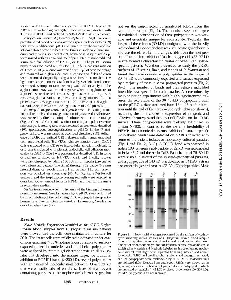
Frozen blood samples from P. falciparum malaria patientswere thawed, and the cells were maintained in culture for30 h. The intact cells were mildly radioiodinated under con-ditions ensuring .98% isotope incorporation to surface-exposed molecular moieties, and the labeled polypeptideswere analyzed by protein gel electrophoresis. In all six iso-lates that developed into the mature stages, we found, inaddition to PfEMP1 bands (.200 kD), several polypeptideswith an estimated molecular mass between 33 and 39 kDthat were readily labeled on the surfaces of erythrocytescontaining parasites at the trophozoite/schizont stages, but
not on the ring-infected or uninfected RBCs from thesame blood sample (Fig. 1). The number, size, and degreeof radiolabel incorporation of these polypeptides was vari-able and essentially unique for each isolate. Typically, thelargest of these bands (39 kD) comigrated with the heavilyradioiodinated monomer chains of erythrocytic glycophorin Aand was therefore often indistinguishable from the host pro-tein. One to three additional labeled polypeptides 33–37 kDin size formed a characteristic cluster of bands with isolate-specific patterns. We then proceeded to study the pRBCsurfaces of 17 strains, lines, and clones of P. falciparum andfound that radioiodinatable polypeptides in the range of30–45 kD were commonly exported and surface expressedby a majority of these in vitro–propagated parasites (Fig. 2,A–C). The number of bands and their relative radiolabelintensities was specific for each parasite. As determined byradioiodination experiments with highly synchronized cul-tures, the expression of the 30–45-kD polypeptide clusteron the pRBC surface occurred from 16 to 18 h after inva-sion until the end of the erythrocytic cycle (data not shown),matching the time course of expression of antigenic andadhesive phenotypes and the onset of PfEMP1 on the pRBCsurface. These polypeptides were partially solubilized inTriton X-100, in contrast to the extreme insolubility ofPfEMP1 in nonionic detergents. Additional parasite-specificradiolabeled bands were detected on pRBCs infected withsome of the patient isolates or laboratory-cultured parasites(Fig. 1 and Fig. 2, A–C). A 20-kD band was observed inisolate 199, whereas a polypeptide of 22 kD was radiolabeledin isolate 347 and the strain Dd2. Faint bands of 76–80 kDwere visible in several of the in vitro–propagated parasites,and a polypeptide of 140 kD was detected in TM180, a strainalso expressing several smaller (33–39 kD) polypeptides. Most
Figure 1. Novel variable antigens expressed on the surfaces of erythro-cytes harboring clinical isolates of P. falciparum. Frozen blood samplesfrom malaria patients were thawed, maintained in culture until the devel-opment of trophozoite stages, and subsequently surface-radioiodinated asexplained in Materials and Methods. Labeled erythrocytes bearing tropho-zoite and schizont stages were separated from ring-infected and nonin-fected cells (RBC) in Percoll/sorbitol gradients and detergent extracted,and the polypeptides were fractionated by SDS-PAGE. Molecular sizesare indicated (kD). Extracts from autologous RBCs were always run inadjoining lanes for identification of parasite-derived polypeptides, whichare indicated by asterisks (,45 kD) or closed arrowheads (100–200 kD).PfEMP1 polypeptides are not indicated.
on February 21, 2016
jem.rupress.org
Dow
nloaded from
Published November 15, 1999
1396 P. falciparum Exports Multiple Antigens to the Host Cell Surface
prominent was the strongly radioiodinated band of 170 kDfound in two of the clinical isolates (199 and 341) andstrain F32, as well as strain TM284 and several of its clones(only TM284S2 is shown). None of these polypeptideswere detected on pRBCs bearing trophozoite/schizont stageswhen the erythrocytes were surface 125I-labeled before par-asite invasion (data not shown).
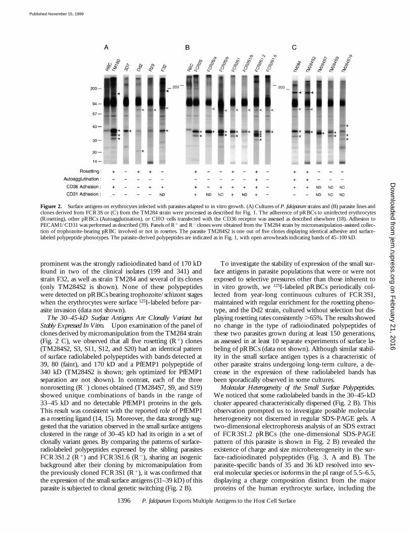
The 30–45-kD Surface Antigens Are Clonally Variant butStably Expressed In Vitro. Upon examination of the panel ofclones derived by micromanipulation from the TM284 strain(Fig. 2 C), we observed that all five rosetting (R1) clones(TM284S2, S3, S11, S12, and S20) had an identical patternof surface radiolabeled polypeptides with bands detected at39, 80 (faint), and 170 kD and a PfEMP1 polypeptide of340 kD (TM284S2 is shown; gels optimized for PfEMP1separation are not shown). In contrast, each of the threenonrosetting (R2) clones obtained (TM284S7, S9, and S19)showed unique combinations of bands in the range of33–45 kD and no detectable PfEMP1 proteins in the gels.This result was consistent with the reported role of PfEMP1as a rosetting ligand (14, 15). Moreover, the data strongly sug-gested that the variation observed in the small surface antigensclustered in the range of 30–45 kD had its origin in a set ofclonally variant genes. By comparing the patterns of surface-radiolabeled polypeptides expressed by the sibling parasitesFCR3S1.2 (R1) and FCR3S1.6 (R2), sharing an isogenicbackground after their cloning by micromanipulation fromthe previously cloned FCR3S1 (R1), it was confirmed thatthe expression of the small surface antigens (31–39 kD) of thisparasite is subjected to clonal genetic switching (Fig. 2 B).
To investigate the stability of expression of the small sur-face antigens in parasite populations that were or were notexposed to selective pressures other than those inherent toin vitro growth, we 125I-labeled pRBCs periodically col-lected from year-long continuous cultures of FCR3S1,maintained with regular enrichment for the rosetting pheno-type, and the Dd2 strain, cultured without selection but dis-playing rosetting rates consistently .65%. The results showedno change in the type of radioiodinated polypeptides ofthese two parasites grown during at least 150 generations,as assessed in at least 10 separate experiments of surface la-beling of pRBCs (data not shown). Although similar stabil-ity in the small surface antigen types is a characteristic ofother parasite strains undergoing long-term culture, a de-crease in the expression of these radiolabeled bands hasbeen sporadically observed in some cultures.
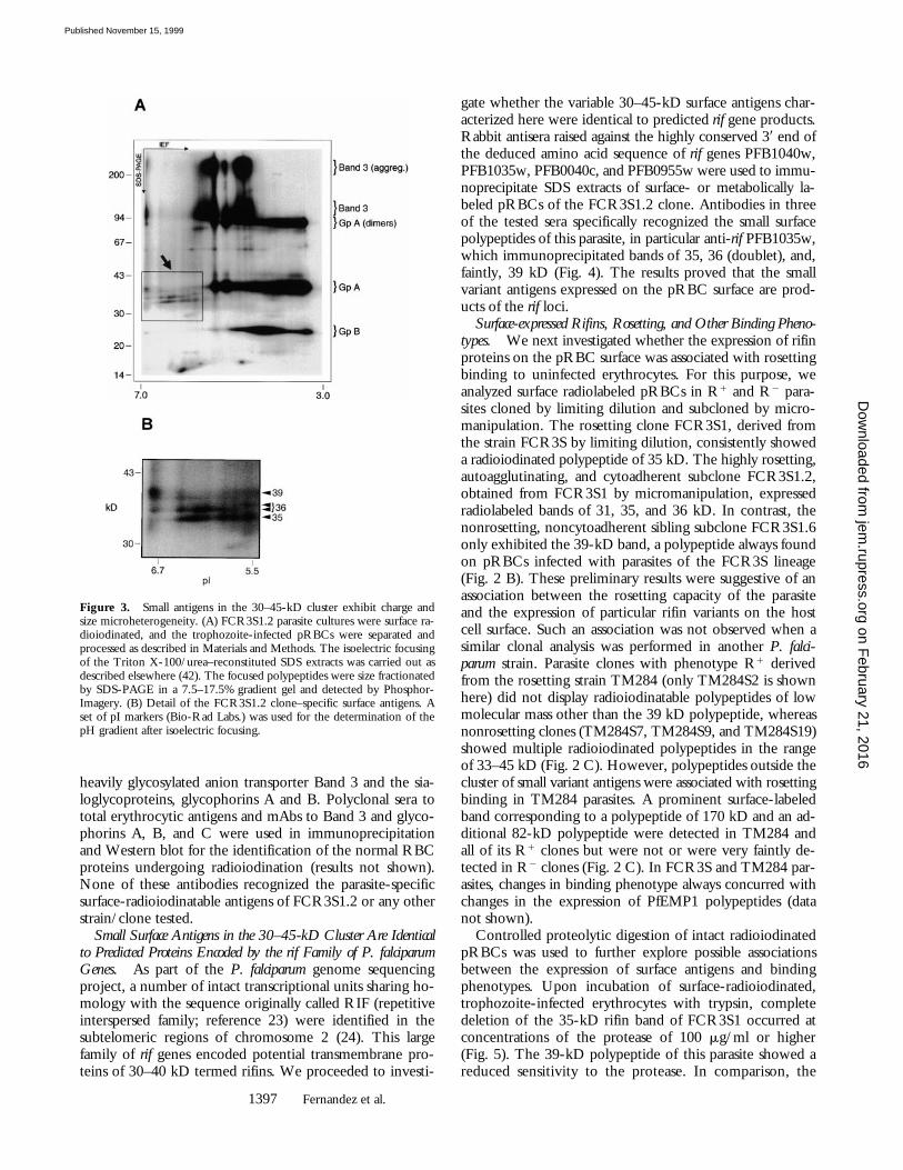
Molecular Heterogeneity of the Small Surface Polypeptides.We noticed that some radiolabeled bands in the 30–45-kDcluster appeared characteristically dispersed (Fig. 2 B). Thisobservation prompted us to investigate possible molecularheterogeneity not discerned in regular SDS-PAGE gels. Atwo-dimensional electrophoresis analysis of an SDS extractof FCR3S1.2 pRBCs (the one-dimensional SDS-PAGEpattern of this parasite is shown in Fig. 2 B) revealed theexistence of charge and size microheterogeneity in the sur-face-radioiodinated polypeptides (Fig. 3, A and B). Theparasite-specific bands of 35 and 36 kD resolved into sev-eral molecular species or isoforms in the pI range of 5.5–6.5,displaying a charge composition distinct from the majorproteins of the human erythrocyte surface, including the
Figure 2. Surface antigens on erythrocytes infected with parasites adapted to in vitro growth. (A) Cultures of P. falciparum strains and (B) parasite lines andclones derived from FCR3S or (C) from the TM284 strain were processed as described for Fig. 1. The adherence of pRBCs to uninfected erythrocytes(Rosetting), other pRBCs (Autoagglutination), or CHO cells transfected with the CD36 receptor was assessed as described elsewhere (18). Adhesion toPECAM1/CD31 was performed as described (39). Panels of R1 and R2 clones were obtained from the TM284 strain by micromanipulation-assisted collec-tion of trophozoite-bearing pRBC involved or not in rosettes. The parasite TM284S2 is one out of five clones displaying identical adhesive and surface-labeled polypeptide phenotypes. The parasite-derived polypeptides are indicated as in Fig. 1, with open arrowheads indicating bands of 45–100 kD.
on February 21, 2016
jem.rupress.org
Dow
nloaded from
Published November 15, 1999
1397 Fernandez et al.
heavily glycosylated anion transporter Band 3 and the sia-loglycoproteins, glycophorins A and B. Polyclonal sera tototal erythrocytic antigens and mAbs to Band 3 and glyco-phorins A, B, and C were used in immunoprecipitationand Western blot for the identification of the normal RBCproteins undergoing radioiodination (results not shown).None of these antibodies recognized the parasite-specificsurface-radioiodinatable antigens of FCR3S1.2 or any otherstrain/clone tested.
Small Surface Antigens in the 30–45-kD Cluster Are Identicalto Predicted Proteins Encoded by the rif Family of P. falciparumGenes. As part of the P. falciparum genome sequencingproject, a number of intact transcriptional units sharing ho-mology with the sequence originally called RIF (repetitiveinterspersed family; reference 23) were identified in thesubtelomeric regions of chromosome 2 (24). This largefamily of rif genes encoded potential transmembrane pro-teins of 30–40 kD termed rifins. We proceeded to investi-
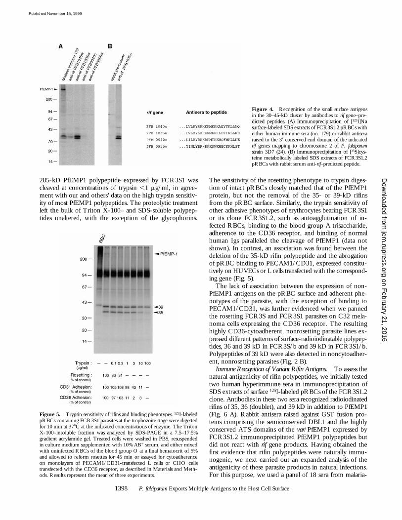
gate whether the variable 30–45-kD surface antigens char-acterized here were identical to predicted rif gene products.Rabbit antisera raised against the highly conserved 39 end ofthe deduced amino acid sequence of rif genes PFB1040w,PFB1035w, PFB0040c, and PFB0955w were used to immu-noprecipitate SDS extracts of surface- or metabolically la-beled pRBCs of the FCR3S1.2 clone. Antibodies in threeof the tested sera specifically recognized the small surfacepolypeptides of this parasite, in particular anti-rif PFB1035w,which immunoprecipitated bands of 35, 36 (doublet), and,faintly, 39 kD (Fig. 4). The results proved that the smallvariant antigens expressed on the pRBC surface are prod-ucts of the rif loci.
Surface-expressed Rifins, Rosetting, and Other Binding Pheno-types. We next investigated whether the expression of rifinproteins on the pRBC surface was associated with rosettingbinding to uninfected erythrocytes. For this purpose, weanalyzed surface radiolabeled pRBCs in R1 and R2 para-sites cloned by limiting dilution and subcloned by micro-manipulation. The rosetting clone FCR3S1, derived fromthe strain FCR3S by limiting dilution, consistently showeda radioiodinated polypeptide of 35 kD. The highly rosetting,autoagglutinating, and cytoadherent subclone FCR3S1.2,obtained from FCR3S1 by micromanipulation, expressedradiolabeled bands of 31, 35, and 36 kD. In contrast, thenonrosetting, noncytoadherent sibling subclone FCR3S1.6only exhibited the 39-kD band, a polypeptide always foundon pRBCs infected with parasites of the FCR3S lineage(Fig. 2 B). These preliminary results were suggestive of anassociation between the rosetting capacity of the parasiteand the expression of particular rifin variants on the hostcell surface. Such an association was not observed when asimilar clonal analysis was performed in another P. falci-parum strain. Parasite clones with phenotype R1 derivedfrom the rosetting strain TM284 (only TM284S2 is shownhere) did not display radioiodinatable polypeptides of lowmolecular mass other than the 39 kD polypeptide, whereasnonrosetting clones (TM284S7, TM284S9, and TM284S19)showed multiple radioiodinated polypeptides in the rangeof 33–45 kD (Fig. 2 C). However, polypeptides outside thecluster of small variant antigens were associated with rosettingbinding in TM284 parasites. A prominent surface-labeledband corresponding to a polypeptide of 170 kD and an ad-ditional 82-kD polypeptide were detected in TM284 andall of its R1 clones but were not or were very faintly de-tected in R2 clones (Fig. 2 C). In FCR3S and TM284 par-asites, changes in binding phenotype always concurred withchanges in the expression of PfEMP1 polypeptides (datanot shown).
Controlled proteolytic digestion of intact radioiodinatedpRBCs was used to further explore possible associationsbetween the expression of surface antigens and bindingphenotypes. Upon incubation of surface-radioiodinated,trophozoite-infected erythrocytes with trypsin, completedeletion of the 35-kD rifin band of FCR3S1 occurred atconcentrations of the protease of 100 mg/ml or higher(Fig. 5). The 39-kD polypeptide of this parasite showed areduced sensitivity to the protease. In comparison, the
Figure 3. Small antigens in the 30–45-kD cluster exhibit charge andsize microheterogeneity. (A) FCR3S1.2 parasite cultures were surface ra-dioiodinated, and the trophozoite-infected pRBCs were separated andprocessed as described in Materials and Methods. The isoelectric focusingof the Triton X-100/urea–reconstituted SDS extracts was carried out asdescribed elsewhere (42). The focused polypeptides were size fractionatedby SDS-PAGE in a 7.5–17.5% gradient gel and detected by Phosphor-Imagery. (B) Detail of the FCR3S1.2 clone–specific surface antigens. Aset of pI markers (Bio-Rad Labs.) was used for the determination of thepH gradient after isoelectric focusing.
on February 21, 2016
jem.rupress.org
Dow
nloaded from
Published November 15, 1999
1398 P. falciparum Exports Multiple Antigens to the Host Cell Surface
285-kD PfEMP1 polypeptide expressed by FCR3S1 wascleaved at concentrations of trypsin ,1 mg/ml, in agree-ment with our and others’ data on the high trypsin sensitiv-ity of most PfEMP1 polypeptides. The proteolytic treatmentleft the bulk of Triton X-100– and SDS-soluble polypep-tides unaltered, with the exception of the glycophorins.
The sensitivity of the rosetting phenotype to trypsin diges-tion of intact pRBCs closely matched that of the PfEMP1protein, but not the removal of the 35- or 39-kD rifinsfrom the pRBC surface. Similarly, the trypsin sensitivity ofother adhesive phenotypes of erythrocytes bearing FCR3S1or its clone FCR3S1.2, such as autoagglutination of in-fected RBCs, binding to the blood group A trisaccharide,adherence to the CD36 receptor, and binding of normalhuman Igs paralleled the cleavage of PfEMP1 (data notshown). In contrast, an association was found between thedeletion of the 35-kD rifin polypeptide and the abrogationof pRBC binding to PECAM1/CD31, expressed constitu-tively on HUVECs or L cells transfected with the correspond-ing gene (Fig. 5).
The lack of association between the expression of non-PfEMP1 antigens on the pRBC surface and adherent phe-notypes of the parasite, with the exception of binding toPECAM1/CD31, was further evidenced when we pannedthe rosetting FCR3S and FCR3S1 parasites on C32 mela-noma cells expressing the CD36 receptor. The resultinghighly CD36-cytoadherent, nonrosetting parasite lines ex-pressed different patterns of surface-radioiodinatable polypep-tides, 36 and 39 kD in FCR3S/b and 39 kD in FCR3S1/b.Polypeptides of 39 kD were also detected in noncytoadher-ent, nonrosetting parasites (Fig. 2 B).
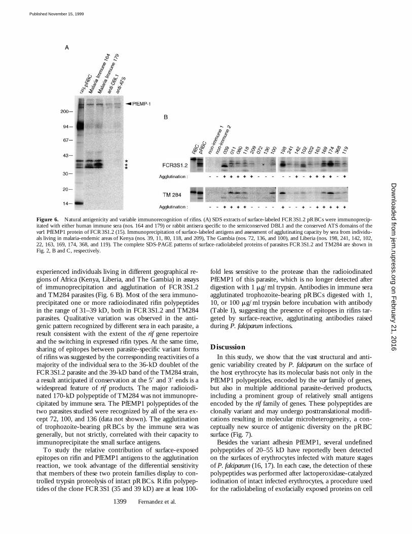
Immune Recognition of Variant Rifin Antigens. To assess thenatural antigenicity of rifin polypeptides, we initially testedtwo human hyperimmune sera in immunoprecipitation ofSDS extracts of surface 125I-labeled pRBCs of the FCR3S1.2clone. Antibodies in these two sera recognized radioiodinatedrifins of 35, 36 (doublet), and 39 kD in addition to PfEMP1(Fig. 6 A). Rabbit antisera raised against GST fusion pro-teins comprising the semiconserved DBL1 and the highlyconserved ATS domains of the var/PfEMP1 expressed byFCR3S1.2 immunoprecipitated PfEMP1 polypeptides butdid not react with rif gene products. Having obtained thefirst evidence that rifin polypeptides were naturally immu-nogenic, we next carried out an expanded analysis of theantigenicity of these parasite products in natural infections.For this purpose, we used a panel of 18 sera from malaria-
Figure 4. Recognition of the small surface antigensin the 30–45-kD cluster by antibodies to rif gene–pre-dicted peptides. (A) Immunoprecipitation of [125I]Nasurface-labeled SDS extracts of FCR3S1.2 pRBCs witheither human immune sera (no. 179) or rabbit antiseraraised to the 39 conserved end domain of the indicatedrif genes mapping to chromosome 2 of P. falciparumstrain 3D7 (24). (B) Immunoprecipitation of [35S]cys-teine metabolically labeled SDS extracts of FCR3S1.2pRBCs with rabbit serum anti-rif–predicted peptide.
Figure 5. Trypsin sensitivity of rifins and binding phenotypes. 125I-labeledpRBCs containing FCR3S1 parasites at the trophozoite stage were digestedfor 10 min at 378C at the indicated concentrations of enzyme. The TritonX-100–insoluble fraction was analyzed by SDS-PAGE in a 7.5–17.5%gradient acrylamide gel. Treated cells were washed in PBS, resuspendedin culture medium supplemented with 10% AB1 serum, and either mixedwith uninfected RBCs of the blood group O at a final hematocrit of 5%and allowed to reform rosettes for 45 min or assayed for cytoadherenceon monolayers of PECAM1/CD31-transfected L cells or CHO cellstransfected with the CD36 receptor, as described in Materials and Meth-ods. Results represent the mean of three experiments.
on February 21, 2016
jem.rupress.org
Dow
nloaded from
Published November 15, 1999
1399 Fernandez et al.
experienced individuals living in different geographical re-gions of Africa (Kenya, Liberia, and The Gambia) in assaysof immunoprecipitation and agglutination of FCR3S1.2and TM284 parasites (Fig. 6 B). Most of the sera immuno-precipitated one or more radioiodinated rifin polypeptidesin the range of 31–39 kD, both in FCR3S1.2 and TM284parasites. Qualitative variation was observed in the anti-genic pattern recognized by different sera in each parasite, aresult consistent with the extent of the rif gene repertoireand the switching in expressed rifin types. At the same time,sharing of epitopes between parasite-specific variant formsof rifins was suggested by the corresponding reactivities of amajority of the individual sera to the 36-kD doublet of theFCR3S1.2 parasite and the 39-kD band of the TM284 strain,a result anticipated if conservation at the 59 and 39 ends is awidespread feature of rif products. The major radioiodi-nated 170-kD polypeptide of TM284 was not immunopre-cipitated by immune sera. The PfEMP1 polypeptides of thetwo parasites studied were recognized by all of the sera ex-cept 72, 100, and 136 (data not shown). The agglutinationof trophozoite-bearing pRBCs by the immune sera wasgenerally, but not strictly, correlated with their capacity toimmunoprecipitate the small surface antigens.
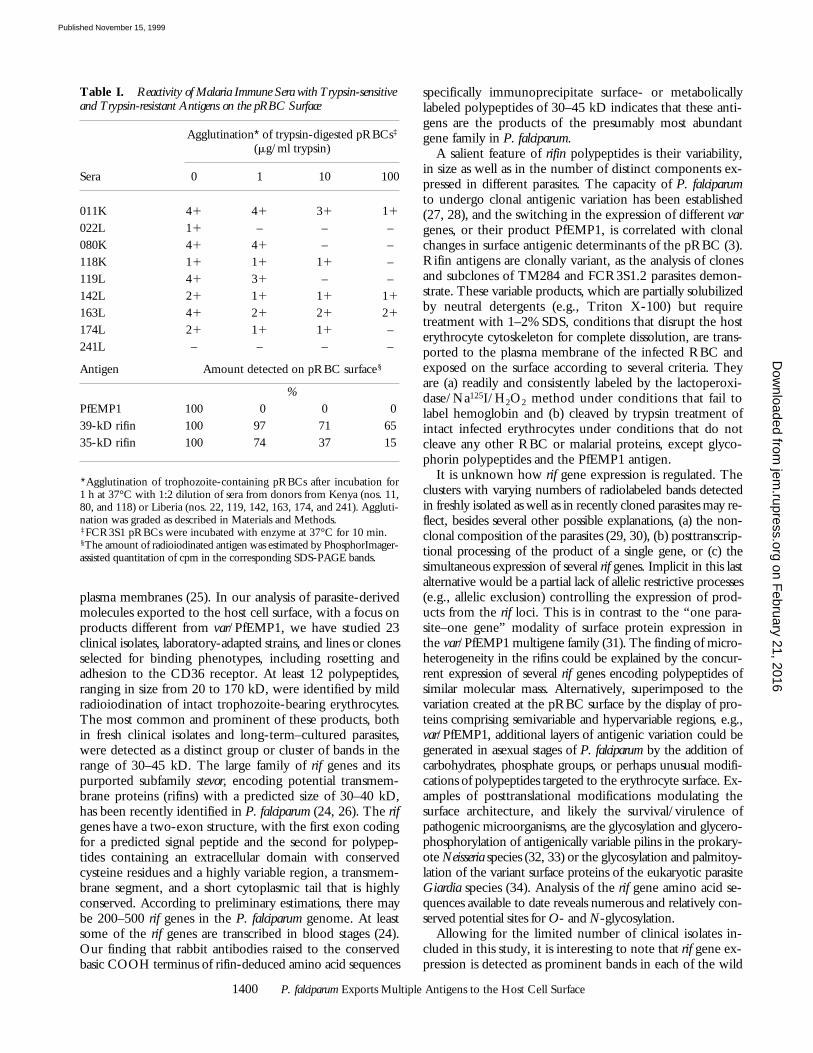
To study the relative contribution of surface-exposedepitopes on rifin and PfEMP1 antigens to the agglutinationreaction, we took advantage of the differential sensitivitythat members of these two protein families display to con-trolled trypsin proteolysis of intact pRBCs. Rifin polypep-tides of the clone FCR3S1 (35 and 39 kD) are at least 100-
fold less sensitive to the protease than the radioiodinatedPfEMP1 of this parasite, which is no longer detected afterdigestion with 1 mg/ml trypsin. Antibodies in immune seraagglutinated trophozoite-bearing pRBCs digested with 1,10, or 100 mg/ml trypsin before incubation with antibody(Table I), suggesting the presence of epitopes in rifins tar-geted by surface-reactive, agglutinating antibodies raisedduring P. falciparum infections.
DiscussionIn this study, we show that the vast structural and anti-
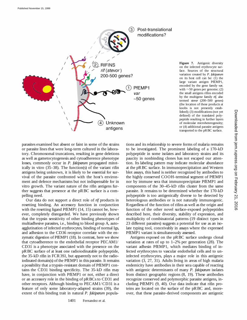
genic variability created by P. falciparum on the surface ofthe host erythrocyte has its molecular basis not only in thePfEMP1 polypeptides, encoded by the var family of genes,but also in multiple additional parasite-derived products,including a prominent group of relatively small antigensencoded by the rif family of genes. These polypeptides areclonally variant and may undergo posttranslational modifi-cations resulting in molecular microheterogeneity, a con-ceptually new source of antigenic diversity on the pRBCsurface (Fig. 7).
Besides the variant adhesin PfEMP1, several undefinedpolypeptides of 20–55 kD have reportedly been detectedon the surfaces of erythrocytes infected with mature stagesof P. falciparum (16, 17). In each case, the detection of thesepolypeptides was performed after lactoperoxidase-catalyzediodination of intact infected erythrocytes, a procedure usedfor the radiolabeling of exofacially exposed proteins on cell
Figure 6. Natural antigenicity and variable immunorecognition of rifins. (A) SDS extracts of surface-labeled FCR3S1.2 pRBCs were immunoprecip-itated with either human immune sera (nos. 164 and 179) or rabbit antisera specific to the semiconserved DBL1 and the conserved ATS domains of thevar1 PfEMP1 protein of FCR3S1.2 (15). Immunoprecipitation of surface-labeled antigens and assessment of agglutinating capacity by sera from individu-als living in malaria-endemic areas of Kenya (nos. 39, 11, 80, 118, and 209), The Gambia (nos. 72, 136, and 100), and Liberia (nos. 198, 241, 142, 102,22, 163, 169, 174, 368, and 119). The complete SDS-PAGE patterns of surface-radiolabeled proteins of parasites FCR3S1.2 and TM284 are shown inFig. 2, B and C, respectively.
on February 21, 2016
jem.rupress.org
Dow
nloaded from
Published November 15, 1999
1400 P. falciparum Exports Multiple Antigens to the Host Cell Surface
plasma membranes (25). In our analysis of parasite-derivedmolecules exported to the host cell surface, with a focus onproducts different from var/PfEMP1, we have studied 23clinical isolates, laboratory-adapted strains, and lines or clonesselected for binding phenotypes, including rosetting andadhesion to the CD36 receptor. At least 12 polypeptides,ranging in size from 20 to 170 kD, were identified by mildradioiodination of intact trophozoite-bearing erythrocytes.The most common and prominent of these products, bothin fresh clinical isolates and long-term–cultured parasites,were detected as a distinct group or cluster of bands in therange of 30–45 kD. The large family of rif genes and itspurported subfamily stevor, encoding potential transmem-brane proteins (rifins) with a predicted size of 30–40 kD,has been recently identified in P. falciparum (24, 26). The rifgenes have a two-exon structure, with the first exon codingfor a predicted signal peptide and the second for polypep-tides containing an extracellular domain with conservedcysteine residues and a highly variable region, a transmem-brane segment, and a short cytoplasmic tail that is highlyconserved. According to preliminary estimations, there maybe 200–500 rif genes in the P. falciparum genome. At leastsome of the rif genes are transcribed in blood stages (24).Our finding that rabbit antibodies raised to the conservedbasic COOH terminus of rifin-deduced amino acid sequences
specifically immunoprecipitate surface- or metabolicallylabeled polypeptides of 30–45 kD indicates that these anti-gens are the products of the presumably most abundantgene family in P. falciparum.
A salient feature of rifin polypeptides is their variability,in size as well as in the number of distinct components ex-pressed in different parasites. The capacity of P. falciparumto undergo clonal antigenic variation has been established(27, 28), and the switching in the expression of different vargenes, or their product PfEMP1, is correlated with clonalchanges in surface antigenic determinants of the pRBC (3).Rifin antigens are clonally variant, as the analysis of clonesand subclones of TM284 and FCR3S1.2 parasites demon-strate. These variable products, which are partially solubilizedby neutral detergents (e.g., Triton X-100) but requiretreatment with 1–2% SDS, conditions that disrupt the hosterythrocyte cytoskeleton for complete dissolution, are trans-ported to the plasma membrane of the infected RBC andexposed on the surface according to several criteria. Theyare (a) readily and consistently labeled by the lactoperoxi-dase/Na125I/H2O2 method under conditions that fail tolabel hemoglobin and (b) cleaved by trypsin treatment ofintact infected erythrocytes under conditions that do notcleave any other RBC or malarial proteins, except glyco-phorin polypeptides and the PfEMP1 antigen.
It is unknown how rif gene expression is regulated. Theclusters with varying numbers of radiolabeled bands detectedin freshly isolated as well as in recently cloned parasites may re-flect, besides several other possible explanations, (a) the non-clonal composition of the parasites (29, 30), (b) posttranscrip-tional processing of the product of a single gene, or (c) thesimultaneous expression of several rif genes. Implicit in this lastalternative would be a partial lack of allelic restrictive processes(e.g., allelic exclusion) controlling the expression of prod-ucts from the rif loci. This is in contrast to the “one para-site–one gene” modality of surface protein expression inthe var/PfEMP1 multigene family (31). The finding of micro-heterogeneity in the rifins could be explained by the concur-rent expression of several rif genes encoding polypeptides ofsimilar molecular mass. Alternatively, superimposed to thevariation created at the pRBC surface by the display of pro-teins comprising semivariable and hypervariable regions, e.g.,var/PfEMP1, additional layers of antigenic variation could begenerated in asexual stages of P. falciparum by the addition ofcarbohydrates, phosphate groups, or perhaps unusual modifi-cations of polypeptides targeted to the erythrocyte surface. Ex-amples of posttranslational modifications modulating thesurface architecture, and likely the survival/virulence ofpathogenic microorganisms, are the glycosylation and glycero-phosphorylation of antigenically variable pilins in the prokary-ote Neisseria species (32, 33) or the glycosylation and palmitoy-lation of the variant surface proteins of the eukaryotic parasiteGiardia species (34). Analysis of the rif gene amino acid se-quences available to date reveals numerous and relatively con-served potential sites for O- and N-glycosylation.
Allowing for the limited number of clinical isolates in-cluded in this study, it is interesting to note that rif gene ex-pression is detected as prominent bands in each of the wild
Table I. Reactivity of Malaria Immune Sera with Trypsin-sensitive and Trypsin-resistant Antigens on the pRBC Surface
Sera
Agglutination* of trypsin-digested pRBCs‡
(mg/ml trypsin)
0 1 10 100
011K 41 41 31 11
022L 11 – – –080K 41 41 – –118K 11 11 11 –119L 41 31 – –142L 21 11 11 11
163L 41 21 21 21
174L 21 11 11 –241L – – – –
Antigen Amount detected on pRBC surface§
%PfEMP1 100 0 0 039-kD rifin 100 97 71 6535-kD rifin 100 74 37 15
*Agglutination of trophozoite-containing pRBCs after incubation for1 h at 37°C with 1:2 dilution of sera from donors from Kenya (nos. 11,80, and 118) or Liberia (nos. 22, 119, 142, 163, 174, and 241). Aggluti-nation was graded as described in Materials and Methods.‡FCR3S1 pRBCs were incubated with enzyme at 37°C for 10 min.§The amount of radioiodinated antigen was estimated by PhosphorImager-assisted quantitation of cpm in the corresponding SDS-PAGE bands. on F
ebruary 21, 2016jem
.rupress.orgD
ownloaded from
Published November 15, 1999
1401 Fernandez et al.
parasites examined but absent or faint in some of the strainsor parasite lines that were long-term cultured in the labora-tory. Chromosomal truncations, resulting in gene deletionsas well as gametocytogenesis and cytoadherence phenotypelosses, commonly occur in P. falciparum propagated mitot-ically in vitro (35–38). The function(s) of the variant rifinantigens being unknown, it is likely to be essential for sur-vival of the parasite confronted with the host’s environ-ment and defence mechanisms but not indispensable for invitro growth. The variant nature of the rifin antigens fur-ther suggests that presence at the pRBC surface is a com-pelling need.
Our data do not support a direct role of rif products inrosetting binding. An accessory function in conjunctionwith the rosetting ligand PfEMP1 (14, 15) cannot be, how-ever, completely disregarded. We have previously shownthat the trypsin sensitivity of other binding phenotypes ofmultiadhesive parasites, i.e., binding to blood group A, auto-agglutination of infected erythrocytes, binding of normal Igs,and adhesion to the CD36 receptor correlate with the en-zymatic digestion of PfEMP1 (18). In contrast, here we showthat cytoadherence to the endothelial receptor PECAM1/CD31 is a phenotype associated with the presence on thepRBC surface of at least one radioiodinatable polypeptide,the 35-kD rifin in FCR3S1, but apparently not to the radio-iodinated domain(s) of the PfEMP1 in this parasite. It remainsa possibility that a trypsin-resistant domain of PfEMP1 con-tains the CD31 binding specificity. The 35-kD rifin mayhave, in conjunction with PfEMP1 or not, either a director an accessory role in the binding of pRBCs to CD31 andother receptors. Although binding to PECAM1/CD31 is afeature of only some laboratory-adapted strains (39), theextent of this binding trait in natural P. falciparum popula-
tions and its relationship to severe forms of malaria remainsto be investigated. The prominent labeling of a 170-kDpolypeptide in some isolates and laboratory strains and itspaucity in nonbinding clones has not escaped our atten-tion. Its labeling pattern may indicate molecular abundanceat the pRBC surface. In immunoprecipitation and Westernblot assays, this band is neither recognized by antibodies tothe highly conserved COOH-terminal segment of PfEMP1nor by immune sera that immunoprecipitate PfEMP1 andcomponents of the 30–45-kD rifin cluster from the sameparasite. It remains to be determined whether the 170-kDpolypeptide is too antigenically diverse to be detected byheterologous antibodies or is not naturally immunogenic.Regardless of the function of rifins as well as the origin andfunction of the other novel surface-exposed polypeptidesdescribed here, their diversity, stability of expression, andmultiplicity of combinatorial patterns (19 distinct types in23 different parasites) suggests a potential for use as an iso-late typing tool, conceivably in assays where the expressedPfEMP1 variant is simultaneously assessed.
Antigens exposed on the pRBC surface undergo clonalvariation at rates of up to 1–2% per generation (28). Thevariant adhesin PfEMP1, which mediates binding of in-fected erythrocytes to vascular endothelial cells and to un-infected erythrocytes, plays a major role in this antigenicvariation (3, 27, 31). Adults living in areas of high malariaendemicity have antibodies in their sera capable of reactingwith antigenic determinants of many P. falciparum isolatesfrom distinct geographic regions (8, 19). These antibodiesrecognize conserved and polymorphic parasite antigens, in-cluding PfEMP1 (9, 40). Our data indicate that rifin pro-teins are located on the surface of the pRBC and, more-over, that these parasite-derived components are antigenic
Figure 7. Antigenic diversityon the infected erythrocyte sur-face. Sources of the structuralvariation created by P. falciparumon its host cell can be: (1) thelarge variant antigen PfEMP1,encoded by the gene family var,with z50 genes per genome; (2)the small antigens rifins encodedby the multigene family rif, alsotermed stevor (200–500 genes)(the location of these products atknobs is not presently estab-lished); (3) modifications (not yetdefined) of the translated poly-peptide resulting in further layersof molecular microheterogeneity;or (4) additional parasite antigenstransported to the pRBC surface.
on February 21, 2016
jem.rupress.org
Dow
nloaded from
Published November 15, 1999
1402 P. falciparum Exports Multiple Antigens to the Host Cell Surface
in the course of a P. falciparum infection, eliciting a substan-tial humoral immune response. Furthermore, we show herethat human immune sera agglutinate infected erythrocytesunder conditions such that most, if not all, PfEMP1 prod-ucts have been removed from the pRBC surface. The pres-ence in sera of agglutinating or opsonizing antibodies reac-tive with the infected erythrocyte surface has been shownto correlate with protection from lethal disease (8, 41). Thispreliminary data opens a new avenue for studies conduciveto the eventual elucidation of targets for immune protec-tion against P. falciparum malaria. The relative contribution
of the rifins, PfEMP1, and other surface antigens on thepRBC to the triggering of the antibody response, Ig classes,and immunity buildup have yet to be determined.
This represents the first report of natural immune re-sponses to surface molecules of intraerythrocytic P. falci-parum distinct from PfEMP1. The results indicate that morethan one family of genes encoding variable surface-targetedproteins, perhaps with additional epigenetic generation ofsubmolecular variation, accounts for the remarkable diver-sity generated by the parasite in its host cell.
We thank K. Berzins and A. Barragan for the gift of sera. We are also indebted to A. Barragan, M. Schlichtherle,and S. Svärd for helpful discussions and critical reading of the manuscript.
These studies were supported by grants from the United Nations Developing Program/World Bank/World Health Organization Special Program for Research and Training in Tropical Diseases (TDR) andthe Swedish Medical Research Council. V. Fernandez was supported in part by the Karolinska Institutet andthe Swedish Society for Medical Research.
Address correspondence to Mats Wahlgren, MTC, Karolinska Institutet, Box 280, S-17177 Stockholm,Sweden. Phone: 46-8-457-2510; Fax: 46-8-310525; E-mail: [email protected]
Submitted: 26 February 1999 Revised: 3 August 1999 Accepted: 3 September 1999
References1. Miller, L.H., M.F. Good, and G. Milon. 1994. Malaria
pathogenesis. Science. 264:1878–1883.2. Baruch, D.I., B.L. Pasloske, H.B. Singh, X. Bi, X.C. Ma, M.
Feldman, T.F. Taraschi, and R.J. Howard. 1995. Cloningthe P. falciparum gene encoding PfEMP1, a malarial variantantigen and adherence receptor on the surface of parasitizedhuman erythrocytes. Cell. 82:77–87.
3. Smith, J.D., C.E. Chitnis, A.G. Craig, D.J. Roberts, D.E.Hudson-Taylor, D.S. Peterson, R. Pinches, C.I. Newbold,and L.H. Miller. 1995. Switches in expression of Plasmodiumfalciparum var genes correlate with changes in antigenic andcytoadherent phenotypes of infected erythrocytes. Cell. 82:101–110.
4. Su, X., V.M. Heatwole, S.P. Werttheimer, F. Guinet, J.A.Herrfeldt, D.S. Peterson, J.A. Ravetch, and T.E. Wellems.1995. The large diverse gene family var encodes proteins in-volved in cytoadherence and antigenic variation of Plasmo-dium falciparum-infected erythrocytes. Cell. 82:89–100.
5. Howard, R.J. 1988. Malarial proteins at the membrane ofPlasmodium falciparum-infected erythrocytes and their in-volvement in cytoadherence to endothelial cells. Prog. Al-lergy. 41:98–147.
6. Forsyth, K.P., G. Philip, T. Smith, E. Kum, B. Southwell,and G.V. Brown. 1989. Diversity of antigens expressed onthe surface of erythrocytes infected with mature Plasmodiumfalciparum parasites in Papua New Guinea. Am. J. Trop. Med.Hyg. 41:259–265.
7. Iqbal, J., P. Perlmann, and K. Berzins. 1993. Serological di-versity of antigens expressed on the surface of erythrocytesinfected with Plasmodium falciparum. Trans. R. Soc. Trop. Med.Hyg. 87:583–588.
8. Barragan, A., P.G. Kremsner, W. Weiss, M. Wahlgren, and J.
Carlson. 1998. Age-related buildup of humoral immunityagainst epitopes for rosette formation and agglutination in af-rican areas of malaria endemicity. Infect. Immun. 66:4783–4787.
9. Marsh, K., and R.J. Howard. 1986. Antigens induced onerythrocytes by P. falciparum: expression of diverse and con-served determinants. Science. 231:150–153.
10. Newbold, C.I., R. Pinches, D.J. Roberts, and K. Marsh.1992. Plasmodium falciparum: the human agglutinating anti-body response to the infected red cell surface is predomi-nantly variant specific. Exp. Parasitol. 75:281–292.
11. Bull, P.C., B.S. Lowe, M. Kortok, C.S. Molyneux, C.I.Newbold, and K. Marsh. 1998. Parasite antigens on the in-fected red cell surface are targets for naturally acquired im-munity to malaria. Nat. Med. 4:358–360.
12. Bull, P.C., B.S. Lowe, M. Kortok, and K. Marsh. 1999. An-tibody recognition of Plasmodium falciparum erythrocyte sur-face antigens in Kenya: evidence for rare and prevalent vari-ants. Infect. Immun. 67:733–739.
13. Baruch, D.I., J.A. Gormley, C. Ma, R.J. Howard, and B.L.Pasloske. 1996. Plasmodium falciparum erythrocyte membraneprotein 1 is a parasitized erythrocyte receptor for adherenceto CD36, thrombospondin, and intercellular adhesion mole-cule 1. Proc. Natl. Acad. Sci. USA. 93:3497–3502.
14. Rowe, A., J.M. Moulds, C.I. Newbold, and L.H. Miller.1997. P. falciparum rosetting mediated by a parasite-varianterythrocyte membrane protein and complement-receptor 1.Nature. 388:292–295.
15. Chen, Q., A. Barragan, V. Fernandez, A. Sundström, M.Schlichtherle, A. Sahlen, J. Carlson, S. Datta, and M. Wahl-gren. 1998. Identification of Plasmodium falciparum erythro-cyte membrane protein 1 (PfEMP1) as the rosetting ligand ofthe malaria parasite P. falciparum. J. Exp. Med. 187:15–23.
on February 21, 2016
jem.rupress.org
Dow
nloaded from
Published November 15, 1999
1403 Fernandez et al.
16. Helmby, H., L. Cavelier, U. Petersson, and M. Wahlgren.1993. Rosetting Plasmodium falciparum-infected erythrocytesexpress unique strain-specific antigens on their surface. Infect.Immun. 61:284–288.
17. Stanley, H.A., and R.T. Reese. 1986. Plasmodium falciparumpolypeptides associated with the infected erythrocyte plasmamembrane. Proc. Natl. Acad. Sci. USA. 83:6093–6097.
18. Fernandez, V., C.J. Treutiger, G.B. Nash, and M. Wahlgren.1998. Multiple adhesive phenotypes linked to rosetting-bind-ing of erythrocytes in Plasmodium falciparum malaria. Infect.Immun. 66:2969–2975.
19. Aguiar, J.C., G.R. Albrecht, P. Cegielski, B.M. Greenwood,J.B. Jensen, G. Lallinger, A. Martinez, I.A. McGregor, J.N.Minjas, J. Neequaye, et al. 1992. Agglutination of Plasmodiumfalciparum-infected erythrocytes from east and west Africanisolates by human sera from distant geographic regions. Am.J. Trop. Med. Hyg. 47:621–632.
20. Carlson, J., G. Holmquist, D.W. Taylor, P. Perlmann, andM. Wahlgren. 1990. Antibodies to a histidine-rich protein(PfHRP1) disrupt spontaneously formed Plasmodium falci-parum erythrocyte rosettes. Proc. Natl. Acad. Sci. USA. 87:2511–2515.
21. Magowan, C., W. Wollish, L. Anderson, and J. Leech. 1988.Cytoadherence by Plasmodium falciparum–infected erythrocytesis correlated with the expression of a family of variable pro-teins on infected erythrocytes. J. Exp. Med. 168:1307–1320.
22. Scholander, C., C.J. Treutiger, K. Hultenby, and M. Wahl-gren. 1996. Novel fibrillar structure confers adhesive prop-erty to malaria-infected erythrocytes. Nat. Med. 2:204–208.
23. Weber, J.L. 1988. Interspersed repetitive DNA from Plasmo-dium falciparum. Mol. Biochem. Parasitol. 29:117–124.
24. Gardner, M.J., H. Tettelin, D.J. Carucci, L.M. Cummings, L.Aravind, E.V. Koonin, S. Shallom, T. Mason, K. Yu, C.Fujii, et al. 1998. Chromosome 2 sequence of the human ma-laria parasite Plasmodium falciparum. Science. 282:1126–1132.
25. Howard, R.J., J.W. Barnwell, V. Kao, W.A. Daniel, and S.B.Aley. 1982. Radioiodination of new protein antigens on thesurface of Plasmodium knowlesi schizont-infected erythrocytes.Mol. Biochem. Parasitol. 6:343–367.
26. Cheng, Q., N. Cloonan, K. Fischer, J. Thompson, G.Wayne, M. Lanzer, and A. Saul. 1998. stevor and rif are Plas-modium falciparum multicopy gene families which potentiallyencode variant antigens. Mol. Biochem. Parasitol. 97:161–176.
27. Biggs, B.-A., L. Gooze, K. Wycherley, W. Wollish, B.Southwell, J.H. Leech, and G.V. Brown. 1991. Antigenicvariation in Plasmodium falciparum. Proc. Natl. Acad. Sci. USA.88:9171–9174.
28. Roberts, D.J., A.G. Craig, A.R. Berendt, R. Pinches, G.Nash, K. Marsh, and C.I. Newbold. 1992. Rapid switchingto multiple antigenic and adhesive phenotypes in malaria.Nature. 357:689–692.
29. Thaithong, S., G.H. Beale, B. Fenton, J. McBride, V. Rosario,A. Walker, and D. Walliker. 1984. Clonal diversity in a sin-gle isolate of the malaria parasite Plasmodium falciparum. Trans.R. Soc. Trop. Med. Hyg. 78:242–245.
30. Conway, D.J., B.M. Greenwood, and J.S. McBride. 1991.The epidemiology of multiple-clone Plasmodium falciparum
infections in Gambian patients. Parasitology. 103:1–6.31. Chen, Q., V. Fernandez, A. Sundström, M. Schlichtherle, S.
Data, P. Hagblom, and M. Wahlgren. 1998. Developmentalselection of var gene expression in Plasmodium falciparum. Na-ture. 394:392–395.
32. Stimson, E., M. Virji, K. Makepeace, A. Dell, H.R. Morris,G. Payne, J.R. Saunders, M.P. Jennings, S. Barker, and M.Panico. 1995. Meningococcal pilin: a protein substitutedwith digalactosyl 2,4-diacetoamido-2,4,6-trideoxyhexose.Mol. Microbiol. 17:1201–1214.
33. Stimson, E., M. Virji, S. Barker, M. Panico, I. Blench, J.R.Saunders, G. Payne, E.R. Moxon, A. Dell, and H.R. Morris.1996. Discovery of a novel protein modification: alpha-glyc-erophosphate is a substitute of meningococcal pilin. Biochem.J. 316:29–33.
34. Papanastasiou, P., M.J. McConville, J. Ralton, and P.Kohler. 1997. The variant-specific surface protein of Giardia,VSP4A1, is a glycosylated and palmitoylated protein. Bio-chem. J. 322:49–56.
35. Langreth, S.G., and E. Peterson. 1985. Pathogenicity, stabil-ity, and immunogenicity of a knobless clone of Plasmodiumfalciparum in Colombian owl monkeys. Infect. Immun. 47:760–766.
36. Shirley, M.W., B.-A. Biggs, K.P. Forsyth, H.J. Brown, J.K.Thompson, G.V. Brown, and D.J. Kemp. 1990. Chromo-some 9 from independent clones and isolates of Plasmodiumfalciparum undergoes subtelomeric deletions with similarbreakpoints in vitro. Mol. Biochem. Parasitol. 40:137–145.
37. Scherf, A., R. Carter, C. Petersen, P. Alano, R. Nelson, M.Aikawa, D. Mattei, L. Pereira da Silva, and J. Leech. 1992.Gene inactivation of Pf11-1 of Plasmodium falciparum by chro-mosome breakage and healing: identification of a gameto-cyte-specific protein with a potential role in gametocytogen-esis. EMBO (Eur. Mol. Biol. Organ.) J. 11:2293–2301.
38. Day, K.P., F. Karamalis, J. Thompson, D.A. Barnes, C.Peterson, H. Brown, G.V. Brown, and D.J. Kemp. 1993.Genes necessary for expression of a virulence determinantand for transmission of Plasmodium falciparum are located on a0.3-megabase region of chromosome 9. Proc. Natl. Acad. Sci.USA. 90:8292–8296.
39. Treutiger, C.J., A. Heddini, V. Fernandez, W.A. Muller, andM. Wahlgren. 1997. PECAM-1/CD31, an endothelial re-ceptor for binding Plasmodium falciparum-infected erythro-cytes. Nat. Med. 3:1405–1408.
40. van Schravendijk, M.R., E.P. Rock, K. Marsh, Y. Ito, M.Aikawa, J. Neequaye, D. Ofori-Adjei, R. Rodriguez, M.E.Patarroyo, and R.J. Howard. 1991. Characterization and lo-calization of Plasmodium falciparum surface antigens on in-fected erythrocytes from west African patients. Blood. 78:226–236.
41. Marsh, K., L. Otoo, R.J. Hayes, D.C. Carson, and B.M.Greenwood. 1989. Antibodies to blood stage antigens ofPlasmodium falciparum in rural Gambians and their relation toprotection against infection. Trans. R. Soc. Trop. Med. Hyg.83:293–303.
42. O’Farrell, P.H. 1975. High resolution two dimensional elec-trophoresis of proteins. J. Biol. Chem. 250:4007–4021.
on February 21, 2016
jem.rupress.org
Dow
nloaded from
Published November 15, 1999