Skin Diseases in Donkeys and Mules—An Update - MDPI
11
animals Review Skin Diseases in Donkeys and Mules—An Update Telma S. Lima 1 , Raquel A. F. Silva 2 , Raquel M. F. Pereira 1 , Karoline L. Soares 1 , Nayadjala T. A. Santos 1 , Mônica S. Sousa 2 ,Fábio S. Mendonça 1 and Ricardo B. Lucena 2,3, * Citation: Lima, T.S.; Silva, R.A.F.; Pereira, R.M.F.; Soares, K.L.; Santos, N.T.A.; Sousa, M.S.; Mendonça, F.S.; Lucena, R.B. Skin Diseases in Donkeys and Mules—An Update. Animals 2021, 11, 65. https://doi.org/ 10.3390/ani11010065 Received: 10 November 2020 Accepted: 3 December 2020 Published: 31 December 2020 Publisher’s Note: MDPI stays neu- tral with regard to jurisdictional claims in published maps and institutional affiliations. Copyright: © 2020 by the authors. Li- censee MDPI, Basel, Switzerland. This article is an open access article distributed under the terms and conditions of the Creative Commons Attribution (CC BY) license (https://creativecommons.org/ licenses/by/4.0/). 1 Graduate Program in Veterinary Medicine, Federal Rural University of Pernambuco (UFRPE), Rua Dom Manuel de Medeiros, S/N Dois Irmãos, 52171-900 Recife-PE, Brazil; [email protected] (T.S.L.); [email protected] (R.M.F.P.); [email protected] (K.L.S.); [email protected] (N.T.A.S.); [email protected] (F.S.M.) 2 Graduate Program in Animal Science and Health, Rural Health and Technology Center (CSTR), Federal University of Campina Grande (UFCG), Avenida Universitária, S/N Jatobá, 58708-110 Patos-PB, Brazil; [email protected] (R.A.F.S.); [email protected] (M.S.S.) 3 Graduate Program in Animal Science, Department of Agrarian Sciences (CCA), Federal University of Paraíba (UFPB), 12, Rod. PB-079, 58397-000 Areia-PB, Brazil * Correspondence: [email protected] Simple Summary: Equids are part of the history of many countries, including Brazil, where they were used in trade routes and expansion of the current states. Several skin diseases affect these animals; however, visibility is higher on horses than on donkeys and mules, which is linked to regional cultural and socioeconomic factors, even resulting in a decline of the world population of these animals. In this context, the objective of this study was to review which skin diseases have been reported in the scientific literature with emphasis on skin pathologies. Abstract: The skin of donkeys and mules represents a promising source of income; however, cultural, productive, and infectious factors can directly interfere with the quality of the integumentary tissue and well-being of these species. The objective of this study is to present a literature review on equine dermatopathies. This literature review included scientific articles related to equine medicine and breeding according to pre-established search terms and expressions published in recently articles. The evaluation of the clinical and pathological behavior of dermatopathies implies the use of control strategies and the recognition of pathological patterns that may be particular to the species. Keywords: skin; integument; donkey; mule 1. Introduction Donkeys and mules represent an important share of the world’s equids, totaling about 44 million in recent years, of which approximately 1 million are in Brazil [1,2]. The breeding of these animals is important in developing countries, and their introduction in the territories was quite variable, being associated with the colonial period in South America [3]. The work of donkeys and mules ranges from field work to recreation and entertainment [4], safeguarding socioeconomic importance in many states of Brazil, especially in eastern Brazil. Many of the phenotypic and physiological characteristics of these animals favor sus- tainable breeding to increase regionaleconomy. Donkey breeding, however, still presents major challenges in Brazil, which sees their population gradually decreasing over the years [5]. On the other hand, there is a trend toward expansion of the consumer market for meat, milk, and especially donkey skin, given the demand for these products in Asian countries such as China [6]. In this context, the investigation of diseases affecting donkeys and mules becomes important and gives rise to discussions on animal welfare and public health. This is demonstrated particularly in serological studies that show the participation of donkeys Animals 2021, 11, 65. https://doi.org/10.3390/ani11010065 https://www.mdpi.com/journal/animals
-
Upload
khangminh22 -
Category
Documents
-
view
0 -
download
0
Transcript of Skin Diseases in Donkeys and Mules—An Update - MDPI

animals
Review
Skin Diseases in Donkeys and Mules—An Update
Telma S. Lima 1, Raquel A. F. Silva 2, Raquel M. F. Pereira 1 , Karoline L. Soares 1 , Nayadjala T. A. Santos 1 ,Mônica S. Sousa 2 , Fábio S. Mendonça 1 and Ricardo B. Lucena 2,3,*
�����������������
Citation: Lima, T.S.; Silva, R.A.F.;
Pereira, R.M.F.; Soares, K.L.; Santos,
N.T.A.; Sousa, M.S.; Mendonça, F.S.;
Lucena, R.B. Skin Diseases in
Donkeys and Mules—An Update.
Animals 2021, 11, 65. https://doi.org/
10.3390/ani11010065
Received: 10 November 2020
Accepted: 3 December 2020
Published: 31 December 2020
Publisher’s Note: MDPI stays neu-
tral with regard to jurisdictional claims
in published maps and institutional
affiliations.
Copyright: © 2020 by the authors. Li-
censee MDPI, Basel, Switzerland. This
article is an open access article distributed
under the terms and conditions of the
Creative Commons Attribution (CC BY)
license (https://creativecommons.org/
licenses/by/4.0/).
1 Graduate Program in Veterinary Medicine, Federal Rural University of Pernambuco (UFRPE),Rua Dom Manuel de Medeiros, S/N Dois Irmãos, 52171-900 Recife-PE, Brazil; [email protected] (T.S.L.);[email protected] (R.M.F.P.); [email protected] (K.L.S.);[email protected] (N.T.A.S.); [email protected] (F.S.M.)
2 Graduate Program in Animal Science and Health, Rural Health and Technology Center (CSTR),Federal University of Campina Grande (UFCG), Avenida Universitária, S/N Jatobá, 58708-110 Patos-PB,Brazil; [email protected] (R.A.F.S.); [email protected] (M.S.S.)
3 Graduate Program in Animal Science, Department of Agrarian Sciences (CCA),Federal University of Paraíba (UFPB), 12, Rod. PB-079, 58397-000 Areia-PB, Brazil
* Correspondence: [email protected]
Simple Summary: Equids are part of the history of many countries, including Brazil, where theywere used in trade routes and expansion of the current states. Several skin diseases affect theseanimals; however, visibility is higher on horses than on donkeys and mules, which is linked toregional cultural and socioeconomic factors, even resulting in a decline of the world population ofthese animals. In this context, the objective of this study was to review which skin diseases have beenreported in the scientific literature with emphasis on skin pathologies.
Abstract: The skin of donkeys and mules represents a promising source of income; however, cultural,productive, and infectious factors can directly interfere with the quality of the integumentary tissueand well-being of these species. The objective of this study is to present a literature review on equinedermatopathies. This literature review included scientific articles related to equine medicine andbreeding according to pre-established search terms and expressions published in recently articles.The evaluation of the clinical and pathological behavior of dermatopathies implies the use of controlstrategies and the recognition of pathological patterns that may be particular to the species.
Keywords: skin; integument; donkey; mule
1. Introduction
Donkeys and mules represent an important share of the world’s equids, totalingabout 44 million in recent years, of which approximately 1 million are in Brazil [1,2]. Thebreeding of these animals is important in developing countries, and their introduction in theterritories was quite variable, being associated with the colonial period in South America [3].The work of donkeys and mules ranges from field work to recreation and entertainment [4],safeguarding socioeconomic importance in many states of Brazil, especially in easternBrazil.
Many of the phenotypic and physiological characteristics of these animals favor sus-tainable breeding to increase regional economy. Donkey breeding, however, still presentsmajor challenges in Brazil, which sees their population gradually decreasing over theyears [5]. On the other hand, there is a trend toward expansion of the consumer marketfor meat, milk, and especially donkey skin, given the demand for these products in Asiancountries such as China [6].
In this context, the investigation of diseases affecting donkeys and mules becomesimportant and gives rise to discussions on animal welfare and public health. This isdemonstrated particularly in serological studies that show the participation of donkeys
Animals 2021, 11, 65. https://doi.org/10.3390/ani11010065 https://www.mdpi.com/journal/animals
Animals 2021, 11, 65 2 of 11
and mules in the epidemiological chain of reportable diseases [7,8]. It is furthermoreimportant to highlight that cultural factors may influence the frequency of these diseases,affecting both animal and human health and welfare. Integrating these concepts withinthe population of an increasingly technological world is perhaps the greatest challenge ofsustainable breeding of donkeys and mules today.
Regarding skin diseases in productive animals, the literature lacks studies on der-matopathies and their risk factors, especially in Brazil. Until recently, retrospective studiesin ruminants and equids [9,10] have been highlighted in the country, whereas studies ondonkeys are still incipient [11,12]. In this context, the objective of this study was to presenta literature review on skin diseases in donkeys and mules.
To identify the main dermatopathies diagnosed in equids today, this study consists ofa literature review containing the main studies on equine medicine and breeding, in whichfour major themes were initially proposed to direct the bibliographic research: (a) the useof donkeys and mules in Brazil and in the world; (b) the main diseases of donkeys andmules; (c) skin diseases diagnosed in donkeys and mules; and (d) equid dermatopathies innortheastern Brazil.
The search terms or expressions were chosen based on the major topics listed aboveand then typed into the major digital veterinary medicine libraries. Where necessary,Boolean operators such as and or not were used. The eligibility criteria were the termsand/or expressions, timeliness, status, and relevance of the publication according to themajor themes. For this purpose, studies published or in press preferably from 2016 to 2020were considered. The search was expanded to the last ten years when the terms and/orexpressions were not covered in the period previously stipulated. In this case, studies from2010 onwards were evaluated. Studies published in English were considered; however,those written in other languages, such as Portuguese, with an abstract in English, were notremoved from the archives.
2. Relationship between Disease Occurrence and Type of Donkey and Mule Farming2.1. Overview of Donkey and Mule Farming in Brazil
Donkeys and mules represent a growing source of income, especially in Brazil,which has approximately 1 million donkeys [2,4]. However, although there is a potentialconsumer market, the Brazilian market is still not very expressive, which may be linked tothe gradually decreasing number of these animals over the years, a decline that reached37.08% in 2016 [5].
Another factor to consider is the local culture, which associates the use of equidsmostly for traction and other field activities. Despite the economic importance, the exactnumber of donkeys and mules is uncertain, partially due to extensive farming and ab-sence of technical guidelines, and cultural aspects that associate these animals to poverty,generating abandonment resulting in traffic accidents in the region [5,13].
Despite similarities, donkeys and mules are distinct animals that originated fromthe cross-breeding of Equus asinus and Equus caballus species [14]. These animals haveintermediate characteristics between the progenitor species making their rearing relativelyeasy due to their rusticity, easy breeding, longevity, less selective eating habits, docile tem-perament, and dexterity in performing agricultural activities. In Brazil, three types ofdonkeys which originated from Europe and Africa are recognized; the northeastern donkeyis well adapted to the semiarid climate [5]. Donkeys and mules are part of the herd in allBrazilian states, which may be linked to their use in the colonial period while expandingroutes in the interior of the country.
The identification of these characteristics favored the investment in research directedto the biotechnology of reproduction, serological research of microorganisms, as well as inthe food sector, raising the topic of the welfare of these animals. In the state of Bahia, Brazil,a private company producing donkey meat and donkey-hide gelatin became an importantexporter to China [4,5]. The significant increase in the export of donkey hide to China [4].

Animals 2021, 11, 65 3 of 11
Similarly to ruminants, the erroneous notion that donkeys and mules are resistantto the environment and adverse conditions is disseminated among farmers, generatingcarelessness with nutritional and sanitary management, it is not uncommon to observeworking animals with chronic disease and skin lacerations due to whipping [13,15]. Thevisibility that the articles achieve is essential to raise awareness and disseminate technicalinformation. In this context, Non-Governmental Organizations and other entities com-monly promote care in the farming of these animals, although government agencies taketime to follow their recommendations [16].
There is further a growing number of articles published in journals relevant to thescientific community addressing the health of these animals. A previous study trackingpublications on donkeys [13] showed that until 2018, there were only 114 publications from56 different countries discussing varying topics. In this study, the themes of reproductionfollowed by studies on anatomy, pathology, surgery, and equine medicine were highlighted.This shows, among other things, that there is still a vast field of research to be exploredwith donkeys and mules.
2.2. Disease Profile of Donkeys and Mules
Undoubtedly, diseases are the main obstacle in animal husbandry, so much so thatoccurrence of disease outbreaks can generate significant losses. The role of diagnosticlaboratories is essential, considering that infectious diseases stand out among all otherdiseases that affect herbivores, and are generally due to digestive disorders [17–19].
Even considering the economic relevance, the diagnosis of equine diseases is stillprecarious in the literature. Diseases in donkeys and mules are largely unknown [20].Generally, these animals develop diseases similar to horses, however, certain irregularitiesmust be considered before a presumptive diagnosis. The clinical differences tend to bequite subtle, such as demonstration of pain, which is generally not as expressive as inhorses, justifying differentiated physical examination [14].
Therefore, studies aimed at diagnosing these diseases should be stimulated. Theimportance of these studies is even more relevant when considering zoonotic agents,such as the West Nile virus, a micro-organism that has re-emerged in recent years and thatleads to severe neurological lesions in horses and humans [21]. A serological investigationwas performed on donkeys and mules in southern Spain after the detection of West Nilevirus infection in the region. It was observed that nine of the 90 herds evaluated containedat least one seropositive animal and antibodies against the virus were further detectedin 1/4 of the donkeys coming from farms where the cases were confirmed in horses,demonstrating that serological surveillance of sentinel donkeys and mules is necessary forthe epidemiological monitoring of these diseases [8].
In Brazil, a recent study demonstrated the frequency of antibodies to some equine dis-eases in donkeys in the state of São Paulo. Of the 85 serums evaluated, it was estimated that50.6% exhibited antibodies against the H3N8 subtype of the influenza virus, 47% againstthe equine Herpesvirus, and 20% against the equine arteritis virus, demonstrating thatthese agents circulate among donkeys in the region and reiterating the importance ofepidemiological monitoring of equids in Brazil [7].
It is important to highlight, however, that a significant portion of diseases diagnosedin domestic animals is performed through necropsy examination. Many retrospectivestudies include pathological results and estimate the frequency of the diseases in a certaingeographical region, thus improving local clinical diagnosis, as it is possible to drawan epidemiological and clinical-pathological profile in most of these studies. In Brazil,only some studies approach the main equine diseases [17,22,23]. However, the literature islimited regarding equine diseases, with only two studies [11,12] conducted in the Braziliansemiarid region being found.
A North American study on causes of death and reason for euthanasia in equidshighlighted digestive disorders and lesions in the pituitary gland and locomotor system.This study focused especially on geriatric diseases, represented by neurological, urinary,

Animals 2021, 11, 65 4 of 11
and neoplastic disorders, which alone accounted for more than 18% of the causes of deathin all animals evaluated [24]. These results are important when considering that donkeysand mules have great longevity and often need appropriate veterinary medical attention.
2.3. Skin Diseases Diagnosed in Donkeys and Mules
Skin lesions have been reported in horses in several regions of the world, mainly ascase reports with the occasional retrospective study. Regarding skin diseases, there isvariation in the origin of the lesions, suggesting individual differences of the species orfactors related to the environment, for example traumatic injuries and lesions. Infectiousand parasitic dermatopathies impact the quality of life and skin byproducts of these animals.Thus, certain studies report the occurrence or frequency of integument pathologies thatmay interfere with donkey and mule skin health.
As for infectious and parasitic diseases, the first report of skin besnoitiosis byBesnoitia besnnetti in donkeys in the United Kingdom was recently published, referring tothe analysis of 20 tissue samples characterized by nodular lesions on skin, mucous mem-branes, and muco-cutaneous transitions [25]. A very important issue in this study was thefact that the lesions were frequently attributed to sarcoid when macroscopically evaluated,the histopathological findings essential for the definitive diagnosis of a disease that hadnever before been diagnosed in the country, showing the need to pay more attention toroutine cases for skin and mucous membrane lesions in donkeys and mules.
These studies on skin diseases further the understanding of the epidemiology andclinical presentation of diseases and infer the prognosis of the patients. A study conductedin Egypt to evaluate filarial infection in donkeys showed a significant number of filariallesions in a period of two years, which were not restricted only to the integument. Of the188 animals studied, 163 were parasitized by Onchocerca cervicalis, followed by Setaria equina,Parafilaria multipapillosa, and Onchocerca reticulata being the infection most common in adultmales aged five to fifteen years [26].
A retrospective study on skin diseases in donkeys from European countries andthe USA has recently been published, emphasizing the geographical distribution of der-matopathies on donkeys in the region. In this study, the following diseases were high-lighted: hypersensitivity to insect bites, sarcoid, habronemiasis, superficial pyoderma,and dermatophytosis, among other less expressive diseases. Dermatopathies were commonpathologies in equids and, as was already expected, factors such as age, sex, and diagnosisvaried by geographical location, showing the importance of a thorough dermatologicalexamination, regardless of the reason and clinical presentation of these diseases [27].
The following tables (Tables 1 and 2) show some of the main skin disorders caused byfungi, bacteria, parasites and oomycetes described in donkeys and mules in the veterinaryliterature.

Animals 2021, 11, 65 5 of 11
Table 1. Clinical and pathological characteristics of skin diseases caused by fungi and bacteria in donkeys and mules.
Disease Gross Pathology Histopathology Clinical References
Dermatophilosis(Dermatophilus congonlensis)
Lesions are usually crusted,alopecic, circumscribed and
with marked agglutination ofhair, or spread diffusely.
These areas characterize anexudative dermatitis with the
formation of crusts interspersed withlayers of exudate.
In general, muzzle, face, eyes, limbsand back are the main affected areas,
however they can manifest in awidespread way.
[12,28,29]
Dermatophytosis
Unique, multifocal areas slightlyelevated and with regular edges,
accompanied by alopecia,flaking and grayish crusts.
This lesion represents hyperplasticdermatitis with suppurativefolliculitis, hyperkeratosis,
epidermal acanthosisand microabscesses.
The lesions are generally not itchy andstart in areas of abrasions with loins,
rump and head, but which can expandto the back and flank.
[12,28,29]
Epizootic lymphangitis(Histoplasma farciminosum)
Single or multiple nodular areas,of slow growth, which ulcerate
and drain purulent content.There is usually granulation
tissue surroundingthese lesions.
Generally granulomatous lesionswith adjacent granulation tissue and
intrahistiocytic and extracellularyeasts, stained positively with
Grocott’s methenamine silver andPeriodic acid-Schiff stains; that can
even show budding.
It can occur on lymphatic lines of thelegs, neck region, on the skin or in the
nasolacrimal region, limbs (inparticular after localized trauma).
[27,28,30,31]
Glanders(Burkholderia mallei)
Lesions range from nodularswelling in lymph vessels
(rosary beads)to abscesses, alopecia,
ulcerations and edema.
These lesions are irregular andcharacterized by a necrosis center
surrounded by a granulomatous topyogranulomatous infiltrate and
adjacent fibrous connective tissue.
The nodules usually follow thedistribution of the lymphatic vessels,but are observed particularly in thelimbs and flank, head and neck, and
can “float” on palpation.
[32–34]
Table 2. Clinical and pathological characteristics of skin diseases caused by parasites and oomycetes in donkeys and mules.
Disease Gross Pathology Histopathology Clinical References
Habronemiasis(Habronema sp.)
Small, crusted nodular lesions,which progressively increase involume and acquire a spongy
and reddish appearance.
These nodulations represent severeeosinophilic dermatitis and
panniculitis, with fibroplasia and thepresence of intralesional larvae.
It can occur on the skin of the limbs,withers, penis or in the ocular
conjunctiva. In conjunctival form itaccompanies ocular discharge.
[12,27,29]
Filariosis(Onchocerca sp.; Setaria equina;
Parafilaria multipapillosa)
Nodular swelling, of variablesize, which may ulcerate.
These lesions correspond to agranulomatous inflammation that
usually forms in responseto the larvae.
Skin infections are often associatedwith Oncocerca cervicalis, in thenuchal ligament but can affect
tendons and ligaments of the limbs.
[26,28,35]
Pythiosis(Pythium insidiosum)
Ulcerated nodules and drainsero-bloody secretion.
Accompanied by fibrous,whitish and shiny fabric,interspersed by kunkers.
Areas of necrosis and eosinophilicinfiltrate, surrounded by granulation
tissue and fibrosis, with negativeimages of intralesional hyphae;
Grocott-Gomori methenamine silverstain positive.
Lesions are seen in the limbs, ventralabdominal region, chest, neck, face,lips, breast and genitals, and can be
itchy, predisposing toself-mutilation.
[12,28,36–38]
Besnoitiosis(Besnoitia spp.)
They may appear as small,multiple, round,
yellowish-white, punctatelesions, with thickening,
peeling, formation ofwrinkles/folds
and lichenification.
These lesions represent a mixedinflammatory infiltrate involving the
cysts of Besnoitia spp. In addition,hypeceratosis, scales and crusts
can be observedaccompanying dermatitis.
Lesions can be seen in the neck, head,limbs and perineum are particularlyaffected. The lesions can also appearin areas that have suffered previoustrauma or in self-inflicted trauma.
[25,27]
Ulcerative lymphangitis is caused by bacteria belonging to the genera Corynebac-terium pseudotuberculosis, Staphylococcus spp., Streptococcus spp., Pseudomonas aeruginosa andRhodococcus equi. They initially occur as swelling that fistulates and drains purulent content,progressing to cutaneous or subcutaneous abscesses accompanied by edema. Lesions canbe seen particularly in the limbs, such as hocks and fetlock, usually accompanying thelymphatic chain [39–41]. As for the ectoparasites that affect the skin of donkeys and mules,there are different agents involved such as diptera causing myiasis, lice and scabies.
Dermatobia hominis, Gasterophilus nasalis, and Oestrus ovis are examples of myiasis-causing flies. Infestation by small numbers of larvae may have little or no clinical effecton the host. In general, these lesions do not require a biopsy to confirm the diagnosis.Infestations are greater in rainy periods when the proliferation of flies and mosquitoes,however the occurrence of myiasis does not follow seasonality, considering that traumaticinjuries and untreated injuries that determine their occurrence. The injuries are variableand depend on the source of the injury [42].
Lice and scabies are frequently observed and their visualization already gives a diag-nosis, however the skin may exhibit flaking and hyperemia resulting from the blood meal(sucking lice particularly). The parasites can be observed dispersed in the animal’s coat,usually in those most weakened immunosuppressed. Secondary infections are common,

Animals 2021, 11, 65 6 of 11
particularly in traumatized skin. The distribution in the animal organism varies accordingto the species of mite, being Sarcoptes scabiei var. equi but common in the head and neck;Psoroptes equi at the base of the mane and tail hair and Chorioptes equi below the hockand knees. Chorioptes equi is the most common parasite reported in donkeys and mules,associated with intense itching, which can stimulate the habit of self-mutilation [12,27,28].
Neoplastic diseases are an important group of skin diseases in most animals (Table 3),whether during companionship or reproduction. A study conducted using the records offive North American institutions, concluded that 125 of the 357 donkeys evaluated werediagnosed with neoplasia. The skin tumors stood out, with sarcoid being the most common,followed by soft tissue sarcomas. A very important finding highlighted by the authorswas the presence of different neoplasm behavior in donkeys when compared to horses.For example, squamous cell carcinoma and melanoma were considered unusual to raretumors in donkeys, as well as lymphosarcoma, implying that there may be carcinogenesisdifferences among equids, which should be considered at the time of diagnosis and therapy,as well as at the time of informing veterinarians of the prevalence of tumors in thesespecies [43].
Table 3. Clinical and pathological characteristics of the main primary cutaneous neoplasms diagnosed in donkeys and mules.
Disease Gross Pathology Histopathology Clinical References
Sarcoids
Nodulations are classified based onmorphological patterns, being ofthe type, fibroblastic, verrucous
and mixed.
These lesions are characterized asproliferative masses that have two
histological constituents: an epithelialtissue and a dermal connective tissue
with marked disorganizedproliferation of connective tissue.
Commonly affected sites includeears, labial commissures, ventral
trunk and feet.[43,44]
Squamous cell carcinoma
The lesions are nodular, expansive,usually firm and sessile, whichgradually increase in volume,ulcerating, with easy bleeding
and crusting.
These nodulations correspond toinfiltrative and irregular masses withcells arranged in cords or nests whose
center may contain aggregates ofkeratin (corneal pearls) according to
the degree of differentiation.
Its occurrence is often attributed tosun exposure in anatomical regionsunprotected from pigmentation orhair, particularly in eyelids, ears,
snout, perineum and udder.
[12]
PapillomaThe lesions are often arborescent orfiliform, with a dry surface and thatdetach from the skin under traction.
This proliferation is benign andcorresponds to an epithelial
hyperplasia, forming papillaryprojections, with moderate supporting
connective tissue.
The anatomical location is variablebut can be seen particularly in thehead, neck, belly, limbs, face, penis
and base of the tail. They are usuallybenign and self-limiting.
[12,43]
Melanoma
Melanocytic tumors occur as singleor multiple, shiny masses, usually
blackened and multilobulated, witha high rate of metastasis.
Neoplastic melanocytes can exhibitintense pleomorphism with high
mitotic activity, and arearranged in nests.
They are commonly seen in old,light-haired equines, particularly inthe perineum, base of the tail and
external genitalia.
[12,44]
Skin wounds are a frequent problem in equids, especially those of traumatic originlinked to traction activities.
A study evaluating 148 donkeys subjected to workloads three to five hours a day forthree to five days a week in Tanzania showed the presence of wounds (one or more) in 56.1%of the animals. The lesions were mostly on the back and neck, and resulted from contactwith harnesses, breast collars, and the cart. According to the authors, the lesions werevariable in size and extension, most of them affecting the skin and subcutaneous tissues,some deepening to the musculature, while another portion was associated to granulationtissue, exudation, besides hemorrhages and necrosis [45]. An important finding of thisstudy was the report of owners using substances such as motor oil for the treatment ofinjuries, a common practice in the northeast of Brazil, attributing to the oil protection andlubrication of the injured skin.
In Ethiopia, of a total of 997 horses, the most common skin diseases included skinwounds, followed by ectoparasites, dermatophilosis, sarcoid, and dermatophytes. A veryimportant finding of this study refers to risk factors for the occurrence of these lesions,with the body score being very important for the occurrence of injuries, as well as whatkind of work these animals performed [15]. The use of donkeys and mules for a multitudeof tasks in Ethiopia is part of the culture of the country; however, sanitary and nutritionalconditions do not always follow the degree of effort to which these animals are subjected to,

Animals 2021, 11, 65 7 of 11
thus generating the aforementioned injuries. The approach in these cases should be moreholistic to consider cultural and socioeconomic issues and, at the same time, to improvethe health and welfare of the animals.
The following table (Table 4) show some of the main skin disorders caused by factorsrelated to the environment and traumatic injuries lesions.
Table 4. Environmental disease described in donkeys and mules.
Disease Gross Pathology Histopathology Clinical References
Photosensitization
The lesions are characterized byerosions and crusts accompanied
by hyperemia, serous exudate,and, subsequently, to cracks and
cutaneous detachment.
Microscopically, there arehyperkeratosis, ulcers in the
epidermis, crusting andinfiltration in the dermis that
varies from polymorphonuclear tomononuclear cells.
It particularly affects depigmentedareas such as the snout, udder,back and vulva. This conditioncan occur primary or secondary.
The animal exhibits intenseitching, contributing
to self-mutilation.
[46,47]
Wounds, exuberantgranulation tissue
Initial wounds can be of variedcauses, leading to ulcerative andcrusted lesions. These lesions can
evolve into the exuberantgranulation tissue becoming
spongy, irregular, with noevident exudation.
In these lesions, there is a markedproliferation of fibrous connective
tissue, neovascularization,fibroplasia, in addition to achronic active inflammatory
infiltrate, depending on whetherthe pathogenic stimulus
persists or not.
Varied location, usually associatedwith the use of ropes, saddles and
whips for containment, beingcommonly observed in the neck,
limbs, back and tail.
[12,28,45]
Hypersensitivity to mosquito bites in general are progressive lesions starting withpapules and crusts, accompanied by alopecia and erythema and may progress to ulcera-tion. Mixed and perivascular dermatitis occurs with hyperplasia, orthokeratosis, crusts,edema, hyperkeratosis in follicles and hyperplasia of adnexal glands [27,28,48]. Typically,the disease manifests itself as chronic and itchy in character, particularly in the periocularregion and external auditory canal, extending to the neck, back, abdomen, tail and limbs.Secondary mutilation and infections can occur secondarily and itching is variable, and theexposure of the injured areas to the sun’s rays can cause serious complications [49,50].
Among the autoimmune diseases, pemphigus foliaceus stands out as being progres-sive and may contain epidermal collarette, papules and crusts, progressing to a scaly andalopecic lesion with or without exudation and easy hair removal. Microscopically theselesions correspond to a dermatitis with intraepidermal pustules with acantholysis andinfiltration of intralesional and dermal neutrophils and eosinophils. Affected areas aremultifocal that can coalesce and become generalized. They usually start in the face andlimbs, but can affect mucocutaneous junctions [27,28,51].
3. Equine Dermatopathies in Northeast Brazil
Much of the attention given to donkey skin is due to the commercial value in the inter-national market. In this context, similar to bovine leather, skin lesions may depreciate thefinal product and interfere with quality. In Brazil however, reports of integument disordersin donkeys and mules are little reported when compared to North American, European,and Asian countries. The low value attributed to these animals in the northeastern ruralcommunities, associated with particularities of the species regarding clinical presentationof some diseases, probably impact the owner’s decision not to take these animals to theveterinarian, resulting in many diseases being underdiagnosed in the region.
A study in the semiarid region of Paraíba State reported that 258 donkeys and mulesseen in 10 years presented integumental diseases as the main causes of consultation(88 cases), mainly traumatic wounds, sarcoids, and abscesses in donkeys and traumaticwounds, squamous cell carcinomas, and habronemiasis in mules. A very important findingraised in this study was that most diseases diagnosed could be linked to mistreatmentor lack of attention to these animals [11], a factor associated to the cultural habits ofmany owners.
In the same year, another retrospective study exclusively on equid skin diseases in thesemiarid northeast was published [11]. According to this study, only one case of pythiosis
Animals 2021, 11, 65 8 of 11
was reported in donkeys in the northeast, which was subsequently approached regardingclinical, epidemiological, pathological, immunohistochemical, and molecular aspects [36].According to the authors, the pattern of the lesion was similar to cases of pythiosis in cattle,and the origin of the disease was attributed to grazing in flooded areas, similarly to what isdescribed in equines.
Other diseases that deserve to be highlighted, despite the less expressive diagnosis,are photosensitizing and allergic diseases, recently diagnosed in donkeys and mules inthe northeast of Brazil. A case of allergic dermatitis to a Culicoides bite was reported inthe state of Pernambuco, characterized by crusted papule and pruritic skin lesions with aclinical progression of two years. A very important finding highlighted in this study wasthe difficult clinical diagnosis due to similarities with other equid dermatopathies [48].
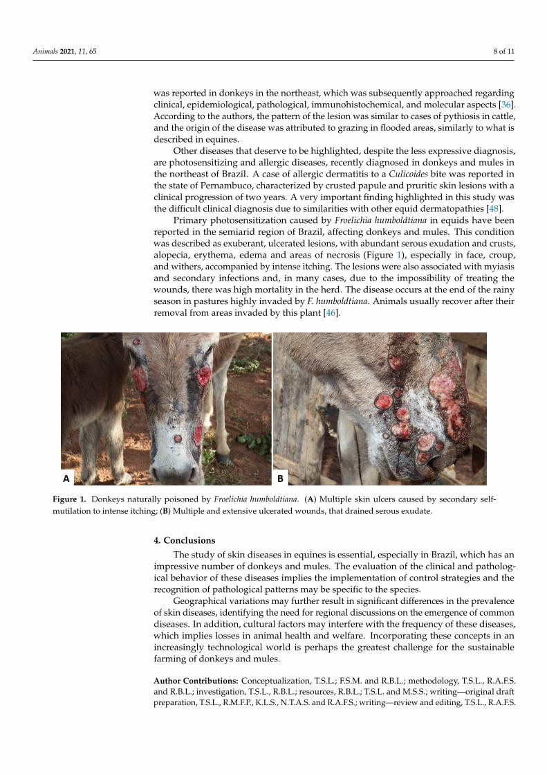
Primary photosensitization caused by Froelichia humboldtiana in equids have beenreported in the semiarid region of Brazil, affecting donkeys and mules. This conditionwas described as exuberant, ulcerated lesions, with abundant serous exudation and crusts,alopecia, erythema, edema and areas of necrosis (Figure 1), especially in face, croup,and withers, accompanied by intense itching. The lesions were also associated with myiasisand secondary infections and, in many cases, due to the impossibility of treating thewounds, there was high mortality in the herd. The disease occurs at the end of the rainyseason in pastures highly invaded by F. humboldtiana. Animals usually recover after theirremoval from areas invaded by this plant [46].
Animals 2020, 10, x 8 of 12
interfere with quality. In Brazil however, reports of integument disorders in donkeys and mules are little reported when compared to North American, European, and Asian countries. The low value attributed to these animals in the northeastern rural communities, associated with particularities of the species regarding clinical presentation of some diseases, probably impact the owner’s decision not to take these animals to the veterinarian, resulting in many diseases being underdiagnosed in the region.
A study in the semiarid region of Paraíba State reported that 258 donkeys and mules seen in 10 years presented integumental diseases as the main causes of consultation (88 cases), mainly traumatic wounds, sarcoids, and abscesses in donkeys and traumatic wounds, squamous cell carcinomas, and habronemiasis in mules. A very important finding raised in this study was that most diseases diagnosed could be linked to mistreatment or lack of attention to these animals [11], a factor associated to the cultural habits of many owners.
In the same year, another retrospective study exclusively on equid skin diseases in the semiarid northeast was published [11]. According to this study, only one case of pythiosis was reported in donkeys in the northeast, which was subsequently approached regarding clinical, epidemiological, pathological, immunohistochemical, and molecular aspects [36]. According to the authors, the pattern of the lesion was similar to cases of pythiosis in cattle, and the origin of the disease was attributed to grazing in flooded areas, similarly to what is described in equines.
Other diseases that deserve to be highlighted, despite the less expressive diagnosis, are photosensitizing and allergic diseases, recently diagnosed in donkeys and mules in the northeast of Brazil. A case of allergic dermatitis to a Culicoides bite was reported in the state of Pernambuco, characterized by crusted papule and pruritic skin lesions with a clinical progression of two years. A very important finding highlighted in this study was the difficult clinical diagnosis due to similarities with other equid dermatopathies [48].
Primary photosensitization caused by Froelichia humboldtiana in equids have been reported in the semiarid region of Brazil, affecting donkeys and mules. This condition was described as exuberant, ulcerated lesions, with abundant serous exudation and crusts, alopecia, erythema, edema and areas of necrosis (Figure 1), especially in face, croup, and withers, accompanied by intense itching. The lesions were also associated with myiasis and secondary infections and, in many cases, due to the impossibility of treating the wounds, there was high mortality in the herd. The disease occurs at the end of the rainy season in pastures highly invaded by F. humboldtiana. Animals usually recover after their removal from areas invaded by this plant [46].
Figure 1. Donkeys naturally poisoned by Froelichia humboldtiana. (A) Multiple skin ulcers caused by secondary self-mutilation to intense itching; (B) Multiple and extensive ulcerated wounds, that drained serous exudate.
4. Conclusions
The study of skin diseases in equines is essential, especially in Brazil, which has an impressive number of donkeys and mules. The evaluation of the clinical and pathological behavior of these
Figure 1. Donkeys naturally poisoned by Froelichia humboldtiana. (A) Multiple skin ulcers caused by secondary self-mutilation to intense itching; (B) Multiple and extensive ulcerated wounds, that drained serous exudate.
4. Conclusions
The study of skin diseases in equines is essential, especially in Brazil, which has animpressive number of donkeys and mules. The evaluation of the clinical and patholog-ical behavior of these diseases implies the implementation of control strategies and therecognition of pathological patterns may be specific to the species.
Geographical variations may further result in significant differences in the prevalenceof skin diseases, identifying the need for regional discussions on the emergence of commondiseases. In addition, cultural factors may interfere with the frequency of these diseases,which implies losses in animal health and welfare. Incorporating these concepts in anincreasingly technological world is perhaps the greatest challenge for the sustainablefarming of donkeys and mules.
Author Contributions: Conceptualization, T.S.L.; F.S.M. and R.B.L.; methodology, T.S.L., R.A.F.S.and R.B.L.; investigation, T.S.L., R.B.L.; resources, R.B.L.; T.S.L. and M.S.S.; writing—original draftpreparation, T.S.L., R.M.F.P., K.L.S., N.T.A.S. and R.A.F.S.; writing—review and editing, T.S.L., R.A.F.S.

Animals 2021, 11, 65 9 of 11
and R.B.L.; supervision, R.B.L. and F.S.M. All authors have read and agreed to the published versionof the manuscript.
Funding: This research was funded by Coordination for the Improvement of Higher EducationPersonnel grant number Code 001 And The APC was funded by Federal University of Paraíba.
Conflicts of Interest: The authors declare no conflict of interest.
References1. The Food and Agriculture Organization-FAO. 2016. Available online: http://www.fao.org/faostat/en/#home (accessed on
23 April 2020).2. The Food and Agriculture Organization-FAO. 2018. Available online: http://www.fao.org/faostat/en/#data/QA (accessed on
23 April 2020).3. Luís, C.; Bastos-Silveira, C.; Cothran, E.G.; Oom, M.D.M. Iberian origins of New World horse breeds. J. Hered. 2006, 97, 107–113.
[CrossRef] [PubMed]4. Baker, M. Sob a Pele-O Comércio Emergente de Pele de Asno e Suas Implicações Para o Bem-Estar e os Meios de Subsistência dos Asnos,
1st ed.; The Donkey Sanctuary: Sidmouth, UK, 2017; Available online: https://www.thedonkeysanctuary.org.uk/sites/uk/files/201711/under_the_skin_report_portuguese.pdf (accessed on 10 July 2020). (In Portuguese)
5. Carneiro, G.F.; Cavalcante Lucena, J.E.; de Oliveira Barros, L. The current situation and trend of the donkey industry in SouthAmerica. J. Equine Vet. Sci. 2018, 65, 106–110. [CrossRef]
6. Köhle, N. Feasting on Donkey Skin. In Conspicuous Consumption, 1st ed.; Smith, C.A., Kohle, N., Jaivin, L., Eds.; ANU Press:Canberra, Australia, 2018; ISBN (ebook): 9781760462031.
7. Lara, M.C.C.S.H.; Villalobos, E.M.C.; Cunha, S.E.M.; Oliveira, J.V.; Castro, C.; Nassar, A.F.C.; Silva, L.M.P.; Okuda, L.H.; Romaldini,A.H.C.N.; Cunha, M.S.; et al. Occurrence of viral diseases in donkeys (Equus asinus) in São Paulo State, Brazil. Braz. J. Vet. Res.An. Sci. 2017, 54, 154–158. [CrossRef]
8. García-Bocanegra, I.; Arenas-Montes, A.; Jaén-Téllez, J.A.; Napp, S.; Fernández-Morente, M.; Arenas, A. Use of sentinelserosurveillance of mules and donkeys in the monitoring of West Nile virus infection. Vet. J. 2012, 194, 262–264. [CrossRef]
9. Lima, T.S. Caracterização Clínico-patológica e Epidemiológica das Dermatopatias de Ruminantes no Agreste da Paraíba. Master’sDissertation, Universidade Federal da Paraíba, Areia, Paraíba, 2019. Available online: https://repositorio.ufpb.br/jspui/handle/123456789/14355 (accessed on 10 July 2020).
10. Assis-Brasil, N.D.; Marcolongo-Pereira, C.; Stigger, A.L.; Fiss, L.; Santos, B.L.; Coelho, A.C.B.; Sallis, E.S.V.; Fernandes, C.G.;Schild, A.L. Dermatopatias em equinos na região sul do Rio Grande do Sul: Estudo de 710 casos. Ciên. Rural. 2015, 45, 519–524.(In Portuguese) [CrossRef]
11. Pessoa, A.F.A.; Pessoa, C.R.M.; Miranda Neto, E.G.; Riet-Correa, F. Doenças de asininos e muares no semiárido brasileiro. Pesq. Vet.Bras. 2014, 34, 1210–1214. (In Portuguese) [CrossRef]
12. Pessoa, A.F.A.; Pessoa, C.R.M.; Miranda Neto, E.G.; Dantas, A.F.M.; Riet-Correa, F. Doenças de pele em equídeos no semiáridobrasileiro. Pesq. Vet. Bras. 2014, 34, 743–748. (In Portuguese) [CrossRef]
13. McLean, A.K.; Navas Gonzalez, F.J. Can scientists influence donkey welfare? Historical perspective and a contemporary view.J. Equine Vet. Sci. 2018, 65, 25–32. [CrossRef]
14. Miranda, A.L.S.; Palhares, M.S. Muares: Características, origem e particularidades clínico-laboratoriais. Rev. Cient. Med. Vet. 2017,1–8. Available online: http://faef.revista.inf.br/imagens_arquivos/arquivos_destaque/qqT3b4TLANsOU7k_2018-6-30-10-43-55.pdf (accessed on 10 July 2020). (In Portuguese).
15. Tesfaye, A.; Tekle, Y.; Taddele, H.; Gezahagn, K.; Yihdego, H. Survey of Common Skin Problem of Working Equines in andAround Mekelle, North Ethiopia. Acad. J. Anim. Dis. 2015, 4, 30–38. [CrossRef]
16. Davis, E. Donkey and Mule Welfare. Vet. Clin. N. Am. Equine Pract. 2019, 35, 481–491. [CrossRef] [PubMed]17. Pierezan, F.; Rissi, D.R.; Rech, R.R.; Fighera, R.A.; Brum, J.S.; Barros, C.S.L. Achados de necropsia relacionados com a morte de
335 eqüinos: 1968–2007. Pesq. Vet. Bras. 2009, 29, 275–280. (In Portuguese) [CrossRef]18. Lucena, R.B.; Pierezan, F.; Kommers, G.D.; Irigoyen, L.F.; Fighera, R.A.; Barros, C.S.L. Doenças de bovinos no Sul do Brasil: 6.706
casos. Pesq. Vet. Bras. 2010, 30, 428–434. (In Portuguese) [CrossRef]19. Marques, A.L.A.; Aguiar, G.M.N.; Lira, M.A.A.; Miranda Neto, E.G.; Azevedo, S.S.; Simões, S.V.D. Enfermidades do sistema
digestório de bovinos da região semiárida do Brasil. Pesq. Vet. Bras. 2018, 38, 407–416. [CrossRef]20. Barrandeguy, M.E.; Carossino, M. Infectious diseases in donkeys and mules: An overview and update. J. Equine Vet. Sci. 2018, 65,
98–105. [CrossRef]21. Mavrouli, M.; Vrioni, G.; Kapsimali, V.; Tsiamis, C.; Mavroulis, S.; Pervanidou, D.; Billinis, C.; Hadjichristodoulou, C.; Tsakris, A.
Reemergence of West Nile Virus Infections in Southern Greece, 2017. Am. J. Trop. Med. Hyg. 2019, 100, 420–426. [CrossRef]22. Souza, T.M.; Brum, J.S.; Fighera, R.A.; Brass, K.E.; Barros, C.S.L. Prevalência dos tumores cutâneos de equinos diagnosticados
no Laboratório de Patologia Veterinária da Universidade Federal de Santa Maria, Rio Grande do Sul. Pesq. Vet. Bras. 2011, 31,379–382. (In Portuguese) [CrossRef]
23. Marcolongo-Pereira, C.; Estima-Silva, P.; Soares, M.P.; Sallis, E.S.V.; Grecco, F.B.; Fernandes, C.G.; Raffi, M.B.; Schild, A.L. Doençasde equinos na região Sul do Rio Grande do Sul. Pesq. Vet. Bras. 2014, 34, 205–210. (In Portuguese) [CrossRef]
http://faef.revista.inf.br/imagens_arquivos/arquivos_destaque/qqT3b4TLANsOU7k_2018-6-30-10-43-55.pdf

Animals 2021, 11, 65 10 of 11
24. Miller, M.A.; Moore, G.E.; Bertin, F.R.; Kritchevsky, J.E. What’s New in Old Horses? Postmortem Diagnoses in Mature and AgedEquids. Vet. Pathol. 2015, 53, 390–398. [CrossRef]
25. Elsheikha, H.M.; Schares, G.; Paraschou, G.; Sullivan, R.; Fox, R. First record of besnoitiosis caused by Besnoitia bennetti in donkeysfrom the UK. Parasites Vectors 2020, 13, 1–10. [CrossRef]
26. Radwan, A.M.; Ahmed, N.E.; Elakabawy, L.M.; Ramadan, M.Y.; Elmadawy, R.S. Prevalence and pathogenesis of some filarialnematodes infecting donkeys in Egypt. Vet. World 2016, 9, 888–892. [CrossRef] [PubMed]
27. White, S.D.; Bourdeau, P.J.; Brement, T.; Vandenabeele, S.I.; Haspeslagh, M.; Bruet, V.; Van Oldruitenborgh-Oosterbaan, M.M.S.Skin disease in donkeys (Equus asinus): A retrospective study from four veterinary schools. Vet. Dermatol. 2019, 30, 247–276.[CrossRef] [PubMed]
28. Knottenbelt, D.C. Skin Disorders of the Donkey and Mule. Vet. Clin Equine. 2019, 35, 493–514. [CrossRef] [PubMed]29. Alvarez, J.A.C.; Socarras, T.O.; Tous, M.G. Dermopatias en burros de trabajo (Equus asinus) en areas rurales de Cordoba (Colombia).
Rev. Med. Vet. 2017, 34, 81–91. (In Spanish) [CrossRef]30. Meselu, D.; Abebe, R.; Mekibib, B. Prevalence of Epizootic Lymphangitis and bodily distribuition of lesions in cart-mules in
Behair Dar Town, Northwest Ethiopia. J. Vet. Sci. Technol. 2018, 9, 1–4. [CrossRef]31. Mahendra, P. Occurrence of Cutaneous Epizootic Lymphangitis in Working Donkeys in Debre Zeit, Ethiopia. EC Microbiol. 2019,
15, 382–384.32. Mota, R.A.; Oliveira, A.A.F.; Pinheiro Junior, J.W.; Silva, L.B.G.; Brito, M.F.; Rabelo, S.S.A. Glanders in donkeys (Equus asinus) in
the state of Pernambuco, Brazil: A case report. Braz. J. Microbiol. 2010, 41, 146–149. [CrossRef]33. Rabelo, S.S.A.; Soares, P.C.; Silva, L.B.G.; Cunha, A.P.; Nascimento Sobrinho, E.; Pinheiro Junior, J.W.; Barbosa, M.A.G.; Mota, R.A.
Indicadores Clínicos em Muares Naturalmente Infectados pela Burkholderia mallei. Vet. Zootec. 2006, 13, 54–62. Availableonline: https://docplayer.com.br/71952462-Indicadores-clinicos-em-muares-naturalmente-infectados-pela-burkholderia-mallei-resumo.html (accessed on 10 July 2020). (In Portuguese).
34. Mota, R.A.; Brito, M.F.; Castro, F.J.C.; Massa, M. Mormo em eqüídeos nos estados de Pernambuco e Alagoas. Pesq. Vet. Bras. 2000,20, 155–159. (In Portuguese) [CrossRef]
35. Ghahvei, Y.; Mirzaei, M.; Hashemnia, S.; Golchin, M.; Kheirandish, R.; Uni, S.; Endoza-Roldan, J.A.; Otranto, D.; Sazmand, A.Scanning electron microscopy of Onchocerca fasciata (Filarioidea: Onchocercidae) adults, microfilariae and eggs with notes onhistopathological findings in camels. Parasit. Vectors 2020, 13, 1–10. [CrossRef]
36. Maia, L.A.; Olinda, R.G.; Araújo, T.F.; Firmino, P.R.; Nakazato, L.; Miranda Neto, E.G.; Riet-Correa, F.; Dantas, A.F.M. Cutaneouspythiosis in a donkey (Equus asinus) in Brazil. J. Vet. Diagn. Invest. 2016, 28, 436–439. [CrossRef] [PubMed]
37. Tabosa, I.M.; Medeiros, V.T.; Dantas, A.F.M.; Azevedo, E.O.; Maia, J.C. Pitiose cutânea em equinos no semi-árido da Paraíba.ABMVZ 1999, 51, 27–30. (In Portuguese)
38. Santos, C.E.P.; Santurio, J.M.; Colodel, E.M.; Juliano, R.S.; Silva, J.A.; Marques, L.C. Contribuição ao Estudo da Pitose Cutâneaem Equídeos do Pantanal Norte, Brasil. ARS Vet. 2011, 27, 134–140. Available online: http://arsveterinaria.org.br/ars/article/download/310/337 (accessed on 10 July 2020). (In Portuguese).
39. Leite, N.M.; Rocha, M.V.; Souza, K.M.; Vago, P.B. Linfangite ulcerativa em equino: Relato de caso. PUBVET 2019, 13, 1–6.(In Portuguese) [CrossRef]
40. Sureshjani, M.H.; Atyabi, N.; Tazikeh, A.; Falahatipour, S.K.; Hashemian, M. Isolation of Rhodococcus equi from a mule withcutaneous wound. Comp. Clin. Pathol. 2014, 1–3. [CrossRef]
41. Prescott, J.F. Rhodococcus equi: An animal and human pathogen. Clin. Microbiol. Rev. 1991, 4, 1–20. [CrossRef]42. Guimarães, J.H.; Papavero, N.; Prado, A.P. As miíases na região neotropital (identificação, biologia, bibliografia). Rev. Bras. Zool.
1982, 1, 239–416. [CrossRef]43. Davis, C.R.; Valentine, B.A.; Gordon, E.; McDonough, S.P.; Schaffer, P.A.; Allen, A.L.; Pesavento, P. Neoplasia in 125 donkeys
(Equus asinus): Literature review and a survey of five veterinary schools in the United States and Canada. J. Vet. Diagn. Investig.2016, 28, 662–670. [CrossRef]
44. Alberti, T.S.; Zamboni, R.; Venancio, F.R.; Scheid, H.V.; Bermann, C.S.; Raffi, M.B.; Sallis, E.S.V. Melanoma anaplásico em equinode pelagem tordilha com metástase em osso e músculo. Cien. Anim. 2019, 29, 129–134. (In Portuguese)
45. Rayner, E.; Airikkala-Otter, I.; Susheelan, A.; Gibson, A.; Itaba, R.; Mayani, T.; Mellanby, R.J.; Gamble, L. Prevalence of skinwounds in working donkeys in Bukombe, Tanzania. Vet. Rec. 2019, 186, 1–3. [CrossRef]
46. Knupp, S.N.R.; Knupp, L.S.; Riet-Correa, F.; Lucena, R.B. Plants that cause photosensitivity in ruminants in Brazil. Semin. Ciênc.Agrár. 2016, 37, 2009–2020. [CrossRef]
47. Amado, G.P.; Silva, C.C.B.; Barbosa, F.M.S.; Nascimento, H.H.L.; Malta, K.C.; Azevedo, M.V.; Lacerda-Lucena, P.B.; Lucena, R.B.Surtos de fotossensibilização e dermatite alérgica em ruminantes e equídeos no Nordeste do Brasil. Pesq. Vet. Bras. 2018, 38,889–895. (In Portuguese) [CrossRef]
48. Silva, T.I.B.; Melchior, L.A.K.; Baptista Filho, L.C.F.; Fernandes, A.C.C.; Silva, L.G.; Vasconcelos, K.F.; Revorêdo, R.G.; Silva, D.D.;Melo, L.E.H. Dermatite alérgica à picada de Culicoides em muar: Relato de caso. Arq. Bras. Med. Vet. Zootec. 2017, 69, 1407–1412.(In Portuguese) [CrossRef]
49. Schaffartzik, A.; Hamza, E.; Janda, J.; Crameri, R.; Marti, E.; Rhyner, C. Equine insect bite hypersensitivity: What do we know?Vet Immunol. Immunopathol. 2012, 147, 113–126. [CrossRef] [PubMed]

Animals 2021, 11, 65 11 of 11
50. Corrêa, T.G.; Ferreira, J.M.; Riet-Correa, G.; Ruas, J.L.; Schild, A.L.; Riet-Correa, F.; Guimarães, A.; Felippe-Bauer, M.L. Seasonalallergic dermatitis in sheep in southern Brazil caused by Culicoides insignis (Diptera: Ceratopogonidae). Vet. Parasitol. 2007, 10,181–185. [CrossRef] [PubMed]
51. Monteiro, G.A.; Souza, M.V.; Conceição, L.G.; Balbi, C.L.; Borba, R.; Moreira, M.A.S. Pênfigo foliáceo em um equino. Ciência Rural2007, 37, 594–598. (In Portuguese) [CrossRef]