Singlet oxygen quenching by thione analogues of canthaxanthin, echinenone and rhodoxanthin
6
Singlet oxygen quenching by thione analogues of canthaxanthin, echinenone and rhodoxanthin Samsun B.B. Mohamad a,b , Yaser A. Yousef a,c , Thor-Bernt Melø a , Tama ´s Ja ´vorfi a,d , Vassilia Partali e , Hans-Richard Sliwka e , K. Razi Naqvi a, * a Department of Physics, Norwegian University of Science and Technology (NTNU), N-7491 Trondheim, Norway b Centre for Computational and Theoretical Sciences, International Islamic University Malaysia (IIUM), 53100 Jalan Gombak, Kuala Lumpur, Malaysia c Department of Chemistry, Yarmouk University, Irbid, Jordan d Biological Research Center, Hungarian Academy of Sciences, P.O. Box 521, H-6701 Szeged, Hungary e Department of Chemistry, NTNU, Norway Received 3 November 2005; received in revised form 22 January 2006; accepted 2 February 2006 Available online 24 March 2006 Abstract Thione analogues of three naturally occurring carotenones (canthaxanthin, echinenone, and rhodoxanthin) were synthesized just over ten years ago, and it was reported that substitution of the oxygen atom by sulphur brings about a large red shift and some broadening in the optical absorption spectrum of the compound. Since the three carotenothiones are scarce, determination of their molar absorption coefficients presents a challenge. A method for relating the molar absorption coefficient of a carotenothione (Car-S) to that of its ketone analogue (Car-O) has been developed, which has revealed that the peak molar absorption coefficient of a Car-S is only about 60% of the corresponding value for Car-O. Using methylene blue as the sensitizer and acetonitrile as the solvent, we have also investigated the quenching (under photostationary conditions) of the 1270 nm phosphorescence emission of singlet oxygen by each of the six carotenoids. The data conform to the Stern–Volmer relation, and show that substitution of a carbonyl oxygen atom by sulphur does not lead to an appreciable change in the value of the quenching constant, which is close to 1.5 · 10 10 M 1 s 1 for all six quenchers. Ó 2006 Elsevier B.V. All rights reserved. Keywords: Carotenoids; Carotenothiones; Singlet oxygen 1. Introduction Among the many striking photophysical properties of naturally occurring carotenoid (Car) molecules is their abil- ity to deactivate, through a physical process, an oxygen molecule which is in its lowest electronically excited state, commonly known as singlet oxygen [1]. The quenching process becomes spin-allowed only if a singlet oxygen mol- ecule comes into contact with a Car as a result of a bimo- lecular encounter, and the two molecules exchange electrons with opposite spins [2]. The net result is not only an exchange of electrons but also the transfer of electronic excitation from singlet oxygen to Car. Pictorially, the pro- cess (quenching due to exchange-mediated energy transfer) may be depicted as follows: O 2 ð"#Þþ Car 0 ð*+Þ! O 0 2 ð"*Þþ Car y ð#+Þ. ð1Þ Here the superscripts denote absence of electronic excita- tion (0), and presence of triplet ( ) or singlet excitation ( * ). The recent growth of interest in the so-called push–pull polyenes [3,4] and in self-assembled monolayers [5] has motivated the synthesis of thione [6] and oxime [7] ana- logues of some naturally occurring carotenones. Experi- ence gained from studies dealing with a wide variety of conjugated ketones leads one to believe that replacement of the carbonyl oxygen by sulphur is equivalent to a sub- stantial increase in the size of the conjugated system. First, 1011-1344/$ - see front matter Ó 2006 Elsevier B.V. All rights reserved. doi:10.1016/j.jphotobiol.2006.02.006 * Corresponding author. Address: Department of Physics, Norwegian University of Science and Technology (NTNU), N-7491 Trondheim, Norway. Fax: +47 73 59 77 10. E-mail address: [email protected] (K. Razi Naqvi). www.elsevier.com/locate/jphotobiol Journal of Photochemistry and Photobiology B: Biology 84 (2006) 135–140
-
Upload
independent -
Category
Documents
-
view
0 -
download
0
Transcript of Singlet oxygen quenching by thione analogues of canthaxanthin, echinenone and rhodoxanthin

www.elsevier.com/locate/jphotobiol
Journal of Photochemistry and Photobiology B: Biology 84 (2006) 135–140
Singlet oxygen quenching by thione analogues of canthaxanthin,echinenone and rhodoxanthin
Samsun B.B. Mohamad a,b, Yaser A. Yousef a,c, Thor-Bernt Melø a, Tamas Javorfi a,d,Vassilia Partali e, Hans-Richard Sliwka e, K. Razi Naqvi a,*
a Department of Physics, Norwegian University of Science and Technology (NTNU), N-7491 Trondheim, Norwayb Centre for Computational and Theoretical Sciences, International Islamic University Malaysia (IIUM), 53100 Jalan Gombak, Kuala Lumpur, Malaysia
c Department of Chemistry, Yarmouk University, Irbid, Jordand Biological Research Center, Hungarian Academy of Sciences, P.O. Box 521, H-6701 Szeged, Hungary
e Department of Chemistry, NTNU, Norway
Received 3 November 2005; received in revised form 22 January 2006; accepted 2 February 2006Available online 24 March 2006
Abstract
Thione analogues of three naturally occurring carotenones (canthaxanthin, echinenone, and rhodoxanthin) were synthesized just overten years ago, and it was reported that substitution of the oxygen atom by sulphur brings about a large red shift and some broadening inthe optical absorption spectrum of the compound. Since the three carotenothiones are scarce, determination of their molar absorptioncoefficients presents a challenge. A method for relating the molar absorption coefficient of a carotenothione (Car-S) to that of its ketoneanalogue (Car-O) has been developed, which has revealed that the peak molar absorption coefficient of a Car-S is only about 60% of thecorresponding value for Car-O. Using methylene blue as the sensitizer and acetonitrile as the solvent, we have also investigated thequenching (under photostationary conditions) of the 1270 nm phosphorescence emission of singlet oxygen by each of the six carotenoids.The data conform to the Stern–Volmer relation, and show that substitution of a carbonyl oxygen atom by sulphur does not lead to anappreciable change in the value of the quenching constant, which is close to 1.5 · 1010 M�1 s�1 for all six quenchers.� 2006 Elsevier B.V. All rights reserved.
Keywords: Carotenoids; Carotenothiones; Singlet oxygen
1. Introduction
Among the many striking photophysical properties ofnaturally occurring carotenoid (Car) molecules is their abil-ity to deactivate, through a physical process, an oxygenmolecule which is in its lowest electronically excited state,commonly known as singlet oxygen [1]. The quenchingprocess becomes spin-allowed only if a singlet oxygen mol-ecule comes into contact with a Car as a result of a bimo-lecular encounter, and the two molecules exchangeelectrons with opposite spins [2]. The net result is not only
1011-1344/$ - see front matter � 2006 Elsevier B.V. All rights reserved.
doi:10.1016/j.jphotobiol.2006.02.006
* Corresponding author. Address: Department of Physics, NorwegianUniversity of Science and Technology (NTNU), N-7491 Trondheim,Norway. Fax: +47 73 59 77 10.
E-mail address: [email protected] (K. Razi Naqvi).
an exchange of electrons but also the transfer of electronicexcitation from singlet oxygen to Car. Pictorially, the pro-cess (quenching due to exchange-mediated energy transfer)may be depicted as follows:
O�2ð"#Þ þ Car0ð*+Þ ! O02ð"*Þ þ Caryð#+Þ. ð1Þ
Here the superscripts denote absence of electronic excita-tion (0), and presence of triplet (�) or singlet excitation (*).
The recent growth of interest in the so-called push–pullpolyenes [3,4] and in self-assembled monolayers [5] hasmotivated the synthesis of thione [6] and oxime [7] ana-logues of some naturally occurring carotenones. Experi-ence gained from studies dealing with a wide variety ofconjugated ketones leads one to believe that replacementof the carbonyl oxygen by sulphur is equivalent to a sub-stantial increase in the size of the conjugated system. First,

136 S.B.B. Mohamad et al. / Journal of Photochemistry and Photobiology B: Biology 84 (2006) 135–140
Becker and his collaborators prepared thione analogues ofseveral coumarins, psoralens, and (furo)chromones [8].They observed, on comparing the absorption spectrum ofa ketone with its thione analogue, that the lowest p* ptransition in the latter occurred at considerably lowerenergy, and with appreciably greater intensity (manifestedby both an enhancement of the peak extinction coefficientand an increase in the bandwidth). Secondly, one finds inRef. [6], which deals with molecules shown in Scheme 1,a brief statement to the effect that—compared with cantha-xanthin (K1), echinenone (K2) and rhodoxanthin (K3)—thecorresponding thione (T1, T2 and T3, respectively) displaysa visible absorption band that is broader and considerablyred-shifted; the molar absorption coefficients of the carot-enoid thiones were not measured in this study. Finally,Eckart and coworkers [9] have performed ab initio calcula-tions of static electric properties of the cyclic conjugatedsystems C2n+1H2nA (n = 1,2,3; A = O,S). Their study,aimed primarily at examining the nonlinear optical(NLO) properties of these molecules, showed that thechange in the first and the second hyperpolarizabilityresulting from the substitution of sulphur for oxygen ismuch larger than that resulting from an increment in thevalue of n (from 1 to 3 or from 3 to 5).
The results recounted above tempt one to conjecturethat the gap between T1, the first excited triplet state, andS0, the singlet ground state, is smaller in a carotenoid thi-one than in its ketone analogue. This makes it interestingto investigate the quenching of singlet oxygen by the newlysynthesized molecules. Our purpose here is to explore thisaspect by making time-resolved and steady-state measure-ments of the near infrared (1270 nm) phosphorescenceemission which results from the b1Dg X 3R�g transitionin molecular dioxygen.
O
X
X
O
X
X = O K1X = S T1
X = O K2X = S T2
X = O K3X = S T3
Scheme 1.
The second-order rate constant for quenching (kq) can-not be determined without the knowledge of the quencherconcentration, a quantity which can be established mostconveniently through absorption spectrophotometry—pro-vided that the molar absorption coefficient of the quencheris known. Since such data for T1, T2 and T3 are notknown, and these substances were not available in quanti-ties large enough to permit accurate weighing, a specialstrategy had to be devised for determining their molarabsorption coefficients.
2. Materials and methods
All measurements reported here were carried out atambient temperature (20–22 �C). The other details aredescribed below.
2.1. Chemicals
The preparation of the carotenoid thiones and thesource of the carotenones have already been described[6]. Methylene blue (MB), rose bengal (RB) andacetonitrile (analytical reagent) were purchased fromFluka. Absorption spectra of samples containing thesensitizer were recorded by using a home-made diodearray spectrometer [10]; in the experiments involvingthe determination of the molar absorption coefficients,a commercial scanning instrument was employed(Hitachi U-3010).
2.2. Determination of the molar absorption coefficients
Approximately 1 mg of a carotenone was added to aflask containing 2 ml benzene; an aliquot (40 ll) wasremoved from the resulting solution, diluted in acetonitrile(5 ml), and the absorption spectrum of the diluted samplewas recorded. Lawesson’s reagent (approximately 1 mg)was then added to the flask, and the mixture was stirredand heated (at 40 �C) to promote the reaction (conversionof the carotenone to the corresponding thione); the processof removing an aliquot, dilution and measurement of theabsorption spectrum was repeated at regular intervals(approximately 20 min) until the peak absorbance reducedto about 75% of its initial value. The set of absorption spec-tra so obtained were fitted, according to a proceduredetailed elsewhere [11], to Eq. (4), which will be stated inSection 3.1. It will be shown below that this approachallows one to determine the molar absorption coefficientof a carotenothione (if its value for the ketone analogueis known).
2.3. Time-resolved phosphorescence
The decay time of the phosphorescence emitted by sin-glet oxygen was recorded by using an apparatus con-structed in the laboratory. The source of excitation was alight pulse (ca. 7 ns in duration) derived from an optical
0.0
0.5
1.0 BK2
T2
Abs
orba
nce
0.0
0.5
1.0 AK1
T1
Abs
orba
nce
400 500 600 7000.0
0.5
1.0
Wavelength/nm
T3
K3 C
Abs
orba
nce
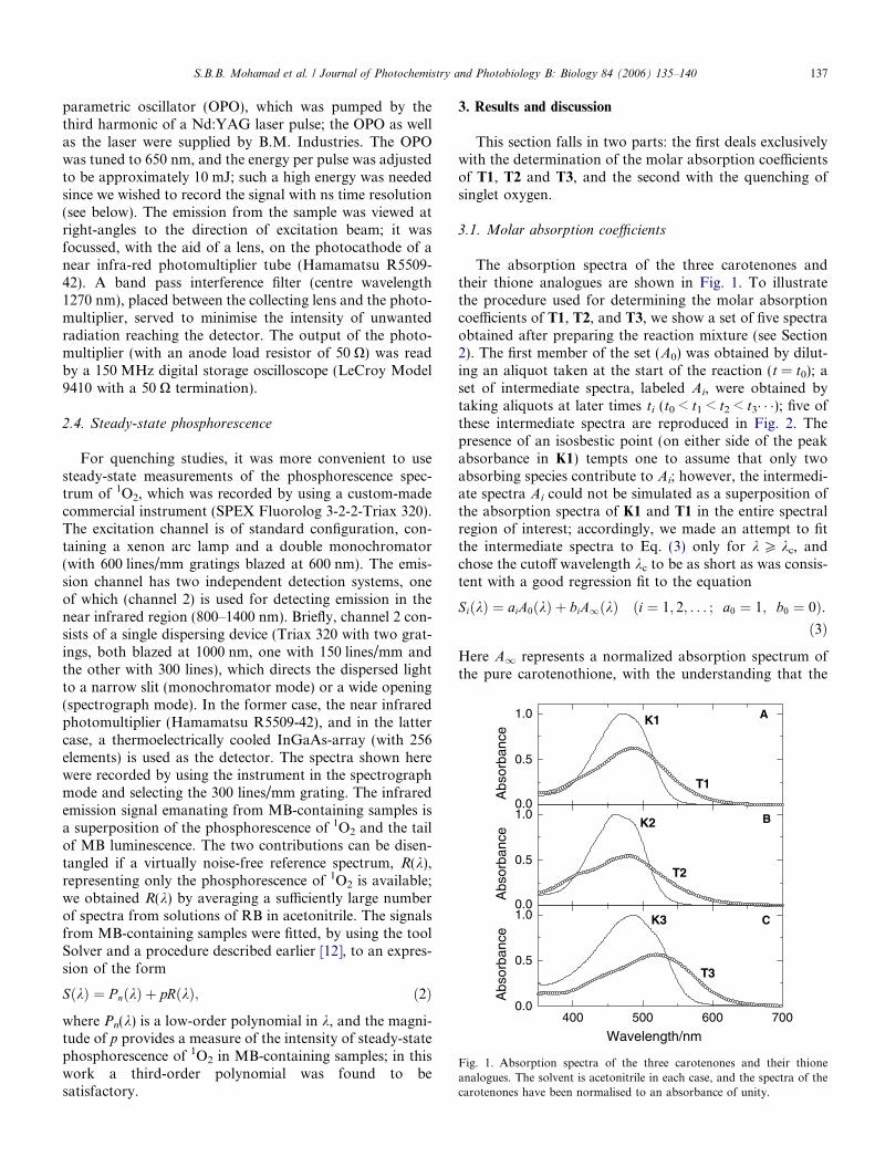
Fig. 1. Absorption spectra of the three carotenones and their thioneanalogues. The solvent is acetonitrile in each case, and the spectra of thecarotenones have been normalised to an absorbance of unity.
S.B.B. Mohamad et al. / Journal of Photochemistry and Photobiology B: Biology 84 (2006) 135–140 137
parametric oscillator (OPO), which was pumped by thethird harmonic of a Nd:YAG laser pulse; the OPO as wellas the laser were supplied by B.M. Industries. The OPOwas tuned to 650 nm, and the energy per pulse was adjustedto be approximately 10 mJ; such a high energy was neededsince we wished to record the signal with ns time resolution(see below). The emission from the sample was viewed atright-angles to the direction of excitation beam; it wasfocussed, with the aid of a lens, on the photocathode of anear infra-red photomultiplier tube (Hamamatsu R5509-42). A band pass interference filter (centre wavelength1270 nm), placed between the collecting lens and the photo-multiplier, served to minimise the intensity of unwantedradiation reaching the detector. The output of the photo-multiplier (with an anode load resistor of 50 X) was readby a 150 MHz digital storage oscilloscope (LeCroy Model9410 with a 50 X termination).
2.4. Steady-state phosphorescence
For quenching studies, it was more convenient to usesteady-state measurements of the phosphorescence spec-trum of 1O2, which was recorded by using a custom-madecommercial instrument (SPEX Fluorolog 3-2-2-Triax 320).The excitation channel is of standard configuration, con-taining a xenon arc lamp and a double monochromator(with 600 lines/mm gratings blazed at 600 nm). The emis-sion channel has two independent detection systems, oneof which (channel 2) is used for detecting emission in thenear infrared region (800–1400 nm). Briefly, channel 2 con-sists of a single dispersing device (Triax 320 with two grat-ings, both blazed at 1000 nm, one with 150 lines/mm andthe other with 300 lines), which directs the dispersed lightto a narrow slit (monochromator mode) or a wide opening(spectrograph mode). In the former case, the near infraredphotomultiplier (Hamamatsu R5509-42), and in the lattercase, a thermoelectrically cooled InGaAs-array (with 256elements) is used as the detector. The spectra shown herewere recorded by using the instrument in the spectrographmode and selecting the 300 lines/mm grating. The infraredemission signal emanating from MB-containing samples isa superposition of the phosphorescence of 1O2 and the tailof MB luminescence. The two contributions can be disen-tangled if a virtually noise-free reference spectrum, R(k),representing only the phosphorescence of 1O2 is available;we obtained R(k) by averaging a sufficiently large numberof spectra from solutions of RB in acetonitrile. The signalsfrom MB-containing samples were fitted, by using the toolSolver and a procedure described earlier [12], to an expres-sion of the form
SðkÞ ¼ P nðkÞ þ pRðkÞ; ð2Þ
where Pn(k) is a low-order polynomial in k, and the magni-tude of p provides a measure of the intensity of steady-statephosphorescence of 1O2 in MB-containing samples; in thiswork a third-order polynomial was found to besatisfactory.
3. Results and discussion
This section falls in two parts: the first deals exclusivelywith the determination of the molar absorption coefficientsof T1, T2 and T3, and the second with the quenching ofsinglet oxygen.
3.1. Molar absorption coefficients
The absorption spectra of the three carotenones andtheir thione analogues are shown in Fig. 1. To illustratethe procedure used for determining the molar absorptioncoefficients of T1, T2, and T3, we show a set of five spectraobtained after preparing the reaction mixture (see Section2). The first member of the set (A0) was obtained by dilut-ing an aliquot taken at the start of the reaction (t = t0); aset of intermediate spectra, labeled Ai, were obtained bytaking aliquots at later times ti (t0 < t1 < t2 < t3� � �); five ofthese intermediate spectra are reproduced in Fig. 2. Thepresence of an isosbestic point (on either side of the peakabsorbance in K1) tempts one to assume that only twoabsorbing species contribute to Ai; however, the intermedi-ate spectra Ai could not be simulated as a superposition ofthe absorption spectra of K1 and T1 in the entire spectralregion of interest; accordingly, we made an attempt to fitthe intermediate spectra to Eq. (3) only for k P kc, andchose the cutoff wavelength kc to be as short as was consis-tent with a good regression fit to the equation
SiðkÞ ¼ aiA0ðkÞ þ biA1ðkÞ ði ¼ 1; 2; . . . ; a0 ¼ 1; b0 ¼ 0Þ.ð3Þ
Here A1 represents a normalized absorption spectrum ofthe pure carotenothione, with the understanding that the

300 400 500 600 7000.0
0.1
0.2
0.3
0.4
0.5
0.6
ΔA 8
A0
Abs
orba
nce
Wavelength/nm
Fig. 2. A set of spectra showing the conversion of K1 to T1. Thecontribution of T1 to the initial spectrum, labeled A0, is negligible,whereas the final spectrum, A1, pertains to pure T1, and has beennormalized so as to pass through the isosbestic point at 514 nm. Theintermediate spectra arise from mixtures of K1, T1, and unknown reactionintermediates, whose spectrum is labeled as D.
400 500 600 7000.0
0.2
0.4
0.6
0.8
1.0
AC
Am
AS
AS: MBAm: MB+CarAC: CarAC=Am-αASA
bsor
banc
e
Wavelength/nm
Fig. 3. Absorption spectrum of MB (AS) and of a mixture of MB and T2
(Am); the solvent is acetonitrile. The contribution made by the carotenoidto the latter spectrum is calculated by setting AC = Am � aAS andadjusting a until absorption by MB reduces to the noise level.
1220 1240 1260 1280 1300 13200.0
0.5
1.0
RB MB+T2 MB+T2 (fit)
MB MB (fit)
Pho
spho
resc
ence
Inte
nsity
(au
)
Wavelength/nm
Fig. 4. Plots of luminescence signals from solutions (in acetonitrile)containing RB and MB (with and without T2).
138 S.B.B. Mohamad et al. / Journal of Photochemistry and Photobiology B: Biology 84 (2006) 135–140
normalization forces the spectrum to pass through the isos-bestic point (514 nm); in other words, A1 would coincidewith the final spectrum that would be obtained if the reac-tion resulted in complete conversion of the ketone to thethione, and the other products did not absorb in the spec-tral region of interest. The difference Di(k) ” Ai(k) � Si(k),assumed to be vanishingly small for k P kc, representsthe contribution by the reaction intermediate(s) and/orsome photoproduct(s). Fortunately, the shape of the differ-ence spectrum Di(k) turned out to be practically the same inall the intermediate spectra; this finding is crucial, for it al-lows us to reconstruct each intermediate spectrum as a lin-ear combination of three known spectra. Thus, we write
AiðkÞ ¼ aiA0ðkÞ þ biA1ðkÞ þ ciDðkÞði ¼ 1; 2; . . . ; a0 ¼ 1; b0 ¼ 0 ¼ c0Þ; ð4Þ
where D(k) is the average of all the intermediate spectra,and the identity of the species contributing to D(k) neednot be ascertained. The simulated spectra are not shownin Fig. 2 because they are practically coincident with theirmeasured counterparts. If we denote the molar absorptioncoefficients of the ketone and the thione by eK and eT,respectively, we can now write eT = eK A1/A0. Fig. 2 showsthat, for the pair K1/T1, eT ’ 0:6eK, where the circumflexindicates the peak value of the molar absorption coeffi-cient. Similar results (not shown here) were obtained forthe other two pairs (K2/T2, and K3/T3); the only signifi-cant difference is that the pair K3/T3 exhibited only oneisosbestic point (at 535 nm), showing that the spectra ofK3 and T3 do not intersect at the short wavelength sidesof their peaks (see panel C in Fig. 1).
3.2. Quenching of singlet oxygen
The choice of MB as the sensitizer was based on thelocation of its main absorption band, which does not over-
lap with the absorption spectrum of the carotenoids used inthis work; this feature, illustrated in Fig. 3, facilitated thedetermination of the absorbance contributed by the Carin samples which contained both MB and Car. Fig. 4shows two traces of the emission spectrum from samplescontaining MB and different amounts of T2, together withthe reference spectrum R(k) obtained by using RB alone;the simulated spectrum S(k) for each trace, resulting fromEq. (2), is also superimposed on the experimental data.
The decay of the phosphorescence emission of 1O2 inacetonitrile is plotted in Fig. 5; when analysed for single-exponential decay, the data lead to a value ofs0 = 72.4 ± 0.5 ls. An examination of the available data[13] reveals that most of the recent values in acetonitrile,reported during the 1991–1993 period, fall in the range71–77 ls, which leads us to believe that our result, foundby using a relatively high laser energy, is indeed reliable.
For all six carotenoids investigated here, the steady-statequenching data conform to the Stern–Volmer relation P0/P = 1 + kqs0 CQ = 1 + KSV CQ, where CQ is the quencherconcentration, P(CQ) is the steady-state intensity of thephosphorescence, P0 ” P(CQ = 0), and s0 is the decay time

0
5
10
15
20
25
30S
igna
l/mV
0 100 200 300 400-6
0
6
104 xR
es
Time/μs
Fig. 5. The decay of the intensity of the sensitized phosphorescence ofsinglet oxygen after laser excitation of the sensitizer (MB); the solvent isacetonitrile. Residuals showing departure from a single exponential fit(corresponding to a decay time of 72.4 ls) are also shown (aftermultiplication by a factor of 104).
0.0 0.5 1.0 1.5
1.0
1.5
2.0
2.5
3.0
KSV
P 0/P
CQ/μM
K3 1.48E6
T3 1.28E6
0. 0 0.5 1.0 1.5 2.0
1.0
1.5
2.0
2.5
3.0
3.5
KSV
P 0/P
K2 1.19E6
T2 0.95E6
0.0 0.5 1.0 1.5 2.0 2.5
1
2
3
4
5
KSV
P 0/P
K1 1.36E6
T1 1.19E6
Fig. 6. Stern–Volmer plots for the quenching of singlet oxygen by threecarotenones and their thione analogues.
S.B.B. Mohamad et al. / Journal of Photochemistry and Photobiology B: Biology 84 (2006) 135–140 139
of the phosphorescence in the absence of the quencher. Thevalues of the quenching rate constant kq are listed in Table1. The Stern–Volmer plots for the quenching of 1O2 by thesix carotenoids are displayed in Fig. 6, and the quenchingdata for the three pairs are presented in Table 1. In quotingthe literature values for the molar absorption coefficients ofK1, K2 and K3, we have not allowed for the influence of thesolvent, and kept only three significant figures, since thetabulated data [14] are ‘‘considered to have significantuncertainty’’, and this uncertainty also affects our valuesfor KSV and kq, which may be as high as 10%; on the otherhand, the relative values of these quantities are believed tobe more accurate.
Oliveros and coworkers [15] investigated the quenchingof singlet oxygen by K1 and K3 in benzene solutions, andfound kq to be 1.3 · 1010 M�1 s�1 and 1.2 · 1010 M�1 s�1,respectively; since the viscosity of benzene is about 1.9times that of acetonitrile [16], one would expect the values
Table 1Wavelengths of peak absorbance (kmax), the corresponding molarabsorption coefficients ðeÞ, and rate constants (kq) for singlet oxygenquenching for the six carotenoids studied in this work (solvent:acetonitrile)
Molecule kmax/nm e=104 M�1 cm�1 kq/1010 M�1 s�1
K1 471 12.5 1.9T1 487 7.8 1.6K2 460 11.5 1.6T2 481 6.4 1.3K3 487 14 2.0T3 520 8.0 1.8
The values of e for K1, K2 and K3 have been taken from Ref. [14] androunded off to three significant figures.
of kq in acetonitrile to be nearly twice the value reportedfor benzene; Table 1 shows that this is indeed the case.
4. Concluding remarks
The data presented above show that, contrary to expec-tations based on previous studies concerning the differencesin the spectral and NLO properties of ketones and their thi-one analogues, there is no substantial difference betweenthe singlet oxygen quenching abilities of a carotenoneand its thione analogue. The fact that, compared to itsketone analogue, a thione is slightly less efficient in quench-ing singlet oxygen can be rationalised by assuming that thequenching is dominated by process (1), and that the energyof the lowest triplet level of the carotenoid changes (whenthe oxygen atom is replaced by sulphur) so as to lowerthe Franck-Condon factor in the expression for thequenching rate constant [17]. It remains to be seen whetherthe carotenethiones turn out to be superior, in terms ofNLO response, to their ketone counterparts, but investiga-tion of this aspect falls outside the scope of our study.

140 S.B.B. Mohamad et al. / Journal of Photochemistry and Photobiology B: Biology 84 (2006) 135–140
References
[1] R. Edge, D. McGarvey, T.G. Truscott, The carotenoids as anti-oxidants—a review, J. Photochem. Photobiol. B—Biol. 41 (1997)189–200.
[2] G.J. Hoytink, Intermolecular electron exchange, Accounts Chem.Res. 2 (1969) 114–120.
[3] M. Blanchard-Desce, J.M. Lehn, M. Barzoukas, I. Ledoux, J. Zyss,Chain-length dependence of the quadratic hyperpolarizability ofpush–pull polyenes and carotenoids. Effect of end-groups andconjugation path, Chem. Phys. 181 (1994) 281–289.
[4] R. Andreu, M.J. Blesa, L. Carrasquer, J. Garin, J. Orduna, B.Villacampa, R. Alcala, J. Casado, M.C. Ruiz Delgado, J.T. Lopez,M. Allain, Tuning first molecular hyperpolarizabilities through theuse of proaromatic spacers, J. Am. Chem. Soc. 127 (2005) 8835–8845.
[5] A. Ion, V. Partali, H.-R. Sliwka, F.G. Banica, Electrochemistry of acarotenoid self-assembled monolayer, Electrochem. Commun. 4(2002) 674–678.
[6] H.-R. Sliwka, S. Liaaen-Jensen, Synthetic sulfur carotenoids. III.Carotenoid thiones—first preparation and spectroscopic properties,Acta Chem. Scand. 48 (1994) 679–683.
[7] H.-R. Sliwka, S. Liaaen-Jensen, Synthetic nitrogen carotenoids:optically active carotenoid amines, Tetrahedron: Asymmetry 4(1993) 2377–2382.
[8] R.S. Becker, S. Chakravorti, C.A. Gartner, M.G. Miguel, Photosen-sitizers: comprehensive photophysics/photochemistry and theory ofcoumarins, chromones, their homologues and thione analogues, J.Chem. Soc. Faraday Trans. 89 (1993) 1007–1019.
[9] U. Eckart, M.P. Fulscher, L. Serrano-Andres, A.J. Sadlej, Staticelectric properties of conjugated cyclic ketones and thioketones, J.Chem. Phys. 113 (2000) 6235–6244.
[10] K. Razi Naqvi, T.B. Melø, H.-R. Sliwka, S.B.B. Mohamad, V.Partali, Photochemical and photophysical behaviour of vitamin E:interaction of its long-lived transient photoproducts with carotenoids,Photochem. Photobiol. Sci. 2 (2003) 381–385.
[11] K. Razi Naqvi, T.H. Hassan, Y.A. Naqvi, Expeditious implementa-tion of two new methods for analysing the pigment composition ofphotosynthetic specimens, Spectrochim. Acta, Part A 60 (2004) 2783–2791.
[12] K. Razi Naqvi, M.N. Merzlyak, T.B. Melø, Absorption andscattering of light by suspensions of cells and subcellular particles:an analysis in terms of Kramers–Kronig relations, Photochem.Photobiol. Sci. 3 (2004) 132–137.
[13] <http://www.rcdc.nd.edu/compilations/SingOx/table1/SOX_1__3.HTM>.
[14] G. Britton, S. Liaaen-Jensen, H. Pfander (Eds.), Carotenoids Hand-book, Birkhauser, Basel, 2004.
[15] E. Oliveros, A.M. Braun, T. Aminian-Saghafi, H.-R. Sliwka, Quench-ing of singlet oxygen (1Dg) by carotenoid derivatives: kinetic analysisby near-infrared luminescence, New J. Chem. 18 (1994) 535–539.
[16] S.L. Murov, I. Carmichael, G.L. Hug, Handbook of Photochemistry,Dekker, New York, 1993.
[17] C. Serpa, L.G. Arnaut, S.J. Formosinho, K. Razi Naqvi, Calculationof triplet–triplet energy transfer rates from emission and absorptionspectra. The quenching of hemicarcerated biacetyl by aromatichydrocarbons, Photochem. Photobiol. Sci. 2 (2003) 616–623.