X-Ray Diffraction, FTIR, and NMR Characterization of Sol–Gel Alumina Doped with Lanthanum and Cerium
Upload
independentCategory
view
0download
0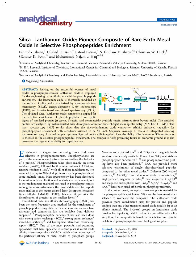
Silica−Lanthanum Oxide: Pioneer Composite of Rare-Earth MetalOxide in Selective Phosphopeptides EnrichmentFahmida Jabeen,† Dilshad Hussain,† Batool Fatima,† S. Ghulam Musharraf,‡ Christian W. Huck,§
Gunther K. Bonn,§ and Muhammad Najam-ul-Haq*,†,§
†Division of Analytical Chemistry, Institute of Chemical Sciences, Bahauddin Zakariya University, Multan 60800, Pakistan‡H. E. J. Research Institute of Chemistry, International Center for Chemical and Biological Sciences, University of Karachi, Karachi75270, Pakistan§Institute of Analytical Chemistry and Radiochemistry, Leopold-Franzens University, Innrain 80-82, A-6020 Innsbruck, Austria
*S Supporting Information
ABSTRACT: Relying on the successful journey of metaloxides in phosphoproteomics, lanthanum oxide is employedfor the engineering of an affinity material for phosphopeptideenrichment. The lanthanum oxide is chemically modified onthe surface of silica and characterized by scanning electronmicroscopy (SEM), energy-dispersive X-ray spectroscopy(EDX), and Fourier transform infrared spectroscopy (FTIR).The obtained silica−lanthanum oxide composite is applied forthe selective enrichment of phosphopeptides from trypticdigest of standard protein (α-casein, β-casein, and commercially available casein mixtures from bovine milk). The enrichedentities are analyzed by matrix-assisted laser desorption/ionization time-of-flight mass spectrometry (MALDI-TOF MS). Themass spectroscopy (MS) results show that the silica−lanthanum oxide composite exhibits enhanced capability forphosphopeptide enrichment with sensitivity assessed to be 50 fmol. Sequence coverage of casein is interpreted showingsuccessful recovery. As a real sample, a protein digest of nonfat milk is applied. Also, the ability of lanthanum in different formatsis checked in the selective phosphopeptides enrichment. The composite holds promising future in economic ground as it alsopossesses the regenerative ability for repetitive use.
Enrichment strategies are becoming more and moreselective in phosphoproteomics. Phosphorylations are
part of the common mechanisms for controlling the behaviorof a protein.1 Phosphorylation takes place mainly on serineresidues (86.4%), followed by threonine residues (11.8%) andtyrosine residues (1.8%).2 With all of these modifications, it isassumed that up to 30% of all proteins may be phosphorylated,some multiple times. Mass spectrometry has been developedfor maximum data collection and analysis after enrichment, as itis the predominant analytical tool used in phosphoproteomics.Among the mass instruments, the most widely used for peptidemass analysis is the matrix-assisted laser desorption ionizationtime-of-flight (MALDI TOF), because it permits theidentification of proteins at the smallest levels.3,4
Immobilized metal ion affinity chromatography (IMAC) hasbeen the most frequently used method for the enrichment ofphosphopeptides using different metal ions on various basematerials and commercial kits are available from differentsuppliers.5−7 Phosphopeptide enrichment has also been donewith strong cation exchange (SCX),8 strong anion exchange,9
mixed-bed sorbents,10 and hydrophilic interaction chromatog-raphy (HIC).11 One of the most powerful and promisingapproaches that have appeared in recent years is metal oxideaffinity chromatography (MOAC), which takes advantage ofthe particular affinity of metal oxides to phosphate groups.
More recently, packed tips12 and TiO2-coated magnetic beadsare also commercially available. Research on TiO2 materials forphosphopeptide enrichment13−15 and phosphoproteome profil-ing have also been published.16 ZrO2 has provided moreselective enrichment of singly phosphorylated peptides,compared to the other metal oxides.17 Different ZrO2-coatedmaterials,18 Al(OH)3
19 and aluminum oxide nanomaterials,20
Ga2O3-coated magnetic particles,21 bare magnetite (Fe3O4)22
and magnetic microspheres with TiO2,23 Al2O3,
24 Ga2O3,25 and
ZrO226 have been used efficiently in phosphoproteomics.
In the present work, we report a new composite material forthe phosphopeptide enrichment. Silica and lanthanum oxide areselected to synthesize the composite. The lanthanum oxideprovides more coordination sites for protein and peptidebinding than any other transition-metal oxide used so far as anaffinity material. The hydroxyl groups on lanthanum oxideprovide hydrophilicity, which makes it compatible with silicaand, thus, the composite is beneficial in efficient and specificisolation of phosphopeptides from biological samples.
Received: September 23, 2012Accepted: November 7, 2012Published: November 7, 2012
Technical Note
pubs.acs.org/ac
© 2012 American Chemical Society 10180 dx.doi.org/10.1021/ac3023197 | Anal. Chem. 2012, 84, 10180−10185
■ EXPERIMENTAL SECTION
Synthesis of the Silica−Lanthanum Oxide Composite.The synthesis of the silica−lanthanum oxide compositeinvolved four steps: (a) hydroxyl functionalization of silicagel, increased by immersing 2.0 g of silica gel in a 1:1 mixture of50% aqueous sulfuric acid:30% hydrogen peroxide for 30min;27 (b) chlorination of hydroxyl functionalized silica, usingthionyl chloride;28 (c) amino modification of lanthanum oxide(0.4 g) by aminopropylsilane (0.2 g), with anhydrous tolueneas a solvent (30 mL); and, finally, (d) synthesis of thecomposite by using product from steps (b) and (c) in thepresence of toluene (30 mL) with magnetic stirring at 80 °C for24 h. The particles were washed with anhydrous toluene, driedovernight under vacuum at 140 °C, and labeled as native SiO2−La2O3.Half of the synthesized composite was end-capped by
treating with 5 mL of hexamethyldisilazane solution in toluene(1:4, v/v) at 60 °C for half an hour, after thorough washingwith 80% ACN and dry toluene (20 mL). Finally, they werewashed with 80% ACN, dried under vacuum, and labeled asend-capped SiO2−La2O3.Sample Preparation: Tryptic Digestion of Standard
Proteins/Nonfat Milk. Tryptic digestion of phosphoproteinsstandards (α and β-casein), commercially available casein, andnonfat milk was carried out by the reported methodology.Detailed digestion protocol is given in the SupportingInformation.Sample Preparation for Spiked Serum. Four different
concentrations of spiked serum were prepared using 40 μL ofserum sample spiked with tryptic digest of β-casein inconcentrations of 1 μg (50 pmol), 100 ng (5 pmol), 10 ng(500 fmol), and 1 ng (50 fmol). The composite was activated,washed and eluted in the same way as in the case ofphosphopeptide enrichment using standard proteins (α- or β-casein).Phosphopeptide Enrichment. Enrichment experiments
were performed by using peptide samples (1 mg/mL) withsilica−lanthanum oxide particles. The peptide sample (trypticdigests of standard proteins and nonfat milk) was loaded afteractivation of the particles by 100 μL 80% ACN in 0.1% (v/v)TFA and incubated for 30 min at room temperature. The
particles were washed with 200 μL of 80% ACN in 0.1% (v/v)TFA, followed by several washings with deionized water. Thesupernatant containing nonphosphorylated peptides wascollected for sequence coverage analysis. Solid material wascollected for material-enhanced laser desorption/ionization(MELDI) analysis29−32 of loaded phosphopeptides. Boundphosphopeptides were eluted by 20 μL ammonium hydroxidesolution (pH 10). The eluted phosphopeptides were acidifiedwith 2 μL of TFA 10% (v/v) for MALDI-MS analysis.
MALDI-MS Analysis of Phosphopeptides. Mass spectrawere recorded on Ultraflex II MALDI-TOF/TOF-MS, (BrukerDaltonics), with CHCA in 50% acetonitrile:0.1% (v/v) TFA(1:1) as matrix. The Flex Analysis Version 3.0 (Bruker, Bremen,Germany) software was used for data processing. Databasesearch was performed by the Mascot program (www.matrixscience.com) to query the SWISS-PROT database,which correctly identified the phosphorylated peptides for α-casein (αS1 and αS2), β-casein, and κ-casein (SWISS-PROTAccession Nos. P02662, P02663, P02666, and P02668,respectively).
Regeneration of the Silica−Lanthanum Oxide Com-posite. To regenerate the already used silica−lanthanum oxidecomposite, particles were washed with an activation buffer (100μL 80% ACN in 0.1% (v/v) TFA) at room temperature. Theactivated composite was then incubated with equilibrationbuffer to restore all the surface functionalities. The regeneratedcomposite was loaded with tryptic casein digest andphosphopeptide analysis was carried out as describedpreviously.
■ RESULTS AND DISCUSSIONChoice of Materials. The ideology behind the selection of
silica and lanthanum oxide for the synthesis of the composite inthis research is supported by various facts. Keeping in view thepriority of research interest, the discussion about the choice ofmaterials and the characterization of the composite by EDAXand SEM is given in the Supporting Information (Figure S1).
Feasibility of Composite in Phosphopeptide Enrich-ment. Bovine β-casein is used to evaluate the performance ofthe silica−lanthanum oxide composite for selective phospho-peptides enrichment. Tryptic digest of β-casein (1 pmol, 2 μL)diluted with 0.1% TFA is incubated with both native and end-
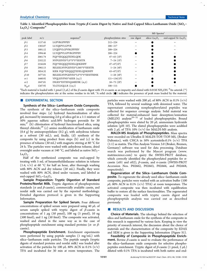
Table 1. Identified Phosphopeptides from Tryptic β-Casein Digest by Native and End-Capped Silica-Lanthanum Oxide (SiO2−La2O3) Compositea
MS Spectrac
peak label m/z sequenceb phosphorylation sites raw digest silica−La2O3 end-capped Si−La2O3
β-1 1157.45 LGPVRGPFPIIV 212−224 − ● −β-2 1383.87 LLYQEPVLGPVR 206−217 − ● −β-3 1881.12 LYQEPVLGPVRGPFPIIV 208−224 − ● −β-4 1994.46 LLYQEPVLGPVRGPFPIIV 206−224 ● ● −β-5 2061.74 FQS*EEQQQTEDELQDK 47−63 (1P) − ● ●β-6 2352.22 NVPGEIVES*LS*S*S*EESITR 7−24 (1P) − ● ●β-7 2556.04 FQS*EEQQQTEDELQDKIHPF 47−67(1P) − ● ●β-8 2965.13 RELEELNVPGEIVES*LS8S*S*EESITR 1−24 (4P) − ● ●β-9 3054.32 KIEK FQS*EEQQQTEDELQDKIHPF 43−67(1P) − ● ●β-10 3477.81 RELEELNVPGEIVES*LS*S*S*EESITRINK 1−28 (4P) − − ●α-1 1660.92 VPQLEIVPNS*AEER (αS1) 121−134(1P) − ● ●α-2 1847.01 DIGES*ESTEDQAMEDIK (αS1) 58−73 (2P) − ● ●Nppα 1267.92 YLGYLEQLLR ((αS1) 106−115 ● ● −
aEach material is loaded with 1 pmol (2 μL) of the β-casein digest with 1% α-casein as an impurity and eluted with 0.01M NH4OH.bAn asterisk (*)
indicates the phosphorylation site at the serine residue to its left. cA solid circle (●) indicates the presence of peak mass loaded by the material.
Analytical Chemistry Technical Note
dx.doi.org/10.1021/ac3023197 | Anal. Chem. 2012, 84, 10180−1018510181
capped composite. Keeping in view the role of buffer conditionsto minimize the nonspecific bindings, the activation and loadingbuffer systems are comprised of TFA, for pH control, andacetonitrile (at levels of 50%−80%), to prevent the hydro-phobic interactions. Using harsh washing conditions with highconcentrations of additives may improve specificity, but canalso result in the loss of weakly bound (mono) phosphopep-tides. Therefore, an activation buffer (80% ACN in 0.1% (v/v)TFA) and Milli-Q water are used for washings. A pH shift toalkaline conditions using ammonium hydroxide (pH 10) isemployed for elution to have better recovery of enrichedphosphopeptides. For comparison, direct analysis of the β-casein digest is performed; the result is presented in Figure S-1A in the Supporting Information. None of the characteristicphosphopeptides derived from β-casein are present in the massspectroscopy (MS) spectrum of raw digest. Some abundantnonphosphopeptides can be observed, which result in lowsignal-to-noise ratio (S/N) for phosphopeptides, if present.Therefore, enrichment is done using native and end-cappedSiO2−La2O3 composites, and the corresponding MALDI-TOFMS spectra are illustrated in Figure S-2 in the SupportingInformation. The identified peptides from β-casein digest arelabeled as β-1 to β-10, with detailed descriptions given in Table1. The comparison is helpful to strengthen the use of end-capping in order to avoid the nonspecific bindings. The MSspectra demonstrate the enrichment of phosphopeptides to thecomposite surface by showing major peaks of phosphopeptidesfrom β-casein (β-5, β-6, β-7, β-8, β-9, and β-10). Since theoriginal protein sample contains a small amount of α-casein,phosphopeptide residues (α-1 and α-2) derived from thisimpurity have also been enriched and detected. In the case ofthe SiO2−La2O3 composite, nonphosphopeptides peaks alsoare observed after enrichment, indicating that there isnonspecific adsorption of peptides onto the SiO2 surface,whereas the end-capped SiO2−La2O3 composite yieldssignificantly higher signal intensity, allowing unambiguousdetection of less-abundant phosphopeptides, such as β-10,and displays a clean background.Selective Enrichment of Phosphopeptides. To evaluate
the selectiveness of the synthesized composite, a semicomplexmixture analysis is done by employing BSA for increasingcomplexity of digested casein mixture. To mimic a complexbiological sample, BSA is added to the tryptic digest of α-caseinand β-casein at different molar ratios (α-casein:β-casein:BSA =1:1:50 and 1:1:100). At a molar ratio of 1:1:50, afterenrichment, the phosphopeptides from α-casein (α-1 at1237.56, α-2 at 1253.32, α-3 at 1411.99, α-4 at 1660.34(PO4
3− adduct), α-5 at 1832.67, α-6 at 1927.90, and α-7 at2247.98), as well as those from β-casein (β at m/z 2061.19) aredetected (see Figure S3 in the Supporting Information). Whenthe molar ratio decreases to 1:1:100, after enrichment, still thephosphopeptides from α-casein (α-4, α-5, and α-6), as well asfrom β-casein (β at 2061.46) can be distinguished, even in thepresence of an exceedingly high concentration of BSA. Theseresults prove that the trace amount of phosphopeptides can beenriched by the SiO2−La2O3 composite, even in the presenceof a large amount of interfering protein.Complex Mixture Analysis. Most common type of casein
phosphorylation involves the formation of phosphate esterbonds with the hydroxyl side chains of serine and, much lessfrequently, threonine. Thus, bovine caseins are phosphorylatedat different levels, and the common genetic variants of αS1-CN,αS2-CN, β-CN, and κ-CN caseins normally contain 8−9, 11−
13, 5, and 1−2 phosphate groups, respectively. Because of thelow abundance of κ-casein (∼1.2%−2%), the phosphopeptidespresent are difficult to enrich; however, the silica−lanthanumoxide composite successfully enriches the phosphopeptides ofκ-casein (the residue Ser-170),33 which is fully phosphorylated.This proves its working efficiency for low abundancephosphopeptides in complex mixtures. Amino modificationsfor casein variants are given as follows: for αS1-CNpSer56,61, 63, 79, 81, 82, 83, 90, 130;34 for αS2-CNpSer23, 24, 25,31, 71, 72, 73, 76, 144, 146, 158;35 for β-CNpSer30, 32, 33,34, 50;36 and for κ-CNpSer 148, 170.37 Therefore, trypticdigest of commercially available casein is taken as a complexmixture containing all three variants of casein (αS1, αS2, β as amaximum, with a trace amount of κ-casein) in the ratio4:1:4:1.38
After loading, the SiO2−La2O3 composite is investigatedregarding its applicability as a MELDI carrier material, followedby MS analysis (see Figure S-4A in the SupportingInformation). MELDI analysis provides a complete profile fortryptic casein digest; however, for identification purposes, theelution of adsorbed peptides is necessary. All the phosphopep-tides shown by MELDI MS spectra have been successfullyeluted (see Figure S-4B in the Supporting Information) whichconfirms that the adsorption of peptides on composite isreversible. It is important to mention that the direct MALDIanalysis of loaded sample on material (MELDI), as well as theanalysis of eluted phosphopeptides, has resulted in the samepeak pattern. The MS spectrum shows characteristic peaks ofcasein variants. A complete list of identified peptide signals,along with amino acid sequences and phosphorylation sites, isgiven in Table S-5 in the Supporting Information. MS spectrafor nonphosphorylated peptides are also recorded for thesequence coverage.
Sequence Coverage. Earlier sequence coverage for trypticcasein digest (αS1-CN (52.8%), αS2-CN (18.9%), β-CN(39.7%), and κ-CN (11.0%) has been reported by usingZrO2.
39 The commercially available casein is selected as thesample, because it contains many nonphosphorylated counter-parts (κ-casein as glycoprotein),40 which complicate theselective enrichment of phosphopeptides from the trypticdigest. The databases are available for casein variants whichhelp to score the data with better confidence level and easier tocompare and report. It strengthens the in-solution proteomicapproach, giving a relatively good characterization of proteinmixture of moderate complexity. The sequence coverage afterenrichment of casein digest by SiO2−La2O3 composite hasshown compatible results (see Table S6 in the SupportingInformation), which helps to move onto the real sampleapplication using the same enrichment protocol.
Regeneration Studies. The regeneration of adsorbents is acrucial step in all affinity separation techniques. The mosteconomic aspect of SiO2−La2O3 composite is its regenerativeproperty. The MS spectra of eluted fractions show that all ofthe enriched phosphopeptides have been successfully eluted.Therefore, the solid material is collected after the elution andagain spotted onto the MALDI target plate. Surprisingly, thereis not a single peak in MS spectrum (see Figure S-7A in theSupporting Information). The idea of regeneration popped upinstantly and the used composite is again activated by applyingpreviously described conditions. The used composite isthoroughly washed with deionized water and then withactivation buffer, to restore all the surface chemistry of thecomposite. The entire enrichment procedure is performed
Analytical Chemistry Technical Note
dx.doi.org/10.1021/ac3023197 | Anal. Chem. 2012, 84, 10180−1018510182
using casein digest and MS spectra are recorded for MELDIand eluted phosphopeptides (see Figures S-7B and 7C in theSupporting Information). It can be observed that most of thephosphopeptides has been enriched by the regeneratedcomposite, showing its successful regeneration. It providesthe possibility of using the composite several times, however,the reuse over a longer period of time depends substantially onachieving and maintaining the chemical functionalities of thecomposite. The comparison shows that the regenerated SiO2−La2O3 composite has enriched phosphopeptides as efficiently asthe original composite.Real Sample Application. Sensitivity Assessment for
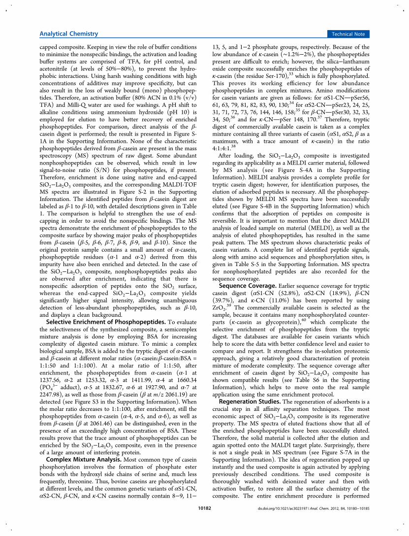
Serum. In the case of advanced cancer treatment withantiangiogenesis agents and protein kinase inhibitors,41 thephospho-modified proteins are secreted from cells into thecirculation system and are available in the serum. However,direct determination of these modified proteins in a biologicalsystem has been difficult, because of the serum complexity andlower concentrations. There is no method available to studythis aspect in detail with consistency.42 In order to estimate thedetection limit for phosphopeptides enriched from serum, asensitivity assessment is done using spiked serum with trypticdigest of β-casein in different ratios (40 μL serum samplespiked with 1 μg (50 pmol), 100 ng (5 pmol), 10 ng (500fmol), and/or 1 ng (50 fmol) β-casein). The identifiedphosphopeptides from β-casein are at m/z 1103(KFQS*EEQQQT), 1473 (KKIEKFQS*EEQQQT), and2061 (FQS*EEQQQTEDELQDK), with relatively good MSpeak intensity. However, a 1-ng β-casein-spiked sample yieldedlow MS peak intensity (see Figures 1A−D). These resultsindicate that developed method allows the detection of serum
phosphoproteins/phosphopeptides with amounts greater than∼10−100 fmol. The identification also shows that method hashuge potential to enrich the phospho- content from complexreal samples such as serum.
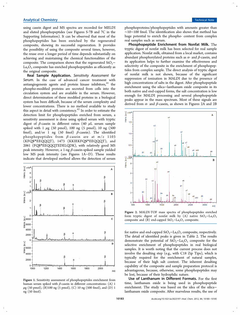
Phosphopeptide Enrichment from Nonfat Milk. Thetryptic digest of nonfat milk has been selected for real sampleapplication. Nonfat milk, obtained from a local market, containsabundant phosphorylated proteins such as α- and β-casein, andits application helps to further examine the effectiveness andselectivity of the composite in the enrichment of phosphopep-tides from complex sample. The direct analysis of tryptic digestof nonfat milk is not shown, because of the significantsuppression of ionization in MALDI due to the presence ofhigh concentrations of salts in the digest. After phosphopeptideenrichment using the silica−lanthanum oxide composite in itsboth native and end-capped forms, the salt concentration is lowenough for MALDI processing and several phosphopeptidepeaks appear in the mass spectrum. Most of these signals arederived from α- and β-casein, as shown in Figures 2A and 2B
for native and end-capped SiO2−La2O3 composite, respectively.The detail of identified peaks is given in Table 2. The resultsdemonstrate the potential of SiO2−La2O3 composite for theselective enrichment of phosphopeptides in real biologicalsamples. It is worth noting that the current process does notinvolve the desalting step (e.g., with C18 Zip Tips), which istypically required for the enrichment of natural samples,because of their high salt content. The inherent desaltingcapability of the composite and sample preparation protocol isadvantageous, because, otherwise, some phosphopeptides maybe lost, because of their hydrophilic nature.
Use of Lanthanum in Different Formats. For the firsttime, lanthanum oxide is being used in phosphopeptideenrichment. The study was based on the idea of the silica−lanthanum oxide composite. After marvelous results, the use of
Figure 1. Sensitivity assessment of phosphopeptides enrichment fromhuman serum spiked with β-casein in different concentrations: (A) 1μg (50 pmol), (B)100 ng (5 pmol), (C) 10 ng (500 fmol), and (D) 1ng (50 fmol).
Figure 2. MALDI-TOF mass spectra of phosphopeptides enrichedform tryptic digest of nonfat milk by (A) native SiO2−La2O3composite and (B) end-capped SiO2−La2O3 composite.
Analytical Chemistry Technical Note
dx.doi.org/10.1021/ac3023197 | Anal. Chem. 2012, 84, 10180−1018510183
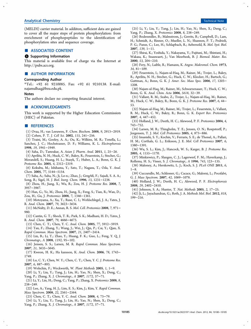
lanthanum in different formats was also investigated to comparethe enrichment efficiency from digested protein samples. Thelanthanum is loaded on IMAC material as La3+, as thecounterpart in a composite (SiO2−La2O3 composite), and as anoxide itself (La2O3). Tryptic β-casein digest is employed toinvestigate the use of lanthanum and to study the differences inthe enrichment of phosphopeptides, if any. The MS results (seeFigures 3A−C) provide the evidence that, in any format,lanthanum ensures maximum enrichment without any prom-inent difference. The efficient enrichment of phosphopeptidesin the case of La3+ ions loaded on IMAC material might be dueto the presence of an f-orbital, which offers coordination bondsto phosphopeptides, as in the case of transition metals (Fe3+,Zr4+, Ti4+) used in IMAC loadings so far. As a composite, itsworking efficiency has been checked in detail as the SiO2−La2O3 composite (see Table S8 in the Supporting Informa-tion). The presence of nonspecific bindings in the case ofcomposite or loaded IMAC material can be attributed to thebase material. Therefore, end-capping of the IMAC orcomposite base material is necessary to reduce the nonspecificbindings. As a pure oxide (La2O3), there is neat spectrum andmaximum phosphopeptide enrichment. So, this comparisonprovides choices in the research field, showing that lanthanumcan be used in solution, as well as particle form, withoutcompromising on the loss of phosphopeptides.
■ CONCLUSION
In nature, protein modifications, such as phosphorylation,contribute to the complex signaling pathways. There is alwaysroom for new materials to enrich them in order to study theprotein phosphorylation. Silica−lanthanum oxide compositesynthesized from chemical derivatization of silica andlanthanum oxide provides an efficient media for selectiveenrichment of phosphopeptides from standard protein digestsand real samples such as milk and serum. The elution
conditions are compatible for balanced recovery of singly andmultiply phosphorylated peptides. The regeneration phenom-enon enhances the commercial value of the silica−lanthanumoxide composite. The material also has been proven to be anefficient material-enhanced laser desorption/ionization
Table 2. Identified Tryptic Peptide Fragments of Nonfat Milk Digest Enriched by the Silica−Lanthanum Oxide (SiO2−La2O3)Composite
peak label [M+H]+ amino acid sequencea sequence No. native SiO2−La2O3b end-capped SiO2−La2O3
b,c
α-Caseinα-1 1197.009 KNMAINPS*KENL (αS2) 39−50 (1P) ● ●α-2 1253.301 TVDMMES*TEVF (αS2) 153−162 (1P) ● ●α-3 1330.388 EQLS*TS*EENSK (αS2) 141−151 (2P) ● ●α-4 1660.510 VPQLEIVPNS*AEER (αS1) 121−134 (1P) ● ●α-5 1954.668 YKVPQLEIVPNS*AEER (αS1) 119−134 (1P) ● ●α-6 2202.873 YKVPQLEIVPNS*AEERLHS (αS1) 119−137 (1P) ● ●α-7 2247.190 KEKVNELS*KDIGS*ES*TEDQA (αS1) 49−68 (3P) ● ●α-8 2362.623 PNS*VEQKHIQKEDVPSERY (αS1) 88−106 (1P) ● ●α-9 2453.609 NTMEHVS*S*S*EES*IISQETY (αS2) 17−35 (4P) ● ●α-10 2616.678 NTMEHVS*S*S*EES*IISQETYK (αS2) 17−36 (4P) ● ●
β-Caseinβ-1 847.054 KFQS*EEQQ 47−54 (1P) ● ●ß-2 975.768 KFQS*EEQQQ 47−55 (1P) ● ○β-3 1103.906 KFQS*EEQQQT 47−56 (1P) ● ●β-4 1473.009 KKIEKFQS*EEQQQT 43−56 (1P) ● ●β-5 1688.459 LTDVENLHLPLPLLQSW 142−157 ● ○β-6 1881.107 LLYQEPVLGPVRGPFPIIV 206−224 ● ○β-7 2186.740 DMPIQAFLLQEPVLGPVR 199−217 ● ●β-8 2910.255 DMPIQAFLLQEPVLGPVRGPFPIIV 199−224 ● ○β-9 3054.779 KKIEKFQS*EEQQQTEDELQDKIHPFA 43−67 (1P) ● ●
aAsterisk (*) indicates the phosphorylation site at the serine residue to its left. bSolid circle indicates the presence of peak mass loaded by thematerial. cOpen circle indicates the absence of peak mass not enriched by material.
Figure 3. MALDI-TOF mass spectra of phosphopeptides enrichedfrom mixture of tryptic digest of α-casein and β-casein (1 pmol, 2 μL)in ratio of 1:1 by (A) IMAC material loaded with La3+ (0.01 M LaCl3solution for loading); (B) SiO2−La2O3 composite; and (C) activatedLa2O3 particles (commercially available).
Analytical Chemistry Technical Note
dx.doi.org/10.1021/ac3023197 | Anal. Chem. 2012, 84, 10180−1018510184
(MELDI) carrier material. In addiiton, sufficient data are gainedto cover all the major steps of protein phosphorylation: fromenrichment of phosphopeptides to the identification ofphosphorylation sites and sequence coverage.
■ ASSOCIATED CONTENT*S Supporting InformationThis material is available free of charge via the Internet athttp://pubs.acs.org.
■ AUTHOR INFORMATIONCorresponding Author*Tel.: +92 61 9210085. Fax: +92 61 9210138. E-mail:[email protected] authors declare no competing financial interest.
■ ACKNOWLEDGMENTSThis work is supported by the Higher Education Commission(HEC) of Pakistan.
■ REFERENCES(1) Ovaa, H.; van Leeuwen, F. Chem. Biochem. 2008, 9, 2913−2919.(2) Cohen, P. T. J. Cell Sci. 2002, 115, 241−256.(3) Traini, M.; Gooley, A. A.; Ou, K.; Wilkins, M. R.; Tonella, L.;Sanchez, J. C.; Hochstrasser, D. F.; Williams, K. L. Electrophoresis1998, 19, 1941−1949.(4) Saha, D.; Tamrakar, A. Asian J. Pharm. Anal. 2011, 1, 25−26.(5) Aprilita, N. H.; Huck, C. W.; Bakry, R.; Feuerstein, I.; Stecher, G.;Morandell, S.; Huang, H. L.; Stasyk, T.; Huber, L. A.; Bonn, G. K. J.Proteome Res. 2005, 4, 2312−2319.(6) Kokubu, M.; Ishihama, Y.; Sato, T.; Nagasu, T.; Oda, Y. Anal.Chem. 2005, 77, 5144−5154.(7) Saha, A.; Saha, N.; Ji, Lo-n.; Zhao, J.; GregAfi, F.; Sajadi, S. A. A.;Song, B.; Sigel, H. J. Biol. Inorg. Chem. 1996, 13, 1231−1238.(8) Zhou, H.; Jiang, X.; Wu, R.; Zou, H. J. Proteome Res. 2008, 7,3957−3967.(9) Han, G.; Ye, M.; Zhou, H.; Jiang, X.; Feng, S.; Tian, R.; Wan, D.;Zou, H.; Gu, J. Proteomics 2008, 7, 1346−1361.(10) Motoyama, A.; Xu, T.; Ruse, C. I.; Wohlschlegel, J. A.; Yates, J.R. Anal. Chem. 2007, 79, 3623−3634.(11) McNulty, D. E.; Annan, R. S.Mol. Cell. Proteomics 2008, 7, 971−980.(12) Cantin, G. T.; Shock, T. R.; Park, S. K.; Madhani, H. D.; Yates, J.R. Anal. Chem. 2007, 79, 4666−4673.(13) Chen, C. T.; Chen, Y. C. Anal. Chem. 2005, 77, 5912−5919.(14) Tan, F.; Zhang, Y.; Wang, J.; Wei, J.; Qin, P.; Cai, Y.; Qian, X.Rapid Commun. Mass Spectrom. 2007, 21, 2407−2414.(15) Lin, B.; Li, T.; Zhao, Y.; Huang, F. K.; Guo, L.; Feng, Y. Q. J.Chromatogr., A 2008, 1192, 95−102.(16) Jensen, S. S.; Larsen, M. R. Rapid Commun. Mass Spectrom.2007, 21, 3635−3645.(17) Kweon, H. K.; Ha kansson, K. Anal. Chem. 2006, 78, 1743−1749.(18) Lo, C. Y.; Chen, W. Y.; Chen, C. T.; Chen, Y. C. J. Proteome Res.2007, 6, 887−893.(19) Wolschin, F.; Weckwerth, W. Plant Methods 2005, 1, 1−9.(20) Li, Y.; Liu, Y.; Tang, J.; Lin, H.; Yao, N.; Shen, X.; Deng, C.;Yang, P.; Zhang, X. J. Chromatogr., A 2007, 1172, 57−71.(21) Li, Y.; Lin, H.; Deng, C.; Yang, P.; Zhang, X. Proteomics 2008, 8,238−249.(22) Lee, A.; Yang, H. J.; Lim, E. S.; Kim, J.; Kim, Y. Rapid Commun.Mass Spectrom. 2008, 22, 2561−2564.(23) Chen, C. T.; Chen, Y. C. Anal. Chem. 2008, 4, 73−79.(24) Li, Y.; Liu, Y.; Tang, J.; Lin, H.; Yao, N.; Shen, X.; Deng, C.;Yang, P.; Zhang, X. J. Chromatogr., A 2007, 1172, 57−71.
(25) Li, Y.; Liu, Y.; Tang, J.; Lin, H.; Yao, N.; Shen, X.; Deng, C.;Yang, P.; Zhang, X. Proteomics 2008, 8, 238−249.(26) Bodenmiller, B.; Malmstrom, J.; Gerrits, B.; Campbell, D.; Lam,H.; Schmidt, A.; Rinner, O.; Mueller, L. N.; Shannon, P. T.; Pedrioli,P. G.; Panse, C.; Lee, H.; Schlapbach, R.; Aebersold, R. Mol. Syst. Biol.2007, 139, 1−11.(27) Shirai, K.; Yoshida, Y.; Nakayama, Y.; Fujitani, M.; Shintani, H.;Wakasa, K.; Snauwaert, J.; Van Meerbeek, B. J. Biomed. Mater. Res.2000, 53, 204−210.(28) Fery, N.; Laible, R.; Hamann, K. Angew. Makromol. Chem. 1973,34, 81−109.(29) Feuerstein, I.; Najam-ul-Haq, M.; Rainer, M.; Trojer, L.; Bakry,R.; Aprilita, N. H.; Stecher, G.; Huck, C. W.; Klocker, H.; Bartsch, G.;Guttman, A.; Bonn, G. K. J. Amer. Soc. Mass Spec. 2006, 17, 1203−1208.(30) Najam-ul-Haq, M.; Rainer, M.; Schwarzenauer, T.; Huck, C. W.;Bonn, G. K. Anal. Chim. Acta 2006, 5618, 32−39.(31) Vallant, R. M.; Szabo, Z.; Trojer, L.; Najam-ul-Haq, M.; Rainer,M.; Huck, C. W.; Bakry, R.; Bonn, G. K. J. Proteome Res. 2007, 6, 44−53.(32) Najam-ul-Haq, M.; Rainer, M.; Trojer, L.; Feuerstein, I.; Vallant,R. M.; Huck, C. W.; Bakry, R.; Bonn, G. K. Expert Rev. Proteomics2007, 4, 447−452.(33) Holland, J. W.; Deeth, H. C.; Alewood, P. F. Proteomics 2004, 4,743−752.(34) Larsen, M. R.; Thingholm, T. E.; Jensen, O. N.; Roepstorff, P.;Jorgensen, T. J. Mol. Cell Proteomics 2005, 4, 873−886.(35) Imanishi, S. Y.; Kochin, V.; Ferraris, S. E.; de Thonel, A.; Pallari,H. M.; Corthals, G. L.; Eriksson, J. E. Mol. Cell Proteomics 2007, 6,1380−1391.(36) Wu, S. L.; Kim, J.; Hancock, W. S.; Karger, B. J. Proteome Res.2005, 4, 1155−1170.(37) Minkiewicz, P.; Slangen, C. J.; Lagerwerf, F. M.; Haverkamp, J.;Rollema, H. S.; Visser, S. J. Chromatogr., A 1996, 743, 123−135.(38) Maloney, A.; Herskowitz, L. J.; Koch, S. J. PLoS ONE 2011, 6,1−8.(39) Cuccurullo, M.; Schlosser, G.; Cacace, G.; Malorni, L.; Pocsfalvi,G. J. Mass Spectrom. 2007, 42, 1069−1078.(40) Holland, J. W.; Deeth, H. C.; Alewood, P. F. Electrophoresis2008, 29, 2402−2410.(41) Johnson, S. A.; Hunter, T. Nat. Methods 2005, 2, 17−25.(42) Ji, L.; Jayachandran, G.; Roth, J. A. Methods Mol. Biol. 2012, 818,199−216.
Analytical Chemistry Technical Note
dx.doi.org/10.1021/ac3023197 | Anal. Chem. 2012, 84, 10180−1018510185
Copyright © 2022 FDOKUMEN