Sildenafil 4.0—Integrated Synthetic Chemistry, Formulation ...
Upload
independentCategory
view
1download
0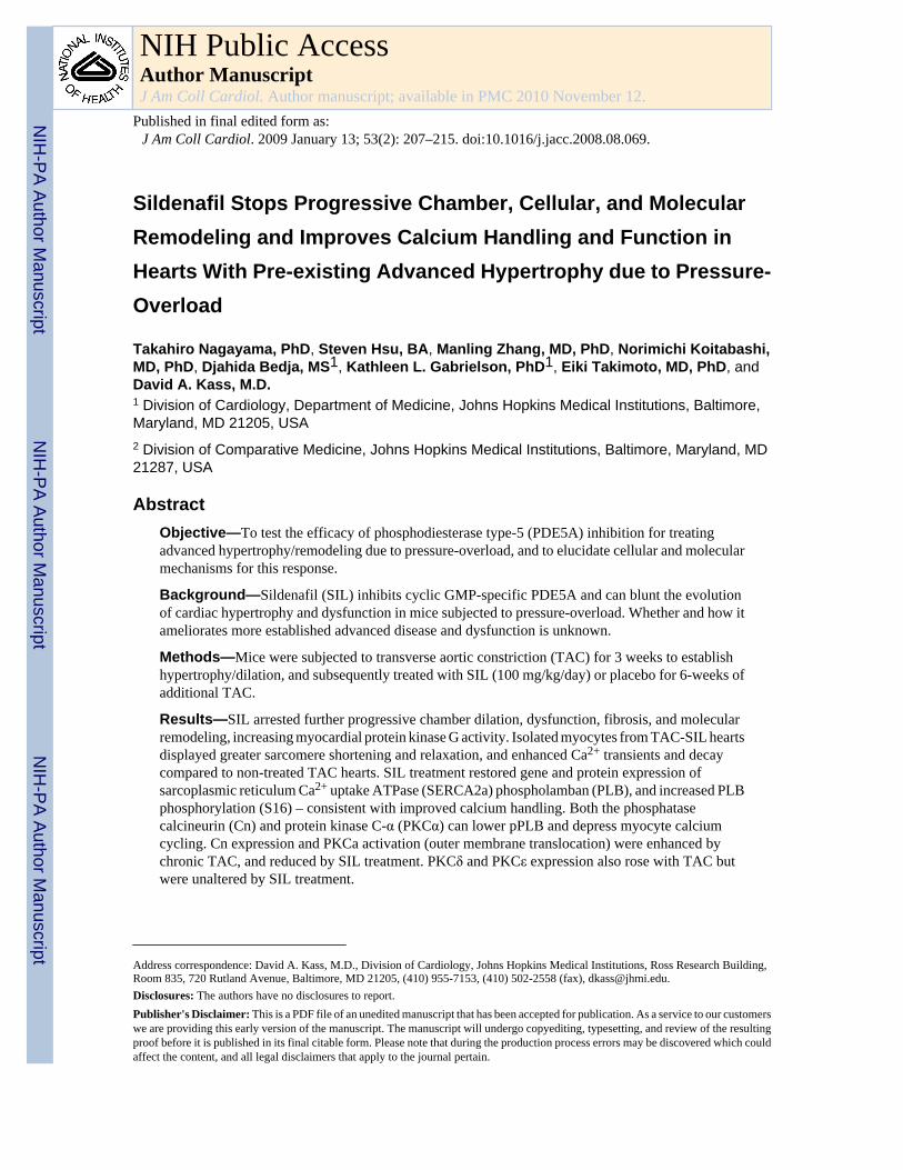
Sildenafil Stops Progressive Chamber, Cellular, and MolecularRemodeling and Improves Calcium Handling and Function inHearts With Pre-existing Advanced Hypertrophy due to Pressure-Overload
Takahiro Nagayama, PhD, Steven Hsu, BA, Manling Zhang, MD, PhD, Norimichi Koitabashi,MD, PhD, Djahida Bedja, MS1, Kathleen L. Gabrielson, PhD1, Eiki Takimoto, MD, PhD, andDavid A. Kass, M.D.1 Division of Cardiology, Department of Medicine, Johns Hopkins Medical Institutions, Baltimore,Maryland, MD 21205, USA2 Division of Comparative Medicine, Johns Hopkins Medical Institutions, Baltimore, Maryland, MD21287, USA
AbstractObjective—To test the efficacy of phosphodiesterase type-5 (PDE5A) inhibition for treatingadvanced hypertrophy/remodeling due to pressure-overload, and to elucidate cellular and molecularmechanisms for this response.
Background—Sildenafil (SIL) inhibits cyclic GMP-specific PDE5A and can blunt the evolutionof cardiac hypertrophy and dysfunction in mice subjected to pressure-overload. Whether and how itameliorates more established advanced disease and dysfunction is unknown.
Methods—Mice were subjected to transverse aortic constriction (TAC) for 3 weeks to establishhypertrophy/dilation, and subsequently treated with SIL (100 mg/kg/day) or placebo for 6-weeks ofadditional TAC.
Results—SIL arrested further progressive chamber dilation, dysfunction, fibrosis, and molecularremodeling, increasing myocardial protein kinase G activity. Isolated myocytes from TAC-SIL heartsdisplayed greater sarcomere shortening and relaxation, and enhanced Ca2+ transients and decaycompared to non-treated TAC hearts. SIL treatment restored gene and protein expression ofsarcoplasmic reticulum Ca2+ uptake ATPase (SERCA2a) phospholamban (PLB), and increased PLBphosphorylation (S16) – consistent with improved calcium handling. Both the phosphatasecalcineurin (Cn) and protein kinase C-α (PKCα) can lower pPLB and depress myocyte calciumcycling. Cn expression and PKCa activation (outer membrane translocation) were enhanced bychronic TAC, and reduced by SIL treatment. PKCδ and PKCε expression also rose with TAC butwere unaltered by SIL treatment.
Address correspondence: David A. Kass, M.D., Division of Cardiology, Johns Hopkins Medical Institutions, Ross Research Building,Room 835, 720 Rutland Avenue, Baltimore, MD 21205, (410) 955-7153, (410) 502-2558 (fax), [email protected]: The authors have no disclosures to report.Publisher's Disclaimer: This is a PDF file of an unedited manuscript that has been accepted for publication. As a service to our customerswe are providing this early version of the manuscript. The manuscript will undergo copyediting, typesetting, and review of the resultingproof before it is published in its final citable form. Please note that during the production process errors may be discovered which couldaffect the content, and all legal disclaimers that apply to the journal pertain.
NIH Public AccessAuthor ManuscriptJ Am Coll Cardiol. Author manuscript; available in PMC 2010 November 12.
Published in final edited form as:J Am Coll Cardiol. 2009 January 13; 53(2): 207–215. doi:10.1016/j.jacc.2008.08.069.
NIH
-PA Author Manuscript
NIH
-PA Author Manuscript
NIH
-PA Author Manuscript
Conclusions—SIL treatment applied to well established hypertrophic cardiac disease can preventfurther cardiac and myocyte dysfunction and progressive remodeling. This is associated withimproved calcium cycling, and reduction of calcineurin and PKCα activation may be important tothis improvement.
KeywordsPDE5; pressure-overload; hypertrophy; myocyte; cardiac function
INTRODUCTIONHeart failure is a leading cause of morbidity and mortality worldwide, commanding a largeshare of health care resources, and despite recent advances, better treatment options remainsorely needed(1). Hypertension and corresponding hypertrophic heart disease are major riskfactors for heart failure, and the therapeutic suppression of hypertrophy is associated withimproved mortality(2;3). To date, treatments designed to blunt hypertrophic remodeling andrelated dysfunction have largely focused on reducing vascular load and neuro-hormonestimulation. However, intracellular pathways are also now being targeted which may providepotent benefits by interfering with strategic nodes in more distal signaling processes(1;4).
An intriguing approach that first arose somewhat as a surprise is the inhibition of cGMP-specific phosphodiesterase 5A (PDE5A) by compounds such as sildenafil. Such drugs arewidely used to treat erectile dysfunction, and long thought to have little role in the heart.However, growing evidence supports cardiac expression and functionality of PDE5A(5)physiologic effects from its inhibitors. For example, PDE5A inhibition can improve ischemiccardiomyopathy (6), doxorubicin toxicity(7) and pressure-overload induced hypertrophy(8),all thought coupled to enhanced protein kinase G activation(9;10). Clinical studies of sildenafilin heart failure patients have reported improved exercise capacity coupled to reducedpulmonary vascular resistance and better endothelial function(11;12). Based on such findings,the NIH is to initiate a multi-center clinical trial of sildenafil for the treatment of heart failurewith a preserved ejection fraction.
We previously reported that sildenafil exerts anti-hypertrophic effects against pressure-overload when the treatment is initiated at the onset of the stress or shortly (1-week) thereafter(8). In the latter case, hearts first developed concentric hypertrophy with no dilation, and thenimproved with treatment. However, it is unknown whether sildenafil can ameliorate moreadvanced disease, a non-trivial question as PDE5A modulation appears to target nitric oxide-derived cGMP(9;10;13) and nitric oxide synthase (NOS) activity declines with sustainedpressure-overload(14). Furthermore, the mechanisms of sildenafil-improved cardiac functionin pressure-overload hearts(8) and whether they apply to more advanced disease remainunknown. Accordingly, the present study tested the efficacy of delayed sildenafil treatment ofhearts subjected to pressure-overload. We show evidence of chamber, cellular, and molecularbenefits of PDE5A inhibition in advanced disease, and reveal novel mechanisms for functionalimprovement related to calcium cycling and its molecular modulation.
MATERIALS AND METHODSAnimal models
Male C57Bl/6 mice (age 9–12 wks; Jackson Labs, Bar Harbor, ME) were used. Pressure-overload was produced by transverse aortic constriction (TAC) (8), with shams undergoingthe same operation without aortic constriction. Oral sildenafil (100 mg/kg/d) was provided insoft diet (Bioserve, MA)(8) starting on week 4 following TAC and continuing for 5 additionalweeks. While this dose is high for humans, the mouse metabolizes sildenafil at a higher rate
Nagayama et al. Page 2
J Am Coll Cardiol. Author manuscript; available in PMC 2010 November 12.
NIH
-PA Author Manuscript
NIH
-PA Author Manuscript
NIH
-PA Author Manuscript

(15), and this dose yields a free plasma concentration of 10–15 nM, within the specific andtherapeutic range for PDE5A(8). Control data for chronic sildenafil only have been previouslyreported(8), and effects found to be negligible. The Johns Hopkins University Animal Careand Use Committee approved the protocol.
Physiological studiesCardiac function was assessed by transthoracic echocardiography in conscious mice (SequoiaC256, Siemens, NY) using a 15 MHz linear-array transducer, and by invasive pressure-volumeanalysis using the conductance catheter method (SPR-839, Millar Instruments, Inc., Houston,TX). The catheter was inserted via the LV apex in open-chest, anesthetized mice, andpositioned along the long-axis as described(8)
HistologyFormalin (10%) fixed paraffin embedded LV myocardium was sectioned (5 μm) and stainedwith hematoxylin/eosin or picrosirius red. Myocyte diameter and interstitial and peri-vascularcollagen fraction were determined by digital image analysis (Adobe Photoshop 7.0, NIH ImageJ), with the observer blinded as to tissue source. Four different hearts in each group, with fiveseparate fields of cells (total 50–70 cells for each heart) were analyzed.
Isolated myocyte physiologic studiesSarcomere shortening (Myocam, IonOptix, Milton, MA) and whole-cell Ca2+ transient (Fura-2AM μmol/L; Invitrogen, Carlsbad, CA) was assessed in freshly isolated adult cardiomyocytes(4 hearts/group) at 27°C using 0.5 Hz field stimulation (15–20 cells from each heart) aspreviously described(10). Though lower temperatures can depress adrenergic modulation, wehave shown marked concordance of adrenergic regulation by PDE5 inhibitors under variousconditions between such in vitro conditions and in vivo hearts(9;10). Data at rest and afterisoproterenol (ISO; 10 nmol/L) stimulation were obtained.
Protein and Gene ExpressionProtein and mRNA isolates were prepared from LV tissue flash frozen in liquid nitrogen, andexpression assessed using standard techniques (see online-supplemental methods).
Immunofluorescent HistologyPKCα myocyte localization was examined with antisera by confocal fluorescentimmunohistochemistry as described (10) (See online supplemental methods).
Statistical AnalysisData are presented as mean ± SEM. Differences between groups were assessed by 1- or 2-wayanalysis of variance (with or without repeated measures, as required) followed by a Tukey’smultiple comparisons test. In instances where within-group variance differed substantially, anon- parametric Kruskal-Wallis test and Bonferoni correction was used.
RESULTSSildenafil inhibits progressive hypertrophy and dilation in mice subjected to TAC
After 3-wk TAC, hearts had a +135% rise in LV mass, chamber end-systolic (+91%) and end-diastolic (+10%) dimensions, and reduced fractional shortening (−42%; Fig 1a). Subsequenttreatment with sildenafil fully arrested progressive remodeling whereas control hearts furtherdilated and hypertrophied after 9 weeks TAC. Post-mortem analysis confirmed both heart andlung weight, normalized to tibia length, was lower with sildenafil treatment (Fig 1b). Myocyte
Nagayama et al. Page 3
J Am Coll Cardiol. Author manuscript; available in PMC 2010 November 12.
NIH
-PA Author Manuscript
NIH
-PA Author Manuscript
NIH
-PA Author Manuscript

cross sectional dimension and interstitial and peri-vascular fibrosis was also reduced in SIL-treated myocardium (Fig 1c).
A- and B-type natriuretic peptide gene expression rose markedly after 3-wks-TAC. Bothincreased 30–50% after 9-wks-TAC in non-treated hearts but remained at 3-wk levels in thosereceiving sildenafil (Fig 2a). β-myosin heavy chain also increased, but this was not reduced insildenafil treated hearts.
Modulation of PKG activity and fetal gene expressionWe next assessed the activation of cGMP-stimulated protein kinase G (PKG) in hearts with orwithout delayed sildenafil treatment. Enzyme activity assessed by S-239-VASPphosphorylation and in vitro kinase assay both showed increases after 9wk-TAC that werefurther enhanced in SIL treated animals (Fig 2b). TAC led to increased PKG-1α (primarycardiac isoform) protein expression (Fig 2c), but this declined to normal levels with SILtreatment, supporting post-translational (cGMP-stimulation) mechanisms in this setting.PDE5A protein expression was unaltered among the various conditions.
Sildenafil treatment improves cardiac contractility and relaxation in vivo and in vitroFigure 3A shows the results of LV pressure-volume analyses. After 3-wks-TAC, hearts dilatedand had increased end-systolic elastance (Ees) and contractility (dP/dtmax/IP and maximalpower index), as reported in humans with hypertrophic heart disease(16). More sustained TACreduced contractility and prolonged relaxation in untreated hearts but this was prevented bySIL.
To identify mechanisms for improved cardiac function, LV myocytes were studied (Fig 3b).Compared to sham controls, 9-wk-TAC myocytes had depressed sarcomere shortening andprolonged relaxation, and SIL treatment restored both to control levels. These changes wereaccompanied by improved calcium handling. The peak Ca2+-transient was not significantlyaltered after 9-wk-TAC but the time for 50% decline prolonged. The latter improved towardscontrol values with SIL treatment, while peak amplitude rose. Contraction, relaxation andCa2+-transients all improved similarly with ISO in each group (p-value for interaction by 2-Way ANOVA ranged 0.2–0.94); thus, this cascade was not differentially affected by SILtreatment.
Sildenafil treatment modifies SR Ca2+ handling protein expression and phosphorylationSince CA2+-cycling improved in SIL treated TAC hearts, we examined sarcoplasmic reticulum(SR) handling proteins that might be modified by the therapy. Gene expression of SR-Ca2+
ATPase (SERCA2a) and phospholamban declined after 3-wks-TAC (Fig 4a) and this persistedafter 9-wks-TAC. Protein expression was declined at this time as well. SIL treatment increasedboth SERCA2a and PLB expression, and enhanced PLB s-23 phosphorylation (increasingCa2+-uptake), in comparison to non-treated TAC.
Sildenafil suppresses calcineurin and PKCα activationIncreased PLB phosphorylation by SIL is consistent with enhanced PKG activity, but it couldalso result from suppressed phosphatase activity. One mechanism involves calcineurin (Cn)which inhibits I-1 activity resulting in the dis-inhibition of the phosphatase PP1 to lower PLBphosphorylation(17;18). Cn protein expression and activity (indexed by RCAN-1 mRNA) roseafter 3-wks-TAC by 3.2 and 2.5-fold, respectively (both p<0.01, on-line supplemental Figure1), and remained elevated at 9-wks-TAC in non-treated hearts but declined with SIL (Fig. 4c).Another mechanism for I-1 inactivation is its phosphorlyation by PKCα at S67 and T75 whichalso enhances PP1 activity and thus PLB de-phosphorylation (19). PKCα (and δ,ε isoforms)
Nagayama et al. Page 4
J Am Coll Cardiol. Author manuscript; available in PMC 2010 November 12.
NIH
-PA Author Manuscript
NIH
-PA Author Manuscript
NIH
-PA Author Manuscript
protein expression rose markedly after 9-wks-TAC (Fig 4d), and SIL treatment did not reducethem. However, PKC-α activity is coupled to its translocation to the outer membrane, and thiswas observed after both 3 and 9-wks of TAC (Fig 5a). PKC-α returned to a diffuse distributionconsistent with reduced activation following SIL treatment. Confocal analysis was confirmedby cell fractionation/immunoblot analysis (Fig 5b). To test if SIL directly targeted PKC-αactivation, freshly isolated cardiomyocytes were incubated with the PKC activator PMA. PMAinduced rapid PKC-α membrane translocation (Fig. 5c) but which SIL co-incubation did notsuppress, suggesting the in vivo effect was more likely indirect.
DISCUSSIONCardiac hypertrophy and attendant myocardial remodeling and myocyte and chamberdysfunction remain major causes of morbidity and mortality worldwide, and new approachesto combat this pathophysiology are needed. In a prior study, we first showed that PDE5Ainhibition coupled to activation of PKG may offer a novel approach to treating this disorder(8). The present results substantially extend this finding. First therapy was initiated only afterthe hypertrophic disease process was far more established, yet improvements in function,remodeling, and molecular signaling were achieved. Second, isolated myocytes were studiedrevealing enhanced myocyte contraction/relaxation and Ca2+ handling under both rest and β-AR stimulated conditions. Third, we extended prior mechanistic analysis, showingimprovement of SR calcium handling proteins coupled with suppression of both Cn and PKC-α activation. These findings further support a translational potential for PDE5A inhibitors inestablished hypertrophic heart disease.
Treating hypertrophy and cardiac failure via a cGMP/PKG/PDE5 pathwayThough the potential for cGMP/PKG signaling to suppress cardiac hypertrophy has beenrecognized for some time, it has been difficult to translate into an effective therapy. Prior studieshave focused on increasing cGMP synthesis via natriuretic peptides or nitric oxide, but thisremains compromised by peripheral vasodilation and tachyphylaxis in part due to feedbackinhibition by phosphodiesterases(20;21). Even in genetically engineered animals with NP orNOS pathways modulated(22;23), TAC-induced hypertrophy changes have been modest, andno study has examined a situation where the disease was already well established.
Suppression of cGMP hydrolysis provides an alternative approach. Of three PDE speciesidentified in heart to date(5), two are dual substrate (PDE1 and PDE2), the former requiringCa2+-calmodulin activation and the latter also acting as a cGMP stimulated cAMP hydrolyticenzyme. Their role in physiologic cardiac cGMP regulation remains largely unknown. PDE5awas the first selective cGMP-PDE discovered and remains the best characterized(5). Thoughfirst thought to have little role in the heart, growing evidence supports its regulation of alocalized cGMP pool that can potently modulate cardiac stress responses(5–8), and the currentdata strongly supports this further.
cGMP hydrolysis by PDE5A is compartmentalized and appears to particularly target NOS-generated cGMP(9;10;24). However, sustained pressure-overload results in functional NOSuncoupling, reducing its synthesis of NO and increasing its generation of superoxide(14). Thefact that SIL remains effective in such a setting is important and may relate to observationsthat PDE5a inhibition reduces NO-ROS interaction decline in nitrotyrosine and improves NOScoupling and activity in TAC hearts(13). The mechanism for this is presently under study. Itis also worth noting that therapies targeting NO-sGMP-PKG do not necessarily lead to thesame end-modulation (i.e. PKG activation). For example, hearts exposed to sustained pressure-overload but then treated with tetrahydrobiopterin, an essential cofactor required for NOScoupling and thus normal function, also display suppression of progressive remodeling,
Nagayama et al. Page 5
J Am Coll Cardiol. Author manuscript; available in PMC 2010 November 12.
NIH
-PA Author Manuscript
NIH
-PA Author Manuscript
NIH
-PA Author Manuscript
dysfunction, hypertrophy and fibrosis, yet unlike SIL, do not have heightened PKG activity(25).
PDE5a inhibition and cardiac contractilityAcute PDE5A inhibition minimally alters resting heart or myocyte function as basal cGMP-cyclase activity is low(9;10); however, it can substantially blunt β-adrenergic stimulationwhich co-activates the cyclase(10;26). Likewise, chronic SIL (same dose used in the presentstudy) also minimally affects the normal heart(8), yet improves function in hearts and myocytessubjected to sustained pressure-overload. The apparent paradox between acute negative yetchronic positive contractility responses shares some similarity to β-blockade. Acute β-blockadeis negatively inotropic but this effect depends upon the level of adrenergic tone, whereaschronic blockade in a stressed/failing heart enhances contraction likely by suppressingsustained catecholamine cytotoxicity(27). However, β-blockers are far less potent insuppressing pressure-overload hypertrophy and their utility in hypertensive heart disease hasbeen questioned(28). By contrast, PDE5A inhibitors target more distal signaling and suppresshypertrophy and maladaptive molecular changes triggered by mechanical and neurohumoralstress even without altering blood pressure. By blocking pathways that otherwise depressCa2+ handling, myocyte function, and remodeling, the net chronic effect positively impactcontractility.
Our results contrast to those of a recent study performed in chronic right ventricularhypertrophied rat hearts, where PDE5A inhibition acutely augmented contractility linked to arise in cAMP (not cGMP), and activation of PKA (not PKG) (29). In this study, myocyteshortening was equally enhanced by isoproterenol, sildenafil, or their combination, suggestingthe same pathway was activated by both. RV hypertrophy reduced PKG activity (and pVASP)and increased PDE5A expression. In contrast, we found ISO augmented LV myocyteshortening similarly in mice subjected to TAC±SIL, hypertrophy enhanced PKG activity (andpVASP) and sildenafil raised it further, and PDE5a expression was unchanged. In our priorstudy, myocardial cAMP was also similar in hearts subjected to chronic TAC±SIL(8). Thedifference may lie between chronic versus acute interventions, right versus left ventricle, orconceivably species. With regard to the latter, PDE5A inhibitors blunt β-adrenergic stimulationin humans(26) similar to their effect in mice(10).
PDE5a inhibition and calcium cycling regulationSildenafil acutely depresses ISO-stimulated contraction without altering the whole cell Ca2+
transient(10). This may reflect a balance between a PKG-mediated decline in L-type Ca2+
current(30), reduced myofilament sensitivity(31), and PLB phosphorylation(32). With chronicpressure-overload, however, other regulators appear involved. Reduced PLB phosphorylationis found in experimental and human heart failure(32), and both Cn and PKC-α are thought playan role. PKC-α is the dominant isozyme in heart and translocates to the outer sarcolemmalmembrane upon stimulation(33). It phosphorylates and thus inhibits I-1 (inhibitor of thephosphatase PP1), reducing PLB phosphorylation. Genetic(19) and pharmacologic(34)blockade of PKC-α improves function, though its impact on hypertrophy is less marked. WhileSIL did not alter PKC-α (or other PKC isoform) protein expression, it shift it away from theouter membrane consistent with de-activation. I-1 can also be inhibited by Cn via de-phosphorlyating sites normally stimulated by PKA (Thr-35) (17;35). Future studies are neededto directly prove the role of I-1 modulation to SIL-modified cardiac function.
ConclusionDelayed sildenafil treatment inhibits the progression of hypertrophy, fibrosis, and chamberremodeling, and improves basal and β-stimulated contractility and relaxation in a model of
Nagayama et al. Page 6
J Am Coll Cardiol. Author manuscript; available in PMC 2010 November 12.
NIH
-PA Author Manuscript
NIH
-PA Author Manuscript
NIH
-PA Author Manuscript
established pressure-overload induced hypertrophic heart disease. Functional improvement iscoupled to enhanced Ca2+ handling, and increased expression of SR Ca2+ uptake proteins andPLB phosphorylation likely contribute. The capacity of SIL to block maladaptive remodelingeven when administered later in the disease process provides important experimental supportfor currently planned clinical studies, where disease will already be present.
Supplementary MaterialRefer to Web version on PubMed Central for supplementary material.
AcknowledgmentsSources of Funding: This study was supported by National Institute of Health Grants: HL-59408; HL-07227; HL-084946, Abraham and Virginia Weiss Professorship, Peter Belfer Laboratory, and and Robert Kubicki Research Fund(DAK), fellowship grant from Daiichi-Sankyo Inc. (TN), T32-HL-07227 (MZ), American Heart Association SDGAward and HL-084946 (ET), and a fellowship grant from Japan Heart Foundation (NK).
References1. Mudd JO, Kass DA. Tackling heart failure in the twenty-first century. Nature 2008;451:919–28.
[PubMed: 18288181]2. Wachtell K, Okin PM, Olsen MH, et al. Regression of electrocardiographic left ventricular hypertrophy
during antihypertensive therapy and reduction in sudden cardiac death: the LIFE Study. Circulation2007;116:700–5. [PubMed: 17664372]
3. Devereux RB, Wachtell K, Gerdts E, et al. Prognostic significance of left ventricular mass changeduring treatment of hypertension. JAMA 2004;292:2350–6. [PubMed: 15547162]
4. McKinsey TA, Kass DA. Small-molecule therapies for cardiac hypertrophy: moving beneath the cellsurface. Nat Rev Drug Discov 2007;6:617–35. [PubMed: 17643091]
5. Kass DA, Champion HC, Beavo JA. Phosphodiesterase type 5: expanding roles in cardiovascularregulation. Circ Res 2007;101:1084–95. [PubMed: 18040025]
6. Salloum FN, Abbate A, Das A, et al. Sildenafil (Viagra) Attenuates IschemicCardiomyopathy andImproves Left VentricularFunction in Mice. Am J Physiol Heart Circ Physiol. 2008
7. Fisher PW, Salloum F, Das A, Hyder H, Kukreja RC. Phosphodiesterase-5 inhibition with sildenafilattenuates cardiomyocyte apoptosis and left ventricular dysfunction in a chronic model of doxorubicincardiotoxicity. Circulation 2005;111:1601–10. [PubMed: 15811867]
8. Takimoto E, Champion HC, Li M, et al. Chronic inhibition of cyclic GMP phosphodiesterase 5Aprevents and reverses cardiac hypertrophy. Nat Med 2005;11:214–22. [PubMed: 15665834]
9. Takimoto E, Belardi D, Tocchetti CG, et al. Compartmentalization of cardiac beta-adrenergic inotropymodulation by phosphodiesterase type 5. Circulation 2007;115:2159–67. [PubMed: 17420342]
10. Takimoto E, Champion HC, Belardi D, et al. cGMP catabolism by phosphodiesterase 5A regulatescardiac adrenergic stimulation by NOS3-dependent mechanism. Circ Res 2005;96:100–9. [PubMed:15576651]
11. Lewis GD, Lachmann J, Camuso J, et al. Sildenafil improves exercise hemodynamics and oxygenuptake in patients with systolic heart failure. Circulation 2007;115:59–66. [PubMed: 17179022]
12. Guazzi M, Samaja M, Arena R, Vicenzi M, Guazzi MD. Long-term use of sildenafil in the therapeuticmanagement of heart failure. J Am Coll Cardiol 2007;50:2136–44. [PubMed: 18036451]
13. Kass DA, Takimoto E, Nagayama T, Champion HC. Phosphodiesterase regulation of nitric oxidesignaling. Cardiovasc Res 2007;75:303–14. [PubMed: 17467673]
14. Takimoto E, Champion HC, Li M, et al. Oxidant stress from nitric oxide synthase-3 uncouplingstimulates cardiac pathologic remodeling from chronic pressure load. J Clin Invest 2005;115:1221–31. [PubMed: 15841206]
15. Walker DK, Ackland MJ, James GC, et al. Pharmacokinetics and metabolism of sildenafil in mouse,rat, rabbit, dog and man. Xenobiotica 1999;29:297–310. [PubMed: 10219969]
Nagayama et al. Page 7
J Am Coll Cardiol. Author manuscript; available in PMC 2010 November 12.
NIH
-PA Author Manuscript
NIH
-PA Author Manuscript
NIH
-PA Author Manuscript
16. Pak PH, Maughan WL, Baughman KL, Kieval RS, Kass DA. Mechanism of acute mechanical benefitfrom VDD pacing in hypertrophied heart: Similarity of responses in hypertrophic cardiomyopathyand hypertensive heart disease. Circulation 1998;98:242–8. [PubMed: 9697824]
17. El Armouche A, Bednorz A, Pamminger T, et al. Role of calcineurin and protein phosphatase-2A inthe regulation of phosphatase inhibitor-1 in cardiac myocytes. Biochem Biophys Res Commun2006;346:700–6. [PubMed: 16774736]
18. MacDonnell SM, Kubo H, Harris DM, et al. Calcineurin inhibition normalizes beta-adrenergicresponsiveness in the spontaneously hypertensive rat. Am J Physiol Heart Circ Physiol2007;293:H3122–H3129. [PubMed: 17827263]
19. Braz JC, Gregory K, Pathak A, et al. PKC-alpha regulates cardiac contractility and propensity towardheart failure. Nat Med 2004;10:248–54. [PubMed: 14966518]
20. Forfia PR, Lee M, Tunin RS, Mahmud M, Champion HC, Kass DA. Acute phosphodiesterase 5inhibition mimics hemodynamic effects of B-type natriuretic peptide and potentiates B-typenatriuretic peptide effects in failing but not normal canine heart. J Am Coll Cardiol 2007;49:1079–88. [PubMed: 17349888]
21. Kim D, Rybalkin SD, Pi X, et al. Upregulation of phosphodiesterase 1A1 expression is associatedwith the development of nitrate tolerance. Circulation 2001;104:2338–43. [PubMed: 11696475]
22. Holtwick R, van Eickels M, Skryabin BV, et al. Pressure-independent cardiac hypertrophy in micewith cardiomyocyte-restricted inactivation of the atrial natriuretic peptide receptor guanylyl cyclase-A. J Clin Invest 2003;111:1399–407. [PubMed: 12727932]
23. Zahabi A, Picard S, Fortin N, Reudelhuber TL, Deschepper CF. Expression of constitutively activeguanylate cyclase in cardiomyocytes inhibits the hypertrophic effects of isoproterenol and aorticconstriction on mouse hearts. J Biol Chem 2003;278:47694–9. [PubMed: 14500707]
24. Castro LR, Verde I, Cooper DM, Fischmeister R. Cyclic guanosine monophosphate compartmentationin rat cardiac myocytes. Circulation 2006;113:2221–8. [PubMed: 16651469]
25. Moens AL, Takimoto E, Tocchetti CG, et al. Reversal of cardiac hypertrophy and fibrosis frompressure overload by tetrahydrobiopterin: efficacy of recoupling nitric oxide synthase as a therapeuticstrategy. Circulation 2008;117:2626–36. [PubMed: 18474817]
26. Borlaug BA, Melenovsky V, Marhin T, Fitzgerald P, Kass DA. Sildenafil inhibits beta-adrenergic-stimulated cardiac contractility in humans. Circulation 2005;112:2642–9. [PubMed: 16246964]
27. Brodde OE. Beta-adrenoceptor blocker treatment and the cardiac beta-adrenoceptor-G-protein(s)-adenylyl cyclase system in chronic heart failure. Naunyn Schmiedebergs Arch Pharmacol2007;374:361–72. [PubMed: 17216434]
28. Bangalore S, Messerli FH, Kostis JB, Pepine CJ. Cardiovascular protection using beta-blockers: acritical review of the evidence. J Am Coll Cardiol 2007;50:563–72. [PubMed: 17692739]
29. Nagendran J, Archer SL, Soliman D, et al. Phosphodiesteras type 5 (PDE5) is highly expressed inthe hypertrophied human right ventricle and acute inhibition of PDE5 improves contractility.Circulation 2007;116:238–48. [PubMed: 17606845]
30. Yang L, Liu G, Zakharov SI, Bellinger AM, Mongillo M, Marx SO. Protein kinase G phosphorylatesCav1.2 alpha1c and beta2 subunits. Circ Res 2007;101:465–74. [PubMed: 17626895]
31. Layland J, Li JM, Shah AM. Role of cyclic GMP-dependent protein kinase in the contractile responseto exogenous nitric oxide in rat cardiac myocytes. J Physiol 2002;540:457–67. [PubMed: 11956336]
32. MacLennan DH, Kranias EG. Phospholamban: a crucial regulator of cardiac contractility. Nat RevMol Cell Biol 2003;4:566–77. [PubMed: 12838339]
33. Jalili T, Takeishi Y, Song G, Ball NA, Howles G, Walsh RA. PKC translocation without changes inGalphaq and PLC-beta protein abundance in cardiac hypertrophy and failure. Am J Physiol1999;277:H2298–H2304. [PubMed: 10600849]
34. Hambleton M, Hahn H, Pleger ST, et al. Pharmacological- and gene therapy-based inhibition ofprotein kinase Calpha/beta enhances cardiac contractility and attenuates heart failure. Circulation2006;114:574–82. [PubMed: 16880328]
35. MacDonnell SM, Kubo H, Harris DM, et al. Calcineurin inhibition normalizes beta-adrenergicresponsiveness in the spontaneously hypertensive rat. Am J Physiol Heart Circ Physiol2007;293:H3122–H3129. [PubMed: 17827263]
Nagayama et al. Page 8
J Am Coll Cardiol. Author manuscript; available in PMC 2010 November 12.
NIH
-PA Author Manuscript
NIH
-PA Author Manuscript
NIH
-PA Author Manuscript
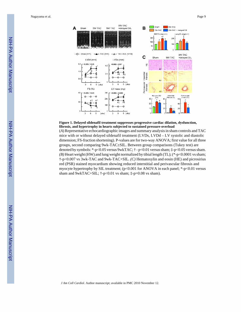
Figure 1. Delayed sildenafil treatment suppresses progressive cardiac dilation, dysfunction,fibrosis, and hypertrophy in hearts subjected to sustained pressure-overload(A) Representative echocardiographic images and summary analysis in sham controls and TACmice with or without delayed sildenafil treatment (LVDs, LVDd – LV systolic and diastolicdimension; FS-fraction shortening). P-values are for two-way ANOVA; first value for all threegroups, second comparing 9wk-TAC±SIL. Between group comparisons (Tukey test) aredenoted by symbols: *-p<0.05 versus 9wkTAC; † - p<0.01 versus sham; ‡-p<0.05 versus sham.(B) Heart weight (HW) and lung weight normalized by tibial length (TL); (*-p<0.0001 vs sham;†-p<0.007 vs 3wk-TAC and 9wk-TAC+SIL. (C) Hematoxylin and eosin (HE) and picrosiriusred (PSR) stained myocardium showing reduced interstitial and perivascular fibrosis andmyocyte hypertrophy by SIL treatment; (p<0.001 for ANOVA in each panel; *-p<0.01 versussham and 9wkTAC+SIL; †-p<0.01 vs sham; ‡-p=0.08 vs sham).
Nagayama et al. Page 9
J Am Coll Cardiol. Author manuscript; available in PMC 2010 November 12.
NIH
-PA Author Manuscript
NIH
-PA Author Manuscript
NIH
-PA Author Manuscript
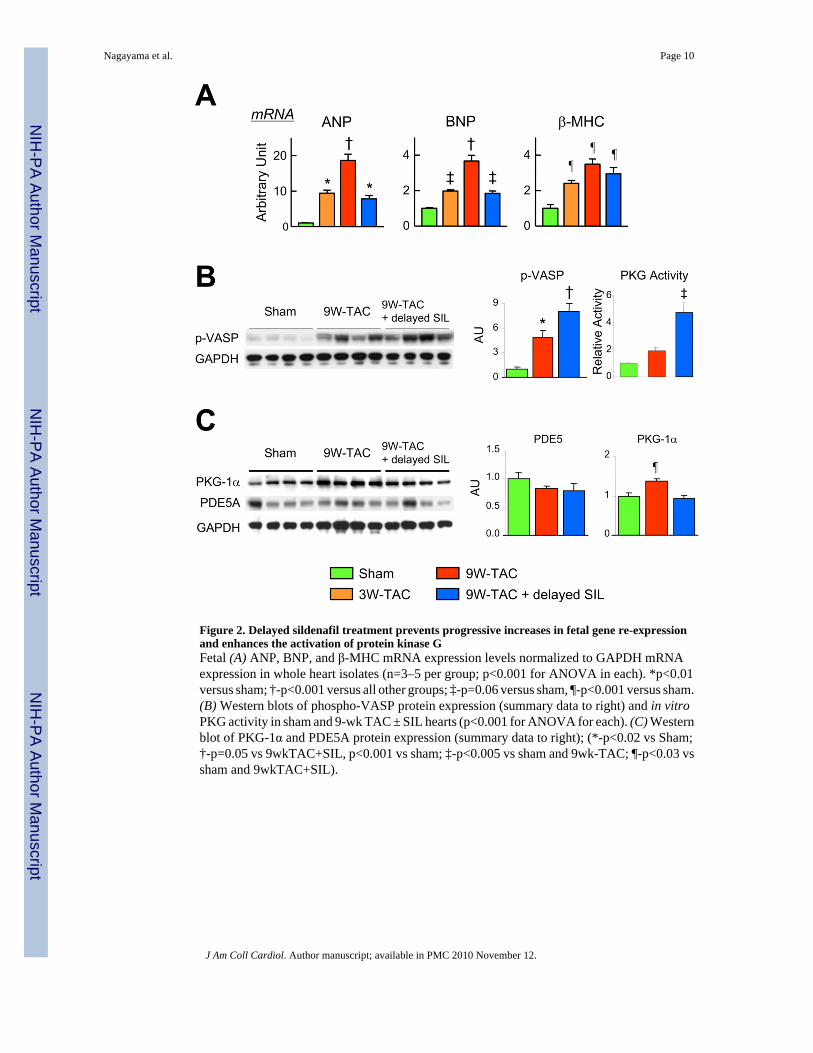
Figure 2. Delayed sildenafil treatment prevents progressive increases in fetal gene re-expressionand enhances the activation of protein kinase GFetal (A) ANP, BNP, and β-MHC mRNA expression levels normalized to GAPDH mRNAexpression in whole heart isolates (n=3–5 per group; p<0.001 for ANOVA in each). *p<0.01versus sham; †-p<0.001 versus all other groups; ‡-p=0.06 versus sham, ¶-p<0.001 versus sham.(B) Western blots of phospho-VASP protein expression (summary data to right) and in vitroPKG activity in sham and 9-wk TAC ± SIL hearts (p<0.001 for ANOVA for each). (C) Westernblot of PKG-1α and PDE5A protein expression (summary data to right); (*-p<0.02 vs Sham;†-p=0.05 vs 9wkTAC+SIL, p<0.001 vs sham; ‡-p<0.005 vs sham and 9wk-TAC; ¶-p<0.03 vssham and 9wkTAC+SIL).
Nagayama et al. Page 10
J Am Coll Cardiol. Author manuscript; available in PMC 2010 November 12.
NIH
-PA Author Manuscript
NIH
-PA Author Manuscript
NIH
-PA Author Manuscript

Figure 3. Delayed sildenafil treatment in hearts subjected to pressure-overload improves cardiacsystolic and diastolic function, and myocyte shortening and calcium handling(A) Representative in vivo pressure-volume loops and summary analysis. 3-wk-TAC heartswere somewhat dilated with a higher end-systolic elastance (Ees) and contractility (dP/dtmaxnormalized to instantaneous developed pressure; peak power divided by end-diastolic volume(PWRmax/EDV), and preserved relaxation (tau). Contractility declined and relaxationprolonged to levels at or below control after 9-wks-TAC, and hearts displayed marked dilation/remodeling. SIL treatment prevented this functional deterioration; (p-value in each panel showsANOVA result or for Ees and V100, Kruskal-Wallis test; post hoc tests: *-p<0.001 versus sham;†-p=0.06 vs 9wkTAC; ‡-p<0.04 vs sham; ¶-p<0.1 vs 9wkTAC and sham; #-p<0.001 versusother groups; ¢-p<0.05 versus sham and 3wkTAC; §-p<0.001 vs 3wkTAC and 9wkTAC+SIL.(B) Isolated myocyte sarcomere shortening (SS), relengthening rate, peak calcium transient,and time for 50% transient decay measured at rest and in response to isoproterenol (ISO)stimulation; (n=4 hearts for each experimental group, 15–20 myocytes per heart). Results of2-way ANOVA: p<0.001 for ISO and for group effect, p>0.2 for interaction effect – for allparameters; within group ANOVA; *-p<0.005 versus sham, †-P<0.005 versus 9wkTAC; ‡-P<0.02 vs 9wkTAC; ¶-p<0.001 vs sham.
Nagayama et al. Page 11
J Am Coll Cardiol. Author manuscript; available in PMC 2010 November 12.
NIH
-PA Author Manuscript
NIH
-PA Author Manuscript
NIH
-PA Author Manuscript
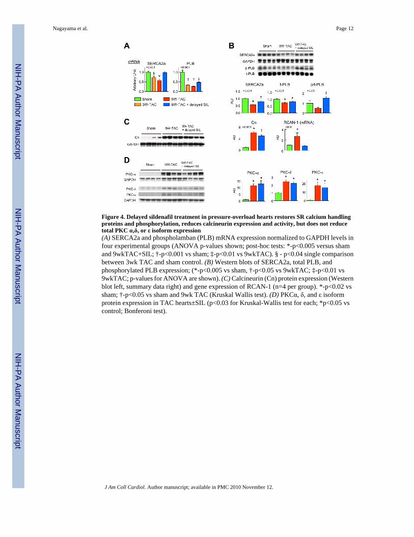
Figure 4. Delayed sildenafil treatment in pressure-overload hearts restores SR calcium handlingproteins and phosphorylation, reduces calcineurin expression and activity, but does not reducetotal PKC α,δ, or ε isoform expression(A) SERCA2a and phospholamban (PLB) mRNA expression normalized to GAPDH levels infour experimental groups (ANOVA p-values shown; post-hoc tests: *-p<0.005 versus shamand 9wkTAC+SIL; †-p<0.001 vs sham; ‡-p<0.01 vs 9wkTAC). § - p<0.04 single comparisonbetween 3wk TAC and sham control. (B) Western blots of SERCA2a, total PLB, andphosphorylated PLB expression; (*-p<0.005 vs sham, †-p<0.05 vs 9wkTAC; ‡-p<0.01 vs9wkTAC; p-values for ANOVA are shown). (C) Calcineurin (Cn) protein expression (Westernblot left, summary data right) and gene expression of RCAN-1 (n=4 per group). *-p<0.02 vssham; †-p<0.05 vs sham and 9wk TAC (Kruskal Wallis test). (D) PKCα, δ, and ε isoformprotein expression in TAC hearts±SIL (p<0.03 for Kruskal-Wallis test for each; *p<0.05 vscontrol; Bonferoni test).
Nagayama et al. Page 12
J Am Coll Cardiol. Author manuscript; available in PMC 2010 November 12.
NIH
-PA Author Manuscript
NIH
-PA Author Manuscript
NIH
-PA Author Manuscript
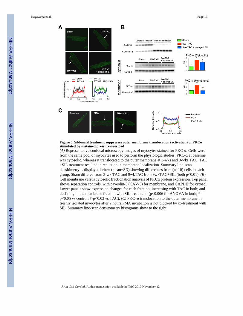
Figure 5. Sildenafil treatment suppresses outer membrane translocation (activation) of PKCαstimulated by sustained pressure-overload(A) Representative confocal microscopy images of myocytes stained for PKC-α. Cells werefrom the same pool of myocytes used to perform the physiologic studies. PKC-α at baselinewas cytosolic, whereas it translocated to the outer membrane at 3-wks and 9-wks TAC. TAC+SIL treatment resulted in reduction in membrane localization. Summary line-scandensitimetry is displayed below (mean±SD) showing differences from (n=10) cells in eachgroup. Sham differed from 3-wk TAC and 9wkTAC from 9wkTAC+SIL (both p<0.01). (B)Cell membrane versus cytosolic fractionation analysis of PKCα protein expression. Top panelshows separation controls, with caveolin-3 (CAV-3) for membrane, and GAPDH for cytosol.Lower panels show expression changes for each fraction; increasing with TAC in both; anddeclining in the membrane fraction with SIL treatment; (p<0.006 for ANOVA in both; *-p<0.05 vs control; †-p<0.02 vs TAC). (C) PKC–α translocation to the outer membrane infreshly isolated myocytes after 2 hours PMA incubation is not blocked by co-treatment withSIL. Summary line-scan densitometry histograms show to the right.
Nagayama et al. Page 13
J Am Coll Cardiol. Author manuscript; available in PMC 2010 November 12.
NIH
-PA Author Manuscript
NIH
-PA Author Manuscript
NIH
-PA Author Manuscript
Copyright © 2022 FDOKUMEN