
Shroud like coloration of linen, conservation measures and perception of patterns onto the Shroud of...
11
SHS Web of Conferences 15, 00005 (2015) DOI: 10.1051 / shsconf / 20151500005 C Owned by the authors, published by EDP Sciences, 2015 Shroud like coloration of linen, conservation measures and perception of patterns onto the Shroud of Turin Paolo Di Lazzaro a and Daniele Murra ENEA, Dept. Applications of Radiation, Via E. Fermi 45, 00044 Frascati, Italy Abstract. We present new experimental results on ultraviolet laser processing of linen fabrics that complete and update the previous studies at the ENEA Research Centre of Frascati seeking a coloration mechanism able to reproduce the microscopic complexity of the body images embedded onto the Shroud of Turin. The achievement of a Shroud-like coloration of linens by using vacuum ultraviolet light pulses allowed to recognize the main photochemical reactions possibly involved in the coloration process. The identification of these reactions offered hints for long-term conservation measures of the body images on the Shroud of Turin. Finally, we discuss how image processing of photographs of the Shroud may cause misleading effects, which led some researchers to perceive, e.g., inscriptions and a second face on the backside of the cloth, which are unlikely to be on the Shroud, confirming that there is a narrow boundary between image enhancement and manipulation. 1. Introduction The odd chemical and physical characteristics of the image of a scourged man on the linen cloth of the Shroud of Turin (TS) make difficult creating an image that matches (or even approaches) its peculiar superficiality and chemistry at the microscopic level. After the most recent in-depth experimental analysis of the images on the TS in 1978, the team of the Shroud of Turin Research Project (STuRP) wrote in the official summary of their final report: “We can conclude for now that the Shroud image is that of a real human form of a scourged, crucified man. It is not the product of an artist. The blood stains are composed of haemoglobin and also give a positive test for serum albumin. The image is an ongoing mystery and until further chemical studies are made, perhaps by this group of scientists, or perhaps by some scientists in the future, the problem remains unsolved.”[1]. The above conclusion was a consequence of the on-site analyses made by microscopy, vis- ible ultraviolet and infrared spectrometry, X-ray fluorescence spectrometry, and thermography. Later observations on linen fibres and threads sampled from the TS were made by microchem- istry, petrographic microscopy, scanning-electron microscopy, energy dispersive X-ray analysis, pyrolysis-mass-spectrometry, and laser-microprobe Raman analyses [2, 3]. In particular, • reflectance spectra, chemical tests, laser-microprobe Raman spectra, pyrolysis mass spectrometry, and X-ray fluorescence all show that the image is not painted with any of the expected, historically-documented pigments and media. a Corresponding author: [email protected] This is an Open Access article distributed under the terms of the Creative Commons Attribution License 4.0, which permits unrestricted use, distribution, and reproduction in any medium, provided the original work is properly cited. Article available at http://www.shs-conferences.org or http://dx.doi.org/10.1051/shsconf/20151500005
Transcript of Shroud like coloration of linen, conservation measures and perception of patterns onto the Shroud of...
SHS Web of Conferences 15, 00005 (2015)DOI: 10.1051/shsconf/20151500005C© Owned by the authors, published by EDP Sciences, 2015
Shroud like coloration of linen, conservation measures andperception of patterns onto the Shroud of Turin
Paolo Di Lazzaroa and Daniele Murra
ENEA, Dept. Applications of Radiation, Via E. Fermi 45, 00044 Frascati, Italy
Abstract. We present new experimental results on ultraviolet laser processing of linenfabrics that complete and update the previous studies at the ENEA Research Centre ofFrascati seeking a coloration mechanism able to reproduce the microscopic complexity ofthe body images embedded onto the Shroud of Turin. The achievement of a Shroud-likecoloration of linens by using vacuum ultraviolet light pulses allowed to recognize the mainphotochemical reactions possibly involved in the coloration process. The identification ofthese reactions offered hints for long-term conservation measures of the body images on theShroud of Turin. Finally, we discuss how image processing of photographs of the Shroudmay cause misleading effects, which led some researchers to perceive, e.g., inscriptionsand a second face on the backside of the cloth, which are unlikely to be on the Shroud,confirming that there is a narrow boundary between image enhancement and manipulation.
1. Introduction
The odd chemical and physical characteristics of the image of a scourged man on the linen cloth of theShroud of Turin (TS) make difficult creating an image that matches (or even approaches) its peculiarsuperficiality and chemistry at the microscopic level. After the most recent in-depth experimentalanalysis of the images on the TS in 1978, the team of the Shroud of Turin Research Project (STuRP)wrote in the official summary of their final report: “We can conclude for now that the Shroud image isthat of a real human form of a scourged, crucified man. It is not the product of an artist. The blood stainsare composed of haemoglobin and also give a positive test for serum albumin. The image is an ongoingmystery and until further chemical studies are made, perhaps by this group of scientists, or perhaps bysome scientists in the future, the problem remains unsolved.” [1].
The above conclusion was a consequence of the on-site analyses made by microscopy, vis-ible ultraviolet and infrared spectrometry, X-ray fluorescence spectrometry, and thermography.Later observations on linen fibres and threads sampled from the TS were made by microchem-istry, petrographic microscopy, scanning-electron microscopy, energy dispersive X-ray analysis,pyrolysis-mass-spectrometry, and laser-microprobe Raman analyses [2, 3]. In particular,
• reflectance spectra, chemical tests, laser-microprobe Raman spectra, pyrolysis massspectrometry, and X-ray fluorescence all show that the image is not painted with any of theexpected, historically-documented pigments and media.
a Corresponding author: [email protected]
This is an Open Access article distributed under the terms of the Creative Commons Attribution License 4.0, which permitsunrestricted use, distribution, and reproduction in any medium, provided the original work is properly cited.
Article available at http://www.shs-conferences.org or http://dx.doi.org/10.1051/shsconf/20151500005
SHS Web of Conferences
• both kinetics studies and fluorescence measurements support the image was formed by a low-temperature process. As a consequence, it is unlikely a Shroud-like image can be produced bycontact with a heated statue, as discussed in [4];
• the coloration depth is extremely shallow, in the range 200–600 nm. Recent measurements onimage-fibres of the TS suggest that the coloration depth is 200 nm, which corresponds to thethickness of the primary cell wall of each linen fibre [5];
• the coloration process of the TS image involves oxidation, dehydration and conjugation ofpolysaccharide structure of fibres, to produce a conjugated carbonyl group as the chromophore;
• the microscopy images show the so called “half tone effect”, that is, shading of the body imagesis not accomplished by varying the colour, but by varying the number of colour fibres per unitarea at the microscopic level. In other words, the image seen at the macroscopic level is anareal density image. As the diameter of each fibre in the Shroud’s yarns ranges between 15 and30 micrometers, the half tone effect suggests that the image cannot have been hand-coloured,unless the artist had a micrometer-size brush and a microscope;
• blood tests positive for haemoglobin, serum albumin and immunoglobulin. The anatomicalconsistency of blood and serum versus wounds, including typical halo fluorescence aroundthe main bloodstains, would be consistent with a haemolytic process caused by torture.
All the countless attempts to create a Shroud-like image since the Pia’s photographs in 1898 havefailed to adequately reproduce the above characteristics all together. Some researchers have obtainedcoloration and/or images that look similar [6] but no one has created images that match both microscopicand macroscopic features of the TS image. The answer to the question of how the image was producedor what produced the image is still unknown. After, the inability to replicate the microscopic complexityof the image on the TS prevents formulating a reliable hypothesis on the process of the imageformation.
In this paper we present the survey of the overall research work on Shroud-related issues done duringthe years 2005–2013 by the Excimer Laser Laboratory at the ENEA Research Centre of Frascati [7].Section 2 summarizes our attempts to find a photochemical coloration mechanism able to reproducethe microscopic complexity of the body images on the TS. Our identification of the specific groupsand reactions involved in the laser coloration process offered hints for long-term conservation measuresof the TS, as discussed in Sect. 3. Finally, we made a study of the misleading effect of deep imageprocessing of photographs of the TS, as detailed in Sect. 4.
2. Laser irradiations of linen fabrics at ENEA
It is well known that the energy carried by short-wavelength radiation breaks the chemical bonds of theirradiated material without inducing a significant heating: we say short-wavelength radiation generatesa “photochemical reaction”. Moreover, linen has a molar absorptivity which increases when decreasingthe radiation wavelength: the smaller the wavelength, the thinner the material necessary to absorb all theradiation. Then, we chosen the ultraviolet (UV) radiation to obtain at least two of the main characteristicsof the TS image: a thin coloration depth and a low-temperature image-formation.
We first measured the absolute reflectance spectra of modern linens we have used in our experiments.These linen fabrics were hand-made in the period 1936–1950 and never used before, The results showeda lower reflectance of our linens vs. the TS in the spectral region 520 nm–600 nm, confirming our linensare less yellowish than the TS, as expected because of the different age and different aging-relatedoxidation and dehydration. However, we measured a perfect overlap of our linens with the absolutereflectance of the TS in the UV and vacuum UV (VUV) spectral regions, [8]. Thus, when irradiated inthe UV and VUV, our linen fabrics behave like the linen of the TS.
00005-p.2
ATSI 2014
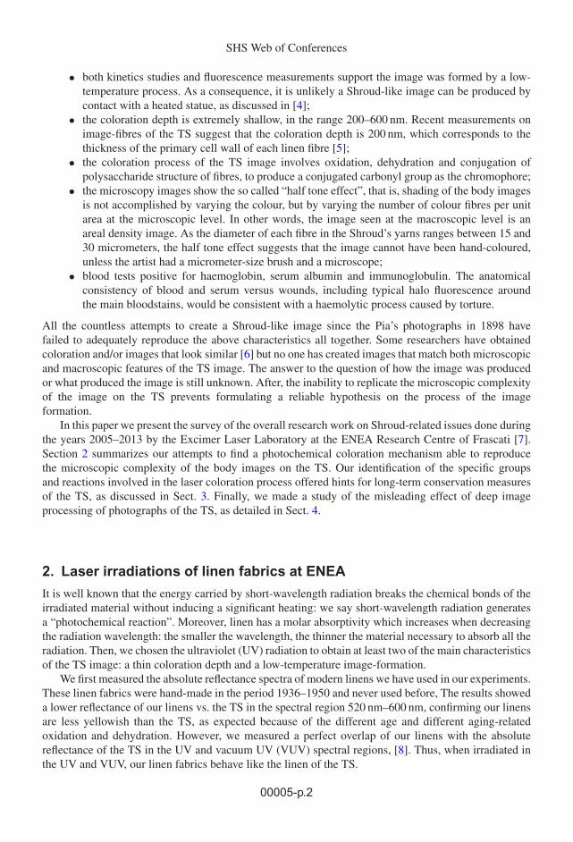
Figure 1. Above: faint linen coloration after 10 laser shots at 308-nm. Fluence/shot 0.15 J/cm2. Intensity/shot4.5 MW/cm2. 1 Hz repetition rate. Source: ENEA. Below: cross section of a linen yarn irradiated at 308 nm, contrastenhanced. The red ellipse shows the depth of the coloured region. From [10].
2.1 XeCl excimer lasers irradiations
At the beginning we irradiated linens by an XeCl excimer laser facility named Hercules, which emits UV308-nm laser pulses, each one lasting 120 ns, without obtaining any coloration results. Namely, linensirradiated with high fluence/intensity were carbonized, while at intermediate and low fluence/intensityvalues we did not observe any change of the linen threads, both at macroscopic and microscopic level.These bad results were in agreement with the analogous failure at Los Alamos National Laboratories(LANL) using 50-ns long excimer laser pulses [9].
Then, we irradiated the linen by using another XeCl laser (LPX 305) emitting pulses 4 times shorterand with an energy per pulse 12 times smaller than the Hercules laser, at the same 308 nm wavelength.In this configuration we achieved a permanent coloration of linens in a narrow range of pulse duration,intensity, number of laser pulses and time interval between successive pulses [10]. This coloration wassuperficial at the thread level, see Fig. 1.
In this way, we have shown that the duration of the UV light pulses plays a main role in thephotochemical reactions producing linens coloration, and that the experimental failure experienced atLANL using excimer lasers [9] was due to parameters (in particular the laser pulse-width) outside thenarrow range of values able to generate the linen coloration.
2.2 ArF excimer lasers irradiations
In order to obtain a thinner coloration depth than that obtained by the XeCl laser, we have used the VUVradiation emitted by an ArF excimer laser (emission wavelength 193 nm, pulse-width 12 ns). In this
00005-p.3
SHS Web of Conferences
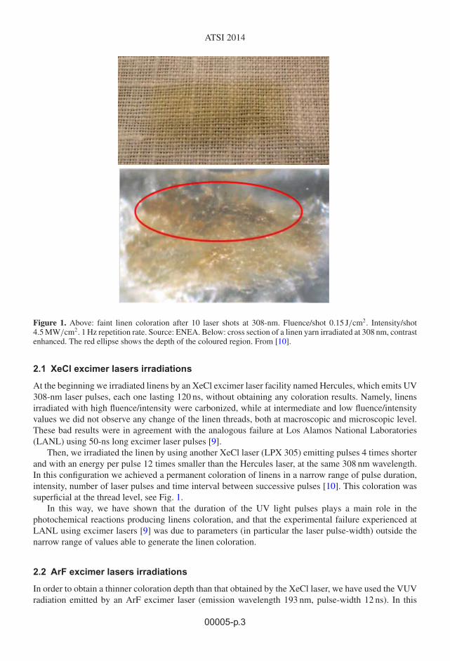
Figure 2. Microscope view of a single linen fibre mechanically damaged after it was coloured by 193-nm laserirradiation. Circles show pieces of the broken 200-nm-thin external layer of the fibril (the so called “primary cellwall”). Note these pieces are yellowish, compared with the uncoloured medulla (secondary cell wall) of the fibre.From [11].
respect, our experiments were successful, as we obtained a permanent coloration of the outside fibres ofthe linen threads [8], and using a particular set of irradiation parameters we obtained a coloration limitedto the primary cell wall of the linen fibres [8, 11], that is, a coloration superficial at the fibre level similarto that of the TS [5], see Fig. 2.
In addition, we observed the hue of yellow colour slowly changes with the number of consecutivelaser pulses, thus allowing an accurate control of the RGB value within 2% by selecting the totalirradiation fluence/intensity [11], e.g., by varying the number of laser pulses.
2.3 Excimer laser coloration vs. Turin Shroud coloration
The linen coloration is a threshold effect for both 308 nm and 193 nm irradiations, i.e. the colouris obtained only when the laser fluence/intensity on the linen is above a given level. However, afterlaser irradiations just below-threshold that at first do not generate a visible coloration of linen, a latentcoloration appears either by artificial or natural aging of linen, as detailed in [8, 10, 11]. Latent colorationis interesting for the synergy between radiation and the oxidative-dehydrating effect of aging whichtrigger the coloration process, and for historians, attracted by the possibility that, whatever has causedthe TS image, the coloration may have “developed” over time. Latent coloration can be explained byoxidation and dehydration of the cellulose (caused by heat or by natural aging) amending the newchemical bonds induced by laser irradiation, thus facilitating the formation of conjugated unsaturatedstructures that are essential part of the photochemical reactions involved, as detailed in [11]. The synergybetween heat and UV light on cellulose is discussed in [12], showing how the process initiated by UVradiation is accelerated and reinforced by heat.
The partial inhibition of fluorescence induced by both UV and VUV laser radiation shown in[8, 10, 11] is an additional feature of our coloration similar to the TS image.
Excimer laser irradiations of linens activate a “cold” photochemical coloration process. In fact,we have experimentally measured a thermal heating of few degrees centigrade after UV and VUVirradiations [11] which is irrelevant compared to colouring linens by scorching. In addition, the spectralenergies of single UV and VUV photons (4 eV for 308 nm, and 6.4 eV for 193 nm) are higher thanthe energies of most chemical bonds in cellulose, thus allowing the direct breaking of the same bondsand the creation of new chemical bonds with a negligible secondary heat generation. Photochemicalreactions fit the “cold” coloration process of the TS estimated by STuRP [2].
00005-p.4
ATSI 2014
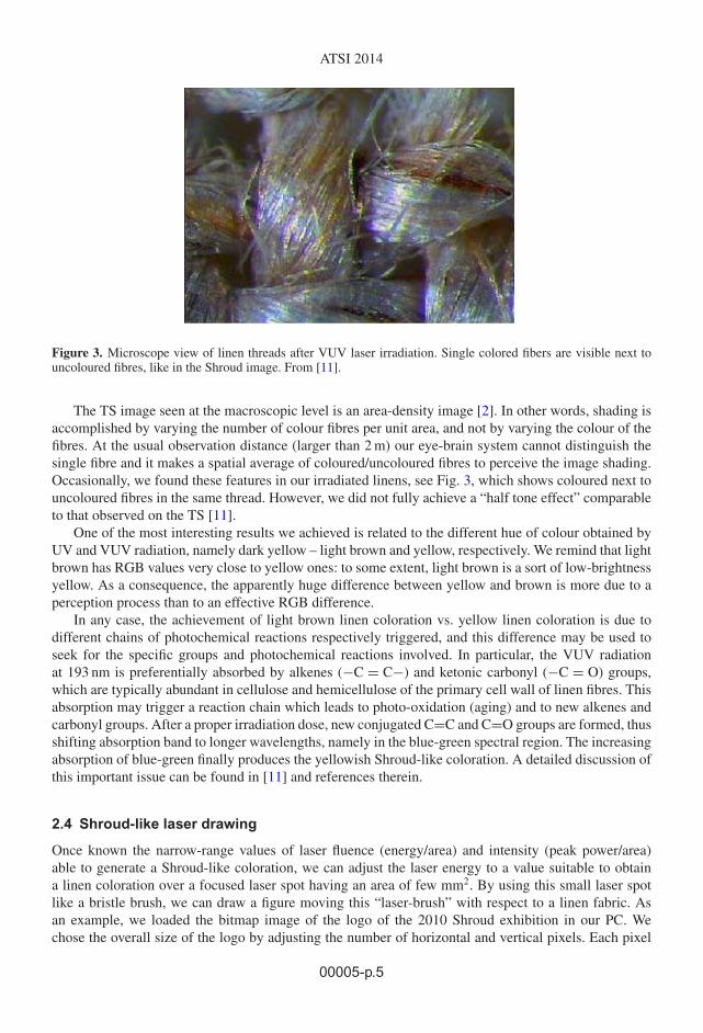
Figure 3. Microscope view of linen threads after VUV laser irradiation. Single colored fibers are visible next touncoloured fibres, like in the Shroud image. From [11].
The TS image seen at the macroscopic level is an area-density image [2]. In other words, shading isaccomplished by varying the number of colour fibres per unit area, and not by varying the colour of thefibres. At the usual observation distance (larger than 2 m) our eye-brain system cannot distinguish thesingle fibre and it makes a spatial average of coloured/uncoloured fibres to perceive the image shading.Occasionally, we found these features in our irradiated linens, see Fig. 3, which shows coloured next touncoloured fibres in the same thread. However, we did not fully achieve a “half tone effect” comparableto that observed on the TS [11].
One of the most interesting results we achieved is related to the different hue of colour obtained byUV and VUV radiation, namely dark yellow – light brown and yellow, respectively. We remind that lightbrown has RGB values very close to yellow ones: to some extent, light brown is a sort of low-brightnessyellow. As a consequence, the apparently huge difference between yellow and brown is more due to aperception process than to an effective RGB difference.
In any case, the achievement of light brown linen coloration vs. yellow linen coloration is due todifferent chains of photochemical reactions respectively triggered, and this difference may be used toseek for the specific groups and photochemical reactions involved. In particular, the VUV radiationat 193 nm is preferentially absorbed by alkenes (−C = C−) and ketonic carbonyl (−C = O) groups,which are typically abundant in cellulose and hemicellulose of the primary cell wall of linen fibres. Thisabsorption may trigger a reaction chain which leads to photo-oxidation (aging) and to new alkenes andcarbonyl groups. After a proper irradiation dose, new conjugated C=C and C=O groups are formed, thusshifting absorption band to longer wavelengths, namely in the blue-green spectral region. The increasingabsorption of blue-green finally produces the yellowish Shroud-like coloration. A detailed discussion ofthis important issue can be found in [11] and references therein.
2.4 Shroud-like laser drawing
Once known the narrow-range values of laser fluence (energy/area) and intensity (peak power/area)able to generate a Shroud-like coloration, we can adjust the laser energy to a value suitable to obtaina linen coloration over a focused laser spot having an area of few mm2. By using this small laser spotlike a bristle brush, we can draw a figure moving this “laser-brush” with respect to a linen fabric. Asan example, we loaded the bitmap image of the logo of the 2010 Shroud exhibition in our PC. Wechose the overall size of the logo by adjusting the number of horizontal and vertical pixels. Each pixel
00005-p.5

SHS Web of Conferences
a)
b)
Figure 4. a) Faint yellow image of the face of the logo of the 2010 Shroud exhibition drawn on linen by usingthe excimer laser beam as a contactless brush. Color is normalized to the reference white. b) Negative of the sameimage in a). Source: ENEA.
corresponds to the size of our “laser-brush”. The linen was put on a frame placed on a XY doubletranslation stage with a maximum stroke of 200 mm and one micrometer accuracy of repositioning [13].A dedicated software allowed to move the double stage according to the bitmap of the logo and to triggerthe laser shots just when the stages reached a given point corresponding to the colour pixel. In less than10 minutes we have drawn the 50 cm2 face shown in Fig. 4a. Note the yellowish image is extremely faintand difficult to see, showing a low-contrast similar to that of the TS image. On the contrary, the negativeof the same image shows a clearly visible face, and even the single laser-shot-pixel can be identified,see Fig. 4b.
In this preliminary attempt, we did not pay attention to the image shading, having reproduced alogo which has a binary coloration. However, a shaded image can be obtained simply reducing theXY movement between successive laser shots to be less than one pixel, that is, less than the laser spotdiameter. Obviously, the laser fluence of each shot must be reduced accordingly.
The linen in Fig. 4 is presently exposed at the Museum of the Shroud in Turinhttp://www.sindone.it/#museo_tour&LL=en.
3. Conservation measures
The irradiation results briefly summarized in Sect. 2 show a dramatic capability of UV and VUVradiation to modify and age the cellulose of linens. In particular, by using a petrographic microscope wehave observed some defects induced by UV radiation in the structure of irradiated linen fibres, similarlyto very old linen fabrics [10]. We can therefore infer that UV pulses change the crystalline structureof cellulose in a similar manner as aging and low-intensity natural radiation accumulated in a longperiod do. In this respect, the knowledge of the photochemical processes able to generate a Shroud-likecoloration, as outlined in Sect. 2.3, is essential to plan the long-term conservation of both the cloth andthe image on it.
We have identified Radon (Rn) as the most dangerous source of natural radiation, potentiallyaffecting the long term conservation of the image contrast and visibility. In fact, at sea level Rn (aninvisible, odourless, radioactive gas formed by the disintegration of Thorium and of Radium) is themain source of natural radiation. In fact, Rn emits alpha particles and produces several solid radioactiveproducts called Rn-daughters, which in turn emit beta-particles and ionizing radiation.
00005-p.6
ATSI 2014
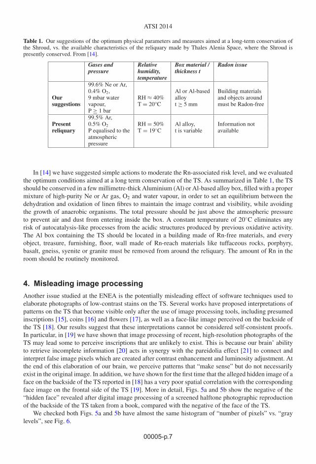
Table 1. Our suggestions of the optimum physical parameters and measures aimed at a long-term conservation ofthe Shroud, vs. the available characteristics of the reliquary made by Thales Alenia Space, where the Shroud ispresently conserved. From [14].
Gases and Relative Box material / Radon issuepressure humidity, thickness t
temperature99.6% Ne or Ar,0.4% O2, Al or Al-based Building materials
Our 9 mbar water RH ≈ 40% alloy and objects aroundsuggestions vapour, T = 20°C t ≥ 5 mm must be Radon-free
P ≥ 1 bar99.5% Ar,
Present 0.5% O2 RH = 50% Al alloy, Information notreliquary P equalised to the T = 19◦C t is variable available
atmosphericpressure
In [14] we have suggested simple actions to moderate the Rn-associated risk level, and we evaluatedthe optimum conditions aimed at a long term conservation of the TS. As summarized in Table 1, the TSshould be conserved in a few millimetre-thick Aluminium (Al) or Al-based alloy box, filled with a propermixture of high-purity Ne or Ar gas, O2 and water vapour, in order to set an equilibrium between thedehydration and oxidation of linen fibres to maintain the image contrast and visibility, while avoidingthe growth of anaerobic organisms. The total pressure should be just above the atmospheric pressureto prevent air and dust from entering inside the box. A constant temperature of 20◦C eliminates anyrisk of autocatalysis-like processes from the acidic structures produced by previous oxidative activity.The Al box containing the TS should be located in a building made of Rn-free materials, and everyobject, treasure, furnishing, floor, wall made of Rn-reach materials like tuffaceous rocks, porphyry,basalt, gneiss, syenite or granite must be removed from around the reliquary. The amount of Rn in theroom should be routinely monitored.
4. Misleading image processing
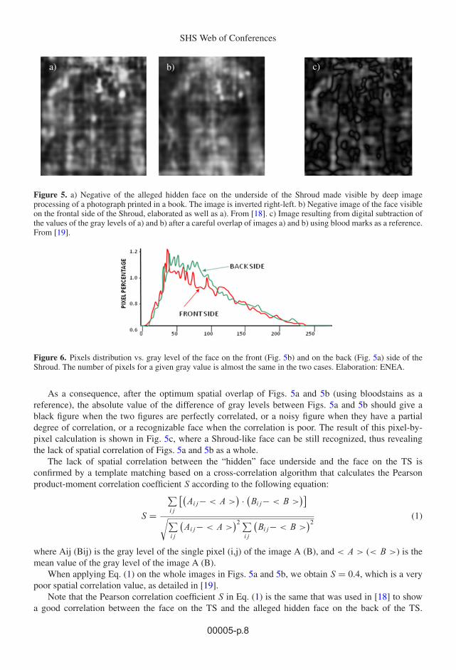
Another issue studied at the ENEA is the potentially misleading effect of software techniques used toelaborate photographs of low-contrast stains on the TS. Several works have proposed interpretations ofpatterns on the TS that become visible only after the use of image processing tools, including presumedinscriptions [15], coins [16] and flowers [17], as well as a face-like image perceived on the backside ofthe TS [18]. Our results suggest that these interpretations cannot be considered self-consistent proofs.In particular, in [19] we have shown that image processing of recent, high-resolution photographs of theTS may lead some to perceive inscriptions that are unlikely to exist. This is because our brain’ abilityto retrieve incomplete information [20] acts in synergy with the pareidolia effect [21] to connect andinterpret false image pixels which are created after contrast enhancement and luminosity adjustment. Atthe end of this elaboration of our brain, we perceive patterns that “make sense” but do not necessarilyexist in the original image. In addition, we have shown for the first time that the alleged hidden image of aface on the backside of the TS reported in [18] has a very poor spatial correlation with the correspondingface image on the frontal side of the TS [19]. More in detail, Figs. 5a and 5b show the negative of the“hidden face” revealed after digital image processing of a screened halftone photographic reproductionof the backside of the TS taken from a book, compared with the negative of the face of the TS.
We checked both Figs. 5a and 5b have almost the same histogram of “number of pixels” vs. “graylevels”, see Fig. 6.
00005-p.7
SHS Web of Conferences
a) b) c)
Figure 5. a) Negative of the alleged hidden face on the underside of the Shroud made visible by deep imageprocessing of a photograph printed in a book. The image is inverted right-left. b) Negative image of the face visibleon the frontal side of the Shroud, elaborated as well as a). From [18]. c) Image resulting from digital subtraction ofthe values of the gray levels of a) and b) after a careful overlap of images a) and b) using blood marks as a reference.From [19].
Figure 6. Pixels distribution vs. gray level of the face on the front (Fig. 5b) and on the back (Fig. 5a) side of theShroud. The number of pixels for a given gray value is almost the same in the two cases. Elaboration: ENEA.
As a consequence, after the optimum spatial overlap of Figs. 5a and 5b (using bloodstains as areference), the absolute value of the difference of gray levels between Figs. 5a and 5b should give ablack figure when the two figures are perfectly correlated, or a noisy figure when they have a partialdegree of correlation, or a recognizable face when the correlation is poor. The result of this pixel-by-pixel calculation is shown in Fig. 5c, where a Shroud-like face can be still recognized, thus revealingthe lack of spatial correlation of Figs. 5a and 5b as a whole.
The lack of spatial correlation between the “hidden” face underside and the face on the TS isconfirmed by a template matching based on a cross-correlation algorithm that calculates the Pearsonproduct-moment correlation coefficient S according to the following equation:
S =
∑ij
[(Aij− < A >
) · (Bij− < B >
)]√∑
ij
(Aij− < A >
)2 ∑ij
(Bij− < B >
)2(1)
where Aij (Bij) is the gray level of the single pixel (i,j) of the image A (B), and < A > (< B >) is themean value of the gray level of the image A (B).
When applying Eq. (1) on the whole images in Figs. 5a and 5b, we obtain S = 0.4, which is a verypoor spatial correlation value, as detailed in [19].
Note that the Pearson correlation coefficient S in Eq. (1) is the same that was used in [18] to showa good correlation between the face on the TS and the alleged hidden face on the back of the TS.
00005-p.8
ATSI 2014
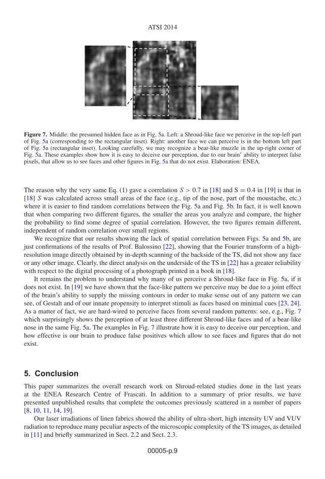
Figure 7. Middle: the presumed hidden face as in Fig. 5a. Left: a Shroud-like face we perceive in the top-left partof Fig. 5a (corresponding to the rectangular inset). Right: another face we can perceive is in the bottom left partof Fig. 5a (rectangular inset). Looking carefully, we may recognize a bear-like muzzle in the up-right corner ofFig. 5a. These examples show how it is easy to deceive our perception, due to our brain’ ability to interpret falsepixels, that allow us to see faces and other figures in Fig. 5a that do not exist. Elaboration: ENEA.
The reason why the very same Eq. (1) gave a correlation S > 0.7 in [18] and S = 0.4 in [19] is that in[18] S was calculated across small areas of the face (e.g., tip of the nose, part of the moustache, etc.)where it is easier to find random correlations between the Fig. 5a and Fig. 5b. In fact, it is well knownthat when comparing two different figures, the smaller the areas you analyze and compare, the higherthe probability to find some degree of spatial correlation. However, the two figures remain different,independent of random correlation over small regions.
We recognize that our results showing the lack of spatial correlation between Figs. 5a and 5b, arejust confirmations of the results of Prof. Balossino [22], showing that the Fourier transform of a high-resolution image directly obtained by in-depth scanning of the backside of the TS, did not show any faceor any other image. Clearly, the direct analysis on the underside of the TS in [22] has a greater reliabilitywith respect to the digital processing of a photograph printed in a book in [18].
It remains the problem to understand why many of us perceive a Shroud-like face in Fig. 5a, if itdoes not exist. In [19] we have shown that the face-like pattern we perceive may be due to a joint effectof the brain’s ability to supply the missing contours in order to make sense out of any pattern we cansee, of Gestalt and of our innate propensity to interpret stimuli as faces based on minimal cues [23, 24].As a matter of fact, we are hard-wired to perceive faces from several random patterns: see, e.g., Fig. 7which surprisingly shows the perception of at least three different Shroud-like faces and of a bear-likenose in the same Fig. 5a. The examples in Fig. 7 illustrate how it is easy to deceive our perception, andhow effective is our brain to produce false positives which allow to see faces and figures that do notexist.
5. Conclusion
This paper summarizes the overall research work on Shroud-related studies done in the last yearsat the ENEA Research Centre of Frascati. In addition to a summary of prior results, we havepresented unpublished results that complete the outcomes previously scattered in a number of papers[8, 10, 11, 14, 19].
Our laser irradiations of linen fabrics showed the ability of ultra-short, high intensity UV and VUVradiation to reproduce many peculiar aspects of the microscopic complexity of the TS images, as detailedin [11] and briefly summarized in Sect. 2.2 and Sect. 2.3.
00005-p.9

SHS Web of Conferences
We have used the laser beam as a contactless brush to draw the face of the man of the TS, stylizedaccording to the binary logo of the 2010 Shroud exposition. The result of this preliminary attempt isquite interesting, see Sect. 2.4.
The different coloration results obtained by diverse laser irradiation parameters (wavelength,intensity, fluence, repetition rate, number of shots) allowed to recognize the photochemical reactionsthat may have contributed to the Shroud-like coloration, thus offering hints to plan the long-termconservation of the cloth and of the image on it, see Sect. 3 and [14].
Our study of the misleading effect of digital image processing of photographs of low-contrast imagesconfirmed that there is a narrow boundary between image enhancement and manipulation. In particular,data and analyses in Sect. 4 demonstrate the lack of spatial correlation between the face on the TSand the alleged “hidden” face underside which was “discovered” after a heavy image processing of aphotograph printed in a book. This lack of correlation calls the existence of a face underside into seriousdoubt, and supports the results of a previous in-depth analysis [22] which showed the absence of anyface on the backside of the TS. In addition, our results in [19] show that some alleged inscriptions hiddenon the TS [15] are unlikely to exist.
This paper summarizes more than eight years of experiments, calculations and discussions. Here we wish to thankthe Colleagues who gave a contribution to different phases of this long-term work, including, in alphabeticalorder, G. Baldacchini, G. Fanti, E. Marinelli, E. Nichelatti, A. Santoni, B. Schwortz. Without their skill, ideasand collaborative approach the successful results summarized in this paper could not have been achieved.
References
[1] The official summary of the final report of STuRP can be read at the webpagehttp://www.shroud.com/78conclu.htm
[2] E. Jumper, A.D. Adler, J.P. Jackson, S.F. Pellicori, J.H. Heller, J.R. Druzik, ArchaeologicalChemistry III: ACS Advances in Chemistry 205 (American Chemical Society, Washington, 1984),pp. 447–476
[3] A complete list of scientific papers published by the STuRP team can be found inhttp://www.shroud.com/78papers.htm
[4] J.P. Jackson, E.J. Jumper, and W.R. Ercoline, Appl. Opt. 23, 2244–2270 (1984)[5] G. Fanti, J. Botella, P, Di Lazzaro, R. Schneider, N. Svensson, J. Im. Sci. Technol. 54, 040201-(8)
(2010)[6] The most interesting and best documented attempt to reproduce a life-size Shroud-like
image is described in L. Garlaschelli, J. Im. Sci. Technol. 54, 040301-040301(14) (2010).References therein offer a comprehensive list of other attempts. See a critical review ofthis paper in: T. Heimburger, G. Fanti, Proceedings of the International Workshop on theScientific approach to the Acheiropoietos Images, IWSAI 2010, (ENEA, 2010) pp. 19-28www.acheiropoietos.info/proceedings/proceedings.php
[7] Web page of Shroud-related studies of the Excimer Laser Laboratory:http://www.frascati.enea.it/fis/lac/excimer/sindone/sindone.html The overallactivities of our Lab can be found at the website www.frascati.enea.it/fis/lac/excimer/labeccimeri_eng.html
[8] P. Di Lazzaro, D. Murra, E. Nichelatti, A Santoni, G. Baldacchini, J. Im. Sci. Technol. 54,040302-(6) (2010)
[9] R. Rogers, A. Arnoldi, www.shroud.com/pdfs/rogers2.pdf (2002). In this document, Rogersand Arnoldi wrote: “Very intense, 50-ns-long bursts of UV ablated the cloth surface, and the
00005-p.10
ATSI 2014
samples were reduced to a cloud of very fine particles. We could not get a color with a flash oflight.” In another document (www.shroud.com/pdfs/rogers6.pdf (2004)) Rogers criticisedthe radiative hypothesis of Jackson published in Shroud Spectrum International 34, 3-29 (1990)and wrote: “Experiments we did with pulsed ultraviolet lasers on linen resulted in ablation anddestructive shock waves. Samples often were converted into a little amorphous powder and gas,ablate and convert linen into little amorphous powder and gas”. One of the authors (PDL) in1991 was invited by Robert (Bob) Sze to visit his Excimer Laser Group at Los Alamos NationalLaboratory (LANL). All the “non restricted” experiments of Sze’s group were equipped withexcimer lasers emitting laser pulses each having a width in the 50–150-ns range, like those emittedby the Hercules ENEA excimer laser. Rogers was working at LANL, and we can speculatehe asked Sze’s group to make excimer irradiations of linen, and that they used the 50–150 nslaser pulsewidth they had at hands. As explained in Sect. 2.1, we obtained the same ablationresults when irradiating linens with the 120 ns long Hercules laser pulses. The results describedin Sects. 2.1 and 2.2 which are obtained using shorter-than-30-ns UV and VUV laser pulsesdemonstrate the paramount importance of the laser pulsewidth to achieve a Shroud-like colorationof linen
[10] G. Baldacchini, P. Di Lazzaro, D. Murra, G. Fanti, Appl. Opt. 47, 1278–1283 (2008)[11] P. Di Lazzaro, D. Murra, E. Nichelatti, A Santoni, G. Baldacchini, Appl. Opt. 51, 8567–8578
(2012)[12] M. Yatagai, S.H. Zeronian, Cellulose 1, 205–214 (1994)[13] Phisik Instrumente model M-521. Each stage was independently driven by a Mercury C-863 DC
motor controller[14] P. Di Lazzaro, D. Murra, A. Santoni, E. Nichelatti, EAI special issue on Knowledge, Diagnostics
and Preservation of Cultural Heritage, 89–94 (2012). http://www.enea.it/it/produzione-scientifica/pdf-eai/speciale-cultural-heritage/14-the-conservation-pdf
[15] A. Marion, Opt. Eng. 37, 2308–2313 (1998)[16] A.D. Whanger, M. Whanger, Appl. Opt. 24, 766–772 (1985)[17] A. Danin, A.D. Whanger, U. Baruch, M. Whanger, Flora of the Shroud of Turin (Missouri
Botanical Garden Press, St. Louis MO, 1999)[18] G. Fanti, R. Maggiolo, J. Opt. A: Pure and Applied Optics 6, 491–503 (2004)[19] P. Di Lazzaro, D. Murra, B. Schwortz, Pattern Recognition 46, 1964–1970 (2013)[20] G. Kanizsa, Scientific American 234, 48–52 (1976)[21] See, e.g., J. Voss, K. Federmeier, K. Paller, Cerebral Cortex 22, 2354–2364 (2012). For an
elementary introduction, see http://en.wikipedia.org/wiki/Apophenia[22] N. Balossino, Sindon 19, 57–69 (2003)[23] R. Palermo, G. Rhodes, Neuropsychologia 45, 75–92 (2007)[24] A J. Calder, G. Rhodes, M. Johnson, The Oxford Handbook of Face perception (Oxford University
Press, 2011)
00005-p.11


![Passio Christi. Passio hominis. Śladami Całunu Turyńskiego [Passio Christi. Passio hominis. Traces of the Shroud of Turin]](https://static.fdokumen.com/doc/165x107/6312dd3e5cba183dbf06d64f/passio-christi-passio-hominis-sladami-calunu-turynskiego-passio-christi-passio.jpg)


