Deep Ultraviolet Radiation Simulates the Turin Shroud Image
6
Deep Ultraviolet Radiation Simulates the Turin Shroud Image Paolo Di Lazzaro, Daniele Murra and Antonino Santoni ENEA, Department of Physical Technologies and New Materials, Frascati Research Center, via E. Fermi 45, 00044 Frascati, Italy E-mail: [email protected] Giulio Fanti Department of Mechanical Engineering, University of Padua, via Venezia 1, 35131 Padua, Italy Enrico Nichelatti ENEA, Department of Physical Technologies and New Materials, Casaccia Research Center, via Anguillarese 301, 00123 Rome, Italy Giuseppe Baldacchini via Quattrucci 246, 00046 Grottaferrata, Italy Abstract. The faint yellowed body image embedded into the linen cloth of the Turin Shroud has peculiar chemical and physical char- acteristics that at the moment cannot be replicated all together in laboratory. The authors present experimental results of ArF excimer laser irradiation (wavelength 193 nm) of a raw linen fabric, seeking for coloration similar to that of the Shroud image. The authors achieved a permanent yellow coloration of linen as a threshold ef- fect of the laser beam intensity and number of shots. Most impor- tant, the authors have achieved for the first time a submicrometer depth of coloration of the outermost part of the fibers, leaving a colorless fiber medulla. The authors also obtained latent coloration that appears after artificial aging of linen following laser irradiations that at first did not generate any visible effect. The authors have recognized different physical and chemical processes involved in both coloration and latent coloration. The comparison of the Turin Shroud image with the results of our ArF laser irradiation shows an interesting overlap of the main physical and chemical features. © 2010 Society for Imaging Science and Technology. DOI: 10.2352/J.ImagingSci.Technol.2010.54.4.040302 INTRODUCTION The Turin Shroud is a single piece of linen cloth measuring about 4.4 m by 1.1 m. Faint frontal and dorsal images of an apparently crucified man are embedded into the Shroud. These yellowish body images have peculiar chemical and physical characteristics 1 that have stimulated a worldwide scientific debate. 2–19 Most of the scientific data on the Shroud image are from the work carried out by a team of 26 scientists under the auspices of the Shroud of Turin Research Project, Inc., (STURP) (1978), that performed an in-depth examination on the Shroud with electromagnetic energy, from infrared to x-rays, obtaining data leading to the analysis of the sub- stances making up the body image and bloodstains. 4–12 The STURP measurements show that the body image is not painted, printed, singed by a heated bas-relief, or rubbed on a sculpture; moreover, the image color resides on the top- most fibers in the cloth weave. Reference 1 listed more than forty chemical and physical features of the Shroud image, and up to date all attempts to reproduce an image with the same microscopic and macroscopic aspect as well as all the chemical and physical characteristics have been unsuccessful. In this respect, the origin of the body image is still unknown. This worldwide interest declined after the results of ra- diocarbon tests that placed the origins of the Shroud in the range 1260–1390 C.E. 20 However, recent results suggest the sample used for radiocarbon dating was not representative of the whole Shroud. 21,22 Independently of the controversial results of the radiocarbon test, a main concern is the origin of the body image on the Shroud, and this article addresses the search of a possible mechanism of the image formation. Following the hints of independent works 14–19 showing that a burst of electromagnetic energy may account for the main image characteristics, we have considered the ultravio- let (UV) radiation as one of the best candidate for obtaining two of the main characteristics of the Shroud image, namely, a thin coloration depth and a low-temperature image- formation process. 1,11 Then, we used XeCl excimer laser ra- diation (wavelength 308 nm) to color linen by a sequence of UV laser pulses corresponding to a hypothetical burst of energy correlated with the Shroud image. 23 We have ob- tained a permanent coloration of linens that matches some characteristics of the Shroud images (appearance in cross- polarized light, fragility). However, the hue was too dark with respect to the Shroud images and the coloration thick- Received Dec. 7, 2009; accepted for publication Jan. 26, 2010; published online Jun. 15, 2010. 1062-3701/2010/544/040302/6/$20.00. Journal of Imaging Science and Technology® 54(4): 040302–040302-6, 2010. © Society for Imaging Science and Technology 2010 J. Imaging Sci. Technol. Jul.-Aug. 2010 040302-1
Transcript of Deep Ultraviolet Radiation Simulates the Turin Shroud Image

AcallfaftdcttrbSi2�
ITaaTps
ft(o
Ro
1
Journal of Imaging Science and Technology® 54(4): 040302–040302-6, 2010.© Society for Imaging Science and Technology 2010
J
Deep Ultraviolet Radiation Simulates the TurinShroud Image
Paolo Di Lazzaro, Daniele Murra and Antonino SantoniENEA, Department of Physical Technologies and New Materials, Frascati Research Center, via E. Fermi 45,
00044 Frascati, ItalyE-mail: [email protected]
Giulio FantiDepartment of Mechanical Engineering, University of Padua, via Venezia 1, 35131 Padua, Italy
Enrico NichelattiENEA, Department of Physical Technologies and New Materials, Casaccia Research Center, via Anguillarese
301, 00123 Rome, Italy
Giuseppe Baldacchini
via Quattrucci 246, 00046 Grottaferrata, ItalyxsSpamfascIu
drsorot
tmltafdUetcp
bstract. The faint yellowed body image embedded into the linenloth of the Turin Shroud has peculiar chemical and physical char-cteristics that at the moment cannot be replicated all together in
aboratory. The authors present experimental results of ArF excimeraser irradiation (wavelength 193 nm) of a raw linen fabric, seekingor coloration similar to that of the Shroud image. The authorschieved a permanent yellow coloration of linen as a threshold ef-
ect of the laser beam intensity and number of shots. Most impor-ant, the authors have achieved for the first time a submicrometerepth of coloration of the outermost part of the fibers, leaving aolorless fiber medulla. The authors also obtained latent colorationhat appears after artificial aging of linen following laser irradiationshat at first did not generate any visible effect. The authors haveecognized different physical and chemical processes involved inoth coloration and latent coloration. The comparison of the Turinhroud image with the results of our ArF laser irradiation shows an
nteresting overlap of the main physical and chemical features. ©010 Society for Imaging Science and Technology.DOI: 10.2352/J.ImagingSci.Technol.2010.54.4.040302�
NTRODUCTIONhe Turin Shroud is a single piece of linen cloth measuringbout 4.4 m by 1.1 m. Faint frontal and dorsal images of anpparently crucified man are embedded into the Shroud.hese yellowish body images have peculiar chemical andhysical characteristics1 that have stimulated a worldwidecientific debate.2–19
Most of the scientific data on the Shroud image arerom the work carried out by a team of 26 scientists underhe auspices of the Shroud of Turin Research Project, Inc.,STURP) (1978), that performed an in-depth examinationn the Shroud with electromagnetic energy, from infrared to
eceived Dec. 7, 2009; accepted for publication Jan. 26, 2010; publishednline Jun. 15, 2010.
w062-3701/2010/54�4�/040302/6/$20.00.
. Imaging Sci. Technol. 040302-
-rays, obtaining data leading to the analysis of the sub-tances making up the body image and bloodstains.4–12 TheTURP measurements show that the body image is notainted, printed, singed by a heated bas-relief, or rubbed onsculpture; moreover, the image color resides on the top-ost fibers in the cloth weave. Reference 1 listed more than
orty chemical and physical features of the Shroud image,nd up to date all attempts to reproduce an image with theame microscopic and macroscopic aspect as well as all thehemical and physical characteristics have been unsuccessful.n this respect, the origin of the body image is stillnknown.
This worldwide interest declined after the results of ra-iocarbon tests that placed the origins of the Shroud in theange 1260–1390 C.E.20 However, recent results suggest theample used for radiocarbon dating was not representativef the whole Shroud.21,22 Independently of the controversialesults of the radiocarbon test, a main concern is the originf the body image on the Shroud, and this article addresseshe search of a possible mechanism of the image formation.
Following the hints of independent works14–19 showinghat a burst of electromagnetic energy may account for the
ain image characteristics, we have considered the ultravio-et (UV) radiation as one of the best candidate for obtainingwo of the main characteristics of the Shroud image, namely,
thin coloration depth and a low-temperature image-ormation process.1,11 Then, we used XeCl excimer laser ra-iation (wavelength 308 nm) to color linen by a sequence ofV laser pulses corresponding to a hypothetical burst of
nergy correlated with the Shroud image.23 We have ob-ained a permanent coloration of linens that matches someharacteristics of the Shroud images (appearance in cross-olarized light, fragility). However, the hue was too dark
ith respect to the Shroud images and the coloration thick-Jul.-Aug. 20101
n(S
u�lcyidtstot
tTcWr
OLwhfiacpocmwo
cds
oawcabobma
ALTnt
wtasopti
ossNfla
w
FmlR
Ft9
Di Lazzaro et al.: Deep ultraviolet radiation simulates the Turin Shroud image
J
ess of linen yarns was too large compared to the topmostapproximately 0.2 �m thick) fiber coloration of thehroud body image.1,11,19
In this article we present the results of linen irradiationsing an ArF excimer laser emitting a deep UV wavelength=193 nm significantly shorter than that emitted by XeCl
asers. In this way we obtained a substantial reduction in theoloration depth down to the outermost part of the linenarns and a better overlap with the features of the Shroudmage, including the hue of color (yellow after 193 nm irra-iation, brown after the 308 nm irradiation). Most impor-
ant, we have locally achieved for the first time aubmicrometer depth of coloration of the outermost part ofhe fibers, leaving a colorless fiber medulla. We also singleut some physical and chemical processes possibly leading tohe improved results.
The main characteristics of the linen that are relevant inhe interaction with deep UV radiation are described below.he next section summarizes the experimental setup and theonditions to achieve linen coloration by ArF laser pulses.e then analyze the irradiation results, and offer concluding
emarks that summarize the main results.
PTICAL CHARACTERISTICS OF LINENinen fibers spun to produce linen yarns from which Shroudas woven are basically made of cellulose andemicellulose.24,25 The yellow-sepia Shroud image is super-cial as it is located on the surface of the uppermost fibers,nd the inside of these image-fibers does not showoloration.1,10,11,26 Coloration was formed by an unknownrocess that caused oxidation, dehydration, and conjugationf the polysaccharide structure of the linen fibers to yieldarbonyl groups and conjugated alkenes as the chro-ophore, i.e., a kind of premature aging process of the linenhere double bonds CvC ultimately lead to the colorationf the image fibers of the Turin Shroud.9,10,26
We irradiated a linen fabric recently manufactured ac-ording to the ancient technology. It has 15 yarns/cm, theiameter of each yarn is 300 �m, and the diameter of theingle fiber is ranging between 15 and 25 �m.
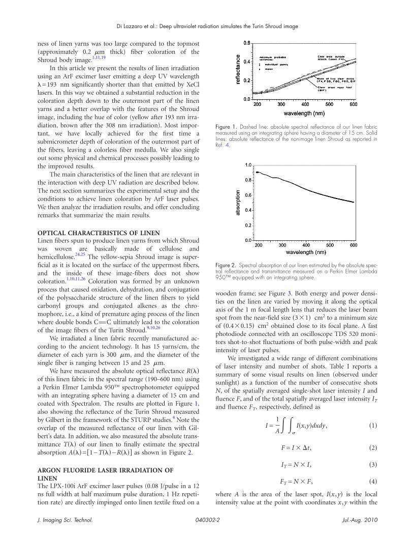
We have measured the absolute optical reflectance R���f this linen fabric in the spectral range (190–600 nm) usingPerkin Elmer Lambda 950™ spectrophotometer equippedith an integrating sphere having a diameter of 15 cm and
oated with Spectralon. The results are plotted in Figure 1,lso showing the reflectance of the Turin Shroud measuredy Gilbert in the framework of the STURP studies.4 Note theverlap of the measured reflectance of our linen with Gil-ert’s data. In addition, we also measured the absolute trans-ittance T��� of our linen to finally estimate the spectral
bsorption A���= �1−T���−R���� as shown in Figure 2.
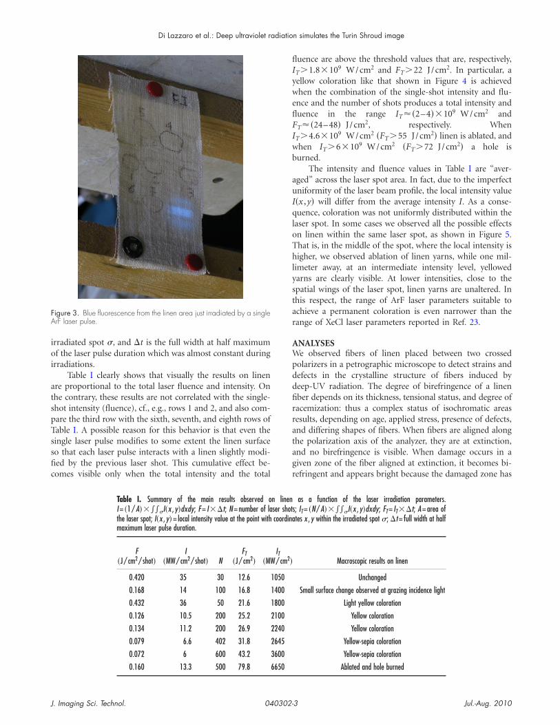
RGON FLUORIDE LASER IRRADIATION OFINENhe LPX-100i ArF excimer laser pulses (0.08 J/pulse in a 12s full width at half maximum pulse duration, 1 Hz repeti-
ion rate) are directly impinged onto linen textile fixed on a i
. Imaging Sci. Technol. 040302-
ooden frame; see Figure 3. Both energy and power densi-ies on the linen are varied by moving it along the opticalxis of the 1 m focal length lens that reduces the laser beampot from the near-field size �3�1� cm2 to a minimum sizef �0.4�0.15� cm2 obtained close to its focal plane. A fasthotodiode connected with an oscilloscope TDS 520 moni-
ors shot-to-shot fluctuations of both pulse-width and peakntensity of laser pulses.
We investigated a wide range of different combinationsf laser intensity and number of shots. Table I reports aummary of some visual results on linen (observed underunlight) as a function of the number of consecutive shots, of the spatially averaged single-shot laser intensity I anduence F, and of the total spatially averaged laser intensity IT
nd fluence FT, respectively, defined as
I =1
A� �
�
I�x,y�dxdy , �1�
F = I � �t , �2�
IT = N � I , �3�
FT = N � F , �4�
here A is the area of the laser spot, I�x ,y� is the local
igure 1. Dashed line: absolute spectral reflectance of our linen fabriceasured using an integrating sphere having a diameter of 15 cm. Solid
ines: absolute reflectance of the nonimage linen Shroud as reported inef. 4.
igure 2. Spectral absorption of our linen estimated by the absolute spec-ral reflectance and transmittance measured on a Perkin Elmer Lambda50™ equipped with an integrating sphere.
ntensity value at the point with coordinates x ,y within the
Jul.-Aug. 20102
ioi
atspTssfic
flIyweflFIwb
auIqloThlystar
AWpddfirratagr
FA
Di Lazzaro et al.: Deep ultraviolet radiation simulates the Turin Shroud image
J
rradiated spot �, and �t is the full width at half maximumf the laser pulse duration which was almost constant during
rradiations.Table I clearly shows that visually the results on linen
re proportional to the total laser fluence and intensity. Onhe contrary, these results are not correlated with the single-hot intensity (fluence), cf., e.g., rows 1 and 2, and also com-are the third row with the sixth, seventh, and eighth rows ofable I. A possible reason for this behavior is that even theingle laser pulse modifies to some extent the linen surfaceo that each laser pulse interacts with a linen slightly modi-ed by the previous laser shot. This cumulative effect be-omes visible only when the total intensity and the total
Table I. Summary of the main results observed oI = �1 / A�����I�x , y�dxdy; F = I��t; N = number of lathe laser spot; I�x , y�= local intensity value at the point withmaximum laser pulse duration.
F�J / cm2 / shot�
I�MW/ cm2 / shot� N
FT�J / cm2� �M
0.420 35 30 12.6
0.168 14 100 16.8
0.432 36 50 21.6
0.126 10.5 200 25.2
0.134 11.2 200 26.9
0.079 6.6 402 31.8
0.072 6 600 43.2
0.160 13.3 500 79.8
igure 3. Blue fluorescence from the linen area just irradiated by a singlerF laser pulse.
. Imaging Sci. Technol. 040302-
uence are above the threshold values that are, respectively,
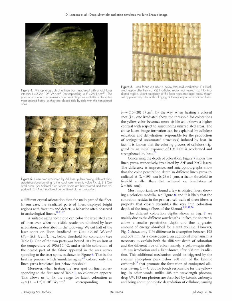
T �1.8�109 W/cm2 and FT �22 J /cm2. In particular, aellow coloration like that shown in Figure 4 is achievedhen the combination of the single-shot intensity and flu-
nce and the number of shots produces a total intensity anduence in the range IT ��2–4��109 W/cm2 and
T ��24–48� J /cm2, respectively. When
T �4.6�109 W/cm2 �FT �55 J /cm2� linen is ablated, andhen IT �6�109 W/cm2 �FT �72 J /cm2� a hole isurned.
The intensity and fluence values in Table I are “aver-ged” across the laser spot area. In fact, due to the imperfectniformity of the laser beam profile, the local intensity value�x ,y� will differ from the average intensity I. As a conse-uence, coloration was not uniformly distributed within the
aser spot. In some cases we observed all the possible effectsn linen within the same laser spot, as shown in Figure 5.hat is, in the middle of the spot, where the local intensity isigher, we observed ablation of linen yarns, while one mil-
imeter away, at an intermediate intensity level, yellowedarns are clearly visible. At lower intensities, close to thepatial wings of the laser spot, linen yarns are unaltered. Inhis respect, the range of ArF laser parameters suitable tochieve a permanent coloration is even narrower than theange of XeCl laser parameters reported in Ref. 23.
NALYSESe observed fibers of linen placed between two crossed
olarizers in a petrographic microscope to detect strains andefects in the crystalline structure of fibers induced byeep-UV radiation. The degree of birefringence of a linenber depends on its thickness, tensional status, and degree ofacemization: thus a complex status of isochromatic areasesults, depending on age, applied stress, presence of defects,nd differing shapes of fibers. When fibers are aligned alonghe polarization axis of the analyzer, they are at extinction,nd no birefringence is visible. When damage occurs in aiven zone of the fiber aligned at extinction, it becomes bi-efringent and appears bright because the damaged zone has
as a function of the laser irradiation parameters.s; IT = �N / A�����I�x , y�dxdy; FT = IT ��t; A = area oftes x , y within the irradiated spot �; �t = full width at half
Macroscopic results on linen
Unchanged
Small surface change observed at grazing incidence light
Light yellow coloration
Yellow coloration
Yellow coloration
Yellow-sepia coloration
Yellow-sepia coloration
Ablated and hole burned
n linenser shotcoordina
ITW/ cm2�
1050
1400
1800
2100
2240
2645
3600
6650
Jul.-Aug. 20103
aIri
oil�Tttshl
sTI
Fstcaoofgs
lTtrfi�
icpd
maaFana1tsceid
Fiymo
Faop
Fado
Di Lazzaro et al.: Deep ultraviolet radiation simulates the Turin Shroud image
J
different crystal orientation than the main part of the fiber.n our case, the irradiated parts of fibers displayed brightegions with fractures and defects, a behavior often observedn archeological linens.10,11,27
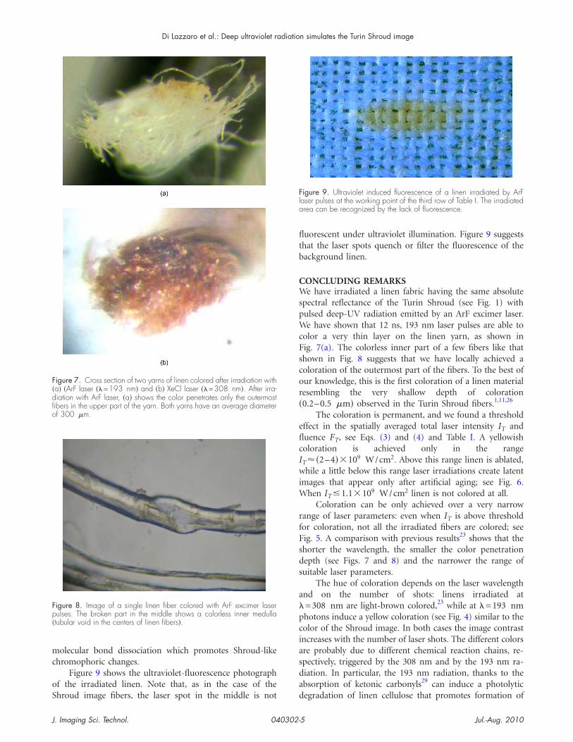
A suitable aging technique can color the irradiated areaf linen even when no visible results are obtained by laser
rradiation, as described in the following. We cut half of theaser spots on linen irradiated at IT =1.4�109 W/cm2
FT =16.8 J /cm2�, i.e., below threshold for coloration (seeable I). One of the two parts was heated 10 s by an iron athe temperature of 190±10 °C, and a visible coloration ofhe heated part of the fabric appeared in the area corre-ponding to the laser spots, as shown in Figure 6. That is, theeating process, which simulates aging,28 colored only the
inen yarns irradiated just below threshold.Moreover, when heating the laser spot on linen corre-
ponding to the first row of Table I, no coloration appears.his allows us to fix the range for latent coloration as
9 2
igure 4. Microphotograph of a linen yarn irradiated with a total laserntensity IT=2.2�109 W/cm2 �corresponding to FT=26 J/cm2�. Thearn was opened by tweezers in order to improve visibility of the outer-ost colored fibers, as they are placed side by side with the noncolorednes.
igure 5. Linen area irradiated by ArF laser pulses having different char-cteristics corresponding to the local laser intensity value I�x ,y�. �1� Col-red area. �2� Ablated area where fibers are first colored and then va-orized. �3� Area irradiated below threshold for coloration.
T ��1.1–1.7��10 W/cm corresponding to a
. Imaging Sci. Technol. 040302-
T ��13–20� J /cm2. By the way, when heating a coloredpot (i.e., one irradiated above the threshold for coloration)he yellow color becomes more visible as it shows a higherontrast with respect to surrounding unirradiated areas. Thebove latent image formation can be explained by cellulosexidation and dehydration (responsible for the productionf conjugated unsaturated structures) induced by heat. In
act, it is known that the coloring process of cellulose trig-ered by an initial exposure of UV light is accelerated andtrengthened by heat.28
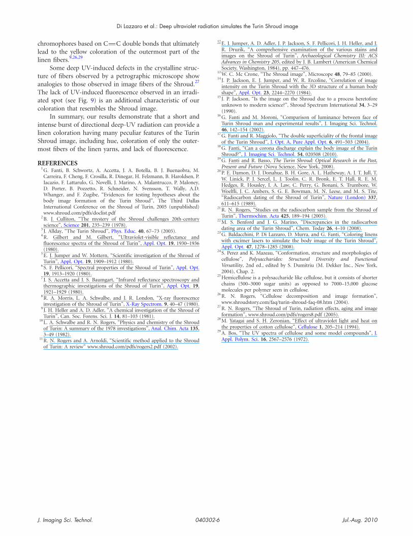
Concerning the depth of coloration, Figure 7 shows twoinen yarns, respectively, irradiated by ArF and XeCl lasers.he difference is impressive, and microphotographs show
hat the color penetration depth in different linen yarns ir-adiated at (�=193 nm is 26±4 �m, a factor threefold tovefold smaller than that achieved on irradiation at=308 nm).
Most important, we found a few irradiated fibers show-ng a colorless medulla, see Figure 8, and it is likely that theoloration resides in the primary cell walls of these fibers, aroperty that closely resembles the very thin colorationepth of the image fibers of the Shroud.1,10,11,26
The different coloration depths shown in Fig. 7 areainly due to the different wavelengths: in fact, the shorter �
llows a smaller penetration depth and thus a greatermount of energy absorbed for a unit volume. However,ig. 2 shows only 11% difference in absorption between 193nd 308 nm. As a consequence, an additional mechanism isecessary to explain both the different depth of colorationnd the different hue of color, namely, a yellow-sepia after93 nm irradiation and a light-brown after 308 nm irradia-ion. This additional mechanism could be triggered by thepectral absorption peak below 260 nm of the ketonicarbonyls29 that promote the formation of conjugated alk-nes having CvC double bonds responsible for the yellow-ng. In other words, unlike 308 nm wavelength photons,eep UV, 193 nm photons are absorbed by ketonic carbonyls
igure 6. Linen fabric cut after a below-threshold irradiation. �1� Irradi-ted region after heating. �2� Irradiated region not heated. �3� Not irra-iated region. Latent coloration of the linen area irradiated below thresh-ld appears only after artificial aging of the upper part of irradiated linen.
nd bring about photolytic degradation of cellulose, causing
Jul.-Aug. 20104
mc
oS
fltb
CWspWcFscor�
eflcIwiW
rfFsds
a�pciasda
F�dfio
Fp�
Fla
Di Lazzaro et al.: Deep ultraviolet radiation simulates the Turin Shroud image
J
olecular bond dissociation which promotes Shroud-likehromophoric changes.
Figure 9 shows the ultraviolet-fluorescence photographf the irradiated linen. Note that, as in the case of the
igure 7. Cross section of two yarns of linen colored after irradiation witha� �ArF laser ��=193 nm� and �b� XeCl laser ��=308 nm�. After irra-iation with ArF laser, �a� shows the color penetrates only the outermostbers in the upper part of the yarn. Both yarns have an average diameterf 300 �m.
igure 8. Image of a single linen fiber colored with ArF excimer laserulses. The broken part in the middle shows a colorless inner medullatubular void in the centers of linen fibers�.
hroud image fibers, the laser spot in the middle is not d
. Imaging Sci. Technol. 040302-
uorescent under ultraviolet illumination. Figure 9 suggestshat the laser spots quench or filter the fluorescence of theackground linen.
ONCLUDING REMARKSe have irradiated a linen fabric having the same absolute
pectral reflectance of the Turin Shroud (see Fig. 1) withulsed deep-UV radiation emitted by an ArF excimer laser.e have shown that 12 ns, 193 nm laser pulses are able to
olor a very thin layer on the linen yarn, as shown inig. 7(a). The colorless inner part of a few fibers like thathown in Fig. 8 suggests that we have locally achieved aoloration of the outermost part of the fibers. To the best ofur knowledge, this is the first coloration of a linen materialesembling the very shallow depth of coloration0.2–0.5 �m� observed in the Turin Shroud fibers.1,11,26
The coloration is permanent, and we found a thresholdffect in the spatially averaged total laser intensity IT anduence FT, see Eqs. (3) and (4) and Table I. A yellowisholoration is achieved only in the range
T ��2–4��109 W/cm2. Above this range linen is ablated,hile a little below this range laser irradiations create latent
mages that appear only after artificial aging; see Fig. 6.hen IT �1.1�109 W/cm2 linen is not colored at all.
Coloration can be only achieved over a very narrowange of laser parameters: even when IT is above thresholdor coloration, not all the irradiated fibers are colored; seeig. 5. A comparison with previous results23 shows that thehorter the wavelength, the smaller the color penetrationepth (see Figs. 7 and 8) and the narrower the range ofuitable laser parameters.
The hue of coloration depends on the laser wavelengthnd on the number of shots: linens irradiated at=308 nm are light-brown colored,23 while at �=193 nmhotons induce a yellow coloration (see Fig. 4) similar to theolor of the Shroud image. In both cases the image contrastncreases with the number of laser shots. The different colorsre probably due to different chemical reaction chains, re-pectively, triggered by the 308 nm and by the 193 nm ra-iation. In particular, the 193 nm radiation, thanks to thebsorption of ketonic carbonyls29 can induce a photolytic
igure 9. Ultraviolet induced fluorescence of a linen irradiated by ArFaser pulses at the working point of the third row of Table I. The irradiatedrea can be recognized by the lack of fluorescence.
egradation of linen cellulose that promotes formation of
Jul.-Aug. 20105
cll
taTac
ilSm
R
Di Lazzaro et al.: Deep ultraviolet radiation simulates the Turin Shroud image
J
hromophores based on CvC double bonds that ultimatelyead to the yellow coloration of the outermost part of theinen fibers.9,26,29
Some deep UV-induced defects in the crystalline struc-ure of fibers observed by a petrographic microscope shownalogies to those observed in image fibers of the Shroud.27
he lack of UV-induced fluorescence observed in an irradi-ted spot (see Fig. 9) is an additional characteristic of ouroloration that resembles the Shroud image.
In summary, our results demonstrate that a short andntense burst of directional deep-UV radiation can provide ainen coloration having many peculiar features of the Turinhroud image, including hue, coloration of only the outer-ost fibers of the linen yarns, and lack of fluorescence.
EFERENCES1 G. Fanti, B. Schwortz, A. Accetta, J. A. Botella, B. J. Buenaobra, M.
Carreira, F. Cheng, F. Crosilla, R. Dinegar, H. Felzmann, B. Haroldsen, P.Iacazio, F. Lattarulo, G. Novelli, J. Marino, A. Malantrucco, P. Maloney,D. Porter, B. Pozzetto, R. Schneider, N. Svensson, T. Wally, A.D.Whanger, and F. Zugibe, “Evidences for testing hypotheses about thebody image formation of the Turin Shroud”, The Third DallasInternational Conference on the Shroud of Turin, 2005 (unpublished)www.shroud.com/pdfs/doclist.pdf
2 B. J. Culliton, “The mystery of the Shroud challenges 20th-centuryscience”, Science 201, 235–239 (1978).
3 J. Allday, “The Turin Shroud”, Phys. Educ. 40, 67–73 (2005).4 R. Gilbert and M. Gilbert, “Ultraviolet-visible reflectance and
fluorescence spectra of the Shroud of Turin”, Appl. Opt. 19, 1930–1936(1980).
5 E. J. Jumper and W. Mottern, “Scientific investigation of the Shroud ofTurin”, Appl. Opt. 19, 1909–1912 (1980).
6 S. F. Pellicori, “Spectral properties of the Shroud of Turin”, Appl. Opt.19, 1913–1920 (1980).
7 J. S. Accetta and J. S. Baumgart, “Infrared reflectance spectroscopy andthermographic investigations of the Shroud of Turin”, Appl. Opt. 19,1921–1929 (1980).
8 R. A. Morris, L. A. Schwalbe, and J. R. London, “X-ray fluorescenceinvestigation of the Shroud of Turin”, X-Ray Spectrom. 9, 40–47 (1980).
9 J. H. Heller and A. D. Adler, “A chemical investigation of the Shroud ofTurin”, Can. Soc. Forens. Sci. J. 14, 81–103 (1981).
10 L. A. Schwalbe and R. N. Rogers, “Physics and chemistry of the Shroudof Turin: A summary of the 1978 investigations”, Anal. Chim. Acta 135,3–49 (1982).
11 R. N. Rogers and A. Arnoldi, “Scientific method applied to the Shroudof Turin: A review” www.shroud.com/pdfs/rogers2.pdf (2002).
. Imaging Sci. Technol. 040302-
12 E. J. Jumper, A. D. Adler, J. P. Jackson, S. F. Pellicori, J. H. Heller, and J.R. Druzik, “A comprehensive examination of the various stains andimages on the Shroud of Turin”, Archaeological Chemistry III: ACSAdvances in Chemistry 205, edited by J. B. Lambert (American ChemicalSociety, Washington, 1984), pp. 447–476.
13 W. C. Mc Crone, “The Shroud image”, Microscope 48, 79–85 (2000).14 J. P. Jackson, E. J. Jumper, and W. R. Ercoline, “Correlation of image
intensity on the Turin Shroud with the 3D structure of a human bodyshape”, Appl. Opt. 23, 2244–2270 (1984).
15 J. P. Jackson, “Is the image on the Shroud due to a process heretoforeunknown to modern science?”, Shroud Spectrum International 34, 3–29(1990).
16 G. Fanti and M. Moroni, “Comparison of luminance between face ofTurin Shroud man and experimental results”, J. Imaging Sci. Technol.46, 142–154 (2002).
17 G. Fanti and R. Maggiolo, “The double superficiality of the frontal imageof the Turin Shroud”, J. Opt. A, Pure Appl. Opt. 6, 491–503 (2004).
18 G. Fanti, “Can a corona discharge explain the body image of the TurinShroud?”, J. Imaging Sci. Technol. 54, 020508 (2010).
19 G. Fanti and R. Basso, The Turin Shroud: Optical Research in the Past,Present and Future (Nova Science, New York, 2008).
20 P. E. Damon, D. J. Donahue, B. H. Gore, A. L. Hatheway, A. J. T. Jull, T.W. Linick, P. J. Sercel, L. J. Toolin, C. R. Bronk, E. T. Hall, R. E. M.Hedges, R. Housley, I. A. Law, C. Perry, G. Bonani, S. Trumbore, W.Woelfli, J. C. Ambers, S. G. E. Bowman, M. N. Leese, and M. S. Tite,“Radiocarbon dating of the Shroud of Turin”, Nature (London) 337,611–615 (1989).
21 R. N. Rogers, “Studies on the radiocarbon sample from the Shroud ofTurin”, Thermochim. Acta 425, 189–194 (2005).
22 M. S. Benford and J. G. Marino, “Discrepancies in the radiocarbondating area of the Turin Shroud”, Chem. Today 26, 4–10 (2008).
23 G. Baldacchini, P. Di Lazzaro, D. Murra, and G. Fanti, “Coloring linenswith excimer lasers to simulate the body image of the Turin Shroud”,Appl. Opt. 47, 1278–1285 (2008).
24 S. Perez and K. Mazeau, “Conformation, structure and morphologies ofcellulose”, Polysaccharides: Structural Diversity and FunctionalVersatility, 2nd ed., edited by S. Dumitriu (M. Dekker Inc., New York,2004), Chap. 2.
25 Hemicellulose is a polysaccharide like cellulose, but it consists of shorterchains (500–3000 sugar units) as opposed to 7000–15,000 glucosemolecules per polymer seen in cellulose.
26 R. N. Rogers, “Cellulose decomposition and image formation”,www.shroudstory.com/faq/turin-shroud-faq-08.htm (2004).
27 R. N. Rogers, “The Shroud of Turin, radiation effects, aging and imageformation”, www.shroud.com/pdfs/rogers8.pdf (2005).
28 M. Yatagai and S. H. Zeronian, “Effect of ultraviolet light and heat onthe properties of cotton cellulose”, Cellulose 1, 205–214 (1994).
29 A. Bos, “The UV spectra of cellulose and some model compounds”, J.Appl. Polym. Sci. 16, 2567–2576 (1972).
Jul.-Aug. 20106


![Passio Christi. Passio hominis. Śladami Całunu Turyńskiego [Passio Christi. Passio hominis. Traces of the Shroud of Turin]](https://static.fdokumen.com/doc/165x107/6312dd3e5cba183dbf06d64f/passio-christi-passio-hominis-sladami-calunu-turynskiego-passio-christi-passio.jpg)