Shallow-water Desmophyllum dianthus (Scleractinia) from Chile: characteristics of the biocoenoses,...
42
Freiwald A, Roberts JM (eds), 2005, Cold-water Corals and Ecosystems. Springer-Verlag Berlin Heidelberg, pp 937-977 Shallow-water Desmophyllum dianthus (Scleractinia) from Chile: characteristics of the biocoenoses, the bioeroding community, heterotrophic interactions and (paleo)- bathymetric implications Günter Försterra 1,2 , Lydia Beuck 3 , Vreni Häussermann 1,2 , André Freiwald 3 1 Departamento de Biologia Marina, Universidad Austral de Chile, Avda. Inés de Haverbeck, casas 9, 11 y 13, Campus Isla Teja, casilla 567, Valdivia, Chile ([email protected]) 2 Department Biologie II, Ludwig-Maximilians-Universität München, Karlstr. 23- 25, D-80333 München, Germany 3 Institute of Paleontology, Erlangen University, Loewenichstr. 28, D-91054 Erlangen, Germany Abstract. We report an unusually shallow-water occurrence of habitat-forming Desmophyllum dianthus (Esper, 1794) from the Chilean fjord region. Most occurrences of the cosmopolitan D. dianthus are known from the bathyal zone. In the northern Chilean fjord region, however, this coral is reported within the euphotic zone. The upper limit of distribution was found at 7 m water depth and is confined to the lower boundary of the low salinity layer. Large accumulations both as living aggregations and as sediment-formers typically occur from 20 m water depth and beyond. The corals prefer to colonise the undersides of rock ledges with downward- facing corallites. The motivation of this study is to analyse and discuss the existence of an azooxanthellate coral that generally thrives in aphotic environment but here extends into the photic zone by means of screening of bioerosion patterns. Based on the detailed analysis of scratching and boring traces, we compare the ichnocoenosis found within the Chilean D. dianthus with the established bathymetrically indicative ichnocoenoses from other areas around the world. These indicator ichnocoenoses are widely used to reconstruct relative water depths of depositional settings in the geological past. The study of the bioeroding assemblage from two living D. dianthus collected at 28 m water depth in the Reñihue Fjord, Chile, shows some remarkable patterns that shed light on the complex way in which the coralʼ s soft tissue expands and retracts at the apical zone of the corallum in response to in vivo infestation of endolithic algae. The role of this heterotypic interaction is discussed. To visualise the endolithic ichnocoenoses, we applied several methodologies such as the vacuum cast embedding technique combined with scanning electron
Transcript of Shallow-water Desmophyllum dianthus (Scleractinia) from Chile: characteristics of the biocoenoses,...

Freiwald A, Roberts JM (eds), 2005, Cold-water Corals and Ecosystems. Springer-Verlag Berlin Heidelberg, pp 937-977
Shallow-water Desmophyllum dianthus (Scleractinia) from Chile: characteristics of the biocoenoses, the bioeroding community, heterotrophic interactions and (paleo)-bathymetric implications
Günter Försterra1,2, Lydia Beuck3, Vreni Häussermann1,2, André Freiwald3
1 Departamento de Biologia Marina, Universidad Austral de Chile, Avda. Inés de Haverbeck, casas 9, 11 y 13, Campus Isla Teja, casilla 567, Valdivia, Chile
([email protected])2 Department Biologie II, Ludwig-Maximilians-Universität München, Karlstr. 23-
25, D-80333 München, Germany3 Institute of Paleontology, Erlangen University, Loewenichstr. 28, D-91054
Erlangen, Germany
Abstract. We report an unusually shallow-water occurrence of habitat-forming Desmophyllum dianthus (Esper, 1794) from the Chilean fjord region. Most occurrences of the cosmopolitan D. dianthus are known from the bathyal zone. In the northern Chilean fjord region, however, this coral is reported within the euphotic zone. The upper limit of distribution was found at 7 m water depth and is confi ned to the lower boundary of the low salinity layer. Large accumulations both as living aggregations and as sediment-formers typically occur from 20 m water depth and beyond. The corals prefer to colonise the undersides of rock ledges with downward-facing corallites. The motivation of this study is to analyse and discuss the existence of an azooxanthellate coral that generally thrives in aphotic environment but here extends into the photic zone by means of screening of bioerosion patterns. Based on the detailed analysis of scratching and boring traces, we compare the ichnocoenosis found within the Chilean D. dianthus with the established bathymetrically indicative ichnocoenoses from other areas around the world. These indicator ichnocoenoses are widely used to reconstruct relative water depths of depositional settings in the geological past. The study of the bioeroding assemblage from two living D. dianthus collected at 28 m water depth in the Reñihue Fjord, Chile, shows some remarkable patterns that shed light on the complex way in which the coralʼs soft tissue expands and retracts at the apical zone of the corallum in response to in vivo infestation of endolithic algae. The role of this heterotypic interaction is discussed.
To visualise the endolithic ichnocoenoses, we applied several methodologies such as the vacuum cast embedding technique combined with scanning electron

938 Försterra, Beuck, Häussermann, Freiwald
microscopy, fl uorescence microscopy and x-ray analyses. In total, 20 different trace makers are identifi ed. Based on the analysis of the indicator ichnospecies, the endolithic community is indicative for the dysphotic zone. This result is compatible with the sciaphile environment of D. dianthus, living under rock ledges in the photic zone.
Keywords. Desmophyllum, Chilean fjords, euphotic bioerosion, ichnocoenosis, paleobathymetry, heterotrophic interaction
Introduction
Bathyal coral communities thrive in the absence of light in the aphotic zone. The lack of phototrophic organisms represents a major difference to shallow-water coral communities, where the herbivore food-web plays an important part in ecosystem functioning. Moreover, analysing the presence or absence of phototrophic organisms is a major tool in paleontology for reconstructing the relative paleobathymetry of ancient communities. Here we document the case of the cosmopolitan, azooxanthellate scleractinian Desmophyllum dianthus (Esper, 1794) that typically occurs in bathyal depths. In the northern Chilean fjord region, however, this species occurs in the photic zone and forms extended coral banks in water depths as shallow as 20 m (Försterra and Häussermann 2003).
Overview of the Chilean fjord region
The Chilean fjord region extends over more than 1500 km (straight line). With its numerous archipelagos, channels and fjords it is one of the morphologically most structured coastal areas in the world, and is also one of the least studied regions. Hydrographic, taxonomic and ecologic information is fragmentary and substantial portions of the marine environment in this region must be regarded as terra incognita (Arntz 1999; Fernández et al. 2000). Data on the benthic life on rocky substrates, which dominate in the shallow water of the channels and fjords, are especially scarce. A biogeographic barrier for shallow-water organisms, separating the southern from the northern Chilean fjord province, seems to be located at the Taitao Peninsula at approximately 46°S (Fig. 1; Brattström and Johanssen 1983; Lancellotti and Vásquez 1999; Häussermann 2004; Häussermann and Försterra accepted).
Overall precipitation in the fjord region is extremely high, and varies locally between 1040 mm/year (Isla Guafo; Pickard 1971), and 6700 mm/year (Isla Guarello). This results in an enormous freshwater infl ux into the fjords (Pickard 1971; Castilla et al. 1993). Furthermore, large amounts of organic and inorganic matter are imported through the rivers into the fjord system. Many fjords of the southern fjord region are under the infl uence of glaciers that extend to sea level (Pickard 1971). The constant high freshwater fl ow produces a superfi cial low salinity layer that decreases in thickness from the head of the fjord towards the mouth (Pickard 1971), but below 20 m depth salinities are generally more stable, and reach higher values. In the past, most sampling in the fjord region has been carried

Shallow-water Desmophyllum dianthus (Scleractinia) from Chile 939
Fig. 1. Geographical setting of the Chilean fjord region

940 Försterra, Beuck, Häussermann, Freiwald
out from boats, equipped with dredges and grabs (e.g., Challenger Expedition 1872-1876, Joint Magellan “Victor Hensen” Campaign 1994, CIMAR Fiordo expeditions 1995-2004), or in the intertidal which excluded rocky subtidal substrates. Therefore, it is not surprising that there are still many undiscovered species in the shallow water of Chilean Patagonia. What is more surprising is the existence of entire benthic communities that have not been documented in this form before, especially the discovery of benthic biocoenoses on the steep rock walls of the inner fjords (Försterra and Häussermann 2003).
The incredibly fast economic development in the Chilean fjord region, led by the booming salmon farming industry, is in contrast to the poor understanding of the affected marine systems. The biocoenoses in the fjords and channels may be very sensitive to the impacts of human activities. As a consequence, the unique benthic communities, such as the coral banks in shallow-water, could sustain lasting damage or could even disappear before they can be studied (Försterra and Häussermann 2003).
Distribution of shallow-water Desmophyllum dianthus
A striking feature is undoubtedly the large accumulation of azooxanthellate Scleractinia in shallow water in the euphotic zone (Försterra and Häussermann 2003). The dominant species in these communities is D. dianthus. According to Cairns and Zibrowius (1997), Desmophyllum cristagalli is a junior synonym of Desmophyllum dianthus. This coral is a cosmopolitan species found in a large depth interval from 35 to 2460 m. Moreover, the majority of occurrences are typically located at bathyal depths. In the North Atlantic Desmophyllum dianthus is often associated with the prevalent framework builder Lophelia pertusa (Linné, 1758), and with Madrepora oculata Linné, 1758 (see compilation in Freiwald et al. 2004). Off the Chilean coast, D. dianthus exists between 300 and 800 m water-depth, where it was already known to reach high densities (Cairns 1982). New fi ndings of D. dianthus in shallow water of the northern Chilean fjord region encounter completely different ecological conditions, including co-existence with photosynthetically active organisms, such as coralline algae. As a characteristic element of the bathyal zone both recent and in the geological record, a co-occurrence of D. dianthus with crustose coralline algae
Fig. 2. A Desmophyllum dianthus settling hard substrates on the undersides of rock ledges and associated with the red rockfi sh Sebastes capensis; width 2 m. B The skeleton of living D. dianthus infested by red algae (r), green algae (g), and endolithic sabellids (s); scale bar 5 cm. C Top of the boulder incrusted by corallinaceans; on its fl anks various organisms settle, amongst them are solitary corals ( Caryophyllia sp. and D. dianthus); scale bar 50 cm. D Red rockfi sh Sebastes capensis resting on corallinaceans, D. dianthus and Caryophyllia sp.; scale bar 10 cm. E Bryozoans, sponges and the producer of the Sabellid Trace 1; scale bar 1 cm. F Overview of ʻCorallum 1ʼ. Apical part (right side) is covered by dried tissue. Its surface shows a multitude of epilithic organisms, e.g., (s) sabellid worm tube, (c) Caryophyllia sp., (d) D. dianthus; scale bar 3 cm. G X-ray image of ʻCorallum 1 ̓with 26 (+1) tabulae. The dashed line separates the upper green-coloured side from the whitish side (below the line). The basal 5.5 cm are heavily infested by endolithic sponges; (s) bore hole of a boring sabellid, (d) D. dianthus; scale bar 3 cm

Shallow-water Desmophyllum dianthus (Scleractinia) from Chile 941

942 Försterra, Beuck, Häussermann, Freiwald
has been considered as unlikely so far (see Figs. 2C, D). Single individuals were found as shallow as 7 m, but larger accumulations are generally found at depths around 20 m and deeper (Försterra and Häussermann 2003). In the northern fjord region, the presence of coral communities in shallow water seems to diminish from North to South. Vast coral populations occur in the Comau and Reñihue Fjords, but single and small corals can also be found outside the fjords in the shallow water of the channels such as Cailín Island (43°09'02.1''S; 73°35'30.9''W), Bahía Santa Domingo (43°58'18.4''S; 73°07'0.6''W) and Archipelago de las Guaitecas (43°53'S; 73°44'W). South of 46° - 47°S (Taitao Peninsula) no scleractinians have yet been reported from shallow water. This might be due to deeper extensions of the low salinity layer, a higher input of sediments of glacial origin, a zoogeographic isolation, or may be a factor of the limited sampling in this region. Salinities measured in habitats of D. dianthus generally vary between 28.5 ‰ and 34 ‰, temperatures range from approximately 8°C to 13.5°C. The upper bathymetrical distribution limit of coral banks seems to coincide with the maximum extension of the low salinity surface layer.
Scope of this study
An important observation from D. dianthus is the temporary expansion, and subsequent retraction of the soft tissue at the ʻedge zone ̓of the polyp (Stolarski 1996). Once this ʻedge zone ̓is retracted, the bare skeleton is exposed and prone to colonisation by larvae and spores of other organisms. Amongst these benthic organisms, some of them can actively bore into the coral skeleton, and are referred to as euendoliths (Golubic et al. 1975). The infested carbonate substrate provides protection for endolithic organisms against abiotic fl uctuations such as salinity, temperature, and water movements. Furthermore, it opens a third dimension to reduce competition for space, and protects from grazing organisms (Vogel and Glaub 2004). Endolithic dwellers play a well-known role of destruction and sediment production consequently (Ekdale et al. 1984; Acker and Risk 1985; Bromley 1992; Schumacher et al. 1995; Schönberg and Wilkinson 2001). Moreover, grazing organisms live on exposed surfaces and feed directly upon sessile invertebrates, or endolithic organisms by etching, rasping or scraping, causing incidental skeletal or substrate damage (Bromley 1975; Voigt 1977). This skeletal degradation by living organisms is called bioerosion (Neumann 1966). The association of ichnospecies, their abundance, spatial distribution, as well as their morphological habit can be used as a bathymetric indicator, and represents a powerful tool for paleoenvironmental reconstructions (Vogel and Glaub 2004).
The focus of this study is the biologically-driven skeletal alteration processes of D. dianthus caused by various groups of benthic organisms. Using the vacuum embedding technique as outlined in Beuck and Freiwald (2005; modifi ed after Golubic 1972), we attempt to characterise endolithic traces (ichnocoenoses), their spatial distribution within the skeletons, and identifi cation of their correlated producers. Furthermore, we compare the ichnocoenoses of the Chilean shallow-water D. dianthus with those from non-bathyal, and bathyal coral-infesting ichnocoenoses

Shallow-water Desmophyllum dianthus (Scleractinia) from Chile 943
from various locations in the North Atlantic (Kosterfjord, Sweden), Orphan Knoll (550 km northeast of Newfoundland, north of Flemish Cap), Propeller Mound (Porcupine Seabight), and from the Early Pleistocene of Rhodes (Mediterranean Sea; Boerboom 1996; Beuck and Freiwald 2005, Bromley 2005, Wisshak et al. 2005). These data are completed and compared with in situ data and observations on associated organisms and on habitat and dynamics.
Material and methodology
During three major expeditions between 1998 and 2001, we have observed and sampled cold-water corals by means of SCUBA diving in the northern part of the Chilean fjord region between Puerto Montt and Puerto Chacabuco. Since 2003, continuous studies are carried out at the Huinay Scientifi c Field Station (42°23'S, 72°21'W) and focus on coral assemblages in the Fjords Comau and Quintupeu (Försterra and Häussermann 2003; Cairns et al. submitted).
Habitat and population parameters (such as depth, substrate inclination and population densities) in shallow water were extracted from scaled transect photos showing an inclination and a depth gauge. Deeper coral banks were recorded in the Fjord Comau down to 255 m with a ROV of the model “Spy” in November 2004. The macrobenthic organisms associated with the corals in shallow water were sampled, identifi ed with primary literature or sent to specialists for identifi cation. The specimens of Desmophyllum dianthus were from Caleta Gonzalo (42°32'46.6''S, 72°37'00.2''W), Reñihue Fjord, in the northern part of Chilean Patagonia. The corals were sampled by means of SCUBA diving in depths of 28 m, in February 2001. The living individuals of D. dianthus were in parts chiselled off the rocky substratum, and subsequently dried for preservation. In this study, two trumpet- to cylindrically-shaped coralla of D. dianthus were analysed. During the lifetime of the corals, the outer zone of the coralla (ʻedge zone ̓sensu Stolarski 1996), was covered by living polyp tissue (see Table 1). Underwater photos were made with a Nikonos V amphibian camera and with a Canon EOS 5 and a Coolpix 990 digital camera in underwater housings and amphibic fl ashes as light source. Colour slides were digitised with a Nikon Coolscan slight scanner and quantitative analysis of the digital images were made with the software Wega Image Viewer and Adobe Photoshop®. From one specimen, thereafter referred as ʻCorallum 1ʼ, a radiograph was taken using x-ray radioscopic equipment of Siemens® (94 cm distance; 45 kV; 5 mAs) to study the distribution of macroborings within the skeleton. Longitudinal thin sections were made of apical green-coloured and whitish-coloured corallum parts and these were used to analyse the spatial distribution of boring organism remains using fl uorescence microscopy. In addition, the vacuum embedding technique of Beuck and Freiwald (2005) was used to provide a three-dimensional view of traces for ichnotaxonomic analyses. Trace casts were documented via a scanning electron microscope (SEM), and individual trace measurements were made using the software Adobe Photoshop®.

944 Försterra, Beuck, Häussermann, Freiwald
Results and interpretation
Underwater fi eld observations
Any hard substratum and even shells of living bivalves, barnacle plates, or synthetic material, like a fi bre-glass ship hull, were observed to be accepted as settling ground, where it exceeds inclinations of 80° (e.g., rock walls, overhangs and caves). The largest individuals of Desmophyllum dianthus (40 cm length) and highest population densities (>1500 individuals/m2) are found on well-ventilated overhanging walls having inclinations exceeding 110° at depth greater than 25 m. Here, D. dianthus often dominates the benthic communities, shows downward facing coralla and generally forms pseudo-colonies, consisting of up to 20 individuals (Figs. 2A, 3A-B). Its restricted distribution to vertical and overhanging walls indicates sensitivity to sedimentation (Försterra and Häussermann 2003). In addition, the red rockfi sh Sebastes capensis (Valenciennes, 1833) is closely associated with Desmophyllum dianthus, taking shelter within the protective coral meadows (Figs. 2A, D). On more gently inclined slopes, the substratum that is left between the basal plates of D. dianthus in shallow water is mainly covered with barnacles such as Austromegabalanus psittacus (Molina, 1782), sponges (mainly encrusting) such as Cliona chilensis Thiele, 1905, Geodia magellani (Sollas, 1886), Mycale thielei Hajdu and Desqueyroux-Faundez, 1994, Iophon sp., bryozoans ( Cellaria malvinensis (Busk, 1852)), brachiopods ( Magellania venosa (Dixon, 1789)), tube-forming polychaetes (e.g., Apomatus sp.), and corals ( Caryophyllia sp., Tethocyathus sp.). Further associated fauna of D. dianthus consists of other anthozoans, such as actinarians (e.g., Actinostola chilensis McMurrich, 1904, Bolocera aff. occidua and Hormathia aff. pectinata; Häussermann in press), stoloniferans and zoanthids; bryozoans ( Adeonella aff. patagonica); corallinaceans (Figs. 2C-D); gastropods (e.g., Crepidula sp.), and mytilids ( Aulacomya atra Molina, 1782). The sea anemones Actinostola chilensis, Bolocera aff. occidua and Hormathia aff. pectinata and the sponge Geodia magellani are typical deep-
Fig. 3 A Desmophyllum dianthus meadows growing from overhangs; width 2 m. B Cylindrical growth habit of D. dianthus (after Försterra and Häussermann 2003); scale bar 5 cm. C Coral rubble deposit used as substrate by various organisms, among others the sea urchin Pseudechinus magellanicus and the brachiopod Magellania venosa; scale bar 2 cm. D Tissue of D. dianthus (in situ) restricted to the upper part of the corallum that is infested by endolithic algae (r, g). In contrast, the ʻedge zone ̓of the neighbouring polyp is much further progressed (e); scale bar 2 cm. E Endolithic algae are able to grow from exposed corallum areas into the zone that is covered by polyp tissue; scale bar 2 cm. F The echinoid Arbacia dufresnei, a common grazer in the coral habitat and the presumed producer of Gnathichnus pentax; scale bar 1 cm. G Overview of ʻCorallum 2ʼ. Attached are several barnacles whose plates again provide new space for epibenthic organisms; scale bar 1 cm. 1 Endolithic green alga Ostreobium quekettii infesting septa of living tissue; scale bar 2 mm. 2 ʻEdge zone ̓of the polyp; scale bar 2 mm. 3 Basally the polypʼs ʻedge zoneʼ, the skeleton is settled by encrusting organisms, and already heavily grazed by echinoids (gnawing trace Gnathichnus pentax); scale bar 2 mm

Shallow-water Desmophyllum dianthus (Scleractinia) from Chile 945

946 Försterra, Beuck, Häussermann, Freiwald
water species. With increasing depth, lesser macroinvertebrates are associated with the corals. Below 100 m depth, corals are strongly associated with bivalves of the species Acesta patagonica (Dall, 1902). Other accompanying macrofauna are, e.g., anthozoans, such as the sea anemone Hormathia aff. pectinata, Actinostola chilensis and Bolocera aff. occidua. Considering the shape of individual coralla from dense coral aggregations, a pronounced cylindrical growth-form of the coralla is developed. In addition, these corals have thinly calcifi ed theca. This peculiar shape is considered to be indicative of fast growth rates with strong intra-specifi c competition for space between neighbouring individuals, and exposure to currents (Cairns et al. submitted). In contrast, where coral densities are low, D. dianthus remains short in length, and tends to grow trumpet-shaped with thicker theca.
The basal portion of larger coralla, which is not covered by polyp tissue, is generally intensively colonised by epibiontic organisms such as foraminiferans, sponges, bryozoans, hydroids, chitons, polychaetes, gastropods (mainly Crepidula), or even mytilids (Table 1; see also Figs. 2B-E, 3B-F, 4). The apical portion of the coralla, which is covered by polyp tissue, is often stained pink, brown or green due to the presence of endolithic algae. In particular, the pink colour is always confi ned to the most strongly illuminated side of the corallum, whereas the greenish colour is often distributed non-specifi cally (Figs. 2B, 3B, D-E, G).
Interseptal spaces of dead corals or cavities in corallites that have been perforated by bioeroding organisms are often inhabited by polychaetes, ophiuroids, crustaceans and other cryptic organisms (see Fig. 3C).
Skeletal features of the analysed specimens
The two specimens of Desmophyllum dianthus to some extend show on one side a greenish-coloured corallum wall that highly contrasts with the remaining whitish-coloured parts of the corallum (Fig. 2F). The entire length of the specimen ʻCorallum 1 ̓is 14 cm (see Table 1). One centimetre below the apical part, its theca measures 0.7 mm in thickness on the whitish side, and 1.5 mm on the green side in thickness. Within the coralla, 27 tabulae are found, of which 26 are completely developed. Distances between single tabulae of ʻCorallum 1 ̓enlarge apically from about 0.3 to 0.9 cm in length (Fig. 2G). Minimum growth rates, calculated from specimens found on a sunken boat, could be estimated approximately with 2.3 mm/year linear skeletal extension and approximately 1.6 mm/year increase of diameter (Försterra and Häussermann 2003). First datings of 20 cm long Desmophyllum dianthus from the Chilean fjords show a lifespan of approximately 60 years (personal communication Malcom McCulloch, Canberra, Australia). Assuming a linear skeletal extension rate, the annual growth advance may reach 3 mm. Consequently, ʻCorallum 1 ̓should have an age less than 50 years (Fig. 2F).
The base of ʻCorallum 2 ̓is not preserved, and the inner thecal part is completely hollowed out as far as six centimetres below the apical part of the corallum. However, three barnacles settled on the skeleton, and again provide settlement space for a multitude of organisms (Fig. 3G). On the green-coloured side of ʻCorallum 2ʼ, the polypʼs tissue extends further basal ward than on the whitish side (see Table 1). In

Shallow-water Desmophyllum dianthus (Scleractinia) from Chile 947
addition, compared to the whitish side, the theca on the green side is about four times thicker.
Trace analysis
In the investigated ichnocoenoses, epilithic grazers as well as endolithic organisms contribute synergistically to the process of erosion. We documented 20 traces, taxonomically correlated to Cyanobacteria (1), Chlorophyta (1), Rhodophyta (1), Porifera (2), Fungi (1), Foraminifera (1), Bryozoa (2), Polychaeta (5), Echinoida (1), Brachiopoda (1), and four traces of uncertain producer. In the following, bioeroders are distinguished by their taxonomic classifi cation, and are described by their morphology, and abundance (see Table 2; Fig. 5A).
EchinoidsRegular echinoids graze by using their jaw apparatus ( Aristotleʼs lantern). They
remove and ingest calcareous material, small invertebrates, and boring organisms.
Corallum 1 Corallum 2
Physical Dimensionstotal length (along axis)attachment base basal constriction average diameter distal diametercoverage by tissuegreen-coloured sidewhitish sidetabulaenumberlengththickness of theca1 cm below apical part2.5 cm below apical part
14 (14.7) cm1.5 - 1.4 cm0.8 - 1 cm0 - 8 cm: 1.3 - 1.7 cm8 - 14 cm: 1.5 - 3 cm1.5 - 3.5 cm
0.7 cm0.3 cm
26 (+1)0.4 - 0.9 cm
0.7 (whitish) - 1.5 mm (green)0.9 (whitish) - 1.6 mm (green)
9 cmnot preservednot preserved0 - 3 cm: 1.5 - 2 cm3 - 9 cm: 2.5 - 4 cm2.5 - 4.5 cm
2.2 cm0.9 cm
50.3 - 0.7 cm
0.5 (whitish) - 2 mm (green)0.7 (whitish) - 3.5 mm (green)
Substrate corallum remaining attached to corallum
Epiliths cheilostome bryozoans (Fenestrulina sp.); Caryophyllia sp.; polyps of Desmophyllum dianthus; cirriped Austromegabalanus psittacus (Molina, 1782); ctenostome bryozoan colonies; several foraminiferans amongst Cibicides lobatulus (Walker and Jacob, 1798), and Placopsilina confusa Cushman, 1920; serpulid Helicosiphon platyspira Knight-Jones, 1978; spirorbids (e.g., Paralaeospira levinseni Caullery and Mesnil, 1897); sponges
foraminiferans (e.g., Cibicides lobatulus, Placopsilina confusa); several serpulids; cirriped Austromegabalanus psittacus
Table 1 Description of specimens. Shown are physical dimensions, substrate, and epilithic organisms of studied coralla

948 Försterra, Beuck, Häussermann, Freiwald
Shallow-water Desmophyllum dianthus (Scleractinia) from Chile 949
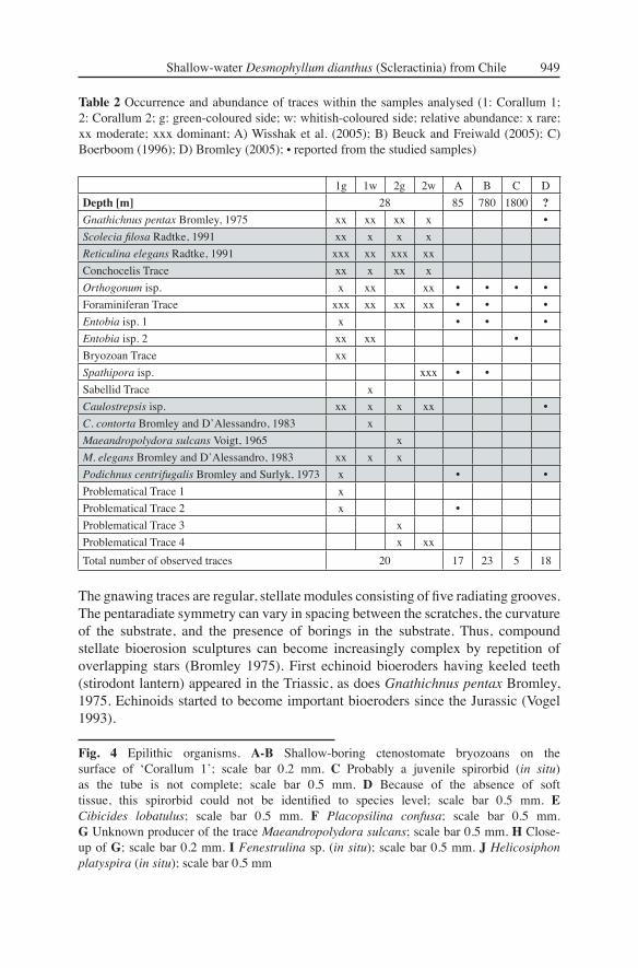
The gnawing traces are regular, stellate modules consisting of fi ve radiating grooves. The pentaradiate symmetry can vary in spacing between the scratches, the curvature of the substrate, and the presence of borings in the substrate. Thus, compound stellate bioerosion sculptures can become increasingly complex by repetition of overlapping stars (Bromley 1975). First echinoid bioeroders having keeled teeth (stirodont lantern) appeared in the Triassic, as does Gnathichnus pentax Bromley, 1975. Echinoids started to become important bioeroders since the Jurassic (Vogel 1993).
Fig. 4 Epilithic organisms. A-B Shallow-boring ctenostomate bryozoans on the surface of ʻCorallum 1ʼ; scale bar 0.2 mm. C Probably a juvenile spirorbid (in situ) as the tube is not complete; scale bar 0.5 mm. D Because of the absence of soft tissue, this spirorbid could not be identifi ed to species level; scale bar 0.5 mm. E Cibicides lobatulus; scale bar 0.5 mm. F Placopsilina confusa; scale bar 0.5 mm. G Unknown producer of the trace Maeandropolydora sulcans; scale bar 0.5 mm. H Close-up of G; scale bar 0.2 mm. I Fenestrulina sp. (in situ); scale bar 0.5 mm. J Helicosiphon platyspira (in situ); scale bar 0.5 mm
1g 1w 2g 2w A B C D
Depth [m] 28 85 780 1800 ?
Gnathichnus pentax Bromley, 1975 xx xx xx x •
Scolecia fi losa Radtke, 1991 xx x x x
Reticulina elegans Radtke, 1991 xxx xx xxx xx
Conchocelis Trace xx x xx x
Orthogonum isp. x xx xx • • • •
Foraminiferan Trace xxx xx xx xx • • •
Entobia isp. 1 x • • •
Entobia isp. 2 xx xx •
Bryozoan Trace xx
Spathipora isp. xxx • •
Sabellid Trace x
Caulostrepsis isp. xx x x xx •
C. contorta Bromley and DʼAlessandro, 1983 x
Maeandropolydora sulcans Voigt, 1965 x
M. elegans Bromley and DʼAlessandro, 1983 xx x x
Podichnus centrifugalis Bromley and Surlyk, 1973 x • •
Problematical Trace 1 x
Problematical Trace 2 x •
Problematical Trace 3 x
Problematical Trace 4 x xx
Total number of observed traces 20 17 23 5 18
Table 2 Occurrence and abundance of traces within the samples analysed (1: Corallum 1; 2: Corallum 2; g: green-coloured side; w: whitish-coloured side; relative abundance: x rare; xx moderate; xxx dominant; A) Wisshak et al. (2005); B) Beuck and Freiwald (2005); C) Boerboom (1996); D) Bromley (2005); • reported from the studied samples)
950 Försterra, Beuck, Häussermann, Freiwald
Gnathichnus pentax Bromley, 1975 (Figs. 3F, 3G)
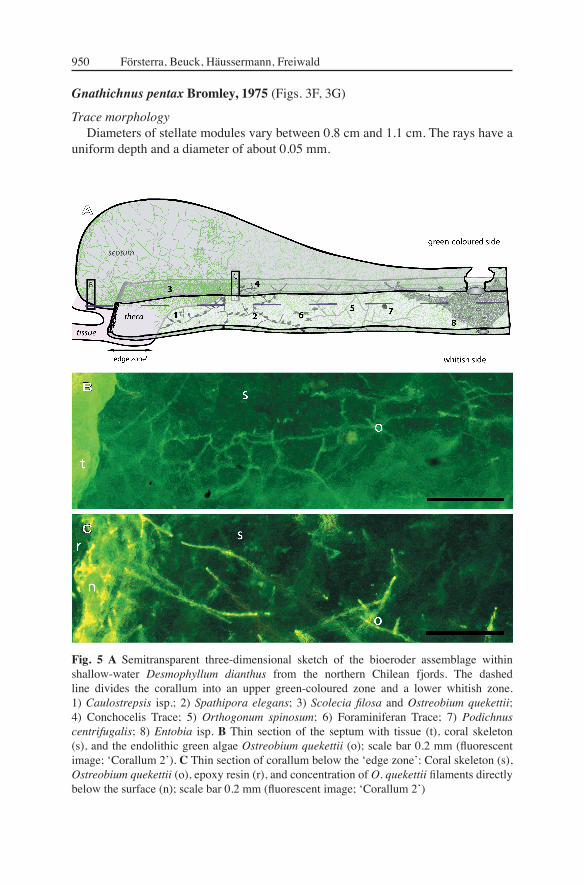
Trace morphologyDiameters of stellate modules vary between 0.8 cm and 1.1 cm. The rays have a
uniform depth and a diameter of about 0.05 mm.
Fig. 5 A Semitransparent three-dimensional sketch of the bioeroder assemblage within shallow-water Desmophyllum dianthus from the northern Chilean fjords. The dashed line divides the corallum into an upper green-coloured zone and a lower whitish zone. 1) Caulostrepsis isp.; 2) Spathipora elegans; 3) Scolecia fi losa and Ostreobium quekettii; 4) Conchocelis Trace; 5) Orthogonum spinosum; 6) Foraminiferan Trace; 7) Podichnus centrifugalis; 8) Entobia isp. B Thin section of the septum with tissue (t), coral skeleton (s), and the endolithic green algae Ostreobium quekettii (o); scale bar 0.2 mm (fl uorescent image; ʻCorallum 2ʼ). C Thin section of corallum below the ʻedge zoneʼ: Coral skeleton (s), Ostreobium quekettii (o), epoxy resin (r), and concentration of O. quekettii fi laments directly below the surface (n); scale bar 0.2 mm (fl uorescent image; ʻCorallum 2ʼ)

Shallow-water Desmophyllum dianthus (Scleractinia) from Chile 951
ProducerIn the habitat studied, common grazers are the echinoids Arbacia dufresnei
Blainville, 1872 and Pseudechinus magellanicus (Philippi, 1857), which are most likely the producer of the bite traces.
RemarksDirectly below the polypʼs tissue a complex cluster of stellate trace sculptures
is present in ʻCorallum 2ʼ. The surface of ʻCorallum 1 ̓ exposes a multitude of scratches. A clear identifi cation to their browsing producer is not given. Thus, we consider synergistic degradation effects caused by a variety of specialised grazers, e.g., gastropods, sea urchins and chitons.
CyanobacteriaEndolithic Cyanobacteria are restricted to the photic zone, owing to their
autotrophic metabolism, and have their highest abundance in shallow water (LeCampion-Alsumard 1979; LeCampion-Alsumard and Golubic 1985; Schuhmacher et al. 1995; Gektidis 1999; Kaehler 1999). Amongst the group of endolithic Cyanobacteria, the correlated ichnospecies Fasciculus acinosus Glaub, 1994, and F. dactylus Radtke, 1991 are the most dominant traces in the marine intertidal zone, which led to the defi nition of a microboring index ichnocoenosis, characterised by these two ichnospecies (Glaub 1999). In zones of high light penetration, Cyanobacteria tend to grow perpendicularly to the surface (Golubic et al. 1975), whereas in deeper photic zones, they predominantly grow parallel to the surface (Budd and Perkins 1980; Ekdale et al. 1984). The oldest microborers with cyanobacterial affi nities are known since the Precambrian (Zhang and Golubic 1987; Golubic et al. 1999).
Scolecia fi losa Radtke, 1991 (Figs. 6A-B)
Trace morphologyElongated fi ligree fi laments of 1.4-2.2 μm in diameter with a partially rough
surface texture form interwoven networks that can penetrate the entire theca. The trace is sparsely branched in rectangular angles (T-branches).
Producer Plectonema terebrans Bornet and Flahault, 1889 is regarded as the producer of
the ichnospecies S. fi losa (Radtke 1991).
RemarksP. terebrans is cosmopolitan, and characterised by a wide bathymetric range
(Radtke 1991; Gektidis 1997). Günther (1990) describes a bathymetric distribution of P. terebrans from 1 m to 42 m at Cozumel, Mexico. Studies at Lee Stocking Island show P. terebrans occurrences down to 100 m depth (Vogel et al. 2000). Budd and Perkins (1980) document six endolithic Cyanobacteria from the Puerto Rican Shelf, of which P. terebrans was the most abundant down to 85 m depth.

952 Försterra, Beuck, Häussermann, Freiwald

Shallow-water Desmophyllum dianthus (Scleractinia) from Chile 953
Chlorophyta A multitude of boring algae are known from tropical shallow-water environments
(LeCampion-Alsumard et al. 1995; Gektidis 1997). However, several endolithic algae are also reported from the cold-temperate, upwelling area off Mauritania (Glaub 2004). Endolithic species are restricted to different water depths appropriate to the wavelengths absorbed by their photosynthetically active pigments. Apart from the physical factors, such as irradiance or temperature, the distribution of endolithic algae is also infl uenced by the grazing activity of herbivores (Highsmith 1981; Fork and Larkum 1989; Schlichter et al. 1997). First borings of endolithic green algae are recorded from the Early Paleozoic (Podhalanska 1984; Vogel 1993).
Reticulina elegans Radtke, 1991 (Figs. 5B, C, 7)
Trace morphology Spherical to fl attened fi laments measure between 1.85-3.15 μm in diameter.
These fi laments can ramify at distances of 3.4 μm and up to several 100 μm in preferred angles of 90° or 120° (morphological structure 1). Directly below the surface, the trace is highly branched, creating a reticulated network. Distances between successive ramifi cations increase proportionally with penetration depth, producing wide-meshed networks. The smooth-textured fi laments can penetrate the entire skeleton. Directly below the surface, the fi lamentous tubes may enlarge to diameters of about 15 μm (morphological structure 2). This tube peculiarity is located terminally and features slight constrictions at distances of about 10 μm. Morphological structure 2 has a granular surface texture of about 1 μm small units. Its blind-ending tubes end hemispherically and can penetrate the carbonate up to 75 μm deep. In addition, up to ten fi laments of morphological structure 1 may coalesce in a polygonal, fl attened structure (morphological structure 3) that is about 2.7 μm thick and can measure 18-25 μm in diameter. The surface texture of these fl attened structures consists of spherical to ʻvermiculate ̓granules.
Morphological structure 1 is interpreted to be ichnospecies Reticulina elegans. This trace is known since the Silurian (Vogel et al. 1995). However, morphological structure 2 is similar to Fasciculus isp. Traces resembling morphological structure 3 have not been found in the literature.
ProducerThe trace morphology correlates with the shape of the green alga Ostreobium
quekettii (Bornet and Flahault, 1889) (Chlorophyta, Bryopsidales, Ostreobiaceae). The fi ligree tubes harbour the siphonal, unpartitioned thallus. Tube enlargements
Fig. 6 SEM images of resin casts. A Scolecia fi losa boring of the producer Plectonema terebrans associated with tube casts of Ostreobium quekettii; scale bar 30 μm. B Scolecia fi losa; scale bar 10 μm. C Brachiopod trace Podichnus centrifugalis; scale bar 30 μm. D Conchocelis Trace; scale bar 100 μm. E Spatial distribution of elongated linear fi laments of Conchocelis Trace; scale bar 300 μm. F Older stage of Conchocelis Trace; scale bar 300 μm. G Problematical Trace 2; scale bar 100 μm. H Detail of G showing a xenoglyphic surface texture; scale bar 10 μm

954 Försterra, Beuck, Häussermann, Freiwald

Shallow-water Desmophyllum dianthus (Scleractinia) from Chile 955
(morphological structure 2) are interpreted to be sporangia, bearing quadrifl agellate, vegetative zoospores (Kornmann and Sahling 1980). Lukas (1974) describes intercalar tube enlargements of O. quekettii that measure up to 25 μm in width, and are especially located at sites of branching. The description corresponds to the morphology of morphological structure 3. However, observations of spores, or rather gametes are lacking so the function of morphological structure 3 remains uncertain.
Remarks The trace occurs within the entire coralla of both specimens, but has a higher
intra-skeletal density on the green-coloured sides (see Fig. 5B). However, the apical fi rst centimetre of the whitish side (ʻCorallum 2ʼ) has not been infested by the green alga. Living thalli of O. quekettii are restricted to septal and thecal walls, which are at least temporarily covered by the ʻedge zone ̓of the polyp (sensu Stolarski 1996). Their siphonal thalli seem to infest the skeleton at exposed areas, growing into the apical live zone of D. dianthus. Lukas (1974) notes that after infesting living scleractinians, the direction and rate of algal growth follows the hostʼs outer circumference. This growth habit of O. quekettii has also been reported from tropical shallow-water corals, such as from Porites lobata Dana, 1846 at Moorea, French Polynesia (LeCampion-Alsumard et al. 1995), from Porites compressa Dana, 1846 at Kaneohe Bay, Hawaii (Shashar and Stambler 1992), and from Mycedium elephantotus (Oken, 1815) in the Gulf of Aqaba, Red Sea (Schlichter et al. 1997). Highsmith (1981) reports annual growth rates of 9.5 mm for O. quekettii in Porites lutea Quoy and Gaimard, 1833.
Ostreobium quekettii becomes abundant under low light levels, and is also found in sediments of the deep euphotic zone, suggesting a low rate of respiration (Budd and Perkins 1980). Vogel et al. (2000) reported O. quekettii occurrences in tropical environments down to 300 m depth. Halldal (1968) showed that photosynthetic activities of different Ostreobium species in the scleractinian coral Favia sp. are restricted to the peripheral ʻgreen-bandsʼ. Thus short ramifi cation distances creating dense networks directly below the surface can be linked to improvement of photosynthetic productivity (see also Fig. 5C).
Fig. 7 SEM images of resin casts. Morphological structures of the green algal trace Reticulina elegans that are produced by Ostreobium quekettii. A Dense network of morphological structure 1. The spatial arrangement of the overlying linear resin tubes is an artefact caused by the dissolution of the carbonate: long wide meshed fi laments originally penetrated the entire carbonate; scale bar 100 μm. B Young growth stage with its characteristic reticulated network directly below the surface; scale bar 30 μm. C Cluster of young stages of morphological structure 2; scale bar 100 μm. D Dense cluster of morphological structure 2 partially covered by morphological structure 1; scale bar 100 μm. E Tube enlargements of about 15 μm in diameter with slight constrictions every 10 μm (morphological structure 2); scale bar 30 μm. F Close-up of E. Tubes of morphological structure 2 have a granular surface texture and end hemispherically; scale bar 10 μm. G Polygonal structures (morphological structure 3) with a surface texture of ʻvermiculate ̓granules; scale bar 30 μm. H Morphological structure 3; scale bar 10 μm

956 Försterra, Beuck, Häussermann, Freiwald
RhodophytaEndolithic red algae appear to be cosmopolitan on rocky shorelines. The greatest
diversity is found in cold-temperate and boreal regions. They are present in the order Bangiales that contains currently a single family (Bangiaceae Engler, 1892). Amongst the order Bangiales, some species have a heteromorphic life history as documented from Porphyra. Its carpospores germinate to a trichous phase (Conchocelis phase), where fi laments in nature are always found boring into a calcium carbonate substrate, and are interpreted to be the diploid sporophyte generation (Sitte et al. 2002). Its conchospore formation is a photoperiodic response, induced by reducing daylengths. Meiosis takes place during germination of the conchospores to a blade-like haploid gametophytic phase that requires cooler seasons (Maggs and Callow 2002). Fossils related to Porphyra are dated to 500 million years ago (Campbell 1980). Moreover, borings resembling the Conchocelis stage of Porphyraʼs sibling genus, Bangia, have been discovered in Proterozoic (1.2 billion years) deposits in Arctic Canada, thus displaying on of the oldest taxonomically resolved eukaryotes in the fossil record (Butterfi eld et al. 1990; Butterfi eld 2000).
Conchocelis Trace (Figs. 6D-F)
Trace morphologyUnbranched linear tubes intrude perpendicularly from the surface as much as
1100 μm into the substrate. The whip-shaped tubes are distributed irregularly. Tube diameters taper slightly towards their ends from basally 33-45 μm to terminally 22 μm and show slight constrictions at intervals of about 13.5 μm.
ProducerSimply constructed trichous algae with an intercalary growth are present in the
order Bangiales ( Rhodophyta). However, a lack of observations of the endolithic thallus denies a clear phylogenetic relationship.
RemarksSimilarly sized whip-shaped tubes are produced by the boring foraminifer
Hyrrokkin sarcophaga Cedhagen, 1994. Here, however the spatial arrangement of single fi laments lacks any visible pattern.
FungiEndolithic fungi are attributed to the orders Saprolegniales, Peronosporales
(Oomycota), and Cytridiomycetes (Eumycota). They are distinguished by their size, shape, mode of ramifi cation, and sporangia (Glaub 1994). Fungal fi laments (hyphae) secrete acids mainly apically, which enable them to grow into the substrate (Sand 1995; Sitte et al. 2002). The fi lamentous morphology of their mycelia facilitates absorption of nutrients.
Orthogonum spinosum Radtke, 1991 (Figs. 8A-D)
Trace morphologyHomogeneous tubes of about 24 μm ramify preferably rectangularly and end
hemisherically. Infrequently, a multitude of small hair-like fi laments of about

Shallow-water Desmophyllum dianthus (Scleractinia) from Chile 957
1.7 μm in diameter protrude from the smooth-textured surface, having an average length of 25 μm. In the ichnogenus Orthogonum right-angular branching traces with homogeneous tube diameters are present (Radtke 1991). Orthogonum spinosum is characterised by tube diameters of 25-50 μm, similar sized hair diameters, as described above, but maximal hair lengths of only 12 μm.
ProducerDue to diffi culties in laboratory cultivation, the taxonomic relationship of
endolithic fungi is complicated, and the producer of Orthogonum spinosum has still not been identifi ed.
RemarksHowever, the ichnogenus Orthogonum has already been described from various
recent habitats (Budd and Perkins 1980; Beuck and Freiwald 2005; Wisshak et al. 2005) and is known since the Silurian (Bundschuh 2000).
ForaminiferaBoring foraminiferans are known from different families, such as Astrorhizidae
Brady, 1881, and Rosalinidae Reiss, 1963. Amongst these some parasitic forms are described, such as Hyrrokkin sarcophaga Cedhagen, 1994, recorded from the northern Atlantic on various substrates, e.g., Acesta excavata (Fabricius, 1779), sponges of the family Geodiidae, and scleractinians, such as Lophelia pertusa, Caryophyllia sp. and Madrepora oculata (see Cedhagen 1994; Freiwald and Schönfeld 1996; Beuck and Freiwald 2005; López Correa et al. 2005).
Foraminiferan Trace (Figs. 9C-H)
Trace morphologyThis dendroid boring system branches in a fan-like pattern, and mature trace
stages measure up to 1000 μm across. Its tubes are of variable diameters, however, and in mature stages they can measure up to 80 μm. Tubes ramify at different angles, and are rounded apically. The texture is slightly verrucous. The average base diameter of the single hemispherical warts is 10 μm. From the trace surface a multitude of homogeneous hair-like fi laments protrude and taper distally from basal diameters of 3.2 μm to about 1 μm. They have a rough surface texture. Mature stages are characterised by long hairs (up to 500 μm). Individual traces can occur in compounds of three up to fi ve and can have penetration depths up to 300 μm. Glaub (1994) described small rosette borings having a spinous tube relief as Semidendrina-Form.
ProducerThe dendroid trace bears similarities to traces of the foraminiferan Globodendrina
monile Plewes, Palmer and Haynes, 1993 that is phylogenetically related to the family Astrorhizidae Brady, 1881. The morphology is believed to be an adaptation to high energy environments (Cherchi and Schröder 1991). However, in the analysed specimen, only the plane dendroid boring system is observed, and not the spherical part that is looming into the substrate.

958 Försterra, Beuck, Häussermann, Freiwald

Shallow-water Desmophyllum dianthus (Scleractinia) from Chile 959
RemarksSimilar traces are also reported in the skeleton of Lophelia pertusa from the North
Atlantic (Beuck and Freiwald 2005; Wisshak et al. 2005), and as ʻhirsute camerate form ̓from Early Pleistocene coral limestones on Rhodes (Bromley 2005).
Porifera Endolithic sponges are phylogenetically related to the order Hadromerida
(Hooper and Soest 2002) and the order Haplosclerida (Soest and Hooper 2002), belonging to the group Demospongiae. By excavating the protective carbonate, they create vast galleries of chambers, which are connected with the exterior via aperture canals. Sponge trace casts are usually visible to the naked eye, and thus belong to the group of macroborings (Bromley 2005). Traces of different ichnospecies can vary, e.g., in morphology, construction of inter-chamber canals, aperture size, and surface ornamentation (see Bromley and DʼAlessandro 1984; Calcinai et al. 2003). Sponges play a central role in the destruction of reefal ecosystems (Acker and Risk 1985), but only 2-3 % of the substrate is dissolved by etching cells (Rützler and Rieger 1973; Pomponi 1980), while the rest is removed as characteristical-shaped chips. They are expelled through the oscula, and can accumulate to form sediments (Rützler 1975; Freiwald and Wilson 1998).
Entobia isp. 1 (Figs. 9A-B)
Trace morphologyThe spherical trace is 77 μm in diameter. Its surface is sculptured by hemispherical,
smooth textured units which are between 25-31 μm in diameter. It penetrates the carbonate to about 85 μm.
ProducerThe spherical trace is a juvenile endolithic sponge chamber, formed by specialised
archaeocytes, the etching cells (Rützler and Rieger 1973; Pomponi 1976, 1979). The absence of sponge spicules prevented closer identifi cation of its producer.
RemarksThe trace morphology as well as the surface ornamentation is similar to the
blackberry-shaped juvenile stage of Spiroxya heteroclita Topsent, 1896, which was found in the skeleton of Lophelia pertusa at the Propeller Mound (Beuck and Freiwald 2005). However, a common bioeroding sponge in this fjord habitat is Cliona chilensis, which is the most harmful eroder. Most coralla that are broken
Fig. 8 SEM images of resin casts. A-D Ichnospecies Orthogonum spinosum. A Ichnocoenosis of Entobia isp. (arrow 1) and O. spinosum (arrow 2); scale bar 1000 μm. B The homogenous tubes often branch in a rectangular angle; scale bar 100 μm. C Filigree hairs; scale bar 10 μm. D The tubes end hemispherical; scale bar 30 μm. E-H Problematic Trace 4. E Young trace stages show homogeneous tubes, on which sporangia insert; scale bar 100 μm. F Filaments of the sporangia are highly branched, and feature slight constrictions; scale bar 100 μm. G Two different distinctive features of the sporangia; scale bar 30 μm. H In mature stages sporangia can enlarge to tubes of diameters of up to 55 μm; scale bar 30 μm

960 Försterra, Beuck, Häussermann, Freiwald

Shallow-water Desmophyllum dianthus (Scleractinia) from Chile 961
off show strong infestation by and degradation from this sponge which might have been the main cause for the breaking.
Entobia isp. 2 (Fig. 2G)
Trace morphologyThe trace consists of spherical chambers, which are between 1.75-2 mm in
diameter. They are interconnected by intercameral canals, which are up to 350 μm in diameter. The surface texture of its etching cells eluded documentation, due to a high abundance of the green algal trace Reticulina elegans.
ProducerThe spicules have not been documented. Thus, identifi cation to its producer is
not possible.
RemarksOwing to the relatively thick intercameral canals, as well as the size of their
chambers, the trace is morphologically similar to Entobia retiformis (Stephenson, 1952) (see also Bromley and DʼAlessandro 1987). X-ray radiographs of ʻCorallum 1 ̓ document in the basal 5.5 cm a conspicuous infestation by this sponge. The largest part of the vast sponge chamber galleries was constructed within septa. Due to the lack of massive carbonate within the non-thecal coral parts, as well as the presence of high amounts of sponge aperture canals, detailed analysis of non-thecal coral casts is not applicable in thinly calcifi ed coral septa.
BryozoaTaxonomically, endolithic bryozoans are attributed to the order Ctenostomata.
Single individuals (zooids) of a colony are interconnected by stolons, which underlie the surface. In the deep-water environments of the northern Atlantic, the most common bryozoan trace within the skeleton of cold-water corals is Spathipora isp. Its incremental growth stages in the substrate are documented from trace casts by Beuck and Freiwald (2005).
Bryozoan Trace 1 (Figs. 10A-B)
Trace morphologyBilaterally symmetrical shallow cast elevations of about 350 μm x 120-186 μm
are horizontally disposed to tube casts throughout their entire length. Lateral tubes
Fig. 9 SEM images of resin casts. A-B Entobia isp. A Three centimetres below the ̒ edge zone ̓a juvenile sponge chamber (B) is located within dense networks of Reticulina elegans; scale bar: 300 μm. B Close-up of A. The surface texture of the individual etching-cell imprints is smooth; scale bar 30 μm. C Probably a young stage of a foraminiferan boring, but the lack of hairs also advocate a juvenile sponge boring; scale bar 100 μm. D-H Foraminiferan trace 1. D Young stage of a foraminiferan boring; scale bar 30 μm. E Young stages are characterised by short fi laments, emanating from the surface; scale bar 10 μm. F Dendroid tube system with a multitude of long whip-like fi laments scale bar 300 μm. G Overview of aggregates of Foraminiferan Trace 1; scale bar 1000 μm. H Close-up of dendroid tube fi laments of G; scale bar 30 μm

962 Försterra, Beuck, Häussermann, Freiwald

Shallow-water Desmophyllum dianthus (Scleractinia) from Chile 963
emanate opposite one another near middle length of the shallow cast elevations. The interconnecting tubes measure 12 μm in diameter. Distances between single cast elevations vary between 415 μm to 505 μm. The trace penetrates the carbonate to a depth of up to 67 μm.
ProducerNon-pedunculate growth types are present in the family Orbignyoporidae
Pohowsky, 1978, and Immergentiidae Silén, 1946. The morphological diagnosis of the family Orbignyoporidae corresponds to the growth morphology of Bryozoan Trace 1. The family Immergentiidae is characterised by zooids that protrude essentially vertically into the substratum. However, Immergentia patagoniana Pohowsky, 1978, described from Argentina, is characterised by location of zooids and stolons at the bottom of relatively broad depressions in the surface of the substratum, which is apparently a unique feature in the family Immergentiidae (see Pohowsky 1978).
RemarksOne colony with about 12 zooids is present 7.5 cm below the apical part of
ʻCorallum 1ʼ. In addition, a ctenostomate colony on the corallumʼs surface is located in the corresponding area. However, its exposed zooid lengths are smaller in size than the total lengths of the trace cast elevations.
Spathipora elegans Fischer, 1866 (Figs. 10C-H)
Trace morphology Directly below the surface, a network of homogeneous tubes of about 11 μm in
diameter connects elongated chambers, which are fully grown between 500 and 600 μm in length and about 75 μm in diameter. Casts of the chambers are oriented dextrally to the tube casts and are connected with the tubes via a tubular pedunculus that measures about 9 μm in diameter. The surface texture of the trace elements is smooth. Unilateral ramifi cations usually have an expanded junction to the main stolon, in contrast, the connection of bilateral ramifi cations is slightly thickened. One chamber with a diameter of 190 μm is tilted nearly vertically, and gives rise to three tubes that are, with respect to the peripheral ones, thicker in diameter. In addition, an ovoid chamber of 30 μm in diameter and 75 μm in height is connected to tubes that insert opposite one another.
Fig. 10 SEM images of resin casts. A-B Bryozoan Trace 1. A Casts of shallow-boring bryozoans; scale bar 1000 μm. B Bilaterally symmetrical shallow cast elevation, which is interconnected by tubes that contain stolons; scale bar 100 μm. C-H Bryozoan ichnogenus Spathipora elegans. C Nine chambers that harboured the autozooids are interconnected by tubes that contained the stolons; scale bar 300 μm. D (1) adult autozooid chamber; (2) stolon tube; (3) juvenile foraminiferal trace; (4) the green algal trace Reticulina elegans Radtke, 1991; scale bar 100 μm. E Chamber of an autozooid that is connected via pedunculus to the stolon tube; scale bar 100 μm. F Bilateral ramifi cation slightly thickened; scale bar 100 μm. G Vertical chamber (ancestrula); scale bar 100 μm. H Ovoid chamber (gonozooid); scale bar 30 μm

964 Försterra, Beuck, Häussermann, Freiwald
ProducerCharacteristic for endolithic Bryozoa of the suborder Stolonifera are tube casts
bearing the stolons, which interconnect the single individuals (zooids). The vertical chamber is interpreted to be the ancestrula. The stolons interconnect the specialised zooids with each other that can be distinguished between autozooids, dwelling in elongated chambers, and gonozooids, which are contained in ovicell-shaped chambers. Similar trace features are described for the bryozoan trace Spathipora elegans Fischer, 1866 from the coast of Chile (Pohowsky 1978).
RemarksFirst traces of the sinistral bryozoan colony appear on the light-averted side
of the ʻCorallum 2 ̓directly below the area, which was frequently covered by the polyp. One colony is found with more than 100 zooids. Since individuals have a fi ltering lifestyle, they are restricted to coral skeletons that are exposed to water. The ichnogenus Spathipora is common in aphotic trace communities (ichnocoenoses) from the North Atlantic (Beuck and Freiwald 2005), where the cold-water coral Desmophyllum dianthus is associated with the main framework builder Lophelia pertusa and Madrepora oculata.
Polychaeta48 species of polychaetes, belonging to the families Spionidae, Sabellidae, and
Cirratulidae have been reported as borers of other living marine organisms (Martin and Britayev 1998). Amongst the parasitic boring polychaetes, more than 53 % are additionally considered to be facultative symbionts, boring also into non-living substrates. The boring habit is interpreted to be a protective advantage as well as the easiest way to contact with the water column just above the sediment surface (Martin and Britayev 1998). Boring worms have inhabited in reefs since the Lower Cambrian (Vogel 1993).
Sabellid Trace (Figs. 2E-G)
Trace morphologyA circular tube of 2.8 mm in diameter exits to the surface. Only the region from
the surface and in to 1.7 mm is preserved as trace cast, which was originally located in the massive carbonate of the theca. Thus, the penetration depth has not been measured. In addition, its surface texture eluded documentation, owing to attached fi laments of the trace Reticulina elegans.
ProducerThe hole contains remains of a dark coloured tube that is thick-walled and
gelatinous, belonging to a euendolithic sabellid worm. The endolithic sabellid Perkinsiana corcovadensis (Hartmann-Schröder, 1965) is described from southern Chile at a depth of 190 m (see also Knight-Jones 1983).
RemarksThe cavity is located about 7 cm above the base of ʻCorallum 1 ̓on the green
side. The boring mechanism seems to be chemical. This euendolithic sabellid worm

Shallow-water Desmophyllum dianthus (Scleractinia) from Chile 965
is a common bioeroder in the habitat studied mainly in coral substrates, but also often found in shells of the gastropod Crepidula sp.
Caulostrepsis contorta Bromley and DʼAlessandro, 1983
Trace morphologyThe boring is curved and circular in shape. Its limbs are 290 μm in diameter and
are interconnected by a well-developed vane. One millimetre below the surface, the limb is constructed in a surface-parallel plane that has a hemispherical course (diameter 1 mm). The trace exits to the surface by an aperture that is 270 μm in diameter.
ProducerA putative producer of the trace is discussed within the genus Polydora Bosc,
1802 (see Bromley and DʼAlessandro 1983). The boring spionids Polydora cf. ricksettsi Woodwick, 1961, Dipolydora huelma Sato-Okoshi and Takatsuka, 2001 and Dipolydora giardi Mesnil, 1896, are known from this region to bore on Crepidula sp. (Sato-Okoshi and Takatsuka 2001). The spionid worm Polydora websteri Hartman, 1943 forms similar-shaped traces. However, their trace shape varies strongly with different host substrates (Blake and Evans 1973).
RemarksIn contrast to Maeandropolydora elegans Voigt, 1970, limbs of this trace do not
parallel each other, but have a well-developed vane of varying shape.
Caulostrepsis isp. (Figs. 11B-D)
Trace morphology Pouches are bent in a narrow U-form, and in cast tower up to 2.3 mm perpendicular
to slightly tilt into the substrate. The inward-facing walls of the limbs are fused by complete removal of the intervening substrate (open pocket structure; Bromley and DʼAlessandro 1983), and show a slight axial depression in between limbs. Transverse sections of the trace are fl attened-elliptically (250 μm x 425 μm), and increase slightly in width towards their aperture. However, the aperture of one trace enlarges up to diameters of 430 μm x 573 μm. The transverse surface ornamentation seems to derive from the structural heterogeneity of the substrate (xenoglyph; Kelly and Bromley 1984), and refl ects growth increments which vary between 125 μm and 145 μm. The trace is morphologically similar to the ichnospecies Caulostrepsis cretacea (Voigt, 1971). However, Caulostrepsis cretacea decreases in size towards its aperture (Bromley and DʼAlessandro 1983).
ProducerU-shaped burrows are described from boring activities within spionid polydorids.
Blake and Evans (1973) display morphologically similar traces produced by Polydora ciliata (Johnston, 1838). However, Bromley (1978) published similar borings produced by the eunicid Lysidice ninetta. Thus, it seems that we shall not come closer to the producer of Caulostrepsis than polychaete annelids.

966 Försterra, Beuck, Häussermann, Freiwald
Shallow-water Desmophyllum dianthus (Scleractinia) from Chile 967
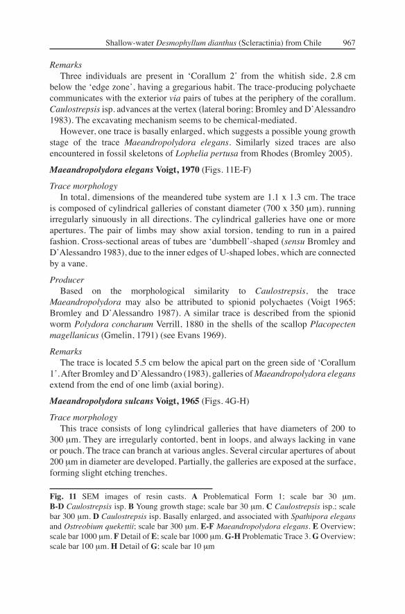
RemarksThree individuals are present in ʻCorallum 2 ̓ from the whitish side, 2.8 cm
below the ʻedge zoneʼ, having a gregarious habit. The trace-producing polychaete communicates with the exterior via pairs of tubes at the periphery of the corallum. Caulostrepsis isp. advances at the vertex (lateral boring; Bromley and DʼAlessandro 1983). The excavating mechanism seems to be chemical-mediated.
However, one trace is basally enlarged, which suggests a possible young growth stage of the trace Maeandropolydora elegans. Similarly sized traces are also encountered in fossil skeletons of Lophelia pertusa from Rhodes (Bromley 2005).
Maeandropolydora elegans Voigt, 1970 (Figs. 11E-F)
Trace morphology In total, dimensions of the meandered tube system are 1.1 x 1.3 cm. The trace
is composed of cylindrical galleries of constant diameter (700 x 350 μm), running irregularly sinuously in all directions. The cylindrical galleries have one or more apertures. The pair of limbs may show axial torsion, tending to run in a paired fashion. Cross-sectional areas of tubes are ʻdumbbellʼ-shaped (sensu Bromley and DʼAlessandro 1983), due to the inner edges of U-shaped lobes, which are connected by a vane.
ProducerBased on the morphological similarity to Caulostrepsis, the trace
Maeandropolydora may also be attributed to spionid polychaetes (Voigt 1965; Bromley and DʼAlessandro 1987). A similar trace is described from the spionid worm Polydora concharum Verrill, 1880 in the shells of the scallop Placopecten magellanicus (Gmelin, 1791) (see Evans 1969).
RemarksThe trace is located 5.5 cm below the apical part on the green side of ʻCorallum
1ʼ. After Bromley and DʼAlessandro (1983), galleries of Maeandropolydora elegans extend from the end of one limb (axial boring).
Maeandropolydora sulcans Voigt, 1965 (Figs. 4G-H)
Trace morphologyThis trace consists of long cylindrical galleries that have diameters of 200 to
300 μm. They are irregularly contorted, bent in loops, and always lacking in vane or pouch. The trace can branch at various angles. Several circular apertures of about 200 μm in diameter are developed. Partially, the galleries are exposed at the surface, forming slight etching trenches.
Fig. 11 SEM images of resin casts. A Problematical Form 1; scale bar 30 μm. B-D Caulostrepsis isp. B Young growth stage; scale bar 30 μm. C Caulostrepsis isp.; scale bar 300 μm. D Caulostrepsis isp. Basally enlarged, and associated with Spathipora elegans and Ostreobium quekettii; scale bar 300 μm. E-F Maeandropolydora elegans. E Overview; scale bar 1000 μm. F Detail of E; scale bar 1000 μm. G-H Problematic Trace 3. G Overview; scale bar 100 μm. H Detail of G; scale bar 10 μm

968 Försterra, Beuck, Häussermann, Freiwald
ProducerChughtai and Knight-Jones (1988) describe similar traces etched by sabellid
worms. However, its taxonomical relationship has not been ascertained.
RemarksA similar shaped trace but larger-sized is described from the Pleistocene of Italy
by Bromley and DʼAlessandro (1983).
BrachiopodaAttached pedicle of brachiopods produce groups of fi nger-shaped borings. The
spatial arrangement of the single tubes to each other, as well as their surface texture, is indicative of different producers (see Bromley and Surlyk 1973). First brachiopod borings are reported from the Silurian (Bundschuh 2000).
Podichnus centrifugalis Bromley and Surlyk, 1973 (Fig. 6C)
Trace morphologyAbout a dozen fi nger-shaped tubes of similar size tower up (in cast) to 55 μm
perpendicularly into the substrate, and are grouped without visible pattern. Directly below the surface, they are 10.5 μm in diameter, however, their diameters taper off slightly towards their ends. The trace measures about 200 μm across.
ProducerIn the habitat, Magellania venosa was regularly observed to grow on
D. dianthus.
RemarksPodichnus centrifugalis has been found in Swedish Kosterfjord (Wisshak et al.
2005), as well as from Early Pleistocene coral limestones on Rhodes (Bromley 2005). The trace has a rich fossil record back to the Silurian (Bromley 2004).
Problematical Trace 1 (Fig. 11A)
Trace morphologyThe tubular trace is 28 μm in diameter, and penetrates the substrate perpendicularly
to a depth of 195 μm. The trace surface shows light constrictions, creating 27-40 μm long ovoid segments. Its surface texture is smooth, but bears hair-like fi laments, which are expanded up to 35 μm into the substrate with diameters of about 2 μm. A comparable trace has not been found in the literature.
Problematical Trace 2 (Figs. 6G-H)
Trace morphologyFrom a group of slight elevations (in cast) that are up to 72 μm high, conical
tubes arise perpendicularly. The diameter of the base of the tube is up to 23 μm. However, the trace narrows, and ends after about 65 μm at a spiky, or rounded termination. The surface texture of the trace is a xenoglyph. An individual trace measures about 450 μm across. A similar trace that exhibits a large cluster of straight intrusions and that penetrates from shallow depressions is described as ̒ Problematic Form 2 ̓ from Wisshak et al. (2005). The authors discuss the trace as a possible

Shallow-water Desmophyllum dianthus (Scleractinia) from Chile 969
attachment scar produced by a heterotrophic epilithic organism, such as brachiopod, or foraminiferan.
Problematical Trace 3 (Figs. 11G-H)
Trace morphologyThe cylindrical tube system lies directly below the surface and has an entire
length of about 620 μm. It exits to the surface by a circular aperture that is 30 μm in diameter. The tube enlarges after 15 μm to 46 μm and bifurcates with an angle of 40° into two tubes of each 25 μm in diameter. After about 490 μm, one of the two tubes splits again with a similar angle, as mentioned above, into two tubes that are each 21 μm in diameter. The surface texture of the tubes is characterised by a multitude of small, hemispherical-shaped knobs that are each 2.4 μm in diameter.
Problematical Trace 4 (Figs. 8E-H)
Trace morphologyBranched tubes ramify at different angles and are, owing to local constrictions, of
inconstant diameters between 7-17 μm. To this local agglomeration, a homogeneous tube of 6.5-7.5 μm in diameter can be connected. The surface texture of the trace is xenoglyphic. In older stages, the constricted tubes are enlarged and irregular-shaped, becoming less branched and measuring up to 55 μm in diameter. This distinctive trace can cover, directly below the surface, an area of up to 600 μm in diameter.
The irregular-shaped part is interpreted to be a sporangium. However, this trace can be distinguished from the morphological structure 2 of Reticulina elegans by its thicker and irregular-shaped tube diameters, as well as the different surface texture. In contrast to Orthogonum spinosum, the tubes lack hairs.
Discussion
Biocoenotic interactions
Contractions of the scleractinian polypʼs ʻedge zoneʼ, exposing the skeleton to direct contact with the water column, are reported by several authors, e.g., Harmelin (1990), Stolarski (1996), Lazier et al. (1999). Consequently, epi- and endolithic organisms can settle on the skeleton, competing for space (Fig. 5A). Their distribution and abundance is mainly infl uenced by abiotic factors, such as hydrodynamics, seasons, sedimentation rates, as well as biotic ones, such as grazing activity. Epi- and endolithic organisms can be pests stressing the host polyp to keep them away from the interior (LeCampion-Alsumard et al. 1995; Martin and Britayev 1998). Although, the migrating ʻedge zone ̓can consecutively repress growth of adjacent fi ltering endoliths and attached epiliths (see Harmelin 1990), living thalli of the endolithic green alga Ostreobium quekettii are reported in skeletal areas frequently covered by the polypʼs ʻedge zone ̓(LeCampion-Alsumard et al. 1995; Schlichter et al. 1997). Beneath the polypʼs tissue, endolithic algae can take shelter from sedimentation and grazers, as well as from epilithic organisms that shade the skeleton (LeCampion-Alsumard et al. 1995). Adjacent to the polyp

970 Försterra, Beuck, Häussermann, Freiwald
ʻedge zoneʼ, the coralla of the Desmophyllum dianthus specimens analysed are sculptured by dense patterns of grazing traces, fostering the hypothesis that endolithic algae benefi t from the protection by the tissue of their host organisms. Owing to their photoautotrophic nutrition, green algae are restricted to above the light compensatory depth. Schlichter et al. (1997) calculated that their bathymetric distribution is limited to a minimum surface light intensity of 0.0009 %. Reductions of irradiance are caused by water depth, ʻedge zoneʼ, coral tissue thickness, light-transmissivity of the skeleton, and self-shading of algal fi laments. But light-yield can be improved by chromatic adaptation, showing variable photosynthetic-active pigment compositions (Schlichter et al. 1997). Most notable are studies on the green alga Ostreobium quekettii in the tropical azooxanthellate coral Tubastrea micranthus Ehrenberg, 1834 that show transport of photoassimilates released from the algal fi laments to the coral tissue via the uptake mechanisms for dissolved organic substances of cnidarians (Schlichter et al. 1995). This interaction raises the polypʼs fi tness by increasing productivity and biomass. In return, metabolic end products can be used by the endolithic alga. But productivity of endolithic algae reaches a maximum of just 10 % of that of zooxanthellae (Schlichter et al. 1997).
Up to now, no examples for mutualistic symbiotic relationships with deep-water corals are known (Buhl-Mortensen and Mortensen 2004). Nevertheless, the unilateral progression of the polypʼs ʻedge zone ̓on the green-coloured side, in contrast to the whitish side, where Q. quekettii is absent in the zone of living tissue, can indicate an interaction between the green algal thallus of Ostreobium quekettii and its host Desmophyllum dianthus (see Table 1). Additionally, we observed a thicker theca on the green-coloured side of both of the investigated coralla, which could be linked with the photosynthetic activity of the endolithic green algae, strengthening the coral individual by stimulating its skeletal mineralisation (see Macintyre and Towe 1976). Both factors can indicate a putative facultative mutualistic ectosymbiosis between endolithic algae and their coral host, but further studies are required to enlighten potential benefi cial interactions.
Ichnocoenoses as environmental indicators
The lifespan of Desmophyllum dianthus is mainly ruled by skeletal growth versus the infestation intensity of macro-borers, such as sponges and polychaetes, which increase the intraskeletal porosity considerably, thus weakening the architectural stability of the coral (Försterra and Häussermann 2003). Eventually corals become detached from the substrate and are piled up in deeper depositional environments (Boerboom 1996; Försterra and Häussermann 2003). Thus, the investigation of trace communities (ichnocoenoses) within coral skeletons and its comparison to growth rates of corals provides a powerful tool to defi ne ecological health conditions for cold-water corals.
The analysed ichnocoenoses are dominated by traces of heterotrophic organisms, which are accompanied by Scolecia fi losa and Reticulina elegans. After Glaub (2004), the present assemblage is a characteristic feature of the dysphotic zone that extends from 1 % level to about 0.01 % or 0.001 % of surface light. This quite shallow occurrence of a dysphotic zone ichnoassemblage can be attributed
Shallow-water Desmophyllum dianthus (Scleractinia) from Chile 971
to the growth of Desmophyllum dianthus on the undersides of rock ledges, where the light intensity is strongly reduced. Therefore, trace assemblages in fossil Desmophyllum communities from near coast and cliff-bound environmental settings have to be regarded carefully, since the ichnocoenosis index could indicate much deeper paleodepths than those that actually prevailed at the time of formation. For bathymetrical reconstructions, the past relief and possible environmental conditions have to be evaluated carefully.
True deep-water ichnocoenoses (Table 2) have been reported by Wisshak et al. (2005) from the Kosterfjord off Sweden (-85 m), by Beuck and Freiwald (2005) from the Porcupine Seabight in the Northeast Atlantic (-780 m), and by Boerboom et al. (1998) from Orphan Knoll, off Newfoundland (-1800 m).
In this study, the investigated shallow-water ichnocoenoses have typical deep-water characteristics, but also contain shallow-water elements, due to their occurrence within the photic zone. A general similarity to the deep-water situation is depicted by the common endolithic traces of fungi ( Orthogonum), foraminiferans ( Semidendrina), bryozoans ( Spathipora), and brachiopods ( Podichnus), as well as by associated epilithic organisms ( Cibicides lobatulus). The infl uence of light is shown by the presence of boring algae. Additionally, the non-light-dependant polychaete ichnogenera Caulostrepsis and Maeandropolydora indicate shallow marine conditions (Bromley and DʼAlessandro 1983).
A similar ichnocoenosis, which appears contradictory at fi rst glance, has been reported from Bromley (2005) from Early Pleistocene deposits of Rhodes. Here the trace community and the general fauna assemblage clearly indicate deep-water conditions, while the presence of the ichnogenus Caulostrepsis isp. provides the only hint to actually shallower conditions. Despite fi rst studies in the deep Mediterranean, another curiosity is that traces of the bryozoan ichnogenus Spathipora have not yet been recorded (Bromley 2005; own observation; personal comm. Max Wisshak).
Deep-water fauna has already been recorded from shallow cryptic habitats from the Mediterranean Basin, especially from caves (Bromley and Asgaard 1993; Vacelet et al. 1994; Vacelet 1996; Harmelin 1997). However, the studied occurrence of deep-water biocoenoses (Försterra and Häussermann 2003) and ichnocoenoses in shallow-waters is strongly linked to the hydrographic conditions of the fjord region with strong freshwater run off, which forces an internal circulation system, allowing marine deep-waters to enter the troughs. These conditions with shallow deep-water fauna and also shallow deep-water ichnocoenosis are also known from other fjord areas, as in Scandinavia and New Zealandʼs Fjordland (South Islandʼs west coast) (Grange et al. 1981; Wisshak et al. 2005). Within the Chilean fjord system, the upper limit of Desmophyllum dianthus is only limited by the lower boundary of the low salinity layer.
Conclusions
• In the Chilean fjords, Desmophyllum dianthus has been observed to exist over a wide bathymetrical range under different environmental conditions. Although the structure of the coral banks in different depths may look
972 Försterra, Beuck, Häussermann, Freiwald
similar on fi rst sight, the composition of the associated biocoenoses differ with depth.
• The actinarians and sponges associated with D. dianthus in shallow water contain typical shallow-water as well as deep-water species.
• An inventory of 20 traces is given for the ichnocoenosis within the skeleton of D. dianthus. They are taxonomically correlated to Cyanobacteria (1), Chlorophyta (1), Rhodophyta (1), Porifera (2), Fungi (1), Foraminifera (1), Bryozoa (2), Polychaeta (5), Echinoida (1), Brachiopoda (1), and four traces of uncertain producer.
• The investigated shallow-water ichnocoenosis comprises typical shallow-water endoliths, as well as deep-water elements.
• The ichnocoenoses are dominated by heterotrophic organisms which are accompanied by Scolecia fi losa, and Reticulina elegans, indicating dysphotic conditions at 28 m depth.
• D. dianthus settles on near vertical walls and, preferably, on the undersides of rock ledges with downward facing polyps. This pattern can be observed down to at least 255 m and is probably due to its sensitivity to sedimentation.
• Thus, in photic environments, ichnoassemblages within the skeleton of D. dianthus can only be used as a tool for (paleo-) bathymetrical reconstructions with caution.
Acknowledgements
The authors thank Agostina Vertino (Institute of Paleontology, Erlangen, Germany) for providing expertise in coral morphotype discussion and the Radiological Department of the Universitätsklinik für Zahn-, Mund- und Kieferkrankheiten, Erlangen for x-ray imaging. Marlies Neufert (Institute of Paleontology, Erlangen, Germany) is thanked for her assistance in fl uorescence microscopy. The identifi cation of Foraminifera was assisted by Joachim Schönfeld (IFM-GEOMAR - Leibniz Institute of Marine Sciences, Kiel, Germany); the Bryozoa were kindly identifi ed by Antonietta Rosso (Catania University, Italy) and Hugo Moyano (Universidad de Concepción, Chile), the serpulids by Rosanna Sanfi lippo (Catania University, Italy) and Nicolas Rozbaczylo (Universidad Católica de Chile), sponges by Eduardo Hajdu (National Museum of Rio de Janeiro, Brazil), brachiopods by Lloyd Peck (British Antarctic Survey, UK) and echinoids by David Pawson (Smithsonian Institution, Washington D.C., USA). Earlier drafts of the manuscript were fruitfully discussed with Matthias López Correa and Max Wisshak (both Institute of Paleontology, Erlangen, Germany). The authors thank Richard G. Bromley (University of Copenhagen, Denmark) and J. Murray Roberts (Scottish Association for Marine Science, Oban, UK) for the provision of very constructive reviews. This is publication number 8 from Huinay Scientifi c Field Station. This bioerosion study is a comparative study of the ESF EUROMARGINS ʻMoundforce ̓ programme, funded by the Deutsche Forschungsgemeinschaft to André Freiwald (Fr-1134/8).

Shallow-water Desmophyllum dianthus (Scleractinia) from Chile 973
ReferencesAcker KL, Risk MJ (1985) Substrate destruction and sediment production by the boring
sponge Cliona caribbaea on Grand Cayman Island. J Sediment Petrol 55: 705-711Arntz WE (1999) Magellanic-Antarctic: Ecosystems that drifted apart. Summary review. In:
Arntz WE, Ríos C (eds) Magellanic-Antarctic: Ecosystems that drifted apart. Inst Ciènc Mar, C.S.I.C., Barcelona, pp 503-511
Beuck L, Freiwald A (2005) Bioerosion patterns in a deep-water Lophelia pertusa (Scleractinia) thicket (Propeller Mound, northern Porcupine Seabight). In: Freiwald A, Roberts JM (eds) Cold-water Corals and Ecosystems. Springer, Berlin Heidelberg, pp 915-936
Blake JA, Evans JW (1973) Polydora and related genera as borers in mollusk shells and other calcareous substrates. Veliger 15: 235-249
Boerboom CM (1996) Bioerosion and micritization in the deep sea. MSc thesis, McMaster Univ
Boerboom CM, Smith JE, Risk, MJ (1998) Bioerosion and micritization in the deep sea coral Desmophyllum cristagalli. Hist Biol 13: 53-60
Brattström H, Johanssen A (1983) Ecological and regional zoogeography of the marine benthic fauna of Chile. Sarsia 68: 289-339
Bromley RG (1975) Comparative analysis of fossil and recent echinoid bioerosion. Palaeontology 18: 725-739
Bromley RG (1978) Bioerosion of Bermuda reefs. Palaeogeogr Palaeoclimatol Palaeoecology 23: 169-197
Bromley RG (1992) Bioerosion: eating rock for fun and profi t. In: Maples CG, West RR (eds) Short Courses in Paleontology. 5. Trace Fossils. Paleont Soc, Knoxville, pp 121-129
Bromley RG (2004) A stratigraphy of marine bioerosion. In: McIlroy D (ed) The Application of Ichnology to Palaeoenvironmental and Stratigraphic Analysis. Geol Soc London, Spec Publ 228: 455-479
Bromley RG (2005) Preliminary study of bioerosion in the deep-water coral Lophelia, Pleistocene, Rhodes, Greece. In: Freiwald A, Roberts JM (eds) Cold-water Corals and Ecosystems. Springer, Berlin Heidelberg, pp 895-914
Bromley RG, Asgaard U (1993) Endolithic community replacement on a Pliocene rocky coast. Ichnos 2: 93-116
Bromley RG, DʼAlessandro A (1983) Bioerosion in the Pleistocene of southern Italy: Ichnogenera Caulostrepsis and Maeandropolydora. Riv Ital Paleont Stratigr 89: 283-309
Bromley RG, DʼAlessandro A (1984) The ichnogenus Entobia from the Miocene, Pliocene and Pleistocene of southern Italy. Riv Ital Paleont Stratigr 90: 227-296
Bromley RG, DʼAlessandro A (1987) Bioerosion of the Plio-Pleistocene transgression of Southern Italy. Riv Ital Paleont Stratigr 93: 379-442
Bromley RG, Surlyk F (1973) Borings produced by brachiopod pedicels, fossil and Recent. Lethaia 6: 349-365
Budd DA, Perkins RD (1980) Bathymetric zonation and paleoecological signifi cance of microborings in Puerto Rican shelf and slope sediments. J Sediment Petrol 50: 881-904
Buhl-Mortensen L, Mortensen PB (2004) Symbiosis in deep-water corals. Symbiosis 37: 33-61
Bundschuh M (2000) Silurische Mikrobohrspuren – Ihre Beschreibung und Verteilung in verschiedenen Faziesräumen (Schweden, Litauen, Großbritannien und U.S.A.). Unpubl PhD thesis, Johann Wolfgang Goethe Univ, Frankfurt am Main
974 Försterra, Beuck, Häussermann, Freiwald
Butterfi eld NJ (2000) Bangiomorpha pubescens n. gen., n. sp.: implications for the evolution of sex, multicellularity, and the Mesoproterozoic/Neoproterozoic radiation of eukaryotes. Paleobiology 26: 386-404
Butterfi eld NJ, Knoll AH, Swett K (1990) A bangiophyte red alga from the Proterozoic of Arctic Canada. Science 250: 104-107
Cairns SD (1982) Antarctic and Subantarctic Scleractinia. Antarct Res Ser 34: 1-74Cairns SD, Zibrowius H (1997) Cnidaria Anthozoa: Azooxanthellate Scleractinia from the
Philippine and Indonesian Regions. Mém Mus Natl Hist Nat Paris 172: 27-243Cairns SD, Häussermann V, Försterra G (submitted): A review of the Scleractinia (Cnidaria:
Anthozoa) of Chile, with the description of two new species. ZootaxaCalcinai B, Arillo A, Cerrano C, Bavestrello G (2003) Taxonomy-related differences in the
excavating micro-patterns of boring sponges. J Mar Biol Assoc UK 83: 37-39Campbell SE (1980) Palaeoconchocelis starmachii, a carbonate boring microfossil from the
Upper Silurian of Poland (425 million years old): implications for the evolution of the Bangiaceae (Rhodophyta). Phycologia 19: 25-36
Castilla JC, Navarrete SA, Lubchenco J (1993) Southeastern Pacifi c coastal environments: main features, large-scale perturbations, and global climate change. In: Mooney HA, Fuentes ER, Kronberg BI (eds) Earth System Responses to Global Change: Contrasts between North and South America. Academic Press, pp 167-188
Cedhagen T (1994) Taxonomy and biology of Hyrrokkin sarcophaga gen. et sp. n., a parasitic foraminiferan (Rosalinidae). Sarsia 79: 65-82
Cherchi A, Schroeder R (1991) Perforations branchues dues à des Foraminifères cryptobiontiques dans des coquilles actuelles et fossiles. C R Acad Sci Paris, Ser II, 312: 111-115
Chugthai I, Knight-Jones EW (1988) Burrowing into limestone by sabellid polychaetes. Zool Scr 17: 231-238
Ekdale AA, Bromley RG, Pemberton SG (1984) Ichnology - trace fossils in sedimentology and stratigraphy. SEPM Short Courses 15, 317 pp
Evans JW (1969) Borers in the shell of the sea scallop, Placopecten magellanicus. Amer Zool 9: 775-782
Fernández M, Jaramillo E, Marquet PA, Moreno CA, Navarrete SA, Ojeda FP, Valdovinos, CR, Vásquez, JA (2000) Diversity, dynamics and biogeography of Chilean benthic nearshore ecosystems: an overview and guidelines for conservation. Rev Chil Hist Nat 73: 797-830
Försterra G, Häussermann V (2003) First report on large scleractinian (Cnidaria: Anthozoa) accumulations in cold-temperate shallow water of south Chilean fjords. Zool Verh Leiden 345: 117-128
Fork DC, Larkum WD (1989) Light harvesting in the green algae Ostreobium sp., a coral symbiont adapted to extreme shade. Mar Biol 103: 381-385
Freiwald A, Schönfeld J (1996) Substrate pitting and boring pattern of Hyrrokkin sarcophaga Cedhagen, 1994 (Foraminifera) in a modern deep-water coral reef mound. Mar Micropaleont 28: 199-207
Freiwald A, Wilson JB (1998) Taphonomy of modern deep, cold-temperate water coral reefs. Hist Biol 13: 37-52
Freiwald A, Fosså JH, Grehan, A, Koslow T, Roberts JM (2004) Cold-water coral reefs. UNEP-WCMC Biodiv Ser 22, 84 pp
Gektidis M (1997) Vorkommen, Ökologie und Taxonomie von Mikrobohrorganismen in ausgewählten Riffbereichen um die Inseln Lee Stocking Island (Bahamas) und One Tree Island (Australien). Unpubl PhD thesis, Johann Wolfgang Goethe Univ, Frankfurt am Main

Shallow-water Desmophyllum dianthus (Scleractinia) from Chile 975
Gektidis M (1999) Cyanobacteria and associated microorganisms characterize coarse shoreline carbonates of One Tree Island, Australia. Bull Inst Oceanogr Monaco 19: 137-133
Glaub I (1994) Mikrobohrspuren in ausgewählten Ablagerungsräumen des europäischen Jura und der Unterkreide (Klassifi kation und Palökologie). Cour Forschinst Senckenberg 174: 1-324
Glaub I (1999) Paleobathymetric reconstructions and fossil microborings. Bull Geol Soc Denmark 45: 143-146
Glaub I (2004) Recent and sub-recent microborings from the upwelling area off Mauritania (West Africa) and their implications for palaeoecology. Geol Soc London, Spec Publ 228: 63-76
Golubic S (1972) Scanning electron microscopy of recent boring cyanophyta and its possible paleontological application. In: Desikachary TV (ed) Proceedings of the Symposium on taxonomy and Biology of blue-green algae. Conf Proc, Madras, pp 167-170
Golubic S, Perkins RD, Lukas KJ (1975) Boring microorganisms and microborings in carbonate substrates. In: Frey RW (ed) The Study of Trace Fossils - A Synthesis of Principles, Problems and Procedures in Ichnology. Springer, New York, pp 229-259
Golubic S, Le Campion-Alsumard T, Campbell SE (1999) Diversity of marine Cyanobacteria. In: Charpy L, Larkum AWD (eds) Marine Cyanobacteria. Bull Inst Océanogr Monaco, Spec Issues 19: 53-76
Grange KR, Singleton RJ, Richardson JR, Hill PJ, Main de LM (1981) Shallow rock-wall biological associations of some southern fjords of New Zealand. New Zealand J Zool 8: 209-227
Günther A (1990) Distribution and bathymetric zonation of shell-boring endoliths in Recent reef and shelf environments: Cozumel, Yucatan (Mexico). Facies 22: 233-262
Häussermann V (2004) Neue integrative Ansätze für das Sammeln, Bearbeiten und Beschreiben skelettloser Hexacorallia am Beispiel chilenischer Seeanemonen. PhD thesis, Ludwig-Maximilians-Univ, Munich
Häussermann V (in press) The sea anemone genus Actinostola Verrill, 1883: variability and utility of traditional taxonomic features, and a re-description of Actinostola chilensis McMurrich, 1904. Pol Biol
Häussermann V, Försterra G (accepted) Distribution patterns of Chilean shallow-water sea anemones (Cnidaria: Anthozoa: Actiniaria, Corallimorpharia); with a discussion of the taxonomic and zoogeographic relationships between the actinofauna of the South East Pacifi c, the South West Atlantic and Antarctica. Sci Mar, Spec Vol
Halldal P (1968) Photosynthetic capacities and photosynthetic action spectra of endozoic algae of the massive coral Favia. Biol Bull 134: 411-424
Harmelin JG (1990) Interactions between small sciaphilous scleractinians and epizoans in the northern Mediterranean, with particular reference to bryozoans. Mar Ecol 11: 351-364
Harmelin JG (1997) Diversity of bryozoans in a Mediterranean sublittoral cave with bathyal-like conditions: role of dispersal processes and local factors. Mar Ecol Progr Ser 153: 139-152
Highsmith RC (1981) Lime-boring algae in hermatypic coral skeletons. J Exp Mar Biol Ecol 55: 267-281
Hooper JNA, Soest RWM van (2002) Order Hadromerida Topsent, 1894. In: Hooper JNA, Soest RWM van (eds) Systema Porifera: a guide to the classifi cation of sponges. Kluwer Academic/Plenum Publ, New York, pp 169-290
Kaehler S (1999) Incidence and distribution of phototrophic shell degrading endoliths of the brown mussel Perna perna (L.). Mar Biol 135: 505-514

976 Försterra, Beuck, Häussermann, Freiwald
Kelly SRA, Bromley RG (1984) Ichnological nomenclature of clavate borings. Paleontology 27: 793-807
Knight-Jones P (1983) Contributions to the taxonomy of Sabellidae (Polychaeta). Zool J Linn Soc 79: 245-295
Kornmann P, Sahling PH (1980) Ostreobium quekettii (Codiales, Chlorophyta). Helgol Meeresunt 34: 115-122
Lancellotti DA, Vásquez JA (1999) Biogeographical patterns of benthic macroinvertebrates in the southeastern Pacifi c littoral. J Biogeogr 26: 1001-1006
Lazier AV, Smith JE, Risk MJ, Schwarcz HP (1999) The skeletal structure of Desmophyllum cristagalli: the use of deep-water corals in sclerochronology. Lethaia 32: 119-130
LeCampion-Alsumard T (1979) Les Cyanophycées endoliths marines. Systèmatique, ultrastructure, écologie et biodestruction. Oceanol Acta 2: 143-156
LeCampion-Alsumard T, Golubic S (1985) Hyella caespitosa Bornet et Flahault and Hyella balani Lehman (Pleurocapsales, Cyanophyta): a comparative study. Arch Hydrobiol Suppl 71: 119-148
LeCampion-Alsumard T, Golubic S, Priess K (1995) Fungi in corals: symbiosis or disease? Interaction between polyps and fungi causes pearl-like skeleton biomineralization. Mar Ecol Progr Ser 117: 137-147
López Correa M, Freiwald A, Hall-Spencer J, Taviani M (2005) Distribution and habitats of Acesta excavata (Bivalvia: Limidae), with new data on its shell ultrastructure. In: Freiwald A, Roberts JM (eds) Cold-water Corals and Ecosystems. Springer, Berlin Heidelberg, pp 173-205
Lukas KJ (1974) Two species of the chlorophyte genus Ostreobium from skeletons of atlantic and caribbean reef corals. J Phycol 10: 331-335
Macintyre IG, Towe KM (1976) Skeletal calcite in living scleractinia corals: Microboring fi llings, not primary skeletal deposits. Science 193: 701-702
Maggs CA, Callow ME (2002) Algal Spores. Encycl Life Sci, Macmillian Publ Ltd, pp 1-6Martin D, Britayev TA (1998) Symbiotic polychaetes: review of known species. Oceanogr
Mar Biol Ann 36: 217-340Neumann AC (1966) Observations on coastal erosion in Bermuda and measurements of the
boring rate of the sponge, Cliona lampa. Limnol Oceanogr 54: 92-108Pickard GL (1971) Some physical oceanographic features of inlets of Chile. J Fish Res Board
Canad 28: 1077-1106Podhalanska T (1984) Microboring assemblage in Lower/Middle Ordovician limestones
from northern Poland. N Jb Geol Paläont Mh 1984: 497-511Pohowsky RA (1978) The boring ctenostomate Bryozoa: taxonomy and paleobiology based
on cavities in calcareous substrata. Bull Amer Paleont 73: 1-192Pomponi SA (1976) An ultrastructural study of boring sponge cells and excavated substrata.
Scan Electr Microsc 2: 569-575Pomponi SA (1979) Ultrastructure of cells associated with excavation of calcium carbonate
substrates by boring sponges. J Mar Biol Assoc UK 59: 777-784Pomponi SA (1980) Cytological mechanisms of calcium carbonate excavation by boring
sponges. Int Rev Cytol 65: 301-319Radtke G (1991) Die mikroendolithischen Spurenfossilien im Alt-Tertiär West-Europas und
ihre palökologische Bedeutung. Cour Forschinst Senckenberg 138: 1-185Rützler K (1975) The role of burrowing sponges in bioerosion. Oecologia 19: 203-216Rützler K, Rieger G (1973) Sponge burrowing: Fine structure of Cliona lampa penetrating
calcareous substrata. Mar Biol 21: 144-162Sand W (1995) Mineralische Werkstoffe. In: Brill H (ed) Mikrobielle Materialzerstörung und
Materialschutz. Fischer, Jena, pp 78-110

Shallow-water Desmophyllum dianthus (Scleractinia) from Chile 977
Sato-Okoshi W, Takatsuka M (2001) Polydora and related genera (Polychaeta, Spionidae) around Puerto Montt and Chiloé Island (Chile), with description of a new species of Dipolydora. Bull Mar Sci 68: 485-503
Schlichter D, Zscharnack B, Krisch H (1995) Transfer of photoassimilates from endolithic algae to coral tissue. Naturwiss 82: 561-564
Schlichter D, Kampmann H, Conrady S (1997) Trophic and photoecology of endolithic algae living within coral skeletons. Mar Ecol 18: 299-317
Schönberg CHL, Wilkinson CR (2001) Induced colonization of corals by a clionid bioeroding sponge. Coral Reefs 20: 69-76
Schumacher H, Kiene W, Dullo W-Chr, Gektidis, M, Golubic S, Heiss GA, Kampmann H, Kroll DK, Kuhraus ML, Radtke G, Reijmer JG, Reinicke GB, Schlichter D, Vogel K (1995) Factors controlling Holocene reef growth: an interdisciplinary approach. Facies 32: 145-188
Shashar N, Stambler N (1992) Endolithic algae within corals- life in an extreme environment. J Exp Mar Biol Ecol 163: 277-286
Sitte P, Weiler EW, Kadereit JW, Bresinsky A, Körner C (2002) Strasburger - Lehrbuch der Botanik. Spektrum, Heidelberg Berlin, 1123 pp
Soest RWM van, Hooper JNA (2002) Order Haplosclerida Topsent, 1928. In: Hooper JNA, Soest RWM van (eds) Systema Porifera: a guide to the classifi cation of sponges. Kluwer Academic/Plenum Publ, New York, pp 831-1019
Stolarski J (1996) Gardineria- a scleractinian living fossil. Acta Palaeont Pol 41: 339-367Vacelet J (1996) Deep-sea sponges in a Mediterranean cave. In: Uiblein F, Ott J, Stachowistch
M (eds) Deep-sea and extreme shallow-water habitats: affi nities and adaptions. Vienna, pp 299-312
Vacelet J, Boury-Esnault N, Harmelin JG (1994) Hexactinellid cave, a unique deep-sea habitat in the scuba zone. Deep-Sea Res 41: 965-851
Vogel K (1993) Bioeroders in fossil reefs. Facies 28: 109-114Vogel K, Glaub I (2004) 450 Millionen Jahre Beständigkeit in der Evolution endolithischer
Mikroorganismen? Steiner, Stuttgart, 42 ppVogel K, Bundschuh M, Glaub I, Hofmann K, Radtke G, Schmidt H (1995) Hard substrate
ichnocoenoses and their relations to light intensity and marine bathymetry. N Jb Geol Paläont Abh 195: 49-61
Vogel K, Gektidis M, Golubic S, Kiene WE, Radtke G (2000) Experimental studies on microbial bioerosion at Lee Stocking Island, Bahamas and One Tree Island, Great Barrier Reef, Australia: implications for paleoecological reconstructions. Lethaia 33: 190-204
Voigt E (1965) Über parasitische Polychaeten in Kreide-Austern sowie einige andere in Muschelschalen bohrende Würmer. Paläont Z 39: 193-221
Voigt E (1977) On grazing traces produced by the radula of fossil and Recent gastropods and chitons. In: Crimes TP, Harper JC (eds) Trace fossils 2. Geol J Spec Issue 9: 335-346
Wisshak M, Freiwald A, Lundälv T, Gektidis M (2005) The physical niche of the bathyal Lophelia pertusa in a non-bathyal setting: environmental controls and palaeoecological implications. In: Freiwald A, Roberts JM (eds) Cold-water Corals and Ecosystems. Springer, Berlin Heidelberg, pp 979-1001
Zhang Y, Golubic, S (1987) Endolithic microfossils (Cyanophyta) from early Proterozoic stromatolites, Hebei, China. Acta Micropaleont Sinica 4: 1-12