Sex Steroids and the Construction and Conservation of the ...
24
Sex Steroids and the Construction and Conservation of the Adult Skeleton B. LAWRENCE RIGGS, SUNDEEP KHOSLA, AND L. JOSEPH MELTON, III Division of Endocrinology and Metabolism (B.L.R., S.K., L.J.M.), Department of Internal Medicine, and Section of Clinical Epidemiology (L.J.M.), Department of Health Sciences Research, Mayo Clinic and Mayo Foundation, 200 First Street SW, Rochester, Minnesota 55905 Here we review and extend a new unitary model for the patho- physiology of involutional osteoporosis that identifies estro- gen (E) as the key hormone for maintaining bone mass and E deficiency as the major cause of age-related bone loss in both sexes. Also, both E and testosterone (T) are key regulators of skeletal growth and maturation, and E, together with GH and IGF-I, initiate a 3- to 4-yr pubertal growth spurt that doubles skeletal mass. Although E is required for the attainment of maximal peak bone mass in both sexes, the additional action of T on stimulating periosteal apposition accounts for the larger size and thicker cortices of the adult male skeleton. Aging women undergo two phases of bone loss, whereas aging men undergo only one. In women, the menopause initiates an accelerated phase of predominantly cancellous bone loss that declines rapidly over 4 – 8 yr to become asymptotic with a subsequent slow phase that continues indefinitely. The accel- erated phase results from the loss of the direct restraining effects of E on bone turnover, an action mediated by E recep- tors in both osteoblasts and osteoclasts. In the ensuing slow phase, the rate of cancellous bone loss is reduced, but the rate of cortical bone loss is unchanged or increased. This phase is mediated largely by secondary hyperparathyroidism that re- sults from the loss of E actions on extraskeletal calcium me- tabolism. The resultant external calcium losses increase the level of dietary calcium intake that is required to maintain bone balance. Impaired osteoblast function due to E defi- ciency, aging, or both also contributes to the slow phase of bone loss. Although both serum bioavailable (Bio) E and Bio T decline in aging men, Bio E is the major predictor of their bone loss. Thus, both sex steroids are important for develop- ing peak bone mass, but E deficiency is the major determinant of age-related bone loss in both sexes. (Endocrine Reviews 23: 279 –302, 2002) I. Introduction II. Skeletal Effects of Sex Steroids A. Synthesis and metabolism of sex steroids B. Physiological effects of estrogen C. Physiological effects of androgens D. Transduction by sex steroid receptors E. Molecular mediators of sex steroid action on bone cells F. Interaction with biomechanical forces III. Patterns of Skeletal Growth and Maturation IV. Role of Sex Steroids in Skeletal Maturation V. Patterns of Age-Related Bone Loss A. Patterns in women B. Patterns in men VI. Mechanism of the Early Accelerated Phase of Post- menopausal Bone Loss VII. Mechanisms of the Late, Slow Phase of Age-Related Bone Loss in Women A. Secondary hyperparathyroidism B. Effects of estrogen deficiency on extraskeletal cal- cium metabolism C. Relationship between the direct and indirect mech- anisms of estrogen deficiency on bone and the re- sultant two phases of bone loss D. Effects of decreased bone formation VIII. Mechanism of Age-Related Bone Loss in Men A. Age-related bone loss and osteoporosis in men B. Changes in serum sex steroids with age C. Mechanisms of the age-related decreases in bio- available estrogen and testosterone D. Relative effects of estrogen and testosterone on the male skeleton E. Direct skeletal effects of testosterone in men IX. Causes of Individual Differences in Skeletal Responsiveness A. Differences in serum sex steroid levels B. Differences in bone responsiveness to sex steroids X. Causes of Bone Loss Other Than Sex Steroid Deficiency A. Age-related decreases in muscle mass B. Other endocrine abnormalities C. Peak bone mass D. Genetic polymorphisms not affecting sex steroids E. Behavioral and environmental causes XI. Summation of Mechanisms XII. Epilogue I. Introduction I NVOLUTIONAL OSTEOPOROSIS IS one of the most se- rious diseases facing the aging population. Age-related bone loss is universal, affecting older women and men in Abbreviations: AR, Androgen receptor; ArKO, aromatase knockout; BERKO, ER knockout; Bio, bioavailable; BMD, bone mineral density; BMU, basic multicellular units; DERKO, double ER knockout; DHEA, dehydroepiandrosterone; DHT, dihydrotestosterone; DXA, dual energy x-ray absorptiometry; E, estrogen; E 1 , estrone; E 2 , estradiol; ER, E re- ceptor; ERKO, ER knockout; HSD, hydroxysteroid dehydrogenase; M-CSF, macrophage colony-stimulating factor; OHE 1 , hydroxyestrone; 25(OH)D, 25-hydroxyvitamin D; 1,25(OH) 2 D, 1,25-dihydroxyvitamin D; OPG, osteoprotegerin; PGE 2 , prostaglandin E 2 ; RANK, receptor activa- tor of nuclear factor-B; RANKL, RANK ligand; T, testosterone. 0163-769X/02/$20.00/0 Endocrine Reviews 23(3):279 –302 Printed in U.S.A. Copyright © 2002 by The Endocrine Society 279 Downloaded from https://academic.oup.com/edrv/article/23/3/279/2424167 by guest on 18 July 2022
-
Upload
khangminh22 -
Category
Documents
-
view
2 -
download
0
Transcript of Sex Steroids and the Construction and Conservation of the ...

Sex Steroids and the Construction and Conservation ofthe Adult Skeleton
B. LAWRENCE RIGGS, SUNDEEP KHOSLA, AND L. JOSEPH MELTON, III
Division of Endocrinology and Metabolism (B.L.R., S.K., L.J.M.), Department of Internal Medicine, and Section of ClinicalEpidemiology (L.J.M.), Department of Health Sciences Research, Mayo Clinic and Mayo Foundation, 200 First Street SW,Rochester, Minnesota 55905
Here we review and extend a new unitary model for the patho-physiology of involutional osteoporosis that identifies estro-gen (E) as the key hormone for maintaining bone mass and Edeficiency as the major cause of age-related bone loss in bothsexes. Also, both E and testosterone (T) are key regulators ofskeletal growth and maturation, and E, together with GH andIGF-I, initiate a 3- to 4-yr pubertal growth spurt that doublesskeletal mass. Although E is required for the attainment ofmaximal peak bone mass in both sexes, the additional actionof T on stimulating periosteal apposition accounts for thelarger size and thicker cortices of the adult male skeleton.Aging women undergo two phases of bone loss, whereas agingmen undergo only one. In women, the menopause initiates anaccelerated phase of predominantly cancellous bone loss thatdeclines rapidly over 4–8 yr to become asymptotic with asubsequent slow phase that continues indefinitely. The accel-erated phase results from the loss of the direct restraining
effects of E on bone turnover, an action mediated by E recep-tors in both osteoblasts and osteoclasts. In the ensuing slowphase, the rate of cancellous bone loss is reduced, but the rateof cortical bone loss is unchanged or increased. This phase ismediated largely by secondary hyperparathyroidism that re-sults from the loss of E actions on extraskeletal calcium me-tabolism. The resultant external calcium losses increase thelevel of dietary calcium intake that is required to maintainbone balance. Impaired osteoblast function due to E defi-ciency, aging, or both also contributes to the slow phase ofbone loss. Although both serum bioavailable (Bio) E and BioT decline in aging men, Bio E is the major predictor of theirbone loss. Thus, both sex steroids are important for develop-ing peak bone mass, but E deficiency is the major determinantof age-related bone loss in both sexes. (Endocrine Reviews 23:279–302, 2002)
I. IntroductionII. Skeletal Effects of Sex Steroids
A. Synthesis and metabolism of sex steroidsB. Physiological effects of estrogenC. Physiological effects of androgensD. Transduction by sex steroid receptorsE. Molecular mediators of sex steroid action on bone cellsF. Interaction with biomechanical forces
III. Patterns of Skeletal Growth and MaturationIV. Role of Sex Steroids in Skeletal MaturationV. Patterns of Age-Related Bone Loss
A. Patterns in womenB. Patterns in men
VI. Mechanism of the Early Accelerated Phase of Post-menopausal Bone Loss
VII. Mechanisms of the Late, Slow Phase of Age-RelatedBone Loss in WomenA. Secondary hyperparathyroidismB. Effects of estrogen deficiency on extraskeletal cal-
cium metabolism
C. Relationship between the direct and indirect mech-anisms of estrogen deficiency on bone and the re-sultant two phases of bone loss
D. Effects of decreased bone formationVIII. Mechanism of Age-Related Bone Loss in Men
A. Age-related bone loss and osteoporosis in menB. Changes in serum sex steroids with ageC. Mechanisms of the age-related decreases in bio-
available estrogen and testosteroneD. Relative effects of estrogen and testosterone on the
male skeletonE. Direct skeletal effects of testosterone in men
IX. Causesof IndividualDifferences inSkeletalResponsivenessA. Differences in serum sex steroid levelsB. Differences in bone responsiveness to sex steroids
X. Causes of Bone Loss Other Than Sex Steroid DeficiencyA. Age-related decreases in muscle massB. Other endocrine abnormalitiesC. Peak bone massD. Genetic polymorphisms not affecting sex steroidsE. Behavioral and environmental causes
XI. Summation of Mechanisms
XII. Epilogue
I. Introduction
INVOLUTIONAL OSTEOPOROSIS IS one of the most se-rious diseases facing the aging population. Age-related
bone loss is universal, affecting older women and men in
Abbreviations: AR, Androgen receptor; ArKO, aromatase knockout;BERKO, ER� knockout; Bio, bioavailable; BMD, bone mineral density;BMU, basic multicellular units; DERKO, double ER knockout; DHEA,dehydroepiandrosterone; DHT, dihydrotestosterone; DXA, dual energyx-ray absorptiometry; E, estrogen; E1, estrone; E2, estradiol; ER, E re-ceptor; �ERKO, ER� knockout; HSD, hydroxysteroid dehydrogenase;M-CSF, macrophage colony-stimulating factor; OHE1, hydroxyestrone;25(OH)D, 25-hydroxyvitamin D; 1,25(OH)2D, 1,25-dihydroxyvitamin D;OPG, osteoprotegerin; PGE2, prostaglandin E2; RANK, receptor activa-tor of nuclear factor-�B; RANKL, RANK ligand; T, testosterone.
0163-769X/02/$20.00/0 Endocrine Reviews 23(3):279–302Printed in U.S.A. Copyright © 2002 by The Endocrine Society
279
Dow
nloaded from https://academ
ic.oup.com/edrv/article/23/3/279/2424167 by guest on 18 July 2022

every population. By one analysis, 35% of postmenopausalwhite women and 19% of white men have osteoporosis asassessed by bone mineral density (BMD) measurements atthe hip, spine, or distal forearm (1). Nonwhite men andwomen are affected to a lesser, but still substantial, degree(2). In the United States alone, osteoporosis leads to an es-timated 1.3 million fractures each year, at a cost to the healthcare system at least 14 billion dollars annually for only directexpenditures (3). Moreover, the lifetime risk for these frac-tures is 40% for women and nearly 15% for men, and thesefigures will rise substantially based on projected increases inlife expectancy (4). Few serious diseases have such a highpenetrance in their target populations.
Sixty years ago, Albright et al. (5) related the causation ofpostmenopausal osteoporosis to estrogen (E) deficiency andfound that E treatment improved calcium balance in post-menopausal women. These pioneering studies were vali-dated some 30 yr later by densitometric studies demonstrat-ing that the accelerated bone loss induced by ovariectomycould be prevented by E therapy (6, 7). Although the meno-pause came to be well accepted as a cause of postmenopausalbone loss, a number of other age-related factors were alsoimplicated in both women and men. These included second-ary hyperparathyroidism (8), impaired vitamin D metabo-lism (9), and impaired osteoblast function (10). In addition,nutritional vitamin D deficiency (11) and inadequate calciumintake (12) were found to cause bone loss in subsets of theaging population. Thus, E deficiency was believed to be butone of the multiple causes of involutional osteoporosis andits effect largely limited to bone loss in women during thefirst decade after menopause.
In 1998, however, we (13) proposed a new unitary modelfor the pathophysiology of involutional osteoporosis thatidentified E deficiency as the major cause of both the early,accelerated and the late, slow phases of bone loss in post-menopausal women and as a contributing cause of bone lossin elderly men. We now update this model based on new datathat have been published subsequently and extend it byexamining the effect of sex steroids on skeletal growth andmaturation and on the sensing of biomechanical strain bybone cells.
II. Skeletal Effects of Sex Steroids
A. Synthesis and metabolism of sex steroids
In premenopausal women, more than 95% of serum es-tradiol (E2) and most of serum estrone (E1) is derived fromovarian secretion. Peripheral conversion of steroid precur-sors accounts for the remainder in premenopausal womenand for almost all of the circulating estrogens in postmeno-pausal women. Also, in men, more than 95% of the majorpotent circulating androgen, testosterone (T), is derived fromtesticular secretion. For serum T in premenopausal women,25% is derived from ovarian secretion, 25% from adrenalsecretion, and 50% from peripheral conversion, and thesources are similar in postmenopausal women except thatovarian secretion of T decreases. In many target tissues,5�-dihydrotestosterone (DHT), formed from T through theaction of the enzyme 5�-reductase, is the main source of
androgenic activity. 5�-Reductase is present as two isoforms.Almost all of the circulating DHT arises from back diffusioninto the circulation from this extragonadal conversion, ratherthan from direct gonadal secretion. In addition, the adrenalcortex and, to a lesser extent, the gonads secrete largeamounts of C19 androgens, chiefly dehydroepiandrosterone(DHEA), DHEA sulfate (DHEA-S), and �4-androstenedione.Although only weakly androgenic themselves, they are animportant source of substrate for the extragonadal synthesisof potent sex steroids [see reviews (14, 15) for details of sexsteroid biosynthesis]. Table 1 gives mean values for circu-lating sex steroids in young adult and elderly women andmen based on the data of Labrie et al. (16) and of Khosla etal. (17).
The levels of circulating active sex steroids are functionsof both their rates of production and removal. Althoughother hydroxylation pathways exist, the two main ones forthe removal of circulating E involve 2-hydroxylation and16�-hydroxylation. The 2-hydroxylated estrogens are inac-tive or, in some experimental systems, antagonistic, whereasthe 16-hydroxylated estrogens retain E activity (18). The ma-jor pathway for degradation of circulating T is oxidation to17-ketosteroids.
Extragonadal biosynthesis plays a minor role in sex steroidbiosynthesis in lower mammals (except for the brain in mostmammals and the placenta in some ruminants), but in hu-mans and higher primates, extragonadal biosynthesis is re-markably well developed (19, 20). Thus, multiple peripheraltissues including bone can synthesize E1 from circulating C19steroids, and E2 and DHT can be synthesized directly fromT. The concentrations of circulating C19 precursors are high.For example, serum DHEA-S levels in adult men and womenare 100- to 500-fold higher than T and 1,000- to 10,000-foldmore than E2 (19). Thus, although the conversion rate is only1–2%, the quantity of active new steroids generated extrago-nadally is appreciable. The principal site of this conversionis adipose tissue. The rate of extragonadal biosynthesis isincreased in obese persons and is also increased in agingpostmenopausal women (20).
Labrie et al. (16) have given the name “intracrinology” tothe process by which active steroids are synthesized by a
TABLE 1. Mean serum levels of sex steroids and percursors inyoung women and untreated elderly postmenopausal women andmen
VariableWomen Men
20–30 yr 70–80 yr 20–30 yr 70–80 yr
DHEA (nmol/liter) 24 7 23 5DHEA-S (nmol/liter) 6 2 12 2.5AND-4 (nmol/liter) 3.7 1.5 3.5 1.7AND-5 (nmol/liter) 3 1.5 5 2.5Total T (nmol/liter) 1.4 1.1 20 16Bio T (nmol/liter) 0.3 0.2 6.6 3.3DHT (nmol/liter) 1.0 0.8 2.8 3.2Total E1 (pmol/liter) 221 133 150 130Total E2 (pmol/liter) 338 78 124 121Bio E2 (pmol/liter) 108 20 70 43
Data are from Labrie et al. (16) and Khosla et al. (17). All samplesin premenstrual women were taken during the first 5 d of the follic-ular phase of the menstrual cycle. See text and Fig. 1 for definitionof abbreviations.
280 Endocrine Reviews, June 2002, 23(3):279–302 Riggs et al. • Sex Steroids and Bone Metabolism
Dow
nloaded from https://academ
ic.oup.com/edrv/article/23/3/279/2424167 by guest on 18 July 2022

peripheral target cell, in which the action of the steroid isexerted without its release into the extracellular fluid. Theextragonadal intracrine tissues that synthesize E1 and E2utilize the same enzymatic pathways that are employed forgonadal synthesis except that they are unable to synthesizeC19 steroids and must depend on circulating precursors forsubstrate. Figure 1 shows the major pathways for the ex-tragonadal synthesis of the potent sex steroids. The key en-zymes involved in this process—the seven isoforms of 17�-hydroxysteroid dehydrogenase (17�-HSD) (21), aromatase(CYP19) (22–24), steroid sulfatase (25), 3�-hydroxysteroiddehydrogenase (3�-HSD) (26), and 5�-reductase (27, 28)—are present in osteoblast-lineage cells. Aromatase is also ex-pressed in chondrocytes (29). The sex steroids synthesizedextragonadally undoubtedly also have paracrine actions.
Recently, gonadal secretion and extragonadal synthesis ofE and T have been shown to play interactive roles in sexsteroid action. Of active sex steroids in peripheral tissues,Labrie et al. (16) estimated that 50% of total androgens inadult men, 75% of estrogens in premenopausal women, andnearly all of the estrogens in postmenopausal women orig-inate from extragonadal synthesis. The biosynthesis of sexsteroids is highly tissue specific: thus, for the gonads, themajor estrogen synthesized is E2; for adipose tissue, it is E1;and, for the placenta, it is estriol. Expression of the aromatasegene at these various sites is under control of tissue-specificpromoters that are regulated by different transcription fac-tors and cytokines. The intracrine biosynthesis of sex steroidsis economical because only the concentration required by thecell is synthesized, and the large dilution in the extracellularfluids that occurs after endocrine secretion is avoided. More-over, there is evidence that extragonadal E production is aregulated process. IL-6 has been shown to regulate the ac-tivity of 17�-HSD in breast cancer cells (30) and IL-1� and
TNF� have been shown to regulate the activity of CYP19aromatase in osteoblasts (20). Interestingly, however, theproduction of these same proinflammatory cytokines in thebone microenvironment is increased by E deficiency (31).Finally, Eyre et al. (21) demonstrated that the rat osteoblasticcell line, ROS 17/2.8, could synthesize E1, E2, and T from�4-androstenedione and that these syntheses could be up-regulated by 1,25-dihydroxyvitamin D [1,25(OH)2D] anddown-regulated by glucocorticoids.
B. Physiological effects of estrogen
E has specific functions at the organ, tissue, and cellularlevels of the skeleton. At the organ level, E acts to conservebone mass. Indeed, the actions of E and those of biome-chanical strain are the major physiological mechanisms forbone mass conservation. In fact, with a few exceptions, suchas states of corticosteroid excess, major decreases in bonemass do not occur unless one of these two homeostatic mech-anisms is affected. At the tissue level, E tonically suppressesbone turnover and maintains balanced rates of bone forma-tion and bone resorption (as reviewed in Ref. 32). At thecellular level, E affects the generation, lifespan, and func-tional activity of both osteoclasts and osteoblasts. E decreasesosteoclast formation and activity and, by increasing apopto-sis, it decreases osteoclast lifespan (33). As will be discussedlater, controversy exists about the action of E on osteoblasts.Some evidence suggests that E increases osteoblast forma-tion, differentiation, proliferation, and function, althoughresults have varied among different model systems (34–36).Recently, two groups (32, 37) have demonstrated that E an-tagonizes glucocorticoid-induced osteoblast apoptosis and,thus, extends osteoblast lifespan.
As originally pointed out by Frost (38), the activities ofosteoclasts and osteoblasts are combined into functional as-semblies called basic multicellular units (BMUs). A remod-eling cycle begins with formation of a new BMU on a pre-viously inactive surface of bone. The lining cells disappearand are replaced by multinucleated osteoclasts that constructa resorption lacunae on the endosteal surface of bone over a2-wk interval. The resorption phase then is terminated, prob-ably by osteoclast apoptosis, and after a brief reversal phase,a team of osteoblasts is recruited that fill in the resorptioncavity with new bone. In cortical bone, osteoclasts form theleading edge of a cutting cone that creates a resorption tun-nel, and osteoblasts follow in their wake to convert it into astructural osteon [Haversian system (for reviews, see Refs. 32and 39)].
E deficiency affects remodeling in several ways. First, itincreases the activation frequency (“birth rate”) of BMUs,which leads to higher bone turnover. Second, it induces aremodeling imbalance by prolonging the resorption phase[osteoclast apoptosis is reduced (33)] and shortening theformation phase [osteoblast apoptosis is increased (32)].Also, increased osteoclast recruitment extends the progres-sion of the BMU. As a consequence of these changes, thevolume of the resorption cavity is increased beyond the ca-pacity of the osteoblasts to refill it. In cancellous bone, theextended osteoclast lifespan increases resorption depth,leading to trabecular plate perforation and loss of trabecular
FIG. 1. Main pathways for extragonadal synthesis of active andro-gens (boxes) and potent estrogens (circles) in humans. The various17�-HSD isoforms are given as numbers within the broken outline.The arrows indicate whether the reaction is reversible or irreversible.Asterisks indicate the enzymes that have been shown to be present inosteoblasts. S, Sulfate; AND, androstenediol. See text for derivationof other abbreviations. [Adapted with permission from F. Labrie et al.:J Mol Endocrinol 25:1–16, 2000 (19). © the Society for Endocrinology.]
Riggs et al. • Sex Steroids and Bone Metabolism Endocrine Reviews, June 2002, 23(3):279–302 281
Dow
nloaded from https://academ
ic.oup.com/edrv/article/23/3/279/2424167 by guest on 18 July 2022

connectivity (39–41). In cortical bone, the rapid phase isassociated with subendocortical cavitation, and eventually,the inner third of the cortex may assume cancellous-likecharacteristics (39). The consequences of the effects of per-forative resorption on cancellous bone are shown in Fig. 2,which compares the three-dimensional microstructure oflumbar spine bone samples from an E-replete premeno-pausal woman with those from an E-deficient woman withpostmenopausal osteoporosis. In contrast to the osteoclast-mediated disruption of the cancellous bone microarchitec-ture during the rapid phase, the subsequent slow phase ofbone loss is characterized by trabecular thinning in whichimpaired osteoblast activity plays a prominent causal role(39).
C. Physiological effects of androgens
As with E, the major action of T at the tissue level is toreduce bone resorption (42). However, much of this action isindirect via aromatization of T to E (43). As with E, T alsoincreases the lifespan of both osteoblasts (32) and osteoclasts(44) by affecting apoptosis. T also has a modest effect onosteoblast proliferation (45). Both effects of T (stimulation ofproliferation and inhibition of apoptosis) contribute to itsaction on enhancing bone formation. Moreover, T may alsodiffer from E by acting at different stages of osteoblast dif-ferentiation, and T and E may affect osteoblasts differently atvarious skeletal locations. Thus, T increases periosteal ap-position of bone (46), whereas E opposes it (47). This differ-ential effect accounts, in part, for the larger skeleton achievedby the male during puberty. More research is needed toclarify the relative effects of T and E on bone cells.
D. Transduction by sex steroid receptors
Before 1988, sex steroids were believed to affect the skel-eton only indirectly by regulating secretion of systemic cal-citrophic hormones. However, it now is firmly establishedthat osteoblasts (48, 49), osteoclasts (50, 51), and osteocytes(52, 53) contain functional E receptors (ERs), although theirconcentration is lower than in reproductive tissues. In ad-dition to the classical ER�, a genetically distinct second re-
ceptor, ER�, has recently been discovered that has extensivehomology with the ligand and DNA binding domains of ER�(54). ER�/ER� heterodimers also have been described (55).ER� mediates most of the actions of E on bone cells, whereasER�, in some circumstances, can act as a dominant negativeantagonist to ER� (56, 57). Bone cells contain both receptors,although their distributions within bone differ. Immunohis-tological studies of developing human bone have demon-strated that ER� is the predominant species in cortical bonebut that ER� is the predominant species in cancellous bone(58). Moreover, variation in the sequence of expression ofER� and ER� during osteoblast differentiation could con-tribute to developmental differences in expression of ER-responsive genes. Thus, in human fetal osteoblastic cells, ER�mRNA increases only slightly (3-fold), whereas ER� in-creases markedly (20-fold) and exponentially during osteo-blast differentiation (59). Chondrocytes in human growthplate cartilage also contain both ER� and ER� (60, 61). Fi-nally, both osteoblasts (62) and osteoclasts (63) also containhigh affinity androgen receptors (ARs).
Much has been learned from studies of the skeletal phe-notypes of ER� knockout (�ERKO) (64), ER� knockout(BERKO) (65), and double ER knockout (DERKO) (65) mice.However, the findings in these mutant mice do not com-pletely reproduce those of E-deficient women. For example,the �ERKO and DERKO mice have shortened femoral length(64, 65), whereas E-deficient girls and the single reported caseof a male with homozygous null mutations of the ER� gene(64) have elongated limb bones due to failure of the epiph-yseal growth plate to fuse. In �ERKO or DERKO mice, thereis a decrease in appendicular bone growth (associated withand possibly due to a decrease in serum IGF-I levels) that isgreater in females than in males (64, 66). However, �ERKOmice have a cortical osteopenia and increased bone turnoverthat is greater in the male than in the female (64). In contrast,the skeletal phenotype in BERKO males is similar to that ofthe wild-type males (65). However, the BERKO females havean increase in cortical bone associated with increased peri-osteal apposition that develops during growth (3–6 monthsold) and is maintained in adults (12–13 months old; Refs. 67and 68). The adult BERKO females are also protected against
FIG. 2. Three-dimensional reconstruc-tion by microcomputed tomography of alumbar spine sample from a young-adult normal woman and from a womanwith postmenopausal osteoporosis. Inthe osteoporotic woman, not only isbone mass reduced, but there is micro-architectural deterioration of bonestructure. Whereas the rod-like struc-ture in the normal case is very isotropic,the structure in the osteoporotic caseshows preferential loss of horizontalstruts and a concomitant loss of trabec-ular connectivity. These changes lead toa reduction in bone strength that ismore than would be predicted by thedecrease in BMD. Images courtesy ofRalph Muller, Ph.D., Beth Israel Dea-coness Medical Center and HarvardMedical School (Boston, MA).
282 Endocrine Reviews, June 2002, 23(3):279–302 Riggs et al. • Sex Steroids and Bone Metabolism
Dow
nloaded from https://academ
ic.oup.com/edrv/article/23/3/279/2424167 by guest on 18 July 2022
the age-related cancellous bone loss that occurs in the wild-type mice, and although the growth plate width is unaf-fected, histological indices of formation and resorption aredecreased (67, 68). Interestingly, after ovariectomy, adultDERKO females undergo the same degree of cancellous boneloss as wild-type females, and the bone loss can be preventedby E treatment, but at a 5-fold higher dosage than is requiredto prevent bone loss in wild-type mice (69). The reason forthese observations is unclear at present, but they could becaused by the persistence of ER� splice variants in theDERKO mice, or possibly, by a sex-nonspecific, nongenomicmechanism involving the AR (70).
Although the exact meaning of the data from these mutantmice is unclear and more studies are needed, several tenta-tive interpretations can be made. First, most of E-dependentbone growth is mediated through ER� because growth isdisrupted in the �ERKO and DERKO mice, but not in theBERKO mice. Second, ER� may account for at least part ofthe sexual dimorphic changes in the skeleton becauseBERKO females, but not BERKO males, have larger corticalbone width than the wild-type females. These changes maybe the result of ER�-antagonism of ER�-stimulated perios-teal bone formation. Third, because 1-yr-old BERKO femaleshave more cancellous bone than do wild-type females, ER�may be permissive for age-related bone loss in females, pos-sibly by stimulating bone resorption on cancellous and en-docortical surfaces or by inhibiting a stimulatory effect ofER� on bone formation. Alternatively, the deletion of ER�may lead to enhanced sensitivity of bone to ER� and, hence,to an increase in E action despite age-related decreases inserum E levels.
The testicular feminized male (TFM) rat has a spontaneoushomozygous null mutation of the AR gene, resulting in an-drogen resistance (71). The animals have a female skeletalphenotype, but are not osteoporotic. The aromatase knock-out (ArKO) mouse is E deficient because of targeted deletionof the CYP19 aromatase gene. Both ArKO males and femalesare osteoporotic. However, as assessed by histomorphom-etry and serum osteocalcin levels, the ArKO females havehigh bone turnover, whereas ArKO males have decreasedbone turnover (72). The explanation for this sexually dimor-phic response in the ArKO mice is not clear at present.
E. Molecular mediators of sex steroid action on bone cells
During the last decade, but especially during the past 3 yr,major progress has been made on elucidating the molecularmechanisms of E action on bone cells. Early studies focusedon the role of E deficiency in increasing the production inbone of the proinflammatory cytokines IL-1, IL-6, TNF�,granulocyte-macrophage colony-stimulating factor, macro-phage colony-stimulating factor (M-CSF), and prostaglan-din-E2 (PGE2). These cytokines increase bone resorption,mainly by increasing the pool size of preosteoclasts in bonemarrow (31, 32, 73). Moreover, ovariectomy-induced in-creases in osteoclastogenesis are attenuated or prevented bymeasures that impair the synthesis or response to IL-1, IL-6,TNF�, or PGE2 (31, 32, 74). E also up-regulates TGF-� (75),an inhibitor of bone resorption that acts directly on oste-
oclasts to decrease their activity (33) and rate of apoptosis(32).
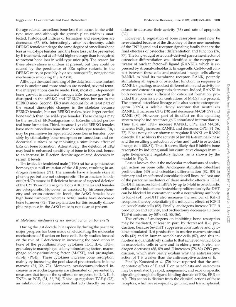
However, E regulation of bone resorption must now bere-evaluated because of the discovery of three new membersof the TNF ligand and receptor signaling family that are thefinal effectors of osteoclast differentiation and function (76,77). The long-sought osteoblast-derived paracrine effector ofosteoclast differentiation was identified as the receptor ac-tivator of nuclear factor-�B ligand (RANKL), which is ex-pressed by stromal-osteoblastic lineage cells. Cell-to-cell con-tact between these cells and osteoclast lineage cells allowsRANKL to bind its membrane receptor, RANK, potentlystimulating all aspects of osteoclast function: in response toRANKL signaling, osteoclast differentiation and activity in-crease and osteoclast apoptosis decreases. Indeed, RANKL isboth necessary and sufficient for osteoclast formation, pro-vided that permissive concentrations of M-CSF are present.The stromal-osteoblast lineage cells also secrete osteoprote-gerin (OPG), a soluble decoy receptor that neutralizesRANKL. E increases OPG (78) and decreases M-CSF (79) andRANK (80). However, part of its effect on this signalingsystem may be indirect through E-stimulated intermediaries.Thus, IL-1 and TNF� increase RANKL, OPG, and M-CSF,whereas PGE2 increases RANKL and decreases OPG (31, 76,77). E has not yet been shown to regulate RANKL or RANKdirectly. E also blocks the activity of Jun NH2-terminal kinaseand the resulting production of c-Jun and JunD in osteoclastlineage cells (80, 81). Thus, it seems likely that E inhibits boneresorption by inducing small but cumulative changes in mul-tiple E-dependent regulatory factors, as is shown by themodel in Fig. 3.
Less is known about the molecular mechanisms of andro-gen action on bone cells. Androgens stimulate osteoblastproliferation (45) and osteoblast differentiation (82, 83) inprimary and transformed osteoblastic cell lines. At least oneof the paracrine mediators of these anabolic effects is IGF-I.5�-DHT increases IGF-I mRNA by up to 6-fold in osteoblasticcells, and the induction of osteoblast proliferation by 5�-DHTcan be blocked by cotreatment with a neutralizing antibodyto IGF-I (84). 5�-DHT also increases the number of IGF-IIreceptors, thereby potentiating the mitogenic effects of IGF-IIon osteoblastic cells (82). Finally, androgens increase TGF-�production and activity, and orchiectomy decreases all threeTGF-� isoforms by 80% (82, 85, 86).
The effects of androgens on inhibiting bone resorptionmay be mediated, at least in part, by decreased IL-6 pro-duction, because 5�-DHT suppresses constitutive and cyto-kine-stimulated IL-6 production in murine marrow stromalcells (42) and in human osteoblastic cells (87), and this in-hibition is quantitatively similar to that achieved with E. Bothin osteoblastic cells in vitro and in elderly men in vivo, an-drogen decreases (88, 89) and E increases (78, 89) OPG pro-duction, which may partly explain why the antiresorptiveaction of T is weaker than the antiresorptive action of E.
Finally, Kousteni et al. (70) have reported that the anti-apoptotic effects of E and T on osteoblasts and osteocytesmay be mediated by rapid, nongenomic, and sex-nonspecificsignaling through the ligand binding domain of ER�, ER� ,orAR. This action is distinct from the classical actions of thesereceptors, which are sex-specific, genomic, and transcriptional.
Riggs et al. • Sex Steroids and Bone Metabolism Endocrine Reviews, June 2002, 23(3):279–302 283
Dow
nloaded from https://academ
ic.oup.com/edrv/article/23/3/279/2424167 by guest on 18 July 2022
Thus, the autocrine and paracrine basis for sex steroidaction on bone cells has recently come into sharper focus.However, how to weigh the importance of the various cy-tokines and how to quantify their complex interactions inmediating the sex steroid effects are subjects for furtherresearch.
F. Interaction with biomechanical forces
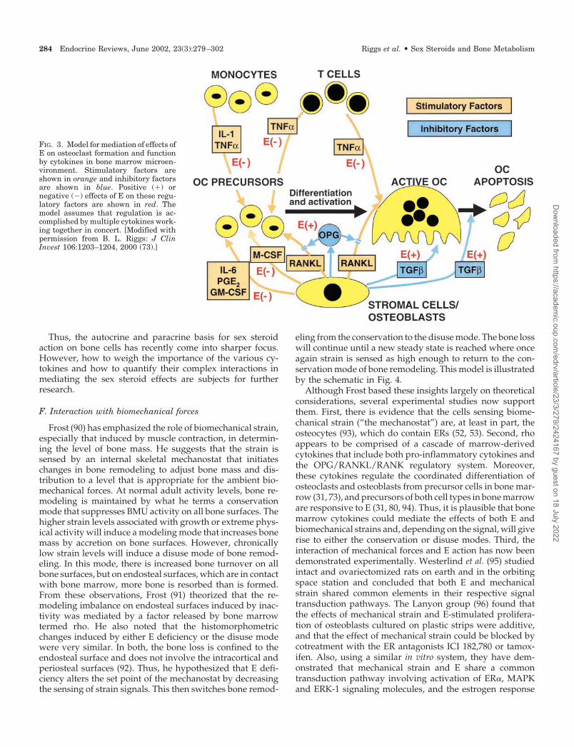
Frost (90) has emphasized the role of biomechanical strain,especially that induced by muscle contraction, in determin-ing the level of bone mass. He suggests that the strain issensed by an internal skeletal mechanostat that initiateschanges in bone remodeling to adjust bone mass and dis-tribution to a level that is appropriate for the ambient bio-mechanical forces. At normal adult activity levels, bone re-modeling is maintained by what he terms a conservationmode that suppresses BMU activity on all bone surfaces. Thehigher strain levels associated with growth or extreme phys-ical activity will induce a modeling mode that increases bonemass by accretion on bone surfaces. However, chronicallylow strain levels will induce a disuse mode of bone remod-eling. In this mode, there is increased bone turnover on allbone surfaces, but on endosteal surfaces, which are in contactwith bone marrow, more bone is resorbed than is formed.From these observations, Frost (91) theorized that the re-modeling imbalance on endosteal surfaces induced by inac-tivity was mediated by a factor released by bone marrowtermed rho. He also noted that the histomorphometricchanges induced by either E deficiency or the disuse modewere very similar. In both, the bone loss is confined to theendosteal surface and does not involve the intracortical andperiosteal surfaces (92). Thus, he hypothesized that E defi-ciency alters the set point of the mechanostat by decreasingthe sensing of strain signals. This then switches bone remod-
eling from the conservation to the disuse mode. The bone losswill continue until a new steady state is reached where onceagain strain is sensed as high enough to return to the con-servation mode of bone remodeling. This model is illustratedby the schematic in Fig. 4.
Although Frost based these insights largely on theoreticalconsiderations, several experimental studies now supportthem. First, there is evidence that the cells sensing biome-chanical strain (“the mechanostat”) are, at least in part, theosteocytes (93), which do contain ERs (52, 53). Second, rhoappears to be comprised of a cascade of marrow-derivedcytokines that include both pro-inflammatory cytokines andthe OPG/RANKL/RANK regulatory system. Moreover,these cytokines regulate the coordinated differentiation ofosteoclasts and osteoblasts from precursor cells in bone mar-row (31, 73), and precursors of both cell types in bone marroware responsive to E (31, 80, 94). Thus, it is plausible that bonemarrow cytokines could mediate the effects of both E andbiomechanical strains and, depending on the signal, will giverise to either the conservation or disuse modes. Third, theinteraction of mechanical forces and E action has now beendemonstrated experimentally. Westerlind et al. (95) studiedintact and ovariectomized rats on earth and in the orbitingspace station and concluded that both E and mechanicalstrain shared common elements in their respective signaltransduction pathways. The Lanyon group (96) found thatthe effects of mechanical strain and E-stimulated prolifera-tion of osteoblasts cultured on plastic strips were additive,and that the effect of mechanical strain could be blocked bycotreatment with the ER antagonists ICI 182,780 or tamox-ifen. Also, using a similar in vitro system, they have dem-onstrated that mechanical strain and E share a commontransduction pathway involving activation of ER�, MAPKand ERK-1 signaling molecules, and the estrogen response
FIG. 3. Model for mediation of effects ofE on osteoclast formation and functionby cytokines in bone marrow microen-vironment. Stimulatory factors areshown in orange and inhibitory factorsare shown in blue. Positive (�) ornegative (�) effects of E on these regu-latory factors are shown in red. Themodel assumes that regulation is ac-complished by multiple cytokines work-ing together in concert. [Modified withpermission from B. L. Riggs: J ClinInvest 106:1203–1204, 2000 (73).]
284 Endocrine Reviews, June 2002, 23(3):279–302 Riggs et al. • Sex Steroids and Bone Metabolism
Dow
nloaded from https://academ
ic.oup.com/edrv/article/23/3/279/2424167 by guest on 18 July 2022
element of DNA (97–98). These data suggest that the inter-relationship between the effects of mechanical strain and Eon osteoblast function occur because they both share a com-mon afferent pathway. Thus, although additional studies areneeded, major advances have been made in defining thecellular and molecular basis of the mechanostat and its in-teraction with E action.
III. Patterns of Skeletal Growth and Maturation
Sex steroids are responsible for the maturation and thesexual dimorphism of the skeleton. Skeletal size and volu-metric BMD are similar in prepubertal girls and boys. Be-tween the onset of puberty and young adulthood, however,skeletal mass doubles (100). The rates of increase in staturalheight and bone remodeling are greatest in early puberty andthen decline progressively (100–104). In contrast, maximalincreases in volumetric BMD occur 2 yr later—at menarchein girls and late puberty in boys. The pattern of growth ofboys differs from that of girls in two ways: boys have twomore years of prepubertal growth because of their later pu-berty (age 14, rather than age 12 as in girls), and their pubertalgrowth spurt lasts for 4 yr rather than the 3 yr that it lasts ingirls (100–104). These differences largely account for the 10%greater statural height and the 25% greater peak bone massachieved by males. For the most part, the greater bone massin males is due to their greater bone size.
Skeletal growth occurs mainly by modeling that increasesthe size and shape of bones. Linear bone growth occurs byossification of the endochondral growth plates. Radial bonegrowth occurs by periosteal apposition, and the marrowcavity size increases by endosteal resorption. Prepubertalgrowth is proportionately greater in the legs, whereas pu-bertal growth is proportionately greater in the trunk (105).The excess in periosteal bone apposition over endosteal bone
resorption that occurs during the pubertal growth spurt in-creases both the size and the volumetric BMD (the total bonemass contained within a volume of bone) of the extremities(106). Puberty is terminated by epiphyseal plate closure, bywhich time volumetric BMD has reached about 90–95% ofpeak mass. A process termed “consolidation” then brings theskeleton to its maximal values by continued periosteal ap-position and, possibly, also by trabecular thickening. Un-doubtedly, part of the increase in BMD during consolidationrelates to the decline in the high intracortical porosity asso-ciated with the rapid pubertal phase of bone growth. Howlong consolidation continues is disputed: some find that itlasts only until the end of the second decade (101), whereasothers find that for vertebral BMD it may last until the endof the third decade (107).
IV. Role of Sex Steroids in Skeletal Maturation
Before puberty, basal levels of the GH/IGF-I axis maintainslow, but continuous, bone growth. Puberty is triggered byincreased pulsatile secretion of GnRH by the hypothalamus,leading to increases in serum gonadotropins and, thus, toincreases in gonadal secretion of sex steroids (108). The in-creases in serum E enhance pulsatile GH secretion in bothsexes by 1.5- to 3.0-fold; these increases, in turn, increasecirculating, and possibly osteoblast, IGF-I concentrations by1.5- to 3.0-fold (109, 110). The increases in GH, IGF-I, and Eact coordinately to support the pubertal growth spurt. Thehigh pubertal levels of GH and IGF-I are maintained duringthe 3–4 yr of rapid growth but then gradually decrease toprepubertal levels over several years, although the serum sexsteroids remain at adult levels (109). However, it is the in-crease in serum E that is responsible for the pubertal growthspurt. Males with homozygous mutations in ER� or aro-matase genes do not undergo rapid adolescent growth, de-
FIG. 4. Schematic of mechanism of Einteraction with biomechanical strainto regulate bone mass based on publi-cations of Frost (90–92). Internal mech-anostat in bone senses strain generatedby muscle contraction or skeletal load-ing. Mechanostat provides cyberneticfeedback by activating bone cells thatinduce either bone gain or bone loss un-til a new steady state is achieved. Highbone mass is associated with low strainsignals, and low bone mass is associatedwith high strain signals. However, thesensing of strain by the mechanostat ismodulated by ambient E concentrationin the bone microenvironment. Atmenopause, low E levels lead to im-paired sensing. Because of the resultantlow strain signals, the mechanostat er-roneously senses that bone mass is in-creased. This leads to rapid bone lossthat continues until strains are sensedas being the same as those present be-fore menopause, at which point boneloss ceases.
Riggs et al. • Sex Steroids and Bone Metabolism Endocrine Reviews, June 2002, 23(3):279–302 285
Dow
nloaded from https://academ
ic.oup.com/edrv/article/23/3/279/2424167 by guest on 18 July 2022

spite normal or increased levels of serum T (110–114). More-over, it is the continued rise in serum E levels during pubertythat is the probable cause of epiphyseal closure in both sexes,because young adult males who are unable to respond to Ebecause of homozygous mutations of the ER� gene (111) orthe aromatase gene (112–114) have open epiphyses, whereasmen with testicular feminization due to null mutations of ARachieve epiphyseal closure (115, 116). Experimental studiesin juvenile ovariectomized rabbits have demonstrated that Eaccelerates the programmed senescence in the proliferationrate and number and size of chondrocytes, leading to epiph-yseal plate fusion (60). Thus, E both initiates the pubertalgrowth spurt and then ends it by inducing epiphyseal clo-sure. Sex steroids also appear to increase bone mass duringskeletal maturation independently of the effects of circulat-ing levels of GH and IGF-I. The 25% greater bone mass inpostpubertal boys over postpubertal girls is likely duemainly to the pubertal increase in serum T, because increasesin GH secretion and IGF-I production are similar or evengreater in girls than in boys. However, as will be reviewedlater, E contributes substantially to volumetric BMD in bothsexes. Based on measurements made in a young adult malewho was unable to synthesize E because of a genetic defectin aromatase activity, BMD was reduced by 25–40% of pre-dicted values at various skeletal scanning sites (114). Thus,both T and E have substantial effects on bone size and volu-metric BMD, although E appears to play the dominant role.
V. Patterns of Age-Related Bone Loss
As pointed out by Seeman (106), the compartments (en-dosteal, intracortical, and periosteal) of bone may changedifferentially during bone growth and maturation and dur-ing bone loss. However, the changes in the individual com-partments cannot be assessed by dual energy x-ray absorp-tiometry (DXA), the most commonly employed method forassessing bone density, which provides only an integral mea-sure of BMD. Moreover, DXA provides data as areal BMD(g/cm2), which overestimates volumetric BMD (g/cm3) inlarger bones by failing to account for the variable dimensionof depth. Finally, bone size is ignored in the results of con-ventional assessments of BMD despite biomechanical datashowing that, for a given bone mass, larger bones are stron-ger (117, 118). Thus, sex steroid sufficiency and deficiencyexert differential effects on BMD and bone size that are notcaptured by most clinical measurements. Age and sex dif-ferences in bone size and shape are shown schematically inFig. 5.
In women, there may be bone loss, especially from theproximal femur, during the 5 yr before menopause when sexsteroid levels begin to decline (119). Whether substantialpremenopausal bone loss occurs at other sites in women, orwhether bone loss occurs in young men, is controversial. Inwomen, some (107, 120), but not other, cross-sectional studiessuggest that premenopausal bone loss may occur at the ver-tebrae, and a 16-yr longitudinal study demonstrated that itoccurred at the hip (121). If these findings are correct, theysuggest that a remodeling imbalance begins to occur inwomen after the cessation of growth and consolidation,which is subsequently enhanced by menopause.
A. Patterns in women
Women undergo two major phases of involutional boneloss: an early, but transient, accelerated phase that begins atmenopause and a slow, continuous phase. The early phasedeclines exponentially over 4–8 yr to merge asymptoticallywith the subsequent slow phase. This phase accounts forlosses of 20–30% in cancellous bone, but for only 5–10% incortical bone. Because natural menopause occurs at differentages, the skeletal consequences of menopause are mostclearly apparent after ovariectomy. In a 2-yr longitudinalstudy of middle-aged women who underwent ovariectomy,Genant et al. (7) found losses of 18% in cancellous bone byquantitative computed tomography but of only 4% in corticalbone by single photon absorptiometry. By contrast, Recker etal. (122) and Guthrie et al. (123) followed cohorts of 75 and224 perimenopausal women, respectively, across naturalmenopause. In the 3 yr preceding the cessation of menses,Recker et al. (122) found losses of 4% in BMD of the lumbarspine and proximal femur associated with declining serumE levels, whereas Guthrie et al. (123) found losses of only1–2%. After cessation of menses, Recker et al. reported lossesof about 7% over 3 yr when an asymptote was reached. Intheir more extended follow-up, Guthrie et al. reported thatthe total excess bone loss was 14% and that the asymptotewas not reached until after 8–10 yr. There are two reasons forthe higher rate of bone loss in the study of Genant et al. (7)as compared with the latter two investigations (122–123).First, the more precipitous fall in serum E and, to a lesser
FIG. 5. Schematic representation in cortical bone of the differentialeffects of gender on growth and maturation and on bone loss withaging. Before puberty there are no differences in cortical bone be-tween sexes. The bone of the prepubertal female and that of the adultfemale are similar in proportions, but the bone of the latter is larger.However, owing to a greater rate of periosteal apposition duringpuberty, the cortical bone of the young-adult male is larger and thecortex is thicker. By old age, there has been extensive loss of corticalbone due to endosteal resorption in both sexes, but because womenundergo menopause, they lose more cortical bone than men do. Theendosteal resorption with aging is partially offset by a continuedperiosteal apposition that is 3-fold greater in men than in women. Notshown in the cartoon are age-related increases in intracortical po-rosity that present in both sexes, but more so in women because ofmenopause. Cartoons are based on published data (105, 141).
286 Endocrine Reviews, June 2002, 23(3):279–302 Riggs et al. • Sex Steroids and Bone Metabolism
Dow
nloaded from https://academ
ic.oup.com/edrv/article/23/3/279/2424167 by guest on 18 July 2022

extent, T, after ovariectomy led to a more rapid rate of boneloss. Second, the high rate of bone loss found by Genant etal. was determined using quantitative computed tomogra-phy that specifically measured the more responsive cancel-lous bone in the vertebral centrum as compared with DXAthat measured overall (both cortical and cancellous) vertebralbone loss in the latter two studies.
B. Patterns in men
Men do not undergo the equivalent of menopause and,thus, lack the early, accelerated phase of bone loss experi-enced by women. Castrated men (male sex offenders inCzechoslovakia) have a pattern of rapid bone loss similar tothat of women after menopause (124). However, aging menexhibit a slow phase of bone loss that is virtually identicalwith the late slow phase that is experienced by postmeno-pausal women, leading to overall losses of about 20–25% inboth cortical and cancellous bone. Periosteal apposition inthe appendicular skeleton continues through life in both menand women, but men add 3-fold more bone by this processthan do women (106). This increases the width of the longbones, including the proximal femur, and the same amountof bone distributed over a wider area is stronger. Thus, thegreater biomechanical strength afforded by the wider bonespartially compensates for age-related decreases in BMD. In-deed, Beck et al. (117) reanalyzed data from DXA measure-ments of the proximal femur on a non-Hispanic, whitesubgroup (2719 men and 2904 women) of subjects from thethird National Health and Nutrition Examination Survey(NHANES). As a result of the increased bone width, theycalculated that the femoral neck section modulus, an indexof mechanical strength, was reduced over life by 14% inelderly women and by 6% in elderly men. However, theseanalyses did not include measurements of cancellous bone inthe proximal femur and, thus, did not take into account thedecrease in mechanical strength due to cancellous bone lossthat occurred concomitantly with the changes in bone size.Thus, bone strength was undoubtedly reduced more thanthey estimated.
VI. Mechanism of the Early Accelerated Phase ofPostmenopausal Bone Loss
This phase begins at the menopause, can be prevented byE replacement (6, 7), and clearly results from loss of ovarianfunction. During the 2- or 4-yr menopausal transition, serumE2 levels fall to 10–15% of the premenopausal level, althoughlevels of serum E1, a 4-fold weaker E, fall to about 25–35% ofthe premenopausal level (125). Also, serum T decreases aftermenopause because of decreases of ovarian T production(126), but this decrease is only moderate, because T continuesto be produced by adrenal cortex and by the ovarian inter-stitium. As assessed by biochemical markers, bone resorp-tion increases by 90% at the menopause, whereas bone for-mation markers increase by only 45% (127). The increase inbone turnover and remodeling imbalance lead to acceleratedbone loss, particularly on the endosteal surface of bone. Al-though the menopause induces rapid bone loss, part of thedecrease in BMD that is measured by bone densitometry
relates to an increase in the remodeling space induced by thelarge increase in BMU numbers (128). The rapid bone loss inthis phase produces an increased outflow of calcium frombone into the extracellular pool, but hypercalcemia is pre-vented by compensatory increases in urinary calcium excre-tion (129) and decreases in intestinal calcium absorption(130), and by a partial suppression of PTH secretion (13).Although bone responsiveness to infused PTH is enhancedduring this phase (131), this may reflect only the overallincrease in BMU numbers.
As reviewed earlier, it is possible that the early rapid phaseof bone loss results from a reduced sensing of biomechanicalstrain by bone cells induced by acute E deficiency. If thisconcept is correct, it would rationalize the otherwise difficultto explain observation that the rapid phase of postmeno-pausal bone loss subsides after 4–8 yr. Thus, when bonemass is reduced to such a level that the mechanostat againsenses bone strains as similar to those present before meno-pause, when E was sufficient, rapid bone loss will cease.Indeed, Heshmati et al. (132) found that reducing serum Eamong postmenopausal women to virtually undetectablelevels by administration of an aromatase inhibitor resulted inincreased bone resorption and decreased serum PTH, whichis evidence that the rapid phase of bone loss had been re-activated. Had the effect of increased E deficiency been onexternal calcium homeostasis, aromatase treatment wouldhave increased serum PTH further. Also, had the mechanismterminating the rapid phase of bone loss been a high degreeof cancellous bone depletion, induction of a more severedegree of E deficiency should not have reactivated the rapidphase of bone loss. Nonetheless, an effect of reduced bonemass per se on tapering the rate of bone loss cannot beexcluded.
VII. Mechanisms of the Late, Slow Phase of Age-Related Bone Loss in Women
A. Secondary hyperparathyroidism
The late, slow phase of bone loss is associated with pro-gressive increases in levels of serum PTH and in biochemicalmarkers of bone turnover (Fig. 6), and these increases cor-relate with each other (13). Moreover, when serum PTHlevels were suppressed by a 24-h calcium infusion in groupsof young premenopausal and elderly postmenopausalwomen, the increases in biochemical markers in the post-menopausal women that were present on the control daywere no longer present on the calcium infusion day, stronglysuggesting that the increased serum PTH was the cause of theincrease in bone turnover (133). Because the increases inserum PTH are not associated with increases in serum ioniccalcium levels or major abnormalities in renal function, theyare indicative of secondary hyperparathyroidism caused byage-related abnormalities in extraskeletal calcium homeosta-sis. Indeed, many studies have shown that age impairs cal-cium absorption (134) and especially impairs the ability toadapt to a lower calcium intake by increasing intestinal cal-cium absorption (135). Aging also impairs renal calcium con-servation (133, 136). Both abnormalities lead to external cal-cium wasting. Thus, unless dietary calcium is substantially
Riggs et al. • Sex Steroids and Bone Metabolism Endocrine Reviews, June 2002, 23(3):279–302 287
Dow
nloaded from https://academ
ic.oup.com/edrv/article/23/3/279/2424167 by guest on 18 July 2022

increased to offset these losses, PTH secretion increases tomaintain normal levels of serum ionic calcium by resorptionof bone that contains 99% of body calcium stores. If thehypothesis that calcium wasting is the cause of the secondaryhyperparathyroidism and increased bone resorption associ-ated with aging is correct, these abnormalities should becorrected by calcium supplementation. In fact, many studieshave now shown that calcium supplementation in elderlywomen retards bone loss and, possibly, also reduces fractureoccurrence in late, postmenopausal women (12). Moreover,McKane et al. (137) demonstrated that a chronically highcalcium intake reduced the elevated levels of serum PTH andbone turnover markers in elderly women to within the nor-mal range for premenopausal women (Fig. 7). However, thelevel of calcium intake (2400 mg/d) in the treatment groupof that study was far higher than the average calcium intakeamong American postmenopausal women of 700 mg/dfound in the National Health and Nutrition ExaminationSurvey (NHANES) survey (138).
The serum PTH begins to increase in women about 10–15yr after the menopause (Fig. 6), which is 5–10 yr after therapid phase of bone loss has subsided. Thus, there may be atransitional interval before the processes leading to second-ary hyperparathyroidism become dominant over the directeffect of E deficiency on bone cell function. Thereafter, serumPTH increases throughout life, a progression that may bedue, at least in part, to abnormal parathyroid gland function.
Ledger et al. (139) conducted formal studies of parathyroidsecretory dynamics by sequential infusions of calcium orEDTA. Compared with young adult women, they found thatelderly women had greater basal, maximal, and nonsup-pressible levels of PTH secretion without alterations in theset-point (Table 2). These abnormalities are similar to thosefound in patients in early renal failure associated with sec-ondary hyperparathyroidism and parathyroid hyperplasiaand are consistent with a histological autopsy study showinga trend to parathyroid hyperplasia in elderly women andmen (140).
B. Effects of estrogen deficiency on extraskeletalcalcium metabolism
Tradition holds that the slow phase of bone loss in elderlywomen is caused largely by age-related abnormalities inextraskeletal calcium metabolism and that E deficiency playseither no role or only a minor one. However, in 1998 (13), weproposed that E deficiency is, in fact, the principal cause ofboth the abnormal extraskeletal calcium metabolism and the
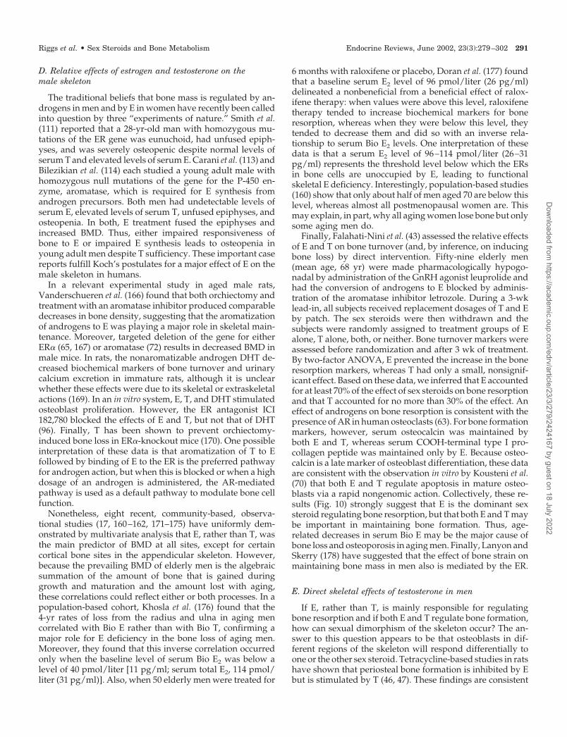
FIG. 7. Levels of serum PTH and bone resorption [assessed by uri-nary excretion of deoxypyridinoline (Dpd)] are increased (P � 0.001for both variables) in elderly postmenopausal women as comparedwith premenopausal women. Either a high calcium (Ca) intake (2400mg/d over 3 yr) or chronic E therapy reduced values to levels that werenot significantly different from premenopausal women. Values werereduced to a greater degree after E replacement therapy (ERT) thanafter Ca supplementation. [Data are from W. R. McKane et al.: J ClinEndocrinol Metab 81:1699–1703, 1996 (137), and Proc Assoc AmPhysicians 109:174–180, 1997 (142).]
FIG. 6. Changes in serum PTH (upper panels) and in bone turnovermarkers (lower panels) as a function of age in men and women overthe age of 50. Data are from a population sample from Rochester,Minnesota (17). Results are expressed as the percentage change fromyoung-adult values. Serum osteocalcin is a marker for bone formation,and urine N-telopeptide of type I collagen (NTx) is a marker for boneresorption. For changes in markers of bone turnover, note that in-creases in women begin at menopause and continue progressivelywith aging. In men, the increases begin later in life. Note also that theincrease in bone resorption exceeds that of bone formation at all ages,indicating a persistent remodeling imbalance. Serum PTH levels in-crease in both sexes. Although the proportional increase in men overmidlife values is greater than in women, absolute values late in lifeare similar in both sexes. This discrepancy occurs because the changeover life in men is parabolic. There are higher values in young adult-hood, which decrease in midlife and then increase in old age. Incontrast, the increase in serum PTH levels in women begins earlierand increase continuously.
TABLE 2. PTH secretory dynamics in young and elderly women
VariableMean � SE
PYoung Elderly
n 10 10 �Age, yr 30.3 � 0.8 73.7 � 0.6 �Basal PTH, pM 2.7 � 0.4 3.8 � 0.5 �0.05Set point, mM 1.19 � 0.01 1.18 � 0.01 NSMaximal PTH, pM 12.8 � 1.0 16.6 � 1.1 �0.05Minimal PTH, pM 0.4 � 0.1 0.8 � 0.1 �0.001
Adapted from Ledger et al. (139).NS, Not significant.
288 Endocrine Reviews, June 2002, 23(3):279–302 Riggs et al. • Sex Steroids and Bone Metabolism
Dow
nloaded from https://academ
ic.oup.com/edrv/article/23/3/279/2424167 by guest on 18 July 2022

secondary hyperparathyroidism and, thus, is the ultimatecause of the slow phase of bone loss. The compelling data thatsupported this hypothesis are found in two studies by ourgroup (125, 142) demonstrating that elderly postmenopausalwomen receiving long-term E treatment had levels of serumPTH and bone turnover markers that were identical withthose of young premenopausal women, whereas the un-treated controls had the expected high levels for both vari-ables (Fig. 7).
We have attempted to resolve the apparent paradox ofhow E deficiency produces opposite types of parathyroidfunction in the two phases of bone loss (reviewed above) byhypothesizing that there are two types of E action on bone—adirect action on bone cells and an indirect action that ismediated by changes in PTH secretion resulting from E ef-fects on extraskeletal calcium metabolism. E increases intes-tinal calcium absorption both in experimental animals (143,144) and in humans (130, 145), acting through intestinal ER(143). E also increases renal calcium conservation (136, 146)by enhancing tubular calcium resorption (146). Thus, the lossof the direct actions of E on the gut and kidney will result incontinued calcium wasting. Unless these losses are compen-sated for by very large increases in dietary calcium intake,they will lead to secondary hyperparathyroidism and willcontribute to the slow phase of bone loss.
C. Relationship between the direct and indirect mechanismsof estrogen deficiency on bone and the resultant two phasesof bone loss
Although both phases of postmenopausal bone loss arecaused by E deficiency, the mechanisms by which the Edeficiency produces the bone loss differ. We suggest that thisaccounts for the different patterns observed in the two phasesof postmenopausal bone loss. The major characteristics of theearly, rapid phase are that it is self-limiting and inducesdisproportionate cancellous bone loss. As reviewed earlier,both of these characteristics can be explained by E deficiencyresetting the mechanostat. When bone strain is sensed as“normal” by the reset mechanostat, the accelerated bone lossceases. The remodeling characteristics of this phase of boneloss follow what Frost (90–92) has termed the “disuse mode,”which affects mainly bone on endosteal surfaces. Because ofits greater proportion of surfaces interfacing with the bonemarrow, cancellous bone, rather than cortical bone, is pref-erentially lost in this mode.
The major characteristics of the late, slow phase are that itcontinues indefinitely and that there are similar or evengreater losses of cortical than of cancellous bone. Because thebone loss is driven by the PTH excess, rather than by thesensing of biomechanical strain by bone cells, it will continueas long as the secondary hyperparathyroidism persists. Theaction of PTH also determines the remodeling characteristics,and the bone loss is not restricted to the endosteal-marrowinterface but affects all bone surfaces. These remodeling char-acteristics are consistent with those observed in patients withmild primary hyperparathyroidism who maintain cancel-lous volume and structure but lose cortical bone (147, 148).They are also consistent with the findings that transgenicmice expressing constitutively active PTH receptors in os-
teoblasts have increased density of cancellous bone but de-creased density of cortical bone (149). The relative sparing ofcancellous bone may be due to the anabolic action of PTHthat is manifested in certain circumstances (150).
D. Effects of decreased bone formation
Although increased bone resorption is the predominantcause of bone loss in postmenopausal women, decreasedbone formation also contributes. Because the components ofbone turnover are tightly coupled, an increase in bone re-sorption will not cause substantial bone loss unless the com-pensatory increase in bone formation is impaired. In bothphases of postmenopausal bone loss in women, however,bone resorption at the tissue level is higher than formation,indicating impaired compensation (Refs. 125 and 127 andFig. 6). Moreover, Lips et al. (10) have demonstrated by his-tomorphometry that late postmenopausal women have de-creased wall thickness of trabecular packets, which is strongevidence of decreased bone formation at the cellular level.
These abnormalities generally have been attributed to age-related factors, particularly to decreases in paracrine pro-duction of growth factors (151) or to decreases in circulatinglevels of GH (109, 152) and IGF-I (153–155). However, if Estimulates bone formation, postmenopausal E deficiencycould also be a contributing cause. Indeed, impaired boneformation becomes apparent soon after menopause (156). Eincreases production of IGF-I (157), TGF-� (75), and procol-lagen synthesis by osteoblastic cells in vitro (157) and in-creases osteoblast lifespan by decreasing osteoblast apopto-sis (32, 37). Direct evidence that E can stimulate boneformation after cessation of skeletal growth was provided byKhastgir et al. (158), who obtained iliac biopsies for histo-morphometry in 22 elderly women (mean age, 65 yr) beforeand 6 yr after percutaneous administration of high dosagesof E. They found a 61% increase in cancellous bone volumeand a 12% increase in the wall thickness of trabecular packets.Tobias and Compston (159) have reported similar results. Itis unclear whether these results represent only pharmaco-logical effects or are an augmentation of physiological effectsof E that are ordinarily not large enough to detect.
Thus, accumulating data implicate E deficiency as a con-tributing cause of decreased bone formation with aging.Nonetheless, there is not a clear consensus on whether Estimulates osteoblast function, and, if it does, what is therelative contribution of increased proliferation and de-creased apoptosis.
VIII. Mechanism of Age-Related Bone Loss in Men
A. Age-related bone loss and osteoporosis in men
Although osteoporosis is often considered to be mainly adisease of women, men lose half as much bone with agingand have one third as many fragility fractures that womendo (13). Except in the infrequent older man who developsovert hypogonadism, levels of total serum E and T decreasein men only slightly with aging. Thus, the prevailing opinionhas been that sex steroid deficiency is not a major cause of
Riggs et al. • Sex Steroids and Bone Metabolism Endocrine Reviews, June 2002, 23(3):279–302 289
Dow
nloaded from https://academ
ic.oup.com/edrv/article/23/3/279/2424167 by guest on 18 July 2022

age-related bone loss in men. However, in the last few years,thinking on this issue has undergone a paradigm shift.
B. Changes in serum sex steroids with age
It is now clear that the failure of earlier studies to findmajor decreases in serum levels of total sex steroid in agingmen was due to their failure to account for the confoundingeffect of a 2-fold age-related rise in levels of serum SHBG(Ref. 160 and Table 3). Circulating sex steroids that are boundto SHBG have restricted access to target tissues, whereas the1–3% fraction that is free and the 35–55% fraction that isloosely bound to albumin are readily accessible. Althoughthere is controversy about the reliability of bioavailable (Bio;non-SHBG-bound) sex steroid measurements, they correlatewell with the more well accepted measurement of free levels.Several groups have reported substantial decreases in serumlevels of free or Bio sex steroid levels with aging (17, 160, 161).Figure 8 and Table 3 show changes with age of serum SHBG,Bio E, and Bio T in 350 women and 350 men of a population-based, age-stratified sample from Rochester, Minnesota(161). In aging men, Bio T and E are decreased substantiallydue to progressive increases in serum SHBG. The physio-logical importance of these decreases is reinforced by thereciprocal increases in serum FSH and LH.
C. Mechanisms of the age-related decreases in bioavailableestrogen and testosterone
Although Bio E and Bio T decrease with aging in both sexes(17), the mechanism of the decrease differs: in women, it iscaused by menopausal ovarian failure, whereas, in men, it iscaused by the progressive age-related increase in serumSHBG. Changes in the regressions of SHBG and Bio sexsteroid levels on age are shown in Fig. 8. Although the testisdoes not fail suddenly, as the ovary does, stimulation studieswith clomiphene citrate have established that aging menhave a decreased testicular secretory reserve capacity (162).Because T decreases the hepatic production of SHBG, de-creased secretion of T with aging will increase levels of serumSHBG. In addition, decreases in circulating levels of Bio E inaging men will negatively feed back on the hypothalamus toreduce GH pulsatile secretion further (163). This then willdecrease the production of IGF-I and IGF-binding protein 3(164) that will increase SHBG synthesis still further (165). Theincreased serum SHBG binds tightly to serum T, renderinga progressively larger fraction unavailable to tissues. Al-though the decrease in Bio T increases gonadotropin secre-tion, the aging testis is unable to respond by increasing serum
levels of Bio T and E to within the young adult range. Thus,as shown schematically in Fig. 9, a vicious cycle is initiatedthat leads to progressive age-related decreases in the Biolevels of both sex steroids in men.
Although aging women also have decreased secretion ofGH and decreased levels of serum IGF-I and IGF-bindingprotein 3, serum SHBG levels do not increase as they do inmen. This can be explained by the different actions of the twosex steroids: T decreases SHBG synthesis, whereas E in-creases it. Thus, the effect of age-related decreases in GH andIGF-I production on stimulating SHBG production is largelyoffset by postmenopausal decreases in serum E that reduceit. These offsetting effects account for the parabolic relation-ship of serum SHBG to age in women (Fig. 8).
FIG. 8. Patterns of age-related changes in serum values for SHBG(panel A), Bio E (panel B), and Bio T (panel C) among an age-stratifiedsample of Rochester, Minnesota, men (solid lines) and women (dashedlines). Note that changes in serum Bio T are plotted logarithmicallyto accommodate large differences in levels between sexes. [Data arefrom Khosla et al.: J Clin Endocrinol Metab 83:2226–2274, 1998 (17).]
TABLE 3. Sex-specific changes over life in serum sex steroids andgonadotropins
Hormone Men(% change)
Women(% change)
Bio E �47** �83**Bio T �64** �28*SHBG �124** �1LH �285** �731**FSH �505** �1805**
Adapted from Khosla et al. (17).*, P � 0.05, **, P � 0.005.
FIG. 9. Model for causation of increases in serum SHBG in agingmen. This is a complex interaction driven by a reduced secretorycapacity of GH by the pituitary and T by the testes. Because T de-creases SHBG synthesis, the progressive decreases in Bio T lead tohigher values, which further reduce Bio T. Decreased GH secretionreduces IGF-I production that also reduces SHBG synthesis. Thisleads to a vicious cycle: as SHBG increases progressively, it willfurther reduce circulating levels of Bio T.
290 Endocrine Reviews, June 2002, 23(3):279–302 Riggs et al. • Sex Steroids and Bone Metabolism
Dow
nloaded from https://academ
ic.oup.com/edrv/article/23/3/279/2424167 by guest on 18 July 2022
D. Relative effects of estrogen and testosterone on themale skeleton
The traditional beliefs that bone mass is regulated by an-drogens in men and by E in women have recently been calledinto question by three “experiments of nature.” Smith et al.(111) reported that a 28-yr-old man with homozygous mu-tations of the ER gene was eunuchoid, had unfused epiph-yses, and was severely osteopenic despite normal levels ofserum T and elevated levels of serum E. Carani et al. (113) andBilezikian et al. (114) each studied a young adult male withhomozygous null mutations of the gene for the P-450 en-zyme, aromatase, which is required for E synthesis fromandrogen precursors. Both men had undetectable levels ofserum E, elevated levels of serum T, unfused epiphyses, andosteopenia. In both, E treatment fused the epiphyses andincreased BMD. Thus, either impaired responsiveness ofbone to E or impaired E synthesis leads to osteopenia inyoung adult men despite T sufficiency. These important casereports fulfill Koch’s postulates for a major effect of E on themale skeleton in humans.
In a relevant experimental study in aged male rats,Vanderschueren et al. (166) found that both orchiectomy andtreatment with an aromatase inhibitor produced comparabledecreases in bone density, suggesting that the aromatizationof androgens to E was playing a major role in skeletal main-tenance. Moreover, targeted deletion of the gene for eitherER� (65, 167) or aromatase (72) results in decreased BMD inmale mice. In rats, the nonaromatizable androgen DHT de-creased biochemical markers of bone turnover and urinarycalcium excretion in immature rats, although it is unclearwhether these effects were due to its skeletal or extraskeletalactions (169). In an in vitro system, E, T, and DHT stimulatedosteoblast proliferation. However, the ER antagonist ICI182,780 blocked the effects of E and T, but not that of DHT(96). Finally, T has been shown to prevent orchiectomy-induced bone loss in ER�-knockout mice (170). One possibleinterpretation of these data is that aromatization of T to Efollowed by binding of E to the ER is the preferred pathwayfor androgen action, but when this is blocked or when a highdosage of an androgen is administered, the AR-mediatedpathway is used as a default pathway to modulate bone cellfunction.
Nonetheless, eight recent, community-based, observa-tional studies (17, 160–162, 171–175) have uniformly dem-onstrated by multivariate analysis that E, rather than T, wasthe main predictor of BMD at all sites, except for certaincortical bone sites in the appendicular skeleton. However,because the prevailing BMD of elderly men is the algebraicsummation of the amount of bone that is gained duringgrowth and maturation and the amount lost with aging,these correlations could reflect either or both processes. In apopulation-based cohort, Khosla et al. (176) found that the4-yr rates of loss from the radius and ulna in aging mencorrelated with Bio E rather than with Bio T, confirming amajor role for E deficiency in the bone loss of aging men.Moreover, they found that this inverse correlation occurredonly when the baseline level of serum Bio E2 was below alevel of 40 pmol/liter [11 pg/ml; serum total E2, 114 pmol/liter (31 pg/ml)]. Also, when 50 elderly men were treated for
6 months with raloxifene or placebo, Doran et al. (177) foundthat a baseline serum E2 level of 96 pmol/liter (26 pg/ml)delineated a nonbeneficial from a beneficial effect of ralox-ifene therapy: when values were above this level, raloxifenetherapy tended to increase biochemical markers for boneresorption, whereas when they were below this level, theytended to decrease them and did so with an inverse rela-tionship to serum Bio E2 levels. One interpretation of thesedata is that a serum E2 level of 96–114 pmol/liter (26–31pg/ml) represents the threshold level below which the ERsin bone cells are unoccupied by E, leading to functionalskeletal E deficiency. Interestingly, population-based studies(160) show that only about half of men aged 70 are below thislevel, whereas almost all postmenopausal women are. Thismay explain, in part, why all aging women lose bone but onlysome aging men do.
Finally, Falahati-Nini et al. (43) assessed the relative effectsof E and T on bone turnover (and, by inference, on inducingbone loss) by direct intervention. Fifty-nine elderly men(mean age, 68 yr) were made pharmacologically hypogo-nadal by administration of the GnRH agonist leuprolide andhad the conversion of androgens to E blocked by adminis-tration of the aromatase inhibitor letrozole. During a 3-wklead-in, all subjects received replacement dosages of T and Eby patch. The sex steroids were then withdrawn and thesubjects were randomly assigned to treatment groups of Ealone, T alone, both, or neither. Bone turnover markers wereassessed before randomization and after 3 wk of treatment.By two-factor ANOVA, E prevented the increase in the boneresorption markers, whereas T had only a small, nonsignif-icant effect. Based on these data, we inferred that E accountedfor at least 70% of the effect of sex steroids on bone resorptionand that T accounted for no more than 30% of the effect. Aneffect of androgens on bone resorption is consistent with thepresence of AR in human osteoclasts (63). For bone formationmarkers, however, serum osteocalcin was maintained byboth E and T, whereas serum COOH-terminal type I pro-collagen peptide was maintained only by E. Because osteo-calcin is a late marker of osteoblast differentiation, these dataare consistent with the observation in vitro by Kousteni et al.(70) that both E and T regulate apoptosis in mature osteo-blasts via a rapid nongenomic action. Collectively, these re-sults (Fig. 10) strongly suggest that E is the dominant sexsteroid regulating bone resorption, but that both E and T maybe important in maintaining bone formation. Thus, age-related decreases in serum Bio E may be the major cause ofbone loss and osteoporosis in aging men. Finally, Lanyon andSkerry (178) have suggested that the effect of bone strain onmaintaining bone mass in men also is mediated by the ER.
E. Direct skeletal effects of testosterone in men
If E, rather than T, is mainly responsible for regulatingbone resorption and if both E and T regulate bone formation,how can sexual dimorphism of the skeleton occur? The an-swer to this question appears to be that osteoblasts in dif-ferent regions of the skeleton will respond differentially toone or the other sex steroid. Tetracycline-based studies in ratshave shown that periosteal bone formation is inhibited by Ebut is stimulated by T (46, 47). These findings are consistent
Riggs et al. • Sex Steroids and Bone Metabolism Endocrine Reviews, June 2002, 23(3):279–302 291
Dow
nloaded from https://academ
ic.oup.com/edrv/article/23/3/279/2424167 by guest on 18 July 2022
with the observation that one of the major skeletal differencesbetween sexes is that men have larger appendicular bonesand thicker cortical widths. Moreover, it is interesting thatthe homozygous null mutations of the ER� gene result inincreased skeletal size in female mice but not in male mice(179). Thus, it is also possible that the smaller bone size of thefemale skeletal phenotype is mediated by the greater con-centrations of serum E in females acting through ER�. Inaddition to these direct skeletal effects, androgens also in-crease intestinal calcium absorption (180), although it is un-clear whether they enhance renal calcium homeostasis as Edoes.
IX. Causes of Individual Differences inSkeletal Responsiveness
The patterns of bone gain and bone loss described earlierand the mechanisms that cause them occur in everyone. Yetonly about one in two women and one in six men developfractures due to osteoporosis. Even accounting for the oc-currence of falls and trauma that predispose certain indi-viduals to fracture, there remains a wide variability of peakbone mass and of rates of bone loss with aging. Because ofthe overriding importance of sex steroids in determining and
in maintaining bone mass in both sexes, variability in thelevels of serum sex steroids, in the responsiveness of bone toa given serum level, or both could contribute to the vari-ability of bone gain during puberty and bone loss with aging.
A. Differences in serum sex steroid levels
The relationship between individual differences in sexsteroid levels and rates of bone acquisition during pubertyneeds further examination. Cadogan et al. (104) found that64% of the variance in total body bone mineral in pubertalgirls could be explained by serum E2 levels and lean bodymass. However, Lorentzon et al. (181) were unable to relatechanges in linear growth to serum E levels in pubertal boys.See the review by Grumbach (110) for more details.
There also is insufficient information about the relationshipbetween individual differences in sex steroid levels and the rateand duration of the accelerated phase of bone loss in the earlypostmenopause. We have hypothesized that women whodevelop vertebral or distal forearm fractures during the first15–20 yr after menopause are those who have experienceddisproportionate cancellous bone loss. We have termed thisclinical syndrome “type I osteoporosis” and have suggestedthat it may be caused by E deficiency plus some additionalfactor(s) that increases the rate or extends the duration of theaccelerated, early phase of postmenopausal bone loss (182).This contrasts with type II osteoporosis, which occurs in theentire population of aging women and men, is associatedwith hip and other fractures later in life, and can be attributedto the effects of the slow phase of bone loss. Women with typeI osteoporosis have higher bone turnover and a larger re-modeling imbalance (183) but do not have consistently lowerlevels of serum sex steroids (184) as compared with non-osteoporotic control women. However, these earlier studiescould be criticized because the assays for assessing sex ste-roid levels then available were relatively insensitive. Thus,we have recently (185) reexamined this issue using newultrasensitive assays in 40 typical type I osteoporotic womenwith vertebral fractures and in 40 age-matched controlwomen. We found that serum levels of E2, E1, and T wereindistinguishable between the groups, whereas bone turn-over markers were increased by up to 50% in the osteoporoticgroup. Previous studies (186) have shown that E replacementwill normalize bone turnover in these patients. Thus, the dataare consistent with the hypothesis that the type I osteoporosisfracture syndrome is mainly the result of increased respon-siveness of bone to E deficiency that is evident in the presenceof low serum E levels but that is overcome by restoringpremenopausal high serum E levels. This is likely to becaused by a genetically determined change such as poly-morphism(s) of a gene or genes involved in receptor or post-receptor sex steroid signaling (see next section). Moreover, itis possible that these same polymorphisms also may lead toimpaired E enhancement of skeletal growth and maturation,resulting in reduced peak bone mass.
More information is available on the relationship betweenlate postmenopausal bone loss and serum E levels assessedby ultrasensitive assays. In three nested case-control studiesfrom the Study of Osteoporotic Fractures, elderly womenwith lower levels of serum E and higher levels of serum
FIG. 10. Experimental testing of the relative importance of E and Tin suppressing bone turnover in elderly men. After suppression of sexsteroid production by GnRH agonist treatment and blocking conver-sion of androgens to E with aromatase inhibition, subjects were ran-domly assigned to groups treated with T, E, both, or neither. Panel Ashows the effects of treatment on the resorption markers, urinarydeoxypyridinoline (Dpd) and N-telopeptide of type I collagen (NTx).By ANOVA, E, but not T, prevented increases in bone resorptionmarkers. The possibility of a small effect on T on opposing this in-crease cannot be excluded. Panel B shows the effects on bone forma-tion markers, serum osteocalcin, and N-terminal extension peptide oftype I procollagen (PINP). Levels of serum bone alkaline phosphatasedid not change (data not shown). Withdrawal of E and T leads to adecrease in markers (indicating that bone formation was being stim-ulated). For serum osteocalcin, a marker of late osteoblast function,ANOVA showed that T and E were equally effective in reversingdecreases, whereas for serum PINP, a marker of all stages of osteo-blast function, E, but not T, was effective. For significance of changefrom baseline: *, P � 0.05; **, P � 0.01; and ***, P � 0.001. [Reprintedwith permission from A. Falahati-Nini et al.: J Clin Invest 106:1553–1560, 2000 (43).]
292 Endocrine Reviews, June 2002, 23(3):279–302 Riggs et al. • Sex Steroids and Bone Metabolism
Dow
nloaded from https://academ
ic.oup.com/edrv/article/23/3/279/2424167 by guest on 18 July 2022

SHBG had lower cross-sectional BMD values at the calca-neus, proximal radius, proximal femur, and lumbar spine(187); higher rates of bone loss from the calcaneus and prox-imal femur (188); and increased risk for vertebral and hipfractures (189) after adjusting for age. We have reanalyzedour population-based data from Rochester, Minnesota. Afteradjusting for age, we also find a positive correlation betweenproximal femur BMD and serum Bio E2 (r � 0.31, P � 0.001)in untreated elderly women.
Although the correlations between serum E levels andbone loss in late postmenopausal women were significant,they may underestimate the restraining effect on bone loss ofextragonadal E synthesis, which is virtually the exclusivesource of circulating E levels in women after menopause.Depending on the gradient between circulating concentra-tions and intracellular concentrations in intracrine cells, localsynthesis could play a major role in sex steroid action. Basedon the effect on bone turnover markers induced by admin-istration of the potent aromatase inhibitor letrozole to post-menopausal women, Heshmati et al. (132, 190) estimated thatthe remodeling imbalance present in postmenopausalwomen would be 50% higher except for the presence ofaromatase-dependent extragonadal E synthesis.
Moreover, because the process is substrate limited, it islikely that the large age-related decreases in levels of circu-lating C19 adrenal precursors reduce extragonadal E syn-thesis and, thus, further enhance bone loss. As shown inTable 1, these decrease by as much as 75% between youngadulthood and old age in both sexes. Interestingly, the de-cline begins in young adulthood and, for the most part,continues throughout life (191). This raises the possibilitythat part of the bone loss that has been documented to occurin premenopausal women (120, 121) may relate to thesedecreases. Finally, it is possible that changes in the pattern ofserum E metabolism could affect bone loss. After the revers-ible 17�-HSD-mediated conversion of E2 to E1, there is amutually exclusive C-2 or C-16� hydroxylation. Using theovariectomized mouse model, Westerlind et al. (18) demon-strated that the 2-hydroxyestrone (2-OHE1) metabolite wasinactive, whereas the 16�-OHE1 metabolite was equipotentwith E2. Indeed, Lim et al. (193) reported that women withpostmenopausal osteoporosis had a low 16-OHE1/2-OHE1ratio, although the validity of these measurements has beenchallenged (194).
B. Differences in bone responsiveness to sex steroids
After T or E binds to its respective receptor, the hormone-receptor complex disassociates from heat shock proteins,dimerizes, forms complexes with various coactivator pro-teins (195), and binds to E or T response elements in DNAdirectly or by protein-protein bridging to other DNA bindingsites. Binding of the various E/ER complexes to DNA acti-vates genes involved in several signaling systems. Geneticpolymorphisms could modify any step of this complex path-way, thus affecting the responsiveness of bone cells to E.Also, individual differences in the concentration or ratio ofER� and ER� in bone cells could alter bone responsivenessto E, but this has not been systematically studied.
Recently, a number of investigators have related various
allelic variants in the ER� gene to BMD or to fracture risk. Ina multivariate analysis in healthy adolescent boys, Lorentzonet al. (181) found that the XbaI and PvuII genotypes inde-pendently predicted volumetric BMD of the lumbar spine,and that the PP allelic variant predicted statural height. Inpostmenopausal women, a TA repeat polymorphism wasassociated with lower BMD (197, 198) and increased risk forosteoporotic fractures (198). Others (199, 200) found that thePx haplotype was associated with significantly lower BMDvalues in postmenopausal women and, in one study (196),accounted for 16% and 23% of the observed variance in BMDat the lumbar spine and proximal femur, respectively. In agroup of 322 early postmenopausal Finnish women followedfor 5 yr, Salmen et al. (201) found that those expressing thepp variant of the PvuII genotype lost less bone than did thoseexpressing the Pp or PP variants. In a group of Italian post-menopausal women with normal BMD, osteopenia, or os-teoporosis, the combination of the PPXX ER� and vitamin Dreceptor AABBtt genotypes (202) predicted lumbar spineBMD best. Others, however, have been unable to demon-strate a relationship between ER� polymorphisms and BMD(203, 204). Allelic variants of the ER� gene have thus far notbeen reported to increase the risk for osteoporosis. Althoughthese associations are intriguing, they are far from conclu-sive. Moreover, there is, as yet, no direct experimental evi-dence that polymorphisms of the ER� gene alter phenotypesuch as has been demonstrated between the polymorphicbinding site of the collagen type I A1 gene and altered col-lagen synthesis in vitro (205).
Finally, individual differences in the effect of E deficiencyon extraskeletal calcium metabolism could affect osteoporo-sis risk. This possibility was suggested by Heshmati et al.(206), who found that a group of 20 women with postmeno-pausal osteoporosis had impaired renal tubular reabsorptionof calcium as compared with 20 postmenopausal womenwithout osteoporosis. Whether this defect is part of an al-teration in the E-signaling system is not known. Variabilityin the concentration of serum T, the rate of aromatization ofT to E, or bone responsiveness to T also could affect the rateof bone loss in aging men. Polymorphisms of the aromatasegene have been related to both female (207) and male (208)osteoporosis.
X. Causes of Bone Loss Other Than SexSteroid Deficiency
A. Age-related decreases in muscle mass
As a corollary to his mechanostat hypothesis, Frost hassuggested in various publications (summarized in Refs. 209and 210) that the loss of muscle mass with aging is theprincipal cause of involutional osteoporosis. He cites studiesof 1066 persons by three groups that show high correlations(r � 0.89–0.94) between lean body mass and total body bonemineral (210). Indeed, in a population sample, Proctor et al.(211) found that physical activity declined by 34% and 38%and lean body mass declined by 18% and 17% with aging inwomen and men, respectively. Moreover, Center et al. (171)have shown that a 1 sd decrease in quadricep muscle strengthis associated with a 3-fold increase in the risk of fracture.
Riggs et al. • Sex Steroids and Bone Metabolism Endocrine Reviews, June 2002, 23(3):279–302 293
Dow
nloaded from https://academ
ic.oup.com/edrv/article/23/3/279/2424167 by guest on 18 July 2022
Nonetheless, there are a number of reasons for believingthat the action of sex steroids on bone is of equal, or of evengreater, importance to the conservation of bone mass. First,the high correlation between total skeletal muscle mass andtotal body bone mass is an overestimate because both vari-ables are highly correlated with body size. In our age-strat-ified, population-based sample of 348 adult men and 351adult women from Rochester, Minnesota, when both heightand body mass index were entered into the multivariatemodel, total skeletal muscle mass accounted for 18% of thevariance in proximal femur BMD in both women and men(211). In the same multivariate model, serum sex steroidlevels accounted for 21% and 20% of the variance of proximalfemur BMD in women and men, respectively (160). Second,part of the high correlation between muscle and bone massmay be related to changes in age-related factors that affectboth correlates, such as decreases in serum T, GH, and IGF-I.Third, E therapy initiated soon after menopause essentiallyprevents significant bone loss for at least 8 yr after meno-pause (212), but a regular exercise program has not beendemonstrated to do so. Indeed, in a 2-yr intervention studyin early postmenopausal women, the rate of change in distalforearm BMD in the group receiving only a formal exerciseprogram was �2.6%, whereas in the group receiving bothexercise and E therapy, it was �2.7% (213). Even older post-menopausal osteoporotic women receiving E therapy willgain 12% in lumbar spine BMD over 3 yr (214), whereasexercise programs in older postmenopausal women gener-ally increase BMD by only 1–3% (215). Finally, elite womandistance runners who become amenorrheic develop severebone loss despite subjecting their skeleton to large biome-chanical loads (216). This is consistent with the concept thatthe major function of the mechanostat is to add bone duringgrowth but that it is less able to add bone to the adult skeleton(217). It should be also noted that T has a direct effect onincreasing muscle mass and strength (218). Studies should bemade to quantify the independent effects of biomechanicalstrains and sex steroid action on the maintenance of bonemass and, especially, to determine in vivo the interactionbetween these two positive determinants.
B. Other endocrine abnormalities
Other than changes in serum levels of sex steroids andPTH, the two most important causes of age-related bone lossare abnormalities in the vitamin D-endocrine and in theGH-IGF-I regulatory systems. Reduced serum concentra-tions of both of the active vitamin D metabolites—25-hydroxyvitamin D [25(OH)D] and 1,25-(OH)2D—occur withaging in both sexes. In several population-based studies,25(OH)D, an indicator of vitamin D nutrition, decreased by30–60% (219). Nutritional vitamin D deficiency may con-tribute to the secondary hyperparathyroidism and bone losswith aging because decreases in serum 25(OH)D correlateinversely with serum PTH levels and directly with BMD(125). However, nutritional vitamin D deficiency is unlikelyto be the major cause of secondary hyperparathyroidism inmost elderly women because, as mentioned earlier, E re-placement normalizes the increase in serum PTH levels (125,142). Nonetheless, house-bound persons with inadequate
exposure to UV radiation and poor nutrition, especially pop-ulations who reside in countries with higher latitudes, suchas Great Britain and France, and where dairy products arenot fortified with vitamin D, may be at risk for vitamin Ddeficiency bone loss. Chapuy et al. (11) found that supple-menting the diet of elderly, house-bound French womenwith 800 U/d of vitamin D and 1000 mg/d of calcium de-creased the incidence of hip fracture by 43% over the next 18months. However, Lips et al. (220) could not demonstrate thisin a Dutch cohort receiving somewhat smaller supplements.Serum levels of the physiologically active vitamin D metab-olite, 1,25-(OH)2D, also decrease with aging, at least relativeto the concomitant increases in serum PTH (134). Elderlywomen infused with PTH had a blunting of the stimulatedincreases in serum 1,25-(OH)2D levels relative to changes inyoung adults (221). Thus, reduction in the activity of25(OH)D 1�-hydroxylase, the renal enzyme that is respon-sible for the conversion of 25(OH)D to 1,25-(OH)2D, may alsocontribute to the secondary hyperparathyroidism and in-creased bone resorption associated with aging.
Aging decreases the amplitude and frequency of GH se-cretion (152), which leads to decreased hepatic production ofIGF-I. Indeed, serum IGF-I levels decrease by 60% with agingin women, and there are also smaller decreases in serumIGF-II levels (153, 154). Thus, decreased systemic and skeletalproduction of IGF-I may contribute to decreases in boneformation with aging.
Other changes in endocrine function with aging appear tomake smaller contributions to bone loss. Among the weakadrenal androgens, levels of serum DHEA and DHEA-SO4decrease by about 80% (16). Because cortisol secretion re-mains constant or increases throughout life, the decrease inadrenal androgenic steroids leads to an increase in the cat-abolic/anabolic ratio of circulating adrenal steroid hormoneswith aging that could also contribute to bone loss (222, 223).
C. Peak bone mass
Those persons who achieve a higher peak bone mass inyoung adulthood are less likely to develop osteoporosis asage-related bone loss ensues, whereas those with low levelsare clearly at greater risk (105, 111). The relative contributionof peak bone mass and bone loss to the BMD in an elderlywoman or man is unclear. Some have estimated, however,that about half of the variance in cancellous BMD and onethird of the variance of cortical BMD in women by age 70 isdue to bone loss (224–226). In a study of women with ver-tebral fractures and their daughters, Tabensky et al. (227)found that the daughters had half of the deficit in Z-scoresthat the mothers did, indicating an important contribution ofpeak bone mass. As reviewed in Section IX, differences inserum levels of or responsiveness to sex steroids could con-tribute to the large variability in peak bone mass. Nonethe-less, the combined effects of nonhormonal factors such asheredity, activity, calcium intake, and protein and caloricnutrition are substantial (228).
D. Genetic polymorphisms not affecting sex steroids
The heritable component of peak bone mass has beenestimated to be about 50–70% and that for age-related bone
294 Endocrine Reviews, June 2002, 23(3):279–302 Riggs et al. • Sex Steroids and Bone Metabolism
Dow
nloaded from https://academ
ic.oup.com/edrv/article/23/3/279/2424167 by guest on 18 July 2022

loss is thought to be much less. In addition to genetic poly-morphisms involving sex steroids that affect BMD, otherallelic variations have been described. These include poly-morphic variants for the vitamin D receptor gene, the TGF-�gene, and the Sp1 binding site in the collagen type I A1 gene.In addition, studies in inbred mice have made quantitativetrait localizations of additional genes that affect bone density,size, and structure [see the review by Nguyen et al. (229) fordetails].
E. Behavioral and environmental causes
Sporadic factors that affect some, but not other, membersof the aging population may contribute to fracture risk inabout 40% of men and 20% of women (230). These includeuse of certain drugs such as corticosteroids; diseases such asmalabsorption, anorexia nervosa, and renal hypercalciuria;and behavioral factors such as smoking, alcohol abuse, andinactivity. Some of these sporadic factors, however, mayexert their effect on bone by impairing production of sexsteroids. For example, smoking increases the catabolism of E(231) and anorexia nervosa (232) or excessive exercise (216)may result in hypothalamic-induced amenorrhea. It is im-portant to recognize that bone loss from these secondaryfactors is superimposable on that induced by decreases in sexsteroid production.
XI. Summation of Mechanisms
We propose that E deficiency is the major cause of both theearly, accelerated and the late, slow phases of bone loss inpostmenopausal women and contributes substantially to thecontinuous phase of bone loss in aging men. To this can beadded the important roles of E and T in the development ofpeak bone mass during and after the pubertal growth spurt.Although E deficiency is the primary cause of the two phasesof bone loss in women and the single phase in men, thedownstream mediators differ, as is shown schematically inFig. 11. The acute loss of the restraining effect of E on boneturnover and remodeling imbalance at menopause initiatesthe early, accelerated phase of bone loss. The predominantlycancellous bone loss may be mediated by a resetting of themechanostat induced by E deficiency, leading to a reductionin bone mass to such a level that bone strain is again sensedas normal. When this level is reached after 4–8 yr, furtherbone loss from this mechanism ceases. During the acceler-ated bone loss, the outpouring of calcium from bone into theextracellular fluids is compensated for by decreases in PTHsecretion and 1,25-(OH)2D production, leading to excretionof the excess calcium (Fig. 11A).
In the late, slow phase of bone loss in aging women, thereis continued cortical bone loss but smaller losses or a main-tenance of cancellous bone. This phase also is mainly caused
FIG. 11. Flow diagram of the suggested mechanisms for the two phases of bone loss in elderly women and the single phase in elderly men. Theyare similar in that all three are driven by E deficiency, but the downstream effects differ. In panel A (early postmenopausal women), the lossof the direct inhibitory effects of E on bone leads to increased outflow of bone and inhibition of PTH secretion and 1,25-(OH)2D production. Inpanel B (late postmenopausal women), the loss of effects of E on external calcium metabolism leads to calcium wasting and to increased PTHsecretion that secondarily induces bone loss. In panel C (aging men), the mechanism is the same as in phase B in women except that there areadditional effects due to T deficiency (open arrows). OB, Osteoblast; TRCa, renal tubular reabsorption of calcium.
Riggs et al. • Sex Steroids and Bone Metabolism Endocrine Reviews, June 2002, 23(3):279–302 295
Dow
nloaded from https://academ
ic.oup.com/edrv/article/23/3/279/2424167 by guest on 18 July 2022

by E deficiency, although age effects may also contribute,especially the decrease in bone formation. In contrast to thepreceding rapid loss phase, the primary mechanism in thisphase is the loss of the extraskeletal actions of E on stimu-lating intestinal calcium absorption and renal calcium con-servation. This leads to external calcium losses that, unlessrestored by large increases in dietary calcium intake, willlead to compensatory increases in PTH secretion. This re-sultant secondary hyperparathyroidism is progressive be-cause, after a number of years, the parathyroid gland func-tion becomes semiautonomous (139). The age-relatedimpairment of osteoblast function that prevents compensa-tory increases in bone formation from fully offsetting in-creases in bone resorption also is due, at least partially, to Edeficiency. Because the slow phase of bone loss is mediatedby increased PTH secretion, rather than by a resetting of themechanostat by E deficiency, it continues indefinitely. Thatcalcium supplementation is ineffective in preventing boneloss in the early, rapid phase (233) but has a substantial effectin the late, slow phase (12) is further evidence for two in-dependent mechanisms. The sequence of changes leading tothe slow phase of bone loss is given in Fig. 11B. There isindirect evidence that those women who develop vertebralor distal forearm fractures in the first 15–20 yr after meno-pause (type I osteoporosis) have an increase in the rate or anextended duration of the early, accelerated phase of boneloss. This could be caused by a genetically determined in-creased responsiveness to the effects of E deficiency.
In aging men, the continuous phase of bone loss withincreases in serum PTH and bone resorption markers has apattern that is similar to that of the late, slow phase in agingwomen. Because men do not have the equivalent of meno-pause, they lack the early, accelerated phase that is inducedin women by the precipitous fall in serum estrogens soonafter menopause. However, serum levels of both Bio E andBio T decrease substantially in aging men, although the de-crease in Bio E appears to be the dominant cause of their boneloss. A major difference between the mechanisms of bone lossin aging men and that of the slow phase of bone loss in agingwomen, however, is the added effects of T deficiency, asshown in Fig. 11C. First, and not shown in the figure, youngadult men have larger, denser bones because of the addedeffect of T during the pubertal growth spurt, and thus, menwill have more bone late in life than women for a similar rateof loss. Moreover, because larger bones are stronger, menhave additional protection against fractures than womenwho have smaller skeletons. Second, because aromatase canconvert T to E2, T can be considered to be a prohormone forE (20). Thus, a deficiency in T will exacerbate E deficiency byreducing substrate. Third, T clearly enhances bone formationthrough its antiapoptotic effect on osteoblasts and, possibly,through increased osteoblast proliferation. Moreover, T de-ficiency decreases the rate of periosteal bone apposition,whereas E deficiency opposes it. Fourth, T has an anti-resorptive action, although it is not as great as that of E. Someof the antiresorptive effect of T is probably mediated byenhancing calcium absorption (180). However, because os-teoclasts contain functional AR (63), undoubtedly there isalso a direct effect.
Thus, sex steroids play key roles in the construction and
in the conservation of the adult skeleton, and E deficiency isan important cause of involutional osteoporosis in bothsexes.
XII. Epilogue
The view that E plays a central role in the regulation ofbone mass in both sexes leads to a number of unansweredquestions. Why are sex steroids rather than the primarycalcitrophic hormones the major regulators of bone mass?Why does E have major regulatory effects on external cal-cium homeostasis? Why is E, rather than T, the major reg-ulator of bone mass in men? Why are the onset of puberty andthe accelerated bone growth leading to adult bone mass soclosely linked? And why is the overall system so complex?
It is often said that biology can be understood only in thecontext of evolution. We speculate that the major role of E inregulating bone mass evolved from its primary role in sup-porting reproduction. When a new function is needed, theevolutionary process characteristically adapts an existingmechanism rather than developing a completely new one.The therapsips (mammal-like reptiles) of the Triassic erawere the immediate ancestors of mammals and were ovip-arous. In birds, up to 38% of bone mineral is mobilized tosupply calcium for eggshell mineralization (234). Once egglaying is completed, bone mineral of the skeleton is rapidlyrestored by enhanced utilization of dietary calcium and re-duced renal calcium excretion. When mammals became vi-viparous, it is likely that this earlier system was co-opted toprovide calcium for mineralizing the fetal skeleton and forsubsequent lactation. Once in place in females, it could alsobe used to conserve bone mass in males. Indeed, ER appearsto have been the first steroid nuclear receptor to evolve invertebrates and was present long before the AR evolved(235). Moreover, even the role of the sex steroids in inducingand supporting the pubertal growth spurt can be understoodin evolutionary terms. The tight coupling between the onsetof puberty and the skeletal growth spurt ensures that repro-duction cannot occur until there is sufficient skeletal mass tosupport pregnancy. Finally, because reproductive success isthe keystone of natural selection, the surprising complexityof sex steroid regulation of bone mass in mammals can beexplained by its evolutionary linkage to ancient reproductivemechanisms.
Acknowledgments
We are grateful to Drs. Shreyasee Amin, Richard Eastell, A. MichaelParfitt, Gideon A. Rodan, and Ego Seeman for their helpful suggestionsduring the preparation of this manuscript.
Address all correspondence and requests for reprints to: B. LawrenceRiggs, M.D., Mayo Clinic and Mayo Foundation, 200 First Street SW,North 6 Plummer, Rochester, Minnesota 55905.
This work was supported in part by NIH Grants P01-AG04875 andR01-AR27065. E-mail: [email protected]
This work was presented, in part (by B.L.R.), as a plenary lecture atthe 82nd Annual Meeting of the Endocrine Society, Toronto, Ontario,Canada, June 21–24, 2000.
296 Endocrine Reviews, June 2002, 23(3):279–302 Riggs et al. • Sex Steroids and Bone Metabolism
Dow
nloaded from https://academ
ic.oup.com/edrv/article/23/3/279/2424167 by guest on 18 July 2022
References
1. Melton III LJ, Chrischilles EA, Cooper C, Lane AW, Riggs BL 1992Perspective. How many women have osteoporosis? J Bone MinerRes 7:1005–1010
2. Looker AC, Orwoll ES, Johnston Jr CC, Lindsay RL, Wahner HW,Dunn WL, Calvo MS, Harris TB, Heyse SP 1997 Prevalence of lowfemoral bone density in older U.S. adults from NHANES III. J BoneMiner Res 12:1761–1768
3. Ray NF, Chan JK, Thamer M, Melton III LJ 1997 Medical expen-ditures for the treatment of osteoporotic fractures in the UnitedStates in 1995: report from the National Osteoporosis Foundation.J Bone Miner Res 12:24–35
4. Cooper C, Campion G, Melton III LJ 1992 Hip fractures in theelderly: a world-wide projection. Osteoporos Int 2:285–289
5. Albright F, Smith PH, Richardson AM 1941 Postmenopausal os-teoporosis. JAMA 116:2465–2474
6. Lindsay R, Aitken JM, Anderson JB, Hart DM, MacDonald EB,Clarke AC 1976 Long-term prevention of postmenopausal osteo-porosis by oestrogen: evidence for an increased bone mass afterdelayed onset of oestrogen treatment. Lancet 1:1038–1041
7. Genant HK, Cann CE, Ettinger B, Gordan GS 1982 Quantitativecomputed tomography of vertebral spongiosa: a sensitive methodfor detecting early bone loss after oophorectomy. Ann Intern Med97:699–705
8. Wiske PS, Epstein S, Bell NH, Queener SF, Edmondson J,Johnston CC 1979 Increases in immunoreactive parathyroid hor-mone with age. N Engl J Med 300:1419–1421
9. Gallagher JC, Riggs BL, Eisman J, Hamstra A, Arnaud SB, DeLucaHF 1979 Intestinal calcium absorption and serum vitamin D me-tabolites in normal subjects and osteoporotic patients: effect of ageand dietary calcium. J Clin Invest 64:729–736
10. Lips P, Coupron P, Meunier PJ 1978 Mean wall thickness of tra-becular bone packets in the human iliac crest: changes with age.Calcif Tissue Res 26:13–17
11. Chapuy MC, Arlot ME, Duboeuf F, Brun J, Crouzet B, Arnaud S,Delmas PD, Meunier PJ 1992 Vitamin D3 and calcium to preventhip fractures in elderly women. N Engl J Med 327:1637–1642
12. Heaney RP 2000 Calcium, dairy products and osteoporosis. J AmColl Nutr 19:83S–99S
13. Riggs BL, Khosla S, Melton III LJ 1998 A unitary model forinvolutional osteoporosis: estrogen deficiency causes both type Iand type II osteoporosis in postmenopausal women and contrib-utes to bone loss in aging men. J Bone Miner Res 13:763–773
14. Longcope C 1998 Metabolism of estrogens and progestins. In:Fraser IS, Jansen RP, Lobo RA, Whitehead MI, eds. Estrogens andprogestens in clinical practice. New York: Churchill Livingstone;89–94
15. Griffin JE, Wilson JD 1998 Disorders of the testis and male re-productive tract. In: Wilson JD, Foster DW, Kronenberg HM,Larsen PR, eds. Williams textbook of endocrinology. 9th ed. Phil-adelphia: W.B. Saunders Co; chap 16:819–875
16. Labrie F, Belanger A, Cusan L, Gomez JL, Candas B 1997 Markeddecline in serum concentrations of adrenal C19 sex steroid pre-cursors and conjugated androgen metabolites during aging. J ClinEndocrinol Metab 82:2396–2402
17. Khosla S, Melton III LJ, Atkinson EJ, O’Fallon WM, Klee GG,Riggs BL 1998 Relationship of serum sex steroid levels and boneturnover markers with bone mineral density in men and women:a key role for bioavailable estrogen. J Clin Endocrinol Metab 83:2266–2274
18. Westerlind KC, Gibson KJ, Malone P, Evans GL, Turner RT 1998Differential effects of estrogen metabolites on bone and reproduc-tive tissues of ovariectomized rats. J Bone Miner Res 13:1023–1031
19. Labrie F, Luu-The V, Lin SX, Simard J, Labrie C, El-Alfy M,Pelletier G, Belanger A 2000 Intracrinology: role of the family of17�-hydroxysteroid dehydrogenases in human physiology anddisease. J Mol Endocrinol 25:1–16
20. Simpson E, Rubin G, Clyne C, Robertson K, O’Donnell L, JonesM, Davis S 2000 The role of local estrogen biosynthesis in malesand females. Trends Endocrinol Metab 11:184–188
21. Eyre LJ, Bland R, Bujalska IJ, Sheppard MC, Stewart PM, Hewi-son M 1998 Characterization of aromatase and 17�-hydroxysteroid
dehydrogenase expression in rat osteoblastic cells. J Bone MinerRes 13:996–1004
22. Bruch H-R, Wolf L, Budde R, Romalo G, Schweikert HU 1992Androstenedione metabolism in cultured human osteoblast-likecells. J Clin Endocrinol Metab 75:101–105
23. Roa-Pena L, Zreik T, Harada N, Spelsberg TC, Riggs BL, NaftolinF 1994 Human osteoblast-like cells express aromatase immunore-activity. Menopause 1:73–77
24. Shozu M, Simpson ER 1998 Aromatase expression of human os-teoblast-like cells. Mol Cell Endocrinol 139:117–129
25. Fujikawa H, Okura F, Kuwano Y, Sekizawa A, Chiba H, Shimo-daira K, Saito H, Yanaihara T 1997 Steroid sulfatase activity inosteoblast cells. Biochem Biophys Res Commun 231:42–47
26. Kuwano Y, Fujikawa H, Watanabe A, Shimodaira K, Sekizawa A,Saito H, Yanaihara T 1997 3�-Hydroxysteroid dehydrogenase ac-tivity in human osteoblast-like cells. Endocr J 44:847–853
27. Nakano Y, Morimoto I, Ishida O, Fujihira T, Mizokami A, Tani-moto A, Yanagihara N, Izumi F, Eto S 1994 The receptor, metab-olism and effects of androgen in osteoblastic MC3T3–E1 cells. BoneMiner 26:245–259
28. Shimodaira K, Fujikawa H, Okura F, Shimizu Y, Saito H,Yanaihara T 1996 Osteoblast cells (MG-63 and HOS) have aro-matase and 5�-reductase activities Biochem Mol Biol Int 39:109–116
29. Sasano H, Uzuki M, Sawai T, Nagura H, Matsunaga G, Kashi-moto O, Harada N 1997 Aromatase in human bone tissue. J BoneMiner Res 12:1416–1423
30. Turgeon C, Gingras S, Carriere MC, Blais Y, Labrie F, Simard J1998 Regulation of sex steroid formation by interleukin-4 and in-terleukin-6 in breast cancer cells. J Steroid Biochem Molec Biol65:151–162
31. Pacifici R 1996 Estrogen, cytokines, and pathogenesis of postmeno-pausal osteoporosis. J Bone Miner Res 11:1043–1051
32. Manolagas SC 2000 Birth and death of bone cells: basic regulatorymechanisms and implications for the pathogenesis and treatmentof osteoporosis. Endocr Rev 21:115–137
33. Hughes DE, Dai A, Tiffee JC, Li HH, Mundy GR, Boyce BF 1996Estrogen promotes apoptosis of murine osteoclasts mediated byTGF-�. Nat Med 2:1132–1136
34. Chow J, Tobias JH, Colston KW, Chambers TJ 1992 Estrogenmaintains trabecular bone volume in rats not only by suppressionof bone resorption but also by stimulation of bone formation. J ClinInvest 89:74–78
35. Majeska RJ, Ryaby JT, Einhorn TA 1994 Direct modulation ofosteoblastic activity with estrogen. J Bone Joint Surg Am 76A:713–721
36. Qu Q, Perala-Heape M, Kapanen A, Dahllund J, Salo J, VaananenHK, Harkonen P 1998 Estrogen enhances differentiation of osteo-blasts in mouse bone marrow culture. Bone 22:201–209
37. Gohel A, McCarthy MB, Gronowicz G 1999 Estrogen preventsglucocorticoid-induced apoptosis in osteoblasts in vivo and in vitro.Endocrinology 140:5339–5347
38. Frost HM 1966 Bone dynamics in metabolic bone disease. J BoneJoint Surg 48:1192–1203
39. Parfitt AM 2000 Skeletal heterogeneity and the purposes of boneremodeling: implications for the understanding of osteoporosis. In:Marcus R, Feldman D, Kelsey J, eds. Osteoporosis. 2nd ed. SanDiego: Academic Press; 433–447
40. Parfitt AM, Mathews CHE, Villaneuva AR, Kleerekoper M,Frame B, Rao DS 1983 Relationships between surface, volume, andthickness of iliac trabecular bone in aging and in osteoporosis. J ClinInvest 72:1396–1409
41. Eriksen EF, Landgahl B, Vesterby A, Rungby J, Kassem M 1999Hormone replacement therapy prevents osteoclastic hyperactivity:a histomorphometric study in early postmenopausal women.J Bone Miner Res 14:1217–1221
42. Bellido T, Jilka R, Boyce B, Girasole G, Broxmeyer H, DalrympleS, Murray R, Manolagas S 1995 Regulation of interleukin-6, os-teoclastogenesis, and bone mass by androgens. J Clin Invest 95:2886–2895
43. Falahati-Nini A, Riggs BL, Atkinson EJ, O’Fallon WM, Eastell R,Khosla S 2000 Relative contributions of testosterone and estrogenin regulating bone resorption and formation in normal elderly men.J Clin Invest 106:1553–1560
Riggs et al. • Sex Steroids and Bone Metabolism Endocrine Reviews, June 2002, 23(3):279–302 297
Dow
nloaded from https://academ
ic.oup.com/edrv/article/23/3/279/2424167 by guest on 18 July 2022

44. Chen JR, Kousteni S, Bellido T, Han L, DiGregorio GB, Jilka RL,Manolagas SC 2001 Gender-independent induction of murine os-teoclast apoptosis in vitro by either estrogens or non-aromatizableandrogens. J Bone Miner Res 16(Suppl 1):S159 (Abstract)
45. Kasperk CH, Wergedal JE, Farley JR, Linkhart TA, Turner RT,Baylink DJ 1989 Androgens directly stimulate proliferation of bonecells in vitro. Endocrinology 124:1576–1578
46. Wakley GK, Schutte Jr HD, Hannon KS, Turner RT 1991 Andro-gen treatment prevents loss of cancellous bone in the orchidecto-mized rat. J Bone Miner Res 6:325–330
47. Turner RT, Colvard DS, Spelsberg TC 1990 Estrogen inhibition ofperiosteal bone formation in rat long bones: down-regulation ofgene expression for bone matrix proteins. Endocrinology 127:1346–1351
48. Eriksen EF, Colvard DS, Berg NJ, Graham ML, Mann KG, Spels-berg TC, Riggs BL 1988 Evidence of estrogen receptors in normalhuman osteoblast-like cells. Science 241:84–86
49. Komm BS, Terpening CM, Benz DJ, Graeme KA, O’Malley BW,Haussler MR 1988 Estrogen binding receptor mRNA, and biologicresponse in osteoblast-like osteosarcoma cells. Science 241:81–84
50. Oursler MJ, Osdoby P, Pyfferoen J, Riggs BL, Spelsberg TC 1991Avian osteoclasts as estrogen target cells. Proc Natl Acad Sci USA88:6613–6617
51. Oursler MJ, Pederson L, Fitzpatrick L, Riggs BL 1994 Human giantcell tumors of the bone (osteoclastomas) are estrogen target cells.Proc Natl Acad Sci USA 91:5227–5231
52. Braidman I, Baris C, Wood L, Selby P, Adams J, Freemont A,Hoyland J 2000 Preliminary evidence for impaired estrogen re-ceptor-� protein expression in osteoblasts and osteocytes from menwith idiopathic osteoporosis. Bone 26:423–427
53. Tomkinson A, Gevers EF, Wit JM, Reeve J, Noble BS 1998 The roleof estrogen in the control of rat osteocyte apoptosis. J Bone MinerRes 13:1243–1250
54. Kuiper GG, Enmark E, Pelto-Huikko M, Nilsson S, GustafssonJA 1996 Cloning of a novel estrogen receptor expressed in ratprostate and ovary. Proc Natl Acad Sci USA 93:5925–5930
55. Pattersson K, Grandien K, Kuiper GGJM, Gustafsson JA 1997Mouse estrogen receptor beta forms estrogen response elementbinding heterodimers with estrogen receptor-�. Mol Endocrinol11:1486–1496
56. Barkhem T, Carlsson B, Nilsson Y, Enmark E, Gustafsson JA,Nilsson S 1998 Differential response of estrogen receptor � andestrogen receptor � to partial estrogen agonists/antagonists. MolPharmacol 54:105–112
57. Hall JM, McDonnell DP 1999 The estrogen receptor �-isoform(ER�) of the human estrogen receptor modulates ER� transcrip-tional activity and is a key regulator of the cellular response toestrogens and antiestrogens. Endocrinology 140:5566–5578
58. Bord S, Horner A, Beavan S, Compston J 2001 Estrogen receptors� and � are differentially expressed in developing human bone.Endocrinology 86:2309–2314
59. Arts J, Kuiper GG, Janssen JM, Gustafsson JA, Lowik CW, PolsHA, Van Leeuwen JP 1997 Differential expression of estrogenreceptors � and � mRNA during differentiation of human osteo-blast SV-HFO cells. Endocrinology 138:5067–5070
60. Weise M, DeLevi S, Barnes KM, Gafni RI, Abad V, Baron J 2001Effects of estrogen on growth plate senescence and epiphysealfusion. Proc Natl Acad Sci USA 98:6871–6876
61. Nilsson LO, Boman A, Savendahl L, Grigelioniene G, Ohlsson C,Ritzen EM, Wroblewski J 1999 Demonstration of estrogen recep-tor-� immunoreactivity in human growth plate cartilage. J ClinEndocrinol Metab 84:370–373
62. Colvard DS, Eriksen EF, Keeting PE, Wilson EM, Lubahn DB,French FS, Riggs BL, Spelsberg TC 1989 Identification of androgenreceptors in normal human osteoblast-like cells. Proc Natl Acad SciUSA 86:854–857
63. Pederson L, Kremer M, Judd J, Pascoe D, Spelsberg TC, Riggs BL,Oursler MJ 1999 Androgens regulate bone resorption activity ofisolated osteoclasts in vitro. Proc Natl Acad Sci USA 96:505–510
64. Couse JF, Korach KS 1999 Estrogen receptor null mice: what havewe learned and where will they lead us? Endocr Rev 20:358–417
65. Vidal O, Lindberg MK, Hollberg K, Baylink DJ, Andersson G,Lubahn DB, Mohan S, Gustafsson J, Ohlsson C 2000 Estrogen
receptor specificity in the regulation of skeletal growth and mat-uration in male mice. Proc Natl Acad Sci USA 97:5474–5479
66. Vidal O, Lindberg M, Savendahl L, Lubahn DB, Ritzen EM,Gusfafsson JA, Ohlsson C 1999 Disproportional body growth infemale estrogen receptor-�-inactivated mice. Biochem Biophys ResCommun 265:569–571
67. Windahl SH, Hollberg K, Vidal O, Gustafsson J-A, Ohlsson C,Andersson G 2001 Female estrogen receptor ��/� mice are par-tially protected against age-related trabecular bone loss. J BoneMiner Res 16:1388–1398
68. Ke HZ, Chidsey-Frink KL, Qi H, Crawford DT, Simmons HA,Brown TA, Petersen DN, Allen MR, McNeish JD, Thompson DD2001 The role of estrogen receptor-� (ER-�) in the early age-relatedbone gain and later age-related bone loss. J Bone Miner Res16(Suppl 1):S160 (Abstract)
69. Sims NA, Dupont S, Krust A, Clement-Lacroix P, Minet D,Resche-Rigon M, Gaillard-Kelly M, Baron R 2002 Deletion ofestrogen receptors reveals a regulatory role for estrogen recep-tors-� in bone remodeling in females but not in males. Bone 30:18–25
70. Kousteni S, Bellido T, Plotkin LI, O’Brien CA, Bodenner DL, HanL, Han K, DiGregorio GB, Katzenellenbogen JA, Katzenellen-bogen BS, Roberson PK, Weinstein RS, Jilka RL, Manolagas SC2001 Nongenotropic, sex-nonspecific signaling through the estro-gen or androgen receptors: dissociation from transcriptional ac-tivity. Cell 104:1–20
71. Vanderschueren D, Van Herck E, Suiker AMH, Visser WJ, SchotLPC, Chung K, Lucas RS, Einhorn TA, Bouillon R 1993 Bone andmineral metabolism in the androgen-resistant (testicular femi-nized) male rat. J Bone Miner Res 8:801–809
72. Oz OK, Zerwekh JE, Fisher C, Graves K, Nanu L, Millsaps R,Simpson ER 2000 Bone has a sexually dimorphic response to aro-matase deficiency. J Bone Miner Res 15:507–514
73. Riggs BL 2000 The mechanisms of estrogen regulation of boneresorption. J Clin Invest 106:1203–1204
74. Kawaguchi H, Pilbeam CC, Vargas SJ, Morse EE, Lorenzo JA,Raisz LG 1995 Ovariectomy enhances and estrogen replacementinhibits the activity of bone marrow factors that stimulate prosta-glandin production in cultured mouse calvariae. J Clin Invest 96:539–548
75. Oursler MJ, Cortese C, Keeting PE, Anderson MA, Bonde SK,Riggs BL, Spelsberg TC 1991 Modulation of transforming growthfactor-� production in normal human osteoblast-like cells by 17�-estradiol and parathyroid hormone. Endocrinology 129:3313–3320
76. Suda T, Takahashi N, Udagawa N, Jimi E, Gillespie MT, MartinTJ 1999 Modulation of osteoclast differentiation and function by thenew members of the tumor necrosis factor receptor and ligandfamilies. Endocr Rev 20:345–357
77. Hofbauer LC, Khosla S, Dunstan CR, Lacey DL, Boyle WJ, RiggsBL 2000 The roles of osteoprotegerin and osteoprotegerin ligand inthe paracrine regulation of bone resorption. J Bone Miner Res15:2–12
78. Hofbauer LC, Khosla S, Dunstan CR, Lacey DL, Spelsberg TC,Riggs BL 1999 Estrogen stimulates gene expression and proteinproduction of osteoprotegerin in human osteoblastic cells. Endo-crinology 140:4367–4370
79. Kimble RB, Srivastava S, Ross FP, Matayoshi A, Pacifici R 1996Estrogen deficiency increases the ability of stromal cells to supportmurine osteoclastogenesis via an interleukin-1- and tumor necrosisfactor-mediated stimulation of macrophage colony-stimulatingfactor production. J Biol Chem 271:28890–28897
80. Shevde NK, Bendixen AC, Dienger KM, Pike JW 2000 Estrogenssuppress RANK ligand-induced osteoclast differentiation via astromal cell independent mechanism involving c-Jun repression.Proc Natl Acad Sci USA 97:7829–7834
81. Srivastava S, Weitzmann MN, Cenci S, Ross FP, Adler S, PacificiR 1999 Estrogen decreases TNF gene expression by blocking JNKactivity and the resulting production of c-Jun and JunD. J ClinInvest 104:503–513
82. Kasperk C, Fitzsimmons R, Strong D, Mohan S, Jennings J,Wergedal J, Baylink D 1990 Studies of the mechanism by whichandrogens enhance mitogenesis and differentiation in bone cells.J Clin Endocrinol Metab 71:1322–1329
298 Endocrine Reviews, June 2002, 23(3):279–302 Riggs et al. • Sex Steroids and Bone Metabolism
Dow
nloaded from https://academ
ic.oup.com/edrv/article/23/3/279/2424167 by guest on 18 July 2022

83. Benz DJ, Haussler MR, Thomas MA, Speelman B, Komm BS 1991High-affinity androgen binding and androgenic regulation of�1(I)-procollagen and transforming growth factor-� steady statemessenger ribonucleic acid levels in human osteoblast-like osteo-sarcoma cells. Endocrinology 128:2723–2730
84. Gori F, Hofbauer LC, Conover CA, Khosla S 1999 Effects of an-drogens on the insulin-like growth factor system in an androgen-responsive human osteoblastic cell line. Endocrinology 140:5579–5586
85. Bodine PVN, Riggs BL, Spelsberg TC 1995 Regulation of c-fosexpression and TGF-� production by gonadal and adrenal andro-gens in normal human osteoblastic cells. J Steroid Biochem Mol Biol52:149–158
86. Gill RK, Turner RT, Wronski TJ, Nell NH 1998 Orchiectomymarkedly reduces the concentration of the three isoforms of trans-forming growth factor � in rat bone, and reduction is prevented bytestosterone. Endocrinology 139:546–550
87. Hofbauer L, Ten RM, Khosla S 1999 The anti-androgen hydroxy-flutamide and androgens inhibit interleukin-6 production by anandrogen-responsive human osteoblastic cell line. J Bone Miner Res14:1330–1337
88. Hofbauer LC, Hicok K, Chen D, Khosla S Regulation of osteo-protegerin gene expression and protein production by androgensand anti-androgens in human osteoblastic cells. Program of the83rd Annual Meeting of the Endocrine Society, Denver, CO, 2001,pp 254–255 (Abstract P1-496)
89. Khosla SK, Atkinson EJ, Dunstan CR, O’Fallon WM 2002 Effectof estrogen versus testosterone on circulating osteoprotegerin andother cytokine levels in normal elderly men. J Clin EndocrinolMetab 87:1550–1554
90. Frost HM 1992 The role of changes in mechanical usage set pointsin the pathogenesis of osteoporosis. J Bone Miner Res 7:253–261
91. Frost HM 1998 On rho, a marrow mediator, and estrogen: theirroles in bone strength and “mass” in human females, osteopenias,and osteoporoses–insights from a new paradigm. J Bone MinerMetab 16:113–123
92. Frost HM 1999 Perspective: on the estrogen-bone relationship andpostmenopausal bone loss. J Bone Miner Res 14:1473–1477
93. Lanyon LE 1993 Osteocytes, strain detection, bone modeling andremodeling. Calcif Tissue Int 53:S102–S107
94. Jilka RL, Takahashi K, Munshi M, Williams DC, Roberson PK,Manolagas SC 1998 Loss of estrogen upregulates osteoblastogen-esis in the murine bone marrow: evidence for autonomy fromfactors released during bone resorption. J Clin Invest 101:1942–1950
95. Westerlind KC, Wronski TJ, Ritman EL, Luo ZP, An KN, Bell NH,Turner RT 1997 Estrogen regulates the rate of bone turnover butbone balance in ovariectomized rats is modulated by prevailingmechanical strain. Proc Natl Acad Sci USA 94:4199–4204
96. Damien E, Price JS, Lanyon LE 2000 Mechanical strain stimulatesosteoblast proliferation through the estrogen receptor in males aswell as females. J Bone Miner Res 15:2169–2177
97. Zaman G, Cheng MZ, Jessop HL, White R, Lanyon LE 2000 Me-chanical strain activates estrogen response elements in bone cells.Bone 27:233–239
98. Jessop HL, Sjoberg M, Cheng MZ, Zaman G, Wheeler-Jones CPD,Lanyon LE 2001 Mechanical strain and estrogen activate estrogenreceptor � in bone cells. J Bone Miner Res 16:1045–1055
99. Deleted in proof100. Katzman DK, Bachrach LK, Carter DR, Marcus R 1991 Clinical
and anthropometric correlates of bone mineral acquisition inhealthy adolescent girls. J Clin Endocrinol Metab 73:1332–1339
101. Theintz G, Buchs B, Rizzoli R, Slosman D, Clavien H, SizonenkoPC, Bonjour JP 1992 Longitudinal monitoring of bone mass accu-mulation in healthy adolescents: evidence for a marked reductionafter 16 years of age at the levels of lumbar spine and femoral neckin female subjects. J Clin Endocrinol Metab 75:1060–1065
102. Lu P, Cowell CT, Lloyd-Jones SA, Briody JN, Howman-Giles R1996 Volumetric bone mineral density in normal subjects, aged5–27 years. J Clin Endocrinol Metab 81:1586–1590
103. Gilsanz V, Roe TF, Mora S, Costin G, Goodman WG 1991Changes in vertebral bone density in black girls and white girlsduring childhood and puberty. N Engl J Med 352:1597–1600
104. Cadogan J, Blumsohn A, Barker ME, Eastell R 1998 A longitudinal
study of bone gain in pubertal girls: anthropometric and biochem-ical correlates. J Bone Miner Res 13:1602–1612
105. Bass S, Delmas PD, Pearce G, Hendrich E, Tabensky A, SeemanE 1999 The differing tempo of growth in bone size, mass, anddensity in girls is region-specific. J Clin Invest 104:795–804
106. Seeman E 1997 From density to structure: growing up and growingold on the surfaces of bone. J Bone Miner Res 12:509–521
107. Matkovic V, Jelic T, Wardlaw GM, Ilch JZ, Goel PK, Wright JK,Andon MB, Smith KT, Heaney RP 1994 Timing of peak bone massin Caucasian females and its implication for the prevention ofosteoporosis. J Clin Invest 93:799–808
108. Terasawa E, Fernandez DL 2001 Neurobiological mechanisms ofthe onset of puberty in primates. Endocr Rev 22:111–151
109. Giustina A, Veldhuis JD 1998 Pathophysiology of the neuroregu-lation of growth hormone secretion in experimental animals andthe human. Endocr Rev 19:717–797
110. Grumbach MM 2000 Estrogen, bone, growth and sex: a sea changein conventional wisdom. J Pediatr Endocrinol Metab 13:1439–1455
111. Smith EP, Boyd J, Frank GR, Takahashi H, Cohen RM, SpeckerB, Williams TC, Lubahn DB, Korach KS 1994 Estrogen resistancecaused by a mutation in the estrogen-receptor gene in a man.N Engl J Med 331:1056–1061
112. Morishima A, Grumbach MM, Simpson ER, Fisher C, Qin K 1995Aromatase deficiency in male and female siblings caused by a novelmutation and the physiological role of estrogens. J Clin EndocrinolMetab 80:3689–3698
113. Carani C, Qin K, Simoni M, Faustini-Fustini M, Serpente S, BoydJ, Korach KS, Simpson ER 1997 Effect of testosterone and estradiolin a man with aromatase deficiency. N Engl J Med 337:91–95
114. Bilezikian JP, Morishima A, Bell J, Grumbach MM 1998 Increasedbone mass as a result of estrogen therapy in a man with aromatasedeficiency. N Engl J Med 339:599–603
115. Zachman M, Prader A, Sobel EH, Crigler Jr JF, Ritzen EM, AtaresM, Fernandez A 1986 Pubertal growth in patients with androgeninsensitivity: indirect evidence for the importance of estrogen inpubertal growth in girls. J Pediatr 108:694–697
116. Marcus R, Leary D, Schneider DL, Shane E, Favus M, Quigley CA2000 The contribution of testosterone to skeletal development andmaintenance: lessons from the androgen insensitivity syndrome.J Clin Endocrinol Metab 85:1032–1037
117. Beck TJ, Looker AC, Ruff CB, Sievanen H, Wahner HW 2000Structural trends in the aging femoral neck and proximal shaft:analysis of the third national health and nutrition examinationsurvey dual-energy x-ray absorptiometry data. J Bone Miner Res15:2297–2304
118. Bouxsein ML 1999 Application of biomechanics to the aging hu-man skeleton. In: Glowacki J, Rosen CJ, Bilezikian JP, eds. The agingskeleton. San Diego: Academic Press; chap 27:315–330
119. Slemenda C, Hui SL, Longcope C, Johnston CC 1987 Sex steroidsand bone mass: a study of changes about the time of menopause.J Clin Invest 80:1261–1269
120. Riggs BL, Wahner HW, Melton III LJ, Richelson LS, Judd HL,Offord KP 1986 Rates of bone loss in the appendicular and axialskeletons of women: evidence of substantial vertebral bone lossbefore menopause. J Clin Invest 77:1487–1491
121. Melton III LJ, Atkinson EJ, O’Connor MK, O’Fallon WM, RiggsBL 2000 Determinants of bone loss from the femoral neck in womenof different ages. J Bone Miner Res 15:24–31
122. Recker R, Lappe J, Davies K, Heaney R 2000 Characterization ofperimenopausal bone loss: a prospective study. J Bone Miner Res15:1965–1973
123. Guthrie JR, Ebeling PR, Hopper JL, Barrett-Connor E, Denner-stein L, Dudley EC, Burger HG, Wark JD 1998 A prospective studyof bone loss in menopausal Australian-born women. OsteoporosInt 8:282–290
124. Stepan JJ, Lachman M, Zverina J, Pacovsky V 1989 Castrated menexhibit bone loss: effect of calcitonin treatment on biochemicalindices of bone remodeling. J Clin Endocrinol Metab 69:523–527
125. Khosla SK, Atkinson EJ, Melton III LJ, Riggs BL 1997 Effects ofage and estrogen status on serum parathyroid hormone levels andbiochemical markers of bone turnover in women: a population-based study. J Clin Endocrinol Metab 82:1522–1527
126. Horton R, Romanoff E, Walker J 1966 Androstenedione and tes-
Riggs et al. • Sex Steroids and Bone Metabolism Endocrine Reviews, June 2002, 23(3):279–302 299
Dow
nloaded from https://academ
ic.oup.com/edrv/article/23/3/279/2424167 by guest on 18 July 2022

tosterone in ovarian venous and peripheral plasma during ovari-ectomy for breast cancer. J Clin Endocrinol Metab 26:1267–1269
127. Garnero P, Sornay-Rendu E, Chapuy M, Delmas PD 1996 In-creased bone turnover in late postmenopausal women is a majordeterminant of osteoporosis. J Bone Miner Res 11:337–349
128. Heaney RP 1994 The bone-remodeling transient: implications forthe interpretation of clinical studies of bone mass change. J BoneMiner Res 9:1515–1523
129. Young MM Nordin BEC 1967 Effects of natural and artificial meno-pause on plasma and urinary calcium and phosphorus. Lancet2:118–120
130. Gennari C, Agnusdei D, Nardi P, Civitelli R 1990 Estrogen pre-serves a normal intestinal responsiveness to 1,25-dihydroxyvita-min D3 in oophorectomized women. J Clin Endocrinol Metab 71:1288–1293
131. Cosman F, Shen V, Xie F, Seibel M, Ratcliffe A, Lindsay R 1993Estrogen protection against bone resorbing effects of parathyroidhormone infusion. Ann Intern Med 118:337–343
132. Heshmati HM, Khosla S, Robins SP, Geller N, McAlister CA,Riggs BL 2002 Endogenous residual estrogen levels determinebone resorption even in late postmenopausal women. J Bone MinerRes 17:172–178
133. Ledger GA, Burritt MF, Kao PC, O’Fallon WM, Riggs BL, KhoslaS 1995 Role of parathyroid hormone in mediating nocturnal andage-related increases in bone resorption. J Clin Endocrinol Metab80:3304–3310
134. Eastell R, Yergey AL, Vieira N, Cedel SL, Kumar R, Riggs BL 1991Interrelationship among vitamin D metabolism, true calcium ab-sorption, parathyroid function and age in women: evidence of anage-related intestinal resistance to 1,25(OH)2D action. J Bone MinerRes 6:125–132
135. Ireland P, Fordtran JS 1973 Effect of dietary calcium and age onjejunal calcium absorption in humans studied by intestinal perfu-sion. J Clin Invest 52:2672–2681
136. Nordin BEC, Need AG, Morris HA, Horowitz M, Robertson WG1991 Evidence for a renal calcium leak in postmenopausal women.J Clin Endocrinol Metab 72:401–407
137. McKane WR, Khosla S, Egan KS, Robins SP, Burritt MF, RiggsBL 1996 Role of calcium intake in modulating age-related increasesin parathyroid function and bone resorption. J Clin EndocrinolMetab 81:1699–1703
138. Looker AC, Harris TB, Madans JH, Sempos CT 1993 Dietarycalcium and hip fracture risk: the NHANES I epidemiologic fol-low-up study. Osteoporos Int 3:177–184
139. Ledger GA, Burritt MF, Kao PC, O’Fallon WM, O’Fallon WM,Riggs BL, Khosla S 1994 Abnormalities of parathyroid hormonesecretion in elderly women that are reversible by short term ther-apy with 1,25-dihydroxyvitamin D3. J Clin Endocrinol Metab 79:211–216
140. Akerstrom G, Rudberg C, Grimelius L, Bergstrom R, JohanssonH, Ljunghall S, Rastad J 1986 Histologic parathyroid abnormalitiesin an autopsy series. Hum Pathol 17:520–527
141. Garn SM 1972 The course of bone gain and the phases of bone loss.Orthop Clin North Am 3:503–520
142. McKane RW, Khosla S, Risteli J, Robins SP, Muhs JM, Riggs BL1997 Role of estrogen deficiency in pathogenesis of secondary hy-perparathyroidism and increased bone resorption in elderlywomen. Proc Assoc Am Physicians 109:174–180
143. Arjmandi BH, Salih MA, Herbert DC, Sims SH, Kalu DN 1993Evidence for estrogen receptor-linked calcium transport in the in-testine. Bone Miner 21:63–74
144. Bolscher MT, Netelenbos JC, Barto R, Van Buuren LM, Van DerVijgh WJF 1999 Estrogen regulation of intestinal calcium absorp-tion in the intact ovariectomized adult rat. J Bone Miner Res 14:1197–1202
145. Gallagher JC, Riggs BL, DeLuca HF 1980 Effect of estrogen oncalcium absorption and serum vitamin D metabolites in postmeno-pausal osteoporosis. J Clin Endocrinol Metab 51:1359–1364
146. McKane WR, Khosla S, Burritt MF, Kao PC, Wilson DM, Ory SJ,Riggs BL 1995 Mechanism of renal calcium conservation withestrogen replacement therapy in women in early postmeno-pause—a clinical research center study. J Clin Endocrinol Metab80:3458–3464
147. Parisien M, Mellish RWE, Silverberg SJ, Shane E, Lindsay R,Bilezikian JP, Dempster DW 1992 Maintenance of cancellous boneconnectivity in primary hyperparathyroidism: trabecular strutanalysis. J Bone Miner Res 7:913–919
148. Silverberg SJ, Gartenberg F, Jacobs TP, Shane E, Siris E, StaronRB, Bilezikian JP 1995 Longitudinal measurements of bone densityand biochemical indices in untreated primary hyperparathyroid-ism. J Clin Endocrinol Metab 80:723–728
149. Calvi LM, Sims NA, Hunzelman JL, Knight MC, Giovannetti A,Saxton JM, Kronenberg HM, Baron R, Schipani E 2001 Activatedparathyroid hormone/parathyroid hormone-related protein re-ceptor in osteoblastic cells differentially affects cortical and trabec-ular bone. J Clin Invest 107:277–286
150. Dempster DW, Cosman F, Parisien M, Shen V, Lindsay R 1993Anabolic actions of parathyroid hormone on bone. Endocr Rev14:690–709
151. Marie PJ, Hott M, Launay JM, Graulet AM, Gueris J 1993 In vitroproduction of cytokines by bone surface-derived osteoblastic cellsin normal and osteoporotic postmenopausal women: relationshipwith cell proliferation. J Clin Endocrinol Metab 77:824–830
152. Ho KY, Evans WS, Blizzard RM, Veldhuis JD, Merriam GR,Samojlik E, Furlanetto R, Rogol AD, Kaiser DL, Thorner MO 1987Effects of sex and age on the 24-hour profile of growth hormonesecretion in man: importance of endogenous estradiol concentra-tions. J Clin Endocrinol Metab 64:51–58
153. Bennet A, Wahner HW, Riggs BL, Hintz RL 1984 Insulin-likegrowth factors I and II, aging and bone density in women. J ClinEndocrinol Metab 59:701–704
154. Boonen S, Mohan S, Dequeker J, Aerssens J, Vanderschueren D,Verbeke G, Broos P, Bouillon R, Baylink DJ 1999 Down-regula-tion of the serum stimulatory components of the insulin-likegrowth factor (IGF) system (IGF-I, IGF-II, IGF binding protein[BP]-3, and IGFBP-5) in age-related (type II) femoral neck osteo-porosis. J Bone Miner Res 14:2150–2158
155. Pfeilschifter J, Diel I, Kloppinger T, Bismar H, Schuster EM,Balbach S, Ziegler R, Baylink DJ, Mohan S 2000 Concentrationsof insulin-like growth factor (IGF)-I, IGF-II, and IGF binding pro-tein-4 and -5 in human bone cell conditioned media do not changewith age. Mech Ageing Dev 117:109–114
156. Heaney RP, Recker RR, Saville PD 1978 Menopausal changes inbone remodeling. J Lab Clin Med 92:964–970
157. Ernst M, Heath JK, Rodan GA 1989 Estradiol effects on prolifer-ation, messenger ribonucleic acid for collagen and insulin-likegrowth factor-I, and parathyroid hormone-stimulated adenylatecyclase activity in osteoblastic cells from calvariae and long bones.Endocrinology 125:825–833
158. Khastgir G, Studd J, Holland N, Alaghband-Zadeh J, Fox S, ChowJ 2001 Anabolic effect of estrogen replacement on bone in post-menopausal women with osteoporosis: histomorphometric evi-dence in a longitudinal study. J Clin Endocrinol Metab 86:289–295
159. Tobias JH, Compston JE 1999 Does estrogen stimulate osteoblastfunction in postmenopausal women? Bone 24:121–124
160. Slemenda CW, Longcope C, Zhou L, Hui SL, Peacock M, JohnstonC 1997 Sex steroids and bone mass in older men: positive associ-ations with serum estrogens and negative associations with an-drogens. J Clin Invest 100:1755–1759
161. Greendale GA, Edelstein S, Barrett-Connor E 1997 Endogenoussex steroids and bone mineral density in older women and men: theRancho Bernardo study. J Bone Miner Res 12:1833–1843
162. Tenover JS, Matsumoto AM, Plymate SR, Bremner WJ 1987 Theeffects of aging in normal men on bioavailable testosterone andluteinizing hormone secretion: response to clomiphene citrate. JEndocrinol Metab 65:1118–1126
163. Veldhuis JD, Metzger DL, Martha Jr PM, Mauras N, Kerrigan JR,Keenan B, Rogol AD, Pincus SM 1997 Estrogen and testosterone,but not a nonaromatizable androgen, direct network integration ofhypothalamo-somatotrope (growth hormone)-insulin-like growthfactor I axis in the human: evidence from pubertal pathophysiologyand sex-steroid hormone replacement. J Clin Endocrinol Metab82:3414–3420
164. Rajaram S, Baylink DJ, Mohan S 1997 Insulin-like growth factor-binding proteins in serum and other biological fluids: regulationand functions. Endocr Rev 18:801–831
300 Endocrine Reviews, June 2002, 23(3):279–302 Riggs et al. • Sex Steroids and Bone Metabolism
Dow
nloaded from https://academ
ic.oup.com/edrv/article/23/3/279/2424167 by guest on 18 July 2022

165. von Schoultz B, Carlstrom K 1989 On the regulation of sex-hormone-binding globulin–a challenge of an old dogma and out-lines of an alternative mechanism. J Steroid Biochem 32:327–334
166. Vanderschueren D, Van Herck E, Nijs J, Ederveen AGH, DeCoster R, Bouillon R 1997 Aromatase inhibition impairs skeletalmodeling and decreases bone mineral density in growing male rats.Endocrinology 138:2301–2307
167. Schmidt A, Seedor JG, Gentile MA, Pennypacker BL, Rodan GA,Kimmel DB 1999 Femoral bone density and length in male andfemale estrogen receptor-� (ER�) knockout mice. J Bone Miner Res14(Suppl 1):S456
168. Deleted in proof169. Mason RA, Morris HA 1997 Effects of dihydrotestosterone on bone
biochemical markers in sham and oophorectomized rats. J BoneMiner Res 12:1431–1437
170. Vandenput L, Ederveen AGH, Erben RG, Stahr K, Swinnen JV,Van Herck E, Verstuyf A, Boonen S, Bouillon R, VanderschuerenD 2001 Testosterone prevents orchidectomy-induced bone loss inestrogen receptor-� knockout mice. Biochem Biophys Res Com-mun 285:70–76
171. Center JR, Nguyen TV, White CP, Eisman JA 1997 Male osteo-porosis predictors: sex hormones and calcitropic hormones. J BoneMiner Res 11:S368
172. Ongphiphadhanakul B, Rajatanavin R, Chanprasertyothin S, Pi-aseau N, Chailurkit L 1998 Serum oestradiol and oestrogen-receptor gene polymorphism are associated with bone mineraldensity independently of serum testosterone in normal males. ClinEndocrinol (Oxf) 49:803–809
173. van den Beld AW, de Jong FH, Grobbee DE, Pols HAP, LambertsSWJ 200 Measures of bioavailable serum testosterone and estradioland their relationships with muscle strength, bone density, andbody composition in elderly men. J Clin Endocrinol Metab 85:3276–3282
174. Amin S, Zhang Y, Sawin CT, Evans SR, Hannan MT, Kiel DP,Wilson PWF, Felson DT 2000 Association of hypogonadism andestradiol levels with bone mineral density in elderly men from theFramingham study. Ann Intern Med 133:951–963
175. Szulc P, Munoz F, Claustrat B, Garnero P, Marchand F 2001Bioavailable estradiol may be an important determinant of osteo-porosis in men: the MINOS study. J Clin Endocrinol Metab 86:192–199
176. Khosla S, Melton III LJ, Atkinson EJ, O’Fallon WM 2001 Rela-tionship of sex steroids to longitudinal changes in bone mineraldensity and bone resorption in young versus elderly men: effectsof estrogen on peak bone mass and on age-related bone loss. J ClinEndocrinol Metab 86:3555–3561
177. Doran PM, Riggs BL, Atkinson EJ, Khosla S 2001 Effects of ralox-ifene, a selective estrogen receptor modulator, on bone turnovermarkers and serum sex steroid and lipid levels in elderly men.J Bone Miner Res 16:2118–2125
178. Lanyon L, Skerry T 2001 Postmenopausal osteoporosis as a failureof bone’s adaptation to functional loading: a hypothesis. J BoneMiner Res 16:1937–1947
179. Windahl SH, Vidal O, Andersson G, Gustafsson JA, Ohlsson C1999 Increased cortical bone mineral content but unchanged tra-becular bone mineral density in female ER��/� mice. J Clin Invest104:895–901
180. Hope, WG, Ibarra MJ, Thomas ML 1992 Testosterone alters du-odenal calcium transport and longitudinal bone growth rate inparallel in the male rat. Proc Soc Exp Biol Med 200:536–541
181. Lorentzon M, Lorentzon R, Backstrom T, Nordstrom P 1999 Es-trogen receptor gene polymorphism, but not estradiol levels, isrelated to bone density in healthy adolescent boys: a cross-sectionaland longitudinal study. J Clin Endocrinol Metab 84:4597–4601
182. Riggs BL, Melton III LJ 1983 Evidence for two distinct syndromesof involutional osteoporosis. Am J Med 75:899–901
183. Eriksen EF, Hodgson SF, Eastell R, Cedel SL, O’Fallon WM, RiggsBL 1990 Cancellous bone remodeling in type I (postmenopausal)osteoporosis: quantitative assessment of rates of formation, resorp-tion, and bone loss at tissue and cellular levels. J Bone Miner Res5:311–319
184. Davidson BJ, Riggs BL, Wahner HW, Judd HL 1983 Endogenous
cortisol and sex steroids in patients with osteoporotic spinal frac-tures. Obstet Gynecol 61:275–278
185. Riggs BL, Melton III LJ, Atkinson EJ, O’Fallon WM, Dunstan C,Khosla S 2001 Evidence that postmenopausal women with verte-bral fractures due to type I osteoporosis have enhanced bone re-sponsiveness to estrogen deficiency. J Bone Miner Res 17(Suppl1):S281
186. Lufkin EG, Wahner HW, O’Fallon WM, Hodgson SF, KotowiczMA, Lane AW, Judd HL, Caplan RH, Riggs BL 1992 Treatment ofpostmenopausal osteoporosis with transdermal estrogen. Ann In-tern Med 117:1–9
187. Ettinger B, Pressman A, Sklarin P, Bauer DC, Cautley JA, Cum-mings SR 1998 Associations between low levels of serum estradiol,bone density, and fractures among elderly women: the study ofosteoporotic fractures. J Clin Endocrinol Metab 83:2239–2243
188. Stone K, Bauer DC, Black DM, Sklarin P, Ensrud KE, CummingsSR 1998 Hormonal predictors of bone loss in elderly women: aprospective study. J Bone Miner Res 13:1167–1174
189. Cummings SR, Browner WS, Bauer D, Stone K, Ensrud K, JamalS, Ettinger B 1998 Endogenous hormones and the risk of hip andvertebral fractures among older women. N Engl J Med 339:733–738
190. Heshmati HM, Khosla S, Robins SP, O’Fallon WM, Melton III LJ,Riggs BL 2002 Role of low levels of endogenous estrogen in reg-ulation of bone resorption in late postmenopausal women. J BoneMiner Res 17:172–178
191. Arlt W, Callies F, Koehler I, van Vlijmen JC, Fassnacht M, Stras-burger CJ, Seibel MJ, Huebler D, Ernst M, Oettel M, Reincke M,Schulte HM, Allolio B 2001 Dehydroepiandrosterone supplemen-tation in healthy men with an age-related decline of dehydroepi-androsterone secretion. J Clin Endocrinol Metab 86:4686–4692
192. Deleted in proof193. Lim SK, Won YJ, Lee JH, Kwon SH, Lee EJ, Kim KR, Lee HC, Huh
KB, Chung BC 1997 Altered hydroxylation of estrogen in patientswith postmenopausal osteopenia. J Clin Endocrinol Metab 82:1001–1006
194. Adlercreutz H, Fotsis T 1998 Comment on altered hydroxylationof estrogen in patients with postmenopausal osteopenia. J ClinEndocrinol Metab 83:4170–4171
195. McKenna NJ, Lanz RB, O’Malley BW 1999 Nuclear receptor co-regulators: cellular and molecular biology. Endocr Rev 20:321–344
196. Deleted in proof197. Sano M, Inoue S, Hosoi T, Ouchi Y, Emi M, Shiraki M, Orimo H
1995 Association of estrogen receptor dinucleotide repeat poly-morphism with osteoporosis. Biochem Biophys Res Commun 217:378–383
198. Langdahl BL, Lokke E, Carstens M, Stenkjaer LL, Eriksen EF 2000A TA repeat polymorphism in the estrogen receptor gene is asso-ciated with osteoporotic fractures but polymorphisms in the firstexon and intron are not. J Bone Miner Res 15:2222–2230
199. Kobayashi S, Inoue S, Hosoi T, Ouchi Y, Shiraki M, Orimo H 1996Association of bone mineral density with polymorphism of theestrogen receptor gene. J Bone Miner Res 11:306–311
200. Albagha OME, McGuigan FEA, Reid DM, Ralston SH 2001 Es-trogen receptor � gene polymorphisms and bone mineral density:haplotype analysis in women from the United Kingdom. J BoneMiner Res 16:128–134
201. Salmen T, Heikkinen A-M, Mahonen A, Kroger H, KomulainenM, Saarikoski S, Honkanen R, Maenpaa PH 2000 Early post-menopausal bone loss is associated with PvuII estrogen receptorgene polymorphism in Finnish women: effect of hormone replace-ment therapy. J Bone Miner Res 15:315–321
202. Gennari L, Becherini L, Masi L, Mansani R, Gonnelli S, CepollaroC, Martini S, Montagnani A, Lentini G, Becorpi AM, Brandi ML1998 Vitamin D and estrogen receptor allelic variants in Italianpostmenopausal women: evidence of multiple gene contribution tobone mineral density. J Clin Endocrinol Metab 83:939–944
203. Bagger YZ, Jorgensen HL, Heegaard AM, Bayer L, Hansen L,Hassager C 2000 No major effect of estrogen receptor gene poly-morphisms on bone mineral density or bone loss in postmeno-pausal Danish women. Bone 26:111–116
204. Brown MA, Haughton MA, Grant SFA, Gunnell AS, HendersonNK, Eisman JA 2001 Genetic control of bone density and turnover:
Riggs et al. • Sex Steroids and Bone Metabolism Endocrine Reviews, June 2002, 23(3):279–302 301
Dow
nloaded from https://academ
ic.oup.com/edrv/article/23/3/279/2424167 by guest on 18 July 2022

role of the collagen 1 �-1, estrogen receptor, and vitamin D receptorgenes. J Bone Miner Res 16:758–764
205. Mann V, Hobson EE, Li B, Stewart TL, Grant SFA, Robins SP,Aspden RM, Ralston SH 2001 A COL1A1 Sp1 binding site poly-morphism predisposes to osteoporotic fracture by affecting bonedensity and quality. J Clin Invest 107:899–907
206. Heshmati HM, Khosla S, Burritt MF, O’Fallon WM, Riggs BL1998 A defect in renal calcium conservation may contribute to thepathogenesis of postmenopausal osteoporosis. J Clin EndocrinolMetab 83:1916–1920
207. Masi L, Becherini L, Gennari L, Amedei A, Colli E, Falchetti A,Farci M, Silvestri S, Gonnelli S, Brandi ML 2001 Polymorphismof the aromatase gene in postmenopausal Italian women: distri-bution and correlation with bone mass and fracture risk. J ClinEndocrinol Metab 86:2263–2269
208. Gennari L, Brandi ML 2001 Genetics of male osteoporosis. CalcifTissue Int 69:200–204
209. Frost HM 1997 On our age-related bone loss: insights from a newparadigm. J Bone Miner Res 12:1539–1546
210. Frost HM 2000 Why the ISMNI and the Utah paradigm? Their rolein skeletal and extraskeletal disorders. J Musculoskel Interact 1:4–8
211. Proctor DN, Melton LJ, Khosla S, Crowson CS, O’Connor MK,Riggs BL 2000 Relative influence of physical activity, muscle massand strength on bone density. Osteoporos Int 11:944–952
212. Lindsay R, Hart DM, Forrest C, Baird C 1980 Prevention of spinalosteoporosis in oophorectomised women. Lancet 2:1151–1154
213. Prince RL, Smith M, Dick IM, Price RI, Webb PG, Henderson NK,Harris MM 1991 A comparative study of exercise, calcium sup-plementation, and hormone-replacement therapy. N Engl J Med325:1189–1195
214. Lufkin EG, Riggs BL 1996 Three-year follow-up on effects of trans-dermal estrogen. Ann Intern Med 125:77 (letter)
215. Marcus R 1998 Exercise: moving in the right direction. J Bone MinerRes 13:1793–1796
216. Drinkwater BL, Nilson K, Chesnut III CH, Bremner WJ, Shain-holtz S, Southworth MB 1984 Bone mineral content of amenorrheicand eumenorrheic athletes. N Engl J Med 311:277–281
217. Parfitt AM 1994 The two faces of growth: benefits and risks to boneintegrity. Osteoporos Int 4:382–398
218. Bhasin S, Storer TW, Berman N, Calliegari C, Clevenger B, Phil-lips J, Bunnell TJ, Tricker R, Shirazi A, Casaburi R 1996 Theeffects of supraphysiologic doses of testosterone on muscle size andstrength in normal men. N Engl J Med 335:1–7
219. Eastell R, Riggs BL 1997 Vitamin D and osteoporosis. In: FeldmanD, Glorieux FH, Pike JW, eds. Vitamin D. San Diego: Academicpress; 695–711
220. Lips P, Graafmans WC, Ooms ME, Bezemer PD, Bouter LM 1996
Vitamin D supplementation and fracture incidence in elderly per-sons. A randomized, placebo-controlled clinical trial. Ann InternMed 124:400–406
221. Tsai K-S, Heath III H, Kumar R, Riggs BL 1984 Impaired vitaminD metabolism with aging in women: possible role in pathogenesisof senile osteoporosis. J Clin Invest 73:1668–1672
222. Kalmijn S, Launer LJ, Stolk RP, de Jong FH, Pols AP, Hofman A,Breteler MMB, Lamberts SWJ 1998 A prospective study on cor-tisol, dehydroepiandrosterone sulfate, and cognitive function in theelderly. J Clin Endocrinol Metab 83:3487–3492
223. Murakami K, Nakagawa T, Shozu M, Uchide K, Koike K, InoueM 1999 Changes with aging of steroidal levels in the cerebrospinalfluid of women. Maturitas 33:71–80
224. Hui SL, Slemenda CW, Johnston CC 1990 The contribution of boneloss to postmenopausal osteoporosis. Osteoporos Int 1:30–34
225. Hansen MA, Overgaard K, Riis BJ, Christiansen C 1991 Role ofpeak bone mass and bone loss in postmenopausal osteoporosis: 12year study. BMJ 303:961–964
226. Block JE, Smith R, Gluer C, Steiger P, Ettinger B, Genant HK 1989Models of spinal trabecular bone loss as determined by quantitativecomputed tomography. J Bone Miner Res 4:249–257
227. Tabensky A, Duan Y, Edmonds J, Seeman E 2001 The contributionof reduced peak accrual of bone and age-related bone loss to os-teoporosis at the spine and hip: insights from the daughters ofwomen with vertebral or hip fractures. J Bone Miner Res 161101–1107
228. Rizzoli R, Bonjour J-P 1999 Determinants of peak bone mass andmechanisms of bone loss. Osteoporos Int 9 S2:S17–S23
229. Nguyen TV, Blangero J, Eisman JA 2000 Genetic epidemiologicalapproaches to the search for osteoporosis genes. J Bone Miner Res15:392–401
230. Riggs BL, Melton LJ 1986 Medical progress series: involutionalosteoporosis. N Engl J Med 314:1676–1686
231. Jensen J, Christiansen C, Rodbro P 1985 Cigarette smoking, serumestrogens, and bone loss during hormone-replacement therapyearly after menopause. N Engl J Med 313:973–975
232. Rigotti NA, Nussbaum SR, Herzog DB, Neer RM 1984 Osteopo-rosis in women with anorexia nervosa. N Engl J Med 311:1601–1606
233. Riis B, Thomsen K, Christiansen C 1987 Does calcium supple-mentation prevent postmenopausal bone loss: a double-blind, con-trolled clinical study. N Engl J Med 316:173–177
234. Sturkie PD 1976 Reproduction in the female and egg production.In: Sturkie PD, ed. Avian physiology. Ithaca, NY: Springer; 302–330
235. Thornton JW 2001 Evolution of vertebrate steroid receptors froman ancestral estrogen receptor by ligand exploitation and serialgenome expansions. Proc Natl Acad Sci USA 98:5671–5676
302 Endocrine Reviews, June 2002, 23(3):279–302 Riggs et al. • Sex Steroids and Bone Metabolism
Dow
nloaded from https://academ
ic.oup.com/edrv/article/23/3/279/2424167 by guest on 18 July 2022