Difficulty Rating of Sudoku Puzzles: Comparison of Several ...
Upload
independentCategory
view
0download
0
Several pathways of hydrogen peroxide action that damage the E. coligenome
Nasser Ribeiro Asad1, Lidia Maria Buarque Oliveira Asad1, Carlos Eduardo Bonacossa de Almeida3,
Israel Felzenszwalb1, Januário Bispo Cabral-Neto2 and Alvaro Costa Leitão2
1Universidade do Estado do Rio de Janeiro, Departamento de Biofísica e Biometria,
Instituto de Biologia Roberto Alcantara Gomes, Rio de Janeiro, RJ, Brazil.2Universidade Federal do Rio de Janeiro, Instituto de Biofísica Carlos Chagas Filho,
Rio de Janeiro, RJ, Brazil.3Comissão Nacional de Energia Nuclear, Divisão de Monitoração Individual,
Instituto de Radioproteção e Dosimetria, Rio de Janeiro, RJ, Brazil.
Abstract
Hydrogen peroxide is an important reactive oxygen species (ROS) that arises either during the aerobic respirationprocess or as a by-product of water radiolysis after exposure to ionizing radiation. The reaction of hydrogen peroxidewith transition metals imposes on cells an oxidative stress condition that can result in damage to cell componentssuch as proteins, lipids and principally to DNA, leading to mutagenesis and cell death. Escherichia coli cells are ableto deal with these adverse events via DNA repair mechanisms, which enable them to recover their genome integrity.These include base excision repair (BER), nucleotide excision repair (NER) and recombinational repair. Otherimportant defense mechanisms present in Escherichia coli are OxyR and SosRS anti-oxidant inducible pathways,which are elicited by cells to avoid the introduction of oxidative lesions by hydrogen peroxide. This reviewsummarizes the phenomena of lethal synergism between UV irradiation (254 nm) and H2O2, the cross-adaptiveresponse between different classes of genotoxic agents and hydrogen peroxide, and the role of copper ions in thelethal response to H2O2 under low-iron conditions.
Key words: hydrogen peroxide, cross-adaptive response, lethal synergism, copper and iron.
Received: February 13, 2004; Accepted: February 19, 2004.
General Aspects
The appearance of aerobic forms of life was an im-
portant step in the evolutionary process, since oxygen con-
sumption leads to the production of ten-fold more energy
from glucose than does anaerobic metabolism (Meneghini,
1987). However, this process imposes constraints on cell
viability, because of the generation of reactive oxygen spe-
cies during respiration.
The consecutive univalent reduction of molecular ox-
ygen to water produces three active intermediates:
superoxide anion (O2-•), hydrogen peroxide (H2O2) and
hydroxyl radical (OH•). These intermediates, collectively
referred to as reactive oxygen species (ROS) are potent oxi-
dants of lipids, proteins, and nucleic acids (Halliwell and
Gutteridge, 1984; Mello-Filho and Meneghini, 1985; Me-
neghini, 1988). Among the oxidative DNA lesions, one of
the major classes of DNA damage leads to modification in
purine and pyrimidine bases, together with oligonucleotide
strand breaks, DNA-protein cross-links and abasic sites. In-
creasing evidence suggests that the cumulative damage
caused by ROS contributes to numerous degenerative dis-
eases associated with aging, such as atherosclerosis, rheu-
matoid arthritis and cancer (Ames et al., 1993; Halliwell
and Gutteridge, 1999).
Living organisms have developed specific mecha-
nisms to prevent the production and effects of ROS. The re-
duction of O2 by cytochrome oxidase without yielding
ROS, the superoxide dismutase catalysis of O2-• into H2O2
through a dismutation reaction, the decomposition of H2O2
by catalase and peroxidases, and the scavenging of ROS by
some vitamins comprise part of the set of cellular antioxi-
dant defenses (Halliwell and Gutteridge, 1999). Synthesis
of the enzymes that catalyze these reactions is a part of the
adaptive response triggered by the stress posed by ROS.
Genetics and Molecular Biology, 27, 2, 291-303 (2004)
Copyright by the Brazilian Society of Genetics. Printed in Brazil
www.sbg.org.br
Send correspondence to Dr. Alvaro C. Leitão. Universidade Fed-eral do Rio de Janeiro, Instituto de Biofísica Carlos Chagas Filho,Centro de Ciências da Saúde, Bloco G, Ilha do Fundão, 21949-900Rio de Janeiro, RJ, Brasil. E-mail: [email protected].
Review Article
Despite these cellular defenses, many lesions can be
produced in cellular targets, mainly in the DNA molecule,
leading to mutagenesis and cell death. However, cells are
able to deal with these adverse events via DNA repair
mechanisms, which enable them to recover their integrity.
These include base excision repair (BER), nucleotide exci-
sion repair (NER) and recombinational repair (Miles and
Sancar, 1989).
Several studies indicate that the killing of E. coli cells
exposed to H2O2 is mainly due to damage to DNA (Imlay
and Linn, 1986; Imlay and Linn, 1988; Hagensee and Mo-
ses, 1989; Asad and Leitão, 1991) and a wide variety of
DNA lesions are formed (Halliwell and Gutteridge, 1999)
as a by-product of the H2O2 reaction. Some of these lesions
are miscoding acting as an important source of mutagenesis
in aerobically growing cells.
Participation of OH• as the main damaging agent has
been suggested by studies using scavengers of OH•. Repine
et al. (1981) and Brakely et al. (1990) have demonstrated
that dimethyl sulfoxide partially inhibits DNA base dam-
age by H2O2, and Brandi et al. (1989) have noted that E. coli
bacteria are partially protected against the lethal effects of
H2O2 by pretreatment with ethanol, dimethyl sulfoxide, or
thiourea.
The induction of DNA damage by H2O2 in E. coli as
well as in mammalian cells can be either impaired or en-
hanced by the presence of transition metal ion chelators
(Mello-Filho and Meneghini, 1985: Asad and Leitão,
1991). In addition, H2O2 can cause membrane lesions
through lipid peroxidation and, by promoting alterations in
several amino acids can lead to the inactivation of enzymes
(Farr and Kogona 1991).
Recent reviews on the mechanisms of oxidative DNA
damage and repair have focussed on prevention and repair
(Cooke et al., 2003), biochemical features (Cadet et al.,
2003), substrate specificities for glycosylases (Dizdaroglu,
2003), biological consequences (Wallace, 2002: Bjelland
and Seeberg, 2003) and the role of iron (Kruszewski, 2003).
In this review, we discuss several aspects of DNA
damage and cellular inactivation induced by hydrogen per-
oxide; the importance of transition metals such as iron and
copper in this context; DNA repair pathways involved in
the cellular response to H2O2, and related antioxidant cell
defenses.
H2O2 and transition metals
H2O2 reacts with O2-• resulting in OH• production
through the so-called Haber-Weis cycle. However, this re-
action depends on the presence of transition metals such as
Cu+ and/or Fe2+, which work as reducing agents, according
to the reactions below (Kehrer, 2000).
Fe3+ + O2-• → Fe2+ + O2 eq. (I)
Fe2+ + H2O2 → Fe3+ + OH- + OH• eq. (II)
Net reaction : O2-• + H2O2 → O2 + OH- + OH• eq. (III)
H2O2, per se, is considered a weak oxidant agent.
Nevertheless, it easily crosses the cellular membrane and
reacts with transition metals, generating OH•. Evidence for
the importance of transition metals comes from studies
with chelators such as dipyridyl, phenanthroline and desfe-
rioxamine (Fe chelators) and neocuproine (a Cu chelator)
(Brandi et al., 1989; Brakely et al., 1990; Hallywell and
Gutteridge, 1999). Pretreatment with iron chelators pro-
tects both prokaryotic and eukaryotic cells against the le-
thal effects of H2O2 (Mello-Filho and Meneghini, 1985;
Imlay and Linn, 1988; Asad and Leitão, 1991). Addi-
tionally, cultures of Staphylococcus aureus are more sensi-
tive to H2O2 when iron is added to the culture media
(Repine et al., 1981) and in E. coli cultures the same is ob-
served (Touati et al., 1995).
In E. coli the metabolism of iron is involved in the cel-
lular antioxidant response. A transcription factor denoted
Fur, the global repressor of ferric ion uptake, regulates
about 30 genes implicated in iron uptake from the environ-
ment (Braun, 1997; Braun et al., 1998). Most Fur-regulated
genes are derepressed in low iron concentrations and re-
pressed when a high concentration of iron is present
(Hantke, 1981; Hantke, 2002). The finding that fur mutants
are sensitive to H2O2 and that they suffer an increase in oxi-
dative DNA damage leading to mutations under aerobic
conditions supports the hypothesis that Fur has a role in the
defense against oxidative stress (Touati et al., 1995). Fur-
thermore, the regulators of E. coli responses to oxidative
stress, OxyR and SoxRS, activate the expression of Fur and
suggest that control of iron metabolism in E. coli is an inte-
gral part of the antioxidant response (Zheng et al., 1999)
DNA Repair Pathways and Antioxidant DefenseSystems
Base excision repair (BER)
Lesions produced by H2O2 are typically repaired by
base excision repair (BER) mechanisms. This kind of DNA
repair is initiated by DNA glycosylases, enzymes that rec-
ognize the modified bases and act by cleaving the gly-
cosylic bond, thereby removing the damaged base from the
sugar phosphate backbone and, as a result, producing an
apurinic/apyrimidinic (AP) site (Sancar and Sancar, 1988).
Some DNA glycosylases also display a class I AP
lyase activity that incises the phosphodiester linkage on the
3’ side of the AP lesion and generates a 5’-phosphate group
and a 3’-terminus that needs removal by a class II AP
endonuclease/3’-diesterase prior to repair synthesis and li-
gation (Piersen et al., 2000). The cleavage of AP sites can
also be catalyzed by class II AP endonucleases which incise
the 5’ side of the AP site, leaving a 3’-OH terminus and a
5’-abasic residue that is removed by a deoxyribophos-
phodiesterase (dRPase) (Mol et al., 2000).
In E. coli two enzymes are representative of the class I
AP lyases, the Fpg/MutM DNA glycosylase, product of the
292 Asad et al.
fpg/mutM gene, which recognizes primarily oxidized pur-
ines, and the DNA endonuclease III, product of the nth
gene, which recognizes primarily oxidized pirimidines. In-
terestingly, the E. coli repair-deficient fpg/mutM, as well as
the nth mutants, are not more sensitive to killing by H2O2
than wild-type cells (Cunningham and Weiss, 1985;
Boiteux and Huisman, 1989). Subsequently, endonuclease
VIII and IX were purified from endonuclease III-deficient
E. coli cells (Wallace et al., 1988; Melamede et al., 1994).
Endonuclease VIII was found to exhibit a thymine glycol
DNA glycosylase activity as well as an AP lyase activity.
Endonuclease IX recognizes urea residues and β-ureidoi-
sobutyric acid in DNA; however, DNA containing thymine
glycol or dihydrothymine is not a substrate for this enzyme
(Friedberg et al., 1995). Later, the gene for endonuclease
VIII (nei) was isolated (Jiang et al., 1997; Saito et al.,
1997). Endonuclease VIII is present in extracts of E. coli at
5 to 10% of the level of endonuclease III and is responsible
for the repair of 10% of the thymine glycol in E. coli
(Wallace et al., 1988; Wallace, 1988).
The nth nei double mutants are hypersensitive to
H2O2 (Jiang et al., 1997). Moreover, endonuclease VIII can
recognize 8-oxo-7,8-dihydroxyguanine (8-oxoG) lesions, a
kind of lesion also recognized by Fpg protein (Blaisdell et
al., 1999). The multiplicity of DNA glycosylases that rec-
ognize and attack sites of oxidative damage in DNA con-
firms the importance of this form of base damage.
Class II AP endonucleases of E. coli are mainly repre-
sented by the exonuclease III, the xthA gene product, and
endonuclease IV, the nfo gene product.
The role of exonuclease III in repairing oxidative
damage was highlighted by the demonstration that xth mu-
tants are extremely sensitive to H2O2 (Demple et al., 1983).
On the other hand, nfo mutants are not sensitive to H2O2
(Cunningham et al., 1986), and deletion of nfo increases the
killing of xth mutants to H2O2, indicating that many of the
repair activities of exonuclease III and endonuclease IV
overlap.
Endonuclease IV normally represents about 10% of
the total endonuclease activity; superoxide-generating
agents induce a 10- to 20-fold increase in the level of this
enzyme through the soxRS response (Chan and Weiss,
1987).
Nucleotide excision repair (NER) in response toH2O2
In E. coli, a complex of proteins encoded by the uvrA,
uvrB and uvrC genes is required for lesion recognition and
the dual incisions. This complex eliminates DNA lesions
that cause significant distortions in the phosphodiester
backbone of the molecule (Friedberg et al., 1995;
Hanawalt, 2001).
This kind of repair is of fundamental importance for
the correction of UV (254 nm) lesions, mainly cyclobutane
pyrimidine dimers. It appears not to be related to the repair
of H2O2 lesions, since the uvrABC mutants are not sensitive
to H2O2 (Imlay and Linn, 1987).
The finding that the triple mutant uvrA nfo xthA can-
not be constructed, despite the fact that the double mutants
are viable (Saporito et al., 1989), has raised the question of
the connection between BER and NER DNA repair path-
ways. Additionally, the UvrABC complex is able to re-
move AP sites generated by oxidative lesions in the DNA
molecule (Lin and Sancar, 1989). So, these results suggest
that both repair systems correct the lesions produced by ox-
idant agents produced during cellular respiration.
These findings suggest that some intermediate prod-
ucts of base excision repair may be substrates for the
UvrABC complex. This hypothesis was confirmed by Kow
et al. (1990), who demonstrated the role of this complex in
the repair of thymine glycols in the DNA of replicative
form of phage ΦX174. The survival of phage containing
thymine glycols is lower in the nth uvrA double mutant than
in the nth single mutant.
SOS response
The best-studied transcriptional response to DNA
damage is the SOS response (Friedberg et al., 1995;
Walker, 1996). Single-stranded DNA produced by several
DNA-damaging agents and repair mechanisms can be
bound by RecA protein, resulting in conversion of this pro-
tein to its activated form. Once activated, RecA interacts
with LexA protein, the repressor of the SOS genes (Wagner
et al., 1999). This interaction triggers the autocatalytic
cleavage of LexA and consequent destruction of its ability
to function as a repressor, which results in the derepression
of SOS genes (Mustard and Little, 2000; Fernandez De
Henestrosa et al., 2000). By using DNA microarray tech-
niques Courcelle et al. (2001) have shown that in E. coli the
expression of 43 genes is controlled by LexA.
The expression of LexA-controlled genes allows the
increased phenotypic expression of mutagenesis (umuDC
genes), nucleotide excision repair (uvrA and uvrB genes),
genetic recombination (recA, recN, recQ and recD genes),
cellular filamentation (sulA and sulB genes), and survival
and mutagenesis of irradiated phages (W-reactivation and
W-mutagenesis) (Friedberg et al., 1995).
A low concentration of H2O2 (1-3 mM) results in SOS
gene induction in wild-type cells (Imlay and Linn, 1987;
Goerlich et al., 1989). However, H2O2 can induce some
SOS responses without SOS induction. An example is the
cell filamentation induced by H2O2 in sulA and recA mu-
tants and the mutagenesis that occurs at the same level in
wild-type and umuC mutant cells treated with H2O2 (Imlay
and Linn, 1987). On the other hand, the induction of SOS
by H2O2 is an important event, since recA and recBC mu-
tant cells are very sensitive to H2O2 treatment probably due
to the lack of recombinational repair necessary for the re-
pair of H2O2-induced lesions (Imlay and Linn, 1987). Addi-
tionally, Konola et al., (2000) have shown that the ruvA
Hydrogen peroxide action that damage the E. coli genome 293
mutants are 10- to 15-fold more sensitive to H2O2 (1-3 mM)
than the wild-type cells. Together with RuvB, the RuvA
protein generates the RuvAB complex, which stimulates
strand migration in the Holliday junctions (West, 1996).
Other inducible events: anti-oxidant andcross-adaptive responses triggered by H2O2
Most genes encoding DNA repair enzymes that act on
oxidative damage appear to be expressed constitutively in
actively growing cells. This is presumably because oxida-
tive DNA damage is continuously produced by ROS, which
are normal by-products of aerobic metabolism (Demple
and Harrison, 1994; Henle and Linn, 1997). However, in
order to deal with elevated levels of peroxide in the envi-
ronment, cells have evolved changes in metabolism that
help to protect DNA from ROS.
Most inducible genes that respond to oxidative dam-
age prevent, rather than repair DNA damage. However, a
notable exception is endonuclease IV, the nfo gene product,
an AP endonuclease that repairs 3’ phosphate residues to
3’OH groups that can prime DNA synthesis (Chaudhry et
al., 1999; Izumi et al., 2000).
Two key protective responses have been described in
E. coli - one controlled by soxRS genes and the other by
oxyR (Tsaneva and Weiss, 1990; Storz and Imlay, 1999;
Gonzalez-Flecha and Demple, 2000).
The SoxRS
Low concentrations of superoxide-generating com-
pounds such as paraquat and menadione render the cells re-
sistant to higher doses of these agents (Geenberg and
Demple, 1989) in a manner dependent on the integrity of
the soxRS locus.
The soxRS regulatory system acts in two steps, with
SoxR serving both as a sensor and as an activator protein.
When activated by the univalent oxidation of the 2Fe-2S
clusters of the protein through a not yet explained mecha-
nism (Storz and Imlay, 1999) SoxR induces transcription of
soxS, a positive regulator that stimulates transcription of
more than 16 other superoxide responsive genes (Wu and
Weiss, 1992; Hidalgo et al., 1995). Although this system
responds to oxidative stress when cells are exposed to
superoxide radical-generating agents, it is not induced by
H2O2 (Chan and Weiss, 1987; Tsaneva and Weiss, 1990;
Hidalgo et al., 1997). However, it was demonstrated that in
some conditions H2O2 as well as singlet molecular oxygen
could activate the SoxRS regulon in vivo (Manchado et al.,
2000; Agnez-Lima et al., 2001).
The products of the induced soxRS regulon include:
Mn-superoxide dismutase (sodA), DNA repair endonuclea-
se IV (nfo), glucose-6-phosphate dehydrogenase (zwf), aco-
nitase (acnA), stable fumarase (fumC), ferredoxin reductase
(fpr), toxin and antibiotic efflux pumps (acrAB), an anti-
sense RNA for the ompF porin mRNA (micF) and an
iron-binding repressor of iron transport (fur) (Amabile-
Cuevas and Demple, 1991;Gaudu and Weiss, 1996; Gaudu
et al., 1997; Pomposiello and Demple, 2001).
Regulation of the OxyRS response to oxidativedamage
Bacterial cells possess an adaptive response to oxidiz-
ing agents, which means that exposure to low levels of
H2O2 allows bacterial cells to survive further toxic doses of
H2O2 (Demple and Halbrook, 1983; Demple, 1991). The
expression of nine proteins induced by H2O2 treatment is
under the control of the oxyR gene (Christman et al., 1985).
Several proteins whose expression is regulated by oxyR
have been identified, including catalase and an alkyl
hydroperoxide reductase (Morgan et al., 1986; Storz et al.,
1990).
The level of OxyR protein does not change with H2O2
treatment, indicating that it is activated post-translationally
(Storz et al., 1990). H2O2 activates the transcriptional activ-
ity of OxyR by oxidizing two of its cysteine residues
(Zheng et al., 1998; Aslung et al., 1999; Storz and Tole-
dano, 1999). When activated, OxyR activates transcription
of genes that include katG (catalase hydroperoxidase I),
ahpCF (alkylhidroperoxide-NADPH oxido-reductase),
grxA (glutaredoxin), gorA (glutathione reductase), dps (a
protein that protects DNA from peroxide damage) (Altuvia
et al., 1994; Martinez and Kolter, 1997) and fur (an iron-
binding repressor of iron transport). Under oxidative-stress
conditions with the influence of OxyR and SoxRS, the
number of Fur molecules per cell increases from 5,000 to
about 10,000 (Zheng et al., 1999).
In addition, OxyR activates the synthesis of oxyS
(Altuvia et al., 1997; Zhang et al., 1997), which encodes an
untranslated mRNA that appears to regulate as many as 20
additional genes, possibly by an antisense mechanism
(Altuvia et al., 1997; Argaman and Altuvia, 2000). The
oxyS gene product is independent of OxyR in limiting the
endogenous production of H2O2 in E. coli (Gonzalez-Flexa
and Demple 1999).
Through random transcriptional fusions some new
genes, the expression of which require OxyR, have been
detected. These include henF (coproporphyrinogen III
oxidase), which participates in the synthesis of photoheme
IX, which is required for activity of both HPI (katG) and
HPII (Mukhopadhyay and Schellhorn, 1997). DNA
microarray techniques have detected several other new
OxyR-activated genes including the henH heme bio-
synthetic gene; the six-gene suf operon, which may partici-
pate in Fe-S cluster assembly of repair; and four genes of
unknown function (Zheng et al., 2001).
Cross-Adaptive Responses
Cross-adaptive response occurs when cells exposed
to doses of a sub-lethal agent develop resistance against
challenging doses of another lethal agent. For instance, it is
294 Asad et al.
well known that E. coli cells exposed to low doses of H2O2
develop resistance against heat shock, ethanol (Jenkins et
al., 1988), ultraviolet A (UVA) (Tyrrell, 1985), formalde-
hyde (Nunoshiba et al., 1991), menadione and cumene
hydroperoxide (Christman et al., 1985). In contrast, prior
exposure of E. coli to low doses of H2O2 has shown little or
no effect on the resistance of these cells to UVC or
alkylating agents (Demple and Halbrook, 1983). This sec-
tion is meant to provide a general view into the
cross-adaptive response induced by H2O2 against effects of
UV (254 nm), N-methyl-N’-nitro-N-nitrosoguanidine
(MNNG) and cumene hydroperoxide.
Cross-adaptive response between H2O2 and UV(254 nm)
Demple and Halbrook (1983) have shown that prior
exposure of E. coli to low doses of H2O2, in the micromolar
range, engenders little or no effect on the resistance of these
cells to UV. However, pretreatment with 2.5 mM H2O2 pro-
tected wild-type cells against UV-irradiation. This protec-
tion is independent of the SOS response, since it is also
observed in a lexA mutant, and is dependent on DNA exci-
sion repair, since this protection is not observed in an uvrA
mutant (Asad et al., 1994). The possibility that DNA repair
activities other than those known to be involved in SOS re-
sponse could be induced during H2O2 pretreatment. was
verified by examining the ability of H2O2-pretreated cells to
repair incoming damaged DNA. In this way, experiments
similar to those described by Weigle (1953), but involving
treatment of the cells with 2.5 mM H2O2 instead of UV,
were carried out with UV-irradiated phages. An enhanced
survival of UV-irradiated phages in the wild-type,
H2O2-pretreated cells has been shown. The same results
were obtained with the lexA1 mutant, indicating that the re-
sponse is not dependent on the SOS system; nevertheless
RecA and UvrA proteins are involved, since there is no
UV-damaged phage reactivation in recA and uvrA mutant
cells pretreated with 2.5 mM H2O2. The oxy∆3 mutant, in
which the adaptive response to H2O2 does not occur (Imlay
and Linn, 1987), presented similar results to those observed
in wild-type and the lexA1 mutant, indicating that the oxyR
regulon does not participate in the UV-damaged phage re-
pair which takes place in H2O2-treated cells.
The induction of some proteins after UV irradiation
in lexA (Def) mutants has been observed (Lesca et al.,
1991), and these proteins may act in mutagenesis and in the
DNA repair of UV-irradiated bacteriophage lambda
(Calsou et al., 1987). In fact, Petit et al. (1993) have charac-
terized an E. coli gene, dinY, whose induction does not re-
quire the cleavage of LexA repressor, although it may be
considered a member of the SOS regulon (Petit et al., 1993;
Friedberg et al., 1995). The protective effect induced by
H2O2 against UV is shown to be independent of the induc-
tion of dinY gene or other genes under the same control as
dinY (Asad et al., 2000). Besides, this cross-protection re-
sponse is not induced when the SOS regulon is constitu-
tively expressed. The most consistent view of these obser-
vations seems to be that the induction of this response may
be related to the induction of genes that are not under the
control of LexA, and are inhibited by the expression of SOS
genes (Asad et al., 2000).
Cross-adaptive response between H2O2 and MNNG
The Ada and Ogt proteins are involved in the DNA
repair of some lesions caused by alkylating agents, as
O6-methylguanine and O4-methylthymine. Moreover, Ada
induces the expression of different genes such as ada, alkA
and aidB in cells treated with alkylating agents, leading to
the development of adaptive responses induced by pretreat-
ment with sublethal concentrations of alkylating agents
(Friedberg et al., 1995).
A cross-adaptive response against lethal effects
caused by alkylating agents does not occur when cells of E.
coli are exposed to low doses of H2O2 (micromolar)
(Demple and Halbrook, 1983), but on the other hand, pre-
treatment with 5 mM H2O2 protects the wild-type strain, as
well as ada, ogt, ada-ogt, alkA and aidB mutants against the
lethal effect of MNNG (Assad et al., 1997). Since H2O2 is
able to oxidize thiols (Halliwell and Gutteridge, 1999),
which are necessary to convert MNNG into the mutagenic
methylnitrosamine (Sedgwick and Robins, 1980), similar
experiments were performed with N-nitroso-N-methylurea
and N-nitroso-N-ethylurea, which do not require activation
by thiols, and the results were similar to those obtained with
MNNG (Assad et al., 1997). This protection is accompa-
nied by a reduction in the mutation frequency in the
wild-type cells and in the ogt mutant, but not in the ada mu-
tant. So, Ada protein is able to decrease the mutagenic le-
sions induced by MNNG in H2O2-pretreated cells. Horsfall
et al. (1990) have shown that the DNA context influences
both the distribution and reparability of alkylation damage.
This observation may provide information on the
cross-adaptive response between H2O2 and MNNG, since
the DNA alkylation pattern induced by MNNG can be al-
tered when the DNA has already been oxidized.
Cross-adaptive response between H2O2 and cumenehydroperoxide
Ahp protein expression is under the control of OxyR
protein. Ahp plays an important role in protecting bacterial
cells against alkyl hydroperoxides, such as cumene
hydroperoxide (Storz et al., 1989), and the active enzyme
requires the presence of two subunits with molecular
weights of 22 kDa (AhpC) and 57 kDa (AhpF).
It has been shown that 2.5 mM H2O2 pre-treatment
protects wild-type cells against cumene hydroperoxide kill-
ing (Asad et al., 1998). Unexpectedly, this protection is ob-
served in the oxy∆ 3 mutants, meaning that it is an
OxyR-independent phenomenon. The protection is medi-
ated by Ahp protein and there is no requirement for novel
Hydrogen peroxide action that damage the E. coli genome 295
protein synthesis (Asad et al., 1998). Electrophoretic pro-
file studies of proteins from wild-type cells treated with
H2O2 (2.5 mM) for 20 min have shown that a 22 kDa pro-
tein (possibly Ahp protein) observed in untreated wild-type
cells is faintly visible in H2O2-treated cells. In the latter, in-
stead of the 22 kDa band a 24 kDa band appears. This modi-
fication was not observed when wild type was treated with
lower concentrations of H2O2 (micromolar).
It is known that H2O2 causes modification in several
E. coli proteins (Farr and Kogona, 1991). So, these results
suggest that millimolar concentrations of H2O2 might in-
duce an alteration in the electrophoretic profile of the
smaller subunit of AhpC (22 kDa). It is important to bear in
mind that inhibition of protein synthesis does not prevent
the appearance of the 24-kDa band promoted by H2O2.
These results are interpreted to mean that AhpC 22-kDa
subunit undergoes conversion to different oxidized forms
when challenged with higher doses of H2O2, resulting in a
small alteration of its molecular weight. Since Ahp may
prevent oxidative damage and repair lesions caused by ac-
tive oxygen species (Farr and Kogona, 1991), the AhpC 24
kDa form can be interfering with the repair of lesions
caused by cumene hydroperoxide (Asad et al., 1998).
DNA damage and repair in E coli challenged by H2O2
in the presence of iron chelators
The protection conferred by iron scavengers against
the effect of H2O2 was first demonstrated by Imlay and
Linn (1988). In 1991, Asad and Leitão showed that prior
treatment with iron chelators such as o-phenanthroline,
dipyridyl and desferrioxamine protected the cells against
the H2O2 lethal effect, but the number of induced
DNA-single strand breaks (DNA-SSB) was similar even in
the presence of iron chelators. More significantly, the
breaks observed after treatment with metal chelators and
H2O2 are repaired 60 min after H2O2 elimination in xthA but
not polA mutant cells (Asad and Leitão, 1991).
Since then, in a series of experiments spanning a full
decade, DNA damage and the genetic repair mechanisms of
the H2O2-induced DNA lesions in a low-iron condition,
which means cells previously treated with metal-ion
chelators, have been examined. Asad et al. (1995) demon-
strated a striking hypersensitivity of the fpg and uvrA mu-
tant strains to H2O2 under low-iron conditions, suggesting
that the processing of DNA lesions induced in this situation
may occur in a different way from that found under physio-
logical iron conditions.
Other investigations have demonstrated common
pathways in the cell response to H2O2 challenge in a
low-iron condition and in physiological iron conditions.
SOS and the OxyR pathways are the ones involved in the
response of E. coli cells subjected to both challenges (Asad
et al., 1997).
Evidence for the participation of endonuclease IV
(the nfo gene product) in the repair of DNA lesions gener-
ated by H2O2 under low-iron conditions came from studies
of the processing of DNA strand breaks in different E. coli
strains (Galhardo et al., 2000). Survival experiments with
xthA, nfo, and xthA nfo mutant strains in which the cultures
were treated with 5 mM H2O2 confirm data previously de-
scribed in the literature, so that the xthA mutant was sensi-
tive to H2O2 (Demple et al., 1983) while the nfo strain was
not (Cunningham et al., 1986), and the xthA nfo double mu-
tant was even more sensitive than the xthA mutant. Experi-
ments with cultures pretreated with dipyridyl showed a
distinct pattern of dependence on AP endonucleases for
bacterial survival. Neither the xthA nor the nfo single mu-
tants were significantly inactivated by H2O2 under low-iron
conditions. However, the xthA nfo double mutant was
highly sensitive to this treatment, indicating that both
exonuclease III and endonuclease IV may act in the repair
of the oxidative lesions generated under such conditions.
Analysis of the sedimentation profile in alkaline sucrose
gradients also demonstrated that both xthA and nfo mutants,
but not the xthA nfo double mutant, could carry out com-
plete repair of DNA-SSB generated by H2O2 under
low-iron conditions (Galhardo et al., 2000). Thus, these
findings support the idea that both exonuclease III and
endonuclease IV act in the repair of DNA damage induced
by H2O2 in iron-depleted E. coli, and then strongly suggest
that the lesions caused by H2O2 in the presence of dipyridyl
are qualitatively different from those found in the absence
of this iron chelator. It is interesting to note that despite the
severe repair defect of the xthA nfo strain, significant levels
of repair were observed under both physiological and
low-iron conditions. The exact nature of this repair mecha-
nism remains to be elucidated.
Further studies indicated that the formation of sub-
strates for exonuclease III and endonuclease IV is mediated
296 Asad et al.
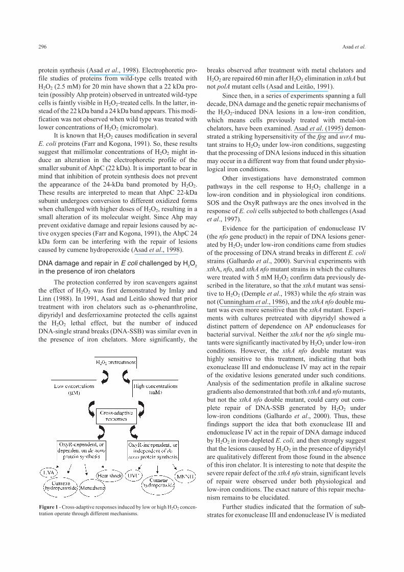
Figure 1 - Cross-adaptive responses induced by low or high H2O2 concen-
tration operate through different mechanisms.
by the Fpg DNA glycosylase, since the fpg mutation in-
creases cell survival and repair of DNA strand breaks in a
null AP endonuclease background (xthA nfo double mu-
tant). Recently, Speck et al. (2002) showed that the same
phenomenon is observed for nitric oxide treatment, that is,
in a null AP endonuclease background, fpg and ung muta-
tions are able to increase cell survival.
The role of copper ions in the lethality induced byH2O2 in low-iron conditions
Most of the work on the effects of H2O2 in living or-
ganisms has reported that DNA damage induced by H2O2
can be explained basically by the generation of hydroxyl
radicals through the iron-mediated Fenton reaction. Some
authors have attributed the same role to copper ions, also
present in biological systems (Sagripanti and Kraemer,
1989; Aruoma et al., 1991; Dizdaroglu et al., 1991). The
suggested mechanism for this effect takes into account the
formation of a DNA-Cu2+ complex in which this metal
would be reduced and then the DNA-Cu+ would react with
H2O2 to produce oxidative damage, via Fenton-type reac-
tions (Aruoma et al., 1991; Byrnes et al., 1992; Lloyd et al.,
1997; Lloyd and Phillips, 1999). Although copper ions
seemed not to participate in the genotoxicity of H2O2 in E.
coli under physiological iron conditions (Asad and Leitão,
1991), these ions have been shown to take part in the
genotoxicity of H2O2 under conditions of low iron avail-
ability. In fact, neocuproine (copper ion chelator) can in-
hibit cell inactivation and DNA strand breakage caused by
H2O2 in the presence of iron chelators (Almeida et al.,
2000). This phenomenon can only be detected in high con-
centrations of H2O2 (15 mM), suggesting that these ions
only interact with H2O2 in the intracellular environment in
the absence of iron and under severe oxidative stress.
Copper-induced DNA damage has been studied in
several systems. It has been demonstrated that such damage
is targeted preferentially to adjacent polyguanosines
(Aruoma et al., 1991). Analysis of the oxidative base le-
sions generated by copper-mediated Fenton reactions
showed that purine residues are the preferred targets of the
DNA-damaging species (Sagripanti and Kraemer 1989;
Dizdaroglu et al., 1991; Lloyd and Phillips, 1999], with
8-oxoG being the most abundant lesion formed. Frelon et
al. (2003) have shown that only 8-oxoG is formed upon in-
cubation of DNA with Cu(II) ions and H2O2 and suggested
that in vitro the Fenton reaction triggered by copper ions
generates singlet oxygen as the predominant reactive spe-
cies, with hydroxyl radical being produced predominantly
when the Fenton reaction is triggered by iron ions. In fact,
8-oxoG is the only lesion excised from H2O2/Cu2+-treated
DNA at detectable levels by the yOgg1 glycosylase from
Saccharomyces cerevisiae (Karahalil et al., 1998). The
finding of a remarkable increase in mutagenesis in uvrA
fpg-strain cultures pretreated with dipyridyl, and its inhibi-
tion by neocuproine (Almeida et al., 2000), strongly sug-
gest that 8-oxoG is formed in large amounts by such treat-
ment. The data suggesting a significant role for Fpg and
UvrA proteins in the repair of DNA lesions correlate well
with the hypothesis of copper participation, since it was
demonstrated that these proteins are important in the repair
of lesions induced by singlet oxygen, another well-known
guanine-damaging agent. Taken together these results may
indicate the production of singlet oxygen as the predomi-
nant ROS when E. coli cultures are challenged with H2O2
under low-iron conditions.
On the other hand some interesting results were ob-
tained from mutagenesis assays performed by our group
(manuscript in preparation) regarding the nature of lesions
produced by H2O2 in cells previously treated with dipy-
ridyl. Using an assay based on lac- reversion through a sin-
gle base change in mutated lacZ codon (Cupples and
Miller, 1989), we found that concentrations of H2O2 above
15 mM induce almost exclusively A:T→ T:A
transversions. However, in cells previously treated with
dipyridyl a massive and significant presence of G:C→ A:T
transitions is detected, a clearly different profile of induced
mutations. Considering that this transversion is not re-
ported to be induced by 8-oxoG the authors have suggested
that H2O2 under low-iron conditions may generate the le-
sion 5-hydroxy-2’-deoxycytidine, a highly mutagenic
product of cytosine oxidation that also constitutes a sub-
strate for Fpg repair protein (Hatahet et al., 1994; Feig et
al., 1994).
A qualitative difference exists between the DNA le-
sions generated by H2O2 plus copper (in the presence of
iron chelators) and iron (physiologic iron condition), which
can be deduced by the different DNA repair requirements
for cell survival in these situations. The reason for this dif-
ference is not known, and there exists an as yet unsolved
question about the participation of hydroxyl radical in the
damage to DNA caused by copper plus H2O2 (Sagripanti
and Kraemer, 1989; Yamamoto and Kawanishi, 1989).
Free-radical scavengers partially inhibit E. coli inactivation
by H2O2 (Imlay et al., 1988), and we observed that thiourea
protects the cells against the lethality produced by H2O2
both in the presence of dipyridyl (our unpublished results)
and in physiological iron conditions (Asad and Leitão,
1991).
Synergistic lethal interactions
In the last three decades, many studies have been pub-
lished regarding synergistic lethal effects between H2O2
and different physical and chemical agents. Some of them
will be described in this section.
Lethal synergism between UV (254 nm) and H2O2
Synergistic lethal interaction between UV (254 nm)
and X-ray irradiation, a well known system of generating
H2O2 and DNA strand breaks was described three decades
Hydrogen peroxide action that damage the E. coli genome 297
ago in E. coli (Haynes, 1966; Martignoni and Smith, 1973).
However, synergistic lethal interaction was not observed
between UV (254 nm) and H2O2 (Hartman and Eisenstark,
1980). On the other hand, synergistic killing of E. coli and
S. thyphimurium by near-UV (300-400 nm) radiation and
H2O2 has been described (Hartman and Eisenstark, 1978;
Hartman and Eisenstark, 1980; Kramer and Ames, 1987).
An analysis of the UV and X-ray effects (Martignoni
and Smith, 1973), the results of repair of H2O2 lesions in
several mutant strains (Ananthaswany and Eisenstark,
1977), and our own results for UV and reductone (an
H2O2-generating keto-aldehyde) (Leitão et al., 1981a; Lei-
tão et al., 1981b) suggested that a synergistic lethal interac-
tion between UV and H2O2 (254 nm) should be observed.
Indeed, Leitão and Carvalho (1988) observed that prior UV
(254 nm) irradiation strongly increased the sensitivity to
H2O2 of wild-type E. coli cells, and this synergistic lethal
interaction was also observed to a reduced extent in a polA
mutant, suggesting that UV lesions are potentiated by the
additional damage produced by H2O2, a result similar to
those observed by Martignoni and Smith (1973) for UV and
X-rays.
The detection of DNA double-strand breaks by DNA
sedimentation on neutral sucrose gradients (Leitão and
Carvalho, 1988) clearly indicates a mechanism responsible
for the synergistic lethal interaction observed. Although
H2O2 can produce DNA strand breaks with low efficiency
(Demple and Linn, 1982), the repair of UV lesions by the
action of the uvr gene products generated single-stranded
DNA regions and it was supposed that, in the presence of
H2O2, DNA double-strand breaks might arise in these re-
gions, produced by the action of exonuclease III (the xthA
gene product). This hypothesis was confirmed by the ab-
sence of synergistic lethal interaction between UV (254
nm) and H2O2 in xthA as well as in uvrA mutant strains
(Leitão and Carvalho, 1988). It seems that the same kind of
mechanism may be operating in the synergisms observed
with near-UV and H2O2, UV and reductone and UV and
X-rays.
Lethal synergism between phenanthrolines and H2O2
As described earlier, prior treatment with iron chela-
tors such as desferrioxamine, dipyridyl or o-phenanthroline
(1,10-phenanthroline) protects E. coli cells against the le-
thal effects of H2O2. However, Asad et al. (1994) detected a
strong lethal interaction when xthA mutant cells were
treated simultaneously with H2O2 and o-phenanthroline. In
the same way Almeida et al. (1999) have also detected a le-
thal synergistic interaction between neocuproine (2,9-di-
methyl 1,10-phenanthroline) and H2O2. In both cases the
phenomenon of synergism was accompanied by an increase
in the number of DNA strand breaks.
In the case of the synergism described by Asad et al.
(1994), it was argued that the formation and dissociation
equilibrium of Fe2+-Phe complex, which follows equations
IV, V and VI (Lee et al., 1948) shown below, would explain
the observed phenomenon.
Fe2+ + Phe → FePhe2+ rapidly established equi-
librium
(eq. IV)
FePhe2+ + Phe → FePhe22+ rapidly established equi-
librium
(eq. V)
FePhe22+ + Phe → FePhe3
2+ rate-determining step (eq. VI)
Since the equilibrium of the bis and mono complexes
of Fe2+-Phe are rapidly established and since these bis and
mono complexes react quickly with H2O2 (Burgers and
Prince, 1965), there would be H2O2-mono and H2O2-bis
complex formation and these complexes may be extremely
lethal to the cells, therefore justifying the lethal interaction
observed. In this case, the ability of o-phenanthroline to
penetrate into the DNA duplex, acting as a shuttle for the
Fe2+ ions would increase the efficiency of OH generation
close to DNA (Furtado et al., 1997). Recently Furtado
(2002) showed that this interaction also occurs in vitro. By
298 Asad et al.
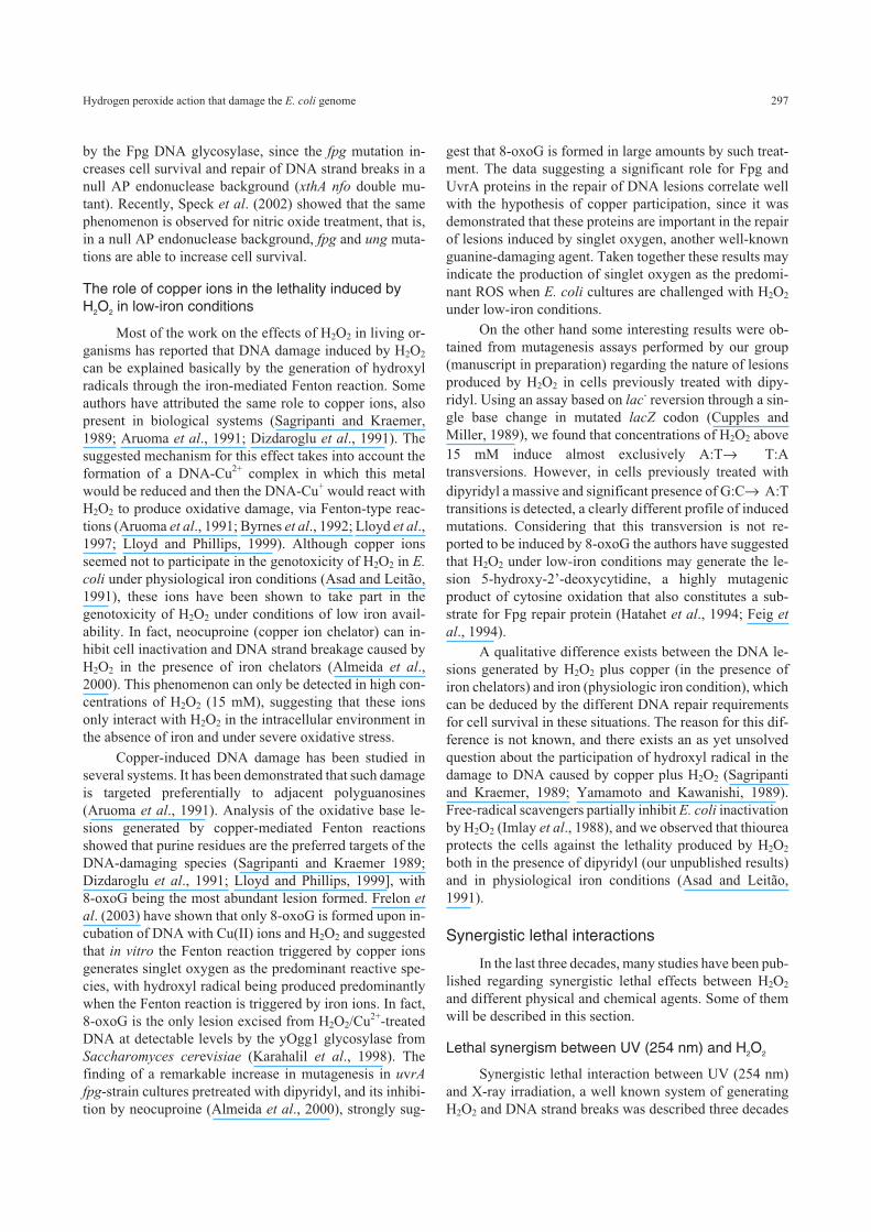
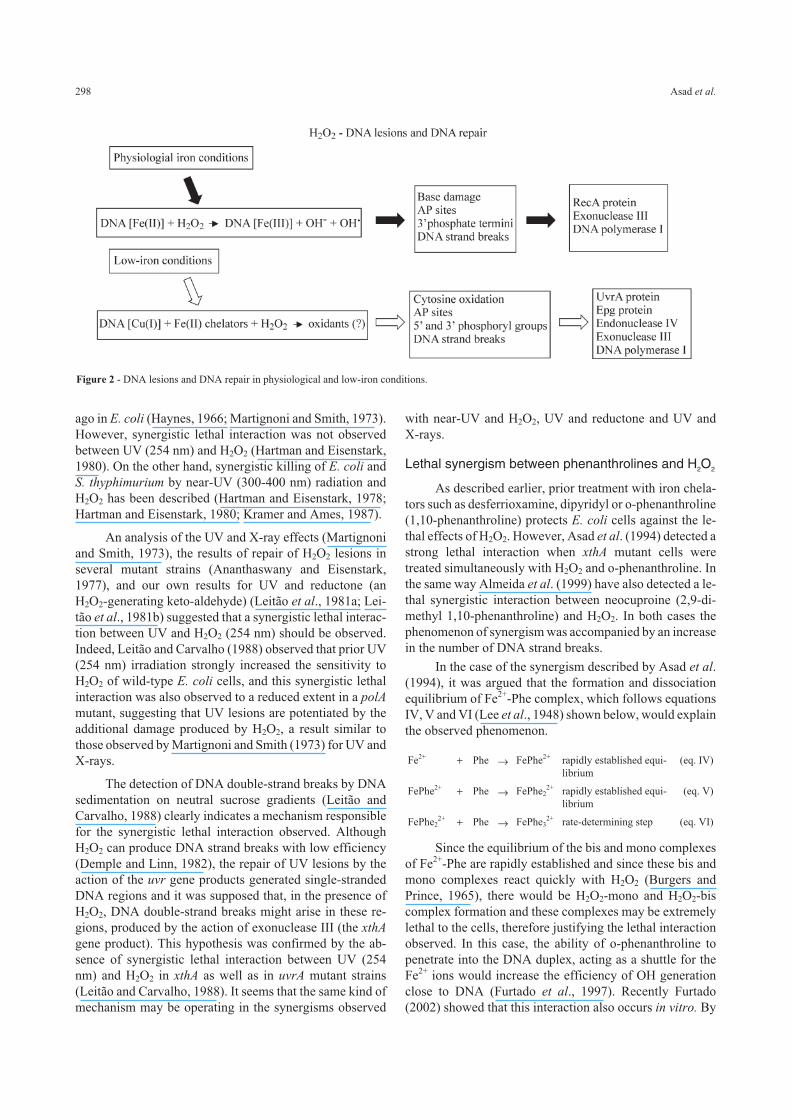
Figure 2 - DNA lesions and DNA repair in physiological and low-iron conditions.
using plasmid DNA, he showed that the high number of
breaks, as measured by the transformation of supercoiled to
relaxed form, is obtained when iron and o-phenanthroline
are added to the reaction medium immediately before
H2O2.
As mentioned above, Almeida et al. (1999) showed
that prior incubation of fpg, uvrA and lexA mutant strains
with neocuproine led to an increased sensitivity to H2O2.
The chemistry of this synergistic lethal interaction was sug-
gested to occur via the iron-mediated Fenton reaction,
which would be responsible for displacement of copper
ions from the complex Neo2Cu+, as Florence et al., (1985)
have reported to occur in vitro. In fact, neocuproine, as well
as other phenanthroline derivatives, have the ability to pen-
etrate into DNA, guiding metal ions to this site, which can
contribute to radical formation in the vicinity of this mole-
cule (Furtado et al., 1997). Indeed, it is possible that
neocuproine can guide copper ions to the DNA molecule,
thereby promoting the occurrence of radical generation at
this site as H2O2 reacts with these ions.
Conclusions
The most remarkable outcome from the genetic stud-
ies on H2O2-mediated genotoxicity is the striking sensitiv-
ity of xthA mutant E. coli cells to H2O2. In fact, this
observation provides some insights into the nature of
H2O2-induced lesions. Nevertheless, it is important to real-
ize that the oxidative stress produced by H2O2 results in the
induction of a diverse set of physiological responses, which
include some paradoxical effects. In this context, the most
unexpected phenomenon investigated so far is the bimodal
pattern of inactivation of E. coli by H2O2 described by
Imlay and Linn (1986). In the same way, it is now evident
that although pre-treatment of E. coli xthA cultures with
iron chelators confers protection against the lethal effects
of H2O2, DNA lesions can still be formed under these con-
ditions. We have to keep in mind that in contrast with what
is observed in the repair of damage induced by H2O2 under
physiological iron conditions, after treatment with H2O2 in
low-iron conditions: (i) the lesions observed may be re-
paired in xthA mutant cells (ii) both endonuclease IV and
exonuclease III as well as Fpg and UvrA proteins partici-
pate in the repair of these lesions. Such a difference in the
repair suggests a qualitative difference in the formation of
DNA lesions that is independent of the presence of iron.
Iron chelators partially inhibit E. coli inactivation by H2O2.
However, the precise mechanism for the genotoxic effect of
H2O2 under low-iron conditions remains to be elucidated. It
is also curious to see that some metal-ion chelators such as
o-phenanthroline and neocuproine, which should generally
inhibit the lethal effect of H2O2 can sometimes enhance cell
inactivation. Moreover, the data available so far suggest
that H2O2 (2.5 mM) can induce protection against UV light,
MNNG and cumene hydroperoxide independently of the
adaptive response, indicating that H2O2 can produce cross-
protection responses through various mechanisms that
might not involve the induction of de novo protein synthe-
sis.
Acknowledgements
We thank C.F.M. Menck, D.P. Carvalho and M. So-
renson for critical reading of this manuscript. Research sup-
ported by CNPq, CAPES, FUJB, FAPERJ and PRONEX.
References
Agnez-Lima LF, Di Mascio P, Demple B and Menck CFM (2001)
Singlet molecular oxygen triggers the soxRS regulon of
Escherichia coli. Biol Chem 382:1071-1075.
Almeida CEB, Felício DL, Galhardo RS, Cabral-Neto JB and
Leitão AC (1999) Synergistic lethal effect between hydro-
gen peroxide and neocuproine (2,9-dimethyl 1,10-phenan-
throline) in Escherichia coli. Mutat Res 433:59-66.
Almeida CEB, Galhardo RS, Felício DL, Cabral-Neto JB and
Leitão AC (2000) Copper ions mediate the lethality induced
by hydrogen peroxide in low iron conditions in Escherichia
coli. Mutat Res 460:61-67.
Altuvia S, Almiron M, Huisman G, Kolter R and Storz G (1994)
The dps promoter is activated by OxyR during growth and
by IHF and σs in stationary phase. Mol Microbiol 13:265-
272.
Altuvia S, Weinstein-Fischer D, Zhang A, Postow L and Storz G
(1997) A small, stable RNA induced by oxidative stress: role
as a pleiotropic regulator and antimutator. Cell 90:43-53.
Amabile-Cuevas CF and Demple B (1991) Molecular character-
ization of the soxRS genes of Escherichia coli: two genes
control a superoxide stress regulon. Nucleic Acids Res
19:4479-4484.
Ames BN, Shigenaga MK and Hagen TM (1993) Oxidants, anti-
oxidants and the degenerative diseases of aging. Proc Natl
Acad Sci USA 90:7915-7922.
Ananthaswamy HN and Eisenstark A (1977) Repair of hydrogen
peroxide-induced single-strand breaks in E. coli deoxyribo-
nucleic acid. J Bacteriol 130:187-191.
Argaman L and Altuvia S (2000) fhlA repression by OxyS RNA:
kissing complex formation at two sites results in a stable
antisense-target RNA complex. J Mol Biol 300:1101-1112.
Aruoma OI, Halliwell B, Gajewski E and Dizdaroglu M (1991)
Copper-ion-dependent damage to the bases in DNA in the
presence of hydrogen peroxide. Biochem J 273:601-604.
Asad LMBO, Almeida CEB, Silva AB, Asad NR and Leitão AC
(1994) Hydrogen peroxide induces the repair of UV-
damaged DNA in Escherichia coli: A lexA-independent but
uvrA- and recA-dependent mechanism. Curr Microbiol
29:291-294.
Asad LMBO, Asad NR, Silva AB, Almeida CEB and Leitão AC
(1997) Role of SOS and OxyR systems in the repair of Esch-
erichia coli submitted to hydrogen peroxide under low iron
conditions. Biochimie 79:359-364.
Asad LMBO, Asad NR, Silva AB, Felzenszwalb I and Leitão AC
(1997) Hydrogen peroxide induces protection against N-
methyl-N’-nitro-N-nitrosoguanidine (MNNG) effects in
Escherichia coli. Mutat Res 383:137-142.
Hydrogen peroxide action that damage the E. coli genome 299
Asad LMBO, De Carvalho AA, Felzenszwalb I, Leitão AC and
Asad NR (2000) H2O2-induced cross-protection against
UV-C killing in Escherichia coli is blocked in a lexA (Def)
background. J Phochem Photobiol (B) 54:67-71.
Asad NR, Almeida CEB, Asad LMBO, Felzenswalb I and Leitão
AC (1995) Fpg and UvrA proteins participate in the repair of
DNA lesions induced by hydrogen peroxide in low iron
level in Escherichia coli. Biochimie 77:262-264.
Asad NR, Asad LMBO, Almeida CEB and Leitão AC (1994) Le-
thal interaction between hydrogen peroxide and o-phenan-
throline in Escherichia coli. Braz J Med Biol Res
27:2551-2555.
Asad NR, Asad LMBO, Silva AB, Felzenszwalb I and Leitão AC
(1998) Hydrogen peroxide induces protection against lethal
effects of cumene hydroperoxide in Escherichia coli cells:
An Ahp independent but OxyR dependent system? Mutat
Res 407:253-259.
Asad NR and Leitão AC (1991) Effects of metal ion chelators on
DNA strand breaks and inactivation produced by hydrogen
peroxide in Escherichia coli: Detection of iron-independent
lesions. J Bacteriol 173:2562-2568.
Aslund F, Zheng M, Beckwith J and Storz G (1999) Regulation of
OxyR transcription factor by hydrogen peroxide and the cel-
lular thiol-disulfide status. Proc Natl Acad Sci USA
96:6161-6165.
Bjelland S and Seeberg E (2003) Mutagenicity, toxicity and repair
of DNA base damage induced by oxidation. Mutat Res
531:37-80.
Blaisdell JA, Hatahet Z and Wallace SS (1999) A novel role for
Escherichia coli endonuclease VIII in prevention of sponta-
neous G T transversions. J Bacteriol 181:6396-6402.
Boiteux S and Huisman O (1989) Isolation of a formamidopy-
rimidine-DNA glycosylase (fpg) mutant of Escherichia coli
K-12. Mol Gen Genet 215:300-305.
Brakely WF, Fuciarelli AF, Wegher BJ and Dizdaroglu M (1990)
Hydrogen peroxide-induced base damage in deoxyribonu-
cleic acid. Radiat Res 121:338-343.
Brandi G, Cattabeni F, Albano A and Cantoni C (1989) Role of
hydroxyl radicals in Escherichia coli killing induced by hy-
drogen peroxide. Free Radic Res Commun 6:47-55.
Braun V (1997) Avoidance of iron toxicity through regulation of
bacterial iron transport. Biol Chem 378:779-786.
Braun V, Hantke K and Koster W (1998) Bacterial ion transport:
mechanisms, genetics and regulation. In Sigel A and Sigel H
(eds) Metal Ions in Biological Systems. Marcel Dekker,
New York, NY, pp 67-145.
Burgers J and Prince RH (1965) Kinetics of reactions of ligand-
substitued tris-(2-2’-dipyridyl) iron (II) complexes. J Chem
Soc 1132:6061-6066.
Byrnes RW, Antholine WE and Petering DH (1992) Oxidation-
reduction reactions in Ehrlich cells treated with copper-
neocuproine. Free Rad Biol Med 13:469-478.
Cadet J, Douki T, Gasparuto D and Ravanat J-L (2003) Oxidative
damage to DNA: Formation, measurement and biochemical
features. Mutat Res 531:5-23.
Calsou P, VillaVerde A and Defais M (1987) Activated RecA pro-
tein may induced expression of gene that is not controlled by
the LexA repressor and whose function is record for muta-
genesis and repair of UV-irradiated bacteriophage lambda. J
Bacteriol 169:4816-4821.
Chan E and Weiss B (1987) Endonuclease IV of Escherichia coli
is induced by paraquat. Proc Natl Acad Sci USA 84:3189-
3193.
Chaudhry MA, Dedon PC, Wilson DM III, Demple B and Wein-
feld M (1999) Removal by human apurinic/apirimidinic
endonuclease 1 (Ape1) and Escherichia coli exonuclease III
of 3’-phosphoglycolates from DNA treated with neocar-
zinostatin, calicheamicin and gamma-radiation. Biochem
Pharmacol 57:531-538.
Christman MF, Morgan RW, Jacobson FS and Ames BN (1985)
Positive control of a regulon for defenses against oxidative
stress and some heat-shock proteins in Salmonella
typhimurium. Cell 41:753-762.
Cooke MS, Evans MD, Dizdaroglu M and Lunec J (2003) Oxida-
tive DNA damage: Mechanisms, mutation, and disease.
FASEB J 17:1195-1214.
Courcelle J, Khoudursky A, Peter B, Brown PO and Hanawalt PC
(2001) Comparative gene expression profiles following UV
exposure in wild-type and SOS-deficient Escherichia coli.
Genetics 158:41-64.
Cunningham RP, Saporito SM, Spitzer SG and Weiss B (1986)
Endonuclease IV (nfo) mutant of Escherichia coli. J Bac-
teriol 168:1120-1127.
Cunningham RP and Weiss B (1985) Endonuclease III (nth) mu-
tants of Escherichia coli. Proc Natl Acad Sci USA 82:474-
478.
Cupples C and Miller, J (1989) A set of lacZ mutations in Esche-
richia coli which allow rapid detection of each of the six
base substitutions. Proc Natl Acad Sci USA 86:5345-5349.
Demple B (1991) Regulation of bacterial oxidative stress genes.
Annu Rev Genet 25:315-337.
Demple B and Halbrook J (1983) Inducible repair of oxidative
DNA damage in Escherichia coli. Nature 304:466-468.
Demple B, Halbrook J and Linn S (1983) Escherichia coli xth mu-
tants are hypersensitive to hydrogen peroxide. J Bacteriol
153:1079-1082.
Demple B and Harrison L (1994) Repair of oxidative DNA dam-
age to DNA: Enzymology and biology. Annu Rev Biochem
63:915-948.
Demple B and Linn S (1982) 5,6-saturated thymine lesions in
DNA: Production by ultraviolet light and hydrogen perox-
ide. Nucleic Acid Res 10:3781-3789.
Dizdaroglu M (2003) Substract specificities and excision kinetics
of DNA glycosylases involved in base-excision repair of ox-
idative DNA damage. Mutat Res 531:109-126.
Dizdaroglu M, Rao G, Halliwell B and Gajewski E (1991) Dam-
age to the DNA bases in mammalian chromatin by hydrogen
peroxide in the presence of ferric and cupric ions, Arch
Biochem Biophys 285:317-324.
Farr SB and Kogoma T (1991) Oxidative stress responses in Esch-
erichia coli and Salmonella typhimurium. Microbiol Rev
55:561-585.
Feig DI, Sowers LC and Loeb LA (1994) Reverse chemical muta-
genesis: Identification of the mutagenic lesions resulting
from reactive oxygen species-mediated damage to DNA.
Proc Natl Acad Sci USA 91:6609-6613.
Fernandez De Henestrosa AR, Ogi T, Aoyagi S, Chafin D, Hayes
JJ, Ohmori H and Woodgate R (2000) Identification of addi-
tional genes belonging to the lexA regulon in Escherichia
coli. Mol Microbiol 35:1560-1572.
300 Asad et al.
Florence TM, Stauber JL and Mann KJ (1985) The reaction of
copper-2,9-dimethyl-1,10-phenanthroline with hydrogen
peroxide. J Inorg Biochem 23:243-254.
Frelon S, Douki T, Favier A and Cadet J (2003) Hydroxyl radical
is not the main reactive species involved in the degradation
of DNA bases by copper in the presence of hydrogen perox-
ide, Chem Res Toxicol 16:191-197.
Friedberg EC, Walker GC and Siede W (1995) DNA Repair and
Mutagenesis. Academic Press, New York, 698 pp.
Furtado FAC (2002) Caracterização adicional do fenômeno de
interação letal entre 1,10-fenantrolina e peróxido de hidro-
gênio em Escherichia coli. PhD Thesis, Universidade Fede-
ral do Rio de Janeiro, Rio de Janeiro.
Furtado FAC, Asad NR and Leitão AC (1997) Effects of 1,10-
phenanthroline and hydrogen peroxide in Escherichia coli:
Lethal interaction. Mutat Res 385:251-258.
Galhardo RS, Almeida CEB, Leitão AC and Cabral-Neto JB
(2000) Repair of DNA lesions induced by hydrogen perox-
ide in the presence of iron chelators in Escherichia coli: Par-
ticipation of endonuclease IV and Fpg. J Bacteriol
182:1964-1968.
Gaudu P, Moon N and Weiss B (1997) Regulation of the soxRS
oxidative stress regulon. Reversible oxidation of the Fe-S
centers of SoxR in vivo. J Biol Chem 272:5082-5086.
Gaudu P and Weiss B (1987) SoxR a [2Fe-2S] transcription fac-
tor, is active only in its oxidized form. Proc Natl Acad Sci
USA 84:3189-3193.
Geenberg JT and Demple B (1989) A global response induced in
Escherichia coli by redox-cycling agents overlaps with that
induced by peroxide stress. J Bacteriol 171:3922-3939.
Goerlich O, Quillardet P and Hofnung M (1989) Induction of the
SOS response by hydrogen peroxide in various Escherichia
coli mutants with altered protection against oxidative DNA
damage. J Bacteriol 171:6141-6147.
Gonzalez-Flecha B and Demple B (2000) Genetic responses to
free radicals. Homeostasis and gene control. Ann New York
Acad Sci 899:69-87.
Hagensee ME and Moses RE (1989) Multiple pathways for repair
of hydrogen peroxide-induced DNA damage in Escherichia
coli. J Bacteriol 171:991-995.
Halliwell B and Gutteridge JMC (1984) Oxygen toxicity, oxygen
radicals, transition metals and disease. J Biochem 219:1-14.
Halliwell B and Gutteridge JMC (1999) Free Radicals In Biology
and Medicine. Oxford University Press, New York, 936 pp.
Hanawalt PC (2001) Controling the efficiency of excision repair.
Mutat Res 485:3-13.
Hantke K (1981) Regulations of ferric ion transport in Esche-
richia coli K12: Isolation of a constitutive mutant. Mol Gen
Genet 182:288-292.
Hantke K (2002) Members of the Fur protein family regulate iron
and zinc transport in E. coli and characteristics of the Fur-
regulated fhuF protein. J Mol Microbiol Biotechnol
4:217-222.
Hartman PS and Eisenstark A (1978) Synergistic killing of Esche-
richia coli by near-UV radiation and hydrogen peroxide.
Distinction between RecA-repairable and RecA-non-
repairable damage. J Bacteriol 133:769-774.
Hartman PS and Eisenstark A (1980) Killing of Escherichia coli
K-12 by near-ultraviolet radiation in the presence of hydro-
gen peroxide: Role of double-strand breaks in absence of
recombinational repair. Mutat Res 72:31-42.
Hatahet Z, Kow YW, Purmal AA, Cunninghan RP and Wallace
SS (1994) New substrates for old enzymes. 5-hydroxy-
2’-deoxicytidine and 5-hydroxy-2’-deoxyuridine are sub-
strates for Escherichia coli endonuclease III and formami-
dopyrimidine DNA N-glycosylase, while 5-hydroxy-2’-
deoxyuridine is a substrate for uracil DNA N-glycosylase. J
Biol Chem 269:18814-18820.
Haynes RH (1966) The interpretation of microbial inactivation
and recovery phenomena. Radiat Res Suppl 6:1-29.
Henle ES and Linn S (1997) Formation, prevention and repair of
DNA damage by iron/hydrogen peroxide. J Biol Chem
272:19095-19098.
Hidalgo E, Bollinger Jr JM, Bradley TM, Walsh CT and Demple
B (1995) Binuclear [2Fe-2S] clusters in the Escherichia coli
SoxR protein and role of the metal centers in transcription. J
Biol Chem 270:20908-20914.
Hidalgo E, Ding H and Demple B (1997) Redox signal transduc-
tion mutations shifting [2Fe-2S] centers of SoxR sensor-
regulated to the oxidized form. Cell 88:121-129.
Horsfall MJ, Gordon JE, Burns PA, Zielenska M, Van der Viet
GME and Glickman BW (1990) Mutational specificity of
alkylating agents and the influence of DNA repair. Environ
Mol Mut 15:107-122.
Imlay JA, Chin SM and Linn S (1988) Toxic DNA damage by hy-
drogen peroxide through the Fenton reaction in vivo and in
vitro. Science 240:640-642.
Imlay JA and Linn S (1986) Bimodal pattern of killing of DNA-
repair-defective or anoxically grown Escherichia coli by hy-
drogen peroxide. J Bacteriol 166:519-527.
Imlay JA and Linn S (1987) Mutagenesis and stress responses in-
duced in Escherichia coli by hydrogen peroxide. J Bateriol
169:2967-2976.
Imlay JA and Linn S (1988) DNA damage and oxygen radical tox-
icity. Science 240:1302-1309.
Izumi T, Hazra TK, Boldogh I, Tomkinson AE, Park MS, Ikeda S
and Mitra S (2000) Requeriment for human AP endo-
nuclease 1 for repair of 3’-blocking damage at DNA single-
strand breaks induced by reactive oxygen species.
Carcinogenesis 21:1329-1334.
Jenkins DE, Schultz JE and Martin A (1988) Starvation-induced
cross protection against heat or H2O2 challenge in Esche-
richia coli. J Bacteriol 170:3910-3914.
Jiang D, Hatahet Z, Blaisdell JO, Melamede RJ and Wallace SS
(1997) Escherichia coli endonuclease VIII: Cloning, se-
quencing, and overexpression of the nei structural gene and
characterization of nei and nei nth mutants. J Bacteriol
179:3773-3782.
Karahalil B, Girard PM, Boiteux S and Dizdaroglu M (1998) Sub-
strate specificity of the Ogg1 protein of Saccharomyces
cerevisiae: excision of guanine lesions produced in DNA by
ionizing radiation-or hydrogen peroxide/metal ion-
generated free radicals. Nucleic Acids Res 26:1228-1232.
Kehrer JP (2000) The Haber-Weiss reaction and mechanisms of
toxicity. Toxicology 149:43-50.
Konola JT, Sargent KE and Gow JB (2000) Efficient repair of hy-
drogen peroxide-induced DNA damage by Escherichia coli
requires SOS induction of RecA and RuvA proteins. Mutat
Res 459:187-194.
Kow YW, Wallace SS and Van Houten B (1990) UvrABC
nuclease complex repairs thymine glycol, an oxidative DNA
base damage. Mutat Res 235:147-156.
Hydrogen peroxide action that damage the E. coli genome 301
Kramer GF and Ames BN (1987) Oxidative mechanisms of toxic-
ity of low-intensity near-UV light in Salmonella
typhimurium. J Bacteriol 169:2259-2266.
Kruszewski M (2003) Labile iron pool: the main determinant of
cellular responses to oxidative stress. Mutat Res 531:81-92.
Lee TS, Kolthoff IM and Leusing DL (1948) Reaction of ferrous
and ferric ions with 1,10-phenanthroline. II. Kinetics of for-
mation and dissociation of ferrous phenanthroline. J Am
Chem Soc 70:3596-3600.
Leitão AC, Caldas LR and Alcantara Gomes R (1981a) Inhibition
of DNA repair and production of single-strand breaks in
Escherichia coli by reductone. Photochem Photobiol
34:49-53.
Leitão AC and Carvalho RES (1988) Synergistic killing of Esche-
richia coli K-12 by UV (254nm) and H2O2. Int J Radiat Biol
53:477-488.
Leitão AC, Motta HC and Alcantara Gomes R (1981b) Kinetics of
DNA-induced breaks by reductone treatment: in vitro and in
vivo studies. Photochem Photobiol 34:745-748.
Lesca C, Petit C and Defais M (1991) UV induction of lexA inde-
pendent proteins which could be involved in SOS repair.
Biochimie 73:407-409.
Lin JJ and Sancar A (1989) A new mechanism for repairing oxida-
tive damage to DNA: (A)BC exonuclease removes AP sites
and thymine glycols from DNA. Biochemistry 28:7979-
7984.
Lloyd DR and Phillips DH (1999) Oxidative DNA damage medi-
ated by copper(II), iron(II) and nickel(II) Fenton reactions:
evidence for site-specific mechanisms in the formation of
double-strand breaks, 8-hydroxydeoxyguanosine and puta-
tive intrastrand cross-links. Mutat Res 424:23-36.
Lloyd DR, Phillips DH and Carmichael PL (1997) Generation of
putative intrastrand cross-links and strand breaks in DNA by
transition metal ion-mediated oxygen radical attack. Chem
Res. Toxicol 10:393-400.
Manchado M, Michán C and Pueyo C (2000) Hydrogen peroxide
activates the SosRS regulon in vivo. J Bacteriol 182:6842-
6844.
Martignoni KD and Smith KC (1973) The synergistic action of ul-
traviolet and X-radiation on mutants of Escherichia coli
K-12. Photochem Photobiol 18:1-8.
Martinez A and Kolter R (1997) Protection of DNA during oxida-
tive stress by the nonspecific DNA-binding protein Dps. J
Bacteriol 179:5188-5194.
Melamede RJ, Hatahet Z, Kow IW, Ide H and Wallace SS (1994)
Isolation and characterization of endonuclease VIII from
Escherichia coli. Biochemistry 33:1255-1264.
Mello-Filho AEC and Meneghini R (1985) Protection of mamma-
lian cells by o-phenanthroline from lethal and DNA-
damaging effects produced by active oxygen species.
Biochim Biophys Acta 847:82-89.
Meneghini R (1987) A toxicidade do oxigênio. Ci Hoje 5:57-62.
Meneghini R (1988) Genotoxicity of active oxygen species in
mammalian cells. Mutat Res 195:215-230.
Miles GM and Sancar A (1989) DNA repair. Chem Res Toxicol
2:197-226.
Mol CD, Hosfield DJ and Tainer JA (2000) Abasic site recogni-
tion by two apurinic/apyrimidinic endonucleases families in
DNA base excision repair: the 3’ ends justify the means.
Mutat Res 460:211-229.
Morgan RW, Christman MF, Jacobson FS, Storz G and Ames BN
(1986) Hydrogen peroxide-inducible proteins in Salmonella
typhimurium overlap with heat shock and other stress pro-
teins. Proc Natl Acad Sci USA 83:8059-8063.
Mukhopadhyay S and Schellhorn HE (1997) Identification and
characterization of hydrogen peroxide-sensitive mutants of
Escherichia coli: Genes that require OxyR for expression. J
Bacteriol 179:330-338.
Mustard JA and Little JW (2000) Analysis of Escherichia coli
RecA interactions with LexA, lambda CI, and UmuD by
site-directed mutagenesis of recA. J Bacteriol 182:1659-
1670.
Nunoshiba T, Hashimoto M and Nishioka H (1991) Cross-
adaptive response in Escherichia coli caused by pretreat-
ment with H2O2 against formaldehyde and other aldehyde
compounds. Mutat Res 255:265-271.
Petit C, Cayrol C, Lesca C, Kaiser P, Thompson C and Defais M
(1993) Characterization of dinY, a new Escherichia coli
DNA repair gene whose products are damage inducible even
in a lexA (Def.) background. J Bacteriol 175:642-646.
Piersen CE, McCullough AK and Lloyd RS (2000) AP lyases and
dRPases: commonality of mechanism. Mutat Res 459:43-
53.
Pomposiello PJ and Demple B (2001) Redox-operated genetic
switches: the SoxR and OxyR transcription factors. Trends
Biotechnol 19:109-114.
Repine JE, Fox RB and Berger EM (1981) Hydrogen peroxide
kills Staphylococcus aureus by reacting with staphylococcal
iron to form hydroxyl radical. J Biol Chem 256:7094-7096.
Repine JE, Pfenniger OW, Talmage DW, Berger EM and
Pettijohn DE (1981) Dimethyl sulfoxide prevents DNA
nicking mediated by ionizing radiation or iron/hydrogen
peroxide-generated hydroxyl radical. Proc Natl Acad Sci
USA 78:1001-1003.
Sagripanti JL and Kraemer KH (1989) Site-specific oxidative
DNA damage at polyguanosines produced by copper plus
hydrogen peroxide. J Biol Chem 264:1729-1734.
Sancar A and Sancar GB (1988) DNA repair enzymes. Ann Rev
Biochem 57:29-67.
Saporito SM, Gedenk M and Cunningham RP (1989) Role of
exonuclease III and endonuclease IV in repair of pyrimidine
dimers initiated by bacteriophage T4 pyrimidine dimmer-
DNA glycosylase. J Bacteriol 171:2542-2548.
Sayto Y, Uraki F, Nakajima S, Asaeda A, Ono K, Kubo K and
Yamamoto K (1997) Characterization of endonuclease III
(nth) and endonuclease VIII (nei) mutants of Escherichia
coli K-12. J Bacteriol 179:3783-3785.
Sedgwick B and Robins P (1980) Isolation of mutants of Esche-
richia coli with increased resistance to alkylating agents:
Mutants deficient in thiols and mutants constitutive for the
adaptive response. Molec Gen Genet 180:85-90.
Spek EJ, Vuong LN, Matsuguchi T, Marinus MJ and Engelward
BP (2002) Nitric oxide-induced homologous recombination
in Escherichia coli is promoted by DNA glycosylases. J
Bacteriol 184:3501-3507.
Storz G, Jacobson FS, Tartaglia LA, Morgan RW, Silveira LA and
Ames BN (1989) An Alkyl hydroperoxide reductase in-
duced by oxidative stress in Salmonella typhimurium and
Escherichia coli: Genetic characterization and cloning of
ahp. J Bacteriol 171:2049-2055.
302 Asad et al.
Storz G and Imlay JA (1999) Oxidative stress. Curr Opin
Microbiol 2:188-194.
Storz G, TartagliaLA and Ames BN (1990) Transcriptional regu-
lator of oxidative stress-inducible genes: Direct activation
by oxidation. Science 248:189-194.
Storz G ands Toledano MB (1994) Regulation of bacterial gene
expression in response to oxidative stress. Methods
Enzymol 236:196-207.
Touati D, Jacques M, Tardat B, Bouchard L and Despied S (1995)
Lethal oxidative damage and mutagenesis are generated by
iron in fur mutants of Escherichia coli: Protective role of
superoxide dismutase. J Bacteriol 177:2305-2314.
Tsaneva IR and Weiss B (1990) soxR, a locus governing a
superoxide response regulon in Escherichia coli K-12. J
Bacteriol 172:4197-4205.
Tyrrell RM (1985) A common pathway for protection of bacteria
against damage by solar UV(A) (334 nm, 365 nm) and an
oxidizing agent (H2O2). Mutat Res 145:129-136.
Wagner J, Cruz P, Kim SR, Yamada M, Matsui K, Fuchs RP and
Nohmi T (1999) The dinB gene encodes a novel E. coli DNA
polymerase, DNA pol IV, involved in mutagenesis. Mol
Cell 4:281-286.
Walker GC (1996) The SOS responses of Escherichia coli. In:
Curtis III R, Ingraham JL, Lin ECC, Low KB, Magasanick
B, Reznicoff WS, Riley M, Schaechter M and Umbarger HE
(eds) Escherichia coli and Salmonella typhimurium: Cellu-
lar and Molecular Biology. v. 1, American Society for Mi-
crobiology, Washington DC, pp 1400-1416.
Wallace SS (1988) Ap endonucleases and DNA glycosylases that
recognize oxidative DNA damage. Environ Mol Mutagen
12:431-477.
Wallace SS (2002) Biological consequences of free radical-
damaged DNA bases. Free Rad Biol Med 33:1-14.
Wallace SS, Ide H, Kow YM, Laspia MF, Melamede RJ, Petrullo
LA and LeClerc E (1988) Processing of oxidative DNA
damage in Escherichia coli. In: Fridberg EC and Hanawalt
PC (eds) Mechanisms and Consequences of DNA Damage
Processing. Allan R. Liss, Inc., New York, pp 151-157.
Weigle JJ (1953) Induction of mutation in a bacterial virus. Proc
Natl Acad Sci USA 39:628-636.
West SC (1996) The RuvABC proteins and Holliday junctions
processing in Escherichia coli. J Bacteriol 178:1237-1241.
Wu J and Weiss B (1992) Two-stage induction of the soxRS
(superoxide response) regulon of Escherichia coli. J
Bacteriol 174:3915-3920.
Yamamoto K and Kawanishi S (1989) Hydroxyl radical is not the
main active species in site-specific DNA damage induced by
copper (II) ion and hydrogen peroxide. J Biol Chem
264:15435-15440.
Zhang A, Altuvia S and Storz G (1997) The novel oxyS RNA reg-
ulates expression of the sigma s subunit of Escherichia coli
RNA polymerase. Nucleic Acids Symp Ser 36:27-28.
Zheng M, Aslund F and Storz G (1998) Activation of OxyR tran-
scription factor by reversible disulfide bond formation. Sci-
ence 279:1718-1721.
Zheng M, Doan B, Schneider TD and Storz G (1999) OxyR and
SoxRS regulation of fur. J Bacteriol 181:4639-4643.
Zheng M, Wang X, Templeton LR, Smulski DR, Larossa RA and
Storz G (2001) DNA microarray-mediated transcriptional
profiling of the Escherichia coli response to hydrogen per-
oxide. J Bacteriol 183:4562-4570.
EditorAssociado: Carlos F.M. Menck
Hydrogen peroxide action that damage the E. coli genome 303
Copyright © 2022 FDOKUMEN